Embed Size (px)
Citation preview

Journal of Clinical InvestigationVol. 41, No. 11, 1962
IN VITRO INTERCONVERSIONOF 16-C14-ESTRONE AND 16-C14-ESTRADIOL-17/3BY ERYTHROCYTESFROMNORMALSUBJECTSAND FROM
SUBJECTSWITH A DEFICIENCY OF RED CELL GLU-COSE-6-PHOSPHATEDEHYDROGENASE
ACTIVITY *
By CLAUDEJ. MIGEON, ONOL. LESCURE, WILLIAM H. ZINKHAM, ANDJAMESB. SIDBURY, JR.
(From the Department of Pediatrics, Johns Hopkins University School of Medicine, and theHarriet Lane Home of the Johns Hopkins Hospital, Baltimore, Md.)
(Submitted for publication July 24, 1961; accepted July 26, 1962)
Werthessen and his collaborators (2-4) reportedthat incubation of estrone in blood produced thedestruction of the 17-ketone function of the steroid,accompanied by a loss of biological activity. Onthe other hand, Bischoff and associates (5, 6) in-cubated the red blood cells of rabbit or man withestrone and observed a marked increase of thebiological activity of the added steroid. The dis-crepancy in the two sets of data was ascribed atthe time to differences either in assay techniquesor in experimental conditions. A similar increasein biological activity was reported by Repke andMarkwardt (7), who used red cells of the guineapig, rabbit, and man. It now appears that spe-cies differences were the cause of this discrepancy.In most of these experiments, Werthessen andco-workers (3, 4) used bovine blood, and Axelrodand Werthessen (8) have found that in this spe-cies estrone is converted to the extent of 88 percent into estradiol-17a. The investigations ofBischoff and associates (5, 6) were carried outwith rabbit and human red cells, and these au-thors (9) have shown that the main reactionproduct resulting from estrone incubation in theirsystem was estradiol; the latter report, however,failed to mention whether the steroid detected wasthe a- or 8-isomer, but there are reasons to be-lieve that reaction product was estradiol-17/3.This isomer is biologically more active than estroneor estradiol-17a; hence the difference in the re-sults reported by Werthessen and co-workers(3, 4) and Bischoff and associates (5, 6). In a
* This work was made possible by grants-in-aid A-180(CIO), H-3995, and B-1612 from the National Instituteof Health, U. S. Public Health Service. The data werepresented at the First International Congress of Endo-crinology, Copenhagen, 1960 (1).
study of the stereospecificity of the activity of es-tradiol dehydrogenases in the erythrocytes of manand of various animals, Portius and Repke (10)showed that an estradiol-17,8 dehydrogenase waspresent in the red cells of the 15 species investi-gated, whereas an estradiol-17a dehydrogenasewas found only in ruminants such as cow, sheep,and goat. The same authors (10) reported thatthe activity of the fl-enzyme was the highest in therat and the lowest in man.
Bischoff and associates (6) have investigatedsome of the conditions influencing the "activation"of estrone, but the degree of transformation of thesteroid was determined by bio-assay. Repke andMarkwardt (7) have also studied this problemusing the Allen-Doisy test as an index of "activa-tion" of estrone, and have found that neither thesuccinic dehydrogenase systems nor the enzymesystems involved in red cell glycolysis were ableto transform estrone to estradiol-17,8. In anotherpaper, the same workers (11 ) reported that prep-arations of glucose-6-phosphate dehydrogenase ofeither guinea pig red cells or yeast (12) could inthe presence of glucose-6-phosphate and TPN re-duce estrone to estradiol-17,8. Bischoff, Torres,and Lopez (13) confirmed some of these findings.More recently, Portius and Repke (14) havestudied the properties of estradiol-17,3 dehydroge-nase of rat erythrocytes and have attempted topurify the enzyme.
It is the purpose of the present paper to reportthe results of our personal experience with theinterconversion of 16-C14-estrone and 16-C14-es-tradiol-17,8 by the red blood cells of normal sub-jects and individuals who have a hereditary al-teration of glucose-6-phosphate dehydrogenase ac-tivity in their erythrocytes (15).
2025

MIGEON, LESCURE, ZINKHAM, AND SIDBURY, JR.
METHODS
Purity and standardization of the radioactive steroids.The 16-C14-estrone and 16-C14-estradiol-17,8 were pur-
chased from Charles E. Frosst Company, Montreal,Canada. Their specific activities were 0.729 and 0.734mc per mM. Their purity was tested as previously de-scribed (16) and was found to be 96 to 98 per cent.
The steroids were dissolved in 100 per cent ethanol.This concentrated solution was diluted with 0.9 per centNaCl solution, the final mixture containing 5 per cent ofethanol.
Basic experimental systemn and its variations in ex-
periments with nonhemolvzed erythrocytes. Except forone experiment where the effect of defibrination, citra-tion, and decalcification was studied, all the blood sampleswere heparinized. The samples were centrifuged im-mediately at 2,000 rpm for 30 minutes. The plasma andbuffy coat were removed as completely as possible, andthe red cells were washed twice with three volumes of0.9 per cent NaCl solution.
The basic system for 16-C14-estrone as starting ma-
terial consisted of 0.5 ml of washed red blood cells, 0.5ml of 0.9 per cent NaCl solution, 0.1 ml of 5 per centglucose solution, and 0.2 ml of estrone solution (approxi-mately 4,000 cpm or 1.3 Asg of steroid). In the basic sys-
tem for 16-C'4-estradiol-17p, the glucose solution was re-
placed by 0.1 ml of 0.9 per cent NaCl solution.The mixture was incubated at 370C for 24 hours. On
several occasions, the incubation mixtures were checkedfor the presence of microorganisms, but the cultures were
negative. At the end of the incubation, the steroids were
extracted twice with 4 ml of ethanol and once with 4 mlof a 1: 1 (vol/vol) mixture of ethanol and butanol. Thepooled extracts were then evaporated to dryness and theresidue was mixed with 3 ml of distilled water. Theaqueous phase was extracted five times with 6 ml ofchloroform, and the combined chloroform was evaporatedto dryness. The residue was applied on Whatman paper
No. 2 and chromatographed as previously described(16). The paper chromatogram was radioautographed(16), the area corresponding to estrone and estradiol-17j,was eluted with ethanol, and the amount of radioactivitypresent in the eluates was determined.
When variations were made in the basic system, thefinal volume of 1.3 ml was maintained by an appropriatechange in the volume of saline solution added. The onlyexception was in the experiments on the effect of theamount of red cells used (with 2 and 3 ml of red cells,the final volumes were 2.3 and 3.3 ml, respectively). Inthe experiments on the effect of pH, 0.5 ml of buffer solu-tion replaced the 0.5 ml of saline solution. In other ex-
periments, the saline solution was replaced by 0.5 ml of1, 0.5, 0.25, and 0.12 per cent citric acid in saline solutionin order to bring the initial pH of the mixture to 5.2, 5.8, i6.3, and 6.6, respectively; 0.5 ml of 0.75, 0.5, and 0.25 per scent Na2CO3 in saline solution was used for obtaining an iinitial pH of 9.2, 8.7, and 7.9. In the inhibition experi- cments, methylene blue and sodium fluoride were dis- X
solved in saline solution, and 0.5 ml of these mixtures re-placed the regular saline solution.
Basic experimental system in experiments with he-molyzed erythrocytes. A volume of 0.5 ml of washed redcells was transferred by pipet into a test tube which wasplaced in a freezer at - 50'C for 30 minutes. Afterthawing, various solutions were added as indicated in eachcorresponding Table. Finally, the estrogen solution wasadded and the final volume was brought to 2.1 ml with0.9 per cent NaCl solution. The samples were thentreated as described for the intact red cells.
In experiments in which red cell ghosts were removed,this procedure was followed: After the freezing of aknown volume of red cells, saline solution was added(1.5 the volume of red cells) ; the mixture was centri-fuged at 28,000 X G, at 50C for 60 minutes; 1.25 ml ofthe aqueous solution was used for these experiments.
For the (NH4)2SO4 precipitation, the red cell ghostswere removed, and to 4 ml of the red cell hemolysate anequal volume of either 50 per cent, 75 per cent, or 100per cent (NH4)2SO was added, giving a final concentra-tion of 25, 37.5, and 50 per cent, respectively. The mix-ture was centrifuged at 28,000 X G at 50C for 60 minutes.Two ml of supernatant fluid (corresponding to 0.5 ml ofinitial red cells), 0.3 ml of 0.01 M TPN, and 0.3 ml of16-C14-estradiol-17,8 were incubated and then treated asdescribed above. Each of the precipitates was dissolvedin 2 ml of saline; 0.5 ml of this solution, 0.3 ml of 0.01M TPN, 0.3 ml of 16-C14-estradiol-17j,6 and 1.5 ml ofsaline were also incubated.
The DEAE (diethylaminoethyl cellulose) used for col-umn fractionation was washed with IN NaOH, 0.9 percent NaCl solution, 95 per cent ethanol, and then wasdried. Prior to use, 0.9 per cent NaCl solution was addedto DEAEand the pH adjusted to 7.0. A column of 1.8cm in diameter and 8.0 cm in length was prepared.Prior to applying the red cell hemolysate, the column waswashed with 50 ml of cold 0.005 MNaCl. Elution of thecolumn was then carried out by adding increasing con-centrations of NaCl solution and by collecting 10 ml
-fractions. A 4-ml sample of each of these fractions, 0.3ml of 0.01 M TPN, and 0.3 ml of 16-C14-estradiol-17fiwere incubated and then treated as described above.
Recovery. The radioactivity recovered after extrac-tion of the basic experimental system and prior to paperchromatography represented 94 to 103 per cent of theradioactivity originally added to the system. In all theexperiments, the sum of the activities eluted from theareas of the paper chromatograms corresponding to es-trone and estradiol-17,8 represented 85 to 92 per cent ofthe radioactivity added at the beginning of the experiment.
When the extract of more than 0.5 ml of red cells waschromatographed, the increased amount of nonsteroidalimpurities was found to produce a displacement of thesteroids on the paper chromatogram. For this reason,in experiments in which the amount of red cells was 1, 2,or 3 ml, extract samples corresponding to 0.5 ml of cellswere chromatographed separately.
2026

ESTRADIOL-178 DEHYDROGENASEOF HUMANRED CELLS
Experimental subjects. In all the experiments, theblood samples were collected from normal young adultsubjects. In order to study the possibility of daily vari-ations, blood samples were obtained from five normalfemales 23 to 32 years of age, at various times of themenstrual cycle, and from four normal males 28 to 37years of age at weekly intervals.
Samples were collected from ten individuals with a
deficiency of red cell glucose-6-phosphate dehydrogenaseactivity; six were American negroes with a 90 per centreduction of enzyme activity, one was a Sephardic Jewwith a 95 to 98 per cent decrease in enzyme activity, andthree were Caucasians in whom no activity could be de-termined in the crude hemolysate. In the last group, theabnormality was associated with a congenital nonsphero-cytic hemolytic anemia.
Activity of glucose-6-phosphate dehydrogenase and6-phosphogluconic dehydrogenase in the red cells was de-termined by a modification of the method of Glock andMcLean (17, 18). The normal range for glucose-6-phosphate dehydrogenase was found to be 135 to 186 Uper 100 ml of erythrocytes with an average of 156 (18).
RESULTSWITH NONHEMOLYZEDRED BLOODCELLS
1. Characterization of the products of the reac-
tion. The reaction product resulting from the in-cubation of 16-C14-estrone with red cells was
chromatographed on paper in two different sys-
tems of solvents (isooctane: toluene: methanol: wa-
ter, 25: 75: 80: 20; and benzene: Skelly C: metha-nol: water, 40: 60: 70: 30) and found to have an
RFvalue identical with that of estradiol-17,8 in bothsystems. The material resulting from several in-cubations was eluted from the paper chromato-gram. An exact amount of pure estradiol-17,8(25 mg) was added to the pooled eluates (48,200cpm), and on three successive crystallizations, thespecific activity was constant as shown in Table I.The remaining mother solutions and crystallinematerial were pooled. Methylation was carried
out as described by Brown (19), and the specificactivity of the methylated compound on two suc-
cessive crystallizations is shown in Table I. Asample of the pooled mother solutions of the twopreceding crystallizations was chromatographedas described by Brown (19), and the amount ofKober chromogen and of radioactivity was de-termined in the estradiol fraction. From theamount of estradiol present as determined by theKober reaction, the specific activity was calculatedto be 3,080 cpm per mg.
By similar criteria, the product resulting fromthe incubation of 16-C14-estradiol-17,8 with redblood cells was found to be radioactive estrone(see Table I).
Some of the radioautograms showed that a cer-
tain amount of radioactivity (about 4.8 per cent ofthe total activity) remained on the starting lineof the papergram. It is thought to be due to non-
specific adsorption of the radiocompounds on thearea of the paper where the extract was applied.On occasion a small quantity of radioactivity was
detected also at the solvent front of the paper;
such radioactivity, however, was closely relatedto nonpolar impurities of the extracts that were
detectable by their fluorescence in ultraviolet lightand by their absorption of this light when a fluor-escence scanner was used (20). In view of our
experience with nonspecific adsorption of a ra-
diosteroid by some components of extracts of redcells, it is thought that the radioactivity found atthe solvent front is probably not a metabolite ofestrone or estradiol-17,/ resulting from the redcell incubation.
2. Specificity of the red cells. When the redcells were replaced by an equivalent volume ofeither saline or plasma, or plasma protein frac-
TABLE I
Specific activity of compound resulting from the incubation of human intact erythrocytes with16-C'4-estrone or 16-C'4-estradiol-1715
Crystallization
Incubation of RBC Resulting compound Initial First Second Third
With 16-C'4-estrone Radioestradiol-1770 2,940 3,100 2,910 3,030Methyl ether * 2,890 2,960
With 16-Cl4-estradiol-17j0 Radioestrone 2,440 2,330 2,390 2,500Methyl ether * 2,490 2,410
* Corrected for the difference in the molecular weight of the methyl ether.
2027

MIGEON, LESCURE, ZINKHAM, AND Stl)BtJRY, JR.
TABLE II
Effect of defibrination, citration, decalcification, and heparinization on the conversion of16-C"4-estrone to 16-C'4-estradiol-17j3
Defibrination1 Citration2 Decalcification Heparinization4
Whole blood 5 7.0 5.0 6.6 5.3Whole blood 5
+ 0.1 ml glucose 16.5 12.0 17.1 19.00.5 ml unwashed RBC
+ 0.1 ml glucose 24.0 29.0 32.0 31.0+ 0.85 ml saline
0.5 ml washed RBC+ 0.1 ml glucose 34.0 32.4 39.5 37.3+ 0.85 ml saline
15 glass beads added to 10 ml blood. Swirled 20 minutes.21 ml 3.8 per cent Na Citrate per 10 ml blood.31 ml 0.05 per cent EDTAper 10 ml blood.4 0.1 ml heparin per 10 ml blood.5 Blood volumes adjusted to correspond to 0.5 ml of RBC. If necessary, normal saline was added to give a final
volume of 1.55 ml.
tions,' the transformation was negligible (0.2 to0.8 per cent).
Since the washed red cell preparations were notcompletely freed of white cells, one might wonderwhether the white cells were the active elementsin the metabolism of estrone. In order to studythis possibility, a solution containing 2,700 whitecells and 18,000 red cells per mm3was incubatedwith glucose and estrone as usual. The resultswere compared with those obtained with a solutioncontaining 300 white cells and 5,320,000 red cellsper mm3. The reaction mixture containing pre-dominantly white cells produced a 5.8 per centtransformation of estrone to estradiol-17,8, whereasthe second one gave a 28.5 per cent transformationunder the same reaction conditions.
3. Other enzymatic activities displayed by redblood cells. When 4-C14-testosterone replaced es-tradiol-17,8, a 65 per cent transformation to C14-A4-androstene-3,17-dione was observed.
4. Effect of defibrination, citration, and decalci-fication as compared with heparinization. Theconversion of estrone to estradiol-17,8 was not in-fluenced by the technique of anticoagulation em-ployed (Table II). The percentage conversionwas smaller when unwashed red cells were usedand even smaller with whole blood.
5. Glucose requirement. As can be seen in Fig-ure 1, glucose was necessary for the transforma-
1 The plasma protein fractions were supplied by Dr.Harry N. Antoniades, of Protein Foundation in Boston,Mass., and were prepared as described elsewhere (21).
tion of estrone to estradiol-17,8, maximum trans-formation being obtained with concentration ofglucose greater than 3.5 mg per 1 ml of mixtureand a 2 to 4 per cent transformation being achievedwhen no glucose was added. At optimal concen-trations, fructose was as effective as glucose butsucrose, galactose, ribose, xylose, and glucose-6-phosphate produced a transformation similar tothat when no glucose was added. In similar ex-periments, but with the saline solution being re-placed by plasma and no glucose added, a 5Ato 8per cent transformation was attained, whereas the
EFFECT OF
GLUCOSE CONCENTRATIONS
OF E TO E2
zw0
9la4L
I/IOII.
illOF E2 TO El
I . I . I0 4 a 12 i6
GLUCOSE CONCENTRATION MG./ ML. OF MIXTURE
FIG. 1. EFFECT OF GLUCOSECONCENTRATIONSON THE
INTERCONVERSION OF 16-C -ESTRONE (Ej) AND 16-C -ESTRADIOL-17# (E,) BY HUMANINTACT RED CELLS.
60
20
F SOSo-l
0
, 40-<
2028
20 -
ESTRADIOL-178 DEHYDROGENASEOF HUMANRED CELLS
FIG. 2. EFFECT OF THE LENGTH OF INCUBATION AT
370C ON THE INTERCONVERSIONOF 16-C'-ESTRONE (E1)AND 16-C1'-ESTRADIOL-17,8 (E,).
addition of glucose produced a 19.0 per cent trans-formation (Table II).
When estradiol-17,8 was used as substrate, itstransformation was inhibited by glucose (Figure1).
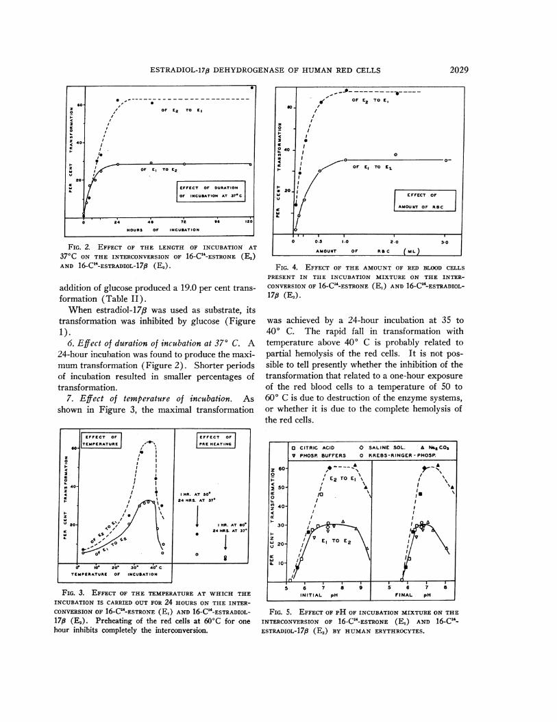
6. Effect of duration of incubation at 37° C. A24-hour incubation was found to produce the maxi-mumtransformation (Figure 2). Shorter periodsof incubation resulted in smaller percentages oftransformation.
7. Effect of temperature of incubation. Asshown in Figure 3, the maximal transformation
FIG. 3. EFFECT OF THE TEMPERATUREAT WHICH THE
INCUBATION IS CARRIED OUT FOR 24 HOURSON THE INTER-
CONVERSIONOF 16-C14-ESTRONE (E,) AND 16-C14-ESTRADIOL-17,8 (E2). Preheating of the red cells at 60'C for onehour inhibits completely the interconversion.
FIG. 4. EFFECT OF THE AMOUNTOF RED BLOOD CELLS
PRESENT IN THE INCUBATION MIXTURE ON THE INTER-
CONVERSIONOF 16-C -ESTRONE (Ej) AND 16-C1-ESTRADIOL-17,8 (E2).
was achieved by a 24-hour incubation at 35 to400 C. The rapid fall in transformation withtemperature above 40° C is probably related topartial hemolysis of the red cells. It is not pos-
sible to tell presently whether the inhibition of thetransformation that related to a one-hour exposure
of the red blood cells to a temperature of 50 to600 C is due to destruction of the enzyme systems,or whether it is due to the complete hemolysis ofthe red cells.
FIG. 5. EFFECT OF PH OF INCUBATION MIXTUREON THE
INTERCONVERSION OF 16-C1-ESTRONE (Ej) AND 16-C -
ESTRADIOL-17,8 (E2) BY HUMANERYTHROCYTES.
S
o ,'oF E2 TO El@0 /
0
X U
s~~~~~FFECT oF DURATION
if | ~~~~~~oFINCUBATION AT 37- C|
0 2'4 41 A2 *6 12 0
HOURS OF INCUBATION
2029

MIGEON, LESCURE, ZINKHAM, AND SIDBURY, JR.
ETHANOL CONCENTRATIONS (IN % Of VOL. OF MIXTURE)
FIG. 6. EFFECT OF ETHANOLCONCENTRATIONSON THE
INTERCONVERSION OF 1-C -ESTRONE (E,) AND 16-C -
ESTRADIOL-17, (E2) BY HUMANRED CELLS.
8. Effect of amount of red cells. The amountof red cells used in the incubation was found tohave an influence on the degree of conversion as
seen in Figure 4. Maximal transformation was
TABLE III
Steroid interconversion by red cellsof normal subjects
%Steroid transformationSubjects
El to Es* E2 to El
Mean Range Mean RangeFive Normal Females
Menstrual cycle days2-5 35 (32-28) 54 (50-S58)9-12 34.5 (26-42) 58 (49-70)
16-19 29.5 (29-32) 57 (50-65)23-26 31.1 (26-38) 53 (49-60)
Four Normal MalesExperimental day
1 35.5 (28-40) 61 (5S6-68)8 35.5 (31-42) 57.5 (53-61)
15 30.5 (27-33) 54 (49-61)
*E =estrone; E2 =estradiol-17,6.
obtained with approximately 1 ml of red cells un-
der the conditions of the reaction.9. Effect of pH. It was observed that 0.1 M
phosphate buffer (Clark and Sub's standard mix-ture of 50 ml 0.2 M KH2PO4 plus variable vol-umes of 0.2 M NaOH, diluted to 100 ml) or
LE IV
Enzyme activity and steroid interconversion by the red cells of-subjects with a congenital deficiency of glucose-6-phosphatedehydrogenase in their erythrocytes *
RBCenzymes %Steroid transformation
Subject Sex Age G-6-PDt 6-P-GDt El to E2 Es to El
yrs
American Negroes with primaquine-sensitive RBC
F. Mc Gr. F 42 29 173 12.9 58.2L. Mc Ne. M 45 23 187 5.0 51.5Ja. Mc Gr. M 11 15 146 9.1 62.2Sy. Mc Gr. M 9 13 122 11.8 76.2Sh.McGr. F 2 15 254 9.0M. BI F 32 38 254 10.2 68.1
Sephardic Jew with primnaquine-sensitive RBC
R. Da. M 32 2 165 8.9 65.4
Patients with congenital non-spherocytic hemolytic anemiaand family members
D. Iy. M 56 0 150 7.5 60.6His Wife 162 143 30.4 59.8A. Ki. M 11 0 153 8.1 57.0His Father 132 167 34.6 70.1His Mother 77 152 22.0 62.4E. Ro. M 6 0 172 7.6 63.3His Father 146 147 28.8 65.1His Mother 115 148 20.2 61.0
* Abbreviations: G-6-PD = glucose-6-phosphate dehydrogenase; 6-P-GD 6-phosphate-gluconic dehydrogenase;E,= estrone; E2 = estradiol-17j3.
t Activity is expressed as units of enzyme per 100 ml of RBC.
2030
a_~~~~~EFC OF|60- ETHANOL CONCENTRATIONS
z
0
40' 0
z %
F6 41,OF E2 TO El
zhi20-
OF El TO E2
0 2 4 6 8 0

ESTRADIOL-17,p DEHYDROGENASEOF HUMANRED CELLS
Krebs-Ringer-phosphate solution (22) were in-adequate in the amounts used to produce sufficientbuffering capacity until the end of the incubation.The effect of initial pH on the interconversion ofestrone and estradiol-17,/ is shown in Figure 5.The final pH values differed from the initial val-ues by as much as 1.0 pH unit for the highest pHto 0.25 pH units at the lowest pH tested. Thiscould be explained by either the formation of lac-tate during the incubation or the effect of hemo-globin buffering capacity, or both.
With the more concentrated solutions of citricacid, a great deal of hemolysis occurred whichcould in fact account for the smaller conversions.
10. Inhibition by ethanol, sodium fluoride, andmnethylene blue. Ethanol produced marked in-
hibition of the transformation (Figure 6) when itwas present in concentrations of more than 2 per
cent (per volume). Inhibition by sodium fluoridewas demonstrable with concentrations greater than5 x 10-3M.
Methylene blue appeared to have also some in-hibitory action at concentrations greater than 0.01mg per 1 ml of mixture.
11. Serial determinations in normal females andmales. The conversion of estrone to estradiol-17,8and the reversal of the reaction are shown inTable III. It can be seen that the percentage oftransformation in males was somewhat variablefrom one week to the next one, but the results re-
mained in the fairly narrow limit of 26 to 43 per
cent transformation for estrone and 49 to 70 per
cent for estradiol-17,8. The results obtained innormal females varied similarly, and the menstrualcycle did not appear to influence significantly thepercentage of transformation.
z
0
2a:0
U-
L
zw
zLi
ul
80
w
60-0
N 40-
o 20
40-
30-0
- 20-oi
PR MAQUINE
SENSITIVE
0
7s I .
..:.: .:. . . . . ..
.. ...
.. :. .. :..._A_
00
FIG. 7. IN VITRO INTERCONVERSIONOF ESTRONE ANDESTRADIOL-17fi (E2) BY THE RED CELLS OF SUBJECTS WITH
CONGENITAL DEFICIENCY OF GLUCOSE-6-PHOSPHATEDEHY-DROGENASEIN THEIR ERYTHROCYTES. The shaded areas
represent the range of transformation obtained with thered cells of normal individuals.
12. Subjects with a deficiency of the red cell glu-cose-6-phosphate dehydrogenase and their rela-tives. The results are shown in Table IV. In thepatients with congenital nonspherocytic hemolyticanemia, the percentage of transformation of estroneto estradiol-17,8 was markedly reduced. It was
normal in the fathers of the patients and the wifeof one, but it was at the lower limit of the normalrange in the two tested mothers of patients. TableIV shows that these two mothers had a somewhatdecreased glucose-6-phosphate dehydrogenase ac-
tivity in their red cells and are usually referred to
TABLE V
Effect of addition of various cofactors on steroid interconversion by hemolyzed red cells of normal subjects *
Solutions added (ml) %Steroid transformation
G-6-P TPN TPNH DPNGlucose (5%) (0.5 M) (0.01 M) (0.01 M) (0.01 M) E, to En En to El
0.1 2.6 (5) 2.1 (5)3.2 (6) 4.1 (4)
0.3 4.4 (4) 70.2 (7)0.1 4.1 (4)0.1 0.3 35.1 (16) 2.4 (2)
0.3 34.3 (3) 2.9 (2)0.3 2.2 (3) 3.1 (3)
* The results are the mean values of a number of experiments as indicated in parentheses. Abbreviations: G-6-P= glucose-6-phosphate; El = estrone; E2 = estradiol-17,9.
2031

MIGEON, LESCURE, ZINKHAM, AND SIDBURY, JR.
as "female intermediates" (23). It is not possibleat the present time to tell whether the slight de-crease in transformation of estrone in these twomothers was due to the relative decrease in en-zyme activity of their red cells.
The six Negroes and the Jewish subject withprimaquine-sensitive erythrocytes also had a re-duced transformation of estrone to estradiol-17,8.
On the other hand, the conversion of estradiol-17,3 to estrone by the red cells of the subjects stud-ied was found to be in the range of the values ob-tained for normal males and females (Figure 7).
RESULTS WITH HEMOLYZEDRED BLOODCELLS
1. Necessary cofactors. When no cofactorswere added or when glucose alone was added, nosignificant transformation was observed (TableV). The transformation of estradiol-17,8 to es-trone required TPN; DPN could not replaceTPN. The reverse reaction required eitherTPNHor TPN and glucose-6-phosphate.
2. Effect of removal of red cell ghosts. In theabsence of red cell ghosts, results similar to thoseshown in Table V were obtained.
3. (NH,)2SO4 precipitation. With 25 per cent(NH4)2SO4 the enzymatic activity was found tobe entirely in the supernatant fluids, whereas with37.5 per cent the activity was in both the super-natant fluids and the precipitates. With 50 percent, only the precipitates transformed estradiol-17,8 to estrone.
FIG. 8. DEAE COLUMNFRACTIONATION OF THE ES-
TRADIOL-17,8 (E2) DEHYDROGENASEACTIVITY IN RED CELL
HEMOLYSATE. The activity is expressed as percentage oftransformation of estradiol-17p8 (E2) to estrone.
4. DEAE column fractionation. As shown inFigure 8, three peaks of enzymatic activity wereobtained. The red cells of four different subjectsshowed similar peaks; however, the height of eachwas found to be variable.
DISCUSSION
The results show that intact red blood cells ofman can convert 16-C14-estrone to 16-C14-es-tradiol-17,/, confirming the data of Bischoff andhis collaborators (5, 9). These authors, however,did not specify the stereoisomeric position of the17-hydroxyl group. It is most interesting to notethe species differences reported by Axelrod andWerthessen (8), who have found that estradiol-17a was the major conversion product of 16-C14_estrone in pregnant cow blood. In an analogousmanner, Lindner (24) has observed that A4-an-drostene-3,17-dione was transformed into epites-tosterone by bovine blood, and we have foundthat the same substrate gave rise to testosteronewhen incubated with red blood cells of man.
The reversibility of the in vitro conversion ofestrone to estradiol-17,8 has been demonstratedby the data presented above, as well as by otherinvestigators (10-12, 14). The evidence suggeststhat the reaction is enzymatically mediated. Bis-choff and associates (6) have suggested the nameof "estronase" for the enzyme. "Estradiol-17,8dehydrogenase" would be more appropriate, asproposed by Portius and Repke (14). In addition.it is quite possible that the interconversion ofA4-andostene-3,17-dione and testosterone that wehave observed may also be mediated by the sameenzyme, a 17,8-hydroxysteroid dehydrogenase.
Our study of the factors influencing the trans-formation of estrone to estradiol-17,8 by intacthuman red blood cells confirms the data publishedby Repke and his collaborators (12, 14), who usedmainly rat erythrocytes and a yeast preparation.These results are also in agreement with theearlier work of Bischoff and associates (6). Animportant difference with the latter data is thefact that in our experiments hemolysis of the redcells arrested the transformation. If glucose-6-phosphate and TPN were added, however, estronewas converted to estradiol-173, whereas the ad-dition of TPN only to hemolyzed cells permittedthe reverse reaction. Hemolysis of red cells is
60-
I-
2
0 40-
0 Z
CUtI
RCION NO. 5 10
N0.15 M 0.2M i 0.3M'0.5M'
2032

ESTRADIOL-17p DEHYDROGENASEOF HUMANRED CELLS
known to destroy hexokinase and TPN activity,and it is not surprising that the addition of glu-cose alone could not affect the transformation ofestrone. The interconversion was found to be in-hibited by very small concentrations of ethanol,but the effect of sodium fluoride or methylene bluewas not so marked. One might wonder whetherthe inhibition produced by these substances wasnot related to a certain degree of hemolysis of thered cells. The same comment could apply to theeffect of preheating the red cells.
The need of glucose for the transformation ofestrone to estradiol-17,8 by intact red cells of nor-mal man has suggested that the reaction is relatedto the glycolysis that takes place in the cells (6).Since the reaction involves a hydrogen transfer,it was reasonable to assume that a hydrogendonor was required. Repke and Markwardt haveshown that the glucose-6-phosphate dehydrogenaseand TPN system was involved in the steroid con-version (7).
In subjects with a deficiency of glucose-6-phos-phate dehydrogenase, the transformation of glu-cose-6-phosphate to 6-phospho-gluconolactone wasvery limited, resulting in decreased formation ofTPNH. Since the transformation of estrone toestradiol-17,8e was greatly limited in these subjects,this indicates that TPNH is the hydrogen donorfor the reaction catalysed by estradiol-17,8 dehy-drogenase. On the other hand, the transformationof estradiol-17,8 to estrone that required a hydro-gen acceptor was normal in these subjects. Thefact that in subjects with red cell glucose-6-phos-phate dehydrogenase deficiency some estrone wastransformed probably means that the deficiencywas not complete. At the same time, if glucose-6-phosphate dehydrogenase is not completely ab-sent, one would expect that enough TPNHwouldbe generated to reduce the minute amounts ofestrone that are being offered as substrate in thepresent experiments unless more active and moreabundant TPNH-oxidizing systems are also pres-ent in the red cells. Such a possibility is to beconsidered, although it remains to be proved. Thetwo dehydrogenases of the hexose monophosphateshunt are TPN-specific. The two dehydrogenasespresent in the Embden-Meyerhof-Parnas glyco-lytic pathway are DPN-dependent. In addition tothe glucose-6-phosphate dehydrogenase system,one of the three other hydrogen transfers of the
glycolysis could be coupled with the hydrogentransfer by the estradiol-17/3 dehydrogenase. Theaddition of DPN to hemolyzed red cells did notproduce any significant transformation of estra-diol-17,8. Therefore, only the 6-phospho-gluconicdehydrogenase could possibly be coupled to theestradiol-17,/ dehydrogenase system; this, how-ever, has not been determined.
The occurrence of pyridine nucleotide transhy-drogenase activity in animal tissue mitochondriahas been reported (25). An enzyme system whichis specifically stimulated by estrogens added invitro has been demonstrated in human placentalpreparations (26), and it was found that this sys-tem catalyzed a pyridine nucleotide transhydrog-enation (27, 28). More recently, an estradiol-17,/dehydrogenase has been separated from the es-trogen-sensitive transhydrogenase (29). Whetherthe human red cell enzyme is identical with the es-tradiol-17/8 dehydrogenase found in human pla-centa has not been determined. Since it is impos-sible to remove all the blood contained in placentaltissue, placental estradiol-17,B dehydrogenase ac-tivity could be partially of red cell origin. Thebehavior of the placental system described byRyan and Engel (30), however, would seem toargue against erythrocyte origin for the placentaldehydrogenase.
The role and the importance of the estradiol-17,8-dehydrogenase in human red cells is notknown. The fact that this enzyme system is inlarge part coupled with the glucose-6-phosphatedehydrogenase might suggest that it plays a rolein the glycolysis of the red cell. The slow rateof interconversion of estrone and estradiol-17/3,however, would appear to limit the importance ofthis system. It should be added that in the pres-ence of plasma, the extent of the interconversionis smaller. This is due probably to the fact thata large fraction of the steroid is bound to plasmaprotein and therefore unavailable to red cells (31-34). Since a similar situation would be found inthe general circulation of man, this would alsoreduce the importance of the system in vivo.
Estradiol-17,8 is known to be a more biologi-cally active estrogen than estrone. Bischoff andhis collaborators (6) studied only the transforma-tion of estrone to estradiol, and have consideredthat red cells contained an "activating" enzyme asfar as biological activity is concerned. The reac-
2033
MIGEON, LESCURE, ZINKHAM, AND SIDBURY, JR.
tion is reversible, however, and the equilibriumdepended upon glucose concentrations. It is notpossible at the present time to tell where theequilibrium would be situated in in vivo condi-tions, nor can we state the in vivo importance ofthe enzyme system in metabolizing estrogens ofendogenous origin.
SUMMARY
The presence of an estradiol-17,8 dehydrogenasein human red cells is confirmed. The enzyme isTPN-dependent. It is coupled with the glucose-6-phosphate dehydrogenase for the transformation ofestrone to estradiol-17,8. The thermolability andthe pH optimum of the enzyme in intact cells hasbeen determined. The red cells of subjects withglucose-6-phosphate deficiency have a marked butnot complete decrease of the transformation, indi-cating that the interconversion of estrone and es-tradiol -17/8 is related to the activity of the hexosemonophosphate shunt.
The physiological importance of the red cellestradiol-17,/ dehydrogenase remains to be de-termined.
REFERENCES
1. Migeon, C. J., Lescure, 0. L., and Sidbury, J. B., Jr.In vitro studies with 16-C'4-estrone: conversion toestradiol by red blood cells of man. Acta endocr.(Kbh.) (supplement) 1960, 51, 731.
2. Werthessen, N. T., Baker, C. F., and Borci, B. Thepresence of an alcoholic, ketonic derivative of es-trone in human and rabbit blood. Science 1948,107, 64.
3. Werthessen, N. T., Baker, C. F., and Field, N. S.Conversion of estrone on incubation in blood. J.biol. Chem. 1950, 184, 145.
4. Werthessen, N. T., Schwenk, E., Baker, C. F., andField, N. S. Estrone conversion capacity of bloodof postmenopausal women with carcinoma of thebreast. Cancer Res. 1950, 10, 679.
5. Bischoff, F., Katherman, R. E., and Yee, Y. S. Ac-tivation of estrone by mammalian red cells. Amer.J. Physiol. 1951, 164, 774.
6. Bischoff, F., Katherman, R. E., Favati, V., andGray, C. L. Metabolic effect of red blood cellson estrone. Amer. J. Physiol. 1952, 171, 100.
7. Repke, K., and Markwardt, F. Uber fermentativeReduktion von Oestron. Naunyn-Schmiedeberg'sArch. exp. Path. Pharmak. 1954, 223, 271.
8. Axelrod, L. R., and Werthessen, N. T. The metab-olism of estrone-16-C"4 in bovine blood. Arch.Biochem. 1960, 86, 53.
9. Gray, C. L., and Bischoff, F. Conversion of estroneto estradiol by mammalian red cells. Amer. J.Physiol. 1955, 180, 279.
10. Portius, H. J., and Repke, K. Uber Stereospezifitatund Aktivitit der Oestradiol-Dehydrogenasen inden Erythrocyten von Mensch und Tier. Naunyn-Schmiedeberg's Arch. exp. Path. Pharmak. 1960,239, 184.
11. Markwardt, F., and Repke, K. Die Reduktion vonOestron zu Oestradiol-17p durch das Glucose-6-phosphat-Dehydrogenasesystem. Naunyn-Schmiede-berg's Arch. exp. Path. Pharmak. 1955, 224, 341.
12. Repke, K. Die Kinetik der Umsetzung von Oestronund Oestradiol-17j3 durch Hefe. Naunyn-Schmiede-berg's Arch. exp. Path. Pharmak. 1957, 230, 178.
13. Bischoff, F., Torres, A., and Lopez, G. Glucose-6-phosphate in estronase enzyme systems. Amer. J.Physiol. 1957, 189, 447.
14. Portius, H. J. and Repke, K. Eine Oestradiol-17,6-Dehydrogenase in den Erythrocyten der Ratte.Naunyn-Schmiedeberg's Arch. exp. Path. Pharmak.,1960, 239, 144.
15. Childs, B., and Zinkham, W. H. The genetics ofprimaquine sensitivity of the erythrocytes in CibaFoundation Symposium on Biochemistry of HumanGenetics. Boston, Little, Brown, 1959, p. 76.
16. Migeon, C. J., Wall, P. E., and Bertrand, J. Someaspects of the metabolism of 16-C14-estrone innormal individuals. J. clin. Invest. 1959, 38, 619.
17. Glock, G. E., and McLean, P. Further studies onthe properties and assay of glucose 6-phosphatedehydrogenase and 6-phosphogluconate dehydroge-nase of rat liver. Biochem. J. 1953, 55, 400.
18. Zinkham, W. H., and Lehnard, R. E., Jr. Metabolicabnormalities of erythrocytes from patients withcongenital nonspherocytic hemolytic anemia. J.Ped. 1959, 55, 319.
19. Brown, J. B. A chemical method for the determina-tion of oestriol, oestrone and oestradiol in humanurine. Biochem. J. 1955, 60, 185.
20. Haines, W. J., and Drake, N. A. Fluorescencescanner for evaluation of papergrams of adrenalcortical hormones. Fed. Proc. 1950, 9, 180.
21. Migeon, C. J., Lescure, 0. L. Antoniades, H. N.Further in vitro studies with 16-C1'-estrone: dis-tribution between plasma protein fractions andred blood cells of man. Bull. Johns Hopk. Hosp.1960, 106, 317.
22. Umbreit, W. W., Burris, R. H., and Stauffer, J. F.Manometric techniques. Minneapolis, Burgess,1957, p. 148.
23. Childs, B., Zinkham, W., Browne, E. A., Kimbro,E. L., and Torbert, J. V. A genetic study of adefect in glutathione metabolism of the erythrocyte.Bull. Johns Hopk. Hosp. 1958, 102, 21.
24. Lindner, H. R. Androgens -in the bovine testis andspermatic vein blood. Nature (Lond.) 1959, 183,1605.
2034

ESTRADIOL-17p DEHYDROGENASEOF HUMANRED CELLS
25. Kaplan, N. O., Colowick, S. P., Neufeld, E. F., andCiotti, M. M. Pyridine nucleotide transhydrogenaseIV. Effect of adenylic acid a on the bacterial trans-hydrogenases. J. biol. Chem. 1953, 205, 17.
26. Villee, C. A., and Hagerman, D. D. Effects of es-tradiol on the metabolism of human placenta invitro. J. biol. Chem. 1953, 205, 873.
27. Talalay, P., and Williams-Ashman, H. G. Activa-tion of hydrogen transfer between pyridine nu-cleotides by steroid hormones. Proc. nat. Acad.Sci. (Wash.) 1958, 45, 15.
28. Villee, C. A., and Hagerman, D. D. On the identityof the estrogen-sensitive enzyme of human pla-centa. J. biol. Chem. 1958, 233, 42.
29. Hagerman, D. D., and Villee, C. A. Separation ofhuman placental estrogen-sensitive transhydroge-nase from estradiol-17p dehydrogenase. J. biol.Chem. 1959, 234, 2031.
30. Ryan, K. J., and Engel, L. L. The interconversionof estrone and estradiol by human tissue slices.Endocrinology 1953, 52, 287.
31. Bischoff, F., and Katherman, R. E. Distribution ofestradiol between serum and red cells. Amer. J.
Physiol. 1948, 152, 189.32. Sandberg, A. A., and Slaunwhite, W. R., Jr. Stud-
ies on phenolic steroids in human subjects. II.The metabolic fate and hepato-biliary-enteric cir-culation of C"-estrone and C'4-estradiol in women.
J. clin. Invest. 1957, 36, 1266.33. Sandberg, A. A., Slaunwhite, W. R., Jr., and
Antoniades, H. N. The binding of steroids andsteroid conjugates to human plasma proteins. Re-cent Progr. Hormone Res. 1957, 13, 209.
34. Wall, P. E., and Migeon, C. J. In vitro studies with16-C'-estrone: distribution between plasma andred blood cells of man. J. clin. Invest. 1959, 38,611.
2035


![]HUIRULQVLWX9LVFRHODVWLF &KDUDFWHUL]DWLRQRI ... › sites › default › files › ... · 1 JMM-104661 1 1 A MEMS Dynamic Mechanical Analyzer for in situ Viscoelastic Characterization](https://img.dokumen.tips/doc/110x75/5f0cda987e708231d4377575/huirulqvlwx9lvfrhodvwlf-kdudfwhuldwlrqri-a-sites-a-default-a-files.jpg)