Embed Size (px)
Citation preview

AGE-INDEPENDENT OSTEOPATHOLOGY INSKELETONS OF A SOUTH AMERICAN CERVID, THEPATAGONIAN HUEMUL (HIPPOCAMELUS BISULCUS)
Authors: Flueck, Werner T., and Smith-Flueck, Jo Anne M.
Source: Journal of Wildlife Diseases, 44(3) : 636-648
Published By: Wildlife Disease AssociationURL: https://doi.org/10.7589/0090-3558-44.3.636
BioOne Complete (complete.BioOne.org) is a full-text database of 200 subscribed and open-access titlesin the biological, ecological, and environmental sciences published by nonprofit societies, associations,museums, institutions, and presses.
Your use of this PDF, the BioOne Complete website, and all posted and associated content indicates youracceptance of BioOne’s Terms of Use, available at www.bioone.org/terms-of-use.
Usage of BioOne Complete content is strictly limited to personal, educational, and non - commercial use.Commercial inquiries or rights and permissions requests should be directed to the individual publisher ascopyright holder.
BioOne sees sustainable scholarly publishing as an inherently collaborative enterprise connecting authors, nonprofitpublishers, academic institutions, research libraries, and research funders in the common goal of maximizing access tocritical research.
Downloaded From: https://bioone.org/journals/Journal-of-Wildlife-Diseases on 26 Jan 2020Terms of Use: https://bioone.org/terms-of-use

AGE-INDEPENDENT OSTEOPATHOLOGY IN SKELETONS OF A
SOUTH AMERICAN CERVID, THE PATAGONIAN HUEMUL
(HIPPOCAMELUS BISULCUS)
Werner T. Flueck1,2,4 and Jo Anne M. Smith-Flueck3
1 National Research Council (CONICET), C.C. 176, 8400 Bariloche, Argentina2 Swiss Tropical Institute, CH-4002 Basel, Switzerland3 Instituto de Analisis de Recursos Naturales, Universidad Atlantida, 7600 Mar del Plata, Argentina4 Corresponding author (email:[email protected])
ABSTRACT: The huemul (Hippocamelus bisulcus), an endemic Patagonian deer, has beenendangered for decades. Although conservation in Argentina has been directed at the 350–600remaining huemul, the population has not recovered. In initial studies on the potential effects ofdiseases on huemul population dynamics, skeletal remains collected between 1993 and 2007 in theAndes (41–45uS, 71.5–72uW) were examined macroscopically for osteopathologic changes. Bonesfrom six huemul were free of lesions, findings were inconclusive in 13 huemul with less than threebones, and osteopathologic processes were detected in 13 adults. Considering the limited remains/case, the prevalence of osteopathy (52%) among adults probably is conservative; 63% showedmandibular, 100% maxillary, and 78% appendicular lesions. Although predation represented theactual cause of death, the observed skeletal lesions would affect predator avoidance, possiblyexplaining the low average adult age (3.1 yr) and lack of population recovery. Compared with otherstudies in ungulates, huemul were affected at a younger age, and they had more severe pathologicchanges. Due to the chronic nature of disease, low huemul population density, physiognomy, andspatiotemporal pattern of lesions, we discard senescence; gender; fulminating infections;congenital anomalies; metabolic, endocrine, genetic, or neurologic disorders; parasitism ormarasmus; and fluorosis as primary etiologic factors. We hypothesize that generalized secondarychronic alveolar osteomyelitis and osteoarthritis in huemul is related to the nutritional ecology ofthese animals. Selenium deficiency, which impairs bone metabolism and causes periodontitis inruminants, occurs in the region and it is more prevalent at high altitudes. Traditional wintergrounds at low elevations, sometimes far from high mountains, have been converted to livestockproduction, which has eliminated migratory behavior and keeps huemul in remote high-elevationrefuges. Although this descriptive study contributes to huemul conservation, additionalapproaches are needed to investigate the etiology of this osteopathy and to close other gaps inknowledge on biology and ecology of huemul.
Key words: Arthritis, chronic alveolar osteomyelitis, Hippocamelus bisulcus, huemul, lumpyjaw, osteopathology, periodontitis.
INTRODUCTION
The Patagonian huemul (Hippocamelusbisulcus), a cervid endemic to the South-ern Cone of Latin America, has beenconsidered endangered for several de-cades (Diaz and Smith-Flueck, 2000).Habitat conditions for the species includewet temperate rain forests to the west,and, due to the Andean Mountains’ rainshadow, drier forests and grasslands to theeast. The latter habitat types occur mainlyin Argentina, and they are the focus ofthis report. Huemul numbers and area ofoccupancy likely began to decline after theincrease in numbers of the pre-Columbiantribes, and again after 1536 with arrival of
the Spaniards, who then introduced do-mestic livestock, horses, and firearms tothis part of the continent. Native peoplethen adopted an equestrian lifestyle,which improved hunting efficiency andallowed them to dominate the regionaleconomy for the next 300 years. Subduedin 1881, their territories opened up tofrontiersmen, colonists, and explorers. Theearliest accounts about huemul frominterior Patagonia thus described a land-scape that had been modified for severalhundred years. Diaz (1993) left no doubtthat explorers of the late 19th centurywere describing the last occurrences ofhuemul far east of the Andes. Even then,in the Andean foothills more westerly,
Journal of Wildlife Diseases, 44(3), 2008, pp. 636–648# Wildlife Disease Association 2008
636
Downloaded From: https://bioone.org/journals/Journal-of-Wildlife-Diseases on 26 Jan 2020Terms of Use: https://bioone.org/terms-of-use

they were generally already consideredrare. Later accounts of huemul cameexclusively from interior Andean mountainareas where seasonal climatic extremes,steep topography, and closed vegetationdelayed permanent human colonization.Scientifically oriented interest in theArgentine huemul started in the mid-1980s, beginning with an inventory ofremaining subpopulations; these effortswere reviewed in Diaz and Smith-Flueck(2000). Conservation efforts have beendirected at the estimated 350–600 animalsremaining in Argentina, most of which arefound outside of protected areas. Theseefforts, though, have not resulted in anydocumented recovery; instead, subpopu-lations have vanished, even in protectedareas.
Securing reliable data is difficult on aspecies so reduced in numbers andoccurring mainly in remote refuges. Thus,as noted by Smith-Flueck and Flueck(2001b) who reported the impact frompredation in relation to sex and age classesbased on surveyed huemul carcasses, theeffect of diseases on population dynamicshas never been studied in huemul. Theknowledge base on huemul is rudimenta-ry, and a CrossSearch of ISI Web-of-Knowledge (http://isiknowledge.com) and17 external databases (1945–2006) listed16 entries on H. bisulcus, with only nineoriginal studies (Flueck and Smith-Flueck,2006a). Given the lack of knowledge onthe role of disease in reduced huemulpopulations, the aim of this study was toevaluate the potential of disease to con-tribute to the species’ morbidity. Specifi-cally, skeletal remains of huemul wereexamined to provide essential baselinedata on bone diseases.
MATERIALS AND METHODS
The study area comprises part of the easternAndean Mountain district (41–45uS, 71.5–72uW), which phytogeographically belongs tothe subantarctic province, and it is character-ized by mature and dense forests primarilyconsisting of lenga beech trees (Nothofagus
pumilio), with the understory predominatedby small shrubs such as Maytenus disticha,Gaultheria mucronata, Myoschilos oblongum,and Berberis serrata-dentata (Smith-Flueckand Flueck, 1997, 2001b). The mean annualprecipitation is 100–200 cm; however, thereare large annual and seasonal variations amonglocalities. The mean temperature during Juneand August varies between 24 C and 22 C,with mean precipitation between 300 mm and400 mm, principally as snow. Elevations in thestudy area range from about 900 m to2,000 m.
We collected huemul skeletal remainsopportunistically between 1993 and 2007.Upon finding signs of a dead ungulate, acircular area of approximately 50 m wassearched intensely, and all remains werecollected for later identification and inspectionfor macroscopic lesions. Additional samplescollected by others also were inspected. Sexwas determined from the physiognomy ofpelvis, sacrum, axis, frontals, and from com-parative morphometry because the huemul isdimorphic (Smith-Flueck, 2003). Adults wereaged by analysis of cementum annuli inincisors or dental wear. Ages of some malesalso were determined by comparing thedimensions of the pedicels to males whoseage had been determined using their teeth(Smith-Flueck and Flueck, 2001b). Age classesused were fawns and adults based on thecondition of the epiphyseal plate, size of longbones, and hoof physiognomy (Smith-Flueck,2003; Flueck and Smith-Flueck, 2005).
RESULTS
Remains of 32 individuals, found atelevations from 930 m to 1,200 m, wereinspected. The average age (withoutfawns, n57) was 3.1 yr (range 1.5–5.5 yr,SD51.2, n520). Fairly complete skele-tons of three fawns and three adults wereconsidered to be free of bone lesions,although one adult had no skull parts.Animals represented by less than threebones (nine adults and four fawns), allwithout lesions, could not be conclusivelydetermined to be free from osteopathy,because positive individuals frequentlyhad skeletal portions unaffected by dis-ease. These nine adults and four fawnsthus might have suffered from lesions inother skeletal parts. The remaining 13cases had clear signs of osteopathologic
FLUECK AND SMITH-FLUECK—OSTEOPATHOLOGY IN HUEMUL 637
Downloaded From: https://bioone.org/journals/Journal-of-Wildlife-Diseases on 26 Jan 2020Terms of Use: https://bioone.org/terms-of-use

processes (Table 1). Among diseased ani-mals that had mandibles (n58), 63% hadmandibular lesions, whereas maxillarybones of all diseased animals had lesions(n59). The only animal with healthymaxillary bones was also free of otherbone lesions. Among diseased animals thathad some appendicular material (n59),78% had lesions.
Description of main lesions
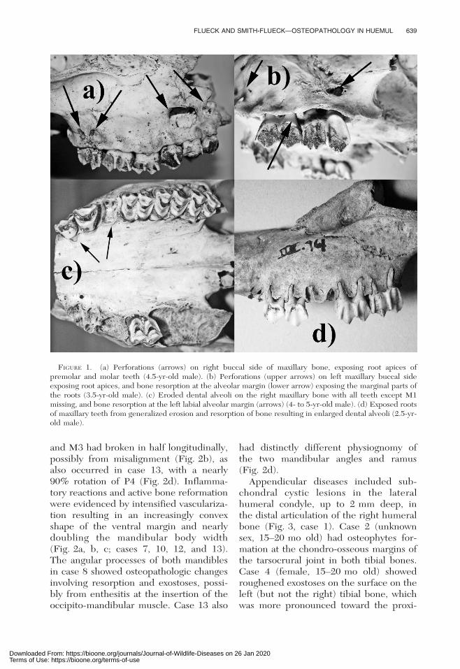
Maxillary lesions included enlargeddental alveoli from osteolytic processes,creating open spaces up to 3.5 mmbetween the alveolar margin and themarginal root of molars and premolars(Fig. 1c; case 1, 3- to 4-yr-old male withantlers shed; case 6, 2.5-yr-old male; case7, 4.5-yr-old female; case 8, 3.5-yr-oldmale; case 9, 2.5-yr-old male; case 10, 3- to4-yr-old female; case 11, 4.5-yr-old male;case 13, 4- to 5-yr-old male). Anothertypical lesion was bone resorption on thebuccal side exposing roots of teeth par-tially or completely to the apex (Fig. 1d,case 3, 3.5-yr-old female; cases 6, 7, 8, 9,10, and 13). The root apices often wereclub shaped from crystalline deposits, attimes 8 mm across (Fig. 1a), or they wereeroded away (cases 7, 8, and 11). More-over, the buccal side of the maxillary bone
showed perforation from lytic processesindicative of periapical abscesses, up to638 mm, at the level of root apices ofmolars and premolars (Fig. 1a, b; cases 8,9, 10, 11, and 13).
Mandibular lesions included enlargeddental alveoli from osteolytic processes,creating open spaces up to several milli-meters between the alveolar margin andthe marginal root of molars and premolars(cases 7, 10, and 13 and case 12, 5.5-yr-oldmale). Continued osteolytic processes inthe periapical cystic cavity resulted inlarge perforations in the mandibular wallon buccal and lingual sides (Fig. 2a; cases7, 8, 10, 12, and 13). Case 13 had verticalbone loss of the lingual alveolar margin atM3 taking on a U-shape, because no bonematrix remained at the margin and com-pletely exposed molar roots. Furthermore,the spongy bone from remodeling ventralto the perforation had a pathologic green-stick fracture such that the mandibularheight was reduced by approximately 70%
(Fig. 2d). The root apices of molars andpremolars were frequently club shapedfrom crystalline deposits (possibly hyper-cementosis; cases 7, 8, 10, 12, and 13).Tissue reaction in the mandible of case 12resulted in apparent lysis of the M2 tooth,reducing it to an amorphous structure;
TABLE 1. Summary of the major osteopathologic lesions in huemul (sample size in parentheses).
Female (4) Male (8) Unknown (1)
Eroded maxillary alveoli 2 6Perforated buccal margin of maxillary bone 1 4Reduced maxillary bone exhibiting teeth roots 3 4Eroded mandibular alveoli 1 3Reduced mandibular bone exhibiting teeth roots 2 2Perforated mandibular body 2 3Exostoses on mandible 1Thickening of mandibular body 1 2Increasingly convex ventral margin of mandible 2 2Crystalline deposits on tooth roots 2 4Eroded distal articulation of humeral bone 2Exostoses or remodeling on metacarpal bone 2Exostoses on tibial bone 1 1Exostoses or remodeling on metatarsal bone 1Deformed hoof or phalanges 2Exostoses on vertebra 1
638 JOURNAL OF WILDLIFE DISEASES, VOL. 44, NO. 3, JULY 2008
Downloaded From: https://bioone.org/journals/Journal-of-Wildlife-Diseases on 26 Jan 2020Terms of Use: https://bioone.org/terms-of-use
and M3 had broken in half longitudinally,possibly from misalignment (Fig. 2b), asalso occurred in case 13, with a nearly90% rotation of P4 (Fig. 2d). Inflamma-tory reactions and active bone reformationwere evidenced by intensified vasculariza-tion resulting in an increasingly convexshape of the ventral margin and nearlydoubling the mandibular body width(Fig. 2a, b, c; cases 7, 10, 12, and 13).The angular processes of both mandiblesin case 8 showed osteopathologic changesinvolving resorption and exostoses, possi-bly from enthesitis at the insertion of theoccipito-mandibular muscle. Case 13 also
had distinctly different physiognomy ofthe two mandibular angles and ramus(Fig. 2d).
Appendicular diseases included sub-chondral cystic lesions in the lateralhumeral condyle, up to 2 mm deep, inthe distal articulation of the right humeralbone (Fig. 3, case 1). Case 2 (unknownsex, 15–20 mo old) had osteophytes for-mation at the chondro-osseous margins ofthe tarsocrural joint in both tibial bones.Case 4 (female, 15–20 mo old) showedroughened exostoses on the surface on theleft (but not the right) tibial bone, whichwas more pronounced toward the proxi-
FIGURE 1. (a) Perforations (arrows) on right buccal side of maxillary bone, exposing root apices ofpremolar and molar teeth (4.5-yr-old male). (b) Perforations (upper arrows) on left maxillary buccal sideexposing root apices, and bone resorption at the alveolar margin (lower arrow) exposing the marginal parts ofthe roots (3.5-yr-old male). (c) Eroded dental alveoli on the right maxillary bone with all teeth except M1missing, and bone resorption at the left labial alveolar margin (arrows) (4- to 5-yr-old male). (d) Exposed rootsof maxillary teeth from generalized erosion and resorption of bone resulting in enlarged dental alveoli (2.5-yr-old male).
FLUECK AND SMITH-FLUECK—OSTEOPATHOLOGY IN HUEMUL 639
Downloaded From: https://bioone.org/journals/Journal-of-Wildlife-Diseases on 26 Jan 2020Terms of Use: https://bioone.org/terms-of-use

mal end. Whereas a metatarsal, twofemoral, and two tibial bones had nolesions in case 5 (adult male), the lefthumeral bone was greatly deformed. Thedistal part of the latter was eroded, andbone remodeling resulted in an irregularlyshaped, mineralized but spongy, prolifer-ating bone mass also involving the articu-
lar surface (Fig. 4b). The medial thirdphalanx of the left front leg in case 9 hadlesions affecting the hoof shape. Thedeformed claw capsule resulted in a hoofwidth approximately twice that of thenormal hoof. Also, extra keratinous growthcovering the hoof pad lacked abrasions,which indicated the animal did not usethis hoof (Fig. 4c). The metacarpal boneof this case also had exostoses at the distalend. In case 11, the distal end of the leftmetacarpal bone was grossly deformedsuch that the shaft reached a width nearlydouble that of the normal right (Fig. 4a).The medial metacarpal trochlea was de-formed, leaving behind only a porous massof bone matrix. The lateral side ofcondoyle 4, at the level of the tubercle,showed a substantial bony overgrowth,and eroded depression in the articularsurface. The proximal half of the firstmedial phalange had been eroded awaywith no articular surface remaining(Fig. 4a). The medial phalange also hadsome exostoses on its base. In case 13, ofthe available appendicle bones, the prox-imal end of the left metatarsal enclosed toa tarsal bone, probably the os cuneiformlateral.
The only case revealing addition oste-opathy was case 4, in which a fragment of
FIGURE 2. (a) Perforation on lingual side expos-ing roots of mandibular teeth (arrow) and convexshape of the right mandibular body (4.5-yr-oldfemale). (b) Perforations on lingual (shown) andbuccal sides, bone resorption at mandibular margin,tooth misalignment, split M3 and apparently lysis ofthe M2 tooth (5.5-yr-old male). (c) Bilateral thick-ening of mandibular bodies (4- to 5-yr-old male). (d)Bilateral differing physiognomy of mandibular anglesand ramus, pathological fracture at M3 (upperarrow), tooth misalignment and perforations (lowerarrows) (4- to 5-yr-old male).
FIGURE 3. Focal subchondral cystic lesion (ar-row, caudolateral view) in the lateral condyle of distalright humeral articulation (3- to 4-yr-old male).
640 JOURNAL OF WILDLIFE DISEASES, VOL. 44, NO. 3, JULY 2008
Downloaded From: https://bioone.org/journals/Journal-of-Wildlife-Diseases on 26 Jan 2020Terms of Use: https://bioone.org/terms-of-use

a thoracic vertebra had clear exostoses onthe spinous process and the body, and theonly other vertebra, the axis, also hadextensive exostoses on the body.
DISCUSSION
Considering the limited amount ofskeletal remains per case, the high prev-
FIGURE 4. (a) Dorsal view of deformed medial metacarpal trochlea, leaving behind only a porous mass ofbone matrix, and erosion of the proximal half of the first medial phalange (4.5-yr-old male). (b) Frontal (top)and dorsal (bottom) view of deformed distal end of the left humerus consisting of an irregularly shaped,spongy, proliferating mass of bone matrix (normal ends on left for comparison, adult male). (c) Deformed clawcapsule of the left front leg with keratin lacking abrasion, covering the hoof pad (arrow, 2.5-yr-old male).
FLUECK AND SMITH-FLUECK—OSTEOPATHOLOGY IN HUEMUL 641
Downloaded From: https://bioone.org/journals/Journal-of-Wildlife-Diseases on 26 Jan 2020Terms of Use: https://bioone.org/terms-of-use

alence of osteopathologic changes is strik-ing. The rate of 52% affected among adulthuemul is conservative, because another36% were inconclusive, being representedby too few bones. However, becausemarginal gingivitis and early stages ofarthritis cannot be diagnosed from suchmaterial, the prevalence of paradontaldisease and arthritis in the populationwould almost certainly be much higherthan indicated by the advanced stagesreported here. Similar to what has beenobserved in other cervids (Leader-Wil-liams, 1980; Peterson, 1988), fawns appar-ently were not yet affected; arthritis ofarticular surfaces of vertebral bodies andleg joints, for example, tends to occur afterepiphyseal plates are ossified (Greer et al.,1977).
Lesions described here can affect thehuemuls’ performance to varying degrees,dependent on location and severity. Post-cranial lesions might interfere with theenergetic balance and general diseaseresistance, but they likely exert their maineffect by reducing mobility and thuscapacity to escape predation. In addition,skull lesions also have an energetic costand reduce foraging efficacy and diseaseresistance. Thus, in advanced stages,survival strategies such as the capacity toavoid predation and reproductive successwould be diminished. Of significance,67% of diseased deer with both cranialand postcranial material available hadlesions in both areas. Due to the limitedmaterial in other specimens, this is almostcertainly an underestimate. The perfor-mance of animals with such widespreadlesions would have been most affected.Although we cannot dismiss that puma(Puma concolor), the main predator,selected for diseased huemul and thusinflate the prevalence of osteopathy, noremains of a huemul older than 5.5 yr(average 3.1 yr) have been found duringthe many field campaigns between 1993and 2007; thus, neither diseased norhealthy huemul survive to a mature age(Smith-Flueck and Flueck, 2001b).
The type of skull lesions, particularlythose resulting in an edentulous mouth,would result in debilitations during foodprocessing, thereby affecting body condi-tion (Leader-Williams, 1982; Loe et al.,2006). Ongoing erosive processes arelikely paralleled by pain, discomfort,secondary infections, and a drain ofenergy. Teeth eventually become looseor misaligned, and once lost, foragingefficacy is diminished. In similar habitatin New Zealand, the prevalence of ad-vanced paradontal disease in chamois(Rupicapra rupicapra) was found to in-crease with age, and it was highest among7+ yr olds (44%). Thus, the developmentfrom marginal gingivitis to periodontitis toadvanced alveolar osteomyelitis was as-sumed to take a long time (Pekelharing,1974). The preponderance of advancedcases in young huemul, however, indicatesthat the disease developed here in arelatively short time. Furthermore, olderchamois frequently survived the disease,with teeth missing but healed jaws (Pe-kelharing, 1974). We found no cases ofhealed lesions and no huemul was olderthan 5.5 yr, but this difference might beexplained by the absence of predators inNew Zealand. The appendicular arthriticlesions described here indicate a gradientfrom pain and discomfort during locomo-tion to complete lameness. The osteoar-thritis found in the vertebrae would bedebilitating as well. The various lesionsdescribed in these huemul cases wouldthus have affected body condition andlocomotive capacity, both of which areimportant for effective predator avoid-ance. Although predation by puma hasbeen shown to be an important ultimatecause of death in one population of thestudy area (Smith-Flueck and Flueck,2001b), these results indicate that asignificant proximal cause might be osteo-pathologic processes, which would in-crease the susceptibility to predation, aswas found for moose (Alces alces; Peter-son, 1988). It might explain why theaverage age of the adult sample is only
642 JOURNAL OF WILDLIFE DISEASES, VOL. 44, NO. 3, JULY 2008
Downloaded From: https://bioone.org/journals/Journal-of-Wildlife-Diseases on 26 Jan 2020Terms of Use: https://bioone.org/terms-of-use

about 3.1 yr, although huemul are knownto live to at least 15 yr old (Diaz andSmith-Flueck, 2000). Such a truncated agedistribution toward a young populationindicates high adult mortality rates. Fe-male reindeer (Rangifer tarandus) afflict-ed with the similar jaw problems on SouthGeorgia Island (Falkland Islands) raisedfewer calves (Leader-Williams, 1982),which also might be expected in compro-mised huemul living in a strongly seasonalenvironment. This together with highadult mortality rates could explain the lackof recovery of any known subpopulationin Argentina, as reflected by absence ofrecolonization of neighboring watersheds(Flueck and Smith-Flueck, 2006a). Theonly other incidence of osteopathy inhuemul known to us has been reportedby Milano et al. during the 1995 ArgentineMammal Society meeting, describing asimilar case from similar habitat: of twoskulls, one male had eroded maxillary andmandibular dental alveoli and missingteeth; the second male apparently lackedlesions.
Different etiologic factors can result in asimilar physiognomy of skull and appen-dicular lesions, including rare cases ofcongenital aberrations, osteosarcoma, ortoxicosis. The skull lesions described hereare often referred to as ‘‘lumpy jaws’’;however, it is merely a colloquialism usedto identify anatomic facial bone abnormal-ities, because it does not describe anactual disease, nor does it represent amorphologic diagnosis. Initial deformingbony lesions are the clinical manifestationof subsequent chronic alveolar osteomye-litis (Fagan et al., 2005), the etiology ofwhich remains complex. Moreover, clini-cal evidence of infection is not necessarilypresent. Lesions in lumpy jaws are gener-ally of mixed microbial type developingwithin the body of facial bones. Becausethese tend to originate from the normaloral flora, none of Koch’s postulates apply,and they have to be considered ofsecondary relevance. Walling off throughfibroplasia also can favor anaerobic organ-
isms within the cystic cavity, enlarging thecavity and even resulting in pathologicfractures (Fagan et al., 2005), as wasobserved in our sample. The reduction ofbone substance, particularly in the maxil-lary bone with frequently exposed dentalroots, is indicative of osteoporosis. AsWhalen and Krook (1996) observed,periodontal disease is frequently anearly manifestation of generalized osteo-porosis.
Lumpy jaw has included descriptions ofparadontal disease involving only maxillarybones (in deer less than 12 yr old; Geigeret al., 1992), only mandibles (Leader-Williams, 1980, 1982; Hoefs and Bunch,2001), or both (in deer .13 yr old; Geigeret al., 1992; this study). Conditions result-ing in lumpy jaws without postcranial bonelesions include infection with the arterialnematode Elaeophora schneideri, whichcan cause partial paralysis of jaw muscles,resulting in food impaction and concom-itant jaw disease, including tooth loss andfractured jaws (Davidson and Nettles,1988). However, we are unaware of anyarterial nematodes present in the studyarea or in other Argentine cervids, al-though domestic sheep and frequentimports of wild, exotic cervids could bepotential sources. Developmentally de-formed teeth also can enable intraoralorganism and foreign bodies from foodmaterial to be impacted into the dentalpulp chamber during mastication. Regard-less, we discard this as a generalized causebecause not all individuals showed mis-alignments, and the observed misalignedteeth apparently resulted secondarily dueto elimination of supporting alveoli. Alter-natively, actinomyces invade lesions sec-ondarily and result in lumpy jaw, but theyproduce diagnostic sulfur granules (Faganet al., 2005). Dry material from a typicallyaffected maxillary bone in our sample wasanalyzed histologically without findingfilaments or rods. Fluorosis also resultsin strikingly similar patterns of secondaryskull infections and osteolytic processes,and chronic fluoride intoxication causes
FLUECK AND SMITH-FLUECK—OSTEOPATHOLOGY IN HUEMUL 643
Downloaded From: https://bioone.org/journals/Journal-of-Wildlife-Diseases on 26 Jan 2020Terms of Use: https://bioone.org/terms-of-use

hyperostosis in the postcranial skeletonand hoof deformities (Krook and Justus,2006). The Andes are volcanically veryactive, and fluorine has been shown to bedeposited at hundred of kilometers fromvolcanic sources (Witham et al., 2005),causing lesions in domestic livestock(Araya et al., 1990). Given that there areno known overt fluorosis cases amongpeople or livestock near the study site, andthat typical macroscopic lesions wereabsent in huemul and feral red deer(Cervus elaphus) in northern Patagonia(Flueck and Jones, 2006), fluorosis as aprimary cause seems unlikely. Infectionsfrom Fusobacterium necrophorum alsoproduce oral and postcranial lesions (nec-robacillosis). Commonly part of the nor-mal intestinal flora, it can invade lesionssecondarily; however, progress is generallyfulminating, often through septicemia,including from inhalation. Besides lumpyjaw, vertebral osteomyelitis and arthritiswith ankylosis in chronic or healed caseshave been described previously (Rosen,1981). Moreover, necrobacillosis is fre-quently involved in pododermatitis, affect-ing hooves, joints, and leg bones inadvanced stages (Rosen, 1981). Converse-ly, the lesions described here in huemulindicate that there was a protracted tissuereaction to a chronic process as evidencedby the extensive bone remodeling. More-over, the infectious processes was con-trolled such that at least 62% of theanimals died from predation with stillactive chronic osteomyelitis (remainingspecimens had too little material todetermine predation) (Smith-Flueck andFlueck, 2001b).
Similar osteopathy has been describedin other cervids. Wobeser and Runge(1975) found 16% of white-tailed deer(Odocoileus virginianus) affected withdegenerative skeletal lesions, based oncomplete carcasses. The femoral/tibialjoint was involved in 95% of the positivecases. However, 90% of affected deerwere 5 yr or older, with the most ad-vanced cases at old age, and the absence of
lesions in skulls and leg extremities in all128 animals indicates that the etiologydiffered from the present study. Skeletalremains of .2,400 moose from Isle Royalwere examined for osteoarthritis, osteopo-rosis, periodontal disease, and other boneabnormalities (Peterson, 1988; Hindelangand Peterson, 1996). Prevalence was 32%
and mainly erosive lesions on top of theskull without osteomyelitis. Furthermore,the prevalence increased sharply after 7 yrof age, and it was rare in earlier ageclasses, in contrast to our study, whichsuggests different etiologic origins. Osteo-porosis in moose and reindeer wererelated to undernutrition from overcrowd-ing (Leader-Williams, 1982; Peterson,1988; Ytrehus et al., 1999), which wouldbe unlikely as a major population ofhuemul in the study area occurred at alow density of 1.5 deer/km2 (Smith-Flueckand Flueck, 2001a) and femoral marrowfat ranged between 88% and 98% (Smith-Flueck and Flueck, 2001b). Miller andTessier (1971) examined 1,226 skulls of R.tarandus and found 43 anomalies mainlyas supernumerary teeth and lack of orvariation in root developments, but nopathologic changes as described here forhuemul. In contrast, Leader-Williams(1980, 1982) found a high and density-dependent prevalence of mandibular os-teopathy (but none in maxillary bones orother skeletal parts) of reindeer intro-duced to South Georgia Island. Becausehuemul densities were 2 orders of magni-tude less than in South Georgia andmaxillary bones were frequently involvedin our findings, the underlying etiologylikely differed. Mandibles of .41,000Norwegian red deer (Cervus elaphus)showed a low frequency of innate prob-lems that increased with age, most com-monly as missing teeth, but no otherchanges were reported (Loe et al., 2006).In contrast, for red deer from centralGermany, periodontal disease was com-mon (Geiger et al., 1992). The prevalenceof lesions in 267 skulls increased from11% in 1–2 yr olds to 96% in 13+ yr old
644 JOURNAL OF WILDLIFE DISEASES, VOL. 44, NO. 3, JULY 2008
Downloaded From: https://bioone.org/journals/Journal-of-Wildlife-Diseases on 26 Jan 2020Terms of Use: https://bioone.org/terms-of-use

deer. However, among 1–8 yr olds(n5207), only maxillary bones were af-fected, and they included many of thesymptoms described for huemul maxillarybone (i.e., exposed roots, perforations). Anadditional sample of 431 mandibles cor-roborated that no deer ,12 yr of ageshowed pathologic changes in lower jaws.Only the 13+ yr age class also hadmandibles affected (27% prevalence),always accompanied with maxillary dis-ease. Thus, only 2.7% of 698 mandibleswere affected (Geiger et al., 1992). Theclearly age-dependent periodontitis inmandibles was interpreted to result fromthe reduced size of worn molars in olderanimals, leading to a higher chance forfood impaction, and therefore infections.The etiologic factors in their study thusnot only differed from the Norway samplebut also from the present study, wherehuemul at a young age had distinctly moresevere pathologic changes, and with man-dibles affected in 63% of diseased huemulwith mandibles present. An additionalpattern of lumpy jaw was described forwild sheep species (Ovis spp.) acrossNorth America (Hoefs and Bunch, 2001).Although only osteomyelitis from bacteriaor fungus was found mainly in mandibles,there were no cases of actinomyces(n54,387).
In conclusion, we hypothesize that thelesions described here represent second-ary chronic alveolar osteomyelitis andosteoarthritis among young adult huemul,which suggests the occurrence of osteo-chondrosis. Senescence and gender can beexcluded as predisposing factors. The dataindicate a systemic phenomenon as 67%
of the more complete specimens exhibitcranial and postcranial lesions, suggestingthat this is the common pathophysiog-nomic expression.
Although at the individual level thereare numerous possible etiologic factorsthat could explain the observed changes,such as metabolic or endocrine disorders,posttraumatic congenital or developmen-tal problems, genetic or neurologic disor-
ders, parasitism or marasmus, none ofthese would be expected to occur amongsuch different age/sex classes and acrosssuch a large area and so many years.Moreover, we are not aware of any studyshowing the existence of such a wide-spread disease pattern that has one ofthese factors mentioned above as theunderlying cause in a wild cervid popula-tion. Rather, the disease pattern reportedhere suggests that there are one or morefundamental common external factors.Preliminary investigations allow us tosuggest that a likely underlying scenariois one in the realm of nutritional ecology.Southern Chile, which coincides with thehuemul distribution, is known to bedeficient in selenium (Se; Wittwer et al.,2002; Leyan et al., 2004), and thisdeficiency is associated with overt pathol-ogy in livestock (Contreras et al., 2005).Although there is no data for Argentina,the geologic features resulting in Sedeficiency in Chile, namely, the type ofbedrock and widespread volcanism(Flueck and Smith-Flueck, 2006b), alsoapply to the Argentine side of the Andeandistribution of huemul. Se deficiency notonly reduces host defense mechanisms butalso impairs bone metabolism, causingosteopenia and osteoarthritis (Moreno-Reyes et al., 2001; Kohrle et al., 2005).In similar environments of New Zealand,Se deficiency in ruminants was shown tobe the underlying factor for periodontitis,mandibular thickening, premature toothshedding, and reduced bone density(Andrews et al., 1968; Porter et al.,1970). Se is not distributed homogeneous-ly in the landscape, being often moreconcentrated in lower elevations and driersites (Carter et al., 1970; Ren et al., 1987).Domestic ruminants have been shown tobe Se deficient at high, but not at lowelevation in the Columbian Andes, with Seenzyme activity differing by 41% (Jara-millo et al., 2005). Huemul in the studiedpopulation (and likely most others occur-ring along the eastern slopes of theAndes), used to migrate to western
FLUECK AND SMITH-FLUECK—OSTEOPATHOLOGY IN HUEMUL 645
Downloaded From: https://bioone.org/journals/Journal-of-Wildlife-Diseases on 26 Jan 2020Terms of Use: https://bioone.org/terms-of-use

lowlands and valley bottoms. Early explor-ers reported wintering huemul at 200 kmfrom the Andes in treeless grasslands, andin groups of .100 huemul (Prichard,1902; Hatcher, 1903). Once those areashad all been converted to livestock ranch-ing, overhunting then eliminated thehuemul’s migratory behavior, and theysurvived only in the least-accessible ref-uges toward the continental divide. It hasrecently been shown that bighorn sheep(Ovis canadensis) made bimonthly shorttrips during the summer, to visit minerallicks at up to 2,000-m elevation lower intraditional winter ranges, which replen-ished an otherwise Se-deficient summerdiet (Mincher et al., 2008). Similarly, wehave hypothesized that the lack of accessto traditional winter ranges might havecreated a nutritional bottleneck, compro-mising the immune and reproductivesystem of huemul in Argentina (Diaz andSmith-Flueck, 2000; Flueck and Smith-Flueck, 2006b).
Although surveillance and descriptivestudies are valuable for species or diseas-es that have received little attention,limiting the research effort to merereporting is of limited value if manage-ment recommendations are not given atthe same time. Thus, additional experi-mental approaches are needed to producesubstantial information that will enableauthorities to make targeted managementrecommendations (Gortazar et al., 2007).Given the difficulties to effectively studywild huemul populations, a conservationcenter for semicaptive huemul was pro-posed, to be supported by long-termfinancial commitments by internationalnongovernmental organization (Smith-Flueck and Flueck, 2001c). If authorized,such a center could provide the mecha-nism to investigate the etiology of thebone changes observed in this study, toclose other gaps in the knowledge onbiology and ecology of this species, and touse an experimental approach throughmonitored reintroductions (Smith-Fluecket al., 2004).
ACKNOWLEDGMENTS
We thank the Departments of Fauna of theChubut and Rio Negro provinces for permit-ting the study. We are grateful for activeparticipation with information or fieldwork tothe following: Esther Colinetti, CathrineDavis, Garry Elliott, Susi Leiva, and BeatFuchs. We also appreciate the excellentreviews and constructive criticism by severalanonymous reviewers. Financial and logisticsupport were mainly provided by DeerLab,and additionally from Gernot Langes-Swar-ovski, World Nature Association, Scott Neo-tropical Fund, Zoologische Gesellschaft furArten- und Populationsschutz, Wildlife Con-servation International, Carlos and FrancisMallman, Juan Brondo, Eduardo Mayer,Swazi NZ, Cyon GmbH, and El Retorno-Bariloche.
LITERATURE CITED
ANDREWS, E. D., W. J. HARTLEY, AND A. B. GRANT.1968. Selenium-responsive diseases of animals inNew Zealand. New Zealand Veterinary Journal16: 3–17.
ARAYA, O., F. WITTWER, A. VILLA, AND C. DUCON.1990. Bovine fluorosis following volcanic activityin the Southern Andes. Veterinary Record 126:641–642.
CARTER, D. L., C. W. ROBBINS, AND M. J. BROWN.1970. Selenium concentrations in forage onsome high northwestern ranges. Journal ofRange Management 23: 234–238.
CONTRERAS, P. A., E. PAREDES, F. WITTWER, AND S.CARRILLO. 2005. Clinical case: Outbreak of whitemuscle disease or nutritional muscular dystrophyin calves. Revista Cientıfica, FCV-LUZ 15: 401–405.
DAVIDSON, W. R., AND V. F. NETTLES. 1988. Fieldmanual of wildlife diseases in the SoutheasternUnited States. Southeastern Cooperative Wild-life Disease Study. University of Georgia,Athens, Georgia, 309 pp.
DIAZ, N. I. 1993. Changes in the range distribution ofHippocamelus bisulcus in Patagonia. Zeitschriftfur Saugetierkunde 58: 344–351.
———, AND J. SMITH-FLUECK. 2000. The Patagonianhuemul. A mysterious deer on the brink ofextinction. Literature of Latin America. BuenosAires, Argentina, 149 pp.
FAGAN, D. A., J. E. OOSTERHUIS, AND K. BENIRSCHKE.2005. ‘‘Lumpy jaw’’ in exotic hoof stock: Ahistopathologic interpretation with a treatmentproposal. Journal of Zoo and Wildlife Medicine36: 36–43.
FLUECK, W. T., AND A. JONES. 2006. Potentialexistence of a sylvatic cycle of Taenia oviskrabbei in Patagonia, Argentina. VeterinaryParasitology 135: 381–383.
646 JOURNAL OF WILDLIFE DISEASES, VOL. 44, NO. 3, JULY 2008
Downloaded From: https://bioone.org/journals/Journal-of-Wildlife-Diseases on 26 Jan 2020Terms of Use: https://bioone.org/terms-of-use

———, AND J. M. SMITH-FLUECK. 2005. Hoof growthin neonatal Patagonian huemul (Hippocamelusbisulcus): A tentative tool for aging. Journal ofNeotropical Mammalogy 12: 245–248.
———, AND ———. 2006a. Predicaments of endan-gered huemul deer, Hippocamelus bisulcus, inArgentina: A review. European Journal ofWildlife Research 52: 69–80.
———, AND ———. 2006b. Why the Patagonianhuemul deer in Argentina fails to recover: Anecological hypothesis. In Advances in deerbiology, L. Bartos, A. Dusek, R. Kotrba and J.Bartosova (eds.). Research Institute of AnimalProduction, Praha, Czech Republic, pp. 181–185.
GEIGER, G., H. THOME, AND A. KUEHL. 1992.Parodontal bedingte Veranderungen am Proces-sus alveolaris beim Rotwild. Zeitschrift furJagdwissenschaft 38: 107–115.
GORTAZAR, C., E. FERROGLIO, U. HOFLE, K. FROLICH,AND J. VICENTE. 2007. Diseases shared betweenwildlife and livestock: A European perspective.European Journal of Wildlife Research 53: 241–256.
GREER, M., J. K. GREER, AND J. GILLINGHAM. 1977.Osteoarthritis in selected wild mammals. Pro-ceedings of the Oklahoma Academy of Science57: 39–43.
HATCHER, J. B. 1903. Reports of the PrincetonUniversity expeditions to Patagonia, 1896–1899,Vol. I: Narrative of the expeditions. In Geogra-phy of Southern Patagonia, W. B. Scott (ed.).Princeton University Press, Princeton, NewJersey, 314 pp.
HINDELANG, M., AND R. O. PETERSON. 1996. Osteo-porotic skull lesions in moose at Isle Royalenational park. Journal of Wildlife Diseases 32:105–108.
HOEFS, M., AND T. D. BUNCH. 2001. Lumpy jaw inwild sheep and its evolutionary implications.Journal of Wildlife Diseases 37: 39–48.
JARAMILLO, S., N. A. VILLA, A. F. PINEDA, A. B.GALLEGO, P. TABARES, AND A. CEBALLOS. 2005.Actividad sanguınea de superoxido dismutasa yglutation peroxidasa en novillas a pastoreo.Pesquisa Agropecuaria Brasileira 40: 1115–1121.
KOHRLE, J., B. CONTEMPRE, J. E. DUMONT, AND F.JAKOB. 2005. Selenium, the thyroid, and theendocrine system. Endocrine Reviews 26: 944–984.
KROOK, L. P., AND C. JUSTUS. 2006. Fluoridepoisoning of horses from artificially fluoridateddrinking water. Fluoride 39: 3–10.
LEADER-WILLIAMS, N. 1980. Dental abnormalitiesand mandibular swellings in South Georgiareindeer. Journal of Comparative Pathology 90:315–330.
———. 1982. Relationship between a disease, hostdensity and mortality in a free-living deer
population. Journal of Animal Ecology 51: 235–240.
LEYAN, V., F. WITTWER, P. A. CONTRERAS, M. PHIL,AND J. KRUZE. 2004. Serum and colostrumimmunoglobulin concentrations from seleniumdeficient cows and in the blood of their calves.Archivos de Medicina Veterinaria 36: 155–162.
LOE, L. E., C. BONENFANT, R. LANGVATN, A.MYSTERUD, V. VEIBERG, AND N. C. STENSETH.2006. Increased effect of harsh climate in reddeer with a poor set of teeth. Oecologia 147: 24–30.
MILLER, F. L., AND G. D. TESSIER. 1971. Dentalanomalies in caribou, Rangifer tarandus. Journalof Mammalogy 52: 164–174.
MINCHER, B. J., J. MIONCZYNSKI, P. A. HNILICKA, R. D.BALL, AND T. X. HOUGHTON. 2008. Some aspectsof geophagia in Wyoming bighorn sheep (Oviscanadensis). European Journal of Wildlife Re-search 54: 193–198.
MORENO-REYES, R., D. EGRISE, J. NEVE, J.-L.PASTEELS, AND A. SCHOUTENS. 2001. Seleniumdeficiency—Induced growth retardation is asso-ciated with an impaired bone metabolism andosteopenia. Journal of Bone and Mineral Re-search 16: 1556–1563.
PEKELHARING, C. J. 1974. Paradontal disease as a newcause of tooth loss in a population of chamois(Rupricapra rupricapra L.) in New Zealand.Zeitschrift fur Saugetierkunde 39: 250–255.
PETERSON, R. O. 1988. Increased osteoarthritis inmoose from Isle Royale. Journal of WildlifeDiseases 24: 461–466.
PORTER, W. L., R. S. SCOTT, AND B. W. MANKTELOW.1970. The occurrence of paradontal disease insheep in relation to superphosphate topdressing,stocking rate and other related factors. NewZealand Veterinary Journal 18: 21–27.
PRICHARD, H. H. 1902. Through the heart ofPatagonia. D. Appleton and Co., New York,346 pp.
REN, J.-Z., Z.-Y. ZHOU, B. PAN, AND W. CHEN. 1987.Selenium distribution in four grassland classes ofChina. In Selenium in biology and medicine, G.F. Comb, J. E. Spallholz, O. A. Levander andJ. E. Oldfield (eds.). AVI Book, New York,pp. 769–774.
ROSEN, M. N. 1981. Necrobacillosis. In Infectiousdiseases of wild mammals, J. W. Davis, L. H.Karstad and D. O. Trainer (eds.). Iowa StateUniversity Press, Ames, Iowa, pp. 332–338.
SMITH-FLUECK, J. M. 2003. The ecology of theendangered huemul deer (Hippocamelus bisul-cus) in the Andean Patagonia of Argentina andconservation considerations. PhD Dissertation,Universidad Nacional del Comahue, Bariloche,Argentina, 361 pp.
———, AND W. T. FLUECK. 1997. Survey of a huemulpopulation in the province of Rio Negro,
FLUECK AND SMITH-FLUECK—OSTEOPATHOLOGY IN HUEMUL 647
Downloaded From: https://bioone.org/journals/Journal-of-Wildlife-Diseases on 26 Jan 2020Terms of Use: https://bioone.org/terms-of-use

Argentina. Journal of Neotropical Mammalogy 4:25–33.
———, AND ———. 2001a. Conservation problemsfor an unusual concentration of huemul (Hippo-camelus bisulcus) by Lago La Plata, province ofChubut. Journal of Neotropical Mammalogy 8:72–83.
———, AND ———. 2001b. Natural mortalitypatterns in a population of southern Argentinahuemul (Hippocamelus bisulcus), an endangeredAndean cervid. European Journal of WildlifeResearch 47: 178–188.
———, AND ———. 2001c. Una vision conceptualsobre la conservacion del huemul en Argentina.In Actas del Taller: Hacia un Plan Nacional deConservacion y Recuperacion del Huemul enArgentina, M. Cosse, D. Paz Barreto and S.Gonzalez (eds.). IUCN Deer Specialist Group,Montevideo, Uruguay.
———, N. I. DIAZ, AND W. T. FLUECK. 2004. Crıa dehuemules en cautiverio: las perspectivas actualesconsiderando las experiencias historicas. In Crıaen cautividad de fauna Chilena, A. Iriarte, C.Tala, B. Gonzalez, B. Zapata, G. Gonzalez andM. Maino (eds.). Servicio Agrıcola y Ganadero-Parque Metropolitano, Zoologico Nacional-Uni-versidad de Chile, Santiago, Chile, pp. 457–470.
WHALEN, J. P., AND L. KROOK. 1996. Periodontaldisease as the early manifestation of osteoporo-sis. Nutrition 12: 53–54.
WITHAM, C. S., C. OPPENHEIMER, AND C. J. HORWELL.2005. Volcanic ash-leachates: A review andrecommendations for sampling methods. Journalof Volcanology and Geothermal Research 141:299–326.
WITTWER, F., P. ARANEDA, A. CEBALLOS, P. A.CONTRERAS, M. ANDAUR, AND H. BOHMWALD.2002. Glutathione peroxidase activity (GSH-Px)in grazing dairy cattle in the south of Chile (IXthRegion) and their relation with selenium con-tents in the forage. Archivos de MedicinaVeterinaria 34: 49–57.
WOBESER, G., AND W. RUNGE. 1975. Arthropathy inwhite-tailed deer and a moose. Journal ofWildlife Diseases 11: 116–121.
YTREHUS, B., H. SKAGEMO, G. STUVE, T. SIVERTSEN, K.HANDELAND, AND T. VIKOREN. 1999. Osteoporo-sis, bone mineralization, and status of selectedtrace elements in two populations of moosecalves in Norway. Journal of Wildlife Diseases35: 204–211.
Received for publication 19 November 2007.
APPENDIX
After the acceptance of the final MS, partialremains of three more huemul became avail-able from another subpopulation, all positiveand thus raising the prevalence of affectedadult huemul to 57%. A 2.5-yr-old male hadaffected maxillae (bone resorption and perfo-ration exposing roots of teeth: Fig. 1a, d) andmandibles (periodontitis, enlarged alveoli, re-duced margin, and thickening from osteomye-
litis: Fig. 2a, c). A mature male had affectedmaxillae (bone resorption, perforations expos-ing roots of teeth, and enlarged dental alveoli:Fig. 1a, b, c, d). A mature female had affectedmaxillae (bone resorption and perforationexposing roots of teeth: Fig. 1a, b, c, d) andmandibles (periodontitis, enlarged alveoli, re-duced margin, thickening from osteomyelitis,broken teeth, greenstick fracture, and reducedjaw height: Fig. 2a, b, c, d).
648 JOURNAL OF WILDLIFE DISEASES, VOL. 44, NO. 3, JULY 2008
Downloaded From: https://bioone.org/journals/Journal-of-Wildlife-Diseases on 26 Jan 2020Terms of Use: https://bioone.org/terms-of-use