Embed Size (px)
Citation preview
Connecting Nrd1 to the nuclear exosome
1
Exosome Cofactors Connect Transcription Termination to RNA Processing by Guiding Terminated Transcripts to the Appropriate Exonuclease within the Nuclear Exosome
Kyumin Kim1,2, Dong-hyuk Heo3, Iktae Kim4, Jeong-Yong Suh4,5 and Minkyu Kim1,2*
1Department of Cellular and Molecular Pharmacology, University of California, San Francisco, San Francisco, CA 94158, USA
2California Institute for Quantitative Biosciences, San Francisco, CA 94158, USA
3Department of Biochemistry, University of Oxford, Oxford, OX1 3QU, UK
4Department of Agricultural Biotechnology, Seoul National University, 1 Gwanak-Ro, Gwanak-Gu, Seoul 08826, Korea
5Institute for Biomedical Sciences, Interdisciplinary Cluster for Cutting Edge Research, Shinshu University, Matsumoto, Nagano 390-8621, Japan
Running title: Connecting Nrd1 to the Nuclear Exosome
*To whom correspondence should be addressed: Minkyu Kim, Department of Cellular and Molecular Pharmacology, University of California, San Francisco, 1650 Owens Street, San Francisco, CA 94158, Tel: 415-734-2782, Email: [email protected]
Keywords: Transcription termination, nuclear RNA, RNA processing, exosome complex, yeast two-hybrid, Nrd1, Mpp6, Trf4 ABSTRACT
The yeast Nrd1 interacts with the C-terminal domain (CTD) of RNA polymerase II (RNApII) through its CTD-interacting domain (CID) and also associates with the nuclear exosome, thereby acting as both a transcription termination and RNA processing factor. Previously, we found that the Nrd1 CID is required to recruit the nuclear exosome to the Nrd1 complex, but it was not clear which exosome subunits were contacted. Here, we show that two nuclear exosome cofactors, Mpp6 and Trf4 directly and competitively interact with the Nrd1 CID, and differentially regulate the association of Nrd1 with two catalytic subunits of the exosome. Importantly, Mpp6 promotes the processing of Nrd1-terminated transcripts preferentially by Dis3, whereas Trf4 leads to Rrp6-dependent processing. This suggests that Mpp6 and Trf4 may play a role in choosing a particular RNA processing route for Nrd1-terminated transcripts within the exosome by guiding the transcripts to the appropriate exonuclease.
INTRODUCTION
The Nrd1-Nab3-Sen1 (NNS) complex terminates transcription of small non-coding RNAs by RNApII (1-4). Nrd1 and Nab3 are sequence-specific RNA binding proteins and Sen1 helicase (Senataxin in humans) has an ATPase activity that directly dissociates RNApII from the templates (5). Nrd1 also recognizes the Serine 5-phosphorylated (Ser5P) CTD of RNApII using its CID (6). Because the Ser5P CTD is prevalent in the early stage of transcription, the Nrd1 CID-RNApII CTD interaction has been suggested to dictate a regional specificity of NNS-dependent transcription termination (7). Indeed, Nrd1 having the CID of Rtt103 that recognizes the Serine 2-phosphorylated CTD becomes capable of triggering RNApII termination at regions where Nrd1-Nab3 binding sites and Serine 2-phosphorylated CTD are co-localized, satisfyingly confirming this model (8).
The RNAs generated via NNS-dependent termination are trimmed or degraded by the exosome, mediated by Nrd1 complex interactions with this 3’-5’ exonuclease (9). Intriguingly, swapping or deletion of the Nrd1 CID reduced the interaction between Nrd1 and the exosome (8), indicating that the Nrd1 CID also plays an
http://www.jbc.org/cgi/doi/10.1074/jbc.M116.715771The latest version is at JBC Papers in Press. Published on April 13, 2016 as Manuscript M116.715771
Copyright 2016 by The American Society for Biochemistry and Molecular Biology, Inc.
by guest on June 14, 2019http://w
ww
.jbc.org/D
ownloaded from

Connecting Nrd1 to the nuclear exosome
2
important role in coupling termination and RNA processing by recruiting the exosome.
The nuclear exosome consists of the core exosome and a nuclear-specific subunit Rrp6 (PM/Scl100 in humans) that function in RNA 3’-end processing using 3’-5’ exoribonuclease activity (10-13). The core exosome is a catalytically inactive barrel-shaped complex composed of nine subunits [Exo-9: RNase PH-like proteins (Rrp41/42/43/45/46, and Mtr3) and S1/KH domain proteins (Rrp4/40, and Csl4)] as well as Dis3 (also known as Rrp44), which is a 3’-5’ exo/endonuclease. Located at the bottom of Exo-9, Dis3 trims or degrades the RNA substrates passed through the central pore of Exo-9 (14). In contrast, Rrp6 sits on top of the Exo-9 S1/KH ring above the central channel, and the RNAs traverse the S1/KH ring and enter into the active site of Rrp6 for degradation (15,16).
The TRAMP (Trf4/5-Air1/2-Mtr4 polyadenylation) complex is a well-characterized cofactor of the nuclear exosome. It contains a non-canonical poly(A) polymerase, Trf4/5; a putative RNA-binding protein containing zinc knuckle motifs, Air1/2; and the DExH-box RNA helicase, Mtr4. Upon stimulation by the TRAMP complex, the nuclear exosome trims or degrades RNAs (17,18). Recently, Trf4 was shown to interact with the Nrd1 CID, suggesting that degradation of Nrd1-terminated transcripts by the exosome is coordinated, at least in part, via Trf4 (19). Other known cofactors of the nuclear exosome are Rrp47 (C1D in humans) and Mpp6 (20,21), which preferentially bind to structured and pyrimidine-rich RNAs, respectively (21,22). Rrp47 directly interacts with the PMC2NT domain of Rrp6 (22) and forms a composite surface for recruiting Mtr4 (23), suggesting that Rrp47 and TRAMP may be functionally linked to the activity of Rrp6. Mpp6 is a nuclear exosome-associated RNA-binding protein involved in 5.8S rRNA maturation in humans (24), and roles in RNA surveillance and degradation of non-coding RNAs have been reported in yeast (21). But the precise role of Mpp6 in exosome function has been unclear.
Despite overlapping enzymatic activities, Rrp6 and Dis3 do not seem to be redundant in processing many RNA substrates. For example, Dis3 initially degrades 3’-ends of precursor 5.8S rRNAs and many sn/snoRNAs to make intermediates that are then trimmed to final mature length by Rrp6 (25,26). When analyzed genome-wide, a remarkable specificity of Dis3 toward intron-containing pre-mRNA transcripts
and tRNA precursors was observed while Dis3 and Rrp6 play largely overlapping roles in degrading cryptic unstable transcripts and stable unannotated transcripts (27). Also, Rrp6 carries out some of its critical functions independently of the core exosome (28). When tested in vitro, selection of RNA degradation by Rrp6 or Dis3 is stochastic (15), indicating that there might be a mechanism in vivo for choosing a particular RNA degradation route within the exosome. But what regulates the choice and how RNA substrates are specifically directed to one or the other exonuclease remain largely unknown.
In this study, we investigated Nrd1 interactions with the exosome using the yeast two-hybrid (Y2H) assay, and found that two nuclear exosome cofactors Mpp6 and Trf4 directly and mutually exclusively interact with the Nrd1 CID, thus connecting Nrd1 to RNA processing by the exosome. Intriguingly, Mpp6 promotes the association of Nrd1 with Dis3 via RNA, while Trf4 enhances the Nrd1-Rrp6 interaction. Furthermore, deletion of MPP6 showed a cumulative RNA processing defect when combined with Rrp6 depletion, while deletion of TRF4 did so with Dis3 depletion. Consistently, mutually exclusive interaction of Mpp6 with Nrd1 and Rrp6 makes it unlikely that Mpp6 stimulates degradation of Nrd1-terminated transcripts by Rrp6. These results suggest two processing pathways (Mpp6-Dis3 and Trf4-Rrp6) that determine the RNA degradation route within the exosome for multiple Nrd1-terminated transcripts.
EXPERIMENTAL PROCEDURES Yeast Strains – Strains used in this study are
listed in Table 1. For C-terminal 5x myc-tagging, a tandem affinity purification (TAP)-tag on each gene (29) was initially switched to 5x myc-tag via homologous recombination by transforming each TAP-tagged strain with an epitope switching cassette amplified from pFA6a-Myc-KlURA3 (30). Subsequently, 5x myc tag on each gene was re-amplified with ~300 bp flanking sequences on both ends from genomic DNAs, and these cassettes were used to tag endogenous genes by homologous recombination.
Yeast two-hybrid (Y2H) analysis – A yeast strain PJ69-4a (harboring selectable GAL UAS-dependent HIS3 and ADE2 reporter genes) and improved Gal4 activation domain (pGAD) and binding domain (pGBD) fusion plasmids were used to identify specific protein-protein
by guest on June 14, 2019http://w
ww
.jbc.org/D
ownloaded from

Connecting Nrd1 to the nuclear exosome
3
interaction, as described previously (31). Each gene was PCR amplified from genomic DNA and cloned into pGAD and pGBD, respectively. Transformed cells with pGAD and pGBD plasmids were plated on SC -Leu -Trp -His to select clones allowing activation of the HIS3 reporter gene. After growth at 30°C for 3 days under this low stringency condition, each clone was replica-plated onto SC -Leu -Trp -Ade medium to select transformants that also allow activation of the more stringent ADE2 reporter. For spotting analysis, cells were grown in SC -Leu -Trp medium for ~16 hr and adjusted to OD600 ≈ 0.3. A small aliquot (~5 μl) from each cell suspension was put on SC -Leu -Trp, -Leu -Trp -His or SC -Leu -Trp -Ade plates. Representative spotting images from multiple (at least three) independent experiments were shown.
Precipitation of TAP-tagged proteins and Western Blotting Analysis – Cells were grown in SC medium to an OD600 ≈ 1.6 and broken by glass beads in lysis buffer (20 mM Tris pH 7.6, 100 mM NaCl, 5 mM MgCl2, 1 mM EDTA, 10% glycerol, 0.05% NP-40, 1 mM DTT) supplemented with protease inhibitors and phosphatase inhibitors. About 8 mg of extracts from each TAP-tagged yeast strain were incubated O/N with IgG sepharose 6 Fast Flow beads (GE healthcare) at 4°C. Pelleted beads were washed three times with ice-cold lysis buffer and boiled for 5 min with 2x SDS loading buffer. After SDS-PAGE, co-precipitated proteins were monitored by specific antibodies: 9E10 for myc-tagged proteins (Covance, MMS-150R), and peroxidase anti-peroxidase for TAP-tagged Nrd1, Nrd1∆CID and Rtt103 (SIGMA, P1291). Chemiluminescent bands were detected and captured at multiple time points by ChemiDoc XRS+ imaging system (Bio-Rad). Three independent co-precipitation experiments were performed for each protein and representative blots were shown along with quantification graphs (where necessary).
Cloning, Expression, and Purification – The Nrd1 CID (residues 1−154) and the Mpp6 CTD (residues 122−186) were cloned into a modified pET32a vector (Merck Millipore) with N-terminal His6 and thioredoxin tags and verified by DNA sequencing. The plasmids were introduced into Escherichia coli strain BL21star (DE3) (Invitrogen) cells for expression. Transformed cells were grown in LB or minimal medium (with 15NH4Cl and/or 13C6-glucose as sole nitrogen or carbon sources, respectively). Protein expression was induced by 1 mM isopropyl-D-
thiogalactopyranoside at an OD600 of 0.6~0.8, and the cells were harvested by centrifugation after 5 hr of induction.
For purification of the Nrd1 CID, cell pellets were resuspended in 50 mL (per liter of culture) of lysis buffer I (20 mM Tris, pH 7.4, 200 mM NaCl, 10 mM β-mercaptoethanol, 1 mM phenylmethylsulfonyl fluoride), lysed using Emulsiflex C3 (Avestin, Canada), and centrifuged at 25,000 × g for 20 min. The supernatant fraction was loaded onto HisTrap HP column (GE Healthcare), and the fusion protein was eluted with a 100-ml gradient of imidazole (0−500 mM). It was subsequently dialyzed against buffer D1 (50 mM Tris, pH 8.0, 50 mM NaCl, and 10 mM β-mercaptoethanol), and the His6 tag was cleaved by TEV protease. Digestion reaction mixture was loaded onto HisTrap column to remove uncleaved proteins. The Nrd1 CID was further purified by size exclusion chromatography using HiLoad Superdex 75 column (GE Healthcare).
For purification of the Mpp6 CTD, cell pellets were resuspended in 50 mL (per liter of culture) of lysis buffer II (20 mM Tris, pH 7.4, 1 M NaCl, 6.3 M urea, 1 mM phenylmethylsulfonyl fluoride), lysed, and centrifuged as in the Nrd1 CID purification. The supernatant fraction was loaded onto HisTrap column (GE Healthcare) and refolded on column using 10 CV of (20 mM Tris, pH 7.4, 200 mM NaCl). After the fusion protein was eluted with a 100-ml gradient of imidazole (10–500 mM) and dialyzed against buffer D1, His6 tag was cleaved by TEV protease. Digestion reaction mixture was loaded onto HisTrap column. The cleaved Mpp6 CTD was equilibrated with (20 mM Tris, pH 7.4, 100 mM NaCl), loaded onto Mono S column (GE Healthcare), and eluted with a gradient (0.1-1 M NaCl). Fractions containing the target protein were identified by SDS-polyacrylamide gel electrophoresis. For NMR spectroscopy and ITC, all protein samples were further dialyzed against buffer D2 (50 mM sodium phosphate, pH 7.4, 100 mM NaCl, and 10 mM β-mercaptoethanol).
Isothermal Titration Calorimetry – ITC was performed at 25°C using iTC200 calorimeter (GE Healthcare). The Mpp6 CTD (0.3 mM) in a cell was titrated with 3 mM of the Nrd1 CID, and 0.16 mM of the Trf4 NIM in a cell with 1.6 mM of the Nrd1 CID. Twenty consecutive 2 μl aliquots of protein were titrated into the cell. The duration of each injection was 4 sec, and injections were made at intervals of 150 sec. The heats associated
by guest on June 14, 2019http://w
ww
.jbc.org/D
ownloaded from

Connecting Nrd1 to the nuclear exosome
4
with dilution of the substrates were subtracted from the measured heats of binding. ITC titration data were analyzed with the Origin program (version 7.0). Representative plots were shown from two independent experiments.
NMR Spectroscopy – NMR spectra were recorded at 25°C on Bruker 600 and 800 MHz spectrometers equipped with a z-shielded gradient triple resonance probe. The NMR sample contained 1 mM 13C,15N-Nrd1 CID in buffer D2. Sequential and side chain assignments of 1H, 15N, and 13C resonances were achieved by three-dimensional triple resonance through-bond scalar correlation CBCACONH and HNCACB experiments. For NMR titration, 1H−15N HSQC spectra recorded on 0.2 mM of the Nrd1 CID was stoichiometrically titrated with the Mpp6 CTD or the Trf4 NIM, and changes in the backbone amide chemical shifts were monitored twice. Weighted average chemical shift perturbation was calculated using the equation ∆δ = ([(∆δHN)2 + (∆δN)2/25]/2)1/2, and ∆δ values larger than 0.1 were selected to represent the binding interfaces.
Peptide Binding Assay using Magnetic Beads – N-terminal biotinylated-peptides (Ser5P CTD [B-YSPTS(p)PSYSPTS(p)PSYSPTS(p)PS], Trf4 NIM [B-TVSSEDDDEDGYNPYTL], and Mpp6-C2 [B-RDAKDKEFTGSQDDGEDEYDLDKLFKDS) were bound to streptavidin-coated magnetic beads (Dynabeads MyOne streptavidin T1, Invitrogen) in binding buffer (25 mM Tris pH 7.6, 50 mM NaCl, 1 mM DTT, 5% Glycerol, 0.02% Triton X-100). Beads were then washed with binding buffer to remove unbound peptides. The recombinant Nrd1 CID proteins were incubated in incubation buffer (50 mM Tris pH 7.6, 100 mM NaCl, 1 mM DTT, 5% Glycerol, 0.02% NP-40, 0.02% Triton X-100) for ~16 hr at 4°C and washed with incubation buffer. Subsequently, increasing amounts of the recombinant Mpp6 or Rrp6-Exo protein were added to the beads and further incubated for ~ 8hr at 4°C. After washing with incubation buffer three times, pelleted beads were boiled for 5 min with 2x SDS loading buffer for SDS-PAGE analysis. Representative gel images were shown from three independent experiments.
Northern Blot Analysis – Total RNAs were isolated using hot phenol extraction method and quantified by NanoDrop 2000c (Thermo Scientific). About 45 µg of total RNAs were loaded onto 1.5% MOPS-formaldehyde agarose gel or urea-containing 8% acrylamide gel. RNAs
were transferred onto NYTRAN N membrane (Whatman) using capillary method for 20~24 hr, and prehybridization was carried out for 2 hr at 65°C in prehybridization buffer (300 mM sodium phosphate buffer (pH 7.2), 1% BSA (w/v), 7% SDS, 1 mM EDTA). Hybridization was performed in the same conditions with a radio-labeled probe for ~16 hr, and the membranes were washed in 2X SSC/0.1% SDS and 0.2X SSC/0.1% SDS at RT or 42 °C. After washes, the membranes were analyzed by phosphoimager (BAS1500, Fuji). Single-strand probes were generated by uni-directional PCR. Briefly, about 5 ng of purified DNA templates (150-250 bp) were assembled with 200 µM each of dGTP, dATP, and dTTP, 5 µM oligo (anti-sense to the RNA being analyzed), 50 µCi (5 µl) of [α-32P] dCTP (3,000 Ci/mmol), and Taq DNA polymerase in a total volume of 20 µl. PCR was performed for 40 cycles (94°C, 30 sec; 55°C, 30 sec; 72°C, 50 sec), and unincorporated radio-nucleotide was removed by ProbeQuant G-50 Micro Column (GE Healthcare). The purified probe was denatured by heating at 95°C for 5 min, chilled on ice, and added into 15 ml hybridization solution. rRNA and SCR1 were used as a loading control. Representative blots were shown from at least three independent experiments.
pGAL::RRP6 and pGAL::DIS3 strains were grown on 2% galactose and 1% raffinose medium at 30°C to an OD600 ≈ 0.8 and shifted to 2% glucose media after washing with sterile water. Cells were then incubated at 30°C for 12 hr, transferred once again to fresh 2% glucose media, and further incubated for 12 hr (total 24 hr).
RESULTS Identification of proteins interacting with
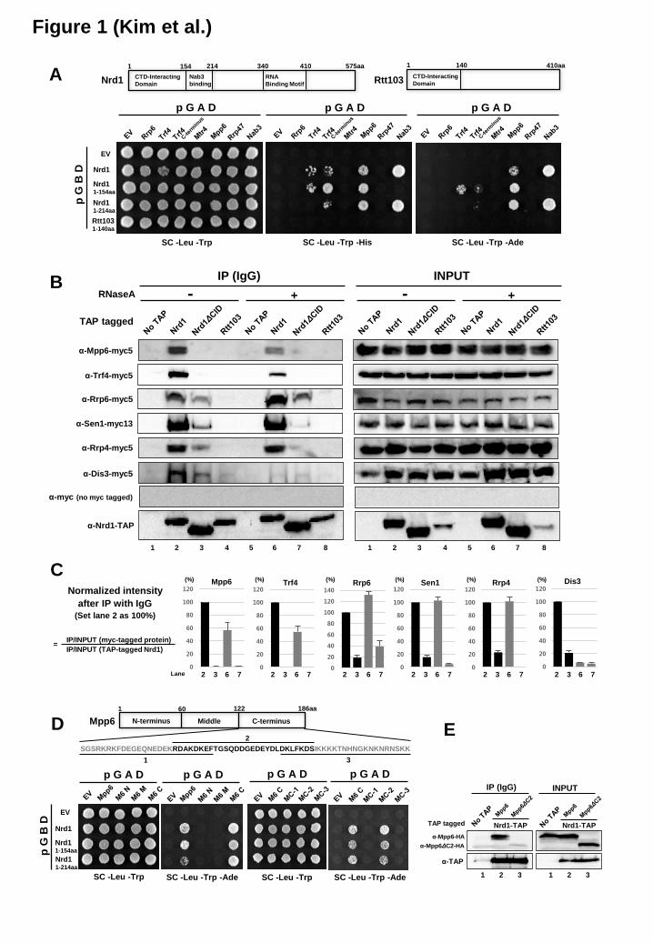
the Nrd1 CID – Replacing the Nrd1 CID with that of Rtt103 changes not only the CTD-binding specificity of Nrd1, but also the processing of Nrd1-terminated transcripts by the exosome because of reduced interaction of Nrd1 with Rrp6 and Trf4 (8). Deletion of the Nrd1 CID leads to considerable loss of interaction with Rrp6 and Trf4, suggesting that the Nrd1 CID recruits the nuclear exosome and TRAMP complex to the NNS complex. To identify components of nuclear exosome that recognize the Nrd1 CID, we performed the yeast two-hybrid (Y2H) screening using full-length or N-terminal CID (1-154aa) Nrd1 as bait, and found that Mpp6 and Trf4 specifically interact with the Nrd1 CID but not the Rtt103 CID (Figure 1A).
by guest on June 14, 2019http://w
ww
.jbc.org/D
ownloaded from

Connecting Nrd1 to the nuclear exosome
5
To validate the Y2H results, TAP-tagged Nrd1 proteins were precipitated with IgG beads, and associated exosome/TRAMP components (C-terminal 5x myc-tagged) were analyzed by western blotting. Mpp6 and Trf4 co-precipitated with Nrd1 but not with Nrd1ΔCID (Figure 1B and 1C, lanes 2 and 3). The association of Mpp6 and Trf4 with Nrd1 is relatively resistant to RNase A treatment, compared to Dis3 (Figure 1B and 1C, lanes 2 and 6), supporting protein-protein (either direct or indirect) interactions of Mpp6 and Trf4 with the Nrd1 CID. However, only about 50% of Mpp6 and Trf4 remains bound to Nrd1 in the presence of RNase A, indicating that RNAs may also play a role in reinforcing these interactions (Figure 1B and 1C, lanes 2 and 6). Structural evidence for interaction between the Trf4 C-terminus and the Nrd1 CID was also shown recently (19). Associations of Nrd1 with Rrp6, Sen1, and Rrp4 are completely resistant to RNase A treatment (Figure 1B and 1C, lanes 2 and 6), and significantly decreased (about 20% of intact Nrd1 case) upon deletion of the CID (Figure 1B and 1C, lanes 2 and 3, lanes 6 and 7), implying that the Nrd1 CID potentially along with other parts of Nrd1 and/or Nab3 may mediate the interaction with Rrp6, Sen1, and Rrp4, independently of RNA. In contrast, the interaction between Nrd1 and Dis3 relies upon RNA and the CID (Figure 1B and 1C, lanes 2-7).
We divided Mpp6 into several parts to narrow down the region interacting with the Nrd1 CID. A segment that contains 140-168 aa (MC-2) interacted with Nrd1 as well as full-length Mpp6 in the Y2H assay (Figure 1D). Indeed, this region is critical for interaction, as Nrd1 no longer associates with Mpp6 lacking the MC-2 (Figure 1E). This region has multiple glutamic acid residues, and is similar to the Nrd1-interacting motif (NIM) of Trf4 (Figure 2A).
Characterization of the Nrd1 CID-Mpp6 interaction – To analyze the binding of Nrd1 CID to Mpp6 and Trf4, backbone chemical shifts of the Nrd1 CID in the free state were initially obtained using three-dimensional heteronuclear correlation NMR spectroscopy. Then, chemical shift perturbations (CSP) were measured as 15N-Nrd1 CID was titrated with either the C-terminal domain of Mpp6 (65 aa residues as in Figure 1D, referred to as Mpp6 CTD) or the Trf4 NIM using 2D 1H−15N heteronuclear single quantum correlation spectroscopy. The CSP profiles were very similar between two titrations, indicating
that the Nrd1 CID largely employed the same binding interface for the Mpp6 CTD and the Trf4 NIM (Figure 2B). When residues with large CSPs were visualized on the three-dimensional structure of Nrd1, the interfaces were mainly located on α-helices 2, 4, and 7 (Figure 2C and 2D). It is notable that the interaction surfaces also overlap remarkably well with those observed in the complex formed between the Nrd1 CID and the Ser5P CTD of RNApII (Figure 2E). For example, Ser25 and Arg28 of the Nrd1 CID, which are critical for recognizing the Ser5P of CTD, showed the largest CSPs. In addition, Lys30, Ile68−Ser71, and Arg74 of the Nrd1 CID that are in close contact with the rest of Ser5P CTD exhibited large CSPs for interaction between the Nrd1 CID and the Mpp6 CTD. These results strongly indicate that the Nrd1 CID employs a largely overlapping binding surface to engage with multiple proteins, and that the bindings of Mpp6, Trf4, and Ser5P CTD to the Nrd1 CID are mutually exclusive with each other.
The characteristic electronegative stretch of Trf4 NIM is well maintained in the Mpp6 CTD (a red box in Figure 2A). The Ser5P CTD lacks this electronegative stretch, but instead harbors a phosphorylated serine residue that provides a key electrostatic interaction with Nrd1. The Trf4 NIM and the Ser5P CTD share a characteristic β-turn conformation next to the electronegative stretch, presenting the interaction surface for hydrophobic interaction with Nrd1 (a blue box in Figure 2A) (19,32). When we modeled the conformation of Mpp6 CTD based on the complex structure of Trf4 NIM and Nrd1 CID using PyMOL (The PyMOL Molecular Graphics System, Version 1.8 Schrödinger, LLC), Mpp6 in complex with Nrd1 is capable of adopting a similar β-turn conformation (Asp-Leu-Asp-Lys) to Trf4 (Asn-Pro-Tyr-Thr) with marginal backbone and side chain displacement (Figure 2F). Furthermore, the titration of Nrd1 with Mpp6 exhibited exchange broadening of Met126 and Leu127 (marked with red asterisks in Figure 2B), where Trf4 interacts with Nrd1. It is thus likely that Mpp6 employs a similar β-turn to maintain the hydrophobic interaction with Met126 and Leu127 of Nrd1.
We measured the binding affinity of Nrd1 CID to the Mpp6 CTD and the Trf4 NIM by isothermal titration calorimetry (ITC). The equilibrium dissociation constant (KD) between the Nrd1 CID and the Mpp6 CTD was 13.6 µM, and the binding between the Nrd1 CID and the
by guest on June 14, 2019http://w
ww
.jbc.org/D
ownloaded from

Connecting Nrd1 to the nuclear exosome
6
Trf4 NIM showed a KD of 5.7 µM (Figure 3A, 3B and Table 1). Considering that the affinity of Ser5P CTD (two heptapeptide repeats) for the Nrd1 CID was ~130-fold lower than that of the Trf4 NIM (19), both Mpp6 and Trf4 may be able to displace the Ser5P CTD of RNApII from the Nrd1 CID. To test this, we performed an in vitro binding competition assay using recombinant Nrd1 CID and Mpp6 proteins (Figure 3C). The Nrd1 CID was bound to the biotinylated Ser5P CTD peptide (three repeats) immobilized to streptavidin-coated magnetic beads. Adding a molar excess of Mpp6 to this CTD-Nrd1 complex led to a significant loss of the Nrd1 CID from the beads, proving that Mpp6 can indeed compete with Ser5P CTD toward the Nrd1 CID (Figure 3D). Similarly, the Nrd1 CID bound to the Trf4 NIM peptide was removed by increasing amounts of Mpp6, confirming the mutually exclusive bindings of Mpp6 and Trf4 to the Nrd1 CID.
Mpp6 interacts with Rrp6 – Human Mpp6 interacts directly with PM-Scl100 (human Rrp6) by Y2H and co-IP experiments (33,34). We consistently found that yeast Mpp6 also interacts with Rrp6 by Y2H assay (Figure 4A). Mpp6 and Rrp47 do not interact with each other, but instead bind to distinct domains within Rrp6: Mpp6 binds to the central EXO domain, while Rrp47 binds to the N-terminal PMC2NT domain (Figure 4A). This suggests that a stable Mpp6-Rrp6-Rrp47 complex can assemble as previously shown in vitro using human proteins (34). Importantly, the region (MC-2) within Mpp6 that binds to the Nrd1 CID turns out to mediate the interaction with Rrp6 as well (Figure 4B), suggesting that Mpp6 is not able to bind to Nrd1 and Rrp6 simultaneously. To verify this scenario, we performed an in vitro binding competition assay using recombinant Nrd1 CID and Rrp6-Exo (aa 103-391) proteins (Figure 4C). The Nrd1 CID was bound to the biotinylated Mpp6-C2 peptide (aa 140-167) immobilized to streptavidin-coated magnetic beads. Adding a molar excess of Rrp6-Exo to this Mpp6-Nrd1 complex led to a significant removal of the Nrd1 CID from the beads, indicating that Rrp6-Exo can indeed compete with the Nrd1 CID toward Mpp6-C2 (Figure 4D). This result suggests that Mpp6 interacts with Nrd1 and Rrp6, but not both of them simultaneously.
Roles of Mpp6 and Trf4 in coupling Nrd1 to RNA processing by the exosome – To investigate how Mpp6 and Trf4 affect the association of Nrd1
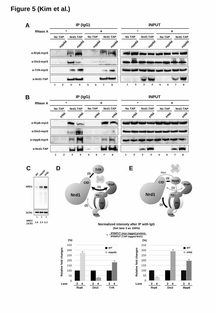
with the nuclear exosome, we performed co-IP experiments. Deletion of MPP6 increases the Nrd1-Rrp6 and Nrd1-Trf4 interactions, but decreases the Nrd1-Dis3 interaction which is RNA-mediated (Figure 5A). However, deletion of TRF4 leads to opposite outcomes: decrease in the Nrd1-Rrp6 interaction, but increase in the Nrd1-Dis3 and Nrd1-Mpp6 interactions (Figure 5B). Since the level of TAP-tagged Nrd1 increases in mpp6Δ and trf4Δ mutants presumably due to the increase of NRD1 mRNA level (Figure 5C), changes in the interactions of Nrd1 with various factors were normalized against the increase and shown in relative numbers based on three independent experiments (Figure 5D and 5E). These results are not only consistent with our competition model between Mpp6 and Trf4 toward Nrd1 (Figure 3D), but also revealing that Mpp6 promotes Nrd1 to associate with Dis3 via RNA, whereas Trf4 facilitates Nrd1 to associate with Rrp6 via protein-protein interaction. Presumably it might be, at least in part, due to mutually exclusive interaction of Nrd1 and Rrp6 with Mpp6 (Figure 4B and 4D).
We monitored how the altered association of Nrd1 with two exonucleases subsequently affects the RNA processing of Nrd1-terminated transcripts by Northern blotting. Since deleting both MPP6 and RRP6 causes cell lethality (21), we depleted Rrp6 using a repressible Gal promoter (pGal-RRP6) in a mpp6Δ mutant. Deletion of MPP6 alone had no obvious RNA processing defect, while depletion of Rrp6 led to small amounts of unprocessed snoRNA transcripts (pre-snoRNA, marked with black arrowhead in Figure 6). When depletions of these two factors were combined, the cells failed to grow on glucose and pre-snoRNAs were massively accumulated for both SNR13 and SNR33 (Figure 6A, compare lanes 6 and 8). In contrast, when mpp6Δ was combined with Dis3 depletion using pGal-DIS3, accumulation of pre-snoRNAs was not considerably increased relative to Dis3 depletion alone (Figure 6B, lanes 6 and 8). Based upon these Northern blot and co-IP results, one plausible interpretation would be that deletion of MPP6 renders the RNA processing of Nrd1-terminated transcripts more dependent upon Rrp6, as Mpp6 is no longer present to promote the association of Nrd1 and/or Nrd1-terminated transcripts with Dis3. The lack of significant additive effect between Mpp6 and Dis3 implies that these two proteins could be in the same pathway for processing Nrd1-terminated transcripts analyzed. However, we
by guest on June 14, 2019http://w
ww
.jbc.org/D
ownloaded from

Connecting Nrd1 to the nuclear exosome
7
cannot exclude that Mpp6 and Trf4 function redundantly in both pathways, therein Mpp6 could still stimulate degradation of Nrd1-terminated pre-snoRNA transcripts by Rrp6 at a certain level, as proposed in pre-rRNA processing (21,24). Nonetheless, mutually exclusive interaction of Mpp6 with Nrd1 and Rrp6 might significantly hamper the access of Rrp6 to Mpp6/Nrd1-bound transcripts (Figure 4B and 4D).
Double deletion of MPP6 and TRF4 resulted in a cell growth defect and synergistic accumulation of pre-snoRNAs (Figure 6C, lanes 2-4), indicating that Mpp6 and Trf4 have non-redundant roles in stimulating RNA processing by the exosome. TRAMP is known to enhance RNA degradation by Rrp6 independently of the core exosome (28), and Trf4 (a component of TRAMP) facilitates the association between Nrd1 and Rrp6 (Figure 5B). These findings propose that Trf4 and Rrp6 comprise a RNA processing pathway that could be parallel and/or partially redundant to the one with Mpp6 and Dis3 for degrading Nrd1-terminated transcripts (Figure 6D). To test this model, we simultaneously disrupted two proteins either in the same pathway or in the different pathway, and monitored the probable cumulative RNA processing defect. Indeed, mpp6Δ accumulated a much higher level of pre-snoRNAs and other non-coding RNAs (NEL025c and SUT145) when combined with pGal-RRP6 (different pathway) than pGal-DIS3 (same pathway) (Figure 6E and 6F, compare lanes 12 and 14). Similarly, trf4Δ produced greater amounts of premature transcripts and non-coding RNAs with pGal-DIS3 (different pathway) than pGal-RRP6 (same pathway) (Figure 6E and 6F, compare lanes 16 and 18). Although we do not rule out that Trf4 and Mpp6 function to stimulate both Rrp6 and Dis3, to some extent, these results imply that Mpp6 promotes degradation of Nrd1-terminated transcripts by Dis3, while Trf4 does so by Rrp6. Based on our results, we propose a model that Mpp6 and Trf4 play a role, at least in part, in selecting a RNA processing route by directing transcripts to their associated exonuclease (Figure 7).
DISCUSSION The CID of Nrd1 contributes to recruiting
Nrd1 to early elongating RNApII by recognizing Ser5P CTD of the polymerase (6). Deletion of the CID has a strong impact on Nrd1 recruitment, but also greatly reduces the interaction with Rrp6 and
Trf4 (8). Thus, the Nrd1 CID couples termination with subsequent RNA processing by recruiting the exosome. However, the molecular basis for this coupling was not clearly understood. In this work, we found that the nuclear exosome cofactors Mpp6 and Trf4 bind to the Nrd1 CID, bridging Nrd1 and/or Nrd1-terminated transcripts to the exosome. Furthermore, the interaction of Nrd1 with these two proteins seems to affect the exonucleolytic route within the exosome for processing of Nrd1-terminated transcripts.
Our structural analysis demonstrates that the Nrd1 CID has a common binding surface for Ser5P CTD of RNApII, Mpp6, and Trf4, and that these three proteins share a similar motif with a β-turn conformation (Figure 2). It indicates that they bind to the Nrd1 CID in a mutually exclusive manner, which was confirmed by binding competition experiments (Figure 3D). Unfortunately, we could not detect chromatin immunoprecipitation signals for Mpp6 and Trf4 on several genes tested, implying that their interactions with Nrd1 may be transient and/or occur off the chromatin after termination. In a simple model, the Nrd1 CID would first bind to Ser5P CTD soon after RNApII begins transcription at the 5’-ends of genes, and then be handed over to either Mpp6 or Trf4 once RNApII terminates at 3’-ends to set out RNA processing by the exosome.
The association with Nrd1 and subsequent RNA processing by Rrp6 and Dis3 were significantly but differentially affected by deletion of MPP6 or TRF4 (Figure 5 and 6). These results suggest there are two parallel and/or partially overlapping RNA processing pathways for Nrd1-terminated transcripts (Figure 7). Reconstituted exosome complex revealed that the RNA degradation route to Rrp6 or Dis3 is randomly chosen (i.e. stochastic) in vitro (15), but RNAs are often preferentially degraded by one or the other exonuclease in vivo, indicating that some exosome-associated factors may affect the route selection. Based upon our results, we propose that Mpp6 and Trf4 play a role in choosing a particular RNA processing route. When Mpp6 binds to the Nrd1 CID, it would not only preclude Trf4 from interacting with the CID, but it also would block the association between Mpp6 and Rrp6 since Mpp6 cannot accommodate both Nrd1 and Rrp6 simultaneously due to a common binding motif (MC-2) (Figure 1C, 1D, and Figure 4). Thus, Mpp6 could guide the RNAs to Dis3-dependent processing (Figure 7). Along the same line, the mpp6Δ mutation is
by guest on June 14, 2019http://w
ww
.jbc.org/D
ownloaded from

Connecting Nrd1 to the nuclear exosome
8
synthetic lethal with rrp47Δ and rrp6Δ mutations (21), making it unlikely that Mpp6 functions with Rrp47 in targeting RNA substrates to Rrp6. In this sense, Mpp6 has been previously proposed to stimulate the activity of Dis3 (21). A recent report that Mpp6 directly binds to the core exosome independently of Rrp6 may also reflect a role of Mpp6 in Dis3-mediated RNA degradation (23). On the other hand, binding of Trf4 to the Nrd1 CID would exclude Mpp6 and lead to Rrp6-dependent processing of the transcripts via Trf4-Rrp6 and/or Trf4-Mtr4-Rrp6/Rrp47 interactions (19,23). Consistently, deletion of MPP6 increases the interaction of Nrd1 with Rrp6 (Figure 5A and 5C), and makes the RNA processing of Nrd1-terminated transcripts more dependent upon Rrp6 (Figure 6A, 6E and 6F).
Although our results suggest a ‘two parallel (Mpp6-Dis3 and Trf4-Rrp6) pathways’ model for processing Nrd1-terminated transcripts, we do not rule out that there might be a partially overlapping role of Mpp6 and Trf4 in stimulating both Rrp6 and Dis3. For instance, Mpp6 may interact with Mtr4 as seen in human cells (33,34), and thus still indirectly stimulate the activity of Rrp6. To make it clear, our results do not exclude the role of Mpp6 in Rrp6-mediated processing of various RNA substrates. We propose that the contribution of Mpp6 to Rrp6-mediated processing could be relatively smaller in Nrd1-terminated transcripts than other RNA substrates due to mutually exclusive interaction of Nrd1 and Rrp6 with Mpp6. A common binding motif for Nrd1 and Rrp6 within Mpp6 predicts that Mpp6 is able to associate with Rrp6 independently of Nrd1. Considering that Mpp6 binds to the EXO domain of Rrp6 (Figure 4), Mpp6 may facilitate
the entry of the RNA substrate into the catalytic center of Rrp6. It is unclear whether Mpp6 interacts with Rrp6 alone or along with the core exosome and/or TRAMP complex. In either case, when unbound to Nrd1, Mpp6 may assist Rrp6 to degrade or trim various RNA substrates (including 5.8S rRNA precursors) through interaction with Rrp6 and/or RNAs. But Mpp6 may still be able to stimulate Dis3-mediated RNA processing, independently of Nrd1.
It would be interesting to uncover how the competition between Mpp6 and Trf4 toward the Nrd1 CID is regulated at 3’-ends. Since Trf4 is more abundant (1.9 or 5.6-fold higher protein molecules/cell) than Mpp6 under normal growth condition (29,35), it may predict that Trf4-Rrp6 is a more dominant pathway for processing Nrd1-terminated transcripts. However, given that Mpp6 is a RNA-binding protein with a preference for poly(U) and poly(C) but not for poly(A) (21,24), pyrimidine-rich RNA sequences at 3’-ends may favor Mpp6 over Trf4. Another interesting feature that potentially biases the competition might be phosphorylation. According to the phosphoGRID database (http://www.phosphogrid.org), the regions within Mpp6 and Trf4 that interact with the Nrd1 CID contain experimentally verified in vivo phosphorylation sites (T148 and S150 in Mpp6, S570 and S571 in Trf4) (36,37). Phosphorylation of these residues might considerably affect the interactions of Mpp6 and Trf4 with Nrd1, as seen for the CTD of RNApII. How the phosphorylation affects the interactions and when it is added and removed remain to be investigated in future studies.
ACKNOWLEDGEMENTS We thank Stephen Buratowski (Harvard Medical School) for providing Y2H reagents (library, strain, and plasmids) and helpful comments; Won-Ki Huh (Seoul National University) for TAP-tagged strains and epitope switching cassettes; and the Korea Basic Science Institute and the National Center for Inter-university Research Facilities for high-field NMR facility support. CONFLICT of INTEREST The authors declare that they have no conflict of interest with the contents of this article. AUTHOR CONTRIBUTIONS KK and DH generated plasmids/strains and performed co-IP, and Northern blot experiments. KK and IK purified proteins. KK performed Y2H and in vitro binding competition assays. IK performed CSP and ITC experiments. KK and MK designed the experiments. JS and MK supervised the research. KK, JS, and MK wrote the paper.
by guest on June 14, 2019http://w
ww
.jbc.org/D
ownloaded from
Connecting Nrd1 to the nuclear exosome
9
REFERENCES
1. Steinmetz, E. J., Conrad, N. K., Brow, D. A., and Corden, J. L. (2001) RNA-binding protein Nrd1 directs poly(A)-independent 3'-end formation of RNA polymerase II transcripts. Nature 413, 327-331
2. Arigo, J. T., Eyler, D. E., Carroll, K. L., and Corden, J. L. (2006) Termination of cryptic unstable transcripts is directed by yeast RNA-binding proteins Nrd1 and Nab3. Mol Cell 23, 841-851
3. Thiebaut, M., Kisseleva-Romanova, E., Rougemaille, M., Boulay, J., and Libri, D. (2006) Transcription termination and nuclear degradation of cryptic unstable transcripts: a role for the nrd1-nab3 pathway in genome surveillance. Mol Cell 23, 853-864
4. Kim, M., Vasiljeva, L., Rando, O. J., Zhelkovsky, A., Moore, C., and Buratowski, S. (2006) Distinct pathways for snoRNA and mRNA termination. Mol Cell 24, 723-734
5. Porrua, O., and Libri, D. (2013) A bacterial-like mechanism for transcription termination by the Sen1p helicase in budding yeast. Nat Struct Mol Biol 20, 884-891
6. Vasiljeva, L., Kim, M., Mutschler, H., Buratowski, S., and Meinhart, A. (2008) The Nrd1-Nab3-Sen1 termination complex interacts with the Ser5-phosphorylated RNA polymerase II C-terminal domain. Nat Struct Mol Biol 15, 795-804
7. Buratowski, S. (2009) Progression through the RNA polymerase II CTD cycle. Mol Cell 36, 541-546
8. Heo, D. H., Yoo, I., Kong, J., Lidschreiber, M., Mayer, A., Choi, B. Y., Hahn, Y., Cramer, P., Buratowski, S., and Kim, M. (2013) The RNA polymerase II C-terminal domain-interacting domain of yeast Nrd1 contributes to the choice of termination pathway and couples to RNA processing by the nuclear exosome. J Biol Chem 288, 36676-36690
9. Vasiljeva, L., and Buratowski, S. (2006) Nrd1 interacts with the nuclear exosome for 3' processing of RNA polymerase II transcripts. Mol Cell 21, 239-248
10. Butler, J. S. (2002) The yin and yang of the exosome. Trends Cell Biol 12, 90-96 11. Liu, Q., Greimann, J. C., and Lima, C. D. (2006) Reconstitution, activities, and structure of the
eukaryotic RNA exosome. Cell 127, 1223-1237 12. Lykke-Andersen, S., Brodersen, D. E., and Jensen, T. H. (2009) Origins and activities of the
eukaryotic exosome. J Cell Sci 122, 1487-1494 13. Houseley, J., and Tollervey, D. (2009) The many pathways of RNA degradation. Cell 136, 763-
776 14. Schneider, C., and Tollervey, D. (2013) Threading the barrel of the RNA exosome. Trends
Biochem Sci 38, 485-493 15. Wasmuth, E. V., Januszyk, K., and Lima, C. D. (2014) Structure of an Rrp6-RNA exosome
complex bound to poly(A) RNA. Nature 511, 435-439 16. Makino, D. L., Schuch, B., Stegmann, E., Baumgartner, M., Basquin, C., and Conti, E. (2015)
RNA degradation paths in a 12-subunit nuclear exosome complex. Nature 524, 54-58 17. Vanacova, S., Wolf, J., Martin, G., Blank, D., Dettwiler, S., Friedlein, A., Langen, H., Keith, G.,
and Keller, W. (2005) A new yeast poly(A) polymerase complex involved in RNA quality control. PLoS Biol 3, e189
18. Wyers, F., Rougemaille, M., Badis, G., Rousselle, J. C., Dufour, M. E., Boulay, J., Regnault, B., Devaux, F., Namane, A., Seraphin, B., Libri, D., and Jacquier, A. (2005) Cryptic pol II transcripts are degraded by a nuclear quality control pathway involving a new poly(A) polymerase. Cell 121, 725-737
19. Tudek, A., Porrua, O., Kabzinski, T., Lidschreiber, M., Kubicek, K., Fortova, A., Lacroute, F., Vanacova, S., Cramer, P., Stefl, R., and Libri, D. (2014) Molecular basis for coordinating transcription termination with noncoding RNA degradation. Mol Cell 55, 467-481
by guest on June 14, 2019http://w
ww
.jbc.org/D
ownloaded from
Connecting Nrd1 to the nuclear exosome
10
20. Mitchell, P., Petfalski, E., Houalla, R., Podtelejnikov, A., Mann, M., and Tollervey, D. (2003) Rrp47p is an exosome-associated protein required for the 3' processing of stable RNAs. Mol Cell Biol 23, 6982-6992
21. Milligan, L., Decourty, L., Saveanu, C., Rappsilber, J., Ceulemans, H., Jacquier, A., and Tollervey, D. (2008) A yeast exosome cofactor, Mpp6, functions in RNA surveillance and in the degradation of noncoding RNA transcripts. Mol Cell Biol 28, 5446-5457
22. Stead, J. A., Costello, J. L., Livingstone, M. J., and Mitchell, P. (2007) The PMC2NT domain of the catalytic exosome subunit Rrp6p provides the interface for binding with its cofactor Rrp47p, a nucleic acid-binding protein. Nucleic Acids Res 35, 5556-5567
23. Schuch, B., Feigenbutz, M., Makino, D. L., Falk, S., Basquin, C., Mitchell, P., and Conti, E. (2014) The exosome-binding factors Rrp6 and Rrp47 form a composite surface for recruiting the Mtr4 helicase. EMBO J 33, 2829-2846
24. Schilders, G., Raijmakers, R., Raats, J. M., and Pruijn, G. J. (2005) MPP6 is an exosome-associated RNA-binding protein involved in 5.8S rRNA maturation. Nucleic Acids Res 33, 6795-6804
25. Allmang, C., Mitchell, P., Petfalski, E., and Tollervey, D. (2000) Degradation of ribosomal RNA precursors by the exosome. Nucleic Acids Res 28, 1684-1691
26. van Hoof, A., Lennertz, P., and Parker, R. (2000) Yeast exosome mutants accumulate 3'-extended polyadenylated forms of U4 small nuclear RNA and small nucleolar RNAs. Mol Cell Biol 20, 441-452
27. Gudipati, R. K., Xu, Z., Lebreton, A., Seraphin, B., Steinmetz, L. M., Jacquier, A., and Libri, D. (2012) Extensive degradation of RNA precursors by the exosome in wild-type cells. Mol Cell 48, 409-421
28. Callahan, K. P., and Butler, J. S. (2008) Evidence for core exosome independent function of the nuclear exoribonuclease Rrp6p. Nucleic Acids Res 36, 6645-6655
29. Ghaemmaghami, S., Huh, W. K., Bower, K., Howson, R. W., Belle, A., Dephoure, N., O'Shea, E. K., and Weissman, J. S. (2003) Global analysis of protein expression in yeast. Nature 425, 737-741
30. Sung, M. K., Ha, C. W., and Huh, W. K. (2008) A vector system for efficient and economical switching of C-terminal epitope tags in Saccharomyces cerevisiae. Yeast 25, 301-311
31. James, P., Halladay, J., and Craig, E. A. (1996) Genomic libraries and a host strain designed for highly efficient two-hybrid selection in yeast. Genetics 144, 1425-1436
32. Kubicek, K., Cerna, H., Holub, P., Pasulka, J., Hrossova, D., Loehr, F., Hofr, C., Vanacova, S., and Stefl, R. (2012) Serine phosphorylation and proline isomerization in RNAP II CTD control recruitment of Nrd1. Genes Dev 26, 1891-1896
33. Lehner, B., and Sanderson, C. M. (2004) A protein interaction framework for human mRNA degradation. Genome Res 14, 1315-1323
34. Schilders, G., van Dijk, E., and Pruijn, G. J. (2007) C1D and hMtr4p associate with the human exosome subunit PM/Scl-100 and are involved in pre-rRNA processing. Nucleic Acids Res 35, 2564-2572
35. Kulak, N. A., Pichler, G., Paron, I., Nagaraj, N., and Mann, M. (2014) Minimal, encapsulated proteomic-sample processing applied to copy-number estimation in eukaryotic cells. Nat Methods 11, 319-324
36. Bodenmiller, B., Wanka, S., Kraft, C., Urban, J., Campbell, D., Pedrioli, P. G., Gerrits, B., Picotti, P., Lam, H., Vitek, O., Brusniak, M. Y., Roschitzki, B., Zhang, C., Shokat, K. M., Schlapbach, R., Colman-Lerner, A., Nolan, G. P., Nesvizhskii, A. I., Peter, M., Loewith, R., von Mering, C., and Aebersold, R. (2010) Phosphoproteomic analysis reveals interconnected system-wide responses to perturbations of kinases and phosphatases in yeast. Sci Signal 3, rs4
37. Soufi, B., Kelstrup, C. D., Stoehr, G., Frohlich, F., Walther, T. C., and Olsen, J. V. (2009) Global analysis of the yeast osmotic stress response by quantitative proteomics. Mol Biosyst 5, 1337-1346
by guest on June 14, 2019http://w
ww
.jbc.org/D
ownloaded from
Connecting Nrd1 to the nuclear exosome
11
38. Rasmussen, T. P., and Culbertson, M. R. (1998) The putative nucleic acid helicase Sen1p is required for formation and stability of termini and for maximal rates of synthesis and levels of accumulation of small nucleolar RNAs in Saccharomyces cerevisiae. Mol Cell Biol 18, 6885-6896
FOOTNOTES This work was supported by the Cancer Cell Map Initiative and the California Institute for Quantitative Biosciences at UCSF (to MK); and the National Research Foundation of Korea [Basic Science Research Program Grants 2013R1A1A2A10013757 to MK, 2013R1A1A2010856 to JS] The abbreviations used are: RNApII, RNA polymerase II; CTD, C-terminal domain; Ser5P, phosphorylated serine 5; CID, CTD-interacting domain; NIM, Nrd1-interacting motif; co-IP, co-immunoprecipitation; snoRNA, small nucleolar RNA.
by guest on June 14, 2019http://w
ww
.jbc.org/D
ownloaded from
Connecting Nrd1 to the nuclear exosome
12
FIGURE LEGENDS
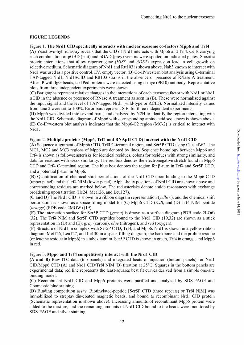
Figure 1. The Nrd1 CID specifically interacts with nuclear exosome co-factors Mpp6 and Trf4 (A) Yeast two-hybrid assay reveals that the CID of Nrd1 interacts with Mpp6 and Trf4. Cells carrying each combination of pGBD (bait) and pGAD (prey) vectors were spotted on indicated plates. Specific protein interactions that allow reporter gene (HIS3 and ADE2) expression lead to cell growth on selective medium. Schematic diagram of Nrd1 and Rtt103 is shown above. Nab3 known to interact with Nrd1 was used as a positive control. EV, empty vector. (B) Co-IP/western blot analysis using C-terminal TAP-tagged Nrd1, Nrd1ΔCID and Rtt103 strains in the absence or presence of RNase A treatment. After IP with IgG beads, co-IPed proteins were detected using α-myc (9E10) antibody. Representative blots from three independent experiments were shown. (C) Bar graphs represent relative changes in the interactions of each exosome factor with Nrd1 or Nrd1 ΔCID in the absence or presence of RNase A treatment as seen in (B). These were normalized against the input signal and the level of TAP-tagged Nrd1 (wild-type or ΔCID). Normalized intensity values from lane 2 were set to 100%. Error bars represent S.E. for three independent experiments. (D) Mpp6 was divided into several parts, and analyzed by Y2H to identify the region interacting with the Nrd1 CID. Schematic diagram of Mpp6 with corresponding amino acid sequences is shown above. (E) Co-IP/western blot analysis indicates that the Mpp6-C2 region (MC-2) is critical to interact with Nrd1. Figure 2. Multiple proteins (Mpp6, Trf4 and RNApII CTD) interact with the Nrd1 CID (A) Sequence alignment of Mpp6 CTD, Trf4 C-terminal region, and Ser5P CTD using ClustalW2. The MC1, MC2 and MC3 regions of Mpp6 are denoted by lines. Sequence homology between Mpp6 and Trf4 is shown as follows: asterisks for identical residues, colons for residues with strong similarity, and dots for residues with weak similarity. The red box denotes the electronegative stretch found in Mpp6 CTD and Trf4 C-terminal region. The blue box denotes the region for β-turn in Trf4 and Ser5P CTD, and a potential β-turn in Mpp6. (B) Quantification of chemical shift perturbations of the Nrd1 CID upon binding to the Mpp6 CTD (upper panel) and the Trf4 NIM (lower panel). Alpha-helix positions of Nrd1 CID are shown above and corresponding residues are marked below. The red asterisks denote amide resonances with exchange broadening upon titration (Ile24, Met126, and Leu127). (C and D) The Nrd1 CID is shown in a ribbon diagram representation (yellow), and the chemical shift perturbation is shown as a space-filling model for (C) Mpp6 CTD (red), and (D) Trf4 NIM peptide (orange) (PDB code 2MOW) (19). (E) The interaction surface for Ser5P CTD (green) is drawn as a surface diagram (PDB code 2LO6) (32). The Trf4 NIM and Ser5P CTD peptides bound to the Nrd1 CID (19,32) are shown as a stick representation in (D) and (E): gray (carbon), blue (nitrogen), and red (oxygen). (F) Structure of Nrd1 in complex with Ser5P CTD, Trf4, and Mpp6. Nrd1 is shown in a yellow ribbon diagram; Met126, Leu127, and Ile130 in a space-filling diagram; the backbone and the proline residue (or leucine residue in Mpp6) in a tube diagram. Ser5P CTD is shown in green, Trf4 in orange, and Mpp6 in red. Figure 3. Mpp6 and Trf4 competitively interact with the Nrd1 CID (A and B) Raw ITC data (top panels) and integrated heats of injection (bottom panels) for Nrd1 CID/Mpp6 CTD (A) and Nrd1 CID/Trf4 NIM (B) titration at 25°C. Squares in the bottom panels are experimental data; red line represents the least-squares best fit curves derived from a simple one-site binding model. (C) Recombinant Nrd1 CID and Mpp6 proteins were purified and analyzed by SDS-PAGE and Coomassie blue staining. (D) Binding competition assay. Biotinylated-peptide [Ser5P CTD (three repeats) or Trf4 NIM] was immobilized to streptavidin-coated magnetic beads, and bound to recombinant Nrd1 CID protein (Schematic representation is shown above). Increasing amounts of recombinant Mpp6 protein were added to the mixture, and the remaining amounts of Nrd1 CID bound to the beads were monitored by SDS-PAGE and silver staining.
by guest on June 14, 2019http://w
ww
.jbc.org/D
ownloaded from
Connecting Nrd1 to the nuclear exosome
13
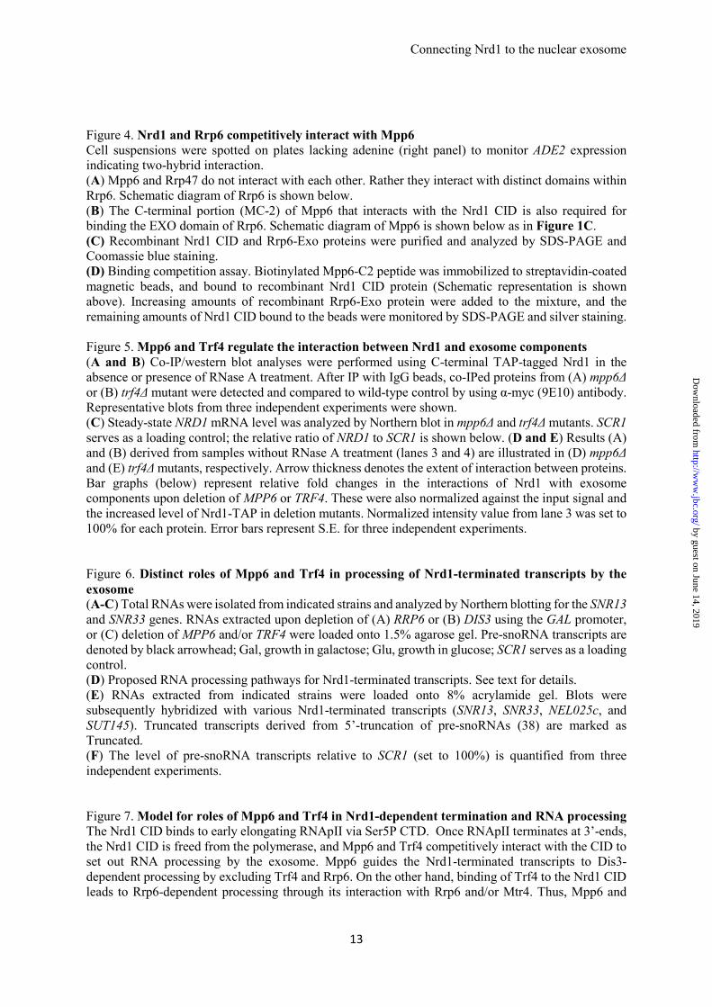
Figure 4. Nrd1 and Rrp6 competitively interact with Mpp6 Cell suspensions were spotted on plates lacking adenine (right panel) to monitor ADE2 expression indicating two-hybrid interaction. (A) Mpp6 and Rrp47 do not interact with each other. Rather they interact with distinct domains within Rrp6. Schematic diagram of Rrp6 is shown below. (B) The C-terminal portion (MC-2) of Mpp6 that interacts with the Nrd1 CID is also required for binding the EXO domain of Rrp6. Schematic diagram of Mpp6 is shown below as in Figure 1C. (C) Recombinant Nrd1 CID and Rrp6-Exo proteins were purified and analyzed by SDS-PAGE and Coomassie blue staining. (D) Binding competition assay. Biotinylated Mpp6-C2 peptide was immobilized to streptavidin-coated magnetic beads, and bound to recombinant Nrd1 CID protein (Schematic representation is shown above). Increasing amounts of recombinant Rrp6-Exo protein were added to the mixture, and the remaining amounts of Nrd1 CID bound to the beads were monitored by SDS-PAGE and silver staining. Figure 5. Mpp6 and Trf4 regulate the interaction between Nrd1 and exosome components (A and B) Co-IP/western blot analyses were performed using C-terminal TAP-tagged Nrd1 in the absence or presence of RNase A treatment. After IP with IgG beads, co-IPed proteins from (A) mpp6Δ or (B) trf4Δ mutant were detected and compared to wild-type control by using α-myc (9E10) antibody. Representative blots from three independent experiments were shown. (C) Steady-state NRD1 mRNA level was analyzed by Northern blot in mpp6Δ and trf4Δ mutants. SCR1 serves as a loading control; the relative ratio of NRD1 to SCR1 is shown below. (D and E) Results (A) and (B) derived from samples without RNase A treatment (lanes 3 and 4) are illustrated in (D) mpp6Δ and (E) trf4Δ mutants, respectively. Arrow thickness denotes the extent of interaction between proteins. Bar graphs (below) represent relative fold changes in the interactions of Nrd1 with exosome components upon deletion of MPP6 or TRF4. These were also normalized against the input signal and the increased level of Nrd1-TAP in deletion mutants. Normalized intensity value from lane 3 was set to 100% for each protein. Error bars represent S.E. for three independent experiments. Figure 6. Distinct roles of Mpp6 and Trf4 in processing of Nrd1-terminated transcripts by the exosome (A-C) Total RNAs were isolated from indicated strains and analyzed by Northern blotting for the SNR13 and SNR33 genes. RNAs extracted upon depletion of (A) RRP6 or (B) DIS3 using the GAL promoter, or (C) deletion of MPP6 and/or TRF4 were loaded onto 1.5% agarose gel. Pre-snoRNA transcripts are denoted by black arrowhead; Gal, growth in galactose; Glu, growth in glucose; SCR1 serves as a loading control. (D) Proposed RNA processing pathways for Nrd1-terminated transcripts. See text for details. (E) RNAs extracted from indicated strains were loaded onto 8% acrylamide gel. Blots were subsequently hybridized with various Nrd1-terminated transcripts (SNR13, SNR33, NEL025c, and SUT145). Truncated transcripts derived from 5’-truncation of pre-snoRNAs (38) are marked as Truncated. (F) The level of pre-snoRNA transcripts relative to SCR1 (set to 100%) is quantified from three independent experiments. Figure 7. Model for roles of Mpp6 and Trf4 in Nrd1-dependent termination and RNA processing The Nrd1 CID binds to early elongating RNApII via Ser5P CTD. Once RNApII terminates at 3’-ends, the Nrd1 CID is freed from the polymerase, and Mpp6 and Trf4 competitively interact with the CID to set out RNA processing by the exosome. Mpp6 guides the Nrd1-terminated transcripts to Dis3-dependent processing by excluding Trf4 and Rrp6. On the other hand, binding of Trf4 to the Nrd1 CID leads to Rrp6-dependent processing through its interaction with Rrp6 and/or Mtr4. Thus, Mpp6 and
by guest on June 14, 2019http://w
ww
.jbc.org/D
ownloaded from

Connecting Nrd1 to the nuclear exosome
14
Trf4 play a key role in choosing a particular RNA processing route within the exosome. Proteins are not drawn to scale. See Discussion for details.
by guest on June 14, 2019http://w
ww
.jbc.org/D
ownloaded from

Connecting Nrd1 to the nuclear exosome
15
Table 1. Yeast Strains Used in This Study Strain Genotype Source
YF1(BY4741) YF47 YF88 YF120/pJ69-4A YF86/YSB2084 YMK117 YMK160 YMK161 YMK162 YMK165 YMK166 YMK167 YMK168 YMK178 YMK179 YMK180 YMK197 YMK201 YMK203 YMK204 YMK205 YMK206 YMK233 YMK238 YMK242 YMK244 YMK245 YMK247 YMK249 YMK250 YMK252 YMK254 YMK292 YMK293 YMK322 YMK326 YMK327 YMK328 YMK334 YMK423 YMK424 YMK430 YMK431 YMK432 YMK435 YMK436 YMK437 YMK438 YMK444 YMK445 YMK446
MATa, ura3∆0, leu2∆0, his3∆1, met15∆0 MATa, ura3∆0, leu2∆0, his3∆1, met15∆0, rrp6∆::KanMX MATa, ura3∆0, leu2∆0, his3∆1, met15∆0, mpp6∆::KanMX MATa, ura3-52, leu2-3,112, trp1-901, his3∆200, gal4∆, gal80∆, GAL2-ADE2, LYS2::GAL1-HIS3, met2::GAL7-lacZ MATa, ura3-52, leu2∆1, trp1∆63, SEN1-Myc13::KanMX6 MATa, ura3-52, leu2∆1, trp1∆63, NRD1-TAP::HIS3, SEN1-Myc13::KanMX6 MATa, ura3∆0, leu2∆0, his3∆1, met15∆0, DIS3-Myc5::KlURA3 MATa, ura3∆0, leu2∆0, his3∆1, met15∆0, RRP4-Myc5::KlURA3 MATa, ura3∆0, leu2∆0, his3∆1, met15∆0, RRP6-Myc5::KlURA3 MATa, ura3∆0, leu2∆0, his3∆1, met15∆0, TRF4-Myc5::KlURA3 MATa, ura3∆0, leu2∆0, his3∆1, met15∆0, NRD1-TAP::HIS3, DIS3-Myc5::KlURA3 MATa, ura3∆0, leu2∆0, his3∆1, met15∆0, NRD1-TAP::HIS3, RRP4-Myc5::KlURA3 MATa, ura3∆0, leu2∆0, his3∆1, met15∆0, NRD1-TAP::HIS3, RRP6-Myc5::KlURA3 MATa, ura3∆0, leu2∆0, his3∆1, met15∆0, RTT103-TAP::HIS3, DIS3-Myc5::KlURA3 MATa, ura3∆0, leu2∆0, his3∆1, met15∆0, RTT103-TAP::HIS3, RRP4-Myc5::KlURA3 MATa, ura3∆0, leu2∆0, his3∆1, met15∆0, RTT103-TAP::HIS3, RRP6-Myc5::KlURA3 MATa, ura3∆0, leu2∆0, his3∆1, met15∆0, NRD1-TAP::HIS3, TRF4-Myc5::KlURA3 MATa, ura3∆0, leu2∆0, his3∆1, met15∆0, RTT103-TAP::HIS3, TRF4-Myc5::KlURA3 MATa, ura3∆0, leu2∆0, his3∆1, met15∆0, nrd1∆6-150- TAP tag::HIS3, TRF4-Myc5::KlURA3 MATa, ura3∆0, leu2∆0, his3∆1, met15∆0, nrd1∆6-150- TAP tag::HIS3, DIS3-Myc5::KlURA3 MATa, ura3∆0, leu2∆0, his3∆1, met15∆0, nrd1∆6-150- TAP tag::HIS3, RRP4-Myc5::KlURA3 MATa, ura3∆0, leu2∆0, his3∆1, met15∆0, nrd1∆6-150- TAP tag::HIS3, RRP6-Myc5::KlURA3 MATa, ura3∆0, leu2∆0, his3∆1, met15∆0, MPP6-Myc5::KlURA3 MATa, ura3∆0, leu2∆0, his3∆1, met15∆0, NRD1-TAP::HIS3, MPP6-Myc5::KlURA3 MATa, ura3∆0, leu2∆0, his3∆1, met15∆0, RTT103- TAP tag::HIS3, MPP6-Myc5::KlURA3 MATa, ura3∆0, leu2∆0, his3∆1, met15∆0, nrd1∆6-150- TAP tag::HIS3, MPP6-Myc5::KlURA3 MATa, ura3∆0, leu2∆0, his3∆1, met15∆0, DIS3-Myc5::KlURA3, mpp6∆::KanMX MATa, ura3∆0, leu2∆0, his3∆1, met15∆0, RRP6-Myc5::KlURA3, mpp6∆::KanMX MATa, ura3∆0, leu2∆0, his3∆1, met15∆0, TRF4-Myc5::KlURA3, mpp6∆::KanMX MATa, ura3∆0, leu2∆0, his3∆1, met15∆0, NRD1-TAP::HIS3, DIS3-Myc5::KlURA3, mpp6∆::KanMX MATa, ura3∆0, leu2∆0, his3∆1, met15∆0, NRD1-TAP::HIS3, RRP6-Myc5::KlURA3, mpp6∆::KanMX MATa, ura3∆0, leu2∆0, his3∆1, met15∆0, NRD1-TAP::HIS3, TRF4-Myc5::KlURA3, mpp6∆::KanMX MATa, ura3∆0, leu2∆0, his3∆1, met15∆0, HIS3::pGAL-RRP6 MATa, ura3∆0, leu2∆0, his3∆1, met15∆0, mpp6∆::KanMX, HIS3::pGAL-RRP6 MATa, ura3∆0, leu2∆0, his3∆1, met15∆0, trf4∆::KanMX, mpp6∆::HPH, pRS416-MPP6 MATa, ura3∆0, leu2∆0, his3∆1, met15∆0, trf4∆::KanMX, RRP6-Myc5::KlURA3 MATa, ura3∆0, leu2∆0, his3∆1, met15∆0, NRD1-TAP::HIS3, trf4∆::KanMX, RRP6-Myc5::KlURA3 MATa, ura3∆0, leu2∆0, his3∆1, met15∆0, trf4∆::KanMX, MPP6-Myc5::KlURA3 MATa, ura3∆0, leu2∆0, his3∆1, met15∆0, NRD1-TAP::HIS3, trf4∆::KanMX, MPP6-Myc5::KlURA3 MATa, ura3∆0, leu2∆0, his3∆1, met15∆0, nrd1∆6-150-TAP tag::HIS3, SEN1-Myc13::KanMX MATa, ura3∆0, leu2∆0, his3∆1, met15∆0, RTT103-TAP tag::HIS3, SEN1-Myc13::KanMX MATa, ura3∆0, leu2∆0, his3∆1, met15∆0, HIS3::pGAL-DIS3 MATa, ura3∆0, leu2∆0, his3∆1, met15∆0, mpp6∆::KanMX, HIS3::pGAL-DIS3 MATa, ura3∆0, leu2∆0, his3∆1, met15∆0, trf4∆::KanMX, HIS3::pGAL-DIS3 MATa, ura3∆0, leu2∆0, his3∆1, met15∆0, trf4∆::KanMX, HIS3::pGAL-RRP6 MATa, ura3∆0, leu2∆0, his3∆1, met15∆0, rrp6∆::KanMX, HIS3::pGAL-DIS3 MATa, ura3∆0, leu2∆0, his3∆1, met15∆0, trf4∆::KanMX, DIS3-Myc5::KlURA3 MATa, ura3∆0, leu2∆0, his3∆1, met15∆0, NRD1-TAP tag::HIS3, trf4∆::KanMX, DIS3-Myc5::KlURA3 MATa, ura3∆0, leu2∆0, his3∆1, met15∆0, mpp6∆::KanMX, pRS315-MPP6-HA3 MATa, ura3∆0, leu2∆0, his3∆1, met15∆0, NRD1-TAP tag::HIS3, mpp6∆::KanMX, pRS315-MPP6-HA3 MATa, ura3∆0, leu2∆0, his3∆1, met15∆0, NRD1-TAP tag::HIS3, mpp6∆::KanMX, pRS315-MPP6∆C2-HA3
(29) (29) (29) (31) (8) (8) (8) (8) (8) (8) (8) (8) (8) (8) (8) (8) (8) (8) (8) (8) (8) (8) This study This study This study This study This study This study This study This study This study This study This study This study This study This study This study This study This study This study This study This study This study This study This study This study This study This study This study This study This study
Table 2. Thermodynamic parameters for the complex formation between Nrd1 CID and Trf4 NIM, and between Nrd1 CID and Mpp6 CTD obtained by isothermal titration calorimetry at 25°C.
Description KD (μM) ∆G (kcal/mol) ∆H (kcal/mol) −T∆S (kcal/mol)
Nrd1 CID + Trf4 NIM 5.7 ± 0.4 −7.2 ± 0.5 −10.6 ± 0.1 3.4 ± 0.5
Nrd1 CID + Mpp6 CTD 13.6 ± 0.4 −6.6 ± 0.2 −7.3 ± 0.0 0.7 ± 0.2
by guest on June 14, 2019http://w
ww
.jbc.org/D
ownloaded from
E
Figure 1 (Kim et al.)
B
A
EV
Nrd1
Nrd11-154aa
Nrd11-214aa
Rtt1031-140aa
SC -Leu -Trp SC -Leu -Trp -His SC -Leu -Trp -Ade
p G A D
p G
B D
p G A D p G A D
1 60 186aa122
N-terminus C-terminusMiddleMpp6
1
2
3
p G
B D
EV
Nrd1
Nrd11-154aa
Nrd11-214aa
p G A D p G A D
SC -Leu -Trp SC -Leu -Trp -Ade
p G A D p G A D
SC -Leu -Trp SC -Leu -Trp -Ade
INPUTIP (IgG)
+ - +RNaseA -
α-Nrd1-TAP
α-Trf4-myc5
α-Rrp6-myc5
1 2 3 4 6 7 851 2 3 4 6 7 85
TAP tagged
α-Dis3-myc5
α-Sen1-myc13
α-Mpp6-myc5
α-Rrp4-myc5
Rtt103
1 140 410aa
CTD-Interacting
Domain
340214 410
RNA
Binding Motif
Nab3
binding
CTD-Interacting
Domain
1 154 575aa
Nrd1
INPUT
TAP tagged
α-Mpp6-HA
α-TAP
α-Mpp6ΔC2-HA
IP (IgG)
1 2 31 2 3
Nrd1-TAP Nrd1-TAP
C
SGSRKRKFDEGEQNEDEKRDAKDKEFTGSQDDGEDEYDLDKLFKDSIKKKKTNHNGKNKNRNSKK
α-myc (no myc tagged)
Dis3
0
20
40
60
80
100
120
0
20
40
60
80
100
120
0
20
40
60
80
100
120
0
20
40
60
80
100
120
0
20
40
60
80
100
120
140
Mpp6 Trf4 Rrp6 Sen1 Rrp4
D
0
20
40
60
80
100
120
(%) (%) (%)
72 3 6 72 3 6 72 3 672 3 672 3 6 72 3 6Lane
(%) (%)(%)
IP/INPUT (myc-tagged protein)
IP/INPUT (TAP-tagged Nrd1)
Normalized intensity
after IP with IgG
(Set lane 2 as 100%)
=
by guest on June 14, 2019http://w
ww
.jbc.org/D
ownloaded from

F
A
Figure 2 (Kim et al.)
B
E
C D
S25
R28
S25
G23~
G23
R28~
K30
K30
G64
G64
I68
I68~
S71
S~71
R74
R74 A
75N78
N78
N111 K
121
R125
L127
** *
by guest on June 14, 2019http://w
ww
.jbc.org/D
ownloaded from
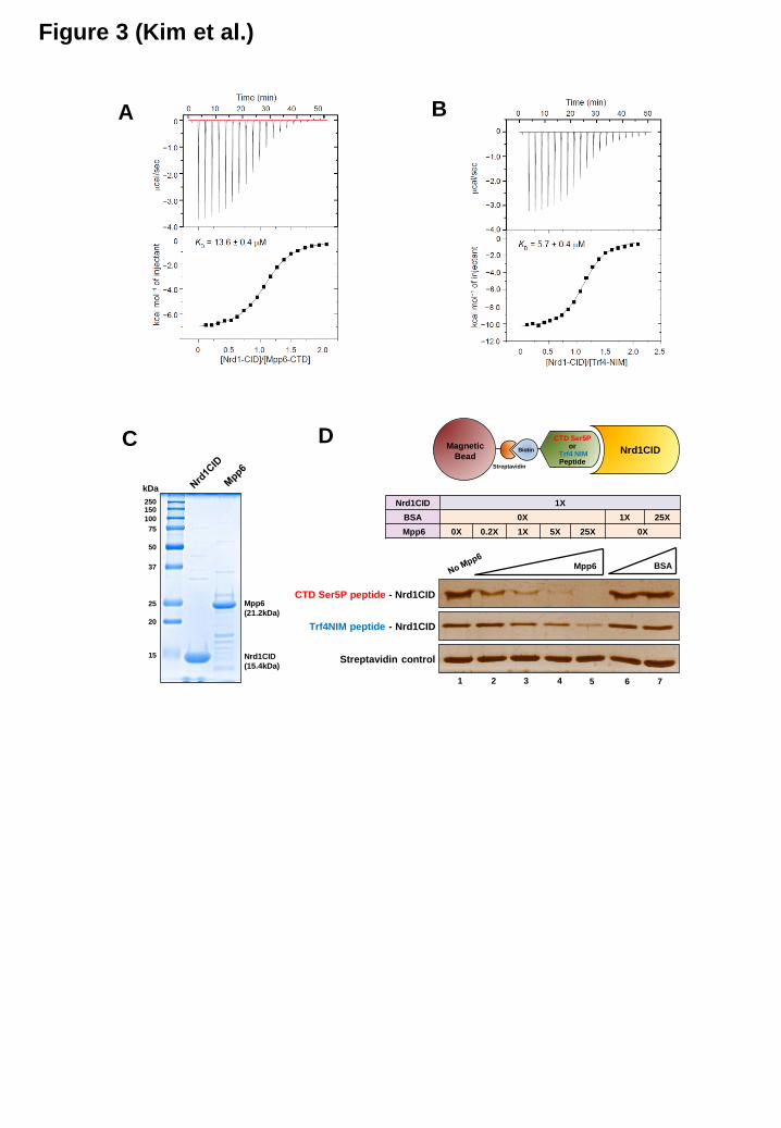
Figure 3 (Kim et al.)
D
Nrd1CID 1X
BSA 0X 1X 25X
Mpp6 0X 0.2X 1X 5X 25X 0X
Magnetic
BeadStreptavidin
CTD Ser5Por
Trf4 NIMPeptide
Nrd1CID
Mpp6 BSA
CTD Ser5P peptide - Nrd1CID
Streptavidin control
Trf4NIM peptide - Nrd1CID
1 2 3 4 6 75
C
Nrd1CID
(15.4kDa)
Mpp6
(21.2kDa)
150
100
75
50
37
25
20
15
kDa
250
Biotin
A B
by guest on June 14, 2019http://w
ww
.jbc.org/D
ownloaded from

A
Figure 4 (Kim et al.)
B
EV
Rrp6
Exop G
B D
p G A Dp G A D
SC -Leu -Trp SC -Leu -Trp -Ade
Rrp6
1 103 512386 733aa
PMC2NT EXOⅠ/Ⅱ/Ⅲ HRDC1 HRDC2 / NLS
1 60 186aa122
N-terminus C-terminusMiddleMpp6
2 31
p G
B D EV
Mpp6
Rrp47
SC -Leu -Trp SC -Leu -Trp -Ade
p G A Dp G A D
Nrd1CID 1X
BSA 0X 1X 25X
Rrp6EXO 0X 0.2X 1X 5X 25X 0X
Magnetic
BeadStreptavidin
Mpp6 C2peptide Nrd1CID
Rrp6EXO BSA
Mpp6 C2 peptide - Nrd1CID
Streptavidin control
1 2 3 4 6 75
BiotinDC
Nrd1CID
(15.4kDa)
Rrp6EXO
(34.1kDa)
150
75
50
37
25
20
15
kDa
100
250
by guest on June 14, 2019http://w
ww
.jbc.org/D
ownloaded from
A
Figure 5 (Kim et al.)
α-Rrp6-myc5
α-Nrd1-TAP
1 2 3 4 6 7 851 2 3 4 6 7 85
INPUTIP (IgG)
+ - +RNase A -
Nrd1-TAPNo TAP Nrd1-TAPNo TAP Nrd1-TAPNo TAPNrd1-TAPNo TAP
α-Dis3-myc5
INPUTIP (IgG)
+ - +RNase A -
Nrd1-TAPNo TAP Nrd1-TAPNo TAP Nrd1-TAPNo TAPNrd1-TAPNo TAP
α-mpp6-myc5
α-Nrd1-TAP
1 2 3 4 6 7 851 2 3 4 6 7 85
α-Dis3-myc5
α-Rrp6-myc5
B
α-Trf4-myc5
E
Nrd1
CID
Trf4
Rrp6
Nrd1
CIDRrp6
DRNA
Dis3
PH
KH
Dis3
PH
Mpp6
KH
(1)
(3)
(1)
(2)
(3)Mpp6
Trf4
(2)
NRD1/SCR1
NRD1
SCR1
3.41.0
2 31
6.3
C
IP/INPUT (myc-tagged protein)
IP/INPUT (TAP-tagged Nrd1)
Normalized intensity after IP with IgG
(Set lane 3 as 100%)
=
Dis3
Rela
tive f
old
ch
an
ges
Trf4Rrp6 Dis3
Rela
tive f
old
ch
an
ges
Mpp6Rrp6
trf4Δ
WT
mpp6Δ
WT
0
50
100
150
200
250
300
350
(%)
Lane 4 3 4 3 43
(%)
0
50
100
150
200
250
300
350
Lane 4 3 4 3 43
by guest on June 14, 2019http://w
ww
.jbc.org/D
ownloaded from

Figure 6 (Kim et al.)
D
Glu
Ga
l
Glu
Ga
l
Glu
Ga
l
Glu
Ga
l
2 3 4 5 6 7 81
SCR1
SNR13
SNR33
B
SCR1
SNR13
SNR33
2 3 4 5 6 7 81
Glu
Ga
l
Glu
Ga
l
Glu
Ga
l
Glu
Ga
l
F
A
SNR13
Truncated
2 3 4 5 6 7 8 9 10 11 12 13 14 15 171 18 19 2016
Glu
Ga
l
Glu
Ga
l
Glu
Ga
l
Glu
Ga
l
Glu
Ga
l
Glu
Ga
l
Glu
Ga
l
Glu
Ga
l
Glu
Ga
l
Glu
Ga
l
SNR33
Truncated
pre-SNR33 transcripts relative to SCR1 (%)
SCR1
SUT145
NEL025c
SUT145 transcripts relative to SCR1 (%)
pre-SNR13 transcripts relative to SCR1 (%)
NEL025c transcripts relative to SCR1 (%)
0
50
100
150
200
250
1 2 3 4 5 6 7 8 9 10 11 12 13 14 15 16 17 18 19 20
0
50
100
150
200
250
1 2 3 4 5 6 7 8 9 10 11 12 13 14 15 16 17 18 19 20
0
50
100
150
200
250
300
350
400
1 2 3 4 5 6 7 8 9 10 11 12 13 14 15 16 17 18 19 20
0
50
100
150
200
250
300
350
1 2 3 4 5 6 7 8 9 10 11 12 13 14 15 16 17 18 19 20
Nrd1-
terminated
RNAs
Mpp6 Trf4
Rrp6Dis3
E
2 3 41
SCR1
SNR13
SNR33
C
by guest on June 14, 2019http://w
ww
.jbc.org/D
ownloaded from
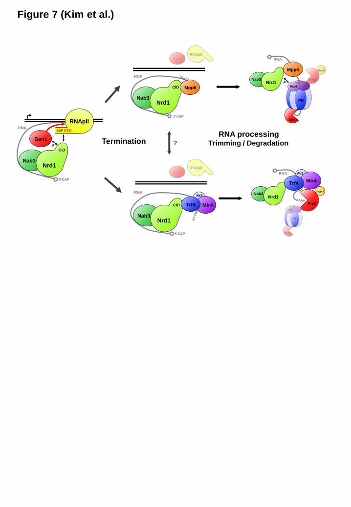
Figure 7 (Kim et al.)
Termination
Nrd1
CID
Nab3
RNApII
Sen1
S5P CTD
5’CAP
RNA
Nrd1
CID
Nab3
RNA
5’CAP
Mtr4
RNApIISen1
Nrd1
CID
Nab3
RNApIISen1
5’CAP
RNA
?
RNA processingTrimming / Degradation
Mtr4
Rrp47
Nrd1Nab3
Nrd1Nab3
Dis3
Rrp47
Rrp6
Air2
Trf4
Mpp6
Trf4
Air2
RNA
RNA
Mpp6
PH
Rrp4
Rrp6
Dis3
PH
KH
by guest on June 14, 2019http://w
ww
.jbc.org/D
ownloaded from
Kyumin Kim, Dong-hyuk Heo, Iktae Kim, Jeong-Yong Suh and Minkyu KimNuclear Exosome
Guiding Terminated Transcripts to the Appropriate Exonuclease within the Exosome Cofactors Connect Transcription Termination to RNA Processing by
published online April 13, 2016J. Biol. Chem.
10.1074/jbc.M116.715771Access the most updated version of this article at doi:
Alerts:
When a correction for this article is posted•
When this article is cited•
to choose from all of JBC's e-mail alertsClick here
by guest on June 14, 2019http://w
ww
.jbc.org/D
ownloaded from