Embed Size (px)
Citation preview
11
Treatment of Metastatic BoneDisease
Martin Malawer
OVERVIEW
Few skeletal metastases require surgical intervention. Radiotherapy, chemotherapy or both often provide sympto-matic relief. An impending or actual pathologic fracture requires operative fixation because fractures through atumor-bearing bone rarely heal without such intervention.
The goals of fixation are to relieve pain, improve function and ambulation, facilitate medical and nursing care,and improve psychological well-being (Figures 11.1 and 11.2). The primary functional goal of surgical interventionis to allow immediate weight-bearing. Surgery should be avoided if this cannot be achieved. A variety of tech-niques, including prosthetic reconstruction (especially about the hip) or a combination of internal fixationcombined with polymethyl methacrylate (PMMA), provides immediate fixation and stability. After the wound hashealed, radiotherapy is usually used to arrest local tumor growth, permit bony repair, and prevent re-growth oftumor around the fixation device. This chapter discusses the techniques of treatment of long bone metastases.
Malawer Chapter 11 21/02/2001 15:30 Page 215
INTRODUCTION
The role of the orthopedic surgeon in the managementof skeletal metastases is to: (1) confirm the diagnosis; (2)treat pathologic fractures; and (3) monitor patients atrisk for pathologic fracture. Surgery can play animportant role in reducing pain, improving function,and increasing quality of life, even in patients with veryshort life expectancies. Additionally, aggressive treat-ment of solitary skeletal metastases may improve long-term survival in selected patients. For example, patientswith renal cell cancer and a solitary skeletal metastasisamenable to wide resection can achieve a 30–35% 5-year survival.
All cancer patients with new onset of pain must beassumed to have a skeletal metastasis until provenotherwise. The initial evaluation of such a patient,however, must not rule out the possibility that thelesion is unrelated to the cancer. In approximately 45%of cases a solitary hot spot seen on a bone scan in acancer patient otherwise free of disease is associatedwith an unrelated process. Needle biopsy has beenshown to be effective in diagnosing skeletal metastases
in patients with a history of cancer. Most patients,however, present with multiple skeletal lesions, makingthe diagnosis of metastatic disease certain.
With few exceptions, patients who present with apainful pathologic fracture are candidates for surgicalintervention. Management must be tailored to theindividual; this entails balancing the benefit of surgery
Musculoskeletal Cancer Surgery216
Figure 11.1 Three-phase bone scan showing multiple “hotspots” involving the ribs and shoulder girdle. Thisdemonstrates the typical spread of carcinoma to the skeletalsystem.
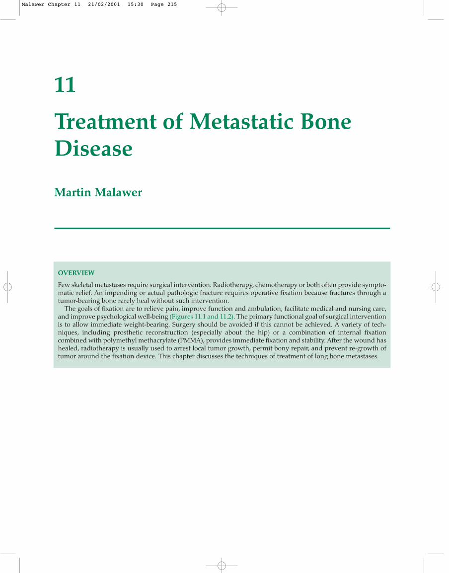
Figure 11.2 Composite photograph showing several typesof proximal femoral replacements used to reconstruct largeskeletal defects following the resection of metastatic tumorsof the proximal femur. The prosthesis (arrow), is a customprosthesis to replace approximately one-half of the femur.The other two prostheses are modular prostheses that arealso utilized in the treatment of primary bony sarcomas. Themajor indications for segmental replacement of the proximalfemur are large, destructive, lytic lesions involving the head,neck, and shaft; solitary metastases; and recurrent tumorfollowing previous attempts at intramedullary fixation withor without cementation.
Malawer Chapter 11 21/02/2001 15:30 Page 216
and the risks associated with operating on a patientwith a limited life expectancy and who is in poormedical condition. Isolated fractures of non-weight-bearing bones in the upper extremities can frequentlybe managed with palliative radiotherapy and casting orbracing. A patient with lower-extremity lesions requiresgreater use of the upper extremities for transfers andcrutch or walker-assisted ambulation. Under thesecircumstances surgical intervention is often essential.All extremities must be carefully examined beforeembarking on a course of treatment.
The goals of surgical fixation are to relieve pain,improve function and ambulation, facilitate medical andnursing care, and improve psychological well-being.This requires a different approach from that used fornon-neoplastic lesions. Immediate fixation must beobtained at the time of surgery: these patients rarelytolerate multiple surgical procedures. Bony union almostnever occurs without surgery and radiotherapeutictreatment. The basic principle of surgical managementis internal fixation or prosthetic replacement combinedwith PMMA. Cementation permits immediate stabilityand early mobilization and pain reduction.
IMPENDING PATHOLOGIC FRACTURES
Any skeletal lesion may cause a pathologic fracture.Criteria for selecting patients for prophylactic fixationhave slowly evolved. Early criteria were based solely onretrospective observations of pathologic fractures in theproximal femur and hip. This was of great importanceto orthopedists because of the technological difficultyin fixing such fractures, as well as the high mortalityrates associated with hip fractures (Figures 11.3 and 11.4).
The first set of combined guidelines (1986) forprophylactic fixation of proximal femur were:1 (1)greater than 50% cortical destruction seen on CT, (2) alytic lesion of the proximal femur > 2.5 cm in diameter,and (3) avulsion of the lesser trochanter. While theseguidelines were helpful for lytic lesions of the femur,they failed to account for other patterns of mixed orpermeative lesions and would not be readily applied toother sites. In addition, these guidelines failed toaccount for lesions amenable to nonsurgical treatments.Increasingly, effective adjuvant treatments haveresulted in improved patient survival, increasing thetime at risk for any given lesion to fracture. Therefore,
Treatment of Metastatic Bone Disease 217
A B
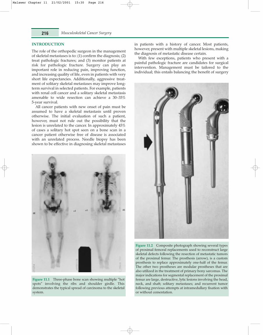
Figure 11.3 (see above and following page).
Malawer Chapter 11 21/02/2001 15:30 Page 217
Thompson2 revised the Harrington1 criteria defined asfollows: (1) large lytic lesions occupying 50% or more ofthe cortical diameter unless protected until reconsti-tuted with radiation therapy, medical management, orboth; and (2) all destructive lesions of the femoral neckin patients with a survival estimate of > 3 months.
PREOPERATIVE EVALUATION ANDINTERVENTIONS
Special preoperative considerations are needed becausethese patients often have extensive metabolic, hema-tologic, and nutritional deficiencies. The risk ofinfection is increased because of possible multiplesources of sepsis (e.g. colostomy, urinary tract infection),neutropenia from chemotherapy or other adjuvantmodalities, generalized nutritional deficits, and poor
local skin condition from prior radiotherapy or otherprocedures. Perioperative antibiotics are recommendedfor all patients. All patients should have hematologicand clotting evaluation, because many may suffer fromanemia of chronic disease or depletion of clotting factorsbecause of a vitamin K deficiency or tumor involve-ment of the liver. Adequate blood replacement shouldbe available because curettage of many carcinomas –especially myeloma, thyroid tumor, and renal cellcarcinoma – often leads to substantial blood loss. Thecombination of pre-existing anemia and expected bloodloss mandates the use of preoperative bloodtransfusions. Thrombocytopenia occasionally occursintraoperatively and should be treated aggressivelywhen it occurs. Disseminated intravascular coagulation(DIC) has been noted.
As many of these patients are older, coexistingdiseases (e.g. hypertension, diabetes, renal insufficiency,peripheral vascular disease, and cardiopulmonarydisease) must be identified and controlled. Thepresence of other sites of skeletal disease may requirespecial precautions at the time of surgery to preventadditional pathologic fractures. Rib involvement,common in advanced multiple myeloma, may makerespiration difficult, leading to prolonged or permanentventilator dependence following general anesthesia.Specific disorders associated with skeletal metastases
Musculoskeletal Cancer Surgery218
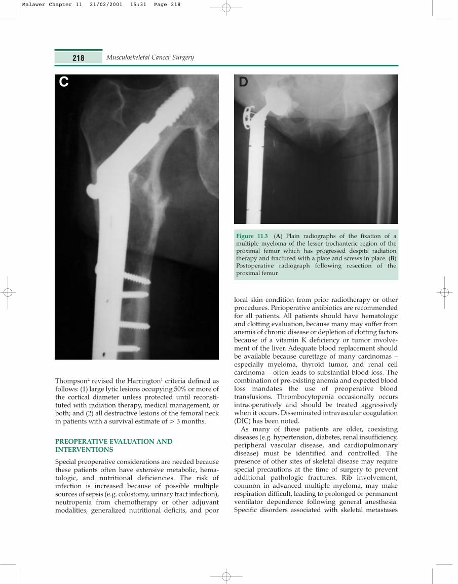
Figure 11.3 (A) Plain radiographs of the fixation of amultiple myeloma of the lesser trochanteric region of theproximal femur which has progressed despite radiationtherapy and fractured with a plate and screws in place. (B)Postoperative radiograph following resection of theproximal femur.
C D
Malawer Chapter 11 21/02/2001 15:31 Page 218
must also be controlled. Of particular concern ishypercalcemia, which can lead to sudden death duringanesthesia. The use of bisphosphonates, includingpamidronate, have been shown to be effective in reduc-ing the serum calcium to normal levels.
Evaluation of the extent of local disease, the amountof bone involved, and the presence of multiple lesionswithin the same bone are necessary to determine theoptimal surgical approach, the amount of tumor to beremoved, and the method of reconstruction. The
following are studies commonly used; the choice ofimaging techniques depends on the individual patientand tumor location and type.
Bone Scintigraphy
Technicium-99 bone scans can demonstrate theintraosseous extent of tumor and the site of the lesion:whole-body scanning provides information aboutother possible sites of disease. Additional lesions are
Treatment of Metastatic Bone Disease 219
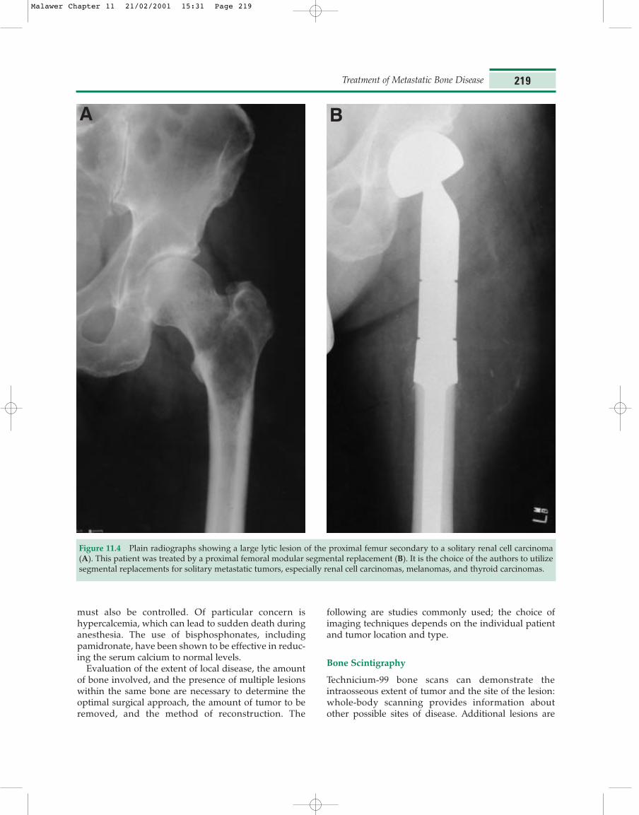
Figure 11.4 Plain radiographs showing a large lytic lesion of the proximal femur secondary to a solitary renal cell carcinoma(A). This patient was treated by a proximal femoral modular segmental replacement (B). It is the choice of the authors to utilizesegmental replacements for solitary metastatic tumors, especially renal cell carcinomas, melanomas, and thyroid carcinomas.
A B
Malawer Chapter 11 21/02/2001 15:31 Page 219
commonly found within the same bone. All lesionswithin the same bone generally require simultaneoustreatment; this usually requires placement of an IM rodor an extended-length endoprosthesis.
Computed Tomography and Magnetic ResonanceImaging
Computed tomography remains the standard forevaluation of cortical bone involvement and is requiredfor lesions of the pelvis, shoulder girdle, and spine.With the exception of lesions being evaluated forprophylactic fixation, it is rarely required for extremitylesions. Tumors of the bony pelvis often have large soft-tissue components that may bleed excessively or lead tomechanical failure of the reconstruction if notrecognized preoperatively. MRI is most useful in theevaluation of the soft-tissue extent of the tumor. It canalso reveal intramedullary tumor involvement.
Angiography
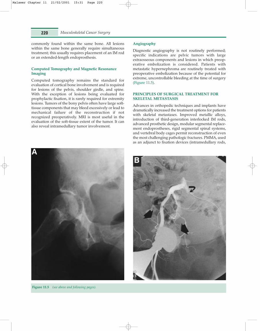
Diagnostic angiography is not routinely performed;specific indications are pelvic tumors with largeextraosseous components and lesions in which preop-erative embolization is considered. Patients withmetastatic hypernephroma are routinely treated withpreoperative embolization because of the potential forextreme, uncontrollable bleeding at the time of surgery(Figure 11.5).
PRINCIPLES OF SURGICAL TREATMENT FORSKELETAL METASTASIS
Advances in orthopedic techniques and implants havedramatically increased the treatment options for patientswith skeletal metastases. Improved metallic alloys,introduction of third-generation interlocked IM rods,advanced prosthetic design, modular segmental replace-ment endoprostheses, rigid segmental spinal systems,and vertebral body cages permit reconstruction of eventhe most challenging pathologic fractures. PMMA, usedas an adjunct to fixation devices (intramedullary rods,
Musculoskeletal Cancer Surgery220
Figure 11.5 (see above and following pages).
AB
Malawer Chapter 11 21/02/2001 15:31 Page 220

plates and screws) and prostheses permits instantfilling reconstruction of large defects and immediatestabilization. Adjuvant treatment of the tumor isnecessary to protect the reconstruction. Therefore, allpatients with skeletal metastases require a multidis-ciplinary approach coordinating chemotherapy andradiation with surgical intervention.
Principles of management of pathologic or impend-ing fractures are as follows:
1. Preoperative embolization of suspected vasculartumors.
2. Administration of perioperative antibiotics.3. Correction of underlying hypercalcemia.4. Transfusion to correct pre-existing anemia, throm-
bocytopenia and coagulation deficits.5. Modification of standard surgical approaches to
avoid prior radiation fields and ensure adequatesoft-tissue coverage and closure.
Treatment of Metastatic Bone Disease 221
C
Figure 11.5 (see above and following page).
Malawer Chapter 11 21/02/2001 15:31 Page 221
6. Curettage to remove all gross disease.7. Use of immediate rigid fixation consisting of inter-
nal fixation with PMMA or cemented prostheticreplacement.
8. Filling of defects with PMMA.9. Postoperative nutritional supplementation to
promote wound healing.
10. Adjuvant radiotherapy, with or without adjuvantchemotherapy.
The common local surgical procedures for metastatictumors of the extremities are: tumor excision, compositeosteosynthesis, joint replacement, segmental recon-struction, cryosurgery and amputation.
Tumor Excision
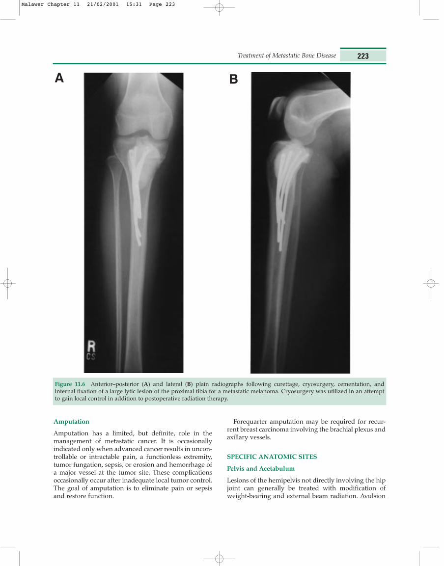
Tumor removal and bone stabilization best meet thegoals of diagnosis, functional stability, and pain relief.For this reason the metastatic lesion should generallybe curetted. Treatment options include intralesional(marginal) and extralesional (wide) excision. Intralesionalcurettage of tumor is usually performed in or around afracture site at the time of stabilization (Figure 11.6).Extralesional excision, i.e. resection, is usually per-formed for a solitary metastatic lesion.
Composite Osteosynthesis
Internal fixation devices (bone plates, screws, IM rodsand nails) are used to stabilize impending or actualpathologic fractures. This technique is most often usedfor metastatic lesions in the shaft diaphysis of longbones, most commonly the humerus and femur.
Joint Replacement
Joint replacement entails resection and reconstructionof a joint using a prosthesis combined with PMMA.Hemijoint replacement involves resection of half of thejoint surface and adjacent bone. Total joint replacementis rarely required. Metastatic lesions of the proximalfemur are most commonly treated by endoprostheticreplacement.
Segmental Reconstruction
Segmental reconstruction is the resection of a largesegment of bone combined with segmental prostheticreplacement and PMMA. This technique is less com-mon than joint replacement and is used for largelesions for which the remaining bone cannot bereconstructed by cementation and internal fixation(Figures 11.7 and 11.8).
Cryosurgery
Cryosurgery is the use of liquid nitrogen as a surgicaladjunct to tumor curettage to freeze any residual tumorcells. It may be combined with any of the above pro-cedures in order to increase local tumor control.
Musculoskeletal Cancer Surgery222
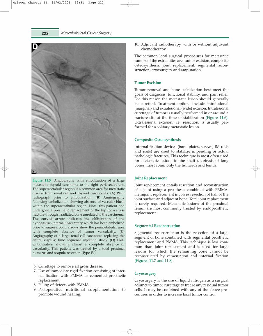
Figure 11.5 Angiography with embolization of a largemetastatic thyroid carcinoma to the right periacetabulum.The superacetabular region is a common area for metastaticdisease from renal cell and thyroid carcinomas. (A) Plainradiograph prior to embolization. (B) Angiographyfollowing embolization showing absence of vascular blushwithin the superacetabular region. Note: this patient hadundergone a prosthetic replacement of the hip for a stressfracture through irradiated bone unrelated to the carcinoma.The curved arrow indicates the obliteration of thehypogastric (internal iliac) artery which has been embolizedprior to surgery. Solid arrows show the periacetabular areawith complete absence of tumor vascularity. (C)Angiography of a large renal cell carcinoma replacing theentire scapula; time sequence injection study. (D) Post-embolization showing almost a complete absence ofvascularity. This patient was treated by a total proximalhumerus and scapula resection (Type IV).
D
Malawer Chapter 11 21/02/2001 15:31 Page 222
Amputation
Amputation has a limited, but definite, role in themanagement of metastatic cancer. It is occasionallyindicated only when advanced cancer results in uncon-trollable or intractable pain, a functionless extremity,tumor fungation, sepsis, or erosion and hemorrhage ofa major vessel at the tumor site. These complicationsoccasionally occur after inadequate local tumor control.The goal of amputation is to eliminate pain or sepsisand restore function.
Forequarter amputation may be required for recur-rent breast carcinoma involving the brachial plexus andaxillary vessels.
SPECIFIC ANATOMIC SITES
Pelvis and Acetabulum
Lesions of the hemipelvis not directly involving the hipjoint can generally be treated with modification ofweight-bearing and external beam radiation. Avulsion
Treatment of Metastatic Bone Disease 223
Figure 11.6 Anterior–posterior (A) and lateral (B) plain radiographs following curettage, cryosurgery, cementation, andinternal fixation of a large lytic lesion of the proximal tibia for a metastatic melanoma. Cryosurgery was utilized in an attemptto gain local control in addition to postoperative radiation therapy.
A B
Malawer Chapter 11 21/02/2001 15:31 Page 223

fractures of the anterior superior/inferior iliac spines,iliac crest, and superior/inferior pubic rami are commonand should be treated nonoperatively. Large supra-acetabular lesions that do not respond to radiotherapyshould be curetted and packed with PMMA combinedwith Steinmann pin fixation (Figures 11.9 and 11.10).
Proximal Femur (Hip)
The hip is most common site of pathologic fracture.This is because of the high incidence of metastases inthis area and the magnitude of force concentrated inthis area with normal activity. In general, all pathologic
Musculoskeletal Cancer Surgery224
Figure 11.7 Anterior–posterior and lateral photographs of a large segmental replacement of the distal humerus and elbow jointfor a large destructive melanoma of the elbow (A) shows the AP view; (B) shows the lateral view. Melanomas, similar to renalcell carcinomas, may cause large lytic destruction of a bone simulating a primary sarcoma. Local control is often difficult toobtain. The authors prefer resection of large metastatic melanomas in-lieu of curettage and cementation.
A B
Malawer Chapter 11 21/02/2001 15:31 Page 224
Treatment of Metastatic Bone Disease 225
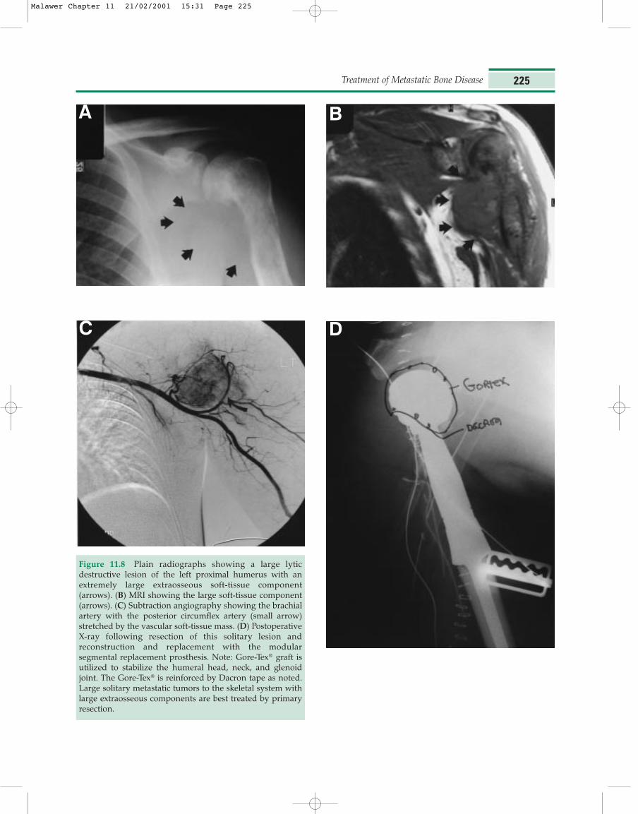
Figure 11.8 Plain radiographs showing a large lyticdestructive lesion of the left proximal humerus with anextremely large extraosseous soft-tissue component(arrows). (B) MRI showing the large soft-tissue component(arrows). (C) Subtraction angiography showing the brachialartery with the posterior circumflex artery (small arrow)stretched by the vascular soft-tissue mass. (D) PostoperativeX-ray following resection of this solitary lesion andreconstruction and replacement with the modularsegmental replacement prosthesis. Note: Gore-Tex® graft isutilized to stabilize the humeral head, neck, and glenoidjoint. The Gore-Tex® is reinforced by Dacron tape as noted.Large solitary metastatic tumors to the skeletal system withlarge extraosseous components are best treated by primaryresection.
A B
C D
Malawer Chapter 11 21/02/2001 15:31 Page 225
fractures of the hip require surgical reconstruction andpostoperative radiation. Surgery is often warranted evenin the severely weakened, nonambulatory patient; torelieve pain, to simplify nursing care, and to regaintransfer ability.
Radiographs and bone scans of the femur and acetab-ulum must be obtained preoperatively. It is notuncommon to detect other lesions further down theshaft; a situation that indicates the need for simulta-neous fixation. In general, a long-stem prosthesis willbe adequate for both femoral neck and diaphyseal
lesions. If the acetabulum is affected by disease, surgeryshould include curettage of the lesion (Figure 11.11).
Surgical Treatment
Metastatic fractures of the hip may be intracapsular(involving the femoral neck), intertrochanteric, or sub-trochantic. Surgical treatment of intracapsular fracturesentails endoprosthetic replacement (usually long-stem)or total hip replacement (Figures 11.12). Managementof intertrochanteric and subtrochantic fractures varies;
Musculoskeletal Cancer Surgery226
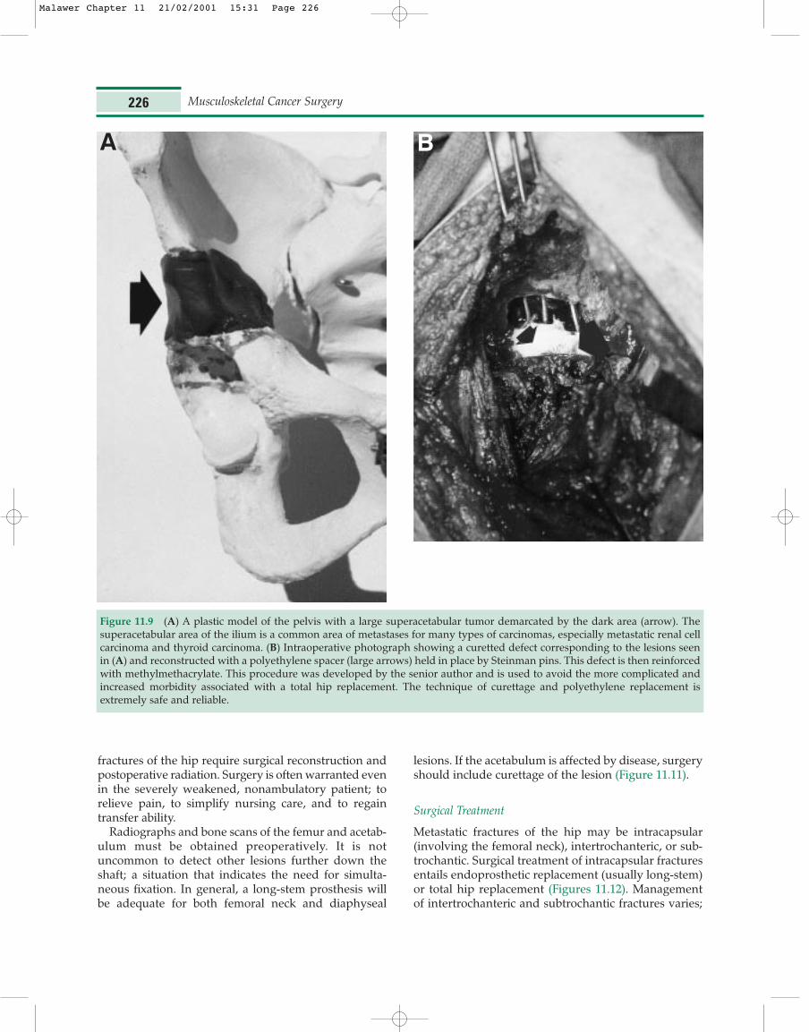
Figure 11.9 (A) A plastic model of the pelvis with a large superacetabular tumor demarcated by the dark area (arrow). Thesuperacetabular area of the ilium is a common area of metastases for many types of carcinomas, especially metastatic renal cellcarcinoma and thyroid carcinoma. (B) Intraoperative photograph showing a curetted defect corresponding to the lesions seenin (A) and reconstructed with a polyethylene spacer (large arrows) held in place by Steinman pins. This defect is then reinforcedwith methylmethacrylate. This procedure was developed by the senior author and is used to avoid the more complicated andincreased morbidity associated with a total hip replacement. The technique of curettage and polyethylene replacement isextremely safe and reliable.
A B
Malawer Chapter 11 21/02/2001 15:31 Page 226
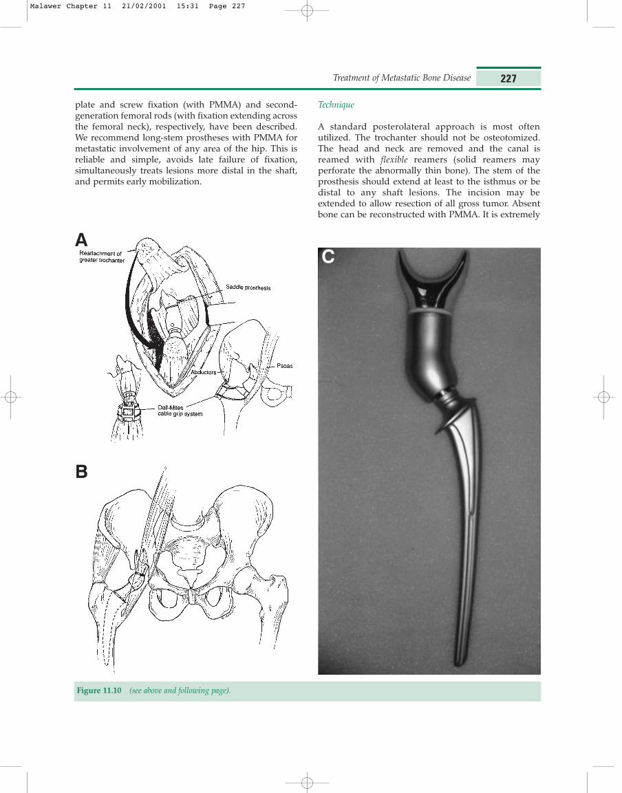
plate and screw fixation (with PMMA) and second-generation femoral rods (with fixation extending acrossthe femoral neck), respectively, have been described.We recommend long-stem prostheses with PMMA formetastatic involvement of any area of the hip. This isreliable and simple, avoids late failure of fixation,simultaneously treats lesions more distal in the shaft,and permits early mobilization.
Technique
A standard posterolateral approach is most oftenutilized. The trochanter should not be osteotomized.The head and neck are removed and the canal isreamed with flexible reamers (solid reamers mayperforate the abnormally thin bone). The stem of theprosthesis should extend at least to the isthmus or bedistal to any shaft lesions. The incision may beextended to allow resection of all gross tumor. Absentbone can be reconstructed with PMMA. It is extremely
Treatment of Metastatic Bone Disease 227
A
B
Figure 11.10 (see above and following page).
C
Malawer Chapter 11 21/02/2001 15:31 Page 227
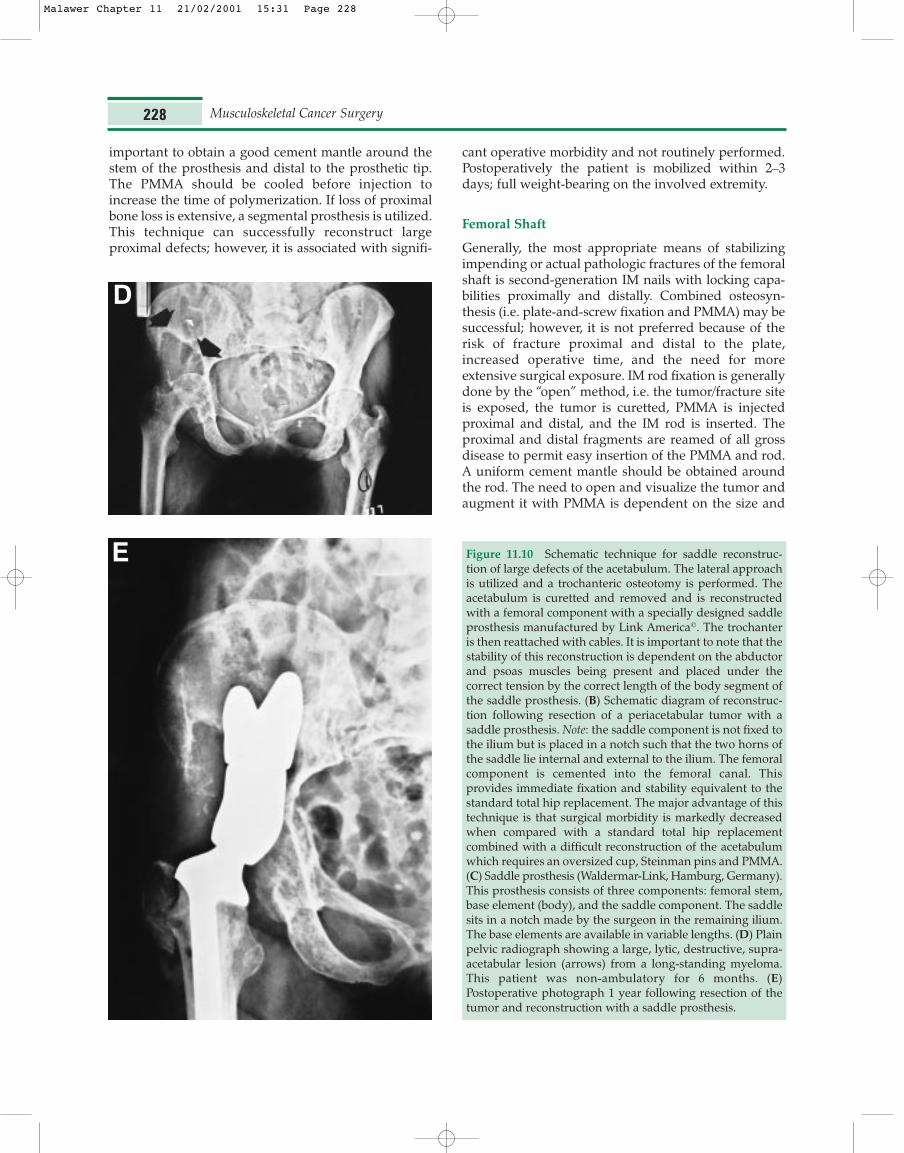
important to obtain a good cement mantle around thestem of the prosthesis and distal to the prosthetic tip.The PMMA should be cooled before injection toincrease the time of polymerization. If loss of proximalbone loss is extensive, a segmental prosthesis is utilized.This technique can successfully reconstruct largeproximal defects; however, it is associated with signifi-
cant operative morbidity and not routinely performed.Postoperatively the patient is mobilized within 2–3days; full weight-bearing on the involved extremity.
Femoral Shaft
Generally, the most appropriate means of stabilizingimpending or actual pathologic fractures of the femoralshaft is second-generation IM nails with locking capa-bilities proximally and distally. Combined osteosyn-thesis (i.e. plate-and-screw fixation and PMMA) may besuccessful; however, it is not preferred because of therisk of fracture proximal and distal to the plate,increased operative time, and the need for moreextensive surgical exposure. IM rod fixation is generallydone by the “open” method, i.e. the tumor/fracture siteis exposed, the tumor is curetted, PMMA is injectedproximal and distal, and the IM rod is inserted. Theproximal and distal fragments are reamed of all grossdisease to permit easy insertion of the PMMA and rod.A uniform cement mantle should be obtained aroundthe rod. The need to open and visualize the tumor andaugment it with PMMA is dependent on the size and
Musculoskeletal Cancer Surgery228
Figure 11.10 Schematic technique for saddle reconstruc-tion of large defects of the acetabulum. The lateral approachis utilized and a trochanteric osteotomy is performed. Theacetabulum is curetted and removed and is reconstructedwith a femoral component with a specially designed saddleprosthesis manufactured by Link America©. The trochanteris then reattached with cables. It is important to note that thestability of this reconstruction is dependent on the abductorand psoas muscles being present and placed under thecorrect tension by the correct length of the body segment ofthe saddle prosthesis. (B) Schematic diagram of reconstruc-tion following resection of a periacetabular tumor with asaddle prosthesis. Note: the saddle component is not fixed tothe ilium but is placed in a notch such that the two horns ofthe saddle lie internal and external to the ilium. The femoralcomponent is cemented into the femoral canal. Thisprovides immediate fixation and stability equivalent to thestandard total hip replacement. The major advantage of thistechnique is that surgical morbidity is markedly decreasedwhen compared with a standard total hip replacementcombined with a difficult reconstruction of the acetabulumwhich requires an oversized cup, Steinman pins and PMMA.(C) Saddle prosthesis (Waldermar-Link, Hamburg, Germany).This prosthesis consists of three components: femoral stem,base element (body), and the saddle component. The saddlesits in a notch made by the surgeon in the remaining ilium.The base elements are available in variable lengths. (D) Plainpelvic radiograph showing a large, lytic, destructive, supra-acetabular lesion (arrows) from a long-standing myeloma.This patient was non-ambulatory for 6 months. (E)Postoperative photograph 1 year following resection of thetumor and reconstruction with a saddle prosthesis.
D
E
Malawer Chapter 11 21/02/2001 15:31 Page 228
degree of cortical destruction. Immediate ambulationwith full weight-bearing is permitted within a few daysafter surgery. Pain relief is almost universal andapproximately 90% of patients maintain ambulation.
Prophylactic Femoral Shaft Fixation
Small lesions of the femoral shaft may be treated priorto fracture by the “closed” method; that is, fluoroscop-ically inserting an IM rod from a small incision at the tipof the greater trochanter and anterograding it throughthe lesion to obtain good distal fixation. When utilizingthis procedure it is difficult to insert PMMA, and thismethod is therefore indicated only for small lesions ofthe femoral shaft with normal proximal bone. Carefulpreoperative evaluation of the hip is required, becausesubsequent treatment of an undetected hip lesion isextremely difficult once an IM rod is in place.
Supracondylar Femoral Fixation
Metastatic lesions of the distal femoral diaphysis andcondyles are best treated by medial and lateral Zickelrods with PMMA. Large distal femoral metaphyseallesions, especially those associated with intra-articularextension and/or large soft-tissue components, are best
treated with a custom or modular distal femoralendoprosthetic replacement.
Humerus
Surgical stabilization is recommended for patients at riskfor fracture and in whom stable fixation can be assured.It is also recommended to permit crutch/walker use inpatients with concomitant lower extremity lesions.Large lesions, or those with a pathologic fracture, arebest treated by curettage, intramedullary fixation, andPMMA. Treatment options for the proximal humerus(metaphysis, surgical neck and head) include stabili-zation with plate and screws or IM rods supplementedwith PMMA and conventional or custom head andlong-stem endoprosthetic replacement. Proximalhumeral lesions are approached through a standarddeltopectoral incision. Tumors of the shaft may betreated with anterograde or retrograde intramedullarynailing, plate-and-screw fixation augmented withPMMA, and customized diaphyseal spacers.
Lesions of the distal humerus are a difficulttherapeutic challenge. Treatment options includecrossed rush rods introduced into medial and lateralepicondyles, medial and lateral column reconstructionwith plate-and-screw constructs supplemented with
Treatment of Metastatic Bone Disease 229
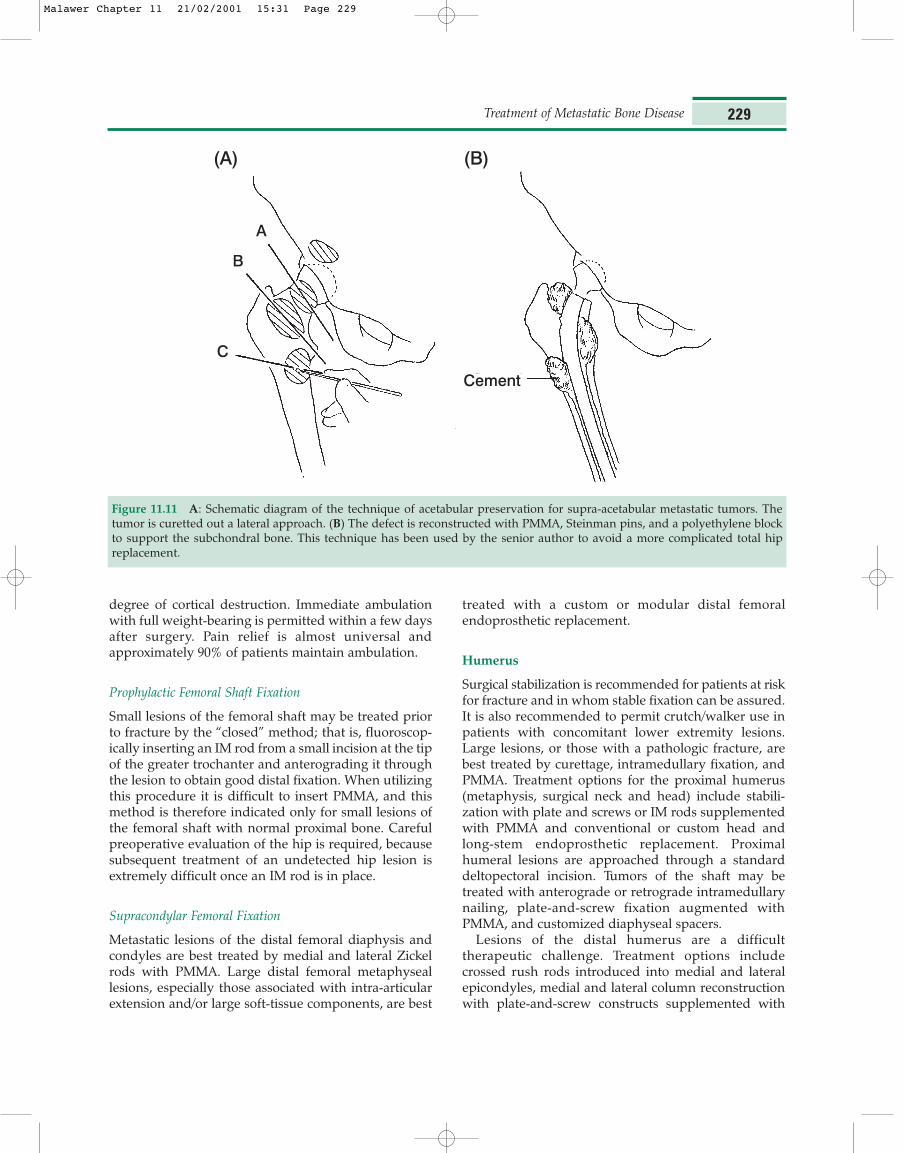
Figure 11.11 A: Schematic diagram of the technique of acetabular preservation for supra-acetabular metastatic tumors. Thetumor is curetted out a lateral approach. (B) The defect is reconstructed with PMMA, Steinman pins, and a polyethylene blockto support the subchondral bone. This technique has been used by the senior author to avoid a more complicated total hipreplacement.
(A) (B)
A
Cement
C
B
Malawer Chapter 11 21/02/2001 15:31 Page 229

PMMA, and conversion to a long-stem (humeral andulnar) constrained elbow arthroplasty. The surgicalexposure should not include olecranon osteotomybecause of the likelihood of nonunion withpostoperative radiotherapy.
Lesions Distal to the Knee and Elbow
The most common primary cancers associated withdistal metastases are those of the lung, kidney, breast,and gastrointestinal tract. Tumors of the forearm ortibia are best treated by IM fixation with supplementalPMMA. Tumors of the hand often require amputation.Substantial experience with extremity preservation hasbeen accumulated with multimodality therapy forprimary sarcomas of the hand and foot; however, thisexperience often does not apply to the management ofmetastases at these sites. Radiation should be used forattempted palliation in patients with metastatic diseaseto the hand or foot. Amputation may be used forpatients who do not respond to radiotherapy.
Musculoskeletal Cancer Surgery230
Figure 11.12 (A) Composite photograph of various sizes oftotal hip replacements.
1 Harrington KD. Impending pathological fractures formetastatic malignancy: evaluation and management.Instruct Course Lect. 1986;35:357–81.
2. Thompson RC. Impending fracture associated with bonedestruction. Orthopaedics. 1992;15:547–50.
References
Malawer Chapter 11 21/02/2001 15:31 Page 230


![Evaluation of bone metastatic burden by bone SPECT/CT in ... · to metastatic prostate cancer patients receiving radium (Ra)-223 therapy [17], and compared the utility of this technique](https://img.dokumen.tips/doc/110x75/5ed99fd85139c40fce67555c/evaluation-of-bone-metastatic-burden-by-bone-spectct-in-to-metastatic-prostate.jpg)