Embed Size (px)
Citation preview

Pak. J. Pharm. Sci., Vol.27, No.1, January 2014, pp.73-81 73
A new natural gel of Fagonia indica Burm f. extract for the treatment of burn on rats
Bazigha Kadhim Abdul Rasool*1, Naglaa Gamil Shehab2, Saeed Ahmed Khan3 and Fatehia Aly Bayoumi4 1Department of Pharmaceutics and Pharmacy Practice, Dubai Pharmacy College, Dubai, UAE
2Department of Pharmacognosy, Faculty of Pharmacy, Cairo University, Egypt 3Department of Pharmaceutical and Medicinal Chemistry, Dubai Pharmacy College, Dubai, UAE 4Department of Pathology, Dubai Medical College, Dubai, UAE
Abstract: Fagonia indica Burm f. (Mushikka or white spine) is a plant distributed in the deserts of Asia and Africa and reported to be medicinal in the scientific literature as well as in the folk medicine. Earlier investigations, the authors isolated a number of bioactive constituents from the plant including flavonoids, sterols and tritepenoids; In addition its flavonoidal content was found remarkably high reaching 3% (calculated as flavonol on dry weight). The present study is an attempt to formulate, characterize and evaluate a natural wound-healing gel preparation containing the crude plant extract. Three formulae (F1-F3) were prepared. The gel properties such as viscosity, swelling ratio, bio-adhesion, in vitro release, stability, microbiological studies, in vivo burn healing test on rats and histopathological features were assessed. The results of the in vitro evaluation and stability studies showed that F3 (0.5% (w/w) of plant extract in 4% (w/w) chitosan) was significantly (p<0.05) the superior compared to other formulations. Besides, from the in vivo burn healing and histological results, F3 enhanced the skin wound re-epithelialization and speed up the healing process compared to the conventional commercial product. Thus, the Fagonia extract loaded chitosan topical gel would be used successfully in burn wound care. Keywords: Fagonia indica; chitosan; gel; burn; wound infections. INTRODUCTION Plants are the oldest source of pharmacologically active compounds and have provided human kind with many medicinally useful compounds from centuries. Today more than two thirds of the world’s populations rely on plant derived drugs (Evans, 2002). Genus Fagonia includes about 35 species that are distributed in the deserts of Arabic countries and dry areas in India, Tropical Africa, Chile and South West USA (Chopra et al., 1982). Some species of Fagonia have showed cytotoxic (Saeed, 1969) anti-microbial (Hash and Nag, 1988) and antihypertensive (Gibbons and Oriowo, 2001) activities. They have been also found to suppress serum prolactin and to increase cortisol levels in rabbit (Seed et al., 1999; Miyase et al., 1996; El-negoumy et al., 1986). These pharmacological activities were attributed to a variety of active phytochemical constituents (Ansari et al., 1987), mainly flavonoidal compounds that have been isolated from various Fagonia species (Al-Wakeel and Shahnaz, 1992; Abdel-Kader et al., 1994).
In a previous publication on Fagonia indica Burm. f., the authors isolated and identified a number of bioactive metabolites. These included the flavonoids quercetin and isorhamnetin aglycones and their glycosides, as well as certain steroids and triterpenoids namely oleanolic acid, β-sitosterol and stigmasterol and their glycosides. All of
the isolated flavonoids were of the flavonol type and their total percentage, colorimetrically estimated as quercetin, reached 3% (calculated on dry weight). Furthermore, the plant was found to be safe (high LD50) and exhibited a remarkable analgesic activity probably attributed to its high flavonoidal content (Naglaa and Amina, 2009). The most common topical preparations available in UAE pharmaceutical market are effective burn treatment however; these products possess disadvantages affecting patient compliance to treatment such as undesirable odor, greasy consistency, light sensitivity and repetitive administration. On the other hand, among natural gel formulations, those containing chitosan are favored for wound healing due to their nontoxic, bioadhesive, biocompatibile and anitimicrobial activity (Prabaharan, 2008). Thus the present study was designed in order to develop a new natural and safe gel preparation containing Fagonia indica Burm. f. plant extract, a natural product rich in flavonoids, which is expected to be simultaneously inexpensive and effective in treatment of burns and wounds and to achieve more patient compliance. MATERIALS AND METHODS Plant Material The whole plants of Fagonia indica Burm f. were *Corresponding author: e-mail: [email protected]

A new natural gel of Fagonia indica Burm f. extract for the treatment of burn on rats
Pak. J. Pharm. Sci., Vol.27, No.1, January 2014, pp.73-81 74
collected during September till October (2010) from the desert plants growing in El Mohisana, Dubai, United Arab Emirates. Plant’s identity was kindly verified by Dr. Hassnaa Ahmed Hosny, Professor of Plant Taxonomy, Department of Botany, Faculty of Science, Cairo University, Egypt. Chemicals Chitosan (MW 2.5×10³ Da), hydroxypropylmethyl cellulose (HPMC) and sodium carboxymethylcellulose (Na CMC) were purchased from Loba Chemie (Bombay, India). All the reagents used were of analytical grade. Preparation of the plant extract The alcoholic extract was obtained by exhaustive cold maceration of the air-dried powdered plant material (500 g) in 70% ethanol (2 L×2). The collective alcoholic extracts were then evaporated to dryness, under vacuum at a temperature not exceeding 50°C. The solvent-free residue (20/g) was dissolved in Tween 20 and stored till further studies. Phytochemical screening Samples of air-dried powdered plant were subjected to tests for carbohydrates and/or glycosides, flavonoids, tannins, unsaturated sterols and/ or triterpenes, alkaloids, saponins, anthraquinones and cardiac glycosides. Preparation of the gel formulations Fagonia indica Burm. f. gel formulations were prepared using three different gel bases. The compositions of the prepared formulations are shown in table 1. Na CMC gel (F1) was prepared by the fusion method described by Cooper and Gunn’s (Cooper and Gunn’s, 1987), while HPMC (F2) and chitosan (F3) gels were prepared by dispersion method (Abdul Rasool et al., 2010). For all the prepared formulae, glycerol 1% w/w and PG 0.5% w/w were incorporated at the end step, as solubilizers to enhance the release of the active constituents from the gel bases. Formulations were sterilized by autoclaving at 10 lb for 30 minutes and kept in well-closed plastic containers and stored at 4°C till experiment. However Na CMC gel develops microbial
growth within a short period after the container opening; therefore methyl paraban was added as a preservative. Physical evaluation of Fagonia gel formulations Determination of gel pH and viscosity The pH and viscosity of the prepared gel formulations were determined using pH meter (HANNA instruments, HI8417, Portugal) and a rotational digital viscometer (Brookfield DV-II, USA) at 25°C. The procedure was carried out as per the method of (Abdul Rasool et al., 2010). Swelling Study The swelling studies were carried out gravimetrically in pH 7.4 phosphate buffer (PBS) according to the method of (Noble et al., 1999). In vitro bioadhesion studies The bioadhesive strength of gel formulations was evaluated by employing a method described by Peh (Peh and Wong, 1999) using TA-XTPlus Texture Analyzer (Stable Micro System, UK). In vitro release studies A cellulose nitrate synthetic membrane, 0.45 µm pore size (Whatman, UK) was mounted on a Franz diffusion cell (Copley Scientific, UK). One gram of each formulation (contains 5mg of plant extract) was applied to the membrane over an area of 2.543 cm2 across the donor compartment. The receptor compartment contained 18 mL of phosphate buffer (pH 7.4). The donor cell was exposed to ambient temperature and covered with parafilm to prevent evaporation. The temperature of the receptor compartment was maintained at 37±0.5°C while the buffer solution was stirred continuously with a magnetic bar. Samples were withdrawn from the release medium at predetermined time intervals and replaced with an equal volume (0.5 mL) of fresh buffer solution to maintain sink conditions. The gel formulations (F1, F2 and F3) were analyzed by UV spectrophotometer (NMB-6200, 1182, USA) at 325 nm (Mabry et al., 1970). The concentration of quercetin was assayed as per the regression equation generated from quercetin calibration curve in phosphate buffer (pH 7.4); y = 1.6298x (R2 = 0.9983).
Table 1: Composition of the prepared gel formulations
Formulae Code Plant extract (%w/w)
Na CMC (%w/w)
HPMC (%w/w)
Chitosan (%w/w)
Glycerol (%w/w)
PG (%w/w)
Methyl paraben (w/w%)
F1 0.5 5 1 0.5 0.1 B1 5 1 0.5 0.1 F2 0.5 10 1 0.5 B2 10 1 0.5 F3 0.5 4 1 0.5 B3 4 1 0.5
F: Formula; B: Blank; B1, B2 and B3 indicate blanks of F1, F2 and F3, respectively.

Bazigha Kadhim Abdul Rasool et al
Pak. J. Pharm. Sci., Vol.27, No.1, January 2014, pp.73-81 75
Kinetic studies The kinetics of the in vitro release data was analyzed by using two models, the zero-order kinetics (Hadjiioannou et al., 1993), C=Co–Ko t (Eq. 1), where Q is the amount of drug released at time t and Ko is the release rate; and Higuchi model (Higuchi, 1963), Q=K t1/2 (Eq. 2), where Q is the amount of drug released at time t and K is the diffusion rate constant. The mechanism of quercetin (flavanol) release from the gels was predicted according to the correlation coefficients R2. Also the release data were further analyzed as per Korsmeyer-Peppas equation (Korsmeyer et al., 1983), Mt /M∞ = K tn (Eq. 3), where Mt/M∞ is fraction of drug released at time t, k is the rate constant and n is the release exponent. The exponential release constant n value was used to characterize different release mechanisms. For non-Fickian anomalous release, n value is between 0.5 to 1.0, for Fickian diffusion, n≤0.5; for zero order release, n =1; for super case transport II, n>1. The rate constant k and the diffusion exponent were estimated from the inter-cept and the slope of the linear regression of ln (Mt/M∞) vs. ln t, where the fractional release of the drug expressed in percent and k is expressed as percentage per unit time. Stability study Fagonia-chitosan gel (F3) was packed in metallic collapsed tubes for semisolid preparations (total weight was 20g gel each tube) and stored at the room temperature for ten months. Gel samples were examined every two months for their appearance, pH and viscosity (Ozcan et al., 2009). Besides, antimicrobial bioassay was conducted to compare their activity comparing to the standard antibiotics discs (Norfloxacin and Azithromycin) as well as with a reference commercial product. In vitro antimicrobial bioassay Samples were tested at a dose of 200 mg. Selected strains of bacteria were used adopting the agar diffusion technique (Delignette-Mullera and Flandroisa, 1994). The microorganisms were: Gram positive bacteria (Staphylococcus aureus and Streptococcus beta hemolytica) and Gram negative bacteria (Escherichia coli, Pseudomonas aeruginosa and Klebsiella pneumoniae). The antimicrobial effects of the gel formulations were compared with standard antibiotics (Penicillin-G 10 Units, Doxycycline HCL 30µ, Cephataxime 30 µ, Flucloxacillin 30µ, Azithromycin 30 µ and Norfloxacin 30µ) (HIMEDIA, Mumbai, India). Evaluation of the antimicrobial activities was based on measuring the diameters of the observed zones of the inhibition after 24h of incubation. The results were given as mean ±SD (n=3). In vivo burn wound test The protocol for this study was approved by the Research Ethics Committee of Dubai Pharmacy College, and it was
performed in accordance with the “Guide for the Care and Use of Laboratory Animals” (NIH Publication no. 85-23) (STM F719, 2007). Male Wistar rats weighing 245±51g were used. The animals were housed in individual cages under standard conditions of light and dark cycle and had free access to food and tap water. Each rat was previously anesthetized with diethyl ether fume inside a well closed glass jar for few minutes. The dorsum hair on the area to be burned was shaved by electrical clipper as well hair removal cream. Shaved area was wiped with a piece of cotton immersed with absolute ethanol. The burn infliction was made by holding a stainless steel stamp of 6.5cm2 surface areas, previously heated at 100 °C, inflicted over the exposed skin for 1 min. The weight of the aluminum stamp was 150g. Postoperative pain was treated with Diclofenac injection given I.M (10 mg/kg) once a day for two days. Burn treatment The animals were divided into three groups (each group n=5). The wounds of group I was control (without treatment), group II was treated with a reference commercial product and group III was treated with F3 (All animals groups were daily treated topically on the wounds. The wounds were kept opened (without dressing) during the entire experiment. Evaluation of wounds The condition of each wound was examined daily in the morning and the time taken to reach complete epithelialization was considered as the healing time (in days). For wound size measurement, at the predetermined intervals the wounds were traced on a transparency paper and the tracings were measured. The relative wound size reduction was calculated as follows: Relative wound size reduction (%) = [(Ao-At) /Ao] x 100 Where Ao and At are the wound size at initial time and time “t”, respectively. Histopathological study On the last day of treatment, three rats of each group were sacrificed by excess of diethyl ether for histopathological examination. Full thickness biopsy (2 x 2 cm) was taken from wound area and epithelialized wound. The samples were stored in formaldehyde 10% for 48h prior to inclusion in paraffin. Skin sections (5µ) were cut and stained with hematoxylin and eosin. Slides were analyzed with a light microscope (magnification 100x), Olympus model BX50. The inflammatory cells, fibroblast and new blood vessels were quantified. STATISTICAL ANALYSIS The statistical analysis of the results was performed by the unpaired Student’s t-test. The level of significance was set at p<0.05.

A new natural gel of Fagonia indica Burm f. extract for the treatment of burn on rats
Pak. J. Pharm. Sci., Vol.27, No.1, January 2014, pp.73-81 76
RESULTS Preliminary Phytochemical Screening Results of phytochemical screening carried out on the crude plant extract revealed the presence of carbohydrates and/or glycosides, flavonoids, tannins, unsaturated sterols and or triterpenes and traces of saponins. Anthraquinones and cardiac glycosides were absent. These results are in great extent correlated with our previous investigation of the plant. Physical evaluation of Fagonia gels The developed gel formulations (F1, F2 and F3, table 1) were yellow to brown in color and showed good homogeneities with absence of lumps. The pH of the prepared formulae fell within a range of 4.8 to 5.6. Also mild reduction in the pH value of the gel formulations was observed after plant extract incorporation, as shown in table 2. Viscosity measurements showed that the addition of the plant extract resulted in a considerable decrease in the apparent viscosity of the prepared formulae. Moreover, Formulae F1 and F2 were significantly (p<0.05) more viscous than F3 (table 2). However F3 has a viscosity equivalent to the reference product (table 2). Also gel formulations prepared with chitosan base possessed the superior swelling capacity in comparison with other bases (fig. 1a). Furthermore, the swelling ratio of Fagonia gels was found to be higher than the blank gel
formulations (fig. 1b), probably due to the hydrophilic nature of the constituents of the extract especially the flavonoidal glycosides.
The mucoadhesion test was performed to measure the adhesive strength of gel formulations to the wound area. The findings showed that F3 exhibited the highest (p<0.05) mucoadhesion in all formulations analyzed and incorporation of Fagonia extract to gel formulae enhanced the mucoadhesion ability of gels (fig. 2).
Fig. 2: Mucoadhesion property of Fagonia gel formulations (n=3). Bars indicate ±SD. In- vitro release study F3 released 1.051 mg of flavonol compounds, while only
Table 2: Physical characteristics of Fagonia gel formulations
Formulae Physical characteristics F1 B1 F2 B2 F3 B3
Reference
pH 4.8 5.3 4.9 5.1 5.4 5.6 5.4 Viscosity* (cP) 1785 ( 2.1) 1810 (7.9) 1896 (5.1) 2024 (2.3) 180 (4.8) 209 (8.1) 184 (3.5)
*Results are given as mean ±SD (n =3).
Fig. 1: Effect of gelling agent (A) and plant extract (B) on the swelling ratio of gel formulations (n=3). Bars indicate ±SD.
A

Bazigha Kadhim Abdul Rasool et al
Pak. J. Pharm. Sci., Vol.27, No.1, January 2014, pp.73-81 77
0.594 mg and 0.365 mg was obtained from F1 and F2, respectively. No lag time was observed in release from all gels, fig. 3.
Fig. 3: Mean quercetin (flavonol) flux with time from gel formulations. Each value represents the mean ±SD (n=5); *P<0.05 compared with other formulas. Release kinetics study The drug release rate constants, R2 and diffusion exponent of the test gel formulations are presented in table 3. Results indicated that, the release data of flavonols from gel formulae more fitted to Higuchi model than to zero-order kinetics. This means, Higuchi model is the suitable one to describe the release kinetics of flavonoidal content from these formulas. However further kinetics study was performed by using Korsmeyer-Peppas equation to find out the pattern of the drug release from the gels. Straight
lines were obtained from plotting ln Mt/ M∞ vs. ln t. The values of n obtained for F1 and F3 were less than 0.5 which suggested the drug release via Fickian diffusion mechanism. However, the value of n for the drug released from F2 was greater than 0.5 indicating the anomalous drug release from the gel by both diffusion and polymer erosion mechanisms. In vitro microbiological evaluation Under the experimental conditions used and as shown in table 4, it is concluded that F3 at a dose of 200 mg exhibited marked antimicrobial activity against all the tested bacteria comparing to the alcoholic extract and the standard antibiotic. All formulae showed negligible effect against Candida albicans. Stability study The results of the stability study indicated that, insignificant (p<0.05) changes in the organoleptic properties of F3 as well the pH, viscosity and the antimicrobial activity (fig. 4) after the storage period, were detected. F3 may, therefore, be considered as a stable formula for burn treatment. Wounds healing Application of the previously heated aluminum oval stamp on the dorsum of the rats caused a second degree burn. The relative size reduction of the wounds treated with various preparations is shown in fig. 5. At 15 days post-operation, the wound size reduction was significantly
Table 3: Release rate constants and correlation coefficients (R2) of quercetin (flavonol) from the prepared gel formulations
Kinetic parameters Zero-order kinetic Higuchi kinetics Korsmeyer-Peppas kinetics Formulae Ko(mg. h-1) R2 KH (h-1/2) R2 KKP (h-n) R2 n value Diffusion model
F1 0.043 0.953 0.154 0.958 31.218 0.945 0.405 Fickian Diffusion F2 0.042 0.898 0.157 0.968 20.801 0.955 0.614 Anomalous Diffusion F3 0.065 0.938 0.238 0.976 39.488 0.988 0.337 Fickian Diffusion
Table 4: Antimicrobial effect of Fagonia gel formulations on the selected bacteria and fungi
Diameter of inhibition zone (mm) Formulas Staphylococcus
aureus Streptococcus
beta hemolytica Escherichia
coli Pseudomonas
aeruginosa Klebsiella
pneumoniae Candida albicans
Penicillin-G 19 21 8 R 7 R Doxycycline 12 15 14 18 19 R Cephataxime 21 20 R 20 R R Flucloxacillin 22 19 18 18 20 R Azithromycin 12 14 12 15 18 R Norfloxacin 23 10 R 26 18 R Alcoholic extract 10 17 20 15 12 R F1 R R R R R R F2 R R R R R R F3 24 31* 20 30* 23* R
F1, F2, F3: formulae; R: Resistant. * P<0.05 compared with antibiotics.
A new natural gel of Fagonia indica Burm f. extract for the treatment of burn on rats
Pak. J. Pharm. Sci., Vol.27, No.1, January 2014, pp.73-81 78
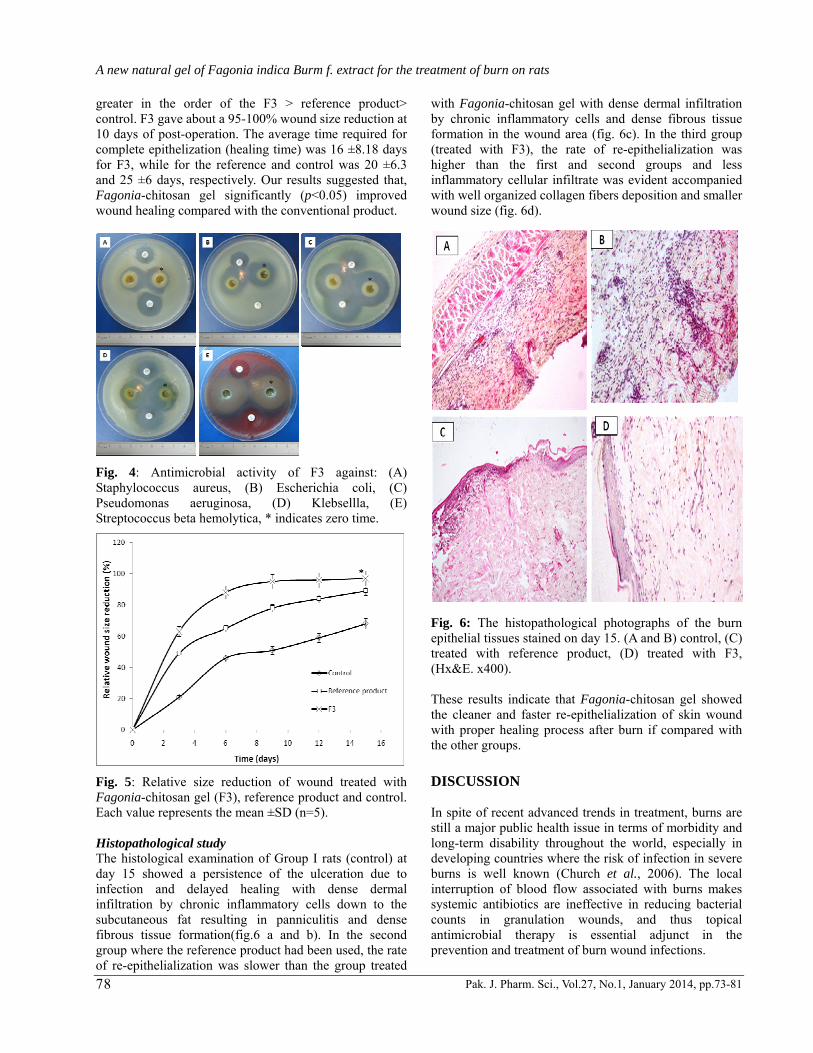
greater in the order of the F3 > reference product> control. F3 gave about a 95-100% wound size reduction at 10 days of post-operation. The average time required for complete epithelization (healing time) was 16 ±8.18 days for F3, while for the reference and control was 20 ±6.3 and 25 ±6 days, respectively. Our results suggested that, Fagonia-chitosan gel significantly (p<0.05) improved wound healing compared with the conventional product.
Fig. 4: Antimicrobial activity of F3 against: (A) Staphylococcus aureus, (B) Escherichia coli, (C) Pseudomonas aeruginosa, (D) Klebsellla, (E) Streptococcus beta hemolytica, * indicates zero time.
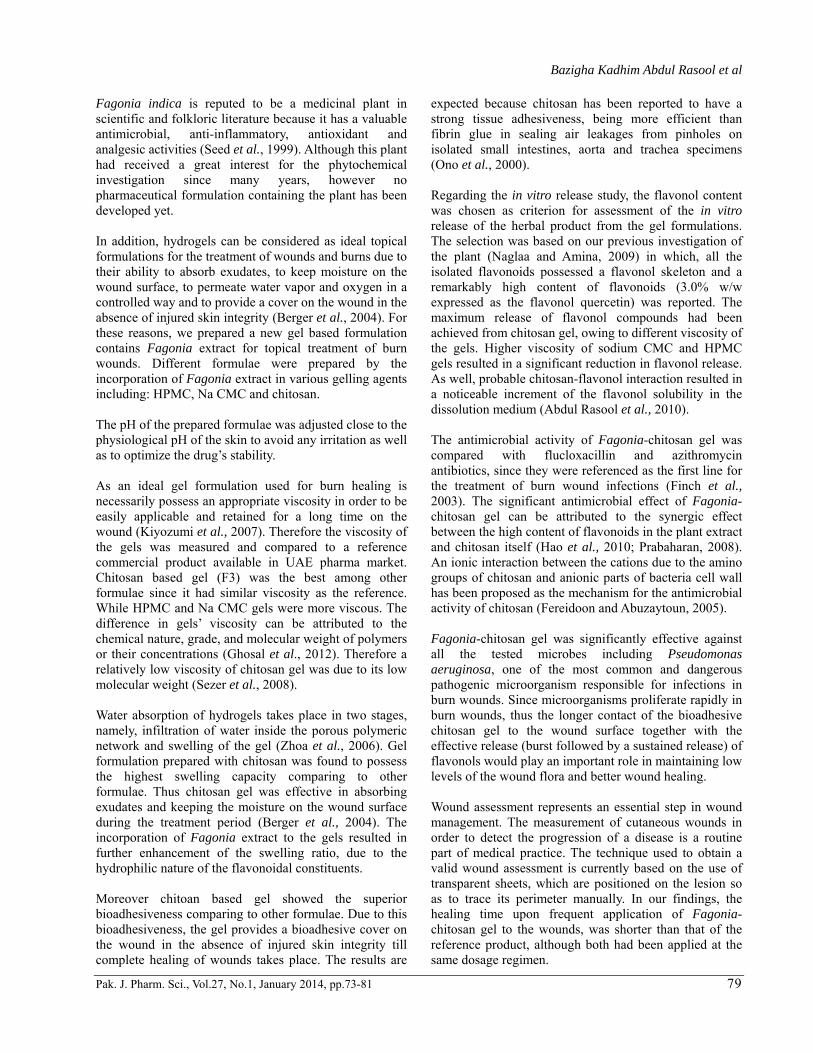
Fig. 5: Relative size reduction of wound treated with Fagonia-chitosan gel (F3), reference product and control. Each value represents the mean ±SD (n=5). Histopathological study The histological examination of Group I rats (control) at day 15 showed a persistence of the ulceration due to infection and delayed healing with dense dermal infiltration by chronic inflammatory cells down to the subcutaneous fat resulting in panniculitis and dense fibrous tissue formation(fig.6 a and b). In the second group where the reference product had been used, the rate of re-epithelialization was slower than the group treated
with Fagonia-chitosan gel with dense dermal infiltration by chronic inflammatory cells and dense fibrous tissue formation in the wound area (fig. 6c). In the third group (treated with F3), the rate of re-epithelialization was higher than the first and second groups and less inflammatory cellular infiltrate was evident accompanied with well organized collagen fibers deposition and smaller wound size (fig. 6d).
Fig. 6: The histopathological photographs of the burn epithelial tissues stained on day 15. (A and B) control, (C) treated with reference product, (D) treated with F3, (Hx&E. x400). These results indicate that Fagonia-chitosan gel showed the cleaner and faster re-epithelialization of skin wound with proper healing process after burn if compared with the other groups. DISCUSSION In spite of recent advanced trends in treatment, burns are still a major public health issue in terms of morbidity and long-term disability throughout the world, especially in developing countries where the risk of infection in severe burns is well known (Church et al., 2006). The local interruption of blood flow associated with burns makes systemic antibiotics are ineffective in reducing bacterial counts in granulation wounds, and thus topical antimicrobial therapy is essential adjunct in the prevention and treatment of burn wound infections.
Bazigha Kadhim Abdul Rasool et al
Pak. J. Pharm. Sci., Vol.27, No.1, January 2014, pp.73-81 79
Fagonia indica is reputed to be a medicinal plant in scientific and folkloric literature because it has a valuable antimicrobial, anti-inflammatory, antioxidant and analgesic activities (Seed et al., 1999). Although this plant had received a great interest for the phytochemical investigation since many years, however no pharmaceutical formulation containing the plant has been developed yet. In addition, hydrogels can be considered as ideal topical formulations for the treatment of wounds and burns due to their ability to absorb exudates, to keep moisture on the wound surface, to permeate water vapor and oxygen in a controlled way and to provide a cover on the wound in the absence of injured skin integrity (Berger et al., 2004). For these reasons, we prepared a new gel based formulation contains Fagonia extract for topical treatment of burn wounds. Different formulae were prepared by the incorporation of Fagonia extract in various gelling agents including: HPMC, Na CMC and chitosan. The pH of the prepared formulae was adjusted close to the physiological pH of the skin to avoid any irritation as well as to optimize the drug’s stability. As an ideal gel formulation used for burn healing is necessarily possess an appropriate viscosity in order to be easily applicable and retained for a long time on the wound (Kiyozumi et al., 2007). Therefore the viscosity of the gels was measured and compared to a reference commercial product available in UAE pharma market. Chitosan based gel (F3) was the best among other formulae since it had similar viscosity as the reference. While HPMC and Na CMC gels were more viscous. The difference in gels’ viscosity can be attributed to the chemical nature, grade, and molecular weight of polymers or their concentrations (Ghosal et al., 2012). Therefore a relatively low viscosity of chitosan gel was due to its low molecular weight (Sezer et al., 2008). Water absorption of hydrogels takes place in two stages, namely, infiltration of water inside the porous polymeric network and swelling of the gel (Zhoa et al., 2006). Gel formulation prepared with chitosan was found to possess the highest swelling capacity comparing to other formulae. Thus chitosan gel was effective in absorbing exudates and keeping the moisture on the wound surface during the treatment period (Berger et al., 2004). The incorporation of Fagonia extract to the gels resulted in further enhancement of the swelling ratio, due to the hydrophilic nature of the flavonoidal constituents. Moreover chitoan based gel showed the superior bioadhesiveness comparing to other formulae. Due to this bioadhesiveness, the gel provides a bioadhesive cover on the wound in the absence of injured skin integrity till complete healing of wounds takes place. The results are
expected because chitosan has been reported to have a strong tissue adhesiveness, being more efficient than fibrin glue in sealing air leakages from pinholes on isolated small intestines, aorta and trachea specimens (Ono et al., 2000). Regarding the in vitro release study, the flavonol content was chosen as criterion for assessment of the in vitro release of the herbal product from the gel formulations. The selection was based on our previous investigation of the plant (Naglaa and Amina, 2009) in which, all the isolated flavonoids possessed a flavonol skeleton and a remarkably high content of flavonoids (3.0% w/w expressed as the flavonol quercetin) was reported. The maximum release of flavonol compounds had been achieved from chitosan gel, owing to different viscosity of the gels. Higher viscosity of sodium CMC and HPMC gels resulted in a significant reduction in flavonol release. As well, probable chitosan-flavonol interaction resulted in a noticeable increment of the flavonol solubility in the dissolution medium (Abdul Rasool et al., 2010). The antimicrobial activity of Fagonia-chitosan gel was compared with flucloxacillin and azithromycin antibiotics, since they were referenced as the first line for the treatment of burn wound infections (Finch et al., 2003). The significant antimicrobial effect of Fagonia-chitosan gel can be attributed to the synergic effect between the high content of flavonoids in the plant extract and chitosan itself (Hao et al., 2010; Prabaharan, 2008). An ionic interaction between the cations due to the amino groups of chitosan and anionic parts of bacteria cell wall has been proposed as the mechanism for the antimicrobial activity of chitosan (Fereidoon and Abuzaytoun, 2005). Fagonia-chitosan gel was significantly effective against all the tested microbes including Pseudomonas aeruginosa, one of the most common and dangerous pathogenic microorganism responsible for infections in burn wounds. Since microorganisms proliferate rapidly in burn wounds, thus the longer contact of the bioadhesive chitosan gel to the wound surface together with the effective release (burst followed by a sustained release) of flavonols would play an important role in maintaining low levels of the wound flora and better wound healing. Wound assessment represents an essential step in wound management. The measurement of cutaneous wounds in order to detect the progression of a disease is a routine part of medical practice. The technique used to obtain a valid wound assessment is currently based on the use of transparent sheets, which are positioned on the lesion so as to trace its perimeter manually. In our findings, the healing time upon frequent application of Fagonia-chitosan gel to the wounds, was shorter than that of the reference product, although both had been applied at the same dosage regimen.

A new natural gel of Fagonia indica Burm f. extract for the treatment of burn on rats
Pak. J. Pharm. Sci., Vol.27, No.1, January 2014, pp.73-81 80
Besides, wound healing is a complex and dynamic process of restoring cellular structures and tissue layers. Wound healing process can be divided into three distinct phases: the inflammatory phase, the proliferative phase, and the remodeling phase (Cotran et al., 2011). One of the most important stages in the wound healing process is the inflammatory stage, where the inflammatory cells clean foreign agents in the wound area (Rongxiang, 1989). Therefore, the presence of inflammatory cells indicates that the healing process and tissue repair are occurring. According to our histopathologic results chitosan gel showed obliviously cleaner and faster re-epithelialization of skin wound with proper healing process after burn if compared with the other groups. This probably indicates that our new formula, Fagonia-chitosan gel, is of advantage for burn wounds management. CONCLUSION The topical use of Fagonia-chitosan gel accelerates the re-epithelialization of skin wound with proper healing process after burn as compared with the commercial product. As well as, this new formulation showed advantageous swelling, bioadhesion and rheological properties. The formula was stable and released the drug efficiently. These results provide rational pharmacological evidence for the traditional use of the genus Fagonia in Emirates folk medicine for the treatment of various types of wounds. ACKNOWLEDGMENTS Authors are grateful to the undergraduate students (Ayesha Bin-Shafar, Mayes Sarmad, Fatma AL-Doukhi and Asma Abdo), for their technical help in the release study. REFERENCES Abdel-Kader MS, Omar AA, Abdel-Salam NA and
Stermitz FR (1994). Erythroxan diterpenes from Fagonia species. Phytochem., 36: 1431-1433.
Abdul Rasool BK, Abu-Gharbieh EF, Al-Mahdy JJ, Nessa F and Ramzi HR (2010). Preparation and characterization of aspirin-chitosan complex: An attempt for its solubility and stability improvement. J. Pharmacy Research, 3(6): 1349-1354.
Abdul Rasool BK, Abu-Gharbieh EF, Fahmy SA, Saad HA and Khan SA (2010). Development and evaluation of ibuprofen transdermal gel formulations. Trop. J. Pharm. Res., 9(4): 355-363.
Al-Wakeel SA and Shahnaz AM (1992). Significance of flavonoid chemistry in the Egyptian Fagonia glutinosa and F. isothricha complex. Biochem. Syst. Ecol., 20: 259-264.
Ansari AA, Kenne L, Atta-ur-R and Wehler T (1987). Isolation and characterization of two saponins from
Fagonia indica. Phytochem., 27: 3979-82. ASTM F719-81 (2007). Standard practice for testing
biomaterials in rabbits for primary skin irritation, West Conshohocken, Philadelphia, pp.178-179.
Berger J, Reist M, Mayer JM, Felt O, Peppas NA and Gurny R (2004). Structure and interactions in covalently and ionically crosslinked chitosan hydrogels for biomedical applications. Eur. J. Pharm. Biopharm., 57: 35-52.
Chopra RM, Handa KL, Kepur LD and Chopra I (1982). Indigenous Drugs of India, 2nd ed., Academic, New Delhi, p.597.
Church D, Elsayed S, Reid O, Winston B and Lindsay R (2006). Burn wound infections. Clin. Microbiol. Rev., 19: 403-34.
Cooper and Gunn’s (1987). Dispensing for pharma-ceutical students, Pitman medical and scientific publishing company LTD, London, pp.214-222.
Cotran RS, Kumar V and Collins T (2011). Acute and chronic inflammation. In: Kumar V, Abbas AK., Fausto N, Aster JC editors. Pathologic basis of disease, 8th ed., WB Saunders Co., Philadelphia, pp.50-88.
Delignette-Mullera ML and Flandroisa JP (1994). An accurate diffusion method for determining bacterial sensitivity to antibiotics. Antimicrob. Chemother., 34(1): 73-81.
El-negoumy SI, Al Wakeel SA, El-Hadidi MN and Saleh NA (1986). The flavonoids of the Fagonia Arabic complex. Phytochem., 25: 2423.
Evans WC (2002). Trease and Evans Pharmacognosy, 5th ed., WB Saunders Co., London, Toronto, Sydney.
Fereidoon S and Abuzaytoun RC (2005). Chitosan and co-products: Chemistry, production, applications and health effects. Adv. Food Nutr. Res., 49: 93-135.
Finch RG, Greenwood D, Norrby SR and Whitley RJ (2003). Antibiotics and chemotherapy: Anti-infective agents and their use in therapy, 8th ed., Churchill Livingstone, London, pp.333-235.
Ghosal K, Chandra A, Rajabalaya R, Chakraborty S and Nanda A (2012). Mathematical modeling of drug release profiles for modified hydrophobic HPMC based gels. Pharmazie, 67(2): 147-155.
Gibbons S and Oriowo MA (2001). Antihypertensive effect of an aqueous extract of Zygophyllum coccineum L. in rats. Phytother. Res., 15(5): 452-455.
Hadjiioannou TP, Christian GD, Koupparis MA and Macheras PE (1993). Quantitative calculations in pharmaceutical practice and research, VCH Publishers Inc, New York, pp.345-348.
Hao L, Yan M, Jianglin Z, Jihua W, Ligang Z and Mingan W (2010). Flavonoids from Halostachys caspica and their antimicrobial and antioxidant activities. Molecules, 15: 7933-7945.
Hash ML and Nag TN (1988). Flavonoids with antimicrobial activities of arid zone plants. Geobios, 15: 32-35.

Bazigha Kadhim Abdul Rasool et al
Pak. J. Pharm. Sci., Vol.27, No.1, January 2014, pp.73-81 81
Higuchi T (1963). Mechanism of sustained action medication: Theoretical analysis of rate of release of solid drugs dispersed in solid matrices. J. Pharm. Sci., 52: 1145-1149.
Kiyozumi T, Kanatani Y, Ishihara M, Saitoh D, Shimizu J and Yura H (2007). The effect of chitosan hydrogel containing DMEM/F12 medium on full-thickness skin defects after deep dermal burn. Burns, 33: 642-648.
Korsmeyer W, Gurny R, Doelker E, Buri P and Peppas N (1983). Mechanisms of solute release from porous hydrophilic polymers. Int. J. Pharm., 15: 25-35.
Mabry TJ, Markham KR and Thomas MB (1970). The systematic identification of flavonoids, Springer-Verlag, New York, p.44.
Miyase J, Melek FR, El-Gindi OD, Abdel-Khalik SM, El-Gindi MR, Haggag MY and Hilal SH (1996). Saponins from Fagonia arabica. Phytochemistry, 41(4): 1175-1179.
Naglaa GS and Amina M (2009). Constituents and analgesic activity of the alcoholic extract of Fagonia indica Burm f. New E. J. Med., 40(4): 357-371.
Noble L, Gray AI, Sadiq L and Uchegbu IF (1999). A non-covalently crosslinked chitosan based hydrogel. Int. J. Pharm., 192: 173-182.
Ono K, Saito Y, Yura H, Ishikawa K, Kurita A and Akaike T (2000). Photocrosslinkable chitosan as a biological adhesive. J. Biomed. Mater. Res., 49: 289-295.
Ozcan I, Abaci O, Uztan AH, Aksu B, Boyacıoglu H, Guneri T and Ozer O (2009). Enhanced topical delivery of terbinafine hydrochloride with chitosan hydrogels. AAPS Pharm. Sci. Tech., 10(3): 1024-1031.
Peh KK, Wong CF (1999). Polymeric film as vehicle for buccal delivery: Swelling, mechanical and bioadhesive properties. J. Pharm. Pharm. Sci., 2: 53-61.
Prabaharan M (2008). Review paper: Chitosan derivatives as promising materials for controlled drug delivery. J. Biomater. Appl., 23: 5-36.
Rongxiang Xu (1989). A great historical turn in the burn. Medical science Chinese. J. Burns Wounds Surf. Ulcers, 1: 62.
Saeed MA (1969). Hamdard Pharmacopoeia of Eastern Medicine, Hamdard Academy, Karachi, pp.41-43.
Seed MA, Khan Z and Sabir AW (1999). Effects of Fagonia cretica L. constituents on various endocrinological parameters in rabbits. Turkish J. Biol., 23(2): 187-197.
Sezer AD, Cevher E, PogLu FH, OgUrtan Z, Bas AL and Akbug AJ (2008). Preparation of fucoidan-chitosan hydrogel and its application as burn healing accelerator on rabbits Biol. Pharm. Bull., 31(12): 2326-2333.
Zhao L, Xu L, Mitomo H and Yoshii F (2006). Synthesis of pH-sensitive PVP/CM-chitosan hydrogels with improved surface property by irradiation. Carbohydr. Polym., 64: 473-480.
![[XLS] · Web viewTintura Yumel Gel caléndula Gel cantharis Gel fucus Gel hamamelis Gel sulphur Gel thuja Gel bálsamo para contusiones Gel sepia Gel ledum Gel de graphites Gel de](https://img.dokumen.tips/doc/110x75/5ac4a6697f8b9a220b8ced85/xls-viewtintura-yumel-gel-calndula-gel-cantharis-gel-fucus-gel-hamamelis-gel-sulphur.jpg)