Embed Size (px)
Citation preview
11
THE MAJOR THE MAJOR NEURONAL CIRCUITSNEURONAL CIRCUITS

22
THE MOTOR CORTEXTHE MOTOR CORTEX
The anatomical region of the brain known as The anatomical region of the brain known as Area 4 is Area 4 is the the primary motor cortexprimary motor cortex (PMC) (PMC)
Focal stimulations elicited muscle contractionsFocal stimulations elicited muscle contractions Is organized somatotopically Is organized somatotopically The surface area varies. The surface area varies. Is proportional to the precision of the movementsIs proportional to the precision of the movements

33
The motor cortex also includes Area 6,The motor cortex also includes Area 6, Which lies rostrally to Area 4 Divided into Which lies rostrally to Area 4 Divided into
Premotor areaPremotor area or or premotorpremotor cortex(PMA) cortex(PMA) SSupplementary motor areaupplementary motor area. (SMA). (SMA)
The PMA is believed to helpThe PMA is believed to help Regulates posture by dictating an optimal position to Regulates posture by dictating an optimal position to
the motor cortex for any given movement. the motor cortex for any given movement.
The SMA, for its part, seems to influence The SMA, for its part, seems to influence • The planning and initiation of movementsThe planning and initiation of movements• On the basis of past experienceOn the basis of past experience
The mere anticipation of a movement triggers neural The mere anticipation of a movement triggers neural transmissions in the SMA transmissions in the SMA

44

55
Somatotopic representations in the motor cortexSomatotopic representations in the motor cortex
66
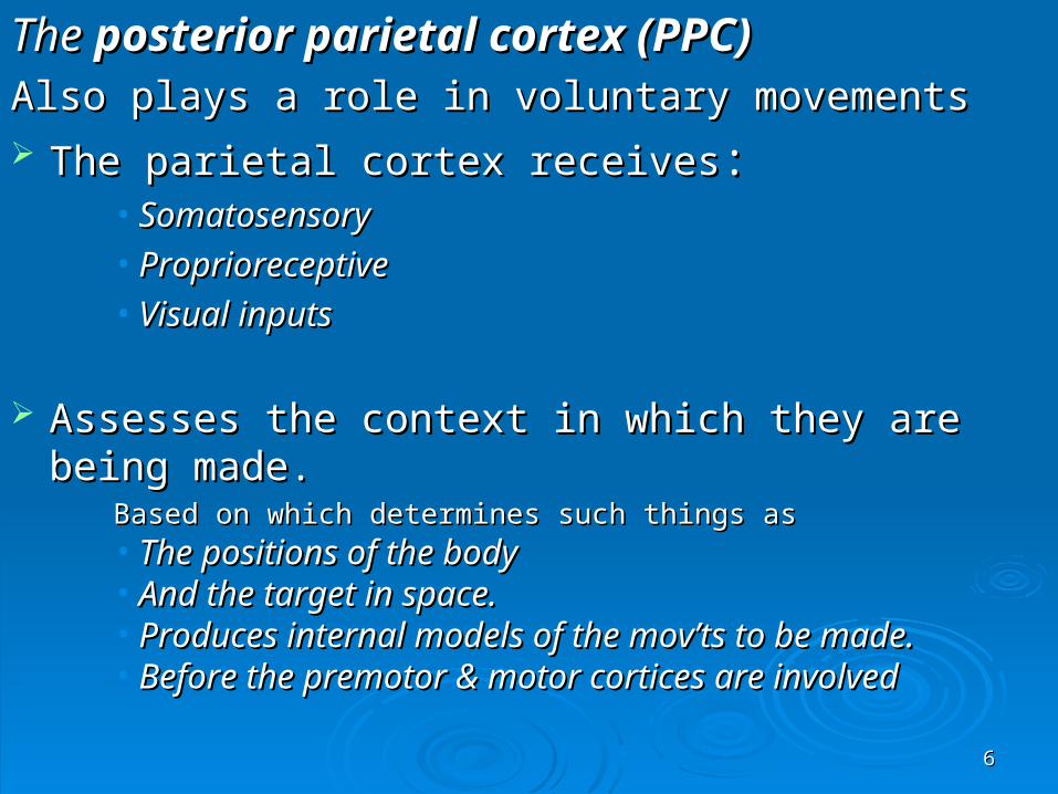
The The posterior parietal cortex (PPC)posterior parietal cortex (PPC)Also plays a role in voluntary movements Also plays a role in voluntary movements The parietal cortex receivesThe parietal cortex receives: :
• SomatosensorySomatosensory• ProprioreceptiveProprioreceptive• Visual inputsVisual inputs
Assesses the context in which they are being made. Assesses the context in which they are being made. Based on which determines such things asBased on which determines such things as• The positions of the body The positions of the body • And the target in space.And the target in space.• Produces internal models of the mov’ts to be made.Produces internal models of the mov’ts to be made.• Before the premotor & motor cortices are involved Before the premotor & motor cortices are involved

77
Within the Within the PPCPPC, two particular areas , two particular areas
Area 5 & Area 7 Area 5 & Area 7
Area 5 receives information fromArea 5 receives information from• Somatosensory areas 1, 2, & 3 of the cortex.Somatosensory areas 1, 2, & 3 of the cortex.
Area 7 further integratesArea 7 further integrates• The already highly integrated signals from the visual The already highly integrated signals from the visual
areas of the cortex, such as MT and V5.areas of the cortex, such as MT and V5.
88
The The parietal lobesparietal lobes are themselves closely are themselves closely
interconnected with the interconnected with the prefrontal areas.prefrontal areas.
The two regions represent the highest level of The two regions represent the highest level of integration in the motor control hierarchy. integration in the motor control hierarchy.
Decisions are made about what action to take.Decisions are made about what action to take.
99
The PP & PFA send their axons to Area 6 The PP & PFA send their axons to Area 6
Which, once it has been informed about the Which, once it has been informed about the kind of action to takekind of action to take
Helps to determine the characteristics of the Helps to determine the characteristics of the appropriate movement for this purpose appropriate movement for this purpose
1010
These two elongated regions face each otherThese two elongated regions face each other
Have the same Have the same somatotopic organizationsomatotopic organization
MC & in the SSC, the scale of this map is not MC & in the SSC, the scale of this map is not constant. constant.
1111
In the In the somatosensory somatosensory cortex, it varies with:cortex, it varies with:
Each body part's sensitivity to sensory Each body part's sensitivity to sensory information.information.
In the motor cortex, it varies with:In the motor cortex, it varies with:
The precision of the movements controlled in The precision of the movements controlled in the body part in question. the body part in question.
1212
AXONS ENTERING AND LEAVING THE MOTOR CORTEXAXONS ENTERING AND LEAVING THE MOTOR CORTEX
Receive inputs from PMA &Receive inputs from PMA & SMASMA The The pyramidal neuronspyramidal neurons of the PMC receive of the PMC receive
information directly from:information directly from: Somatosensory areas 3, 1, & 2. Somatosensory areas 3, 1, & 2.
Axons mainly from the thalamus, Axons mainly from the thalamus,
Caudal Caudal ventrolateralventrolateral nucleusnucleus ( VLc) ( VLc)
Relays information from the cerebellum.Relays information from the cerebellum.

1313
The PMC projects its axons mainly to the The PMC projects its axons mainly to the Corticospinal tractCorticospinal tract, ,
which is composed of thewhich is composed of the
1.1. LateralLateral
2.2. VentromedialVentromedial systems systems. .
1414
The The lateral system haslateral system has two main neural pathways two main neural pathways The The Lateral Corticospinal tractLateral Corticospinal tract. (. (The largerThe larger) )
Arising mainly in Areas 4 and 6 of the frontal lobe, Arising mainly in Areas 4 and 6 of the frontal lobe, which together constitute the motor cortexwhich together constitute the motor cortex , ,
Is the longest neural pathwayIs the longest neural pathway
One of the largest in terms of the number of axons containing 1 milionOne of the largest in terms of the number of axons containing 1 milion
The other axons in this tract arise mainly in the The other axons in this tract arise mainly in the somatosensorysomatosensory areas and the areas and the parietal lobeparietal lobe..
1515
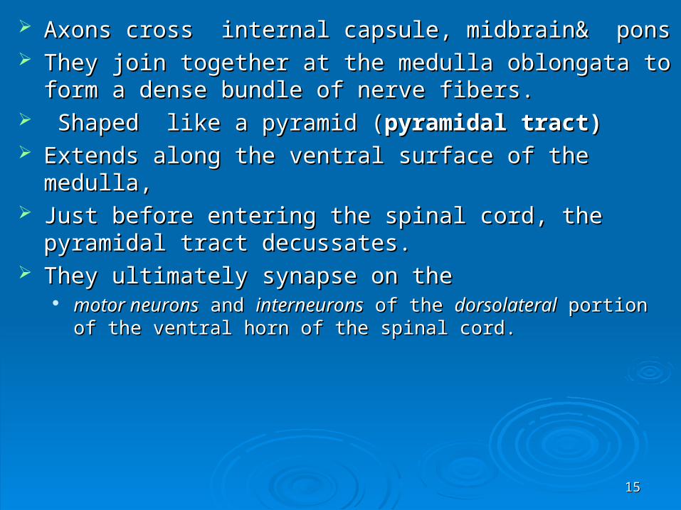
Axons cross internal capsule, midbrain& ponsAxons cross internal capsule, midbrain& pons They join together at the medulla oblongata to form a dense They join together at the medulla oblongata to form a dense
bundle of nerve fibers.bundle of nerve fibers. Shaped like a pyramid (Shaped like a pyramid (pyramidal tract)pyramidal tract) Extends along the ventral surface of the medulla,Extends along the ventral surface of the medulla, Just before entering the spinal cord, the pyramidal tract Just before entering the spinal cord, the pyramidal tract
decussates.decussates. They ultimately synapse on the They ultimately synapse on the
motor neuronsmotor neurons and and interneuronsinterneurons of the of the dorsolateraldorsolateral portion of the portion of the ventral horn of the spinal cord.ventral horn of the spinal cord.

1616
The lateral systemThe lateral system

1717
Rubrospinal tractRubrospinal tract the 2 the 2ndnd in the lateral system. in the lateral system.
Arising from the neurons of the Arising from the neurons of the redred nucleusnucleus, in the , in the midbrain. midbrain.
This nucleus receives information from the frontal cortex.This nucleus receives information from the frontal cortex.
Which also projects massively to the corticospinal tract. Which also projects massively to the corticospinal tract.

1818
The other major descending pathway, is the The other major descending pathway, is the Ventromedial systemVentromedial system
Is composed of four tracts that originate in various Is composed of four tracts that originate in various areas of the areas of the brainstembrainstem
Contribute chiefly toContribute chiefly to:: postural control postural control
certain reflex movements. certain reflex movements.
1919
The originating neurons of these tracts receive The originating neurons of these tracts receive sensory information related to:sensory information related to:
Balance Balance Body positionBody position And the visual environmentAnd the visual environment

2020
1.1. The The Vestibulospinal tractVestibulospinal tract
Originates in the Originates in the vestibular nucleivestibular nuclei..
Maintains the head in position in relation to the Maintains the head in position in relation to the shouldershoulder
Which is essential for continuing to look in a given Which is essential for continuing to look in a given direction while the body is moving direction while the body is moving

2121
2. 2. The The Tectospinal tractTectospinal tract
Arises in the superior colliculus in the midbrain.Arises in the superior colliculus in the midbrain.
The superior colliculus receives:The superior colliculus receives:
some visual information directly from the retinasome visual information directly from the retina
somatosensory and auditory informationsomatosensory and auditory information
the tectospinal tract contributes to visual orientation the tectospinal tract contributes to visual orientation
2222
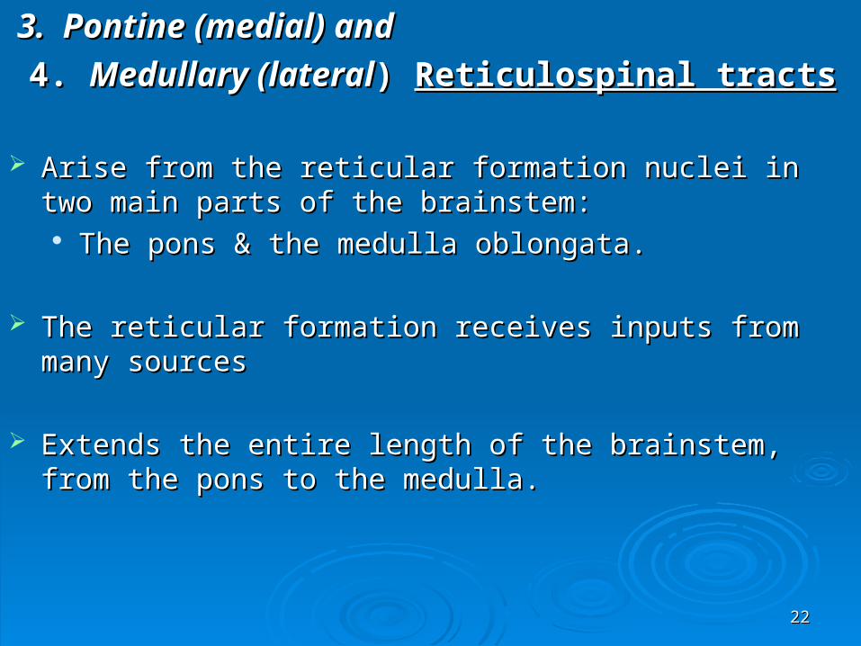
3. Pontine (medial) and3. Pontine (medial) and
4. 4. Medullary (lateralMedullary (lateral) ) Reticulospinal tractsReticulospinal tracts
Arise from the reticular formation nuclei in two main parts of the Arise from the reticular formation nuclei in two main parts of the brainstem: brainstem: The pons & the medulla oblongata. The pons & the medulla oblongata.
The reticular formation receives inputs from many sourcesThe reticular formation receives inputs from many sources
Extends the entire length of the brainstem, from the pons to the Extends the entire length of the brainstem, from the pons to the
medulla.medulla.
2323
These two tracts help to maintain posture.These two tracts help to maintain posture.
Pontine axons enhance the spinal antigravity Pontine axons enhance the spinal antigravity reflexes.reflexes.
The medullary have the opposite effect, The medullary have the opposite effect, releasing the muscles releasing the muscles
Thus facilitating other movementsThus facilitating other movements
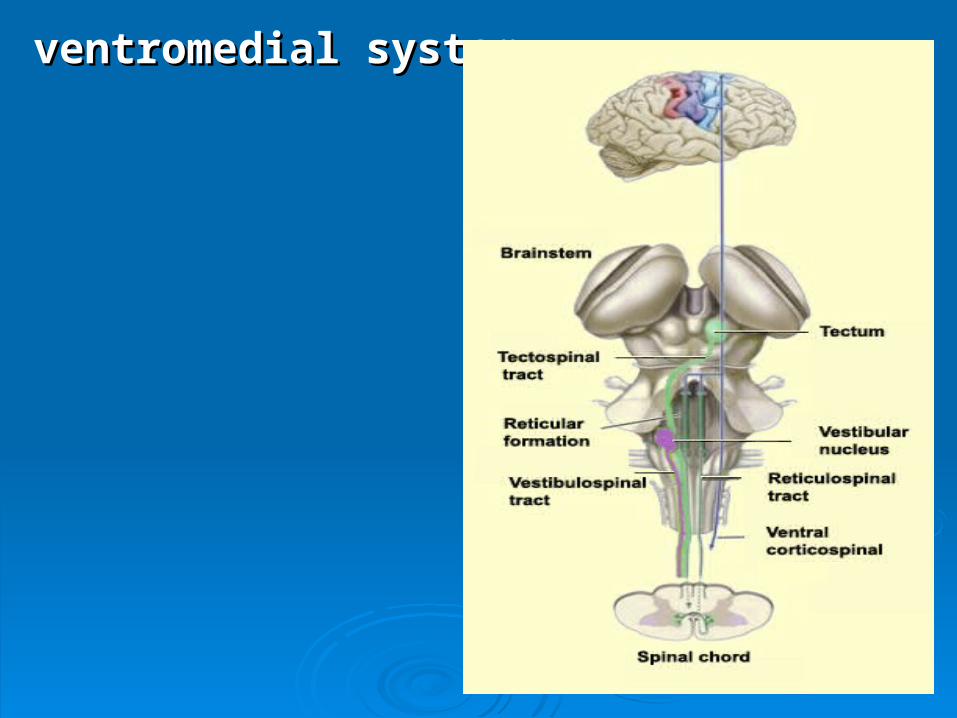
2424
ventromedial systemventromedial system

2525
THE BASAL GANGLIATHE BASAL GANGLIA
2626

2727
The basal ganglia and its connectionsThe basal ganglia and its connections
No input from ascending sensory path wayNo input from ascending sensory path way
No direct spinal projectionNo direct spinal projection
Cortico-striato-pallido-thalamo-cortical loopCortico-striato-pallido-thalamo-cortical loop
Cortical projections of to the basal ganglia are Cortical projections of to the basal ganglia are directed mainly to the directed mainly to the striatumstriatum and and subthalamic subthalamic nucleusnucleus (STN) (STN)

2828
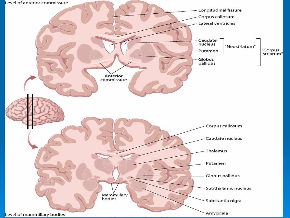
The striatum receives inputs from all areas of the The striatum receives inputs from all areas of the cerebral neocortexcerebral neocortex
Cortical inputs to the subthalamus come mainly from Cortical inputs to the subthalamus come mainly from the frontal lobethe frontal lobe
The striatum sends GABAergic inhibitory projections: The striatum sends GABAergic inhibitory projections: Globus pallidus externa GPeGlobus pallidus externa GPe Globus pallidus interna GPiGlobus pallidus interna GPi Substantia nigra pars reticulata SNrSubstantia nigra pars reticulata SNr

2929
GPe sends GABAergic inhibitory projections to:GPe sends GABAergic inhibitory projections to:
GPi / SNr & STNGPi / SNr & STN
STN sends excitatory Glutamergic fibers to GPe STN sends excitatory Glutamergic fibers to GPe and GPi/ SNrand GPi/ SNr
Thus there are to types of path waysThus there are to types of path ways
1.1. Direct :Direct :a. Striatum-Gpi/SNra. Striatum-Gpi/SNr
2.2. Indirect: Indirect: a. Striatum-GPe-STN-GPi / SNr a. Striatum-GPe-STN-GPi / SNr
b. Striatum-Gpe-Gpi/SNrb. Striatum-Gpe-Gpi/SNr

3030
Anterior putamenMost of caudate
P.Comm. Putamen D.lat. head of caudate
N. AccumbensOlfactory tubercleVentral putamenVentral caudate
DORSAL
VENTROLATERAL
ROSTROMEDIAL
GLOBUSPALLIDUS
(E)
GLOBUS
PALLIDUS(I)
SUBSTANTIA
NIG RA ®
SENSORY MOTORS.MOTOR
LIMBIC
ASSOCIATIVE ASSOCIATIVE
LIMBIC
MDLIMBIC
VL/VASEN-
MOTOR
ASS
VAMD

3131
ASSOCIATIVEAnterior putamenMost of caudate
SENSORY MOTORPostcommissural
DorsolateralHead of caudate
LIMBICAccumbens
Olfactory tubercleVentral putamen Ventral caudate
INTERNAL AND EXTERNA L PALLIDAL SEGMENTS
DORSAL VENTROLATERAL ROSTROMEDIAL
3232
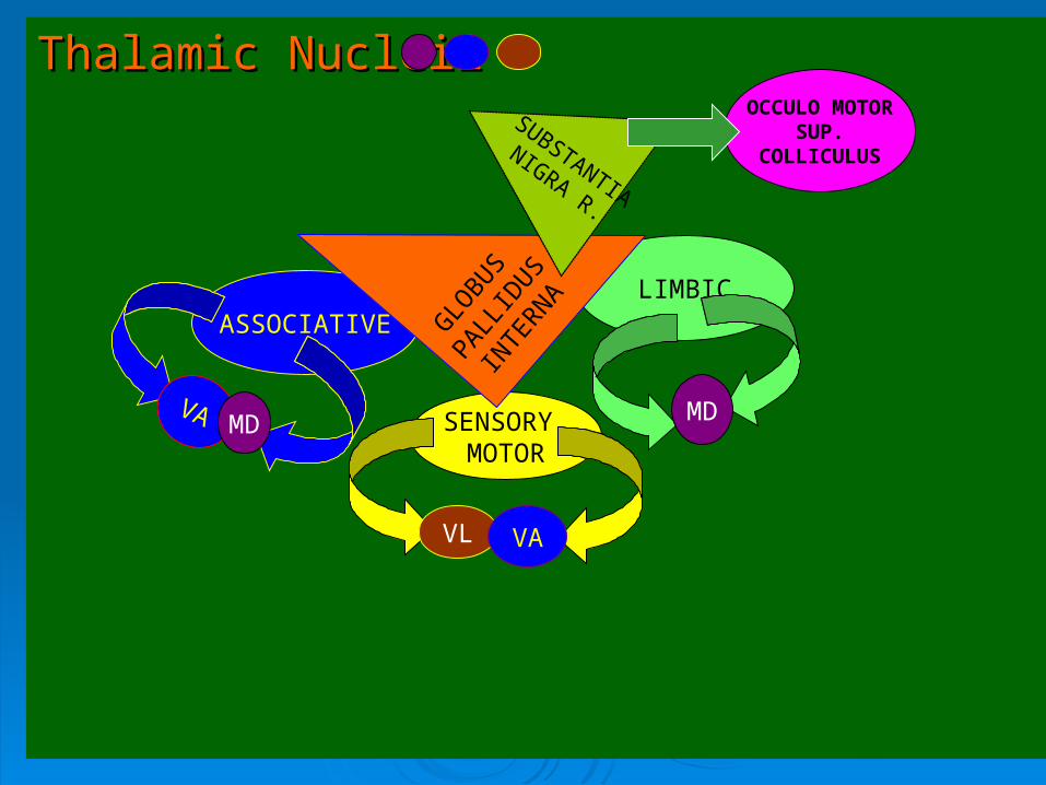
Thalamic NucleiiThalamic Nucleii
ASSOCIATIVE
SENSORY MOTOR
LIMBIC
VA MD
VL VA
MD
OCCULO MOTORSUP.
COLLICULUS
GLO
BUS
PALLID
US
INTE
RNA
SUBSTANTIA
NIGRA R.

3333
GLUTAMATEExcitatory
STRIATUMD1 GABA/P D2GABA/E D1GABA/P
Globus Pallidus Extrna
Gpi/SNr
SNc DA
STN PPN/MEAAch/GlU
RET
MD
CM/PF
VA/VL
DIRECT PATH
GABA
INDIRECT PATH
GABA
INDIRECT PATH 1
GABA
Glu
INDIRECT PATH 2
GABA GABA
Glu

3434
ParkinsonismParkinsonism
1.1. Rest tremor upper or lower extrimitiesRest tremor upper or lower extrimities
2.2. Rigidity,akinesia and bradykinesiaRigidity,akinesia and bradykinesia
3.3. Incapacitating type of action tremor ULIncapacitating type of action tremor UL
Loss of dopamine in the nigrostriatal pathwayLoss of dopamine in the nigrostriatal pathway Facilitates the direct path wayFacilitates the direct path way Inhibits the indirect path way Inhibits the indirect path way Net result is increased and abnormally Net result is increased and abnormally
phasic activity in STN and GPi/SNrphasic activity in STN and GPi/SNr
3535
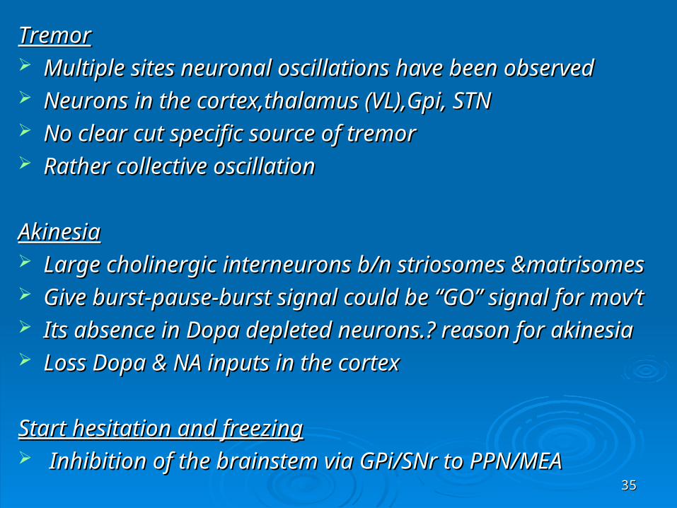
TremorTremor Multiple sites neuronal oscillations have been observedMultiple sites neuronal oscillations have been observed Neurons in the cortex,thalamus (VL),Gpi, STNNeurons in the cortex,thalamus (VL),Gpi, STN No clear cut specific source of tremorNo clear cut specific source of tremor Rather collective oscillationRather collective oscillation
AkinesiaAkinesia Large cholinergic interneurons b/n striosomes &matrisomesLarge cholinergic interneurons b/n striosomes &matrisomes Give burst-pause-burst signal could be “GO” signal for mov’tGive burst-pause-burst signal could be “GO” signal for mov’t Its absence in Dopa depleted neurons.? reason for akinesiaIts absence in Dopa depleted neurons.? reason for akinesia Loss Dopa & NA inputs in the cortexLoss Dopa & NA inputs in the cortex
Start hesitation and freezingStart hesitation and freezing Inhibition of the brainstem via GPi/SNr to PPN/MEAInhibition of the brainstem via GPi/SNr to PPN/MEA
3636
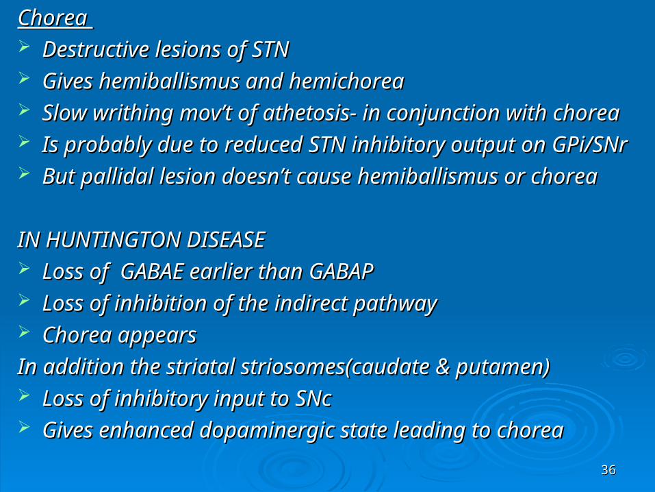
Chorea Chorea Destructive lesions of STN Destructive lesions of STN Gives hemiballismus and hemichoreaGives hemiballismus and hemichorea Slow writhing mov’t of athetosis- in conjunction with choreaSlow writhing mov’t of athetosis- in conjunction with chorea Is probably due to reduced STN inhibitory output on GPi/SNrIs probably due to reduced STN inhibitory output on GPi/SNr But pallidal lesion doesn’t cause hemiballismus or choreaBut pallidal lesion doesn’t cause hemiballismus or chorea
IN HUNTINGTON DISEASEIN HUNTINGTON DISEASE Loss of GABAE earlier than GABAPLoss of GABAE earlier than GABAP Loss of inhibition of the indirect pathwayLoss of inhibition of the indirect pathway Chorea appearsChorea appears
In addition the striatal striosomes(caudate & putamen)In addition the striatal striosomes(caudate & putamen) Loss of inhibitory input to SNcLoss of inhibitory input to SNc Gives enhanced dopaminergic state leading to choreaGives enhanced dopaminergic state leading to chorea
3737
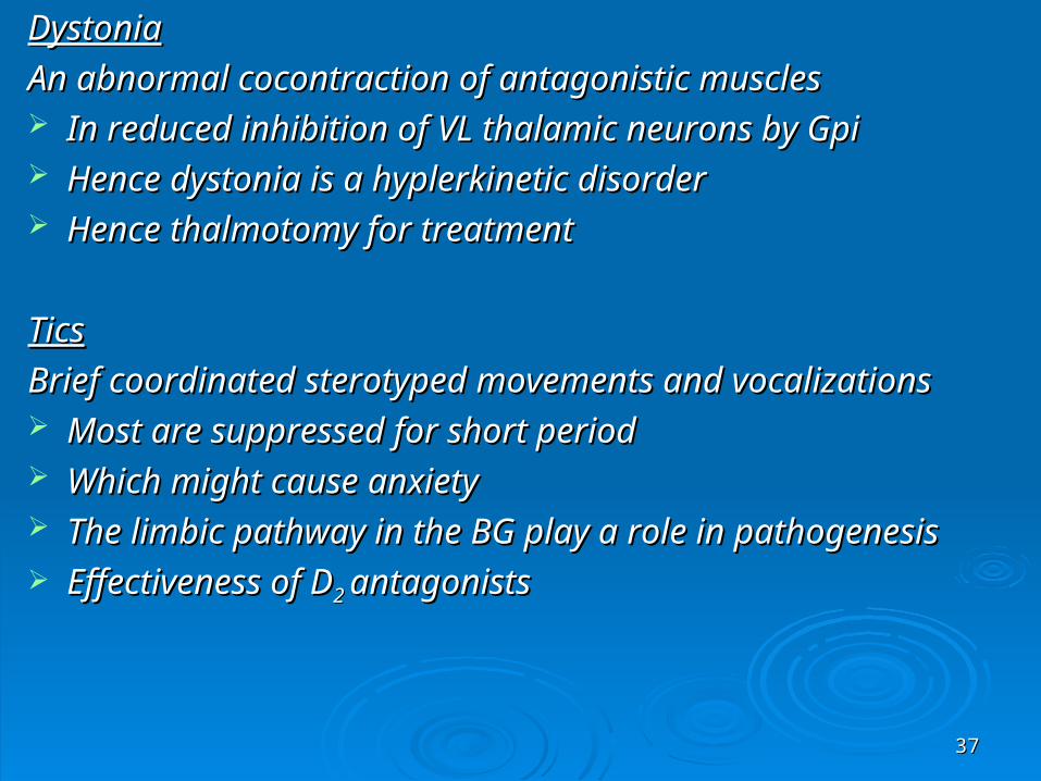
DystoniaDystonia
An abnormal cocontraction of antagonistic musclesAn abnormal cocontraction of antagonistic muscles In reduced inhibition of VL thalamic neurons by GpiIn reduced inhibition of VL thalamic neurons by Gpi Hence dystonia is a hyplerkinetic disorderHence dystonia is a hyplerkinetic disorder Hence thalmotomy for treatmentHence thalmotomy for treatment
TicsTics
Brief coordinated sterotyped movements and vocalizationsBrief coordinated sterotyped movements and vocalizations Most are suppressed for short period Most are suppressed for short period Which might cause anxietyWhich might cause anxiety The limbic pathway in the BG play a role in pathogenesisThe limbic pathway in the BG play a role in pathogenesis Effectiveness of DEffectiveness of D2 2 antagonistsantagonists

3838
THE CEREBELLUMTHE CEREBELLUM
The pathologies of the cerebellum have long The pathologies of the cerebellum have long revealed that this part of the brain is revealed that this part of the brain is involved in motor co-ordinationinvolved in motor co-ordination
The cerebellum The cerebellum is divided into threeis divided into three regions, regions, each of which is connected to a specific each of which is connected to a specific structure in the brain and involved in a structure in the brain and involved in a specific function specific function

3939
1,The 1,The archicerebellum (vestibulocerebellum)archicerebellum (vestibulocerebellum)
First appeared in fish. First appeared in fish. It is connected to the It is connected to the vestibulevestibule of the inner ear of the inner ear And is involved in And is involved in balance.balance.

4040
2,2,TheThe PalaeocerebellumPalaeocerebellum (Spinocerebellum)(Spinocerebellum)
Consists mainly of the vermis.Consists mainly of the vermis.
Is connected to the spinal cord Is connected to the spinal cord
Controls postural muscle activity by influencing Controls postural muscle activity by influencing muscle tonus.muscle tonus.
The cerebellum therefore controls muscle tension at The cerebellum therefore controls muscle tension at all times all times
while releasing those muscles required to execute while releasing those muscles required to execute movements.movements.
4141
3,N3,Nneocerebellumneocerebellum (Cerebrocerebellum)(Cerebrocerebellum)
It is more voluminous in primates and It is more voluminous in primates and especially so in humans.especially so in humans.
It consists of the Cerebellar hemispheres,It consists of the Cerebellar hemispheres,
Is connected to the cortex, and contributes to Is connected to the cortex, and contributes to
the co-ordination of voluntary movements. the co-ordination of voluntary movements.

4242
Cerebellum schemaCerebellum schema

4343
The cerebellar grey matter organized like the The cerebellar grey matter organized like the cerebral grey mattercerebral grey matter
A cortex forming the grey matter at the A cortex forming the grey matter at the surface, surface,
Deep nuclei that serve as relays for the Deep nuclei that serve as relays for the efferent pathways leaving this cortex . efferent pathways leaving this cortex .
4444
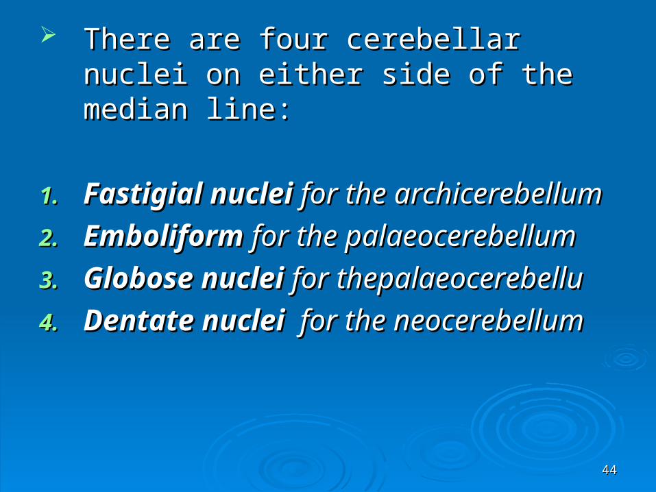
There are four cerebellar nuclei on either There are four cerebellar nuclei on either side of the median line: side of the median line:
1.1. Fastigial nucleiFastigial nuclei for the archicerebellum for the archicerebellum
2.2. Emboliform Emboliform for the palaeocerebellumfor the palaeocerebellum
3.3. Globose nuclei Globose nuclei for thepalaeocerebellufor thepalaeocerebellu
4.4. Dentate nuclei Dentate nuclei for the neocerebellum for the neocerebellum

4545
Provides control over the timing of the Provides control over the timing of the body's movements. body's movements.
Via loop circuit that connects it to the MC Via loop circuit that connects it to the MC
Modulates the signals that the motor Modulates the signals that the motor cortex sends to the motor neurons cortex sends to the motor neurons
Analyzes the visual signals associated Analyzes the visual signals associated with movementwith movement
4646
Signals from the movement of objects Signals from the movement of objects
Or from moving body segments themselves.Or from moving body segments themselves.
Calculate the speed of these movements and Calculate the speed of these movements and adjust the motor commands accordingly.adjust the motor commands accordingly.
Errors in such calculations largely account for Errors in such calculations largely account for the poor motor controlthe poor motor control
Participates in language, attention, memory, Participates in language, attention, memory,
and emotionsand emotions
4747
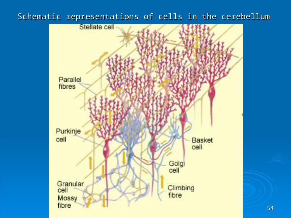
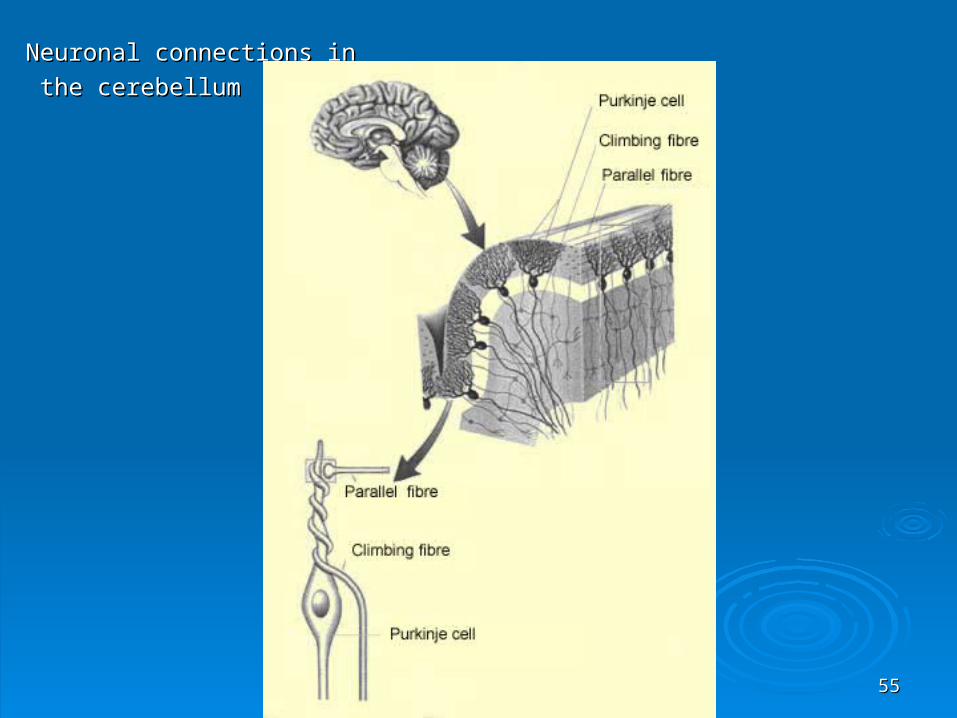
The The Purkinje cells Purkinje cells are the most characteristic are the most characteristic type of neurons in the cerebellum.type of neurons in the cerebellum.
The axons of the Purkinje cells synapse on the The axons of the Purkinje cells synapse on the neurons of the Dentate Nuclei of the neurons of the Dentate Nuclei of the Cerebellum. Cerebellum.
These nuclei relay the information to the These nuclei relay the information to the thalamusthalamus
Which then projects to the cortex and the Which then projects to the cortex and the Striatum.Striatum.
4848
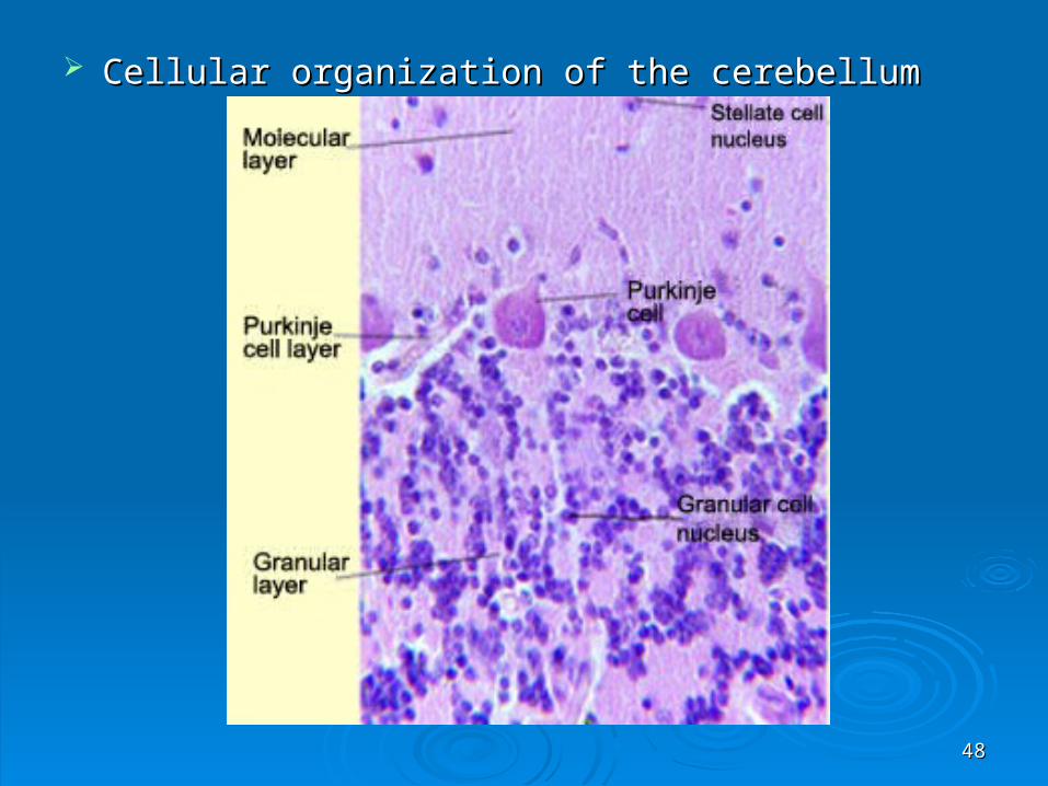
Cellular organization of the cerebellumCellular organization of the cerebellum

4949
The dendrite branches of each Purkinje cell The dendrite branches of each Purkinje cell receive synapses from of a single afferent receive synapses from of a single afferent Climbing FiberClimbing Fiber. .
This fiber is the axon of a neuron in the inferior This fiber is the axon of a neuron in the inferior olive, a nucleus in the medulla oblongata. olive, a nucleus in the medulla oblongata.
The inferior olive integrates the information from The inferior olive integrates the information from the muscle proprioceptors.the muscle proprioceptors.

5050
Each climbing fiber winds closely around Each climbing fiber winds closely around the dendrites of its corresponding Purkinje the dendrites of its corresponding Purkinje cellcell
So that the activation of this fiber will So that the activation of this fiber will cause a massive excitation of this cell. cause a massive excitation of this cell.

5151
Mossy FibersMossy Fibers, act in a highly diffuse fashion, act in a highly diffuse fashion
Are the axons of neurons in the pontine nucleiAre the axons of neurons in the pontine nuclei
They receive information from the cerebral They receive information from the cerebral cortex.cortex.
They carry this information to synapses with They carry this information to synapses with the small the small granular cells granular cells in the deep layer of in the deep layer of the cerebellum. the cerebellum.
5252
The axons of these granular cells ascend into The axons of these granular cells ascend into the surface layer of the cerebellum the surface layer of the cerebellum
where they branch into T shapes to form the where they branch into T shapes to form the parallel fibresparallel fibres. .

5353
The parallel fibers then run perpendicular to The parallel fibers then run perpendicular to the Purkinje cell dendrite fans,the Purkinje cell dendrite fans,
Thus crossing many Purkinje cells and Thus crossing many Purkinje cells and connecting them into a single contact. connecting them into a single contact.
Each parallel fiber makes only one contact Each parallel fiber makes only one contact with each Purkinje cell that it crosses,. with each Purkinje cell that it crosses,.
Likewise, each Purkinje cell receives over Likewise, each Purkinje cell receives over 100 000 synapses from 100 000 different 100 000 synapses from 100 000 different parallel fibers.parallel fibers.
5454
Schematic representations of cells in the cerebellumSchematic representations of cells in the cerebellum
5555
Neuronal connections inNeuronal connections in
the cerebellumthe cerebellum
5656
Pontine n.Pontine n. Biggest source of mossy fibersBiggest source of mossy fibers Premotor ,suplementary motor,primay Premotor ,suplementary motor,primay
motor,somatosensory, posterior motor,somatosensory, posterior parietalextrasriataal visual and cingulate parietalextrasriataal visual and cingulate corteces and auditory cortex corteces and auditory cortex
Reticular n.Reticular n. Inputs mainly from sensory motor cortexInputs mainly from sensory motor cortex Mosssy fiber input from dorsal an ventral Mosssy fiber input from dorsal an ventral
spinocerbellar pathwaysspinocerbellar pathways From vestibulocerebellar patwayFrom vestibulocerebellar patway

5757
Cerebellar nuclei project toCerebellar nuclei project to The brainstem nuclei and ventrolateral thalamus The brainstem nuclei and ventrolateral thalamus
containing N. Ventralis intermediuscontaining N. Ventralis intermedius Sources are mainly from Dentae, Globoce and Sources are mainly from Dentae, Globoce and
Emboliform nucleiiEmboliform nucleii Fastigium mainly gives to the ventromedial Fastigium mainly gives to the ventromedial
thalamus and also to VLthalamus and also to VL It also project to Red N., Reticular formation, inferior It also project to Red N., Reticular formation, inferior
olive, lateral vestibular n.olive, lateral vestibular n. Dentate n. receives input from lateral cerebellar Dentate n. receives input from lateral cerebellar
cortex (no direct somatosensory input)cortex (no direct somatosensory input) Activated during cognitive and sensory processingActivated during cognitive and sensory processing
5858
Interposed nucleiiInterposed nucleii receive input from the paramedian receive input from the paramedian cerebellar cortexcerebellar cortex
Receive abundant input from the SMCReceive abundant input from the SMC Involved in the control of muscle antagonistsInvolved in the control of muscle antagonists Inactivation causes ipsilateral dysmetria and intention Inactivation causes ipsilateral dysmetria and intention
tremortremor
FastigumFastigum receives input from the cerebellar vermis receives input from the cerebellar vermis and floculonodular lobeand floculonodular lobe
Participate in control of vestibulo-occular control and Participate in control of vestibulo-occular control and locomotionlocomotion
Inactivation causes severe truncal disequilibrium, gait Inactivation causes severe truncal disequilibrium, gait ataxia falls to the side of the lesionataxia falls to the side of the lesion

5959
Comparison of the basal ganglia and cerebellar control circuitsComparison of the basal ganglia and cerebellar control circuits
FeaturesFeatures Basal gangliaBasal ganglia CerebellumCerebellum
Cortical inputsCortical inputs Wide spread (predominantly Wide spread (predominantly frontal lobe)frontal lobe)
Wide spread Wide spread
Receptive componentsReceptive components Striatum( caudate& Putamen) Striatum( caudate& Putamen) Subthalamic nucleiSubthalamic nuclei
Cerebellar cortex Purkinje Cerebellar cortex Purkinje cellscells
Effectors componentEffectors component Globus pallidus internal Globus pallidus internal componentcomponent
Cerebellar NucleiiCerebellar Nucleii
Regulatory componentRegulatory component Substantia Nigra CSubstantia Nigra C inferior Olivary nucleusinferior Olivary nucleus
Thalamic nucleusThalamic nucleus Ventral anterior & othersVentral anterior & others Ventral lateral)Ventral lateral)
Motor cortex targetMotor cortex target Supplementary & Premotor Supplementary & Premotor corticescortices
Primary motor cortex Primary motor cortex (supplementary & premotor)(supplementary & premotor)
Brainstem targetBrainstem target Pedenculopontine nucleus, Pedenculopontine nucleus, Superior colliculusSuperior colliculus
Red nucleus, Vestibular nuclei Red nucleus, Vestibular nuclei Reticular formationReticular formation
Direct spinal inputDirect spinal input NoNo YesYes
FunctionFunction Selection of motor programsSelection of motor programs Initiation an execution of Initiation an execution of motor actsmotor acts
Clinical correlatesClinical correlates Hypokinesis, rigidity, tremor Hypokinesis, rigidity, tremor at rest abnormal mov’t at rest abnormal mov’t HyperkinesisHyperkinesis
Disequilibrium Incoordination, Disequilibrium Incoordination, ataxia, action tremorataxia, action tremor
Localization of clinical Localization of clinical findingsfindings
Contralateral to te lesionContralateral to te lesion Ipsilatral to lesionIpsilatral to lesion
6060
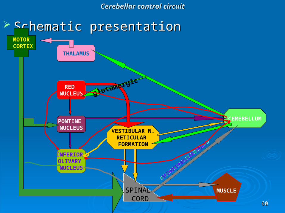
Cerebellar control circuitCerebellar control circuit
Schematic presentationSchematic presentation
RED NUCLEUS
PONTINE NUCLEUS
INFERIOR OLIVARY NUCLEUS
MOTOR CORTEX
THALAMUS
CEREBELLUM
SPINAL CORD
MUSCLE
SPINOCERBELLAR TRACT
VESTIBULAR N.RETICULAR FORMATION
glutamergic

6161

6262

6363

6464