Embed Size (px)
Citation preview

Malaria is known to infect more than 250 million individuals throughout the world and is killing more than 1 million people a year, most of them are children. There are 5 species of malaria that infect humans 1. Plasmodium falciparum 2. Plasmodium vivax 3. Plasmodium malariae 4. Plasmodium ovale 5. Plasmodium knowlesi
P. falciparum, and P. vivax account for ∼95% of the infections. P. vivax has the widest distribution, extending throughout the tropics although much less in subsaharian Africa, subtropics, and temperate zones. P. falciparum is generally confined to the tropics, P. malariae is sporadically distributed, P. ovale is confined mainly to central West Africa and South Pacific islands and P. knowlesi which is a primate malaria is commonly found in Southeast Asia, especially Malaysia and Indonesia.

Of these 5 species, P. falciparum is the most severe and the leading cause of the fatal infection. It is known as “malignant malaria” because of its potential severity and fatal outcome. The pathophysiology of falciparum malaria results from the interaction between the parasite and the host. These include red cell invasion by merozoites, adherence and sequestration of mature parasite-‐infected red cells to endothelial cells, the rosetting of uninfected red cells around mature parasite-‐infected red cells, the reduction in deformability of the infected and uninfected red cells (Cranston et al. 1984; Dondorp et al. 1997) and the production of toxins by infected red cells. The main pathophysiological mechanism of falciparum malaria is sequestration of parasite-‐infected red cells in deep vascular beds. Sequestration is the process whereby red cells infected with matured parasites specifically adhere to the endothelial cells of capillaries and venules (Howard & Gilladoga, 1989). Plasmodium falciparum malaria after invasion of red blood cells, the merozoites develop into more mature stages and induce knob-‐like structures on the infected red cell surface that adhere to capillaries or venules obstructing blood supply and causing vital organ dysfunction. This major pathological process leads to severe falciparum infection (Berendt et al. 1990). It is the leading cause of microvasculatory obstruction, reducing of oxygen and substrate supply leading to anaerobic glycolysis and lactic acidosis and hence the vital organ dysfunctions.
The disease severity of falciparum malaria is determined by not only parasitaemia but also by the parasite stage distribution in peripheral blood. Because of mature parasite sequestration, these mature forms (known as trophozoites or schizonts) of P. falciparum are therefore not usually seen in peripheral blood films. Patients with falciparum malaria who present with mature stage parasites in the peripheral blood usually have severe infection (Silamut et al, 1993) indicating that severity of falciparum malaria is not only determined by parasitaemia but also the sequestered parasite biomass. Mature stages are more pathogenic than early stages. Therefore, accurate measurements of parasite age, morphology and stage distribution provide valuable prognostic, pathophysiological and therapeutic information.
A long duration of untreated infection with falciparum malaria may result in a synchronised infection with parasite sequestration, resulting in the appearance of trophozoites and schizonts in the peripheral blood. Morphological assessment of parasite development in a peripheral blood film should reflect the proportion of
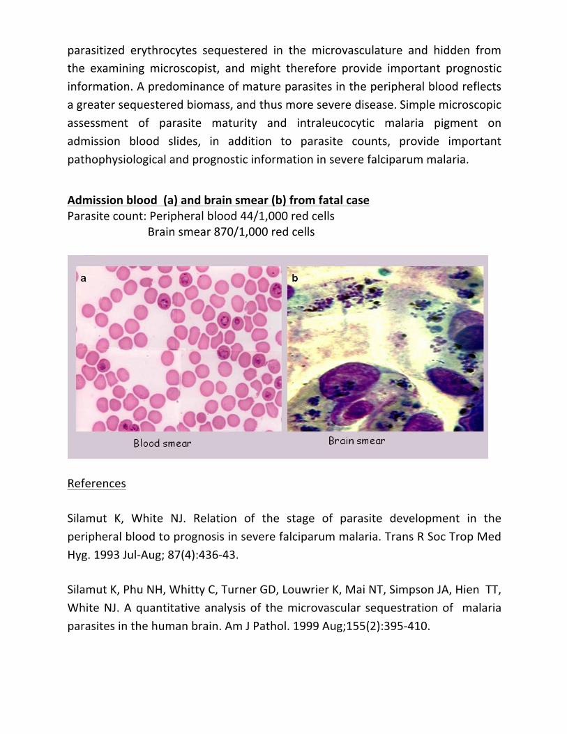
parasitized erythrocytes sequestered in the microvasculature and hidden from the examining microscopist, and might therefore provide important prognostic information. A predominance of mature parasites in the peripheral blood reflects a greater sequestered biomass, and thus more severe disease. Simple microscopic assessment of parasite maturity and intraleucocytic malaria pigment on admission blood slides, in addition to parasite counts, provide important pathophysiological and prognostic information in severe falciparum malaria.
Admission blood (a) and brain smear (b) from fatal case Parasite count: Peripheral blood 44/1,000 red cells Brain smear 870/1,000 red cells
References
Silamut K, White NJ. Relation of the stage of parasite development in the peripheral blood to prognosis in severe falciparum malaria. Trans R Soc Trop Med Hyg. 1993 Jul-‐Aug; 87(4):436-‐43.
Silamut K, Phu NH, Whitty C, Turner GD, Louwrier K, Mai NT, Simpson JA, Hien TT, White NJ. A quantitative analysis of the microvascular sequestration of malaria parasites in the human brain. Am J Pathol. 1999 Aug;155(2):395-‐410.
Assessment of parasite stage development in thin smears:
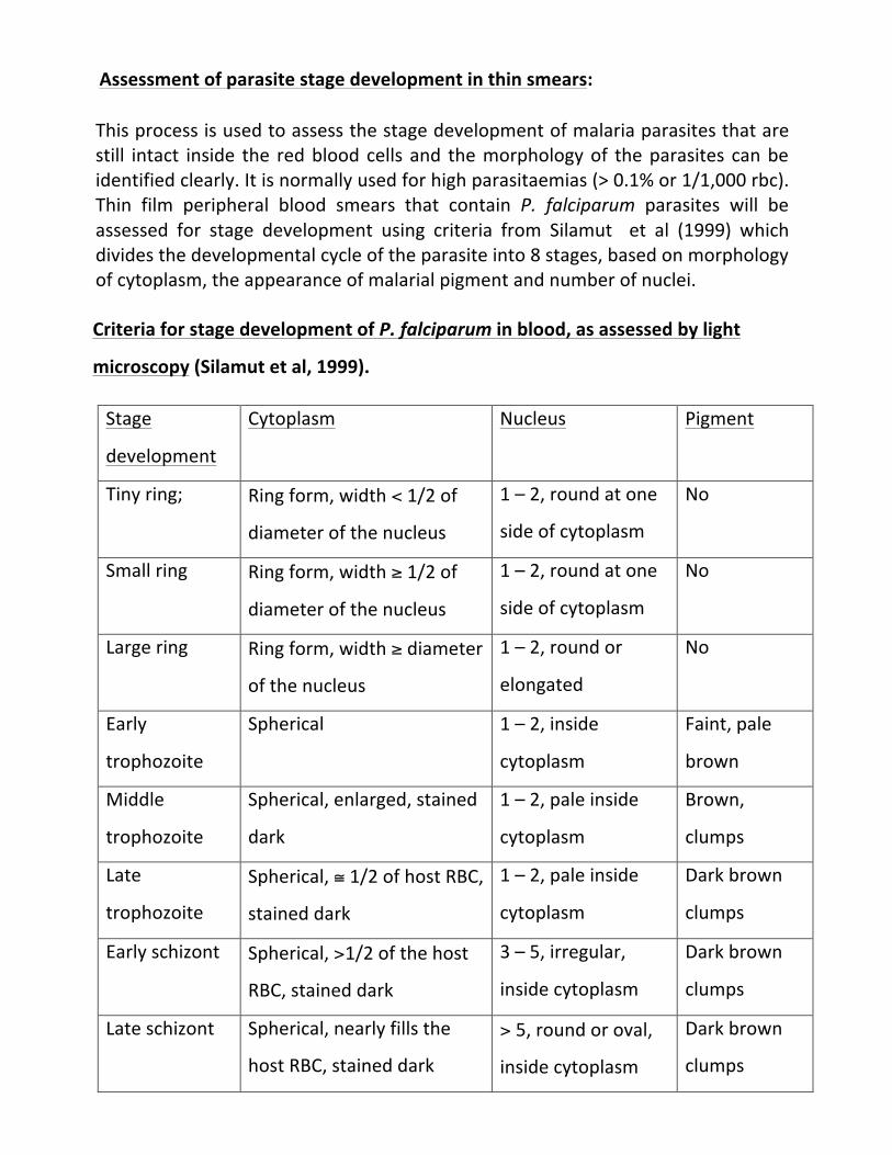
This process is used to assess the stage development of malaria parasites that are still intact inside the red blood cells and the morphology of the parasites can be identified clearly. It is normally used for high parasitaemias (> 0.1% or 1/1,000 rbc). Thin film peripheral blood smears that contain P. falciparum parasites will be assessed for stage development using criteria from Silamut et al (1999) which divides the developmental cycle of the parasite into 8 stages, based on morphology of cytoplasm, the appearance of malarial pigment and number of nuclei. Criteria for stage development of P. falciparum in blood, as assessed by light
microscopy (Silamut et al, 1999).
Stage
development
Cytoplasm Nucleus Pigment
Tiny ring; Ring form, width < 1/2 of
diameter of the nucleus
1 – 2, round at one
side of cytoplasm
No
Small ring Ring form, width ≥ 1/2 of
diameter of the nucleus
1 – 2, round at one
side of cytoplasm
No
Large ring Ring form, width ≥ diameter
of the nucleus
1 – 2, round or
elongated
No
Early
trophozoite
Spherical 1 – 2, inside
cytoplasm
Faint, pale
brown
Middle
trophozoite
Spherical, enlarged, stained
dark
1 – 2, pale inside
cytoplasm
Brown,
clumps
Late
trophozoite
Spherical, ≅ 1/2 of host RBC,
stained dark
1 – 2, pale inside
cytoplasm
Dark brown
clumps
Early schizont Spherical, >1/2 of the host
RBC, stained dark
3 – 5, irregular,
inside cytoplasm
Dark brown
clumps
Late schizont Spherical, nearly fills the
host RBC, stained dark
> 5, round or oval,
inside cytoplasm
Dark brown
clumps
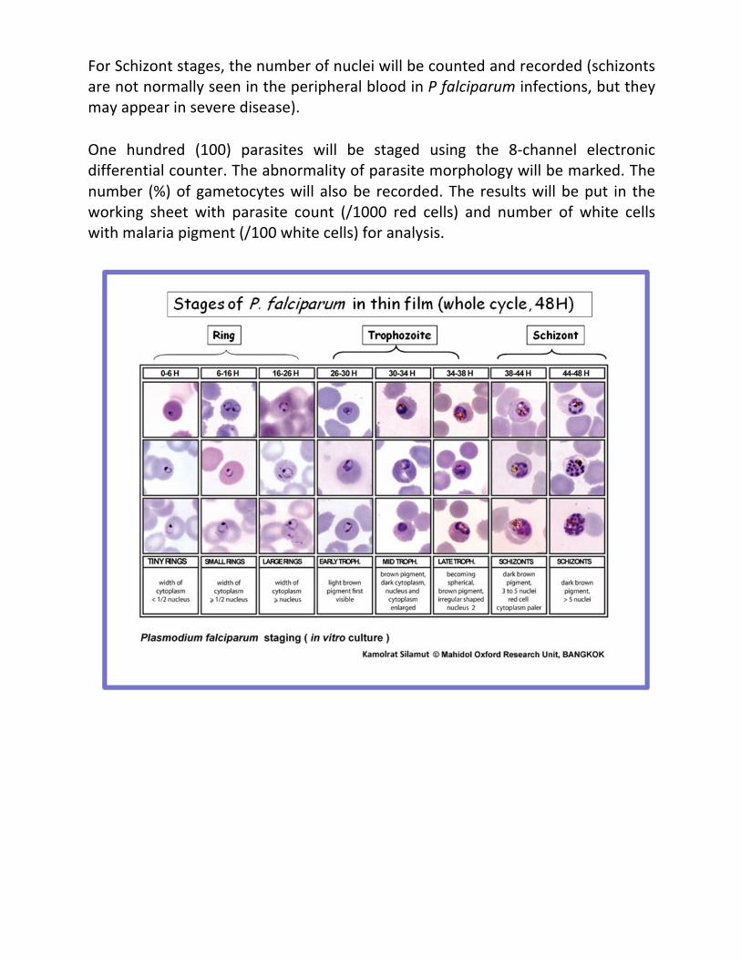
For Schizont stages, the number of nuclei will be counted and recorded (schizonts are not normally seen in the peripheral blood in P falciparum infections, but they may appear in severe disease).
One hundred (100) parasites will be staged using the 8-‐channel electronic differential counter. The abnormality of parasite morphology will be marked. The number (%) of gametocytes will also be recorded. The results will be put in the working sheet with parasite count (/1000 red cells) and number of white cells with malaria pigment (/100 white cells) for analysis.