Embed Size (px)
Citation preview
10
Overview: r ehabilitation of n atural t eeth
1
Prosthodontics at a Glance, First Edition. Irfan Ahmad. © 2012 Irfan Ahmad. Published 2012 by Blackwell Publishing Ltd.
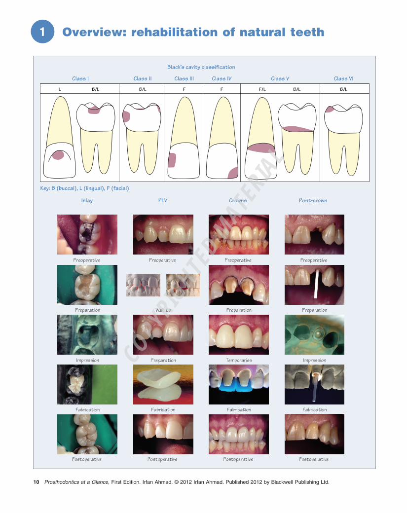
Black’s cavity classification
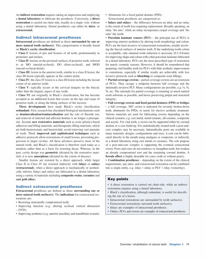
Preoperative
Preparation
Impression
Fabrication
Postoperative
Inlay
Key: B (buccal), L (lingual), F (facial)
Class I Class II Class III Class IV Class V Class VI
Preoperative
Wax-up
Preparation
Fabrication
Postoperative
PLV
Preoperative
Preparation
Temporaries
Fabrication
Postoperative
Crowns
Preoperative
Preparation
Impression
Fabrication
Postoperative
Post-crown
a b
L B/L B/L B/LF/L B/LF F
COPYRIG
HTED M
ATERIAL
Overview: rehabilitation of natural teeth Chapter 1 11
An indirect restoration requires taking an impression and employing a dental laboratory to fabricate the prosthesis. Conversely, a direct restoration is carried out chair - side, usually in a single visit, without using a dental laboratory. Dental prostheses can either be intra - or extracoronal.
Indirect i ntracoronal p rostheses Intracoronal prostheses are defi ned as those surrounded by one or more natural tooth surface(s) . This categorisation is broadly based on Black ’ s cavity classifi cation : • Class I : lesions of pits and fi ssures of all teeth, predominantly in premolars and molars; • Class II : lesions on the proximal surfaces of posterior teeth, referred to as MO (mesial - occlusal), DO (disto - occlusal), and MOD (mesial - occlusal - distal); • Class III : lesion in the anterior teeth, similar to a class II lesion, the class III lesion typically appears at the contact point; • Class IV : the class IV lesion is a class III lesion including the incisal corner of an anterior tooth; • Class V : typically occurs at the cervical margins on the buccal, rather than the lingual, aspect of any tooth; • Class VI : not originally in Black ’ s classifi cation, but has become accepted as an additional lesion that occurs on the tips and cusps of posterior teeth, or along the biting surfaces of the incisors.
Three developments have made Black ’ s cavity classifi cation redundant . First, research has elucidated biological mechanisms such as demineralisation/remineralisation and the role of fl uoride ion, and removal of infected and affected dentine is no longer a prerequi-site. Second, new restorative materials such as resin (plastic) - based adhesives and fi lling materials, and therapeutic fi lling materials, which are both bacteriostatic and bactericidal, avoid removing vast amounts of tooth. Third, improved and sophisticated techniques such as adhesive protocols allow restorations of small lesions, preventing pro-gression to larger cavities. All these advances preserve more of the natural tooth, and Black ’ s classifi cation is therefore used today as a notation, rather than as a basis for restoring decay. Whereas, in the past, cavity design was geometric (dictated by the restorative mate-rial), it is now amorphous (dictated by the extent of disease).
Smaller lesions are restored by a direct approach, while larger Class II or Class IV are restored indirectly with inlays or onlays (extracoronal) , when a direct approach is mechanically or aestheti-cally inferior. Inlays and onlays are fabricated in a dental laboratory using a variety of materials including composite resins , ceramics and cast gold alloy s.
Indirect e xtracoronal p rostheses Extracoronal prostheses are defi ned as those surrounding one or more natural tooth surface(s) . The indications for extracoronal res-torations are: • Restoring structurally compromised teeth; • Improving function (e.g. altering occlusal vertical dimension – OVD); • Improving aesthetics (e.g. anterior maxillary and mandibular sextant);
• Abutments for a fi xed partial denture (FPD). Extracoronal prostheses are categorised as:
• Inlays and onlays – the difference between an inlay and an onlay is the extent of tooth loss requiring replacement. Broadly speaking, an inlay ‘ fi ts into ’ , while an onlay incorporates cuspal coverage and ‘ fi ts onto ’ the tooth; • Porcelain laminate veneers (PLV) – the principal use of PLVs is improving anterior aesthetics by altering tooth morphology and colour. PLVs are the least invasive of extracoronal restorations, usually involv-ing the buccal surfaces of anterior teeth. If the underlying tooth colour is acceptable, only minimal tooth reduction is necessary (0.3 – 0.8 mm) for improving shape and colour with a thin porcelain laminate fabricated in a dental laboratory. PLVs are the most prescribed type of restoration for purely cosmetic reasons. However, it should be remembered that preparing vital healthy teeth for PLVs with little aesthetic improvement is contentious, especially if similar results are achievable with less invasive protocols such as bleaching or composite resin fi llings; • Partial coverage crowns – partial coverage crowns are an extension of PLVs. They occupy a midpoint between full coverage and the minimally invasive PLV. Many confi gurations are possible, e.g. ½ , ¾, ⅞, etc. The rationale for partial coverage is retaining as much natural tooth substrate as possible, and hence preserving pulpal and structural integrity; • Full coverage crowns and fi xed partial dentures (FPD) or bridges – a full coverage, 360 ° crown is indicated for severely broken - down teeth, abutments for FPDs, or rarely for elective aesthetic treatment. Various materials are used for fabricating crowns depending on the clinical scenario, e.g. cast metal, metal - ceramic, all - ceramic, composite and acrylic. For vital teeth, a crown can be supported either by coronal dentine or a core build - up. For endodontically treated teeth a post - and - core complex may be necessary. Intraradicular posts are available in many materials, designs, confi gurations and sizes. A core can be fabri-cated directly in the mouth using amalgam or composite, or indirectly in a dental laboratory using cast metals or ceramics. The sole purpose of a post - and - core complex is supporting the eventual extracoronal crown. Posts and cores do not reinforce or strengthen teeth, but weaken an already compromised root and the remaining coronal dentine. A ferrule effect is highly desirable for cores (with or without posts); • Combination prostheses – depending on the extent of the clinical requirements, any intra - and extracoronal restoration can be combined into a single entity, e.g. inlay + onlay or PLV + inlay (veneerlay).
Key p oints • A direct restoration is carried out chair - side, while an indirect restoration requires using a dental laboratory. • Black ’ s classifi cation, although redundant, is useful for describ-ing the site of a lesion. • Intracoronal restorations are surrounded by tooth surface(s). • Extracoronal restorations surround tooth surface(s). • Inlays are examples of intracoronal prostheses. • Onlays, PLVs and crowns are examples of extracoronal prostheses.