Embed Size (px)
Citation preview

Fig. 1.1
1 Interpreting Chest X-Rays CA SE 1
Case 1. A 35-year-old male presented with fever, cough, and purulentsputum for one week. This was his CXR (Fig. 1.1). What is the diagnosis?
© Cambridge University Press www.cambridge.org
Cambridge University Press0521607329 - Interpreting Chest X-Rays: Illustrated with 100 CasesPhilip Eng and Foong-Koon CheahExcerptMore information

CA SE 1 PNEUMONIAThe CXR shows a focal shadow in the right lower lobe with air bronchograms sug-
gestive of pneumonia. It is clearly in the right lower lobe because the right hemidi-
aphragm is effaced. Right middle lobe shadows would efface the right heart border.
The presence of air bronchograms indicates pathology in the alveoli, as the con-
ducting airways remain patent with air. Water or blood can also occupy the alveoli
as a result of pulmonary edema or pulmonary hemorrhage respectively. There
should be other supporting signs such as cardiomegaly, upper lobe diversion, and
Kerley B lines with pulmonary edema. The differential diagnoses of a focal shadow
with air bronchograms include bronchoalveolar cell carcinoma and lymphoma. It
is important to follow-up the CXR to ensure that total resolution of infection
occurs. This may take up to three months in the elderly but generally some
improvement usually occurs within a week. The borders of the heart on a PA CXR
are shown in Fig. 1.2. SVC – superior vena cava, RA – right atrium, Ao – aortic
knuckle, LA – left atrium, LV – left ventricle
Interpreting Chest X-Rays 2CA SE 1
Fig. 1.2
SVC
RA
Ao
LA
LV
© Cambridge University Press www.cambridge.org
Cambridge University Press0521607329 - Interpreting Chest X-Rays: Illustrated with 100 CasesPhilip Eng and Foong-Koon CheahExcerptMore information

Fig. 2.1
3 Interpreting Chest X-Rays CA SE 2
Case 2. This 25-year-old had sudden onset of left-sided chest pain. TheCXR is shown (Fig. 2.1).
© Cambridge University Press www.cambridge.org
Cambridge University Press0521607329 - Interpreting Chest X-Rays: Illustrated with 100 CasesPhilip Eng and Foong-Koon CheahExcerptMore information

CA SE 2 LEFT PRIMARY SPONTANEOUS PNEUMOTHORAXThe CXR shows the visceral pleura (Fig. 2.2) separated from the parietal pleura by
air which now occupies the potential space in the pleural cavity. The visceral
pleura must not be mistaken for skin-fold shadows which usually occur in supine
or obese patient CXR. In addition, the line from skin folds can be seen to cross the
chest wall. In the patient above, the lungs appear otherwise healthy and this condi-
tion is called primary spontaneous pneumothorax. It occurs classically in young
males. This is in contradistinction to secondary pneumothorax which occurs in
diseased lungs, e.g. chronic obstructive pulmonary diseases (COPD).
Pneumothorax in an erect film is usually seen at the apex. See Case 60.
Interpreting Chest X-Rays 4CA SE 2
Fig. 2.2
© Cambridge University Press www.cambridge.org
Cambridge University Press0521607329 - Interpreting Chest X-Rays: Illustrated with 100 CasesPhilip Eng and Foong-Koon CheahExcerptMore information

Fig. 3.1
5 Interpreting Chest X-Rays CA SE 3
Case 3. 50-year-old male presented to the Emergency Room with shockand a four-day history of a febrile illness. He required intubation andwas started on inotropes. This was his CXR (Fig. 3.1).
© Cambridge University Press www.cambridge.org
Cambridge University Press0521607329 - Interpreting Chest X-Rays: Illustrated with 100 CasesPhilip Eng and Foong-Koon CheahExcerptMore information

CA SE 3 RUPTURED L IVER ABSCESSIt is important to look at the “blind areas” of the CXR in order not to miss impor-
tant clues. These areas are under the diaphragm, behind the heart, the hilum, and
the soft tissues. This CXR shows a lucency over the liver density. The lucency does
not conform to the usual bowel configuration. In this clinical context, an impor-
tant differential diagnosis to be considered is a ruptured liver abscess. This can be
confirmed either by bedside ultrasound or CT (Fig. 3.2). Liver abscesses are usually
due to organisms like Klebsiella or Amoebiasis. All patients with Klebsiella bac-
teremia of unknown origin should have imaging studies of the abdomen to rule
out a liver abscess.
Interpreting Chest X-Rays 6CA SE 3
Fig. 3.2
© Cambridge University Press www.cambridge.org
Cambridge University Press0521607329 - Interpreting Chest X-Rays: Illustrated with 100 CasesPhilip Eng and Foong-Koon CheahExcerptMore information

Fig. 4.1
7 Interpreting Chest X-Rays CA SE 4
Case 4. This elderly male has exertional dyspnea, orthopnea, and parox-ysmal nocturnal dyspnea. His CXR is shown (Fig. 4.1).
© Cambridge University Press www.cambridge.org
Cambridge University Press0521607329 - Interpreting Chest X-Rays: Illustrated with 100 CasesPhilip Eng and Foong-Koon CheahExcerptMore information
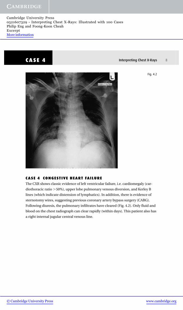
CA SE 4 CONGESTIVE HEART FAILUREThe CXR shows classic evidence of left ventricular failure, i.e. cardiomegaly (car-
diothoracic ratio �50%), upper lobe pulmonary venous diversion, and Kerley B
lines (which indicate distension of lymphatics). In addition, there is evidence of
sternotomy wires, suggesting previous coronary artery bypass surgery (CABG).
Following diuresis, the pulmonary infiltrates have cleared (Fig. 4.2). Only fluid and
blood on the chest radiograph can clear rapidly (within days). This patient also has
a right internal jugular central venous line.
Interpreting Chest X-Rays 8CA SE 4
Fig. 4.2
© Cambridge University Press www.cambridge.org
Cambridge University Press0521607329 - Interpreting Chest X-Rays: Illustrated with 100 CasesPhilip Eng and Foong-Koon CheahExcerptMore information

Fig. 5.1
9 Interpreting Chest X-Rays CA SE 5
Case 5. A 65-year-old male presented with cardiogenic shock. He had anemergency CABG which was associated with a very stormy peri-opera-tive period. This was his CXR (Fig. 5.1) taken upon arrival at theIntensive Care Unit (ICU). What is the most significant abnormality?
© Cambridge University Press www.cambridge.org
Cambridge University Press0521607329 - Interpreting Chest X-Rays: Illustrated with 100 CasesPhilip Eng and Foong-Koon CheahExcerptMore information

CA SE 5 FOREIGN BODY RIGHT LOWER ZONEThe CXR shows an opaque density in the region of the right lower zone (Fig. 5.2).
Each lung field on an erect CXR is divided into three zones. The upper zone is an
area which lies above a horizontal line drawn from the medial end of the second
rib anteriorly. The middle zone lies below this and is bordered inferiorly by a line
drawn similarly from the fourth rib. The lower zone lies below this. This opaque
density is similar in configuration to a tooth which was dislodged during emer-
gency intubation of this patient. Foreign bodies are not as common in adults com-
pared with children. It can occur silently in patients with decreased conscious
level. The typical site is in the right main stem bronchus, as this has a more vertical
course than the left. An example is seen in this CT (Fig. 5.3). Bronchoscopic
removal is the usual initial treatment of choice.
Interpreting Chest X-Rays 10CA SE 5
Fig. 5.2
Fig. 5.3
© Cambridge University Press www.cambridge.org
Cambridge University Press0521607329 - Interpreting Chest X-Rays: Illustrated with 100 CasesPhilip Eng and Foong-Koon CheahExcerptMore information