Embed Size (px)
Citation preview
11
I N F L A M M A T I O I N F L A M M A T I O NN
Assistant of professorAssistant of professor
Nechiporenko G. V.Nechiporenko G. V.
22
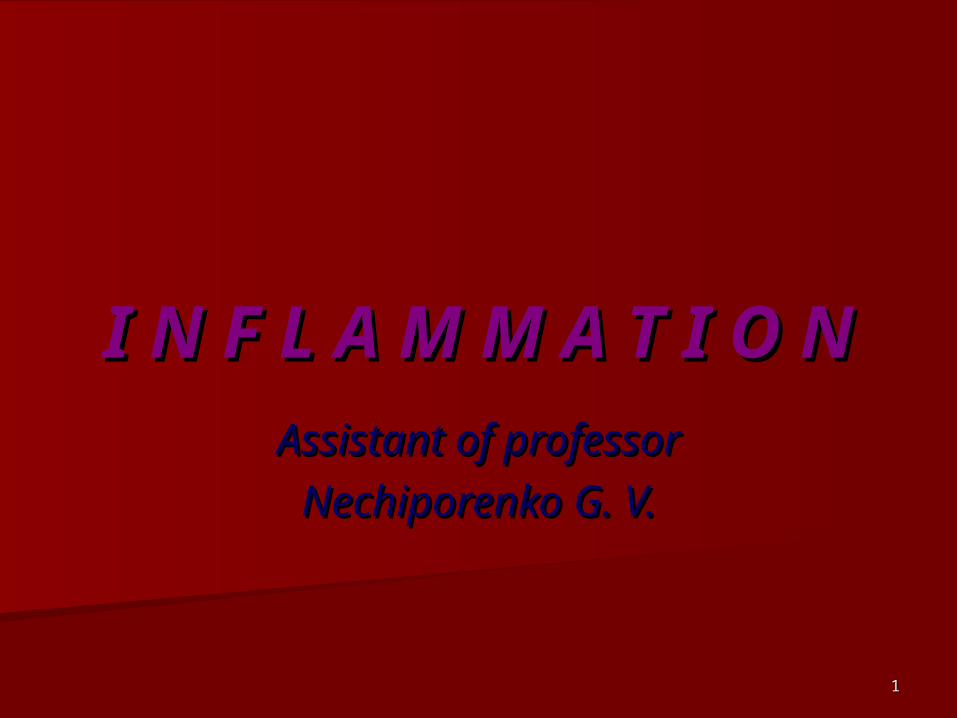
InflammationInflammation- is a local vascular-- is a local vascular-mesenchymal reaction to injury of mesenchymal reaction to injury of living tissue due to different agents. living tissue due to different agents.
This reaction has protective-adaptive This reaction has protective-adaptive character. character.
It can kill agent caused injury and It can kill agent caused injury and repair injured tissue.repair injured tissue.
Inflammation contains elements of Inflammation contains elements of pathology and physiology.pathology and physiology.
Heat (calor), redness (rubor), edema Heat (calor), redness (rubor), edema (tumor), pain (dolor), and loss of (tumor), pain (dolor), and loss of function (functio laesa).function (functio laesa).
33
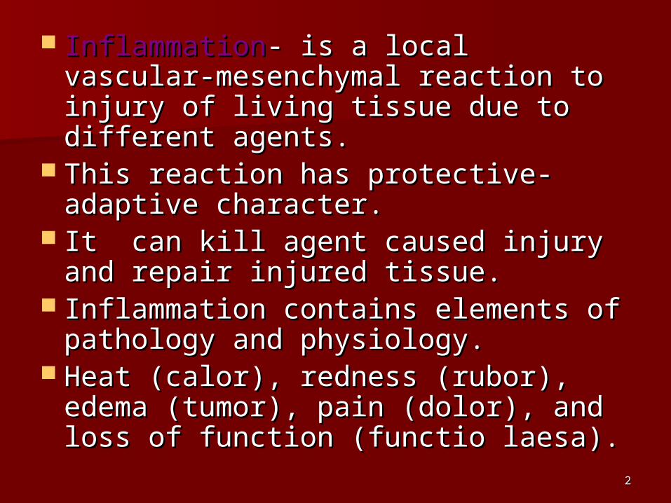
Types of inflammationTypes of inflammation
Morphological types: alterative, Morphological types: alterative, exudative, and proliferative exudative, and proliferative (productive)(productive)
Due to etiology: specific and non-Due to etiology: specific and non-specific (banal)specific (banal)
Due to duration: acute, subacute, Due to duration: acute, subacute, and chronicand chronic
Due to type of tissue’s reaction: Due to type of tissue’s reaction: normergic, hyperergic and hypoergicnormergic, hyperergic and hypoergic

44

55
Cells marginated along the dilated venule wall Cells marginated along the dilated venule wall (arrow) are squeezing through the basement (arrow) are squeezing through the basement membrane (diapedesis) and spilling out into membrane (diapedesis) and spilling out into extravascular space.extravascular space.

66

77
Exudative inflammationExudative inflammation
CatarrhalCatarrhal SerousSerous Purulent (suppurative)Purulent (suppurative) FibrinousFibrinous HemorrhagicHemorrhagic PutrefactivePutrefactive MixedMixed

88
Serous inflammationSerous inflammation
It is characterized by exudate It is characterized by exudate contained about 2% proteins, single contained about 2% proteins, single neutrophilic polymorphs and neutrophilic polymorphs and mesothelial cells. It occurs in serosa, mesothelial cells. It occurs in serosa, mucosa, meninges, skin and internal mucosa, meninges, skin and internal organs.organs.
Outcome is usually favorable. In Outcome is usually favorable. In severe cases it can be progressive severe cases it can be progressive into serous-purulent.into serous-purulent.

99
Serous inflammationSerous inflammation
1010
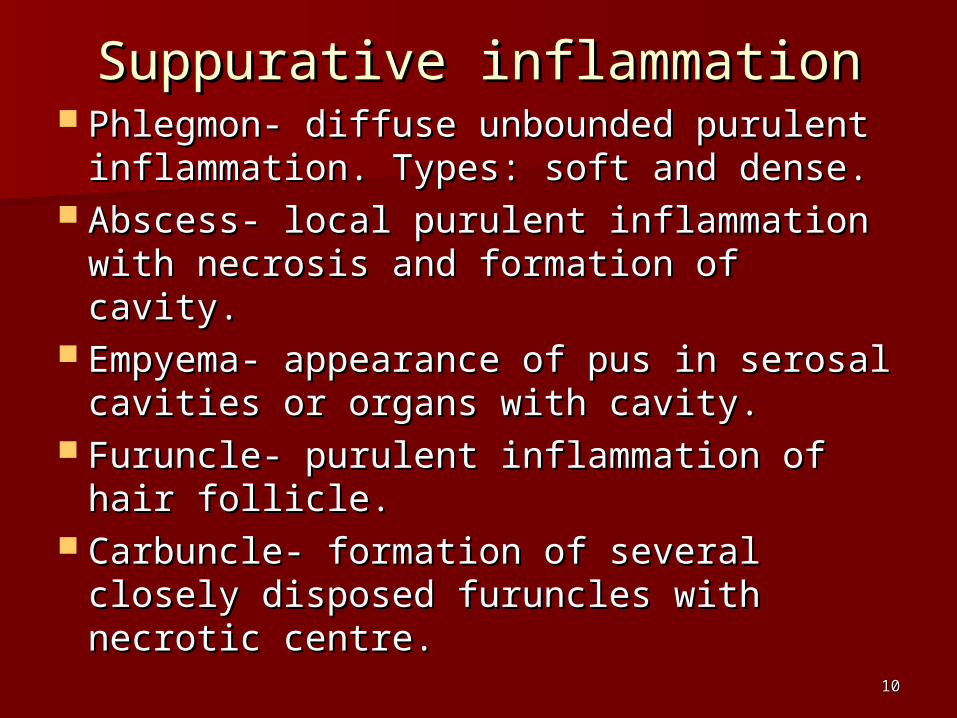
Suppurative inflammationSuppurative inflammation Phlegmon- diffuse unbounded purulent Phlegmon- diffuse unbounded purulent
inflammation. Types: soft and dense.inflammation. Types: soft and dense. Abscess- local purulent inflammation Abscess- local purulent inflammation
with necrosis and formation of cavity.with necrosis and formation of cavity. Empyema- appearance of pus in Empyema- appearance of pus in
serosal cavities or organs with cavity.serosal cavities or organs with cavity. Furuncle- purulent inflammation of Furuncle- purulent inflammation of
hair follicle.hair follicle. Carbuncle- formation of several Carbuncle- formation of several
closely disposed furuncles with closely disposed furuncles with necrotic centre.necrotic centre.

1111
Hand, staphylococcal abscess Hand, staphylococcal abscess
1212
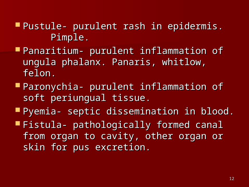
Pustule- purulent rash in epidermis. Pustule- purulent rash in epidermis. Pimple. Pimple.
Panaritium- purulent inflammation of Panaritium- purulent inflammation of ungula phalanx. Panaris, whitlow, felon.ungula phalanx. Panaris, whitlow, felon.
Paronychia- purulent inflammation of Paronychia- purulent inflammation of soft periungual tissue.soft periungual tissue.
Pyemia- septic dissemination in blood. Pyemia- septic dissemination in blood. Fistula- pathologically formed canal Fistula- pathologically formed canal
from organ to cavity, other organ or from organ to cavity, other organ or skin for pus excretion.skin for pus excretion.

1313
Acute abscesses Acute abscesses of the lung in of the lung in upper lobe and upper lobe and lower lobe.lower lobe.

1414
Here is abscess in the lung. The Here is abscess in the lung. The alveoli in that area are alveoli in that area are destroyed.destroyed.

1515
CChronic abscess hronic abscess in the right in the right middle lobe middle lobe of of the lung.the lung.

1616
Acute phlegmonous Acute phlegmonous appendicitisappendicitis
1717
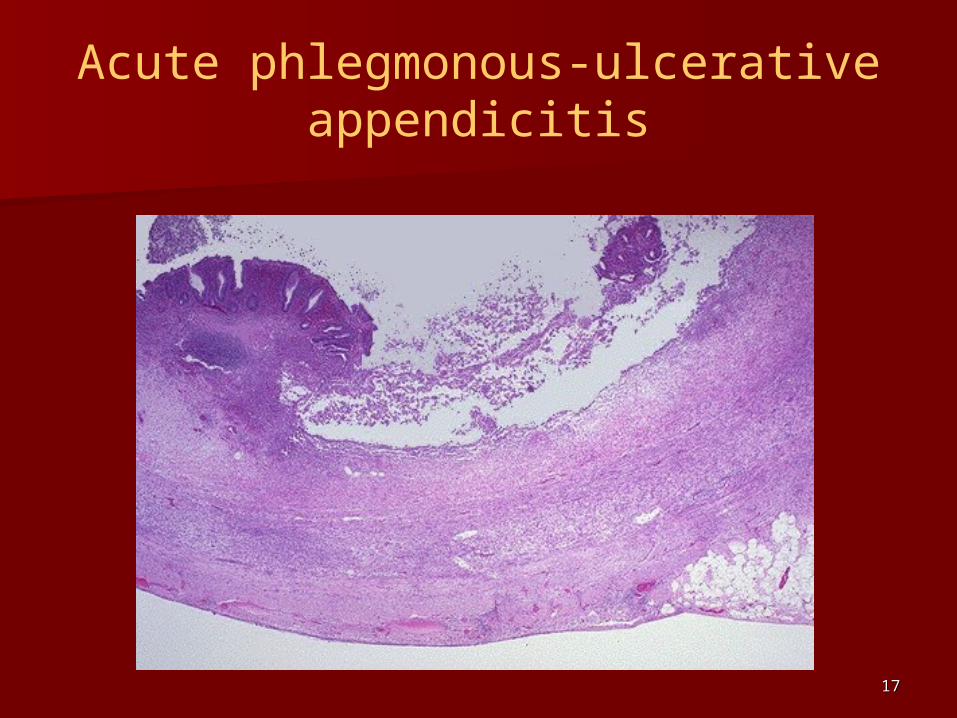
Acute phlegmonous-ulcerative appendicitis
1818
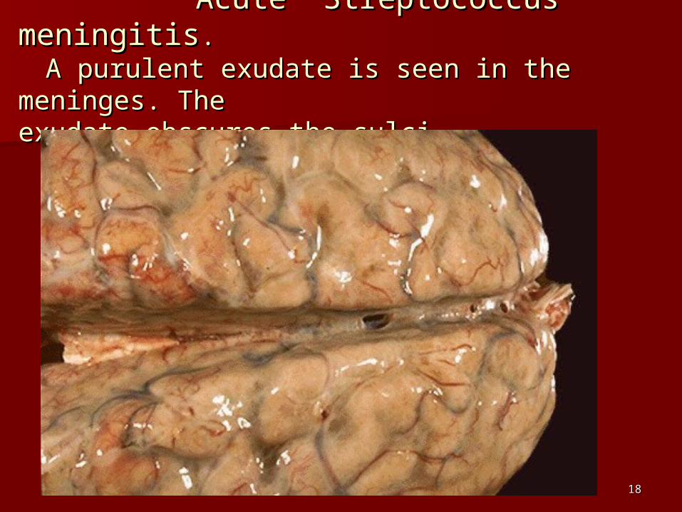
Acute Streptococcus meningitisAcute Streptococcus meningitis.. A purulent exudate is seen in the meninges. A purulent exudate is seen in the meninges. The The exudate obscures the sulciexudate obscures the sulci..
1919
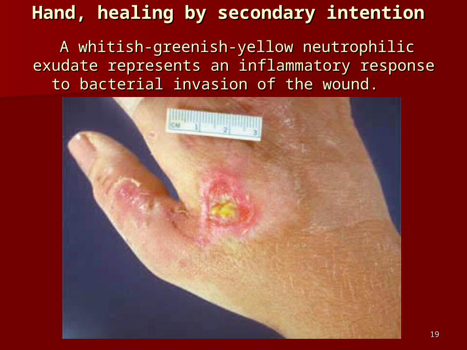
Hand, healing by secondary intention Hand, healing by secondary intention A whitish-greenish-yellow neutrophilic exudate A whitish-greenish-yellow neutrophilic exudate
represents an inflammatory response to bacterial represents an inflammatory response to bacterial invasion of the wound. invasion of the wound.

2020
StreptococcStreptococcal purulent al purulent inflammatioinflammation at lower n at lower
leg.leg.

2121
Outcomes of purulent Outcomes of purulent inflammationinflammation Resolution,Resolution,
scarring,scarring, petrification,petrification, organization,organization, incapsulation,incapsulation, acute toxemia, acute toxemia, pyemia, pyemia, sepsissepsis keloidkeloid chronic duration, chronic duration, amyloidosisamyloidosis

2222
Fibrinous inflammationFibrinous inflammation
Exudate contains fibrin, neutrophils Exudate contains fibrin, neutrophils and necrotic tissue elements. and necrotic tissue elements.
It occurs in mucosa, serosa, meninges, It occurs in mucosa, serosa, meninges, and internal organs (lungs). and internal organs (lungs).
Croupous and dyphtheritic types of Croupous and dyphtheritic types of fibrinous inflammation.fibrinous inflammation.
2323
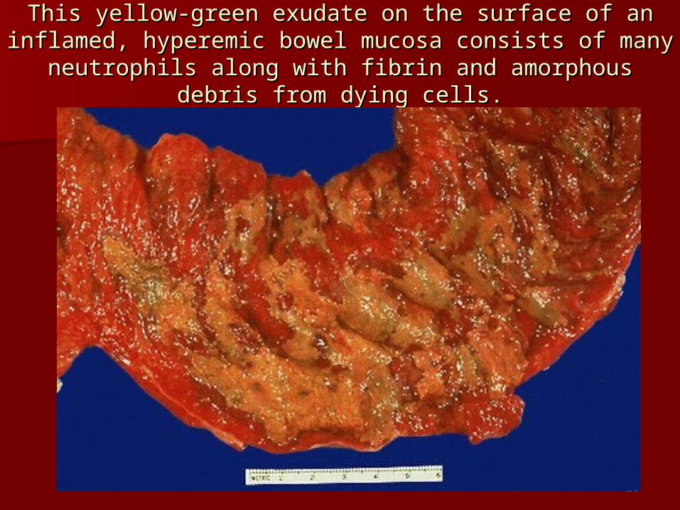
This yellow-green exudate on the surface of an inflamed, This yellow-green exudate on the surface of an inflamed, hyperemic bowel mucosa consists of many neutrophils hyperemic bowel mucosa consists of many neutrophils
along with fibrin and amorphous debris from dying cells.along with fibrin and amorphous debris from dying cells.

2424
Microscopically, exudate consists of inflammatory cells, Microscopically, exudate consists of inflammatory cells, necrotic epithelium, and mucus in which the overgrowth necrotic epithelium, and mucus in which the overgrowth of microorganisms takes place. The underlying mucosa of microorganisms takes place. The underlying mucosa shows congested vessels, but is still intact.shows congested vessels, but is still intact.

2525
Croupous pneumoniaCroupous pneumonia

2626
Here, the Here, the pericardial pericardial cavity has been cavity has been opened to reveal opened to reveal fibrinous fibrinous pericarditis with pericarditis with strands of strands of stringy pale stringy pale fibrin between fibrin between visceral and visceral and parietal parietal pericardiumpericardium..
2727
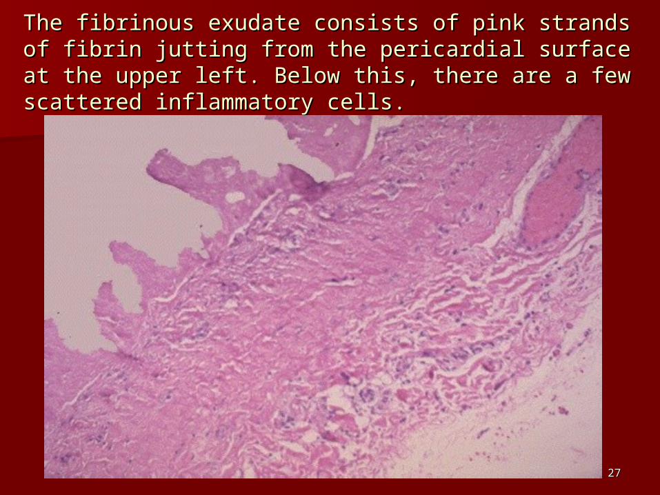
The fibrinous exudate consists of pink strands of fibrin The fibrinous exudate consists of pink strands of fibrin jutting from the pericardial surface at the upper left. jutting from the pericardial surface at the upper left. Below this, there are a few scattered inflammatory cells.Below this, there are a few scattered inflammatory cells.
2828
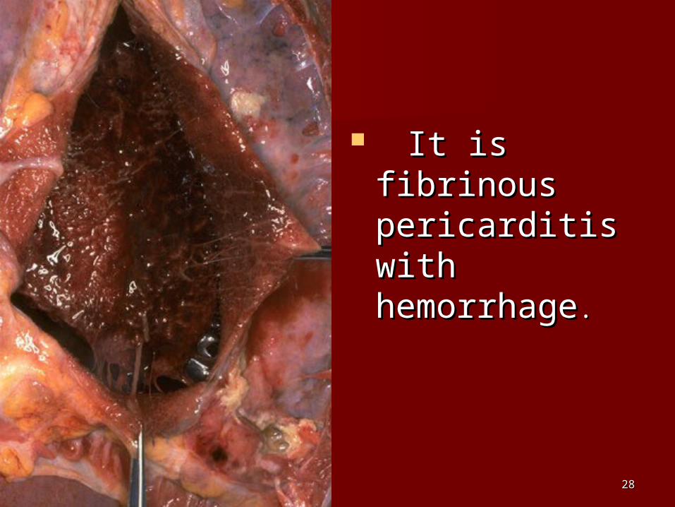
It is fibrinous It is fibrinous pericarditis pericarditis with with hemorrhagehemorrhage. .

2929
3030
Outcomes of fibrinous Outcomes of fibrinous inflammationinflammation
ResolutionResolution OrganizationOrganization UlcersUlcers ScarringScarring Obliteration of cavityObliteration of cavity Chronic duration Chronic duration
3131
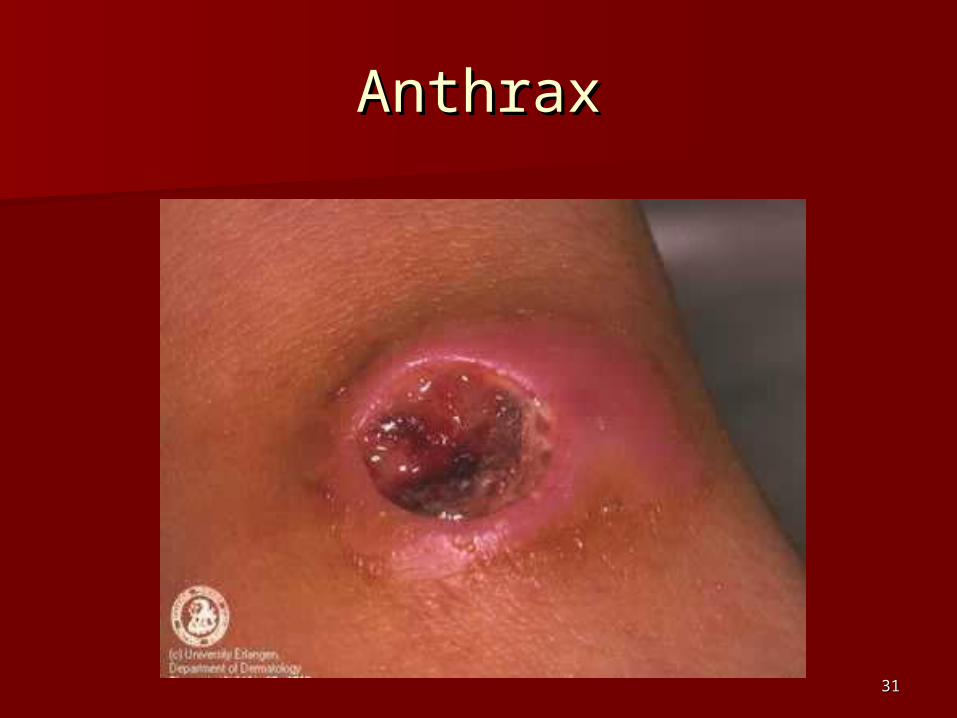
AnthraxAnthrax

3232
3333
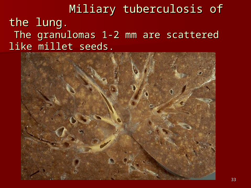
MMiliary tuberculosisiliary tuberculosis of the lung of the lung.. The granulomasThe granulomas 11-2-2 mm are scattered like mm are scattered like millet seeds.millet seeds.

3434
Here are two pulmonary granulomas in tuberculosis. Here are two pulmonary granulomas in tuberculosis. They typically consist of epithelioid macrophages, They typically consist of epithelioid macrophages, giant cells, lymphocytes, plasma cells, and giant cells, lymphocytes, plasma cells, and fibroblasts. fibroblasts.

3535
These are These are epithelioid cellsepithelioid cells around the center of a around the center of a granuloma. They get their name from the fact that granuloma. They get their name from the fact that they have lots of pink cytoplasm similar to they have lots of pink cytoplasm similar to squamous epithelial cells. squamous epithelial cells. Their nuclei tend to be Their nuclei tend to be long and stringy.long and stringy.

3636
lepromaleproma

3737
LLepromatous leprosy. epromatous leprosy. Large collections of foamy Large collections of foamy
macrophages (Virchow cells) infiltrate macrophages (Virchow cells) infiltrate the dermis.the dermis.

3838
NNecrotizing granulomatous vasculitisecrotizing granulomatous vasculitis ( (Wegener's Wegener's granulomatosisgranulomatosis))..
This is a renal biopsy This is a renal biopsy withwith a focal lesion centered around a focal lesion centered around a blood vessel. a blood vessel.

3939
SSarcoidosisarcoidosis,, granulomatous inflammationgranulomatous inflammation inin SpleenSpleen
Granulomas are scattered diffusely throughout the Granulomas are scattered diffusely throughout the splenic parenchyma.splenic parenchyma.
4040
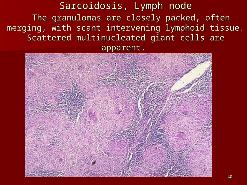
SSarcoidosisarcoidosis, , Lymph nodeLymph node The granulomas are closely packed, often merging, The granulomas are closely packed, often merging,
with scant intervening lymphoid tissue. Scattered with scant intervening lymphoid tissue. Scattered multinucleated giant cells are apparent.multinucleated giant cells are apparent.
4141
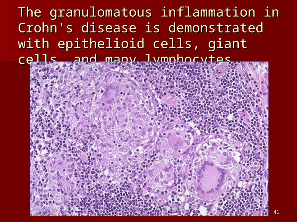
The granulomatous inflammation in The granulomatous inflammation in Crohn's disease is demonstrated with Crohn's disease is demonstrated with epithelioid cells, giant cells, and many epithelioid cells, giant cells, and many lymphocytes. lymphocytes.
4242
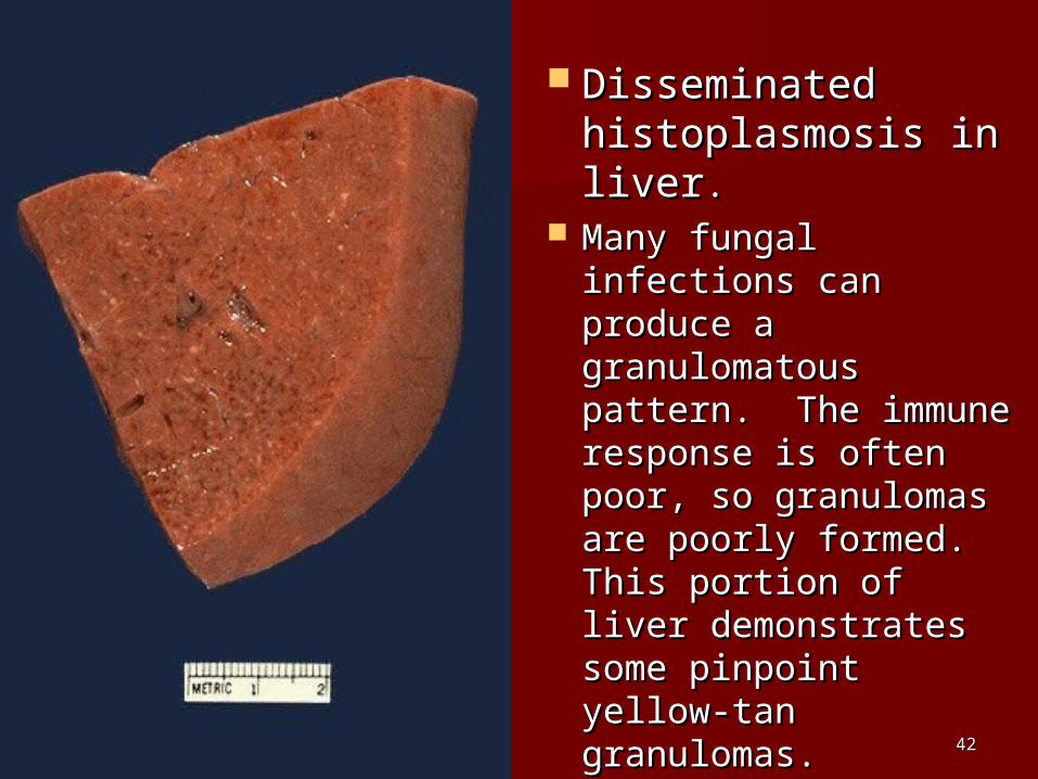
Disseminated Disseminated histoplasmosis in histoplasmosis in liverliver..
Many fungal Many fungal infections can infections can produce a produce a granulomatous granulomatous pattern. The immune pattern. The immune response is often response is often poor, so granulomas poor, so granulomas are poorly formed. are poorly formed. This portion of liver This portion of liver demonstrates some demonstrates some pinpoint yellow-tan pinpoint yellow-tan granulomas.granulomas.

4343
This is infection with Histoplasma capsulatum. Note This is infection with Histoplasma capsulatum. Note how each macrophage is filled with numerous small how each macrophage is filled with numerous small organisms. The organisms have a clear zone around organisms. The organisms have a clear zone around a central blue nucleus which gives the cell a central blue nucleus which gives the cell membrane the appearance of a capsule. membrane the appearance of a capsule.

4444
Hydatid disease of liverHydatid disease of liver

4545
Sometimes the inflammatory reaction is mainly one of Sometimes the inflammatory reaction is mainly one of scarring, as seen here with a silicotic nodule of the scarring, as seen here with a silicotic nodule of the lung. The inhaled silica persists indefinitely and lung. The inhaled silica persists indefinitely and produces an inflammatory reaction that is marked by produces an inflammatory reaction that is marked by prominent fibrosis. Dense pink collagen is seen in the prominent fibrosis. Dense pink collagen is seen in the center of the nodule.center of the nodule.

4646
The interstitial lymphocytic infiltrates The interstitial lymphocytic infiltrates with little with little necrosis necrosis are characteristic for a viral myocarditis are characteristic for a viral myocarditis
((Coxsackie BCoxsackie B). It). It may be a cause for sudden may be a cause for sudden death in young persons.death in young persons.
4747
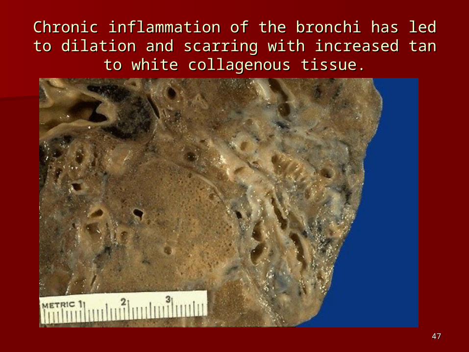
CChronic inflammation of the bronchi has led to hronic inflammation of the bronchi has led to dilation and scarring with increased tan to white dilation and scarring with increased tan to white
collagenous tissue.collagenous tissue.

4848
The end result of inflammation can be scarring. The end result of inflammation can be scarring. Here, the alveolar walls are thickened and filled Here, the alveolar walls are thickened and filled
with pink collagen.with pink collagen.
4949
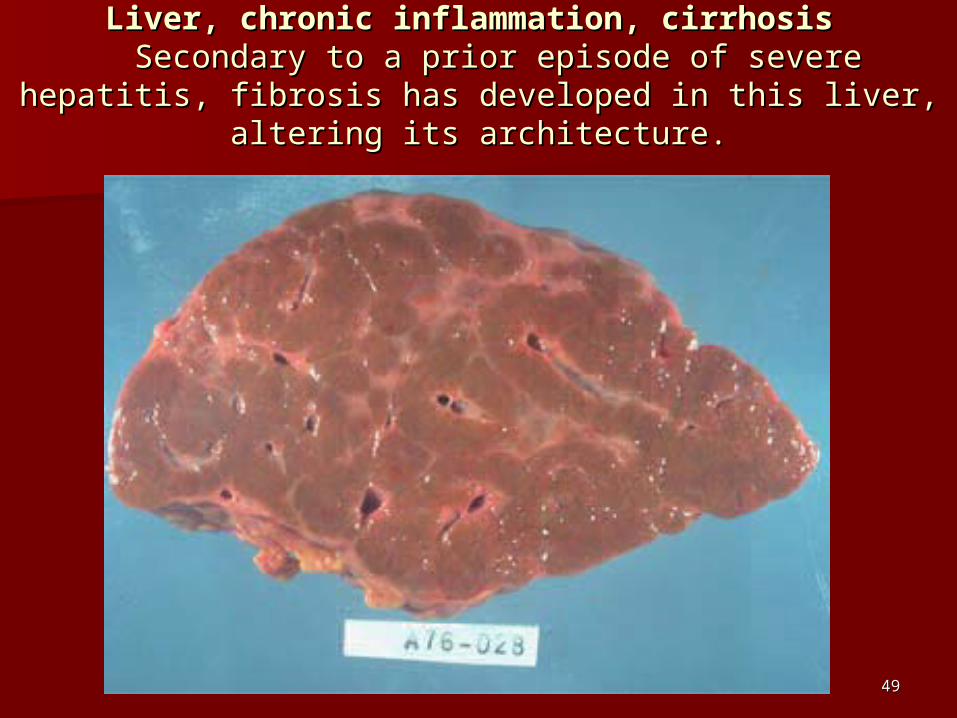
Liver, chronic inflammation, cirrhosis Liver, chronic inflammation, cirrhosis Secondary to a prior episode of severe hepatitis, fibrosis Secondary to a prior episode of severe hepatitis, fibrosis
has developed in this liver, altering its architecture.has developed in this liver, altering its architecture.


![]HOHQjosefuvdul.eu/wp-content/uploads/UP2_HLV_Josefův_Důl.pdf · 2016. 3. 3. · 748 m n. m. 0D[RYVNì YUFK 884 m n. m. ÿHUQì YUFK 908 m n. m. 'ORXKi VH 956 m n. m. 830 m n. m](https://img.dokumen.tips/doc/110x75/5fdf760f48400048041ed86e/-vdlpdf-2016-3-3-748-m-n-m-0dryvn-yufk-884-m-n-m-huq-yufk.jpg)
![±#ã 4('¼ æ 9 W - 大田区 Ota City...£ M N% "â ]#ã l1* x M N% "â M N% ¾"â ] N% ¾"â ] M N% ß M N% )s @ c U N% ! U N% Y2 N% b · N% b ¬ N% b D N% p ¬ N% p · N% 0Y ±](https://img.dokumen.tips/doc/110x75/5f143cffb0afb163706c4363/-4-9-w-coe-ota-city-m-n-l1-x-m-n-.jpg)