Embed Size (px)
Citation preview

Facial clefts and associated limb anomalies: description of 3 cases and a review of the 1
literature 2
3 M.C.Obdeijn1 MD, P.J. Offringa2 MD PhD , R.R.M. Bos3 DMD PhD, A.A.E. Verhagen4 4 MD JD PhD, F.B. Niessen1 MD PhD, N.A. Roche5 MD 5
6
1 Plastic Surgeon, University Medical Center Groningen, University of Groningen, 7
Department of Plastic, Reconstructive and Handsurgery Surgery, Groningen, The 8
Netherlands 9
2 Pediatrician, St.Maarten Medical Center, Department of Paediatrics, St Maarten, 10
Netherlands Antilles, 11
3 Professor in Maxillofacial Surgery, University Medical Center Groningen, University of 12
Groningen, Department of Maxillofacial Surgery, Groningen, the Netherlands 13
4 Pediatrician, University Medical Center Groningen, University of Groningen, 14
Department of Paediatrics, Groningen, The Netherlands 15
5 Assistent Professor in Plastic Surgery, University Hospital of Gent, Department of 16
Plastic and Reconstructive Surgery, Gent, Belgium 17
18
Corresponding Author: 19
Mrs. M.C.Obdeijn, MD 20
Current work address: 21
Academic Medical Center Amsterdam 22

Department of Plastic, Reconstructive and Hand Surgery 23
P.O.Box 22660 1105 AZ Amsterdam 24
The Netherlands 25
Tel: 00-31-20-5664039 Fax: 00-31-20-6917549 26
28
This paper was presented and discussed at the fall meeting of the Dutch Association for 29
Palate and Craniofacial Anomalies, November 2007, Zwolle, the Netherlands 30
31
Running title: Facial clefts and limb anomalies 32
33
34 35
36

Abstract 37
Facial clefts are rare congenital malformations. In the literature these are sometimes 38
reported in combination with limb malformations, especially ring constrictions. This 39
article describes 3 cases of children with facial clefts and limb ring constrictions with 40
various expressions. The first case has a lateral cleft with associated limb malformations. 41
This combination has, to our knowledge, not yet been reported. The literature about facial 42
clefting and the amniotic band syndrome and the possible aetiology of clefting and 43
constrictions in these cases are discussed. 44
45
Key words: Facial cleft, limb malformations, etiology, Tessier, Amnion band46

47
INTRODUCTION 48
49
Facial clefts, as clearly classified by Tessier (1976), are rare congenital malformations 50
with an unknown etiology. They have been reported in association with other congenital 51
malformations, particularly of the extremities. Especially limb constriction bands and 52
amputations are consistently noted. Amniotic band syndrome has been associated with 53
these malformations, but the exact mechanisms causing this condition are unclear. 54
Proposed theories include interruption of fetal blood supply, genetic programming errors, 55
defects of embryological organization and mechanical deformation by amniotic bands. 56
We present 3 patients with facial clefts and limb malformations as described in 57
amniotic band syndrome, treated in Groningen (The Netherlands) and Gent (Belgium). 58
We also discuss the possible etiology by reviewing the literature. By presenting these 59
cases we would like to add new examples of patients with a combination of 60
malformations considered to be part of the amniotic band syndrome. 61
62
CASE-REPORTS 63
Patient 1 64
A 6 months old girl was presented to our outpatient clinic with a bilateral facial cleft and 65
limb malformations. She was the first child of nonconsanguineous parents of Afro-66
Caribbean origin, born after a regular pregnancy and delivered by means of a Caesarian 67
section. There were no visible anomalies of the placenta. Her mother suffered non-insulin 68
dependent diabetes and hypertension during her pregnancy. The family history was 69
negative for craniofacial or other congenital malformations. The mothers’ sister had 70
sickle cell anemia but blood samples of the child were normal. 71
On clinical examination multiple congenital malformations were seen (Fig 1,2): 72
Oblique cleft through her left and right cheek 73
Constriction band around her right upper arm 74
Skin tag on the back of her head 75

Syndactyly of the 2nd, 3rd and 4th toe of the right foot 76
Flexion contracture and hypoplasia of the right great toe 77
At neurological examination she had normal reflexes and a normal movement pattern. 78
Chromosomal analysis of the blood samples showed a normal female karyotype (46,XX). 79
A MRI and CT scan were performed which showed a cartilaginous tumor in the right 80
maxilla with tooth follicles. 81
She was operated and the left hemifacial cleft was closed, the constriction band on her 82
right arm was interrupted with Z-plasties and the skin tag on the back of her head was 83
excised. The maxillary tumor was excised and the right hemifacial cleft was closed. The 84
operation was without complications and post-operative healing was uneventful. 85
Histological examination of the maxillary tumor showed no indications for malignant 86
cells. 87
The patient was seen again in the outpatient clinic 1 year after surgery. The wounds had 88
healed without hypertrofic scarring. Her facial expression was normal. There were no 89
signs of recurrence of the maxillary tumor. 90
91
92
93 Patient 2 94
95
A newborn baby boy was transferred to our clinic because of multiple congenital 96
malformations. He was the first child of nonconsanguineous Caucasian parents, born by 97
means of a Caesarean section after a pregnancy of 36 weeks, during which 98
oligohydramnios was noted. On examination of the placenta, strands and villiform 99
appendages were seen at the umbilical cord and the fetal side of the placenta. The family 100
history was negative for craniofacial or other congenital malformations. 101
On clinical examination multiple congenital malformations were seen (Fig 3,4,5,6): 102
Skin tag on the right forehead 103
Coloboma of the left iris, combined with a chorioretinal and optic nerve coloboma 104
Bizarre shape of the nose with a notch of the right ala and a sinus tract in the left 105
nostril 106
Slight hypertelorism with asymmetric eyebrows 107
Bilateral choanal atresia 108
Narrow, high arched palate with a bifid uvula 109
Constriction bands on the right upper and lower leg with a small skin tag on the 110
ventral side of the lower leg 111
Syndactyly of the 2nd, 3rd and 4th finger of the left hand with amputation on the 112
level of the PIP joint 113
Amputation of the 2nd, 3rd and 4th finger on the level of the DIP joint of the right 114
hand, with constriction rings proximal 115
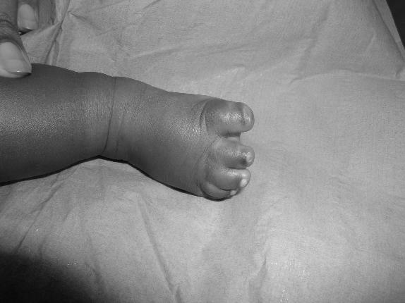
Amputation of the toes of the left foot on the level of the PIP joint 116
There were no abnormalities of the internal organs. Blood samples were normal and 117
genetic examination showed a normal 46 XY karyotype. CHARGE association was 118
excluded as the boy presented with only two of the six malformations described in the 119
CHARGE association (Pagon et al 1981). During his first year he underwent several 120
surgical corrections, during which the skin tag was removed, the constriction bands on 121
the leg were corrected with multiple Z-plasties and the syndactylies of the left hand were 122
corrected. The pathology report of the skin tag showed normal skin and subcutaneous 123
tissues. Surgery and postoperative healing were uneventful. 124
He was followed in the craniofacial team of the UZ Gent and developed normally. At the 125
age of 11 years, the choanal atresia was corrected. For further surgical planning, a CT 126
scan was performed with 3D reconstruction at the age of 14, which revealed a bony cleft 127
on the right side of the nasal bone. Because of the high arched palate and occlusional 128
problems, distraction of the palate with orthodontic treatment was started. A nasal 129
correction was also performed and further corrections are planned in the future. 130
131
Patient 3 132
A newborn baby boy was referred to our clinic because of multiple congenital 133

malformations. He was the first child of nonconsanguineous parents of Mediterranean 134
origin, born after a pregnancy of 38 weeks, during which the mother had hypertension. 135
Examination of the placenta showed an umbilical cord with velamentous insertion. The 136
family history was negative for craniofacial or other congenital malformations. 137
On clinical examination multiple congenital malformations were seen (Fig. 7,8,9,10): 138
Parietal encephalocele with brachycephaly based on right sided coronal suture 139
stenosis 140
Bilateral asymmetric cleft lip and palate 141
Slight hypertelorism with asymmetric eyebrows 142
Retrusion of the right orbital region 143
Syndactyly with amputation of 2nd, 3rd, 4th and 5th finger of the left hand 144
Syndactyly with amputation of all fingers of the right hand 145
Amputation with syndactyly of 2nd and 3rd toe and constriction rings of the 4th and 146
5th toe of the right foot with amputations 147
Syndactyly of the 2nd and 3rd webspace of the left foot, amputation of 2nd, 4th and 148
5th toe 149
Skin tag in the lumbal area 150
During the first two years of life, several corrections were performed of the cleft lip and 151
palate, hands and feet and encephalocele. The pathology report of the excised skin tag 152
showed normal skin and subcutaneous tissues. An epidermoid cyst was found close to the 153
encephalocele. The boy also suffered from epileptic seizures, controlled by anti-epileptic 154
medication. A CT-scan revealed multiple deformities on the right side of the brain 155
(ventricular deformities, lobar holoprosencephaly, kissing thalami). 156
No other abnormalities of the internal organs were discovered. Genetic examination was 157
not performed as the parents refused this. 158
The boy was followed for 12 years in the craniofacial team, further surgical corrections 159
were scheduled but unfortunately the boy was then lost to follow-up. 160
161
DISCUSSION 162

The amniotic band syndrome or amniotic disruption sequence is a constellation of 163
congenital malformations characterized by extreme variability. These include limb 164
reductions, craniofacial malformations, body wall deficiencies, clubfeet and internal 165
organ anomalies (Patterson, 1961; Bamforth, 1992; Froster and Baird, 1993; Van der 166
Meulen,1999; Goncalves and Jeanty, 1999). It is also mentioned under the name of 167
ADAM sequence (amniotic deformity, adhesions, mutilations) which is identified in the 168
study of Yang (1990) as being a complication of EAR (early amnion rupture). 169
Our patients presented with a combination of facial clefts and limb deformities, which 170
clinically seemed to fit in the amniotic band syndrome. 171
Similar malformations have been numerously reported (Sakurai et al., 1966; Jones et al., 172
1974; Mayou and Fenton., 1981; Garza et al., 1988; Coady et al.,1998; Van der Meulen, 173
1999; Kara and Ocsel, 2001; Gokrem et al., 2002; Morovic et al, 2004). 174
In 1998 Coady et al (1998) wrote an extensive report on their population of patients with 175
facial clefts. They found 11 patients with facial clefts and limb deformities, consisting of 176
ring constrictions. All facial clefts were in the central axis of the face and they suggested 177
an association between rare craniofacial clefts and limb ring constrictions and postulated 178
a common etiology. 179
In a case-report by Kara et al. (2001), a patient was described with a Tessier no 5 cleft 180
with extremity malformations (multiple random-pattern asymmetric extremity 181
deformities). In Gökrem’s group of 5 patients with a Tessier no 7 cleft, none of the 182
patients had associated limb malformations (Gokrem et al, 2002). Our patient with the 183
lateral cleft (patient 1) had limb malformations and as far as we are aware, this would be 184
the first case reported in the literature. 185
Jones et al (1974) and Morovic et al (2004) published a series of patients with 186
craniofacial and extremity malformations comparable to the malformations of our 187
patients. They assumed the malformations were due to amniotic bands as a result of early 188
amniotic rupture. 189
Tessier (1976) published an article on the classification of facial, craniofacial and 190
laterofacial clefts. The purpose of his article was to propose a descriptive classification of 191

the clefts, based on the clinical findings, but did not explain the etiology. Van der Meulen 192
et al. (1983) proposed a classification based on morphogenetic characteristics. They 193
introduced the term “dysplasia” and described the malformations according to the 194
localization and timing of developmental arrest based on the embryology. 195
David et al. (1989) added a third dimension to the Tessier classification by describing the 196
anomalies found on CT scans and three-dimensional reconstruction. 197
Little is written about the epidemiology of rare facial clefts or the distribution 198
among races. In a review by Kawamoto (1976), facial clefting has been reported in 1.43 199
to 4.85 in 100,000 births. The Tessier no. 7 cleft is the least rare atypical craniofacial cleft 200
with an incidence from 1:3000 to 1:5642 live births, but bilateral involvement is rare 201
(Tessier, 1976). 202
Concerning the epidemiology of amnion rupture defects, Garza et al. (1988) indicated 203
that the incidence is around 1.16 per 10,000 live births. It occurs 1.76 times more often in 204
blacks and especially young black nulligravidas have a significant higher risk of having a 205
baby with amniotic band syndrome anomalies. 206
The mothers of two of our patients had hypertension during pregnancy but we did not 207
find suggestions in the literature that this could be a risk factor for the development of 208
amniotic bands. Oligohydramnios was noted during the pregnancy of patient 2 and this 209
has been associated with compression-related malformations of the craniofacial area and 210
limbs (Torpin, 1968; Marino, 2004). 211
The etiopathology of facial clefting has been hotly debated for centuries. It has 212
been considered as a failure of fusion of primordial facial processes as described by 213
Dursy in 1869 and His in 1892 (Classic Theory). Later Warbrick (1963) and Stark (1954) 214
described their theory of a failure of migration of mesoderm and neural crest (Modern 215
theory). 216
Multiple theories have arisen around the etiology of the amniotic band syndrome. 217
The intrinsic theory considers focal fetal dysplasias to be the cause of the constrictions 218
(Streeter, 1930). Patterson proposed diagnostic clinical criteria for the diagnosis limb ring 219
constrictions often seen in the amniotic band syndrome: 220

1. Simple ring constrictions 221
2. Ring constrictions with distal deformity plus or minus lymphedema 222
3. Ring constrictions accompanied by syndactyly or acrodactyly 223
4. Amputation 224
After an extensive search of the literature, Patterson (1961) concluded that the underlying 225
cause is a failure of development similar to that of other congenital malformations 226
The malformations are the result of developmental errors in the formation of connective 227
tissue, resulting in facial clefts and limb malformations. Different mechanisms have been 228
suggested including vascular failure with disruption of epiblastic cells, disturbance of 229
neural crest cell migration etc., with secondary limb amputations, constriction bands, 230
cephaloceles, syndactyly, clubfeet, club hands and internal anomalies (Lookwood et al., 231
1989; Bamforth, 1992). In 1986 Hunter and Carpenter discussed in their paper four 232
infants with ABS and additional malformations that were not readily explainable on the 233
basis of band disruptions. Their conclusion was that some extrinsic insult or perhaps 234
occasionally a familial susceptibility leads to loss of fetal vascular integrity, superficial 235
hemorrhage and denudation, with adhesions leading to syndactyly, and constrictions to 236
amputations (Hunter and Carpenter, 1986). 237
The extrinsic theory claims that the bands cause the clefts and limb constrictions. 238
Rupture of the amnion would lead to bands or strands that entangle body parts of the 239
fetus, and swallowing of the strands leads to clefts in the face (Patterson, 1961). In one 240
series, a histologically proven amniotic band was found (Mayou and Fenton, 1981). The 241
problem with the extrinsic theory however is that bands have never been observed in 242
natural creases below the nose or behind the ear. Furthermore bands cannot explain the 243
typical pattern of clefts, cannot produce perfectly symmetric clefts and related anomalies 244
(Van der Meulen, 1999). 245
A third theory assumes that these malformations are part of the amniotic rupture 246
syndrome together with disruptive defects. The resulting oligohydramnios would induce 247
compression related deformities. The earlier the rupture and the more susceptible the 248
developing areas, the more severe the defects including craniofacial malformations, limb 249

anomalies and visceral defects (Higginbottom et al., 1979; Van der Meulen, 1985; 250
Morovic et al., 2004). 251
But even with all these theories it remains difficult to explain the different 252
malformations in one patient. The debate should probably not be focused on the 253
terminology of the malformation but rather on the mechanism causing the malformation 254
or combination of malformations. The first patient had a bilateral oblique cleft. The third 255
patient had an asymmetric bilateral cleft lip and palate. These are primary clefts, which 256
are formed when fusion between the nasal and maxillary or maxillary and mandibular 257
processes is disturbed before the end of the transformation phase (Van der Meulen et al., 258
1983). Patient 2 had a secondary bony cleft originating from disturbed ossification of the 259
facial skeleton. Did these developmental disturbances cause the bands, which in turn 260
caused the extremity malformations? Or were the bands a result of amnion rupture, which 261
led to the formation of the facial clefts and strangulation of the limbs? 262
In our first case, a constriction band could theoretically have caused the facial 263
cleft, although it is difficult to explain the differences in the level of clefting in the right 264
cheek. Constriction bands could certainly have caused the extremity malformations. 265
Furthermore, two articles describe tumors that developed from tissue that was strangled 266
due to an amniotic band (Murata et al., 1992; Tanabe et al., 2002). This phenomenon 267
would account for the tumor that was found in the right maxilla. In the study of Davids et 268
al. (1989) 253 patients were studied but none of the patients had a maxillary tumor like 269
our case, however facial clefts associated with duplication of various oromaxillary 270
components have been reported (Marfeni, 1993) and Converse et al. (1974) found in his 271
series of 280 patients two patients with extramaxillary foci of bone and ectopic dentition. 272
All three patients had a skin tags (back of head, forehead and lumbar region respectively). 273
Ten Donkelaar et al. (2002) described a patient who showed constrictions in both hands 274
and amputations with fresh, healing wounds suggestive for amniotic band syndrome. 275
Their patient also had a skin tag on his head, which after microscopic examination turned 276
out to be rudimentary meningocele with intestinal mucosa probably due to amniotic band 277
syndrome. In patient 1 pathologic examination of the skin tag was not performed but in 278

patients 2 and 3 the skin tags showed no particular anomalies at histological examination. 279
In the second patient there is clear evidence that there were bands involved as strands and 280
villiform appendages at the edge of the placenta and umbilical cord were found. 281
Oligohydramnios with limited fetal movement was also noted during pregnancy. The 282
strands and oligohydramnios may have caused the extremity malformations, as well as 283
the cleft uvula and high arched palate, due to compression. The facial malformations 284
however are very atypical. Because of the asymmetric face, the notch in the right hemi 285
nose, the right-sided orbital dystopia and the skintag on the right forehead, a bony cleft 286
was suspected, which was confirmed on CT scan. Furthermore, a sinus tract in the left 287
nostril, bilateral choanal atresia, coloboma in the left iris and retina cannot be explained 288
with the extrinsic theory of the amniotic band syndrome and must be due to focal 289
dysplasia. 290
The third child had a bilateral asymmetric cleft lip and palate but with slight 291
hypertelorism and asymmetric eyebrows, retrusion of the right orbital region and cranial 292
deformities with an encephalocele. His extremity malformations consisting of syndactyly, 293
partial amputations and constriction rings and the skull deformities, consisting of 294
plagiocephaly, synostosis and encephalocele can be produced by compression and 295
therefore could fit into the spectrum of amniotic rupture sequence. Clinical signs of 296
oligohydramnios however were not found during pregnancy. 297
298
CONCLUSION 299
These three cases illustrate that the etiology is probably intrinsic, that the secondary 300
compression is related and that these malformations occur very early in the embryo. The 301
dysplasias in the craniofacial area may have caused amnion rupture, which in turn may 302
have caused the extremity malformations and other compression related deformities. It 303
also seems logic that the earlier the developmental arrest or disturbance takes place, the 304
more severe external malformations will result, as shown in patients 1 and 3. 305
It seems important to continue to collect and publish data on these patients in order to 306
obtain a better understanding of the etiology of this spectrum of malformations. 307

308
Acknowledgement of source of patient nr 1: P.H. Robinson MD, PhD, UMCG Groningen 309
310
311
REFERENCES 312
Bamforth SJ. Amniotic band sequence: Streeter’s hypothesis re-examined. Am J Med 313
Genet. 1992;44(3):280-287 314
Coady M, Moore M, Wallis K. Amniotic band syndrome: The association between 315
rare facial clefts and limb ring constrictions. Plast Reconstr Surg. 316
1998;101:640-649 317
Converse JM, Wood-Smith D, McCarthy JG, Coccaro PJ. Craniofacial surgery. 318
Clinics in Plastic Surgery. 1974;1(3):499-557 319
David DJ, Moore MH, Cooter RD. Tessier clefts revisited with a third dimension. 320
Cleft Palate Journal. 1989;26(3):163-184 321
Dursy E. Zur Entwicklungsgeschichte des Kopfes des menschen und der hohren 322
Wirbelthiere. Tubingen, Berlag der H.Lauppschen-Buchhandlung, 1869: 99 323
Froster UG, Baird PA. Amniotic band sequence and limb defects: Data from a 324
population-based study. Am J Med Genet. 1993;46:497-500 325
Garza A, Cordero JF, Mulinare J. Epidemiology of the early amnion rupture spectrum 326
of defect. Am Dis Child. 1988;142(5):541-544 327
Gökrem S, Özdemir OM, Katircioglu A, Sen Z, Ersoy A, Can Z, Emiroglu M, Gültan 328
S. A rare craniofacial cleft: Tessier No.7: a retrospective analysis. J of Ankara 329
Med School. 2002;24:63-68 330
Gonçalves LF, Jeanty P. Amniotic band syndrome. HYPERLINK “ HYPERLINK "http:// 331
www.thefetus.net" http://www.thefetus.net” 1999 332
Higginbottom JC, Jones KL, Hall BD, Smith DW. The amniotic band disruption 333
complex. Timing of amniotic rupture and variable spectra of consequent defects. J 334
Pedriatr. 1979;95(4):544-549 335
His W. Die Entwicklung der Menschlichen und thierischer Physiognomen. Arch. 336
Anat. Entwichlungsgesch. 1892: 384 337
Hunter AGW, Carpenter BF. IMplications of malformations not due to amniotic 338
bands in the amniotic band sequence. Am J of Med Gen. 1986;24:691-339
700 340
Jones KL, Smith DW, Hall BD, Hall J, Ebbin AJ, Massoud H, Golbus MS. A pattern 341
of craniofacial and limb defects secondary to abberant tissue bands. J of Ped. 342
1974;84(1): 90-95 343
Kara IG, Öçsel H. The Tessier number 5 cleft with associated extremity anomalies. 344
Cleft Palate Craniofac J. 2001;38:529-532 345
Kawamoto H. The kaleidoscopic world of rare craniofacial clefts: order out of chaos 346
(Tessier classification). Clin Plast Surg. 1976;3(4):529-572 347
Lookwood C, Ghidini A, Romero R. Amniotic band syndrome: reevaluation of its 348
pathogenesis. Am J Obstet Gynecol. 1989;160:1030–1033 349
Marino T. Ultrasound abnormalities of the amniotic fluid, membranes, umbilical cord 350
and placenta. Obstet Gynecol Clin North Am. 2004;31(1):177-200 351
Marfeni JO. The Robin sequence associated with partial maxillary duplication and 352
multiple facial clefts. Afr Dent J. 1993;7:31-34 353
Mayou BJ, Fenton OM. Oblique facial clefts caused by amniotic bands. Plast 354
Reconstr Surg. 1981;68:675-681 355
Murata T, Hashimoto S, Ishibashi T, Inomata H, Sueishi K.A case of amniotic band 356
syndrome with bilateral epibulbar choristoma. Br J Opthalmol 1992;76(11): 357
685-687 358
Morovic CG, Berwart F, Varas J. Craniofacial anomalies of the amniotic band 359
syndrome in serial clinical cases. Plast Reconst Surg. 2004;113(6):1556-1562 360
Patterson TJS. Congenital ring-constrictions. Br J Plast Surg. 1961;14:1-31 361
Pagon RA, Graham JM, Zonana J, Yong S-L. Coloboma, congenital heart disease, 362
and choanal atresia with multiple anomalies: CHARGE assosciation. J of Ped. 363
1981;99(2): 223-227 364
Sakurai EH, Mitchel DF, Holmes LA. Bilateral obliue clefts and amniotic bands: a 365

report of 2 cases. Cleft Palate J. 1966;3:181-185 366
Stark RB. The pathogenesis of harelip and cleft palate. Plast Reconstr Surg. 367
1954:13(1):20-39 368
Streeter GL. Focal deficiency in fetal tissues and their relation to intrauterine 369
amputation. Contrib Embryol Carnegie Inst. 1930;33:1-44 370
Tanabe YN, Kikuchi Y, Nozaki M. Case of constriction band syndrome with annular 371
epidermal cyste. Ann Plast Surg. 2002;48(3):312-314 372
Ten Donkelaar HJ, Hamel BCJ, Hartman E, van Lier JAC, Wessling P. Intestinal 373
mucosa on top of a rudimentary occipital meningocele in amnionitic rupture 374
sequence: disorganization-like syndrome, homeotic transformation, abnormal 375
surface encounter or endoectodermal adhesion? Clinical Dysmorph. 2002;119-13 376
Tessier P. Anatomical classification of facial, cranio-facial and latero-facial clefts. J. 377
Maxillofacial Surg. 1976;4(2):69-92 378
Torpin R. Fetal malformations caused by amnion rupture during gestation. 379
Springfield Il: Charles C Thomas, 1968: 1-76 380
Van der Meulen JC, Mazzola R, Vermey-Keers C, Stricker M, Raphael B. A 381
morphogenetic classification of craniofacial malformations. Plast Reconstr Surg. 382
1983;71: 560-572 383
Van der Meulen JC. The amniotic band syndrome. Plast Reconst Surg. 1999;103(3): 384
1087-1090 385
Van der Meulen JC. Oblique facial clefts: pathology, etiology and reconstruction. 386
Plast Reconstr Surg. 1985;76(2):212-224 387
Warbrick JG. Aspects of facial and nasal development. Sci Basis Med Ann Rev. 388
1963;84:99-122 389
Yang SS. ADAM sequence and innocent amniotic band: Manifestations of early 390
amnion rupture. Am J of Med Gen. 1990;37:562-568 391
392
393
394

Figure Legends 395
Figure 1: Patient 1, right hemifacial cleft and constriction band on right upper arm 396
Figure 2: Patient 1, syndactyly of the right foot and flexion contracture of the right 397
hallux 398
Figure 3: Patient 2, frontal view of the patient with skin tag on the forehead, 399
coloboma of the left iris, bizarre shape of the nose and slight hypertelorism 400
Figure 4: Patient 2, left hand with syndactyly and partial amputations 401
Figure 5: Patient 2, right hand with amputations on the level of the DIP joints 402
Figure 6: Patient 2, constriction band lower extremity 403
Figure 7: Patient 3: frontal view with bilateral asymmetric cleft lip and palate 404
Figure 8: Patient 3, lateral view with parietal encephalocele and brachycephaly 405
Figure 9: Patient 3, left hand with partial amputations of 2nd, 3th, 4th and 5th finger 406
Figure 10: Patient 3, right hand with partial amputations of all fingers 407
408
409
410
411
412
413 414 415 416 417 418 419 420 421 422 423 424 425 426

427 428 429


![MD Antioquia[1]](https://img.dokumen.tips/doc/110x75/55cf8e4d550346703b90b28b/md-antioquia1.jpg)


![3,350 108,000 1.7 M - biblio.ugent.be · forcesforinclusioncomplexationthroughapolar¨apolarinteractionofCDsandtheguest compounds[12,15].ItisforthisreasonthatCDsareusedinwatertreatmenttoremoveEMPs](https://img.dokumen.tips/doc/110x75/5e04c248f3dd6d22fb271acd/3350-108000-17-m-forcesforinclusioncomplexationthroughapolarapolarinteractionofcdsandtheguest.jpg)