Embed Size (px)
Citation preview

06. Cardiovascular and Renal Pharmacology
06.001 Sarcoplasmic reticulum/plasmatic membrane interaction activated by ryanodine-sensitive calcium stores in mice mesenteric artery. Garcia DCG1, Lemos VS2, Côrtes SF1 1UFMG – Farmacologia, 2UFMG – Fisiologia e Biofísica
Introduction: The understanding of the complex interaction between the sarcoplasmic reticulum (SR) and the plasmatic membrane (PM) for the maintenance of the Ca2+ homeostasis in the vascular smooth muscle cells (SMC) has been a subject of extensive investigation in the last three decades. Nevertheless, most of these investigations were done in cell culture. The precise events triggered in the PM after the activation of the ryanodine receptors (RyRs) of the SR are not yet well described in the SMC of resistance arteries. Aim: The present work investigated the mechanisms activated in the PM by the stimulation of RyRs involved in the Ca2+ signaling and contraction in mice mesenteric arteries. Methods: Male Swiss mice (8-12 weeks old) were used and the protocols approved by the ethics committee (CEUA-UFMG, protocol 185/2015). 2nd branch of mesenteric arteries was used for vascular reactivity and epifluorescence microscopy using Fluo-4 as the fluorescent dye for Ca2+. All protocols were done in endothelium-denuded vessels. The results were expressed as mean ± SEM of peak and the area under the curve (AUC) of at least five experiments of vascular reactivity and 10 cells from mesenteric arteries of at least three mice in the experiments of epifluorescence microscopy. One-way ANOVA or unpaired Student’s t-test as statistics analysis and considered significant when P<0.05. Results: Caffeine (10 mM), an agonist of RyRs, induced a transient contraction (peak= 2.30 ± 0.14 mN/mm; AUC= 25.27 ± 2.49 mN/mm.s-1) and increase of the intracellular Ca2+ (peak= 1.16 ± 0.01 F/F0; AUC= 3.27 ± 0.15 F/F0.s
-1). In Ca2+-free solution, the contraction (peak= 1.10 ± 0.04 mN/mm; AUC= 11.15 ± 1.21 mN/mm.s-1) and the intracellular Ca2+ (peak= 1.08 ± 0.01; AUC= 1.20 ± 0.08 F/F0.s
-1) induced by caffeine were significantly reduced (P<0.001). Nifedipine (10 μM), a L-type calcium channel (LTCC) blocker, did not alter the peak (2.72 ± 0.24 mN/mm), but increased the AUC (43.02 ± 3.59 mN/mm.s-1; P<0.05) of the contraction induced by caffeine. However, nifedipine reduced the peak (1.08 ± 0.01 F/F0) and the AUC (1.79 ± 0.19 F/F0.s
-1) of Ca2+ signal induced by caffeine (P<0.001). Ruthenium red (10 μM), a non-selective inhibitor of transient receptor potential vanilloid (TRPV) did not modify the peak (2.69 ± 0.25 mN/mm), but increased the AUC (32.36 ± 4.17 mN/mm.s-1 – P<0.01) of the contraction induced by caffeine, whilst significantly reduced the Ca2+ signal (peak= 1.10 ± 0.01 F/F0 – P<0.001; AUC= 2.54 ± 0.14 F/F0.s
-1 – P<0.05). KB-R7943 (10 μM), a blocker of the reverse mode of Na+-Ca2+-exchanger (NCX), reduced the contraction (peak= 0.37 ± 0.13 mN/mm – P<0.01; AUC= 7.35 ± 2.04 mN/mm.s-1 – P<0.001) and the Ca2+ signal (peak= 1.12 ± 0.01 F/F0 – P<0.05; AUC= 1.16 ± 0.10 F/F0.s
-1 – P<0.001) induced by caffeine. Conclusion: The release of Ca2+ from the SR after activation of RyRs triggers a mechanism in the PM involving the activation of LTCC, TRP and NCX, which are implicated in the control of the calcium influx, membrane potential and of the vascular contraction. Financial support: CNPQ

06.002 Unraveling the enigma of the positive inotropic effect of ATP on the heart of SHR. Rodrigues JQD1, Camara H1, Silva-Junior E D1, Godinho RO1, Jurkiewicz A1 1Unifesp-EPM – Farmacologia
Introduction: Presynaptic nerve terminals from the heart contain a number of membrane receptors, such as ionotropic P2XR and metabotropic P2YR that are involved in positive and negative modulation of neurotransmitter release, respectively. In rat atrium, extracellular ATP (1 µM or higher) produces an initial negative inotropic effect followed by a positive inotropic effect, being the last augmented in the spontaneously hypertensive rat (SHR). Despite the presence of post-synaptic purinergic receptor, this effect may be due to modulation of presynaptic sympathetic neurotransmission. Aims: Herein, we investigate the possible involvement of presynaptic P2XR or P2YR on the positive inotropic effect induced by ATP on the left atrium from normotensive Wistar rats (NWR) and SHR. Methods: Left atria isolated from NWR and SHR (4-5 month-old) were submitted to transmural electrical stimulation (2 Hz, 5 ms, 10-14 V). After an 1 h stabilization, cumulative concentration-response curves for α-β-Me-ATP (1-300 µM; P2X2R/ P2X3R agonist) and non-cumulative concentration- response curves for ATP (P1R and P2R agonist ; 1-1000 µM) were constructed in the presence of MRS 2179 (selective P2Y1R antagonist), suramin (100 µM, non-selective P2XR antagonist), propranolol (100 nM, β-adrenoceptor antagonist), and 6-hydroxydopamine (100 µM, sympathetic-selective neurotoxin). The effects of these agonists were also assessed in atria from reserpine-pretreated rats (24h, 10 mg/Kg, s.c., prevents the neuronal reuptake of norepinephrine). Differences between agonist maximum effect (Emax) were analyzed by unpaired t test. All experiments procedures were approved by the EPM/UNIFESP Ethics Committee (n° 5001150214). Results: 1 µM to 1000 µM ATP produced a biphasic effect on atrial inotropism. For instance, 100 μM ATP produced an initial negative inotropic effect followed by a positive inotropic effect reaching a plateau after 180-200s, returning to 88±1% and 100±2% of basal inotropism in NWR (N= 10) and SHR (N=10), respectively. The α-β-Me-ATP (1 – 300 μM) increased atrium contractile force by 37±8% and 65±11% in NWR and SHR, respectively (N=7). Pre-incubation of propranolol or pretreatment with reserpine did not change either the α-β-Me-ATP or the ATP positive inotropic effects. However, pretreatment of atrium with 6-hydroxydopamine completely abolished the positive inotropic effects of those drugs (N=3-6), in both NWR and SHR. To check the involvement of P2XR, ATP was incubated in the presence of 100 μM suramin, non-selective P2XR antagonist. Suramin reduced in ~50% the positive inotropic effect of ATP in atria from both strains (N=4). The P2Y1R antagonist, 300 nM MRS2179, increased by 19% (NWR) and 13% (SHR) the positive inotropic effect of ATP. Conclusion: The results demonstrated that the positive inotropic effects elicited by
both ATP and α-β-Me-ATP are due to presynaptic effects on 6-hydroxydopamine-sensitive neurons in left atrium of NWR and SHR. These effects involve activation of facilitatory presynaptic P2X2R and P2X3R and inhibitory P2Y1R. Sources of Research Support: FAPESP, CAPES and CNPq.

06.003 Role of aldosterone in the inflamassome activation in macrophages and Type 2 Diabetes. Ferreira NS1, Pereira CA1, Zanotto CZ1, Carlos D2, Tostes RC1 1FMRP-USP – Farmacologia, 2FMRP-USP – Imunologia
Introduction: Aldosterone (Aldo) excess has important effects in the vasculature, contributing to insulin resistance, fibrotic, oxidative and inflammatory processes, that aggravate endothelial dysfunction in diabetes. Aldo exerts its effects via activation of mineralocorticoid receptors (MR). The cytokines IL-1β and IL-18 are released upon activation of molecular platforms named inflammasome in response to stimulation by pathogen-associated molecular patterns (PAMPs) or danger-associated molecular patterns (DAMPS). These signals are recognized by intracellular receptors, e.g. NLRP3 and NLRP1, that are associated with insulin resistance and obesity induction. The inflammasome activation by DAMPS/PAMPs culminates in pro-caspase-1 activation and cleavage of pro-IL-1β and pro-IL-18 to their active forms. There is evidence to support the idea that aldosterone activates the inflammasome and contributes to diabetes-associated inflammatory process. Aims: To evaluate whether aldosterone via MR activation activates the inflammasome in macrophages from mice with type 2 diabetes. Materials and Methods: Second-order mesenteric arteries from control (C57BL6, Cont) and diabetic (db/db) treated with vehicle or spironolactone (spiro - MR antagonist) were used to test vascular reactivity to phenylephrine (Phe) and acetylcholine (ACh). IL-1β levels were determined in the serum of the animals. Macrophages from control mice were stimulated with aldosterone (for 1, 4 and 6 hours) to evaluate expression of the inflammasome components, by RT-PCR. The results are presented as Mean±SEM. Results: Spiro treatment of Cont and db/db mice decreased Phe potency vs. vehicle treatment (pD2: Cont: 6.75±0.06 n=6; db/db: 6.63±0.09 n=5; cont+spiro: 6.17±0.12 n=3; db/db+spiro: 6.33±0.11 n=3, p<0.05). Db/db mice ehxibited reduced ACh maximum response (Emax) compared to control arteries; spiro treatment partly improved ACh responses (Emax: cont: 78.50±4.10 n=6; db/db: 40.53±6.43 n=5; cont+spiro: 77.04±3.83 n=3; db/db+spiro: 62.83±5.89 n=3, p<0.05). IL-1β levels were increased in serum of the diabetic group and the MR receptor antagonist decreased IL-1β levels in both Cont and db/db groups (Cont: 75.3±15.2 n=4; Cont+spiro: 17.2 ±12.1 n=3; db/db: 180.6 n=1; db/db+spiro: 43.5±18.0 n=3). Stimulation of macrophages with aldo increased mRNA for IL-1β (cont: 0.99±0.09 n=4; LPS+Nigericine: 130.9±23.32 n=4; LPS+Aldo 1h: 107.0±19.45 n=4; LPS+Aldo 4h: 132.4±28.87 n=3; LPS+Aldo 6h: 6.3±1.01 n=3, p<0.05) and caspase-1 (cont: 1.0±0.07 n=4; LPS+Nigericine: 3.0±0.02 n=3; LPS+Aldo 1h: 4.5±0.40 n=3; LPS+Aldo 4h: 1.9±0.21 n=3; LPS+Aldo 6h: 4.4±1.15 n=3, p<0.05). Conclusion: These results show that MR contribute to diabetes-associated vascular dysfunction and pro-inflammatory profile. Since increased IL-1β is an indicator of inflammasome activation and the treatment with spironolactone reduces IL-1β levels, we speculate that aldosterone via MR leads to inflammasome activation. In addition, aldosterone activates the inflammasomein machophages, an immune cell that contributes to the inflammatory processes in the vasculature of diabetic animals. Financial support: CAPES, CNPq, FAPESP. The project was approved by Ethics
Committee (protocol: 012/2013-1).

06.004 Activation of a novel estrogen receptor by the agonist G1 ameliorates monocrotaline-induced pulmonary hypertension in male rats. Alencar AKN1,
Montes GC1, Martinez ST2, Pinto AC2, Groban L3, Sudo RT1, Zapata-Sudo G1 1ICB-UFRJ – Desenvolvimento de Fármacos, 2UFRJ – Química, 3Wake Forest University – Anesthesiology – 1ICB-UFRJ – Fármacos, 2UFRJ – Química, 3Wake Forest University – Anesthesiology
Introduction: Pulmonary arterial hypertension (PAH) is characterized by pulmonary vascular remodeling that leads to a pulmonary congestion, uncompensated right ventricular (RV) failure, and premature death. Preclinical studies have been demonstrating that activation of a novel estrogen receptor (GPE30) is cardioprotective in male rats and that its selective agonist G1 elicits endothelial-derived nitric oxide-dependent relaxation in the male rat vasculature. This work investigated the effects of G1 in male rats with monocrotaline (MCT)-induced PAH. Methods and Results: Male Wistar rats received a single intraperitoneal injection of MCT (60 mg/kg) for PAH induction. Experimental groups were: control, MCT + vehicle, and MCT + G1 (400 µg/kg/day s.c.). Animals were treated with vehicle or G1 for 14 days after the onset of disease (n = 5-8 per group). Treadmill test and transthoracic echocardiography were performed to access exercise capacity and cardiac function, respectively. Right ventricular systolic pressure (RVSP), systemic mean arterial pressure (MAP), ratio between RV and body weight (RV/BW) were analyzed. Time to exhaustion in the treadmill (s) was reduced from 1042.0 ± 66.4 (control) to 188.0 ± 53.1 (MCT + vehicle) and recovered to 809.4 ± 61.5 (MCT + G1; P < 0.05). Pulmonary acceleration time
(PAT) (ms) was reduced from 44.7 ± 1.4 (control) to 24.7 ± 1.2 in MCT + vehicle group (P < 0.05) and restored to 41.8 ± 1.0 in MCT + G1 group (P < 0.05). RVSP (mmHg) was increased from 24.6 ± 0.6 (control) to 41.1 ± 1.4 (MCT + vehicle; P < 0.05) and was reduced to 27.5 ± 0.7 (MCT + G1; P < 0.05). MAP (mmHg) was reduced from 100.9 ± 1.1 (control) to 77.1 ± 2.4 (MCT + vehicle; P < 0.05), indicating heart failure induced by the RV dysfunction, and G1 normalized it to 92.4 ± 1.4 (MCT + G1; P < 0.05). RV/BW (mg/g) was increased from 0.68 ± 0.05 (control) to 1.43 ± 0.15 (MCT + vehicle; P < 0.05) and decreased to 0.77 ± 0.09 in MCT + G1 group. Lung weight (g) increased in PAH group (4.0 ± 0.2) compared to control animals (2.3 ± 0.09; p < 0.05) and the activation of GPR30 reverted the pulmonary edema in the MCT + G1 group (2.6 ± 0.3; p < 0.05). Conclusions: G1 was effective to reverse the RV dysfunction, lung edema and exercise intolerance in male rats with PAH, a finding that may have important implications for ongoing clinical evaluation of new drugs for the treatment of PAH. Financial Support: CNPq, FAPERJ, CAPES, INCT-INOFAR, PRONEX, PENSA RIO. Keywords: pulmonary arterial hypertension, GPR30, vascular remodeling, right ventricular dysfunction, echocardiography.

06.005 Mechanisms underlying diuretic effect of Gomphrena celosioides Mart. (Amaranthaceae). Vasconcelos PCP1, Spessoto D1, Gasparotto Junior A1, Kassuya
CAL1 1UFGD – Ciências da Saúde
Introduction: Gomphrena celosioides Mart., belonging to Amaranthaceae family, is a weed known as “perpétua” and has many popular uses, including treatment of gangrenous wounds and as diuretic. Although G. celosioides is used in Brazilian folk
medicine as a diuretic drug, no study has been conducted to this date in order to evaluate this ethnopharmacological statement. Aims: Evaluate the acute diuretic activity of the oral administration of the ethanolic extract (EEGC) of G. celosioides leaves in normotensive rats, and investigate the possible mechanisms involved in this activity. Methods: Diuretic activity of EEGC was assessed through acute measurement of diuresis and natriuresis. Adult male Wistar rats, after receiving an oral load of isotonic saline (5 mL/100 g), were treated orally with EECG (30, 100 or 300 mg/kg), vehicle, or Hydroclorothiazide (HCTZ - 25 mg/kg). Then they were placed in metabolic cages and their urine was collected every 2 hours for 8 hours. Urine volume, density, pH and electrolytes were quantified, as well as serum electrolytes, urea and creatinine. Mechanisms of action were also investigated by evaluating involvement of prostaglandins and nitric oxide (NO) pathways. For this purpose, using the same model, we included groups that, prior to saline load and treatments, received L-NG-nitro arginine methyl ester (L-NAME - 60 mg/kg p.o.) or indomethacin (5 mg/kg p.o.). Results: We observed that EECG was capable of significantly increasing urine volume (UV, expressed as mL/100 g.8h) in the doses of 100 mg/kg [EECG100 (UV = 6,4 ± 0,9)] and 300 mg/kg [EECG300 (UV = 6,8 ± 1,0)] when comparing to control (3,7 ± 0,9) by an amount similar to HCTZ (6,4 ± 0,3). The same occurred to natriuresis (UNa, expressed as μmol/100g.min): control = 0,58 ± 0,14; EEGC100 = 1,15 ± 0,14; EEGC100 = 1,32 ± 0,22; HCTZ = 1,61 ± 0,12. In face of these results, we determined EEGC100 as the best dose and it was selected for mechanisms investigation. After NO pathway blockade with L-NAME, EEGC100 failed to significantly increase UV or UNa comparing to L-NAME control, although the difference between the groups “EEGC100 only” and “L-NAME EEGC100” was not significant either. Similar results were found when prostaglandins pathway was inhibited by indomethacin. None of the serum parameters evaluated were significantly different among the groups. Conclusions: By these findings, we conclude that G. celosioides may act as diuretic by increasing Na excretion, and this effect is most likely, at least in part, dependent on NO and prostaglandins pathways. Further mechanisms elucidations are yet to be carried out. Financial support: PROAP – UFGD, FUNDECT, CNPq, CAPES. Animal Research Ethical Committee: process 01/2015 - CEUA/UFGD

06.006 A new look into hypertension: A1 adenosine receptor function is potentiated in the right atrium of spontaneous hypertensive rats. Câmara H,
Rodrigues JQD, Silva-Junior ED, Godinho RO, Jurkiewicz A Unifesp-EPM – Farmacologia
Introduction: Primary hypertension presents a disorder of sympathetic and parasympathetic neurotransmission. These systems release noradrenaline and acetylcholine, respectively, as neurotransmitter and ATP as a co-transmitter, which is converted into adenosine in the synaptic cleft by ectonucleotidases. Although extracellular adenosine is able to modulate the cardiac inotropism and chronotropism through activation of G protein coupled receptors, it is not clear which adenosine receptor (AR) subtypes (A1, A2A, A2B and/or A3) are involved in the regulation of cardiac function in the heart of spontaneously hypertensive rats (SHR). Aims: In the present study, we investigated the subtypes of adenosine receptor and signaling pathways involved in the chronotropic effects of adenosine in the right atrium of normotensive Wistar rats (NWR) and SHR. Methods: The right atrium was isolated from NWR and SHR (4-5 months old) and mounted in an organ bath. After a 60 min stabilization, cumulative concentration-response curves (CEC) for adenosine (non-specific agonist of AR) and CPA (selective agonist of A1AR) were constructed in the absence and presence of DPCPX (a selective antagonist of A1AR), MRS1523 and ZM241385 (antagonists of A2AR and A3AR, respectively). Also, we tested the influence of the U73122 (inhibitor of phospholipase C – PLC) and NKY 80 (inhibitor of adenylyl cyclase - AC). We further analyzed the effects of an inhibitor of adenosine carrier (Uridine) and adenosine deaminase (EHNA) on the chronotropic effects of the adenosine. The pharmacological parameters pD2, pA2 and Emax were measured and analyzed by unpaired t test and one-way ANOVA (Bonferroni post-hoc; p<0,05). Ethics Committee of UNIFESP (n° 5001120214). Results: In the right atrium, adenosine and CPA
decreased the chronotropism in a concentration-dependent manner, culminating in cardiac arrest (0 bpm) in NWR at 1mM (adenosine) and 300 nM (CPA) and in SHR at 300 µM (adenosine) and 100 nM (CPA). Adenosine was 2-fold more potent in SHR right atrium when compared to NWR (pD2=4.21±0.08, n=14; 3.89±0.08; n=15; respectively). Similarly, potency of CPA was 2.75-fold higher in SHR when compared to NWR (pD2=8.00±0.05, n=32; 7.56±0.07; n=27; respectively). The chronotropic effects of adenosine were not modified by A2 and A3 adenosine receptor antagonists. On the other hand, DPCPX shifted the CPA CEC to the right in both strains, with a Hill slope equal to unity, indicating that A1AR are the only involved in these effects. Interestingly, pre-incubation of EHNA (10 µM) and Uridine (50 µM) produced a ~6-fold potentiation of adenosine effect. Also, we investigated if the difference into adenosine and CPA CEC between SHR and NWR was due to differential coupling of signaling pathway. The inhibition of PLC shifted the CPA curves (NWR-pD2=7.88±0.09; SHR-pD2= 8.35±0.15; n=5-7) and adenosine (NWR-pD2=4.21±0.14; SHR-pD2=4.65±0.16; n=5) to the left, in both strains. Unexpectedly, inhibition of AC with NKY 80 (100 µM) displaced the CPA curve to the right by 28-fold in NWR and 13-fold in SHR rats (n=3), with no effect in the adenosine curve. Conclusion: The chronotropic effects of adenosine and CPA are mediated exclusively by A1AR activation and are more potent in the right atrium of SHR. Also, A1AR coupling to AC are lower in hypertension. Financial support: (FAPESP, Capes and CNPq).

06.008 Nlrp3 inflamassome activation is involved in type 1 Diabetes-associated vascular dysfunction. Pereira CA1, Ferreira NS1, Zanotto CZ1, Carlos D2, Tostes RC1 1USP – Farmacologia, 2USP – Imunologia
Introduction: Diabetes is associated with many micro and macrovascular complications directly related to cardiovascular disease. Prolonged exposure to hyperglycemia, insulin resistance and mediators of inflammation potentially contribute to these complications. NOD-like receptors (NLRP), especially the NLRP3 subfamily, significantly contribute to the installation of an inflammatory process by activating the inflammasome complex. This regulates caspase-1 activation and proteolytic processing of pro-IL-1β and pro-IL-18 to the mature cytokines IL-1β and IL-18, respectively. Key aspects related to the inflammasome complex activation, such as the production of inflammatory cytokines and receptor expression, are exacerbated in type 1 diabetes (DM1). Aim: Since little is known about the involvement of NLRP on DM1-associated vascular dysfunction, we investigated the functional role of NLRP3 receptor on the development of vascular dysfunction in DM1. We tested the hypothesis that genetic deficiency of NLRP3 receptor confers resistance to activation of the inflammatory process in the vasculature of DM1 animals. We also determined whether molecular patterns associated with tissue injury (mitochondrial DNA – mitDNA) contribute to the vascular changes. Methods: Wild type (WT) and NLRP3-deficient (NLRP3-/-) mice were treated with: (i) vehicle (Veh, citrate buffer, 25 mM, pH 4.0) or (ii) streptozotocin (STZ, 40 mg/kg, freshly dissolved in citrate buffer, pH 4.0, i.p., for 5 days). Vascular reactivity was determined in mesenteric resistance arteries, with a Mulvany & Halpern myograph. Cultured vascular smooth muscle cells (VSMC) were stimulated with mitDNA isolated from islet of diabetic and control mice to evaluate NLRP3 inflammasome activation. Caspase-1, IL-18 and IL-1β activation was evaluated in these cells and in mesenteric bed by western blot analyses. Data are presented as (mean ± standard error of mean, Veh vs. DM1, N=3-5). Results: Diabetic mice exhibted decreased ACh-induced vasodilatation (pD2, 6,2±0,22) vs. the vehicle group (pD2, 6,0±0,08, n=4-5, p<0.05), which was not observed in NLRP3-/- diabetic mice. Diabetes significantly increased caspase-1 and IL-1β activation in the mesenteric bed vs. the vehicle group [arbitrary units (a.u.), 20.2±9.7 vs. 0.2±0.05; 4.5±0.86 vs. 0.3±0.01, respectively, p <0.05], but this activation was attenuated in mesenteric bed of diabetic NLRP3-/- mice. MitDNA of diabetic mice significantly increased VMSC NLRP3 inflamassome activation (i.e. activated caspase-1 and increased IL-1β levels) vs. mitDNA of control mice [a.u., 7.3±4.8 vs. 4.4±1.8, p <0.05]. Conclusion: NLRP3 contributes to DM1-associated vascular dysfunction, which may be mediated by mitDNA. Financial Support: CAPES. Approved by the Ethics Committee on Animal Experimentation of Ribeirao Preto Medical School (026/2015).

06.009 Redox-sensitive phosphorylation of AKT and ENOS and nitric oxide pathways are involved in the cardiovascular effects induced by northeastern Brazilian red wine from São Francisco river valley. Ribeiro TP1,2, Oliveira AC1, Mendes-Junior LG1, Vasconcelos WP1, França KC3, Nakao LS3, Schini-Kerth V2, Medeiros IA1 1UFPB – Ciências Farmacêuticas, 2Université de Strasbourg, 3UFPR – Patologia
Introduction: Red wines have been shown to improve the vascular function as indicated by increased endothelium-dependent relaxations. This study investigated the mechanisms underlying the cardiovascular responses evoked by cabernet sauvignon northeastern Brazilian red wine–RIOSOL (RSCS). Methods: Total polyphenol content
of RSCS was measured in spectrophotometer at 760 nm with Folin-Ciocalteu reagent. Two Groups (A and B) of 6 rats received L-NAME (40 mg/kg/day), in the drinking water. When rats became hypertensive, i.e., 12 days after the beginning of L-NAME treatment, group A received vehicle until day 21. Group B received RSCS (100g/kg/day) from day 12 to day 21. Blood pressure and heart rate were directly measured. The rats were euthanized with ketamine (i.p.) and mesenteric arteries were placed in physiological Tyrode’s solution, at 37°C, with 95% O2 + 5% CO2, pH 7.4 and stabilized at 0.75 g for 1h. Porcine coronary endothelial cells were isolated and cultured to determine phosphorylation levels of p-Akt (Ser473) and p-eNOS (Ser1177), by western blot analysis. In addition, NO and ROS production in endothelial cells were detected by flow cytometry. In some experiments, cells were exposed to an appropriated inhibitor for 30 min before the protocols. All experimental procedures were approved by institutional animal care committee-UFPB (Protocol 0310/08). The statistical significance was determined by Student’s t-test or one-way ANOVA following Bonferroni’s post-test (GraphPad Prism software). Results: The total amount of polyphenols found in RSCS was 4.2 mg GAE/L. The flavonoids found in the highest concentrations were quercetin, myricetin and kaempferol. Mean arterial blood pressure was significantly reduced in hypertensive rats chronically treated with RSCS (172.5 ± 6.3 to 143.7 ± 4.7 mmHg, p<0.05), while no changes in heart rate were observed (421.6 ± 11 e 399.5 ± 9 bpm). RSCS caused a potent nitric oxide mediated relaxations in phenylephrine pre-contracted mesenteric rings (endothelium intact: 87.2 ± 3.5%, n=8) and after endothelium removal this effect was attenuated (32.0 ± 2.0%, n=9, p<0.05) and reduced in the presence of L-NAME, ODQ, or tempol (22.6 ± 3.7%, n=8; 37.0 ± 4.6%, n=9, or 36.2 ± 6.0%, n=7, p<0.05, respectively). Furthermore, in endothelial cells, RSCS caused concentration-dependent increases in nitric oxide levels (control: 10.9 ± 2.3; 100 μg/mL: 79.9 ± 0.98 and 300 μg/mL: 295.2 ± 1.8% fluorescence, n=4, p<0.05). Superoxide formation in cultured endothelial cells, was increased by RSCS as compared to controls (control: 100 ± 0.0; RSCS 300 μg/mL: 142.5 ± 4.2; n=6, p<0.05). These responses were associated with redox-sensitive phosphorylation of Akt and eNOS. In conclusion, the results obtained so far demonstrate that alcohol-free lyophilized red wine from São Francisco valley induces hypotension in rats, probably due to an endothelium-dependent vasorelaxant effect, which is probably secondary to the activation of Akt-eNOS-NO pathway, with the involvement of redox-sensitive mechanisms. Financial Support: CNPQ and CAPES.

06.010 Investigation of the mechanisms involved in mesoionic compound (MI-01)-induced vasorelaxant response in rat superior mesenteric artery. Machado
NT, Maciel PMP, Alustau-Fernandes MC, Silva TAF, Melo MP, Cavalcante HC, Assis KS, Fernandes LF, Araújo IGA, Medeiros IA UFPB – Ciências da Saúde
Introduction: The mesoionic compounds are a chemical group of heterocyclic compounds having various biological activities such as antifungal, antitumor, vasorelaxant activities among other. They are known as flat and non-aromatic heterocyclic betaines stabilized by delocalization of electrons. The aim of this study was to evaluate the vasorelaxant effect of a new mesoionic compound, 1,3- dimetyl-2-(4-chlorophenyl)-4-(4-methoxyphenyl)-1,3-diazolium-5-thyolate (MI-01), derivatives of systems 1,3-diazolium-5-thiolate in rat isolated superior mesenteric artery rings. Methods: All protocols were approved by CEUA-UFPB (0304/12). For in vitro experiments, superior mesenteric artery rings were suspended by cotton threads for isometric tension recordings in a Tyrode’s solution at 37 ºC, gassed with 95% O2 and 5% CO2, at pH 7.4, resting tension 0.75 g. The force of contraction was isometrically recorded by a force 202 transducer (MLT020, ADInstruments, Australia) connected to a data acquisition system (ML870/P with LabChart version 7.0, ADInstruments, Australia). Endothelial integrity was assessed qualitatively by the degree of relaxation caused by acetylcholine (ACh - 10 µM). Rings were considered without endothelium when acetylcholine-induced relaxant effects were less than 10%. Furthermore, vessels that exhibited relaxant effects superior than 90% were considered intact endothelium. Results and Discussion: In mesenteric artery rings pre-contracted with phenylephrine
(1 μM), MI-01 (10-12-10-3 M) induced a concentration-dependent vasorelaxation in presence (MR=102.2 ± 3.03.43%; pD2= 5.58 ± 0.11, n = 7, p ˂ 0.05) or absence (MR=102.2 ± 2.9%; pD2= 4.79 ± 0.06, n=6, p ˂ 0.05) of endothelium. In rings without endothelium pre-contracted with K+-depolarizing solution (KCl 60 mM), the concentration response curves for MI-01 was rightward shifted (ME = 95.5 ± 3.6%; pD2 = 4.23 ± 0.08; n= 7, p ˂ 0.05) without change in the maximal effect as compared to the phenylephrine-contracted rings. In addition, MI-01 (10-5 M, 10-4 M, 10-3 M) inhibited contractions induced by cumulative addition of CaCl2 in depolarizing medium without Ca2+ (10-6 –3 x 10⁻² M) in a concentration-dependent manner. (MR: Control = 100 ± 0.02%; 10-5 M = 103.8 ± 1.9%; 10-4 M = 59.5 ± 2.2%; 10-3 M = 41.0 ± 3.9%, n = 6, p ˂ 0.05). In addition, MI-01 relaxed the contractions elicited by the L-type Ca2+ channel activator, S(-)-Bay K 8644 (ME = 68.4 ± 4.8%, n= 7). Conclusion: These results
suggest that IM-01 induce vasorelaxant effect in rat mesenteric artery due, at least in part, to the inhibition of the Ca2+ influx via voltage-dependent L-type Ca2+ channels. Key words: Mesoionic compound, mesenteric artery, vasorelaxation, calcium. Financial support: CNPq and CAPES.

06.011 Ethnopharmacological investigation of the diuretic and hemodynamic properties of native species of the Brazilian biodiversity. Tirloni CAS1, Prando
TBL2, Barboza LN3, Gasparotto FM1, Lourenço ELB2, Gasparotto Junior A1 – 1UFGD – Ciências da Saúde, 2Unipar – Ciências Biológicas, 3UFPR – Ciências Farmacêuticas
Introduction: In South America, several natural products are used as antihypertensive agents primarily due to their diuretic properties. Nevertheless, very few native species have been critically investigated despite the immense biodiversity available in these countries. In Brazil, Echinodorus grandiflorus (Cham. & Schltr.) Micheli, Cuphea carthagenensis (Jacq.) J.F. Macbr., and Phyllanthus tenellus Roxb. are widely used as diuretic, hypotensive and antihypertensive drugs. However, these species lack a thorough ethnopharmacological investigation due to their extensive popular use as diuretics (Bolson et al., 2015). Aim: Evaluate the diuretic and hypotensive activities of ethanol soluble fractions (ES) obtained from these species using normotensive Wistar rats. Methods: The preparation obtained from Echinodorus grandiflorus (ES-EG), Cuphea carthagenensis (ES-CC), and Phyllanthus tenellus (ES-PT) infusions was orally administered in a single dose (30, 100 and 300 mg/kg) to rats (n=5). Urine excretion rate, pH, density, conductivity and Na+, K+, Cl- and HCO3
- contents were measured (after 8 h). Serum electrolytes, total protein, urea, creatinine, and angiotensin converting enzyme (ACE) activity (by indirect fluorimetry) were also investigated. To evaluate the hypotensive activity different groups of rats were anesthetized with ketamine (100 mg/kg) and xylazine (20 mg/kg). The right carotid artery was isolated and polyethylene catheter was inserted for mean arterial pressure (MAP) recording. Moreover, a catheter-type laser Doppler (PowerLab®) was placed on the ventral surface of the kidney for renal cortical blood flow (RCBF) measurement. After this procedure, different groups of rats received ES-EG, ES-PT, ES-CC (30, 100 and 300 mg/kg) intraduodenally. The changes in PAM and RCBF were recorded for 45 min. Moreover, three "in vitro" antioxidant assay (capture the DPPH free radical, AAPH-induced hemolysis inhibition, and nitric oxide radical scavenging) and serum nitrate determination were also performed. Results: Treatment with a single dose of ES-EG significantly increased diuresis after 8 h (ES-EG 100: 4.6 ± 0.2 mL/100g; ES-EG 300: 5.4 ± 0.3 mL/100g; Control: 3.2 ± 0.3 mL/100g; p<0.05). Moreover, ES-EG (100 and 300 mg/kg) also showed an increase in sodium and potassium excretion, with similar effectiveness to the group treated with hydrochlorothiazide (25 mg/kg). Intraduodenal treatment with SEI-EG (300 mg/kg) reduced the MAP in 13.2 ± 3.8 mm Hg (MAP before treatment: 104.5 ± 2.3 mm Hg) and increased RCBF by approximately 25%. Although all extracts present a significant antioxidant activity “in vitro”, only the SEI-EG was able to significantly elevate serum nitrate levels (ES-EG 100: 75.6 ± 4.7 μM; ES-EG 300: 88.8 ± 3.9 μM; control: 50.8 ± 6.6 μM; p<0.05). All other parameters evaluated were not affected by any treatment. Conclusion: The results presented here support, at least in part, the traditional use of infusion obtained from Echinodorus grandiflorus leaves as a diuretic and hypotensive agent and suggest that these effects could be related with an important renal vasodilator effect. In addition, it was shown for the first time that the pharmacological effects of ES obtained from Phyllanthus tenellus and Cuphea carthagenensis do not support its popular use as a diuretic agent.
06.012 Effects of the nytrosil complex[ cis-Ru(2,2’bipyridine)2(thiourea)(NO)] in rat isolated aorta Cabral PHB1, Sampaio TB1, Junior FSG2, Santos CF1, Fonteles MC1, Lopes LGF2, Nascimento NRF1 1UECE – Fisiofarmacologia Cardiorenal, 2UFC – Química Bioinorgânica
Introduction: Endothelial and nitrergic dysfunction, with decreased endogenous production and or response to nitric oxide, are the hallmark of several pathologic conditions such as diabetes, systemic hypertension, pulmonary hypertension, dyslipidemias, etc. New stable nitric oxide donors such as the nytrosil complex cys-Ru(2,2’bipyridine)2(thiourea)(NO)](FOR 0812) have been synthesized and probed by

our group in recent years. Aims: In order to study the pharmacodynamics of FOR 0812 we used rat isolated aortic rings to perform concentration-response curves (CRC - 10-12 to 10-4 M) in different conditions. Methods: Wistar rats (200-250 g) were sacrificed and 4 mm segments of thoracic aortic rings were cut and mounted in organ baths for isometric force studies. The tissues were kept under 1g tension in 5ml organ baths in Krebs-Henseleit solution at 37°C and gassed with 5%CO2 and 95%O2 connected to a isometric force transducer (TRI 202P Panlab, Barcelona, Espanha) and coupled to a data acquisition system (ADInstruments, Sidney, Australia). In a first set of protocols CRC to FOR0812 was performed in either endothelium-intact (e+) or denuded (e-) phenylephrine precontracted aortic rings. Thereafter, the drug was tested in the presence of several pharmacological blockers in endothelium-intact aortic rings. Results and Conclusions: FOR0812-induced relaxation is higher when the endothelium is functional (Emax (e+)=103.0±2.3 vs. (e-)= 77±4.8%). Furthemore, the potency is 110-fold higher in endothelium-intact tissues with a EC50 of 0,2 µM (e+) versus 22,4 µM (e-). The relaxation induced by FOR0812 in endothelium-intact ring had similar maximal response and IC50 when compared to sodium nitroprusside (SNP). The relaxation was not affected by 100 µM L-NG-nitroarginine methyl ester (L-NAME; nitric oxide synthase, nNOS and eNOS, inhibitor) but was completely blocked by 10 µM 1H-[1,2,4] oxadiazolo[4,3-a]quinoxalin-1-one (ODQ; soluble guanylyl cyclase- sGC, inhibitor). Neither 10 µM glibenclamide (ATP-dependent potassium channel blocker), 1 µM apamin + 0.1 µM iberotoxin (calcium-dependent potassium channel blockers) nor 1 mM tetraethylammonium (non-specific potassium channel blocker) were able to reduce the maximal relaxant response to FOR0812. Nevertheless, all the compounds induced a slight right shift in the CRC to FOR0812. The FOR0812-induced relaxation was inhibited by 70% in tissues treated with 100 µM hydroxocobalamin, a free radical NO scavenger, but was not affected by 3 mM L-cysteine a nitroxyl (NO-) scavenger. The inhibition of RhoA kinase with 10 µM fasudil (HA-1077) had no effect in efficacy or potency of FOR0812-induced relaxation in 60 mM K+-precontracted tissues. FOR0812 potently relaxes aortic rings of normotensive rats by activation of the haem-site of sGC by release of nitric oxide with no nitroxyl production. This compound can be a lead compound in the search of medicines to treat pathological conditions that are characterized by endothelial and or nitrergic dysfunction such as systemic or pulmonary hypertension. Financial Support: CNPq, FUNCAP; CEUA protocol number: 2897836/2015

06.013 Pharmacological characterization of the beta-3 agonist, mirabegron in platelets isolated from healthy volunteer. Alexandre EMD, Silvério-Mendes CB, de
Nucci G, Antunes E, Mónica FZ FCM-Unicamp – Farmacologia
Introduction: The beta-3 adrenoceptor (β3-AR) agonist, mirabegron was approved by United States, European Union, Japan and Canada for the treatment of overactive bladder. Beta-3 adrenoceptor receptor is expressed in various tissues such as cardiomyocytes, adipose tissue, vascular and non-vascular smooth muscle, among others. The β3-AR is linked to both Gs and Gi/o and in the cardiovascular system its activation leads to both cyclic adenosine monophosphate (cAMP) accumulation and nitric oxide (NO) release (URSINO, et al,2009;BALLIGAND,2013). Objectives: To assess the effect of mirabegron in the human washed platelet aggregation. Methods: All the experimental protocols were approved by the Human Ethics Committee from UNICAMP (835.659/2014). Human washed platelets were stimulated with thrombin (0.1 U/mL) in the absence (control) and presence of mirabegron (10- 300 µM). In some experiments the β3-AR antagonist (L 748,337, 1 µM) and inhibitors of adenylate cyclase (SQ 22, 536, 100 µM) nitric oxide synthase (L-NAME, 100 µM) and soluble guanylate cyclase (ODQ, 10 µM) were added before the addition of mirabegron. Results: Mirabegron inhibited by, approximately, 43% (P< 0.05) the thrombin-induced
aggregation at 30, 100 and 300 µM. The adenylate cyclase inhibitor, SQ 22, 536 reversed the inhibition induced by mirabegron at 100 and 300 µM. Surprisingly, previous incubation of L-NAME or ODQ significantly reduced the mirabegron-induced inhibition in the platelet aggregation. Conclusion: Mirabegron inhibited the aggregation
induced by thrombin and this effect seems to be related to both cAMP and cGMP accumulation. Further studies need to be carried out to evaluate whether mirabegron leads to NO release in human platelets. Acknowledgement: Alexandre,E.M.D. and Fabiola Z. Mónica thanks CNPq for the financial support.

06.014 Oxidative stress impairs the vasorelaxant effects of sodium nitrite mediated by xanthine oxidoreductase in renovascular hypertension. Blanco
ALF1,2, Oliveira-Paula GH1, Pinheiro LC1, Guimaraes DA1, Tella SOC1, Angelis CD3, Tanus-Santos JE1 1FMRP-USP – Farmacologia, 2FFCLRP-USP – Biologia, 3Unicamp – Farmacologia
Introduction: Xanthine oxidoreductase (XOR) is a molybdoflavin enzyme whose
activityincreases in hypertension. XOR-derived reactive oxygen species (ROS) decrease nitric oxide (NO) bioavailability, contributing to the pathogenesis of hypertension. However, XOR can convert nitrite into NO, and therefore increased XOR activity could exert dual effects on the vasorelaxant effects of nitrite in hypertension. Aims: To examine whether increased vascular ROS formation impairs the vasorelaxant effects of sodium nitrite mediated by XOR in two-kidney one-clip (2K1C) rats.Methods: Tail systolic blood pressure (SBP) of 2K1C and sham rats was assessed weekly.After six weeks of hypertension, aortic rings were precontracted with phenylephrine (10-7M). Relaxating response curves to cumulative concentrations (from 3x10⁻⁸M to 10-2M) of sodium nitrite were constructed in the presence of the XOR inhibitor oxypurinol (3x10⁻⁴M), or the antioxidant tempol (10-4M), or the combination of both drugs, or in the presence of vehicles for these drugs (control experiments). Aortic XOR activity and expression were evaluated by fluorescence and Western blot, respectively. Vascular ROS production was assessed by dihydroethidium. Results: Increased SBP (189±30vs. 119±22mmHg), aortic XOR activity (97.0±24 vs.32.1±8.7 µU/µg protein), and vascular ROS production (44.0±6.3vs. 10.3±3.2 arbitrary units)
were found in 2K1C rats compared with sham rats (all P<0.05), although XOR expression was not different between groups. In aortic rings from 2K1C rats, the XOR inhibitor oxypurinol shifted the concentration–response curve to sodium nitrite to the right (pD2 vehicle=4.19±0.17 vs. pD2 oxypurinol=3.69±0.13, n=6/group; P<0.05) without significant changes in the maximum effect (Emax vehicle= 98.3±2.2% vs. Emax
oxypurinol=97.6±1.3%; n=6/group; P>0.05). Likewise, the combination of tempol and oxypurinol shifted to the right the concentration–response curve to sodium nitrite in aortic rings from 2K1C rats (pD2 vehicle+tempol=4.47±0.15 vs.pD2oxypurinol+tempol=3.66±0.06, n=4/group; P<0.05) without changes in the maximum effect (Emaxvehicle+tempol=100.7±1.0% vs. Emaxoxypurinol+tempol=105.4±2.4%, n=4/group; P>0.05). Interestingly, the change in pD2 produced by oxypurinol was more pronounced in the presence of tempol than in the absence of this drug (ΔpD2 = 0.49±0.09vs.0.81±0.11, respectively; P<0.05). Similar experiments carried out with aortic rings from sham rats showed no effects of oxupurinol or tempol on pD2 and Emax. Conclusion: These results show that vascular XOR mediates the vasorelaxant effect of sodium nitrite in renovascular hypertension, and that oxidative stress impairs this effect. Financial support: CNPq and FAPESP This study was approved by the Institutional Animal Care and Use Committee of the Faculty of Medicine of Ribeirao Preto, University of Sao Paulo (Process 177/2014).

06.015 Sodium nitrate attenuates the vascular effects and the hypotensive responses to sodium nitrite. Angelis CD1, Oliveira-Paula GH2, Pinheiro LC2, Tanus-
Santos JE2 1FCM-Unicamp, 2FMRP-USP
Introduction: Antihypertensive effects were shown for sodium nitrite(1) , and this effect
results of tissue nitrite reduction to nitric oxide (NO) within smooth muscle cells
(SMC)(2) . Nitrite enters the cells through anionic channels or anionic exchangers(3).
Although the formation of NO from nitrite has been shown to be inhibited by nitrate in SMC(4), there is no investigation on the effect of nitrate on the vasodilator and antihypertensive effects of nitrite. Methods: Anesthetized Wistar rats had their femoral artery and vein cannulated for the measurement of mean arterial pressure (MAP) and for drug infusions, respectively. The MAP was recorded with a data acquisition system (MP150CE; Biopac Systems Inc., CA, USA). The rats received Nω-nitro-L- arginine methyl ester (L-NAME) 100 mg/Kg orally to increase MAP and fourty min later received nitrate (4 x 10⁻⁵ mol/kg), or 4,4-diisothiocyanatostilbene-2,2'-disulfonic acid (DIDS; a blocker of anionic channels) 25 mg/Kg, or saline (controls) intravenously. Then increasing doses of sodium nitrite were infused intravenously (0, 1, 3, 10, 30 and 100 x 10-6 mol/Kg). Vascular reactivity assessment: Isolated aortic rings from Wistar rats were placed in bath chambers and isometric measurements were recorded using the LabChart, PowerLab ADInstruments program. The vasorelaxant effects of sodium nitrite (3 x 10⁻⁸ to 10-2 M) were examined in aortic rings precontracted with phenylephrine 10-7M, both in the presence of sodium nitrate (3 x 10⁻⁴ M) or vehicle (saline). Results are shown as mean ± S.E.M. Statistical analysis were performed by two-way or one way ANOVA followed by post hoc test. Results: Nitrite induced a dose-
dependent reduction on the MAP by -1.9±1.2, -4.9±2.2, -13.2±2.5, -25.3±3.8, and -43.4±4.9 mmHg, which was significantly (P< 0,05) attenuated by nitrate (0.8±2.2, 0.8±2.4, -5.7±2.7, -17.9±3.5, and -32.0±3.2 mmHg) in response to nitrite 1, 3, 10, 30 and 100 x 10-6 mol/Kg, respectively (n=5-6). Basal MAP in mmHg was 98.2±3.2 to vehicle, and 96.0±2.4 to nitrate. The MAP after L-NAME was 141.5±1.8 and 141.8±2.2 mmHg, respectively. Similar effects were found when the rats received the blocker of anionic channels DIDS (reduction by -4.3±1.2, -8.8±2.4, -14.6±2.1, -23.9±2.5 and -40.1±3,3 mmHg with vehicle and -1.5±1.9, -2.9±2.7, -10.4±2.1, -23.1±2.4 and -37,7±3,3 mmHg with DIDS, n=7, p<0,05). Basal MAP was 105.1±1.4 to vehicle, and 101.8±3.3 mmHg to DIDS and the MAP after L-NAME was 149.9±3.8 and 149.4±3,5 mmHg, respectively. Preincubation of aortic rings with nitrate shifted the concentration-response curve in response to sodium nitrite to the right and reduced the maximum response (pD2 = 4.2±0.15 and Emax = 91.5±5.0 with nitrate, and pD2 = 4.7±0.08 and Emax = 105.3±1.9 with saline, p<0,05, n=4-8). Conclusion: Our results show that sodium nitrate attenuates the vascular and the hypotensive effects of sodium nitrite, and are consistent with the idea that both ions may compete when crossing anionic channels. References: 1. Classen H-G. J Am Coll Nutr. 9: 500; 1990. 2. Alzawahra W.F. Am J Physiol Hear Circ Physiol. 295: H499; 2008 3. Shingles R. J Bioenerg Biomembr. 29: 611; 1997 4. Madrasi K.J. FIU Elect. Th Dis.Paper 805, 2012. Financial Support: CAPES Approval ethical committee: 179/2014

06.016 Vascular reactivity in rats with different plasmatic Angiotensin I converting enzyme (ACE) activity phenotypes. Pisano Dias ASES1, da Silva RM1,
Souccar C1, Lapa AJ1,2,3, Lima-Landman MTR1 1Unifesp-EPM – Farmacologia, 2CBA, 3UEA
Introduction: In previous studies we have shown that Wistar rats of the 2-BAW colony could be divided in 2 phenotypes concerning to their plasmatic ACE activity: ACEh (high plasmatic ACE activity) and ACEl (low enzymatic activity) phenotypes. (Ninahuaman et al., Phytomedicine, 14: 321, 2007). Despite these different enzymatic phenotypes and considering the involvement of the Renin-Angiotensin System in blood pressure control, the rats are normotensive. However, after Captopril (CAP) treatment, the ACEl rats are more responsive to this drug than ACEh. Aims: This study aimed to evaluate the relationship between plasmatic ACE activity and vascular response to hypotensive and antihypertensive drugs, in ACEh and ACEl rats. Methods: Adult male rats (Wistar INFAR/ACEh and INFAR/ACEl) were used to determine the plasmatic ACE activity (nmol/min/mL), by FRET method. BP (mm Hg) and vascular reactivity (Δ mm Hg) were evaluated by the direct method of blood pressure recording. Briefly, under anesthesia induced by pentobarbital (40 mg/kg, i.p.) plus urethane (800 mg/kg, i.p.), the femoral vein and the carotid artery were cannulated for drug infusion and blood pressure recordings, respectively, by a PowerLab system and analysed by Labchart 5 software. The vascular reactivity was studied by intravenous injection of the agonists: norepinephrine (NOR), angiotensin I (AI), angiotensin II (AII) and acetylcholine (ACh), administered before and after Prazosin (PRAZ), Losartan (LOSAR), Captopril (CAP) and Atropine (ATR). The data were expressed as mean ± sd and compared by ANOVA followed by the Tukey test (p>0.05) for the difference between doses and by the “t” test for comparison of the phenotypes. Results: The plasmatic ACE activity was 82.3 ± 6.2 and 40.5 ± 3.4 nmol/min/mL (n=9) and BP was 117.1 ± 3.6 and 117.8 ± 4.6 mm Hg (n=8), in ACEh and ACEl rats, respectively, confirming the phenotypic pattern of the rats. The pressoric response to NOR (0.01 - 10 µg/kg) and ACh (0.3 – 30 ng/kg), in ACEh and ACEl rats, was dose-dependent and similarly reduced after PRAZ (500 µg/kg) and ATR (10 mg/kg), respectively. When compared to the ACEh rats, the pressure response to AI (1 - 100 ng/kg) and AII (1 - 100 ng/kg), in ACEl rats, was 51% and 65% higher than in the former phenotype. After CAP (2.5 mg/kg) and LOSAR administration (3 mg/kg), this phenotypic difference was abolished. Conclusions: Based on the pressure responses to NOR and ACh, in ACEh and ACEl rats, it can be concluded that the autonomic nervous system is not influenced by ACE phenotypes. The greater hypertensive response to AI and AII detected in ACEI rats suggests that the ACE plasmatic activity interferes only with pressure responses involving the renin-angiotensin system (RAS). The fact that AI and AII responses are greater in ACEl rats indicates that the phenotypic difference to RAS is putatively at the receptor level or posterior to its activation. Future experiments are programmed to confirm this hypothesis by studying the expression of the AT1 receptors in both phenotypes. Financial support: CNPq, Fapesp, Capes Animal Investigation Ethics Committee
Protocol No 1610/11

06.017 Omeprazole increases gastric ph and blunts the antihypertensive effects of sodium nitrite but not of S-Nitrosoglutathione. Vilalva KH, Pinheiro LC, Ferreira
GC, Oliveira GH, Portella RL, Tanus JE FMRP-USP – Farmacologia
Introduction: The antihypertensive effects of nitrate and nitrite are attributed to nitrite-derived NO formation at tissue level. Importantly, when used orally, nitrite reacts with H+ in the stomach and may lead to gastric formation of nitrosothiols. However, it is not clear whether nitrosothiols administered orally exert antihypertensive effects and whether increasing gastric pH may prevent its antihypertensive effects. Aim: This study aimed at examining the antihypertensive effects of S-nitrosoglutathione and whether this effect is affected by increasing gastric pH with omeprazole treatment. Methods:
2K1C hypertensive and sham operated control rats were treated daily with S-nitrosoglutathione (70 mg/Kg), or sodium nitrite (15 mg/Kg), or vehicle by gavage, associated with omeprazole (10mg/Kg; i.p.) or water for one week. Systolic BP (SBP) was measured by tail-cuff plethysmography, and invasively at the end of treatment. Plasma nitrite levels were measured by chemiluminescence and gastric pH was measured with an electrode. The results were analyzed by two-way ANOVA. The results are show as mean ± standard deviation. Results: SBP increased to 180±19 mmHg after 3 weeks in rats treated with vehicle (controls), and treatment with sodium nitrite reduced SBP to 143±37 mmHg (P<0.05 vs. controls), and to 172±13 mmHg when omeprazole was associated with nitrite. Treatment with S-nitrosoglutat reduced SBP to 128±24 mmHg, and to 154±28 mmHg when S-nitrosoglutathione was combined with omeprazole (both P<0.05 vs. controls). Treatment with omeprazole alone had no effects on SBP. However, gastric pH increased by 3.7 units. Conclusion: S-nitrosoglutathione exerts antihypertensive effects that are maintained after treatment with omeprazole, and this contrast with the antihypertensive effects of sodium nitrite, which are blunted by increased gastric pH. These findings are consistent with antihypertensive effects of nitrosothiols. Financial Support: FAPESP, CNPq and CAPES Research approval: Process 188/2014

06.018 S-nitrosothiols formation mediates the antihypertensive effects of oral sodium nitrite. Pinheiro LC1, Amaral JH1, Ferreira GC1, Portella RL1, Toledo Jr JC2,
Tanus-Santos JE1 1FMRP-USP – Farmacologia, 2FFCLRP-USP – Química
Introduction: Many previous studies showed antihypertensive effects of nitrate and nitrite in different hypertension models suggesting that nitrite is converted into nitric oxide (NO•). However, nitrite generates NO•, nitrous anhydride, and other nitrosating species at low pH, and these reactions promote S-nitrosothiols formation when nitrite achieves the stomach. We hypothesized that the antihypertensive effects of orally administered nitrite involved the formation of S-nitrosothiols. Aim: This study aimed at examining the relationship between the formation of nitrosothiols from nitrite and its antihypertensive effects by inhibiting the formation of glutathione. Methods: Normotensive rats were treated with buthionine sulfoximine (BSO; a gluthatione synthase inhibitor) 1.4 or 2.8 mmol/kg every 12 h i.p. or vehicle for three days. Then the rats were anesthetized and cannulated for assessment of mean arterial pressure (MAP). Hypertension was then induced by the administration of the NO synthase inhibitor L-NAME (0,37mmol/Kg; gavage) and the antihypertensive effects of sodium nitrite (0.2 mmol/kg; gavage) were measured. Fifteen minutes after nitrite administration, the rats were anesthetized and blood samples were collected for biochemical analyses. The nitrite and nitrosothiol species were measured by chemiluminescence. Sulfhydryl groups were meansured by reaction with DTNB (Elman’’s reagent). The results were analyzed by two-way ANOVA. The results are show as mean ± standard deviation. Results: After pre-treatment with L-NAME all
groups increase MAP equally (control 151 ± 8 mmHg, BSO1.4 146 ± 9 mmHg and BSO2.8 150 ± 8 mmHg; P>0.05). Treatment with nitrite reduced MAP significantly by 37±6 mmHg (P<0.05, n=10), and pre-treatment with BSO (1.4 and 2.8 mmol/kg) attenuated the decrease in MAP by 27 ± 3 (P<0.05 versus control, n=7) and 21 ± 7 mmHg (P<0.05 versus control, n=7), respectively. The reductions in the antihypertensive effects were associated with 35-50% reduction in non-protein thiol (NPSH) concentrations, both in erythrocytes and in the stomach. While plasma nitrite concentrations were similar in rats treated with BSO or not (control 35 ± 17 µM, BSO 1.4 35 ± 5 µM and BSO2.8 35±8 µM; P>0.05) lower nitrosothiols concentrations were found in rats treated with BSO and nitrite (control 16±7 nM, BSO1.4 6±10 nM and BSO2.8 5±5 nM; P>0.05). Conclusion: The inhibition of gluthathione synthesis decreased thiols and nitrosothiols concentrations and the antihypertensive effects of sodium nitrite, suggesting that formation of nitrosothiols drives the antihypertensive effects of this anion. Financial Support: FAPESP, CNPq e CAPES. Research approval: Process 188/2014

06.019 The venous endothelium: Cell cultures and the expression of Angiotensin II receptors. Torres TC, Fernandes L Unifesp-Diadema
Introduction: The endothelial cell plays a fundamental role in the circulatory system, since it is able to modulate vascular functions by releasing endothelial derived factors. Despite the wide number of studies regarding the endothelial cell function in arterial beds, little is known about its role on the venous physiology and physiopathology. Veins are responsive to the vasoactive peptide Angiotensin II (Ang II); however, the presence, distribution and function of Ang II receptors on the venous endothelium have not been elucidated at the cellular level. Aims: Establish primary cell cultures of venous endothelium obtained from rat Portal Vein (PV) and Vena Cava (VC), and determine the protein expression of Ang II receptors (AT1R and AT2R) on these cells. Methods: Adults and males Wistar rats were anesthetized (ketamine 60 mg/Kg, xylazine 40 mg/Kg; ip) and underwent laparotomy. PV and VC were dissected, cut in longitudinal direction and placed in culture plates with the endothelial layer faced down. Tissues were covered with DMEM (FBS 20% and gentamicin 40 mg/L), pH 7.4, and placed in a CO2 incubator (37oC). Explants were discarded after 5 days, and medium was changed every 2 days. Cells were grown to confluence and further propagated in 1: 2 ratio using trypsin (0.1%). All procedures were performed in cultures between the 4th and 5th passages, and experiments were performed in duplicates, with two reproductions at each step. Endothelial cultures were characterized by positive staining to von Willebrand Factor (vWF) using immunocytochemistry. Cells (104) were incubated with primary antibody (Ab) anti vWF (1: 25), and further incubated with secondary Ab anti IgG–FITC (1: 100). Cell nucleus was counterstained with DAPI (1: 400). Images were obtained by a fluorescence microscope. The protein expression of vWF, AT1R and AT2R was determined by western blot. Equal amounts of protein (75 µg) were separated on 12% SDS-PAGE and proteins were transferred to nitrocellulose membranes (0.45 µm). Membranes were incubated with primary Abs anti vWF (1: 650), anti AT1R (1: 1000), or anti AT2R (1: 500). Blots were incubated with secondary horseradish peroxidase (HRP)-conjugated Ab (1: 2000). Internal controls were performed by expression of β-actin or β-tubulin (1: 2000). Protein expression was detected by chemiluminescence and analyzed by densitometry. Results: Primary endothelial cell cultures from rat PV and VC were successfully obtained. In both groups, immunostaining was positive for vWF for all tested cells, and protein expression was confirmed by detection of 250kDa bands. AT1R and AT2R expression was positive in both cell cultures, corresponding to 43kDa and 50kDa regions, respectively. Our preliminary data suggest different patterns between AT1R expression in PV endothelium (2.2 ± 0.6) and VC (0.9 ±0.1) (arbitrary units). AT2R expression appears to be similar between the two endothelial cultured cells. Conclusions: The
venous endothelium from rat PV and VC can be isolated and cultured by employing a simple technique that avoids enzymatic or mechanical tools. For both venous type, cells grow to confluence and maintain characteristic positive detection to vWF until the 5th passage. Ang II receptors are present in both cell cultures, and patterns of AT1R expression may differ between PV and VC endothelium. Financial Support: CNPq, FAPESP 14/18760-4. Research Ethical Committee (UNIFESP): 2422230514

06.020 Treatment with sodium nitrite attenuates the pressor responses to Angiotensin I and Angiotensin II, but not to Bradykinin. Ferreira GC, Pinheiro LC,
Vilalva KH, Portella RL, Tanus-Santos JE FMRP-USP – Farmacologia
Introduction: Recent studies have shown that inorganic nitrite exerts antihypertensive effects. While the precise mechanism involved in this effect is not clear, it is possible that orally administered sodium nitrite leads to the formation of nitrosative species in the stomach, thus promoting the gastric formation of S-nitrosothiols and transnitrosylation of vascular angiotensin II receptors, which are less responsive to angiotensin II. Aim: To examine whether treatment with oral sodium nitrite changes blood pressure responses to intravenously infused angiotensin I, angiotensin II, and bradykinin. Methods: Wistar rats were treated with sodium nitrite (15 mg/Kg) or vehicle orally for 5 days. Then the rats were anesthetized with ketamine (100mg/kg, i.p.) and xylazine (10mg/kg, i.p.) and cannulated for assessment of invasive mean arterial pressure (MAP) and drug administration. Six hours after surgery, MAP was recorded and the rats received increasing doses of Angiotensin I (0.1, 0.3, 1, 3, e 10 µg/kg i.v.), or angiotensin II (0.03, 0.1, 0.3, 1, e 3 µg/kg iv.), or bradykinin (0,1; 0,3; 1; 3 e 10 µg/kg i.v.). Fifteen min after last administration, the rats were anesthetized and samples were collected to biochemical analyses. The results were analyzed by two-way ANOVA. The results are show as mean ± standard deviation. Results: After five days of nitrite treatment, the hypertensive responses to angiotensin I at 0.1, 0.3, 1, 3, e 10 µg/kg were attenuated 17 (±13) to 11 (±7), 20 (±13) to 19 (±6), 30 (±11) to 24 (±11), 49 (±9) to 40 (±9) and 55 (±9) to 46 (±8) (P<0.05) mmHg respectively, whereas the hypertensive responses to angiotensin II at 0.03, 0.1, 0.3, 1, e 3 µg/kg were attenuated at 20 (±10) to 10 (±7) (P<0.05), 34 (±8) to 24 (±6) (P<0.05), 40 (±7) to 35 (±7), 47 (±9) to 47 (±5) and 47 (±7) to 48 (±7) mmHg respectively. In contrast, the hypotensive responses to bradykinin at 0.1; 0.3; 1; 3 e 10 µg/kg in nitrite-treated rats were -2 (±1.8) to -1 (±0.3), -3 (±3) to -3 (±1), -8 (±4) to -8 (±4), -15 (±3) to -14 (±3), -23 (±5) to -24 (±3) mmHg respectively, similar to those found in vehicle-treated animals. Conclusion: Pretreatment with sodium nitrite attenuates the pressor responses to both angiotensin I and II, suggesting that the antihypertensive effects of this anion involves significant interaction with the renin-angiotensin system. However sodium nitrite probably does not inhibit ACE in vivo. There is also the possibility that treatment with nitrite interact with AT1 receptors. Financial Support: FAPESP, CNPq e CAPES. Research approval: Process 091/2014

06.021 Antihypertensive effects of sodium nitrite are associated with prevention of hypertension-induced increases in vascular MMP-2 and vascular remodeling.
Rizzi E1, Guimaraes DA1, Conde-Tella SO1, Pinheiro LC1, Gerlach RF2, Tanus-Santos JE1 1FMRP-USP – Farmacologia, 2FORP-USP – Morfologia, Estomatologia e Fisiologia
Introduction: Mounting evidence supports the use of sodium nitrite in cardiovascular therapeutics, particularly in hypertension. While the antihypertensive effects of nitrite apparently depend on nitric oxide (NO) formation after enzymatic nitrite reduction to NO, it is possible that similar mechanisms may also prevent hypertensive vascular remodeling. In this regard, increased matrix metalloproteinase (MMP)-2 activity is associated with hypertensive vascular remodeling, and it is possible that nitrite itself or nitrite-derived NO downregulate vascular MMP-2 and the vascular remodeling of hypertension. Aims: To examine whether treatment with sodium nitrite downregulates vascular MMP-2 expression and remodeling in two-kidney, one-clip (2K1C) hypertension. Methods: Sham-operated or 2K1C hypertensive rats were treated with
oral sodium nitrite (15 mg/Kg/day) or water for 4 weeks. Systolic blood pressure (SBP) was monitored weekly. Plasma nitrite concentrations were analyzed using an ozone-based reductive chemiluminescence. Aortic histological changes were examined in hematoxylin/eosin stained-sections. Aortic MMP-2 levels and gelatin zymography were determined. Results: SBP in water-treated 2K1C rats was 187±9 mmHg after 6 weeks of hypertension. Sodium nitrite treatment decreased SBP in 2K1C rats from the first until the last week of the treatment (to 147±15 mmHg; P<0.05). No changes in SBP were found in the Sham groups. Plasma nitrite concentrations increased from 0.5 to 2.0 µM (P<0.05) in nitrite-treated rats. 2K1C hypertension-induced aortic hypertrophic changes included increased number vascular smooth muscle cells per aortic length from 68±8 cells/μm in Sham rats to 94±3 cells/μm in 2K1C rats (P<0.05), and nitrite treatment attenuated this increase (to 75±8 cells/μm; P<0.05). Aortic active MMP-2 increased in 2K1C hypertensive rats when compared with the sham group (from 0.21±0.03 to 0.51±0.10 arbitrary units; P<0.05). Sodium nitrite treatment reduced aortic active MMP-2 levels in 2K1C rats (to 0.35±0.06 arbitrary units; P<0.05). Conclusion: These results show that nitrite treatment improved 2K1C hypertension and 2K1C-induced increases in vascular MMP-2 and vascular hypertrophy, thus suggesting that nitrite may interfere with critical mechanisms promoting hypertension and its vascular alterations. Financial Support: FAPESP and CNPq. Procedures were approved by the FMRP-USP Animal Research Ethics Committee (protocol number: 8/2015).

06.022 Early exposure to air pollutant 1,2-Naphtoquinone and the impact on the control of vascular tonus during puberty. Soares AG1, Amaral ES1, Florenzano J1,
Teixeira SA1, Brain S2, Muscará MN1, Costa SK1 1ICB-USP – Farmacologia, 2King's College London
Introduction: Calcium channel transient receptor potential ankyrin-1 (TRPA1) acts as a molecular sensor for ambient pollutants, such as diesel exhaust particle (DEP), leading to respiratory tract irritation and inflammation (Shapiro – Chem Res Toxicol. 26: 750, 2013). In addition, DEP exposure evokes systemic inflammation, in part, due to its ability to translocate from the lung into the circulatory system, leading to cardiovascular disorders (Robertson – Part Fibre Toxicol. 9: 9, 2012). By comparison, we have shown that neonatal exposure to the chemical ambient pollutant 1,2-naphthoquinone (1,2-NQ), found in DEP, enhances animal susceptibility to airway allergic inflammation during the male, but not female, mice period of puberty due to up regulation of antioxidant system defences seen in the latter (Florenzano – Inflamm. Res. v60: 175, 2011). Aims: This study was designed to evaluate if early exposure to 1,2-NQ acts as one of the critical links between DEP-induced pulmonary illness and vascular disorders, and possible mechanisms related to TRP receptors and antioxidant imbalance during puberty. Methods: Under approval of ICB/USP Animal Ethics
Committee (licence 170/09/CEUA), neonate C57BL/6 male and female mice were exposed, via nebulization, to 1,2-NQ (100 nM, 15 min) or its vehicle on days 6, 8 and 10 of life. After 33 days, animals were anesthetized with isoflurane, and the mesenteric artery (MA) excised, and segments of 2 mm mounted in a wire-myograph system. Arteries were submitted to vascular reactivity in response to the vasoconstrictor (Phenylephrine, Phe) and vasodilator stimuli (Acetylcholine, [Ach]; sodium nitroprusside [SNP]) and to in vitro biochemical assays for antioxidant activities and TRP receptors expression. Data are mean±SEM for n animals, and statistics performed by Student’s t-test. Results: Mesenteric artery responsiveness to Phe and ACh in prior exposed 1,2-NQ male and female mice was similar to the respective vehicle group, except that MA in young female mice had a markedly increased sensitivity to SNP as compared to controls (EC50 6.59 ± 0.05 vs. 7.15 ± 0.10*, respectively). A reduced activity of SOD and GST was observed in MA from young male prior exposed to 1,2-NQ, but no major differences were noted among groups in female MA. Both young male and female mice prior exposed to 1,2-NQ exhibited reduced mRNA levels for Nrf2 compared to vehicle-treated groups, whereas TRPV1 mRNA levels was increased in male, but not in female mice prior exposed to 1,2-NQ. TRPV4 mRNA was expressed in all samples but no differences were noted among groups or genders. Conclusion: TRPV1 mRNA expression is higher in the MA of young male, but not in female, mice prior exposed to 1,2-NQ during the neonatal period, and this seems to be correlated with the MA reduced antioxidant activity but not with impaired responsiveness. Whether male rather than female is at greater risk of developing vascular disorders in areas with 1,2-NQ levels is not yet established. Acknowledgment: FAPESP, CNPq and CAPES for financial support, and Irene Gouvea for technical assistance.

06.023 Effects of the nytrosil complex cis-Ru (2,2’bipyridine)2(thiourea)(NO)] in systemic hemodynamics of anesthetized normotensive rats. Cabral PHB1, Pessoa
TO1, Sampaio TB1, Junior FSG2, Santos CF1, Fonteles MC1, Lopes LGF2, Nascimento NRF1 – 1UECE – Fisiologia e Farmacologia, 2UFC – Química Biológica
Introduction: Endothelial and nitrergic dysfunction is the hallmark of several pathologic conditions such as diabetes, systemic hypertension, pulmonary hypertension, dyslipidemias, etc. Nitrosyl ruthenium complexes such as trans-[RuCl(NO)(cyclam)]2+ (cyclam ) 1,4,8,11-tetraazacyclotetradecane) can be long acting compound to be used in NO-based pharmacological therapies of such conditions (Lang et al., Inorg Chem. 39: 2294, 2000). Cis-Ru(2,2’bipyridine)2(thiourea)(NO)] (FOR0812) is a nitrosyl ruthenium complex that has potent relaxant activity in isolated vessels. Aim: The aim of the present study was to investigate the in vivo impact of this compound on systemic hemodynamic parameters of the normotensive anesthetized rat. Methods: Male Wistar rats (200-250,n=6/group) were anesthetized with i.p. injection of sodium pentobarbital (60 mg/Kg) and the right carotid was isolated and cannulated with a pressure-volume microtip catheter (SPR-869, Millar Instruments; Houston, TX) that was introduced in the left ventricle in order to simultaneously measure left ventricular pressure and volume. The data was acquired by means of an ADInstrument Powerlab system and analyzed with Labchart 7 with the PV loop 2.0 Workflow mode. The mean arterial pressure (MAP) was continuously recorded by means of a SP 844 pressure transducer (ADInstruments, Sidney, Australia) coupled to a cannula inserted in the femoral artery. Lead II electrocardiogram (EKG) was obtained using needle electrodes coupled to a BioAmp bridge (ADInstruments). Drugs were injected through a PE10 catheter inserted in the femoral vein. FOR0812 was injected as bolus, at 5 min-period intervals, in the following doses 0.001, 0.01, 1 and 10 mg/kg. The data was evaluated by the used of ANOVA followed by Bonferroni with the level of significance set at 5%. Results and Conclusions: FOR0812 at 0.001, 0.01, 1 and 10 mg/kg induced a dose-dependent decrease in peripheral total vascular resistance (mmHg/ml/min) from 6.5±0.8 to 5.6±0.7; 4.9±0.7; 4.0±0.8 and 2.6±0.6, respectively. The MAP (mmHg) decreased from 129.3±8.8 to 120.3±8.1; 108.4±9.0; 93.8±11.2 and 74.8±12.3, respectively. Heart rate (bpm) was decreased from 405.9±18.8 to 388.8±14.8; 378.5±15.1; 353.6±25.8 and 323±13.5, respectively. The cardiac index (ml/min/100g) increased from 20.5±1.4 to 22.2±1.6; 23.2±2.1; 25.3±2.5 and 29.8±1.7, respectively. No remarkable alterations were observed in the EKG tracings. The left ventricular developed pressure and consequently the maximal values of the first temporal derivative of left ventricular pressure pulse (dP/dt max) decreased. For example, dP/dt (mmHg/s) decreased from 14,233±1,446 to 13,952±1,459.8; 13,801.0±1,061.8; 10,927±720.25 and 6,535.9±1,005.2. FOR0812 decreases afterload by decreasing vascular resistance and can be a promising lead compound in cardiovascular diseases with increased afterload and impairment of cardiac function and vascular remodeling. Financial Support: FUNCAP, CNPq; CEUA protocol number: 2897836/2015
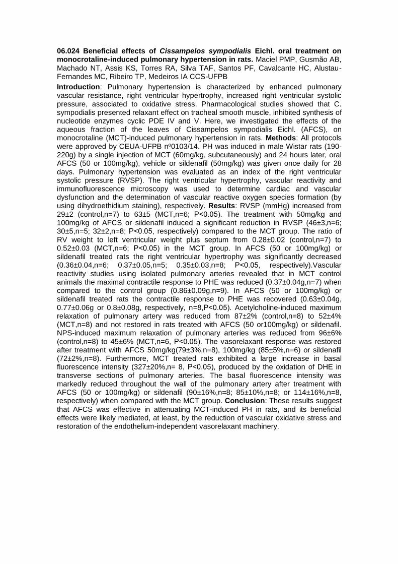
06.024 Beneficial effects of Cissampelos sympodialis Eichl. oral treatment on monocrotaline-induced pulmonary hypertension in rats. Maciel PMP, Gusmão AB,
Machado NT, Assis KS, Torres RA, Silva TAF, Santos PF, Cavalcante HC, Alustau-Fernandes MC, Ribeiro TP, Medeiros IA CCS-UFPB
Introduction: Pulmonary hypertension is characterized by enhanced pulmonary vascular resistance, right ventricular hypertrophy, increased right ventricular systolic pressure, associated to oxidative stress. Pharmacological studies showed that C. sympodialis presented relaxant effect on tracheal smooth muscle, inhibited synthesis of nucleotide enzymes cyclic PDE IV and V. Here, we investigated the effects of the aqueous fraction of the leaves of Cissampelos sympodialis Eichl. (AFCS), on monocrotaline (MCT)-induced pulmonary hypertension in rats. Methods: All protocols were approved by CEUA-UFPB nº0103/14. PH was induced in male Wistar rats (190-220g) by a single injection of MCT (60mg/kg, subcutaneously) and 24 hours later, oral AFCS (50 or 100mg/kg), vehicle or sildenafil (50mg/kg) was given once daily for 28 days. Pulmonary hypertension was evaluated as an index of the right ventricular systolic pressure (RVSP). The right ventricular hypertrophy, vascular reactivity and immunofluorescence microscopy was used to determine cardiac and vascular dysfunction and the determination of vascular reactive oxygen species formation (by using dihydroethidium staining), respectively. Results: RVSP (mmHg) increased from 29±2 (control,n=7) to 63±5 (MCT,n=6; P˂0.05). The treatment with 50mg/kg and 100mg/kg of AFCS or sildenafil induced a significant reduction in RVSP (46±3,n=6; 30±5,n=5; 32±2,n=8; P˂0.05, respectively) compared to the MCT group. The ratio of RV weight to left ventricular weight plus septum from 0.28±0.02 (control,n=7) to 0.52±0.03 (MCT,n=6; P˂0.05) in the MCT group. In AFCS (50 or 100mg/kg) or sildenafil treated rats the right ventricular hypertrophy was significantly decreased (0.36±0.04,n=6; 0.37±0.05,n=5; 0.35±0.03,n=8; P˂0.05, respectively).Vascular reactivity studies using isolated pulmonary arteries revealed that in MCT control animals the maximal contractile response to PHE was reduced (0.37±0.04g,n=7) when compared to the control group (0.86±0.09g,n=9). In AFCS (50 or 100mg/kg) or sildenafil treated rats the contractile response to PHE was recovered (0.63±0.04g, 0.77±0.06g or 0.8±0.08g, respectively, n=8,P˂0.05). Acetylcholine-induced maximum relaxation of pulmonary artery was reduced from 87±2% (control,n=8) to 52±4% (MCT,n=8) and not restored in rats treated with AFCS (50 or100mg/kg) or sildenafil. NPS-induced maximum relaxation of pulmonary arteries was reduced from 96±6% (control,n=8) to 45±6% (MCT,n=6, P˂0.05). The vasorelaxant response was restored after treatment with AFCS 50mg/kg(79±3%,n=8), 100mg/kg (85±5%,n=6) or sildenafil (72±2%,n=8). Furthermore, MCT treated rats exhibited a large increase in basal fluorescence intensity (327±20%,n= 8, P˂0.05), produced by the oxidation of DHE in transverse sections of pulmonary arteries. The basal fluorescence intensity was markedly reduced throughout the wall of the pulmonary artery after treatment with AFCS (50 or 100mg/kg) or sildenafil (90±16%,n=8; 85±10%,n=8; or 114±16%,n=8, respectively) when compared with the MCT group. Conclusion: These results suggest
that AFCS was effective in attenuating MCT-induced PH in rats, and its beneficial effects were likely mediated, at least, by the reduction of vascular oxidative stress and restoration of the endothelium-independent vasorelaxant machinery.

06.025 Effects of barbinervic acid, A triterpene isolated from Eugenia punicifolia in rat thoracic aorta Teixeira RGS1, Pascual R, Lima-Araújo KG1, Gandía L, Silva
CLM2, Santos WC1 – 1UFF, 2UFRJ
Introduction: Terpenes, isoprenoids, terpenoids represent one of the biggest and most diverse secondary metabolites obtained from natural products. Some terpenes have already described vasodilator action, such as borneol, terpinen-4-ol, and carvacrol. Recently, we have reported the isolation of triterpene barbinervic acid was from Eugenia punicifolia leaves, which was able to relax the noradrenaline induced vasopressor effects by a NO-mediated mechanism in renal circulation. Aims: Investigate the effects of barbinervic acid in contractions by phenylephrine and 60 mM K+. Methods: All animal procedures were approved by the Institutional Animal Care and Use Committee. Wistar rats (3 months old), 280–300 g, were killed by decapitation after an isoflurane anesthesia, and thoracic aorta was gently removed and cleared of surrounding tissues and secretions. The organ was immersed in a petri plaque with nutritive solution for obtaining 3-4 mm rings that were suspended in a 15 mL organ bath (Panlab Four Chamber Organ Bath, AD-Instruments, Australia). Tissues were kept in aerated nutrient solution at 37°C with the following composition (mM): NaCl 138; KCl 5.7; CaCl2 1.8; NaH2PO4 0.36; NaHCO3 15; and glucose 5.5, in distilled water, bubbled with a gas mixture of 95% O2 e 5% CO2, pH=7.4. Contractions were recorded through a data acquisition system (PowerLab 8/30 and LabChart Pro, AD-Instruments, Australia) by using isometric transducers, with 20 mN load. After a 60 minutes equilibration period, experiments were started. Tissues were washed-out each 15 minutes. Results and Conclusion: In 1 μM phenylephrine pre contracted rat aorta, addition of 70 µM barbinervic acid relaxed the aorta up to 44%. Cumulative addition of barbinervic acid in 60 mM K+ pre contracted rat aorta caused a significant relaxation (53,2 % at 100 µM; P ≤ 0.05). Barbinervic acid influenced the phenylephrine contractile effects and the vascular tonus produced by high K+. Although we were not able to conclude for a mechanism of action in aorta, the experiments with K+ have revealed the possibility of Ca+2 intracellular mobilizing as tendency to be explored in the sequence of the project and other studies are necessary to clarify the possible involvement of NO-pathway, as it was detected in renal circulation. In conclusion, our results pointed to a strong possibility that triterpene barbinervic acid, isolated from Eugenia punicifolia dichloromethane extract may be a template for developing new molecules to be used in cardiovascular diseases in the near future. Financial Support: CAPES, FAPERJ,
CNPQ, Fundación Carolina (España). Approval by the Animal Research Ethical Committee: Process number nº 364/13. Acknowledgements: Centro de Instrução de Guerra na Selva, CIGS, Manaus, AM, for kindly supplying the plant material.

06.026 Contractile response induced by U46619 and relaxation induced by NCX2121 are similar in coronary arteries isolated from renal hypertensive 2K-1C and normotensive 2K rats. Paula TD, Bendhack LM FCFRP-USP – Física e Química
Background: Vascular endothelium controls the vascular tone by the production and/or release of relaxing factors (EDRF) and constrictor factors (EDCF). In pathological states like hypertension this balance can be altered with reduction in EDRFs like nitric oxide (NO) and increased EDCFs. This imbalance is characterized as endothelial dysfunction which is recognized by reduction in endothelium dependent vasodilatation, increase in vessel contraction, pro-inflammatory monocytes adhesion, coagulation and loss in endothelial monolayer integrity. The major EDCFs are COXs products like thromboxane A2 (TXA2). NCX2121 is the combination of a non-steroidal anti-inflammatory drug indomethacin linked with an NO donor. This study aimed to evaluate the relaxing effect induced by NCX2121 in the coronary artery isolated from hypertensive 2K-1C and to compare this effect with normotensive sham-operated 2K rat arteries. The experimental procedures were approved by the Ethics Committee (CEUA No. 2012.1.120.53.0). Methods: One group of rats was operated to induce renal hypertension 2K-1C and another group was submitted to sham operation (2K). After 6 weeks, the arterial pressure was measured by tail cuff method. The 2K-1C (>160 mmHg) and 2K rats were killed and the hearts were removed and placed in ice cold Krebs solution. The septal coronary arteries were isolated and mounted in myograph (Multi Wire Myograph System, model 610M, DMT) and stimulated with 120 mM KCl. Concentration-effect curves (10 nM-100 µM) were constructed for 5-hydroxytryptamine (5-HT) and on top of the contraction to 5-HT, the arteries were stimulated with acetylcholine (10 µM). After washing, concentration-effect curves were constructed for the TXA2 analogue (U46619, 10 nM-10 µM) and concentration-effect curves were performed for NCX2121 (0.1 nM-100 µM). All the preparations that were used in this work presented the endothelium intact. The responses were compared between arteries from 2K-1C (n=4) and 2K (n=5) rats by using Student´s t test with P<0.05 for significance. Results: The internal diameter of the coronary artery was not different between 2K-1C (260.4 ± 27.6 µm) and 2K (337.4 ± 29.9 µm).The contraction to KCl was not different between 2K-1C (2.98 ± 0.58 mN) and 2K (4.28 ± 0.65 mN). 5-HT- induced contraction with greater potency in 2K-1C (pD2 5.51±0.01) than in 2K (pD2
5.06±0.14) without difference in the maximum effect (ME) (3.35 ± 1.07 mN vs 4.29 ± 0.99 mN). Acetylcholine induced relaxation of 5-HT pre-contracted arteries, but it was not different between the two groups. The activation of TP receptors by U46619 induced contraction in both 2K and 2K-1C arteries, without differences between them. In addition, NCX2121 induced similar relaxation in both arterial groups, 2K-1C and 2K. Conclusion: Our results show that the diameter and contractile responses induced by
U46619 are similar and also the compound NCX2121 induces similar relaxation of the renal hypertensive and normotensive rat coronary arteries. Financial support: CAPES, FAPESP and CNPq in both potency and ME.

06.027 Matrix metalloproteinase inhibition prevents loss of calponin-1 in early hypertensive vascular remodeling. Belo VA, Castro MM, Tanus-Santos JE FMRP-
USP – Farmacologia
Aims: Increased matrix metalloproteinase (MMP)-2 activity contributes to hypertension-induced chronic and maladaptive vascular remodeling. Calponin is a contractile protein, which is cleaved by MMP-2 in oxidative stress injuries, and a differentiation marker of vascular smooth muscle cells (VMSC). Reduced calponin levels are associated with the VSMC phenotype switch, which leads to migration and remodeling. We examined whether MMP-2 may proteolyze calponin-1 levels in the aorta of two-kidney, one-clip (2K1C) hypertensive rats to contribute to the chronic arterial remodeling. Methods: Sham operated or 2K1C rats were treated with either doxycycline at 30 mg/kg/day or vehicle. Treatment with doxycycline started 3 days post-surgery. Systolic blood pressure (SBP) was assessed in sham and 2K1C rats at 1 and 2 weeks following surgery by tail-cuff plethysmography. Gelatinolytic activity was determined in aortas by in situ zymography. Aortic MMP-2 and calponin levels were determined by Western blot. Quantitative morphometry was performed in hematoxylin and eosin stained aortic sections to analyze the structural changes. Results: SBP increased from 153 ± 1.7 mmHg to 176.7 ± 4.9 mmHg in the first to second weeks of hypertension when compared to Sham (p<0.05). Treatment with doxycycline did not reduce SBP in 2K1C rats neither at first or second weeks (p>0.05). We observed increased MMP-2 and reduced calponin-1 levels in aortas of 2K1C rats at first week (p<0.05, r=-0.74), followed by early, non-significant changes, in arterial structure. Treating 2K1C rats with doxycycline decreased aortic MMP-2 levels and gelatinase activities in addition to prevent loss of aortic calponin-1. We observed increased levels of aortic MMP-2 and calponin-1 at the second week of hypertension (p<0.05), which were accompanied by increased aortic media thickness and media to lumen ratio (p<0.05). Conclusion: Increased MMP-2 activity may contribute to calponin-1 reduction in early hypertension and this effect may be a starting point of chronic arterial remodeling. Financial support: Fundação de Amparo a Pesquisa do Estado de São Paulo (FAPESP), Conselho Nacional de Desenvolvimento Científico e Tecnológico (CNPq) and Coordenação de Aperfeiçoamento de Pessoal de Nível Superior (CAPES); (# 4/2015, Human or the Animal Research Ethical Committee of Faculty of Medicine of Ribeirão Preto).

06.028 Effects of continuous and accumulated exercise on endothelial function in rat aorta. Martinez JE, Ledo PBO, Chies AB FAMEMA
Introduction: Although it has already been described the cardiovascular benefits of physical activity, its regular practice and in a continuous and vigorous fashion is not always possible for most people. In this manner, practicing physical activity in short sessions that are repeated throughout the day (accumulated exercise) can be a possible solution. However, it is not yet fully established whether the accumulated exercise can promote benefits to the vascular endothelium, equivalent to the exercise practiced in daily one-hour sessions. Objective: The present study aimed to verify whether accumulated exercise promotes benefits equivalent to continuous exercise on endothelial function in rat aorta. Methods: Male wistar rats (350-400g) were divided into 3 groups: sedentary (SED), trained by continuous exercise (ExC; 1 hour a day, 5 days/week, for 8 weeks) and trained by accumulated exercise (ExA; 4 sessions of 15 minutes regularly distributed along the day, 5 days/week, for 8 weeks). The training consisted in running on treadmill at 60% of maximum capacity of each animal. The gains of both body weight and maximum capacity of running on treadmill were registered along the training period. In the end of the training protocol, animals were sacrificed in CO2 chamber. Aortas were removed from these animals and divided in rings (3-5 mm) to be set in organ bath containing Krebs-Henseleit solution, 37° C, pH 7.4, gassed with O2 95% / CO2 5%. Into the organ baths, under basal tension of 1.5g, these preparations were challenged with norepinephrine (NE), acetylcholine (ACh), KCl and sodium nitroprusside (NPS), either in absence or presence of L-NAME. Some preparations were studied without endothelium. Contractions were registered by isometric transducers and expressed as concentration-response curves. From these curves, were obtained maximal response (Rmax) and pEC50. These parameters were compared between groups by one way ANOVA/Tukey test (significancy if P<0.05). This study was approved by CEUA-FAMEMA (protocol 627/13). Results: During the training, the body mass gain of ExC (12.5±2,3%, n=11), but not of ExA (17.9±1.8%, n=11), was significantly (P<0.05) lower compared to the SED animals (21.4±1.2%, n=11). Moreover, the gain of maximum capacity of running on treadmill gain was significantly higher (P<0.05) in both ExC (42.3±13,2%, n=11) and ExA (36.2±9.8%, n=11), compared to the SED animals (-32.0±5.7%, n=11). In addition, aortas taken from ExC have presented a reduction of NE Rmax in relation to SED animals (from 2.16±0.14 to 1.73±.08, P<0.05; n=7-8). This difference was suppressed in presence of L-NAME or by the endothelium removing. On the other hand, no differences of NE Rmax were observed between aortas taken from ExA (1.78±0.12, n=8) and SED (2.16±0.14; n=7) animals. In parallel, the 10-5M ACh-induced relaxation in aortas taken from ExC, but not ExA, was bigger in comparison to those observed in aortas from SED animals (ExC 77.6±4.0%, n=7; SED: 63.7±4.6%, n=9; P<0.05). Finally, neither ExC nor ExA have modified to aorta responses for KCl or NPS. Conclusion: Both the ExC and the ExA improved the physical conditioning of the studied animals. However, only the ExC apparently was able to reduce the gain of body weight of the animals and improve directly the endothelial function in their aortas.

06.029 Impaired relaxation of mesenteric artery to Nitric Oxide (NO) in rats with ligature-induced periodontitis. Jesus FN1, Neto EAS1, Wenceslau CF2, Teixeira SA1,
Costa SKP1, Muscará MN1 1ICB-USP – Farmacologia, 2ICB-USP – Fisiologia e Biofísica
Introduction: We have previously shown that NO is a key regulator of some of the effects of periodontal disease on remote organs such as heart and kidney, as well as aorta endothelial dysfunction and decreased adrenergic contractililty. Aims: We thus decided to study the effects of periodontitis on the mesenteric resistance artery from Wistar rats. Methods: The protocol was approved by the local Ethics Committee for Animal Experimentation (CEUA-ICB 170, book 2/113, 2011). Male Wistar rats were anesthetized and bilateral periodontitis was induced by placing a cotton ligature around the lower first molars (sham animals had the ligature immediately removed). Seven days later, the animals were euthanized and the vessels (3rd order mesenteric artery branches) were isolated and mounted on a myograph for evaluation of the in vitro
responses to acetylcholine (ACh), phenylephrine (Phe), sodium nitroprusside (SNP) and sildenafil (Sil). From the concentration-response curves, potency (pD2) and maximal response (Emax) values were calculated. The vessels were also analysed for NOS isoenzyme gene expression and activity of the antioxidant enzymes superoxide dismutase (SOD) and catalase (CAT). The results were analysed by unpaired Student t-test. Results: Alveolar bone loss was observed in the animals with ligature, as bilaterally measured at the first and second molar levels. No differences were observed between the groups in terms of vascular responses to Phe or ACh. However, SNP-induced relaxation was less potent in the ligature group (pD 2 : 6,4±0.2 vs. 7.1±0.1; P<0.01), similarly to Sil (pD2: 8.6±0.2 vs. 10.3±0.2; P<0.001), but efficacies (Emax) were unaltered. Decreased SOD activity (0.7±0.05 vs. 0.4±0.05; P<0.01) and increased iNOS mRNA (1.0±0.1 vs. 20.9±5.9; P<0.01) and CAT activity (2.3±0.2 vs.3.2±0.3; P<0.05) were found in the arteries from rats with periodontitis. Conclusions: We conclude that during the early phase of ligature-induced periodontitis in rats, functional changes related to the NO-cyclic GMP pathway occur in the mesenteric artery smooth muscle, which may be secondary to the excessive production of iNOS-derived NO, superoxide anion or their combination. Financial Support: FAPESP, CAPES e CNPQ.

06.030 Exercise training improves the plasma antioxidant defenses in 2 kidneys, one clip (2K1C) hypertensive rats. Oliveira PR, Ledo PBO, Chies AB FAMEMA
Introduction: Previous studies show a close correlation between 2K1C hypertension and oxidative stress. Moreover, it has been suggested that exercise training improves the antioxidant defenses in rats. However, there are few reports in the literature of exercise training effects on the plasmatic antioxidant defenses of 2K1C rats. Objective: the present study aimed to assess the influence of exercise training upon the plasmatic antioxidant defenses in 2K1C rats as well as its possible consequences in the lipid peroxidation and in the nitric oxide (NO) bioavailability. Methods: Wistar rats (♂) 350-400g were divided in 4 groups: resting sedentary, exercised sedentary, resting trained and exercised trained, according to the exercise protocol. The exercise training was performed in treadmill (60% of maximal capacity of each animal), 1 hour per day, 5 day/week, for 10 weeks. To induce the hypertension, clips were implanted in the renal arteries after 5 weeks of exercise training. Five weeks later, in the 10th week of training, sedentary and trained animals (either sham or 2K1C) were killed at rest or after a single bout of exercise to collect plasma samples, that were storage at -20ºC. In these plasma samples, the ferric reducing antioxidant power (FRAP) was determined according to method described by Benzie (Anal. Biochem., 239: 70, 1996). Moreover, the plasma lipid hydroperoxides and nitrite concentrations were quantified by Xylenol orange (FOX) and Griess reaction, respectively. The results (mean ± mean standard error of 8-11 determinations) were compared by two way ANOVA/Bonferroni (differences were considered significant whether P<0.05). This study was approved by CEUA-FAMEMA (protocol 300/14). Results: The physical training, by itself, did not modify the FRAP values in Sham animals (sedentary: 1.02±0.13, n=8; trained: 1.01±0.07, n=08). However, SHAM animals submitted to physical training presented increased FRAP values when re-exposed to a single bout of exercise (from 1,01±0.07-n=8 to 1.35±0,05-n=8; p<0.05). On the other hand, a single bout of exercise as well as the physical training have not changed the FRAP values in 2K1C rats. The employed exercise protocols also has not changed the plasma lipid hydroperoxides or nitrite concentrations in either Sham or 2K1C rats. Conclusion: Sham trained animals, when
re-exposed to a single bout of exercise, exhibit an increment of their plasmatic antioxidant defenses assessed by FRAP. This increment of plasma antioxidant defenses, assessed by FRAP, appears not to occur in 2K1C animals. Moreover, the increment of FRAP observed in Sham animals apparently does not influence the plasma lipid hydroperoxides or nitrite concentrations. Financial support - FAPESP (Proc. 2013/22655-9).

06.031 Apocynin has higher potency than diapocynin to induce vasodilation in mesenteric resistance arteries of Wistar rats. Troiano JA1, Potje SR1, Graton ME1,
Silva DS1, Ximenes VF2, Antoniali C1 1FOA-Unesp – Ciências Básicas, 2FCB-Unesp – Química
Introduction: In phagocytic cells, diapocynin, the active dimer of apocynin, inhibits the translocation of the p47phox subunit for NOX subunits in the membrane, inactivating NOX activity. NOX subunits are sources of superoxide anion and hydrogen peroxide. The beneficial effects of apocynin on cardiovascular oxidative stress has been studied, but little is known about diapocynin effects. Aims: In this study, we compared the effects of apocynin and diapocynin on arterial pressure and resistance mesenteric arteries of normotensive Wistar rats. Methods: Wistar rats (3 months) were anesthetized and catheters were inserted into femoral artery and vein for measurement of Mean Arterial Pressure (MAP) and drug infusion. Drugs were diluted in 0.9% NaCl saline. MAP was recorded before and after infusion of a single dose (30 mg/kg) of apocynin or diapocynin. Mesenteric resistance arteries (2nd and 3rd branches) were isolated. The intact or denuded rings were kept in Krebs solution, pH 7.4, carbogenic mixture, 37 ° C at Mulvany-DMT system. The rings were contracted with phenylephrine (0.1 mmol/L) and stimulated with apocynin or diapocynin (0.1 mmol/L to 1 mmol/L). The concentration of NO in endothelial cells was evaluated by alteration of basal fluorescence intensity (FI) of DAF-2DA probe (5 µmol/L, 20 minutes of incubation), measured in Attune Acoustic Focusing Cytometer® - Applied Biosystem. Results were compared between groups (ANOVA, p <0.05) and n=5 for each group. Results: The
MAP of rats was reduced by both drugs (apocynin: -11.6 ± 0.3 mmHg; diapocynin: -11.1 ± 0.2 mmHg), however, the hypotensive effect of apocynin, but not of diapocynin, was kept until 30 minutes after administration. Dose-dependent vasodilator effects of both drugs were observed in intact but not in denuded rings of resistance mesenteric arteries. In intact rings, the potency of apocynin (pD2= 5.0 ± 0.06) was higher than diapocynin (pD2= 4.5 ± 0.09). FI the DAF-2DA was increased in endothelial cells (basal: 5516.8 ± 121.5 arbitrary units-AU) incubated with apocynin (7752.6 ± 112.9 AU) or diapocynin (6534.6 ± 419.3 AU). Conclusions: Our data indicate that the hypotensive effect of apocynin and diapocynin could be associated with its resistance arteries vasodilation effects. Apocynin and diapocynin vasodilator effects were endothelium and nitric oxide -dependent. Our results demonstrated that potency of apocynin was higher than diapocynin in resistance mesenteric arteries. Financial Support: FAPESP (2012/20398-6), CNPq and CAPES. Ethics Committee: CEUA/FOA Proc. N. 00440/2013.

06.032 Apocynin and Diapocynin reduced the adrenergic vasoconstriction in intact aortas of Wistar rats, however only apocynin reduced the concentration of reactive oxygen species in aortic endothelial cells. Graton ME, Potje SR, Troiano JA, Silva DS, Pereira AAF, Nakamune AC, Ximenes VF, Antoniali C 1FOA-Unesp – Ciências Básicas, 2FCB-Unesp – Química
In phagocytic cells, apocynin is converted into diapocynin by myeloperoxidases. Diapocynin is the active dimer of apocynin, and it inhibits translocation of p47phox subunit of NOX/ NADPH oxidase to membrane, inactivating NOX enzymes. In vascular cells, the effects of these drugs were not completely understood. The aim of this study was to compare the antioxidant effect of apocynin and diapocynin and the effects of those compounds on vasoconstriction stimulated by phenylephrine. We evaluated the antioxidant capacity of apocynin and diapocynin (0.1 – 100 µmol/L) in plasma by FRAP method. Concentration-effect curves to phenylephrine (0.1 nmol/L – 10 μmol/L) were performed in intact aorta rings kept on Krebs solution, pH 7.4, 95% O2, 5% CO2 and 37°C, incubated with apocynin or diapocynin (10 μmol/L, 30 min). The effects of apocynin or diapocynin on concentration of reactive oxygen species in aortic endothelial cells (2,500 events) were evaluated by alteration of basal fluorescence intensity (FI) of DHE probe (2.5 μmol/L, 20 min of incubation), using Attune Acoustic Focusing Cytometer® - Applied Biosystem. The results were compared between drugs (ANOVA p<0.05). Apocynin (999,79 mmol/L) showed higher antioxidant capacity than diapocynin (246,32 mmol/L). The incubation of intact aortas with apocynin (Emax: 0.88±0.14 g, n=4) or diapocynin (1.31±0.15 g, n=4) reduced the maximum effect of concentration-responses curves to phenylephrine. These effects were not observed in denuded aortas (1.80±0.08 g, n=5). Apocynin (2549±195 UA, n=5), but not diapocynin (3502±107 UA, n=5), reduced the reactive oxygen species concentrations in aortic endothelial cells compared to control (3732±134 UA, n=5). In conclusion, our results
demonstrated that apocynin has better antioxidant effect than diapocynin. Although both drugs reduced adrenergic vasoconstriction in intact aortas of Wistar rats, only apocynin reduced the concentration of reactive oxygen species in aortic endothelial cells. Financial support: CAPES and FAPESP (2012/01773-9; 2011/20998-0;
2012/20398-6). Approved by local ethics committee (CEUA/ FOA 00440/2013).

06.033 Involvement of rock and calcium in vasodilator response induced by Glyceryl Trinitrate (GTN) and Tetrahydrofurfuryl Nitrate (NTHF) in human umbilical artery. Alustau-Fernandes MC1, Melo MP2, Silva TAF2, Maciel PMP1, Machado NT1, Gomes SM3, Mendes-Junior LG1, Mendes-Neto JM4, Furtado FF5, Athayde-Filho PF1, Medeiros IA1 1UFPB – Produtos Naturais Sintéticos e Bioativos., 2UFPB – Ciências da Saúde, 3Maternidade Cândida Vargas, 4UFS – Pós-Graduação em Ciências Fisiológicas, 5UFCG – Escola Técnica de Saúde
Introduction: Organic nitrates are the most commonly nitric oxide (NO) donors used in the treatment of cardiovascular diseases. Previous studies have shown that the tetrahydrofurfuryl nitrate (NTHF) and glyceryl trinitrate (GTN) induce vasorelaxation in mesenteric artery with involvement of the NO-cGMP-PKG pathway. The human umbilical artery (HUA) is one of the human vessel models used in the characterization of vasodilators. Aims: The aim of this study was to evaluate the vasodilator responseof organic nitrates (NTHF and GTN) in HUA. Methods: Ethics Committee in Research of
University Hospital (CEP/HUWL) approved all protocols of this study (nº 016/10). Human umbilical cords were obtained from healthy pregnant women hospitalized at HULW/UFPB and Candida Vargas Maternity. The HUA connective tissue was removed and cut into 5 mm long rings segments. The rings were suspended by metallic barfor isometric tension recordings in a Kreb’s Henseleit solution at 37 ºC, gassed with 95% O2 and 5% CO2, at resting tension 3g, for 3 hours. Results: GTN (0.1 nM-1 mM) orNTHF (0.1 nM-3 mM) decreased HUA basal tonus (Maximum Response – MR = 1.19 ± 0.14 g/ 1.25 ± 0.12 g, respectively). Since the intrinsic tone in HUA rings depends on calcium influx by voltage-dependent calcium channels (CaV), this result suggests calcium blocking action induced by both nitrates. GTN or NTHF induced concentration-dependent vasodilation in rings pre-contracted with 5-hidroxytriptamine (5-HT – 10 µM) (MR = 110.0 ± 3.5% and 106.9 ± 2.1%, respectively) or KCl 60 mM (MR = 91.8 ± 1.9% and 68.6 ± 9.2%, respectively). It is well known in the literature that contraction induced by 5-HT has the initial transient component associated with Ca2+ release from the sarcoplasmic reticulum. Pre-incubation withGTN (1 mM) or NTHF (3 mM) decreased transient component of 5-HT contraction (MR = 95.7 ± 2.0% or75.6 ± 14.9%, respectively). In calcium-free medium, sustained contractions elicited by KCl 60 mM and the phasic and tonic components of the contraction induced by 5-HT depend on Rho-associated protein kinase (ROCK) activation. GNT or NTHF relaxed rings pre-contracted with KCl 60 mM in calcium-free medium (MR = 89.9 ± 3,0% or 88.3 ± 9.6%, respectively). Similarly, GTN or NTHF reduced the phasic (MR = 87.6 ± 4.0% or32.3 ± 10.7%, respectively) and tonic (MR = 92.0 ± 1.9% and 74.2 ± 0.1%, respectively) contractions induced by 5-HT in calcium-free medium. Conclusion: These findings suggest that vasodilator response induced by GNT and NTHF is, at least in part, due to blockade of calcium influx (through CaV), decrease of Ca2+release from sarcoplasmic reticulumstores and ROCK inhibition. Financial Support: CNPq Keywords: Glyceryl trinitrate. Tetrahydrofurfurylic nitrate. Human Umbilical Artery. Calcium influx. ROCK.

06.034 Increased levels of matrix Metalloproteinase-2 seem crucial to the transition from cardiac hypertrophy to heart failure in rats with abdominal aorta stenosis. Pereira SC1, dos Santos DO2, Prado FP2, Sanchez ER1, Prado CM2, Castro MM1 1FMRP-USP – Farmacologia, 2FMRP-USP – Patologia
Aims: Hypertension is intrinsically related to cardiac hypertrophy and dysfunction. Increased collagen deposition and cardiomyocytes hypertrophy may contribute to hypertension-induced transition from chronic hypertrophy to heart failure. Increased matrix metalloproteinase (MMP)-2 activity also contributes to hypertension and its compensated cardiac hypertrophy, an effect which is mitigated after treating rats with an MMP inhibitor. We examined whether the temporal changes that accompany cardiac hypertrophy to heart failure, in a model of pressure-overloaded hearts, may be dependent of MMP-2 activity. Methods: Male Wistar rats were subjected to abdominal aortic stenosis to be studied at 30, 60, 90 days after surgery. Cardiac function was analyzed by echocardiogram to determine the ejection fraction and shortening. Picrosirius red was used to quantify interstitial collagen in left ventricles. Left ventricles were also collected to determine protein levels of MMP-2, tissue inhibitor of matrix metalloproteinase (TIMP)-2, integrin and transforming growth factor (TGF)-β by Western blot. Results: Ninety days after surgery, 30% rats showed hypertrophied-
dilated (90d HD) hearts while 70% the hypertrophic (90d HH) profile. Increased interstitial collagen was observed in left ventricles after 30, 60 and 90 days after surgery (p<0.05 vs. Sham) and these levels did not statistically differ between 90d HH and 90d HD. As for interstitial collagen, increased levels of TGF-β was also observed in hearts from 30 to 90 days after surgery (p<0.05 vs. Sham). Furthermore, increased levels of integrin and resulting cardiomyocyte hypertrophy were observed in the left ventricles analyzed after 30, 60 and 90 days (p<0.01 vs. Sham). However, by the echocardiogram analysis, the ejection fraction and shortening significantly reduced by 34% and 37% only in the 90d HD compared to 90d HH (p<0.01), thus showing significantly cardiac dysfunction in the hypertrophied-dilated hearts. Here we also show that MMP-2 levels were only increased at 90 days after surgery, both at 90d HH and 90d HD (p<0.05 vs. Sham). Furthermore, a slight imbalance between MMP-2 and TIMP-2 levels was observed in the left ventricles at 90 days, which may suggest a deregulation of MMP-2 expression by TIMP-2 during the transition from hypertrophy to heart failure. Conclusion: Although increased collagen, TGF-β and integrins are important to cardiac hypertrophy, an imbalance between MMP-2 and TIMP-2 levels seems to be crucial to change hypertrophy to heart failure in hypertension. Financial support: Fundação de Amparo a Pesquisa do Estado de São Paulo (FAPESP), Conselho Nacional de Desenvolvimento Científico e Tecnológico (CNPq) and Coordenação de Aperfeiçoamento de Pessoal de Nível Superior (CAPES). Approved by the committee of ethics in Animal Research of Ribeirão Preto School of Medicine, University of São Paulo, SP,Brazil (Protocol no.204/2009).

06.035 Beta2-adrenoceptor is not essential for the response to environmental stress in the heart. Moura AL1,2, Brum PC3, Cespedes IC4, Spadari RC1,2 1Unifesp –
Farmacologia, 2Unifesp – Biociências, 3USP – Educação Física e Esporte, 4Unifesp – Biociências
Introduction: Stress is the body response to aversive stimuli or unknown situations and its evolutionary purpose is to enable adaptation and survival. However, this system does not always guarantee homeostasis. In atria from rats subjected to foot shock stress the response mediated by and the expression of β1- (β1-AR) and β2-adrenoceptors (β2-AR) are altered, with an increase in the β2 /β1 ratio. It is not known if this response is adaptive or deleterious to the heart and what is the role played by β2-AR. The aims of this study were to investigate the behavior of the β1-AR and of proteins from its intracellular signaling cascade in the absence of β2-AR (KOβ2
knockout mice), and if β2-AR is essential for the animals survival. Methods: FVB and KOβ2 mice were distributed in control and foot shock stressed groups. The isolated right and left atria chronotropic and inotropic responses to isoprenaline, salbutamol and dobutamine were evaluated in the absence or presence of 50 nM ICI118,551 or 300 nM CGP20712A. Protein and mRNA expression of β1-AR, β2-AR, GRK2, GRK5 and β-ARR2 were evaluated by Western blot and real time PCR. Results: There was no
difference in the right atria basal beating rate or in the left atria initial tension between control and foot shock stressed groups in the absence or presence of antagonists. In FVB control mice CGP20712A reduced the right atria maximum response to isoprenaline (315 ± 8 x 216 ± 15 bpm, p<0.05, Student t test). Right and left atria from KOβ2 submitted to stress were subsensitive to isoprenaline, the pD2 value were 9.03 ± 0.06 (control; n = 6) x 9.52 ± 0.21 (stressed; n = 6) in right atria, and 9.3 ± 0.05 (control; n = 6) x 9.7 ± 0.10 (stressed; n = 6) in left atria (p<0.05, Student t test). There was no difference on sensitivity to dobutamine or salbutamol in those tissues. CGP20712A and ICI118,551 canceled the subsensitivity to isoprenaline, as well as the response to salbutamol and decreased the responses to dobutamine. In stressed KOβ2 mice, β1-AR and β-ARR2 mRNA and protein expression was lower, whereas β2-AR, GRK2 or GRK5 proteins expression were unchanged. Conclusion: the β2-AR sub-type is not essential
for the survival of mice submitted to foot shock stress or for the basal chronotropism and inotropism; the response to isoprenaline in right and left atria of KOβ2 mice showed different patterns in stressed FVB compared to KOβ2 mice; the mRNA and protein expression of β1-AR and enzymes of the intracellular signaling cascade support the functional alterations presented in the right and left atria; the reduction of gene and protein expression of β-ARR2 might be an adaptive mechanism of ventricular tissue to excessive sympathetic stimulation during stress. Ethical Committee Approval CEP-UNIFESP, number CEUA N 633386. Financial support: CNPq; FAPESP.
06.036 Alpha1beta1 and integrin-linked kinase interact and modulate Angiotensin II effects in vascular smooth muscle cells. Moraes JA1, Frony AC1, Dias AM1,
Renovato-Martins M1, Rodrigues G1, Marcinkiewicz C2, Assreuy J3, Barja-Fidalgo C1 1UERJ – Biologia Celular e Molecular, 2Temple University, 3UFSC
Introduction: Atherogenesis is a process initiated by a local inflammatory response, characterized by the release of inflammatory mediators which amplifies this process and can induce vascular smooth muscle cells (VSMC) activation. Among the inflammatory molecules in circulation during a vascular injury, Angiotensin II (Ang II) highlights, being able to induce different effects in VSMC. However, the potential link between alpha1beta1 integrin signaling with NOX system and their combined contribution to Ang II effects on VSMC have not been investigated. Aim: This study was carried out to elucidate the molecular mechanisms underlying AngII effect on VSMC functions, focusing on the role of alpha1beta1 integrin and ILK pathway. Methods: Primary VSMC were isolated from rat aorta and VSMC cell lineage A7r5
were obtained from ATCC. The disintegrin Obtustatin (OBT) was used to block alpha1beta1 integrin; Chemotaxis (4h) was performed in Boyden chamber; Intracellular ROS production was quantified by DCF; P47 distribution was analyzed by immunocytochemistry (1h). Actin, AKT, ILK, NOX1 and p21 expression were evaluated by immunoblotting; siRNA assay was performed with validated siRNAs (Ambion) in Lipofectamin 2000; Cell proliferation was measured by cell cycle analysis (PI), [3H] Thymidine incorportation and MTT assay. Results: Ang II-induced VSMC migration (2-fold increase) and proliferation (2.5-fold increase) was modulated by alpha1beta1 integrin, being inhibited by obtustatin, a specific alpha1beta1 integrin blocker. Ang II also stimulates ROS production in VSMC (140%) that is NOX-1 dependent, being completely inhibited in NOX-1 silenced cells. The ROS production develops in two peaks, and the second peak is maintained by NOX-2 activation. Apocynin and obtustatin inhibit the NOX2-associated second peak, but not the first peak of ROS production, which is related to NOX1activation. Corroborating the involvement of alpha1beta1 integrin, the pretreatment of VSMC with obtustatin impaired Ang II-induced FAK phosphorylation, AKT activation, p21 degradation and the increase of ILK expression. Silencing of ILK blocked cell migration, AKT phosphorylation and the second peak of ROS, but partially inhibits (70%) VSMC proliferation induced by Ang II. Conclusion: The data demonstrate a novel role for NOX2 in Ang II effects on VSMC, and suggest alpha1beta1 integrin and ILK as target molecules to the development of more effective therapeutic interventions in cardiovascular diseases. Funding Support: FAPERJ, CAPES, CNPq. Ethics approval of research: The protocol was approved by the Committee on the Ethics of Animal Experimental of Universidade do Estado do Rio de Janeiro (CEUA/003/2013).

06.037 Extract of Syzygium cumini (L.) Skeels fruit peel reduces weight gain and improves vascular response in rats with hypercaloric diet. Torres RA1, Silva TAF2,
Maciel PMP3, Nascimento SM1, Cavalcante HC4, Alustau-Fernandes MC3, Medeiros IA3,2, Veras RC1,2 – 1PPGCN-UFPB, 2DCF-CCS-UFPB, 3PGPNSB-UFPB, 4UFPB – Nutrição
Introduction: Syzygium cumini (L.) Skeels is a tree native from India, but well adapted
in Brazilian northeast. Its fruits are rich in phenolic compounds with antioxidant activity. Several studies have demonstrated biological effects both in human and in rats. The more cited effect is antiglicemic activity and other are anti-inflammatory effects and blood pressure reducing levels. The literature is scarce about the benefits of S. cumini
in cardiovascular system of animals treated with hypercaloric diet. The aimed this study is evaluate the effect of hydroalcoholic extract of S. cumini fruit peel (Sc-pHE) on weight gain and vascular reactivity, as well as a blood pressure in rats fed a hypercaloric diet. Methods: All protocols were approved by CEUA/UFPB (#1001/14).
Rats were randomly divided into six groups and treated for 6 weeks (n=8/group), normocaloric diet + saline (NCT); normocaloric diet + Sc-pHE, (N100; 100 mg/kg); normocaloric diet + Sc-pHE, (N300; 300 mg/kg); hypercaloric diet + saline (HCT); hypercaloric diet + Sc-pHE, (H100; 100 mg/kg); and hypercaloric diet + Sc-pHE, (H300; 300 mg/kg). Body weight was monitored every three days and adiposity index was calculated abdominal fat deposits (retroperitoneal and epididymal fat) and normalized to final body weight (g/100 g of weight of animal). For in vivo experiments, the systolic blood pressure (SBP) levels were recorded on days 0, 8, 20, 30 and 42. For in vitro
experiments, isolated rat superior mesenteric rings were suspended in organ bath for isometric tension recordings in a Tyrode’s solution at 37 ºC, gassed with 95% O2 and 5% CO2. Results: The percentage weight gain was significantly higher in HCT group compared to the NCT group, after six weeks, with 26±3.9 and 15.3±1.7, respectively. The adiposity index (%) was significantly higher in HCT group (6.8±0.45) and treatment with Sc-pHE was effective in reducing abdominal fat deposits 5.6±0.38 and 5.0±0.64, H100 and H300, respectively. Vascular reactivity studies using isolated mesenteric arteries revealed that in animals HCT group the maximal contractile response to Phe was reduced (91.1±8.5%) when compared to the NCT group (100±8%). In Sc-pHE treated rats (100 or 300mg/kg) the contractile response to Phe was recovered (138.3±13.6% and 107.1±12.1%, respectively). The values of pD2 for ACh was reduced from 6.9 ± 0.4 (NCT) to 6.25± 0.37 (HCT) and restored in rats treated with Sc-pHE (100 or 300mg/kg), 7.41±0.24 and 7.33±0.18, respectively (p<0.05 vs HCT). NPS-induced power relaxation of mesenteric arteries was reduced from 7.92±0.05 (NCT) to 7.3±0.09 (HCT; p˂0.05). The vasorelaxant response was restored after treatment with Sc-pHE (100 or 300 mg/Kg) 8.09±0.04 and 8.07±0.04, respectively (p<0.05 vs HCT). The systolic blood pressure levels were not significantly different among the groups during the treatment. Conclusion: The results together suggest that treatment with Sc-pHE decrease the deleterious effect of a hypercaloric diet in these animal models in relation to reducing body weight gain and visceral fat accumulation and improve vascular response in rings of mesenteric artery of rats. Keywords: antioxidant activity, hypercaloric diet, vasorelaxation. Financial support: CNPq and CAPES