Embed Size (px)
Citation preview

3/3/2014
1
Yuri R. Parisky, M.D.
Breast Imager‐Mammoth Hospital
Trustee‐NCoBC
SESSION 42
The diagnostic work‐up of screen detected abnormalities remains the bread and butter of most
mammography practices?
Rhetorical Question:
A question asked without expecting an answer but for the sake of emphasis or effect.
This Presentation, using illustrative cases, demonstrates the when, how and why of the basic diagnostic work‐up of calcifications, masses and densities identified on screening mammography. BIRADS lexicon and classification is incorporated into the case demonstrations. Rationale for recommendations upon completion of work‐up is discussed; when to biopsy, how and when to follow‐up.
3/3/2014
2
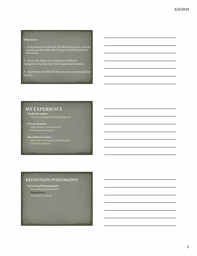
Objectives:
1. Understand the rationale for identifying and correctly classifying the multitude of types of calcifications of the breast.
2. Know the diagnostic imaging available for evaluation of screen detected masses and densities.
3. Appreciate the BIRADS lexicon and recommendation scheme.
Academic center Limited screening; Primarily Diagnostic
Private Practice High volume; numbers driven
Productivity monitored
Rural Breast Center Service to Community; Cost limitation
Uninsured patients
Screening Mammography Reasonable risk population
Asymptomatic
Prevalence of disease

3/3/2014
3
Diagnostic Mammography Screen detected abnormality
Physical Complaint
Follow‐up
Recent Personal Hx of treated BR Ca
PROBLEM SOLVING
Lobar/Lactiferous DuctCross Section
Breast Anatomy
Invasive Ductal Carcinoma (IDC)
Lobar/Lactiferous Duct Cross Section
3/3/2014
4
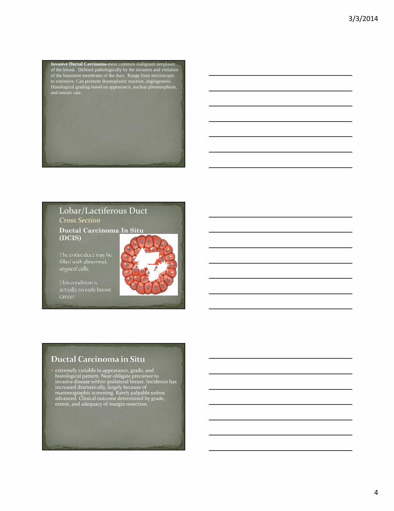
Invasive Ductal Carcinoma-most common malignant neoplasm of the breast. Defined pathologically by the invasion and violation of the basement membrane of the duct. Range from microscopic to extensive. Can promote desmoplastic reaction, angiogenesis. Histological grading based on appearance, nuclear pleomorphism, and mitotic rate.
Ductal Carcinoma In Situ (DCIS)
Lobar/Lactiferous Duct Cross Section
extremely variable in appearance, grade, and histological pattern. Near obligate precursor to invasive disease within ipsilateral breast. Incidence has increased dramatically, largely because of mammographic screening. Rarely palpable unless advanced. Clinical outcome determined by grade, extent, and adequacy of margin resection.
3/3/2014
5
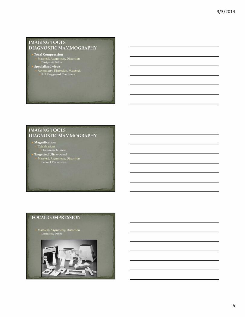
Focal Compression Mass(es), Asymmetry, Distortion
Dissipate & Define
Specialized views Asymmetry, Distortion, Mass(es),
Roll, Exaggerated, True Lateral
Magnification Calcifications
Characterize & Extent
Targetted Ultrasound Mass(es), Asymmetry, Distortion
Define & Characterize
Mass(es), Asymmetry, Distortion Dissipate & Define

3/3/2014
6
In the ACR‐BIRADS, a MASS is defined as a three dimensional structure demonstrating convex borders, usually evident on two orthogonal views
If seen in only one projection, a suspected mass is called an ASYMMETRY.
Asymmetry lacks convex borders and the conspicuity of a mass
Due to confusion with density which describes attenuation characteristics of a mass, the term density has been replaced by asymmetry
FOCAL ASYMMETRY may be a mass obscured by overlying glandular tissue, or it may be superimposition or overlap of normal breast tissues.
SUMMATION SHADOW or pseudomass is the overlap of normal breast tissue
STANDARD VIEWSCraniocaudalMediolateral Oblique
ADDITIONAL EVALUATION90 degree lateralSpot Compression, or rolled view
Spot MagnificationUltrasound
3/3/2014
7
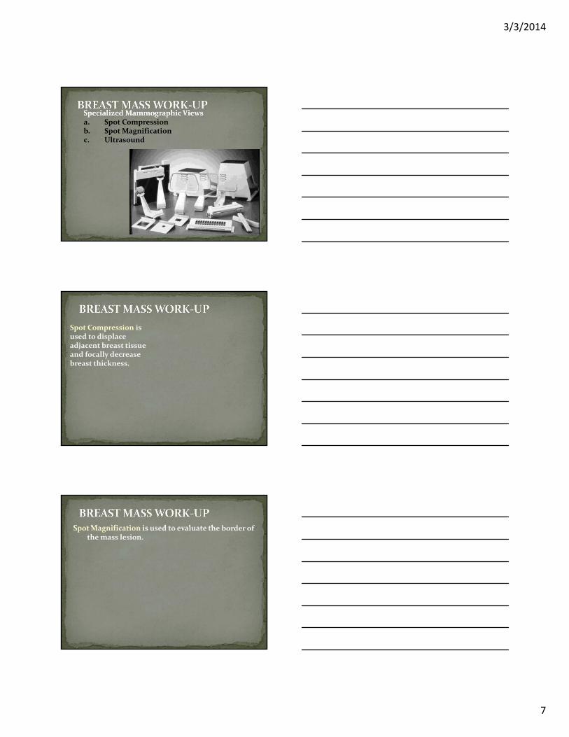
Specialized Mammographic Views a. Spot Compressionb. Spot Magnificationc. Ultrasound
Spot Compression is used to displace adjacent breast tissue and focally decrease breast thickness.
Spot Magnification is used to evaluate the border of the mass lesion.

3/3/2014
8
SHAPE
Margin
Density
Number
Round
Oval
Lobular
Irregular
Architectural Distortion
ROUND:
A mass that is spherical, ball‐shaped, or globular in shape.

3/3/2014
9
LOBULATED:
A mass that has contours with undulations
IRREGULAR:
The lesion’s shape cannot be characterized by any of the above.
ARCHITECTURAL DISTORTION:
The normal architecture is distorted with no definite visible mass:
Spiculations radiating from a point
Focal retraction (puckering) of normal connective tissue lines
3/3/2014
10
ARCHITECTURAL DISTORTION
SHAPE
Shape
MARGIN
Density
Number
Characterization of the edge or transition between a mass and the surrounding normal breast tissue
3/3/2014
11
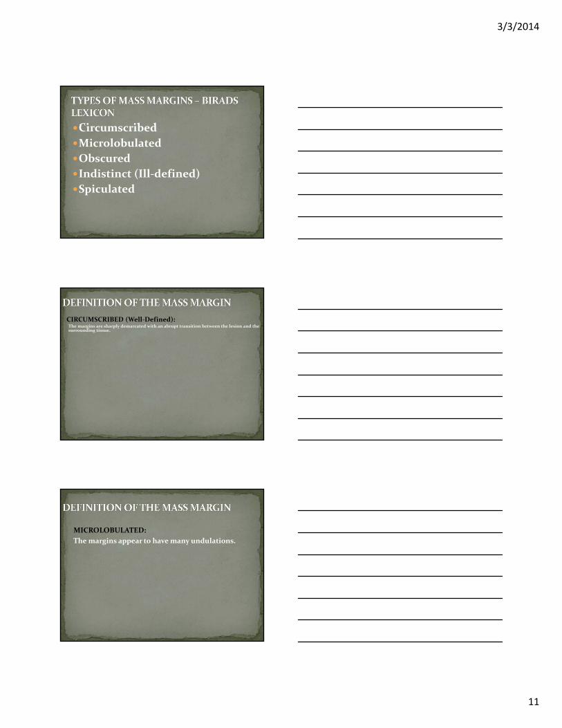
Circumscribed
Microlobulated
Obscured
Indistinct (Ill‐defined)
Spiculated
CIRCUMSCRIBED (Well‐Defined):The margins are sharply demarcated with an abrupt transition between the lesion and the surrounding tissue.
MICROLOBULATED:
The margins appear to have many undulations.

3/3/2014
12
OBSCURED:
This margin is hidden by superimposed or adjacent normal tissue
INDISTINCT (Ill Defined):
The indistinct margin suggests early infiltration of breast tissue by the mass, not likely due to superimposed normal breast tissue.
SPICULATED:
The lesion is characterized by thin lines radiating from the margins of a mass
3/3/2014
13
Shape
Margin
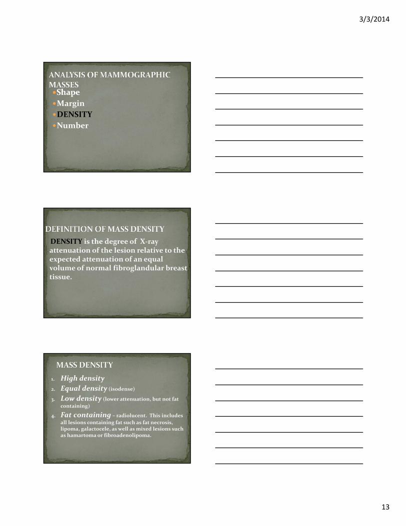
DENSITY
Number
DENSITY is the degree of X‐ray attenuation of the lesion relative to the expected attenuation of an equal volume of normal fibroglandular breast tissue.
1. High density
2. Equal density (isodense)
3. Low density (lower attenuation, but not fat
containing)
4. Fat containing – radiolucent. This includes
all lesions containing fat such as fat necrosis, lipoma, galactocele, as well as mixed lesions such as hamartoma or fibroadenolipoma.

3/3/2014
14
High density
Equal density (isodense)
Low density (lower attenuation, but not fat containing)
3/3/2014
15
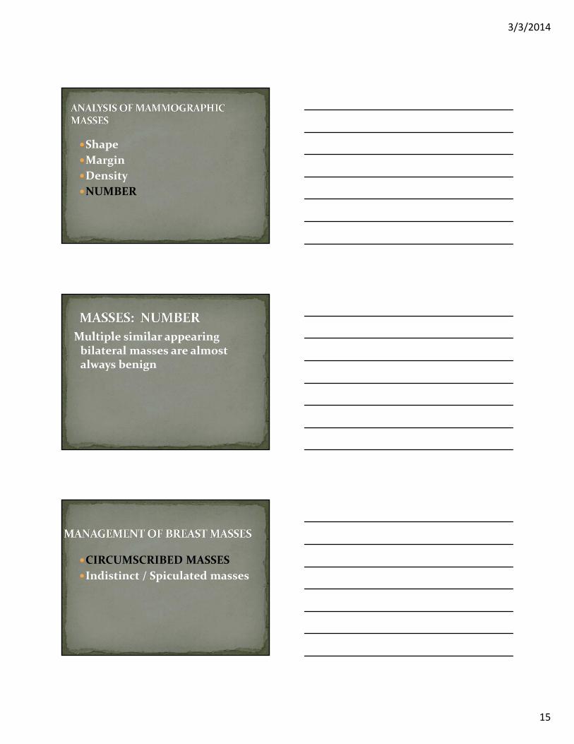
Shape
Margin
Density
NUMBER
Multiple similar appearing bilateral masses are almost always benign
CIRCUMSCRIBED MASSES
Indistinct / Spiculated masses
3/3/2014
16
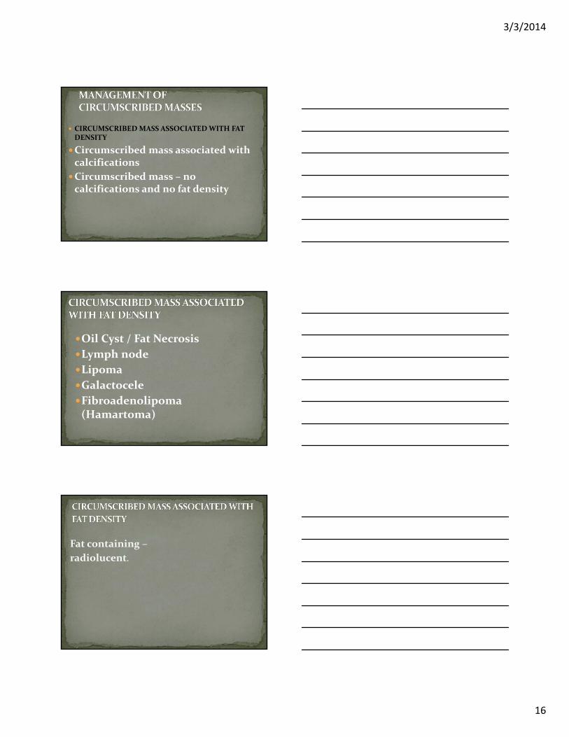
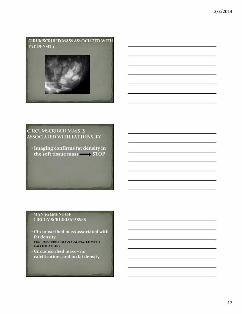
CIRCUMSCRIBED MASS ASSOCIATED WITH FAT DENSITY
Circumscribed mass associated with calcifications
Circumscribed mass – no calcifications and no fat density
Oil Cyst / Fat Necrosis
Lymph node
Lipoma
Galactocele
Fibroadenolipoma (Hamartoma)
Fat containing –
radiolucent.
3/3/2014
17
Imaging confirms fat density in the soft tissue mass STOP
Circumscribed mass associated with fat density
CIRCUMSCRIBED MASS ASSOCIATED WITH CALCIFICATIONS
Circumscribed mass – no calcifications and no fat density
3/3/2014
18
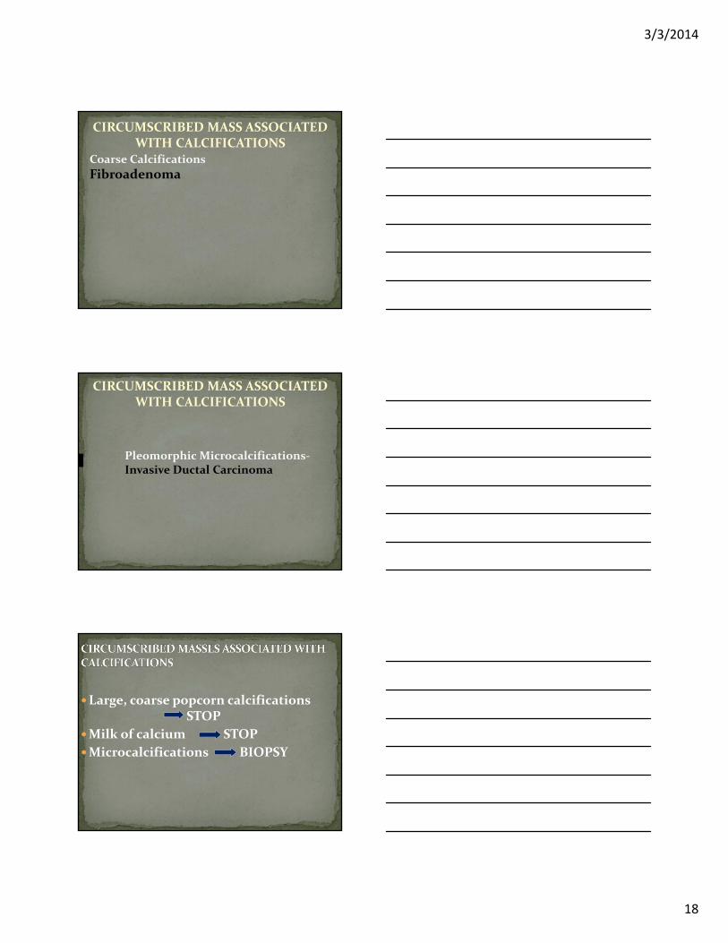
CIRCUMSCRIBED MASS ASSOCIATED WITH CALCIFICATIONS
Coarse Calcifications
Fibroadenoma
CIRCUMSCRIBED MASS ASSOCIATED WITH CALCIFICATIONS
Pleomorphic Microcalcifications‐Invasive Ductal Carcinoma
Large, coarse popcorn calcifications STOP
Milk of calcium STOP
Microcalcifications BIOPSY

3/3/2014
19
Circumscribed mass associated with fat density
Circumscribed mass associated with calcifications
CIRCUMSCRIBED MASS – NO CALCIFICATIONS AND NO FAT DENSITY
Fibroadenoma
Cyst
Dedicated Targeted Breast Ultrasound
Important Adjunctive Procedure :
Evaluate mammographically detected mass ‐Differentiate cystic from solid masses
Evaluate asymmetry and architectural distortion seen on the mammogram
Evaluate clinical findings‐mass, thickening
Evaluate palpable mass in women under 30 years old, in lactating, or pregnant women

3/3/2014
20
ADDITIONAL EVALUATION90 degree lateralOrthogonal Magnification
BIRADS CLASSIFICATIONMorphologyDistribution
Skin
Vascular
Popcorn; Coarse
Large Rod‐Like
Punctate Round
Internally Lucent
Eggshell
Milk of Calcium
Suture Calcifications
Dystrophic
Amorphous
Coarse Heterogeneous
3/3/2014
21
Fine Pleomorphic
Fine Linear
Fine Linear Branching
Diffuse/Scattered
Regional
Clustered or Grouped
Linear
Segmental
Screening 10‐15 minute time slots
Technologist +/‐ Assistant
Scheduling‐minimal
Batch/Stack Interpretation Immediate result decreases productivity
$
3/3/2014
22
Diagnostic Mammography 30 minutes time slot
Technologist +/‐ Assistant
Scheduling‐maximum
Supervision/Interpretation
Result
$$
Ultrasound 15‐30 minutes
Technologist +/‐ Assistant
Scheduling
Physician Intensive Perform exam
Result
$$‐$$$
Screening $200‐230
Diagnostic Uni‐$150‐200
B/L‐$300
Ultrasound $200‐655

3/3/2014
23
Feig: DM reimbursement rates only~11% higher
Present rates: DM 30‐50% higher
Resource Consumption: 2‐3 Times longer
Greater Administrative/Technologist Time
Greater Physician Time
3/3/2014
24
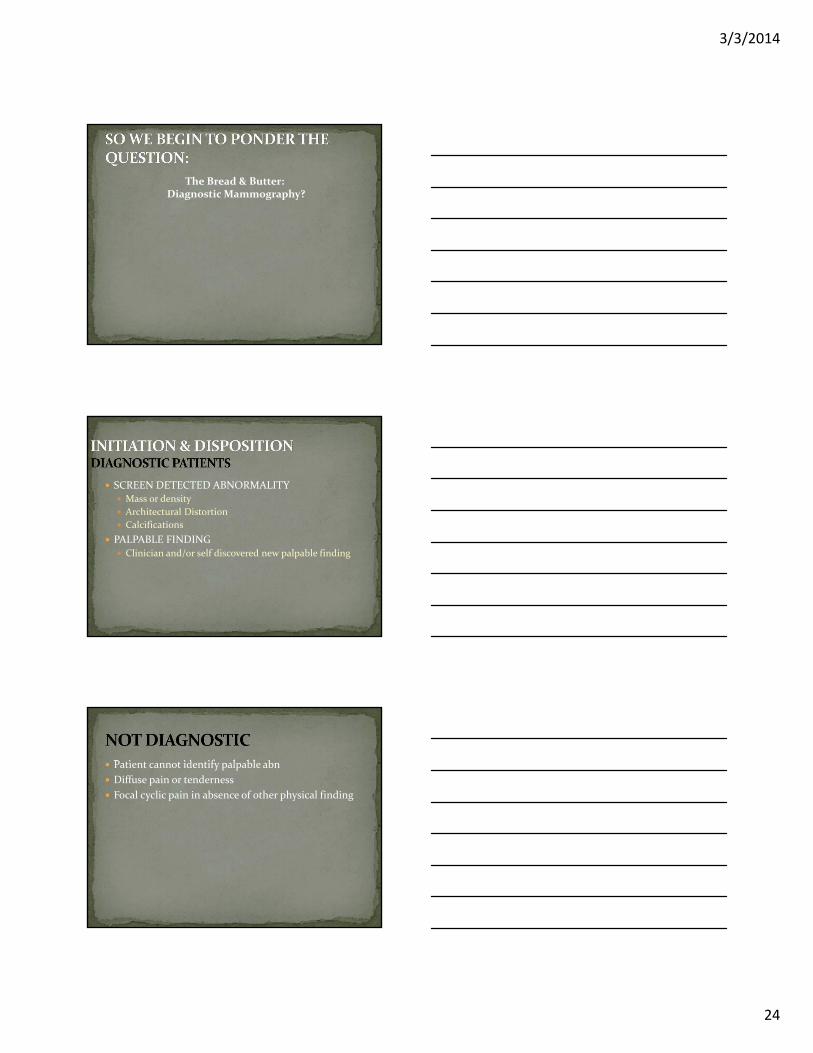
The Bread & Butter:Diagnostic Mammography?
SCREEN DETECTED ABNORMALITY Mass or density
Architectural Distortion
Calcifications
PALPABLE FINDING Clinician and/or self discovered new palpable finding
Patient cannot identify palpable abn
Diffuse pain or tenderness
Focal cyclic pain in absence of other physical finding
3/3/2014
25
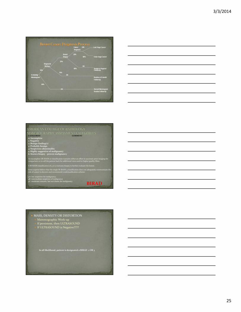
Cancer 20% Late Stage CancerDiagnosis
Breast 20%Biopsy 80% Early Stage Cancer
25%Diagnostic Workup OK
80% Benign or Atypical 10% Follow-Up
75% Screening OK Mammogram Routine or 6 month
Follow-Up
90%
OK Normal MammogramRoutine Follow-Up
CATEGORIES COMMENT
0: Incomplete1: Negative2: Benign finding(s)3: Probably benign4: Suspicious abnormality5: Highly suggestive of malignancy6: Known biopsy – proven malignancy
An incomplete (BI‐RADS 0) classification warrants either an effort to ascertain prior imaging for comparison or to call the patient back for additional views and/or higher quality films.
A BI‐RADS classification of 4 or 5 warrants biopsy to further evaluate the lesion.
Some experts believe that the single BI‐RADS 4 classification does not adequately communicate the risk of cancer to doctors and recommend a subclassification scheme:
4A: low suspicion for malignancy4B: intermediate suspicion of malignancy4C: moderate concern, but not classic for malignancy
BIRAD
MASS, DENSITY OR DISTORTION Mammographic Work‐up
If persistent, then ULTRASOUND
If ULTRASOUND is Negative????
In all likelihood, patient is designated a BIRAD 2 OR 3
3/3/2014
26
True Lateral
Orthogonal Magnification
THIS IS WHERE IT GETS INTERESTING
Mass, Distortion or Asymmetry Is there anything in the Mammographic work‐up that would preclude performing an Ultrasound on a persistent Mass
Then just go to Ultrasound
Ultrasound may eventually replace Mass characterization by Mammography
THIS IS WHERE IT GETS INTERESTING
Calcifications Is there anything in the Mammographic work‐up that would dissuade a biopsy of highly suspicious calcifications seen on initial screening

3/3/2014
27
THIS IS WHERE IT GETS INTERESTING
New Palpable Mass Review Prior Films
Is there anything in the Mammographic work‐up that would dissuade an Ultrasound, if previously the mammogram was negative?

3/3/2014
28
IN SUMMARY
If what we discussed has any credence, then your Breast Centerwould be better served by performing more screening Mammograms, and fewer diagnostic mammograms.
The quickest and most cost effective route from screening to biopsy passes through Ultrasound and Digital mammography, perhaps Tomosynthesis.
Most Critical, before initiating a diagnostic work‐up, is comparing to prior films.
If there is a suspect lesion, and the patient has had prior films, which are not available
BIRAD 0
DETAILED LETTER
Diagnostic Mammo +/‐ US
US +/‐ Diagnostic Mammo

3/3/2014
29
MASS, DENSITY OR DISTORTION Consider US first
If US suspicious, proceed to Biopsy
If US negative, then Mammowork‐up
In all likelihood, patient is designated a BIRAD 2 OR 3
Recognize
Characterize
Biopsy or Follow‐up
3/3/2014
30
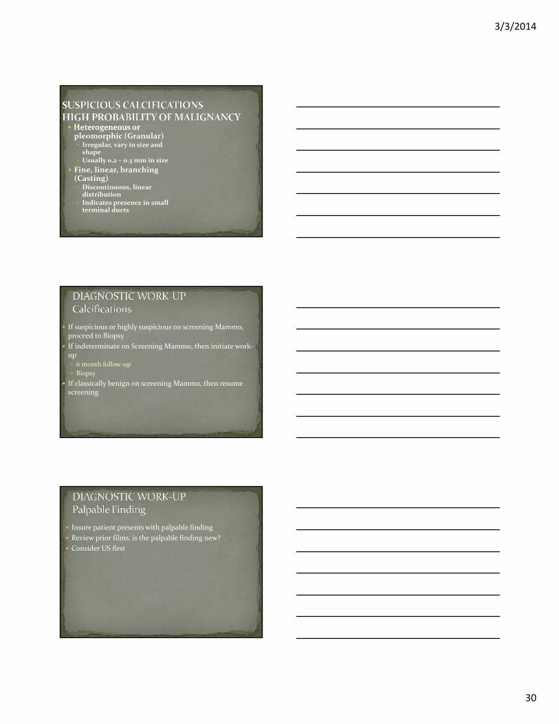
Heterogeneous or pleomorphic (Granular) Irregular, vary in size and shape
Usually 0.2 – 0.3 mm in size
Fine, linear, branching (Casting) Discontinuous, linear distribution
Indicates presence in small terminal ducts
If suspicious or highly suspicious on screening Mammo, proceed to Biopsy
If indeterminate on Screening Mammo, then initiate work‐up 6 month follow‐up
Biopsy
If classically benign on screening Mammo, then resume screening
Insure patient presents with palpable finding
Review prior films, is the palpable finding new?
Consider US first

3/3/2014
31
Excerpt from the poem "Breast Art" by Lisa KatzRaphael`s La Fornarina lives in a Roman palace now,touching her left breast, holding it between thumb and forefingerlike a fruit she wants to prod in the market.Perhaps the artist asked her to demonstratebeckoning a loverplumping up the smaller breastshowing off in front of the mirror.You think she`s coy.Perhaps she wanted to touchthe lump she noticed yesterday.Her eyes look surprised.
10 o’clock LT
3/3/2014
32
Am J Surg. 2003 May;185(5):416-9.
Normal mammography and ultrasonography in the setting of palpable breast cancer.
Beyer T, Moonka R.
Department of General Surgery, Virginia Mason Medical Center, Mailstop C6-SURG, 1100 Ninth Avenue, P.O. Box 900, Seattle, WA 98111, USA.
BACKGROUND: Each year thousands of women present to general surgeons with palpable breast masses, some of which are clinically ambiguous and the majority of which are benign. In addition, surgeons are frequently faced with the question of whether to biopsy those palpable abnormalities in the setting of normal radiographic studies. One might propose that such lesions could be safely observed rather than immediately biopsied. If these lesions were not biopsied, how many cancers would escape detection? To address this issue, a population of patients with known, palpable breast cancer was retrospectively examined to determine the frequency of normal or benign findings on both mammography and ultrasonography.
METHODS: Between January 1998 and December 2001, 351 women with breast carcinoma presented initially with palpable tumors. The medical records of these remaining 351 cases were retrospectively reviewed to examine the radiographic characteristics of the palpable carcinomas. RESULTS: Of the 351 cases in the study group, 13 (3.7%) patients with palpable breast cancers had mammogram and sonogram examinations that were both normal, benign, or nonspecific in appearance. CONCLUSIONS: The results of this study indicate that nearly 4% of women with breast cancer who present with palpable lumps will have normal or benign findings on both mammography and ultrasonography. These data support prior studies of similar false negative rates and may provide some reassurance to surgeons and patients regarding clinical breast lumps, as the decision of whether to biopsy still rests in the surgeon's hands. However, inappropriate reliance on these tests for an evaluation of a palpable abnormality will result in a number of missed tumors.
Breast biopsy avoidance: the value of normal mammograms and normal sonograms in the setting of a palpable lump.
Dennis MA, Parker SH, Klaus AJ, Stavros AT, Kaske TI, Clark SB.
Sally Jobe Breast Centers, Radiology Imaging Associates, 8200 E Belleview Ave, Suite 102, Englewood, CO 80111, USA. [email protected]
PURPOSE: To review the authors' experience with patients who presented with breast lumps and had normal mammograms and normal sonograms. MATERIALS AND METHODS: The findings from 600 lumps in 486 women with no focal ultrasonographic (US) mass or mammographic finding in the area of clinical concern were retrospectively studied. Evaluated parameters included the individual reporting the lump, qualitative descriptors for the physical finding, mammographic density, US characteristics in the area of concern, whether there was a change in imaging and/or physical examination results, and whether there were diagnostic biopsy findings at follow‐up. The study group included 540 lumps in 435 women who had a minimum mammographic and clinical follow‐up of 2 years, as well as 60 additional lumps in 51 patients who underwent biopsy.
3/3/2014
33
RESULTS: No patient in the nonbiopsy group developed carcinoma at the initial site of concern during a mean mammographic and clinical follow‐up period of 43 months, and all biopsy specimens were benign (negative predictive value, 100%).CONCLUSION: Results of this retrospective study suggest that breast biopsy may be avoided in women with palpable abnormalities when both US and mammography depict normal tissue at the lump site.
US first, if suspect lesion then proceed to biopsy
If US normal, +/‐ consider diagnostic mammogram
If imaging negative, follow clinically
FOOD FOR THOUGHT


![多摩市立複合文化施設[パルテノン多摩]parthenon.or.jp/koho/pdf/1311-12.pdfARAJIN Magic Office 042-771-9362 042-374-6142 042-371 -2824 042-371-1558 042-355-21 1 6 042-356-0308](https://img.dokumen.tips/doc/110x75/5fb10d74c5dcf6120d16df17/ceoeefffff-arajin-magic-office-042-771-9362.jpg)