Embed Size (px)
Citation preview

8/14/2019 0081_MICROCIRCULATION MODIFICATIONS BY LOCALIZED MICROWAVE HYPERTHERMIA - 05.pdf
http://slidepdf.com/reader/full/0081microcirculation-modifications-by-localized-microwave-hyperthermia-05pdf 1/5
Bibliotheca Anatomica No 20
Editor: P. Gaehtgens, K61n
Publisher: S. Karger, BaselReprint Printed in Switzerland)
1 lth Europ. Conf. Microcirculation, Garmisch-Partenkirchen 1980
Biblthca anat., No. 20. pp. 628-632 Karger, Basel 1981)
MICROCIRCULATION MODIFICATIONS BY LOCALIZED MICROWAVE
HYPERTHERMIA AND HEMATOPORPHYRIN PHOTOTHERAPY
Haim I. Bicher, Fred W. Hetzel, Peter Vaupel and
Taljit S. Sandhu
Department of Therapeutic Radiology, Henry Ford Hospital,
Detroit, Michigan and the Department of Physiology,
University of Mainz, Mainz, West Germany
Introduction
Due, in part, to increasing clinical interest, active in-vestigation of the physiological phenomena induced by hyper-thermia is in progress (1,2,7,13). The studies by Eddy (7)
and Reinhold (13) employing "chamber systems" have both shownchanges in the microvascular network as a fun ction of tempera-ture and exposure time. Cater et al. (3) reported on changesin tumor oxygen tension with hyperthermia but did not recordchanges in tumor t emperature. Bicher (1,2), in a mouse legtumor system, reported that tumor blood flow increased up to41°C and then decreased to 44°Co The oxygen tension in thetumor, as measured with a platinum electrode, generally fol-lowed the changes in tumor blood flow.
Although blood flow and shifts induced in it by hyper-thermia in both tumor and normal tissue is important, severalother parameters also have significant roles. Several studiesindicate that the pH of interstitial fluid in human and rodentsolid tumors is .3 to .5 un its lower than the normal tissuepH of about 7.4 (8,10).
Reduced pH has also been shown to affect the transplant-
ability of tumor c ells heated in vitro (12). In a recent pa-per, Gerweck has shown (9) that there is a variable influen ceof pH according to temperature and that there is a criticalpoint in t he increased lethality of heat below pH 6.7.
In addition to hyperthermia, Hematoporphyrin derivative(HpD) phototherapy is also showing some promise in clinicalcancer therapy (5,6). This type of therapy employs an inject-able dye (HpD) which is specifically accumulated in some tumorsand/or is specifica ly cleared from nor mal tissues (5,6).When light of specific wavelengths illuminate the dye a photo-chemical reaction takes place yielding the cytotoxic agent
* This work supported in par t by Grant CA25780 from theNational Cancer Institute, DHEW and a grant from the Elsa U.Pardee Foundation

8/14/2019 0081_MICROCIRCULATION MODIFICATIONS BY LOCALIZED MICROWAVE HYPERTHERMIA - 05.pdf
http://slidepdf.com/reader/full/0081microcirculation-modifications-by-localized-microwave-hyperthermia-05pdf 2/5
629
singlet oxygen (7,15). Although the exact mechanism(s) oftumor inactivation have yet to be determined, the rapid anddramatic coloration changes observed in tumors following treat-ment suggest that modification of blood flow with possible con-comitant effects on pO2 and pH may play a prominant role (5).
Materials and Methods
Animal system. All in situ studies were carried out in4th., generation transplants of C3H mammary adenocarcinoma im-planted in the hind leg of C3H SED-BH mice. This is a syn-genetic implantable tumor that is kept at our facility usingsolid tissue transplants that are inoculated subcutaneouslyinto recipient mice. Tumors used for experimentation were ap-proximately 8-10mm in diameter. The mice were anesthetizedduring microelectrode introduction with a combination of Keta-mine 40mg/kg I.M. and Thorazine, 50mg/kg I.M.
Oxygen ultramicroelectrodes. The 02 ultramicroelectrodesused were as described by Cater and colleagues (4). They weremade by~ pulling a glass tube (KG-33, ID 1.5mm, 0D 2.0n~n, Gar-ner Glass Co., Claremont, California), encasing a 20-u goldwire (Sigmund Cohn Corp., Mt. Vernon, New York) in a DavidKopf Model 700C vertical pipette puller. The exposed gold tipis about I0~ i n diameter, and is co ated with a Rhoplex (RhomHaas, Philadelphia, Pennsylvania) membrane as previously des-cribed (2). This probe is used as an "external reference" 02microelectrode.
Electrodes are calibrated as previously described (i) inbuffered saline solutions of known pO2 values. The electrodesare ’conditioned’ by placing them i n buffered saline and ap-plying 0.8 V potential for 2 hrs. After this treatment theyare usually very stable. The current reading at zero oxygentension is very low (.residual current) and the response of themicroelectrode to changes of oxygen tension is very rapid.
In these experiments a polarizing voltage of 0.6 V hasbeen used. The relation between current output and oxygentension is linear, the current per mm Hg being of the orderof magnitude of 0.6 x 10⁻¹¹A.
pH ultramicroelectrodes. Designs for glass pH micro-electrodes employed .in this study have been developed, mostnotably by Hinke (ii). The Hinke-type electrode consists ofa pH sensitive glass micropipette inside a pyrex glass pipettewith the tip of the pH sensitive micropipette extruded from thepyrex glass pipette. A silver silver chloride electrode isinserted into the electrod e stem which is filled with 0.I N HCI.This type of microelectrode with an exposed tip has an almostinstantaneous response time. This is an advantage over othertypes of microelectrodes in which a recessed tip may cause aresponse time of up to several minutes.
Temperature determinations. Tumor and mouse core tem-peratures were recorded using Copper-Constantan microthermo-couples (tip diameter 30-100 microns - MEDTRA Inc.) inserted
8/14/2019 0081_MICROCIRCULATION MODIFICATIONS BY LOCALIZED MICROWAVE HYPERTHERMIA - 05.pdf
http://slidepdf.com/reader/full/0081microcirculation-modifications-by-localized-microwave-hyperthermia-05pdf 3/5
63 0
C 3 H M O U S E M A M M A R Y C A R C I N O M A
F R E Q U E N C Y
Hpd Phototherapy
C3 H MOUSE MAMMARY CARCINOMA
Hyperthermla
F R E Q U E N C Y
Hpd Phototherapy
1 2 3 4 5
l~ssue~02 rnmHg)
5 4 5 6 58 6 0 6 2 6 4 6 6 6 8 7 0 72
tissue pH
C O N T R O Ln=96
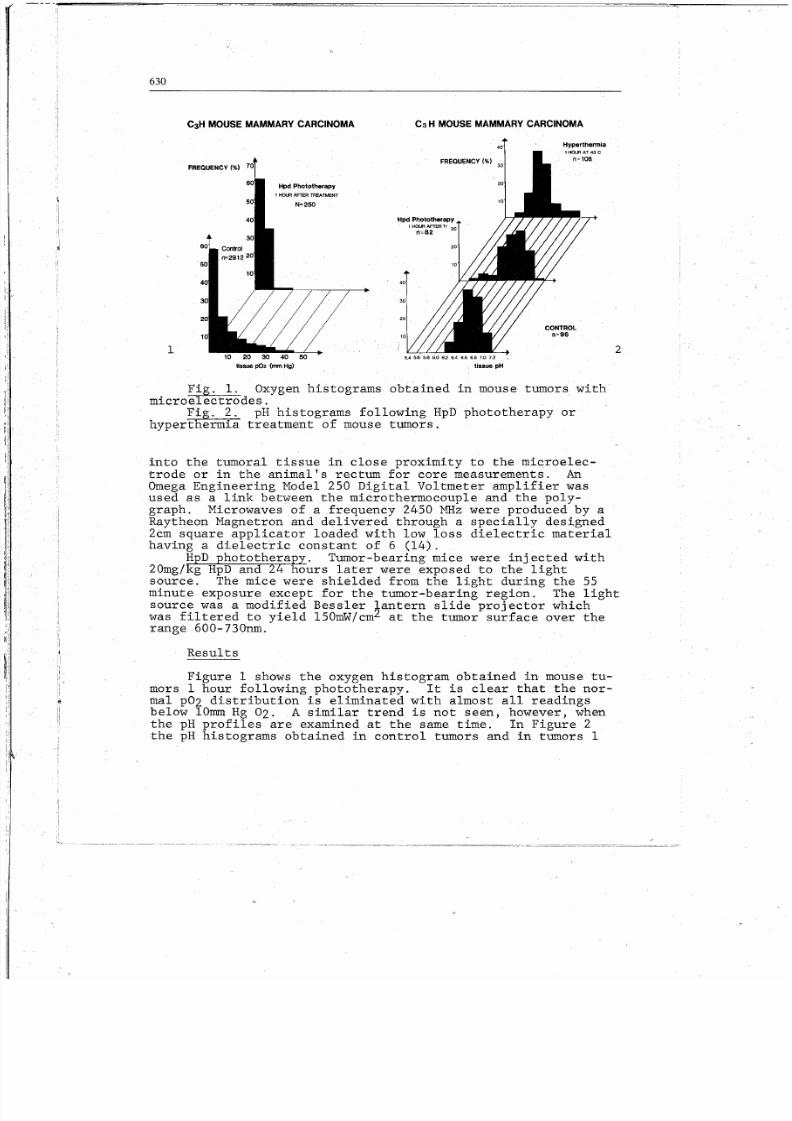
Fig. i. Oxygen histograms obtained in mouse tumors withmicroelectrodes.
Fig. 2. pH histograms following HpD phototherapy orhyperthermia treatment of mouse tumors.
into the tumoral tissue in close proximity to the microelec-trode or in the animal’s rectum for c ore measurements. AnOmega Engineering Model 250 Digital Voltmeter amplifier wasused as a link between the microthermocouple and the poly-graph. Microwaves of a frequency 2450 MHz were produced by aRaytheon Magnetron and delivered through a specially designed2cm square applicator loaded with low loss dielectric materialhaving a dielectric constant of 6 (14).
HpD phototherapy. Tumor-bearing mice were injected with
20mg/kg HpD and 24 hours later were exposed to the lightsource. The mice were shielded from the light dur ing the 55minute exposure except for the tumor-bearing region. The lightsource was a modified Bessler ~antern slide projector whichwas filtered to yield 150mW/cmz at the tumor surface over therange 600-730nm.
Results
Figure 1 shows the oxygen histogram obtained in mouse tu-mors 1 hour following phototherapy. It is clear that the nor-mal pO2 distribution is eliminated with almost all readingsbelow 10mm Hg 02. A similar trend is not seen, however, whenthe pH profiles are examined at the same time. In Figure 2the pH histograms obtained in control tumors and in tumors 1

8/14/2019 0081_MICROCIRCULATION MODIFICATIONS BY LOCALIZED MICROWAVE HYPERTHERMIA - 05.pdf
http://slidepdf.com/reader/full/0081microcirculation-modifications-by-localized-microwave-hyperthermia-05pdf 4/5
631
hour following either 43oc hyperthermia or HpD phototherapyare presented. In this case there is a dramatic shift in pHfrom a mean of 6.8 ÷ 0.2 in contr ols to 6.2 ÷ 0.2 followinghyperthermia. Ther~ is also a trend to lowe~ pH followingphototherapy but the shift is not significant.
Discussion
Dstermination of the mode of tumor inactivation by atreatment modality is critical to its development and futureuse. It has been shown that hyperthermia has many possibleeffects on cell survival either alone or in combination withradiation (9,12,13). It has also been shown to dramaticallymodify blood flow and oxygenation within tumors (1,2). Theresults presented here indicate that a significant reductionin pH is induced by hyperthermia which may result in a signi-ficant increase in cell killing within the tumor (9). It ispossible that this observed pH shift is due to a combinationof hyperthermia stimulated cellular metabolic activity and thesimultaneous reduction in tumor blood flow which is observedat the treatment temperature (i).
The effects seen on pH and pO2 following HpD phototherapyare quite different from those following hyperthermia. It isclear that there is a sharp reduction in pO2 at all areaswithin the tumor without any significant sh~ft in pH. Al-though further studies are now in progress, it is possible tospeculate on the meaning of the results presented here. Mas-sive coagulation necrosis within tumors is reported to followHpD phototherapy (5). It is likely that the cells most af-fected by this treatment are the vascular endot helial cellsof the tumor microvasculature. Their destruction would resultin the observed coagulation necrosis with an abrupt reductionin blood flow. This would result in the low levels of tissue
oxygenation reported here. Since there is no cellular meta-bolic stimulation with this modality and there is direct cyto-toxicity (15) no dramatic shift in pH would be expected andnone was observed.
Further studies are currently in progress to further ex-amine the effects of hyperthermia and HpD phototherapy on tu-mor microphysiology.
References
BICHER, H.I.; HETZEL, F.W.; SANDHU, T.S.; FRINAK, S.;VAUPEL, P.; O’HARA, M.D., and O’BRIEN, T. : Effects ofhyperthermia on normal and tumor microenvironment.Radiology, In Press (1980).
BICHER0 H.I.: Increase ~n brain tissue oxygen availabilityinduced by localized microwave hyperthermia, in SILVER,

8/14/2019 0081_MICROCIRCULATION MODIFICATIONS BY LOCALIZED MICROWAVE HYPERTHERMIA - 05.pdf
http://slidepdf.com/reader/full/0081microcirculation-modifications-by-localized-microwave-hyperthermia-05pdf 5/5
63 2
ERECINSKA, and BICHER, Oxygen transport to tissue;Vol. III, pp. 347-353 (Plenum Press, New York 1978).
CATER, D.B., and SILVER, I.A.: Quantitative measurements ofoxygen tension in normal tissues and in the tumors ofpatients before and after radiotherapy. Acta. Radio-logica. 53: 233-256 (.1960).
CATER, D.B.; SILVER, I.A., and WILSON, G.M.: Apparatus andtechnique for the quantitative measurement of oxygentension in living tissues. Proc. Roy. Soc., Lond.(Series B) 151: 256-276 (1959-1960).
DIAMOND, I.; McDONAGH, A.F.; WILSON, C.B.; GRANELLI, S.G.;NIELSEN, S., and JAENICKE, R.: Photodynamic therapy ofmalignant tumours. Lancet, 2: 1175-1177 1972).
DOUGHERTY, T.J.; KAUFMAN, J.E.; G~LDFARB, A.; WEISHAUPF, K.R.;BOYLE, D., and MITTLEMAN, A.: Photoradiation therapyfor the treatment of malignant tumors. Cancer Res. 38:2628-2633 (1978). --
EDDY, H.A.: Alterations in tumor microvasculature duringhyperthermia. Radiology, In Press (1980).
EDEN, M., and KAHLER, H.: The p--~ o--~ rat tumors measuredin vivo. .J. Nat’l. Cancer Inst. 16: 541-556 (1955).
GERWECK, L.E.: Modification of cell l~hality at elevatedtemperatures: The pH effect. Radiat. Res. 70: 224-235(1977).
GULL~NO, P.M.; GRANTHAM, F.M., and SMITH, S.H.: Modificationsof the acid-base status of the internal milieu of tumors.J. Nat’l. Cancer Inst. 34: 857-860 (1965).
HINKE, J.A.: Cation-selecti~-~microelectrodes for intra-cellulmr use. in EISERMAN, Glass electrodes for hydrogenand other cati ons; (Marcel Dekker, Inc., New York 1973).
OVERGAARD, J.: Influence of extracellular pH on the viabilityand morphology of tumor cells exposed to hyperthermia.J. Nat’l. Cancer Inst. 56: 1243-1250 (1976).
REINHOLD, H.S.; BLACHIEWICZ B. and BERG-BLOK, A.V.D.:Decrease in tumor microcirculation duri ng hyperthermia.in STREFFER, Cancer therapy by hyperthermia and radi-ation; pp. 231-232 (Urban and Schwarzenburg, Baltimore
1 9 7 8SANDHU, T.S.; KOWAL, H.S., and JOHNSON, R.: Development ofhyperthermia applicators. Int. J. Radiat. Oncol. Biol.Phys. 4: 515-519 1978).
WEISHAUPT, ~.R.; GOMER, C.J., and DOUGHERTY, T.J.: Identifi-cation of singlet oxygen as the cytotoxic agent in photo-inactivation of a murine tumor. Cancer Res. 36:2326-2329 (1976).
Haim I. Bicher, M.D., Ph.D., Henry Ford Hospital,2799 West Grand Blvd., Therapeutic Radiology, E&R Bldg.,Room 3056, Detroit, Michigan 48202 (USA)


![Malignant hyperthermia [final]](https://img.dokumen.tips/doc/110x75/58ceb1b71a28abb2218b5123/malignant-hyperthermia-final.jpg)