Embed Size (px)
Citation preview

1
001 WHEEZING SYMPTOMS – DIAGNOSTIC APPROACH IN PRESCHOOL CHILD Saračević E. Clinical Centre University of Sarajevo, Pediatric Clinic, Sarajevo, Bosnia Herzegovina Wheezing (continuous, coarse, whistling sound, crunch, squeak) could be a symptom (may be heard by parents and large children) or physical objective finding during chest auscultation. Often it’s associate with coughing. Phenomena of wheezing indicate existing airway obstruction, especially in middle and small airways. Wheezing came into existence of bronchoconstriction caused by inflammatory mediators and activation cholinergic nerves, bronchial wall callosity (cells infiltration, exudation, extracellular matrix disposal, smooth muscle hypertrophy) and excessive mucus production. Generally, whistling sound appears when turbulent flow arise from bronchoconstriction, specially small airways, with accumulation air in alveolus, that accessory musculature attempt to push out with amplified contraction and result in increasing peribronchial pressure, with consequent reduction of airway radius, for one half or one third. Consequently on structure and small airway volume, wheezing is a common presenting symptom in infant and small children than adults. In tracheal or large airway obstruction wheezing appears as result of stenosis or pressure on airways wall. Epidemiologic studies conducted worldwide have shown that 49-50 percent of children younger than six years of age have a few episodes of wheezing, which is in the most common cases transitory, ending in preschool and early school age. However, great number of children has periodic or often reverse episodes of obstruction, with wheezing, cough and dispnoe, which is easy to diagnosis as asthma in older children and quite heard in small children with different types of whistling sound. The differential diagnosis of wheezing includes a variety of congenital and acquired condition and it is important to distinguish whether it is variable phenomena of one disease or different diseases, conditions with similar manifestations. Basic types of wheezing were defined by study of F. Martines at all. on three groups:
1. Transient early wheezes 2. Late onset wheezes 3. Persistent wheezes
Transient early wheezes First episodes appear in first year of life, with prevalence between second and third year and ending till sixth year. These children have no atopic manifestations, as well as their parents. This syndrome is associated with airway's anatomy, premature, male gender, maternal smoking, and viral infection (RSV). Non atopical wheezing caused by viral infection begins during first year of life and it's not related with inflammation and airway hyperresponsiveness. Late onset wheezes First episode appears after third year and wheezing persists after age of six. These children have normal lung's function. During infant period have negative atopic parameters and in sixth year appear signs of sensibilitations on inhalatory allergens. Persistent wheezes These types appear in 14 percent children till sixth year. Most often, first episode appears during first year and continue after sixth, with progress in early school period. Lungs function shows characteristic obstructive disorders ( ↓ FEV1, PEF, FEF 25-75). Often is some atopic disease present (asthma, eczema, allergic rhinitis) with positive atopic markers ( ↑Eo, ↑IgE, ↑ECP ) and sensibilization on inhalatory allergens. The differential diagnosis of wheezing according to age and period of disease appearance:

2
Obstruction of upper airways with extended expiratory phase and inspiratory stridor: • Upper airways infection • Congenital laryngeal stridor • Croup syndrome • Vocal cord dysfunction ( hoarse voice) • Tumor
Obstruction of lower airways with difficult and extended expiratory phase, high tone, polyphonic wheezing: A) Trachea and bronchi:
• Congenital anomalies • GERD • Infection • Foreign body • Allergy • Hemangioma and other tumors
B) Small airways: • Asthma • Bronchiolitis • BPD • Pneumonia • Bronchiectasiae • Cilliar dyskinesia • Cystic fibrosis • Congenital heart diseases
On level of lung's tissue: • Congenital lobar emphysema • Alpha 1 antitrypsin deficit • Circulation disorders: pulmonary edema, hemorrhage, hyper circulation L-R
shunt • Lung's cyst • Lung's sequestration
Out lung’s disease: • Amiotonia congenital • Poliomyelitis • Poliradiculitis • Dystrophy musculorum
According to age and period of disease appearance in neonatal period wheezing is manifestation of bronhopulmonary dysplasia (BPD), congenital heart anomalies (CHA) and laryngomalation. During infant period (3 month): congenital airways anomalies, CHA, laryngotraheomalation, vascular ring, aspiration of milk, bronchiolitis. During first year of life: bronchiolitis, asthma, cystic fibrosis, aspiration, tracheobronchomalation, passive smoking, cilia's dyskinesia, CHA, immunodeficiency. During preschool period: asthma, cystic fibrosis, cilia's dyskinesia, foreign body, croup syndrome, bronchiolitis, tuberculosis, GERD, congenital anomalies-rare in this period. ASTHMA Asthma is a common chronic disorder of the airways that is complex and characterized by variable and recurring symptoms, airflow obstruction, bronchial hyper responsiveness and an

3
underlying inflammation. The most common repeating of wheezing in small children is associated with diagnosis of asthma, although is difficult to nominate diagnosis asthma, because we don’t have objective parameters for following chronic inflammation as well as difficulties with histological determination bronchial hyperactivities. In this period of life asthma could be named as multifactorial wheezing. PRACTALL study definite phenotype as crucial moment in context “asthma syndrome” for children age till 2 years and from second year till school period for diagnosis of asthma. For diagnosis of asthma in period 0-2 years are required three episodes of recurrent bronchoobstruction in period of six months or in last three months most part of day during one week to persist wheezing. In preschool children (3-5 years) key for define phenotype of asthma is persisting wheezing during last year. Classification of asthma according to PRACTALL study: virus induced, effort induced, allergen induced and unresolved asthma- require once a year evaluation, trying to detect trigger. It is necessary to nominate diagnosis in short time and start with treatment, because of remodeling process that leads to irrecoverable changes in bronchial structure with degradation lung's function. Nominate of diagnosis includes: past medical history, identification cause, atopic familiar constitution, physical examination (nasal symptoms, allergic salute, allergic crease, dermatitis, dry skin, allergic shiners, irritated conjunctive). Acute phase of bronchoobstruction: dyspnoea, activation of accessory musculature, with extended expiratory phase. In older children - measuring lung's function, especially reversible obstruction, provides argument restrictive air flow. Allergic diagnostic evaluation begins with skin prick test and continues with count IgE, Eo in FBC and nose, ECP, FENO and leucotriens. Differential diagnosis of asthma:
• Aspiration • Cystic fibrosis • Immunodeficiency • Tuberculosis • Chronically sinusitis • Recurrent infections of lower airways • Congenital heart anomalies • GERD
In case of persisting recurrent wheezing, without answer on treatment, then is fiberoptic bronchoscopy with bronchoalveolar lavage, X rays of lungs and sinuses, CT scan, esophageal pH probing, Cl in sweat, CFTR genes, ultrasound of heart, immunology and microbiology status. References: 1. Ivković-Jureković I. Recidvno piskanje i astma male djece. Paediatr Croat 2007;51 (Supl1):70-74 2. Martinez FD, Wright AL, Taussig LM, Holberg CJ, Halonen M, Morgan WJ. Asthma and wheezing in the first six years of life. N Engl J Med 1995; 332:133-8. 3. Lowe L, Čustović A, Woodcock A. Childhood asthma. Curr Allergy Asthma Resp. 2004; 4:159-65 4. Nestorović B i sar. Pedijatrijska pulmologija ed. Beograd 2008; 39-42. 5. Bacharier L.B, et al. Diagnosis and treatment of asthma in childhood. PRACTALL consensus report. Allergy 2008; 63:5-34. 6. Rivera-Spoljarić K et al. Sings and symptoms that precede wheezing in children with a Pattern of moderate –to-severe intermittent wheezing. J Pediatr 2009; 154: 877-81.

4
002 SEASONAL AND SPATIAL VARIATIONS OF POLLEN ALLERGENS IN SARAJEVO REGION Redžić S1, Mehić B.2 1Laboratory for Aerobiology, Faculty of Science, University of Sarajevo, Sarajevo, Bosnia Herzegovina and Academy of Sciences & Arts of Bosnia and Herzegovina, Sarajevo, Bosnia Herzegovina 2Clinical Centre University of Sarajevo, Clinic of Lung Diseases and TB, Sarajevo, Bosnia Herzegovina Pollen, together with spores of plant is one of the most common causes of allergies in the population structure of different age groups (1-3). Pollen is present in huge quantities in all parts of the environment of man. In addition, the pollen is continuously present in the atmosphere, just as its concentration varies during the season and the month and during the day (4-5). Increased the concentration of pollen and significant oscillations during the vegetation season, contributing to air pollution and various pollutants (SOx, NOx, COx), and all visible climate change (6). Because of this, we have a growing number of people allergic on pollen and spore plants and mushrooms. That has influence on modern way of life. The number allergic people on pollen in particular have been growing in urbane and industrial areas (7-9). Pollen is microspore the flowering plants in the course of more meiotic division formed sperm cells. Pollen has a specific structure and biochemical composition of which largely depends his allergencity. Pollen of allergenic species has the following key features: (i) the proteins in their walls and cytoplasm, which usually initiate allergic reaction in sensitive persons, (ii) the shape, size and weight suitable for atmospheric transport, (iii) a sufficient concentration in the atmosphere that can cause allergic reaction after inhalation. In mature pollen grains, are represented by the following substances: hydrocarbons, lipids, proteins, amino-acids, enzymes, vitamins, pigments and minerals (10). Due to the presence of pollen in the atmosphere of environment there are more and more people who suffer from allergies caused by pollen. It is anticipated that the number of people suffering only from allergic rhinitis be doubled every 10 years (11). High doses of ultraviolet radiation and polluted air caused changes in the structure of pollen grain, which has resulted in increased number cytosolic of allergenic proteins, and of course the bigger imunogenity of pollen. Diseases most often caused by pollen are: (i) allergic rhinitis (ii) allergic asthma (iii) nettle rash and (iv) atopic dermatitis. Allergic rhinitis is mostly pollinosis. It’s defined by symptoms such as nose mucous membrane disorder that occurs after contact with allergens from the environment in basis of which are inflammatory reactions. According to data from the WHO 10-25% of the population in the world suffering from some form of allergic rhinitis. Depending on the allergens that caused the allergy, simptomatology may be seasonal and perennial (lasts all year round). The most common symptoms of allergic rhinitis include: sneezing, stuffy nose, and leaking water from sewage-nosed, itching and redness nose and eyes, a feeling of constriction and pricking the eyes and irritating cough and a feeling of scratching in throat. From seasonal allergies suffer 15% of people, usually those between 25 and 40 years. The disease usually begins in childhood or puberty with deterioration during the three - four seasons, and often is not associated with the amount of allergens in the atmosphere. In the later era of the situation stabilizes. With ageing the intensity of illness weakling (11). In the younger age there is a difference in sex. Girls get sick rare, but usually they have the more difficult forms of allergies than boys. In a sample of 120 children in the age between 1 and 14 years, it was found that the number of sick male child’s is twice larger than the female. In addition, the higher the frequency of atopic’s his mother's side and if it is sensitive

5
to particular allergens there is five times greater chance that her children will inherit this "feature", but when the father of the word (12). It can be said that there is a certain relationship between allergic rhinitis and bronchial asthma. Studies have shown that 3/4 suffering from asthma has symptoms allergic rhinitis, 1/ 5 patients has allergic rhinitis with asthma, while 1/4 of patients have both diseases simultaneously. Air pollution has a major role in the development and other diseases othorinolaringologic areas such as infection of sinuses and ears. In the treatment of allergic diseases apply to many drugs from different pharmacologic groups, but particular concern is avoiding contact with the sources and triggers of pollen allergies. One way to achieve this state of prevention is based on information on the type and concentration of pollen in the working and life environment (11). In order to improve the quality of life of allergic persons crucial preventive informing them about the impression pollen allergens in the area where they live and work. Therefore monitoring aero pollen (type and concentration) during pollen season is of special importance. Because of monitoring occurrence of certain triggers pollen allergy many European countries began to implement the so-called "Pollen monitoring”. The primary objective of this work is the assessment of the importance of "pollen monitoring" (adequate pollen identification and determination of its concentration in m3 in the troposphere, in which a man takes his life's activities), and to develop effective prevention measures with timely and effective treatment of different pollinosis, with special emphasis on the area of Sarajevo. Methodology sampling pollen from the atmosphere is modern and compared, and based on sampling, standardized, easily repeatable methods (13). 1986 was founded database European Aeroallergen Network (EAN), in Vienna, which gathers data on the concentration of pollen from the so-called. "Monitoring unit, situated in almost all countries of Europe”. From here the data on the impression, the types and concentration of pollen in the air, sent weekly to the national centre, which forwarded the information to the European Coordination Centre. Samples are collected Hirst-type sampler (Lanzoni, Bologna, Italy) that catches pollen and spore (inhalation allergens) actively absorbs of 10 L of air per minute in diameter 10 - 30 km, depending on the directions of wind and other meteorological conditions of investigated area. In Sarajevo, sampler (mantrap pollen) was placed on the flat roof of the building Faculty of Science, University of Sarajevo to the standard prescribed height (about 16m), in order to obtain a representative sample of pollen of all plants in observed region. The sampler has a special gluing tape. It is a silicone coatings solution and winding the carrier tape, and then set the device. The bar moves 2 mm every two hours. Replacement tape is done every seven days and always at the same time. When you strip off the appropriate time can be made on the preparation of the preparations for qualitative and quantitative analysis. "Melinex" strips are cut on a table with time division, and each segment represents 1 day, i.e. 24 h sampling. The bar freezes in gelvatol or glycerol with fuchinom at the glass. Thus prepared preparations are analyzed light microscope with an increase of 400 X. Pollen concentration is expressed as the number of pollen grains / m3 of air and is classified as: absence of pollen, low, medium high, high and very high concentration of tree pollen, weed or grass (Table 1). The results that are presented in this paper were achieved during 2005-2008. Special attention is devoted to results from 2008.

6
Table 1. Pollen Rating Scale (PRS) related to Pollen Density (grains per meter3)
RATING
POLLEN DENSITY (in m3 of air) PRS Trees Grasses Weeds
Absence of pollen 0 0 0 0 Low concentration of pollen 1 - 25 1 - 14 1 - 4 1 - 9 Moderate concentration of pollen 26 - 50 15 - 89 5 - 19 10- 49 High concentration of pollen 51 - 75 90 - 1499 20- 199 50 - 499 Very high concentration of pollen > = 76 > = 1500 > = 200 > = 500
Source: Forsyth County Environmental Affairs department Pollen Rating Scale -National Allergy Bureau (NAB) of the American Academy of Allergy, Asthma & Immunology (AAAAI) (www.co.forsyth.nc.us/envAffairs/pollen.)
Results and Discussion
The ancient city of Sarajevo (diameter approximately 30 km) in protocol period determined by pollen which belongs to a large number of species of which the majority has allergenic character. Identified over 30 different species of plants or groups (genera and family). There dominated the pollen of trees (20 species including the genera and family), and weed pollen and herbaceous plants (10 species or group) and grass pollen (more species from two families Poaceae and Cyperaceae (Table 2). Table 2. List of allergenic plants of Sarajevo region in 2008.
Plant species Intensity of allergic activity Latin name Local name English name
TREES: Taxaceae (Taxus sp.) tise jew Moderate Cupresaceae (Juniperus sp, Thuja sp, Chamaecyparis sp) kleke juniper Moderate Pinaceae (Pinus sp, Picea sp.)
četinari -borovi smrča
coniferous, pine, spruce Low
Corylus sp. lijeska hazelnut Moderate Alnus sp. joha alder Moderate to high Betula sp. breza birch high Carpinus sp. grab hornbeam Moderate Populus sp. topola polar / aspen Low to moderate Salix sp. vrba willow Low Ulmus sp. brijest elm Moderate Acer sp. javor maple / sycamore Moderate Fraxinus sp. jasen ash Low to moderate Fagus sp. bukva beech Moderate
Quercus sp. hrast oak Not enough is known
Aesculus hippocastanum divlji kesten horse - chestnut Low Platanus sp. platan plane tree Low
Juglans sp. orah walnut Not enough is known
Tilia sp. lipa lime / linde Not enough is
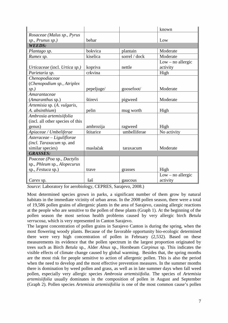
7
known Rosaceae (Malus sp., Pyrus sp., Prunus sp.) behar Low WEEDS: Plantago sp. bokvica plantain Moderate Rumex sp. kiselica sorrel / dock Moderate
Urticaceae (incl. Urtica sp.) kopriva nettle Low – no allergic activity
Parietaria sp. crkvina High Chenopodiaceae (Chenopodium sp., Atriplex sp.) pepeljuge/ goosefoot/ Moderate Amarantaceae (Amaranthus sp.) štirevi pigweed Moderate Artemisia sp. (A. vulgaris, A. absinthium) pelin mug worth High Ambrosia artemisiifolia (incl. all other species of this genus) ambrozija ragweed High Apiaceae / Umbeliferae štitarice umbelliferae No activity Asteraceae – Liguliflorae (incl. Taraxacum sp. and similar species) maslačak taraxacum Moderate GRASSES: Poaceae (Poa sp., Dactylis sp., Phleum sp., Alopecurus sp., Festuca sp.) trave grasses High
Carex sp. šaš gaucous Low – no allergic activity
Source: Laboratory for aerobiology, CEPRES, Sarajevo, 2008.)
Most determined species grown in parks, a significant number of them grow by natural habitats in the immediate vicinity of urban areas. In the 2008 pollen season, there were a total of 19,586 pollen grains of allergenic plants in the area of Sarajevo, causing allergic reactions at the people who are sensitive to the pollen of these plants (Graph 1). At the beginning of the pollen season the most serious health problems caused by very allergic birch Betula verrucosa, which is very represented in Canton Sarajevo. The largest concentration of pollen grains in Sarajevo Canton is during the spring, when the most flowering woody plants. Because of the favorable opportunity bio-ecologic determined there were very high concentration of pollen in February (2,532). Based on these measurements its evidence that the pollen spectrum in the largest proportion originated by trees such as Birch Betula sp., Alder Alnus sp., Hornbeam Carpinus sp. This indicates the visible effects of climate change caused by global warming. Besides that, the spring months are the most risk for people sensitive to action of allergenic pollen. This is also the period when the need to develop and the most effective prevention measures. In the summer months there is domination by weed pollen and grass, as well as in late summer days when fall weed pollen, especially very allergic species Ambrosia artemisiifolia. The species of Artemisia artemisiifolia usually dominates in the composition of pollen in August and September (Graph 2). Pollen species Artemisia artemisifoliia is one of the most common cause’s pollen

8
allergies watched very serious obstructions respiratory systems especially in children and the elderly (14-16). Graph 1. Number of pollen grains per month’s in 2008.
10
2532
3125
3768
3458
2450
2100
1568
560
15
0 500 1000 1500 2000 2500 3000 3500 4000
number of pollen granes
January
February
March
April
May
June
July
August
September
October
Graph 2: Spectrum of selected allergenic plants in September 2008
Poaceae7%
Plantago10%
Artemisia10%
Ambrosia73%
Research shows that there is expressed a connection between the concentration and types of pollen and number suffering from some pollinosis. During 2008 there have been observed 598 people sensitive to pollen allergenic plants in Canton Sarajevo (Table 3). With a specific form of pollens allergies found 115 people or 19.23%. The similar proportions of allergic persons at the pollen season in the sample of children of Sarajevo Canton found by the other authors (3).

9
Table 3. Frequency of pollen allergies caused by allergic weed plants in certain months in the area of Canton Sarajevo
MONTH
NUMBER OF TESTED PERSONS
NUMBER SUFFERING FROM ALLERGIES
(tested by specific allergen of mixed pollens
of weeds –wx3)
PROPORTION
(%)
January 49 5 10,2% February 48 5 10,4% March 50 9 18,0% April 58 14 24,1% May 39 6 15,4% June 41 10 24,4% July 50 16 32,0% August 40 5 12,5% September 59 17 28,8% October 80 17 21,2 % November 40 6 15,0% December 35 5 14,3%
Very high proportion (20%) suffering fortified in spring months when dominated by pollen of trees, and in September when the dominant pollen was Ambrosia artemisifolia (Graph 2). The largest proportion of persons allergic on pollen is in the July (Graph 3) when dominating the pollens of weeds and grasses. From weed pollens there dominated the pollens by herbaceous species from the family Chenopodiaceae, Plantaginaceae, Amaranthaceae, Asteraceae. From the grass there were the species of genera: Poa, Festuca, Phleum, Alopecurus, summer forms of species Dactilys glomerata, species of the family Poaceae, especially Phleum pratense that causes severe allergies in other geographic areas (17).
Graph 3. Proportion of pollen allergy at the population of Canton Sarajevo in 2008.
10,20%
10,40%
18,00%
24,10%
15,40%
24,40%
32,00%
12,50%
28,80%
21,20%
15,00%
14,30%
0,00% 5,00% 10,00% 15,00% 20,00% 25,00% 30,00% 35,00%
January
February
March
April
May
June
July
August
September
October
November
December

10
By the table 4 only in November and December there are no pollen in the atmosphere. In January and October we have very low concentration of pollen. The highest concentrations of pollens are from February - August (Tab. 4. to compare with table 1).
Table 4. Seasonal pollen density of Sarajevo region in 2008.
Month
Number of pollen grains Average/day
January 10 - February 2532 90.43 March 3125 100.81 April 3768 125.6 May 3458 111.55 June 2450 81.67 July 2100 67.74 August 1568 50.58 September 560 18.67 October 15 - Total: 19586 Average 1632.17
High pollen concentration in this period caused frequent pollen allergies in more than 20% of persons who shown reactions to pollen. A similar situation exists in the ecological and geographical related areas - Croatia (18, 19), and the countries of Western and Central Europe (20-23). Concentration of pollens of these species depends on phenophases some species, and especially from the meteorological and environmental conditions of certain areas (24). Determination of period’s pollination of allergic plants with the aim producing calendar of pollination is a very significant task in the prevention pollinoses, especially in high-risk groups such as children and the elderly (25-27). Conclusion
Studies of spatial and seasonal variations allergenic pollen from plants in the area of Sarajevo show high concentrations during vegetation season. In the period from 2005-2008 the total number of pollen grains allergenic plants varies from 15 000 - 20 000 per season. In 2008 year (February - September) found 19 586 pollen grains allergenic plants in an atmosphere of Sarajevo. The average monthly value was 1 632 grains. Daily average range was from 18 grains per m3 in February to 125 grains per m3 in April when we noted the highest concentration of pollen allergenic plants. In the air samples has been elaborated pollen several species of plants. Group of trees belonging to 20 species, grass species from the families: Poaceae and Cyperaceae, and weed ten species. From groups of trees are the most important species from the family Betulaceae: Corylus avellana, C. colurna, Betula verrucosa, Carpinus betulus, and Alnus glutinosa. Pollen of these species has a high concentration and causes a very serious pollinoses in the majority of people sensitive to pollens. From weed dominated species by the family Chenopodiaceae, Plantaginaceae, Amaranthaceae and Asteraceae. Very high concentrations achieved Ambrosia sp. what causes the most serious allergic reactions. From the grass to the species of genera: Dactylis, Poa, Festuca, Phleum and Alopecurus. Pollen season in Sarajevo area usually starts in February and duration till end of September, although the pollen grains present in October, which depends on meteorological conditions. In the spring (February-April) there has been dominated by pollen from trees, family Betulaceae,

11
Salicaceae, Platanaceae, Ulmaceae, Oleaceae, Fagaceae, Tiliaceae, Hippocastanaceae, and Pinaceae and Cupressaceae. In May start the phase of herbs, especially species from the genera Dactilys, Phleum, Poa, Festuca and Alopecurus. From June to September there is period pollination of weed from the family Plantaginaceae, Chenopodiaceae, Amaranthaceae and Compositae. The highest concentration of Ambrosia pollen is during August and September. In addition there is necessary the close cooperation laboratories for aerobiology with the public health sector. Only on this way we can successfully prevent the pollen allergies.
Acknowledgments: The authors are very grateful to Ms. Nezafeta Sejdić, BSc. former associates of Laboratory of Aerobiology of Faculty of Science University of Sarajevo to assist in implementation of the project "Monitoring of pollen allergenic plants of Sarajevo region." We are grateful for and Sedik Velić, technical assistant for kind technical support. References:
1. Cvitanović S., Znaor L., Kanceljak-Macan B., Macan J., Gudelj I., Grbić D. Allergic rhinitis and asthma in southern Croatia: impact of sensitization to Ambrosia elatior. Croat. Med. J. 2007; 48(1): 68-75.
2. Renkonen J., Mattila P., Lehti S., Mäkinen J., Sormunen R., Tervo T., Paavonen T., Renkonen R. Birch pollen allergen Bet v 1 binds to and is transported through conjunctival epithelium in allergic patients. Allergy. 2009; 64: 868-875.
3. Saracević E., Redžić S., Telačević A. The frequency of pollen allergy at the population of Sarajevo region during the 2002 year. Med. Arh. 2005; 59(4): 221-223.
4. Epton M.J., Martin I.R., Graham P., Healy P.E., Smith H., Balasubramaniam R., Harvey I.C., Fountain D.W., Hedley J., Town G.I. Climate and aeroallergen levels in asthma: a 12 month prospective study. Thorax. 1997; 52(6): 528-34.
5. Peternel R, Srnec L, Čulig J, Hrga I, Hercog P. Poaceae pollen in the atmosphere of Zagreb (Croatia), 2002-2005. Grana 2006; 45(2): 130-136. Subiza J., Jerez M., Jiménez JA., Narganes M.J., Cabrera M., Varela S., Subiza E. Allergenic pollen pollinosis in Madrid. J. Allergy. Clin. Immunol. 1995; 96 (1):15-23.
6. de Weger L.A., van der Linden A.C., Terreehorst I., van der Slikke W.J., van Vliet A.J., Hiemstra P.S. Ambrosia in the Netherlands. Allergic sensitisation and the distribution of plants and pollen. Ned. Tijdschr. Geneeskd. 2009; 153(17): 798-803.
7. Peternel R., Milanović SM., Hrga I., Mileta T., Culig J. Incidence of Betulaceae pollen and pollinosis in Zagreb, Croatia, 2002-2005. Ann. Agric. Environ. Med. 2007; 14(1):.87-91.
8. Popovic-Grle S., Vrbica Z., Jankovic M., Klaric I. Different phenotypes of intermittent and persistent respiratory allergy in Zagreb, Croatia. Ann. Agric. Environ. Med. 2009; 16 (1): 137-142.
9. Krell R. Value-added products from beekeeping. FAO Agricultural Services Bulletin No. 124, Food and Agriculture Organization of the United Nations Rome, 1996.
10. Schulte F., Lingott J., Panne U., Kneipp J. Chemical Characterization and Classification of Pollen. Anal. Chem. 2008 Nov 1. [Epub ahead of print].
11. Raos M., Dodig S., Klancir SB., Kovač K. Peludna hunjavica u dječijoj dobi. Pediatr. Croat. 1999; 43 (1): [http://www.paedcro.com; July 16, 2009].
12. Durham O.C. A proposed standard method of gravity sampling counting and volumetric interpolation of results. J. Allergy. 1946; 17:79-86.
13. Peternel R., Čulig J., Hrga I., Hercog P. Airborne ragweed (Ambrosia artemisiifolia L.) pollen concentrations in Croatia, 2002–2004. Aerobiologia. 2006; 22(3): 161-168.

12
14. Peternel R., Music Milanovic S., Srnec L. Airborne ragweed (Ambrosia L.) pollen content in the city of Zagreb and implications on pollen allergy. Ann. Agric. Environ. Med. 2008; 15(1):125-30.
15. Cvitanović S., Znaor L., Perisić D., Grbić D. Hypersensitivity to pollen allergens on the Adriatic Coast. Arh. Hig. Rada. Toksikol. 2004; 55(2-3):147-54.
16. Hejl C., Wurtzen P.A., Kleine-Tebbe J., Johansen N., Broge L., Ipsen H. Phleum pretense alone is sufficient for allergen-specific immunotherapy against allergy to Pooideae grass pollens. Clin. Exp. Allergy. 2009; 39: 752-759.
17. Peternel R. Milanović S.M. Hrga I. Mileta T. Culig J. Incidence of Betulaceae pollen and pollinosis in Zagreb, Croatia, 2002-2005. Ann. Agric. Environ. Med. 2007; 14(1): 87-91.
18. Peternel R., Čulig J., Mitić B., Hrga I. Airborne pollen spectra at three sites in inland Croatia, 2003. Bot. Bull. Acad. Sin. 2005;46: 53-59.
19. D'Amato G., Spieksma F.T., Liccardi G., Jäger S., Russo M., Kontou-Fili K., Nikkels H., Wüthrich B., Bonini S. Pollen-related allergy in Europe. Allergy. 1998; 53(6): 567-78.
20. Sudre B., Vacheyrou M., Braun-Fahrländer C., Normand A.-C., Waser M., Reboux G., Ruffaldi P., von Mutius E., Piarroux R. High levels of grass pollen inside European dairy farms: a role for the allergy-protective effects of environment? Allergy. 2009; 64: 1068-1073.
21. Subiza Garrido-Lestache J. Allergenic pollens in Spain. Allergol. Immunopathol. (Madr). 2004; 32 (3): 121-124.
22. Subiza J., Cabrera M., Valdivieso R., Subiza J.L., Jerez M., Jiménez J.A., Narganes M.J., Ubiza E. Seasonal asthma caused by airborne Platanus pollen. Clin. Exp. Allergy. 1994; 24(12):1123-129.
23. Boehme M.W., Gabrio T., Dierkesmann R., Felder-Kennel A., Flicker-Klein A., Joggerst B., Kersting G., König M., Link B., Maisner V., Wetzig J., Weidner U., Behrendt H. Sensitization to airborne ragweed pollen--a cause of allergic respiratory diseases in Germany? Dtsch. Med. Wochenschr. 2009; 134(28-29):1457-63.
24. Ross M.A., Persky V.W., Scheff P.A., Chung J., Curtis L., Ramakrishnan V., Wadden R.A., Hryhorczuk D.O. Effect of ozone and aeroallergens on the respiratory health of asthmatics. Arch. Environ. Health. 2002; 57(6): 568-78.
25. Redžić S. ed. Seasonal and spatial variation of pollen allergens in Sarajevo region in 2005. Regular publication of the CEPRES, Faculty of Science University of Sarajevo, 2006; 25pp.
26. Redžić S. ed. Seasonal and spatial variation of pollen allergens in Sarajevo region in 2006. Regular publication of the CEPRES, Faculty of Science University of Sarajevo, 2007; 35pp.
27. Redžić S. ed. Seasonal and spatial variation of pollen allergens in Sarajevo region in 2007. Regular publication of the CEPRES, Faculty of Science University of Sarajevo, 2008; 35pp.

13
003 DIAGNOSIS, COMPLICATIONS AND TREATMENT OF CYSTIC FIBROSIS IN CHILDREN Mesihović- Dinarević S. Clinical Centre University of Sarajevo, Paediatric clinic, Sarajevo, Bosnia Herzegovina Cystic fibrosis (CF) is an autosomal recessive inherited multisystem disorder and a major cause of severe chronic lung disease in children, responsible for most exocrine pancreatic insufficiency in early life. The prevalence varies, approximates 1/3500 livebirths. The CF gene codes for a protein of 1480 amino acids called the CF transmembrane regulator (CFTR). CFTR is expressed largely in epithelial cells of airways, the gastrointestinal tract (including pancreas and billiary system), the sweat glands, and the genitourinary system. More than 1500 CFTR polymorphisms are associated with the CF syndrome. The most prevalent mutation of CFTR is the deletion of a single phenylalanine residue at amino acid 508 (delta F508) on the 7th chromosome. Fundamental pathophysiologic importance of CF is based on four long-standing observations: failure to clear mucus secretions, a paucity of water in mucous secretions, an elevated content of sweat and other serious secretions and chronic infection limited to the respiratory tract. The membranes of CF epithelial cells are unable to secrete chloride ions in response to cyclic adenosine monophosphate (cAMP)-mediated signals and, at least in the respiratory tract, excessive amounts of sodium are absorbed through these membranes. These defects can be traced to a dysfunction of CFTR. The postulated epithelial pathophysiology in airways involves an inability to secrete salt and secondarily to secrete water in presence of excessive reabsorpiton of salt and water. The proposed outcome is insufficient water on the airway surface to hydrate secretions. Similar pathophysiologic events takes place in the pancreatic and biliary ducts leading to desiccation of proteinacceous secretion and obstruction. Chronic infection in CF is limited to the airways as a sequence of events starting with failure to clear inhaled bacteria promptly and then proceeding to persistent colonisation and an inflammatory response in airway walls. Chronic bronchiolitis and bronchitis are the initial lung manifestations, but after months to year’s structural changes in airway walls produce bronchiolectasis and bronchiectasis. The CF airway epithelial cells or surface liquids may provide a favourable environment for following organisms such as Staphylococcus aureus, Pseudomonas aeruginosa and Burkholderia cepacia. The complex polysaccharide produced by these organisms generates a biofilm that provides a hypoxic environment and thereby protects Pseudomonas against antimicrobial agents. Although functional deficits may occurs in cellular immunity, mucosal immune function and the alternate pathway for complement as lung infection progress to an advanced stage, the immune system in CF appears to be fundamentally intact. Nutritional deficits, including fatty acid deficiency, have been complicated as predisposing factors for respiratory tract infection. Pathology: the earliest pathologic lesion is bronchiolitis and bronchitis and with long standing disease bronchiolar obliteration, bronchiolectasis and bronchiectasis becomes prominent. Bronchial arteries are enlarged and tortures contributing to haemoptysis, paranasal sinuses are filled with secretion, the pancreas is usually small, cystic, in 85-90% patients the lesion progresses to complete or almost complete disruption of acini an replacement with fibrous tissue and fat. Focal billiary cirrhosis is responsible for occasional cases of prolonged neonatal jaundice. Glands of uterine cervix are distended with mucus, endocervicitis may be prevalent in teenagers, and in >95% of males tail of the epididymis, the vas deferens and the seminal vesicles are obliterated or atretic. Clinical manifestations: mutational heterogenity and environmental factors appear responsible for highly variable involvement of the lungs, pancreas and other organs. The most constant symptom of pulmonary involvement is cough than cor pulmonale, respiratory failure and death eventually supervene unless lung

14
transplantation is accomplished. Common pulmonary complications include atelectasis, haemoptysis, and pneumothorax as well. Nasal polypus is most troublesome between 5 and 20 year of age. In 15–20% of new-born infants with CF the ileum is completely obstructed by meconium (meconuim ileus). Abdominal distension, emesis and failure to pass meconium appear in the 1st 24-48 hrs of life. Meconium plug syndrome occurs with increased frequency in infants with CF but is less specific than ileus. More than 85% of affected children show evidence of maldigestion from exocrine pancreatic insufficiency. Neurologic dysfunction (dementia, peripheral neuropathy) and haemolytic anaemia may occur because of vitamin E deficiency. Hypoprothrombinemia owes to vitamin K deficiency my result in bleeding diathesis. Evidence of liver dysfunction is most often detected in the 1st 15 years of life and can be found in up to 30% individuals. Recurrent, acute pancreatitis occurs occasionally in individuals who have residual exocrine pancreatic function and my be the sole manifestation of two CFTR mutations. Sexual development is often delayed, more than 95%of males are azoospermic, and the female fertility rate is diminished. Excessive loss of salt in the sweat predisposes young children to salt depletion episodes, especially during episodes of gastroenteritis and during warm weather. These children present with hypochloremic alkalosis. Diagnosis and assessment: has been based on a positive quantitative sweat test (Cl more than 60mEq/l) in conjunction with 1 or more of the following: typical chronic obstructive pulmonary disease, documented exocrine pancreatic insufficiency, or a positive family history. Diagnostic criteria have been recommended to include additional testing procedures. The sweat test, using pilocarpine iontophoresis to collect sweat and chemical analysis of its chloride content is the standard approach to diagnosis. More than 60mEq/l of chloride in sweat are diagnostic of CF when 1 or more other criteria are present. DNA testing identifies more than 90%individuals who carry 2CF mutations. Pancreatic function: quantification of elastase 1 activity in a fresh stool sample is useful screening test. Measurement of immunoreactive trypsinogen in serum, used in newborn screening also reliably distinguishes patients with CF, with and without pancreatic insufficiency. Radiology: pulmonary radiologic findings suggest the diagnosis but are not specific. Standardised scoring of roentgenographic changes has been used to follow progression of lung disease. CT of chest can detect and localise thickening of bronchial airway walls, mucus plugging, focal hyperinflation, and early bronchiectasis. Pulmonary function: standard pulmonary function studies are not obtained until 4-6 years of age, by which time many patients show the typical pattern of obstructive pulmonary involvement. Microbiologic studies: the finding of S. Aureus, or P. Aeruginosa on culture of the lower airways (sputum) strongly suggest a diagnosis of CF. In particular, mucoid forms of P. Aeruginosa are often recovered from CF lungs. B. Cepacia recovery also suggests CF. Fiberoptic bronchoscopy is used to gather lower respiratory tract secretions of infants and young children who do not expectorate. Heterozygote detection and prenatal diagnosis: 1997.National Institutes of Health Consensus Conference recommendation to offer prenatal testing to all couples planning to have children in addition to individuals with a family history of CF and partners of CF women according also to American College of Obstetricians and Gynaecologists (ACOG). Termination of pregnancy is a less popular option because the clinical course is not predictable and expected longevity now approaches 4 decades on average. Newborn screening: most newborns with CF can be identifies by determination of immunoreactive trypsinogen (IRT- a pancreatic protein typically elevated in infants with CF) as the primary screen for CF and limited DNA testing. This screening test is 95% sensitive. Newborn diagnosis can prevent early nutritional deficiencies and improve long-term growth; it has advantage on genetic counselling for the family. Complications: Respiratory are: bronchiectasis, bronchitis, bronchiolitis, pneumonia, atelectasis, haemoptysis, pneumothroax, nasal polyps, sinusitis, reactive airway disease, cor pulmonale, respiratory failure, mucoid implication of the bronchi, allergic bronchopulmonary
15
aspergillosis; Gastrointestinal: meconium ileus, meconium plug (neonate) meconium peritonitis (neonate), distal intestinal obstruction syndrome (non-neonatal obstruction), rectal prolapse, intussusception, volvulus, fibrosing colonopathy, intestinal atresia, pancreatitis, billiary cirrhosis (portal hypertension: oesophageal varices, hypersplenism), neonatal obstructive jaundice, hepatic steatosis, gastroesophageal reflux, cholelithiasis, inguinal hernia, growth failure (malabsorption), vitamin deficiency states (vitamins, A, K, E, D), insulin deficiency, symptomatic hyperglycaemia, diabetes, malignancy; Other: infertility, delayed puberty, edema-hypoproteinemia, dehydration-heat exhaustion, hypertrophic osteoarthropathy-artritis, clubbing, amyloidosis, diabetes mellitus. Treatment: the treatment plan should be comprehensive and linked to close monitoring and early, aggressive intervention, including education of patient and parents. Immunoprophylaxis specifically against rubeola, pertusis, and influenza is essential. Protection against exposure to methicillin-resistent S. aureus, P.aeruginosa, B. Cepacia and other resistant gram negatives is essential, including isolation procedures and careful attention to sterilisation of inhalation therapy equipment. A nurse, respiratory therapist, social worker, dietician and psychologist should participate in the care program as needed as well as other specialists: surgeon, otorinolaryngologist, endocrinologist, cardiologist, transplant surgeon. The goal therapy is to maintain a stable condition for prolonged periods. Surgical therapy may be required for the treatment of pneumothorax, massive recurrent or persistent haemoptysis, nasal polypus or persistent and chronic sinusitis. Lung transplant is indicated for end-stage lung disease. The major components of daily care program are pulmonary and nutritional therapy. Pulmonary therapy compresses of inhalation therapy (aerosol) in order to deliver medications and hydrate the lower respiratory tract such as 0.9% saline including albuterol or other beta agonists. Aerosolised antibiotics (dornasa alfa) may reduce symptoms; improve pulmonary function as well as human recombinant Dnase given as a single aerosol dose. Antibiotics (oral: ciprofloxacin or azythromycin; intravenous- IV: vancomycin, tobramycin, meropenem, ciprofloxacin and piperacillin) are the therapy with the aim of reducing intensity of endobronchial infection and to delay progressive lung damage. Treatment of obstructed airways sometimes includes tracheobronchial suctioning or lavage. Several mechanical techniques are used to dislodge sputum and encourage its expectoration: intrapulmonary percussive ventilator-IPV, ventilation, aerobic exercise, bilevel positive airway pressure (BiPAP) ventilators. Gene therapy holds promise as a potential avenue to cure CF. In nutritional therapy patient needs dietary adjustment, pancreatic enzyme replacement, minerals and supplementary fat-soluble vitamins. Prognosis: CF remains a life limiting disorder, although survival has improved dramatically in the past 30-40 years. Life table data now indicate a median cumulative survival exceeding 35 years. With appropriate medical and psychosocial support, children and adolescents with CF generally cope well. Achievement of an independent and productive adulthood is a realistic goal for many. References: 1. Aaron et al.: Combination antibiotic susceptibility testing to treat exacerbation’s of cystic
fibrosis associated with multiresistant bacteria: A randomised, double.blin, controlled clinical trial. Lancet 2005; 366:463-471.
2. Drumm ML, Konstan MW, Schulchter MD et al: genetic modifiers of lung disease in cystic fibrosis. N. Engl J med 2005: 353; 1443-1453.
3. Silverman FN, Kuhn JP: Essentials of Caffreys Pediatric X ray diagnosis, Chicago, Year Book, med. Pub, 1990; p 649.
4. Elkins MR, Robinson M, Rose BR et al: A controlled trial of long term inhaled hypertonic saline on patients with cystic fibrosis, N Engl J. Med 2006: 354: 229-240.

16
5. Emerson J, Rosenfeld M, McNamara S et al: Pseudomonas aeruginosa and other predictors of mortality and morbidity in young children with cystic fibrosis. Pediatr Pulmol 2002: 34; 91-100.
6. Ferkol T, Rosenfeld M, Millia CE:Cystic fibrosis pulmonary exacerbation’s J Pediatr 2006; 148; 259-264.
7. Kappler M, Griese M: Nutritional supplements in cystic fibrosis, BMJ 2006; 332:618-619. 8. Mc Colley SA:Cystic fibrosis lung disease: When does it start, and how can it be
prevented: J Pediatr 2004; 145:6-7. 9. Rao S, Grigg J: New insights into pulmonary inflammation in cystic fibrosis, Arch Dis
Child 2006; 91: 786-788. 10. Smyth RL: Diagnosis and management of cystic fibrosis. Arch Dis Child 2005:
90:ep1.ep6. 11. Sims EJ, McCormick J, Mehta G, Mehta A: Newborn screening for cystic fibrosis is
associated with reduced treatment intensity. J Pediatr 2005: 147:306-311. 12. ACOG Committee Opinion 325: Update on Carrier Screening for Cystic Fibrosis,
Obstetric Gynaecology 2005; 106:1465.

17
004 NON-INVASIVE MECHANICAL VENTILATION (NPPV) Brown R.B. Oklahoma University Health Sciences Centre, Oklahoma City, USA Mechanical ventilation can be defined as the use of a mechanical device to fully or partially provide ventilatory support to a patient. Mechanical ventilation is utilized for acute respiratory failure, for chronic respiratory failure in patients suffering from persistent respiratory insufficiency of any cause, and lastly during general anesthesia. Although most of the concepts discussed in this article apply to mechanical ventilation for chronic respiratory failure, the focus of this discussion will be the application of non-invasive positive pressure ventilation (NPPV) for acute respiratory failure. Non-invasive devices for ventilatory support can be divided into two major types: positive pressure devices and negative pressure devices such as iron lungs. Negative pressure devices are rarely used for acute respiratory failure and will not be discussed further in this brief review. Acute respiratory failure can be subdivided clinically into three separate forms: failure of oxygenation (hypoxemia), failure of ventilation (hypercapnia), and a mixture of both. Hypoxemia occurs in disorders causing ventilation / perfusion mismatching, right to left shunt, alveolar hypoventilation, or diffusing impairment. Ventilatory failure occurs from alveolar hypoventilation due to conditions causing a reduction in respiratory drive or from impaired respiratory pump function in disorders causing respiratory muscle weakness or fatigue. Ventilatory failure may also occur in conditions causing an increase in dead space ventilation, such as pulmonary embolism, and from increases in CO2 production due to disorders that increase the work of breathing, cause fever, or from the excessive feeding of carbohydrates. Initiation of Ventilatory Support The decision as to when to initiate mechanical ventilation for a particular patient is not readily defined by a scientific formula, but falls under the category of the “art of medicine” as practiced by experienced physicians. Clinicians utilize three criteria in making this determination. First is hypoxemia which fails to respond to supplemental oxygen. A PaO2 < 60 mm Hg (< 8kPa) on supplemental oxygen is a potential indication for ventilatory assistance. Progressive hypercapnia in spite of initial therapy is a second determinant. A patient with a rising PaCO2 and a pH < 7.30 should be considered for ventilatory assistance. Lastly, and most importantly, is the deterioration of bedside physical exam findings. Patients becoming progressively obtunded or tachypneic, that increasingly use accessory muscles of respiration, or develop other physical findings of respiratory distress should be considered for ventilatory assistance. Delivery Methods of Positive Pressure Ventilation Positive pressure mechanical ventilation can be termed “invasive” when delivered by an endotracheal or tracheotomy tube. It has been termed “non-invasive” positive pressure ventilation (NPPV) when the interface between the patient and machine is by a mask or occasionally a helmet. The initial decision is whether “invasive” or “non-invasive” ventilation is most appropriate for a given patient. Patients in mild to moderate respiratory distress and having no contra-indications can be given a trial of non-invasive positive pressure ventilation. Patients with severe respiratory distress who do not desire intubation may also be offered NPPV. Other patients in severe respiratory distress or hemodynamic instability should be offered intubation and mechanical ventilation when it is available.

18
Modes of Non-Invasive Positive Pressure Ventilation (NPPV) NPPV is frequently used as an initial mode of treatment for acute respiratory failure. NPPV is also sometimes in attempt to avoid re-intubation for post-extubation respiratory distress. Several different methods have been used for delivering NPPV to patients. Assist /Control Mode Using standard critical care ventilators both volume and pressure assist / control mode have been used to deliver NPPV by mask (Figure 1.). Tidal volumes of 8-10 ml / Kg are used with volume assist / control and pressures of 15-20 cm / H2O used for pressure assist control for NPPV. Standard respiratory rates of 10 / minute are generally used. Supplemental oxygen is adjusted to maintain an arterial Oxygen saturation greater than 90%.
Fig. 1. An airway pressure vs. time waveform for assists / control mode Continuous Positive Pressure Ventilation (CPAP) CPAP (Figure 2.) alone has been used since the 1950’s to provide NPPV via a mechanical ventilator or a high flow oxygen system designed to deliver CPAP by mask. Numerous case reports and studies have shown its benefit in respiratory failure due to hydrostatic edema caused by left heart failure or the volume overload of renal failure. Pressures ranging from 5 to 20 cm/ H2O are generally used. Supplemental oxygen is titrated to maintain an arterial saturation greater than 90%. Most authorities recommend initial treatment of hydrostatic pulmonary edema by CPAP if the patient has normal PaCO2 values. If hypercapnia develops other modes of NPPV like assist-control mode or BiPAP should be used.
Fig.2. An airway pressure vs. time waveform for CPAP at 5 cm / H20 Pressure Support Ventilation (PSV) PSV is a ventilator mode widely used for weaning from “invasive” mechanical ventilation. It has also been used to deliver NPPV by mask. With PSV there is no minimal respiratory rate

19
(as seen in assist /control mode) as every breath must be patient initiated. Therefore, PSV is not safe to use in a patients with an unstable respiratory drive. The clinician sets a pressure support level in cm / H2O which is initiated at the onset of inspiration and terminates when the patient’s inspiratory flow rate drops at the end of the breath. Pressures of 15-20 cm / H2O are generally used. Tidal volume, inspiratory flow rate, and respiratory rate are not controlled by the machine, but are left to the patient’s preferences. Figure 3. Is an airway pressure vs. time waveform for PSV? Note that the peak pressure delivered is identical with each breath. Patients receiving PSV are frequently given CPAP at the same time to decrease their work of breathing and / or improve oxygenation.
Fig. 3. An airway pressure vs. time waveform for PSV Bi-level Positive Airway Pressure (BiPAP) One difficulty in providing NPPV using standard critical care ventilators is that most of these machines are not designed to function with large air leaks which frequently occur around the mask when providing NPPV. Special ventilators (Figure 4.) have been designed for NPPV which tolerate large air leaks and utilize BiPAP mode (Figure 5.). These ventilators are not designed for use in patients with endotracheal or tracheotomy tubes but connect to a mask (Figures 6.). BiPAP functions like PSV plus CPAP with several exceptions. First, BiPAP allows the clinician to set a back-up respiratory rate (e.g. 10 breaths / minute) which is not possible with PSV mode. The second difference has to do with the pressures delivered during inspiration and expiration. With PSV, the inspiratory pressure delivered is in addition to the CPAP level being maintained. For example, if the PSV level is set at 10 cm H2O and the CPAP set at 5 cm, the peak airway pressure will be 15 cm and there will be a 10 cm difference in airway pressure between inspiration and expiration. In BiPAP, the inspiratory positive airway pressure (IPAP) setting is independent from the expiratory positive airway pressure (EPAP) setting, rather than being added to the expiratory pressure setting as is with PSV. So if the IPAP setting is 10cm and the EPAP setting is 5 cm, the peak airway pressure will be 10cm and there will be a 5 cm difference in airway pressure between inspiration and expiration. It is important for the critical care practitioner to understand this distinction between PSV with CPAP versus BiPAP. BiPAP machines have a significant advantage over standard mechanical ventilators in the provision of NPPV in that they are designed to tolerate large air leaks without causing the ventilator to alarm or malfunction. Some BiPAP machines are limited by the maximal value of IPAP (20 cm H2O) they can provide and by their limited ability to increase the patient FiO2 in the setting of hypoxemia which can compromise their effectiveness in the most severe forms of respiratory failure.

20
Fig. 4. Respironics S/T Respironics Vision
Fig. 5. An airway pressure vs. time waveform for BiPAP. The arrows indicate these breaths are initiated by the patient
Fig. 6. Nasal mask Full face mask Advantages of NPPV over “Invasive’ Mechanical Ventilation NPPV has the advantage over “invasive ventilation” in that it may avoid the complications of sinusitis and airway trauma induced by intubation. It also maintains airway defenses compromised by endotracheal tubes (ET) and may reduce in nosocomial infections. Patients are usually able to maintain speech and sometimes able to briefly discontinue NPPV to eat. There may also be improvements in patient comfort in the absence of an ET tube. Numerous

21
studies have revealed NPPV is particularly useful for hypercapnic respiratory failure in exacerbations of COPD. International guidelines, including the GOLD and ATS guidelines, recommend trials of NPPV for severe exacerbations of COPD prior to the use of “invasive ventilation”. Standard BiPAP settings for severe exacerbations of COPD are an IPAP of 12cm H2O and an IPAP of 5 cm. FiO2 is titrated to an oxygen saturation of about 88-90%. The IPAP setting can be incrementally increased to increase tidal volume and CO2 elimination, but values > 20 cm H2O are poorly tolerated by patients and more likely to result in gastric distention. Indications for NPPV Indications for NPPV include the absence of any immediate need for intubation and “invasive” ventilation. Respiratory drive and effort must remain intact. It is optimal when the patient is cooperative with the application of NPPV. The patient should be hemodynamically stable and there should be no excessive secretions or anatomic causes of upper airway obstruction. NPPV in Hypercapnic Respiratory Failure Multiple trials have shown that hypercapnic respiratory failure, particularly when due to a severe exacerbation of COPD, responds well to NPPV. Most authorities now recommend that COPD patients experiencing a severe exacerbation with elevated PaCO2 values should undergo a trial of NPPV prior to intubation and mechanical ventilation. NPPV in Hypoxemic Respiratory Failure NPPV has some use for hypoxemic respiratory failure, particularly if the etiology of the hypoxemia can be quickly reversed, as in hydrostatic pulmonary edema. When NPPV is used for hypoxemic respiratory failure the CPAP or EPAP settings (depending on what mode is being used- assist / control or PSV versus BiPAP) will need to be higher than the CPAP or EPAP values used to treat hypercapnic respiratory failure. For hypoxemic respiratory failure using BiPAP an IPAP setting of 15-20 cm H2O and an EPAP setting of 8-10 cm are reasonable initial values. NPPV in Post-extubation Failure Studies suggest that NPPV is of benefit in patients experiencing respiratory difficulties following extubation if applied very soon after extubation. This is particularly true for patients who developed increases in PaCO2 on blood gases performed during pre-extubation weaning trials. Patients with COPD and hypercapnia may be a particular subset that benefit from the immediate application of NPPV following extubation. Studies suggest that delaying the application of NPPV until respiratory distress develops hours after extubation may worsen patient outcomes. These patients should not receive NPPV, but should be re-intubated and placed back on “invasive” mechanical ventilation. Contraindications to NPPV Some patients are not good candidates for NPPV, including those in full cardiac or respiratory arrest. These patients require intubation and mechanical ventilation. Patients with severe encephalopathy are not good candidates with the exception of those with hypercapnic encephalopathy where a 1-2 hour trial of NPPV accompanied by careful monitoring may be attempted. Those who do not become responsive during that period of time require intubation. Sever upper GI bleeding is a contraindication to NPPV as is facial trauma, facial deformity,

22
upper airway obstruction, intractable vomiting, those with a high risk of aspiration and those unable to clear airway secretions. Intubation is more appropriate in these cases. Initiation of NPPV Patients to receive NPPV should be intensively monitored with pulse oximetry, EKG monitoring, and frequent measurement of vital signs. The head of the patient’s bed should be elevated 30 degrees from supine. Carefully select a mask that best fits the patient’s face. Facial hair-if present- may need to be shaved to reduce air leaks around the mask. Ensure the mechanical ventilator ready for use. If using a critical care ventilator on volume assist / control mode, set the tidal volume at 8-10 ml / kg based on ideal body weight. If using PSV, pressure assist / control, or BiPAP set the inspiratory pressure on 10-12 cm / H2O and the expiratory pressure (CPAP on assist /control and PSV, EPAP on BiPAP) on 3-5 cm / H2O. Set base rate when using BiPAP or assist /control mode on 10 breaths / minute. Gradually increase the inspiratory pressure over a few minutes until dyspnea is reduced, physical signs of respiratory distress are diminished, respiratory rate is reduced and patient-ventilator “synchrony” optimized. Agitated patients may require small doses of iv. morphine (1-2mg) or lorazepam (0.5 mg). Adjust supplemental O2 to maintain oxygen saturation greater than 90%. Adjust mask straps to minimize air leak. Utilize an airway humidification system if available. Monitor frequently and repeat arterial blood gases in 1-2 hours to monitor pH and PaCO2. Discontinuitation of NPPV Trials to establish the optimal way to remove patients from NPPV are lacking and most published studies are vague in their descriptions of the process for the study patients. In patients that have had a rapid resolution of the condition precipitating respiratory failure (e.g., an exacerbation of asthma) NPPV may abruptly discontinued, appropriate supplemental oxygen applied, and the patient monitored closely for recurrent respiratory distress. ABGs several hours after cessation of NPPV are ideal to monitor PaCO2 values, particularly if the patient suffered from hypercapnic respiratory failure. Most patients will need a more gradual reduction in NPPV including reductions in allied inspiratory and expiratory pressures and periods of time off NPPV on supple mental oxygen while under close monitoring. ABGs at the end of the first period off NPPV are useful to monitor for the development of hypercapnia. The patient is alternated on and off NPPV with the periods off gradually lengthened until NPPV can be discontinued. Some patients with ongoing respiratory disease may benefit from nocturnal NPPV while off NPPV by day. NPPV: mechanisms of benefit Possible mechanisms for NPPV’s utility include “rest” of muscles of respiration and improved compliance by reversing of atelectasis. There is some evidence that reductions in PaCO2 with the use of NPPV may after several days increase brain stem sensitivity to elevations in CO2 thus increasing respiratory drive. In patients that suffer from exacerbations of COPD, expiratory airflow limitation may lead to gas trapping within the lung and the development of “auto” or “intrinsic PEEP”. The presence of intrinsic PEEP increases the work of initiating each breath for these patients. When NPPV is utilized the pressure applied during exhalation (CPAP or EPAP depending on the mode of NPPV used) mitigates the effects of intrinsic PEEP on initiating inspiration thus reducing the patient’s work of breathing. Limitations and Complications of NPPV Limitations of NPPV include that it cannot be utilized in uncooperative patients or in patients who require heavy sedation or neuromuscular blockade. It does not provide direct

23
access to the airway for suctioning and clearance of secretions. Leaks around the mask or leaks out the mouth in patients using masks that cover the nose alone can be a problem. The use of chin straps minimizes this problem by keeping the mouth closed. Some patients experience discomfort or claustrophobia related to the mask. Facial skin breakdown secondary to the tightly fitting mask can be a problem. Such breakdown can often be avoided by putting protective padding on the skin beneath the mask. A few patients may develop aerophagia; however, this is generally uncommon using PSV or IPAP settings less than 20 cm H2O. The potential for vomiting with aspiration of gastric contents is a possibility. NPPV should be avoided in patients with intractable nausea and vomiting or bowel obstruction. References:
1. Tobin MJ: Advances in mechanical ventilation. N Engl J Med 2001; 344: 1986-1996. 2. BTS Guidelines. Non-invasive ventilation in acute respiratory failure. Thorax. 2002;
57:192-211. 3. International Consensus Conference in Intensive Care Medicine: non-invasive positive
pressure ventilation for acute respiratory failure. Am J Resp Crit Care Med 2001; 163:283-291.
4. GOLD guidelines. www.goldcopd.com accessed March 31, 2005. 5. Maeshwari V, Hill NS: Utilization of noninvasive ventilation in acute care hospitals: a
regional survey. Chest 2006; 129:1226. 6. Demoule A: Increased use of noninvasive ventilation in French intensive care units.
Intensive Care Med 2006; 32:1747. 7. Nava S: Noninvasive ventilation to prevent respiratory failure after extubation in high
risk patients. Crit Care Med 2005; 33: 2465. 8. Esteban A: Noninvasive positive pressure ventilation for postextubation respiratory
distress. N Engl J Med 2004; 350:2452.

24
005 NON-INVASIVE VENTILATION (NIV) FOR HYPERCAPNIC RESPIRATORY FAILURE IN ACUTE EXACERBATION OF CHRONIC OBSTRUCTIVE PULMONARY DISEASE (AE COPD) Sladić I. Clinical Centre University of Sarajevo, Clinic of Lung Diseases and TB, Sarajevo, Bosnia Herzegovina
Chronic obstructive pulmonary disease (COPD) is the one of the leading causes of chronic morbidity and mortality worldwide. Large number of people suffers from this disease and dies prematurely of its complications. COPD is the fourth leading cause of mortality in the world (1). and in forthcoming decades we can expect further increase in prevalence and mortality (2). COPD is disease which can be prevented and treated and has some extrapulmonary effects contributing to severity for some patients. Its pulmonary component is characterising by not fully reversible air flow limitation. This limitation is progressive and combined with abnormal inflammatory response to harmful particles and gases. Smoking is worldwide number one risk factor for developing of COPD (3). Acute exacerbation of COPD (AE COPD) is defined in different ways but is characterised by worsening of dyspnoea, increased purulence and quantity of sputum, followed by hypoxia and worsening of hypercapnia (4). Hypercapnia is arterial blood gas disturbance with partial pressure of CO2 (pCO2) is more than 6.7 kPa or 50 mmHg. We are using oxygen in treatment of COPD since 1970 for its positive influence on morbidity and mortality. Long term oxygen therapy (LTOT) is generally accepted in last 30 years in treatment of selected continuously hypoxic patients based on two large studies (5, 6). On the other hand when we have intermittent hypoxia (fore example during exercise, feeding or sleep) use of oxygen is still meter of discussion (7). Aim of oxygenotherapy for hospitalised patients is maintaining of pO2 >8 kPa (60 mmHg) and SatO2 >90% to prevent tissue hypoxia and to preserve cellular oxygen (8). Conventional medicament therapy in COPD treatment has an aimed to reduce or reveal symptoms, increase exercise capacity, reduce the number and severity of exacerbations and improve general health. This includes bronchodilatatores, corticosteroids, antibiotics, etc. Mechanical ventilation (MV) assume total or partial takeover of ventilation from patient to machine-ventilator. It is used during anaesthesia for surgery and in cases of acute or chronicle respiratory failure. Mechanical ventilation using negative or positive pressure and can be divided to invasive through endotracheal tube and non invasive applied by some kind of facial interface (9). Non invasive ventilation with positive pressure (NIVPP) is delivering mechanically assisted or generated breathing without endotracheal or tracheostomal tube. In most of the cases ventilation is provided by firmly applied nasal, facial mask or recently by helmet (10). Historical background Positive pressure ventilation was introduced in clinical practice after the iron lung era during the 1950s. NIV development was favored by a rapid progress in ventilator technology and a net survival improvement of patients treated this way .After the poliomyelitis epidemics, NIV was further indicated in patients with chronic respiratory insufficiency secondary to many restrictive disorders like muscular dystrophies and obstructive diseases such as COPD. At the beginning of the 1960s, P. Sadoul, satisfactorarily documented arterial blood gas controls by using volumetric ventilators and facial masks in COPD patients with acute respiratory failure. General advances in respiratory care and rehabilitation, better homecare services and new generations of compact, portable ventilators have prompted renewed interest in long-term mechanical ventilation. Improvement of interfaces such as nasal mask occurred in the 1980s

25
due to new interest in noninvasive mechanical ventilation when improved types of interfaces became available. So NIV is not new, if the important results obtained in polio patients in the 1950s with perithoracic ventilation are considered. In the late 1980s, the publications from Meduri et al. about facial mask ventilation in COPD patients with ARF were confirmed in a controlled fashion successively by Brochard et al. Kramer et al. and Bott et al. Such data favors As a result, NIV was reconsidered for patients with severe hypoxic and hypercapnic COPD whose condition was unstable and who had poor responsiveness to LTOT (11). Patophysiology In COPD patients with acute exacerbation, the increased flow resistance and the inability to complete the expiration before inspiration results in high levels of dynamic hyperinflation. Dynamic hyperinflation alters diaphragm geometry, and reduces its strength and endurance. Also, minor increases in air flow resistance (as caused by airway secretions or bronchospasm) or an augmented ventilatory demand (as in case of fever or infection) in this context can cause respiratory muscle fatigue, with rapid shallow breathing, wasted ventilation, hypercapnia and respiratory acidosis. The work of breathing is increased to overcome the inspiratory threshold load due to auto-PEEP and to drive the tidal volume against increased airway resistances (12). The usual reasons for the use of ventilator assistance in acute respiratory failure complicating COPD are as follows: - To reverse hypoxemia that has not corrected with supplemental oxygen delivered either by nasal cannula or face mask. - To reverse severe respiratory acidosis. - To relieve respiratory distress until the primary disease process reverses or improves. The major reasons for the institution of mechanical ventilation in AE COPD involve deteriorating gas exchange unresponsive to conservative measures, and clinical manifestations of severe and progressive respiratory distress, such as severe dyspnea, tachypnea, accessory muscle recruitment, pulses paradoxus and paradoxic motion of the rib cage and abdomen (13). NIV improves pulmonary gas exchange by increasing alveolar ventilation. Non invasive positive airway pressure during expiration can decrease the work of breathing by partially overcoming intrinsic positive end-expiratory pressure (auto-PEEP). Indications - Acute hypercapnic respiratory failure during acute exacerbations of COPD - Acute respiratory failure due to cardiogenic pulmonary edema - Acute hypoxemic respiratory failure in immunocompromised patients - Facilitation of weaning in patients with COPD Contraindications
- Cardiac or respiratory arrest - Nonrespiratory organ failure e.g. encephalopathy with GCS < 10, severe upper
gastrointestinal bleeding and hemodynamic instability - Facial trauma, injury and deformity - Upper airway obstruction - Uncooperative patient - Unable to protect airway
Ventilators and interfaces There are three commonly used ways of delivering NIV: - Continuous positive airway pressure (CPAP), in which the machine delivers air at a constant positive pressure during inspiration and expiration -Volume-cycled, flow-limited, in which the machine delivers a set tidal volume each time the patient, begins to take a breath - Pressure-limited, which in turn can be of three types?

26
Pressure support, in which the machine delivers air at a set pressure during inspiration every time the patient starts to take a breath Pressure control, in which the machine automatically delivers a set number of breaths per minute at a set pressure Bi-level positive airway pressure (BiPAP), in which the machine delivers different pressures during inspiration and expiration NIV is given through a full-face mask, a nasal mask, or a helmet. There has been some debate about which type of interface is most effective. There is no significant difference between types of masks. The best type of mask is the one with which the doctor and the patient feel most comfortable. Several masks should be available for the patient to try. It is crucial that the mask be tight enough to avoid leakage but not so tight that the patient becomes agitated or to create nasal bridge ulcerations. (14) Settings 1) Sit patient up 2) Explain to patient about NIV and what to expect 3) Hold the mask over the patient’s face gently 4) Start with low inspiratory pressure (IPAP): 8 – 10 H2O water and expiratory pressure (EPAP) 5 cm H2O 5) Gradual increase in IPAP as tolerated by patient up to 20 cm H2O 6) Observe for change in respiratory rate, tidal volume, signs of respiratory distress 7) Adjust FiO2 to maintain SpO2 > 90% 8) Recheck arterial blood gases within 2 hours after application of NIV 9) Apply strapping’s to the mask after the patient has get used to NIPPV (15) References: 1. World Health Report. Geneva: World Health Organization. Available through URL: http://www.who.int/whr/2000/en/statistics.htm; 2000. 2. Lopez AD, Shibuya K, Rao C, Mathers CD, Hansell AL, Held LS, et al. Chronic obstructive pulmonary disease: current burden and future projections. Eur Respir J 2006; 27(2):397-412. 3. GOLD 2006 Executive Committee, Global Strategy for the Diagnosis, Management, and Prevention of Chronic Obstructive Pulmonary Disease 2006; 4. 4. Stoller J. Acute Exacerbations of Chronic Obstructive Pulmonary Disease, N Engl. J. Med, Vol 346, No.13 5. Medical Research Council Working Party, Long-term domiciliary oxygen therapy in chronic hypoxic cor pulmonale complicating chronic bronchitis and emphysema: Lancet 1981; 1(8222):681–686. 6. Nocturnal Oxygen Therapy Trial Group, Continuous or nocturnal oxygen therapy in hypoxemic chronic obstructive lung disease: a clinical trial; Ann Intern Med 1980; 93:391–398. 7. Fletcher E, Nasser Z, Pulmonary and Critical Care Update, American College of Chest Physicians 2002, 1.

27
006 NON INVASIVE VENTILATION IN CARDIOLOGY (Does the sick heart need a pulmonologist) Kovacevic P1, Meyer J2, Gajic O3, Guillaume T4, Stanetic M5, Vidovic J1 1Medical Intensive Care Unit, University hospital Banja Luka, Bosnia Herzegovina 2Medical Intensive Care Unit, University hospital Heidelberg, Germany 3Medical Intensive Care Unit, Mayo Clinic, USA 4Medical Intensive Care Unit, Clinical Center University of Sarajevo, Sarajevo, Bosnia Herzegovina; Medical Intensive Care Unit, Clinical Centre Banja Luka, Bosnia Herzegovina; Medical Intensive Care Unit, St Louis Hospital, University Denis Diderot, Paris, France. 5Clinic for Lung Diseases, University hospital Banja Luka, Bosnia Herzegovina Congestive or chronic heart failure (CHF) i.e. left ventricular systolic dysfunction, progressive disease with increasing incidence and higher prevalence in the elderly. The unfavorable prognosis of CHF is comparable to some malignant diseases, e.g. ovarian or intestinal carcinoma1. Patients with CHF often present with severe symptoms in their daily activities, including dyspnea, exercise limitation or peripheral edema. Therefore, early diagnosis and prompt and adequate medical treatment is essential. Ultimately, heart transplantation is therapeutic opinion in patient with end stage disease. Acute cardiogenic pulmonary edema Acute heart failure is a critical condition that is commonly seen in patients with CHF. The lungs become overfilled with fluid, which impairs oxygen uptake and patients develop fluid overload in the lungs, severe shortness of breath and the sensation of suffocation and fear (acute pulmonary edema). The condition may develop within a few hours or more gradually over days. Even when patients are immediately admitted to hospital, the mortality is as high as 10-20% during the acute episode2. Lung function at rest and during exercise In patients with stabile CHF, lung volumes might be normal. However, lung restriction has been described in CHF – patients at rest as a result of several factors, including cardiomegaly, pulmonary edema and pleural effusion. During exercise, a bronchial obstruction might develop contributing a characteristic increase in end – expiratory lung volume (EELV), phenomena called dynamic hyperinflation. Due to ventilation – perfusion – mismatch and increased dead space ventilation, inefficient ventilation occurs in CHF patients, increasingly during exercise. The ventilatory efficiency is reflected by the ratio of minute ventilation to carbon monoxide production (VE/VCO2) and by calculation the VE/VCO2 slope. The latter being a valuable significant prognostic predictor3, 4, 5. Respiratory muscle function In a large number of CHF – patients, respiratory muscle weakness has been described. Some of the suggested contributing factors include an increase in strain and load on the ventilatory pump, impaired peripheral perfusion, and changes in the muscle fibre composition. Interestingly, the inspiratory muscle strength (maximal inspiratory mouth occlusion pressure, Pimax) has been identified as an independent significant prognostic marker4. Central Sleep Apnea / Periodic Breathing / Cheyne Stokes In advanced CHF, an abnormal breathing pattern – central sleep apnea / periodic breathing / Cheyne – Stokes – pattern might be seen during sleep. The current guidelines of the American Heart Association for the diagnosis and management of CHF recommend the treatment of LV dysfunction with positive airway pressure (CPAP / NIV). However, recent trials indicate that the application of an adaptive bi – level ventilatory support might be necessary to improve prognosis in patients with CHF and central sleep apnea 6.
28
Non invasive ventilation (NIV) As discussed previously, NIV has been shown to be effective in treatment of all above listed conditions related to CHF, especially in cardiogenic pulmonary edema. Definition: Non-invasive ventilation (NIV) refers to the provision of ventilatory support through the patient’s upper airway using a mask or similar device. This technique is distinguished from those which bypass the upper airway with a tracheal tube, laryngeal mask, or tracheostomy and are therefore considered invasive. Continuous positive airway pressure (CPAP) in this document refers to the non-invasive application of positive airway pressure, again using a face or nasal mask rather than in conjunction with invasive techniques7. The use of NIV in acute cardiogenic pulmonary edema is supported by randomized control trials and meta-analyses. Both mask CPAP and NIV (inspiratory combined with positive end-expiratory pressure (PEEP); so-called bi-level ventilation) reduce endotracheal intubation rate and, with a lower level of evidence, mortality rate, compared with standard medical therapy and oxygen. CPAP resulted in easier and less expensive application, and a meta-analysis suggests a greater efficacy in reducing mortality for this modality. However, some studies suggest that NIV may be preferable for patients with persisting dyspnoea or hypercapnia after initiation of CPAP, whereas early concerns about possible greater risks of myocardial infarction with NIV were not confirmed. The main physiological benefit of CPAP in these patients is related to decreased left ventricular pre-load and afterload owing to increased intrathoracic pressure, resulting in improved cardiac performance; an increase in functional residual capacity reopens collapsed alveoli and improves oxygenation. This also reduces work of breathing 8.
Reference:
1. Stewart S, MacIntyre K, Hole DJ, Capewell S, McMurray JJ. More 'malignant' than cancer? Five-year survival following a first admission for heart failure. Eur J Heart Fail. 2001 Jun; 3(3):315-22.
2. Gray A, Goodacre S, Newby DE, Masson M, Sampson F, Nicholl J; 3CPO Trialists. Noninvasive ventilation in acute cardiogenic pulmonary edema. N Engl J Med. 2008 Jul 10; 359(2):142-51.
3. Wasserman K, Zhang YY, Gitt A, Belardinelli R, Koike A, Lubarsky L, Agostoni PG. Lung function and exercise gas exchange in chronic heart failure. Circulation. 1997 Oct 7; 96(7):2221-7.
4. Meyer FJ, Borst MM, Zugck C, Kirschke A, Schellberg D, Kübler W, Haass M. Respiratory muscle dysfunction in congestive heart failure: clinical correlation and prognostic significance. Circulation. 2001 May 1;103(17):2153-8
5. Meyer FJ, Zugck C, Haass M, Otterspoor L, Strasser RH, Kübler W, Borst MM. Inefficient ventilation and reduced respiratory muscle capacity in congestive heart failure. Basic Res Cardiol. 2000 Aug; 95(4):333-42.
6. Scharf C, Cho YK, Bloch KE, Brunckhorst C, Duru F, Balaban K, Foldvary N, Liu L, Burgess RC, Candinas R, Wilkoff BL. Diagnosis of sleep-related breathing disorders by visual analysis of transthoracic impedance signals in pacemakers. Circulation. 2004 Oct 26; 110(17):2562-7. Epub 2004 Oct 18.
7. British Thoracic Society Standards of Care Committee Non-invasive ventilation in acute respiratory failure. Thorax 2002;57:192–211
8. Ambrosino N, Vagheggini G. Noninvasive positive pressure ventilation in the acute care setting: where are we? Eur Respir J. 2008 Apr; 31(4):874-86. Review.

29
007 THE ACUTE RESPIRATORY DISTRESS SYNDROME Guillaume T. Medical Intensive Care Unit, Clinical Centre University of Sarajevo, Bosnia Herzegovina; Medical Intensive Care Unit, Clinical Centre Banja Luka, Bosnia Herzegovina; Medical Intensive Care Unit, St Louis Hospital, University Denis Diderot, Paris, France. Advances in mechanical ventilation in the 1960s led to the recognition of a distinct form of respiratory failure with acute injury to both lungs. Since the first description of the acute respiratory distress syndrome (ARDS) in 1967 by Ashbaugh et al. (1) a high volume of research has led to physicians to precise its definition, to better understand its pathophysiology, and to improve its management. Definition ARDS is defined as a syndrome of acute and persistent lung inflammation with increased vascular permeability. Its definition has changed over the years, according to whether the clinical, radiological or pathophysiological aspects of the disease were taken into account. Finally, in 1994 the North American-European Consensus Conference on ARDS proposed a revised definition for acute lung injury (ALI) and ARDS (2). This definition is based on 4 criteria: acute onset, impaired oxygenation, absence of left heart dysfunction and bilateral infiltrates on the chest X-ray. (Table 1). The partial pressure of arterial oxygen to the fraction of inspired oxygen (PaO2/FiO2) between 201 and 300 mmHg defines the ALI, and the PaO2/Fi02 ration below 200 defines the ARDS. TABLE 1. Table 3 1994 consensus conference definitions of acute lung injury (ALI) and the acute respiratory distress syndrome (ARDS) (2) Onset Acute (4 to 48 hours ) and persistent Oxygenation criteria PaO2/FiO2 <300 for ALI
PaO2/FiO2 <200 for ARDS Exclusion criteria PAOP >18 mm Hg
Clinical evidence of left atrial hypertension Radiographic criteria Bilateral opacities Pathophysiology In the past, many authors equated the clinical disorder ARDS with the pathophysiological entity of permeability pulmonary oedema. Although the current definition does not include pathophysiological criteria, one has to keep in mind that some degree of permeability oedema is invariably present in the ARDS. A healthy lung regulates the movement of fluid to maintain dry alveoli and a small amount of interstitial fluid. This regulation is interrupted by lung injury, which results in excess fluid in both the interstitium and alveoli. Consequences include impaired gas exchange, decreased compliance, and increased pulmonary arterial pressure. The normal pulmonary capillary endothelium is selectively permeable; serum protein remains intravascular, while fluid crosses the membranes under the control of hydrostatic and osmotic forces, according Starling equation. The balance of hydrostatic and oncotic forces normally allows small quantities of fluid into the interstitium, but three mechanisms exist to prevent alveolar oedema [3]: Retained intravascular protein maintains an oncotic gradient favouring reabsorption The interstitial lymphatic’s can return large quantities of fluid to the circulation. The pathophysiology of ARDS is driven by an aggressive inflammatory reaction which impairs the normal barriers to alveolar oedema. Protein escapes from the vascular space, and

30
the oncotic gradient favouring resorption of fluid is lost (4). Fluid pours into the interstitium and overwhelms the capacity of the lymphatics. At the same time, the ability to upregulate alveolar fluid clearance may be lost (5). The result is that the air spaces fill with bloody, proteinaceous oedema fluid and debris from degenerating cells. Functional surfactant is lost, resulting in alveolar collapse. Lung injury has three main consequences. The first is impairment of gas exchange which results from ventilation-perfusion mismatching and physiologic shunting that are the major causes of hypoxemia (6). The second is the decreased lung compliance (7), due to the stiffness of poorly or nonaerated lung. Because only the remaining portions of normally functioning lung meaningfully participate in gas exchange, even small tidal volumes may exceed the lung's inspiratory capacity and cause a dramatic rise in airway pressures. The third is pulmonary hypertension occurs in up to 25 percent of patients with ARDS who undergo mechanical ventilation, and is due to hypoxic vasoconstriction, vascular compression by positive airway pressure, lung parenchymal destruction, airway collapse, hypercarbia (8). Epidemiology Within intensive care units, approximately 10 to 15 percent of admitted patients and up to 20 percent of mechanically ventilated patients meet criteria for ARDS (9). Recent estimates indicate approximately 190,000 cases per year of ALI in the United States each year, with an associated 74,500 deaths per year (10). In the same study, in-hospital mortality was 38.5% for ALI, and 41.1% for ARDS (10). Large trials suggest that the overall mortality of ARDS ranges from 34 to 58 percent (11). The majority of deaths are attributable to sepsis or multiple organ dysfunction syndrome (MODS) rather than primary respiratory failure. Compared with mortality rates of 55-65% reported in the reports in the 1980s and early 1990s, the overall mortality appears to have decreased, perhaps related to changes in the method of mechanical ventilation, and improvement in the supportive care of critically ill patients. Causes The inflammatory response which propagates lung injury may result from direct of indirect aggression of the lungs. The most frequent causes of ARDS are pneumonia, and sepsis (whatever its origin), but many other causes may lead to ARDS. Although it is usual to distinguish direct and indirect ARDS, this distinction has little influence on the ventilator management. The causes of ARDS are listed on table 2. Complications Because of the ventilatory support which is invariably required, the main complications are related to the use of mechanical ventilation. Barotraumas (pneumothorax, subcutaneous emphysema, pneumomediastinum, interstitial emphysema) occurs in 10-15% of the patients (12). Infection occurs in 36 to 60% (13, 14) according to the definition of ventilator associated pneumonia. Other complications include the effects of long term sedation and neuromuscular blockade, and other complications related to the long stay in the ICU (denutrition, hospital acquired infections...). Management of Mechanical Ventilation The goals of ventilating patients with ALI/ARDS should be to maintain adequate gas exchange and avoid ventilator induced lung injury.

31
TABLE 2: Clinical disorders associated with the development of ALI/ARDS Ref: K Atabai, M A Matthay, Thorax 2002; 57:452–458
Oxygenation The main objective is achieving adequate oxygenation, with a target between 90 and 95%. Although direct cellular toxicity oxygen has not been proven in ARDS, many authors recommend to avoid high FiO2 (above 80%), and mostly in order to avoid reabsorption atelectasis. The use of positive end expiratory pressure (PEEP) is thus the cornerstone of the ventilatory management of ARDS, since the first description of the syndrome by Ashbaugh (1). The level of PEEP will be discussed elsewhere. Low volume ventilation: rationale Low tidal volume ventilation is also referred to as lung protective ventilation, and is the main improvement that occurred in the management of ARDS. This approach is based on the physiological studies carried out by Dreyfuss and Saumon who first described the Ventilator Induced Lung Injury (VILI), which is an alveolar overdistension due to the use of excessive tidal volumes (15). This concept has to be distinguished from barotraumas, which is mechanical and/or parietal consequence of high airway pressure. In the 70s and 80s, tidal volumes of 12 to 15 ml/kg were frequently used, with deleterious consequences on the lungs (16). Based on the physiological concept of VILI which highlighted the harmful effect of high tidal volumes, new ventilation strategies were proposed, aiming to protect the lung, by using small tidal volumes, and controlling the plateau pressure. Thanks to these advances, intensivists have changed their ventilation strategies. A wide observational study carried out by Esteban in 1998 showed that the ventilation the mean tidal volume was 8,7 ml/kg, the mean PEEP was 8 cm H20, and the mean plateau pressure was 28 cm H20 (9). That study illustrated the impact of experimental studies on the routine care, and showed that a better comprehension of physiological mechanisms of the ARDS has led to safer ventilatory strategies. Low tidal volume ventilation is generally well tolerated. Hypercapnic respiratory acidosis may occur in some patients but without deleterious consequences. The increase of respiratory frequency is often use to counterbalance the effect
Direct Indirect Common
• Aspiration pneumonia • Pneumonia
Common • Sepsis • Severe trauma with prolonged hypotension
and/or multiple fractures • Multiple transfusions of blood products
Less common • Inhalation injury
Less common • Pulmonary contusion • Acute pancreatitis • Fat emboli • Cardiopulmonary bypass • Near drowning • Drug overdose • Reperfusion injury • Disseminated intravascular coagulation • Burns • Head injury

32
of low tidal volume on hypercapnia, but is limited by the intrinsic PEEP which appears when too the duration of duration is too short. The preponderance of evidence suggests that low tidal volume ventilation improves mortality, as well as other clinically important outcomes in patients with ARDS (17). In the first trial of the ARDS network, 861 mechanically ventilated patients with ARDS were randomly assigned to receive low tidal volume ventilation or conventional mechanical ventilation. The low tidal volume group had a lower mortality rate (31 versus 40 percent). In addition, low tidal ventilation increased the number of ventilator-free days (12 versus 10 days), defined as the number of days from day 1 to day 28 on which a patient breathed without assistance if the period of unassisted breathing lasted at least 48 consecutive hours. A 2004 meta-analysis of five randomized trials found that low tidal volume ventilation significantly improved 28 day mortality compared to conventional ventilation (relative risk 0.74, 95% CI 0.61-0.88) (18). Low volume ventilation: ventilator settings Low tidal volume ventilation can be effectively performed using a tidal volume is set between 6 and 8 mL/kg PBW and the initial respiratory rate is set to meet the patient's minute ventilation requirements. The total minute ventilation can then be read directly off the ventilator screen. The Predicted Body Weigh (PBW) is calculated using the following equations: For females: PBW = (0.65 x height) - 50.74. For males: PBW = (0.73 x height) - 59.42. The respiratory rate is adjusted (up to a maximum of 30 breaths per minute) so that the ventilator delivers the patient's entire minute ventilation. Particular attention must be paid to intrinsic PEEP, which may increase due to excessive respiratory rate. Subsequent tidal volume adjustments are made on the basis of the plateau airway pressure, as measured using a 0.5 second inspiratory breath hold. The plateau airway pressure is checked at least every three hours and after each change in PEEP or tidal volume. The goal plateau airway pressure is ≤ 30 cmH20, and must be maintained as low as possible. The measurement of the peak pressure is of little interest since it only reflects the resistance in the airway system, which includes the tracheal tube and the bronchial tree. Open Lung Ventilation – PEEP Level Open lung ventilation is a strategy that combines low tidal volume ventilation and enough applied PEEP to maximize alveolar recruitment. It aims to mitigate alveolar overdistension and cyclic atelectasis (thereby decreasing the risk of ventilator-associated lung injury) by supplying small tidal volumes and keeping the alveoli open. This goal may be achieved by using very high levels of PEEP, provided that the plateau pressure reminds below 30cm H30. Three clinical trial have been in conducted, all comparing high levels of PEEP (12-18 cm H20) to usual levels (6-10 cmH20) (19-21). None of them found differences in terms of mortality. As a result, both strategies (high PEEP and “usual” PEEP strategies) may be equivalent, and may be recommended. However, in the high peep strategy, one must pay attention to the risk of overdistention by maintaining the plateau pressure below 30 cm H20, and to the risk of hemodynamic instability. Finally two strategies may be proposed: very high PEEP with very low tidal volume (risk of hemodynamic instability), or moderate PEEP with moderate volume (risk of derucrutement). Prone position Prone position was reported to improve oxygenation in patients with ARDS (22-24). The intuitive explanation that regional lung perfusion is primarily dependent on gravity leading to improved perfusion of non-consolidated lung on turning is not substantiated by research. In fact, perfusion to dorsal lung regions predominates whatever the patient’s position, and gravity accounts for less than half the perfusion heterogeneity seen in either the supine or prone position. Thus, recruitment of dorsal lung appears to be the predominant mechanism of improved oxygenation.

33
Three clinical trials have failed in showing an improvement in mortality in patients in prone position. However, the procedure has been shown to be safe and may dramatically improve oxygenation (22-24). Steroids Because of the importance of the inflammatory response in the pathophysiology of the ARDS, steroids have been proposed in the course of the ARDS. Small evidence exists for the efficacy of steroid use after the onset of ARDS, without notable side effects such as new infection. However, the dose and the timing of the steroid therapy remain unestabished (25). Conclusion Major advances have been made in understanding pathophysiology and management of ARDS over the last 20 years. Various attempts have been tried in order to reduce the mortality, but only the reduction of the tidal volume, based on the concept of VILI has been proven to improve survival. As a result, there is currently a strong consensus to use low tidal volumes, between 6 and 8 ml/kg Predicted Body Weigh, and to maintain plateau pressure below 30 cm H20. References:
1- Ashbaugh DG, Bigelow DB, Petty TL, et al. Acute respiratory distress in adults. Lancet 1967; 2(7511):319-23.
2- Bernard GR, Artigas A, Brigham KL, et al. The American-European Consensus Conference on ARDS. Definitions, mechanisms, relevant outcomes, and clinical trial coordination. Am J Respir Crit Care Med 1994; 149:818–24.
3- Hess DR, Kackmarek RM. Essentials of mechanical ventilation. Second edition. Mc Graw Hill 2002.
4- Calandrino FS Jr; Anderson DJ; Mintun MA; Schuster DP. Pulmonary vascular permeability during the adult respiratory distress syndrome: a positron emission tomographic study. Am Rev Respir Dis 1988; 138(2):421-8.
5- Alveolar fluid clearance is impaired in the majority of patients with acute lung injury and the acute respiratory distress syndrome. Ware LB; Matthay MA. Am J Respir Crit Care Med 2001; 163(6):1376-83.
6- Dantzker DR; Brook CJ; Dehart P; Lynch JP; Weg JG. Ventilation-perfusion distributions in the adult respiratory distress syndrome. Am Rev Respir Dis 1979; 120(5):1039-52.
7- Roupie E; Dambrosio M; Servillo G; Mentec H; el Atrous S; Beydon L; Brun-Buisson C; Lemaire F; Brochard L.Titration of tidal volume and induced hypercapnia in acute respiratory distress syndrome. Am J Respir Crit Care Med 1995; 152(1):121-8.
8- Vieillard-Baron A; Schmitt JM; Augarde R; Fellahi JL; Prin S; Page B; Beauchet A; Jardin F. Acute cor pulmonale in acute respiratory distress syndrome submitted to protective ventilation: incidence, clinical implications, and prognosis. Crit Care Med 2001; 29(8):1551-5.
9- Esteban A, Anzueto A, Frutos F, Alía I, Brochard L, Stewart TE, Benito S, Epstein SK, Apezteguía C, Nightingale P, Arroliga AC, Tobin MJ; Mechanical Ventilation International Study Group. Characteristics and outcomes in adult patients receiving mechanical ventilation: a 28-day international study. JAMA. 2002; 287(3):345-5
10- Rubenfeld GD, Caldwell E, Peabody E, et al. Incidence and outcomes of acute lung injury. N Engl J Med 353: 1685-1693, 2005
11- MacCallum NS; Evans TW. Curr Opin Crit Care 2005; 11(1):43-9. Epidemiology of acute lung injury.

34
12- Boussarsar M, Thiery G, Jaber S, Roudot-Thoraval F, Lemaire F, Brochard L. Relationship between ventilatory settings and barotrauma in the acute respiratory distress syndrome. Intensive Care Med. 2002; 28(4):406-13.
13- Markowicz P, Wolff M, Djedaïni K, Cohen Y, Chastre J, Delclaux C, Merrer J, Herman B, Veber B, Fontaine A, Dreyfuss D. Multicenter prospective study of ventilator-associated pneumonia during acute respiratory distress syndrome. Incidence, prognosis, and risk factors. ARDS Study Group. Am J Respir Crit Care Med. 2000; 161(6):1942-8.
14- Delclaux C, Roupie E, Blot F, Brochard L, Lemaire F, Brun-Buisson C. Lower respiratory tract colonization and infection during severe acute respiratory distress syndrome: incidence and diagnosis. Am J Respir Crit Care Med. 1997; 156: 1092-8.
15- Dreyfuss D., Soler P., Basset G., Saumon G. High inflation pressure pulmonary oedema. Respective effects of high airway pressure, high tidal volume, and positive end-expiratory pressure. Am .Rev. Respir. Dis. 1988; 137(5): 1159-1164.
16- Dreyfuss D., Saumon G. Ventilator-induced lung injury: lessons from experimental studies. Am. J. Respir. Crit. Care. Med. 1998; 157(1): 294-323.
17- Acute Respiratory Distress Syndrome Network. Ventilation with lower tidal volumes as compared with traditional tidal volumes for acute lung injury and the acute respiratory distress syndrome. N. Engl. J. Med. 2000; 342(18):1301-1308.
18- Ventilation with lower tidal volumes versus traditional tidal volumes in adults for acute lung injury and acute respiratory distress syndrome. Petrucci N; Iacovelli W. Cochrane Database Syst Rev. 2004 ;( 2):CD00384.
19- Higher versus Lower Positive End-Expiratory Pressures in Patients with the Acute Respiratory Distress Syndrome. The National Heart, Lung, and Blood Institute ARDS Clinical Trials Network. N Engl J Med 2004; 351:327-36.
20- Positive End-Expiratory Pressure Setting in Adults with Acute Lung Injury and Acute Respiratory Distress Syndrome a Randomized Controlled Trial. Mercat A, Richard JCM, Vielle B et al., JAMA. 2008; 299(6):646-655.
21- Ventilation Strategy Using Low Tidal Volumes, Recruitment Manoeuvres, and High Positive End-Expiratory Pressure for Acute Lung Injury and Acute Respiratory Distress Syndrome. Randomized Controlled Trial. Meade MO, Cook DJ, Guyatt GH. JAMA. 2008; 299(6):637-645.
22- Effect of prone positioning on the survival of patients with acute respiratory failure. Gattinoni L, Tognoni G, Pesenti A, et al. N Engl J Med. 2001 Aug 23;345(8):568-73.
23- Effects of Systematic Prone Positioning in Hypoxemic Acute Respiratory Failure. A Randomized Controlled Trial. Guerin C, Gaillard S, Lemasson S, et al. JAMA. 2004; 292:2379-2387.
24- A multicenter trial of prolonged prone ventilation in severe acute respiratory distress syndrome. Mancebo J, Fernández R, Blanch L, et al. Am J Respir Crit Care Med. 2006; 173(11):1233-9.
25- Peter JV, John P, Graham PL, Moran JL, George IA, Bersten A. Corticosteroids in the prevention and treatment of acute respiratory distress syndrome (ARDS) in adults: meta-analysis. BMJ. 2008; 336(7651):1006-9

35
007 ABC of COPD Remetić N. Clinic for Pulmonary Diseases, University Clinical Centre Tuzla, Bosnia Herzegovina COPD is a major health issue causing a huge social and economic problem. There is little awareness of this problem among the general population, media, health care, bio – medical research communities and governments. COPD is underestimated, underdiagnosed and undertreated. Burden of disease Future mortality worldwide in 2020 suggests COPD on the third place. COPD is a leading cause of morbidity and mortality worldwide and results in an economic and social burden that is both substantial and increasing. COPD prevalence, morbidity and mortality vary across countries and across different groups within countries. The burden of COPD is projected to increase in the coming decades due to continued exposure to COPD risk factors and the changing age structure of the world's population. Definitions of COPD ATS/ ERS position paper definition : Chronic Obstructive Pulmonary Disease (COPD) is a preventable and treatable disease state characterized by airflow limitation that is not fully reversible. The airflow limitation is usually progressive and associated with an abnormal inflammatory response of the lungs to noxious particles or gases, primarily caused by cigarette smoking. Although COPD affects the lungs, it also produces significant systemic consequences. There are four key points in the definition of COPD: airflow limitation, not fully reversible, usually progressive, inflammatory response to noxious agents. Components of airflow limitation are: fibrosis and narrowing of airways (irreversible), destruction of alveolar attachments, septa ( irreversible), airway inflammation ( reversible), smooth muscle contraction (reversible) and dynamic hyperinflation (reversible). In COPD an irreversible airflow obstruction is defined with the measurement of indices of airflow obstruction after inhalation of bronchodilator drugs. In addition, a glucocorticosteroid (GCS) reversibility test, after a treatment trial with oral GCS, may be applied. If airflow obstruction is fully reversible with inhalation of bronchodilators it is not COPD. According to four key points of COPD there are assessment for airflow limitation (AL)- spirometry, for non reversibility of AL – bronchodilator test, for progression of AL – follow –up and for inflammatory response – biomarker: biopsy, BLA, sputum, exhaled markers? Diagnosis and differential diagnosis The most important diagnostic criteria for COPD are: progressive symptoms: cough, sputum production, wheeze, and/or dyspnea, history of significant tobacco consumption, airflow obstruction (FEV1 and FEV1/FVC after bronchodilator drug inhalation, showing irreversible obstruction) and exclusion of other causes of airflow obstruction. The most common symptoms of COPD are: cough, sputum production, breathlessness, wheezing, adaptive behavior, other symptoms (e.g. chest pain, ankle swelling, anorexia and weight loss due to muscle wasting, psychological distress and psychiatric morbidity). COPD signs include: normal physical examination or respiratory distress signs such as: tachypnea, cyanosis, activation of accessory respiratory muscles, pursed-lips breathing, barrel chest deformity, inward displacement of lower ribs during inspiration ( Hoover's sign), percussion and palpation not very useful, diminished breath sound with adventitious sounds. In differential diagnosis: asthma, congestive heart failure,

36
bronchiectasis, tuberculosis and bronchiolitis ( obliterative and diffuse) should be considered. Differential diagnosis COPD and asthma: COPD ASTHMA - Onset in mid – life onset early in life (often childhood) - Symptoms slowly progressive symptoms vary from day to day - Long smoking history symptoms at night/ early morning - Dyspnea during exercise allergy, rhinitis, and/or eczema also present - Largely irreversible airflow limitation family history of asthma Largely reversible airflow limitation Staging Classification of COPD severity (GOLD 2008) Stage I Mild FEV1/FVC < 0.70; FEV1 >/= 80% predicted Stage II Moderate FEV1/FVC < 0.70; 50% </= FEV1 < 80% predicted Stage III Severe FEV1/FVC <0.70; 30% </= FEV1 <50% predicted Stage IV Very severe FEV1/FVC <0.70 ; FEV1 <30% predicted or FEV1 <50% predicted plus chronic respiratory failure. Diagnosis of COPD requires spirometry, i.e. A post-bronchodilator FEV1/FVC </= 70%. Spirometric classification predicts development of exacerbations, health status, utilization of health care resources and mortality. In the single patient clinical judgment is important in the assessment of severity (e.g. co–morbidity). COPD patients are at increased risk for: myocardial infarction, angina, osteoporosis, respiratory infection, depression, diabetes, lung cancer. COPD has significant extrapulmonary (systemic) effects including : weight loss, nutritional abnormalities, skeletal muscle dysfunction. BODE INDEX Variables and Point Values Used for the Computation of the Body Mass Index, Degree of Airflow Obstruction and Dyspnea, and Exercise Capacity (BODE) Index Variable Points of BODE Index 0 1 2 3 FEV1 (% of predicted) >/- 65 50-64 36-49 </-35 Distance walked in 6 min (m) >/-350 250-349 150-249 </-149 MMRC dyspnea scale 0-1 2 3 4 Body mass index >21 </- 21 The precise definition of COPD relies mainly on pathophysiological parameters that can be variable and not easy to measure. The ( available) definition should not favor the concept of COPD as a „wast basket“. A re- appreciation of the two main entities , CB and E, with their specific features may be useful for future studies. Etiology and risk factors Current understanding of the etiology of COPD is based on the concept of „risk factor“ for the disease. It implies that having a specific risk factor increases the probability to developed the disease. Risk factors for COPD can be divided in two groups. Host factors are: genetic factors, airway hyper-reactivity, IgE and asthma and lung growth. Exposures include: smoking, socioeconomic status, occupational, environmental

37
pollution, perinatal events and childhood illness, recurrent broncho-pulmonary infections and nutritional factors. Alpha-1 antitrypsin deficiency is associated with a „ genetic“COPD. Pulmonary function tests in the general population and in twin suggest a genetic contribution to variation in pulmonary function. There is a higher rate of airflow obstruction in relatives of COPD patients than in control subjects. AAT deficiency may present as a panlobular emphysema in young smokers. However, it may present as chronic bronchitis, COPD, bronchiectasis, asthma. WHO recommends screening for AAT deficiency in every COPD and asthma patient. Tobacco smoking is the best known risk factor for the development of COPD, with variable degree of mixing between CB and E although recent evidence suggests that this fraction could be even higher. Current understanding of the pathogenesis of COPD relies on the chronic inflammatory ( and immune?) reaction taking place in the airways of smokers. A number of occupational dusts and chemical may cause COPD, or they may have an additional effect to increase the risk of COPD. It may be helpful to think of a person's exposure as the total burden of inhaled particles. Occupational dust or gas exposure is estimated to cause approximately from 10-20% of either symptoms or functional impairment consistent with COPD. Tobacco smoke ( cigarette or other, pack-years), occupational dusts and chemicals, environmental tobacco smoke ( passive smoke, particularly during early life or during fetal life), indoor/ outdoor air pollution have additive effect. References:
1. Global Strategy for the diagnosis, management and prevention of COPD: Global Initiative for Chronic Obstructive Lung Disease (GOLD), 2007.
2. Halpin DM, Miravitlles M.Chronic obstructive pulmonary disease: the disease and its burden to society. Proc Am Thorac Soc 2006; 3(7): 619-23
3. Agusti AG, et al. Systemic effects of chronic obstructive pulmonary disease. Eur Respir J 2003;21(2):347-60
4. Augusti AG.COPD, a multicomponent disease: implications for management. Respir Med 2005; 99(6):670-82
5. WHO. The World Health Report 2002. Reducing risks, promoting healthy life. Geneva: World Health Organization, 2002.
6. Wouters EF, et al. Systemic effects in COPD. Chest 2002; 121 (5 Suppl): 127S-130S.

38
008 ACUTE EXACERBATIONS OF COPD Mehić B. Clinical Centre University of Sarajevo, Clinic of Lung Diseases and TB, Sarajevo, Bosnia Herzegovina The Global Initiative for Chronic Obstructive Lung Diseases proposed a new definition of COPD as characterized by progressive airflow limitation caused by an abnormal inflammatory reaction to chronic inhalation of particles [1]. This definition captures the widespread opinion that respiratory deterioration in emphysema and bronchitis is largely due to inflammation of the airways caused by long-term inhalation of particles, of which those from cigarette smoke are most prevalent. Since baseline respiratory function is impaired in many patients with COPD, it may be severely compromised by acute exacerbations. Unfortunately, there is no widely accepted definition for acute exacerbations. Table 1 shows classifications of exacerbation severity based on health care use [2].
Table 1. Staging of COPD Exacerbations Based on Level of Health Care Use [2] Severity Level of Health Care Use
Mild Patient has increased need for drug, but can be managed in normal environment
Moderate Patient has increased need for drug therapy and feels it necessary to seek medical assistance
Severe Patient or caregiver recognizes obvious and/or rapid deterioration in condition, requiring hospitalization
The severity of underlying COPD, presence of comorbid conditions, and frequency of exacerbations should be taken into account when categorizing severity.
Epidemiology Acute exacerbations are reported to occur in patients with COPD an average of 1-4 times/year in the USA [3]. These patients have approximately twice the number of hospital stays, days of restricted activity, and days confined to bed as those without the disease [4]. Acute exacerbations may account for up to 25% of all emergency room visits for dyspnea [5]. In a prospective study of health care use involving 1016 individuals with acute exacerbations, average hospital stay was 9 days and median cost of stay was $7100, and within 6 months after discharge 446 patients had 754 readmissions [6]. By decreasing the number of exacerbations, health care costs could be reduced. Causes, Characteristics, and Diagnosis The most frequently cited causes of COPD exacerbations are bacterial or viral infections, inhalation of environmental irritants, gastro-esophageal reflux, allergic reactions, and cardio-pulmonary events. Common symptoms are increased dyspnea, productive cough, purulent sputum production, fever, chest tightness, and wheezing. Malaise, fatigue, insomnia, and depression also may be present. Diseases that increase sputum production and cough (asthma, upper respiratory infections, gastroesophageal reflux) or cause rapid respiratory decline (chronic heart failure, arrhythmias, pneumonia, pulmonary embolism) may cause symptoms similar to those of acute exacerbations or occur concurrently with exacerbations. Baseline respiratory status, duration and progression of symptoms, change in sputum quantity or purulence, dyspnea severity, exercise limitations, home therapy regimen, and symptoms of comorbid acute or chronic conditions are key components of the history. On physical examination, evidence of bronchospasm, altered mental status, use of accessory respiratory muscles, cor pulmonale, pneumonia, hemodynamic instability, and acute comorbid conditions should be assessed. Laboratory data usually consist of arterial blood gases, chest radiograph, electro-cardiogram, pulse oximetry, and other studies as indicated. Laboratory assessment for

39
presence of infection may include complete blood count with differential and blood culture and sensitivity. Goals of Therapy and Prevention The goals of therapy are to reverse the acute precipitating cause, to optimize pulmonary function and functional status, to prevent disease progression, and to maintain or improve quality of life. An important goal in the management of stable COPD is to prevent or attenuate the severity of acute exacerbations. Common preventive strategies are smoking cessation, pulmonary rehabilitation, and immunotherapy.
Smoking Cessation Smoking cessation, as confirmed by the Lung Health Study, is the only measure that slows progression of COPD [7]. Cigarette smoking may cause or worsen exacerbations because the irritant effects of smoke may stimulate mucus secretion, promote airway inflammation, and impair mucociliary clearance. Despite evidence that health care providers can affect smoking habits, this intervention remains underused. In essentially any practice setting health professionals can advise patients of the risks of smoking and benefits of quitting, and encourage change in behavior. Nicotine-replacement therapies combined with adjuvant programs such as individual or group counseling help people stop smoking [7]. In a placebo-controlled trial comparing nicotine patch, bupropion, and the combination of bupropion and the patch, groups that received bupropion had higher rates of cessation than those receiving the patch or placebo [8]. Subjects who received bupropion plus the patch had slightly higher success rates than those given bupropion alone.
Pulmonary Rehabilitation Pulmonary rehabilitation is important in managing stable COPD and may help prevent acute exacerbations. Programs consisting of smoking cessation, exercise training, breathing exercises, optimal medical treatment, and health education improved the quality of life in patients with severe disease and reduced health care use [9]. Nutrition support, psychoeducational care, and supplemental oxygen are all considered adjuncts to pulmonary rehabilitation.
Immunotherapy Influenza infection is associated with increased morbidity in patients with COPD. Influenza vaccine reduces pneumonia and hospitalization in elderly patients with COPD, so an annual vaccination is recommended for them [10]. Evidence is less clear regarding the value of the pneumococcal vaccine; however, it is administered as standard practice and is recommended by the Centers for Disease Control and Prevention. A one-time vaccination with the polyvalent pneumococcal vaccination is recommended in individuals under 65 years of age. In patients over 65 years, revaccination is recommended if the first vaccination was more than 5 years earlier and if they were younger than 65 at the time [11].
Additional Strategies Although inhaled corticosteroids and inhaled delayed- or long-acting sympathomimetics are not indicated in acute exacerbations or for shortness of breath in COPD, their long-term use may reduce the frequency of exacerbations. Delayed- or long-acting sympathomimetics, such as salmeterol, may reduce the frequency of infective exacerbations by reducing the adhesion of bacteria such as Haemophilus influenzae to airway epithelial cells. No data support prophylactic antibiotics to prevent exacerbations.
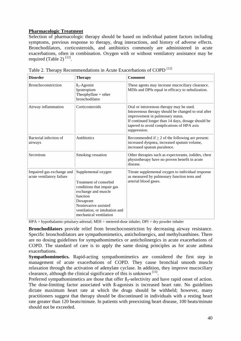
40
Pharmacologic Treatment Selection of pharmacologic therapy should be based on individual patient factors including symptoms, previous response to therapy, drug interactions, and history of adverse effects. Bronchodilators, corticosteroids, and antibiotics commonly are administered in acute exacerbations, often in combination. Oxygen with or without ventilatory assistance may be required (Table 2) [12]. Table 2. Therapy Recommendations in Acute Exacerbations of COPD [12] Disorder Therapy Comment
Bronchoconstriction ß2-Agonist Ipratropium Theophylline + other bronchodilator
These agents may increase mucociliary clearance. MDIs and DPIs equal in efficacy to nebulization.
Airway inflammation Corticosteroids Oral or intravenous therapy may be used. Intravenous therapy should be changed to oral after improvement in pulmonary status. If continued longer than 14 days, dosage should be tapered to avoid complications of HPA axis suppression.
Bacterial infection of airways
Antibiotics Recommended if > 2 of the following are present: increased dyspnea, increased sputum volume, increased sputum purulence.
Secretions Smoking cessation Other therapies such as expectorants, iodides, chest physiotherapy have no proven benefit in acute disease.
Impaired gas exchange and acute ventilatory failure
Supplemental oxygen
Treatment of comorbid conditions that impair gas exchange and muscle function Doxapram Noninvasive assisted ventilation, or intubation and mechanical ventilation
Titrate supplemental oxygen to individual response as measured by pulmonary function tests and arterial blood gases.
HPA = hypothalamic-pituitary-adrenal; MDI = metered-dose inhaler; DPI = dry powder inhaler
Bronchodilators provide relief from bronchoconstriction by decreasing airway resistance. Specific bronchodilators are sympathomimetics, anticholinergics, and methylxanthines. There are no dosing guidelines for sympathomimetics or anticholinergics in acute exacerbations of COPD. The standard of care is to apply the same dosing principles as for acute asthma exacerbations. Sympathomimetics. Rapid-acting sympathomimetics are considered the first step in management of acute exacerbations of COPD. They cause bronchial smooth muscle relaxation through the activation of adenylate cyclase. In addition, they improve mucociliary clearance, although the clinical significance of this is unknown [13]. Preferred sympathomimetics are those that offer ß2-selectivity and have rapid onset of action. The dose-limiting factor associated with ß-agonists is increased heart rate. No guidelines dictate maximum heart rate at which the drugs should be withheld; however, many practitioners suggest that therapy should be discontinued in individuals with a resting heart rate greater than 120 beats/minute. In patients with preexisting heart disease, 100 beats/minute should not be exceeded.

41
Anticholinergics. Anticholinergics improve airway obstruction by inhibiting cholinergic activation of airway smooth muscle, thus producing bronchodilation. Because it has a long onset of action, it will likely not be of benefit in treating acute exacerbations of COPD. Although ipratropium is considered first-line therapy in the management of stable COPD, its role in the treatment of acute exacerbations is less well defined. A greater body of literature supports ß2-agonists as first-line bronchodilators compared with ipratropium [14]. Inhaled ipratropium should be reserved for patients who fail or cannot tolerate ß2-agonist therapy and as part of long-term maintenance once patients are stable. Combination Anticholinergics and Sympathomimetics. Several clinical trials indicate that no significant benefit is achieved when ipratropium is added to inhaled ß2-agonists [15-19]. However, all of these studies evaluated a small number of patients. Methylxanthines. The methylxanthines theophylline and aminophylline may be additive bronchodilator therapy in exacerbations of COPD if response to ß2-agonists and ipratropium is inadequate, although their value is uncertain. Loading doses of theophylline or aminophylline should be administered to achieve therapeutic serum theophylline concentrations rapidly. They are based on actual body weight and may be administered intravenously or orally, as oral theophylline is well absorbed when gastrointestinal function is intact. Intravenous therapy should be reserved for severe, acute decompensation in patients unable to take oral drugs. Corticosteroids Corticosteroids may help reduce airway obstruction by decreasing inflammation and are commonly combined with bronchodilators (Table 5) [20-21]. Those treated with corticosteroids had significantly more rapid improvement in FEV1 and shorter hospital stay than those receiving placebo. The 8-week regimen was not superior to the 2-week regimen. Patients with acute exacerbations of COPD should receive a short course of intravenous or oral corticosteroids. Intravenous therapy should be converted to oral after pulmonary status improves and the patient has a functioning gastrointestinal tract. If steroid treatment is continued for longer than 2 weeks, a tapering oral schedule should be employed to avoid complications associated with hypothalamic-pituitary-adrenal axis suppression. Antibiotics Up to 50% of COPD exacerbations are associated with bacteria. Effective antibiotic therapy results in decreased hospitalizations and good resolution of symptoms. A meta-analysis of the effectiveness of antibiotics in this setting found that patients receiving antibiotics had a greater improvement in PEF than those who did not [21]. It concluded that antibiotics are of most benefit and should be administered if at least two of the following symptoms are present: increased dyspnea, increased sputum volume, and increased sputum purulence. However, these symptoms can also be present if the causative organism is a virus, in which case antibiotic therapy is not warranted [22]. Without definitive sputum cultures it can be difficult to distinguish between bacterial and viral infections. The utility of sputum Gram's stain and culture is questionable, as some patients have chronic bacterial colonization of the bronchial tree between exacerbations [23]. Empiric therapy should be based on organism(s) most likely to be responsible for the infection, given individual patient profiles (Table 6) [24]. The most common organisms for acute exacerbation of COPD are H. influenzae, Moraxella catarrhalis, Streptococcus pneumoniae, and Haemophilus parainfluenzae. More virulent bacteria may be present in patients with more complicated acute exacerbations, including drug-resistant pneumococci, ß-Lactamase-producing H. influenzae, and M. catarrhalis.Pseudomonas aeruginosa and Enterobacteriaceae are most frequently isolated from patients with a FEV1 below 35% [25].

42
Table 3. Recommended Antimicrobial Therapy [24] Patient Characteristics Likely Pathogens Recommended Therapy
Uncomplicated exacerbations: < 4 exacerbations/yr No comorbid illness FEV1 > 50% of predicted
S. pneumoniae, H. influenzae, M. catarrhalis, H. parainfluenzae Resistance uncommon
Macrolide (azithromycin, clarithromycin), 2nd- or 3rd-generation cephalosporin, doxycycline
Therapy not recommended: TMP-SMX, amoxicillin, 1st-generation cephalosporin’s, and erythromycin
Complicated exacerbations: Age > 65 yrs > 4 exacerbations/yr FEV1 < 50% but > 35% of predicted
As above plus drug-resistant pneumococcal, ß-Lactamase-producing H. influenzae and M. catarrhalis, some enteric gram-negatives
Amoxicillin-clavulanate, fluoroquinolone with enhanced pneumococcal activity (levofloxacin, gatifloxacin, moxifloxacin)
Complicated exacerbations with risk of P. aeruginosa: Chronic bronchial sepsisb Need for long-term corticosteroid therapy Resident of nursing home > 4 exacerbations/yr FEV1 < 35% of predicted
As above plus P. aeruginosa Intravenous therapy if required
Fluoroquinolone with enhanced pneumococcal and P. aeruginosa activity (levofloxacin, gatifloxacin, moxifloxacin)
ß-Lactamase-resistant penicillin with antipseudomonal activity
3rd- or 4th-generation cephalosporin with antipseudomonal activity
FEV1 = forced expiratory volume in 1 second. aTMP-SMX should not be given due to increasing pneumococcal resistance; amoxicillin and 1st-generation cephalosporins are not recommended due to ß-Lactamase susceptibility, and erythromycin is not recommended due to insufficient activity against H. influenzae. bIn sepsis double antipseudomonal coverage should be considered (i.e., addition of aminoglycoside).
Therapy should be started within 24 hours of symptoms to prevent hospitalization. Therapy traditionally is continued for 7-10 days; however, studies evaluating shorter regimens (usually 5 days) with fluoroquinolones, second- and third-generation cephalosporins, and macrolide antimicrobials reported efficacy comparable with that of longer regimens. If the patient deteriorates or does not improve as anticipated, hospitalization may be necessary and more aggressive attempts made to identify pathogens responsible for the exacerbation. Parenteral antibiotics may be required. Oxygen Supplemental oxygen should be given to all patients with acute exacerbations of COPD who are hypoxemic. It can be given by nasal cannula, simple face mask, or Venturi-type mask at a flow rate sufficient to produce a resting PaO2 of 60 mm Hg with hemoglobin saturation exceeding 90% [1]. It is recommended that partial pressure of carbon dioxide (PaCO2) and pH be monitored while titrating oxygen therapy. Hypercapnia with subsequent respiratory acidosis is generally not a concern in patients who receive sufficient oxygen to raise the PaO2 to 60 mm Hg; however, the risk does increase if this goal is exceeded. Patients may have to be discharged from the hospital with oxygen but should be reevaluated in 3-4 weeks to justify continued oxygen [1].

43
Ventilatory Assistance Despite optimum medical therapy and supplemental oxygen, some patients develop life-threatening respiratory failure and require ventilatory assistance. Noninvasive positive pressure ventilation (NIPPV) is associated with few intubations, low mortality, and short intensive care admissions [26]. The consensus statement from the American Association for Respiratory Care advocates early NIPPV under the following conditions: respiratory distress with moderate to severe dyspnea, pH less than 7.35 or PaCO2 above 45 mm Hg, and respiratory rate of 25 or more breaths/minute [27,28]. Pressure support ventilation and continuous or bi-level positive airway pressure appear to be best tolerated and most effective in correcting hyper-carbia. Traditionally air oxygen (AirO2) is given in combination with noninvasive ventilation. In patients with severe respiratory failure in whom NIPPV should not be used (cardiovascular instability, inability to clear secretions or protect airways, hemodynamic instability) mechanical ventilation with endotracheal intubation is necessary.
Controversial and New Therapies
Mobilization of Secretions Expectorants and mucolytic agents such as guaifenesin, acetylcysteine, and iodinated glycerol have no proven value in acute exacerbations. Similarly, no data support chest physiotherapy or directed coughing to help clear secretions. However, as mentioned, sympathomimetics may help mobilize secretions, and smoking cessation is recommended as it is associated with impaired mucociliary clearance.
Respiratory Stimulants Although respiratory stimulants have no role in long-term management of COPD, almitrine and doxapram show some utility for acute exacerbations. Doxapram may have value in increasing alveolar ventilation [29]. It is not well studied, does not appear to decrease the need for assisted ventilation, and is not widely administered.
References:
1. Pauwels RA. National and international guidelines for COPD: the need for evidence. Chest 2000; 117:20S-2.
2. Rodriguez-Roisin R. Toward a consensus definition for COPD exacerbations. Chest 2000; 117:398S-401.
3. Hurd S. The impact of COPD on lung health worldwide: epidemiology and incidence. Chest 2000; 117:1S-4.
4. National Heart, Lung, and Blood Institute. Morbidity and mortality chart book. Washington, DC: NHLBI, 2000.
5. Parshall MD. Adult emergency visits for chronic cardiorespiratory disease: does dyspnea matter? Nurs Res 1999; 48:62-70.
6. Connors AF Jr, Dawson NV, Thomas C, et al. Outcomes following acute exacerbations of severe chronic obstructive lung disease: the SUPPORT investigators. Am J Respir Crit Care Med 1996; 154:959-67.
7. Jorenby DE, Leischow SJ, Nides MA, et al. A controlled trial of sustained-release bupropion, a nicotine patch, or both for smoking cessation. N Engl J Med 1999; 340:685-91.

44
8. Lacasse Y, Wong E, Guyatt GH, King D, Cook DJ, Goldstein RS. Meta-analysis of respiratory rehabilitation in chronic obstructive pulmonary disease. Lancet 1996; 348:1115-19.
9. Griffiths TL, Burr ML, Campbell IA, et al. Results at 1 year of outpatient multidisciplinary pulmonary rehabilitation: a randomized controlled trial. Lancet 2000; 355:362-8.
10. Nichol KL, Baken L, Nelson A. Relation between influenza vaccination and outpatient visits, hospitalization, and mortality in elderly persons with chronic lung disease. Ann Intern Med 1999; 130:397-403.
11. Centers for Disease Control and Prevention. Prevention of pneumococcal disease: recommendations of the advisory committee on immunization practices. MMWR 1997; RR-8:1-24.
12. Celli BR, MacNee W, ATS/ERS Task Force. Standards for the diagnosis and treatment of patients with COPD: a summary of the ATS/ERS position paper. Eur Respir J 2004; 23: 932–946.
13. Turner MO, Patel A, Ginsburg S, FitzGerald JM. Bronchodilator delivery in acute airflow obstruction: a meta-analysis. Arch Intern Med 1997; 157:1736-44.
14. Karpel JP, Pesin J, Greenberg D, Gentry E. A comparison of the effects of ipratropium bromide and metaproterenol sulfate in acute exacerbations of COPD. Chest 1990; 98:835-9.
15. Rebuck AS, Chapman KR, Abboud R, et al. Nebulized anticholinergic and sympathomimetic treatment of asthma and chronic obstructive airway disease in the emergency room. Is J Med 1987; 82:59-64.
16. Demirkan K, Kuhl D, Stacey Headley AS, Self T. Can we justify ipratropium therapy as initial management of acute exacerbations of COPD? Pharmacotherapy 1999; 19:838-43.
17. Combivent Inhalation Aerosol Study Group. In chronic obstructive pulmonary disease, a combination of ipratropium and albuterol is more effective than either agent alone. Chest 1994; 105:1411-19.
18. Campbell S. For COPD a combination of ipratropium bromide and albuterol sulfate is more effective than albuterol base. Arch Intern Med 1999; 159:156-60.
19. Moayyedi P, Congleton J, Page RL, Pearson SB, Muers MF. Comparison of nebulized salbutamol and ipratropium bromide with salbutamol alone in the treatment of chronic obstructive pulmonary disease. Thorax 1995; 50:834-7.
20. Davies L, Angus RM, Calverley PMA. Oral corticosteroids in patients admitted to hospital with exacerbations of chronic obstructive pulmonary disease: a prospective randomized controlled trial. Lancet 1999; 354:456-60.
21. Niewoehner DE, Erbland ML, Deupree RH, et al. Effect of systemic glucocorticoids on exacerbations of chronic obstructive pulmonary disease. Department of Veterans Affairs cooperative study group. N Engl J Med 1999; 340:1941-7.
22. Russo RL, D'Aprile M. Role of antimicrobial therapy in acute exacerbations of chronic obstructive pulmonary disease. Ann Pharmacother 2001; 35:576-81.
23. Barnes PJ. Chronic obstructive pulmonary disease. N Engl J Med 2000; 343:269-80. 24. Niederman MS. Antibiotic therapy of exacerbations of chronic bronchitis. Semin
Respir Infect 2000; 15:59-70. 25. Chodosh S, DeAbate CA, Haverstock D, et al. Short-course moxifloxacin therapy for
treatment of acute bacterial exacerbations of chronic bronchitis. Respir Med 2000;94: 18-27.

45
26. Keenan SP, Kernerman PD, Cook DJ, Martin CM, McCormack D, Sibbald WJ. Effect of noninvasive positive pressure ventilation on mortality in patients admitted with acute respiratory failure: a meta-analysis. Crit Care Med 1997; 25:1685-92.
27. Bach JR, Brougher P, Hess DR, et al. Consensus conference: noninvasive positive pressure ventilation. Respir Care 1997; 42:362-9.
28. Anon JM, Garcia de Lorenzo A, Zarazaga A, Gomez-Tello V, Garrido G. Mechanical ventilation of patients on long-term oxygen therapy with acute exacerbations of chronic obstructive pulmonary disease: prognosis and cost-utility analysis. Intensive Care Med 1999; 25:452-7.
29. Moser KM, Luchsinger PC, Adamson JS, et al. Respiratory stimulation with intravenous doxapram in respiratory failure. N Engl J Med 1973; 288:427-31.

46
009 CURRENT THERAPEUTIC STRATEGIES FOR PULMONARY ARTERIAL HYPERTENSION Jakopović M, Samaržija M, Pavičić F. University Hospital for Lung Diseases 'Jordanovac', Zagreb, Croatia Pulmonary hypertension is a disease that leads to sever limitations of functional status and poor survival. Often affects younger people, especially women. Pulmonary hypertension is defined as elevation in mean pulmonary artery pressure over 25 mmHg at rest and over 30 mmHg during exertion. According to mean pulmonary artery pressure (mPAP), pulmonary hypertension is divided in to three degrees of severity: 1. mild, mPAP raging from 25 to 35 mmHg; 2. moderate, mPAP raging from 36 to 45 mmHg; and 3. severe, mPAP over 45 mmHg. Clinical features include progressive dyspnoea, exertion limitation, deterioration of right ventricle function and finally right ventricle failure and death. Mean survival in patient with idiopathic pulmonary hypertension is poor, raging from 2 to 8 years. Survival in patient with pulmonary hypertension previously known as secondary depends on underlying disease that caused elevated pulmonary artery pressure. Knowledge about existence, importance and treatment modalities for pulmonary hypertension was poor for almost one hundred years after the first Romberg's description of pulmonary vascular sclerosis. Due to that reason, pulmonary hypertension was classified into two categories: primary and secondary. Finally, in the last decade, World Health Organization became aware of importance and consequences of pulmonary hypertension, especially on total morbidity and mortality and in 1998 recommended first clinical classification, better known as 'Evian classification', with diagnostic and therapeutic guidelines. Since then, further basic research findings in the biochemistry, pathophysiology and genetics of pulmonary hypertension have been discovered, new diagnostic procedures were developed, and new drugs for the treatment of pulmonary hypertension were introduced. Because of that, during The 2003 Third World Symposium on Pulmonary Arterial Hypertension held in Venice, Italy, second, revised clinical classification of pulmonary hypertension was proposed. Clinical classification of pulmonary hypertension:
1. Pulmonary arterial hypertension 2. Pulmonary hypertension with left heart disease 3. Pulmonary hypertension associated with lung diseases and/or hypoxemia 4. Pulmonary hypertension due to chronic thrombotic and/or embolic disease 5. Miscellaneous
The main vascular changes in pulmonary arterial hypertension are vasoconstriction, smooth-muscle cell and endothelial cell proliferation and dysfunction, and thrombosis in situ. These findings suggest the disturbance in the normal relationship between vasodilators (prostanoids, adenosine, β2 agonists, nitric oxide – NO, atrium natriuretic peptide – ANP, brain natiuretic peptide – BNP) and vasoconstrictors (angiotensin II, endothelin, tromboxan, epinephrine, vasopressin, leukotriens, serotonin), growth inhibitors and mitogenic factors, and disbalance between antithrombotic and prothrombotic factors. These homeostatic imbalances are probably consequence of pulmonary endothelial cell dysfunction or injury caused by hypoxic vasoconstriction and other causes. Diagnostic procedure is consisted of basic and additional tests. Basic diagnostic procedures are medical history, physical examination, laboratory tests, ECG, chest X – ray. Lung function testing and echocardiography. During echocardiography, we can determine pulmonary artery pressure, right ventricle pressure, tricuspid regurgitation. Additional test are: gene markers, immunologic testing, HIV and hepatitis markers, liver enzymes, abdominal

47
ultrasound, chest CT scan, positron emission tomography – PET, MRI of the heart, exertion testing, pulmonary angiography, perfusion and ventilation lung scan. Final diagnostic procedure is the right heart catheterization in rest and exertion, during which hemodynamic testing, reversibility of pulmonary hypertension and pharmacological testing’s performed. Because of importance of pulmonary vasoconstriction in pathophysiology of pulmonary hypertension main therapeutic choice are vasodilators. Over the last decades many drugs form different classes, including adrenergic agonists and antagonists, arterial vasodilators, nitrates, angiotensin – converting enzyme inhibitors and calcium channel blockers along with oxygen were given to patients with pulmonary hypertension. In the last ten years, various prostanoids were introduced, such as intravenous epoprostenol, subcutaneous treprostinil and inhaled iloprost. Significance of NO was recognized and safe application systems for its use were developed. Problem with previously mentioned drugs are high prices and complicated ways of usage, therefore further improvement in treatment of pulmonary hypertension was discovery of orally administrated drugs like phopshodiesetraze inhibitors (sildenafil, tadalafil) and endothelin receptor antagonists (bosentan). Positioning of new drugs in the treatment of pulmonary hypertension is not yet well defined and confirmed in clinical trials, but based on basic and clinical findings it seems that early diagnosis and treatment of this disease is justified. References:
1. McLaughlin VV, Archer SL, Badesch DB and al. ACCF/AHA 2009 Expert Consensus Document on Pulmonary Hypertension: A Report of the American College of Cardiology Foundation Task Force on Expert Consensus Documents and the American Heart Association Developed in Collaboration With the American College of Chest Physicians; American Thoracic Society, Inc; and the Pulmonary Hypertension Association. J am Coll Cardiol 2009; 53:1573-1619.
2. McGoon M, Gutterman D, Steen V, et al. Screening, early detection, and diagnosis of pulmonary hypertension: ACCP evidence –based clinical practice guidelines. Chest 2004; 126:14S-34S.
3. Olschweski H, Simonneau G, Galie N, et al. Inhaled iloprost for severe pulmonary hypertension. N Engl J Med 2002; 347:322-9-

48
010 RESPIRATORY DISTURBANCES AND NEUROMUSCULAR DISORDERS Sinanović O. Department of Neurology, University Clinical Center Tuzla, Faculty of Medicine, Tuzla, Bosnia Herzegovina Neuromuscular disorders (NMDs) included large number different disorders caused by the primary involvement of the motor unit, which is composed of motor neuron, nerve root, peripheral nerve, myoneural junction, and muscles. NMDs can lead to muscle weakness and general fatigue. Respiratory distress often accompanies these diseases due to weakness of the respiratory muscles. Mildly affected patients may show respiratory disturbance only during sleep or sleep-disordered breathing (SDB), but even in severely affected patients it may go unrecognized and untreated. Thus, respiratory muscle weakness is the common consequence of many (myotonic dystrophies, Duchenne's muscular dystrophy, hereditary motor and sensory neuropathies, spinal muscular atrophies, myasthenia gravis, Guillain-Barre syndrome, amyotrophic lateral sclerosis, postpolyo syndrome). It can causes severe ventilatory restriction, results in slowly or rapid progressive respiratory failure, and is the major causes of death. Respiratory failure relates directly to the loss of respiratory muscle force and vital capacity and shows characteristic evolution from normal ventilation during daytime and sleep-induced hypopnoeas at mild degrees of ventilatory restriction to sleep hypoventilation in severe ventilatory restriction. Continuous hypoventilation, common at inspiratory vital capacity (IVC) <40% predicted, precedes daytime hypercapnia. Daytime respiratory failure is highly prevalent at IVC <20% predicted, and represents an accepted indication for supportive non-invasive (positive-pressure) ventilation (NIV). NIV, applied intermittently and preferable during sleep, relieves respiratory muscles from the work of breathing and augments alveolar ventilation. Respiratory investigation is necessary routine procedure in follow up NMD patients (overnight polysomnography is the best) for SDB and nocturnal desaturations. In the management of these patients, NIV (intermittent positive pressure) results in improvement of SDB and breathing.

49
011 CURRENT ASPECTS OF SURGICAL TREATMENT OF THORACIC EMPYEMA Guska S. Clinical Centre University of Sarajevo, Clinic for Thoracic Surgery, Sarajevo, Bosnia Herzegovina Defined as the inflammatory process in the pleural cavity, thoracic empyema (TE) was one of the first recognized thoracic pathological entities that had been and is still a great diagnostic and therapeutic challenge for chest physicians and thoracic surgeons. Generally, the number of potential candidates for TE is permanently growing as a result of increased life expectancy, and extended operability criteria within the scope of thoracic surgery. Antibiotic abuse led to increased numbers of therapy-resistant cases and the tuberculosis did not cease to be a permanent threat either. Immunocompromised conditions - either iatrogenic or as the result of drug abuse and HIV injection -impose further risk in developing of TE [1-20].The heterogeneous nature of the disease, which frequently masquerades its own standard clinical manifestation, is not helpful either. This nature of the condition is exposed by Graham’s comment on the paradigm shifting work of Samson and Burford on the efficacy of decortications of TE, adding:"You men who have been working in World War II have not been seeing empyema. Empyema is an abscess of the pleural cavity. It is a word that was used by Hippocrates to mean abscess ... You have been seeing infections attenuated by drugs which were not known in 1941 ... and much less known to Hippocrates ... if you had talked to Hippocrates about this as empyema, he would have said: “I don’t understand what you are talking about" [1].TE might be scrutinized too complex to discuss as a unique clinical picture. However, the common and dominating inflammatory nature of the disease, the uniform pathological changes and the shared treatment modalities to correct them, provide a sufficient rationale for treating TE as a unique entity. Basic consideration Definition Thoracic empyema is a dynamic process, inflammatory in origin and taking place within a preformed space called pleural cavity. It represents a complex clinical entity, neither a sole clinical, laboratory, nor a radiological diagnosis [1-5, 16-21]. A significant lack of detectable causative microorganisms (sterile pus) reported between 47% and 56% complicates further definition [1, 6-11, 22, 23]. In this article, the entities are discussed according to Light’s classification of the parapneumonic effusions [1, 3]. The following criteria were accepted for the diagnosis, irrespective of their origin:
1. Frank pus at thoracocentesis or organisms demonstrated on Gram stain (direct) or culture (indirect), or all of the tests positive for:
2. pH below 7.2; glucose level of fluid less than 400 mg/l; LDH above 1000 IU/ml; protein level above 3 g/ml and WBC over 15 000 cells/mm3.
3. Physical, radiological and laboratory signs accompanied the relevant clinical picture. The basic methods of anamnesis, physical examination and (guided) tapping and analysis of the pleural specimen are of prominent importance [1-3,13-22,24-27] which are followed with numerous available radiological techniques like chest X-ray, fluoroscopy, chest ultrasound and CT which have their own role in diagnosis of thoracic empyema [1-3,13-22,23-31]. Origin and taxonomy The most common origin of TE is meta- or parapneumonic pleural effusion, representing 40-60% [1, 2, 3, 4, 15, 25-37] of all cases. From 5% to 20% of all meta- or parapneumonic effusions become TE [1, 9, and 17]. Thirty percent or less of all the adult cases of TE develops as a result of thoracic surgical procedures [1,5,6,9,18,19,].About 1.6-4.2% of thoracic trauma results with TE [19,20,23,26,30,33]. Other sources are occasionally

50
mentioned [1, 21].Taxonomy of TE (Table 2.) offers the didactic advantage of exposing the relations among origin, stage and therapeutically options. Decision-making rests upon these three facts.
Table 2. Classification of thoracic empyema Primary (meta/parapneumonic) thoracic
Secondary to lung resection -without BPF -with minor/moderate/significant BPF* Secondary to other surgical trauma Secondary to non-medical trauma Secondary to other sources *BPF-bronchopleural fistula
The question is how and to what extent the individual elements should be estimated in the individual case. An thoracic empyema was designated primary (PTE) if there were no previous surgical interventions involving the chest, no data of other kind of trauma or other secondary sources [1,3,5,10,11,15,17].The most frequent forms are pneumonia related complicated pleural effusions. A thoracic empyema is secondary (STE) if it follows a chest trauma or other secondary sources. Most frequently it is a surgical trauma; in the majority of the cases after a lung resection [1, 18, 27, 29]. Penetrating or blunt chest trauma is another cause of secondary thoracic empyema [19, 20]. Specific empyema, infected malignant pleural effusion and empyema of unknown origin are less frequent [1,4,5,10,11,15,17,24, 27,30,36]. The distinction between primary and secondary thoracic empyema may sound artificial and yet the tag reflects the basic difference in their optimal approaches. Pathophysiology The complete untreated process of development of TE takes about 5-6 weeks, but the length of the single stages is not clearly defined (Fig. 1).
Fig.1.Time-scale and overlapping of stages of thoracic empyema. The ‘fog’ function of an uncertain prehistory is intended as a fine adjusting element helping to approach the theoretical origin of clinical events.
While the date of the diagnosis is usually well documented, the origin of the whole process, especially in PTE, too frequently disappears in the haziness of the personal anamnesis. The triphasic nature of the disease is well established [1-3, 21-23, 28-37, 38, 39, 40, 41]. In Stage I (exudative phase) the visceral pleura remains elastic and dimensions of the thoracic cavity are maintained. Stage II (transitional or fibrinopurulent) is marked by turbid and infected fluid, which becomes thick and purulent. The fibrin deposits construct bridges which septet the effusions creating multiple loculations. In Stage III (organizing phase) this is replaced by
51
formal granulation tissue. A sheet of inflammatory tissue would gradually compress the underlying tissue, causing contraction of the affected hemithorax. Finally, the mediastinum is shifted ipsilaterally, the diaphragm is elevated and the spaces between the ribs are narrowed. Sequence of therapeutic methods The primary surgical therapeutic aim: ‘ubi pus evacuee’ has not changed since the age of Celsus. The main aim of the surgical therapy is control of local infection, but without elimination of the pleural dead space with impending colonization it is hardly achievable. The combinations and sequence of surgical methods listed in Table 3. are summarized in the ‘empyema diamond’ diagram (Fig. 2.). Table 3 Sequence of treatment modalities coping with empyema thoracic Thoracocentesis (tapping) Drainage Simple Empyema tube (von Petzer’s drain) Negative passive drain (underwater drain/von Bu¨lau’s system/Heimlich valve) Active Intermittent suction Continuous suction Irrigation Cyclic (tidal) one tube ore more Continuous suction—irrigation (two or more tubes) Chemical decortications (fibrinolysis) Debridement Open VATS Decortications (Fowler-Delorme procedure) Thoracoplasty without plomb Thoracoplasty with plomb Muscle Omentum Other Open window thoracostomy Eloesser flap and modifications Without flap (fenestration) Combined procedures Clagett procedure Weder procedure

52
Fig. 2. Treatment modalities according to the therapeutic pathways: the empyema diamond Tube/closed thoracostomy/intercostals catheter (ICC)/intercostals drain (ICD) Thoracocentesis with a large bore needle has doubtless diagnostic value, but its therapeutic benefit is at least ambiguous [1-3,5-9,20-25,30].Pleural drainage performed as a single procedure is usually a first-line intervention with a success rate for PTE between 67% and 74% [1-5,26-28,36-39,41,43].Common sense dictates that the more Stage I cases included in a series, the better the success rate. Series focusing on subsequent surgeries report a 36-65% failure rate of simple tube drainage [1,9,17,25,28].The lower efficacy of the method in trauma-related empyema reflects the high proportion of Stage III cases and low-risk younger patients [1,3,10,19,28,30,37].The mortality of TE treated by drainage is 11-24% [3,9,10,28,29]. Improving the efficacy of the drainage by irrigation is a well-established technique [1,2,10,16,19,30,35], either by cyclic or continuous irrigation [1,2,7,9,16,20, 21,34].Saline is the more widely used substance to dilute the causative organisms and to evacuate the debris by irrigation. Strictly speaking, the practice is based on historical observations like the Carrell-Dakin method [30] and personal experience. Usage of local antibacterial treatment lacks firm evidence, but again there are no reliable arguments against those positive observations based on reasonable assumptions. It is agreed that a positive culture and antibiogram are „conditio sine qua non“of this sort of therapy [1, 2, 5, 34, 37, 42, 44,]. Personal experiences, conventions, training, schooling, conviction and accessibility of technicalities, relevant staff are the variables of the details of this technique. In this rich field of individual approaches with firm convictions, controlled data are insufficient. It is obvious that the more sophisticated an empyema treating system is, the higher the chance for a successful outcome. It may be related to the amount of devotion to the patient and the system rather than the real difference in efficacy of the methods. Site of drainage (dependent point) and number and size of drains are empiric. Typically, size of Ch28 seems to be the most frequent accepted lower gauge, but Ch32 or over is not uncommon either. Unfortunately, neither question of pain control nor of physiotherapy as factors influencing the quality of life and outcome was raised specifically in any of the referred reviews [1,9,25-34,40,43]. Also, the force and the type of the suction, the tube setting and specification of systems used are among the unexplored details. According to evidences, drainage is nearly always a first-line therapeutic modality[1-5,10-16,20,33,37, 42,44,45].On rare occasions permanent tube thoracostomy can provide a final solution when others failed [35].

53
Fibrinolysis, enzymatic/chemical decortications Disrupting the septa of the empyema cavity and disintegration of the devitalized, necrotic mass covering the pleural surface by intrapleural instillation of streptokinase in order to make it accessible for drainage was initially described by Tillet et al. in 1951 [1].Contrary to previous convincing reports [2,3,5] recent evidence did not find enzymatic decortications superior to tube thoracostomy treatment [5,7,10].On the other hand the success rate with ICC alone versus ICC and streptokinase were 67.1% versus 87.7%.Multiple regression analysis has proven fibrinolysis as a sole independent factor for better outcome [2]. Where Stage III empyema cases were excluded, similar results were reported [9]. 3.3. Video-assisted empyema surgery (evacuation/debridement) In this group of procedures misnomers flourish. Evacuation of necrotic material from the pleural cavity, essentially from the parietal wall, is by definition, debridement. Decortications, a procedure known since 1885 [1, 2, 30, 38, 39] the peeling of the organized coat of the visceral pleura-is the essence of the operation. Papers heralding VATS decortications uniformly fail to demonstrate that a standard Fowler-Delorme procedure was performed [1, 2, 40, 43, and 47]. This operation is a technically demanding procedure even under direct tactile and full visual control. Therefore, it seems to be reasonable to discuss all video-assisted clearings here under the title of evacuation in which debridement [1, 13, 16, 18, 2, and 42] is the core of the procedure, irrespective of their own usage of terminology. Pathoanatomy offers the evidence, as there is no distinctive and, therefore, removable cortex prior to at least the first 4 weeks [1, 24, 30, 40]. From the mid-1990s, thoracoscopic evacuation of empyema sac has gained popularity [13, 16, and 42]. Subsequent papers have supported the original observations and notes on limitations [40]. Success rate ranges from 68% to 93% [17, 21, 40-43], and seems to be in close correlation with the selection of the investigated patient group. The more the Stage III empyema or, in general, the longer the anamnesis-the higher the failure rate [15, 17, 44], necessitating further surgery such as decortications, open window thoracostomy and thoracoplasty, in order of frequency. The conversion rate is 5-8% [40, 42], and there is 10-25% of a second-stage, open decortications [18, 40, 41]. Patients with a history shorter than 4 weeks had a good chance to be cured by VATS alone [40, 42] while histories over 5 weeks (presumed Stage III) tended to necessitate a decortications [13, 16, 17, 21, 42]. Preselect ion bias interferes with the outcome rather than treatment modalities themselves. There seems to be an undisputed superiority of VATS procedures as far as early posttraumatic cases are concerned [41, 43]. Evidence suggests that following a failed tube thoracostomy a VATS evacuation is more beneficial than after an interim attempted fibrinolysis [21]. An ultrasonic device was recently published [44], which can improve the efficacy of the breakdown maneuvers during VATS debridement. The quality of the underlying lung is a decisive factor in outcome. Potential for „restitutio ad integrum“ restoring the original parenchyma volume in order to fill the space-is the key element. Evacuation of infected posttraumatic effusion facilitates quick re-expansion of the healthy underlying lung, a significantly different situation from a lung recovering from a lobar pneumonia. Underlying diseases such as cancer and tuberculosis, either debilitating the recoil capacity of the lung tissue or destroying the barrier function of the pleural surface, leading to bronchopleural fistula like emphysema and other conditions resulting in honeycomb lung, are the main factors responsible for failure. The 30-day mortality of the patients treated by this modality is 3.4-4.2% [17, 21, 27, 31, and 40]. Open surgical methods for empyema Decortications is the method of choice when the underlying lung is unable to re-expand (trapped lung) due to the formed thick coat and the patient is suitable enough for major intervention. Decortication relies on lung elasticity in order to fill the cavity, freeing the encased parenchyma from the compressing inflammatory coat [2, 30]. When the history is
54
longer than 6 weeks, which is equivalent to a Stage III disease, the recommendations are concordant, if the patient is eligible for surgery [10, 21, 26, and 45]. The majority of the Fowler-Delorme procedures are performed for Stage III post pneumonic empyema [1, 2, 21, and 45] or following trauma [19, 26, 28, and 30]. Patients undergoing VATS for empyema are likely to be converted to open procedure in 3.8-40% depending on the delay in decision even as early as in Stage II [17, 45]. There is a recent shift to muscle-sparing (axillary) thoracotomy, narrowing the aggressively gap between VATS and open surgery. Bronchopleural, pleurocutaneous fistula might necessitate additional parenchyma sparing lung resection in up to 10.1% of the cases [21].With respect to the etiology, up to 80% of posttraumatic empyema requires formal decortications [19,41]. Reoperation rate after failed decortications is half of that following VATS procedure. The mortality of decortications is 1.3—6.6% [1, 2, 26, 29, and 50]. Thoracoplasty. Remodeling the osteomuscular wall of the thoracic cage in order to control the underlying inflammatory process (collapse therapy) was among the first effective thoracic surgical procedures [1]. Nowadays, the aim of the procedure is space filling: either by diminishing the distance between the lung parenchyma by collapsing the roof of the chest and/or filling the space with viable tissue (omentum, muscle transposition). This procedure can be performed alone or in combination with other modalities, like re-do stump closure. In spite of modern prosthesis technology, no recent reports [1] are available on contemporary usage of non-biological plombage. Proper surgical technique and planning are needed to avoid deformities like scoliosis and other related consequences [45]. Thoracoplasty with or without myoplasty is a worthy consolidating step in sequential empyema surgery. Previous procedures include fenestration in 17-72% to sterilize the cavity [40, 42-44]. The usual problem of the plombage, the too small volume of filling material, can be solved by plastic surgical methods [38, 40, and 41]. Combination of thoracoplasty and omental pedicled flap for chronic empyema due to bronchopleural fistula (BPF) can achieve an 82.6% success [43]. In selected cases it is a first-line procedure rather than being a last resort when every previous attempts failed [42]. The 11% failure rate includes those who will carry on with permanent thoracostomy [35]. The overall mortality in these low case number series is about 4.3—5% [39, 41, and 44].
Open window thoracostomy (OWT)/fenestration/empyema marsupialisation For exhausted patients with TE, thoracoplasty is not a amenable alternative and as tube thoracostomy with or without VATS debridement would fail to control the disease, OWT should be offered [1, 2, and 12]. Marsupialisation of the cavity via rib(s) resection and open drainage is a well-established [33, 37, 41] method of low risk. Consequences concerning quality of life in patients with OWT remain unexplored so far. It is the choice of treatment if there is a permanent supply of causative organisms due to bronchopleural fistula (BPF). It can be applied either as a definite treatment with intent to cure, a preliminary procedure prior to definite treatment [38,44] or as a last resort procedure when others have failed to achieve a relatively stable disease [1,2,35,39]. In actual thoracic surgical practice the postoperative empyema, usually with BPF, is the main indication [1,13,34] of the procedure, and is relatively rare (even as low as 3%) in postpneumonic, primary thoracic empyema cases [21,17]. This is an externalization via disclosure of the empyema cavity at the dependent point. The originally created valve mechanism of the Eloesser flap, introduced in 1935 [1],went through several modifications, reaching the present form, where the inverted flap attached to the floor of the cavity gets daily packing’s [38]. Clagett’s procedure [1, 2, 35, 37, 40, and 42] is the best-evidenced method for historical reasons. The complex procedure consists of open pleural drainage, serial operative debridement and eventual chest closure after filling the pleural cavity with antibiotic solution [1]. Window making, fenestration, is

55
part two of the procedure [34]. Daily irrigation with antibiotics [39] or regular open packing’s complete the procedures. Observational studies prove that timing of initiation of therapy is crucial. As an alternative to the staged and time-consuming Clagett procedures, Weder applies the repetitive thoracotomy and debridement policy [45]. Stuffing the cleansed cavity with antiseptic packages and changing them regularly are the essence of the method. This standardized concept of repeated debridement is applied equally to early and late postoperative empyema in a sequential, pre-planned standardized way [35]. This technique seems to be applicable not only to treat but also to prevent empyema in high-risk procedures like completion pneumonectomy for infective diseases [36,37].Solution of chronic or late postoperative empyema also consists of drainage, debridement, closure of BPF when present and space obliteration [43,44]. Generally, the information available from the publications does not offer a clear differentiation in how much of the treatment effect is due to actual treatment differences and how much is due to the assignment of the patients to selective treatment modalities. In the present review of the observed outcomes following different treatment modalities, patient selection, cohort size, methods of randomization and so on were not scrutinized in the study, which definitely weaken the power of the conclusions. The very few controlled trials on this topic came up with conflicting results [1,4,5].The relative lack of randomized controlled trials, reports considered as basics for clinical standard, does not mean that the tremendous clinical experience collected on this topic [1,5,9-34,37-45] would not be able to serve as a proper guideline. The lack of a single ideal treatment modality or policy reflects the complexity of the diagnosis and staging of this heterogeneous disease. Decision-making protocols cannot function without clear and unmistakable categories. Basic elements of intervention-drainage, different evacuation techniques, decortications, open window thoracostomy, and thoracoplasty-are well-established technical modalities; however, neither a universal primary modality nor the gold standard of their sequence is available. The treatment modality should be tailored to the condition of patients and to the healing potential of the persisting cavity. Decision-making relies on the triad of the etiology of empyema, general condition of the patient and actual stage of disease, considering the triphasic nature of it. The basic differences in the behavior of ‘naturally’ developing empyema cavity and of those infected spaces that have followed the removal of lung parenchyma dictate the choice of procedure (Fig. 3). Presence or lack of dead space within the pleural cavity is the definite distinction and decisive factor influencing outcomes following different therapeutically attempts. No effective infection control can be expected in the presence of an active cavity.
Fig. 3. Connection between the stages of thoracic empyema and the best evidenced methods of choice. The theoretical time-scale is not necessarily identical to the documented duration of the disease of the individual patient. The graphical representation is not intended to be considered as an absolute and exclusive scheme.
There are two main sources of the bias limiting the daily routine applicability of the reports. Preselection of patients into different modality groups a priori influences outcomes: the
56
results are mirroring the attitude of the surgeon/team rather than the distance of the investigated method from the ideal treatment. In postpneumonic empyema, the surgeon’s problem starts with the timing and circumstances of referral: called to see the empyema patient only when other conservative methods failed; only not to mention the case, when the typical empyema patient is referred to surgical care on a Friday afternoon, preferably on a long weekend. The highly selected cohort of patients with unreliable information on the origin of the process explains the uncertainties around the value of the individual procedures on the surgical pathway. In the average paper, a hierarchy of methods is evaluated-and with rare exceptions, the actually presented method is ‘proven’ to be the best. The problem with postresectional thoracic empyema patients is similar. The higher the risk they have prior to the original operation, the higher their failure rates for less aggressive modalities when thoracic empyema develops. Drainage remains to be the initial treatment modality in Stage I disease. The weight of additional elements in success/failure such as suction tactics, physiotherapy and nutritional status needs further clarification. The number of drains, their size, location, details of management and caring at multilevel, i.e. doctoral, nursing and physiotherapeutic and of timing, frequency and duration of the exact maneuvers are neglected aspects of studies. Debridement via VATS is a safe, reliable and efficient method in Stage II cases. VATS is not limited exclusively to Stage II disease. Video techniques have a role in Stage III thoracic empyema, too, as far as evacuation is concerned. Organized pleural cavity (Stage III) requires open surgery: formal decortications. A persisting cavity is a challenge without a single and uniform solution (Fig. 4).Open window thoracostomy, either through limited thoracotomy or VATS, represents a simpler and, therefore, safer procedure than thoracoplasty. It is a valuable procedure as an initial step in cavity management and a unique and definite one for high-risk patients, too. Thoracoplasty lost its popularity against the alternative techniques, i.e. decortications and thoracostomy, but it has final step role in pleural space management when other methods failed. Using aggressive methods in space sterilization and obliterate techniques (pedicled muscle or omentum plombs) with preservation of the first rib in case of destroyed lung seems to be worthwhile to consider it as a real alternative. Acute postoperative bronchial stump insufficiency requires immediate surgery, but to what extent? Evacuation of toxic material is mandatory. Closed drainage, open window thoracostomy, repetitive thoracotomies and cavity packing are equally viable options. No single-stage procedure offers solution. Irrespective of the above detailed differences in specific features of thoracic empyema, the basic rules for treatment remain the same:
(1) complete evacuation of the content of infected space (2) elimination of cavity and lung re-expansion (3) control of causative organisms/sterilization (4) forced auxiliary treatment such as aggressive physiotherapy, nutritional support in
every phase of treatment.

57
Fig. 4. Approaches of the persisting space problem: (1) internal closure of BPF (endoscopic methods); (2) external closure of BPF (re-amputation, coverage);(3) diminishing the remaining space by de-roofing the musculoskeletal structures (thoracoplasty); (4) space occupying by muscle/omentum transposition (plombage); (5) establishing a persistent orifice (window/tube).
Summarizing the current divergent attitudes towards this well-known entity of a sinister natural history one can say that thoracic empyema shows that present concepts are based mainly on the expert`s opinion. Therefore, no other option than highly individualized approaches can be recommended. The thoracic surgeon should know all the possible techniques within their limitations to adjust it to individual patient. Flexibility and patience on behalf of the surgeon, nursing staff and the patient furthering the endeavor of hospital management to understand the complexity of this condition are the cornerstones of the treatment. Thoracic empyema is an eminent example, that no established method can be neglected by putting our surgical heritage on the dusty shelves of a distant corner. Thoracic empyema does not allow recommending a clear single sequence of procedures leading to a uniformly predictable successful outcome. Institutional practice, local protocols based on past experience and individual case management with a flexibly optimized sequence of procedures may offer the best outcome. References:
1. Molnar T F: Current surgical treatment of thoracic empyema in adults. Eur J Cardiothorac Surg 2007; 32:422-430.
2. Chudzicka A, Chciałowski A: Parapneumonic pleural effusion - difficulties in making therapeutic decisions. POLSKIE ARCHIWUM MEDYCYNY WEWNĘTRZNEJ 2007; 11 7 (1-2).
3. Light R W: Parapneumonic Effusions and Empyema. Proc Am Thorac Soc Vol 3. 2006; 75–80.
4. Hope WW, Bolton WD, Stephenson JE. The utility and timing of surgical intervention for parapneumonic empyema in the era of video-assisted thoracoscopy. Am Surg. 2005 Jun; 71(6):512-4.
5. Klopp M, Dienemann H. Pleural empyema: surgical treatment. Dtsch Med Wochenschr. 2005 Mar 11; 130(10):530-3.
6. Maskell NA, Davies CWH, Nunn AJ, Hedley EL, Gleeson FV, Miller R, Gabe R, Rees GL, Peto TEA, Woodhead MA, Lane DJ, Darbyshire JH, Davies RJO (MIST1 Group). Controlled trial of intrapleural streptokinase for pleural infection. N Engl J Med 2005; 352(9):865-74
7. Treasure T. The evidence on which to base practice: different tools for different times. Eur J Cardiothorac Surg 2006; 30:819-24.
8. Spencer D. Empyema thoracic: not time to put down the knife. Arch Dis Child 2003; 0:842-3.

58
9. Ozol D, Oktem S, Erdinc E. Complicated parapneumonic effusion and empyema thoracis: microbiologic and therapeutic aspects. Resp Med 2006; 100:286-91.
10. Anstadt MP, Guill CK, Ferguson ER, Gordon HS, Soltero ER, Beall AC, Musher DM. Surgical versus nonsurgical treatment of empyema thoracis: an outcomes analysis. Am J Med Sci 2003; 326(1):9-14.
11. Deas1auriers J, Mehran R. Handbook of perioperative care in general thoracic surgery. Mosby Philadelphia, Pennsylvania, USA: Elsevier; 2005.
12. Chapman SJ, Davies RJ. The management of pleural space infections. Respirology 2004; 9:4-11.
13. De Hoyos A, Sunderasen S. Thoracic empyema. Surg Clin N Am 2002; 82:643-71. 14. Luh SP, Chou MC, Wang LS, Chen JY, Tsai TP. Video-assisted thoracoscopic
surgery in the treatment of complicated parapneumonic effusions or empyemas. Chest 2005; 127:1427-32.
15. Kacprzak G, Marciniak M, Addae-Boateng E, Kolodziej J, Pawelczyk K. Causes and management of postpneumonectomy empyemas: our experience. Eur J Cardiothorac Surg 2004; 26:498-502.
16. Petrakis IE, Kogerakis NE, Drositis IE, Lasithiotakis KG, Bourous D, Chalkiadakis GE. Video-assisted thoracoscopic surgery for thoracic empyema: primarily, or after fibrinolytic therapy failure? Am J Surg 2004; 187:471-4?
17. Athanassiadi K, Gerazounis M, Kalantzi N. Treatment of post-pneumonic empyema thoracis. Thorac Cardiovasc Surg 2003; 51:338—41.
18. Suchar AM, Zureikat AH, Glynn L, Statter MB, Lee J, Liu DC. Ready for the frontline: is early thoracoscopic decortication the new standard of care for advanced pneumonia with empyema? Am Surg 2006; 72(8):688-92.
19. Molnar TF, Hasse J, Jeyasingham K, Rendeki S. Changing dogmas: history of treatment for traumatic hemothorax, pneumothorax and empyema thoracis. Ann Thorac Surg 2004; 77:372-8.
20. Cheng YJ, Wu HH, Chou SH, Kao EL. Video assisted thoracoscopic surgery in the treatment of chronic empyema thoracis. Surg Today 2002; 32:19-25.
21. British National Formulary. BNF49, March 2005, pp. 400 and 594. London, UK: British Medical Association; 2005.
22. Misthos P, Sepsas E, Konstantnou M, Athanassiadi K, Skottis I, Lioulias A. Early use of intrapleural fibrinolytics in the management of postpneumonic empyema. A prospective study. Eur J Cardiothorac Surg 2005; 28:599-603.
23. Kim BY, Oh BS, Jang WC, Min YI, Park YK, Park YC. Video-assisted thoracoscopic decortications for management of postpneumonic pleural empyema. Am J Surg 2004; 188(3):321-4.
24. Oguzkaya F, Akcali Y, Bilgin M. Videothoracoscopy versus intrapleural streptokinase for management of post-traumatic retained haemothorax:a retrospective study of 65 cases. Injury 2005; 36:526-9.
25. Nakamura H, Taniguchi Y, Makihara K, Ohgi S. Ultrasonic pleural debridement of empyema. Ann Thorac Cardiovasc Surg 2001; 7:62-4.
26. Melloni G, Carretta A, Ciriaco P, Negri G, Voci C, Augello G, Zannini P. Decortications for chronic parapneumonic empyema: results of a prospective study. World J Surg 2004; 28 (5): 488—93.
27. Rzyman W, Skokowski J, Romanowicz G, Lass P, Dziadziuszko R. Decortications in chronic pleural empyema - effect on lung function. Eur J Cardiothorac Surg 2002; 21(3):502-7.

59
28. Rzyman W, Skokowski J, Romanowicz G, Lass P, Murawski M, Taraszewska M, Dziadziuszko R. Lung function in patients operated for chronic pleural empyema. Thorac Cardiovasc Surg 2005; 53:245-9.
29. Olgac G, Yilmaz MA, Ortakoylu MG, Kutlu CA. Decision making for lung resection in patients with empyema and collapsed lung due to tuberculosis. Thorac Cardiovasc Surg 2005; 130:131.
30. Lardinois D, Gock M, Pezzeta E, Buchli C, Rousson V, Furrer M, Ris HB. Delayed referral and gram negative organisms increase the conversion thoracotomy rate in patients undergoing video-assisted thoracoscopic surgery for empyema. Ann Thorac Surg 2005; 79(6):1851-6.
31. Tezel CS, Goldstraw P. Plombage thoracoplasty with Lucite balls. Ann Thorac Surg 2005: 1063-79.
32. Okumura Y, Takeda S, Asada H, Inoue M, Sawabata N, Shiono H, Maeda H. Surgical results for chronic empyema using omental predilected flap: longterm follow-up study. Ann Thorac Surg 2005; 79:1857-61.
33. Regnard JF, Alifano M, Puyo P, Fares E, Magdeleinat P, Levasseur P. Open window thoracostomy followed by intrathoracic flap transposition in the treatment of empyema complicating pulmonary resection. J Thorac Cardiovasc Surg 2000; 120:270-5.
34. Shimuzu J, Kinishite T, Tatsuzawa Y, Kawaura Y, Ishikura N, Oda M. Intrathoracic free musculocutaneous flap after open-window thoracostomy for chronic empyema. Thorac Cardiovasc Surg 2001; 49(4):237-9.
35. Deslauriers J, Jacques LF, Gregoire J. Role of Eloesser flap and thoracoplasty in the third millennium. Chest Surg Clin N Am 2002; 12:605-23.
36. Gossot D, Stern JB, Galetta D, Debrosse D, Girard P, Callendro R, Harper L,Grunenwald D. Thoracoscopic management of postpneumonectomy empyema. Ann Thorac Surg 2004; 78(1):273-6.
37. Massera F, Robustellini M, Pona CD, Rossi G, Rizzi A, Rocco G. Predictors of successful closure of open window thoracostomy for postpneumonectomy empyema. Ann Thorac Surg 2006; 82:288-92.
38. Thourani VH, Lancaster RT, Mansour KA, Miller JI. Twenty-six years of experience with the modified eloesser flap. Ann Thorac Surg 2003; 76:401-6.
39. Zaheer S, Allen MS, Cassivi SD, Nichols Jr FC, Johnson CH, Deschamps C, 40. Pairolero PC. Postpneumonectomy empyema: results after the Clagett procedure. Ann
Thorac Surg 2006; 82:279-86. 41. Schneiter D, Kestenholz P, Dutly A, Korom S, Giger U, Lardinois D, Weder W.
Prevention of recurrent empyema after pneumonectomy for chronic infection. Eur J Cardiothorac Surg 2002; 21:644-8.
42. Clemson LA, Walser E, Gill A, Lynch JE, Zwischenberger JB. Transthoracic closure of a postpneumonectomy bronchopleural fistula with coils and cyanoacrylate. Ann Thor Surg 2006; 82:1924-6.
43. Jones NC, Kirk AJ, Edwards RD. Bronchopleural fistula treated with a covered wallstent. Ann Thorac Surg 2006; 81:364-6.
44. Garcia Franco CE, Flandes Aldeyturriaga J, Zapatero Gaviria J. Ultraflex expandable metallic stent for the treatment of a bronchopleural fistula after pneumonectomy. Ann Thorac Surg 2005; 79:386.

60
012 MODERN SURGICAL APPROACH TO BRONCHIAL CARCINOMA Koledin M, Kuhajda I, Ilinčić D, Đurić D, Bijelović M, Milošević M. Thoracic Surgery Clinic, Institute for Pulmonary Diseases of Vojvodina, Sremska Kamenica, Serbia Bronchial carcinoma is the leading cause of death among malignant diseases in almost all countries of the world. Estimates of global occurrence of cancer indicate that the bronchial carcinoma is the first in incidence and the mortality in the world. Approximately 1.2 million people die from bronchial carcinoma per year. With modern technical and technological progress, the role of surgery in the early diagnosis, assessment and treatment of bronchial carcinoma becomes very important. Videoasissted thoracoscopy, as a minimal invasive surgical procedure, today is a standard procedure in the early diagnosis of bronchial carcinoma and resectability boundary cases (up staging and down staging), and also in many surgical-therapeutic procedures (vats lobectomy, vats pneumonectomy). Mediastinoscopy today is the gold standard in assessing operability, as well as the algorithm in the treatment of bronchial carcinoma. Improvement of surgical techniques and the introduction of new surgical tools, operating on the cases that were previously evaluated as non resectable, significantly reduced the number of postoperative days and complications. The introduction of trimodal bronchial carcinoma therapy (chemotherapy, radiotherapy, surgery) as induction or adjuvant therapy, improved the survival of patients with bronchial carcinoma.

61
013 A NEW STAGING OF NON-SMALL CELL LUNG CANCER Mehić B. Clinical Centre University of Sarajevo, Clinic of lung Diseases and TB, Sarajevo, Bosnia Herzegovina The seventh edition of the TNM Classification of Malignant Tumors is due to be published early in 2009. In preparation for this, the International Association for the Study of Lung Cancer established its Lung Cancer Staging Project in 1998. A total of 100,869 cases were submitted to the data center at Cancer Research and Biostatistics. After an initial sift to exclude cases outside the study period, those for whom cell type was not known, cases not newly diagnosed at the point of entry, and those with inadequate information on stage, treatment, or follow-up, 81,015 cases remained for analysis. Of these, 67,725 were NSCLC and 13,290 were small-cell lung cancer (SCLC). Only the NSCLC cases were included in the analyses of the T, N, and M descriptors and the subsequent analysis of TNM subsets and stage groupings. The changes proposed to the current T and M descriptors are highlighted in the full list of descriptors shown in Table 1. The existing N descriptors were validated, and no changes are proposed. The changes proposed suggest that size cutoffs in addition to the 3-cm limit that separates T1 from T2 tumors be established. Tumors that fulfill the definition for T1 and are≤ 2 cm in greatest dimension should be designated T1a, whereas those that are ≥2 cm but ≤3 cm in greatest dimension be designated T1b. Those tumors that fulfill the present definition of T2 and are ≤5 cm in greatest dimension become T2a, whereas those that are ≥5 cm but ≤7 cm in greatest dimension become T2b. Tumor dimension ≥7 cm becomes a T3 descriptor. Additional tumor nodules in the lobe of the primary become T3; nodules in other ipsilateral lobes become T4, whereas nodules in the contralateral lung remain M1 disease. The presence of a malignant pleural effusion, pleural dissemination, or pericardial disease becomes an M descriptor. The M category is subdivided into M1a, which includes the new descriptors added to this category, i.e., cases with pleural nodules or malignant pleural or pericardial effusion and additional pulmonary nodules in the contralateral lung and M1b for those cases with other distant metastases. TABLE 1. Proposed Definitions for T, N, and M Descriptors T (Primary Tumor) TX Primary tumor cannot be assessed, or tumor proven by the presence of malignant cells in sputum or bronchial washings but not visualized by imaging or bronchoscopy T0 No evidence of primary tumor Tis Carcinoma in situ T1 Tumor ≤3 cm in greatest dimension, surrounded by lung or visceral pleura, without bronchoscopic evidence of invasion more proximal than the lobar bronchus (i.e., not in the main bronchus)
T1a Tumor ≤2 cm in greatest dimension T1b Tumor ≥2 cm but ≤3 cm in greatest dimension
T2 Tumor ≥3 cm but ≤7 cm or tumor with any of the following features (T2 tumors with these features are classified T2a if ≤5 cm)
Involves main bronchus, ≤2 cm distal to the carina Invades visceral pleura
62
Associated with atelectasis or obstructive pneumonitis that extends to the hilar region but does not involve the entire lung
T2a Tumor ≥3 cm but ≤5 cm in greatest dimension T2b Tumor ≥5 cm but ≤7 cm in greatest dimension
T3 Tumor ≥7 cm or one that directly invades any of the following: chest wall (including superior sulcus tumors), diaphragm, phrenic nerve, mediastinal pleura, parietal pericardium; or tumor in the main bronchus <2 cm distal to the carina but without involvement of the carina; or associated atelectasis or obstructive pneumonitis of the entire lung or separate tumor nodule(s) in the same lobe T4 Tumor of any size that invades any of the following: mediastinum, heart, great vessels, trachea, recurrent laryngeal nerve, esophagus, vertebral body, carina; separate tumor nodule(s) in a different ipsilateral lobe N (Regional Lymph Nodes) NX Regional lymph nodes cannot be assessed N0 No regional lymph node metastasis N1 Metastasis in ipsilateral peribronchial and/or ipsilateral hilar lymph nodes and intrapulmonary nodes, including involvement by direct extension of the primary tumor N2 Metastasis in ipsilateral mediastinal and/or subcarinal lymphnode(s) N3 Metastasis in contralateral mediastinal, contralateral hilar, ipsilateral or contralateral scalene, or supraclavicular lymph node(s) M (Distant Metastasis) MX Distant metastasis cannot be assessed M0 No distant metastasis M1 Distant metastasis present
M1a Separate tumor nodule(s) in a contralateral lobe; tumor with pleural nodules or malignant pleural (or pericardial) effusionb M1b Distant metastasis outside the lung/pleura
a The uncommon superficial spreading tumor of any size with its invasive component limited to the bronchial wall, which may extend proximally to the main bronchus, is also classified as T1. b Most pleural (and pericardial) effusions with lung cancer are due to tumor. In a few patients, however, multiple cytopathologic examinations of pleural (pericardial) fluid are negative for tumor, and the fluid is nonbloody and is not an exudate. Where these elements and clinical judgment dictate that the effusion is not related to the tumor, the effusion should be excluded as a staging element and the patient should be classified as T1, T2, T3, or T4. The differences in 7th revision in comparison with 6th revision were shown by red color. For example, additional pulmonary nodules in the lobe of the primary tumor, considered to be T4 in the 6th edition, would become T4a, whilst additional pulmonary nodules in other ipsilateral lobes, designated as M in 6th edition would become M1a. Additional pulmonary nodules in the lobe of primary tumor would move from T4 to T3, and additional pulmonary nodules in other ipsilateral lobes would move from M to T4. These proposed changes were incorporated into the data and, after analysis of each TNM subset; the resultant stage groupings were identified. These are summarized in Table 2, in which are highlighted those TNM subsets that it is proposed should move from their present stage grouping. The moving of some cases from within a descriptor in the present staging system to another in the proposals for the seventh edition of the TNM classification and the creation of new descriptors has led to the migration of certain TNM subsets between stage groups.
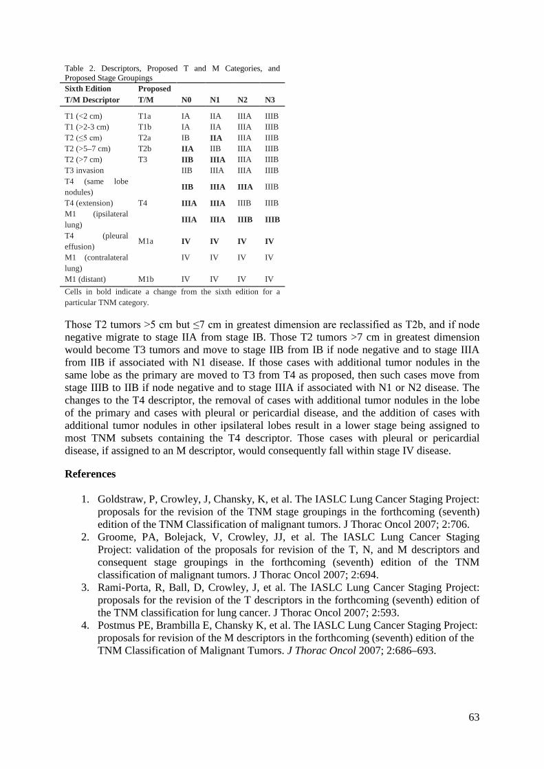
63
Table 2. Descriptors, Proposed T and M Categories, and Proposed Stage Groupings Sixth Edition Proposed T/M Descriptor T/M N0 N1 N2 N3
T1 (<2 cm) T1a IA IIA IIIA IIIB T1 (>2-3 cm) T1b IA IIA IIIA IIIB T2 (≤5 cm) T2a IB IIA IIIA IIIB T2 (>5–7 cm) T2b IIA IIB IIIA IIIB T2 (>7 cm) T3 IIB IIIA IIIA IIIB T3 invasion IIB IIIA IIIA IIIB T4 (same lobe nodules)
IIB IIIA IIIA IIIB
T4 (extension) T4 IIIA IIIA IIIB IIIB M1 (ipsilateral lung)
IIIA IIIA IIIB IIIB
T4 (pleural effusion)
M1a IV IV IV IV
M1 (contralateral lung)
IV IV IV IV
M1 (distant) M1b IV IV IV IV Cells in bold indicate a change from the sixth edition for a particular TNM category. Those T2 tumors >5 cm but ≤7 cm in greatest dimension are reclassified as T2b, and if node negative migrate to stage IIA from stage IB. Those T2 tumors >7 cm in greatest dimension would become T3 tumors and move to stage IIB from IB if node negative and to stage IIIA from IIB if associated with N1 disease. If those cases with additional tumor nodules in the same lobe as the primary are moved to T3 from T4 as proposed, then such cases move from stage IIIB to IIB if node negative and to stage IIIA if associated with N1 or N2 disease. The changes to the T4 descriptor, the removal of cases with additional tumor nodules in the lobe of the primary and cases with pleural or pericardial disease, and the addition of cases with additional tumor nodules in other ipsilateral lobes result in a lower stage being assigned to most TNM subsets containing the T4 descriptor. Those cases with pleural or pericardial disease, if assigned to an M descriptor, would consequently fall within stage IV disease. References
1. Goldstraw, P, Crowley, J, Chansky, K, et al. The IASLC Lung Cancer Staging Project: proposals for the revision of the TNM stage groupings in the forthcoming (seventh) edition of the TNM Classification of malignant tumors. J Thorac Oncol 2007; 2:706.
2. Groome, PA, Bolejack, V, Crowley, JJ, et al. The IASLC Lung Cancer Staging Project: validation of the proposals for revision of the T, N, and M descriptors and consequent stage groupings in the forthcoming (seventh) edition of the TNM classification of malignant tumors. J Thorac Oncol 2007; 2:694.
3. Rami-Porta, R, Ball, D, Crowley, J, et al. The IASLC Lung Cancer Staging Project: proposals for the revision of the T descriptors in the forthcoming (seventh) edition of the TNM classification for lung cancer. J Thorac Oncol 2007; 2:593.
4. Postmus PE, Brambilla E, Chansky K, et al. The IASLC Lung Cancer Staging Project: proposals for revision of the M descriptors in the forthcoming (seventh) edition of the TNM Classification of Malignant Tumors. J Thorac Oncol 2007; 2:686–693.

64
014 STAGE IIIB MANAGEMENT OF NON-SMALL CELL LUNG CANCER Jovanović D. Institute for Lung Diseases of Serbia, Beograd, Serbia Locally advanced non-small cell lung cancer (IIIB NSCLC) is a multifaceted disease that is challenging to manage. In stage IIIB disease, research to date has shown benefits for combined modality therapy with chemotherapy, radiation, or surgery, or both, for properly chosen candidates. The majority of patients can be appropriately treated with a combination of chemotherapy and radiation therapy; however, a subset of stage III patients who are considered surgical candidates may require a modification of this plan. Potentially resectable disease remains the most controversial area: in these cases neoadjuvant chemotherapy decreases the risk of distant metastases and induces high rates of response. A phase II study employing neoadjuvant docetaxel/cisplatin suggested that this regimen is well tolerated, does not increase perioperative morbidity/mortality, and may improve survival and reduce the risk of distant and local relapse. Phase III randomized controlled trials are needed to confirm the survival benefits of neoadjuvant chemotherapy or induction chemoradiation followed by surgical resection in this subset of patients with NSCLC. Treatment options of chemotherapy or radiation may differ among the clinically distinct stage III subsets. Stage IIIB with pathologically confirmed involvement of the pleura (T4, pleural effusion) is synonymous with advanced disease and is best managed with palliative therapy. For the remainder of patients with stage IIIB disease who have potentially curable disease, optimal treatment strategies also remain to be defined. Treatment goals include local tumor control (Radiotherapy +/- chemotherapy) and eradication of distant micro metastases (chemotherapy), while minimizing adverse events. For this unresectable stage IIIB NSCLC, concurrent chemoradiation is the standard of care at the present time for patients with good performance status, good pulmonary function tests, and an acceptable volume of normal lung receiving 20 Gy (V20). The ACCP guidelines for the treatment of unresectable stage III NSCLC recommend combined modality therapy with platinum-based chemotherapy and definitive thoracic radiation, particularly for patients with good performance status. Recent clinical trials and a meta-analysis showed that concurrent chemotherapy and radiotherapy (chemoradiation) affords superior outcomes to sequential therapy. According to ACCP Guidelines for patients with stage IIIB NSCLC and PS 0 or 1 and minimal weight loss ( 5%), concurrent chemoradiotherapy is recommended. Concurrent radiotherapy together with full-dose combination chemotherapy improves survival of patients with locally advanced NSCLC by 5.7% at 3 years mainly due to the decrease of loco-regional progression (6.0% at 3 years) but at the expense of a higher incidence of acute, reversible oesophagitis. Whereas platinum-based doublets remain the standard regimen, the choice of the complementary agent remains a subject of investigation. Over the past decade, agents under consideration for platinum-based doublets include docetaxel, paclitaxel, irinotecan, vinorelbine, and gemcitabine. Of these agents, the taxanes have been the most extensively studied. The most efficacious chemotherapy drugs to be combined with thoracic RT and the number of cycles of chemotherapy needed to yield the best results are still uncertain. No one combination chemotherapy regimen can be recommended. New techniques of radiation may also increase the efficacy and the feasibility of radiation. The development of three-dimensional conformal radiotherapy in the 1990s gave the possibility of dose escalation and concomitant association with chemotherapy to be delivered with manageable toxicities. Multimodal therapeutic sequences are now investigated

65
integrating new techniques of irradiation: intensity modulation allows an even superior dose escalation and an increased focalization of the ballistics etc. Recent investigations have attempted to incorporate the emerging epidermal growth factor and vascular endothelial growth factor receptor targeted therapies into the care of patients with locally advanced NSCLC. Ongoing studies are focused on defining their role in the stage III setting. Investigation of the role of full-dose chemotherapy as induction or consolidation of concurrent chemoradiotherapy seeks to augment control of distant micro metastases further. Phase III trials are currently evaluating the benefit from induction and consolidation chemotherapy in this setting. When compared with sequential or concurrent regimens, results from trials of induction regimens have been disappointing. Three phase III trials failed to demonstrate a significant clinical advantage for induction regimens when compared with either sequential or concurrent regimens in unresectable NSCLC. In the interim, data from studies of consolidation therapy have been promising. To date, consolidation docetaxel after concurrent etoposide, cisplatin and thoracic radiation has shown encouraging survival results in a large SWOG phase II trial. As the cisplatin/etoposide combination can be given at full dose concurrent with radiation, this doublet is considered the standard option for chemoradiotherapy protocols so as not to compromise distant efficacy for local control. Two sequential, non-randomized studies by the SWOG evaluated consolidation with different regimens after cisplatin/ etoposide chemoradiotherapy, and a comparison allows the evaluation of docetaxel as a consolidation agent.
In conclusion, stage IIIB NSCLC is a heterogeneous disease that presents a management challenge to the clinician. Unanswered questions remain about definitive chemoradiotherapy, including the optimal chemotherapy agents; dose, duration, and density of chemotherapy; clarifying the role of consolidation; the integration of targeted agents, identifying the proper radiation dose, schedule, and technique.

66
015 MALIGNANT PLEURAL MESOTHELIOMA Sečen N. Institute for Pulmonary Diseases of Vojvodina, Sremska Kamenica, Serbia Malignant pleural mesothelioma (MPM) is a rare tumor, the incidence is 1.1/100 000 in Germany (1) and 2.1/100 000 in Canada (2), but it is highly aggressive malignancy. The incidence of MPM is increasing worldwide. It is estimated to double within the next 20 years. As the result of widespread exposure to asbestos in recent decades as it is an asbestos-associated cancer. There is a long latent period between first exposure to asbestos and diagnosis of mesothelioma that is rarely less than 15 years and often exceeds 30 years. Median survival of MPM is currently 12 months (3). MPM occurs predominantly in men (ratio of men to women, 5:1) age 45-85 years and 80% of men were exposed to asbestos in the workplace. The most common symptom for MPM patients is chest pain. But, the pain often is not associated directly with the lung pleura and often appears in the shoulder or upper abdomen. Shortness of breath is the main symptom, usually. Cough, weight loss and anorexia are present in some patients, but are less common. Finally, the rapid growth of the pleural mesothelioma enlarges the pleural space, causing it to fill with fluid, which leads to the discomfort or pain associated with first detection of the disease. Diagnosis: Currently, there are no approved screening modalities for the early detection of mesothelioma. Two serum markers, serum mesotheliom-related peptide and osteopontin have recently been developed. Serum mesotheliom-related peptide may be predictive of disease recurrence after surgical resection. Osteopontin, a glycoprotein that binds integrin and CD44 receptors, may distinguish patients with MPM from those who have benign pleural changes resulting from asbestos exposure. Cytogenic studies in MPM have identified changes in all chromosomes. Deletion of 9p21, encompassing the loci for CDKN2A and CDKN2B, have been found in a high percentage of MPM cell lines. Mutation or inactivation of the neurofibromatosis gene type 2 (NF2) at 22q12 locus is present in a high proportion of MPM cell lines.P16/CDKN2A homozygous deletion is a significant independent adverse prognostic factor (4). International TNM staging system for MPM: Stage I Stage Ia - TIa, N0, and M0: primary tumor limited to ipsilateral parietal pleura Stage Ib T1b, N0, M0 Ia+focal involvement of visceral pleural.
Stage II T2, N0, and M0: stage Ia or Ib plus confluent involvement of diaphragm or visceral pleura or involvement of the lung.
Stage III Any T3Mo, any N1Mo, any N2Mo - locally advanced tumor; ipsilateral, bronchopulmonary or hilar lymph node involvement; subcarinal or ipsilateral mediastinal lymph node involvement. Stage IV Any T4, any N3, any M1 - locally advanced technically unresectable tumor;
contralateral mediastinal, internal mammary and ipsilateral or contralateral supraclavicular lymph node involvement; distant metastases.
Cytology and histology, including immunohistochemistry, are the gold standards for diagnosis. Video assisted thoracoscopy or open pleural biopsy are necessary diagnostic tools. There are three main histological types: epithelial, sarcomata’s and mixed, about 60% are epithelial.
67
CT scan of thorax is a basic clinical diagnostic procedure and FDG-PET/CT is useful in evaluation of the treatment. Mediastinoscopy and Video assisted thoracoscopy are useful in determining the stage which is essential for the treatment. Several staging systems are existing, TNM system is in the use, mostly. Treatment Surgery In general terms, most stage I and some stage II and III MPM are potentially resectable, but there are exceptions. Resectability is based not only on the size of the tumor, but also on the subtype (mostly epithelioid tumors are potentially resectable). Stage is an important factor in determining a patient's prognosis, but other factors also play a role. Some factors linked to longer survival times include: good performance status, younger age, epithelioid subtype, not having chest pain, any significant weight loss, earlier stage disease and adequate cardiopulmonary function. Extra-pleural pneumonectomy (EPP) includes removal tissues in the hemithorax, parietal and visceral pleura, involved lung, mediastinal lymph nodes, with resection of the hemidiaphragm and the pericardium en bloc has the potential for a radical treatment and neoadjuvant chemotherapy and/or adjuvant radiotherapy are combined. The palliative treatment of MPIM patients is parietal pleurectomy/decortication (P/D) or talc pleurodesis (5). Chemotherapy Neoadjuvant chemotherapy include platinum doublets (cisplatin plus gemcitabine) or (cisplatin plus pemetrexed) with median survival ranging between 19 to 25 months and median time to progression of 13.1 months, overall survival 16.6 months and 1-yea survival rate 67%. Adjuvant chemoradiotherapy is administer after EPP and there are a few trials were carboplatin and paclitaxel for two cycles plus thoracic radiation of 50 Gy with concurrent paclitaxel weekly plus carboplatin paclitaxel for two cycles are recommended (6).2-year survival rate was 38% and 5 year survival rate was 15%. Clinical benefits in patients with unresectable MPM are on platinum containing regimens - Platinum combined with anthracycline (32.4%), gemcitabine or irinotecan has highest response rates. The combination ciplatin (75mg/m2) and pemetrexed (500mg/m2 given every three weeks was established as a standard-of-care front line regimen. Response rates 41.3%, median overall survival of 12.1 months and median time to progression of 5.7% months. Patient quality of life also improved rapidly, significantly often seen by week 15. Additional front line chemotherapy is gemcitabine plus cisplatin with RR between 12% and 48% and median overall survival times of 9.4 to 13months. Second line chemotherapy-if pemetrexed is not given in the front line setting it should be administered in the salvage setting, either alone or in combination with cisplatin. Gemcitabine plus vinorelbine was found to have some efficacy. Radiotherapy Adjuvant hemithoracic radiotherapy (54Gy) is the treatment modality that decreases the risk of local recurrence and distant metastases. Biologic therapy Patients with MPM have high levels of plasma vascular endothelial growth factors (VEGF). This suggests that antiangiogenic therapy could benefit some patients with MPM, some trials are on. Thalidomide as a single agent has been reported to achieve stabilization in 25% of patients for more than 6 months. Photodynamic Therapy Photodynamic Therapy (PDT) involves administering photosensitive drugs into the mesothelial cells. A laser light is used to activate the photosensitive drugs in order to destroy the surrounding cancer cells. As yet, PDT has not shown success in improving the survival rate for mesothelioma patients.

68
Gene therapy Early work with gene therapy used adenovirus vectors containing the herpes virus thymidine kinase (Ad-HSVtk) suicide gene administered intrapleurally followed by intravenous ganciclovir to selectively kill the tumor cells. Trials are on (6). In conclusion the surgical resection and adjuvant radiotherapy remains the mainstay of treatment for patients with resectable MPM. Systemic treatment is also necessary. For the unresectable MPM patients the antifolates or gemcitabine, given in combination with platinum agent, have made the greatest clinical impact to date. Further progress is needed and novel therapeutic agents should be priority. References:
1. Canadian Medical Association or its licensors. Facts and Figures, Canadian cancer statistics at a glance: mesothelioma. CMAJ March 11, 2008; 178(6).
2. Oluf Dimitri Roe, Endre Anderssen, Helmut Sandeck, Tone Christensen, Erik Larsson, Steinar Lundgren. Lang Cancer, 2009, 3311-36.
3. R A Stahel, W Weder, E. Felip. Malignant pleural mesothelioma: ESMO Clinical Recommendations for diagnosis, treatment and follow-up, Annals of Oncology 20 (Supplement 4), 2009; 73-75.
4. Rolf A Stahel. Malignant pleural mesothelioma: A new standard of care. Lung Cancer (2006) 545, S9-S14.
5. Davide Rice. Surgery for malignant pleural mesothelioma. Annals of Diagnostic Pathology 13 (2009) 65-72.
6. Louise Li, Albiruni Ryan Abdul Razak, Andrew Hughes. Carboplatin and pemetrexed in the management of malignant pleural mesothelioma: A realistic treatment option? Lung Cancer, 64 (2009) 207-210.

69
016 POOR PERFORMANCE STATUS PATIENT IN ADVANCED LUNG CANCER: To Treat or Not to Treat Stanetić M. Clinical Centre Banja Luka, Clinic for Lung Diseases, Banja Luka, Bosnia Herzegovina Treatment of carcinoma of the bronchus as the treatment of any other disease has the goal to cure. Unfortunately, the small number of patients with malignant diseases has the realistic chance of being cured – these are mainly asymptomatic patients discovered in early stage of a disease. The majority of patients are discovered when disease symptoms manifested, in the other words when disease already advanced and when the chances to cure a patient are significantly lower and therapeutic approach is more complex1, 2. The most often the lung cancer is diagnosed in the stage of a disease when the surgical intervention is not so efficient and effective, and therefore the biggest number of patients is treated by chemotherapy, radiotherapy or by combined treatment modalities. Treatment of carcinoma of the bronchus is complex, and it works on not only on curative but also on palliative plan, on the physical as well as the psychosocial level. Considering the fact that the results of treatment frequently do not lead to a cure, the treatment goal is life prolonging with palliation, that is lessen pain and discomfort and improvement in quality of life. Latter studies on quality of live during the chemotherapeutic treatment indicated that improvement or preservation of quality of life mainly depends on the tumor response to treatment. In addition, the basal quality of life could be not only predictor of treatment response and survival rate but also have a significant impact on other known prognostic factors (treatment type, sex, age). Karnofsky3 and Zubord4 developed systems for Evaluations of performance status that are overall accepted. Edited Zubord Criteria performanse status is accepted by Eastern Cooperative Oncology Group (ECOG) and WHO. Numerous studies and papers prove prognostic value of PS. PS is statistically significant prognostic factor that impact the patient survival. Comparison of individual survival curves of all patients indicates that only between the patients with ECOG index 3 and ECOG index 4 there is no statistically significant difference in outcome5.
MD-PS n Median Survival (mo) Pt-PS n Median Survival (mo) 0 11 10.6 0 16 14.4 1 51 10.1 1 35 8.2 2 21 4.6 2 13 7.1 3 9 2.8 3 27 3.8 4 0 4 1 1.8 Total 92 Total 92 p=0.01 MD-PS and p=0.001 Pt-PS
Another problem that obtrudes while determining of therapeutic treatment is advancing of disease, i.e. stadium/stage of disease. Stadium of disease is most often connected with the performance status. If the PC shows worse outcome, by diagnostic methods it will be confirmed that this is an advance stage of disease. Survival length directly depends on stage of disease6.
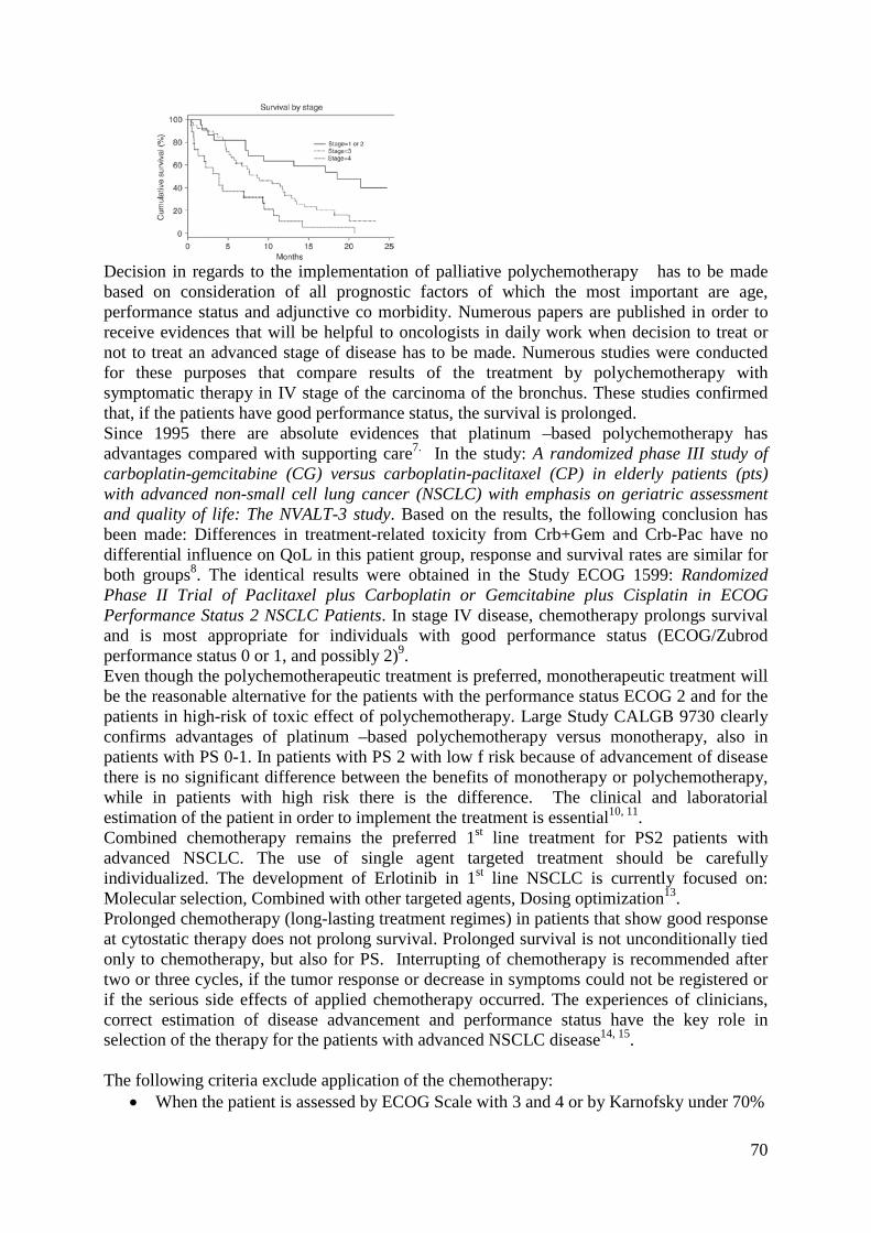
70
Decision in regards to the implementation of palliative polychemotherapy has to be made based on consideration of all prognostic factors of which the most important are age, performance status and adjunctive co morbidity. Numerous papers are published in order to receive evidences that will be helpful to oncologists in daily work when decision to treat or not to treat an advanced stage of disease has to be made. Numerous studies were conducted for these purposes that compare results of the treatment by polychemotherapy with symptomatic therapy in IV stage of the carcinoma of the bronchus. These studies confirmed that, if the patients have good performance status, the survival is prolonged. Since 1995 there are absolute evidences that platinum –based polychemotherapy has advantages compared with supporting care7. In the study: A randomized phase III study of carboplatin-gemcitabine (CG) versus carboplatin-paclitaxel (CP) in elderly patients (pts) with advanced non-small cell lung cancer (NSCLC) with emphasis on geriatric assessment and quality of life: The NVALT-3 study. Based on the results, the following conclusion has been made: Differences in treatment-related toxicity from Crb+Gem and Crb-Pac have no differential influence on QoL in this patient group, response and survival rates are similar for both groups8. The identical results were obtained in the Study ECOG 1599: Randomized Phase II Trial of Paclitaxel plus Carboplatin or Gemcitabine plus Cisplatin in ECOG Performance Status 2 NSCLC Patients. In stage IV disease, chemotherapy prolongs survival and is most appropriate for individuals with good performance status (ECOG/Zubrod performance status 0 or 1, and possibly 2)9. Even though the polychemotherapeutic treatment is preferred, monotherapeutic treatment will be the reasonable alternative for the patients with the performance status ECOG 2 and for the patients in high-risk of toxic effect of polychemotherapy. Large Study CALGB 9730 clearly confirms advantages of platinum –based polychemotherapy versus monotherapy, also in patients with PS 0-1. In patients with PS 2 with low f risk because of advancement of disease there is no significant difference between the benefits of monotherapy or polychemotherapy, while in patients with high risk there is the difference. The clinical and laboratorial estimation of the patient in order to implement the treatment is essential10, 11. Combined chemotherapy remains the preferred 1st line treatment for PS2 patients with advanced NSCLC. The use of single agent targeted treatment should be carefully individualized. The development of Erlotinib in 1st line NSCLC is currently focused on: Molecular selection, Combined with other targeted agents, Dosing optimization13. Prolonged chemotherapy (long-lasting treatment regimes) in patients that show good response at cytostatic therapy does not prolong survival. Prolonged survival is not unconditionally tied only to chemotherapy, but also for PS. Interrupting of chemotherapy is recommended after two or three cycles, if the tumor response or decrease in symptoms could not be registered or if the serious side effects of applied chemotherapy occurred. The experiences of clinicians, correct estimation of disease advancement and performance status have the key role in selection of the therapy for the patients with advanced NSCLC disease14, 15. The following criteria exclude application of the chemotherapy:
• When the patient is assessed by ECOG Scale with 3 and 4 or by Karnofsky under 70%

71
• When the weight loss is higher that 10% • When the patient is older than 70 • When the creatinine clearance rate is under 0,6mmol/l • When the serum creatinine rate is above 130mmol/l • When the leukocyte count is under 3x10⁹/l • When the serum bilirubin is under 35mmol/l.
The prognostic factors in IV stage of NSCLC16: • Performance status remain the most important factor • Careful assessment and estimation is crucial and adequate for selection of patients for
chemotherapy • Benefits from treatment and treatment index are the best in patients with ECOG 0-1 • In patients with ECOG 2 increased toxicity reduces the treatment benefits • ECOG 3-4 patients are not the candidates for treatment by chemotherapy • Performance status ECOG 3-4 assumes only implementation of good supportive care
(because these patients do not have benefit from the cytotoxic therapy). Very often in these patients we do not expect the survival period longer than 12 months.
Symptoms that indicate survival ≤12 months: - Poor performance status ECOG ≥3 or KPS≤50% - Hypercalcemia - Cerebral metastasis - Delirium - Vena cava syndrome - Kachexia / Kachexy - Malignant effusion - Bilirubin ≥X2,5 - Creatinine ≥ X3
Conclusions: Performance status directly influence on the choice of treatment. Patients with poor general status/condition can be excluded from the treatment under the assumption that it will cause more harm than treatment effect and benefits in these patients. The specific antitumor therapy, because of possible side-effects is applied in patients with ECOG performance status 0.1 and 2. These results may help to choose the appropriate treatment in the clinical practice. References:
1. Hyde L, Hyde Cl. Clinical manifestations of lung cancer. Chest 1974; 65: 299-306. 2. Cromartie RS, Parker EF, May JE et al. Carcinoma of the lung: a clinical review. Ann
Thoracic Surg 1980; 30:30-35. 3. Karnofsky DA, Burchenal JH. The clinical evaluation of chemotherapeutic agents in
cancer. In MacLeod CM (ed): Evaluation of chemotherapeutic agents. New Yorrk, Columbia University press, 1949: 191-205.
4. Zubord CG, Schneiderman M, Frei E, et al. Appraisal of methods for the study of chemotherapy of cancer in man: comparative therapeutic trial of nitrogen mustard and triethylene thiophosphamide. J Chron Dis, 1960; 11:7-33.
5. S P Blagden, S C Charman, L D Sharples, L R A Magee and D Gilligan: Performance status score: do patients and their oncologists agree? British Journal of Cancer 2003; 89, 1022–1027.

72
6. E. Dajczman, G. Kasymjanova, N. Swinton, D. St-Pierre, T. Swanson, H. Kreisman, J. S. Agulnik, V. Cohen, N. MacDonald, D. Small: Does NSCLC patient-rated performance status predicts survival more accurately than physician ratings? Journal of Clinical Oncology, 2007 ASCO Annual Meeting Proceedings Part I. Vol 25, No. 18S (June 20 Supplement), 2007: 9022
7. NSCLC-Collaborative Group.B Med J 311 8. WCLC 2007 – Biesma B et al., A randomized phase III study of carboplatin-
gemcitabine (CG) versus carboplatin-paclitaxel (CP) in elderly patients (pts) with advanced non-small cell lung cancer (NSCLC) with emphasis on geriatric assessment and quality of life: The NVALT-3 study. Abstract # P2-239
9. Langer G ea. Randomized Phase II Trial of Paclitaxel Plus Carboplatin A or Gemcitabine Plus Cisplatin B in ECOG Performance Status 2 NSCLC Patients: ECOG 1599.J Clin Oncol 25:418-423
10. American Society of Clinical Oncology Treatment of Unresectable Non–Small-Cell Lung Cancer Guideline: Update 2003. JCO 2004
11. Helbekkmo N, Aasebø U, Sundstrøm SH, von Plessen C, Brunsvig PF, Bremnes RM; Norwegian Lung Cancer Study Group. Treatment outcome in performance status 2 advanced NSCLC patients administered platinum-based combination chemotherapy. Lung Cancer 2008; 62(2):253-60.
12. NCCN Guidelines, 2008, 13. WCLC 2007 – Bonomi P et al., Single agent versus combination therapy in advanced
NSCLC patients with performance status 2: Results from a regression analysis of STELLAR 3 and 4.Abstract # B2-04,
14. Harrington SE, Smith TJ. JAMA. 2008;299(22):2667-2678, 15. Perin B. Veličina i lokalizacija tumora kao faktori koji utiču na ishod liječenja
bolesnika sa nemikrocelularnim karcinomom bronha. Doktorska disertacija. Univerzitet u Novom Sadu,Medicinski fakultet, 1987. (Size and localization of the tumor as the factors that influence the results of treatment of non-microcellular carcinoma of the bronchus. Doctoral Dissertation, University of Novi Sad, Medical Faculty, 1987)
16. Chest; ACCP 2003

73
017 TUBERCULOSIS CONTROL IN BOSNIA HERZEGOVINA IN RECENT YEARS Žutić H. Clinical Center University Sarajevo, Clinic for Lung Disease and TB Sarajevo, Bosnia Herzegovina Worldwide, one person out of three is infected with Mycobacterium tuberculosis – two billion people in total. TB accounts for 2.5 % of the global burden of disease and is the commonest cause of death in young women, killing more women than all causes of maternal mortality combined. TB currently holds the seventh place in the global ranking of causes of death. 1, 2
There are the 18 priority countries for TB control in the Europe in the eastern part of the Region, and Bulgaria, Romania and Turkey in central Europe. Poor economies and public health approaches are the main causes of the resurgence of TB in these countries. Despite the progress achieved in recent years, the level of TB control in Europe is still inadequate. Every year in Europe, 445000 people become sick with TB and 66 000 people die of TB. Of these, 75% are in Eastern Europe. Of the cases registered for treatment and reported to WHO in 2005, the ratio of men to women was 2:1.6 • The European Region has a huge variation in TB incidence rates, ranging from 5 (in Norway) to 198 (in Tajikistan) new TB cases per 100 000 population per year. The Russian Federation is twelfth on the list of the 22 highest TB-burden countries in the world. 3, 4 Bosnia and Herzegovina (BH) is among those countries with an “intermediate” TB burden in WHO European Region, i.e. with high TB incidence but remained steady during the last years and with fairly developed network of health infrastructure that requires upgrading. The last anti-TB drug resistance survey, performed in the year 2007, has shown a prevalence of multi-drug resistant TB (MDR-TB) of 0.1 % in newly TB diagnosed cases and 2.0% in those previously-treated. The reported success rate for new pulmonary TB cases is 95- 96%.5 In 2007, the estimated incidence rate was 51 per 100 000 population and prevalence rate 55 per 100 000 population. The mortality rate was 7; MDR rate was 0.4% in new TB cases, and 6.6% in relapse TB cases. 5 Existing TB strategies in the country entirely reflect WHO-promoted Stop TB strategy, while DOTS elements are the cornerstone of the strategy. Tuberculosis surveillance is identified as the second priority following HIV/AIDS. Furthermore, country level assessment of microbiological laboratories was also completed towards end 2006 and was subject to deliberations between national and international stakeholders throughout 2007. In June 2008 BH signed Stabilization and Association Agreement (SAA) with EU including the obligations related to public health. In addition, since June 1st, 2007 BH is obliged to comply with International Health Regulations. BH received GFATM 5-year grant (R 6 grant) of $ 5.6 million in 2007 for the proposal with four objectives: Increase capacity for case detection, Ensure and maintain access to TB program for vulnerable groups, Maintain control of drug resistance through early detection and effective treatment of new cases and appropriate management of MDR, and Adopt the new Stop TB strategy and strengthen DOTS all over the Country.5, 6
Current surveillance system does not allow objective assessment of the degree of achievement of global and NTP targets. According to the WHO Global Tuberculosis Control 2008 document, reported DOTS new smear positive treatment success in BH is in the rage from 88% (1988) to 98 % (2001) while reported DOTS new smear-positive case detection rate is in the range from 38% (1998) to 95% (2004). Reported figures and fluctuations raise an issue of the reported data quality. TB recording and reporting systems appear to function reasonably in terms of reporting timeliness, but due to communication problems inherent to fragmentation of the health systems and frequent delays caused by insufficient use of modern electronic means of

74
communication there are reasons to believe that the reported data are not entirely reliable. Current registration system considers individual notification upon TB diagnoses, while treatment outcome results present cumulative information, without reference to individual results. Furthermore, information system for data collection and analyses was not developed properly and should be further developed in relation to treatment outcome results. There is also loss of information related to the notification of TB cases, due to the lack of clearly defined reporting lines, especially at the first line of reporting. There is also a gap identified as the lack of essential equipment and appropriate IT system of reporting. The lack of reliable data related to TB/HIV/AIDS co-infection in sexual minorities groups present an obstacle for development of efficient risk behavior prevention measures. Current weaknesses in the laboratory network are the inconsistence of proper terms of reference and the lines of responsibility of all levels laboratories; inappropriate and insufficient equipment (as bio/safety cabinets of appropriate class), as well as infection control measures at the regional (L2 and L3) laboratories; non existence of the routine external and internal L3 and L2 Q/A and Q/C systems; lack of the qualitative and efficient sample transportations mechanisms; still not established laboratory recording and reporting systems and undeveloped plan of supervision and monitoring of the laboratory network within the overall NTP monitoring and supervision plan; lack of human resources and inappropriate workload of health staff in the laboratory network. Infection control measures are not fully implemented in the health facilities, due to the lack of General infection control measures, including the lack of Guideline for TB infection control. There is a lack of social support measures for the TB patients. There is need for commitment of TB patients for adherence to treatment during the continuation phase. Currently, within R 6 grant patronage nurses are visiting only limited number of TB patients, due to the organizational and human resource constrains. Table 1: Key indicators of TB control in Bosnia and Herzegovina, WHO Global TB Report 2008 and 2009 20067 Estimates 20075 Estimates The estimated Population 3 926 406 3 935 000 Incidence (all cases/100 000 pop/yr) 51 51 Incidence (SS+/100 000/yr) 23 19 Prevalence (all cases/100 000 pop/yr) 57 55 Mortality (deaths/100 000 pop/yr) 7.4 7,0 Of new TB cases, % HIV - - Of new TB cases, % MDR-TB 0.4 0,4 Of previously treated cases, % MDR-TB 6.6 6,6 Surveillance and DOTS implementation Notification rate (new and relapse/100 000 pop/yr) 45 60 Notification rate (new SS+/100 000 pop/yr) 14 19 Case detection rate (all new cases, %) 84 110 Case detection rate (new SS+ cases, %) 62 81 DOTS notification rate (new and relapse/100 000 pop/yr) 45 60 DOTS notification rate (new SS+/100 000 pop/yr) 14 19 DOTS case detection rate (all new cases, %) 84 110 DOTS case detection rate (new SS+ cases, %) 62 81 There is no systematic approach for MDR TB management in the country. Some individual TB MDR and DR cases are under the treatment, but there are no appropriate hospital

75
capacities for isolation of these patients, as well as clear guidelines for it. Furthermore, there is lack of second-line TB drugs, which are planned to be procured in the second phase of R6 TB grant (2010-2012), as well as development of DOTS plus strategy. Currently, capacities of TB labs do not allow performing DST for second line TB drugs. Moreover, there is no official registration of DR and MDR TB cases. Medical commission for MDR TB management will be established within R6 grant (phase II) to comply with GLC regulations. 5 References:
1. WHO. Global tuberculosis control: surveillance, planning, finances. Geneva: World Health Organization, 2009.
2. Lopez AD, Mathers CD, Ezzati M, Jamison DT, Murray CJL, eds. Global burden of disease and risk factors. New York: Oxford University Press, the World Bank, 2006.
3. Raviglione MC, Uplekar MW. WHO’s new Stop TB Strategy. Lancet 2006; 367: 952–55.
4. Dye C, Maher D, Weil D, Espinal M, Raviglione M. Targets for global tuberculosis control. Int J Tuberc Lung Dis 2006; 10: 460–62.
5. Anonymus. Round 6: Proposal of Bosnia and Herzegovina to Global Fund to Fight AIDS TB and Malaria. 2006.
6. Global Fund to Fight AIDS TB and Malaria. Partners in impact—results report. Geneva: Global Fund to Fight AIDS, TB and Malaria, 2007.

76
018 HOSPITAL ACQUIRED PNEUMONIA (HAP) – CLINICAL VIEW Vujičić-Cupać LJ. Department for Lung Diseases and TB, University Clinical Hospital, Mostar, Bosnia Herzegovina
Hospital acquired pneumonia HAP - nosocomial pneumonia, is lung infection, which onset 48-72 h after hospitalization of the patients without the lung infection before hospitalization. Patients which are hospitalized have bigger risk of infection:
1. because of basic illness 2. because of application of more aggressive diagnostic procedures and treatment
procedures 3. Because that patients usually has compromised immunity and very often they has
treatment with immunosuppressive. HAP is on third place of most frequent of hospital infections:
1. Urinary infections (40-45%) 2. Infection of a surgical wounds (25-35%) 3. HAP (15-20%) 4. Presence of bacteria in blood culture, the sepsis (5-10%).
HAP appears more with patients which are not in the intensive care unit, but the biggest risk for its appearance, exists in patients that are on mechanical ventilation. That pneumonia presents the subgroup of this pneumonia, which appears in the first two days of mechanical ventilation, the incidence of their appearance is 0.5-1% of all patients which are hospitalized, and that pneumonia appears in 20% of patients which are admitted into the intensive care unit. Usually we recognized early and late HAP according to the time of beginning, from the day of admission to the hospital. Some of patients which are hospitalized have developed pneumonia in less then 4 days after hospitalization. That is the early onset HAP. Both early HAP and the community acquired pneumonia (CAP) have the same source, although atypical microbes can caused the hospital pneumonia too. The late onset pneumonia which begins after the fourth day of hospitalization generally has the source with more microbes, and causes are mostly aerobic Gram negative microbes and Staphylococcus aureus. HAP is a heterogeneous term with consideration of the causes, pathogenesis and consequences. Because of that it is necessary to make differences among few terms. Pneumonia after aspiration at patients that are hospitalized and they have disturbance of consciousness. It is the most include the elder persons or persons which have disturbance of act of swallow or have a weakened reflex cough. They are generally caused by bacteria which make colonization of oral and pharyngeal mucous membrane (anaerobes of oral cavity), but it also includes (especially in patients which are hospitalized for a longer time) the unusual hospital bacteria’s. Pneumonia at patients with immunosuppressant that are hospitalized presents reactivation of intracellular microorganisms (bacteria’s, viruses) most often, but it can be caused by usual hospital causes too. Pneumonia in patients that are been intubation present a special term and it appears in the intensive care unit most often. The most often, but not exclusively is connected with mechanical ventilation, so today it is recommended to use the term mentioned above (1). Hospital acquired pneumonia is connected with high mortality. Of all hospital infection it has the highest death rate, which is between 33% and 71%. Women have a higher risk from dying of hospital acquired pneumonias then men (3).

77
Table 1. Risk factors for activation of hospital acquired pneumonia (2). CLASSIFICATION OF
RISK FACTORS RISK FACTORS
Interventions Mechanical ventilation Endotracheal tubes Lying position Disturbance of mucociliary activity Air humidification Tracheotomy Application of positive pressure at the end of expiration (PEEP-a)
Other interventions Nasal – gastric probe Continuous enteral feeding Toracho – abdominal surgery Sedatives Antibiotics Corticosteroids, immunosuppressives Antacids, H2 antagonists
Patient’s status Old age Patient’s weight Illness difficulty level Malnutrition Disturbances of consciousness Duration of hospitalization
Comorbidity Diabetes mellitus Uremia Chronic obstructive lung disease and chronic heart diseases Malignant diseases Alcoholism Respiratory insufficiency (ARDS)
Infection control Contaminated hands of the staff Contamination of devices
Death result is more frequent in pneumonia caused by P. Aeruginosa (from 30-75%), Citomegalovirus and pneumonia caused by more microbes. Patients on mechanical ventilation have a bigger death rate than the patients that breathe normally (4). Etiology - Streptococcus pneumonia, S. Aureus and Hemophiles influenza (early and post-operating pneumonia) - G negative bacteria’s: Klebsiella pneumonia, Escherichia coli, Pseudomonas aeruginosa, Serratia marcescens, Enterobacter spp, Acinetobacter spp - And others like: Legionella, Pneumocystis carinii, Mycobacterium tuberculosis, Respiratory viruses, and Candida albicans Pathogenesis Bacteria’s can get into the distal part of respiratory tract after aspiration of microorganism which colonized oral and pharynx mucosa membrane, by inhalation of polluted aerosols or by blood expansion from a distant places of infection rarely. It is considered that gastro enteric system is the most often source of colonization of oral and pharyngeal mucous membrane. Criteria of hospital pneumonia diagnosis
- temperature >38ºC - lung infiltrate which begins now or progressive lung infiltrate on chest X ray of
which persist >24 hours - the secretion which contained the suppuration in the trachea - the growth of white cells in the blood (>12x10 9 L) Leucocytosis in DBP the
immature forms and high level of CRP

78
- the finding of auscultation on lungs (not obligated) - positive culture of the trachea aspirate or serologic tests - positive hemoculture (not obligated) - disorder onset during the process of the non-invasive or mechanical ventilation - tachypnoea - tachycardia
These criteria can be misleadingly positive, and in case of mechanically ventilated patients lungs are shadowed, lung atelectasis, hemorrhage, and colonization of trachea can be found after third day of intubation. That is why isolation of the microorganisms from sputum, hemoculture, aspirate trachea, pleural effusion is of great importance. To improve diagnostics biopsy samples are being taken using”protected specimen brush” or BAL (bronchoalveolar lavage). These tests are sensitive in range of 60% - 100%. These methods are invasive, and there is a possibility of complications. Non-bronchoscopic methods such as (protected BAL – pBAL) “blind” catheterization distal airway is often used. From all the listed methods the most frequently useful method is culture of endotracheal aspirate. Application of these bronchoscopic and non-bronchoscopic diagnostic procedures can be the main step towards the identification of epidemiology of HAP, especially at the patients on mechanical ventilation. Mechanic ventilation pneumonia Sustained mechanically assisted ventilation patients have 6 – 21 time greater risk from failing ill of the hospital acquired pneumonias, compared with the patients who are breathing without respirator assistance (6). Risk with using the respirator increases the possibility of pneumonia of 1% a day. Risk is increased because of the oropharyngeal microorganisms which are situated on the way of the endotracheal tubs, and weakened organism of the patients with severe illness. Bacteria can multiply on tubes and form glycocalix (biofilm) which offer protection from antibacterial drugs as well as from immune system. Some say that these bacteria aggregate can be embolized using the ventilation, aspiration or the tubes movement in the lower section of the respiratory tract. Bacteria often move near the bubble of end tracheal tubes. Thoracic percutation with patients who are temporary mechanically ventilated is predisposed risk factor for pneumonia. Pneumonia is connected with extended use of mechanic ventilation, but it is not connected with increased mortality. Mortality linked with HAP at the mechanically ventilated patients is around 50% (7). Prevention Attention must be focused on the prevention of the hospital acquired pneumonia.
1. Risk factors for the occurrence of the hospital pneumonia must be decreased. Flu vaccination, regulation of body weight, dental health, maintaining the oral hygiene, chronic disease control (diabetes, heart disease, COPD).
2. Ensuring the hospital hygiene • Washing the hands before handling the patient with the usage of the medical
apron and gloves • Cleaning, disinfection and sterilization of the items used for treatment of
patient • Regulation of the toxic waste disposal, washing the laundry, kitchen
hygiene control including the heating the water to 68ºC to prevent the appearance of the Legionella
• Rational use of antibiotics for resistance • Staff education • Breaking the chain of infection spreading
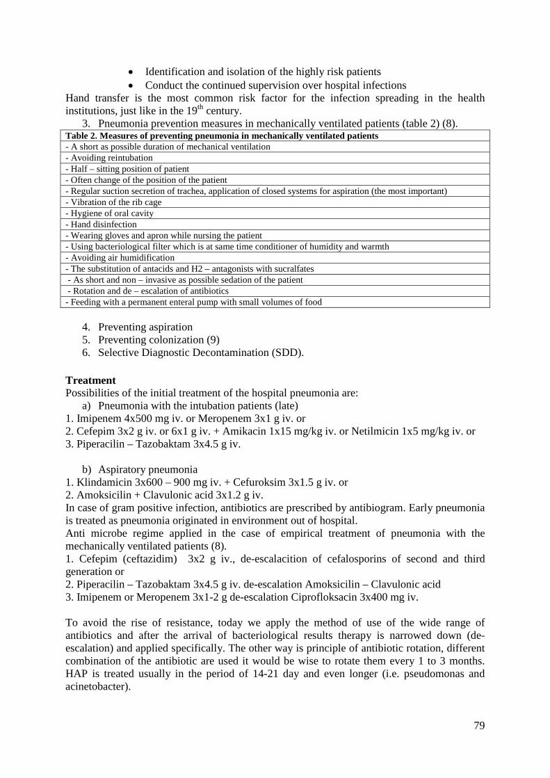
79
• Identification and isolation of the highly risk patients • Conduct the continued supervision over hospital infections
Hand transfer is the most common risk factor for the infection spreading in the health institutions, just like in the 19th century.
3. Pneumonia prevention measures in mechanically ventilated patients (table 2) (8). Table 2. Measures of preventing pneumonia in mechanically ventilated patients - A short as possible duration of mechanical ventilation - Avoiding reintubation - Half – sitting position of patient - Often change of the position of the patient - Regular suction secretion of trachea, application of closed systems for aspiration (the most important) - Vibration of the rib cage - Hygiene of oral cavity - Hand disinfection - Wearing gloves and apron while nursing the patient - Using bacteriological filter which is at same time conditioner of humidity and warmth - Avoiding air humidification - The substitution of antacids and H2 – antagonists with sucralfates - As short and non – invasive as possible sedation of the patient - Rotation and de – escalation of antibiotics - Feeding with a permanent enteral pump with small volumes of food
4. Preventing aspiration 5. Preventing colonization (9) 6. Selective Diagnostic Decontamination (SDD).
Treatment Possibilities of the initial treatment of the hospital pneumonia are:
a) Pneumonia with the intubation patients (late) 1. Imipenem 4x500 mg iv. or Meropenem 3x1 g iv. or 2. Cefepim 3x2 g iv. or 6x1 g iv. + Amikacin 1x15 mg/kg iv. or Netilmicin 1x5 mg/kg iv. or 3. Piperacilin – Tazobaktam 3x4.5 g iv.
b) Aspiratory pneumonia 1. Klindamicin 3x600 – 900 mg iv. + Cefuroksim 3x1.5 g iv. or 2. Amoksicilin + Clavulonic acid 3x1.2 g iv. In case of gram positive infection, antibiotics are prescribed by antibiogram. Early pneumonia is treated as pneumonia originated in environment out of hospital. Anti microbe regime applied in the case of empirical treatment of pneumonia with the mechanically ventilated patients (8). 1. Cefepim (ceftazidim) 3x2 g iv., de-escalacition of cefalosporins of second and third generation or 2. Piperacilin – Tazobaktam 3x4.5 g iv. de-escalation Amoksicilin – Clavulonic acid 3. Imipenem or Meropenem 3x1-2 g de-escalation Ciprofloksacin 3x400 mg iv. To avoid the rise of resistance, today we apply the method of use of the wide range of antibiotics and after the arrival of bacteriological results therapy is narrowed down (de-escalation) and applied specifically. The other way is principle of antibiotic rotation, different combination of the antibiotic are used it would be wise to rotate them every 1 to 3 months. HAP is treated usually in the period of 14-21 day and even longer (i.e. pseudomonas and acinetobacter).

80
It is not advisable to use a single therapy resize. Treatment has to be started with more than one antibiotic. The most commonly used the reference from ATS (American Thoracic Society), which is unified with empiric treatment of hospital pneumonias. There are no unified guidelines which could be applied in the entire world. With antibiotic therapy there must be added the support therapy (rehydratation, hypoxia correction, patient diet, as well as treatment of dysfunction of other organic systems). References:
1. Baršić, Krajinović. Hospital pneumonia – prevention, diagnosis and therapy. Medix. 2004; 52.
2. Cadtree TD, Pelletier SJ, Gleason TG, Pruett TL, Sawyer RG. Gender-dependent Differences in Outcome after the Treatment of Infection in Hospitalized Patients. JAMA. 1999; 282: 2143-8.
3. Chastre J, Traullet JL. Problem pathogens (Pseudomonas aeruginosa and Acinetobacter). Seuni Respir Infect. 2000; 15(4): 287-98.
4. Inglis TJ, Sherratt MJ, Sproat LJ, Gibson JS, Hawkey PM. Gastroduodenal dysfunction and bacterial colonization of the ventilated lung. Lancet. 1993, 341. 911-3.
5. Craven DE, Kunches LM, Kilinski V, Lichten berg DA, Make BJ, Mo Cabe WR. Risk factors of pneumonia and fatality in patients receiving continuo’s mechanical ventilation. Chest Med 1991, 12. 523-43.
6. Rello J, Lorente C, Diaz E, Bodi M, Boque C, Sandiumenge A, Santamaria JM. Incidence etiology and outcome of nosocomial pneumonia in ICU patients requiring percutaneous tracheotomy for mechanical ventilation. Chest 2003, 124(6). 2239-43.
7. Baršić B, Vince A. Intenzivna medicana. Zagreb HR, 2008. 8. Kollef MH. Prevention of hospital-associated pneumonia and ventilator-associated
pneumonia. Crit Care Med. 2004, 32(6): 1396-1405.

81
019 THE USE OF CORTICOSTEROIDS IN SEVERE COMMUNITY-ACQUIRED PNEUMONIA Čehajic M. Department for Lung Diseases and TB, University Clinical Hospital, Mostar, Bosnia Herzegovina Community-acquired pneumonia (CAP) is the first infectious cause of death, and the sixth cause of overall mortality in the developed world (2). Mortality among the hospitalized patients due to CAP is approximately 10-25 %, and it ranges from 30-50 % in patients requiring intensive care unit (ICU) admission. Despite advances in life-support measures and early antimicrobial therapy, the mortality due to severe pneumonia has not varied since the mid-1990's, suggesting that other factors are responsible for the poor outcome of this disease, as well (1,2). The studies showed that deaths occurring within the first five days of treatment were not due to failure to eradicate the microorganism, but rather to an inappropriate response
of the host, being one of the key factors defining the development of pneumonia which seems to increase excessively in severe pneumonia with no response (2). Severe community-acquired pneumonia (SCAP) is usually defined as a case of CAP which requires assisted ventilation and/or ICU care (4). Despite the grave nature of this condition, it is poorly characterized. Although the American Thoracic Society (ATS) had attempted to provide a working set of criteria for this illness, it then adopted the modified prediction rule(s) (presence of two or three minor criteria [systolic blood pressure < 90 mm Hg, multilobar involvement, PaO2/FiO2 < 250] or one of two major criteria [requirement of mechanical ventilation, presence of septic shock]) as a clinical guide for ICU admission (1, 3, 4). The arrival of pathogens in the alveolar space creates a complex inflammatory response with the interaction of several defence mechanisms and the production of a number of inflammatory mediators. The aim of this inflammatory response is to control the progression
of the infection in order to destroy micro organisms, and consists of several pro-inflammatory (tumour necrosis factor (TNF- ) and interleukin (IL)-1β, -6 and -8) and anti-inflammatory cytokines (IL-10, IL-1 receptor antagonist and the soluble 55-kDa and 75-kDa TNF receptors). Cytokines promote the migration of defence cells, such as neutrophils, lymphocytes and platelets, through the circulatory system to inflammatory sites (1, 6). All these processes are beneficial as long as they are limited to the control of local infection. If this reaction is excessive, several systemic consequences negatively influence the clinical
progression of the infection. Monitoring the increase of the relevant inflammatory cytokines in BAL fluid or in serum has shown to be of diagnostic and prognostic value (1). Corticosteroids (CS) inhibit the action of many cytokines involved in the inflammatory response associated with pneumonia. They are mainly transported in the blood complexes to transcortin (or corticosteroid-binding globulin) and albumin, although a small proportion is in a free metabolically activate state. The free GC molecules readily cross the plasma membrane into the cytoplasm. Once in the cytoplasm, GCs bind to their specific receptor, the GC receptor (GR) (5, 6). GR exists as a heterocomplex located in the cytoplasm of nearly all human cells. The anti-inflammatory and immunosuppressive effects of GCs are due to three molecular mechanisms: transactivation, transrepression and so-called nongenomic pathways (GC signalling through membrane-associated receptors and second messengers) (5). Based on the crucial role of the inflammatory response in the progression of infection and the anti-inflammatory potential of GCs, it is not surprising that these drugs have been tested in clinical practice. Nowadays, the results have been controversial, and clear indications for their use are currently sparse (1).

82
The aim of this paper is to present the evidence, found in the relevant literature, which support the beneficial role of GCs in severe pneumonia as well as to try to find their appropriate use in the additional treatment. The recent studies carried both on the patients with SCAP and on the animals, and many lessons could be learned from studies performed on patients with septic shock and ARDS. Current findings indicate/suggest it is possible to modulate the inflammatory response associated to pneumonia by treatment with low dosage/doses of corticosteroids (a decrease in levels of pro-inflammatory cytokines IL-6 and TNF- in serum and BAL fluid), and to improve prognosis in this disease, mainly in cases in which an increased host inflammatory response has been demonstrated (1,3). A retrospective study performed on 308 patients with SCAP (categorized as classes IV and V of PSI) aimed to evaluate the impact of corticosteroid treatment on patients' mortality (2). The aim of the present study was: 1) to determine the predictive factors for mortality in the cohort of the patients with SCAP; and 2) to evaluate the impact on mortality of systemic corticosteroid administration concomitant with antibiotic therapy at the time of CAP diagnosis. Age, antimicrobial treatment, CS dose and its timing, comorbidity and aetiology of pneumonia are important variables to take into account when evaluating the impact of
systemic CS on outcome. In the present cohort, all of these variables were equally represented
among the groups. Only prevalence of COPD was significantly higher among patients on systemic CS. Of 308 patients, in total, 68.5% were male with a mean (range) age of 79.2 (25–100) years and 237 (76.9%) were aged >70 years. Of the patients, 70 received systemic CS
while on antibiotic treatment (47 out of 210 classes IV, 23 out of 98 classes V). The most frequent reason for acute administration of CS was bronchospasm. Patients received a median dose of methylprednisolone of 45.7 mg·day–1 or its equivalent, 11.4 (1–34) days from diagnosis. Mortality was 8 (3.8%) out of 210 and 10 (10.2%) out of 98 in class IV and V patients, respectively. Treatment with systemic CS reduced mortality in the cohort of patients with severe pneumonia. A mean time to death from pneumonia diagnosis was strikingly higher in patients treated with corticosteroids as compared with patients not treated with steroids (13.8 versus 7.1 days, p = 0.005). Mortality was lower than expected as predicted by PSI. COPD was not related to mortality. An important limitation of the current study was that adrenocortical function was not studied in the group of patients treated acutely with systemic CS, since the recent research showed that two thirds of the patients with SCAP had been admitted to the ICU with adrenocortical insufficiency (5). Another limitation was that administration of systemic CS occurred at different times in the course of the disease. Timing of CS administration might play a critical role because inflammatory response is a dynamic process and excessive modulation of any pathway could be the cause for an unwanted response (5). The late mortality in steroid-treated patients suggests that the immune response to resolution of CAP and recovery of lung homeostasis could be seriously compromised by steroid administration at a late stage (2, 3). Another study verified that hydrocortisone attenuates the inflammatory and anti-inflammatory response, but does not have a negative effect on the phagocytic function of macrophages and monocytes (8). The assessment of the efficiency and safety of the administration of a continuous infusion of hydrocortisone to the patients with severe CAP requiring ICU admission demonstrated a mortality reduction in the group treated with hydrocortisone, a better modulation of the systemic inflammatory response (determined by serum CRP level) and a significant improvement in the main clinical end-points, such as chest radiographic
results, multiple organ dysfunction score (severity scale), PaO2 / FiO2 ratio, and duration of ICU and hospital stay (1,9). In an experimental model of Pseudomonas aeruginosa pneumonia in ventilated piglets, the aim was to study potential benefits of CS treatment plus antibiotics. In those treated with
83
antibiotics plus CS the following findings were observed: a decrease in the local inflammatory response; lower bacterial counts in both BALF and pulmonary tissue, and a tendency to suffer from less-severe lesions (10). It was demonstrated in an in vitro study that certain bacterial strains possess receptors for the cytokines IL-1β and TNF- , and that the exposure of bacteria to these cytokines enhances their growth and virulence. It has been proposed that CS might restore the impaired capacity of phagocytic cells produced by excessive inflammation (11). Prolonged CS treatment in sepsis and ARDS is associated with significant improvement in various physiological and clinical parameters, such as improvement in PaO2/FiO2 ratio, hastened reversal of shock and significant reduction in levels of markers of systemic inflammation and duration of mechanical ventilation and ICU stay. The prolonged methylprednisolone infusion (1 mg·kg body weight–1·day–1) in patients with ARDS led to a higher rate of extubation and a lower CRP level by day 7 (12). On the other hand, the prolonged use of GCs can alter the phagocytic action of macrophages and alveolar
granulocytes, which can facilitate the acquisition of severe bacterial and opportunistic infections. High-dose CS increases the risk of secondary infections. There is ample evidence that rapid tapering of CS treatment can induce a hemodynamic and immunological rebound effect if pro-inflammatory cytokine levels increase again and their receptors continue being suppressed. Recent studies of patients receiving assisted ventilation have found a strong association between corticosteroid treatment (methylprednisolone) and muscle weakness (1, 12). In conclusion, a better understanding of the interaction between CS and immune response is indispensable before recommending their use in the treatment of SCAP. The effects of CS administration as immunomodulating agents in an immunocompetent host with SCAP can decrease mortality (1, 2, 3). However, further studies are needed to elucidate not only which subset of patients can potentially benefit from CS administration, but also to determine the type of corticosteroid, the optimal dosage to be administered and the duration of glucocorticoid treatment necessary to achieve the proper balance between the beneficial and harmful effects of the inflammatory response, including: intensive infection surveillance, avoidance of paralytic agents, avoidance of rebound inflammation with premature
discontinuation of treatment, and strict control of hyperglycaemia.
References:
1. Sibila O, Agusti C, and Torres A. Corticosteroids in severe pneumonia Eur Respir J 2008; 32:259-264.
2. Garcia-Vidal C, Calbo E, Pascual V, Ferrer C, Quintana S, Garau J. Effects of systemic steroids in patients with severe community-acquired pneumonia. Eur Respir J 2007; 30:951–956.
3. Salluh JI, Povoa P, Soares M, Castro-Faria-Neto HC, Bozza FA, Bozza PT. The role of corticosteroids in severe community-acquired pneumonia: a systematic review. Crit Care. 2008; 12:R76.
4. Ewig S, Ruiz M, Mensa J, Marcos MA, Martinez JA, Niederman MS, Torres A. Severe community-acquired pneumonia: assessment of severity criteria. Am J Respir Crit Care Med 1998; 158:1102–1108.
5. Schimmer BP, Parker KL. Adrenocorticotropic hormone; adrenocorticol steroids and their synthetic analogs; inhibitors of the synthesis and actions of adrenocorticol hormones. 11th Edn. In: Brunton LL, Lazo JS, Parker KL eds. Goodman and
84
Gilman's: The Pharmacological Basis of Therapeutics. New York, McGraw Hill: 2006; pp. 1587–1612
6. De Bosscher K, Vanden Berghe W, Haegeman G. The interplay between the glucocorticoid receptor and nuclear factor- B or activator protein-1: molecular mechanisms for gene repression. Endocr Rev 2003; 24:488–522.
7. Ioanas M, Ferrer M, Cavalcanti M, et al. Causes and predictors of nonresponsive to treatment of ICU-acquired pneumonia. Crit Care Med 2004; 32:938–945.
8. Keh D, Boenhke T, Weber-Cartens S, et al. Immunologic and hemodynamic effects of "low-dose" hydrocortisone in septic shock. A double blind, randomized, placebo- controlled, crossover study. Am J Respir Crit Care Med 2003; 167:512–520.
9. Confalonieri M, Urbino G, Potena A, et al. Hydrocortisone infusion for severe community-acquired pneumonia. A preliminary randomized study. Am J Respir Crit Care Med 2005; 171:242–248.
10. Sibila O, Luna CM, Agustí C, et al. Effects of glucocorticoids in ventilated piglets with severe pneumonia. Eur Respir J 2008; [Epub ahead of print PMID: 18508831]
11. Meduri GU, Kanangat S, Bronze M, et al. Effects of methylprednisolone on intracellular bacterial growth. Clin Diagn Lab Immunol 2001; 8:1156–1163.
12. Meduri GU, Marik P, Chrousos GP, et al. Steroid treatment in ARDS; a critical appraisal of the ARDS network trial and the recent literature. Intensive Care Med 2008; 34:61–69.

85
020 EXTRINSIC ALLERGIC ALVEOLITIS Prnjavorac B. General Hospital Tešanj, Bosnia Herzegovina In our everyday practice we have many difficulties for understanding chest X-rays. The most common pathology of the chest, including lung diseases, very often is not suitable for easy diagnostic decisions. The problem is much more present if some rare pathology is to be considered. One of the not usual pathology of the lung is the extrinsic allergic alveolitis (EAA), which is the same disease as hypersensitivity pneumonitis. Some of the most important facts about this disease are to be given in this paper. The aim of this article is to help for understanding of the EAA for many doctors, not only for those with complete knowledge of immunology. Extrinsic allergic alveolitis is the immunogenic inflammation of lung tissue, mostly localized in alveolar space. This is the allergic inflammation, caused by two type of hypersensitivity reaction, according to Gell and Coombs classification. The first one is of type III, with immunocomplexes, and the second one is type IV, with granuloma formation. In the beginning of the immunopathogenic process complement activation is done. By these two reactions tissue damage occurs, with tissue inflammation after that. Only allergen particles less than 5 µm could pass the terminal bronchiole and elicit inflammation. On the first phase of inflammation lymphocytes chemotaxis is toward the peripheral airways and surrounding interstitial tissue. In the next phase of inflammation monocytes accumulates in the damaged tissue and mature into foamy macrophages which are involved in granuloma formation. These changes are widely dispersed in the lung tissue. If the inflammation process is not interrupted, the interstitial fibrosis could appear. In extrinsic allergic alveolitis reaction is mediated by production of IgG antibodies. Both class of T lymphocytes were proliferated, CD4 and CD8, but in separated phases of the disease. After inhalation of the allergen, and his passing to the alveolar space, the disease could be divided in three phases. I Macrophage-lymphocyte response (Acute phase of inflammation) Inhaled particles pass through all ventolatory routs toward the alveoli spaces. In the contact with the inhaled antigen (allergen) first phase of immunologic reaction occurs. Antibody of IgG class was formatted. In all other inhalation of the same allergen it bind IgG antibody and the immune complex initiate the complement activation. In complement cascade C5 component was produced. This component has chemotaxic activity which attracts macrophages in the interstitium of alveolar wall. C5 component of complement has proinflamatory activities, so it is the anaphylatoxin. After migration on the alveolar wall macrophages secret chemokines and cytokines. Cytokines attract neutrophils and after eight to ten hours circulating T lymphocytes and monocytes move in the same space. Attracted T lymphocytes were activated during these reactions. There’s no IgE formation in the EAA and no attracted eopsinophyles in the place of inflammation. Some very important interleukins take places in the inflammation of EAA, like IlL-8, inflammatory protein α1 (MIP-1α). IL-8 is chemotaxic factor for T lymphocytes and neutrophiles, so it is known as one of the most important interleukins (as well as IL-1, ubiquitous cytokine, present in all acute phase reactions, responsible for heating of the body, fever, stimulation of to liver for synthesis of acute phase reactants). Increase of body temperature is done by stimulating of cerebral nucleus in hypothalamus; IL-2 factor of maturation of macrophages, one of the most important antigen presenting cells; IL-4 which role is switching of immunoglobulin synthesis toward IgE; IL-6 which is neutrophiles attractant, and had role in many other immunologic processes; MIP-α1 is more important in pathogenesis of EAA than IL-8, because of that it

86
promote differentiation of CD4+ T lymphocytes, Th0 cells and Th1. Thereafter MIPα1 is chemotaxic factor for macrophages and lymphocytes. Not only activation factors, but inhibitory factors of inflammation processes are also involved in the events of EAA. The most important factor of inhibition of inflammatory processes in the body is transforming growth factor β (TGF-β). Like any other of 40 inhibitory cytokines TGF-β inhibits cellular proliferation and induces cellular apoptosis that is programming death of variety of the cells. TGF-β is involved in many other biological processes, like tissue remodeling, wound repair, body development, and hematopoesis. As the most important fact we also should mention that TGF-β inhibits proliferation of many cell types. But in the other hand, it stimulate collagen synthesis, fibronectin production, and, at general, increase fibroblasts activation and production of “hard type of granuloma” with high percentage of the connective tissue in granulomas than the liquid elements. So, reparation of all tissue damage or wound, sponsored by TGF-β surveillance, is made with a lot of connective tissue formation in the repaired organs. It is very easy to understand that involvement of TGF-β in inflammation is connected with large formation of any types of connective tissues, and fibrosis could occur. Fibrosis of the damaged alveolar tissue is the worse outcome of diseases like EAA. So, not only anti-inflammatory drugs, immunosuppressant, and drugs which inhibit some cytokine synthesis and activities, but also drugs which inhibit connective tissue production, could be considered in strategy of the treatment of EAA, as well. The most known drug of that type is pentoxiphillyn, today used primarily in brain and nerve tissue diseases. Some report of the benefit of pentoxiphillyn in kidney diseases and delay of dialysis in the diseases with production of connective tissue element in glomerular basal membrane, was issued in recent literature. If the fibrosis, as the worst outcome of EAA is accused for disease progression, pentoxiphillyn could be considered for treatment of EAA. The most important fact in the imunopathogenesis of EAA is intensity of the formation of fibrosis, and therapeutic approach is directed to delay or interrupt the fibrosis events. That is the reason for the extensive explanation of immunopathogenesis of EAA. As it is known in general, any of the immunological diseases are overlapped in their pathogenesis. Autoimmune diseases are overlapped even in symptoms, signs, and mechanisms of immunopathogenesis, and so on. Understanding of these processes is limiting factor for general approach of the EAA, like any immunological diseases. Many other cytokines are involved in immunopathogenesis of EAA. Interferon γ (INF-γ), presumably produced by activated CD4+ lymphocytes, is necessary for the activation of macrophages to develop granulomas in EAA. In the next step of the pathogenesis of EAA cytokines produced by macrophages include IL-1, and tumor necrosis factor α (TNF-α). TNF-α induces auto-acceleration of the immunopathogenetic events. As mentioned above, IL-1 is ubiquitous cytokine involved in any type of immunologic reaction, accelerate these events per se. TNF-α have suggestible name, but his role is not so simple, as his name suggests. TNF-α had a lot of biological roles, and in general, tumor growth is decreased by his action. For understanding of its biological role, it is to be mentioned some facts. As the first, TNF-α is one of the most potent cytokines for inflammation events. TNF-a is mostly produced by mononuclear phagocytes, but by T lymphocytes, natural killer cells (NK) and mast cells, so. TNF-α is very important in the defense in bacterial infections. Also, TNF-α induces fever, like IL-1, activates the coagulation system, induces hypoglycemia, depresses cardiac contractility, reduces general vascular resistance, and induces production of acute-phase reactant in the liver. So, it is to mention that TNF-α has a lot of biological roles, which is generally possible to be summarized as augmentation and stimulation of inflammatory processes. In the other hand it is to note that inflation could be triggered in many of other

87
clinically not visible diseases, like cardiac decompensation, which are mentioned above. Inflammation is the one of most important defense reaction in the body, but this function could be effective only if the inflammation is regulated in their activity. Some degree of attenuation of the inflammation should be done, if the inflammation tends to be really protective. For this purpose they’re a lot of regulatory systems of inflammation. In acute phase of EAA CD4+ Th1 cells are predominant in bronchoalveolar lavage, but only shortly, in the first time, and thereafter CD8+ cells are predominant. This is in opposite to sarcoidosis, in which CD4+ cells are dominant. II Glaucomatous tissue formation (subacute phase of inflammation) If the inflammation continues the next phase of EAA takes place. Many cytokines are involved in the site of damaged tissue. TGF-β takes role in the inflammation, but liquid materials could be prevalent in this phase. In this phase MIP-α1 has dominant role in inflammation and maturation of macrophages, and chemotaxis of T lymphocytes occurs. T lymphocytes mature in CD4+, subclass of Th1. The young macrophages mature in epitheloid multinucleated giant cells. Transformations of monocytes in maturation toward multinucleated cells, which make up granulomas, remain undefinated. Lymphoid follicles with plasma cells can develop in damaged alveolar tissue in EAA during the subacute phase. In subacute phase of inflammation proliferation of CD4+ Th1 bear the CD40 cluster of differentiation, which is necessary for activation of B cell toward to plasmocytes. It indicated that antibody formation is present in EAA so. Antibody formation occurs locally in the lung tissue. III Fibrosis formation (chronic phase) If the inflammation goes on, the next phase occurs, like in any of glaucomatous diseases. Early collagen formation by fibroblasts occurs and extracellular matrix in granuloma becomes rich in proteoglycans. Alveolar macrophages express increased amounts of TGF-β, which is very potent stimulator of fibrosis formation and angiogenesis. Angionesis perform additional force for inflammation of this third phase. In BAL during this phase of EAA, fluid contains increased amount of procollagen III and fibronectin. Concentration of procollagen III well correlates with the number of mast cells. Mast cell number is increased in the BAL, and they are present in any interstitial diseases, not only in the inflammatory lung diseases. Mastocytes in the lung present in inflamed tissue of EAA bear the same characteristics like in connective tissue type, rather than mucosal type and being related to fibrosis. Antigens involved in EAA development Polysaccharides, other organic or inorganic chemicals. Inhalation of these antigens occurs in most percentage as the occupational events, sometimes on hobbies. Out of these situations other patients could be shown only as the rare isolated cases. So, patient data should be observed very carefully. In some cases only one allergen causes the disease, but it could occur with two or more antigen. As mentioned above only particles less as 5 m could pass whole airway rout and be placed in alveolar space. Many of this allergen could take place in human environment, like in air conditioner, contaminated water, and sewage and so on. CLINICAL PRESENTATION Ιn acute phase of the disease general symptoms of acute phase reaction are present, like fever, malaise, muscle weakness, non-productive cough, chills more or less present dyspnoea. So, the disease shows influenza-like symptoms. In other two phases of the disease only respiratory symptoms are present. The symptoms and signs of the disease are non-specific. If the exposure is persistent in time and with high intensity, symptoms and signs of acute phases are present. But if the exposure is of low level, the disease tends to have only symptoms of chronic phase. In these phases chronic cough and low or high level of dyspnoea occurs. The disease show restrictive syndrome of the lung diseases. Sometimes bronchiole is involved in the disease and some degree of obstructive syndrome could occur. In patients with subacute

88
or chronic form of the disease acute exacerbations could be present. Chronic low-dose exposure mostly results in even more insidious respiratory symptoms with chronic dyspnoea, cough that might be associated with mucopurulent sputum, anorexia, weight loss. The chronic pattern of the disease tends to perform more fibrosis, and the disease if often indistinguishable from other forms of fibrotic pulmonary diseases. DIAGNOSTIC APPROACH The disease is not so often present in general population. The symptoms and signs are not specific for disease, and EAA often goes unrecognized. Acute respiratory infections, in acute phases, and other form of fibrotic pulmonary disease, in subacute and chronic pattern of the disease, are often misdiagnosed. Three patterns of clinical course are likely to lead underdiagnosis of the disease. The symptoms might be intermittent, and manifested after exposure. If the diagnosis is suspicious occupational and environmental history must be kept track of. There is no specific radiological, physiological or specific allergologic test for the diagnosis of EAA. So, disease is suspected when there’s evidence of the respiratory symptoms in combination of environmental or occupational exposure. The diagnosis is considered with the patient’s data and other diagnostic procedures. Chest radiography correspondent with the pattern (or stages) of the disease. In acute phase of the disease bilateral micronodular infiltrate (1-4 mm in diameter) or patchy ground-glass opacities are seen. These changes could be better visualized in high resolution computer tomography (HRCT). The image varies if the bronchiolitis are present or not. In chronic pattern of the disease interstitial fibrosis with the reticulonodular infiltrates, or honey-combing picture could be seen. Laboratory tests are non specific. Erythrocytes sedimentation rate (SE) could be only mildly elevated and leukocytes number could be moderate elevated. Eosinophyles and IgE are usually normal, but total IgG elevated. Serum specific IgG could be found in most cases. Skin specific tests are not so helpful in the EAA diagnosis. Pulmonary function tests, including spirometry, usually demonstrates restrictive syndrome changes, with the impairing diffusion capacity. If the bronchiolitis occurs superimposed airway obstruction might be seen and the reduction of FEV1 and FEV1/FVC ratio decrease. Arterial blood gas analysis show moderate hypoxemia which is worsened in the exercise. Bronchoscopy is usual last step in diagnostic approach to obtain lung tissue specimen for microscopic diagnosis and BAL, so. American Academy of Allergy, Asthma and Clinical Immunology (AAACI) proposed the criteria for diagnosis of EAA (Table 1). The diagnosis is considered confirmed if the patient fulfils 4 or more major criteria and at list 2 minor criteria and if all the other diseases with similar symptom and signs, are excluded. Because of overlapping of symptoms and signs, the diseases to be considered for differential diagnosis are done in Table 2. Table 1. Diagnostic criteria for hypersensitivity pneumonitis Major criteria:
1. History of symptoms compatible with hypersensitivity pneumonitis that appear or worsen within hours after antigen exposure
2. Confirmation of exposure to the offending agent by history, investigation of the environment, serum precipitin test, and/or bronchoalveolar lavage fluid antibody
3. Compatible changes on chest radiography or high-resolution computed tomography of the chest
4. Bronchoalveolar lavage fluid lyphocytosis, if bronchalveolar lavage performed 5. Compatible histological changes, if lung biopsy performed

89
6. Positive “natural challenge” (reproduction of symptoms and laboratory abnormalities after exposure to the suspected environment) or by controlled inhalation challenge
Minor criteria include: 1. Basilar crackles 2. Decreased diffusion capacity 3. Arterial hypoxemia, either at rest or with exercise
Table 2. Differential diagnosis Acute stage: Acute tracheobronchitis, bronchiolitis, or pneumonia Acute endotoxin exposure Organic dust toxic syndrome Allergic bronchopulmonary aspergillosis Reactive airways dysfunction syndrome Pulmonary embolism/infraction Aspiration pneumonitis Bronchiolitis obliterans organizing pneumonia Diffuse alveolar damage Subacute stage: Recurrent pneumonia Allergic bronchopulmonary aspergillosis Granulomatous lung diseases Infection-mycobacterium, fungi Berylliosis Silicosis Talcosis Langerhans’ cell histiocytosis Churg-Strauss syndrome Wegener’s granulomatosis Sarcodiosis Chronic stage: Idiopathic pulmonary fibrosis Chronic obstructive pulmonary disease with pulmonary fibrosis Bronhiectasis/bronchiolectasis Mycobacterium avium complex pulmonary disease TREATMENT AND PROGNOSIS The diagnosis of the disease should be done as soon as possible. Avoidance of the exposure is the first step of the treatment. In the treatment of acute phase of the disease, short treatment with prednisone is to be undertaken (0, 5 mg/kg BW, 2-4 week). In subacute stage of the disease higher dose of corticosteroids is required, and a few months of treatment. Antifibrotic agents, mentioned above, are to be considered for treatment. Among them pentoxyphyllin is in the higher priority to be used for the subacute and chronic pattern of the disease. Prognosis of the disease is in direct correlation with fibrosis involved of the disease. So, chronic form of the disease could lead to serious worsening of restrictive symptoms, hypoxemia, and pulmonary hypertension thereafter, with the development of chronic pulmonary heart disease.

90
References:
1. Denis M. Proinflamatory cytokines in hypersensitivity pneumonitis. Am J Respir Crit Care Med 1995:151:164-9.
2. Agostini C, Zambello R, Sancetta R, Cerutti A, Milani A, Tassinari C, et al. Expression of tumour necrosis factor-receptor super family members by lung T lymphocytes in interstitial lung disease. Am J Respir Crit Care Med 1996:153:1359-67.
3. Pardo A, Barrios R, Gaxiola M, Segura-Valdez L, Carrillo G, Estrada A, et al. Increase of lung neutrophils in hypersensitivity pneumonitis is associated with lung fibrosis. Am J Respir Crit Care Med 2000:161:1698-704.
4. Blackwell TS, Christman JW. Defining the lung’s response to endotoxin. Am J Respir Crit Care Med 2001:163:1516-7.
5. Hisauchi-Kojima K, Sumi Y, Miyashita Y, Miyake S, Toyoda H, Kurup VP, et al. Purification of the antigenic components of pigeon breeder’s disease. J Allergy Clin Immuonol 1991:103:1158-65.
6. Costabel U. The alveolitis of hypersensitivity pneumonitis. Eur Respir J 1988:1:5-9. 7. Richerson HB, Bernstein IL, Fink JN, Hunninghake GW, Novey HS, Reed CE, et al.
Guidelines for the clinical evaluation of hypersensitivity pneumonitis. Report of the Subcommittee on Hypersensitivity Pneumonitis. J Allergy Clin Immunol 1989:84:839-44.
8. Slavagio, E.J. Inhaled particles in respiratory diseses. . J Allergy Clin Immunol. 1994. 94:304-309;

91
021 CHANGING THE FACE OF OCCUPATIONAL LUNG DISEASE RESEARCH Pranjić N. Department of Occupational and Environmental Health, Tuzla University School of Medicine, Tuzla, Bosnia Herzegovina, Department of Occupational pathology and Toxicology, Primary Health Care Centre Tuzla, Bosnia Herzegovina The usual presentation of patients with occupational lung diseases is no different from that of patients with non- occupational disease with one or other of breathlessness, persistent cough, chest pain, or the report of an abnormal chest radiograph (2-9). Occasionally they may present with anxiety that they are being harmed by stress at work. Most of the well- known syndromes of lung disease have one or more occupational causes (Table 1). Maybe, this is reason way we in Bosnia and Herzegovina does not have Register of Occupational disease or we have “clean” workplaces? A number of researchers have pointed out that less is known about occupational determinants of health in women than in men (3). Townsend described hazards as an integral part of stile of life or “poverty” or hereditary causes. Exposures to hazards at work and sometimes, for instance, bringing hazards, even carcinogens, unknowingly home on work clothes (4). Townsend recognized deprivation of the environment as a very specific category of socio-economic deprivation to which certain socio-economic groups would be especially vulnerable (coal workers, sandblasting, foundry workers, wood- industry workers, stone cutting workers, tunnel construction workers and others). Townsend used various measures of environmental deprivation including living where air (is) always or sometimes dirty, smoky or foul-smelling. What is now termed double and treble jeopardy occurs, the environmental deprivation and environmental pollution experienced by those working in plants, coal- mining and “clean” workplaces exposed to pollutants from those workplaces located near their homes (as Banovići coal- mining is). A widely held explicit and implicit public health view: “There are no occupational diseases or work- related diseases and cancer problem and hence no need for any action because:”
• Occupational diseases and cancer as occupational disease is of minor significance in Bosnia and Herzegovina (BH);
• There are only apparent excesses or hot spots of accidents and diseases: confusion of epidemic and endemic risks as a reason for inaction;
• The public don’t understand risks (combat risk aversion and develop risk acceptance strategies);
• Where occupational diseases/ cancer problems exist, they reflect age, smoking, diet, exercise, sunbathing and factors other than work. These are viewed as the key to Bosnia and Herzegovina’s poor public health, especially in the field of occupational health and safety (5).
Definition of occupational lung disease Occupational lung disease is the number - “one cause” of work-related illness in terms of frequency, severity and preventability (2-6). Occupational lung disorders or disease symptoms caused by exposure to inhaled materials (Table 1). It is mainly caused by long-term exposure to irritating or toxic agents in the workplace (mineral and/or organic dusts, smoke, fumes, gases, mists, sprays and vapors). It is possible, however, to develop occupational lung disease from several single exposures. There are two broad categories of occupation lung disease: diseases that are not occupation-specific (a secretary diagnosed Silicosis as occupational disease), but are aggravated at work and diseases related to a specific occupation such as asbestosis, coal worker’s pneumoconiosis (black lung), silicosis, berylliosis, byssinosis (brown lung) and farmer’s lung. Adult-onset asthma, chronic obstructive lung diseases (COPD) and lung cancer can also be triggered by workplace exposures.

92
Table 1 Some occupational causes of lung disease Syndrome Main causes Asthma Animals, hard woods, grains, epoxy resins, isocyanates, etc Allergic alveolitis Mould hay, fungi and actinomycetes Nodular or diffuse fibrosis Silica, coal, asbestos Tuberculosis Mycobacterium tuberculosis (health care workers) Syndrome like Sarcoidosis Beryllium, cobalt, hard metals Hypersensitive pneumonitis Toxic gases, cadmium fumes Pneumonia Legionella pneumophila pneumonia Pleural fibrosis Asbestos, silica Mesothelioma* Asbestos Bronhial carcinoma radiation, asbestos, coal, silica, chlormethyl ethers.. * Never certificated diagnosed Mesothelioma (only one occupational cause) in our department as occupational diseases! Case report 1 A 40- year old man had developed progressive shortness of breath over 12 months. There were no physical signs of respiratory disease other than breathlessness at rest, and a chest film showed diffuse hazy shadowing in both lungs with some irregular fibrosis in the upper zones. Lung functions showed a severe restrictive pattern with gas transfer reduced to 30 percent of the predicted value. He had been a stone mason for 24 years, and for the least four had been working on an old building, renovating the window with newly quarried sandstone, and using pneumatic chisels and saws. The family practitioner made suspicious of diagnosis of silicosis and referred the patient to the Department of Occupational Disease. These physicians disagreed with diagnosis, saying that the X-ray appearances were inconsistent with silicosis. There was therefore a conflict of opinion. Does the patient have silicosis or some other disease? How may this be resolved? The immediate reaction of the physician is the clinical approach- to carry out further diagnostic tests. It was decided to carry out open lung biopsy. The surgeon commented on a nodular feel to the lungs and removed two pieces, one form a part that felt fibrosis and one from a more normal area. The pathologist reported diffuse interstitial fibrosis with desquamation of alveolar cells and no nodular change. Relatively little doubly retractile material was seen and it was concluded that the changes were not due to silicosis but to cryptogenic fibrosis. The patient did not improve with steroid treatment and died of respiratory failure a year later. The death certificate recorded cryptogenic pulmonary fibrosis. At this stage the physician has accepted what seemed to be expert opinion, although he remained uncertain, since the radiological appearances were quite unlike those of cryptogenic pulmonary fibrosis. Is there any other step that you would have taken at this stage? Unfortunately a necropsy was not carried. Two years later, a second stone mason from the same workplace presented to the consultant chest physician with similar symptoms and X-ray changes. On this occasion, an urgent visit to the workplace was arranged. The stone masons were found to be working in primitive conditions, irrespirable quartz dust levels often reaching 100 times the 8-hour exposure limit. The diagnosis of accelerated silicosis was made and review of the first patient’s lung biopsies showed them to contain massive amounts of quartz, generally of particle size too small to show up with polarized light. The occupational Safety and Health Administration Act requires medical monitoring (medical and working history, physical examination and periodic chest radiograph) for workers exposed to coal dust and silica, arsenic, acrilonitryle, vinil chloride, bis – chlormethyl ether,

93
cadmium, coke oven emission and other chemicals. Name the top three occupational exposures associated with lung cancer are: dust, mycobacterium tuberculosis, asbestos, radon and involuntary (passive) smoking. Today, experts have not been able to determine the difference between lung cancer caused by exposure to asbestos and those caused by cigarette smoking (1). The lungs can be affected by substances present in the air of some workplace environments. Although the lungs can withstand short-term exposure to hazardous substances, exposure over a sustained period is dangerous. Only particles smaller than 0.005 mm in diameter - small enough to reach the narrowest air passages and alveoli in the lungs, will cause damage. Exposure to coal dust and silica of fine particles sized > 10μm caused adverse health effects: Silicosis, Asthma, Fibrosis, Emphysema, Chronic obstructive lung diseases- COPD. Exposure to some of sources to nano-particles <0.1μm could be cause lung diseases and cancer (ultrafine or nano particles smaller than 0.1μm; (Table 2). However, because they cannot be expelled from the system, they build up over a lengthen of time. Eventually, they cause thickening and scarring of lung tissue, and may turn into life-threatening conditions (1-3; 8-10; see Case report 1). Table 2 Sources of nano- particles NATURAL MAN MADE
Unintended Constructed Wood fire Gasoline and diesel motor
exhaust Nano- sized: wires, tubes, rings, balls, wafers…
Viruses Aircraft combustion genuine /coated Vulcan- Lava Welding fumes (metals) New designed features:
metals, semiconductors, polymers, carbons See, salt particles Combustion and heating
units ….
Smoke… Epidemiological data of Department of Occupational pathology and toxicology in Primary Health Care Centre Tuzla (only one Clinical Department for verification of Occupational diseases in Bosnia and Herzegovina) indicated those lung diseases like Tuberculosis, Coal Worker’s Pneumoconiosis (black lung disease), Asthma, Hard- metal Pneumoconiosis, Wood workers lung, Hypersensitivity Pneumonitis and Asbestosis (Picture 1). Industrial bronchitis is the leading work-related illnesses in the Bosnia and Herzegovina.
Picture 1 The distribution of most frequently occupational lung diseases in Register of Occupational lung diseases from August 1, 2006 to August 1, 2009.

94
Occupational diseases/ cancers are all due to socio- economic deprivation and bad stile of life. This is also viewed as a major Bosnia and Herzegovina problem. It is public health problem and it is not problem of occupational health or environmental health (the ‘tipped’ solution) (10-12). Sick Building Syndrome Modern, non-industrial workplaces may, because of building techniques, widespread use of synthetic materials and artificial ventilation, create risks for the health and well-being of workers. Indoor air pollution by chemical, biological and sometimes physical agents constitutes a significant risk factor, particularly for the respiratory system. The most common effects of exposure to, and inhalation of, indoor air pollutants include acute and chronic inflammations, acute worsening of pre-existing respiratory symptoms or illnesses and airway sensitization to indoor allergens (13-14). Upper airway disturbances with an allergic or irritative etiology are very frequent. Asthma and Hypersensitivity Pneumonitis are more rarely reported but may become severe and widespread when certain environmental conditions prevail. Respiratory infections may have a human source such as tuberculosis or viral diseases or may originate in ventilation systems such as Legionnaire's disease (Legionella pneumophila pneumonia). As all these pathologies may have high social and economic costs and appropriate therapy is not always available, the specialist in Occupational Medicine plays a pre-eminent role in early diagnosis and prevention of respiratory diseases linked to indoor air pollution in the workplace. Higher indoor concentrations of air pollutants due, in part, to lower ventilation rates are a potential cause of sick building syndrome (SBS) symptoms in office workers. The indoor carbon dioxide (CO2) concentration is an approximate surrogate for indoor concentrations of other occupant-generated pollutants and for ventilation rate per workplace (10). The Investigation (research) of occupational lung disease In the investigation of occupational disease, there are three complementary methods. The referred Case report (1) illustrates the three complementary methods used in the investigation of occupational disease: clinical, workplace and epidemiological. The clinician often finds difficulty with workplace and epidemiological investigation. The clinical approach uses history with working history, examination and special tests to reach a diagnosis of disease and cause. This is often sufficient for the patient’s purposes, so long as the cause may be avoided in the future. However, the clinical approach does not take account of the need to eliminate the cause in order to prevent similar disease in others. For this, a visit to the workplace is necessary. This is best done occupational medicine physicians. The objective of workplace investigation is to find hazards (risk which caused disease). The third approach is the epidemiological, which may be used to investigate possible causes of disease or their effects, to measure risk from exposure to harmful environments or substances and to test the effectiveness of preventive measures. This is best done in Clinic Department of Occupational Pathology and Toxicology (4-5). The importance of taking an occupational history Every physician need take the occupational history and need to think of occupational causes of occupational diseases. However, since in clinical practice such conditions are perceived as simply records the job title of the patient (4, 13). Occupational health physician need determining the cause of disease or to found answers to very important questions: How strong is the association between ill health and possibly causative factors, or how unlikely is it to have occurred by chance? How consistent are all the studies that have investigated the association (Evidence Based Medicine in Occupational health, Cochrane Database).

95
Occupational pulmonary tuberculosis especially in health care workers The prevalence of occupational tuberculosis in the health care workers among patient who were examined in Department of Occupational pathology and toxicology is 50 percent. We found three silicotuberculosis in total of identified pneumoconiosis (Picture1). Pulmonary tuberculosis (TB) is infective, granulomatous disease with endemic dimensions and represents immense medical and social problem. TB, the oldest and most frequent infectious disease in the world! Not past time disease! Health care workers have high- occupational risk serving clients in hospitals, laboratories, intubation, bronchoscopy, and autopsy. Up to 24%- 34% of new cases are multi-drug resistant. This is in accordance with date of Register of occupational disease in our Department of Occupational pathology and toxicology. Tuberculosis exacting urgent action in whole world for reduction incidence, invalidity and mortality from TB in whole world, especially in South-East Europe region. Multidrug resistant and chronic TB is representing challenges for future in control of invalidity and mortality (14). Asbestosis is a condition caused by inhalation of asbestos particles in the air which may take as long as 30 years to develop. Long term exposure to asbestos results in asbestos fibers accumulating around the ends of the bronchioles in the lungs. The lungs deal with these foreign bodies by trying to contain them within scar tissue. However, in the process the lung tissue itself thickens and loses elasticity (5). Mesothelioma is a rare form of cancer that involves the cells that line the lungs, abdominal organs, and heart. It is usually caused by asbestos exposure. People exposed to asbestos fibers for just a short period of time (a few weeks) or even to a small amount may be at risk. On average, 35 to 40 years lapse between exposure and onset of disease. Early symptoms resemble pneumonia, including shortness of breath, difficulty breathing, persistent cough and chest and abdominal pain. Depending on the person’s health, time of diagnosis and other factors, the survival time is about four to 12 months from the onset of symptoms. However, occasionally people may live longer. In our institution we never confirmed Mesothelioma as occupational disease in our department because chest specialists not made suspicious of this (Table 1) (5-6). Occupational lung cancer: are we missing the big picture? Based on a review of published research completed in 2005, estimates of 4% of all cancer deaths being occupational or work- related should be revised upwards with 8% as the lowest estimate, 12% the median and 16% the maximum (10). Silicosis can be miss-diagnosed as something else Silicosis can mimic: Sarcoidosis (in our causes very often, benign inflammation of unknown cause), Idiopathic pulmonary fibrosis (lung scarring of unknown cause), lung cancer and several other lung conditions (chronic infection, collagen-vascular disease, etc.; see Case report 1). Conclusion Targeted interventions based on past and present data on levels of exposure, length of exposure and employees at risk whilst recognizing problems of assessing interventions where there is a long disease latency period. It is needed to understand better how co-morbidity factors may influence work-caused and occupational and work-related diseases and cancers. It is needed to ignore carcinogens that are unlikely to cause more than 2 cancers a year in BH. The strategy fails to address fully current research findings illustrated above in terms of paralysis by analysis?

96
References:
1. Lawrence Martin. In: PowerPoint presentation at Malay’s LexisNexis conference Silicosis- Medical aspects. Available from: www.lakesidepress.com/Silicosis/11-14-05refs.htm (Accessed June 10, 2005).
2. Camp PG, Dimich-Ward H, Kennedy SM. Women and occupational lung disease: sex differences and gender influences on research and diseases outcome. Clin Chest Med. 2004; 25 (2): 269-79.
3. Messing K, Mager Stellman J. Sex, gender and women's occupational health: the importance of considering mechanissm. Environ Res. 2006; 101(2):149-62.
4. Pranjić N. Radna anamneza (Working history). U: Pranjić N (ur) Medicina rada (Occupational medicine). Medicinski fakultet Univerziteta u Tuzli 2007; pp 416-425.
5. Pranjić N. Profesionalne bolesti i bolesti u vezi s radom (Occupational diseases and work related diseases). U: Pranjić N (ur) Medicina rada (Occupational medicine). Medicinski fakultet Univerziteta u Tuzli 2007; pp 228-235.
6. Pranjić N. Intersticijske bolesti pluća (Intersticial lung diseases). U: Pranjić N (ur) Medicina rada (Occupational medicine). Medicinski fakultet Univerziteta u Tuzli 2007; pp 267-279.
7. Pranjić N. Groznica i hipersenzitivni pneumonitis (Febris and hypersensitive pneumonitis) U: Pranjić N (ur) Medicina rada (Occupational medicine). Medicinski fakultet Univerziteta u Tuzli 2007; pp 300-309.
8. Carol L, David J, Charles L, George D, Graham M, Victo H. Social class differences in lung cancer mortality: risk factor explanations two Scotish cohort studies. International J Epidemiol 2001; 30: 268- 74.
9. Churg A. Lung cancer cell type and occupational exposure. In: Samet JM (ed) Epidemiology of lung cancer. Marcel Dekker New York, NY 1994; pp 413-36.
10. Boffetta P, Agudo A, Ahrens W, et al. Multi canter case- control study of exposure to environmental tobacco smoke and lung cancer in Europe. J Nat Cancer Inst 1998; 90:1440-50.
11. Dedić S. Pranjic N. Dietary factors as protectors and predictors for lung cancer: survey control study. HealthMed 2008; 4(2): 198-205.
12. Pranjić N. Aerozagađenje (Air pollution). U: Pranjić N (ur) Zdravstvena ekologija (Environmental health). Medicinski fakultet Univerziteta u Tuzli 2006; pp 63-78.
13. Pranjić N. Profesionalna asthma i bisinosa (Occupational asthma and bysinosis) U: Pranjić N (ur) Medicina rada (Occupational medicine). Medicinski fakultet Univerziteta u Tuzli 2007; pp 309-319.
14. Pranjić N. Ekološke bolesti (Environmental diseases). U: Pranjić N (ur) Zdravstvena ekologija (Environmental health). Medicinski fakultet Univerziteta u Tuzli 2006; pp 243-255.
97
022 MANIFESTATION OF SYSTEMIC DISEASES IN THE LUNG Paralija B. Clinical Centre of University Sarajevo, Clinic of Lung Diseases and TB, Sarajevo, Bosnia Herzegovina
Interstitial lung diseases (ILD) are heterogenic group of diseases affecting alveoli and perialveolar tissue. Although the acute stage of disease may occur, the disease onset is commonly perfidious and chronic. As the consequence of the host reaction to the disease, the inflammation in alveoli and alveolar walls appear causing acute intraluminal and mural alveolitis. In the case of chronic disease, the inflammation spread to the neighbor interstitium and vasculature, and finally leads to interstitial fibrosis. Then the scars appear and alter lung tissue causing significant disorder in gas exchange and ventilatory function. The inflammation can affect airways and bronchiolitis obliterans associated with organizing pneumonia present ILD. ILD has many similar features: symptoms similarity, comparable chest X-rays features, consistent pulmonary physiology changes and typical histological features. There are two groups of alveolar and interstitial inflammatory lung diseases: one with alveolitis, interstitial inflammation and fibrosis, and another, where besides already mentioned, granuloma appear. Alveolar and interstitial inflammatory lung diseases without granuloma are: idiopathic pulmonary fibrosis, collagen-vascular diseases (systemic lupus erythematodes, ankylosing spondylitis, systemic sclerosis, Sjögren's syndrome, polymzositis-dermatomyositis), pulmonary haemorrhagy syndrome (Goodpasture syndrome, idiopathic pulmonary hemosiderosis), pulmonary alveolar proteinosis, lymphocytic infiltration disorders (lymphocytic interstitial pneumonitis associated to collagen-vascular diseases), eosinophilic pneumonia, lymphangioleiomyomatosis, hereditary diseases (sclerosis tuberose, neurofibromatosis), gastrointestinal or hepatic diseases (Chron’s disease, primary biliar cirrhosis, chronic active hepatitis, ulcerous colitis). Alveolar and interstitial inflammatory lung diseases with granuloma are: sarcoidosis, granulomatosis of Langhans cells, granulomatous vasculitis (Wegener's granulomatosis, Churg-Strauss allergic granulomatosis, lymphomatoid granulomatosis). Idiopathic pulmonary fibrosis (IPF) is well defined clinical entity with presence of non-productive cough, progressive dyspnea, reticulonodular shadows of lower lung fields on chest X-ray as well as restrictive changes in pulmonary function tests. It does not involve upper airways, but bronchiolitis of respiratory bronchioles with involvement of alveolar units may exist. Routine blood tests show high erythrocyte sedimentation rate, elevated serum globulin. Antinuclear antibody is present at a titer of 1/10 in nearly 50 per cent of patients. Rheumatoid factor is positive in some patients with or without polyarthralgia. Cell counts reveal a variable increase of neutrophils, macrophages and lymphocytes in bronchoalveolar lavage, but it has been difficult to relate patterns to either activity or prognosis. Large mononuclear cells, i.e. macrophages, are found in the alveolar spaces. Neutrophils, lymphocytes and eosinophils crowd the interstitial space; eventually fibrosis of the alveolar wall occurs. Immunofluorescent techniques applied to lung biopsy have demonstrated IgG, complement and immune complexes. Neutrophils are thought to release mediators and collagenase. Many autoimmune or connective tissue disorders lead to fibrosing alveolitis. Rheumatoid arthritis is autoimmune condition, where the pulmonary nodules are the commonest pulmonary manifestations associated with high levels of circulating rheumatoid factor. Pulmonary nodules are predominantly subpleural in position, occasionally cavitate, and may give rise to pneumothorax. Pleural effusion, fibrosing alveolitis and obstructive bronchiolitis

98
are less common. Caplan's syndrome describes a condition of accelerated lung fibrosis when coal-miner's pneumoconiosis and rheumatoid arthritis coincide. Ankylosing spondylitis may be presented with bilateral fibrosis of upper lung lobe that is sometimes complicated with fibrocavernous disease. Progressive systemic sclerosis associated with alveolitis occurs in a large proportion of patients with visceral disease. Pulmonary vasculitis and pulmonary hypertension are less common. Basal fibrosis is associated with progressive contraction of lung volume, easily detected by serial measurements of total lung capacity. Respiratory failure is a common cause of death. Systemic lupus erythematosis commonly involves lungs, but lung pathology is less often symptomatic. Basal atelectasis and fibrosing alveolitis are observed most of all, but rarely lead to severe contraction of lung volume as in systemic sclerosis. Pleural disease with or without a pleural effusion is reported sooner or later in most progressive forms of the disorder. Diaphragmatic muscle dysfunction aided by intercostal myopathy may contribute to the rare occasions of respiratory failure. Lupus pneumonia characteristically is resistant to antibiotic therapy but responds to high dose of anti-inflammatory corticosteroids. The diagnosis requires histological proof usually by kidney or lung biopsy and raised titers of serum antinuclear factors or DNA antibodies. Sjögren's syndrome may be part of a recognized autoimmune disorder such as rheumatoid arthritis or systemic sclerosis, or may arise independently. Fibrosing alveolitis and pleural effusion are uncommonly associated, but when they occur, they are mild and usually symptomless. The airways have been affected in 40% patients with this disorder (xerotrachea, lymphocytic bronchiolitis, bronchitis). Fibrosing alveolitis evidenced by basal fibrosis and some restriction of total lung capacity is often discovered incidentally with polymyositis, primary biliary cirrhosis, chronic active hepatitis and Hashimoto's thyroiditis. Rarely is the lung pathology the most pressing abnormality. Asymptomatic lymphocytic alveolitis has been found in Crohn's disease that has many similarities with sarcoidosis. Autoimmune inflammatory necrosis of arterioles which may extend to the lung is recognized as a component of many systemic vasculatures such as polyarteritis nodosa, Henoch-Schönlein purport, Behcet disease, giant cell arteritis, Goodpasture’s syndrome and primary idiopathic haemosiderosis. Small-vessel arteritis is a component of many of the described autoimmune disorders as well as the non-sarcoid granulomas such as Wegener's granulomatosis and the Churg-Strauss syndrome. Presentation is characteristically with intermittent episodes of fever, haemoptysis and patchy pneumonitis with exudation on the chest X-ray. The pneumonitis is unresponsive to antibiotics but sometimes recedes on high-dose steroid therapy and cyclophosphamide. Healing may cause irregular fibrosis of lungs, but fibrosing alveolitis is rare. Wegener's granulomatosis involves predominantly the upper respiratory tract, the lungs and the kidneys. It has been regarded as a relatively rare disorder. Granulomatous inflammation with angitis of small arterioles involves the upper airways, the lungs and kidneys, with occasionally the nervous system, skin and spleen. Symptoms may start in the nose with a continuous nasal discharge, often blood-stained. Biopsy of the affected mucosa may be inadequate, yielded only non-specific inflammatory changes, but angitis if seen confirms the diagnosis. Lung lesions rarely cause symptoms unless large numbers are present, when the patient may cough. A chest X-ray appearance of peripheral coin lesions which cavitate is suggestive, and needle or excision biopsy confirms the diagnosis. The lesions can disappear only to reappear. Arthralgia is a common associated symptom. Haematuria, proteinuria and renal failure intervene at any time and are the usual cause of death. Renal biopsy reveals a glomerulitis with thrombosis and necrosis of capillaries. Differentiation from other forms of

99
angitis and granulomatosis such as bronchocentric granulomatosis and lymphomatoid granulomatosis is sometimes difficult. It is generally regarded as an autoimmune disorder, particularly as serum IgA, IgM, IgE, rheumatoid factor and circulating immune complexes are increased. Lymphomatoid granulomatosis usually involves lungs, skin and nervous system, but not the lymph nodes. The histological appearances are those of a highly cellular atypical lymphoreticulosis. For this reason, it is often regarded as a precursor of a lymphoma, and indeed a number of cases progress to this disorder. Infarct-like necrosis is prominent, and at times large atypical cells resembling those seen in lymphoma are found. Angitis is present. Chest radiology reveals masses in the lung fields, sometimes with spreading infiltration. Necrosis occurs. Sarcoidosis is a subacute or chronic multisystem granulomatous disorder of unknown etiology which often involves the lungs. It characteristically affects young people presenting with erythema nodosum and bilateral hilar lymphadenopathy on chest X-ray. Non-caseating epitheloid granuloma have been found in lymph nodes, lungs, skin, eyes, liver, spleen, nervous system, parotid glands and the heart by histological analysis. However, it is important to remember that non-caseating granulomas may be seen in lymph nodes and in the lung and in many other diseases, including Wegener's granulomatosis, lymphomatoid granulomatosis and berylliosis. The clinical features of these disorders help distinguish them from sarcoidosis. In sarcoidosis cell counts from bronchoalveolar lavage show increased lymphocytes often of certain sub-sets. Symmetrical bilateral hilar lymphadenopathy is the commonest presentation of sarcoidosis. Unilateral lymphadenopathy can occur but should make one suspect another cause. Bilateral hilar nodes are often discovered on a routine chest X-ray or because the patient develops erythema nodosum, arthralgia or eye symptoms. At this stage respiratory function is usually normal. Bilateral hilar lymphadenopathy with evidence of interstitial involvement on the chest X-ray (reticulonodular, acinar or large nodules) suggests more extensive disease. Reticulonodular shadowing is most common in the mid-zones. The patient may present with cough or breathlessness. It is important to make a diagnosis by tissue biopsy since tuberculosis, pneumoconiosis, pneumonia, extrinsic alveolitis, fibrosing alveolitis and lymphangitis carcinomatosa are all possible differential diagnoses. The end-stage of sarcoid consists of extensive lung fibrosis with severe progressive breathlessness, irritating non-productive cough, hypoxemia and cor pulmonale. The terminal phases are of a honeycomb lung with cystic spaces and areas of destructive emphysema. Symptoms from sarcoid in other tissues are also present, such as erythema nodosum, infiltrations of scars, cheloids. Ocular sarcoid presents with conjunctivitis, keratoconjunctivitis, anterior or posterior uveitis, or enlarged lachrymal glands. Parotid enlargement with fever is less common. Fibrosis of parotid glands may form part of a later Sjögren's syndrome. Granulomatosis of Langhans cells (eosinophilic granuloma, histiocytosis X) is a disease chiefly of young adults. At the present time it is very rare disease. There is difficulty in distinguishing it from other forms of granulomatosis. The lung is the prime target, with widespread small nodules in the early phase. On biopsy the predominant cells are histiocytes with large vesicular nuclei and eosinophilic cytoplasm. These cells are intermingled with eosinophils, giant cells, lymphocytes and neutrophils. Fibrosis appears early and can proceed to a honeycomb lung. At this stage the eosinophilic character of the granuloma is lost. Patients may progress to extensive lung fibrosis and death in respiratory failure with recurrent pneumothoraces. Other patients run a remarkably benign course. Bronchocentric granulomatosis leads to granulomatous bronchioles destruction. Lung parenchyma inflammation causing ILD is usually present. If asthma and hypersensitivity to mycotic antigens exist, eosinophils may be found inside bronchi. Bronchocentric

100
granulomatosis is to be distinguished from hypersensitive pneumonitis caused by organic dust. Manifestation of systemic diseases in the lung present with involvement of airways, alveoli, vessels, pleura, diaphragm. The different histopathology variants of diffuse parenchymal lung disease vary in frequency. Recognition of type of involvement of various anatomic compartments is possible and important for prognosis and therapy. References:
1. Howard P: Sarcoid and other non-tuberculous granulomatous disorders of lung. U: Respiratory Medicine in Clinical Practice. Edward Arnold, A division of Hodder and Stoughton, London, Melbourne, Auckland 1991: 195-216
2. Dubois R et al: Granulomatous processes. U: The Lung: Scientific Foundations. RG Crystal, JB West, ur. New York, Raven 1991: 1925-1938
3. Johns CJ: Sarcoidosis. U: Pulmonary Diseases and Disorders. AP Fischmann, ur. New York, McGraw-Hill, 1988: 645-666
4. Hart L.A. et al. Sarcoidosis. Int J Tuberc Lung Dis 2001; 5(9): 791-806 5. Mehić B et al. Unifokalna histiocitoza Langerhansovih stanica (Eozinofilni granulom
pluća). Medicinski žurnal 1997;3(1): 76-80 6. Langford C. Update on Wegener granulomatosis. Cleveland Clinic Journal of
Medicine 2005 Aug; 72(8): 689-697 7. Gilliland B, Mannik M. Disorders of the joint and connective tissues. U: Harrisons's
Principles of Internal Medicine, Eight Edition. Thorn G, Adams R, Braunwald E, Isselbacher K, Petersdorf R. New York, McGraw-Hill Book Company 1977: 2048-2073

101
COMPARISION OF TWO SCHEDULES OF IMMUNOTHERAPY TREATMENT OF HOME DUST AND DERMATOPHAGOIDES PTERONISSIMUS ALLERGY Prnjavorac B1, Ekrema M, Jevrić-Čaušević A, Ajanović E1, Sejdinović R1, Abduzaimović K1, Mehić J1, Fejzić J1, Bedak O1. 1General Hospital Tešanj, Tešanj, Bosnia Herzegovina 2 Universities of Sarajevo, Faculty of Pharmacy, Sarajevo, Bosnia Herzegovina 3 Cantonal Hospitals Zenica, Zenica, Bosnia Herzegovina Immunotherapy is long time treatment of controversy. Actually, immunotherapy is one of the regular choices of stepwise approach of asthma treatment according to last update, June 2007. Anti IgE treatment is the last choice for uncontrolled asthmatics, but it is very important to know that it is some of recommended and in large studies conformed treatment. Not only for asthmatic subjects, but much more for allergic rhinitis, atopic dermatitis etc. immunotherapy is widespread recommended treatment. Material and methods: The patients with allergy of home dust and dermatophagoides pteronissimus have been followed up. For all patients intracutanous skin test was performed. Immunoglobulin E (IgE) was measured at the start of subcutaneous specific immunotherapy (SCIT), after two year of immunotherapy, and after five years of immunotherapy. Decreasing of the symptoms and efficacy of SCIT was considered according to Health Related Quality of Life (HRQL) criteria and Asthma Quality of Life (AQLQ) criteria. Results: During period of ten years we tested 521 patients by subcutaneous method. Among them for 312 we performed SCIT. 68 out of them were treated by mixture of dermatophagoides pteronissimus and home dust allergen. For the last ones we performed five year follow up. During this time we performed two schedules of immunotherapy. For the first one (35 patients), the SCIT has been performed according to recommendation of European Asthma Allergy and Clinical Immunology (EAACI) for long course of immunotherapy treatment. After initial doses, for a few week with stepwise increase of the dose of allergen, when the satisfied weal was obtained we performed next doses two times by fortnight, and thereafter monthly, during all other periods. For second group (33 patients) we performed initial treatment in the same protocol, and after two years immunotherapy was given every two month, for one year, and thereafter every three month. Efficacy of the treatment was measured according to criteria of EAACI, and changes of Immunoglobulin E (IgE) serum level. In the first group the mean IgE level was 446 IU/ml (SD 79), in the first control 325 IU/ml (SD 66), and in the third one 276 IU/ml (SD 59). In the second group the IgE levels were 478 IU/ml (SD 87), first control 302 IU/ml (SD 91), and in the third one 277 IU/ml (SD 73). Using the ANOVA test o variance we find no statistical significance (p=0,417). There was no difference according to HRQL and AQLQ criteria. Conclusion: Both schedules showed nearly the same results, for the reduction of the symptoms of allergy, and decreasing of IgE level so. Experience with immunotherapy was reported as long as one year after last doses. But, there were no reports if the effects of immunotherapy were as well as during time of regular maintain therapy. Key words: Allergy, Immunotherapy treatment, Home dust, Dermatophygoideus Pteronissimus

102
COMPARATIVE OBSERVATION OF SPIROMETRIC PARAMETERS AND TOTAL AIRWAY RESISTANCE IN HISTAMINE BRONCHOPROVOCATIVE TEST Ajanović E, Prnjavorac B, Sejdinović R, Mehić J, Bedak O, Fejzić J. General hospital Tešanj, Tešanj, Bosnia Herzegovina Bronchoprovocative test with histamine diphosphate can cause, in smaller number of interviewees, a dyspnoe with lengthened expiratory (no whistling), with no drastic increase of overall airway resistance (Rrs) continuously monitored, nor a significant fall of FEV1 registered with serial monitoring of spirogram, which imposes the need of introducing complementary methods. Aim: To verify the efficiency and reliability of simultaneously monitoring more functional parameters for the estimation of responses of airways during bronchoprovocative test with histamine diphosphate. Material and methods: Bronchoprovocative tests (BPT) were done with 578 interviewees who were suspected to have bronchial asthma. BPT was done by inhaling progressively growing concentration of histaminidiphosphate on Astrograph by method of Takishim (1982) where the Rrs and SaO2% were continuously measured, while FEV1, FEF25 and FEF50 were sequentially monitored on The FLOOP 2001-E. The positive Rrs test was doubling the basic resistance, there was a decrease in 20% for FEV1, and in 40% for FEF25 and FEF50. Results: On the basis of set criteria, respondents were divided into five groups. 92.5% of 120 subjects with positive BPT satisfied the criteria of Astograph, and 90.8% had met the criteria for three spirometry parameters. We have noticed that subjects had greater percentage (85.8) of set criteria for FEF25 and FEF50 than for FEV1 (81.6%). There was no significant growth of Rrs in 7% of respondents, but significant reduction in FEF25, FEF50 and SaO2% was registered, which means that bronchial obstruction in these patients appeared at the level of small airways. Conclusion: In histamine test, measuring of Rrs with Astograph is the most sensible method for proving the bronchial obstruction. Continuously monitoring SaO2% and parameters' increased responses of the airways has advantages over sequential monitoring. Continuously monitoring SaO2 increases the safety of patients. Double increase of basic value of Rrs and/or the decrease of FEV1 are not criteria enough for positive BPT, so criteria should be expanded with simultaneous monitoring of SaO2%, FEF25 and FEF50. Key words: Comparative observation, Spirometric parameters, Histamine bronchprovocative test

103
COUGH IN CHILDREN'S AGE Bašović E, Kavarić N. Primary Health Care Center Podgorica, Podgorica, Montenegro Cough signifies one of the leading causes why children report to pediatric ambulance, especially during winter months. It is an accompanying symptom in nearly all infections of the respiratory system. Physiological cough represents the defensive mechanism which strives to remove the contents from respiratory canals, weather it is a secret or an alien subject. Aim of the work: monitoring patients who report the cough as the leading symptom. Working method: 300 children who reported because of the cough in January – February 2009 were retrospectively examined. Results: 158 boys and 142 girls. There were 143 children until the age of 3, 77 4 – 7 year olds and 80 children above 7. 189 children were treated as beta2 agonists, 137 of them began inhaling in the first year of their life, and 52 of them later. 23 children out of 300 were at least once during their life hospitalized because of bronhoobstruction, and in 141 cases brothers and sisters had similar symptoms. 11 out of 300 children had a positive data on allergies: 2 were allergic to Brufen, 7 to Penicillin, 1 to eggs and 1 to pollen. 7 children had eczema, and 2 had allergic rhinitis. In 244 out of 300 children at least one of the parents smokes. Conclusion: There is no such thing as a more significant difference according to patient's gender in those who report because of the cough. The biggest number of patients reports at age until 3. ¾ of patients start inhaling in the first year of their life. In 91.3 % of the patients the infection is in upper respiratory canals and cough emerges because of the secret's flowing down from nose into pharynx. These patients have gotten a symptomatic therapy (nasal decongestives, mucolitics and a recommendation for consuming warm beverages). Note: In 244 out of 300 patients at least one of the parent’s smokes and it is known that passive smoking is connected to diseases of respiratory systems such as asthma, ear infections, etc. Key words: Cough, Children age, Bronchoobstruction, Parent’s smokes
104
POSITION OF CICLESONIDE – NEW INHALED CORTICOSTEROID IN ASTHMA CONTROL FOR PEDIATRIC PATIENTS Džinović A, Saračević E, Selimović A. Omerčahić-Dizdarević A. Clinical Centre University of Sarajevo, Pediatric Clinic, Sarajevo, Bosnia Herzegovina
In accordance to updated GINA guidelines in 2007 asthma is classificated to controlled, partly controlled and uncontrolled. Aim of the study: Show the position and role of ciclesonide – new inhaled corticosteroid in asthma control and eosinophilic inflammation of bronchia, where we used the level of exhaled nitric oxide (FeNO) as a marker of eosinophilic inflammation. Patients and methods: we included 44 patients with asthma aged 12 to 17, in retrospectively - prospective study, who had been receiving treatment with ciclesonide and who had been controlled in Pulmonary counsel of Pediatric Clinic in second half of 2008. All patients were determined the degree of asthma control according to the updated GINA guidelines from 2007 in the beginning and at the end of the treatment with ciclesonide during 5 months period, as well as the level of exhaled NO as marker of eosinophilic inflammation. Patients with infection of airway, complicated asthma, and patients with weight over 95 % because of irreproducible results of FeNO, were excluded. Results: 44 patients were required, 22 of them were men (50%) with mean weight of 45.64 ± 6.40 kg (M ± SD) and 22 of them were women (50%) with mean weight of 52.82 ± 15.62 kg (M ± SD). Thirty six of 44 patients received ciclesonide as monotherapy (88.82%) and 8/44 (11.18%) patients were treated with ciclesonide combined with per oral anti-leukotriene. 5 patients out of 44 (11.36%) had uncontrolled asthma in the beginning of treatment, 36/44 (81.82) had partly controlled asthma and 3/44 (6.82%) had controlled asthma. Before treatment with ciclesonide the level of FeNO < 20 ppb was determined in 4/44 patients (9.09%)- which is a good result in this age according to Taylor et al 2006; mild eosinophilic inflammation (FeNO = 20-25 ppb) was determined in 5/44 patients (11.36%); moderate eosinophilic inflammation (FeNO = 25-50 ppb) was determined in 25/44 patients (56,82%); severe eosinophilic inflammation (FeNO > 50 ppb) was determined in 10/44 patients (77.27%). After 5 months of treatment with ciclesonide 34/44 (77.27%) had controlled asthma, 10/44 (22.73%) had partly controlled asthma, mild eosinophilic inflammation (20-25 ppb) was determined in 10/44 patients (22.73%), moderate eosinophilic inflammation (25-50 ppb) was determined in 4/44 (9.09%) and 30/44 (68.18%) had good results of FeNO. Conclusion: After five months of treatment, 34/44 (77.27%) patients included in study were moved from uncontrolled and partly controlled to controlled asthma. In the end of treatment 30/44 patients (68.18%) had FeNO < 20 ppb. Key words: Ciclesonide, Exhaled nitric oxide, Asthma control

105
COMPARISON OF FREQUENCY IN SYMPTOMS AND SPIROMETRIC RESULTS IN PATIENTS WITH NON-OCCUPATIONAL AND OCCUPATIONAL ASTHMA Bihorac E1, Pavlović M2. 1Health Center Novi Pazar, 2 Serbian Institute of Occupational Health, Belgrade, Serbia
Occupational asthma accounts for approximately 15% of all asthma cases. The aim of the work was to establish the similarities and differences in symptoms and spirometric results in patients with occupational and non-occupational asthma. Patients and methods: Two groups of patients were examined: 30 patients with non-occupational asthma who were treated at The Institute of Pulmonary Diseases and Tuberculosis, and 30 patients with occupational asthma which was diagnosed at Serbian Institute of Occupational Health. Regarding the age and total years of service, the groups are compatible (t-test: 0.37, p=0.62 for α=0.05). Spirometry was done by means of Jaeger electronic spirometer. Results: Mean frequency of the suffocation attacks in one month was higher in occupational asthma patients – 6.37 versus 4.60, but the difference is not statistically significant. Regarding the severity of asthma, minor persistent asthma was the most common – 80% in both groups, mid-severe was found in 5 (16.7%) patients with occupational and 3 (10%) in non-occupational asthma subjects (p<0.05). Values of all tested spirometric parameters were higher in occupational asthma patients in comparison to non-occupational asthma subjects, and statistically significant difference (p<0.05) was found in comparison from the following tests: 100xFEV1/FVC, FEV1%, PEF% and FEF50%. Conclusion: The patients with occupational asthma have slightly higher values of the spirometric parameters than subjects with non-occupational asthma. Key words: asthma, occupational asthma, symptoms, spirometric parameters
106
THE CONNECTION BETWEEN RESPIRATORY POLYSOMNOGRAPHIC PARAMETERS AND EPWORTH SLEEPINESS SCALE Kopitovic I, Andjelic B, Jovancevic Drvenica M, Kojicic M. Centre for Pathophisiology of dream, Institute for Pulmonary Diseases of Vojvodina, Sremska Kamenica, Serbia In the framework of many sleep disorders analysis (primarily with the sleep apnea syndrome) it is very important to pay attention on patient’s level of vigilance during daytime, because of interference with work and common ability for everyday's activities. Daylight somnolence is quantitatively expressed over the International Epworth sleepiness scale (ESS) where the level of vigilance in specific situations is being scored. On that basis, we estimate not only the sleepiness but also the possibility for sleep disorder existence and we make an indication for polisomnography (PSG). Aim: To determine relationship between respiratory polysomnographic parameters and Epworth sleepiness scale Materials and Methods: Among numerous PSG parameters, we investigated correlation of saturation, tachycardia, and number and length of apnea/hypopnea events with ESS score, in the group of 36 patients which underwent a diagnostic PSG screening with BREAS SC 20. Results: There was a highly significant negative correlation between saturation and ESS score (t-test, p<0.01). Tachycardia was more common among patients with sharp desaturation, below 85% (t-test, p<0.05). Length and frequency of apneas/hypopneas significantly correlated with ESS score (t-test, p<0.05), where the length of respiratory events was found to be less important then the frequency of events (AHI index). Conclusion: The ESS scale is very realistic in reflecting PSG parameters which are used in determining sleep apnea and other sleep disorders. The ESS sleepiness scale is a crucial and irreplaceable component of clinical examination in sleep medicine.
Key words: Respiratory polysomnographic parameters, Epworth sleepiness scale, AHI index

107
BRONCHIAL OBSTRUCTION TREATMENT IN HOUSE VISITS OF EMS TUZLA PHYSICIANS Jahic Z., Alihodzic H., Sehic N. Emergency Medical Service, PHO Health care facility with policlinic “Dr Mustafa Sehovic”, Tuzla, Bosnia Herzegovina
Introduction: Adequate treatment of bronchial obstruction in asthma and chronically obstructive pulmonary disease (COPD) wants to prevent aggravation, and remove or decrease breathing difficulties. Although asthma and COPD are two different diseases, during acute worsening we use the same medication.
Aim: Presentation of incidence in patients with bronchial obstruction and efficiency of treatment in house visits of doctors from Emergency Medical Services (EMS) Tuzla.
Patients and methods: Retrospective examination of patients with bronchial obstruction, whose treatment was conducted during house visits of EMS doctors, was made in three months period (01.01.2009 to 03.31.2009.). Applied therapy data, achieved effects and need for clinical treatment were processed.
Results: In period from 01.01.2009 to 03.31.2009, 1675 house visits were done. Bronchial obstruction was the reason for visit in 146 patients, which is 8.71% of the total number. Aminofilin 250 mg. with Metilprednisolon 40-80 mg. in i.v. infusion was used in 111 cases. 4 patients were treated only with aminofilin 250 mg i.v. 31 patients were treated with corticosteroids (Dexamethason or Metilprednisol). Adequate urgent treatment of bronchial obstruction with applied therapy was achieved in 135 cases, 11 patients or 7.53% needed treatments within Clinic for pulmonary diseases in Tuzla.
Conclusion: Bronchial obstruction is not one of the most common reasons for house visits of EMS Tuzla physicians but it is always among the most important ones. With adequate therapy given on time satisfactory effect is achieved with minimal level of aggravation and small number of hospitalizations.
Key words: Bronchial obstruction, treatment, results of EMS Tuzla

108
GASTROOESOFAGEAL REFLUX DISEASE AS A COMORBIDITY OF PATIENTS WITH CHRONIC OBSTRUCTIVE PULMONARY DISEASE Mutapčić M, Šaranović L, Husremović H. Cantonal Hospital of Zenica, Zenica, Bosnia Herzegovina Gastroooesofageal reflux disease (GERD) is a frequent result in various chronic respiratory diseases, but the effect of GERD on deterioration of chronic obstructive pulmonary disease (COPD) is hardly known. Aim and purpose: To examine the frequency of GERD in COPD, as well as the effect of GERD treatment with inhibitors of proton pump on COPD patients. Patients and methods: We included 59 patients (18 women and 41 men, average age 54.3±10.9) hospitalized in the Plumological Counseling Center of The Cantonal Hospital in Zenica due to the exacerbation of COPD. All patients had to undergo the spirometry testing to estimate the degree of COPD according to the GOLD guidelines (FEV1 30-80%, in average 62%, FEV1/FVC>70%). All patients were subjected to oesophagogastroduodenoscopy (EGDS) for establishing the existence of GERD, and the control endoscopy on GERD after 3 months, to estimate the effect of the therapy. Results: The frequency of GERD in patients hospitalized because of COPD exacerbation was 62%. The average age of patients with exacerbation of COPD and GERD (48.7±8.3) was significantly lower than the age of patients without GERD (57.6±7.8). More severe cases of COPD classified by GOLD guidelines are registered in the group of patients with GERD (3 patients - 8% suffer from a very severe case of COPD), 10 patients (27%) suffer from a severe case, in relation to the group without GERD, with the exacerbation of COPD (none of the patients with a very severe case of COPD, 3 patients (14%) with the severe case). Conclusion: In patients with very severe case and a severe case of COPD, the occurrence of GERD is more frequent, meaning that the phenomena of GERD influence the severity of COPD. The IPP treatment has significantly improved the endoscopic result, but it slightly effective the severity of COPD (FEV1 insignificantly improved). Key words: COPD exacerbations, hospitalized patients, GERD

109
THE ROLE OF EOSINOPHILS IN COPD Čukić V, Žutić H, Ustamujić A, Abazović-Mornjaković J, Sladić I, Krekić S, Maglajlić J. Clinical Centre University of Sarajevo, Clinic of Lung Diseases and TB, Sarajevo, Bosnia Herzegovina Eosinophils have an important role in development of bronchial inflammation and bronchial hyperreactivity (BHR) in allergic bronchial asthma by releasing various substances in the allergic reaction, and the blood level of eosinophil leucocytes is often higher than normal. Objective: to show whether the result for eosinophil leucocytes from the blood of patients suffering from COPD is connected with the development of bronchial inflammation and BEHR. Material and methods: We observed 180 patients with COPD treated at the Clinic “Podhrastovi” in three years period (from 2006. to 2008.). They were divided into groups and subgroups according to the time of the first registration of BHR in the course of the disease and to the number of exacerbations of disease per one year .The number of blood eosinophils were measured during several phases of the disease at the beginning and in the end of the investigation. Results: The blood level of eosinophil leucocytes in patients with COPD is not higher than normal. It is not different between patients with COPD with or without registered BHR (t= 0.791, p>0.05). There is no difference between groups according to the number of exacerbations of disease per one year (t= 0.177, p>0.05). Conclusion: We did not find higher blood level of eosinophil leucocytes in patients with COPD, so did not find connection between eosinophils with bronchial inflammation and BHR. There is the need for future examinations to determine the eventual role of eosinophils in these processes. Key words: COPD, blood, eosinophils

110
THE ACUTE DETERIORATION OF COPD AND METEOROLOGICAL CHANGES DURING WEATHER DAYS IN CITY OF TUZLA Alihodžić H1, Šehić N1, Jahić Z1, Voljevica N2
1Emergency medical service, PHI Health care facility with policlinic “Dr. Mustafa Šehović“ Tuzla, Bosnia Herzegovina 2Federal Institute of Hydrometeorology, Sarajevo, Bosnia Herzegovina Introduction: Leading symptom of acute deterioration of chronic obstructive pulmonary disease (COPD) is aggravated breathing that demands rapid and efficient treatment within Emergency medical service (EMS). Aim: Presentation of correlation between meteorological changes and difference in number of patients with acute deterioration of COPD within EMS Tuzla. Material and methods: Retrospective overview of the protocol for patients of EMS Tuzla in period of 01. 01. 2009. – 31. 03. 2009. The relation between meteorological data for the region of Tuzla gotten from Federal Institute of Hydrometeorology in Sarajevo and frequency of reporting patients with acute deterioration of COPD in EMS Tuzla was analyzed. Results: There were 236 patients with acute deterioration of COPD, which are 2. 66% of the total number of those who reported needing helps in EMS Tuzla. During 90 days, 18 days were registered with more than 5 patients who reported with acute deterioration of COPD. In 12 days the temperature was lower than 2oC, and in 10 days the atmospheric pressure was lower than 980 milibars. The relative air humidity was under 66%, atmospheric pressure under 980 milibars, and during the other 6 days, the temperature was above 5.8oC. There were 7 patients with acute deterioration of COPD who reported in EMS Tuzla on 01. 01. 2009 when the relative air humidity was 92%, the atmospheric pressure 987.3 milibars and the air temperature -2.7oC. Conclusion: There is a necessity in planning a more efficient treatment of patients with acute deterioration of COPD during the days of meteorological changes in air temperature, atmospheric pressure and relative air humidity. Key words: COPD, EMS, meteorological data.

111
SMOKING AS A RISK FACTOR FOR LUNG CANCER Dedić S¹, Pranjić N², Jamakosmanović S1, Mašić A1, Živković J1, Kovačević S1, Umihanić Š1, Remetić N1, Halilović Dž1. ¹Clinic for Lung Diseases and TB, University Clinic Centre Tuzla, Bosnia Herzegovina ²Department of Occupational Medicine, Faculty of Medicine, University of Tuzla, Bosnia Herzegovina It is estimated that one third of lung cancers is due to smoking. Aim: The research is conducted with the goal of identifying risk factors which are related with the development of lung cancer in patients from Tuzla Canton. Patients and Methods: Interviewees were two hundred patients from Tuzla Lung Diseases Clinics who were diagnosed with lung cancer. Control group were hundred patients who did not have lung cancer. There were no significant differences between experimental and control group by age and sex. The data was collected by using anonymous questionnaire which contains socio – demographic questions, questions about the present smoker/non-smoker status, internship, questions about the number of consummated cigarettes per day, period from cessation of smoking, and questions about passive exposure to smoke at home and in the workplace. Results: Statistically, incidence of lung cancer was significantly higher among interviewees living in rural areas than the ones living in urban areas (63% vs.30%, P =0.001). There is a significant difference among social statuses, so that ones with worse social status had statistically bigger incidence in getting the lung cancer than those in better economic position (Z=-4.916, P=0.001). Time period of smoking habit represents a significant risk (OR=5,082) for occurrence and development of lung cancer, as well as being exposed to smoke at home (OR=3,747), being a passive smoker in the working environment (OR= 1,912), and the cigarette type (OR=3.996). Conclusion: Significant risks in lung cancer development are smoking internship over 20 years, consumption of more than 20 cigarettes a day and exposure to smoke at home and in the workplace. Key words: smoking, risk factor, lung cancer

112
COMPLETE REGRESSION OF LOW DIFFERENTIATED NON-SMALL CELL LUNG CANCER AFTER POLYCHEMOTHERAPY AND CHEST IRRADIATION – CASE REPORT Remetić N1, Bošnjić J1, Alidžanović J2, Delibegović N1, Umihanić Š1
1University Clinical Centre Tuzla, Clinic for Pulmonary Diseases and TB, Tuzla, Bosnia Herzegovina 2 University's Clinical Centre Tuzla, Clinic for Internal Diseases, Tuzla, Bosnia Herzegovina In January 2006, a patient was radiologically diagnosed a lung cancer with CT scan of thorax. Definite histological diagnosis of non small cell lung cancer was obtained in October 2006. In period from 2006 to 2009, patients had six cycles of chemotherapy (Cysplatin and Etoposide) and irradiation of primary tumor process and mediastinum with therapy dose of 39 Gy in 13 fractions. Controlled computerized CT scan of thorax and abdomen in May 2009 showed complete regression of pathologic substrates. In this case report we presented successful application of chemotherapy protocol with Cysplatin and Etoposide combined with radiotherapy. In our case this therapy option made complete radiological tumor regression, and minimal changes of hematology, liver and renal function parameters, and an overall survival prolongation. Pulmonary oncology shows many surprises. Prognosis of the disease should never be defined in the beginning and every patient with lung cancer must have an individual approach. Key words: lung cancer, chemotherapy, radiotherapy

113
ATELECTASIS: POSITIVE OR NEGATIVE PROGNOSTIC FACTOR ON SURVIVAL AMONG PATIENTS WITH NON-SMALL CELL LUNG CANCER Tepavac A, Sečen N, Sazdanić Velikić D, Popović G, Perin B, Potić Z, Potić M. Clinic for Lung Oncology, Institute for Pulmonary Diseases of Vojvodina, Sremska Kamenica, Serbia Lung cancer is a leading cause according to morbidity and mortality in our country. Aim: of the study is to evaluate the influence of atelectasis on patients’ survival. Material and Methods: The study was retrospective and conducted in Institute for pulmonary diseases of Vojvodina, Serbia. Study included patients of both sexes with unresectable stage III and IV and good performance status (ECOG ≤2), who were confirmed non small cell lung cancer during 2003 in our Institute. Patients were divided into two groups: with and without atelectasis. Statistical analysis has been done in both groups (level p<0.005) according to: sex, age, histological type, ECOG status and treatment modality. Overall survival was estimated according to Kaplan-Meier curve, and multivariate analysis was used in identification independent prognostic factors. Results: We evaluated 247 patients, 83% males and 17% females; 47/247(19%) of patients had atelectasis. In group with atelectasis IIIA stage had 21% of patients, IIIB stage 46%, and IV stage had 33%. Overall survival was statistically longer in group with atelectasis (15.23 vs. 9.03 month, p=0.001). Patients with atelectasis in stages III and IV had statistically significant longer overall survival than patients without atelectasis in the same stages (p=0.001, p=0.002). Multivariate analysis showed that atelectasis (p=0.001), stage of disease (p=0.001), and treatment (p=0.005) were independent prognostic factors associated with survival. Conclusion: atelectasis is favorable prognostic factor in patients with non-small cell lung cancer. Key words: atelectasis, lung cancer, overall survival, prognostic factors

114
PRESENCE OF LUNG CARCINOMA INSIDE THE GROUP OF BRONCHOSCOPICALLY TREATED PATIENTS WITHIN THE CLINIC FOR LUNG DISEASES AND TB AT UCC TUZLA Zagorčić R, Ćorić M, Paloš I. University Clinical Center Tuzla, Clinic for Lung Diseases and TB, Tuzla, Bosnia Herzegovina Today the lung carcinoma is the most frequent neoplasm in the world and is one of the three leading causes of death. The mortality in Bosnia and Herzegovina caused by malignant lung and bronchi neoplasm is 30/100 000. The latest published research shows that the growth of neoplasm’s diseases is verified in women and both sexes’ nonsmokers. Objective: Identifying the growth of lung carcinoma in relation to sex, age, smoking habits and histological type inside the endoscopic treated patients. Patients and Methods: The retrospective statistical analysis was made within the patients treated in the bronchological cabinet of the Clinic in the period from the beginning of 2004 to the end of 2008. 865 patients were found to have the lung carcinoma. Analysis included histological and (or) cytological confirmation of lung carcinoma. Results: Lung carcinoma is diagnosed in 865 (3.4 %) cases from 2587 bronchoscopes treatments. 752 (86.9 %) patients were male and 113 (13.1 %) were female. Lung cancer was diagnosed mostly at age 60 -69 (33.6 % patients) as well as in age 70 and above (30.1 % patients). Conclusion: It is evident that lung carcinoma is constantly increasing within male population. It is seven times more present in comparison to female population. Female population records slight tendency in increasing of the disease as well as in the nicotine addiction. Plano cellular and microcellular are dominant types of carcinoma. Key words: lung carcinoma, histological type, smoking.

115
EFFICIANCY OF SECOND-LINE CHEMOTHERAPY IN ELDERLY PATIENTS WITH ADVANCED NON-SMALLl-CELL LUNG CANCER Dizdarević-Špago D, Mehić B, Žutić H, Lovre V. Hadžimurtezić Z, Ustamujić A, Krsmanović S, Genjac S. Clinical Centre of Sarajevo University, Clinic for Lung Diseases and TB, Sarajevo, Bosnia Herzegovina Purpose: Elderly patients constitute the last half of patients with advanced non-small-cell lung cancer and their benefit from second-line chemotherapy exists from their ability to derive similar benefits when compared with younger patients. Patients and Methods: 37 elderly patiens above 70 years were randomly assigned to docetaxel 75 mg/m2 and analyzed every 3 weeks regarding the efficacy and toxicity compared with 37 younger patients. Results: Median survival was 8.9 months compared with 8.1 months for younger patients receiving docetaxel. Elderly patients had a longer median time to progression (4.4 months compared with 3.4 months for younger) and longer median overal survival than younger patients but this is not statistically significant. Stable disease rate was 41% for elderly patients and 46.1% for younger patients. Similar level of toxic, except neutropenia and febrile neutropenia, was more frequent in the elderly (neutropenia; 19% compared to younger 3.5%; P= .028). Conclusion: Our results are similar with expiriences of other randomized trials (Southwest Oncology Group, Multicenter Italian Investigators) which are also evaluated as second-line cytotoxic chemotherapy for NSCLC in elderly patients. There was no significant difference in efficasy or toxicyty between the groups of elderly and younger patients. Second-line chemotherapy is more appropriate for elderly patients with good perfomance status. Key words: second-line chemotherapy, advanced non-small.cell lung cancer, elderly patients, docetaxel
116
ERLOTINIB (TARCEVA) IN TREATING PATIENTS WITH NON-SMALL CELL LUNG CANCER: EXPERIENCE OF CLINIC FOR PULMONARY DISEASES AND TUBERCULOSIS TUZLA Bošnjić J1, Remetić N1, Delibegović N1, Selmanović S2 1Clinic for Pulmonary Diseases and Tuberculosis, Clinical Centre of University of Tuzla, Tuzla, Bosnia and Herzegovina 2Department for General and Family Medicine, Public Health Institution “Mustafa Šehović“ Tuzla, Bosnia Herzegovina Treatment with erlotinib, an inhibitor of the epidermal growth factor receptors, has significantly improved the overall survival rate and quality of life in patients with non-small cell lung cancer who had failed the standard first- or second-line chemotherapy. Aim: In this work we presented our experience with erlotinib in treatment of patients with progressive non-small cell lung cancer in two years period. Material and Methods: In Clinic for pulmonary diseases and tuberculosis Tuzla in period from June 2007 to June 2009 we had treated 19 patients in IIIB and IV stage of disease. We have made analysis of patients by gender, age, histological type of cancer, ECOG performance status and line of chemotherapy. We had also analyzed unwanted side-effects, stable disease period and time to progression and death. Results: In period from June 2007 to June 2009 we had 19 patients treated with erlotinib, 16 males and 3 females. Average age was 51.8 years. By histological type we had 10 squamous cell tumors, 5 adenocarcinomas, 3 poorly differentiated and 1 giant cell lung cancers. In 14 patients erlotinib was administered as third line and in 5 patients as second line of chemotherapy. 18 patients had unwanted side-effects. In our analysis we had lethal result in 14 cases with time to progression of disease from 1 to 14 months. Average overall survival period in dead patients were 4.76 months. There are five patients with stable disease. Discussion and conclusion: Our experience shows that treatment of progressive NSCLC with erlotinib provides a significant reduction of symptoms, longer time to progression and significantly better quality of life, which is compatible with results from accessible literature. Key words: erlotinib, experience, non-small cell lung cancer.

117
SIDE EFFECTS AT THE PATIENTS WITH LUNG CANCER TREATED BY TWO DIFFERENT CHEMOTHERAPY REGIMENS Umihanić Š, Remetić N, Delibegović N, Jamakosmanović S, Živković J. University Clinical Center Tuzla, Clinic for Lung Diseases and TB, Tuzla, Bosnia Herzegovina The combination of cisplatin-gemcitabin (PG) or cisplatin-etopozid (PE) has been a standard treatment for patients (pts) with non–small-cell lung cancer (NSCLC). We analyzed toxicity profile of chemotherapy using two different chemotherapy regimens. Patients and Methods: We conducted prospective study in patients with NSCLC. The first group (30 pts) received PG protocol. The second group (30 pts) received PE protocol. After each cycle we followed up the effects of applied chemotherapy according to parameters: red cells, white cells, platelets, hemoglobin, hematocrit, urea, creatinine, aspartat aminotransferase (AST), alanine aminotransferase (ALT). Results: In the First group patients treated with PG protocol after the fourth cycle we found leucopenia at 16.6 % of pts, anemia at 60% pts, and thrombocytopenia at 10% pts. Level of urea was increased at 20 % of pts; increased level of AST was at 13.33%. We recorded decreased levels of hemoglobin and hematocrit at 86.6% pts. In the second group (PE) after the fourth cycle we found leucopenia at 26.6 % of pts, anemia at 63.3% of pts. Level of urea was increased at 53.3% pts; the increased level of AST was at 16.6% pts. We recorded decreased levels of hemoglobin at 86.6% of pts and hematocrit at 73.3% of pts. Conclusion: PG regimen is effective as PE regimen. PG regimen showed better toxicity profile and is more acceptable for patients. Key words: lung cancer, chemotherapy, side effects.

118
QUALITY OF LIFE IN PATIENTS WITH PRIMARY HYPERHIDROSIS AFTER THORACOSCOPIC SYMPATHECTOMY Kuhajda I, Koledin M, Đurić D, Bijelović M., Ilinćić D, Ilić M, Milošević M. Thoracic Surgery Clinic, The Institute for Pulmonary Diseases of Vojvodina, Sremska Kamenica, University of Novi Sad, Serbia Primary hyperhidrosis affects approximately 3% of the world population, especially young adults. It is defined as excessive, profuse sweating of the palms, axillae and face. The conservative treatment includes the use of diverse antiperspirants and drugs, ionophoresis, BOTOX® injections, but only surgical treatment has shown the best long-term results. The aim of this paper is to report the early postoperative results in patients submitted to videothoracoscopic sympathectomy due to primary hyperhidrosis. Patients and Methods: Over the period from January 2008 to June 2009, there were 48 patients operated for primary hyperhidrosis at the Thoracic Surgery Clinic of the Institute for Pulmonary Diseases of Vojvodina in Sremska Kamenica. There were 25 (52.08%) males and 23 (47.92%) females at the mean age of 32+3 years (17-58 years). Results: Postoperatively, 93.75% of patients were satisfied with the effects of the surgery. The mean hospital stay was 1.11±0.69 days long. 3 (6.25%) patients reported compensatory sweating of the back and abdomen, which influenced their quality of life. Postoperative mortality and complications (Horner's syndrome, pneumothorax, and neuralgia) were not registered in this series. Conclusion: Videothoracoscopic sympathectomy is nowadays a standard treatment modality for primary hyperhidrosis associated with excellent postoperative results. Key words: primary hyperhidrosis, excessive sweating, videothoracoscopic sympathectomy, minimally invasive surgery MASSIVE HAEMOPTISIS IN HYPOPLASIO OF UPPER LEFT LUNG LOBE – CASE REVIEW Agić S, Vukotić E, Orašanin C, Pilav I. General hospital “Prim dr Abdulah Nakaš” Sarajevo, Bosnia and Herzegovina Haemoptisis can be very meager by the amount of expectorated blood (small blood in sputum) and massive in a form of very abundant bleeding. Etiology of haemoptisis is wide: they come as a result of different pathological processes: neoplasm, infections, hematological and systemic diseases, vasculatures, malformation of blood vessels, abnormal anasthomoses between bronchial and lung circulation, abnormal anasthomoses between pulmonal and systemic circulation, etc. In this paper the case of massive haemoptisis as a result of hypoplasio of left upper lung lobe, is presented. Haemoptisises had consequences on haemodynamic stability of patients (RBC, a significant fall in number of hemathocryte, hemoglobin and erythrocyte). It was resolved by upper lobectomy of hypoplastic lung lobe and partial resection of apical segment of distal lung lobe. The suggested pulmonary angiographic treatment for proving the anomaly and eventual embolisation of overt blood vessels were not done. Key words: haemoptisis, hypoplasio of the lobe, lobectomy

119
WEGENER'S GRANULOMATOSIS - CASE REPORT Paralija B, Žutić H. Clinical Centre of University Sarajevo, Clinic of Lung Diseases and TB, Sarajevo, Bosnia and Herzegovina Wegener’s granulomatosis (WG) is an uncommon disease involving granulomatous inflammation, necrosis and vacuities that most frequently targets the upper and lower respiratory tract and kidneys. We report a case of WG in 63 years old male with history of fever, fatigue, shortness of breath and runny nose. Sinus inflammation failed to respond to antibiotic therapy used against isolated bacteria (Staphylococcus aureus and Escherichia coli) in nose smear. Chest X-ray and CT scan revealed bilateral lung infiltrates with excavation. Sputum and bronchoalveolar fluid examination for acid-fast bacilli, fungi and malignant cells were negative. Then insulin dependent diabetes mellitus, microhaematuria and mild renal failure occurred. Repeated immunologic tests revealing elevated (65.8) anti-neutrophil cytoplasmic antibodies (c-ANCA) and lung biopsy and confirmed Wegener's granulomatosis. Treatment with ciclophosphamide and prednisone started. The patient’s symptoms improved and normal renal function established very quickly. Lung infiltrates improved markedly 45 days upon treatment initiation. Conclusion: When c-ANCA is elevated in the blood of a patient whose symptoms suggest WG, the likelihood of the diagnosis increases considerably. It is still very important to biopsy involved organ to verify the diagnosis. Key words: Wegener's granulomatosis, anti-neutrophil cytoplasmic antibodies, lung infiltrates
120
GENETICALLY DETERMINED CYSTIC FIBROSIS IN CHILDREN POPULATION OF SARAJEVO REGION Ređić A¹, Saračević E², Dedić A³ ¹University of Sarajevo, Department of Biology and Human Genetics, School of Medicine, Sarajevo, Bosnia Herzegovina. ²Clinical Center of Sarajevo University, Pediatric Clinic, Sarajevo, Bosnia Herzegovina. ³University of Sarajevo, Department of Oral Medicine and Periodontal Diseases, Faculty of Dental Medicine, Sarajevo, Bosnia Herzegovina. Cystic Fibrosis is inherited in accordance with Mendel’s laws of autosomal recessive alleles. Disease is induced by mutation of CFTR gene (Eng. Cystic fibrosis trans- membrane conductance regulator gene). Product of this gene is protein that acts as transmitter of ion of chlorine through membrane of epithelia cells. Disorder of such transmission causes multisystem disease that induces periodontal region and other epithelia organs. Gen is located on long leg of 7th pair of human chromosomes, on locus 7q31. Its size is about 25 kb and has 27 coding regions. Most common mutation is deletion 3 bp on dextral egzone on locus 508 in primary amino acid structure of protein. Aim: To confirm (or decline) suspected Cystic Fibrosis based on anamnesis, clinical image and genetic basis. Material and Methods: 9 children from age 1 to 12 were observed on Pulmonary - Allergic department of Pediatric clinic of Clinical Center of University of Sarajevo, those children had repeated infections and complications with respiratory organs, and later were diagnosed with Cystic Fibrosis. Results: Among 4 girls and 5 boys observed, hacking was asserted and chronicle bronchial obstruction that transformed to asthma in 2 cases. Genetic analysis was conducted in Medical Center for Molecular Biology in Ljubljana and it showed F508 mutation of CFTR gene in 7 cases, in 1 case it showed G542 X mutation and in 1 case it showed R1174 mutation of gene. In 4 (out of 9 observed) cases original diagnosis of Cystic Fibrosis was confirmed among immediate family members. Conclusion: Genetic analysis of Cystic Fibrosis is significant in setting diagnosis of gene mutations, setting therapy, following success of therapy, and most important in genetic counseling in case of doubts through heredogram. It is possible to compare alleles of embryo with alleles of other family members through pre-natal analysis of gene marks within CFTR gene, in order to determine weather embryo is healthy or diseased. Key words: Cystic Fibrosis, CFTR gene, mutation, heredity.

121
THE VALUE OF MODIFIED EARLY WARNING SCORE (MEWS) IN PATIENTS ADMITTED TO A HIGH DEPENDENCE UNIT Obradović D, Sević Joveš B, Sovilj-Gmizić S. Institute for Pulmonary Diseases of Vojvodina, High Dependence Unit, Sremska Kamenica, Serbia The Modified Early Warning score (MEWS) is a simple score that can be applied next to patient’s bed. It secures improvement in quality and safety of management of patients admitted to the general wards. The primary purpose is to prevent delay in intervention or transfer of critically ill patients. Aim: To prove the influence of MEWS on the outcome of treatment in patients admitted to a HDU. Materials and Methods: In this study nearly 26 patients admitted to the high dependence unit (HDI) were retrospectively studied in period of March to the June 2009. The MEWS score was recorded on all patients and the primary end-point was discharge, transfer to the Intensive Care Unit or death. Results: In 26 patients, 6 (19.2%) had a MEWS score ≤ 6, 13 patients (50%) had a MEWS score between 3 and 5 and 8 patients (30.8%) had a MEWS score less than 3. The average MEWS score was 3.9 (range 1 -7). Among 26 patients 20 were discharged (77%) with average MEWS score of 3.6 (range 1-7). One patient was transferred to the intensive care unit with MEWS score 5 and 5 patients died with MEWS score 4.8 (range 2-6). Conclusion: Deceased patients had higher average MEWS score than patients who were discharged from hospital after the treatment. Key words: The Modified Early Warning score (MEWS), MEWS skore, High dependence unit

122
OUR FIRST EXPERIENCES IN THE USAGE OF BACTEC MGIT 960 TB USE Mornjakovic Abazovic, J, Maglajlic J, Cukic V, Ustamujic A, Zutic H. Clinical Centre of University of Sarajevo, Clinic for Lung Diseases and TB, Sarajevo, Bosnia and Herzegovina In May 2006 our laboratory started working on TB diagnostics with BACTEC MGIT 960 TB. Bacteria’s cultivation machine uses liquid base, enriched with dextrose’s, catalysis, bovine albumins and oleic acid. After collecting samples, they are cultivated, inoculated in tubes and incubated in BACTEC for 42 days at 37°C. Each tube is “read” every 60 minutes. Positive cultures are immediately signalized with light and sound signal. Purpose of this work: to show our first results with MGIT 960 machine in comparison with results obtained in Lowenstein-Jensen media. Material and methods: in this work we used biological materials that were sent to micobacteriology diagnostics. Cultivation methods on MGIT and Lowenstein-Jensen media (LJ) were used. Results: From total number of patients (231) that were M. tbc insulated, in 2007, 85% were insulated on media, 12.9% only on MGIT and 2.1% only on LJ media. In 2008, total number of patients with positive cultures was 161. 88.2% of them were positive on media, 9.9% only on MGIT and 1.8% only on LJ media. Conclusion: all bacteria, including mycobacterium, are growing and multiplying faster and efficiently in liquid media. In this period, 9.9%-12.9% more patients were diagnosed TB with MGIT than with the conventional method. These results do not differ to the published results (10%-13%): Ruesch and al. 1999, Katila and al. 1999 and Badok and al. 2000. Key words: TB diagnostics, MGIT and Lowenstein-Jensen media

123
THE SENSITIVITY AND SPECIFICITY OF COMBINING THE LEVEL OF ADENOSINE-DEAMINASAE, CYTOGRAM AND BLIND NEEDLE PLEURAL BIOPSY IN DIAGNOSTICS OF PLEURAL EFFUSION ETIOLOGY Ajanović E, Sejdinović R, Mehić J, Prnjavorac B, Bedak O, Fejzić J. General hospital Tešanj, Tesanj, Bosnia Herzegovina One quarter of all pleural effusions remains etiologically undiagnosed after detailed clinical and radiological workup as well as bacteriological and biochemical analysis of pleural effusions. Thoracoscopy is a method of choice in resolving these problems, but due to technical and financial aspects isn’t available to every pulmologist and each patient. Aim: To determine efficiency and reliability of diagnostic procedures in setting etiological diagnose of pleural effusions by comparative analysis in our own patients. Patients and Method: In prospective working on 41 patients, without knowing etiological diagnosis of pleural effusion, we aimed to setting it by direct comparison of bronchoaspirate, microbiological, biochemical and cytological findings in pleural effusion, blind needle biopsy sec. Abrams and thoracoscopy. Age of patients varied from 17 – 84, there were 28 male (68.4%) and 13 female (31.6%) patients. Results: The final diagnose of TB effusion was set in 33 (82.1%) patients, malignant effusion in 5(12.2%) and idiopathic effusion in 3(6.7%) patients. Biochemical and cytological examinations of pleural effusion showed 80% sensitivity and 86% specificity. The needle biopsy sec. Abrams reached 90% sensitivity and 100% specificity. Combining biochemical and cytological analysis of pleural effusion (level of adenosine deaminase (ADA) and lymphocyte-neutrophile index) with needle biopsy sec. Abrams, resulted in 91% sensitivity and 100^ specificity. Conclusion: Combine examination of pleural effusion: level of ADA, differential cell counting and blind needle biopsy sec. Abrams represents a reliable tool for setting etiological diagnose of pleural effusions. Key words: Adenosine-deaminase, pleural effusion, cytogram, blind needle pleural biopsy, diagnosis

124
OUR EXPERIENCE WITH CLINICAL COURSE OF TUBERCULOSIS AT PATIENTS WITH DIABETES MELLITUS Bedak O, Ajanović E, Prnjavorac B, Sejdinović R, Mehic I, Fejzic J. General Hospital Tešanj, Bosnia Herzegovina
Aim of study: To investigate characteristics of patients hospitalized because of pulmonary tuberculosis (TB), who are primary diabetics. Patients and methods: We analyzed 78 diabetic patients (9 treated with insulin and 69 with oral antidiabetics) retrospectively with a diagnosis of pulmonary TB. Patients have been hospitalized in The Department of Lung Diseases of General Hospital Tešanj during 8 year period (2001 – 2009). Diagnosis of TB has been made by bacteriological, histological examination, as well as convincing radiological picture. Results: The frequency of TB in patients with Diabetes Mellitus (DM) was 4.6%. 54% of patients with TB and diabetes were older than 50. Relapses were present in 8 patients (11%). The most frequent clinical symptoms were: cough (73.8%), weight loss (64.9%), weakness (62.2%), expectoration (50.6%), anorexia and fever (47.6%), sweating (35.8%), chest pain (19.9%), hemoptisis (18.7%). Diagnosis was established bacteriologically in 54% (32% were smear positive and 54% culture positive), histological in 31 % (after bronchoscopy and biopsy) and in 15% the diagnosis has been made radiologically (convincing radiological picture). The localization of disease was not characteristic in 20.2% cases. The duration of bacillarity and the length of the treatment were remarkably longer. Conclusion: Comorbidity of TB and DM is increasing in elderly people. Metabolic changes in uncontrolled diabetics make the clinical picture and evolution of tuberculosis at patients with DM much worse. The course of regression of TB is slow, and stabilization of diseases is longer. Insulin and antituberculotic drugs are the only choice in treatment of TB in diabetic patients. Key words: Tuberculosis, Diabetes mellitus, Clinical course

125
DOES DIABETES MELLITUS ALTER THE RADIOLOGICAL PRESENTATION OF PULMONARY TUBERCULOSIS Paralija B. Clinical Centre of University Sarajevo, Clinic of Pulmonary Diseases and TB, Sarajevo, Bosnia Herzegovina Diabetes mellitus (DM) is known to be an important predisposing factor in the development of pulmonary tuberculosis (PTB). Objective: to assess radiographic images of PTB in diabetic patients and in non-diabetic patients. Patients and Methods: The study comprised 194 tuberculosis (TB) patients divided in two groups, i.e. TB patients with DM, and the patients affected by TB alone. Chest X-rays of both groups were reviewed and compared. Results: The cavitary (p<0.05) and ulcerous TB (p<0.05) were significantly more common in diabetic patients. Radiographic TB lesions were present in the lower lung lobe in 14.5% diabetic, and in 4.5% non-diabetic patients (p<0.05). Upper lung lobe is involved by TB lesions in 42.2% diabetic and 62.5% non-diabetic patients (p<0.01). Both upper and lower lung lobe infiltration were notified in 16.7% diabetic and 8.1% non-diabetic patients (p<0.10). TB lesions in lower lung lobe were more common in female diabetics (69.2%) that were no case in TB patients without DM. Conclusion: PTB in diabetic patients is more likely to present with atypical radiographic images. Clinicians must keep this in mind to avoid misdiagnosis. Key words: diabetes mellitus, pulmonary tuberculosis, chest X-rays

126
MULTIRESISTANT AND EXTREMELY DRUG RESISTANT TUBERCULOSIS EXPERIENCE OF CLINIC FOR PULMONARY DISEASES AND TB TUZLA Prohić-Paravlić K, Sejranić I, Redžepagić A, Bošnjić J, Gulamović R, Martinović L. University Clinical Center Tuzla, Clinic for Pulmonary Diseases and TB, Tuzla, Bosnia Herzegovina Tuberculosis (TB) is infective, but curable disease. Multiresistant (MDR) and extremely drug resistant (XDR) TB represents a special problem in the world and BH. MDR TB implies resistance on at least two antituberculous drugs (Isoniasid and Rifadine). XDR TB implies resistance on the first and second line of antituberculous therapy. The highest prevalence of MDR and XDR TB is in countries of Eastern Europe (former USSR), where percentage of MDR and XDR TB in new registered cases is between 10 and 20%. In BH the percentage of MDR TB in new registered cases of TB, varies between 0.86 and 1.25%. Aim: In this work we have made retrospective analysis of patients with MDR TB in Clinic for pulmonary diseases and tuberculosis Tuzla in period from 2005 to 2009. Material and Methods: Diagnosis of MDR TB was confirmed over the test of resistance on antituberculous therapy in liquid substratum (Bactec MGIT 960), solid substratum (LOW) and based on typisation made in Institute for pulmonary diseases and tuberculosis, Golnik, Slovenia. Results: In Clinic for pulmonary diseases and tuberculosis Tuzla in period from 2005 to 2009, 5 patients with MDR TB, were treated - 4 males and 1 female. 2 patients have died, and 3 are still hospitalized. We did not register a single case of XDR TB. Conclusion: Through this work, we realized that MDR TB has developed in patients which were previously treated because of relapsed tuberculosis, and who were not adequately treated with regular antituberculous therapy. Primary prevention and education of patients are main elements for the obstruction of new cases of MDR TB. Key words: tuberculosis, MDR TB, XDR TB

127
MYCOBACTERIUM TUBERCULOSIS RESISTANCE TO ANTITUBERCULOUS MEDICATIONS IN FEDERATION OF BOSNIA AND HERZEGOVINA Ustamujić A1, Mehić B1, Žutić H1, Maglajlić J1, Dizdarević Z1, Osmić M2, Čukić V1, Abazović J1. 1Clinical Center of Sarajevo University, Clinic for Lung Diseases and TB “Podhrastovi „Sarajevo, Sarajevo, Bosnia Herzegovina 2Primary Health Care Center Tuzla. Tuzla, Bosnia Herzegovina The problem of M. tuberculosis resistance to standard anti-tuberculosis therapy is of interest due to several aspects. The two most important are: radical cost rises of treatment of patients with M. tuberculosis resistance and the tragic infection of HIV positive patients. Aim: To analyze the existing situation with tuberculosis resistance to medication in the 8 - year’s period (2000-2007) in the F BH. Material and Methods: Retrospective epidemiological study of TB cases with drug resistant TB, notified through testing the sensitivity to drugs (DST) in five laboratories in FBH, in line with the recommendation by the WHO and IUATLD in Europe and elsewhere. Results: Males were more frequently represented during all years (male 29-71%; female 12-29%). For males M. tuberculosis resistance is the most often recorded in the age group over 65 years and for females 35-44 years. For female resistance M. tuberculosis is most often found in the age group above 65 years and 25-34 years. Total mono-resistance was the highest in 2003 - 30 cases (2.8%), and the lowest 6 cases (0.63%) in 2007 year. Total multi-drug resistance was the highest in 2007 - 17 cases (1.78%) and the lowest 3 cases (0.29%) in 2000 and 2 cases (0.19%) in 2003. Total poly-resistance was the highest in 2003 - 9 cases (0.86%) and the lowest 2 cases (0.16%) in 2001 and 2 cases (0.21%) in 2007. The highest incidence rates of resistance to anti-TB drugs in the FBH, by cantons, were in: Tuzla (4.0-2003, 4.0-2005), Herceg-Neretva. (4.0-2000, 5.0-2003), Sarajevo (2.0-2002, 2.0-2004). Conclusion: Rates for specific years were the highest in the age group above 65 years. Information about cases registered in FBH showed levels below the stable resistance to medications between 2000 to 2007.godine. Improved documentation on frequency of resistance to medicines remains a high priority and a prerequisite for better control of MDR-TB. Key words: Mycobacterium tuberculosis, mono-resistance, multi-drug resistance, poly-resistance, FBH.

128
EVALUATION OF DRUG RESISTANCE IN PREVIOUSLY TREATED PULMONARY TUBERCULOSIS IN MACEDONIA 2006-2009 Simonovska Lj, Zakoska, M, Nanovic Z. Institute for Lung Diseases and Tuberculosis, Skopje, Macedonia
Resistance to anti-tuberculotic drugs is increasing globally.
Aim: This study was designed to evaluate the prevalence and trend of resistance against four essential anti-tuberculotic drugs among patients with previously treated pulmonary tuberculosis. Patients and Methods: Out of all notified cases in the last three years (2006-2008), 1285 (76.76%) had pulmonary tuberculosis. The sensitivity test (DST) with standard Lowenstein-Jensen media was performed in 76 (37.81%) of previously treated patients with pulmonary tuberculosis. Results: Out of tested patients, 25 (32.89%) were resistant to at least one essential anti-tuberculotic drug. Despite increasing of resistant isolates in 2007, percentage of resistant isolates decreased significantly (P< 0,001), from 47.82 in 2007 to 20.83 in 2008. The most frequent was resistance to isoniazid 27.63% (21/76) of examined isolates. Resistant to Rifampicin were 16/76 (21.05%), to Streptomycin 14/76 (18.52%) and to Ethambutol 8/76 (10.52%) of examined isolates. Compared with resistance in 2007, percentage of resistant isolates to all essential antituberculotic drugs decreased significantly (p<0.001) in 2008. Resistance to one essential antituberculotic drug was confirmed in 6/76 (7.89%), to two drugs in 10/76 (13.15%), to three and four drugs in 5/76 (6.57%) of the isolates. MDR tuberculosis was confirmed in 15 (19.73%) of examined isolates in the last three years. MDR tuberculosis. Conclusion: resistant tuberculosis in previously treated patients with pulmonary tuberculosis is not serious problem for control of tuberculosis in Macedonia.
Key words: drug resistance, antituberculotics, pulmonary tuberculosis, test of sensitivity

129
CHARACTERISTICS OF TUBERCULOSIS CASES TREATED IN CLINIC FOR LUNG DISEASES AND TB - SARAJEVO, DURING 2006-2008 Ustamujić A, Hadžimurtezić Z, Abazović J, Krsmanović S, Hadžiredžepović A, Krekić S, Dizdarević-Špago D, Rustempašić M. Clinical Centre of University of Sarajevo, Clinic for Lung Diseases and TB “Podhrastovi", Sarajevo, Bosnia Herzegovina Aim: To identify demographic characteristics and analyze the profile of our tuberculosis cases treated in the period 2006-2008. Material and methods: Retrospective study of 465 tuberculosis cases observed at the Clinic for Lung Diseases and TB Sarajevo from 2006 to 2008. Results: Gender structure (male 230-49.4%; female 235-50.5%). Specific rates per years were the highest in the age group above 64 years (156-33.5%); 55-64 years (74-15.9%); 45-54 years (71-15.2%). Category cases (new cases of TB 440 - 94.6%; relapse 25-5.3%). Pulmonary TB cases (364-78.2%); extra pulmonary TB cases reported (58-12.4%); pulmonary + extra pulmonary TB cases (43-9.2%). Frequency of pleural form was the largest compared to all other forms of EPT (35-60.3%). According to the methods of diagnosis smear positive TB cases were (150-32.2%) cultural positive TB cases (219-47.0%). Other diagnostic procedures: Needle biopsy pleura (42-60.0%); Biopsy lymphonods (21-30.0%) and Biopsy ex-tempore (7 - 10.0%). The concomitant diseases: Diabetes mellitus (79-42.2%); higher pressure (76-40.6%); COPD (19-10.1%). Discussion: High rates of recorded cases in the older age groups on West are mostly as a result of reactivation of old infections with M. tuberculosis. High rates of recorded cases in younger adults in the East indicate high levels of transmission of TB. Conclusion: Our study showed that females were more frequently represented than males. Age specific rates were the highest in the age group above 64 years. There were 78.2% pulmonary cases and more than 47.0% of cases in our clinic had a culture confirmation. The concomitant diseases were: Diabetes mellitus, higher pressure and COPD. Key words: tuberculosis, retrospective study, Clinic of Lung Diseases and TB Sarajevo
130
TUBERCULOSIS AT PRISONS IN REPUBLIC OF MACEDONIA Zakoska M, Simonovska LJ, Milanovski N, Vragoterova C. PHI Institute for Lung Diseases and TB, Skopje, Republic of Macedonia Prison’s population (yearly approximately 2600 prisoners) is group with higher risk of tuberculosis due to fullness of prisons, bad living conditions and inadequate medical care. Total population’s notification rate decreases continuously but the rate of tuberculosis cases in prisons is still high (385/100 000-2005, 0/100 000-2006, 115/100 000-2007 and 230/100 000-2008). Aim of this survey is to fortify the situation with tuberculosis at prisons in Macedonia. Patients and methods: Data for this survey are taken from the Central TB Registry. The forms of tuberculosis, bacteriological confirmation and treatment outcome between 19 prisoners (period 2005-2009) have been analyzed. Results: In the period 2005-2008, 19 cases of tuberculosis were registered (16 (84.2%) male and 3 (15.8%) female, the ratio man/women is 5.3:1). The pulmonary tuberculosis was identified at 16 (84.2%), extra pulmonary tuberculosis at 3 (15.8%) cases. From all cases, 15 (78.9%) were new and 4 (21.1%) previously treated. Bacteriological confirmation of tuberculosis with direct microscopy identified at 7 (43.8%) and bacteriological confirmation of culture at only 3 (18.7%) cases. The drug sensitivity test demonstrated multi drug resistance in 1 (6.3%) of total 16 cases of pulmonary tuberculosis. Treatment outcomes were following: successful treated 15 (78.9%), dead 1 (5.3%) and 3 (15.8%) default treatment. Conclusion: Rate of tuberculosis at prisons in Macedonia is higher than in total population. Among tuberculosis cases in prisons percentage of bacteriological confirmation is low. Appearance of multi drug resistant form of tuberculosis is bad prognostic sign. Key words: tuberculosis, rate, prisons, prisoners

131
EFFICIENCY OF PNEUMONIA EVALUATION IN FAMILY CARE MEDICINE Alibašić E1, Ljuca F2, Jaganjac E1
1 Department of Family medicine Health Facility Kalesija, Kalesija, Bosnia Herzegovina 2 Institutes for the Physiology, Faculty of Medicine University of Tuzla, Bosnia Herzegovina Pneumonia is a great cause of illness and mortality, and it costs a lot. In the USA 10 000 000 people visits the doctor due to pneumonia, 500 000 is hospitalized and 45 000 dies. Goal: To examine the efficiency of family care medicine doctor in evaluation of seriousness of pneumonias and possible development of pneumonia and taking to decision for starting with antibiotic (AB) therapy. Material and Methods: 32 patients from 4100 medical files which were analyzed additionally in two family care medicine offices in HF Kalesija, in period from March 2008 to march 2009. CURB-65 score has been used in pneumonia severity evaluation, 6 indicators -1 point each: (C-confusion or less than 8 score in AMTS (abbreviated mental test score), U- urea>7, R- respiration >30/min, B-blood pressure<90mm/Hg systolic, <60mm/Hg, and age>65), this was used in making decision of place of treatment and AB therapy initiation. As known predictors of complicated pneumonia 6 indicators were analyzed and valued 1 point each: age, co morbidity (Diabetes mellitus, heart failure), oral glucocorticoide usage, previous AB usage and hospitalization during the last year. Chest X ray, exam of pulmologist and release from hospital were used to confirm the diagnosis during the four week treatment. Results: In 16 patients (50%) whose CURB-65 score was<2, and<2 complicated pneumonia indicators; empiric AB therapy was initiated at home. In 14 from 16 patients (88%) there were marked good outcome and regression. 11 patients (34%) whose CURB-65 score was 2-3 and 2-3 indicators of complicated pneumonia were treated by family care medicine doctor and were supervised by pulmologist. In 9 of 11 patients (82%) were registries regression and good outcome, and 2 (18%) lethal outcome. 5 patients (16%) whose CURB-score >3 and>than 3 of complicated pneumonia predictors, were hospitalized. In 4 of 5 (80%) patients were marked regression and good outcome, and 1 (20%) lethal outcome during the hospitalization. Conclusion: Efficient evaluation of possible course of pneumonia and severity could significantly affect decisions about place of treatment, AB therapy initiation and in such way significantly affect the pneumonia outcome. CURB- 65 score is valid and very useful tool to family care doctors in clinical evaluation of severity, ways and place of treatment, and pneumonia outcome evaluation. Key words: CURB-65 score, pneumonia, pneumonia predictors.

132
PHYSICAL FINDING AS A PROGNOSTIC FACTOR OF THE TREATMENT OUTCOME OF COMMUNITY ACQUIRED PNEUMONIA: EVALUATION Đurić M, Považan Đ. The Institute for Pulmonary Diseases of Vojvodina, Sremska Kamenica, Serbia. As it is necessary to evaluate patient's clinical condition, necessary diagnostic procedures, rational treatment, its costs and final treatment outcome, major risk factors and prognostic score systems should be established. Aim of the study was to evaluate the effects of the physical finding on the treatment outcome of community-acquired pneumonia. Material and Methods: The study evaluated the risk factors of 400 patients with pneumonia hospitalized in the Institute for Pulmonary Diseases of Vovodina, Sremska Kamenica over a five-year period. Certain physical parameters (altered state of consciousness, presence of cyanosis, pulse rate, arterial blood pressure) were assessed as mortality risk factors of patients with pneumonia. Each risk factor was separately analyzed by the univariant analysis, and then the prognostic model was created by the multivariant analysis. Results: The altered state of consciousness was registered in 6.0% of the examined patients. Mortality rates of 4.8% and 91/7% were registered in patients without and with altered state of consciousness respectively, exhibiting a statistically significant difference (p<0.001). Cyanosis was evidenced in 28.5% of the examined patients. Mortality rates of 0.0% and 35.1% were registered in patients without and with cyanosis respectively, exhibiting a statistically significant difference (p<0.001). Accelerated pulse rates were registered in 65% of the examined subjects. The patients with normocardiac pulse had the mortality rate of 0.0%, but in those with tachycardiac pulse, the mortality rate of 15.4% was registered, representing a statistically significant difference (p<0.001). Of the examined patients, 7.5% developed arterial hypotension. Mortality rates of 4.3% and 80.0% were registered in patients without and with hypotension respectively, which was a statistically significant difference (p<0.001). Conclusion: Altered state of consciousness, cyanosis, accelerated pulse rates and arterial hypotension are relevant risk factors for the treatment outcome of community-acquired pneumonia. Key words: community-acquired pneumonia, prognosis, physical finding

133
PNEUMONIA WITH MULTIORGAN DYSFUNCTION AND SEPSIS IN AN IMMUNOCOMPROMISED HEROIN ADDICTED PATIENT: A CASE REPORT Pekovic S, Vucicevic-Trobok J, Bogdanov B, Drvanica-Jovancevic M. The Institute for Pulmonary Diseases of Vojvodina, Sremska Kamenica, Serbia As the literature data suggest, 1.2% of the general population has taken heroin once during the lifetime, and over 100.000 new takers are registered yearly. Chronic heroin taking may induce CNS depression, vein collapse, infection of the cardiac structures, liver disorders, renal disorders (nephropathy – membranoproliferative glomerulonephritis). Pulmonary complications emerge very often, including diverse pneumonia types and abscess. This is a case report of a 26-year old male patient with a long history of heroin addiction, which developed a progressive course of the disease, with his vital organs affected as well. He was admitted to hospital due to pleuropneumonia on the left and deep venous illiophemoral thrombosis of the right leg (at the heroin injection site).The patient was in a rather severe general condition, having positive inflammation markers, anemia, proteinuria and severe partial respiratory insufficiency. His chest X-ray finding was presented with the left pleural effusion, an exudates and neutrophil-granulocytic in type, punctured several times. As it reoccurred, draining was also performed. CTPA eliminated pulmonary thromboembolism. During hospitalization, the patient had high fever, treated with broad-scope antibiotics, albumin infusions, low-molecular heparin, and blood transfusions to correct anemia. In the course of the disease, he developed further blood pressure elevation accompanied with cardiac decompensation, diffuses myocardial ischemia and globally decreased left ventricle systolic function, with 40% EF. The chest X-ray finding revealed stasis lesions in the lungs and right pleural effusion. The pleural puncture provided a transudate. In this period the patient developed anasarca with worsened proteinuria, due to a nephritic syndrome. Having received an intensive treatment, the patient improved his blood pressure, achieving cardiac compensation. Transbronchial biopsy of the right base performed at bronchoscopy established pneumonia in organization, so the corticosteroid treatment was initiated. As a consequence of a long-term heroin taking, the patient developed clinical signs of multiorgan dysfunction and severe sepsis, but responded well to the applied treatment. Key words: heroin, pneumonia, multiorgan dysfunction, sepsis