Embed Size (px)
Citation preview

Part II Module III. Molecular biology. Biochemistry of intercellular communications. Biochemistry of tissues and physiological functions.
Content module 6 “The basis of molecular biology” Topic 3.1. Investigation of purine nucleotides synthesis and degradation. Determination 2-4of final products of their metabolism. Topic 3.2. Investigation of pyrimidine nucleotides metabolism. Determination of 5-8 nucleic acid chemical composition.Topic 3.3. Investigation of DNA replication and RNA transcription. Analysis of DNA 9-11mutation and repair mechanisms.Topic 3.4. Protein synthesis. Investigation of initiation, elongation and termination. 12-14 Inhibition of protein synthesis by antibiotics.
Content module 7 “Molecular mechanisms of hormone action on the target cells and biochemistry of hormonal regulation” Topic 3.5. Investigation of hypothalamus and hypophysis (pituitary gland) hormones. 15-16Topic 3.6. Investigation of pancreas and gastrointestinal tract hormones. 17-19Metabolic changes in diabetes mellitus. Topic 3.7. Endocrine control of glucose concentration in the blood. Glucose tolerance test. 20-24Sugar curves. Hormones of the adrenal gland.Topic 3.8. Hormonal regulation of calcium metabolism. Definition of iodine in the thyroid 25-27gland. Physiologically active eicosanoidsTopic 3.9. Steroid hormones of sex glands. Endocrine control of metabolism in the 28-30 well-fed state. Regulation of metabolism in starvation.Topic 3.10. Interrelation and regulation of all metabolism pathways. 31-33
Content module 8 “Biochemistry and pathobiochemistry of the Blood”Topic 3.11. Investigation of chemical composition and acid-base balance of blood. 34-39The determination of blood rest nitrogen..Topic 3.12. Investigation of coagulation, anti- coagulation and fibrinolytic system of blood 40-42Topic 3.13. Investigation of erythrocytes metabolism. Normal and pathological hemoglobin 43-45varieties. Investigation of heme degradation.
Content module 9 “Biochemistry of tissues and organs”Topic 3.14. Biochemistry of the liver. Microsomal oxidation, cytochrome P -450 46-50Topic 3.15. Studies of biological oxidation of different types. The role of fat-soluble vitamins in functioning of tissues and organs 51-57Topic 3.16. Investigation of normal and pathological components of urine. 58-61Topic 3.17. Biochemistry of nerve and connective tissue. 62-66
Topic 2.18. Summarized control of the Module 3
Questions to Summarized control of Module 3. «Molecular biology. Biochemistry 67-68of intercellular communications. Biochemistry of tissues and physiological functions.
1
Part II Module III. Molecular biology. Biochemistry of intercellular communications. Biochemistry of tissues and physiological functions.
Content module 6 “The basis of molecular biology” Topic 3.1. THE METHODICAL GUIDELINES FOR PRACTICE ACTIVITY ON THE THEME:
Investigation of purine nucleotides synthesis and degradation.Determination of final products of their degradation.
Biomedical importance: Even when humans consume a diet rich in nucleoproteins, dietary purine and pyrimidine bases are not incorporated into tissue nucleic acids. Humans biosynthesize the purines and pyrimidines of tissue nucleic acids, ATP, NAD+, coenzyme A, etc, from amphibolic intermediates. However, injected purine or pyrimidine analogs, including potential anticancer drugs, may be incorporated into DNA. The biosyn-theseses of purine and pyrimidine oxy- and deoxyribonucleotides (NTPs and dNTPs) are precisely regu-lated events coordinated by feedback mechanisms that ensure production in appropriate quantities and at times appropriate to varying physiologic demand (eg, cell division). Human diseases that involve ab-normalities in purine metabolism include gout, Lesch-Nyhan syndrome, adenosine deaminase deficiency, and purine nucleoside phosphorylase deficiency. Diseases of pyrimidine biosynthesis, while more rare, include orotic acidurias. Since, unlike the urates, the products of pyrimidine catabolism are highly soluble (carbon dioxide, ammonia, and β-aminoisobutyrate), there are fewer clinically significant disorders of pyrimidine catabolism.
The purpose: To develop skills in interpreting nucleoprotein structure on the basic of qualitative reactions for their constitutive components for the further estimating of these biopolymers role in storage and expression of genetic information.
The applicable materials: 1. The tutorial book "Principles of biochemistry", 2005. p.271-2792. "Biochemistry", Pamela C. Champe at al.2005.p. 289-294, 296-299 (V). 3. Lecture on the theme «The nucleoproteins metabolism», The main theoretical questions:1. General representation about nucleoproteins.2. Nucleotide structure: 2.1. Purine and pyrimidine nitrogen bases; 2.2. Nucleosides; 2.3. Nucleotides. 2.4. Primary structure of nucleic acids 3. Digestion and absorption of dietary nucleoproteins in GIT.4. Degradation of purine nucleotides. Reactions.5. De novo purine nucleotides synthesis:
5.1. The sources of nitrogen and carbon atoms of purine ring. /Scheme/ 5.2. Synthesis of 5-phosphoribisylamine /Reactions/.
5.3. Conversion of IMP to AMP and GMP /Scheme/.5.4. Conversion of nucleoside monophospates to nucleoside di- and triphosphates.5.5. Regulation of purine synthesis.
6. Salvage pathway for purines.7. The purine metabolism disorders: gout, Lesch-Nyhan syndrome and adenosine deaminase deficiency.
2
8. Сlinical signification of uric acid determination in blood and urin. Practice instructions THE QUANTITATIVE URIC ACID DETERMINATION IN URINE
The essence of the method: The method is based on the ability of uric acid to reduce phosphotungsten reagent in resulting dark blue color product, which color intensity proportionally depends on uric acid concentration. The quantity of this product is determined by titration with K3[Fe(CN)6] to disappearance of blue color.
Sequence of procedure:Do experiments simultaneously in two glasses. With urine With standard uric acid solution.1. Pour 1,5 ml of examined solution into the glasses. (Urine into the 1-st, standard solution of uric acid into the 2-nd).2. Add 1ml of 20% Na2CO3 and 1 ml phosphotungsten Folin reagent both into the tubes and mix well.3. Titrate the solutions with K3[Fe(CN)6] to disappearance of blue color.4. Calculate the uric acid concentration by the formula:
A ×BX = 0.75 ------------ mg/day, = ------------------------------ = mg/day
A0 ×1.5
Where:A, ml – the amount of K3[Fe(CN)6], which was spent on urea titrationA0, ml – the amount of K3[Fe(CN)6], which was spent on standard uric acid solution titration.0.75 – the amount of uric acid in 1.5 ml of standard solution.B – The daily volume of urea (1500 ml)1.5 – the volume of solution for experiment.
To obtain results in SE-system (mmol/day) multiply the data on 0.0059.
Normal contents: 1.6 – 3.5 mmol/dayOr 276-600 mg/day
X SE = X (mg/day) × 0/0059 = mmol/day
Conclusions:
3

Task.
1. Write the structure and show the sources of N atoms of purine ring.2. Write the 2-nd reaction of the purine de novo biosynthesis. Name activators and inhibitors of enzyme.
M.C.Q. 1. The patient, 55 years old, is admitted to a hospital with a joint pain syndrome. During examination the contents of uric acid in the blood was 2.1 mmol/l (increased), in the urine 0,066 g/l ( (little increased). The cause of such state can be: A. podagra (gout) B. phenylketonuria C. branched chain aminoaciduria (maple syrup
disease) D. alkaptonuria E. Homocistinuria
2. The doctor administered allopurinol to a
patient with gout. What biochemical mechanism of allopurinol action promotes therapeutic effect in this case?A. Increased rate of excretion of nitrogen-containing compounds B. Competitive inhibition of xanthinoxidase C. Inhibition of reutilization of pyrimidine nucleotides D. Accelerated biosynthesis of nucleic acidsE. Increased catabolism of pyrimidine nucleotides
3. A patient has increased contents of uric acid in his blood, what is clinically manifested by pain syndrome due to accumulation of urates in his joints. As a result of which process does this acid form in gout? A. Purine bases re-usingB. ProteolysisC. Purine nucleotide degradationD. Heme catabolismE. Pyrimidine nucleotide degradation
4. The four nitrogen atoms of purines are derived from:A. Urea and Ammonia,B. Ammonia, Glycine and GlutamateC. Ammonia, Aspartate and Glutamate
D. Aspartate, Glulamine and Glycine,E. Glycine, Ammonia and Aspartate.
5. Glycine contributes to the following C and N of purine nucleus:A. C-l, C-2 and N-7B. C-8, C-6 and N-9C. C-4, C-5 and N- 7D. C-3, C-4 and N-1E. C-4, C-5 and N-9
6. Inosinic acid is the biological precursor of:A. Cytosinic and Uric AcidB. Adenylic acid and Guanylic acidC. Orotic acid and Uridylic acidD. Adenosine and ThymidineE. Uracil and Thymidine.
7. The probable metabolic defect in gout is:A. A defect in excretion of uric acid by kidneyB. an overproduction of pyrimidinesC. an overproduction of uric acidD. an underproduction of purinesE. rise in calcium leading to deposition of calcium urate
8. Synthesis of GMP from IMP requires the following:A. Ammonia, NAD+, ATPB. Glutamine, NAD+, ATPC. Ammonia, GTP, NADP+D. Glutamine, GTP, NADP+
E. Glutamine, UTP, NADP
4

5
Topic 2.2. THE METHODICAL GUIDELINES FOR PRACTICE ACTIVITY ON THE THEME:Investigation of pyrimidine nucleotides metabolism.Determination of nucleic acid chemical composition.
Biomedical importance: This theme introduces the aromatic heterocyclic purine and pyrimidine and their major derivatives, the nucleosides and nucleotides, which supply the monomer units or building blocks of nucleic acids and serve additional diverse functions essential for life and health.
Major biochemical functions of purine and pyrimidine nucleotides include the numerous phosphate transfer reactions of ATP and other nucleoside that drive otherwise endergonic reactions. UDP-glucose and UDP-galactose function in biosynthesis of carbohydrates and CDP-acylglycerol in phospholipid biosynthesis, as "high-energy intermediates". Nucleotides form a portion of coenzymes such as FAD, NAD+, NADP+, coenzyme A. and 5-adenosylmethionine. Nucleotides also serve regulatory functions. ADP levels regulate the mitochondriaoxidative phosphorylation. Specific nucleotides act as allosteric regulators of enzyme activity, cAMP and cGMP serve "second messenger” functions. Finally, nucleoside triphosphates serve as the monomer unit precursors of the nucleic acids RNA and DNA.
The purpose: To develop skills in interpreting nucleoprotein structure on the basic of qualitative reactions for their compound components for the further estimating of these biopolymers role in storage and expression of genetic information.
The literature: 5. The tutorial book"Principles of biochemistry", 2005. p.279-287, p.305-307. 285-287
2. The «The nucleoproteins», Lecture Materials; 299-304,295-296 (IV), 372-373.393-394,413-4143. The «The nucleoproteins», Lecture Materials;
The main theoretical questions:1. The biological role of nucleotides and nucleoproteins.2. Nucleotide structure: 2.1. Purine and pyrimidine nitrogenous bases; 2.2. Nucleosides; 2.3. Nucleotides.3.Pyrimidine synthesis:
3.1. Syntesis of UMP. (Reactions of orotic acid formation).Regulation.3.2. Synthesis of UTP and CTP /schem/.
4. Conversion of ribonucletides to deoxyribinucleotides.5. Synthesis of thymidine monophosphate from dUTP.6. Degradation of pyrimidine nucleotides. Name final products only.7. The pyrimidine mеtabolism disorders: orotic aciduria.8. Structure of nucleic acids: DNA, mRNA, tRNA, rRNA (primary, secondary, tertiary).9. Structural organization of eukaryotic DNA: histones and formation of nucleosomes.10. Higher levels of organization, DNA folding in a chromatin and chromosomes.
6
Practice instructions: “Qualitative reactions for nucleoprotein components”.
The essence of the method: The «The nucleoproteins», Lecture Materials; The method is based on the qualitative determination of nucleoprotein separate compounds: pentoses, phosphoric acid and protein, which are formed as a result of acid hydrolysis of yeast, reach with nucleoprotein. Students receive yeast hydrolyzate ready for the work.
Work № 1. BIURETIC TEST FOR PROTEIN:
The essence of the method: Peptide bonds of protein forms in alkaline medium with copper (Cu2+) ions complex of violet color. Sequence of procedure: Pour 10 drops of hydrolyzate into the tube. Add 10 drops 10 % of alkali liquor (NAOH) and 1 drop 1 % of copper sulphate (2+) solution. In 15 minutes violet color appears if the tube contains protein.
Work № 2. MOLISH REACTION for PENTOSES:
The essence of the method:The pentoses are dehydrated by concentrated sulfuric acid to yield furfurol, which gives red color product with thymol.
Sequence of procedure: Pour 10 drops of hydrolyzate into the tube.Add 5 drops of methyl-isopropyl phenol alcoholic solution and mix.On a wall of a test tube cautiously add 5 drops of concentrated sulfuric acid. If the tube contains pentose, the product of red color is formed at the bottom upon shaking.
Work № 3. REACTION FOR DEOXYRIBOSE AND RIBOSE:
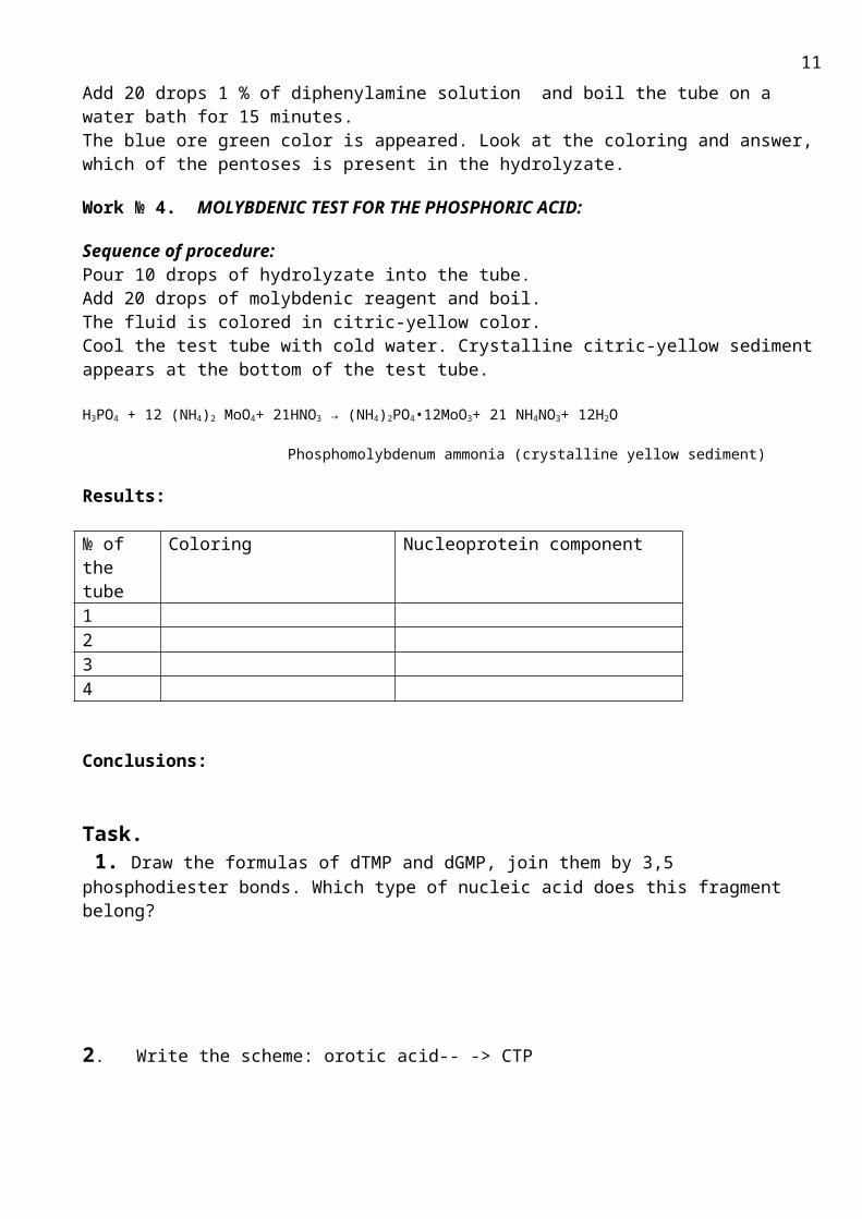
The essence of the method:The diphenylamine gives dark blue coloring with deoxyribose, and green with ribose. Sequence of procedure:Pour 5 drops of hydrolyzate into the tube.Add 20 drops 1 % of diphenylamine solution and boil the tube on a water bath for 15 minutes.The blue ore green color is appeared. Look at the coloring and answer, which of the pentoses is present in the hydrolyzate.
Work № 4. MOLYBDENIC TEST FOR THE PHOSPHORIC ACID:
Sequence of procedure:Pour 10 drops of hydrolyzate into the tube.Add 20 drops of molybdenic reagent and boil. The fluid is colored in citric-yellow color. Cool the test tube with cold water. Crystalline citric-yellow sediment appears at the bottom of the test tube.
H3PO4 + 12 (NH4)2 MoO4+ 21HNO3 → (NH4)2PO4•12MoO3+ 21 NH4NO3+ 12H2O
7
Phosphomolybdenum ammonia (crystalline yellow sediment)
Results:
№ of the tube
Coloring Nucleoprotein component
1234
Conclusions:
Task. 1. Draw the formulas of dTMP and dGMP, join them by 3,5 phosphodiester bonds. Which type of nucleic acid does this fragment belong?
2. Write the scheme: orotic acid-- -> CTP
3. Draw formulas of AMP and CMP, join them by 3,5 phosphodiester bonds. Which type of nucleic acid does this fragment belong?
4. Write the scheme: GMP--- ->dGTP
8
M.C.Q.
1. In a DNA molecule guanosine nucleotide is held by the cytosine nucleotide by thenumber of hydrogen bonds:A. lB. 2C. 3D. 4 E. 5
2. Which one of the following is characteristic of orotic aciduria?A. ImmunodeficiencyB. Genetic deficiency of the enzyme orotidine
phosphate decarboxylaseC. Self-mutilation D. Increased levels of uric acid in bloodE. Impairment of T-cell function
3. In humans, the principal catabolic product of pyrimidines is:
A. uric acidB. allantoin C. hypoxanthine D. -Alanine E. Urea
4. Two nitrogen atoms of pyrimidine ring are obtained from:
A. Glutamine and carbamoyl-P.B. Aspartate and carbamoyl- P.C. Glutamate and ammonia D. Glutamine and ammoniaE. Aspartate and glycine
5. The complementary base sequence in the second strand of DNA for the base sequence CCGATT would be:A. GGCTAAB. GGCUAAC. AATCGGD. CCGATTE. GTACCG
6. Synthesis of what substance is blocked by 5- fluorodesoxiuridine, an inhibitor of thymidilatsynthase?A. DNAB. tRNA
C. ProteinD. ATPE. mRNA
7. Why do two DNA strands form a double helix?A. due to base-pairing phenomenonB. due to the "anti-parallel" orientation;C. due to various combinations between its nucleotides;D. due to ability to make copies of itself;E. due to phosphate sugar backbone of DNA;
8. In humans, the principal catabolic product of Thymidine is:A. AllantoinB. beta-Alanine C. UreaD. Uric acid E. beta-aminoisobutirate
9. A key substance in the committed step of pyrimidine biosynthesis is:A. ATPB. carbamoyl -phosphateC. Ribose-5'-phosphateD. ThiouracilE. Glutamine
10. Which one of the following is allosteric inhibitor of the de novo Pyrimidine synthesis?A. PRPPB. GlutamineC. RiboseD. UTPE. AMP
11.Deficiency of what enzyme is the cause of orotic aciduria?5. Xantineoxidase; 6. Carbamoil-P-synthetase;7. Orotate phosphoribosiltransferase;8. GH PRT;9. Amidotransferase
9

Topic 3.3. THE METHODICAL GUIDELINES FOR PRACTICE ACTIVITY ON THE THEME: Investigation of DNA Replication and RNA Transcription. Analysis of mutations, DNA Repair.
Biomedical importance: Nucleic acids are required for the storage and expression of genetic information. There are two
chemically distinct types of nucleic acids: deoxyribonucleic acid (DNA) and ribonucleic acid (RNA). DNA is present not only in chromosomes in the nucleus of eukaryotic organisms, but also in mitochondria and in the chloroplasts of plants. Prokaryotic cells, which lack nuclei, have a single chromo-some but may also contain nonchromosomal DNA in the form of plastids. The DNA contained in a fertilized egg encodes the information that directs the development of an organism. This development may involve production of billions of cells. Each of these cells is specialized, expressing only those functions that are required for it to perform its role in maintaining the organism. Therefore, the DNA must be able not only to replicate precisely each time a cell divides, but also to have the information that it contains be selectively expressed. RNA participates in the expression of the genetic information stored in the DNA . The genetic master plan of an organism is contained in the sequence of deoxyribonucleotides that constitute the DNA. However, it is through the ribonucleic acid (RNA) "working copies" of the DNA that the master plan is expressed. The copying process, which uses one of the two DNA strands as a template, is called transcription. The messenger RNAs, which are transcripts of certain regions of the DNA, are translated into sequences of amino acids— the polypeptide chains. Ribosomal RNAs, transfer RNAs, and additional small RNA molecules perform specialized structural and regulatory functions without translation.
The purpose: To develop skills in interpreting of nucleic acid structure and functions for molecular basis of
inherited diseases explanation and treatment.
The applicable materials:1. The tutorial book, 2005. p.289-3042. "Biochemistry", Pamela C. Champe at al.2005.p. 393-4283. Lecture on the theme «Bases of molecular genetics. Protein biosynthesis and its regulation”
The main theoretical questions:
1. Genetic information: storage, expression and types of transmission.2. Structure of DNA and RNA 2.1. Primary structure - 3’, 5’-phosphodiester bonds; 2.2 DNA double helix; Base pairing: hydrogen bonds, the complementary rules. 2.3. Organization of eukaryotic DNA, nucleosomes. 3. Structure of mRNA, tRNA and rRNA.4. Steps in DNA synthesis: 4.1. Semi conservative replication mechanism; 4.2. Components required for replication: substrates for DNA synthesis, enzymes (DNA polymerase III and I, helices, topoisomerases, DNA ligase). 4.3. DNA synthesis initiation: separation of the two complementary DNA strands and replication fork formation; RNA primer synthesis. 4.4. Chain elongation: direction of DNA replication; leading and lagging strands; excision of RNA primer and its replacement by DNA; the joining of Okazaki fragments.
5. DNA damage and repair.6. Point mutations: missense and nonsense;7. Transcription of genes: 7.1. Structure of operone.
10
7.2. Components required for transcription. RNA polymerases. 7.3. Steps in prokaryotic RNA synthesis: initiation, elongation, termination. 7.4. Post-transcriptional modification of mRNA.
M.C.Q.
1.Which of the following is NOT correct about nucleic acids?A. they contain both phosphorus and nitrogenB. nucleic acids are useful for buoyancyC. RNA is a type of nucleic acidD. nucleotides are subunits of nucleic acidsE. DNA is a type of nucleic acid
2. One important function of nucleic acids is that they:A. form enzymesB. are structural moleculesC. repel waterD. store energyE. hold genetic information
3. RNA polymerase is involved in which of the following processes?A. PhotosynthesisB. PhagocytosisC. TranscriptionD. SpargingE. Translational
4. Which process takes place under DNA repair?A. Synthesis of Okazaki fragmentsB. Removal of primerC. Replacement of abnormal bases by DNAD. Synthesis of RNA primers E. The replication fork formation
5. Genetic structure of eukaryote is "exon-intron-exon". This structure-functional organization of gene caused transcription peculiarities. What will be pro-i-RNA according to the scheme?A. Intron-exonB. Exon-intronC. Exon-exonD. Exon-exon-intronE. Exon-intron-exon
6. A RNA molecule differs from a DNA molecule because A. RNA is a single strand of nucleotidesB. RNA contains uracilC. RNA contains riboseD. RNA contains pyrimidine nitrogen basesE. All of the above
7. Which components are required for transcription?A. RibonucleotidetriphosphatesB. Amino acidsC. DeoxyribonucleotidesD. RibosomesE. Primer
8. Which enzyme takes part in proofreading of newly synthesized DNA?A. DNA-polymerase III B. DNA-helicase C. DNA-polymerase ID. DNA-ligaseE. DNA-topoisomerase
9. Which one of the following takes part in post- transcriptional modification of RNA?A. DNA-polymerase IIIB. DNA-polymerase IC. Small nuclear ribonucleoprotein particles D. Aminoacyl tRNA-synthetaseE. RNA-polymerase
10. RNA, which is contained in AID virus, penetrated into the middle of leukocytes and made the cell synthetic viral DNA with the help of the enzyme revertase. This process is based on… A. Convariant replication B. Depression of operone C. Repression of operoneD. Reverse transcription E. Reverse translation
11

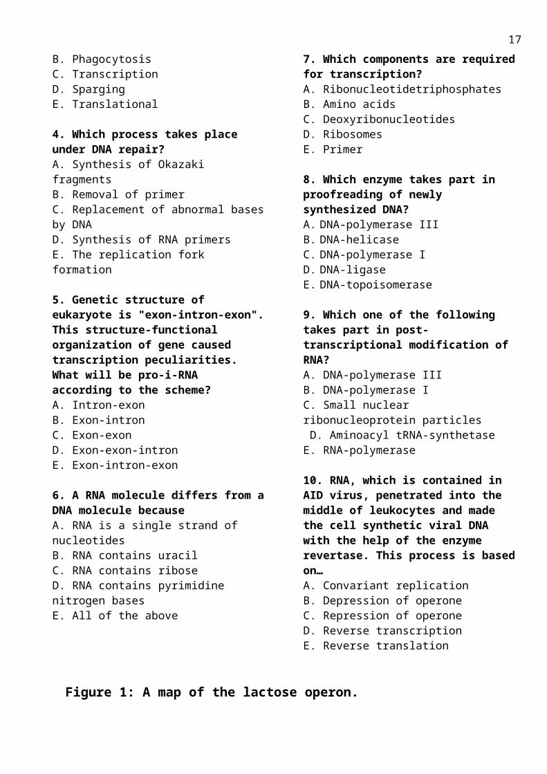
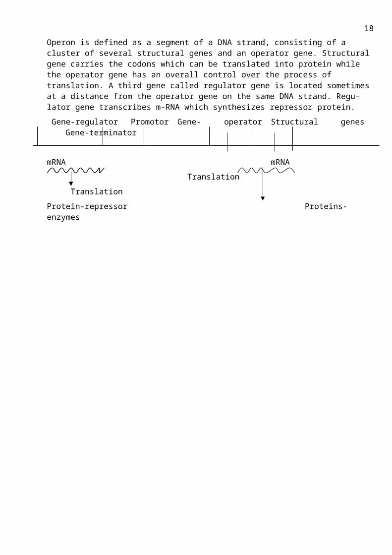
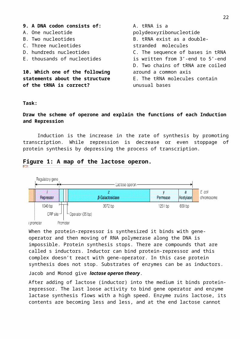
Figure 1: A map of the lactose operon.
Operon is defined as a segment of a DNA strand, consisting of a cluster of several structural genes and an operator gene. Structural gene carries the codons which can be translated into protein while the operator gene has an overall control over the process of translation. A third gene called regulator gene is located sometimes at a distance from the operator gene on the same DNA strand. Regulator gene transcribes m-RNA which synthesizes repressor protein.
Gene-regulator Promotor Gene- operator Structural genes Gene-terminator
mRNA mRNA
Translation
Translation
Protein-repressor Proteins-enzymes
12

Topic 3.4. THE METHODICAL GUIDELINES FOR PRACTICE ACTIVITY ON THE THEME:Protein Synthesis. Investigation of initation, elongation and termination.
Biomedical importance:
Protein synthesis is carried out on large macromolecular complexes called ribosomes. Ribosomes consist of three pieces of RNA and many proteins. Although prokaryotic and eukaryotic ribosomes are very similar, there are sufficient differences to allow selective drug action against prokaryotic protein synthesis. Many of our most important antibiotics (erythromycin, tetracycline, chloramphenicol, gentamycin) act against protein synthesis.
The purpose: To develop skills in interpreting of mechanism of protein synthesis for molecular basis of inherited diseases explanation and treatment.
The applicable materials:1. The tutorial book, 2005. p. 317-3272. “Biochemistry”, Pamela C. Champe at al.2005.p.431-444. 3. Lecture on the theme «Bases of molecular genetics. Protein biosynthesis and its regulation”
The main theoretical questions:
1. Genetic code.1.1 Codons1.2 Characteristics of the genetic code
2. Components required for translation2.1 Amino acids; 2.2 Transfer RNA, structure and functions2.3 Aminoacyl-tRNA synthetases 2.4 mRNA2.5 Functionally competent ribosomes2.6 Protein factors2.7 Energy sources.
3. Steps in protein synthesis3.1 Iinitiation3.2 Elongation3.3 Termination4. Post-translational modification of polypeptide chains.
5. Protein synthesis regulation.
6.Action of antibiotics on template synthesis
13
M.C.Q.
1. The process of making proteins on the RNA template is:A. transcriptionB. translationC. conjunctionD. peptide synthesisE. this process cannot happen
2. The directions used in protein synthesis are provided by:A. mRNA B. centromereC. RNA in the ribosomesD. tRNAE.qRNA
3. The coding of amino acids by multiple sets of nucleotides is referred to as:A. base pairing specificityB. hydrogen bondingC. chemical codingD. triplet codingE. DNA specificity
4. What is the role of ribosomes in protein synthesis?A. they provide a source of amino acidsB. they provide a site for transfer RNAs to link to messenger RNAs C. they translate the basic DNA code using transfer RNAD. they carry the proteins to their site of actionE. It creates rRNA
5. What role does mRNA play in protein synthesis?A. It is what the tRNA matches up to in order to form proteinB. NothingC. It explodesD. It blows upE. It helps form ribosome
6. The genetic code is non-overlapping because:A. It is read in the direction of 3’-5’B. A specific codon always codes the same amino acidsC. It is same for all organismsD. Is read from a fixed starting point as a continuous sequence of codonsE. Is radically differ in prokaryotes and eukaryotes
7. Which one of the following molecules is a component of genetic code?A. ProteinB. UDPC. UMPD. ATPE. dGMP
8. Which one of the following statements about mRNA functions is correct?A. Take part in replicationB. Is a component of ribosomeC. Is required for initiation of transcriptionD. Carries amino acids to the site of protein synthesisE. Carries the genetic information from DNA to cytosol
9. A DNA codon consists of:A. One nucleotideB. Two nucleotidesC. Three nucleotidesD. hundreds nucleotidesE. thousands of nucleotides
10. Which one of the following statements about the structure of the tRNA is correct?A. tRNA is a polydeoxyribonucleotideB. tRNA exist as a double-stranded moleculesC. The sequence of bases in tRNA is written from 3’-end to 5’-endD. Two chains of tRNA are coiled around a common axisE. The tRNA molecules contain unusual bases
14

Task:
Draw the scheme of operone and explain the functions of each Induction and Repression
Induction is the increase in the rate of synthesis by promoting transcription. While repression is decrease or even stoppage of protein synthesis by depressing the process of transcription.
Figure 1: A map of the lactose operon.
When the protein-repressor is synthesized it binds with gene-operator and then moving of RNA polymerase along the DNA is impossible. Protein synthesis stops. There are compounds that are called s inductors. Inductor can bind protein-repressor and this complex doesn’t react with gene-operator. In this case protein synthesis does not stop. Substrates of enzymes can be as inductors.
Jacob and Monod give lactose operon theory.
After adding of lactose (inductor) into the medium it binds protein-repressor. The last loose activity to bind gene operator and enzyme lactase synthesis flows with a high speed. Enzyme ruins lactose, its contents are becoming less and less, and at the end lactose cannot bind protein-repressor. Protein-repressor binds with gene-operator. Transcription stops and later translation stops too.
Figure 2: The operon model, as proposed in 1961 by Jacob and Monod.
15

Content module 7 “Molecular mechanisms of hormone action on the target cells and biochemistry of hormonal regulation”
Topic 3.5. THE METHODICAL GUIDELINES FOR PRACTICE ACTIVITY ON THE THEME:Molecular mechanisms of hormone action on the target cells and biochemistry of hormonal regulation. Investigation of hypothalamus and hypophysis (pituitary gland) hormones.
Biomedical importance: The rational diagnosis and therapy of a disease depend upon understanding the pathophysiology involved and the ability to quantitative it. Diseases of the endocrine system, which are generally due to excessive or deficient production of hormones, are an excellent example of the application of basic principles to clinical medicine. Knowing the general aspects of hormone action and understanding the physiologic and biochemical effects of the individual hormones enable one to recognize endocrine disease syndromes that result from hormone imbalance and to apply effective therapy.
The purpose: To develop skills in interpreting of hormone action on cells and metabolism regulation by hormones of hypothalamus and hypophysis for the further diagnostics and treatment of endocrinal disease.
The applicable materials:1. The tutorial book, 2005. p. 71-76.2. "Biochemistry", Pamela C. Champe at al.2005.p.92-953. The “Molecular mechanisms of hormone action on the target cells and biochemistry of hormonal regulation Lecture Materials”;
The main theoretic questions:1. The common characteristic of hormones. Classification. 2. The relationship among regulation levels of metabolism.3. The mechanism of hormone action:
3.1. Hormone receptors 3.2. Hormones that bind to intracellular receptors. 3.3. Hormones that bind to cell surface receptor. 3.4. cAMP as the second messenger for many hormones 3.5. Calcium and phosphatidylinositols as a mediator of hormone action 3.6. Insuline receptors
4. Investigation of hypothalamus and hypophysis hormones. Chemical nature, action on metabolism.
5. Clinical symptoms of hypophysis hormones disbalanse.
M.C.Q.1.Hormones are: A. chains of nucleotidesB. organic molecules containing only carbonC. messenger molecules that help different parts of the body work togetherD. complex carbohydratesE. combinations of simple sugars into a chain
2.Which of the following would not influence the endocrine system via the hypothalamus?A. strong emotionsB. bright lights
C. painful stimuliD. infectionsE. all of the above would influence the endocrine system via the hypothalamus
3. In which pair of hormones does the first cause increased secretion of the second?A. ACTH; cortisolB. FSH; aldosteroneC. LH; insulinD. TSH; prolactinE. all of these
16
4. Which of the following events could be a result of damage to the hypothalamo-hypophyseal portal system?A. decreased secretion of ADH (vasopressin)B. decreased secretion of oxytocinC. decreased secretion of thyroid stimulating hormoneD. decreased secretion of parathyroid hormoneE. all of these
5. All of the following are functions of Endocrine System except:A. Regulate blood calcium levelsB. Regulate the heart rateC. Control the water balance of the bodyD. Regulate body temperatureE. all of these
6. The hypothalamusA. regulates the secretory activity of the pituitary gland.B. is connected to the pituitary gland by the optic chiasma.C. has neurons that connect to the anterior pituitary.D. contains the infundibulum, which secretes many hormones.
E. all of these
7.Oxytocin secretion causesA. milk ejection in lactating females.B. uterine contractions.C. increased urine volume.D. increased blade volumeE. all of these
8. Steroid hormone receptors:A. are integral membrane proteins that bind steroids on their extracellular domains.B. bind steroids in the blood plasma, but do not enter cells.C. bind steroids in the sytosolD. facilitate the entry of steroid hormone into the cell
9. Match the following hypothalamic hormones with their functions:1. TRH - A. Inhibits production of prolactin2. CRH - B.Stimulates secretion of FSH and LH3. GNRH - C.Triggers secretion of TSH4. DA - D.Stimulates the secretion of GH5. ADH - E.Promotes water reabsorption by the kidneys6. GHRH - F.Causes the secretion of ACTH
Classification of Hormones:
According to Li the hormones can be classified chemically into three major groups.(i) Steroid hormones: These are steroid in nature such as adrenocorticosteroid hormones, androgens, estrogens and progesterone.(ii) Amino acid derivatives: These are derived from amino acid tyrosine e.g., epinephrine, norepinephrine and thyroid hormones. (iii) Peptide/Protein hormones: These are either large proteins or small or medium size peptides, e.g. Insulin, glucagon, parathormone, calcitonin, pituitary hormones, etc.
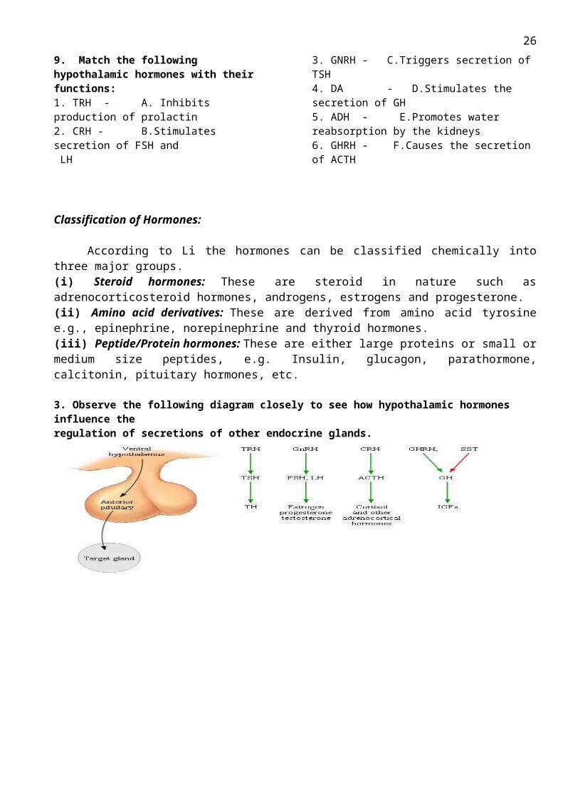
3. Observe the following diagram closely to see how hypothalamic hormones influence the regulation of secretions of other endocrine glands.
17
Topic 3.6. THE METHODICAL GUIDELINES FOR PRACTICE ACTIVITY ON THE THEME: Hormones of pancreas and gastrointestinal tract. Metabolic changes in diabetes mellitus.
Biomedical importance: Individual tissues do not function in isolation, but rather form part of a community in which one
tissue may provide substrates to another, or process compounds produced by other organs. Communication between tissues is mediated by the nervous system, by the availability of circulating substrates, and by variation in the levels of plasma hormones. The integration of energy metabolism is controlled primarily by the actions of hormones, including insulin, glucagon, catecholamines, epinephrine and norepinephrine. Changes in the circulating levels of these hormones allow the body to store energy when food is available in abundance or to make stored energy available, for example, during "survival crises," such as famine, severe injury, and "fight or flight" situations. The central nervous system has an absolute requirement for a continuous supply of blood-borne glucose to serve as a fuel for energy metabolism. Transient hypoglycemia can cause cerebral dysfunction, whereas severe, prolonged hypoglycemia causes brain death. It is therefore not surprising that the body has multiple overlapping mechanisms to prevent or correct hypoglycemia. The most important hormone changes in combating hypoglycemia are elevated glucagon and epinephrine, combined with the diminished release of insulin.
The purpose: To develop skills to interpret affect of pancreas and gastrointestinal tract hormones. Determine of content of blood glucose in and using of these results for carbohydrates metabolism estimation.
The applicable materials:1. The tutorial book "Principles of biochemistry", 2005.p.219-2382. "Biochemistry", Pamela C. Champe at al.2005.p.305-318, 335-3463. The «Hormones» Lecture Materials;
The main theoretic questions:
1. Hormones of pancreas.2. The metabolic effect of insulin: 2.1. Chemical nature 2.2. Regulation of insulin secretion 2.3. Mechanism of action 2.4. Effect of insulin on carbohydrates, lipid and protein metabolism.3. Metabolic changes in diabetes mellitus (insulin-dependent and non- insulin dependent), clinical and biochemistry description.4. The metabolic effects of glucagons: 4.1. Chemical nature 4.2. Mechanism of glucagon action
4.3. Effects of glucagon on carbohydrates, lipid and protein metabolism.5. Gastrointestinal tract hormones: gastrin, cholecystokinin, secretin.
Practice instructions “Quantitative determination of glucose blood level by arseno-molybdenic method”.
The essence of the method: The essence of method is based on glucose ability to reduce Cu(OH)2 ( blue color) to CuOH in alkaline medium.
Cu(OH)2 + glucose => CuOH + gluconic acid
18
Formed CuOH is a reagent towards arsenic-molybdenic reagent (Nelson reagent), which is reduced and give sensitive color reaction. As the blood glucose concentration increases there is a correspondingly changed colors (from green-yellow to green-blue). Intensity of color is measured photocolorimetrically.
Sequence of Procedures:1. Pour 3.7 ml of isotonic solution both into two centrifugal tubes.2. Add 0.1 ml of blood serum and 0.2 ml of Na2WO4 (sodium tungstate) into the first tube
(experimental).3. Add 0.1 ml of standard glucose solution and 0.2 ml of Na2WO4 (sodium tungstate) into the second
tube (standard).4. Further procedures are the same for both tubes!5. Centrifugate tubes for 10 minutes.6. Prepare copper reagent by mixing of 5 ml A reagent and 0.5 ml B reagent.7. Transfer 1 ml of supernatant into an ordinary tube.8. Add 1 ml of prepared copper reagent in order to oxidase glucose, close tubes cotton wool and boil in
water bath for 10 minutes9. Cool the tubs with cold water and add 3 ml of Nelson reagent.10. Measure the optical density of solution by photo colorimeter.11. Calculate glucose concentration in blood by the formula:
Dexp*5.6C = ----------- (mmol/l)
Dstand
Where:Dexp – density of experimental sample.Dstand – density of standard sample.5.6, mmol/l – standard glucose solution concentration.Normal content of blood glucose is 3.3 – 5.5 mmol/l
Conclusions:
M.C.Q.1. The stimulus for release of Insulin is:A. Decreased levels of glucoseB. Increased levels of glucoseC. Hormonal secretion from PituitaryD. Neural stimulation from pituitaryE. Stress
2. Which one of the following is secondary messenger of glucagon?
A. Protein kinase B. Adenilate cyclaseC. AMP D. cAMP E. ATP
3. Following statements regarding insulin are all correct except:A. induces glucokinase activityB. inhibition of acetyl CoA carboxylaseC. converts glycogen phosphorylase to inactive form
D. stimulates F.A synthase activityE. increases activity of glycogen synthase.
4. The primary target cells for glucagon are:A. Skeletal muscle cellsB. PancreasC. KidneysD. LiverE. Brain
5. The presence of ketone bodies in the blood is associated with which endocrine disorder?A. Grave's diseaseB. Cushing's syndromeC. Diabetes mellitusD. AcromegalyE. Diabetes insipidus
6. Type II (non-insulin-dependent) diabetes mellitus is usually caused by:
19
A. Failure of target cells to respond to insulinB. Hyposecretion of insulinC. Autoimmune destruction of the insulin-
secreting cellsD. Hypersecretion of insulinE. Hypersecretion of somatostatin
7. Which hormone is synthesized in pancries?
A. Epinephrine B. Calcitonine C. SomatostatineD. VasopressineE. Thiroliberine
8. The metabolic effect of insulin is:A. Activation of gluconeogenesisB. Breakdown of glycogenC. Activation of glycolysisD. Increase in triacylglycerol degradationE. Increase in b-oxydation of fatty-acids
9. The metabolic effect of glucagon is:A. Inhibition of gluconeogenesisB. Decrease in triacylglycerol degradationC. Breakdown of glycogen in liverD. Activation of fat synthesis
E. Breakdown of glycogen in muscle
10. What kind of activity is associated with insulin receptor?
A. tyrosine kinase activity;B. cysteine kinase activity;C. adenylate cyclase activity;D. guanilate cyclase activity;E. it opens ion channels for sodium
11.In which one of the following tissues is glucose transport into the cell enhanced by insulin?
A. BrainB. LensC. Red blood cellsD. MusclesE. Liver
12. Hormone secreted by the duodenum that stimulates pancreatic secretions in response to contact with acidic stomach content is:
A. SomatostatinB. EphynephrynC. LactothropineD. Secretin
20
Topic 3.7. THE METHODICAL GUIDELINES FOR PRACTICE ACTIVITY ON THE THEME: Endocrine control of glucose concentration in the blood. Glucose tolerance test. Sugar
curves. Hormones of the adrenal gland.
Biomedical importance: The central nervous system has an absolute requirement for a continuous supply of blood-borne glucose to serve as a fuel for energy metabolism. Transient hypoglycemia can cause cerebral dysfunction, whereas severe, prolonged hypoglycemia causes brain death. It is therefore not surprising that the body has multiple overlapping mechanisms to prevent or correct hypoglycemia. The most important hormone changes in combating hypoglycemia are elevated glucagon and epinephrine, combined with the diminished release of insulin.
The purpose: To develop skills in interpreting results of standard oral glucose tolerance test in order to estimate carbohydrate metabolism disturbances and endocrine diseases.
The applicable materials:1. The tutorial book Harper’s Biochemistry R.K.Murrey and all USA 1998, 521-541.2. The «Hormones» Lecture Materials;3. Appendix.
The main theoretic questions:1. Normal concentration of glucose in blood, pathologies (hypo- and hyperglycemias).2. Diabetes mellitus. Diagnostic criteria. 3. The glucose tolerance test: essence of the method, role in diagnostics of carbohydrate metabolism disturbances.4. Types of "sugar curves" in norm and under different types of hypo- and hyperglycemia (Norm, diabetes mellitus, hyperthyroidism, hypothyroidism, insuloma).5. Hormones of the adrenal medulla:
5.1. Chemical structure of catecholamines5.2. Biosynthesis of norepinephrine and epinephrine (reactions)5.3. Mechanism of action with target cells5.4. Influence on the metabolism
6. Hormones of the adrenal cortex:6.1. Chemical structure, classification6.2. Biosynthesis from cholesterol (scheme)6.3. Mechanism of action6.4. Regulation of metabolism by glucocorticoids
7. Regulation of water-mineral metabolism by mineralocorticoids. Renin-Angiotensin system.
8. Disorders of adrenal hormones insufficiency and excess (pheochromocytoma, Icenco-Kushing's syndrome and disease, Conn's syndrome, Addison's disease).
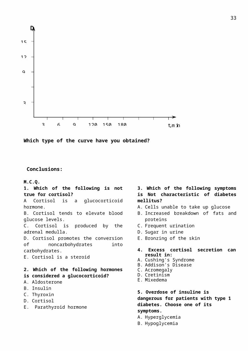
Practice instructions: “Forming-up of sugar curve”
Determine glucose concentration in blood serum of six experimental tubes. ( In different time after glucose uptake) with indicator paper.
Put down the results in the table.
21
By plotting glucose concentration as ordinate versus time as abscissa obtain the glucose tolerance test curve.
No Time after glucose uptaking Optical density Glucose concentration in the sample, mmol/l
1 0 (fasting)2 303 604 905 1206 150
Which type of the curve have you obtained?
Conclusions:
M.C.Q.1. Which of the following is not true for cortisol?А Cortisol is a glucocorticoid hormone.В. Cortisol tends to elevate blood glucose levels.С. Cortisol is produced by the adrenal medulla.D. Cortisol promotes the conversion of noncarbohydrates into carbohydrates.E. Cortisol is a steroid
2. Which of the following hormones is considered a glucocorticoid? A. AldosteroneB. InsulinC. Thyroxin
D. CortisolE. Parathyroid hormone
3. Which of the following symptoms is Not characteristic of diabetes mellitus?A. Cells unable to take up glucoseB. Increased breakdown of fats and proteinsC. Frequent urinationD. Sugar in urineE. Bronzing of the skin
4. Excess cortisol secretion can result in:A. Cushing's Syndrome
22
3
30 60 90 120
9
12
15
150 180

B. Addison's Disease C. Acromegaly D. Cretinism E. Mixedema
5. Overdose of insuline is dangerous for patients with type 1 diabetes. Choose one of its symptoms.A. HyperglycemiaB. HypoglycemiaC. HypercholesterinemiaD. HypocholesterinemiaE. Anemia
6. Which of the following statements is Not true about diabetes mellitus? A.Type II diabetes is much more common then type I. B. Insulin injections are required in both type I and type II diabetes. C. Type I diabetes occurs as a result of destruction of the insulin producing cells. D. One method of treating type II diabetes is exercise and a low fat, low sugar diet. E. Symptoms of diabetes include excessive thirst, frequent urination, and glucose in the urine.
7. Аldosterone is secreted from which cells of the adrenal gland?A. Medulla B. Zona Reticularis C. Zona Fasciculata D. Zona Glomerulosa
8. Choose the characteristic element of the stress response:
A. Secretion of insulin
B. HypoglycemiaC. HyperammoniemiaD. High level of epinephrineE. Secretion of grows hormone9. What is the blood clinical test of insuline productionA. Level of C-peptideB. Concentration of preproinsulineC. Content of proinsulineD. Level of insulineE. Glucose blood concentration
10. Cortisol:A. Decreases gluconeogenesis in the liver B. Increases glucose uptake in adipose tissueC. Decreases protein synthesis in muscle D. Decreases urea production in the liverE. Increases protein synthesis in the bone
11. Steroid hormone receptors:A. bind steroids in the blood plasma, but do not
enter cells.B. are integral membrane proteins that bind
steroids on their extra cellular surfaceC. domains.D. bind steroids and function in the nucleus.E. facilitate the entry of steroid hormone into
the cell
12. Choose the characteristic element of the stress response:
A. Secretion of insulinB.HypoglycemiaC.HyperammoniemiaD. High level of epinephrineE.Secretion of grows hormon
APPENDIX.GLUCOSE TOLERANCE TEST (GTT)What is "Carbohydrate Tolerance"? Diabetes mellitus diagnosis is put on the base of hyperglycemia (glucose concentration in vein blood fasting (on an empty stomach) > 6,1 mmol/l and also there are glucose an keton bodies in urine).The ability of the body to utilize carbohydrates may be ascertained by measuring its carbohydrate tolerance. It is indicated by the nature of blood glucose curve following the administration of glucose. Thus "glucose tolerance" is a valuable diagnostic aid.Decreased Glucose ToleranceThis is seen:In Diabetes mellitus,In hyperactivity of anterior pituitary (Grows hormon)In hyperactivity of adrenal cortex (Steroid diabetes)In hyperthyroidism.( Thyroid diabetes)Increased Tolerance is seen in —Hypopituitarism, (ii) hyperinsulinism, (iii) hypothryroidism, (iv) Adrenal cortical hypofunction (such as Addison's disease).In doubtful cases the standard tolerance test for glucose carries out.
23

Indications: 1. Normal level of blood glucose in fasting in the presence of clinical symptoms.2. In patients with transient or sustained glycosuria, who have no clinical symptoms of Diabetes with normal fasting and P.P. blood glucose.3. In patients with symptoms of Diabetes but with no glycosuria and normal fasting blood glucose level.4. In persons with strong family history but no overt symptoms.5. In patients with glycosuria associated with thyrotoxicosis, infections/sepsis, and Liver diseases, Pregnancy etc.6. In women with characteristically large babies 9 Ibs or individuals who were large babies at birth.7. In patients with neuropathies or retinopathies of undetermined origin.8. In patients with or without symptoms of D.M, showing one abnormal value of blood glucose.Procedure:1. A fasting sample of venous blood is collected in flouride bottle (fasting sample)2. The bladder is emptied completely.3. The individual is given 75 Gms of glucose dissolved in water about 250 ml to drink. Lemon can be added to make it palatable and to prevent nausea/vomiting.Time of oral glucose administration is noted.4. A total of five specimens of venous blood are collected every 1/2 hour after the oral glucose viz. 1/2 hr, 1 hr, 1 1/2 hr, 2 hr and 2 1/2 hr.5. Glucose content of all the six (including fasting sample) samples of blood is estimated. .A curve is plotted which is called as "Glucose tolerance curve".
Explanation and Significance of a Normal Curve1. A sharp rise to a peak, averaging about 50% above the fasting level within 30 to 60 minutes. Extent of the rise varies considerably from person to person, but maximum should not exceed 75 % in normal subjects.Reason:(i) Rise is due directly to the glucose absorbed from the intestine, which temporarily exceeds the capacity of the Liver and tissues to remove it.(ii) As the blood glucose concentration increases, regulatory mechanisms come into play: (a) Increased insulin secretion due to hyperglycemia, (b) hepatic glycogenesis is increased,(c) hepatic glycogenolysis is decreased, and glucose uptake and utilization in tissues increase
2. A sharp fall to approximately the fasting level at the end of 1 1/2 to 2 hrs.Reason — Glucose now leaves the circulation faster than it is entering. This is due to: (i) continuing stimulation of the mechanisms stated above i.e. increased utilization and hepatic glycogenesis, and to slowing or completion of glucose absorption from the intestines.
3. Hypoglycemic "dip": Continued fall to a slightly sub fasting (10 to 15 mg lower than fasting value) act 2 hrs and subsequent rise to fasting level at 2 1/2 to3 hrs.Reason — The hypoglycemic 'dip' is due to "inertia" of the regulatory mechanisms. The decreased output of glucose by Liver and increased utilization induced by the rising blood glucose are not reversed as rapidly as the blood sugar falls.
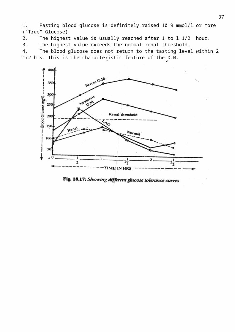
Characteristics of Different Types of GTT a) Normal GTC1. Fasting blood glucose within normal limits of 3,3 to 5,5 mmol/l ('True" glucose)2. The highest peak value is reached within one hour.3. The highest value does not exceed the renal threshold i.e., 7,5 to 9 mmol/l 4. The fasting level is again reached by 2 1/2 hr.
24
b) Diabetic Type of GTC1. Fasting blood glucose is definitely raised 10 9 mmol/l or more ("True" Glucose)2. The highest value is usually reached after 1 to l 1/2 hour.3. The highest value exceeds the normal renal threshold. 4. The blood glucose does not return to the tasting level within 2 1/2 hrs. This is the characteristic feature of the D.M.
25
Topic 3.8. THE METHODICAL GUIDELINES FOR PRACTICE ACTIVITY ON THE THEME: Hormonal regulation of calcium metabolism. Determination of iodine in the thyroid gland. Eicosanoids
Biomedical importance: Diseases of the thyroid are among the most common afflictions involving the endocrine system. Diagnosis and therapy are firmly based on the principles of thyroid hormone physiology and biochemistry. The availability of radioisotopes of iodine has greatly aided in the elucidation of these principles. Radioactive iodine, because it localizes in the gland, is widely used in the diagnosis and treatment of thyroid disorders. Radio iodine has a dangerous aspect as well, since excessive exposure, such as from nuclear fallout, is a major risk factor for thyroid cancer. This is especially true in infants and adolescents, whose thyroid cells are still actively dividing.
Ca2+ is required for the proper functioning of muscle contraction, nerve conduction, hormone release, and blood coagulation. In addition, Ca2+ helps to regulate many enzymes. Maintenance of the body Ca2+ stores depends on dietary Ca2+ intake, absorption of Ca2+ from the GI tract, and renal Ca2+ excretion. The calcium regulating hormones that control this homeostatic system are PTH and vitamin D, which act at bone, kidney, and GI tract to increase serum calcium and calcitonin, which an correspondingly act to decrease serum calcium.
The purpose: To develop skills in interpreting of hormone action on cell and metabolism regulation by thyroid hormones for further diagnostics and treatment of endocrinal disease.
The applicable materials:1. The tutorial book Harper’s Biochemistry R.K.Murrey and all USA 1998, 2. The «Hormones» Lecture Materials;
The main theoretic questions:1. Thyroid hormones.
1.1. Scheme of thyroid hormones biosynthesis. 1.2. Chemical structure1.3. Regulation of metabolism by thyroid hormones in norm.1.4. Pathophysiology of thyroid disease: hyperthyroidism, hypothyroidism
2. Regulation of calcium metabolism. 2.1. Parathormone: chemical nature, the role in calcium and phosphorus homeostasis. 2.2. Calcitonin: chemical nature, influence on a metabolism of calcium and phosphorus
2.3. Calcitriol: chemical structure, biosynthesis (scheme), mechanism of action, influence on a metabolism of calcium and phosphorus
2.4. Pathophysiology of hypo- and hyperfunctions
3. Eicosanoids: general characteristic, classification, biosynthesis (scheme). Biological and medicinal application. Inhibitors of eicosanoids synthesis
Practice instructions:
“The quantitative determination of iodine in thyroid gland”
The essence of the method: Upon thyroid hormones degradation KI is formed, from which I2 is easily can be released by KIO3. Liberation of I2 from KI is an oxidative-reducing reaction, where KI serves as a reducing agent, KIO3 as an oxidant. Excreted I2 is determined using qualitative reaction with starch in acid medium (dark blue color).
26

Sequence of Procedures:Students are given the prepared hydrolyzate of thyroid gland.
1. Pour 0.5 ml of hydrolyzate into the tube.2. Add 0.1 ml KIO3 and 0.5 ml of 10% H2SO4 solution.Red-yellow colour appears as result of free I2 releasing.3. Add 5 drops of 1% starch solution. The freed I2 gives blue colour with starch.
Conclusions:
Task.
Match the following:
1. A. Cushing's Disease, B. Myxoedema, C. Pheochromocytoma, D. Conn's Syndrome, E. Hyperthyroidism1. Periodic elevation of Blood Pressure associated with sweating ________2. Tachycardia, tremor and exophthalmos3. Retention of Na+ and increased excretion of K+ ________4. Obesity, hypertension, glycosuria, hirsutism ________5. Bradycardia, falling of hairs, and thickening of skin ________,
2. A. Tyrosine, B. 3-Methoxy epinephrine, C. "Active" methionine, D. Epinephrine1. Stored in chromatin granules ______2. Major urinary excretory product ____3. Required for formation of epinephrine from Norepinephrine ________4. Starting material for catecholamine synthesis ________
M.C.Q.1. Thyroxin is a hormone that:A. causes the release of milkB. increases the synthesis of insulinC. contracts muscleD. regulates body temperatureE. increases blood pressure
2. Blood calcium is lowered by the hormone: A. CalcitoninB. GlucagonC. AdrenalinD. ThyroxineE. Insuline
3. Thyrotropin (or TSH) stimulates the thyroid gland to release: A. ThyroxinB. CalcitoninC. ParathormoneD. thymosinE. Tyrosine
4. Thyroxin (or thyroid hormone) travels through the bloodstream acting on many target cells to increase: A. Blood sugarB. Blood calciumC. MetabolismD.Anti-inflammatory reactionsE. Exretion of water
5. Hypo secretion of thyroxin could be caused by a decrease in the release of: A. TRH or TSH (thyrotropin)B. TSH or ACTHC. STHRH or STHD. FSH or LH
6. The formation of cholecalciferol (vitamin D3) from cholesterol requires:A. a photochemical step.B. a hydroxylation in kidney.C. a hydroxylation in liver.D. hydroxylation in both kidney and liver. E. ingestion of 1,25-dihydroxycholecalciferol
27
7. Endemic goiter is known to be widespread in certain geochemical areas. The deficiency of what chemical element causes this disease?A. Iron.B. Iodine.C. Zinc.D. CopperE. Cobalt.8. In hyperparathyroidism, which of the following is correct?A. Low serum calciumB. High serum phosphorusC. Low serum calcium and high serum
phosphorusD. High serum calcium and low serum
phosphorusE. None of the above.
9. All of the following are true about the parathormone except:A. Occurs as a single polypeptide chain B. Synthesized initially as prohormoneC. Decreases serum calcium level and increases serum inorganic phosphates
D. Acts on kidneys and bonesE. Stimulates '1-a-hydroxylase' in kidney tubules
10. During the operation on a thyroid gland parathyroid glands were removed by mistake. The patient got tetanic cramps. The metabolism of which chemical element was disturbed?A. Magnesium.B. Calcium.C. Potassium.D. Iron.E. Sodium.
11. A patient complains of body weight loss, excessive irritability, insignificant increase of temperature, exophthalmia. Hyperglycemia and the rise of nitrogen-containing substances in blood serum were detected. Which is the most credible diagnosis in this case?A. Neurosis.B. Bronzed disease.C. Diffuse toxic goiter.D. Tuberculosis of adrenal glands.E. Myxedema.
28
Topic 3.9. THE METHODICAL GUIDELINES FOR PRACTICE ACTIVITY ON THE THEME: Steroid hormones of sex glands. Endocrine control of metabolism in the well-fed state. Regulation of metabolism in starvation.
Biomedical importance: The sex hormones are fat soluble. They are secreted by the gonads mm(testes or ovaries) under
the influence of luteinizing hormone (LH) from the pituitary gland. The secretion of LH is, in turn, determined by the rate of secretion of gonadotropin releasing hormone (GnRH) from the hypothalamus. Both the hypothalamus and the pituitary gland keep a very close eye on the sex hormone concentration in the blood. If the sex hormone level rises above a set value, then less GnRH and LH are immediately secreted to bring the sex hormone concentration in the blood down. The opposite happens if the sex hormone concentration in the blood falls. This is called negative feedback control, which ensures that the sex hormone concentrations are kept as predictable and unchanging (from day to day, and month to month) as are the blood sugars, or plasma calcium levels.
Sex steroids play important role in inducing the body change known as primary sex characteristics and secondary sex characteristics. The two main classes of sex steroids are androgens and estrogens, of whish the most important human example are testosterone and estradiol respectively.
Starvation is a severe reduction in vitamin, nutrient and energy intake, and is the most extreme form of malnutrition. In humans, prolonged starvation (in excess of 1-2 month) causes permanent organ and eventually result in death. Starvation stimulates decreased resting metabolic rate, increased ketogenesis and reliance upon ketone bodies, drop in sex hormones, decreased sexual interest, and muscle weakness, loss of mass and other.
The purpose: To develop skills in interpreting effects of sex hormones for understanding of metabolism disturbances and endocrine diseases. Analyze the phase of homeostasis after meal using concentration of the basic energy sources in plasma.
The applicable materials:1. The tutorial book, Harper’s Biochemistry R.K.Murrey and all USA 19982. "Biochemistry", Pamela C. Champe at al.2005.p. 319-3243. The «Hormones» Lecture Materials;
The main theoretic questions:
1. Sex steroids hormones. The mechanism of hormone action. The regulation of biosynthesis and secretion.
2. Female sex hormones. Estrogen – steroids (C18), progesterone – steroids (C21), physiological and biochemical effects.
3. Male sex hormones. Androgenic hormones – testosterone steroids (C19), physiological and biochemical effects.
4. Clinical use analogues and antagonist of sex hormones.
5. The hormonal regulation of metabolism in the different time after meal and starvation.(Analyse changes in carbohydrate, lipid and proteine metabolism in liver, brain, adipose tissue and mascle in well fed state and different time of starvation).
Practice instructions: “Analysis of blood in different terms after meal”The essence of the method: In different terms after meal the level of hormones and
concentration of the major energy sources in plasma of blood change. You have to determine in blood serum:1. Glucose 2. Total lipids 3 .Ketone bodies. Analyze experimental results and determine the phase of homeostasis, using table 1 and table 2
29

Sequence of Procedures:Determination of glucose in blood serum.
Determine the level of glucose in blood serum using of indicator paper.Determination of total lipids blood serum
1. Pour 1 ml of blood serum into the tube.2. Add 1 ml of phosphovanillin mixture3. Mix the contents of the tube well and leave it for 5 min at room temperature. Blue colour develops.4. Measure the intensity of the colour on photoelectrical colorimeter with green light filter.5. Obtain the amount of total lipids from the standard curve in the sample and calculate their concentration in g/l.The normal content of total lipids in blood serum is 3, 5-8 g/l
Qualitative reaction for acetone.1. Pour 5 drops of urine into a tube 2. Add 5 drops of 10% NaOH solution and 5 drops of Na-nitroprusside. Red-orange color appears.3. Add 10 drops of ice acetic acid. Color changes to cherry-red.
Table 1Phase (time after meal) Nature of glucose in blood
serum Tissue, using glucose Energy source
for brainI (4 hour) food All tissue glucoseII (8-16 hour) glycogen All tissue glucoseIII (16-24 hour) Gluconeogenesis (liver) All tissue glucoseIV (1-24 days) Gluconeogenesis
(liver and kidneys)Brain, erythrocytes, adrenal medulla
Glucose, ketone bodies
V (24-40 day) Gluconeogenesis(liver and kidneys)
Less brain, erythrocytes, adrenal medulla
Ketone bodies, glucose
Table 2metabolite after meal Time after meal
12 hour 3 days 3 weeksInsulin/ glucagon 0.5 0.15 0.05 0.05Glucose in blood, mol/l 6.1 4.8 3.8 3.6Amino acids, mmol 4.5 4.5 4.5 3.1Total lipids, g/l 8 6 6 3.4Ketone bodies, mmol 0.1 0.2 2.0 8-10
Results: 1. Glucose:2. Total lipids:3. Keton bodies:
Conclusions:
M.C.Q.:
1. Three hours after food intake, one can expect the blood to have high levels of all of the following hormones except:1. Insulin2. Glucagon3. Epinephrine4. Growth hormone5. Vasopressin
2.Which one of the following statements concerning the absorptive period is correct?A. 3-Hydroxybutyrate is a major fuel for muscle.Б. Transport of glucose into the adipocyte is decreased.C.Circulating amino acids are used primarily for
gluconeogenesis.D.Hepatic production of NADPH is decreased.
30
E.Glucose is the major fuel used by the brain.
3. Which one of the following is elevated in plasma during the absorptive period (compared to the post-nbsorptive state)?
A. Glucagon ,B. AcetoacetateC. ChylomicronsD. Free fatty acidsE. Lactate
4. Which one of the following statements concerning the well-fed state is correct?
A.Most enzymes that are regulated by covalent modification are in the phosphorylated slate
B. Hepatic fructose 2,6-bisphosphate is elevated.С Acetyl CoA is elevated.D. Insulin stimulates the transport of glucose
into hepatocytes.E. Keton bodies level is elevated
5. Ingesticn of a meal consisting exclusively of protein would result in which one of the following?A. An increased release of insulin. Б Hypoglycemia.C. A decreased release of glucagon.D. Ketoacidosis caused by the metabolism of
ketogenic amino acids.
E. Depletion of liver glycogen.
6. Why do testes shrink when male athletes take synthetic steroid testosterone hormones? A. Testosterone itself has the direct effect of shrinking the testes. B. Synthetic chemicals are not the same in action as natural chemicals. C. The guilt reaction in the brain causes a reverse hormonal action. D. The pituitary controls detect high levels of
testosterone in the bloodstream and reduces FSH and LH. E. Scientists have no explanation for this phenomenon that is opposite of expected.
7. Estradiol is a hormone that: A. Decreases sexual urges. B. Decreases sperm production in the testes. C. Serves in a feedback to the anterior pituitary to regulate testosterone levels. D. Triggers ovulation in females. E. Prevents or inhibits erection.
8. LH is an abbreviation for luteinizing hormone which was described as a female hormone controlling the ovary. In the male, LH: A. Does not exist since males lack ovaries. B. Exists in rudimentary levels since LH is made by the anterior pituitary. C. Is exactly the opposite chemical from male hormones, in an antibody antigen fashion. D. Controls production of testosterone.
9. Which of the following is Not true about estrogen? A. Estrogen causes the endometrium to thicken. B. Estrogen causes the endometrium to become vascular and glandular. C. Estrogen causes a positive feedback on the hypothalamus to secret GnRH. D. Estrogen causes a negative feedback on the anterior pituitary gland. E. Estrogen stimulates the release of FSH
10. Which one of these hormones is produced by females?A. EstrogenB. ProgesteroneC. TestosteroneD. OxytocinE. All of these
31

Topic 3.10. THE METHODICAL GUIDELINES FOR PRACTICE ACTIVITY ON THE THEME: “Interrelation and regulation of all metabolism pathways”.
Biomedical importance: Metabolism is the complete set of chemical reactions that occurs in living cells. These processes
are the basis of life, allowing cells to grow and reproduce, maintain their structures, and respond to their environments. Metabolism is usually divided into two categories. Catabolic reactions yield energy, an example being the breakdown of food in cellular respiration. Anabolic reactions, on the other hand, use this energy to construct components of cells such as proteins and nucleic acids. The chemical reactions of metabolism are organized into metabolic pathways, in which one chemical is transformed to another by a sequence of enzymes. Enzymes are crucial to metabolism because they allow cells to drive desirable but thermodynamically unfavorable reactions by coupling them to favorable ones. Enzymes also allow the regulation of metabolic pathways in response to changes in the cell's environment or signals from other cells. The metabolism of an organism determines which substances it will find nutritious and which it will find poisonous. For example, some prokaryotes use hydrogen sulfide as a nutrient, yet this gas is poisonous to animals. The speed of metabolism, the metabolic rate, also influences how much food an organism will require.
The purpose: Know how to use knowledge of a metabolism of carbohydrates, proteins, lipids and nucleonic acids and their regulation. Use integration of energy for correct interpretation of diseases course character through metabolism changes.
The applicable materials:1. The tutorial book, "Principles of biochemistry", 2005. p. 2. "Biochemistry", Pamela C. Champe at al.2005.3. The «Hormones», «Basic concept of metabolism», «Metabolism of carbohydrates, lipids and proteins» Lecture Materials;.
The main theoretic questions:1. The stages of fuel molecules catabolism as integration of energy formation. Nutrients digestion, its biomedical importance. Acetyl Co-A – general intermediate product of nutrients catabolism in the cells. Tricarboxylic acid cycle – common catabolic pathway as source of energy and substrates for synthetic reactions. Biological significance of cell respiration and oxidative phosphorylation in integration of metabolism. Role of CO2 and endogenous H2O in biosynthetic processes.
2. Relationship between carbohydrate and lipid metabolism.3. Relationship between protein and lipid metabolism.4. Interrelation between protein and carbohydrate metabolism.5. Role of proteins and vitamins as enzymes components.6. Regulatory role of hormones and other bioregulators in integration of carbohydrate, lipid and
protein metabolism. 7. Physiological needs as a basis for various classis of organic compounds interconversion.
M.C.Q.
1. A pathway that requires NADPH as a cofactor is:A. Fatty acid oxidation B. Extramitochondrial de novo fatty acid synthesisC. Ketone bodies formation
D. Glycogenesis E. Gluconeogenesis.
2. The most important source of reducing equivalents for FA synthesis in the Liver is: A. Glycolysis
32
B. HMP-shuntC. ТСА cycleD. Uronic acid pathwayE. Gluconeogenesis
3. The intermediate precursor of mevalonic acid in fatty acid synthesis is:A. Mevalonyl CoAB. Mevalonyl pyrophosphateC. Acetyl CoAD. 3-hydroxy-3-methylglutaryl CoAE. Isopentenyl pyrophosphate
4. Which of the following components is a precursor of both triacylglycerols and phospholipids?A. PhosphatidylethanolamineB. AcetylcholineC. Glycerol 3-phosphateD. Urydine diphosphate glucoseE. Cytidine diphosphate choline (CDP-holine)
5. b-oxydation of fatty acid produces:A. Succinyl CoAB. Propionyl CoAC. Acetyl CoAD. Malonyl CoAE. Acetoacetyl CoA
6. Which cofactor is necessary for the reduction reactions of cholesterol biosynthesis?A. NADH2;B. NADPH2C. FADH2D. FMNH2E. cAMP
7. Which compound is the same both for cholesterol synthesis and ketogenesis?A. squalene B. methylglutaril-CoAC. beta-hydroxybutyrateD. acetoacetateE. mevalonate
8. The urea cycle is linked to the citric acid cycle throughA. ArginineB. aspartate.C. arginosuccinate.D. fumarate.E. Urine
9. Which process is common pathway of metabolism?A. Conversion of pyruvate to acetyl-CoAB. Degradation of glucose to pyruvateC. Formation of pyruvate from fats and proteinsD. Conversion of pyruvate to glucoseE. Formation of pyruvate from lactate
10. During a fast, muscle protein is catabolyzed to free amino acids. All of the following scenarios occur EXCEPT: A. Alanine travels to the liver and is used for gluconeogenesis B. Glutamine travels to the kidney where it's amide group is used to buffer the urine C. Alanine is used for gluconeogenesis in the muscle D. Alanine travels to the liver and donates an amino group to the synthesis of urea E. Most amino acids travel to the liver and are used by the liver for gluconeogenesis
33

A. B. C.
Task 2. For studying biochemical processes to an experimental animal the glucose containing a radioactive isotope of hydrogen 2H in C1 position has been entered. In several hours radioisotope was found in tripalmitate. Make the scheme and specify the basic metabolites of transformation of glucose with the given isotope in this way.
Task 3.For studying biochemical processes to an experimental animal the glucose containing a radioactive isotope of carbon 14С has been entered. Make the scheme and specify the basic metabolites of transformation of glucose with the given isotope in RNA.
alanine А Acetyl-СоА
В
glycogen
Mevalonate
С
2
1. Name substance А, В, С2. Name process 1, 2, 3, 4
Cortisol
1
34
Multiphase processOne-stage process
Task 1.
34
Content module 8 “Biochemistry and path biochemistry of the Blood”
Topic 3.11. THE METHODICAL GUIDELINES FOR PRACTICE ACTIVITY ON THE THEME:Investigation of chemical composition and acid-base balance of blood. The determination of blood rest nitrogen.
Biomedical importance: The blood circulates in what is virtually a closed system of blood vessels. Blood consists of solid ele-ments, the red and white blood cells and the platelets, suspended in a liquid medium, the plasma. As indicated below, blood—and plasma in particular— performs many functions that are absolutely critical for the maintenance of health.The fundamental roles of blood are the maintenance of homeostasis and the ease with which blood can be obtained has meant that the study of its constituents has been of central importance in the development of biochemistry and clinical biochemistry. Hemoglobin, albumin, the immunoglobulins, and the various clotting factors are among the most studied of all proteins. Changes in the amounts of various plasma proteins occur in many diseases and can be monitored by electrophoresis. Alterations of the activities of certain enzymes found in plasma are of diagnostic use in a number of pathologic conditions. Hemorrhagic and thrombotic states can pose serious medical emergencies, and thromboses in the coronary and cerebral arteries are major causes of death in many parts of the world. Rational management of these conditions requires a clear understanding of the bases of blood clotting and fibrinolysis.
The purpose: To develop skills in interpreting of blood function and composition in norm for the future use of this knowledge in clinic diagnostics.
The applicable materials:1. The tutorial book Harper’s Biochemistry R.K.Murrey and all USA 19982. The «Blood» Lecture Materials;3. The electron book:4. Appendix
The main theoretical questions:
1. Functions of blood2. Chemical composition of blood3. Characteristics of major plasma proteins: Albumins; Globulins; α1-Fetoprotein;
Enzymes: functions, diagnostic value;4. Lipoproteins.5. Rest nitrogen components of blood (amino acids, urea, uric acid, creatine, creatinine).6. Physiological buffers system (bicarbonate, phosphate, protein, hemoglobin).7. Acid-base balance in normal health.8. Acid-base imbalance: metabolic and respiratory acidosis; Metabolic and respiratory alkalosis.
Practice instructions: «The quantitative determination of rest nitrogen in blood serum»
The essence of the method: Nitrogen is excreted as gas under the affect of hypobromide alkali solution. The hypobromide rest is determined by iodometric method.
Sequence of Procedures:
35

Take 2 tubes (control and experimental) and do the experiment by the scheme:
N Content of tubes Control Experimental
1.
2.
3.
Supernatant
Precipitant
Hypo bromide
______________
1 ml.
2 ml.
1 ml.
_____________
2 ml.
Mix good and in 2 min add:
4.
5.
6.
KI
18% HCl, mix +
1% starch solution
0.5
2 ml.
2-3 drops
0.5
2 ml.
2-3 drops
Mix the content of tubes and titrate with 0.005N hyposulphite to discoloration.
Results of titration (ml):Control sample (a) =Experiment sample (b) =Calculation: X = (b - a) ∙ 25 =
Coefficient for re-calculation to SI- system is 0,714.
Xsi = X ٠ 0.74 =
Fill results into the table:
Content of non-protein nitrogen
Normal blood content Content in the sample
1. In mg %
2. In mmol/l.
20-24 14.3-28.6
X =
Xsi =
Conclusions:
36
M.C.Q:
1. Marked increase of activity of МВ-forms of CK (creatinkinase) and LDH-1 were revealed on the examination of the patient's blood. What is the most likely pathology? A. Miocardial infarction B. Pancreatitis C. Rheumatism D. Cholecystitis E. Hepatitis
2. The three most important buffer systems in body fluids include the bicarbonate buffer system, the ______________ buffer system, and the protein buffer system.A. calciumB. sodiumC. phosphateD. hemoglobinE. ammonia
3. Disorder of the airways passage in small and middle bronchi was revealed in the patient. What disorder of the acid-base equilibrium can be detected in the blood? A. Respiratory alkalosis B. Metabolic alkalosis C. Metabolic acidosis D. Respiratory acidosis E. Non of above
4. Which of these is considered a secondary defense against changes in pH?A. renal excretion of hydrogen ionsB. the bicarbonate buffer systemC. the phosphate buffer systemD. the protein buffer system
5. The accumulation of nitrogenous wastes in the blood is a condition known as:A. ketonuriaB. proteinuriaC. azotemiaD. acetonuria
6. What is the normal pH of the blood?A. 7-8B. 7.35-7.45C. 7.10-7.50D. 7.3-7.4E. 1,5-2,5
7. Index of pH of the blood changed and became 7,3 in the patient with diabetus mellitus. Detecting of the components of what buffer system is used while diagnosing disorder of the acid-base equilibrium? A. Protein B. Oxyhemoglobin C. Phosphate D. Hemoglobin E. Bicarbonate
8. Chronic glomerulonephritis was diagnosed in a 34-year-old patient 3 years ago. Edema has developed in the last 6 months. What caused it? A. Hyperaldosteronism B. Hyperproduction of vasopressin C. Hyperosmolarity of plasma D. Disorder of aluminous kidneys function E. Proteinuria
9. Which function albumins don’t perform?A. Transport of steroid hormones;B. Free fatty acids transport;C. Contributing of oncotic pressure;D. Blood clottingE. Transport of certain drugs
10. Which kind of activity has α1-antitrypsin?A. cleave glycoside bonds of carbohydratesB. cleave peptide bondsC. cleave phosphoester bondsD. cleave ester bonds in lipidsE. helps in oxidation of Fe2+ to
37
Fe3+
38
Appendix.Acid Base Balance. Under normal conditions, the pH of E.C.F. usually does not vary beyond the range 7.35 to 7.5 and is maintained approximately at 7.4, (pH of arterial blood is approx. 7.43 and venous blood is 7.4). Maintenance of this constant blood reaction is one of prime requisites of life and any material variation on either-side, seriously disturbs the vital process and may lead to death. pH < 7.3 leads to acidosis and pH > 7.5 leads to alkalosis.
ACID-BASE BALANCE IN NORMAL HEALTH
BUFFERSDefinition: A buffer may be defined as a solution which resists the change in pH which might be expected to occur upon the addition of acid or base to the solution. Buffers consist of mixtures of weak acids and their corresponding salts (more important and common in human body), alternatively, weak bases and their salts.Mechanism of Action: Its action against added acid or base may be illustrated as follows: HC1 is a strong acid by virtue of its extensive dissociation into H+ and Cl- ions, Cl- ions is an extremely weak base, because it has very
little capacity for combining firmly with H+ ions. On the other hand, such anions as , ,
and protein- are comparatively strong bases, because they have a relatively strong affinity for H+ ions, forming weak acids (i.e., relatively slight dissociation). 1. Added H+ ions,combine with anions A" (largely from the salt component of the buffer), to form the weakly dissociable HA, so that pH does not become as acid as it would in the absence of the buffer. The capacity to combine with added acid remains so long as there is a supply of the buffer salt in the medium.2. Added OH' ions, in the form of a strong base, combine with H+ ions derived from the acid HA and form the weakly dissociable H2O molecules. Hence pH does not become as alkaline as would happen in absence of the buffer. OH" ions can be buffered as long as some of the acid HA remains to supply the H+ ions.Physiological Buffer SystemsThe capacity of the E.C. fluids for transporting acids from the site of their formation (cells) to the site of their excretion (e.g. Lungs and kidneys), without undue change in pH is dependant chiefly on the presence of efficient buffer systems in these fluids and in the erythrocytes.Blood Buffers: Each of the buffer system consists of a mixture of a weak acid, HA, and its salt B.A., which gives the mixture the ability to resist change in the H+ ion concentration and thus prevents any change of pH of the medium.Most important buffer systems of blood are as follows:
(a) Plasma buffers:NaHCO3, (H2CO3)Na2HPO4(Alk-PO4) NaH2P04 (Acid P04)Na- Proteins , H-ProteinsNa organic acidH organic acid
(b) Buffers of RB Cells:KHCO3, K2 CO3
K2 H P04, K H2P04
KHb, HHbKHbO2, HHb O2 K. organic acid H. organic acid
Note:1. The buffer systems in the interstitial fluids and Lymph are much the same as in the blood plasma, except that proteins are generally present in much smaller quantities.Role of Different Buffer Systems 1. Bicarbonate Buffer System(NaHCO3/H2CO3 = [salt] / [Acid])
39

This consists of weak acid "Carbonic add" (H2CO3) and its corresponding salt with strong base (HCO,""), NaHCO3 (Sodium bicarbonate)
Normal ratio in blood
They are the chief buffers of blood and constitute the so called alkali reserve.
ACID-BASE IMBALANCEAcid-Base imbalance can manifest as acidosis, which can be (1) metabolic acidosis and (2) respiratory acidosis and alkalosis— which can be (1) metabolic alkalosis and (2) respiratory alkalosis.All of the above may be in compensated phase and in uncompensated phase.Metabolic Acidosis.Also called as primary alkali deficit. It is the commonest disturbance of acid-base balance observed clinically.It is caused when there is a reduction in the plasma H2CO3/Na HCO3 with either no or little change in the H2CO3 fraction.Mechanisms: If primary deficit of occurs the ratio /H2CO3 = 20/1, is decreased i.e., pH is decreased resulting in metabolic acidosis (primary bicarbonate deficit). Courses of Metabolic AcidosisI. Abnormal increase in "unions" ("acid-gain" acidosis) resulting from:(a) Endogenous production of acid ions when excessive e.g. occurring in: (i) Diabetic acidosis, (ii) lactic acidosis, (iii) starvation, (iv) high fever, (v) violent exercise, and (vi) shock, hemorrhage and anoxia.2. Abnormal loss of . Metabolic acidosis due to loss of base may occur due to loss of excessive intestinal secretions, as in severe diarrheas, small bowel fistulas, and/or severe biliary fistulas.
Respiratory Acidosis. It is also called as "primary [H2CO3] carbonic acid excess".The underlying abnormality here is increase in H2CO3, in the blood, which follows decreased elimination of CO2, (pCO2 ) in the pulmonary alveoli. This may result from: (a) breathing air containing abnormally high % of CO2, and (b) conditions in which elimination of CO2 through lungs is retarded.
ALKALOSIS.Metabolic AlkalosisAlso called as primary alkali excess. This condition results from an absolute or relative increase in [
].Primary alkali excess or increase in the "alkali reserve" is the most frequent cause of clinically observed alkalosis.Mechanism1. Excess of accumulation (soluble alkali ingestion) causes an increase in the ratio of []/[H2CO3] (i.e., pH is increased t) and it is known as "Metabolic alkalosis" ("bicarbonate excess")2.The respiratory centre (RC) is inhibited by alkalosis causing shallow, irregular breathing. This reduced ventilation will result in CO2 retention and increases in carbonic acid level [H2CO3]. Respiratory AlkalosisAlso called as primary H2CO3 deficit. This condition occurs when there is a decrease in [H2CO3] | fraction with no corresponding change in plasma.Excessive quantities of CO2 may be washed out of the blood by hyper ventilation.Compensatory mechanisms: In this condition, main compensatory mechanism is 'renal'.1. Excretion of alkali in the form of ,2. Decreased excretion of acid3. Decreased excretion of NH3 in the urine
40
Topic 3.12. THE METHODICAL GUIDELINES FOR PRACTICE ACTIVITY ON THE THEME: Investigation of coagulate, anti- coagulate and fibrinolytic system of blood
Biomedical importance: Numerous physiologic and biochemical processes require calcium. Calcium mainly resides in bone in mammals, but a very small percentage of the total is found in the extracellular fluid ECF). ECF calcium is about equally partitioned between protein bound and a free or ionized form (Ca2+). It is the latter that is biologically active. There is little tolerance for deviation from the normal range of Ca2+, which is 1.1-1.3 mmol/L in most species. This rigid control is maintained by a multiorgan (liver, skin, kidney, bone, gut, and parathyroid), multihormone (parathyroid hormone [PTH], calcitriol, and calcitonin) system. Hemostasis is the cessation of bleeding that follows interruption of vascular integrity. It encompasses blood clotting (coagulation) and involve; blood vessels, platelets, and plasma proteins that cause both clotting and dissolution of clots.There are four phases to hemostasis: The first phase is constriction of the injured vessel to diminish blood flow distal to the injury.The second phase consists of the formation of a loose and temporary platelet plug at the site of injury. Platelets bind to collagen at the site of vessel wall injury and are activated by thrombin formed in the coagulation cascade at the same site, or formed by ADP released from other activated platelets. Upon activation, platelets change shape and, in the presence of fibrinogen, they aggregate to form the platelet plug. The third phase is the formation of a fibrin mesh or clot that entraps the platelet plug (white thrombus) and/or red cells (red thrombus), forming a more stable thrombus.The fourth phase is the partial or complete dissolution of the clot by plasmin. In normal hemostasis, there is a dynamic steady-state in which thrombi are constantly being formed and dissolved.
The purpose: To develop skills in interpreting of quantitative definition of blood serum calcium results in normal health and under pathology
The applicable materials:1. The tutorial book; Harper’s Biochemistry R.K.Murrey and all USA 19982. The «Blood» Lecture Materials;3. The test book.
The main theoretic questions:1. The biological role of Ca2+ in the body.2. Regulation of Ca2+ concentration by hormones.
3. Hemostasis and thrombosis. 3.1. Three types of clots (thrombi). 3.2. Characteristics of clotting factors: chemical nature, activation, functions. 3.3. The vitamin K role in blood clotting, chemical nature, sources, evidence of deficiency. 3.4. The role of Ca2+ in blood coagulation. 3.5. Intrinsic and extrinsic pathways of Factor X activation. 3.6. The final common pathway of blood clotting: activation of prothrombin to thrombin; conversion of fibrinogen to fibrin; clot stabilization.4. Anticoagulants: natural (antithrombin III, heparin) and synthetic (derivatives of
coumarin). 5. Fibrinolysis. Streptokinase as fibrinolytic agent.6. Hereditary diseases of coagulation system. Hemophilia A.7. Kallikrein-kinin system
41

Practice instructions: “Quantitative determination of blood serum calcium”
The essence of the method: Ca2+ is precipitated out of blood serum as oxalate. Oxalic acid, which is released upon the solution of the sediment in sulfuric acid, is titrated with KMnO4 solution. The quantity of KMnO4 spent for titration is equivalent to Ca2+ amount.
Sequence of Procedures:1. Pour 1ml of blood serum into the 1-st tube (experimental) and 1ml of distilled water into the
second (control).The next procedures are the same both for control and experimental tubes!
1. Add 1ml oxalic ammonia both into the tubes2. Centrifugate the tubes 10 min.3. Pour off supernatant. 4. Do this procedure carefully in order not to stir up the sediment!5. Add 2ml 1% H2SO4 solution to sediment and mix good by glass stick.6. Put tubes into the water-bath for 1min. Note: not take out the stick.7. Titrate the hot tubes contents by 0,01n KMnO4 solution to pale-pink color appearance,
mixing it by stick 8. Calculate the Ca2+ contents in researching blood serum by formula:X = 0,2 (A - B) . 100 =Xsi = Where:X – contents of Ca2+ in mg/%Xsi - contents of Ca2+ in mmol/lA – result of experimental tube titration (ml)B – result of control tube titration (ml)0, 2 – titr of KMnO4 by Ca2+
Coefficient for re-calculation to SI- system is 0, 2495.Put results into the table:
The amount of KMnO4 solution spent for titration
The amount of Ca2+ in blood serum 1. In mg/% 2. In mmol/l
Experimental A Control B 1. Norm:
2. X: Xsi
Conclusions:
42

M.C.Q.
1. A 6-months-old baby has got frequent and extensive subdermal hemorrhages. The administration of the synthetic analogue of vitamin K (vicasol) was effective. γ-carboxylation of glutamic acid of what protein of blood coagulation system does this vitamin take part in?A. Antihemophilic globulin AB. FibrinogenC. ProthrombinD. Hageman's factorE. Rosental's factor
2. A patient with thrombophlebitis is administered the complex therapy, which influences different stages of clot forming. Which of the given substances contributes to the restoration of the vascular permeability?A. FibrinolysinB. HeparinC. Acetylsalicylic acidD. DipiridamolE. Neodykumarin
3. Which one of following takes part in extrinsic pathway of blood clotting exclusive?A. F XIB. F XIIC. F IXD. ThrombinE. F III
4. Which one of following takes part in intrinsic pathway of blood clotting exclusive?A. F VB. F VIIC. F XIID. F XE. F III
5. Which enzyme catalyses conversion of fibrinogen to fibrin?A. AccelerinB. Convertin
C. FibrinoligaseD. ThrombinE. Antithrombin III
6. Choose thrombin inhibitor which exists in normal plasma:A. HeparinB. Α2-macroglobulinC. StreptokinaseD. CoumarinE. Plasmonogen
7. Which one of the following factors of blood clotting requires the vit. K upon synthesis in the liver?A. Tissue factorsB. FibrinogenC. F XD. ProaccelerinE. Fibrin stabilizing factor
8. Which factor deficiency is hemophilia A due to?A. Tissue factorsB. FibrinogenC. F VIIID. ProaccelerinE. Fibrin stabilizing factor
9. Which enzyme is the main in fibrin dissolving in fibrinolysis?A. AntithrombinB. AntitrypsinC. Α2-antiplasminD. PlasminE. Fibrinoligase
10. Which of the following substances is involved in hepatic formation of active mature clotting factors?A. b-carotene B. a-tocopherol C. Retinol D. Vitamin KE. 1,25-diOH D3
43

Topic 3.13. THE METHODICAL GUIDELINES FOR PRACTICE ACTIVITY ON THE THEME:Investigation of erythrocytes metabolism. Normal and pathological hemoglobin varieties. Investigation of heme degradation.
Biomedical importance:Knowledge of the biochemistry of the porphyrins and of heme is basic to understanding the varied
functions of hemoproteins in the body. The porphyrias are a group of diseases caused by abnormalities in the pathway of biosynthesis of the various porphyrins. They are not very prevalent, but physicians must be aware of them, and in particular dermatologists, herpetologists, and psychiatrists will encounter patients with these conditions. A much more prevalent clinical condition is jaundice, due to elevation of bilirubin in the plasma. This elevation is due to overproduction of bilirubin or to failure of its excretion and is seen in numerous diseases, ranging from hemolytic anaemias to viral hepatitis and to cancer of the pancreas. Hemoproteins, such as hemoglobin and the cytochromes, contain hem.
Hem is an iron-porphyrin (Fe2+-protoporphyrin IX). Biosynthesis of the hem ring occurs in mitochondria and cytosol via eight enzymatic steps. Genetically determined abnormalities of seven of the eight enzymes involved in hem biosynthesis result in the inherited porphyrias. Red blood cells and liver are the major sites of metabolic expression of the porphyrias.
Catabolism of the hem ring is initiated by the enzyme hem oxygenase, producing a linear tetrapyrrole.
Bilirubin is an early product of catabolism and is transported by albumin from peripheral tissues to the liver, where it is taken up by hepatocytes. In the liver, bilirubin is made water-soluble by conjugation with two molecules of glucuronic acid and is secreted into the bile. The action of bacterial enzymes in the gut produces urobilinogen and urobilin, which are excreted in the feces and urine.
Jaundice is due to elevation of the level of bilirubin in the blood. The causes of jaundice can be classified as prehepatic (e.g., hemolytic anaemias), hepatic (e.g., hepatitis), and posthepatic (eg, obstruction of the common bile duct). Measurements of plasma total and nonconjugated bilirubin, of urinary urobilinogen and bilirubin, and of certain serum enzymes as well as inspection of stool samples help distinguish between these causes.
The purpose: To develop skills in interpreting of quantitative definition of bile pigments result in blood and urea from normal health and under pathology (abnormalities under different tapes of jaundices).
The applicable materials:1. The tutorial book Harper’s Biochemistry R.K.Murrey and all USA 19982. The tutorial book, "Principles of biochemistry", 2005. p. 23-38; 211-2163. "Biochemistry", Pamela C. Champe at al.2005. 25-42; 275-2834. The«Blood» Lecture Materials;
The main theoretic questions:1. Chemical composition of erythrocytes 2. Hemoproteins. Hemoglobin’s structure:
a) of globinb) of hem
3. Varieties of normal hemoglobin.4. Hemoglobinopathies. Sickle-cell anaemia.Thalassemias.5. Hemoglobin derivatives: oxyhemoglobin, carboxyhemoglobin, carbaminohemoglobin.6. Methemoglobin, formation and detoxication7. Hemoglobin biosynthesis, regulation (hem), key enzymes. 8. Porphyrias (congenital):
a. Erythropoietic porphyrias; b. Hepatic porphyrias.
9. Hem catabolism:
44
a) The formation of bilirubin;b) Transport of bilirubin;c) Conjugation of bilirubin in the liver (detoxification);d) Conversion of bile pigments in intestine and their excretion.
10. Bile pigments in blood, urea and faces in normal health.11. Jaundices: physiological, hemolytic, hepatic, obstructive. Bile pigments in blood, urea and feces.
Practice instructions: «Quantitative determination of the total bilirubin concentration in blood serum»
The essence of the method: The method is based on the ability of bilirubin to give pink color with Erlih′s diazo-reagent reactant. Intensity of color proportionally depends on bilirubin concentration in blood.
Sequence of Procedures:1. Pour 2ml non-laky blood serum into the measure tube.2. Add 1ml diazo-reagent, 5,8ml 99% alcohol solution and 1ml saturated solution of (NH4)2SO4.
(Ammonia sulfate).3. Mix well and centrifuge until supernatant became clear. (10 min).4. Measure optical density of supernatant by photoelectrocolorimeter.5. Obtain the content of total bilirubin from standard curve.Normal content of total bilirubin in blood serum: 1, 7 – 20 mkmol/l
Results:X =Conclusions:
D
C,mcmo/l/
45
M.C.Q.1. The catabolism of hemoglobin:A. Occurs in the red blood cellsB. Involves the oxidative cleavage of the porphyrin ringC. Results in the liberation of carbon dioxideD. Result in the formation of protoporphyrinogenE. Is the sole source of bilirubin
2. Upon CO binding in hemoglobin, the following occurs:A. The CO binds to the Fe in hem group.B. The CO binds to the amino groups of globine C. Fe 2+ is oxidased to Fe3+ upon CO bindingD. Dissociation of Hb to hem and globineE. Proteolysis of Hb
3. Hemoglobin (Hb) has the following quaternary structure characteristics:A. Hb is a tetramer of four identical myoglobin subunits.B. Hb is a tetramer of four identical myoglobin-like subunits.C. Hb is a tetramer composed of two identical subunits, each with a myoglobin-like structure.D. Hb is a tetramer of four different myoglobin-like subunits.E. None of the above.
4. The allosteric behavior of O2 binding in hemoglobin results in the following:A. Oxygen binds to all four hem groups of hemoglobin with the same binding affinity as the single hem group in myoglobin.B. Hemoglobin binds O2 with decreasing affinity as the number of hem groups in hemoglobin binding O2 increases.C. Hemoglobin binds O2 with increasing affinity as the number of hem groups in hemoglobin binding O2 increases.D. Hemoglobin binds O2 in lungs and tissue with the same affinity.E. Both c. and d.
5. A patient, who suffers from congenital erythropoietic porphyria, has skin photosensitivity. The accumulation of which compound in the skin can cause it?A. Coproporphyrinogen IIIB. ProtoporphyrinC. Uroporphyrinogen ID. Uroporphyrinogen IIE. Hem
6. What enzyme is responsible for the conjugation of bilirubin in liver?
A. FerrohelataseB. NADH2-hemoglobin reductaseC. UDP-glucuronil transferaseD. -ALA synthetaseE. Biliverdin reductase
7. The patient, 60 years old, is admitted to a hospital with complaints of weakness, loss of appetite, yellow color of skin. Blood level of bilirubin is 65 mkmol/l (from it 80% – direct). Doctor supposed obstructive jaundice. Investigation of what value can confirm this supposition? A. Stercobilin in feces B. Urobilin in blood C. Aminotransferases in blood D. Hemoglobin in blood E. Bilirubin in urine
8. In newborn on the 3rd day of life the skin and mucosal are yellow. Blood level of direct bilirubin is 4.5 mkmol/l, indirect bilirubin – 65.5 mkmol/l. This state is the result of what process? A. Hemolysis of erythrocytes B. Obstruction of bile ways C. Fructose intolerance D. Parenchymatose jaundice E. Toxic hepatitis
9. In newborn on the 5th day of life the hypertermia and seizures are observed. Child flaccid, reflexes are lowering. Skin and mucosal are yellow. Blood level of indirect bilirubin is 65.5 mkmol/l. What is more dangerous in this state? A. Kernicterus (nuclear jaundice) B. Nephrotoxicity of bilirubin C. Anaemia D. Accumulation of products of hemolysis E. This is physiologic jaundice (not dangerous)
11. Hemoglobin whose prosthetic group has undergone oxidation (Fe 2+ to Fe 3+) and will no longer bind O2:A. HbFB. MetHbC. HbCOD. HbAE. HbA2
12. In sick-cell Hb the glutamyl in 6 positions of β-chains of normal adult is replaced by:A. LysineB. ThyrosineC. Valine D. LeucineE. Glutamin
46
Content module 9 “Biochemistry of tissues and organs”Topic 3.14. THE METHODICAL GUIDELINES FOR PRACTICE ACTIVITY ON THE THEME:
Biochemistry of the liver. Microsomal oxidation, cytochrome P450.
Biomedical importance: The liver takes a central place in the organism metabolism. Specific organization of the enzymic hepatic apparatus and its anatomic routes to other organs en able the liver to participate practically in all types of metabolism and to maintain at appropriate concentration levels many vitally important blood components in the organism. One may safely assert that the functional specialization of the liver exemplifies a specific "biochemical altruism", i.e. provision of essential conditions for normal functioning of other organs and tissues in the organism. This brings forward an explanation of the specificity of biochemical hepatic processes that, on the one hand, are oriented to the production of various compounds for other organs, and, on the other hand, provide defense for these organs from toxicants formed therein, or from extraneous compounds invading the organism.The liver is involved in the following biochemical functions:
(1) regulatory-homeostatic; (2) ureapoetic; (3) biligenic; (4) excretory; and (5) detoxifying.
The purpose: To develop skills in interpreting of biochemical processes in the liver in norm and pathology for the future use of this knowledge in diagnostics, treatment and prevention of liver diseases.
The applicable materials:1. The tutorial book;2. The «Leaver, Blood» Lecture Materials;3. Appendix
The main theoretic questions:1. Liver functions: metabolic, secretory, excretory, hematologic, protective, storage.
2. Liver role in carbohydrates metabolism.
3. Liver role in lipid metabolism.
4. Liver role in protein and amino acid metabolism.
5. The detoxication mechanisms of natural metabolites and xenobiotics. Microsomal oxidation, cytochrome P -450, conjugation with glucuronic and sulfuric acid
6. The role of the liver in bile pigments metabolism. Jaundices.
7. Liver function abnormalities. Indicator enzymes, value of serum enzymes in liver diseases. (Transaminases, lactatedehydrogenase LDH, alkaline phosphatase).
47
M.C.Q.
1. In a case of jaundice, there is no trace of bile pigments in urine, the most probable diagnosis is:A. Infectious hepatitisB. Obstructive jaundiceC. Serum hepatitisD. Haemolytic jaundiceE. None of the above.
2. What enzymes catalyze reaction of the 1 phase of xenobiotics metabolism?A. LigasesB. Isomerizing enzymesC. MonooxygenasesD. HydrolasesE. Transferring enzymes
3. Von Gierke's disease is characterized by a deficiency of which liver enzyme?A. Glucose-6-phosphataseB. glycogensynthaseC. De-branching enzymeD. GlucokinaseE. alpha -1-6 glucosidase
4. A xenobiotic is:A. A compound that is a foreign to the bodyB. A compound that is synthesized in the thyroid glandC. A compound that is synthesized in intestineD. A compound that is synthesized in the kidney E. A compound that is synthesized in the liver
5. Hyperammonemia which develops under liver cirrhosis can cause:A. Coma and deathB. HypoglycemiaC. DermatitisD. DiarrheaE. Nephritis
6. Amino acid involved in hippuric acid test is:A. AlanineB. GlycineC. Benzoic acidD. GlutamateE. Leucine7. In liver cells bilirubin is conjugated with:A. GlucoseB. Cholic acidC. GlycineD. Glucuronic acidE. Acetic acid
8. The enzyme responsible for conjugation of bilirubin is:A. Bilirubin esteraseB. Hemoglobin reductaseC. Bilirubin conjugaseD. Glucuronil transferaseE. Glutamyl esterase
9. Hepatic detoxification of natural metabolites and xenobiotics in the patient's liver is broken. Name the cytochrome, the activity of which is presumably decreased:A. Cytochrome cr
B. Cytochrome oxidase.С. Hemoglobin.D. Cytochrome b.E. Cytochrome P45O.
10. The genetic deficiency of one enzyme in the liver causes the hypoglycemia in the patient. Name it: A GlucokinaseB Glutamate dehydrogenase C Alcohol dehydrogenase D Glucose-6-Phosphatase E Malate de
hydrogenase
APPENDIX
FUNCTIONS OF THE LIVER
Liver is a versatile organ which is involved in metabolism and independantly involved in many other biochemical functions. Regenerating power of liver cells is tremendous.Although details of the various functions per-frothed by liver have been discussed under their
48
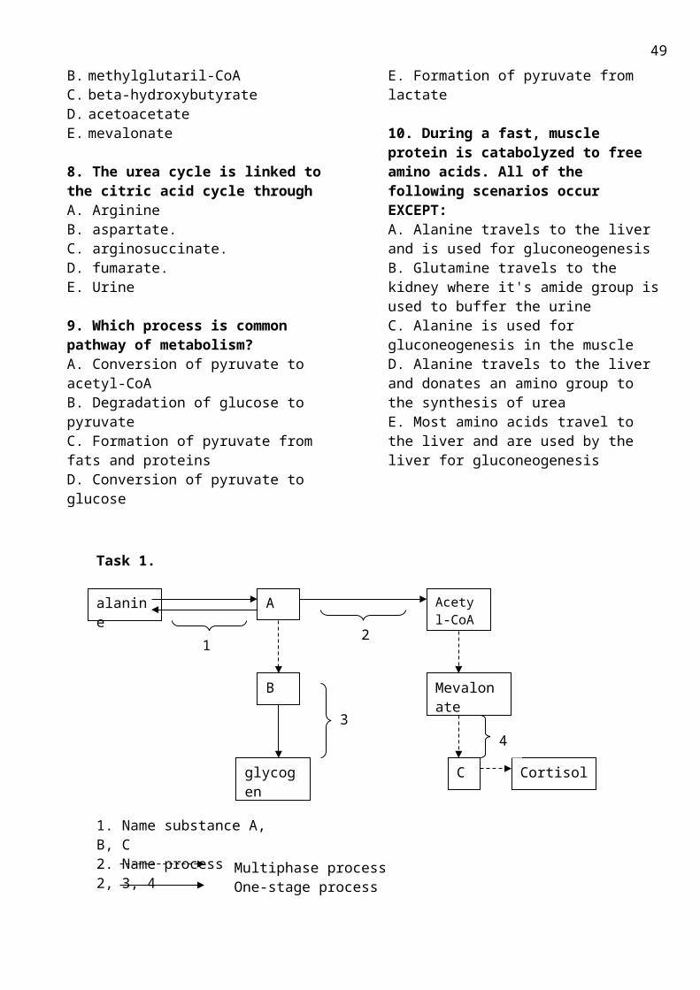
Respective places, a summary of these functions is given below in brief, so that students can easily group the tests of liver associating with its functions.1. Metabolic functions: Liver is the key organ and the principal site where the metabolism ofcarbohydrates, lipids, and proteins take place.(a) Liver is the organ where NH3 is converted to urea.(b) It is the principal organ where cholesterol is synthesized, and catabolised to form bile
acids and bile salts.(c) Esterfication of cholesterol takes place solely in liver.(d) In this organ absorbed monosacharides other than glucose are converted to glucose, viz.,
galactose is converted to glucose, fructose converted to glucose.(e) Liver besides other organs can bring about catabolism and anabolism of nucleic acids.
(f) Liver is also involved in metabolism of vitamins and minerals to certain extent.2. Secretory function: Liver is responsible for the formation and secretion of bile in the
intestine.Bile pigments-bilirubin formed from hem catabolism is conjugated in liver cells and secreted inthe bile.
3. Excretory function: Certain exogenous dyes like BSP (bromsulphthalein) and Rose Bengal dye areexclusively excreted through liver cells.
4. Synthesis of certain blood cogulation factors:Liver produces clotting factors like factor V, VII and X, ets. Fibrinogen involved in blood coagulation is also synthesized in liver cells. It also is responsible for maturation of certain blood cogulation factors (II,VII, IX,X) in the presence of vit. K. .
5. Synthesis of other proteins: Albumin is solely synthesized in liver and also to some extent a and b-globulins.
6. Detoxication function and protective function:Kupffer cells of liver remove foreign bodies from blood by phagocytosis. Liver cells can detoxicatedrugs, hormones and convert them into less toxic substances for excretion.
7. Storage function: Liver stores glucose in the form of glycogen. It also stores vit B12, Vit A etc.8. Miscellaneous functions: Liver is involved in blood formation in embryo and in some abnormal states; it also forms blood in adult.
Liver MetabolismOne of the most important roles of the liver is to serve as a "glucostat," monitoring and stabilizing blood glucose levels. To meet its internal energy needs, the liver can use a variety of fuel sources, including glucose, fatty acids, and amino acids. A primary role of liver is the synthesis of fuel components for use by other organs. Most of the low-molecular-weight metabolites that appear in the blood through digestion are taken up by the liver for this metabolic processing. Compounds synthesized in the liver include the following:
1. Fatty acids - The liver is a major site for fatty acid synthesis.
2. Glucose - The liver produces glucose, both from its own glycogen stores and from gluconeogenesis, the latter using lactate and alanine from muscle, glycerol from adipose tissue, and the amino acids not needed for protein synthesis. An important role of liver is to buffer the level of blood glucose. It does this largely through the action of glucokinase, an enzyme peculiar to liver, with a high KM (about 10 mm) for glucose, and partly through a high-KM transport protein, the glucose transporter. Thus, liver is unique in being able to respond to high blood glucose levels by increasing the uptake and phosphorylation of glucose, which results eventually in its deposition as glycogen. Glucose-6-phosphate accumulation activates the D form of glycogen synthase. In addition, glucose itself binds to glycogen phosphorylase
49
a, increasing the susceptibility of phosphorylase a to dephosphorylation with consequent inactivation. Thus, in addition to hormonal effects (see here), the liver senses the fed state and acts to store fuel derived from glucose. The liver also senses the fasted state and increases the synthesis and export of glucose when blood glucose levels are low. (Other organs also sense the fed state, notably the pancreas, which adjusts its glucagon and insulin outputs accordingly.)
3. Ketone bodies - Ketone bodies are also manufactured largely in the liver. The level of malonyl-CoA in liver, which is related to the energy status of the cell, determines the fate of fatty acyl-CoAs. When fuel is abundant, malonyl-CoA accumulates and inhibits carnitine acyltransferase I, preventing the transport of fatty acyl-CoAs into mitochondria for -oxidation and ketogenesis. On the other hand, shrinking malonyl-CoA pools signal the cells to transport fatty acids into the mitochondria, for generation of energy and fuels.
Detoxication function:
Cytochrome P-450 is a name for a family of heme proteins that perform hydroxylation reactions, as well as epoxidation, peroxygenation, desulfuration, dealkylation, deamination, and dehalogenation reactions. Most vertebrate genomes contain more than 40 different structural genes for cytochrome P-450. The proteins resemble mitochondrial cytochrome oxidase in being able to bind both O2 and carbon monoxide. Cytochrome P-450 proteins are usually found in the endoplasmic reticulum of eukaryotic cells.
Cytochrome P-450 hydroxylates many compounds. These include the hydroxylations of steroid hormone synthesis and the hydroxylation of thousands of xenobiotics (foreign compounds), including drugs such as phenobarbital and environmental carcinogens such as benzpyrene, a constituent of the smoke from tobacco and backyard grills. Hydroxylation of foreign substances usually increases their solubility and is a step in their detoxification, or metabolism and excretion. In some cases, however, some of these reactions activate potentially carcinogenic substances to more reactive species. Aflatoxin B, for example, is converted to a more reactive species either by hydroxylation or epoxidation.A key to the reactivity of cytochrome P-450 is its ability to split O2, with one oxygen atom binding to the cytochrome's heme iron. This bond forms a perferryl ion, which can be represented as FeO3+. This highly reactive group can abstract a hydrogen atom, even from an unreactive substrate such as a hydrocarbon. In such a hydroxylation, reducing equivalents are typically transferred to the cytochrome from NADPH. In this mechanism, substrate binding is followed by O2 binding. Transfer of two electrons reduces one oxygen atom, such that splitting of the oxygen molecule generates water plus the perferryl ion, which then hydroxylates the substrate.Monooxygenases incorporate one atom from O2 into a product and reduce the other atom to water. A monooxygenase has one substrate that accepts oxygen and another that furnishes the two H atoms that reduce the other oxygen to water. Because two substrates are oxidized, enzymes of this type are also called mixed-function oxidases. The general reaction catalyzed by monooxygenases is the following:AH + BH2 +O2 <=> A-OH + B + H2OThe substrate AH usually becomes hydroxylated by this class of ezymes, so they are also called hydroxylases. For example, this type of enzyme is used to hydroxylate steroids.
TESTS BASED ON THE DETOXICATING FUNCTION OF THE LIVER (Hippuric Acid Test of Quick)
1. Best known test for the detoxicating function of liver.2. Liver removes benzoic acid, administered as sodium benzoate, either orally, or intravenously, and
combines with amino acid glycine to form hippuric acid. The amount of hippuric acid excreted in urine in a fixed time is determined.
3. The test thus depends on two factors:(a) the ability of liver cells to produce and provide sufficient glycine and
50
(b) the capacity of liver cells to conjugate it with the benzoic acid.
Interpretations1. Normally, at least 3.0 gms of hippuric acid, expressed as Benzoic acid or 3.5 gms of
sodium benzoate should be excreted in health.2. Smaller amounts are found when there is either acute or chronic liver damage.
Amounts lower than < 1.0 gm may be excreted by patients with infectious hepatitis.Tests in liver diseases: Numerous laboratory investigations have been proposed in the assessment of liver diseases. From among these host of tests — the following battery of blood tests; total bilirubin and Von De- Bergh test, total and differential proteins and Albumin /Globulin ratio and certain enzyme assays as amino-transferases; alkaline phosphatase and γ-GGT have become widely known as "Standard Liver Function Tests" (LFTs).Urine tests for bilirubin and its metabolites and the prothrombin time (PT) and index (PI) are also often included under these heading. .
51
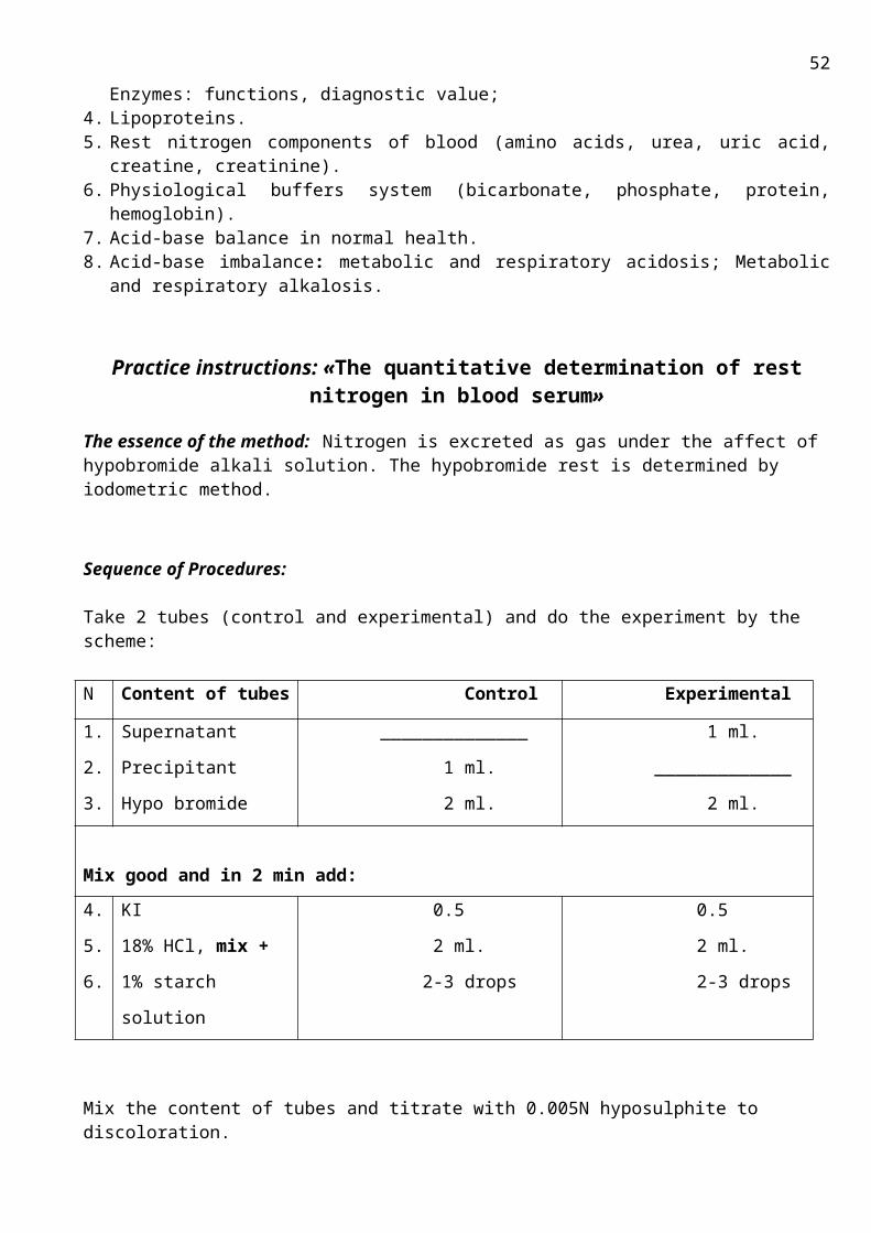
Topic 3.15. THE METHODICAL GUIDELINES FOR PRACTICE ACTIVITY ON THE THEME Studies of biological oxidation of different types. The role of fat-soluble vitamins in functioning of tissues and :organs
Biomedical importance : Although certain bacteria (anaerobes) survive in the absence of oxygen, the life of higher animals is absolutely dependent upon a supply of oxygen. The principal use of oxygen is in respiration, which may be defined as the process by which cells derive energy in the form of ATP from the controlled reactions of hydrogen with oxygen to form water. In addition, molecular oxygen is incorporated into, a variety of substrates by enzymes designated as oxygenases; many drugs pollutants, and chemical carcinogens (xenobiotics) are metabolized by enzymes of this class, known as the cytochrom P 450 system. Administration of oxygen can be life saving in the treatment of patients with respiratory circulatory failure and occasionally, administration of oxygen at high pressure (hyperbaric oxygen therapy) has proved of value, although this can result in oxygen toxicity. The lipid-soluble (fat-soluble) vitamins are apolar hydrophobic molecules, which are all isoprene de-rivatives. They cannot be synthesized by the body in adequate amounts and must, therefore, be supplied by the diet. They can only be absorbed efficiently when normal fat absorption is taking place. Once absorbed, they must be transported in the blood, like any other apolar lipid, in lipoproteins or attached to specific binding proteins. The lipid-soluble vitamins have diverse functions, eg, vitamin A, vision; vitamin D, calcium and phosphate metabolism; vitamin E, antioxidant; vitamin K, blood clotting. Although once thought of solely as a vitamin, vitamin D is in reality a prohormone. Conditions affecting the digestion and absorption of the lipid-soluble vitamins such as steatorrhea and disorders of the biliary system can all lead to deficiencies. Dietary inadequacy or deficiencies due to malabsorption cause syndromes consequent on the vitamins not carrying out their physiologic functions; vitamin A deficiency causes night blindness and xerophthalmia; vitamin D deficiency leads to rickets in young children and osteomalacia in adults; in vitamin E deficiency, which is rare, neurologic disorders and anemia of the newborn may arise; vitamin K deficiency, again very rare in adults, leads to bleeding and hemorrhage of the newborn. Because of the body's ability to store surplus lipid-soluble vitamins, toxicity can result from excessive intake of vitamins A and D. A role in cancer prevention has been ascribed to vitamin A and β-carotene, (provitamin A) as well as vitamin E.
The purpose: To interpret the mechanisms of different biological oxidation reactions; their role and the fat-soluble vitamin action in the human body for the future use of this knowledge in medicine practice. The applicable materials:1. The tutorial book; The tutorial book, "Principles of biochemistry", 2005.p. 53-57, 67-70, 105-107., 258-270. "Biochemistry", Pamela C. Champe at al.2005.p.69-73,89-91,113-115.,379-392 2. The «Bioenergetics» Lecture Materials;3. The appendix.
The main theoretic questions:1. Biological oxidation reaction types. The enzymes, role in organism.2. Active oxygen forms (AOF): hydrogen peroxide, hydroxyl radical and super oxide radical. Formation
(Generation) in the body, reasons of toxicity, (peroxide oxidation of lipid membranes), biological role3. Biological role of vitamin A3.1 The role in visual cycle3.2 Absorption and transport of vitamin3.3 The role of vit. A in tissue grows, differentiation and reproduction3.4 Symptoms of deficiency. Dietary sources, provitamin A, 3.5 Clinical indications.13.6 Toxicity of retinoids4. Biological role of vitamin E
52
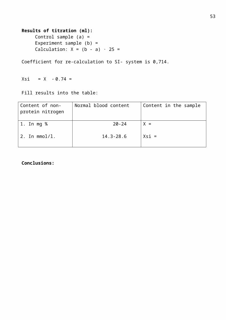
4.1 Distribution, the role in metabolism.4.2 Deficiency of vitamin E.4.3 Clinical indications.5. Fat soluble vitamins as natural antioxidants.
Practice instructions
Work №1.The finding of aldehyddehydrogenase in milk.
The essence of the method: Enzyme is produced by microorganisms, which get into milk from environment. It is flavoprotein in chemical nature and can catalyze aldehydes oxidation, in particular, formaldehyde to formic acid. If this reaction occures in the presence of methylen blue (solution of dark blue color), hydrogen from aldehyde associate with indicator (methylen blue) and reduce it to colorless compound.
Sequence of Procedures:1. Take 2 tubes.2. Pour 15 drops of boiled milk into 1-st tube and 15 drops of no boiled milk into the 2-nd.3. Add 1 drop of 0,4 % formaldehyde solution and 1 drop of 0,01% methylen blue solution both into
the tubes.4. Mix tubes and close them with cork in order to make anaerobic conditions.5. Incubate at room temperature 5 min and observe disappearing of blue color. Make conclusions.
Conclusions:
Work №2. The finding of catalase in blood.
The essence of the method: Catalase is enzyme, which belong to class of oxidoredactase and is found in blood, bone marrow, mucous membranes, kidney and liver. Its function is destruction of hydrogen peroxide formed by action of oxidases in the following reaction: 2 H2O2 2H2O + O2
Sequence of Procedures:1. Pour 10-15 drops of 3% H2O2 solution into the tube.2. Add 1 drop of blood3. Released of oxygen bubbles observe.
Conclusions:
Work №3. The qualitative finding of vitamin A.
a. Drummund reaction with concentrated H2SO4.Sequence of Procedures:1. Pour 3 drops of vitamin A oil solution ore fish fat into the dry tube.2. Add 1 drop of concentrated H2SO4. 3. Violet color, which changes to cherry- red, appears.
B. Reaction with FeSO4.
53
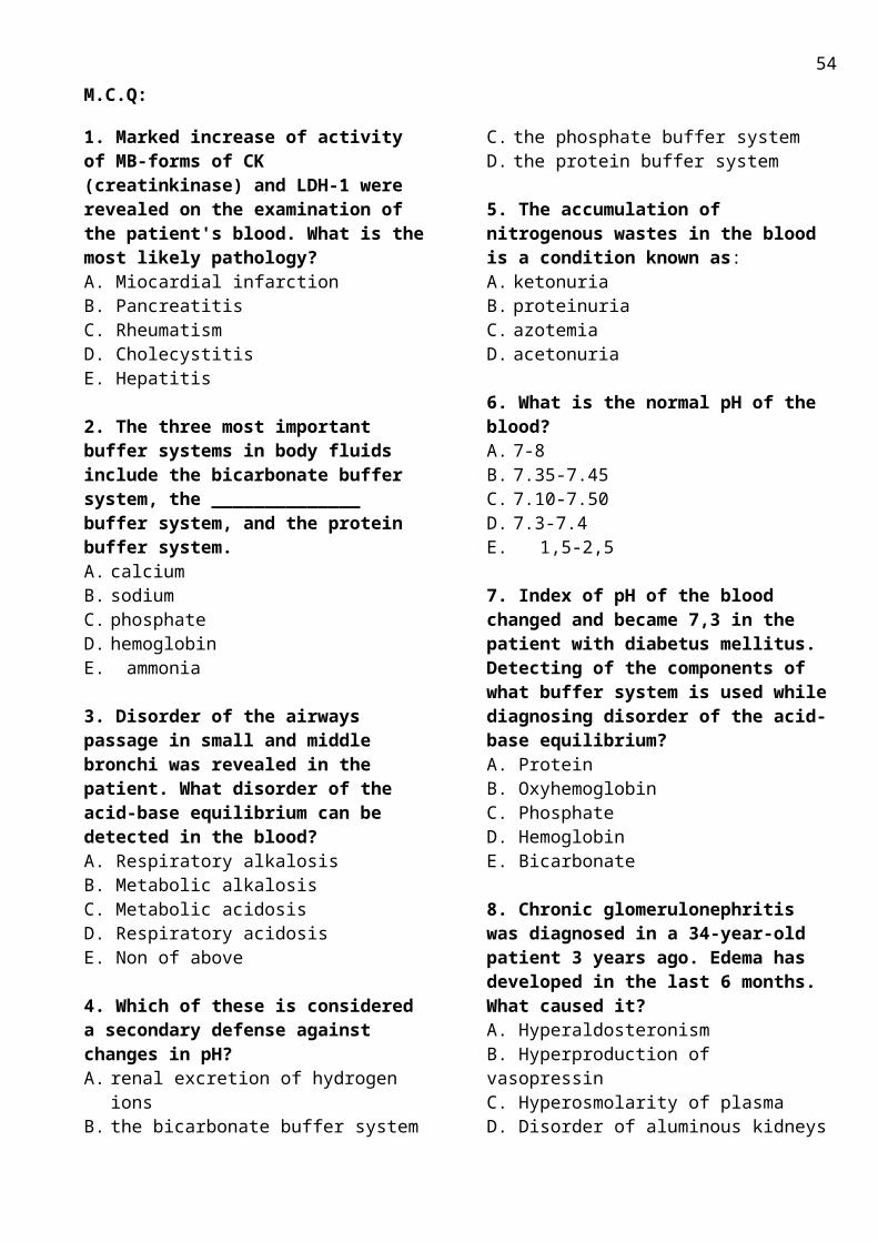
Sequence of Procedures:1 Pour 2-3 drops of vitamin A oil solution ore fish fat into the tube.2. Add 5-10 drops of ice acetic acid, which saturated FeSO4
3. Add 1-2 drops of concentrated H2SO4.Blue color appears and changes to pink-red. (Carotenes give in this reaction green color.)
Work №4.
Qualitative reactions for vitamin Ea. Reaction with nitric acid.Sequence of Procedures:1. Pour 5 drops of vitamin E alcohol solution into the dry tube.2. Add 1 ml of concentrated HNO3.3. Mix well and observe the appearance of red color.The oxidized product of α-thocopherol has quinoid structure.
b. Reaction with FeCl3.Sequence of Procedures:1. Pour 0, 5 ml of vitamin E alcohol solution into the dry tube.2. Add 1 ml 1% FeCl3 and mix.3. Red color appears. (α-tocopherol has been oxidized to α-tocopherilquinone).
Conclusions:
54
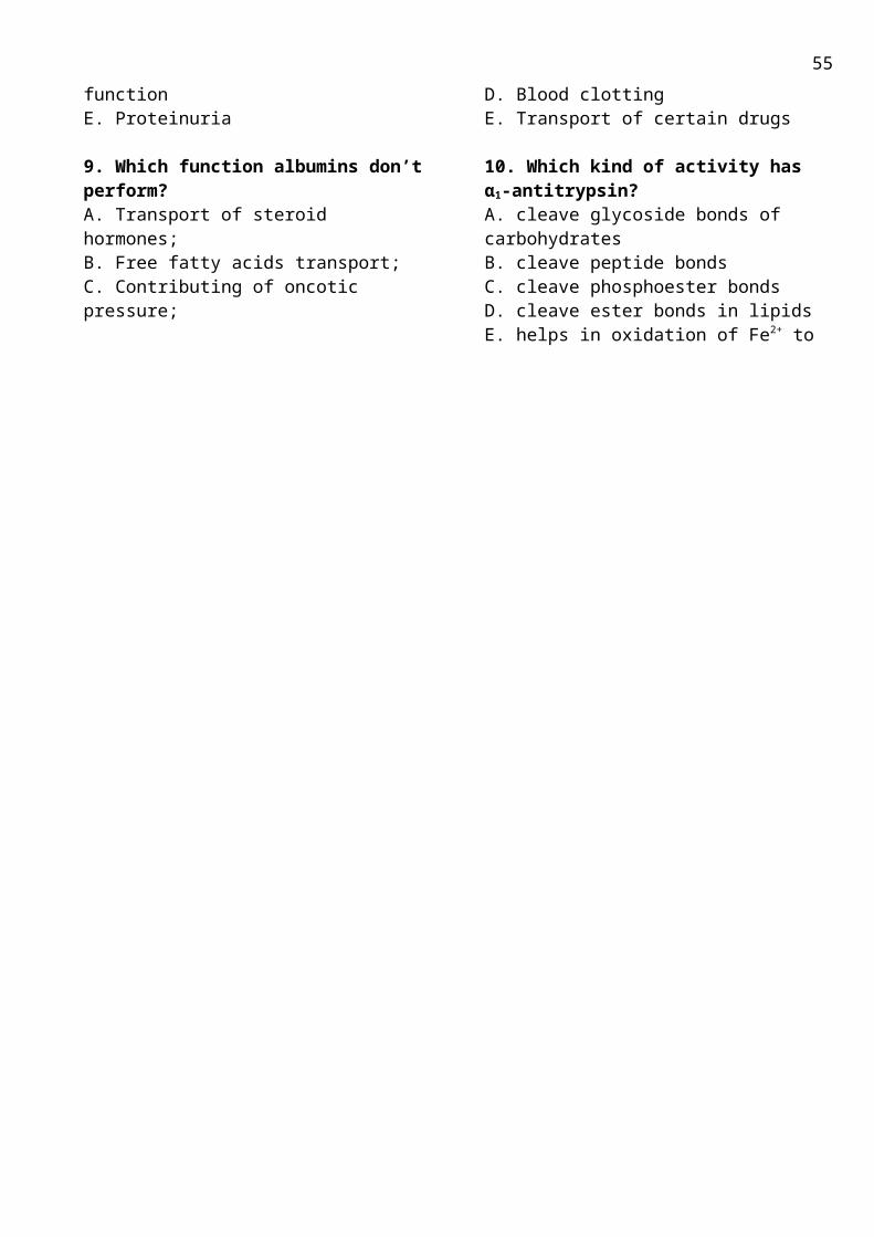
M.C.Q:
1. A patient’s liver cannot normally detoxify its natural metabolites and xenobiotics. The reduced activity of what cytochrome can cause this?A. Cytochrome BB. Cytochrome Р- 450 C. HemoglobinD. CytochromoxidaseE. Cytochrome C1
2. Profuse foam appeared when dentist put hydrogen peroxide on the mucous of the oral cavity. What enzyme caused such activity? A. Cholinesterase B. Acetyltransferase C. Methemoglobinreductase D. Catalase E. Glucose-6-phosphatdehydrogenase
3. Patient with abscess of the cut wound applied to the traumatological department. Doctor for the cleaning of the wound from the pus washed it with 3% hydrogen peroxide. Foam was absencee. What is caused the absence on the drug activity? A. Inherited insufficiency glucose-6-phosphatedehydrogenase of erythrocytes B. Inherited insufficiency of catalase C. Pus in the wound D. Shallow wound E. Low concentration H2O2
4. Muscular dystrophy makes progress under the action of the ultraviolet irradiation. What substance can embarrass to these defects?A. α-tocopherolB. 1, 25-di-OH-cholecalciferolC. RetinolD. Vitamin D3E. phylloquinone
5. Necrosisdystrophic process appeared in the liver of a rescue man, who was working at radiation area. What has to be prescribed to this patient in complex therapy?
A. RetinolB. Ascorbic acidC. vitamin KD. LecithinE. α-tocopherol
6. Vitamin A contains –CHO group in side chain. Which class of compounds it belongs?
A. AcidsB. AlcoholsC. AldehydesD. KetonesE. Fatty acids
7. The absorption of light by cells in the retina of the eye results in the conversion of:A. carotene to retinalB. Rhodopsin to cis-retinal and opsinC. All- trans retinal to cis -retinalD. retinol to retinalE. Rhodopsin to all- trans retinal and opsin
8. Sensitivity of erythrocytes to peroxide is due to a deficiency of:A. Vit. KB. NADPHC. Vit.DD. Vit. EE. Vit. A
9. The biological activity of vit A has been attributed, in part, to its action:A carrier of electronsAn anti-oxidantAn anti-coagulantA reducing substanceA methylation substance
10. The absorption of light by cells in the retina of the eye results in the conversion of:A. all-trans retinal to m-retinal B. cis-retinal to all-trans retinalC. retinal to retinolD. retinol to retinalE. β-carotene to retinal.
55
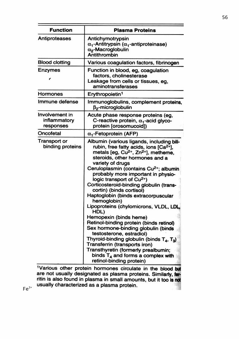
APPENDIX.PATHWAYS FOR OXYGEN CONSUMPTION IN REACTIONS OF BIOLOGICAL OXIDATIONIn most cells, at least 90% of the molecular oxygen consumed is used in oxidative phosphorylation. The oxygen delivered to the cells is consumed not only during oxidation of substrates in the mitochondrial respiratory chain, but also in a wide variety of other specialized metabolic biological reactions. Diverse reactions proceeding via oxygen consumption may be classified into three major types.
The first type may be referred to as an oxidase type. Oxidases are enzymes that catalyze the oxidation of a substrate without incorporating oxygen into the product. A two-electron oxidation is usually involved , so the oxygen is converted to H2O ( cytochrome oxidase in oxidative phosphorylation ) or to H2O2.(Amino acids oxidases ). Schematically, it is represents:
a) S×H2 + 1/2O2 → S + H2O b)S×H2 + O2 → S + H2O2
Most oxidases utilize either a metal (Cu)or a flavin coenzyme.
The second reaction type is an oxigenase type. These reactions are catalyzed by monooxigenases
A×H2 + SH + O2 → A + S-OH + H2O
Where A×H2 is a hydrogen donor and SH is an oxidizable substrate, or by dioxigenases
S + O2 → SO2
The monooxigenase reaction mechanism involves the insertion of one oxygen atom into the oxidizable substrate, and of another one, into a water molecule. The substrate SH usually becomes hydroxylated by this class of ezymes, so they are also called hydroxylases. For example, this type of enzyme is used to hydroxylate steroids.The dioxigenases route leads to the insertion of two atoms of molecular oxygen into the oxidizable substrate.
Monooxigenases exist as soluble enzymes, which are either found in the cellular sap, or included in special oxidation chains located in the membranes of hepatic cells of endoplasmic reticulum, in the mitochondria of adrenal cortex cells. Soluble monooxigenases include copper-containing monooxigenases, for example, dopamine-- monooxigenase participating in the oxidation of intermediates of noradrenalin and adrenalin synthesis in the chromaffin tissue; tyrosinase involved in the oxidative production of the pigment melanin from tyrosine in the skin, the iris and the retina of the eye.
The monooxigenase oxidation chains are short chains for the transfer of electrons and protons mostly supplied by NADP×H, the special cytochrome type, cytochrome P450, being an activator for O2 (below, S is an oxidizable material), ascorbic acid, Fe3+
Monooxigenase chains are used for the oxidation of natural organic materials for synthesizing bile acids and steroid hormones from cholesterol as well as for detoxicating drugs and toxins.
The fourth type of reactions is the free radical oxidation , e.g. peroxid oxidation of unsaturated fatty acids by scheme RH + O2 → ROOH. The products derived by peroxide oxidation of unsaturated lipids are lipid hydroperoxides, alcohols, aldehydes, ketones, malonic and other dialdehydes, and epoxides. These oxygen-consuming reactions proceed in the membranes of mitochondria, endoplasmic reticulum, lysosomes, that is, where unsaturated lipids (most phospholipids) are available as substrates. Apparently, the physiological role of this type consists in regulation of renewal and permeability of biomembrane lipids. Free radicals produced by single-electron reduction of oxygen according to the scheme:
56

act as activators for the peroxide oxidation of lipids. Oxygen radicals (×O2-, superoxide radical; ×OH, hydroxyl
radical; and HO2×, peroxide radical) are very reactive species that are capable to spontaneously accelerate chain reactions in the peroxide oxidation of unsaturated lipids and to react with various biomolecules (proteins, nucleic acids, and many other compounds) causing functional disturbances in them. Under the conditions favourable for the formation of free oxygen radicals, the auto-accelerating process of peroxide oxidation may destroy completely unsaturated lipids in the biomembranes, leading to the imminent death of the cells. However, in phagocytizing cells, the superoxide radical ×O2
- is employed, alongside of H2O2, for destruction of invading microbes and noninfectious materials.
The scheme for the major pathways of cellular oxygen consumption may be presented in the following manner:
The oxygen utilization by each of the above routes is not equivalent and depends on a number of factors. Most of the cellular oxygen fund (80% to 90%) is consumed via the first route (see Scheme above) in the mitochondria in which the oxidative phosphorylation system is operative. Of the remaining oxygen, a major part is consumed via monooxigenase route (third route in the scheme above), mainly in the oxidation chains of cellular endoplasmic network of the liver and other organs and in the mitochondria of adrenal cortex cells.
Water is the major product of oxygen-consuming biological oxidation reactions. In addition, hydrogen peroxide, oxygen radicals, and lipid peroxides, which are toxic for the organism cells when present in higher concentration, are produced in these processes. To neutralize these deleterious products of biological oxidation, special systems are provided for in the organism.
Reactive Oxygen Species May Initiate DiseaseA free radical is an atom or molecule that has one or more unpaired electrons. Its consequent tendency to acquire an electron from other substances makes it highly reactive. However, not all reactive oxygen species are free radicals, e.g., singlet oxygen and H2O2. When oxygen is reduced to water by cytochrome oxidase, 4 electrons are acquired. Electrons, however, can be gained one at a time by univalent reduction, which may account for 1-5% of total oxygen consumption. The individual molecules in univalent reduction are highly reactive and potentially damaging to tissues. They are superoxide, hydrogen peroxide, and the hydroxyl free radical. The last is extremely toxic but short-lived. Other sources of reactive species are xanthine oxidase, which generates superoxide (eg, during reperfusion injury of ischemic organs), and cyclooxygenase and lipoxygenase, which produce hydroxyl and peroxyl radicals. Stimulated neutrophils produce superoxide, which is one mechanism for the destruction of bacteria . Superoxide may also be produced during the metabolism of xenobiotics by cytochrome P-450. Because these molecules are so reactive, they act in situ very close to where they are generated. Therefore, most cell structures are vulnerable, including membranes, structural proteins, enzymes, and nucleic acids, which can lead to mutation and cell death.
These oxygen species include superoxide, formed from a one-electron reduction of O2; hydrogen peroxide (H2O2), generated from a two-electron reduction; and hydroxyl radical, formed via a three-electron reduction. In addition, some enzymes, such as xanthine oxidase and amino acid oxidase, generate hydrogen peroxide as ordinary products. Superoxide, hydrogen peroxide, and hydroxyl radical are more reactive than O2, so they are referred to collectively as reactive oxygen species (ROS).Hydroxyl Radical - Hydroxyl radical damages proteins, nucleic acids, and the fatty acids in membrane lipids (lipid peroxidation). Lipid peroxidation occurs as a chain reaction. Hydroxyl radical is produced as a result of ionizing radiation and represents the most active mutagen derived from ionizing radiation. It is also produced from H2O2 in the Fenton reaction:H2O2 + Fe2+ (or Cu+) -> Fe3+ (or Cu2+) + OH radical + OH-
57
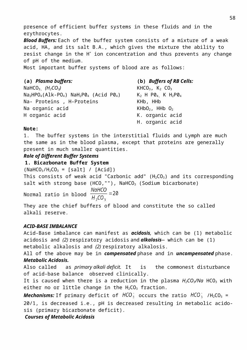
Superoxide is relatively nontoxic. It is a free radical, however, so it combines readily with nitric oxide, another free radical that is a biological signaling agent. The product is peroxynitrite (OONO-), which is also considered a ROS. Peroxynitrite causes lipid peroxidation and also causes nitration of tyrosyl hydroxyl groups in proteins, a reaction particularly damaging to membrane proteins.Defense: Large scale production of reactive oxygen species has the potential to inflict considerable damage on the tissues in which they are produced, a situation called oxidative stress. Antioxidant compounds, such as glutathione, vitamin C and vitamin E, and uric acid provide non-enzymatic protection against oxidative stress because they can scavenge ROS before the ROS can cause damage. Alternatively, antioxidant compounds can prevent oxidative damage from spreading, such as the chain reaction of lipid peroxidation. Vitamin E is the principal lipid-soluble antioxidant compound and plays an important role in preventing membrane damage. -Carotene and other carotenoid compounds related to vitamin A are lipid-soluble antioxidants that also play roles in free radical trapping. Glutathione plays an important role in cellular antioxidant protection. Vitamin C (ascorbic acid) is present in far higher amounts in cellular fluids and probably plays the predominant role in extracellular antioxidant protection. A major antioxidant role of uric acid may be its ability to bind and inactivate peroxynitrite.
Enzymatic mechanisms can defend against ROS, too. 1. Superoxide dismutase (SOD) is a family of metalloenzymes that catalyze dismutation (reactions in which identical molecules have different fates). The reaction catalyzed is as follows:O2- + O2- + 2H+ -> H2O2 + O2The term dismutase describes an enzyme that acts on two identical substrate molecules in a way that results in two different products. The hydrogen peroxide formed in the reaction above can be broken down by the enzyme catalase or, to some extent, by peroxidases:Hydrogen peroxide is metabolized either by catalase or by a more limited family of peroxidases. Catalase catalyzes the following reaction:2H2O2 -> 2H2O + O22. Glutathione peroxidase catalyzes reduction of H2O2 as follows:2GSH + H2O2 -> GSSG + 2H2OHere GSH is reduced glutathione and GSSG is oxidized glutathione.Some cells produce ROS as a normal part of their functioning. Certain white blood cells contribute to defense against infectious agents by phagocytosis. Such cells can engulf a bacterial cell, followed by a respiratory burst - a rapid increase in oxygen uptake. Much of the oxygen is reduced to superoxide ion and to H2O2, which help to kill the engulfed bacterium. Glutathione peroxidase thus protects against oxidative damage by reducing the reactive oxygen species, H2O2 to water. The enzyme is interesting in that it contains the rare amino acid, selenocysteine3. Catalase is a heme-iron-containing enzyme that catalyzes the reaction below:2H2O2 <=> 2H2O + O2Catalase has an extremely high turnover rate (>40,000 molecules per second) and acts to protect against oxidative damage by the reactive oxygen species, H2O2..
Superoxides
What “should” happen…..O2 + 4e- 2H20
What often happensO2 + 2e- 2H202
Even worse!O2 + e- 02
-
*Antioxidants help remove free radicals from circulation Examples: Vit. E, Vit. C, CoQ ½ life of a normal O2
- - 1 millionth of a second½ life of a catalyzed O2
- - 1 billionth of a second
(Although this seems like an infinitesimally small amount of time, it is the difference in life and death for aerobes!)
58
Water
Hydrogen Peroxide
SuperoxideRadical

Topic 3.16. THE METHODICAL GUIDELINES FOR PRACTICE ACTIVITY ON THE THEME:Investigation of normal and pathological urine components.
Biomedical importance: Urine analysis provides valuable informations to a clinician; urine should be examined soon after being passed. For random examination, a random specimen is satisfactory, but for certain investigations urine passed at a particular time of the day is valuable, e.g. (i) to detect glycosuria urine excreted 2 hrs. After meal is examined, and (ii) to control insulin therapy urine excreted in the early morning and at 12 noon is tested for glucose. For quantitative estimation of excretion of chemical constituents, such as urea, uric acid, creatinine, proteins etc, 24 hours of urine sample should be used whenever possible. A suitable preservative to prevent bacterial growth like thymol/cone HC1 can be used.
The purpose: To develop skills in interpreting of urine normal and pathological components results for the future use of this knowledge in clinic subjects.
The applicable materials:1. The tutorial book; (Inherited diseases) 2. The Appendix “Kidney and urine”.3. The test book.
The main theoretic questions:1. Physical and chemical urine characteristics: pH, color, viscosity, transparency. Diagnostic
value of their determination.2. Urine normal chemical components (uric acid, urea, stercobiline, metal ions etc.)3. Pathological components of urine: protein, carbohydrates, blood, bilirubin, keton bodies etc.4. Methods of their determination in urine and diagnostic value5. Pathological components in urine upon inherited diseases and some vitamins deficiency
(B1, B12) (Recall material that you studied earlier) a) amino acid metabolism: phenylketonuria , alcaptonuria, homocystinuria, maple-syrup disease;b) synthesis of urea: citrullinemia; etc.,c) carbohydrate metabolism: glucosuria, fructosuria, galactosemia;d) heme metabolism: porphyrias;e) Orotic aciduria.
Practice instruction: “Normal and pathologic ingredients of urine”
Work №1. Quantitative definition of protein by Brundberg-Roberts-Stolnicov method. Essence of the method: The method is based on Heller's reaction with concentrated nitric acid. Solutions which contain 0,033% of protein give white ring in 2-3 min. after urine filling at nitric acid. Sequence of Procedures:1. Take 5 tubes and prepare urine diluted in 10, 20, 30, 40 and 50 times;2. Pour 1ml of nitric acids into 5 another tubes;3. Carefully make in layers (fill) 1 ml of specific dilution level urine by dropping it from pipette onto the wall of slightly lopsided tube, which contain nitric acid.
Do this procedure for all 5 dilutions of urine.4. Calculate the amount of protein by the tube with the maximum level of urine dilution at which a slightly visible ring appears in 2-3 min. For example, the ring appears after 2nd min elapse upon 1/30 urine dilution. So the concentration of protein in non-dilution urine is 0.033 x 30 = 0.099%.
59
C =
Work №2. The qualitative finding of keton bodies by Legal test.Essence of the method: Acetone and acetoacetate form orange-red color compound with sodium
nitroprusside in alkali medium. Color becames red-cherry after adding of acetic acid.Sequence of Procedures:
1. Pour 0, 1 ml of urine into a tube.2. Add 0, 1 ml 10% of NaOH and 0, 1 ml of sodium nitroprusside.3. Orange-red color appears, if urine contains keton bodies.
Work №3. Herhgard reaction for acetoacetate.Sequence of Procedures:
1. Pour 5 drops of urine into a tube.2. Adding 5% Fecl3 solution by drops until sediment of FePO4 is formed.3. Red-cherry color appears on future addition of FeCl3 if urine contains acetoacetate.
Work №4. . The qualitative determination of carbohydrates in urine.The essence of the method: is based on glucose ability to reduce Cu (OH) 2 (blue color) to CuOH and
Cu2O (red color) in alkaline medium.Sequence of Procedures:1. Pour 1 ml of urine into the tube;:2. Add 5 drops of 30% NaOH and 7% of copper sulphate solution until non-vanishing less of copper
hydroxide appears.3. Boil the tube on the gas. Orange-red color sediment of Cu2O appears, if urine contains glucose.
Work № 5. Take individual tube with urine and determine what pathological components it contains. (±)
Tube № _____ contains: Protein concentration of:________ Keton bodies: ________ Acetoacetate: ________ Glucose: ________Conclusions:
Appendix
URINE COMPONENTS IN NORM AND IN PATHOLOGYThe urinary excretion of various materials reflects alterations in the processes that occur in the
kidney and other tissues and organs of the organism. The daily volume of final urine amounts to 1.5-2 litres and the dry weight of final urine is about 60 g. Since the urine is a filtrate of blood plasma, it appears expedient to consider the urinary concentrations of various groups of biological materials from the standpoint of their occurrence in the blood plasma.
ProteinsIn norm, the daily urinary excretion of proteins amounts to about 30 mg, which is not detectable by
common laboratory techniques and routinely specified as "traces, or absence of urinary proteins". Among the urinary proteins, enzymes are also present. The origin of normal urinary proteins is different. Some of them are blood plasma proteins that have not been completely reabsorbed, others are proteins constitutive of the cells stripped off from the urinary tract walls.
In pathology, the urinary protein concentration may be increased; depending on the location of the damage, prevalent in the urine may be either plasma proteins, or cellular proteins of the urinary tract. In inflammatory renal diseases (glomerulonephritides), the permeability of the basal membrane of nephron glomerulus increases; proteins are filtered in an amount above normal and fail to be reab -sorbed completely. Disturbances in the tubular protein reabsorption (nephroses) are conducive to a similar pathology. For this reason, in glomerulonephritides and nephroses, the urinary excretion of
60

proteins may vary from 1 to 15-40 g per day. An increased concentration of certain normally filterable proteins in the blood plasma may lead to their excessive urinary discharge. Nonetheless, even in such a contingency, the urinary protein concentrations are small and can be detected only using special techniques. For example, if certain enzymes are found in the blood in larger quantities, they are filtered into the urine in a larger proportion. For this reason, an increased enzyme activity is observed in the urine, although this is unlikely to affect the test for total protein performed by less sensitive methods. For example, in pancreatitis, an enhanced activity of a-amylase and trypsin is observed both in blood and urine.
NONPROTEIN NITROGENOUS URINARY COMPONENTSUrea is a major nitrogenous component of the urine. The normal excretion of urea is 333 to
583 mmol per day, which accounts for 60 to 80% of the overall urinary nitrogen. An increased urinary concentration of urea is observed in the states with pronounced catabolism of proteins and other nitrogenous components (starvation, burns, traumatism, atrophy of tissues, etc.). A decreased excretion of urea is observed in affected liver (urea-producing organ) and in impaired glomerular filtration of blood plasma. In the latter case, urea is retained in the blood (this state is called azotemia). A low urinary excretion of urea may also occur in the growing period of the organism or as produced by the action of anabolic preparations.
Uric Acid. Normally, the urinary excretion of uric acid is 2.35 lo 5.9 mmol per day. Its increased urinary concentration is observed in a diet rich in nucleic acids or as produced by breakdown of cells and tissues, for example, leucocytes in patients with leucosis. An increased concentration of uric acid in urine may also be due to enhanced synthesis of uric acid in the organism tissues (Lesch-Nyhan syndrome in children).
Creatinine. In norm, the urinary excretion of creatinine is 4.4 to 17.6 mmol per day; variations in creatinine concentration are dependent on muscular development. Physiological excretion of creatinine is normal only in children. In adult humans, creatinuria is a sign of pathology (e.g. muscular dystrophy).
Amino Acids. In norm, the urinary excretion of amino acids is 0.29 to 5.35 mmol per day (as based on nitrogen). The urinary concentrations of glycine, histidine, and alanine are higher than those of other amino acids.
In pathology (e.g., burns, diabetes mellitus, affected liver, and muscular dystrophy) hyperaminoaciduria may occur. Hereditary hyperaminoaciduria is associated with defective proteins—carriers for amino acids in the proximal renal tubules. As a result, an increased excretion of either all amino acids (the generalized defect of tubular transport systems), or some of them (localized defect of one of the transport systems) is observed. In a disordered amino acid tissue metabolism, the urinary excretion of normally nonexcretable amino acid metabolites occurs (e.g., homogentisic acid, in alcaptonuria; phenylpyruvic, phenylacetic, and phenyllactic acids, in phenylketonuria).
Ammonium Salts. In norm, the urinary excretion of ammonia as a component of ammonium salts (ammonium chloride) is 30-60 mmol per day.
In pathology, an increased urinary elimination of ammonium salts may be ob served (in diseases accompanied by acidosis). A diminished excretion of ammonium salts occurs in diseases associated with alkalosis, in dietary intake of large amounts of alkaline nutrients, and in renal diseases due to affected distal tubules in which ammoniogenesis takes place.
Hippuric Acid . The urinary excretion of hippuric acid is dependent solely on the amount of ingested vegetable food, since in the organism this acid endogeni-cally is not produced. Commonly, the daily urine contains to 5.5 mmol of hippuric acid.
Indican (Indoxyl Sulphuric Acid). Normal urine contains indican (in the form of potassium indoxyl sulphate) in trace amounts. In detectable quantities, indican appears in the urine on excessive alimentary intake of meat products; it also occurs as a byproduct of putrefactive processes in the intestine.
Nitrogenous Pigments. Representative of these is stercobilinogen, a product of hemoprotein breakdown. Stercobilinogen is convertible to stercobilin and normally is excreted in the urine.
In pathology, urinary excretions contain bile acids and a variety of bile pig ments, for example, in affected liver and in toxicoses conducive to hemolysis.
NITROGEN-FREE COMPONENTS OF URINEGlucose and Other Monosaccharides. In norm, the daily urine contains a mere 0.3-1.1 mmol of
glucose. Such amounts escape detection by conventional analytical techniques; for this reason, glucose is not reckoned as a component of normal urine. However, in excessive dietary intake of
61
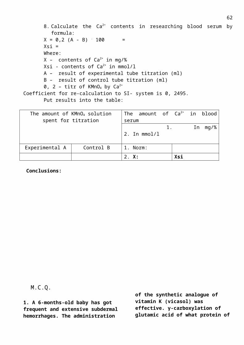
carbohydrates, when the glucose concentration in blood attains a threshold value, i.e. of the order of 8.3-8.8 mmol/ litre, alimentary glucosuria may develop in the organism.
In pathology, glucosuria occurs due either to an increased blood glucose concen tration (exceeding the threshold value), or to a defective carrier protein involved in glucose reabsorption in the renal proximal tubules. The former case is the most commonly encountered in the clinic, for example, in diabetes mellitus or in steroid diabetes. The latter case is the so-called renal diabetes. For example, the occurrence of fructose or pentoses in the urine (renal fructosuria or renal pentosu ria) is indicative of affected transport systems of the renal tubules.
Lactic and Pyruvic Acids. In norm, the daily urinary excretions of lactic and pyruvic acids amount to 1.1 and 0.11 mmol, respectively. An increased concentration of lactic acid in the urine is observed under intensive muscular work and in hypoxia. An increased urinary excretion of pyruvic acid occurs in diabetes mellitus and in B1, hypovitaminosis.
Ketone Bodies. In norm, the daily urine contains 20 to 50 mg of ketone bodies. At this level, they are not detectable by the analytical methods currently em ployed in the clinic. Increased concentrations of ketone bodies, i.e. a state called ketonuria, occur in diabetes mellitus, steroid diabetes, and starvation.
Mineral Salts. In norm, the daily urine contains (in mmol): sodium, 174-222; potassium, 61-79; calcium, about 4.02-4.99; inorganic phosphorus, about 1,7 mg/l.. An increase in urinary excretion of sodium and a decrease in excretion of potassium are observed in the adrenal hypofunction; the reverse situation occurs in hypcral-dosteronemia and when mineralo- and glucocorticoids are prescribed as drugs. A decreased urinary concentration of calcium and a distinct phosphaturia are observed .when large doses of vitamin D and parathyrin are administered; a high urinary loss of calcium is characteristic of rickets and hypoparathyroidism.
Creatine is a precursor of creatine phosphate, the compound that shuttles high-energy phosphate from mitochondria to sites of muscle contraction.Creatine is formed by transmethylation of guanidinoacetate (derived from arginine) (reaction 1 below) and has three resonance forms, as follows:
Creatine participates in the reactions that follow: Arginine + Glycine <=> Guanidinoacetate + Ornithine (catalyzed by glycin-amidin transferase kidney specific enzyme !1. Guanidinoacetate + S-Adenosylmethionine <=> Creatine + S-Adenosylhomocysteine,2. Creatine + ATP <=> Creatine Phosphate + ADP (catalyzed by Creatine Kinase)The latter reaction is strongly endergonic as written. However, the level of ATP is very high in mitochondria, so the reaction proceeds to the right. Creatine phosphate then diffuses from mitochondria to the myofibrils, where it provides the energy for muscle contraction.High levels of ADP formed in the myofibrils during contraction favor the reverse reaction namely, resynthesis of ATP - at the expense of creatine phosphate cleavage to creatine. This example shows that one must consider not only the standard free energy change but also the actual concentrations of all reactants and products when predicting the direction of a reaction in vivo.
62

Topic 3.17. THE METHODICAL GUIDELINES FOR PRACTICE ACTIVITY ON THE THEME:Biochemistry of nervous and connective tissues.
Biomedical importance: The functions of the nervous tissue are carried out by neurons and glial cells. Neurons are necessary for specific function of the nervous tissue. It regulates and coordinates vital functions of whole organism and its interaction with external and internal environment. Nerve cells are responsible for the irritation perception, creation and propagation of nerve impuse.Neuroglia provides support, trophic, separation and defence of nerve cells.The connective tissue, or extracellular matrix, contains three major classes of biomolecules: the structural proteins (collagen, elastin), certain specialized proteins (fibrillin, fibronectin, laminin) and proteoglicans.The connective tissue has been found to be involved in many normal and pathologic processes. It plays an important role in development, in inflammatory states, and in the spread of cancer cells. Involment of certain components of the connective tissue has been documented in both rheumatoid arthritis and osteoarthritis. Several diseases (osteogenesis imperfecta, the Ehlers-Danlos syndrome) are due to genetic disturbances of the synthesis of collagen. Specific components of proteoglicans (the glycosaminoglicans) are affected in the group of genetic disorders known as mucopolysaccaridoses. Certain changes take place in the connective tissue during the aging process.
The purpose: is to know how to explain the peculiarities of biochemical composition, metabolism and their possible changes in these tissues for further use of obtained information in clinic.Main theoretical questions: 1. Peculiarities of the nervous tissue biochemical compositionAmino acids, peptides and proteins of nervous tissue and their role in the nervous tissue functionPeculiarities of the nervous tissue lipids and their role in the nervous tissue functionBiochemical composition of myelin and its function
2. Bioenergetics of brain3. Neurotransmitters (GABA, serotonin, acetylcholine, aspirate, glutamate, glycine, norepinephrine,
dopamine) : their origin and role in the nerve impulse transmission4. Biochemical mechanisms of memory5. Main properties and functions of the connective tissue6. Biochemical composition of the extracellular mattrix
Collagen: structure and its biological importanceElastin: structure and its biological importanceProteoglicans and glycosaminglicans of the connective tissue: structure and their biological importanceFibronectin: structure and its biological impornance
7. Collagen metabolism8. Proteoglicans metabolism9. Biochemical changes in the connective tissue during aging10. Biochemical mechanisms of the connective tissue pathology: collagen structure and metabolism
disorders, mucopolysaccaridoses, collagenosesThe applicable materials:
1. Lectures in biochemistry2. Murray R.K., Granner D.K., Mayers P.A., Rodwell V.W. Harper’s Illustrated Biochemistry,
Twenty-Sixth Edition. – International edition, 2003. – P. 116; 266 – 268; 428; 535 – 5553. Lippincott’s Illustrated Reviews: Biochemistry/ Champe P.C. etc. – 1994. – P. 43 – 52; 155 – 161;
283 – 286
Read the following tasks and choose one correct answer:
63

1. The neurospecific enolase activity of brain cortex is much more than in brain stem. Taking into account these data, answer the question: which metabolic process is prevalent in brain cortex in comparison with brain stem?
A. GlycolysisB. GluconeogenesisC. LipolysisD. Glycogen synthesisE. Myelin synthesis
2. The investigation of amino acids composition in different human tissues has been revealed one of the following substances to be specific only for the nervous tissue. Which substance is implied?
A. Lysine-or-leucineB. Aspartic acidC. N-acetylglutamic acidD. Glutamic acidE. Glutamine
3. The investigation of the mammalian brain bioenergetics has been demonstrated it to have specific pathway called as GABA-bypass. Which is this pathway?
A. Offshoot in the citric acid (Krebs) cycle at the part from alpha-ketoglutarate to succinate
B. Offshoot in the citric acid (Krebs) cycle at the part from succinate to malate
C. It coincides with oxidative nonreversible phase of pentose phosphate pathway
D. Offshoot of glycolysis at the part from 3-phosphoglycerate to 2-phosphoglycerate
E. Offshoot of glycolysis at the part from 2-phosphoglycerate to phosphoenolpyruvate
4. Isocitrate dehydrogenase was revealed to have the most activity in brain in comparison with other organs. Which metabolic process this enzyme takes part in?
A. In citric acid (Krebs) cycleB. In glycolysisC. In glycogen synthesisD. In glycogenolysisE. In pentose phosphate pathway
5. Morphine can be used as pain-relieving drug. What is its action based on?
A. Morphine imitates opioid peptides actionB. Morphine imitates serotonin actionC. Morphine imitates acetylcholine actionD. Morphine imitates glycine actionE. Morphine imitates GABA action
6. Content of hydroxyproline in urea of patient suffered from osteoporosis has been revealed to be increased. Which factors have the influence on this substance quantity?
A. Aldosterone levelB. Elastin degradationC. Depolymerisation of hyaluronic acidD. Collagen degradationE. Biotin deficiency
7. The investigation of collagen biosynthesis has been demonstrated the ascorbic acid and iron (Fe (+2)) to be necessary for its intracellular processing. Which reaction are they important for?
A. For proline and lysine hydroxylationB. For hydroxylisine glycosylationC. For disulfide bonds creation in tropocollagen
moleculeD. For collagen precursor partial proteolysis E. For pyridinoline cross-links creation in
collagen molecule
8. Amino acid analysis of collagen has been revealed the most content of one of the following amino acids in its structure. Which amino acid is implied?
A. GlycineB. ProlineC. HydroxyprolineD. LysineE. Alanine
9. The investigation of the connective tissue compounds biochemical composition has revealed the unusual amino acid which was called lysine-or-leucine. Where it contains in?
A. In fibronectinB. In aggreganC. In elastinD. In collagenE. In heparin
10. To remove the postoperational tissue oedema the doctor prescribed the drug increasing tissue and vessels permeability. Which drug is talked about?
A. About collagenaseB. About elastaseC. About hyaluronidaseD. About heparineE. About ascorbic acid
Correct answers: 2 – C; 3 – A; 5 – A; 8 – A; 10 – C. Brief methodical recommendations for carrying out the lesson
64

At the beginning of the lesson the primary level of knowledge of studied theme is checked up and corrected by teacher. The most part of the lesson is devoted to self-independent work of students. It includes educative tasks, special situation tasks, composition of schemes and tables according to the studying subject. After that the teacher discusses every theoretical task together with students and corrects possible mistakes.The final level of knowledge is checked up at the end of the lesson by tests.
Task for self-dependent work
1. What protein is the most abundant in the human body?
A. AlbuminB. ElastinC. CollagenD. ά-2- macroglobulinE. Hemoglobin
2. What its spatial structure is?A. Triple helix B. Consists of four oligomersC. Double helixD. Spherical moleculeE. Consist of the alternate line and globular
fragments
3. What function has this protein?A catalyticB. structuralC. contractive D. hormoneE. transport
4. What amino acids of collagen are coded genetically?
A. alanineB. prolineC. hydroxylysine D. glycineE. hydroxyproline
5. What amino acids of collagen are not coded genetically?
A. alanine, valineB. serine, glycineC. hydroxyproline, hydroxylysine D. glutamate, aspartateE. phenylalanine, tryptophane
6. What components are need to synthesis of collagen upon translation?
A. DNA – polymeraseB. K, NaC. Amino acidsD. Enzymes: N, C, peptidase E. Purine and pyrimidine bases
7.A disease characterizes by sore, spongy gums, loose teeth, and fragile blood vessels. What is the disease? What vitamins deficiency does result in this disease? How can symptoms be explained?
№ 8It is known that biosynthesis of collagen increases upon wound healing. This process reduces if the deficiency of ascorbic acid, Fe and hypoxia take place. Why?
65
9. Make the table
Collagen ElastinfunctionlocalizationPrevalent amino acidsunique amino acids spatial structure Genetic varieties Presence of carbohydratesintramolecular cross-linkingPresence of signal peptidase in biosynthesis
№ 10. What main vehicle of connective tissue is?
A. Dermatan sulfateB. collagenC. heparinD. hyaluronic acidE. elastin
№ 11 Make the table «Some glycosaminoglycans of connective tissue»
glycosaminoglycans Localization Componentshyaluronic acidChondroitin sulfates
corium, arteries, cornea, sclera galactose, N-acetyl glucosamine-6- sulphate
heparin
№ 12What glycosaminoglycans consist glucuronic acid?
A. hyaluronic acidB. heparinC. keratin sulphatD. chondroitinE. dermatan sulfate
№ 13Synovial fluid decreases the friction in joints. Its viscosity decreases if rheumatism, arthritis takes place because the compound depolymerization. What is this compound?A. collagenB. glycogenC. heparinD. hyaluronic acidE. stearic acid
66

№ 14. Some of the pathogens destroy hyaluronic acid as they synthesize the enzyme hyaluronidase.Explain action of this pathogens in contrast to the pathogens do not synthesize this enzyme.
№ 15What glycosaminoglycan is used for thrombosis prevention?
№ 16What hormones inhibit proteoglycans and collagen synthesis in connective tissue?
A. somatotropinB. somatomedinC. androgenic hormonesD. hydrocortisoneE. insulin
№ 17What biochemical process take place during senescence. Make correct answer.A. Content of hyaluronic acid decreases (increase )B. The quantity of inside and intermolecular cross-link in the molecule of collagen decreases (increase )C. Stability of the molecule of collagenа decreases (increase ) D. Elasticity of collagen fibres decreases (increase ) E. Catabolism of collagen decreases (increase ) G. Ratio of proteogycans/collagen decreases (increase)
Nervous tissue№ 1What mediator forms on base of tyrosine?A. Serotonin B. DopamineC. EnkephalinD. HistamineE. GABA
№ 2What amino acids belong to stimulant neurotransmitters?А. histidine, lysineB GABA, glycineC. Proline, serineD. glutamic acid, aspartic acidE. glutamine, asparagine
№ 3What amino acids belong to inhibitory neurotransmitters?А. histidine, lysineB. GABA, glycineC. Proline, serineD. glutamic acid, aspartic acidE. glutamine, asparagine
67
Questions to Summarized control of Module 3.«Molecular biology. Biochemistry of intercellular communications. Biochemistry of tissues and physiological functions.1. Nucleoproteins structure and functions.Degradation of purine nucleotides,disorders.2. De novo purine nucleotides synthesis. Regulation. Salvage pathway for purine 3. Convertion of ribonucletides to deoxyribinucleotides4. Pyrimidine nucleotides metabolism.Regulation5. Synthesis of thymidine monophosphate from dUTP6. Structure and biological role of DNA 7. Structure and biological role of different tipes of RNA8. Characteristic of the genetic code.9. DNA synthesis (replication).10.DNA damage and repair. Point mutations: missense and nonsense;11.Transcription of genes(RNA synthesis).12.Steps in protein synthesis: initiation, elongation, termination.13.Structure and role of t-RNA in protein synthesis14.Post-translational modification of polypeptide chains.15.Protein synthesis regulation16. Action of antibiotics on protein synthesis.17. Chemical composition and properties of blood.18. Biochemical composition of blood. Characteristics of major plasma proteins.19.Biochemical composition of blood Residual nitrogen (Non-protein nitrogen components of blood):20. Biochemical composition of blood: Enzymes.21.Acid-base balance.Physiological buffers system (bicarbonate, phosphate, hemoglobin). Acidosis and alkalosis.22. Hemostasis and thrombosis. Blood clotting. Characteristics of clotting factors: chemical nature, activation. The vitamin K and Ca2+ role in blood clotting.23. Natural and synthetic anticoagulants24. Fibrinolysis. Blood clotting disorders. Hemophilias, Vitamin K deficiency.25. Hemoglobin’s structure. Varieties of normal hemoglobin.Hemoglobin derivatives26. Hemoglobinopathies: abnormal Hb-S. Methemoglobin, formation and detoxication27. Hemoglobin biosynthesis (cheme). Regulation. Porphyrias.28. Heme catabolism and bile pigments metabolism 29. Jaundices: physiological, hemolytic, hepatic, obstructive. Bile pigments in blood, urea and faces: diagnostic value.30. Liver functions: metabolic, secretory, excretory, hematologic, protective, storage.31. Liver role in protein metabolism32.. Liver role in carbohydrates metabolism.33. Liver role in lipids metabolism34. The mechanism of detoxication of natural metabolites and xenobiotics (mycrosomal oxidation, conjugation reactions35. Chemical carcinogenesis36. Metabolic function of the kidney.37. Physical and chemical urine characteristics: Urine normal chemical components (uric acids, urea, stercobiline.Pathological components of urine: protein, carbohydrates, blood, bilirubin, keton bodies etc. Diagnostic value38. Peculiarities of the nervous tissue biochemical composition39.Peculiarities of the nervous tissue metabolism40.Neurotransmitters (GABA, serotonin, acetylcholine, aspirate, glutamate, glycine, norepinephrine, dopamine) : their origin and role in the nerve impulse transmission41.Biochemical composition of the extracellular mattrix. Collagen: structure and its biological importance42. Collagen metabolism43. Biochemical changes in the connective tissue during aging.Biochemical mechanisms of the connective tissue pathology: collagen structure and metabolism disorders, mucopolysaccaridoses, collagenoses44. The relationship among regulation levels of metabolism. Control of hormone secretion45. The common characteristic of hormone.
68
46. Classification by chemical structure47. The mechanism of hormone action on target cells48. Hormones of hypothalamus. Chemical nature, biological role.49.Pituitary hormones. Chemical nature, influence on metabolism, hypo- and hypersecretion.50.Thyroid hormones: Chemical nature, scheme of biosynthesis, effects on metabolism Pathophysiology of thyroid disease: hyperthyroidism, hypothyroidism. 51.Hormones of pantries. Chemical nature.The metabolic effects of insulin and glucagon on carbohydrates, lipid and protein metabolism. Metabolic changes in hypo- and hyper secretion (diabetes mellitus, insuloma).52. Hormones of the adrenal medulla: Chemical structure of catecholamines;Biosynthesis of norepinephrine and epinephrine Mechanism of action; Influence on the metabolism.Hormones of the adrenal cortex: Chemical structure, classification;Mechanism of action; Regulation of metabolism by glucocorticoid hormones.53.Gonadal hormones: androgens, female sex 54. .hormonesRegulation of water-mineral metabolism by mineralocorticoid hormones and vasopressin. The Renin-Angiotensin System. Diabetes insipidus55. Regulation of P 3+ and Ca2+ metabolism. Calcitonin, Parathhormone and calcitriol (D3)56. Prostaglandins – Chemistry and functions57. Biochemical methods in endocrine disordes diagnostics. Synthetic analoges in medicine.58. Biological oxidation reaction types. The enzymes, role in organism.59.Active oxygen forms (AOF): hydrogen peroxide, hydroxyl radical and super oxide radical. Formation (Generation) in the body, reasons of toxicity, (peroxide oxidation of lipid membranes), biological role60.Biological role of vitamin A (The role in visual cycle,the role in tissue grows, differentiation and reproduction,symptoms of deficiency, Dietary sources, provitamin A, clinical indications.)61. Biological role of vitamin E62. Fat soluble vitamins as natural antioxidants.
69