Embed Size (px)
Citation preview

Genome-wide profiling of Epstein-Barr virus integration by targeted sequencing in
Epstein-Barr virus associated malignancies
Miao Xu1,¶, Wei-Long Zhang2, ¶, Qing Zhu2, ¶,You-yuan Yao1, ¶, Qi-Sheng Feng1, Zhe Zhang3,
Rou-Jun Peng1, Wei-Hua Jia1, Gui-Ping He1, Lin Feng1, Zhao-Lei Zeng1, Bing Luo4, Rui-Hua
Xu1, Mu-Sheng Zeng1, Wei-Li Zhao5, Sai-Juan Chen5, Yi-Xin Zeng1,2*, Yuchen Jiao2,*
1 State Key Laboratory of Oncology in South China, Collaborative Innovation Center for Cancer
Medicine, Sun Yat-sen University Cancer Center, Guangzhou, China.
2 State Key Lab of Molecular Oncology, National Cancer Center/Cancer Hospital, Chinese
Academy of Medical Sciences and Peking Union Medical College; Collaborative Innovation
Center for Cancer Medicine, Beijing, China.
3 Department of Otolaryngology/Head and Neck Surgery, First Affiliated Hospital of Guangxi
Medical University, Nanning, China.
4 Department of Medical Microbiology, Qingdao University Medical College, Qingdao, China.
5 State Key Laboratory of Medical Genomics, Shanghai Institute of Hematology, Rui Jin
Hospital, Shanghai Jiao Tong University (SJTU) School of Medicine and Collaborative
Innovation Center of Systems Biomedicine, Shanghai, China.
* Corresponding author
E-mail: [email protected] (Y.-X.Z.)
[email protected] (Y.-C.J.)
¶ These authors contributed equally to the work.
1
1
2
3
4
5
6
7
8
9
10
11
12
13
14
15
16
17
18
19
20
21
2212

Abstract
Rationale: Epstein-Barr virus (EBV) is associated with multiple malignancies with expression
of viral oncogenic proteins and chronic inflammation as major mechanisms contributing to tumor
development. A less well-studied mechanism is the integration of EBV into the human genome
possibly at sites which may disrupt gene expression or genome stability. Methods: We
sequenced tumor DNA to profile the EBV sequences by hybridization-based enrichment.
Bioinformatic analysis was used to detect the breakpoints of EBV integrations in the genome of
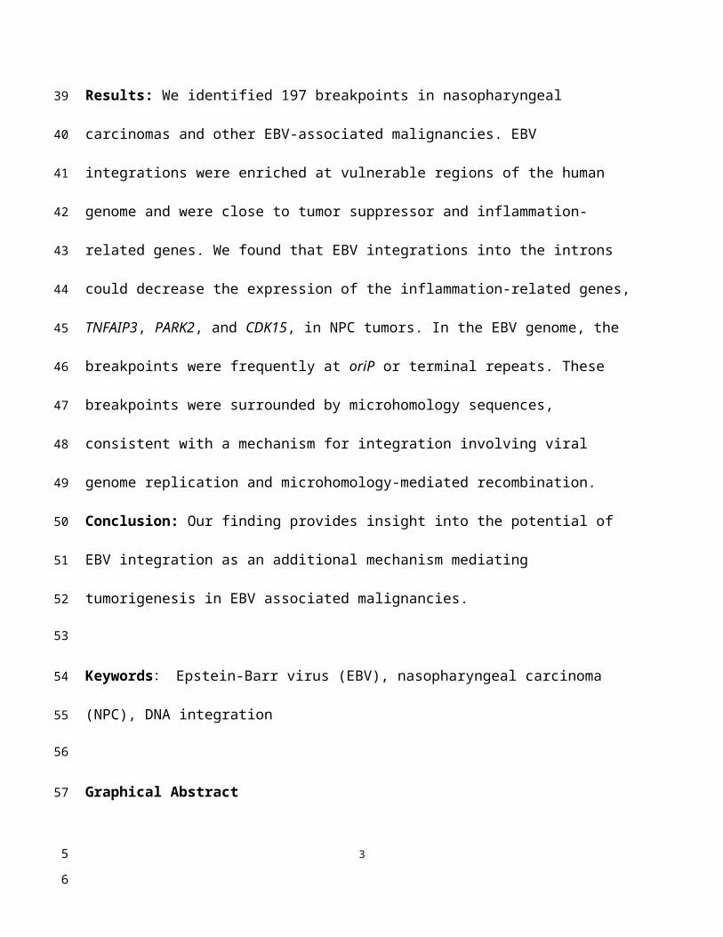
cancer cells. Results: We identified 197 breakpoints in nasopharyngeal carcinomas and other
EBV-associated malignancies. EBV integrations were enriched at vulnerable regions of the
human genome and were close to tumor suppressor and inflammation-related genes. We found
that EBV integrations into the introns could decrease the expression of the inflammation-related
genes, TNFAIP3, PARK2, and CDK15, in NPC tumors. In the EBV genome, the breakpoints
were frequently at oriP or terminal repeats. These breakpoints were surrounded by
microhomology sequences, consistent with a mechanism for integration involving viral genome
replication and microhomology-mediated recombination. Conclusion: Our finding provides
insight into the potential of EBV integration as an additional mechanism mediating
tumorigenesis in EBV associated malignancies.
Keywords: Epstein-Barr virus (EBV), nasopharyngeal carcinoma (NPC), DNA integration
Graphical Abstract
2
23
24
25
26
27
28
29
30
31
32
33
34
35
36
37
38
39
40
41
42
43
34
Introduction
Epstein-Barr virus (EBV) is one of the first described human cancer viruses. EBV is
associated with ~ 1% of cancers worldwide, including Burkitt lymphoma, nasopharyngeal
carcinoma (NPC), Hodgkin lymphomas, NK/T cell lymphomas, and a subset of gastric
carcinomas [1, 2]. The EBV genome typically exists as an episome in infected cells. The most
well-described EBV carcinogenic mechanisms are mediated through EBV viral protein effects or
EBV infection. Expression of EBV proteins, EBNA-1, EBNA-2, EBNA-3A/3B/3C, LMP-1 and
LMP-2, causes B cell and epithelial cell proliferation, increases viability of Burkitt lymphoma
and NPC cells, and induces DNA damage and genomic instability [3-6], while EBV infection
promotes chronic inflammation and reduces anti-tumor immune surveillance in the epithelium
[4].
The first reports of the integration of the EBV genome into host genomes date back to the
1980s [7-10]. Subsequent studies confirmed the frequent integration of full-length EBV genomes
as well as DNA fragments in EBV-positive lymphoma and epithelial carcinomas including NPC
3
44
45
46
47
48
49
50
51
52
53
54
55
56
57
58
59
56

and gastric carcinoma [11-21]. These findings suggest that integrated and episomal EBV DNA
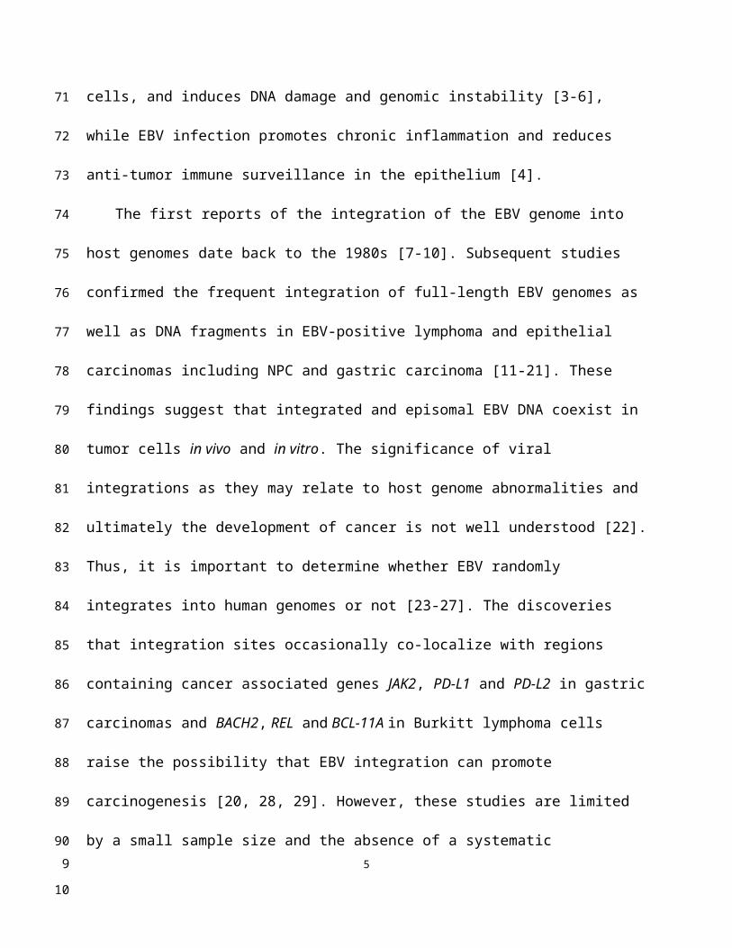
coexist in tumor cells in vivo and in vitro. The significance of viral integrations as they may
relate to host genome abnormalities and ultimately the development of cancer is not well
understood [22]. Thus, it is important to determine whether EBV randomly integrates into human
genomes or not [23-27]. The discoveries that integration sites occasionally co-localize with
regions containing cancer associated genes JAK2, PD-L1 and PD-L2 in gastric carcinomas and
BACH2, REL and BCL-11A in Burkitt lymphoma cells raise the possibility that EBV integration
can promote carcinogenesis [20, 28, 29]. However, these studies are limited by a small sample
size and the absence of a systematic investigation of the EBV integration landscape on a
genome-wide scale. To provide systematic insight into EBV integration in associated
malignancies, we performed EBV-targeted ultra-deep sequencing and conducted a
comprehensive survey of EBV integration in a variety of human malignancies. This work
provides the first unbiased, genome-wide analysis of EBV integrations, and reveals the
involvement of novel inflammation-related genes in NPC.
Results
To perform comprehensive profiling of EBV integration, we conducted EBV-targeted ultra-
deep sequencing on 177 NPCs, 39 gastric carcinomas, 25 NK/T cell lymphomas, 11 Hodgkin
lymphomas, one nasopharyngitis tissue and the EBV-positive NPC cell line C666-1. A total of
197 EBV integration breakpoints were identified from 33 tumors and the C666-1 cell line (Table
1 and Figure S1). The integration rates were higher in the gastric carcinomas (25.6%; 95%
confidence interval (CI): 13.0 - 42.1%) than in the NPC tumors (9.6%; 95% CI: 5.7 - 14.9%).
We observed slightly more EBV integration positive samples in late-stage NPC tumors (stage
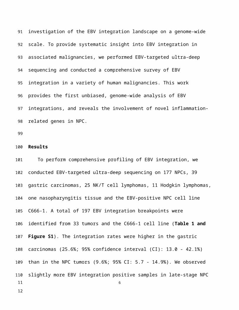
4
60
61
62
63
64
65
66
67
68
69
70
71
72
73
74
75
76
77
78
79
80
81
82
78
III-IV) and large-size gastric cancers (> 5 cm; Table S1). The EBV integration counts in positive
tumor samples varied widely among tumor types and individual cases. Twenty-seven of the 34
positive samples harbored 1-2 breakpoints. The remaining positive samples (n = 7) contained
more than two with one gastric cancer harboring an especially large number (118) of integration
breakpoints. At least 2 EBV integration breakpoints were consistent between matched primary
and metastatic NPC tumors from the same patient (Figure S2).
The 197 breakpoints were distributed over all 23 human chromosomes (Figure 1A). EBV
showed a strong tendency to integrate near common fragile sites in both NPC and gastric cancer
samples (Figure 1B). Similarly, two of the six breakpoints identified in NK/T cell lymphoma
samples were located at the same common fragile site (Table S2). EBV also tended to integrate
into microsatellite repeats in gastric carcinomas, but avoided SINE repeats in NPC and gastric
carcinomas (Figure 1C; For details, see Figure S3 and Table S2). Common fragile regions and
microsatellite repeats are vulnerable to DNA damage, which increases the chance for EBV DNA
insertion into host genomes through microhomology-mediated DNA repair.
We also tested the relative position of EBV integrations to genes in the human genome. We
found that 75 breakpoints (38.1%) were located within known UCSC-annotated genes (Figure
S4). The integration sites were slightly skewed toward gene body and promoter regions (26
breakpoints; 13.2%; Figure S4). Unlike HBV and HPV integrations, no associations were found
between EBV integration sites and CpG islands, the repetitive elements other than microsatellite
repeats, or the binding sites of the genome architecture regulator CCCTC-binding factor (CTCF;
Figure S5).
5
83
84
85
86
87
88
89
90
91
92
93
94
95
96
97
98
99
100
101
102
103
910
We detected EBV integration in the proximity of tumor suppressor genes: KANK1 in one
NK/T cell lymphoma, RB1CC1 in one Hodgkin lymphoma, and DLEC1 in one NPC tumor
(Table S2). Integrations in gastric carcinoma samples were associated with tumor suppressor
genes SETD2, KISS1, FHIT, PTEN and TET2 (Table S2). The integration breakpoints associated
with the histone methyltransferase, SETD2, were located in a region 22 kb upstream of the gene
(Figure 2A). In one NK/T cell lymphoma, a breakpoint was found 27 kb downstream of the
tumor suppressor gene KANK1. The other EBV integration breakpoint in the same sample was
identified 204 kb downstream of JAK2. Amplification was also detected in this region (Figure
2B). Common fragile regions were often co-localized with tumor suppressor genes; for example,
the common fragile site FRA3B lies within the tumor suppressor gene FHIT. A number of
studies have shown that tumor suppressor genes and common fragile regions are frequent targets
of viral DNA integration [30, 31]. The breakpoints associated with tumor suppressor genes in our
study showed significantly higher coverage in the targeted sequencing, which indicates that they
were more likely to be clonal in the tumorigenesis (P < 0.0001, unpaired, two-sided t-test).
Integration could alter the expression or function of tumor suppressor genes and provide host
cells with a selective advantage during tumorigenesis.
We also identified EBV integrations located within the introns of CDK15 in the primary
(Figures S2A-B) and metastatic (Figures 2C, S2C and D) NPC tumors from a single patient,
and TNFAIP3 and PARK2 in two additional NPC tumors from two other patients (Figures 2D-
E). These breakpoints were all supported by a high number of sequencing reads, suggesting
clonal expansion of cancer cells after EBV integration (Table S2). Notably, TNFAIP3, CDK15
and PARK2 are all inflammation-related genes involved in the regulation of TNF-alpha-induced
apoptosis/NF-κB pathways [32-34], and dysregulation of these pathways contributes to the
6
104
105
106
107
108
109
110
111
112
113
114
115
116
117
118
119
120
121
122
123
124
125
1261112
development of EBV-associated cancers, including NPC [35]. We performed the
immunohistochemistry staining of CDK15, TNFAIP3 and PARK2 proteins using the integrated
and non-integrated NPC samples. We found that the protein levels of CDK15, TNFAIP3 and
PARK2 were lower in the samples harboring EBV integrations into the introns of the respective
genes. (Figures 3A-C). Using qPCR of NF-κB targeted genes and a luciferase reporter gene
assay, we found that NF-κB activity was up-regulated in NPC cells with TNFAIP3 knockdown
(Figure S6A), confirming its role as an inhibitor of NF-κB pathway. In contrast, NF-κB activity
was down-regulated, and nuclear localization of p65 after TNF-α treatment was diminished in
NPC cells with CDK15 knockdown (Figure S6B), indicating that CDK15 is positively related to
the activation of the NF-κB pathway.
To investigate the mechanisms of EBV integration, we surveyed the distribution of the 197 EBV
integration breakpoints in the EBV genome. EBV breakpoints were spread over the entire viral
genome with multiple hotspots (Figure 4A). Breakpoints were enriched in the proximity of oriP
and terminal repeats, while no breakpoints were detected within the two long internal repeats
(Figure 4B). The tendency for EBV breakpoints to localize around oriP and terminal repeats
indicated that EBV integration was related to viral genome replication. We further analyzed the
microhomology (MH) sequences in the regions flanking integration sites. We found frequent
microhomologies between the human genome and the EBV genome near integration breakpoints
(Figure S2 and Figure S7). Insertions of 2-10 bp were also observed near the EBV integration
breakpoints (Figures S2 and Figure S7). Two EBV integrations containing MH sequences were
observed in matched primary and metastatic NPC tumors from a single patient (Figure S2).
Methods
7
127
128
129
130
131
132
133
134
135
136
137
138
139
140
141
142
143
144
145
146
147
148
149
1314
Ethics statement
This study was approved by the institutional ethics committees of the Sun Yat-sen
University Cancer Center (Guangzhou, China), the First Affiliated Hospital of Guangxi Medical
University (Nanning, China), the Affiliated Hospital of Qingdao University (Qingdao, China),
and Rui Jin Hospital (Shanghai, China). Written informed consent was obtained from each study
participant.
Sample collection and DNA extraction
Frozen samples were collected for isolation of genomic DNAs from the following tissue
types: NPC (n = 177; the Sun Yat-sen University Cancer Center and the First Affiliated Hospital
of Guangxi Medical University); gastric carcinoma (n = 39; the Sun Yat-sen University Cancer
Center and the Affiliated Hospital of Qingdao University); NK/T cell lymphoma (n = 25; the Sun
Yat-sen University Cancer Center and Rui Jin Hospital); Hodgkin lymphoma (n = 11; the Sun
Yat-sen University Cancer Center); and nasopharyngitis (n = 1; the Sun Yat-sen University
Cancer Center). All histopathological diagnoses were performed according to WHO
classifications and were reviewed by two pathologists. The clinicopathological characteristics of
the study subjects are listed in Table S1.
Genomic DNAs were extracted from the frozen tissue samples using the DNeasy Blood and
Tissue Kit (Qiagen; Germantown, MD, USA).
EBV DNA capture and sequencing
Genomic DNA was subjected to hybrid capture using an EBV-targeting single-stranded
DNA probe developed by MyGenostics (Beijing, China). Sequencing libraries were constructed
8
150
151
152
153
154
155
156
157
158
159
160
161
162
163
164
165
166
167
168
169
170
171
172
1516

by shearing genomic DNA into 150-200 bp fragments, followed by DNA purification, end
blunting and adaptor ligation according to the instructions provided by Illumina (San Diego, CA,
USA). The library concentrations were evaluated with a Bioanalyzer 2100 (Agilent
Technologies; Santa Clara, CA, USA). EBV DNA was captured from the genomic DNA
following the MyGenostics GenCap Target Enrichment Protocol (GenCap Enrichment,
MyGenostics). The libraries were hybridized with EBV probes at 65°C for 24 h and then washed
to remove uncaptured DNA. The eluted DNA fragments were amplified through 18 PCR cycles
to generate libraries for sequencing. The libraries were subsequently quantified and subjected to
paired-end sequencing (2 100 or 150 bp) on an Illumina HiSeq 2000 sequencer according to
the manufacturer’s instructions (Illumina).
Integration detection, validation and annotation
Quality assessment of the raw reads was conducted using TrimGalore to remove adaptor
sequences and low-quality reads. High-quality reads were aligned to the human (NCBI build 37,
hg19) and EBV genomes (NC_007605.1) using the Burrows-Wheeler Aligner (BWA, version
0.7.5a) [36]. Alignments were converted from a sequence alignment map format to sorted and
indexed binary alignment map (BAM) files [37]. The Picard tool was used to remove duplicate
reads. We developed a bioinformatic method based on LUMPY to identify EBV-human chimeric
reads[38]. Briefly, paired-end reads mapping solely to the human or EBV reference genomes
were removed. Next, two types of integration-supportive signals were extracted by LUMPY as
follows: 1) read pairs, in which one of the paired reads was mapped to the EBV genome and the
other to the human genome and 2) chimeric reads, in which one read covered both the EBV
genome sequence and the human genome sequence. Integration events supported by ≥ 3 read
9
173
174
175
176
177
178
179
180
181
182
183
184
185
186
187
188
189
190
191
192
193
194
195
1718
pairs or chimeric reads were retained. ANNOVAR [39] and the UCSC table browser [40] were
used to annotate the breakpoints using fragile regions [41, 42] and the UCSC hg19 CpG island
and RepeatMasker database. We randomly selected 12 integrations and performed PCR and
Sanger sequencing to validate. Ten (83.3%) breakpoints were validated with Sanger sequencing,
while PCR for two breakpoints failed to generate products (Table S3).
Copy number variation and gene expression
CNV data for three NK/T cell lymphomas were retrieved from the study by Jiang et al. [43].
Publicly available gene expression data for normal epithelium (n = 10) and NPC tumors (n = 31)
obtained on the Affymetrix Human Genome U133 Plus 2.0 Array were retrieved from the NCBI
GEO database (GSE12452). Probeset measures of all 41 arrays were calculated by robust
multiarray averaging. The relative RNA expression value was log-transformed using log2. Data
were analyzed with the unpaired t-test and presented as the mean ± SEM. A P-value of < 0.05
was considered to be statistically significant.
siRNA transfection
CDK15 and TNFAIP3 siRNAs were designed and synthesized by RIBOBIO (Guangzhou,
China). Non-targeting siRNA duplexes are denoted as “scr”. Knockdown efficiency was
confirmed with qPCR. Cells (7 × 10⁵) were seeded into 6 well plates, and after incubation
overnight, transfected with the indicated siRNA duplexes using Lipofectiamine RNAiMAX
Transfection Reagent (Thermo Fisher Scientific; Waltham, MA, USA), according to the
manufacturer’s instructions. Sequences of the CDK15 siRNAs were the following:
CDK15-siRNA#1: 5’-GAGGAAGGAGTCCCATTTA-3’
10
196
197
198
199
200
201
202
203
204
205
206
207
208
209
210
211
212
213
214
215
216
217
218
1920

CDK15-siRNA#2: 5’-CCTCAGAACTTACTCATCA-3’
TNFAIP3-siRNA#1: 5’-GCGGAAAGCTGTGAAGATA-3’
TNFAIP3-siRNA#2: 5’-CAACTCACTGGAAGAAATA-3’
Quantitative Real-time PCR
Total RNA was isolated using Trizol reagent (Thermo Fisher Scientific), and cDNA was
prepared from RNA (1 µg) using the PrimeScript RT Reagent Kit (TaKaRa; Tokyo, Japan)
according to the manufacturer’s instructions. Quantitative real-time PCR was performed using
the Platinum SYBR Green qPCR SuperMix (Thermo Fisher Scientific) on a Light cycler 480
Real-time system (Roche; Indianapolis, IN, USA).
The primers sequences used were the following:
CDK15-F: 5’-TATGCGACAGTTTACAAGG-3’
CDK15-R: 5’-TCATGCAGGAGCACAATA-3’
IKBα-F: 5’-CTCCGAGACTTTCGAGGAAATAC-3’
IKBα-R: 5’-GCCATTGTAGTTGGTAGCCTTCA-3’
IL6-F: 5’-ACTCACCTCTTCAGAACGAATT-3’
IL6-R: 5’-CCATCTTTGGAAGGTTCAGGTTG-3’
MCP1-F: 5’-CAGCCAGATGCAATCAATGC-3’
MCP1-R: 5’-TGGAATCCTGAACCCACTTCT-3’
TNF-F: 5’-CCTCTCTCTAATCAGCCCTCTG-3’
TNF-R: 5’-GAGGACCTGGGAGTAGATGAG-3’
GAPDH-F: 5’-TTGCCATCAATGACCCCTTCA-3’
GAPDH-R: 5’-CGCCCCACTTGATTTTGGA-3’
11
219
220
221
222
223
224
225
226
227
228
229
230
231
232
233
234
235
236
237
238
239
240
241
2122
Western blot analysis
Western blot analysis was performed as previously described [44]. Cells were lysed in
RIPA lysis buffer containing protease inhibitors (Roche). The subcellular fractionation was
performed as previously described [45]. The proteins were separated with 10% SDS-PAGE and
transferred to PVDF membranes (Thermo Fisher Scientific). Membranes were blocked with 5%
BSA for 1 h and incubated with primary antibodies overnight in 4°C. Antibodies against p65
(ab53465; Abcam; Cambridge, MA, USA), ß-actin (#3700; Cell Signaling Technology; Danvers,
MA, USA), and GAPDH (60004-1-1 g; Protein-tech; Chicago, IL, USA) were used for western
blot analysis.
Luciferase reporter assay
(CAGA)12-Luc and the control vector pRL-TK (Promega) encoding Renilla luciferase were
cotransfected into HEK293T or NPC cells using PEI. Luciferase activity was measured 24 h
after transfection using the Dual-Luciferase Reporter Assay System (Promega). The firefly
luciferase activity values were normalized to those of Renilla, and the ratios of firefly/Renilla
activities were determined. The experiments were independently performed in triplicate.
Discussion
EBV integration during tumorigenesis has not yet been systematically investigated using
rigorous methods. In this study, we conducted the first large-scale analysis of EBV integration in
multiple malignancies using EBV genome-targeted sequencing. Our method, combining EBV
genome capture and ultra-deep sequencing, efficiently detected integrated EBV sequences from
background “noise” introduced by nuclear EBV episomes. Our results indicate that EBV can
12
242
243
244
245
246
247
248
249
250
251
252
253
254
255
256
257
258
259
260
261
262
263
264
2324

integrate into host genomes at a significant rate in multiple tumor types. We observed that EBV
integration frequencies varied among tumor types as well as the number of integrated EBV
genomes among tumor samples. The heterogeneity of these tumor genomes may underlie the
observed variation of EBV integration in these tumors.
Our study revealed that common fragile regions were preferred sites for EBV integration.
Common fragile regions are genomic hotspots for DNA damage and are susceptible to genome
rearrangement, thereby increasing the chance for EBV DNA insertion through microhomology-
mediated DNA repair, which has an important role in the integration of other tumorigenic
viruses, HBV and HPV [30, 31]. We observed EBV integrations into or near tumor suppressor
genes that were often colocalized with common fragile regions. Integration in the proximity of
tumor suppressor genes may provide host cells with a selective advantage. Moreover, integration
distribution in gastric carcinomas correlated with microsatellite repeats which are vulnerable to
DNA damage, one of the samples which has the 118 integration breakpoints, the whole-exome
sequencing showed the mutation including XRCC2, PARP3, SLX4, and PMS2, which are
involved in DNA repair, further suggesting that host genome stability has a strong impact on
EBV integration. Although genome instability and microhomology-mediated DNA repair are
involved in the integration of EBV, HPV and HBV DNA into the host genome, why does EBV
integration occur at a relatively lower rate (25.6% in GC, 9.6% in NPC) than HPV integration in
cervical cancer (76.3%) and head and neck squamous cell carcinoma (HNSCC; 60.7%), and
HBV in hepatocellular carcinoma (HCC; 92.6%)? [31, 46-49]. There are several possible
reasons. First, mechanisms underlying tumorigenesis and genetic backgrounds differ greatly
between these tumor types associated with the different viruses. The tumor suppressors, TP53
and RB, are inactivated by the expression of HPV-encoded oncogenes E6 and E7 in HPV-
13
265
266
267
268
269
270
271
272
273
274
275
276
277
278
279
280
281
282
283
284
285
286
2872526
associated cervical cancer and HNSCC. Dysfunction of the TP53 pathway has also been
frequently observed in HBV-associated HCC (~ 18-51.8%) [50-52]. The impairment of the TP53
pathway leads to increased genomic instability and accumulation of somatic mutations, possibly
also contributing to the high rate of HPV and HBV integration. However, the TP53 pathway is
not mutated as frequently in NPC (~ 7-10%) and EBV-associated gastric cancer (rarely) as in
HCC [53-55]. Moreover, large-scale whole-genome surveys indicate that NPC and EBV-
associated gastric carcinomas tend to have relatively stable genomes, compared to many other
carcinomas including cervical cancer, HNSCC and HCC [53]. Second, life cycles and genomic
features of the viruses themselves may also affect their integration into the host genome. In latent
infection state, EBV episomes are replicated along with chromosomes in host cells and therefore
are relatively stable. EBV has a much larger genome than HPV or HBV, which may make its
integration by microhomology-mediated DNA recombination more difficult.
In NPC, three integration events were localized to introns of the inflammation-related genes
PARK2, TNFAIP3 and CDK15, which regulate the TNF-alpha-induced apoptosis/NF-κB
pathways. PARK2 deficiency promotes inflammation and genome instability and has an
important role in the development of lung cancer [56]. Dysregulation of NF-κB activation
contributes to the development of various EBV-associated cancers, including NPC [35]. We
found lower expression of PARK2, TNFAIP3 and CDK15 proteins and also dysregulated NF-κB
activity in the integrated NPC tumors. Our results suggest that integration of EBV into these
genes may disrupt their function and contribute to tumorigenesis through the TNF-alpha-induced
apoptosis/NF-κB pathways. The identification of integrations with a high number of supporting
reads associated with inflammatory genes indicates that such EBV integration events are
potentially selected for during tumor development. If these genes are involved in NPC
14
288
289
290
291
292
293
294
295
296
297
298
299
300
301
302
303
304
305
306
307
308
309
3102728

development, they may be frequent targets of somatic mutation in NPC. TNFAIP3 gene as a
mutation hot spot has been already confirmed by previous studies in NPC [53-55]. We also
searched our unpublished data and confirmed that about 5% of NPC tumors harbor copy number
variation at CDK15 and PARK2 loci.
In the EBV genome, the breakpoints identified in this study were concentrated around oriP
and the terminal repeats. During latency, EBNA1 binding to oriP can recruit host cell replication
machinery to facilitate the formation of an efficient origin of replication for the EBV episome
[57]. The terminal repeats are responsible for the circularization of the EBV genome after it
enters the nucleus and cleavage/encapsulation of EBV DNA. Both EBV genome circularization
and cleavage involve recombination events. The microhomology sequences around breakpoints
indicate that EBV integration involves microhomology-mediated DNA repair pathways. These
integrations may be triggered by genomic vulnerability/fragility during genome replication and
the physical proximity of the oriP repeats to the host DNA bridged by EBNA1 and DNA
recombination, which underlies the EBV integration mechanism.
Till now, we still do not know the size of the EBV sequence integrated into the host genome.
Due to the limit of the read length in the second or third generation sequencing, we can only
identify a few hundred base pair to around 10 Kb of EBV sequence fused into the host genome.
Compared with the large size of the EBV genome, it is difficult to determine whether a portion
or the full EBV genome is integrated into human genome. Future development of sequencing
technology could help to map the landscape of EBV genome integrated into the human genome.
In summary, our work provides an unbiased large-scale genome-wide analysis of the EBV
integration landscape in multiple malignancies. EBV integration occurs preferentially within 15
311
312
313
314
315
316
317
318
319
320
321
322
323
324
325
326
327
328
329
330
331
3322930

unstable chromosomal regions of the host genome, surrounding oriP or terminal repeats of the
EBV genome. Several integration sites were located in the proximity of tumor suppressor genes
that are frequently disrupted during cancer progression. We detected multiple integrations into
genes regulating TNF-alpha-induced apoptosis/NF-κB pathways in NPC. These pathways are
closely related to EBV-associated diseases and indicate that EBV integration disrupts the
function of crucial genes, leading to the development of cancer in some cases of latent EBV
infection.
Abbreviations EBV: Epstein-Barr virus; NPC: nasopharyngeal carcinoma; CI: confidence
interval; MH: microhomology.
Acknowledgments
We would like to thank all the participants recruited for this study. We thank Dr. E.D. Kieff
for helpful discussions and suggestions on this manuscript. This work was supported by the
National Natural Science Foundation of China (81872228 and 81430059) and the National Key
R&D Program of China (No. 2016YF0902000).
Author contributions
Y.-X.Z. and Y.-C.J. were the overall principal investigators who conceived the study and
obtained financial support. Y.-X.Z., M.X. and Y.-C. J designed and oversaw the study. M.X.,
W.-L.Z. and Y.Y. performed sample preparation, sequencing and statistical analysis and
validation. S.-J.C., W-L.Z., R.-H.X., Z.Z. and B.L. contributed to sample collection. Q.Z. edited
16
333
334
335
336
337
338
339
340
341
342
343
344
345
346
347
348
349
350
351
352
353
354
3132
the tables and figures. The manuscript was drafted by M.X. and W.-L.Z. under the supervision of
Y.-X.Z. and Y.-C.J. All authors critically reviewed the article and approved the final manuscript.
Competing interests
The authors declare no competing interests.
References:
1. Parkin DM. The global health burden of infection-associated cancers in the year 2002. Int J Cancer. 2006; 118: 3030-44.2. Zur Hausen H, de Villiers EM. Reprint of: cancer "causation" by infections--individual contributions and synergistic networks. Semin Oncol. 2015; 42: 207-22.3. Arvey A, Tempera I, Tsai K, Chen HS, Tikhmyanova N, Klichinsky M, et al. An atlas of the Epstein-Barr virus transcriptome and epigenome reveals host-virus regulatory interactions. Cell Host Microbe. 2012; 12: 233-45.4. Tsao SW, Tsang CM, To KF, Lo KW. The role of Epstein-Barr virus in epithelial malignancies. J Pathol. 2015; 235: 323-33.5. Li R, Liao G, Nirujogi RS, Pinto SM, Shaw PG, Huang TC, et al. Phosphoproteomic Profiling Reveals Epstein-Barr Virus Protein Kinase Integration of DNA Damage Response and Mitotic Signaling. PLoS Pathog. 2015; 11: e1005346.6. Shumilov A, Tsai MH, Schlosser YT, Kratz AS, Bernhardt K, Fink S, et al. Epstein-Barr virus particles induce centrosome amplification and chromosomal instability. Nat Commun. 2017; 8: 14257.7. Henderson A, Ripley S, Heller M, Kieff E. Chromosome site for Epstein-Barr virus DNA in a Burkitt tumor cell line and in lymphocytes growth-transformed in vitro. Proc Natl Acad Sci U S A. 1983; 80: 1987-91.8. Matsuo T, Heller M, Petti L, O'Shiro E, Kieff E. Persistence of the entire Epstein-Barr virus genome integrated into human lymphocyte DNA. Science. 1984; 226: 1322-5.9. Lawrence JB, Villnave CA, Singer RH. Sensitive, high-resolution chromatin and chromosome mapping in situ: presence and orientation of two closely integrated copies of EBV in a lymphoma line. Cell. 1988; 52: 51-61.10. Anvret M, Karlsson A, Bjursell G. Evidence for integrated EBV genomes in Raji cellular DNA. Nucleic Acids Res. 1984; 12: 1149-61.11. Delecluse HJ, Bartnizke S, Hammerschmidt W, Bullerdiek J, Bornkamm GW. Episomal and integrated copies of Epstein-Barr virus coexist in Burkitt lymphoma cell lines. J Virol. 1993; 67: 1292-9.12. Wolf J, Pawlita M, Klevenz B, Frech B, Freese UK, Muller-Lantzsch N, et al.
17
355
356
357
358
359
360
361
362363364365366367368369370371372373374375376377378379380381382383384385386387388389390391392393
3334

Down-regulation of integrated Epstein-Barr virus nuclear antigen 1 and 2 genes in a Burkitt lymphoma cell line after somatic cell fusion with autologous EBV-immortalized lymphoblastoid cells. Int J Cancer. 1993; 53: 621-7.13. Hurley EA, Agger S, McNeil JA, Lawrence JB, Calendar A, Lenoir G, et al. When Epstein-Barr virus persistently infects B-cell lines, it frequently integrates. J Virol. 1991; 65: 1245-54.14. Kripalani-Joshi S, Law HY. Identification of integrated Epstein-Barr virus in nasopharyngeal carcinoma using pulse field gel electrophoresis. Int J Cancer.1994; 56: 187-92.15. Chang Y, Cheng SD, Tsai CH. Chromosomal integration of Epstein-Barr virus genomes in nasopharyngeal carcinoma cells. Head Neck. 2002; 24: 143-50.16. Zhang HY, Qu G, Deng ZW, Yao TH, Glaser R. Epstein-Barr virus DNA in nasopharyngeal biopsies. Virus Res. 1989; 12: 53-9.17. Xiao K, Yu Z, Li X, Li X, Tang K, Tu C, et al. Genome-wide Analysis of Epstein-Barr Virus (EBV) Integration and Strain in C666-1 and Raji Cells. J Cancer. 2016; 7: 214-24.18. Morissette G, Flamand L. Herpesviruses and chromosomal integration. J Virol. 2010; 84: 12100-9.19. Cao S, Strong MJ, Wang X, Moss WN, Concha M, Lin Z, et al. High-throughput RNA sequencing-based virome analysis of 50 lymphoma cell lines from the Cancer Cell Line Encyclopedia project. J Virol. 2015; 89: 713-29.20. Gulley ML. Genomic assays for Epstein-Barr virus-positive gastric adenocarcinoma. Exp Mol Med. 2015; 47: e134.21. Ohshima K, Suzumiya J, Kanda M, Kato A, Kikuchi M. Integrated and episomal forms of Epstein-Barr virus (EBV) in EBV associated disease. Cancer Lett.1998; 122: 43-50.22. Ohshima K, Suzumiya J, Ohga S, Ohgami A, Kikuchi M. Integrated Epstein-Barr virus (EBV) and chromosomal abnormality in chronic active EBV infection. Int J Cancer. 1997; 71: 943-7.23. Lestou VS, De Braekeleer M, Strehl S, Ott G, Gadner H, Ambros PF. Non-random integration of Epstein-Barr virus in lymphoblastoid cell lines. Genes Chromosomes Cancer. 1993; 8: 38-48.24. Jox A, Rohen C, Belge G, Bartnitzke S, Pawlita M, Diehl V, et al. Integration of Epstein-Barr virus in Burkitt's lymphoma cells leads to a region of enhanced chromosome instability. Ann Oncol. 1997; 8 Suppl 2: 131-5.25. Wuu KD, Chen YJ, Wuu SW. Frequency and distribution of chromosomal integration sites of the Epstein-Barr virus genome. J Formos Med Assoc. 1996; 95: 911-6.26. Oh JH, Kim YJ, Moon S, Nam HY, Jeon JP, Lee JH, et al. Genotype instability during long-term subculture of lymphoblastoid cell lines. J Hum Genet. 2013; 58: 16-20.27. Gao J, Luo X, Tang K, Li X, Li G. Epstein-Barr virus integrates frequently into chromosome 4q, 2q, 1q and 7q of Burkitt's lymphoma cell line (Raji). J Virol Methods. 2006; 136: 193-9.
18
394395396397398399400401402403404405406407408409410411412413414415416417418419420421422423424425426427428429430431432433434435436437438439
3536
28. Takakuwa T, Luo WJ, Ham MF, Sakane-Ishikawa F, Wada N, Aozasa K. Integration of Epstein-Barr virus into chromosome 6q15 of Burkitt lymphoma cell line (Raji) induces loss of BACH2 expression. Am J Pathol. 2004; 164: 967-74.29. Luo WJ, Takakuwa T, Ham MF, Wada N, Liu A, Fujita S, et al. Epstein-Barr virus is integrated between REL and BCL-11A in American Burkitt lymphoma cell line (NAB-2). Lab Invest. 2004; 84: 1193-9.30. Zhao LH, Liu X, Yan HX, Li WY, Zeng X, Yang Y, et al. Genomic and oncogenic preference of HBV integration in hepatocellular carcinoma. Nat Commun. 2016; 7: 12992.31. Hu Z, Zhu D, Wang W, Li W, Jia W, Zeng X, et al. Genome-wide profiling of HPV integration in cervical cancer identifies clustered genomic hot spots and a potential microhomology-mediated integration mechanism. Nat Genet. 2015; 47: 158-63.32. Muller-Rischart AK, Pilsl A, Beaudette P, Patra M, Hadian K, Funke M, et al. The E3 ligase parkin maintains mitochondrial integrity by increasing linear ubiquitination of NEMO. Mol Cell. 2013; 49: 908-21.33. Park MH, Kim SY, Kim YJ, Chung YH. ALS2CR7 (CDK15) attenuates TRAIL induced apoptosis by inducing phosphorylation of survivin Thr34. Biochem Biophys Res Commun. 2014; 450: 129-34.34. Wertz IE, Newton K, Seshasayee D, Kusam S, Lam C, Zhang J, et al. Phosphorylation and linear ubiquitin direct A20 inhibition of inflammation. Nature. 2015; 528: 370-5.35. Sun SC, Cesarman E. NF-kappaB as a target for oncogenic viruses. Curr Top Microbiol Immunol. 2011; 349: 197-244.36. Li H, Durbin R. Fast and accurate short read alignment with Burrows-Wheeler transform. Bioinformatics. 2009; 25: 1754-60.37. Li H, Handsaker B, Wysoker A, Fennell T, Ruan J, Homer N, et al. The Sequence Alignment/Map format and SAMtools. Bioinformatics. 2009; 25: 2078-9.38. Layer RM, Chiang C, Quinlan AR, Hall IM. LUMPY: a probabilistic framework for structural variant discovery. Genome Biol. 2014; 15: R84.39. Wang K, Li M, Hakonarson H. ANNOVAR: functional annotation of genetic variants from high-throughput sequencing data. Nucleic Acids Res. 2010; 38: e164.40. Karolchik D, Hinrichs AS, Furey TS, Roskin KM, Sugnet CW, Haussler D, et al. The UCSC Table Browser data retrieval tool. Nucleic Acids Res. 2004; 32: D493-6.41. Fungtammasan A, Walsh E, Chiaromonte F, Eckert KA, Makova KD. Corrigendum: A genome-wide analysis of common fragile sites: What features determine chromosomal instability in the human genome? Genome Res. 2016; 26: 1451.42. Fungtammasan A, Walsh E, Chiaromonte F, Eckert KA, Makova KD. A genome-wide analysis of common fragile sites: what features determine chromosomal instability in the human genome? Genome Res. 2012; 22: 993-1005.
19
440441442443444445446447448449450451452453454455456457458459460461462463464465466467468469470471472473474475476477478479480481482483484485
3738

43. Jiang L, Gu ZH, Yan ZX, Zhao X, Xie YY, Zhang ZG, et al. Exome sequencing identifies somatic mutations of DDX3X in natural killer/T-cell lymphoma. Nat Genet. 2015; 47: 1061-6.44. Ma W, Feng L, Zhang S, Zhang H, Zhang X, Qi X, et al. Induction of chemokine (C-C motif) ligand 5 by Epstein-Barr virus infection enhances tumor angiogenesis in nasopharyngeal carcinoma. Cancer Sci. 2018; 109: 1710-22.45. Holden P, Horton WA. Crude subcellular fractionation of cultured mammalian cell lines. BMC Res Notes. 2009; 2: 243.46. Koneva LA, Zhang Y, Virani S, Hall PB, McHugh JB, Chepeha DB, et al. HPV Integration in HNSCC Correlates with Survival Outcomes, Immune Response Signatures, and Candidate Drivers. Mol Cancer Res. 2018; 16: 90-102.47. Sung WK, Zheng H, Li S, Chen R, Liu X, Li Y, et al. Genome-wide survey of recurrent HBV integration in hepatocellular carcinoma. Nat Genet. 2012; 44: 765-9.48. Zhao LH, Liu X, Yan HX, Li WY, Zeng X, Yang Y, et al. Genomic and oncogenic preference of HBV integration in hepatocellular carcinoma. Nat Commun. 2016; 7: 12992.49. Kawai-Kitahata F, Asahina Y, Tanaka S, Kakinuma S, Murakawa M, Nitta S, et al. Comprehensive analyses of mutations and hepatitis B virus integration in hepatocellular carcinoma with clinicopathological features. J Gastroenterol. 2016; 51: 473-86.50. Guichard C, Amaddeo G, Imbeaud S, Ladeiro Y, Pelletier L, Maad IB, et al. Integrated analysis of somatic mutations and focal copy-number changes identifies key genes and pathways in hepatocellular carcinoma. Nat Genet. 2012; 44: 694-8.51. Fujimoto A, Totoki Y, Abe T, Boroevich KA, Hosoda F, Nguyen HH, et al. Whole-genome sequencing of liver cancers identifies etiological influences on mutation patterns and recurrent mutations in chromatin regulators. Nat Genet. 2012; 44: 760-4.52. Cleary SP, Jeck WR, Zhao X, Chen K, Selitsky SR, Savich GL, et al. Identification of driver genes in hepatocellular carcinoma by exome sequencing. Hepatology. 2013; 58: 1693-702.53. Lin DC, Meng X, Hazawa M, Nagata Y, Varela AM, Xu L, et al. The genomic landscape of nasopharyngeal carcinoma. Nat Genet. 2014; 46: 866-71.54. Zheng H, Dai W, Cheung AK, Ko JM, Kan R, Wong BW, et al. Whole-exome sequencing identifies multiple loss-of-function mutations of NF-kappaB pathway regulators in nasopharyngeal carcinoma. Proc Natl Acad Sci U S A. 2016; 113: 11283-8.55. Li YY, Chung GT, Lui VW, To KF, Ma BB, Chow C, et al. Exome and genome sequencing of nasopharynx cancer identifies NF-kappaB pathway activating mutations. Nat Commun. 2017; 8: 14121.56. Lee S, She J, Deng B, Kim J, de Andrade M, Na J, et al. Multiple-level validation identifies PARK2 in the development of lung cancer and chronic obstructive pulmonary disease. Oncotarget. 2016; 7: 44211-23.57. Hung SC, Kang MS, Kieff E. Maintenance of Epstein-Barr virus (EBV) oriP-
20
486487488489490491492493494495496497498499500501502503504505506507508509510511512513514515516517518519520521522523524525526527528529530531
3940
based episomes requires EBV-encoded nuclear antigen-1 chromosome-binding domains, which can be replaced by high-mobility group-I or histone H1. Proc Natl Acad Sci U S A. 2001; 98: 1865-70.
Table 1. EBV integrations detected in EBV-associated malignancies
No. of integration
positive samples
(total samples)
Integration rate
( 95% confidence
interval)
Total No. of
breakpoints
detected
No. of breakpoints
per sample
Gastric carcinoma 10 (39) 25.6% (13.0 - 42.1%) 153 0 - 118
Hodgkin lymphoma 2 (11) 18.2% (2.3 - 51.8%) 8 0 - 5
NK/T cell lymphoma 4 (25) 16.0% (4.5 - 36.1%) 6 0 - 2
NPC 17 (177) 9.6% (5.7 - 14.9%) 28 0 - 6
Nasopharyngitis 0 (1)
C666-1 cell line 1 2
21
532533534535
536
537
538
539
540
541
542
543
544
4142
Figure 1. Distribution of EBV integration breakpoints. (A) Distribution of the 197
integration breakpoints across the human genome. For each integration breakpoint,
each bar represents the total number of supporting reads at a specific locus in the
human genome. Gene annotations for 23 breakpoints in 10 cancers and the C666.1 cell 22
545
546
547
548
5494344

line supported by ≥ 9 EBV-DNA chimeric read pairs are labeled. (B) Distribution of 197
breakpoints in common fragile regions. The expected (assuming uniform and random
distribution, blue) and observed ratios of EBV integration breakpoints detected in all
samples (n=34, red, total), gastric carcinomas (n=10, green, GCT) and NPC (n=17,
purple, NPCT) in common fragile regions are shown. P-values were calculated using
the binomial exact test. (C) Significant enrichment of integration breakpoints with
microsatellite repeats in gastric carcinomas. The expected and observed ratios of
breakpoints co-localized with repeat elements LINE, SINE, LTR, DNA transposon and
microsatellite in NPC (green, NPCT) and gastric carcinoma (red, GCT) are shown (for
detailed frequencies, see Supplementary Figure. 3). P-values were calculated using the
binomial exact test. NS, non-significant; LINE, long interspersed nuclear element; SINE,
long interspersed nuclear element; LTR, long terminal repeat.
23
550
551
552
553
554
555
556
557
558
559
560
561
562
563
564
565
566
567
568
569
570
571
572
4546

Figure 2. Mapping of EBV integration breakpoints in proximity of tumor
suppressor and inflammation-related genes. (A-E) The EBV integration breakpoints
located in proximity of tumor suppressor genes: SETD2 in one gastric carcinoma (A),
KANK1 in one NK/T cell lymphoma (B), and inflammation-related genes CDK15 (C),
TNFAIP3 (D) and PARK2 (E) in NPC tumors. Red arrows indicate a transcription factor
binding site near the EBV integration breakpoint. Three of the transcription factor
binding sites are in the promoter, and one is in the EBV integration breakpoint.
24
573
574
575
576
577
578
579
580
581
582
583584
4748
Figure 3. Gene expression in normal epithelium and NPC tumors. (A-C)
Immunohistochemical images of CDK15 (A), PARK2 (B), TNFAIP3 (C) expression in
normal epithelium and NPC tumors with and without EBV integration. The red curve
marks the neoplasm or epithelium, the rest is inflammatoryinfiltration. EBV (+) indicates
positive for EBV integration; EBV (-) indicates negative for EBV integration.
25
585586
587
588
589
590
591
592
593
594
595
596
4950

Figure 4. Distribution of integration breakpoints in the EBV genome. (A)
Distribution of breakpoints across the EBV genome. Histogram of the frequency of
breakpoints was constructed for 1000 bp intervals. EBV genome annotation is shown.
(B) Breakpoints enriched in oriP and terminal repeats in the EBV genome. The 26
597
598
599
600
6015152

observed (blue) and expected (red) frequencies of breakpoints within fragments are
shown. P-values were calculated using the binomial exact test.
27
602
603
604
605
606
607
608
609
610
611
612
613
614
615
616
617
618
619
620
5354
Figure S1. Mapping of EBV integration breakpoint detected in C666-1 cell line.
EBV-human chimeric read pairs from bam file to detect EBV DNA integration in C666-1
cell line were mapped to chr20: 57967350-57967359 in the human hg19 reference
genome.
28
621622
623
624
625
626
627
628
629
630
631
632
633
634635
636
5556
Figure S2. Two EBV integrations were observed in both primary and metastatic
NPC tumors from the same patient. (A-D) Two EBV integrations were observed in
both primary (A-B) and metastatic NPC tumors (C-D) from the same patient. Sequence
alignment around the integration site between the human genome and EBV genome are
presented. All EBV and human sequences are from the reference strand. Human
sequences are in blue, and EBV sequences are in yellow. Nucleotides that align to both
reference sequences are micro-homologies and are highlighted in red.
29
637
638
639
640
641
642
643
644
645
5758

Figure S3. Significant enrichment of integration breakpoints with microsatellite
repeats in gastric carcinoma. The expected and observed frequency of breakpoints
from all gastric carcinomas (A) all gastric carcinomas except GCT015 (B) and NPC
tumors (C) co-localized with repeat elements LINE, SINE, LTR, DNA transposon and
microsatellite are shown. P-values were calculated using the binomial exact test. LINE,
long interspersed nuclear element; SINE, long interspersed nuclear element; LTR, long
terminal repeat.
30
646647
648
649
650
651
652
653
654
655
5960

Figure S4. Distribution of EBV integration breakpoints relative to genes. (A)
Proportions of EBV integration breakpoints located near genes and transcriptional start
sites. (B) The observed (upper panel) and expected (lower panel) random distributions
of breakpoints relative to transcriptional start sites (TSS) are shown. Different colored
blocks indicate the distance of breakpoints to TSS and are ordered according to the 5’
to 3’ transcriptional direction. The X-axis and width of colored blocks show the
proportion of breakpoints falling into each category indicated. The breakpoints tended to
occur adjacent (smaller proportion of breakpoints with distance to TSS > 100 kb, purple
block) to TSS more frequently than expected.
31
656
657
658
659
660
661
662
663
664
665
6162

Figure S5. Distribution of EBV integration breakpoints relative to CpG islands and
CTCF binding sites. The observed and expected proportions of EBV integration
breakpoints co-localized with CpG islands and CTCF binding sites are shown.
32
666667
668
669
670
671
672
673
674
6364
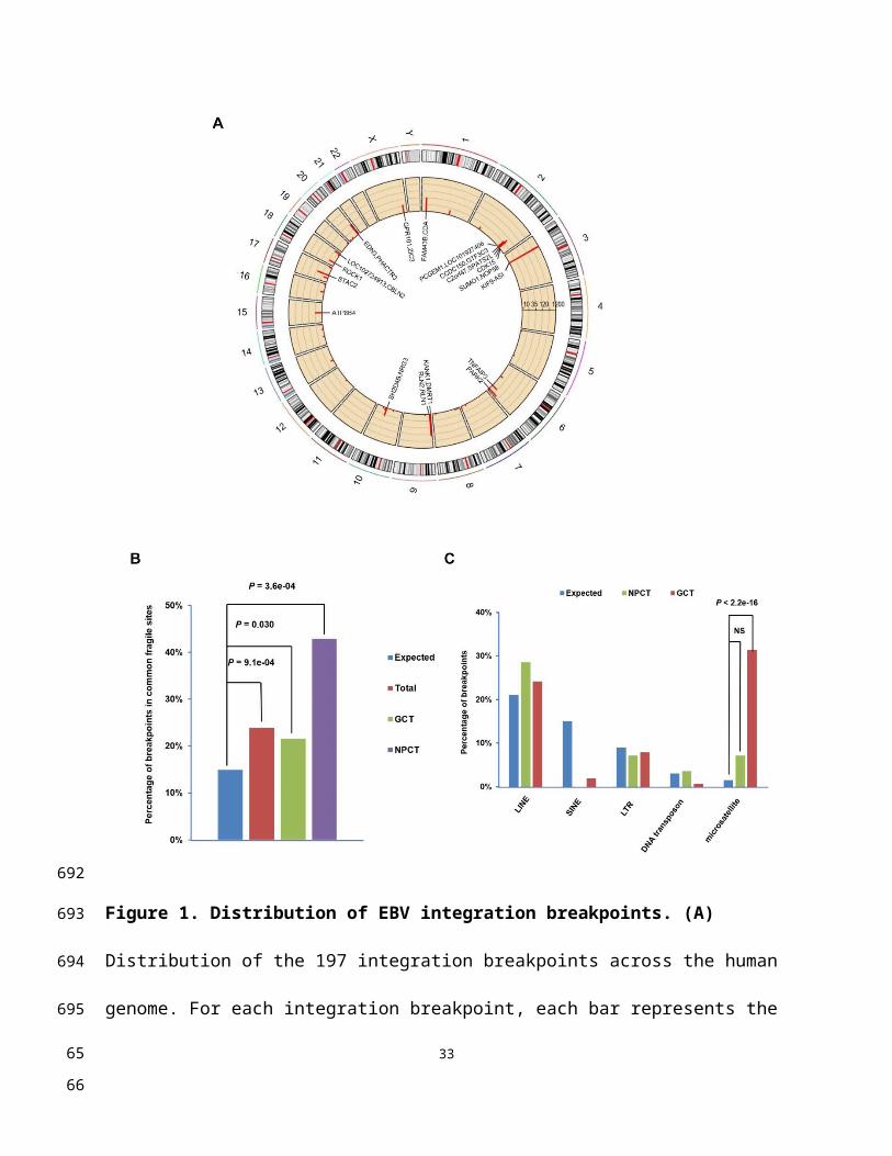
Figure S6. Regulation of NF-κB pathway activity in NPC cells. (A) TNFAIP3
knockdown in NPC cells up-regulates NF-κB pathway activity. NF-κB–specific dual
luciferase promoter assay to detect NF-κB activity in NPC cells with TNFAIP3
knockdown. The relative promoter activity value was compared with the control.
Quantitative PCR performed on RNA prepared from NPC cells with TNFAIP3
knockdown to detect expression levels of NF-κB-pathway-targeted genes. (B) CDK15
knockdown in NPC cells was down-regulated by NF-κB pathway activity. Western blot
performed with nuclear lysates prepared from NPC cells with CDK15 knockdown
treated with TNF-α (10 mg/mL) and incubated with antibody against p65. Histone H3
was used as a control for loading. Quantitative PCR to detect levels of NF-κB-pathway-
targeted genes in NPC cells with CDK15 knockdown.
33
675676
677
678
679
680
681
682
683
684
685
686
687
688
689
6566

Figure S7. Microhomology (MH) sequences in the regions flanking integration
sites and Sanger sequencing validation.
(A-F) Microhomology (MH) sequences in the regions flanking integration sites observed
in NPC (A-D) and NK/T cell lymphoma (E-F) samples. All EBV and human sequences
are from the reference strand. Human partner is in blue, and EBV partner is in yellow.
34
690691
692
693
694
695
6768

Nucleotides highlighted in red are micro-homologies aligning to both reference
sequences.
35
696
697
698699
6970