Embed Size (px)
Citation preview

ANTERIOR ASPECT OF THEFORERARM
&CUBITAL FOSSA
19. 03. 2014
Kaan Yücel
M.D., Ph.D.
http://yeditepeanatomy1.org

Dr. Kaan Yücel http://yeditepeanatomy1.org Anterior aspect of the forearm & Cubital fossa
2
The forearm is the part of the upper limb between the elbow wrist joints. Proximally, most major structures pass between the arm and forearm through, or in relation to, the cubital fossa, which is anterior to the elbow joint. The exception is the ulnar nerve, which passes posterior to the medial epicondyle of the humerus.
ANTERIOR ASPECT OF FOREARMMuscles in the anterior compartment of the forearm flex the wrist and digits and pronate the hand. Muscles
in the posterior compartment extend the wrist and digits and supinate the hand. Major nerves and vessels supply or pass through each compartment.
The flexors and pronators of the forearm in the anterior compartment are served mainly by the median nerve; the one and a half exceptions are innervated by the ulnar nerve. The extensors and supinators of the forearm are in the posterior compartment and are all served by the radial nerve (directly or by its deep branch).
Muscles in the anterior (flexor) compartment of the forearm occur in three layers: • Superficial layer (pronator teres, flexor carpi radialis, palmaris longus, and flexor carpi ulnaris) • Intermediate layer (flexor digitorum superficialis)• Deep layer (flexor digitorum profundus, flexor pollicis longus, and pronator quadratus)Generally, these muscles are associated with: movements of the wrist joint; flexion of the fingers including the thumb; andpronation.
The main arteries of the forearm are the ulnar and radial arteries, which usually arise opposite the neck of the radius in the inferior part of the cubital fossa as terminal branches of the brachial artery.
The superficial veins of the forearm lie in the superficial fascia. The cephalic vein arises from the lateral side of the dorsal venous arch on the back of the hand and winds around the lateral border of the forearm; it then ascends into the cubital fossa and up the front of the arm on the lateral side of the biceps. It terminates in the axillary vein in the deltopectoral triangle. The basilic vein arises from the medial side of the dorsal venous arch on the back of the hand and winds around the medial border of the forearm; it then ascends into the cubital fossa and up the front of the arm on the medial side of the biceps. Its terminates, by joining the venae comitantes of the brachial artery to form the axillary vein.
Nerves in the anterior compartment of the forearm are the median and ulnar nerves, and the superficial branch of the radial nerve.
The median nerve is the principal nerve of the anterior compartment of the forearm. It supplies muscular branches directly to the muscles of the superficial and intermediate layers of forearm flexors (except the flexor carpi ulnaris), and deep muscles (except for the medial [ulnar] half of the flexor digitorum profundus; ring and little fingers) via its branch, the anterior interosseous nerve. The median nerve has no branches in the arm other than small twigs to the brachial artery. Its major branch in the forearm is the anterior interosseous nerve.
Like the median nerve, the ulnar nerve does not give rise to branches during its passage through the arm. In the forearm it supplies only one and a half muscles, the flexor carpi ulnaris muscle (as it enters the forearm by passing between its two heads of proximal attachment) and the ulnar (medial) part (ring and little fingers) of the flexor digitorum profundus muscle.
Unlike the medial and ulnar nerves, the radial nerve serves motor and sensory functions in both the arm and the forearm (but only sensory functions in the hand). However, its sensory and motor fibers are distributed in the forearm by two separate branches, the superficial (sensory or cutaneous) and deep radial/posterior interosseous nerve (motor).
The lateral cutaneous nerve of the forearm (lateral antebrachial cutaneous nerve) is the continuation of the musculocutaneous nerve after its motor branches have all been given off to the muscles of the anterior compartment of the arm.
CUBITAL FOSSAThe pronator teres makes the medial border, whereas the brachioradialis makes the lateral border of the
cubital fossa.The contents of the cubital fossa are the:• Terminal part of the brachial artery and the commencement of its terminal branches, the radial and ulnar
arteries. The brachial artery lies between the biceps tendon and the median nerve.• (Deep) accompanying veins of the arteries • Biceps brachii tendon • Median nerve • Radial nerve

Dr. Kaan Yücel http://yeditepeanatomy1.org Anterior aspect of the forearm & Cubital fossa
The forearm is the part of the upper limb between the elbow wrist joints. Proximally, most major
structures pass between the arm and forearm through, or in relation to, the cubital fossa, which is anterior to
the elbow joint. The exception is the ulnar nerve, which passes posterior to the medial epicondyle of the
humerus.
The bone framework of the forearm consists of two parallel bones, the radius and the ulna which are
joined by an interosseous membrane. Although the proximal boundary of the forearm per se is defined by the
joint plane of the elbow, functionally the forearm includes the distal humerus. The radius is lateral in position
and is small proximally, where it articulates with the humerus, and large distally, where it forms the wrist joint
with the carpal bones of the hand.
As in the arm, the forearm is divided into anterior and posterior compartments. In the forearm, these
compartments are separated by:
A lateral intermuscular septum, which passes from the anterior border of the radius to deep fascia
surrounding the limb;
An interosseous membrane, which links adjacent borders of the radius and ulna along most of their length;
the attachment of deep fascia along the posterior border of the ulna.
The forearm proper is not, in fact, long enough to provide the required length and sufficient area for
attachment proximally, so the proximal attachments (origins) of the muscles must occur proximal to the elbow
—in the arm—and provided by the humerus. The medial epicondyle and supraepicondylar ridge provide
attachment for the forearm flexors, and the lateral formations provide attachment for the forearm extensors.
Thus, rather than lying strictly anteriorly and posteriorly, the proximal parts of the “anterior” (flexor-pronator)
compartment of the forearm lie anteromedially, and the “posterior” (extensor-supinator) compartment lies
posterolaterally.
Spiraling gradually over the length of the forearm, the compartments become truly anterior and
posterior in position in the distal forearm and wrist. These fascial compartments, containing the muscles in
functional groups, are demarcated by the subcutaneous border of the ulna posteriorly (in the proximal
forearm) and then medially (distal forearm) and by the radial artery anteriorly and then laterally. These
structures are palpable (the artery by its pulsations) throughout the forearm. Because neither boundary is
crossed by motor nerves, they also provide sites for surgical incision.
Muscles in the anterior compartment of the forearm flex the wrist and digits and pronate the hand.
Muscles in the posterior compartment extend the wrist and digits and supinate the hand. Major nerves and
vessels supply or pass through each compartment. 3
1. FOREARM

Dr. Kaan Yücel http://yeditepeanatomy1.org Anterior aspect of the forearm & Cubital fossa
The flexors and pronators of the forearm in the anterior compartment are served mainly by the median
nerve; the one and a half exceptions are innervated by the ulnar nerve. The extensors and supinators of the
forearm are in the posterior compartment and are all served by the radial nerve (directly or by its deep branch).
The fascial compartments of the limbs generally end at the joints; therefore, fluids and infections in
compartments are usually contained and cannot readily spread to other compartments. The anterior
compartment is exceptional in this regard because it communicates with the central compartment of the palm
through the carpal tunnel.
Figure 1. Forearm- anterior aspecthttp://www.britannica.com/EBchecked/media/111229/Muscles-of-the-human-forearm
There are 17 muscles crossing the elbow joint, some of which act on the elbow joint exclusively,
whereas others act at the wrist and fingers.
The flexor muscles of the forearm are in the anterior (flexor-pronator) compartment of the forearm and
are separated from the extensor muscles of the forearm by the radius and ulna and, in the distal two thirds of
the forearm, by the interosseous membrane that connects them. The tendons of most flexor muscles are
located on the anterior surface of the wrist and are held in place by the palmar carpal ligament and the flexor
retinaculum (transverse carpal ligament), thickenings of the antebrachial fascia.
Muscles in the anterior (flexor) compartment of the forearm occur in three layers:
Superficial layer (pronator teres, flexor carpi radialis, palmaris longus, and flexor carpi ulnaris)
Intermediate layer (flexor digitorum superficialis)
Deep layer (flexor digitorum profundus, flexor pollicis longus, and pronator quadratus)
Generally, these muscles are associated with:
4
2. MUSCLES

Dr. Kaan Yücel http://yeditepeanatomy1.org Anterior aspect of the forearm & Cubital fossa
movements of the wrist joint;
flexion of the fingers including the thumb; and
pronation.
The five superficial and intermediate muscles cross the elbow joint; the three deep muscles do not. With
the exception of the pronator quadratus, the more distally placed a muscle's distal attachment lies, the more
distally and deeply placed is its proximal attachment.
All muscles in the anterior compartment of the forearm are innervated by the median nerve, except for
the flexor carpi ulnaris muscle and the medial half of the flexor digitorum profundus muscle, which are
innervated by the ulnar nerve. Functionally, the brachioradialis is a flexor of the forearm, but it is located in
the posterior (posterolateral) or extensor compartment and is thus supplied by the radial nerve. Therefore, the
brachioradialis is a major exception to the rule that (1) the radial nerve supplies only extensor muscles and (2)
that all flexors lie in the anterior (flexor) compartment.
SUPERFICIAL LAYERAll four muscles in the superficial layer-flexor carpi ulnaris, palmaris longus, flexor carpi radialis, and
pronator teres-have a common origin from the medial epicondyle of the humerus, and, except for the
pronator teres, extend distally from the forearm into the hand.
Figure 2. Superficial layer muscles of the anterior compartment of the forearmhttp://www.getbodysmart.com/ap/muscularsystem/wristhanddigits/menu/image.gif
The flexor carpi ulnaris muscle is the most medial of the muscles in the superficial layer. having a long
linear origin from the olecranon and posterior border of the ulna (ulnar head), in addition to an origin from the
medial epicondyle of the humerus (humeral head). The ulnar nerve enters the anterior compartment of the
forearm by passing through the triangular gap between the humeral and ulnar heads of flexor carpi ulnaris.
5

Dr. Kaan Yücel http://yeditepeanatomy1.org Anterior aspect of the forearm & Cubital fossa
The flexor carpi ulnaris muscle is a powerful flexor and adductor of the wrist and is innervated by the ulnar
nerve. The flexor carpi ulnaris simultaneously flexes and adducts the hand at the wrist if acting alone. It flexes
the wrist when it acts with the flexor carpi radialis and adducts it when acting with the extensor carpi ulnaris.
This muscle is exceptional among muscles of the anterior compartment, being fully innervated by the
ulnar nerve. The tendon of the flexor carpi ulnaris is a guide to the ulnar nerve and artery, which are on its
lateral side at the wrist.
The flexor carpi radialis muscle is a long fusiform muscle located medial to the pronator teres and
lateral to palmaris longus and has a large and prominent tendon in the distal half of the forearm. Unlike the
tendon of the flexor carpi ulnaris, which forms the medial margin of the distal forearm, the tendon of the flexor
carpi radialis muscle is positioned just lateral to the midline. In this position, the tendon can be easily palpated,
making it an important landmark for finding the pulse in the radial artery, which lies immediately lateral to it.
The flexor carpi radialis tendon is a good guide to the radial artery, which lies just lateral to it.
The flexor carpi radialis is a powerful flexor of the wrist and can also abduct the wrist. It produces
flexion (when acting with the flexor carpi ulnaris) and abduction of the wrist (when acting with the extensors
carpi radialis longus and brevis). When acting alone, the flexor carpi radialis produces a combination of flexion
and abduction simultaneously at the wrist so that the hand moves anterolaterally.
The pronator teres muscle, a fusiform muscle, is the most lateral of the superficial forearm flexors. Its
lateral border forms the medial boundary of the cubital fossa. The median nerve often exits the cubital fossa by
passing between the humeral and ulnar heads of this muscle. The pronator teres rotates the radius over the
ulna during pronation.
The palmaris longus muscle is absent in about 14-15% of the population on one or both sides (usually
the left), but its actions are not missed. The palmaris longus lies between the flexor carpi ulnaris and the flexor
carpi radialis muscles. It is a spindle-shaped muscle with a long tendon, which passes into the hand and
attaches to the flexor retinaculum and to a thick layer of deep fascia, the palmar aponeurosis, which underlies
and is attached to the skin of the palm and fingers. In addition to its role as an accessory flexor of the wrist
joint, the palmaris longus muscle also opposes shearing forces on the skin of the palm during gripping.
INTERMEDIATE LAYER
6

Dr. Kaan Yücel http://yeditepeanatomy1.org Anterior aspect of the forearm & Cubital fossa
The muscle in the intermediate layer of the anterior compartment of forearm is the flexor digitorum
superficialis muscle. This large muscle has two heads: humero-ulnar head and radial head.
The median nerve and ulnar artery pass deep to the flexor digitorum superficialis between the two heads.
In the distal forearm, the flexor digitorum superficialis forms four tendons, which pass through the carpal
tunnel of the wrist and into the four fingers. The tendons for the ring and middle fingers are superficial to the
tendons for the index and little fingers.
In the forearm, carpal tunnel, and proximal regions of the four fingers, the tendons of the flexor
digitorum superficialis are anterior to the tendons of the flexor digitorum profundus muscle.
Near the base of the proximal phalanx of each finger, the tendon of the flexor digitorum superficialis splits into
two parts to pass dorsally around each side of the tendon of the flexor digitorum profundus and ultimately
attach to the margins of the middle phalanx.
The flexor digitorum superficialis flexes the metacarpophalangeal joint and proximal interphalangeal
joint of each finger; it also flexes the wrist joint.
Figure 3. Intermediate layer muscle of the anterior compartment of the forearm: flexor digitorum superficialishttp://www.getbodysmart.com/ap/muscularsystem/wristhanddigits/flexordigitorumsup/tutorial.html
DEEP LAYERThere are three deep muscles in the anterior compartment of the forearm: flexor digitorum profundus, flexor
pollicis longus, and pronator quadratus.
The flexor digitorum profundus muscle is the only muscle that can flex the distal interphalangeal joints
of the fingers. This thick muscle “clothes” the anterior aspect of the ulna. The flexor digitorum profundus
originates from the anterior and medial surfaces of the ulna and from the adjacent half of the anterior surface
of the interosseous membrane. It gives rise to four tendons, which pass through the carpal tunnel into the four
7

Dr. Kaan Yücel http://yeditepeanatomy1.org Anterior aspect of the forearm & Cubital fossa
medial fingers. Throughout most of their course, the tendons are deep to the tendons of the flexor digitorum
superficialis muscle.
Opposite the proximal phalanx of each finger, each tendon of the flexor digitorum profundus passes through a
split formed in the overlying tendon of the flexor digitorum superficialis muscle and passes distally to insert into
the base of the distal phalanx.
In the palm, the lumbrical muscles originate from the sides of the tendons of the flexor digitorum profundus.
Innervation of the medial and lateral halves of the flexor digitorum profundus varies as follows:
lateral half (associated with the index and middle fingers) is innervated by the anterior interosseous nerve
(branch of the median nerve);
medial half (the part associated with the ring and little fingers) is innervated by the ulnar nerve.
The flexor digitorum profundus flexes the distal phalanges of the medial four fingers after the flexor
digitorum superficialis has flexed their middle phalanges (i.e., it curls the fingers and assists with flexion of the
hand, making a fist). Each tendon is capable of flexing two interphalangeal joints, the metacarpophalangeal
joint. Because the tendons cross the wrist, it can flex the wrist joint as well.
The flexor pollicis longus muscle originates from the anterior surface of the radius and the adjacent
half of the anterior surface of the interosseous membrane. It is a powerful muscle and forms a single large
tendon, which passes through the carpal tunnel, lateral to the tendons of the flexor digitorum superficialis and
flexor digitorum profundus muscles, and into the thumb where it attaches to the base of the distal phalanx.
The flexor pollicis longus flexes the thumb and is innervated by the anterior interosseous nerve (branch of the
median nerve).
The pronator quadratus, as its name indicates, is quadrangular and pronates the forearm. It originates
from a linear ridge on the anterior surface of the lower end of the ulna and passes laterally to insert onto the
flat anterior surface of the radius. It lies deep to, and is crossed by, the tendons of the flexor digitorum
profundus and flexor pollicis longus muscles. The pronator quadratus clothes the distal fourth of the radius and
ulna and the interosseous membrane between them. The pronator quadratus is the only muscle that attaches
only to the ulna at one end and only to the radius at the other end.
The pronator quadratus is the prime mover for pronation. The pronator quadratus muscle pulls the
distal end of the radius anteriorly over the ulna during pronation. The pronator quadratus initiates pronation; it
8

Dr. Kaan Yücel http://yeditepeanatomy1.org Anterior aspect of the forearm & Cubital fossa
is assisted by the pronator teres when more speed and power are needed. The pronator quadratus also helps
the interosseous membrane hold the radius and ulna together, particularly when upward thrusts are
transmitted through the wrist (e.g., during a fall on the hand). The pronator quadratus is innervated by the
anterior interosseous nerve (branch of the median nerve).
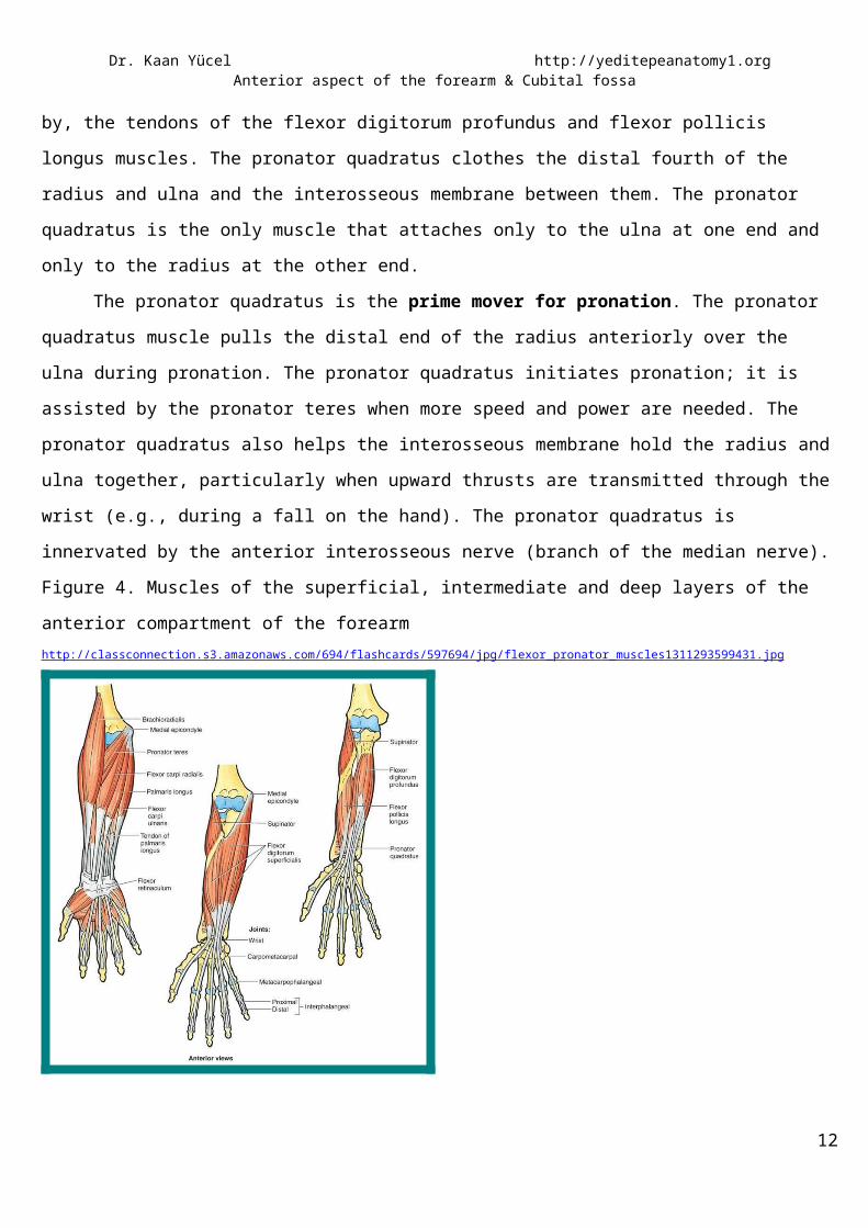
Figure 4. Muscles of the superficial, intermediate and deep layers of the anterior compartment of the forearmhttp://classconnection.s3.amazonaws.com/694/flashcards/597694/jpg/flexor_pronator_muscles1311293599431.jpg
ARTERIES The main arteries of the forearm are the ulnar and radial arteries, which usually arise opposite the neck
of the radius in the inferior part of the cubital fossa as terminal branches of the brachial artery.
Figure 5. Brachial artery and its two terminal branches: radial and ulnar arteries http://teachmeanatomy.net/upper-limb-2/arteries-and-veins-of-the-upper-limb
RADIAL ARTERY9

Dr. Kaan Yücel http://yeditepeanatomy1.org Anterior aspect of the forearm & Cubital fossa
The radial artery originates from the brachial artery at approximately the neck of the radius and passes
along the lateral aspect of the forearm. The radial artery is the smaller of the terminal branches of the brachial
artery.
In the distal forearm, the radial artery lies immediately lateral to the large tendon of the flexor carpi
radialis muscle and directly anterior to the pronator quadratus muscle and the distal end of the radius. In the
distal forearm, the radial artery can be located using the flexor carpi radialis muscle as a landmark. The radial
pulse can be felt by gently palpating the radial artery against the underlying muscle and bone. When the
brachioradialis is pulled laterally, the entire length of the artery is visible.
Branches of the radial artery originating in the forearm include:
1) radial recurrent artery, which contributes to an anastomotic network around the elbow joint
2) small palmar carpal branch
3) superficial palmar branch enters the hand by passing through, or superficial to, the thenar muscles at the
base of the thumb, which anastomoses with the superficial palmar arch formed by the ulnar artery.
Figure 6. Radial artery and its brancheshttp://upload.wikimedia.org/wikipedia/commons/e/e7/Gray528.png
10

Dr. Kaan Yücel http://yeditepeanatomy1.org Anterior aspect of the forearm & Cubital fossa
ULNAR ARTERYThe ulnar artery is larger than the radial artery and passes down the medial side of the forearm. It
leaves the cubital fossa by passing deep to the pronator teres muscle, and then passes through the forearm in
the fascial plane between the flexor carpi ulnaris and flexor digitorum profundus muscles. In distal regions of
the forearm, the ulnar nerve is immediately medial to the ulnar artery.
The ulnar artery leaves the forearm, enters the hand by passing lateral to the pisiform bone and superficial to
the flexor retinaculum of the wrist, and arches over the palm. It is often the major blood supply to the medial
three and one-half digits.
Pulsations of the ulnar artery can be palpated on the lateral side of the flexor carpi ulnaris tendon,
where it lies anterior to the ulnar head.
Figure 7. Ulnar artery and its branches
11
Dr. Kaan Yücel http://yeditepeanatomy1.org Anterior aspect of the forearm & Cubital fossa
VEINSVEINSThe superficial veins of the forearm lie in the superficial fascia. The cephalic vein arises from the lateral
side of the dorsal venous arch on the back of the hand and winds around the lateral border of the forearm; it
then ascends into the cubital fossa and up the front of the arm on the lateral side of the biceps. It terminates in
the axillary vein in the deltopectoral triangle. As the cephalic vein passes up the upper limb, it receives a
variable number of tributaries from the lateral and posterior surfaces of the limb.
The basilic vein arises from the medial side of the dorsal venous arch on the back of the hand and
winds around the medial border of the forearm; it then ascends into the cubital fossa and up the front of the
arm on the medial side of the biceps. Its terminates, by joining the venae comitantes of the brachial artery to
form the axillary vein.
The median cubital vein, a branch of the cephalic vein in the cubital fossa, runs upward and medially
and joins the basilic vein. The basilic vein also receives a variable number of tributaries from the medial and
posterior surfaces of the upper limb.
Figures 8 & 9. Veins in the anterior compartment of the forearmhttp://www.hxbenefit.com/wp-content/uploads/2011/07/Basilic-Vein.gif
12
Branches of the ulnar artery that arise in the forearm include: 1) ulnar recurrent artery with anterior and posterior branches, which contribute to an anastomotic network of vessels around the elbow joint (The anterior and posterior ulnar recurrent arteries anastomose with the inferior and superior ulnar collateral arteries, respectively, thereby participating in the periarticular arterial anastomoses of the elbow)2) numerous muscular arteries, which supply surrounding
muscles3) common interosseous artery, which divides into anterior
and posterior interosseous arteries4) two small carpal arteries (dorsal carpal branch and
palmar carpal branch) Perforating the interosseous membrane in the distal forearm, the anterior interosseous artery terminates by joining the posterior interosseous artery.

Dr. Kaan Yücel http://yeditepeanatomy1.org Anterior aspect of the forearm & Cubital fossa
http://radiographics.rsna.org/content/vol28/issue1/images/large/e28f2.jpeg
Deep veins accompanying arteries are plentiful in the forearm. These accompanying veins (L. venae
comitantes) arise from the anastomosing deep venous palmar arch in the hand. From the lateral side of the
arch, paired radial veins arise and accompany the radial artery; from the medial side, paired ulnar veins arise
and accompany the ulnar artery. The veins accompanying each artery anastomose freely with each other. The
radial and ulnar veins drain the forearm but carry relatively little blood from the hand.
Deep veins of the anterior compartment drain into brachial veins associated with the brachial artery in the
cubital fossa.
NERVESNERVESNerves in the anterior compartment of the forearm are the median and ulnar nerves, and the superficial
branch of the radial nerve.
MEDIAN NERVEThe median nerve is the principal nerve of the anterior compartment of the forearm. It supplies
muscular branches directly to the muscles of the superficial and intermediate layers of forearm flexors (except
the flexor carpi ulnaris), and deep muscles (except for the medial [ulnar] half of the flexor digitorum profundus;
ring and little fingers) via its branch, the anterior interosseous nerve.
13

Dr. Kaan Yücel http://yeditepeanatomy1.org Anterior aspect of the forearm & Cubital fossa
It leaves the cubital fossa by passing between the two heads of the pronator teres muscle and passing
between the humero-ulnar and radial heads of the flexor digitorum superficialis muscle. It leaves the forearm
and enters the palm of the hand by passing through the carpal tunnel deep to the flexor retinaculum.
The median nerve has no branches in the arm other than small twigs to the brachial artery. Its major
branch in the forearm is the anterior interosseous nerve.
1) Articular branches: These branches pass to the elbow joint as the median nerve passes it.
2) Muscular branches: The nerve to the pronator teres usually arises at the elbow. A broad bundle of nerves
pierces the superficial flexor group of muscles and innervates the flexor carpi radialis, palmaris longus, and
flexor digitorum superficialis.
3) Anterior interosseous nerve: The largest branch of the median nerve in the forearm is the anterior
interosseous nerve innervates the muscles in the deep layer (flexor pollicis longus, the lateral half of flexor
digitorum profundus, and pronator quadratus).
4) Palmar cutaneous branch of the median nerve: A small palmar branch passes superficially into the hand and
innervates the skin over the base and central palm. This palmar branch is spared in carpal tunnel syndrome
because it passes into the hand superficial to the flexor retinaculum of the wrist.
ULNAR NERVELike the median nerve, the ulnar nerve does not give rise to branches during its passage through the
arm. In the forearm it supplies only one and a half muscles, the flexor carpi ulnaris muscle (as it enters the
forearm by passing between its two heads of proximal attachment) and the ulnar (medial) part (ring and little
fingers) of the flexor digitorum profundus muscle.
The ulnar nerve enters the anterior compartment of the forearm by passing posteriorly around the
medial epicondyle of the humerus and between the humeral and ulnar heads of the flexor carpi ulnaris muscle.
In the forearm the ulnar nerve gives rise to:
1) Muscular branches to the flexor carpi ulnaris and to the medial half of the flexor digitorum profundus.
2) Two small cutaneous branches; palmar branch passes into the hand to supply skin on the medial side of the
palm; larger dorsal branch innervates skin on the posteromedial side of the back of the hand and most skin
on the posterior surfaces of the medial one and one-half digits.
14

Dr. Kaan Yücel http://yeditepeanatomy1.org Anterior aspect of the forearm & Cubital fossa
Figure 10. Median nervehttp://www.pureprecisionchiro.com/wp-content/uploads/2011/02/MedianNerve.jpg
RADIAL NERVEUnlike the medial and ulnar nerves, the radial nerve serves motor and sensory functions in both the arm
and the forearm (but only sensory functions in the hand). However, its sensory and motor fibers are distributed
in the forearm by two separate branches, the superficial (sensory or cutaneous) and deep radial/posterior
interosseous nerve (motor). The radial nerve bifurcates into deep and superficial branches anterior to the
lateral epicondyle of the humerus, between the brachialis and the brachioradialis, in the lateral border of the
cubital fossa.
The deep branch is predominantly motor and passes between the two heads of the supinator muscle to
access and supply muscles in the posterior compartment of the forearm.
The superficial branch of the radial nerve is sensory. It passes down the anterolateral aspect of the
forearm deep to the brachioradialis muscle. The nerve continues into the hand where it innervates skin on the
posterolateral surface.
Figure 11. Ulnar nerve & Radial nerve15

Dr. Kaan Yücel http://yeditepeanatomy1.org Anterior aspect of the forearm & Cubital fossa
http://karate.butsu.net/anatomy/anterior_view.html
volar= anterior
LATERAL AND MEDIAL CUTANEOUS NERVES OF FOREARM
The lateral cutaneous nerve of the forearm (lateral antebrachial cutaneous nerve) is the continuation of
the musculocutaneous nerve after its motor branches have all been given off to the muscles of the anterior
compartment of the arm.
The medial cutaneous nerve of the forearm (medial antebrachial cutaneous nerve) is an independent
branch of the medial cord of the brachial plexus. With the posterior cutaneous nerve of the forearm from the
radial nerve, each supplying the area of skin indicated by its name, these three nerves provide all the
cutaneous innervation of the forearm. There is no “anterior cutaneous nerve of the forearm.” (Memory device:
This is similar to the brachial plexus, which has lateral, medial, and posterior cords but no anterior cord.)
Figures 12 & 13. Lateral cutaneous nerve of forearmhttp://www.lookfordiagnosis.com/mesh_info.php?term=Musculocutaneous+Nerve&lang=1
16

Dr. Kaan Yücel http://yeditepeanatomy1.org Anterior aspect of the forearm & Cubital fossa
http://www.kmle.co.kr/search.php?Search=antebrachial&Page=1
Although the arteries, veins, and nerves of the forearm have been considered separately, it is important
to place them into their anatomical context. Except for the superficial veins, which often course independently
in the subcutaneous tissue, these neurovascular structures usually exist as components of neurovascular
bundles. These bundles are composed of arteries, veins (in the limbs, usually in the form of accompanying
veins), and nerves as well as lymphatic vessels, which are usually surrounded by a neurovascular sheath of
varying density.
The cubital fossa is an important area of transition between the arm and the forearm. The cubital fossa
is seen superficially as a depression on the anterior aspect of the elbow. Deeply, it is a space filled with a
variable amount of fat anterior to the most distal part of the humerus and the elbow joint.
Superiorly, an imaginary line connecting the medial and lateral epicondyles.
Medially, the mass of flexor muscles of the forearm arising from the common flexor attachment on the
medial epicondyle; most specifically, the pronator teres.
17
3. CUBITAL FOSSA

Dr. Kaan Yücel http://yeditepeanatomy1.org Anterior aspect of the forearm & Cubital fossa
Laterally, the mass of extensor muscles of the forearm arising from the lateral epicondyle and
supraepicondylar ridge; most specifically, the brachioradialis.
As a summary, the pronator teres makes the medial border, whereas the brachioradialis makes the lateral one.
The floor of the cubital fossa is formed by the brachialis and supinator muscles of the arm and forearm,
respectively. The roof of the cubital fossa is formed by the continuity of brachial and antebrachial (deep) fascia
reinforced by the bicipital aponeurosis, subcutaneous tissue, and skin.
The contents of the cubital fossa are the:
Terminal part of the brachial artery and the commencement of its terminal branches, the radial and ulnar
arteries. The brachial artery lies between the biceps tendon and the median nerve.
(Deep) accompanying veins of the arteries
Biceps brachii tendon
Median nerve
Radial nerve
Superficially, in the subcutaneous tissue overlying the fossa are the median cubital vein, lying anterior to the
brachial artery, and the medial and lateral antebrachial cutaneous nerves, related to the basilic and cephalic
veins.
The supratrochlear lymph node lies in the superficial fascia over the upper part of the fossa, above the
trochlea. It receives afferent lymph vessels from the third, fourth, and fifth fingers; the medial part of the hand;
and the medial side of the forearm. The efferent lymph vessels pass up to the axilla and enter the lateral axillary
nodes (The superficial lymph vessels from the thumb and lateral fingers and the lateral areas of the hand and
forearm follow the cephalic vein to the infraclavicular group of nodes. Those from the medial fingers and the
medial areas of the hand and forearm follow the basilic vein to the cubital fossa).
The brachial artery normally bifurcates into the radial and ulnar arteries in the apex of the fossa,
although this bifurcation may occur much higher in the arm, even in the axilla. When taking a blood pressure
reading from a patient, the clinician places the stethoscope over the brachial artery in the cubital fossa.
The median nerve lies immediately medial to the brachial artery and leaves the fossa by passing
between the ulnar and humeral heads of the pronator teres muscle.
The brachial artery and the median nerve are covered and protected anteriorly in the distal part of the
cubital fossa by the bicipital aponeurosis. This flat connective tissue membrane passes between the medial side
of the tendon of the biceps brachii muscle and deep fascia of the forearm. The sharp medial margin of the
bicipital aponeurosis can often be felt.
The radial nerve lies just under the lip of the brachioradialis muscle, which forms the lateral margin of
the fossa. In the cubital fossa the radial nerve gives off the deep branch of the radial nerve and continues as the 18

Dr. Kaan Yücel http://yeditepeanatomy1.org Anterior aspect of the forearm & Cubital fossa
superficial radial nerve. The deep branch supplies the extensor carpi radialis brevis and the supinator in the
cubital fossa and all the extensor muscles in the posterior compartment of the forearm.
The ulnar nerve does not pass through the cubital fossa. Instead, it passes posterior to the medial epicondyle.
The roof of the cubital fossa is formed by superficial fascia and skin. The most important structure within
the roof is the median cubital vein, which passes diagonally across the roof and connects the cephalic vein on
the lateral side of the upper limb with the basilic vein on the medial side. The bicipital aponeurosis separates
the median cubital vein from the brachial artery and median nerve. Other structures within the roof are
cutaneous nerves;-the medial cutaneous and lateral cutaneous nerves of the forearm.
Figure 14. Cubital fossahttp://www.daviddarling.info/images2/cubital_fossa.jpg
Table 1. Muscles of the anterior compartment of the forearm (superficial and intermediate layers)Superficial (first) layer
Pronator teres Median nerve Pronates and flexes forearm (at elbow)Ulnar head Coronoid process Lateral surface of
19

Dr. Kaan Yücel http://yeditepeanatomy1.org Anterior aspect of the forearm & Cubital fossa
radiusHumeral head Medial epicondyle and adjacent supraepicondylar ridge
Flexor carpi radialis (FCR)
Medial epicondyle of humerus
Base of metacarpals II and III
Flexes and abducts hand (at wrist)
Palmaris longus Medial epicondyle of humerus (common flexor origin)
Flexor retinaculum and palmar aponeurosis
Flexes hand (at wrist) and tenses palmar aponeurosis
Flexor carpi ulnaris (FCU)Humeral head Medial epicondyle of
humerus Pisiform &
hamate 5th metacarpal
Ulnar nerve
Flexes and adducts the wrist joint
Ulnar head Olecranon Posterior border of
ulnaIntermediate (second) layer
Flexor digitorum superficialis (FDS)Humeroulnar head Medial epicondyle of
humerus
Adjacent margin of coronoid process
Shafts of middle phalanges of medial four digits
Median nerve Flexes proximal interphalangeal joints of the index, middle, ring, and little fingers; can also flex metacarpophalangeal joints of the same fingers and the wrist joint
Radial head Superior half of anterior border
Muscle Proximal Attachment Distal Attachment Innervation Main Action
Table 2. Muscles of the anterior compartment of the forearm (deep layer)
Muscle Proximal Attachment Distal Attachment Innervation Main ActionFlexor digitorum profundus (FDP)Medial part Proximal three
quarters of medial Bases of distal phalanges of 4th and
Ulnar nerve Flexes distal phalanges 4 and 5 at
20

Dr. Kaan Yücel http://yeditepeanatomy1.org Anterior aspect of the forearm & Cubital fossa
and anterior surfaces of ulna and interosseous membrane
5th digits distal interphalangeal joints
Lateral part Bases of distal phalanges of 2nd and 3rd digits
Anterior interosseous nerve, from median nerve
Flexes distal phalanges 2 and 3 at distal interphalangeal joints
Flexor pollicis longus (FPL)
Anterior surface of radius and adjacent interosseous membrane
Base of distal phalanx of thumb
Flexes phalanges of 1st digit (thumb)
Pronator quadratus Distal quarter of anterior surface of ulna
Distal quarter of anterior surface of radius
Pronates forearm; deep fibers bind radius and ulna together
21