Embed Size (px)
Citation preview

Transforming growth factor- enhances Rho-kinase activity and contraction in airway
smooth muscle via the nucleotide exchange factor ARHGEF1
Yasin Shaifta, Charles E. MacKay, Nneka Irechukwu, Katie A. O’Brien, David B. Wright,
Jeremy P.T. Ward, Greg A. Knock.
Division of Asthma, Allergy and Lung Biology, Faculty of Life Sciences and Medicine,
King’s College London, London, SE1 1UL.
Running Title: TGF-, ARHGEF1 and Rho-kinase in airway smooth muscle
Key words: Airway smooth muscle; TGF-; Rho-kinase
Corresponding author:
Dr Greg Knock
1.20 Henriette Raphael House
Guy’s Campus
King’s College London
London, SE1 1UL
1

Key points Summary
Transforming growth-factor- (TGF-) and RhoA/Rho-kinase are independently implicated in the airway hyper-responsiveness associated with asthma, but how these proteins interact is not fully understood
We examined the effects of pre-treatment with TGF- on expression and activity of RhoA, Rho-kinase and ARHGEF1, an activator of RhoA, as well as on bradykinin induced contraction, in airway smooth muscle
TGF- enhanced bradykinin-induced RhoA translocation, Rho-kinase dependent phosphorylation, and contraction, but partially suppressed BK-induced RhoA activity (RhoA-GTP content)
TGF- enhanced the expression of ARHGEF1, while a siRNA against ARHGEF1 and a RhoGEF inhibitor prevented the effects of TGF- on RhoA and Rho-kinase activity and contraction, respectively
ARHGEF1 expression was also enhanced in airway smooth muscle from asthmatic patients and OVA-sensitized mice
ARHGEF1 is a key TGF- target gene, an important regulator of Rho-kinase activity and is therefore a potential therapeutic target for the treatment of asthmatic airway hyper-responsiveness
2

Abstract
Transforming growth factor- (TGF-), RhoA/Rho-kinase and Src-family kinases (SrcFK)
have independently been implicated in airway hyper-responsiveness, but how they interact in
order to regulate airway smooth muscle contractility is not fully understood. We found that
TGF- pre-treatment enhanced acute contractile responses to bradykinin (BK) in isolated rat
bronchioles, and inhibitors of RhoGEFs (Y16) and Rho-kinase (Y27632), but not the SrcFK
inhibitor PP2 prevented this enhancement. In cultured human airway smooth muscle cells
(hASMC), TGF- pre-treatment enhanced the protein expression of the Rho guanine
nucleotide exchange factor ARHGEF1, MLC20, MYPT-1 and the actin-severing protein
cofilin, but not of RhoA, ROCK2 or c-Src. In hASMC, acute treatment with BK triggered
sub-cellular translocation of ARHGEF1 and RhoA and enhanced auto-phosphorylation of
SrcFK and phosphorylation of MYPT1 and MLC20, but induced de-phosphorylation of
cofilin. TGF- pre-treatment amplified the effects of BK on RhoA translocation and
MYPT1/MLC20 phosphorylation, but suppressed the effects of BK on RhoA-GTP content,
SrcFK auto-phosphorylation and cofilin de-phosphorylation. In hASMC, an ARHGEF1
siRNA suppressed the effects of BK and TGF- on RhoA-GTP content, RhoA translocation
and MYPT1 and MLC20 phosphorylation, but minimally influenced the effects of TGF- on
cofilin expression and phosphorylation. ARHGEF1 expression was also enhanced in ASMC
of asthmatic patients and in lungs of OVA-sensitized mice. Our data indicate that TGF-
enhances BK-induced contraction, RhoA translocation and Rho-kinase activity in airway
smooth muscle largely via ARHGEF1, but independently of SrcFK and total RhoA-GTP
content. A role for smooth muscle ARHGEF1 in asthmatic airway hyper-responsiveness is
worthy of further investigation.
Abbreviations
3

AHR = airway hyper-responsiveness
ASM = airway smooth muscle
BAL = broncho-alveolar lavage
BK = bradykinin
DMEM = Dulbecco’s Modified Eagle Medium
EmGFP = Emerald green fluorescent protein
GPCR = G-protein coupled receptor
hASMC = human airway smooth muscle cells
KPSS = PSS with equimolar substitution of Na+ for K+
MLC20 = myosin light-chain 20 KDa subunit
MLCP = Myosin light-chain phosphatase
MYPT1 = myosin phosphatase targeting subunit-1
PSS = physiological salt solution
RhoGEF = Rho-specific guanine nucleotide exchange factor
SrcFK = Src-family kinases
TBS = Tris buffered saline
TGF- = transforming growth-factor-
4

Introduction
In healthy subjects, airway smooth muscle tone and airflow resistance are low, while in
subjects with allergic asthma, exposure to allergens sensitizes airways to constrictor stimuli,
resulting in inappropriate episodic airway smooth muscle (ASM) constriction (airway hyper-
responsiveness, AHR) and long-term remodelling (Doeing & Solway, 2013). These smooth
muscle phenotypic changes are triggered by inflammatory mediators, produced in the airways
in response to allergen sensitization. One of these mediators, transforming growth factor-beta
(TGF-), is produced at higher basal levels in asthmatic airways and is increased several-fold
after allergen challenge (Redington et al., 1997;Torrego et al., 2007) up to levels that
correlate with the severity of AHR and remodelling (Matsunaga et al., 2006). In connective
tissue, TGF- induces contractile protein expression and stress fiber assembly (Ji et al.,
2014;Vardouli et al., 2005;Sandbo et al., 2011), while in ASM, it can influence both
proliferation and hypertrophy (Halwani et al., 2011) and induce hyper-responsiveness to
bronchoconstrictors (Goldsmith et al., 2006;Oenema et al., 2012).
Smooth muscle cross-bridge cycling requires MLC20 phosphorylation, which is in turn
dependent on the balance between activity of Ca2+-induced myosin light-chain kinase and
myosin light-chain phosphatase (MLCP). The latter is inhibited by phosphorylation of
MYPT1 by Rho-kinase, an effector of the monomeric G-protein RhoA (Somlyo & Somlyo,
2003). Thus, TGF- could mediate enhanced ASM responsiveness to bronchoconstrictors by
enhancing expression or activity of RhoA/Rho-kinase, their downstream targets or their
upstream regulators. Among the latter are the Rho-specific guanine nucleotide exchange
factors (RhoGEFs), a large family of regulatory proteins that activate members of the Rho
family of small G-proteins by catalysing the exchange of bound GDP for bound GTP (Bos et
al., 2007). Two of these, GEF-H1 and Net1, are up-regulated by TGF- in epithelia thus
5

enhancing RhoA-dependent F-actin expression/cell migration and epithelial-to-mesenchymal
transition, respectively (Papadimitriou et al., 2012;Tsapara et al., 2010). Another RhoGEF,
ARHGEF1 (p115-RhoGEF, Lsc), not previously linked with TGF-, has been implicated in
the T-cell dysfunction associated with allergic asthma, and in the contractile effects of
angiotensin II on hypertensive vascular smooth muscle (Brown et al., 2007;Guilluy et al.,
2010). The role of ARHGEF1 in ASM contraction remains to be determined.
We have recently confirmed that bradykinin (BK)-induced MLC20 phosphorylation and
contraction in healthy rat airways and cultured human ASM was dependent on Rho-kinase
activity, which was also partly mediated via activation of Src-family kinases (SrcFK) (Shaifta
et al., 2015). Both Rho-kinase and SrcFK have been suggested as potential therapeutic targets
in asthma, having both being implicated in experimental AHR (Chiba et al., 2005;Schaafsma
et al., 2006;Katsumoto et al., 2013). Smooth muscle contraction is also influenced by factors
that regulate actin cytoskeleton re-organisation and contractile apparatus assembly; in a
number of cell types this is also reportedly mediated by RhoA/Rho-kinase, in part via
inhibitory phosphorylation of cofilin (Arber et al., 1998;Leduc et al., 2016;Vardouli et al.,
2005). Conversely, it has been suggested that particularly in ASM, cofilin has an alternative
role in promoting actin polymerization, perhaps independently of Rho-kinase (Zhang et al.,
2010;Zhao et al., 2008). In this study therefore, we examined the long-term effects of TGF-
on bradykinin-induced ASM contraction, as well as on protein expression and activity of
ARHGEF1, SrcFK, RhoA/Rho-kinase MYPT1/MLC20 and cofilin.
6

Methods
Ethical approval. All studies with rat tissues were performed in vitro after animals were
euthanized by intra-peritoneal sodium pentobarbital injection. OVA-sensitization and lung
function measurement in mice was performed in accord with The Animals (Scientific
procedures) Act (1986) and local ethical approval from King’s College London. Donation of
human tissue was obtained following written informed consent and with the approval of the
South East London Research Ethics Committee, REC reference number: 10/H0804/66. All
clinical procedures conformed to the standards set by the latest Declaration of Helsinki.
Animal Tissue. Investigations of airway reactivity in healthy animals were conducted in
isolated small intralobar bronchioles obtained from male Wistar rats (~250g). Lungs were
removed immediately after killing by lethal injection (pentobarbital i.p.). Bronchioles (~1mm
diameter, ~2mm length) were dissected free of surrounding parenchyma and placed in cold
physiological saline solution (PSS, composition in mM: 118 NaCl; 24 NaHCO3; 1 MgSO4; 4
KCL; 5.56 glucose; 0.435 NaH2PO4; 1.8 CaCl2, pH 7.4). Lung tissue was also obtained from
a mouse model of airway hyper-responsiveness. 129SVJ/Black Swiss mice were immunized
four times at 7 day intervals, with ovalbumin (30µg/ml i.p., OVA-treated), or vehicle
(Al2[OH]3, sham-treated). They were then challenged with aerosolized OVA (30µg/ml, 2 x 25
min per day for four days). Airway hyper-responsiveness in OVA-treated mice was
confirmed by greater methacholine-induced reduction in airway dynamic compliance (Cdyn)
compared to sham-treated (P<0.01) and greater sensitivity to methacholine compared to
sham-treated (Cdyn PC50 reduced by 50%, P<0.001).
Human tissue and cell culture. Human airway smooth muscle cells (hASMC) were
obtained from healthy volunteers (n=11; 7 female, 4 male; age range 22-53 yrs) or volunteers
7

with moderate asthma (n=3; all male; age range 44-53 yrs), using deep endobronchial biopsy.
Healthy volunteers were selected based on a life-long absence of respiratory symptoms, and
lung functions within normal limits. Moderate asthmatics were selected based on the
presence of typical symptoms over at least two years, a minimum of 12% reversibility, FEV1
<80% of predicted for age, and/or a bronchoconstrictor response to a methacholine challenge
of <8mg/ml. All asthmatic subjects were on a combined therapy of short acting beta-agonist
and inhaled corticosteroids (fluticasone propionate <500μg daily or equivalent). Patients
currently receiving leukotriene receptor antagonists, smokers and pregnant/lactating females
were excluded.
ASM bundles were bathed in Dulbecco’s Modified Eagle Medium (DMEM) containing the
following supplements: 10% FBS, L-glutamine (2mM), sodium pyruvate (1mM), non-
essential amino acids and amphotericin B (2µg/ml) and dissected free of adjacent connective
tissue and epithelia. They were then subjected to enzymatic digestion in nominally Ca2+-free
HEPES buffer containing: 5.56 mM glucose, 2mg/ml collagenase Type XI, 1mg/ml papaine,
1mg/ml trypsin inhibitor and 1mM DTT. Cells were then dispersed, incubated in DMEM
(plus supplements) at 37°C, pH 7.4, and typically used for experiments at passage 4-9.
hASMC were seeded onto 60mm dishes, grown to confluence and serum starved for seven
days in DMEM without FBS, supplemented with L-glutamine (2mM), sodium pyruvate
(1mM), non-essential amino acids (1:100), amphotericin B (1.5µg/ml), gentamicin
(50µg/ml), insulin-transferrin-selenium (1:100), 100µM ascorbate and bovine serum albumin
(BSA) (1%). This prolonged serum starvation and media/supplement combination has
previously been shown to restore and preserve a contractile phenotype in ASM (Halayko et
al., 1999;Kim et al., 2005). hASMC were confirmed as smooth muscle by positive staining
for smooth muscle-specific protein markers, as described previously (Shaifta et al., 2015).
8

TGF- treatments. The long-term effects of TGF- on tissue and cellular function and
protein expression was examined by pre-incubation of rat bronchioles or hASMC for 18-
24hrs at concentrations similar to those previously found to exist in the airways of asthmatic
patients (Redington et al., 1997;Torrego et al., 2007). Bronchioles were incubated for 18hr at
37°C with 30 ng/ml TGF- serum-free Ham’s F12 media with supplements and 0.1% BSA,
or in Ham’s F12 media and supplements/BSA alone (Halayko et al., 1999;Kim et al., 2005).
Following this incubation, bronchioles were transferred to PSS for subsequent tension
measurement by wire myography, in the absence of TGF-. Treatment of hASMC with 10
ng/ml TGF- for 24hrs was initiated at day six of serum starvation, followed by washout with
serum-free media (without TGF-) for a further 24hrs, prior to acute treatments with
pharmacological agents and harvesting.
Tension Measurement by Wire Myography. Bronchioles were mounted on a Mulvany-
Halpern small vessel wire myograph (DMT.dk), bathed in PSS gassed with 95% air, 5% CO2
at 37°C, for the measurement of isometric tension. Bronchioles were stretched incrementally,
and alternately exposed to PSS containing 80mM [K+] (equimolar substitution for Na+,
KPSS) in order to determine the point on the length-tension curve at which optimum active
tension development was achieved, as described previously (Moir et al., 2003). Viability of
bronchioles for tension experiments was confirmed by active tension generation of at least
2mN in response to the last exposure to KPSS. Mean internal diameter after stretching was
typically in the range 300-800µm.
RhoA-EmGFP//ARHGEF1-EmGFP cloning. RhoA and ARHGEF1 cDNAs were
separately cloned into pcDNA™6.2/C-EmGFP/TOPO® to produce C-terminal EmGFP
9

fusion vectors (Invitrogen Life Technologies, UK). Oligonucleotide primers were designed in
accordance with the manual and using sequences from the GenBank® database (RhoA
accession no. BC061732; ARHGEF1 accession no. BC091218) as follows: RhoA (forward):
GTTATGGCTGCCATCAGGAAGAAACTGG-3’, RhoA (reverse): 5’-
CAAGATGAGGCACCCCGACT-3’, ARHGEF1 (forward): 5’-
GAGATGGGAGAAGTCGCCGGAGGGGC-3’, ARHGEF1 (reverse): 5’-
TGAAAGGCCTGTCTGAGCAGAGCGC-3’. Clones were checked for the correct
orientation using restriction endonuclease digestion and gel electrophoresis. These were then
sent for sequencing (Source BioScience UK Limited) and selection.
siRNA design and transfection. The human ARHGEF1 siRNA was designed as described
previously (Knock et al., 2008;Shaifta et al., 2015). The 19 nucleotide target sequence
(position 2057–2075, GenBank accession no. NM_199002) was synthesized into 64–65 mer
oligonucleotides with BamHI/HindIII overhangs (Sigma) and cloned into the expression
vector pSilencer 3.0-H1 (Ambion Inc.). All clones were purified (EndoFree Plasmid Maxi
Kit, Qiagen Ltd) and sequenced (Source BioScience UK Limited). As a negative control,
another siRNA was prepared using a scrambled mRNA sequence. Transfection was carried
out in detached confluent hASMC using the Basic Nucleofector™ Kit for Primary
Mammalian Smooth Muscle Cells and a nucleofector device (Nucleofector™ Technology -
Lonza AG). The media was changed the next day and cells grown for a further 48hrs in
DMEM plus supplements, prior to serum-starvation and TGF- treatment, as described
above. Transfection efficiency was >90%, determined separately using pmaxGFP (green
fluorescent protein expressing vector) provided in the kit and confirmed by fluorescence
microscopy. Efficiency of ARHGEF1 knockdown was confirmed by Western blot (Fig 3). If
designed correctly, siRNA can be a highly specific tool for targeted gene knockdown
10

(Semizarov et al., 2003). In order to minimise any non-specific targeting, candidate siRNAs
presenting more than 16 contiguous nucleotides of sequence identity with another mRNA
were discarded (Birmingham et al., 2007). A BLAST ® alignment (NCBI, USA) of the 19
nucleotide ARHGEF1 siRNA sequence showed only 5 contiguous nucleotide homology to
MLC20 (GenBank® accession no. S69022), RhoA (GenBank® accession no. BC061732) and
cofilin (GenBank® accession no. D00682), only 6 contiguous nucleotide homology to PDZ-
RhoGEF (ARHGEF11, GenBank® accession no. BC057394), only 7 contiguous nucleotide
homology to LARG (ARHGEF12, GenBank® accession no. NM01513) and ROCK2
(GenBank® accession no. NM004850), and only 9 contiguous nucleotide homology to
MYPT1 (GenBank® accession no. D87930).
Rho-GTP content measurement by Rhotekin asssay
After treatment, cells were harvested as described above using MLB lysis buffer made up
according to The Rho Assay Reagent protocol (Millipore-Merck). The lysates were then
cleared of insoluble cell debris by centrifugation, a small amount taken to determine protein
concentration and the remainder immediately snap frozen and stored at -80°C. In the pull-
down assay the collected samples were quick-thawed and between 250-500µg of protein was
mixed with 20µg of The Rho Assay Reagent slurry (A GST-tagged fusion protein,
corresponding to residues 7-89 of mouse rhotekin Rho-binding domain bound to glutathione-
agarose) and incubated for 45 minutes at 4°C with gentle agitation using a roller mixer. For
positive control, an extra untreated sample was pre-incubated with GTPγS for 30 minutes at
30°C prior to mixing with The Rho Assay Reagent slurry. After 45 minutes, the mixture was
centrifuged at 4°C and the supernatant discarded followed by 3 washes with ice cold MLB,
again centrifuging each time and discarding the supernatant. After the final wash 40µL of 2X
Laemmli reducing sample buffer containing 50mM DTT (to improve release of RhoA from
11

the beads) was added and the mixture boiled at 95°C for 5 minutes, followed by cooling and
storage at -20°C or below. The final supernatant and agarose pellet were mixed before being
subjected to SDS PAGE and Western blot, as described below.
RhoA-EmGFP/ARHGEF1-EmGFP translocation imaging and quantification.
Coverslips containing serum-starved hASMC were mounted onto a Zeiss Axiovert 200
Microscope and cells visualized using BD™ CARV II Confocal Imager under X40
magnification, bathed in PSS/5% CO2 at 37°C. When illuminated with UV/blue light
(~390nm). Successfully transfected cells were identified by bright green fluorescence. RhoA-
EmGFP/ARHGEF1-EmGFP translocation was stimulated by acute application of 1µM BK.
Images were captured by MetaFluor v.7 software (Molecular Devices). This typically
involved brief (65ms) shutter openings, once every 2sec during stimulation, but less
frequently during rest periods. Regional and temporal changes in fluorescence, indicating
movement of the EmGFP-tagged protein in response to drug treatments were quantified using
ImageJ software (rsb.info.nih.gov). Briefly, a line was drawn across each region of interest
centered around spots or patches on the cell periphery. For each region, a single value of
difference in fluorescence between the ‘spot’ peak intensity and nearby background. Each
region of interest was measured before and during exposure to BK in the same cell. 2-3
regions of interest were analysed and averaged for each cell and BK-induced fluorescence
expressed as a % of control. The only criteria for exclusion of a cell from the analysis was
poor EmGFP transfection, indicated by overall cellular fluorescence below a pre-determined
threshold level.
hASMC acute treatment, harvesting and Western blot. SrcFK, MYPT1, MLC20 and
cofilin phosphorylation/de-phosphorylation was stimulated by acute treatment with BK
12

(1µM, 30 sec) in serum-free DMEM at 37°C, as shown previously (Shaifta et al., 2015).
Cells were then immediately washed twice with ice-cold phosphate buffered saline
(Invitrogen) to terminate reactions and remove any residual media, followed immediately by
application of cell lysis buffer (NEB) containing 1% phosphatase inhibitor cocktails 2 and 3
and 1% protease inhibitor cocktail (all Sigma). Cells were scraped into a tube, agitated
continuously for 30sec, placed on ice and centrifuged at 10000 rpm for 5min. The resultant
pellets were discarded and the remaining supernatant stored at -80°C for future use.
Protein content was determined using the bicinchoninic acid assay, calibrated against BSA
protein standards. Prior to gel loading for SDS-PAGE, lysates were prepared in NuPAGE
LDS Sample Buffer (Invitrogen) and protein content adjusted so that each lane of the gel
would typically contain 20µg of protein. Samples were boiled at 95°C for 5 minutes before
being loaded onto 4-12% NuPAGE Bis-Tris gels (Invitrogen). Gels were run in MOPS
running buffer (Invitrogen) and protein transferred to nitrocellulose membrane (Amersham)
in transfer buffer (25mM Tris, 192mM glycine, 20% methanol).
Membranes were blocked with 5% skimmed milk in Tris Buffered Saline (TBS), followed by
incubation with specific anti-phospho-protein primary antibody (typically 1:1000 dilution) in
TBS with 1% skimmed milk and 0.1% Tween-20 (TBS-T), overnight at 4°C. This was then
followed by washes in TBS-T and the application of appropriate HRP-conjugated secondary
antibody (typically 1:5000 dilution) for 1hr at room temperature, followed by further washes
in TBS-T. ‘Phospho’ proteins were visualised with Super-Signal West Femto chemi-
luminescent Substrate (Thermo scientific). Membranes were then stripped in Restore western
blot stripping buffer (Thermo Scientific), re-blocked and re-incubated with corresponding
‘total’ antibody and appropriate secondary antibodies, as above. ‘Total’ proteins were
13

visualised with either ECL plus or ECL prime (Amersham, GE healthcare). All images were
captured and quantified using the ChemiDoc XRS+ gel imaging system (Biorad).
Changes in protein expression were determined as a ratio of target protein signal, over signal
for the ‘housekeeping’ protein GAPDH, acting as a loading control for each sample. An
estimate of the amount of target protein that was phosphorylated in response to acute
treatments was calculated both as a ratio of ‘phospho’ over ‘total’ signal for each protein
sample, to give an indication of the fraction of each protein that is phosphorylated in response
to BK, and as a ratio of ‘phospho’ over GAPDH, to reveal the influence of TGF- and
ARHGEF1 siRNA on total phosphorylation of each protein, in addition to the acute effect of
BK. The effects of treatments on these ratios were then expressed as a percentage of control
(no BK, no TGF-, run on the same gel).
Materials and Reagents. Antibodies were obtained from: Cell Signalling (anti-phospho-Src
(tyr416), anti-Src, anti-phospho-MLC (ser19), anti-MLC, anti-MYPT1, anti-p115RhoGEF,
anti-RhoA, anti-cofilin, anti-phospho-cofilin (ser3)), Millipore (anti-phospho-MYPT1
(thr696)), Sigma (anti-rabbit IgG, anti-mouse IgG) and Santa Cruz (anti-ROCK2). All
antibodies were used at 1:1000 dilution except for anti-phospho-MYPT1, anti-rabbit IgG and
anti-mouse IgG, which were used at 1:5000. PP2 (4-amino-5-(4-chlorophenyl)-7-
(dimethylethyl) pyrazolo[3,4-d]pyrimidine) was obtained from Calbiochem; bradykinin, Y16
(inhibitor of RGS-domain containing RhoGEFs) and Y27632 (Rho-kinase inhibitor) were
obtained from Sigma. Rhotekin reagent and GTP--S were obtained from Millipore. TGF-
was obtained from R&D Systems. Cell culture and western blot materials were from Cell
Signaling, Invitrogen, GE Healthcare or Thermo Scientific. General reagents were from
Sigma, Calbiochem or VWR.
14

Data analysis and statistics. All values are expressed as mean ± SEM. All statistical analysis
and non-linear regression curve fitting was performed with SigmaPlot 10. Bradykinin
concentration responses were biphasic, so were fitted using a 2-site saturation model, for the
characterization of a high affinity component (PD2-1 and Max-1) and a low affinity
component (PD2-2 and Max-2). In contraction experiments, ‘n’ refers to the number of
bronchioles obtained from a similar number of rats. In western blot experiments, ‘n’ refers to
the number of cell cultures, each derived from a different human subject. In translocation
experiments, ‘n’ refers to the number of cells, selected at random from at least three separate
cell cultures, each derived from a different human subject. Statistical comparisons were by
paired or un-paired t-test (two groups of data, single factor), one-way, two-way or three-way
ANOVA (multiple comparisons, single factor, two-factor or three-factor, respectively), with
Holm-Sidak post-tests where appropriate, and as indicated in figure or table legends.
Differences were considered significant if P<0.05.
Results
TGF- enhances bradykinin-induced constriction via RhoGEFs and Rho-kinase, but
not SrcFK. The contractile effects of bradykinin (BK, 0.01–100µM) on isolated rat
bronchioles were determined with or without prior exposure to TGF-. The BK dose
response was then repeated in each bronchiole in the presence of the SrcFK inhibitor PP2
(30M), the inhibitor of RGS domain-containing RhoGEFs Y16 (30µM, (Shang et al., 2013))
or the Rho-kinase inhibitor Y27632 (10µM). At each dose, BK produced a biphasic
contraction, incorporating a large transient which usually decayed to a near plateau within
5mins. Non-linear regression analysis of the plateau phase at each concentration showed that
the data was best fitted to a two-site saturation model with a high and a low affinity
15

component. In the absence of inhibitor, TGF- enhanced the contractile response compared
to media alone (Fig 1A-B). Specifically, the amplitude of the low affinity component (Bmax-
2) was increased by TGF- (Fig 1F), while other parameters were unaltered (Fig1F-G). All
three inhibitors partially suppressed BK-induced contraction, after both media and TGF-
pre-incubation (Fig 1C-E), showing significant reductions in amplitude of both high and low
affinity components (Bmax-1 and Bmax-2). However, there remained a significant difference
in Bmax2 between media and TGF- pre-treated bronchioles in the presence of PP2, while in
the presence of Y16 or Y27632, these differences were no longer significant. Other curve-fit
parameters were unaffected by the three inhibitors, regardless of pre-exposure to TGF- or
media alone (Fig 1G). TGF- pre-exposure did not alter the amplitude of KPSS-induced
constriction compared to media (media only: 2.9 ± 0.4 mN, n=49 vs. TGF-: 3.0 ± 0.4 mN,
n=49). Repeating BK dose-responses in the absence of inhibitor (time controls), in
bronchioles exposed to TGF- or media alone, showed no changes in any of the curve-fit
parameters (n= 4, data not shown).
In order to see how the interaction between SrcFK inhibition and TGF- pre-incubation on
BK-induced contraction relates to SrcFK expression and activity, we determined the effects
of TGF- on c-Src protein expression in hASMCs. We found that acute treatment with BK
(30sec, 1M) enhanced SrcFK auto-phosphorylation, but that this enhancement was not
potentiated by prior exposure to TGF- and that that pre-exposure to TGF- reduced c-Src
protein content by ~30% (Fig 2A-B). To account for the effect of altered c-Src content on
SrcFK phosphorylation, changes in auto-phosphorylation were expressed two ways:
phospho/total, a proportional measure of the degree of phosphorylation (Fig 2C) and
phospho/GAPDH, a measure of actual phosphorylated SrcFK content, relative to total
cellular protein (Fig 2D). Without prior incubation with TGF-, acute exposure to BK
16

produced a 2-fold increase in SrcFK auto-phosphorylation, while after TGF- pre-incubation
the enhancing effect of BK was significantly reduced. Basal SrcFK phosphorylation was also
suppressed by TGF-, but only when expressed as phospho/GAPDH, linking it to the reduced
c-Src protein content.
TGF- enhances ARHGEF1 expression, while BK triggers ARHGEF1 translocation.
Considering that the TGF--induced enhancement of contraction is dependent on RhoGEFs
and Rho-kinase, we next examined the effects of TGF- on expression, activity and
translocation of RhoA, expression and activity of Rho-kinase and the influence of the
RhoGEF ARHGEF1 on those effects, in hASMC. TGF- had no effect on protein expression
of either RhoA or Rho-kinase, relative to GAPDH (Fig 3A-B) but induced a doubling in
protein content of ARHGEF1, relative to GAPDH (Fig 3C). In hASMC transfected with
EmGFP-tagged ARHGEF1, fluorescence was concentrated in the perinuclear region, but with
lower levels in the cell periphery. Addition of BK (1µM) reversibly enhanced this
fluorescence in distinct peripheral patches or spots (Fig 3D), indicating that ARHGEF1
translocates to these regions in response to stimulation by BK.
TGF- influences RhoA activity and translocation: role of ARHGEF1. Even though
TGF- does not alter expression of RhoA or Rho-kinase, it may alter their activity and/or
cellular localization via the enhanced expression of ARHGEF1. Therefore, we next
determined whether TGF- influences RhoA/Rho-kinase activity by examining the effects of
pre-exposure to TGF- on RhoA-GTP content and MYPT1 and MLC20 phosphorylation in
hASMC. We also examined the effects of TGF- on RhoA-EmGFP translocation. In order to
determine whether any of these changes were mediated via ARHGEF1, the effects of
ARHGEF1 siRNA on these parameters were also investigated. Transfection of hASMC with
17

ARHGEF1 siRNA produced an approximate 75% reduction in ARHGEF1 protein expression
compared to transfection with a scrambled siRNA (Fig 3C). Fig 4 shows the effects of BK,
TGF- and ARHGEF1 siRNA on RhoA-GTP content, a direct measure of total cellular RhoA
activity. The positive control GTPS consistently caused a several-fold increase in RhoA-
GTP content relative to total RhoA (986 ± 69%, n=7). As expected, BK also caused a
several-fold increase in RhoA activity and this increase was significantly inhibited in
hASMCs transfected with an ARHGEF1 siRNA compared to a scrambled (control) siRNA
(Fig 4A, C). The effect of BK was also partially suppressed by TGF- but it was still further
suppressed by the siRNA, resulting in no overall effect of TGF- on the BK-induced
response in hASMCs transfected with ARHGEF1 siRNA (Fig 4A, C). Both TGF- and the
ARHGEF1 siRNA were without effect on RhoA-GTP content in the absence of BK, and total
RhoA content relative to GAPDH was not affected by any of the treatments used (Fig 4B).
In hASMC transfected with RhoA-EmGFP, application of BK induced a modest
enhancement of fluorescence in the cell periphery (Fig 5A). After prior incubation with TGF-
, this BK-induced translocation was enhanced and was increasingly characterised by the
appearance or intensification of spots/patches at the cell periphery or on cellular projections
(Fig 5B). A similar pattern of BK-induced RhoA-EmGFP translocation was observed in
hASMC exposed to TGF- and transfected with scrambled siRNA (Fig 5C), while
translocation was not observed in hASMC exposed to TGF- and co-transfected with
ARHGEF1 siRNA (Fig 5D).
TGF- enhances MYPT-1/MLC20 expression and phosphorylation in hASMC: Role of
ARHGEF1. Considering that RhoA activates Rho-kinase, which in turn enhances MLC20
phosphorylation via inhibition of MLCP, we next examined the effects of TGF- pre-
18

incubation and ARHGEF1 siRNA on protein expression and BK-induced phosphorylation of
MLC20 itself and of MYPT1, which when phosphorylated on Th696, results in MLCP
inhibition.
Pre-incubation with TGF- almost doubled the protein expression of MYPT1 in hASMC
transfected with the scrambled siRNA (relative to GAPDH). The ARHGEF1 siRNA did not
alter this increase (Fig 6A-B). To account for changes in total MYPT1 content, we expressed
changes in MYPT1 phosphorylation in two ways: relative to total MYPT1 (phospho/total, Fig
6C) and relative to total cellular protein (phospho/GAPDH, Fig 6D). In hASMC transfected
with scrambled siRNA and not pre-incubated with TGF-, acutely applied BK enhanced
MYPT1 phosphorylation. In hASMC pre-incubated with TGF- both basal and BK-induced
MYPT1 phosphorylation was enhanced. This enhancement was greatest when expressed as
phospho/GAPDH, but was still evident when expressed as phospho/total. In hASMC
transfected with ARHGEF1 siRNA, acute application of BK was unable to enhance MYPT1
phosphorylation, regardless of whether hASMC were first incubated with TGF- or not. In
hASMC transfected with ARHGEF1 siRNA, TGF- still enhanced basal MYPT1
phosphorylation, but to a much lesser extent than in hASMC transfected with scrambled
siRNA. Furthermore, this residual enhancing effect of TGF- pre-incubation was only
evident when the data was expressed as phospho/GAPDH.
In cells transfected with scrambled siRNA, MLC20 protein content was increased 3-fold by
TGF- (relative to GAPDH). Unlike with MYPT1, this increased protein expression was
partially reversed in cells transfected with ARHGEF1 siRNA (Fig 7A-B). As with MYPT1,
to account for TGF-induced MLC20 protein content, changes in MLC20 phosphorylation
19

were expressed two ways: relative to total MLC20 (phospho/total, Fig 7C) or relative to total
cellular protein (phospho/GAPDH, Fig 7D).
In hASMC transfected with scrambled siRNA and not pre-incubated with TGF-, acutely
applied BK enhanced MLC20 phosphorylation, as expected. After incubation with TGF-, BK
still caused an increase in phosphorylation, but the overall effect of TGF- on basal and BK-
induced MLC20 phosphorylation depended on the way the data was expressed. When
expressed as total/phospho, an approximate 30% suppressing effect of TGF- on
phosphorylation was indicated, but when expressed as phospho/GAPDH, an approximate 2-
fold enhancing effect of TGF- on phosphorylation was indicated.
In hASMC transfected with ARHGEF1 siRNA, acute application of BK was unable to
significantly enhance MLC20 phosphorylation, regardless of whether hASMC were first
incubated with TGF- or not. This inhibitory action of ARHGEF1 siRNA was evident either
way the data was expressed, but was most evident when it was expressed as phospho/total
(Fig 7C). Transfecting hASMC with ARHGEF1 siRNA weakened the effects of TGF- on
MLC20 phosphorylation compared to ASM transfected with scrambled siRNA: it prevented
the apparent TGF--induced suppression when data was expressed as phospho/total and it
suppressed the TGF--induced enhancement when data was expressed as phospho/GAPDH.
BK and TGF- influence cofilin phosphorylation: role of ARHGEF1. In cells transfected
with scrambled siRNA, cofilin protein content was increased by an approximate 2-fold by
TGF- (relative to GAPDH). This increased protein expression was partially reversed in cells
transfected with ARHGEF1 siRNA (Fig 8A-B). As with MYPT1 and MLC20, basal and BK-
induced changes in cofilin phosphorylation were expressed two ways: relative to total cofilin
20

(phospho/total, Fig 8C) or relative to total cellular protein (phospho/GAPDH, Fig 8D). In
hASMC transfected with scrambled siRNA but not treated with TGF-, acute application of
BK caused an approximate 50% de-phosphorylation of cofilin, while in TGF- pre-treated
hASMC, this BK-induced de-phosphorylation was prevented. In the absence of TGF- pre-
treatment, ARHGEF1 siRNA had no effect on the BK-induced cofilin de-phosphorylation,
but in TGF- treated hASMC the siRNA partially but significantly restored the BK-induced
cofilin de-phosphorylation. When data was expressed as phospho/GAPDH, there was also a
TGF--induced increase in total cofilin phosphorylation and a partial suppression of this
effect by the ARHGEF1 siRNA (Fig 8D). These effects presumably relate to the influence of
TGF- on total cofilin protein content.
Considering the importance of ARHGEF1 in the above described effects of TGF- on
RhoA/Rho-kinase and cofilin, we also compared ARHGEF1 protein expression in healthy vs.
asthmatic hASMC and in lung of OVA-sensitised vs. sham-treated mice. ARHGEF1
expression was higher in hASMC of asthmatic donors compared to healthy controls and was
higher in lung of OVA-sensitized mice compared to lung of sham treated controls (Fig 9).
Discussion
The results of this study show that TGF- enhances bradykinin-induced contraction in
isolated rat bronchioles and enhances BK-induced RhoA-EmGFP translocation and MYPT-1
and MLC20 phosphorylation in hASMC, without potentiating BK-induced enhancement of
total cellular RhoA activity. Conversely, TGF- suppresses BK-induced SrcFK auto-
phosphorylation and cofilin de-phosphorylation. The effects of BK and TGF- on RhoA-
EmGFP translocation and activity and MYPT-1/MLC20/cofilin phosphorylation are mediated
in part by the guanine nucleotide exchange factor ARHGEF1.
21

RhoA/Rho-kinase and Src-family kinases (SrcFK) are all implicated in airway smooth
muscle (ASM) responses to bronchoconstrictors (Chiba et al., 2001;Shaifta et al., 2015).
Thus, both pathways have been suggested as potential novel therapeutic targets for asthma
(Katsumoto et al., 2013;Schaafsma et al., 2008). We hypothesised that these signaling
pathways may be altered by the inflammatory mediator TGF- an important modulator of
ASM phenotype, also implicated in asthma (McMillan et al., 2005). We found that, at
concentrations similar to those detected in the airways of asthmatic patients (Redington et al.,
1997;Torrego et al., 2007), pre-incubation with TGF- enhanced BK-induced contractile
responses in isolated rat bronchioles. The BK concentration response was biphasic, as
described previously (Shaifta et al., 2015) and TGF- selectively enhanced the amplitude of
the low affinity component. Although TGF- reportedly induces hypertrophy of ASMCs
(Halwani et al., 2011), an increased smooth muscle mass is unlikely to account for our
observed effects of TGF- on BK-induced contraction because KPSS-induced contraction
was unaffected by TGF-.
SrcFK inhibition with PP2 partially suppressed the bronchiole contractile response to BK and
BK treatment enhanced SrcFK auto-phosphorylation in hASMC, as shown previously
(Shaifta et al., 2015). Conversely, PP2 did not prevent the proportionate enhancing action of
TGF- on contraction compared to media alone, and TGF- exerted a distinct inhibitory
action on Src expression and activity. Thus, even though TGF- reportedly mediates some of
its SMAD-independent effects via activation of SrcFK (Samarakoon et al., 2008), our results
suggest that although SrcFK may contribute to normal contractile responses in ASMC, they
do not contribute to the TGF--mediated enhancement of BK-induced contraction. This is
22

perhaps related to previous evidence that TGF- enhances SrcFK protein degradation as part
of its anti-proliferative action (Atfi et al., 1994;Fukuda et al., 1998).
We also previously showed that BK-induced contraction in rat bronchioles is heavily
dependent on Rho-kinase activity and that BK enhances phosphorylation of the Rho-kinase
target MYPT1 in hASMC, in line with that of MLC20 (Shaifta et al., 2015), suggesting a
strong contribution of Rho-kinase-dependent MLCP inhibition to the contractile response
(Somlyo & Somlyo, 2003). In the present study, the TGF- mediated enhancement of BK-
induced contraction was abolished by the Rho-kinase inhibitor Y27632 and by Y16, an
inhibitor of the RGS domain-containing sub-family of Rho-specific guanine nucleotide
exchange factors (ARHGEF1, ARHGEF11 and ARHGEF12) (Shang et al., 2013). Therefore,
the effects of TGF- on contraction are likely to be mediated via enhanced expression or
activity of components of the RhoGEF/RhoA/Rho-kinase signaling pathway. Investigating
this further, we found that TGF- had no effect on relative protein content of either RhoA or
Rho-kinase in hASMC, but enhanced relative protein content of ARHGEF1, and downstream
targets MYPT1, MLC20, as well as the actin severing protein cofilin. If protein content of
RhoA and Rho-kinase were unaltered by TGF-, then perhaps their activity is being
enhanced via the TGF--mediated upregulation of ARHGEF1. We therefore examined RhoA
activity directly (Rhotekin assay) and its subcellular translocation, which is often taken as a
corollary of activity (but see below), together with activity of Rho-kinase (MYPT1
phosphorylation) and MLC20 phosphorylation.
Measured using the Rhotekin assay, BK clearly causes activation of RhoA, as indicated by a
several-fold increase in total cellular RhoA-GTP content relative to total RhoA. Significant
suppression of this response by the ARHGEF1 siRNA confirms the importance of ARHGEF1
23

to RhoA activation by BK in hASMCs. Similarly, we found that BK triggered sub-cellular
translocation of both ARHGEF1-EmGFP and RhoA-EmGFP to the periphery of live hASMC
and that the RhoA-EmGFP translocation was abolished by co-transfecting hASMC with
ARHGEF1 siRNA. TGF- also enhanced the BK-induced RhoA-EmGFP translocation, and
this enhancement was abolished in hASMC co-transfected with an ARHGEF1 siRNA,
suggesting a relationship between the effects of TGF- on ARHGEF1 expression and its
effects on BK-induced RhoA-EmGFP translocation. Surprisingly however, in hASMC
transfected with scrambled siRNA, TGF- did not enhance the BK-induced increase in
RhoA-GTP content but partially suppressed it, while in cells transfected with ARHGEF1
siRNA, TGF- had no effect at all. Thus following TGF- treatment, there is a dissociation
between RhoA-EmGFP translocation and RhoA activity. This is discussed further below in
conjunction with the effects of BK, TGF- and ARHGEF1 siRNA on MYPT1
phosphorylation.
RhoA activates Rho-kinase, and a good indicator of Rho-kinase activity is the degree of
phosphorylation of MYPT1, which when phosphorylated at Thr696, triggers inhibition of
MLCP, resulting in a net enhancement of MLC20 phosphorylation at any given [Ca2+]i (Feng
et al., 1999). We therefore examined the effects of TGF- pre-treatment and the ARHGEF1
siRNA on BK-induced MYPT1 and MLC phosphorylation in hASMC. Normally, protein
phosphorylation data is expressed as a ratio of ‘phospho’ over ‘total’ for each protein (eg P-
MYPT1/MYPT1), in order to control for random variations in total content of each protein.
However, when the treatment clearly also alters the expression of that protein, as is the case
with our TGF- pre-treatments, phospho-protein content will be influenced by both the
relative activity of the kinase responsible for the phosphorylation and the availability of the
protein being phosphorylated. For this reason, phosphorylation data was expressed both as
24

phospho/total, giving a measure of relative Rho-kinase activity, and as phospho/GAPDH,
giving a measure of actual phospho-protein content relative to total cellular protein content.
As expected, BK induced an increase in MYPT1 phosphorylation, while TGF- enhanced
phosphorylation both in the absence and presence of BK. This is broadly in line with its
enhancing effect on RhoA-EmGFP translocation but not with its partial suppressing effect on
total RhoA-GTP content. The relative increases in MYPT1 phosphorylation were evident
regardless of the way the data was expressed, suggesting that the effect of TGF- was jointly
due to increased Rho-kinase activity and increased availability of MYPT1 for
phosphorylation. The ARHGEF1 siRNA did not alter the TGF--mediated enhancement of
MYPT1 protein content, but rendered the phosphorylation responses to BK statistically
insignificant and greatly suppressed the enhancing effect of TGF- on this phosphorylation,
especially when the data was expressed as phospho/total. These results provide clear
evidence that ARHGEF1 is required for Rho-kinase activity in hASMC, both in the presence
and absence of TGF-.
It is unclear why TGF- enhances RhoA-EmGFP translocation and Rho-kinase activity but
partially suppresses BK-induced enhancement of total cellular RhoA-GTP content.
Translocation of native RhoA to the cell membrane/periphery as visualized by
immunohistochemistry is often taken as a corollary of RhoGEF or GTPase activation (Chiba
et al., 2004;Meyer et al., 2008) because it is assumed that translocation represents the
movement of the RhoGEF to be activated by G12/13 or movement of RhoA to be activated by
RhoGEFs. However, here we have examined the translocation of transfected RhoA-EmGFP.
The advantage of this method is that we were able to see the localisation of the protein both
in the absence and presence of BK in the same cell, but a potential disadvantage is that the
attachment of GFP at the C-terminal of RhoA may have interfered with prenylation of the
25

protein and therefore its correct insertion into the cell membrane. Thus the observed
translocation may not be as direct a corollary of RhoA activity as it would otherwise be,
although both are still dependent on ARHGEF1, albeit to varying degrees. Whatever the
reason for the discrepancy between RhoA-EmGFP translocation and RhoA activity, TGF- is
clearly enhancing Rho-kinase activity in ASM and this enhancement is dependent on
ARHGEF1. The TGF- induced enhancement of ARHGEF1 expression could possibly result
in a greater fraction of active RhoA being directed to parts of the cell where RhoA is able to
activate Rho-kinase, resulting in enhanced MYPT1 phosphorylation, despite an overall
reduction in total cellular RhoA-GTP. Alternatively, TGF- might induce a shift away from
activation of Rho-kinase by RhoA towards activation of Rho-kinase by another small G-
protein, such as RhoB (Vasilaki et al., 2010), which has also been reported to be activated by
ARHGEF1 in other cell types (Jaiswal et al., 2011). Considerable further work will be
required to fully characterize the ways in which RhoGEF/RhoA/Rho-kinase signaling
complexes are influenced by TGF- in ASM and whether or not other Rho proteins also play
a part.
Quantifying MLC20 phosphorylation reflects the balance between MLCK and MLCP activity
as well as the total amount of MLC20 available for phosphorylation. TGF- induced a three-
fold increase in relative MLC20 protein content. This has the potential to support a three-fold
increase in cross-bridge cycling and contraction, but for such an increase to occur there needs
to be a similar fold shift in the balance between MLCK and MLCP activity. In the absence of
TGF- or ARHGEF1 siRNA, BK enhanced MLC20 phosphorylation, as expected, but how
this response was influenced by TGF- depended on the way the data was expressed. When
expressed as phospho/total, TGF- appeared to partially suppress both basal and BK-induced
phosphorylation, but when expressed as phospho/GAPDH, it caused an approximate 2-fold
26

enhancement. A possible explanation for this discrepancy is that MLC20 protein expression
was increased by a greater amount (3-fold) than was total MLC20 phosphorylation
(phospho/GAPDH, 2-fold), resulting in an apparent reduction in relative phosphorylation
(phospho/total) because not all of the extra MLC20 was being phosphorylated. Nevertheless, a
2-fold increase in total MLC20 phosphorylation could support a similar increase in actin-
myosin cross-bridge cycling, bearing in mind that TGF- also enhances expression of myosin
heavy chain and -actin in ASMC (Goldsmith et al., 2006;Oenema et al., 2012), and MLCK
in other cell types (Rossi et al., 2011). By also examining the inhibitory effects of ARHGEF1
siRNA on MLC20 phosphorylation we are able to confirm that BK-induced phosphorylation
and the enhancing effect of TGF- were primarily due to increases in Rho-kinase activity
downstream of ARHGEF1, since the siRNA abolished the effects of TGF- on the
phospho/total signal, which is proportional to relative Rho-kinase activity. Interestingly, the
TGF--induced enhancement of MLC20 protein expression was also partially dependent on
ARHGEF1, supporting previous evidence for RhoA/Rho-kinase contributing to TGF--
induced contractile protein expression (Ji et al., 2014;Tsapara et al., 2010;Zhao et al., 2007).
In retinal epithelium, TGF- enhances expression of another RhoGEF, GEF-H1 (Tsapara et
al., 2010), but to our knowledge, this study provides the first evidence that ARHGEF1 is
important in TGF--induced enhancement of both airway smooth muscle contractile protein
expression and cross-bridge cycling
Cofilin is a key mediator of cellular actin dynamics. By severing actin fibers and leaving
barbed ends, it reduces the number of available contractile fibers, while on the other hand it
increases the number of nucleation sites for the creation of new branched fibers (Albinsson et
al., 2004;Andrianantoandro & Pollard, 2006;Leduc et al., 2016). In the absence of TGF-
pre-treatment, we found that BK triggered an approximate 50% de-phosphorylation of
27

cofilin, and that this effect was unaltered by the ARHGEF1 siRNA, suggesting no role for
ARHGEF1 and therefore potentially no role for RhoA/Rho-kinase, in the regulation of cofilin
activity (Zhang et al., 2015). These results are consistent with a previous report in tracheal
smooth muscle, where ACh activates the phosphatase PP2B, which de-phosphorylates and
activates cofilin (Zhao et al., 2008). As suggested by Zhao et al., this makes sense if cofilin
provides a pool of G actin from which new contractile fibers and their attachments can be
made (Zhao et al., 2008), rather than causing a net reduction in actin polymerization, as
reportedly occurs in other smooth muscles (Dai et al., 2008;Zeidan et al., 2007).
Interestingly, after pre-incubation with TGF- BK no longer induced de-phosphorylation of
cofilin, while at the same time cofilin expression was enhanced. Considering that TGF- also
reportedly enhances actin polymerization in lung slices (Oenema et al., 2013), an alternative
interpretation of the role of cofilin is that bronchoconstrictors could be activating PP2B and
cofilin as a ‘braking action’ against their MLCK/Rho-kinase-dependent pro-contractile
actions (Sandbo et al., 2011;Vardouli et al., 2005). Thus TGF- could potentially be
removing the ‘brake’ by causing a shift in favour of cofilin phosphorylation and inhibition of
its actin severing activity. However, regardless of whether these effects of TGF- on cofilin
are pro-contractile or otherwise, they are only modestly influenced by ARHGEF1. The
effects of the siRNA are to partially reverse the TGF--induced enhancement of cofilin
expression and to only partially restore the BK-induced cofilin de-phosphorylation. Thus
ARHGEF1 is likely to be of minimal importance to TGF--mediated effects on actin
polymerisation.
In summary, our results show that TGF- enhances Rho-kinase activity and Rho-kinase-
dependent contraction in ASM. Furthermore, ARHGEF1, a Rho-specific guanine nucleotide
exchange factor, is an important mediator of both the normal bradykinin-induced responses
28

and their enhancement by TGF- (summarized in Fig 10). In light of these findings and
previous evidence showing the contribution of TGF- to airway remodeling and hyper-
contractility (Matsunaga et al., 2006;Redington et al., 1997;Torrego et al., 2007), we also
show that ARHGEF1 is itself upregulated in hASMC of asthmatic donors compared to
healthy controls and enhanced in lung of OVA-sensitized mice compared to sham-treated
controls. Although a direct link between ARHGEF1, enhanced smooth muscle Rho-kinase
activity and abnormal airway function in asthmatic airways remains to be determined, our
study supports previous work implicating ARHGEF1 in asthmatic T lymphocyte cytokine
production (Brown et al., 2007), making it a prime candidate for pharmacological
intervention in airway responsiveness and asthma.
29

Additional Information Section
Competing interests
None
Funding
Wellcome Trust: #087776
British Heart Foundation: FS/12/43/29608
Acknowledgements
Thanks to Mrs Kheem Jones and other NHS staff for patient recruitment and provision of
bronchoscopy biopsies.
Author contributions to the study:
Intellectual content and study design: G. Knock, J. Ward, Y. Shaifta, C MacKay, N.
Irechukwu
Collection, analysis and interpretation of data: Y. Shaifta, C. MacKay, N. Irechukwu, K.
O’Brien, D. Wright, G. Knock
Drafting manuscript and graphical representation of data: G. Knock
Critical evaluation of manuscript: J. Ward, Y. Shaifta, C. MacKay
All authors approved submission of this version.
30

Fig 1
31

Fig 2
32

Fig 3
33

Fig 4
34

Fig 5
35

Fig 6
36

Fig 7
37
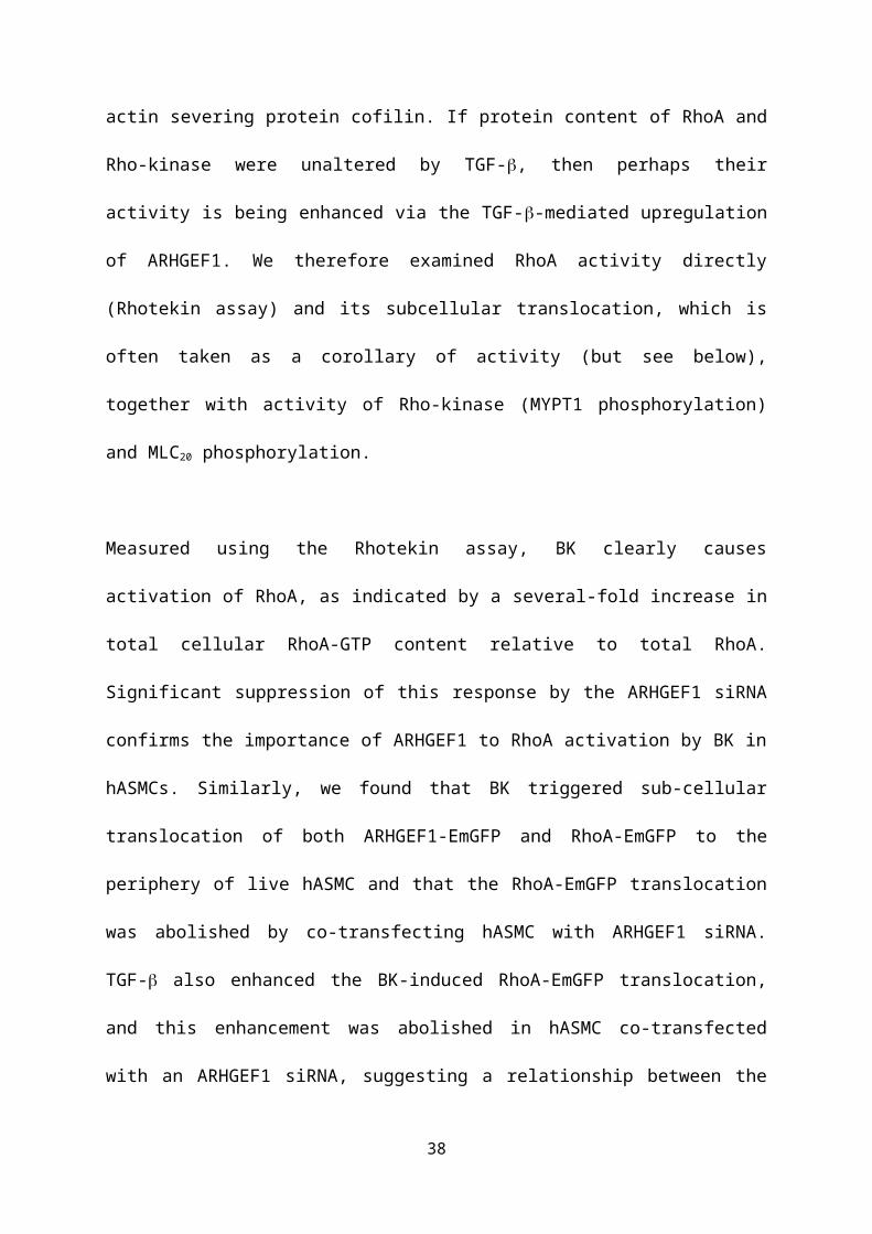
Fig 8
38

Fig 9
39

Fig 10
40

Figure Legends
Figure 1
Effects of TGF- and SrcFK/RhoGEF/Rho-kinase inhibition on BK-induced
contraction. Measurement of isometric tension in isolated rat bronchioles mounted on the
wire myograph. A: Representative traces showing bradykinin (BK) applied cumulatively
(0.01 – 100µM) at 5min intervals, after 18hr pre-incubation in media only or in media with
30ng/ml TGF-. Arrows indicate when the first dose of BK was applied. B-E: Mean
contraction amplitude after TGF- or media pre-incubation measured at the plateau phase of
each BK application, in the absence (B, media: n=24, TGF-: n=23) or presence of 30µM
PP2 (C, media: n=17, TGF-: n=15), 30µM Y16 (D, media: n=9, TGF-: n=9) or 10µM
Y27632 (E, media: n=9, TGF-: n=9). F-G: Non-linear regression data, showing effects of
TGF- and inhibitors on Bmax (F) and PD2 values (G). *P<0.05 vs. control, #P<0.05 vs.
media, 2-way ANOVA.
Figure 2
Effects of BK and TGF- on SrcFK expression and auto-phosphorylation in hASMC.
A: representative blots showing effect of BK (1µM, 30sec) with or without prior exposure to
TGF- (10ng/ml, 24 hrs) on phospho-SrcFK content, total c-Src content and GAPDH as a
loading control. B: data expressed as c-Src/GAPDH shows partial inhibition of c-Src protein
expression after TGF- pre-incubation (#P<0.05 vs. -TGF-, n=7). C-D: SrcFK
phosphorylation is enhanced by BK (*P<0.05, **P<0.01 vs. control, n=7), but suppressed by
TGF- (#P<0.01, ##P<0.01 vs. -TGF- n). All comparisons by 2-way ANOVA.
41

Figure 3
Effects of TGF- and BK on ARHGEF1 expression and translocation. A-C: Effects of
TGF- incubation (10ng/ml, 24 hrs) on protein expression of RhoA (A, n=6), Rho-kinase (B,
ROCK2, n=5) and ARHGEF1 (C, after transfection with scrambled or ARHGEF1 siRNA,
n=9) in hASMC, relative to GAPDH. **P<0.01 vs. -TGF-, ##P<0.01 vs. scrambled siRNA,
2-way ANOVA. D: Fluorescence imaging of live hASMCs transfected with ARHGEF1-
EmGFP. Arrows indicate peripheral regions in which ARHGEF1-EmGFP concentrates when
stimulated by addition of BK (1M). Data shows relative changes in fluorescence in
peripheral regions. **P<0.01 vs. control, n=11, paired t-test.
Figure 4.
Effects of TGF- and ARHGEF1 siRNA on RhoA-GTP content in hASMC. A:
Representative blots showing effects of acute BK treatment (1µM, 30sec) with or without
TGF- pre-treatment (10ng/ml, 24 hrs) in hASMC transfected with either ARHGEF1 siRNA
or a scrambled siRNA control on RhoA-GTP content, total RhoA, or GAPDH as a loading
control. B: Data expressed as total/GAPDH shows no overall effect of BK, TGF- or
ARHGEF1 siRNA on total RhoA expression. C: Data expressed as RhoA-GTP/total shows
significant enhancement by BK (**P<0.01 vs. control) but partial suppression by TGF-
(##P<0.01, vs. –TGF-) and ARHGEF1 siRNA ($P<0.05, $$P<0.01 vs. scrambled siRNA).
n=7 for all data. All comparisons by 3-way ANOVA.
Figure 5
Effects of TGF- and ARHGEF1 siRNA on BK-induced RhoA-EmGFP translocation.
Fluorescence imaging of live hASMCs transfected with RhoA-EmGFP. A: In the absence of
42

TGF- pre-incubation, addition of BK (1M) enhances peripheral fluorescence, as indicated
by arrows. **P<0.001 vs. control, n=16. B: After TGF- pre-incubation (10 ng/ml, 24hrs),
response to BK is enhanced and is concentrated into distinct peripheral spots/patches, as
indicated by arrows. **P<0.001 vs. control, ##P<0.001 vs. -TGF- , n=15. C: In hASMC
pre-treated with TGF- doubly transfected with RhoA-EmGFP and scrambled siRNA,
response to BK is similar to B, as indicated by arrows. **P<0.001 vs. control, n=28. D: In
hASMC pre-treated with TGF- co-transfected with RhoA-EmGFP and ARHGEF1
siRNA, BK does not trigger translocation. ##P<0.001 vs. scrambled siRNA, n=29. All
comparisons by 2-way ANOVA.
Figure 6
Effects of TGF- and ARHGEF1 siRNA on expression and BK-induced
phosphorylation of MYPT-1 in hASMC. A: Representative blots showing effects of acute
BK treatment (1µM, 30sec) with or without TGF- pre-treatment (10ng/ml, 24 hrs) in
hASMC transfected with either ARHGEF1 siRNA, or a scrambled siRNA control, on
phospho-MYPT-1 (Thr-696), total MYPT1, or GAPDH as a loading control. B: Data
expressed as total/GAPDH shows enhancement of MYPT1 protein expression by TGF- pre-
treatment, but no effect of ARHGEF1 siRNA or BK, ##P<0.01 vs. –TGF-. Data expressed as
phospho/total (C) or phospho/GAPDH (D) shows significant enhancement by both BK
(*P<0.05, **P<0.01 vs. control) and TGF- (#P<0.05, ##P<0.01 vs. –TGF-) and suppression
by ARHGEF1 siRNA ($P<0.05, $$P<0.01 vs. scrambled siRNA). n=9 for all data. All
comparisons by 3-way ANOVA.
Figure 7
43

Effects of TGF- and ARHGEF1 siRNA on expression and BK-induced
phosphorylation of MLC20 in hASMC. A: Representative blots showing effects of acute
BK treatment (1µM, 30sec) with or without TGF- pre-treatment (10ng/ml, 24 hrs) in
hASMC transfected with either ARHGEF1 siRNA, or a scrambled siRNA control, on
phospho-MLC20 (Ser19), total MLC20, or GAPDH as a loading control. B: Data expressed as
total/GAPDH shows enhancement of MLC20 protein expression by TGF- pre-treatment
(##P<0.01 vs. –TGF-, and partial suppression of this response by ARHGEF1 siRNA, ($
$P<0.01 vs. scrambled siRNA). Data expressed as phospho/total (C) or phospho/GAPDH (D)
shows significant enhancement by both BK (*P<0.05, **P<0.01 vs. control), effects of TGF-
pre-treatment (#P<0.05, ##P<0.01 vs. –TGF-) and suppression by ARHGEF1 siRNA
($P<0.05, $$P<0.01 vs. scrambled siRNA). n=9 for all data. All comparisons by 3-way
ANOVA.
Figure 8
Effects of BK, TGF- and ARHGEF1 siRNA on cofilin expression and phosphorylation
in hASMC. A: Representative blots showing effects of acute BK treatment (1µM, 30sec)
with or without TGF- pre-treatment (10ng/ml, 24 hrs) in hASMC transfected with either
ARHGEF1 siRNA, or a scrambled siRNA control, on phospho-cofilin (Ser3), total cofilin, or
GAPDH as a loading control. B: Data expressed as total/GAPDH shows enhancement of
cofilin protein expression by TGF- pre-treatment (##P<0.01 vs. –TGF-, and partial
suppression of this response by ARHGEF1 siRNA, ($$P<0.01 vs. scrambled siRNA). Data
expressed as phospho/total (C) or phospho/GAPDH (D) shows significant de-
phosphorylation induced by BK (**P<0.01 vs. control), prevention of
de-phosphorylation/enhancement of basal phosphorylation by TGF- pre-treatment (#P<0.05,
44

##P<0.01 vs. –TGF-) and partial suppression of the effects of TGF- by ARHGEF1 siRNA
($$P<0.01 vs. scrambled siRNA). n=7 for all data. All comparisons by 3-way ANOVA.
Figure 9.
Effects of asthma and OVA-sensitization on lung ARHGEF-1 protein expression.
Protein expression of ARHGEF-1 relative to GAPDH. Expression is enhanced by OVA-
sensitization in mouse lung (A, *P<0.05 vs. sham-treated, n=3 in each group) and is greater
in ASM of asthmatic subjects vs. healthy controls (B, *P<0.05, n=3 subjects in each group).
All comparisons by un-paired t-test.
Figure 10.
Role of ARHGEF1, SrcFK, RhoA, Rho-kinase and cofilin in BK-induced contraction in
ASM and the influence of TGF- on their relative contributions. Without TGF-
treatment: It is established that GPCR-induced ASM contraction is dependent on sequential
activation of RhoA and Rho-kinase, resulting in MYPT1 phosphorylation, MLCP inhibition
and enhanced MLC20 phosphorylation. We show that BK also induces SrcFK activation and
translocation of ARHGEF1. RhoA translocation and activation, MYPT1 and MLC20
phosphorylation are all partially ARHGEF1-dependent. Also, contraction is partially SrcFK,
RhoGEF and Rho-kinase-dependent. SrcFK possibly act upstream of ARHGEF1 (but not
tested in this study). BK inhibits cofilin phosphorylation, but how this influences contraction
remains to be determined. After TGF- treatment : Expression and BK-induced activity of
c-Src are suppressed, while expression of ARHGEF1, MYPT1, MLC20 and cofilin are
enhanced. BK-induced RhoA activity is partially suppressed, while RhoA translocation,
45

MYPT-1 phosphorylation, MLC20 phosphorylation and contraction are all enhanced.
Enhancement of RhoA translocation and MYPT1/MLC20 phosphorylation are partially
ARHGEF1-dependent, while enhanced contraction is Rho-kinase and RhoGEF-dependent.
Enhanced Rho-kinase activity may result from enhanced site-specific RhoA translocation, to
counter the reduced total RhoA activity, or from activation of another Rho-protein (both
untested in this study). Cofilin phosphorylation is enhanced, but is largely independent of
ARHGEF1 and whether this contributes to enhanced contraction remains to be determined.
Key: Size of text/box reflects degree of protein expression. Size of ‘P’ reflects degree of
phosphorylation. Thickness of lines/arrows reflects strength of effect. The row of block
arrows represents translocation. (?) or (±?) denotes ‘not tested in this study’.
Reference List
Albinsson S, Nordstrom I, & Hellstrand P (2004). Stretch of the vascular wall induces smooth muscle differentiation by promoting actin polymerization. J Biol Chem 279, 34849-34855.
Andrianantoandro E & Pollard TD (2006). Mechanism of actin filament turnover by severing and nucleation at different concentrations of ADF/cofilin. Mol Cell 24, 13-23.
Arber S, Barbayannis FA, Hanser H, Schneider C, Stanyon CA, Bernard O, & Caroni P (1998). Regulation of actin dynamics through phosphorylation of cofilin by LIM-kinase. Nature 393, 805-809.
Atfi A, Drobetsky E, Boissonneault M, Chapdelaine A, & Chevalier S (1994). Transforming growth factor beta down-regulates Src family protein tyrosine kinase signaling pathways. J Biol Chem 269, 30688-30693.
Birmingham A, Anderson E, Sullivan K, Reynolds A, Boese Q, Leake D, Karpilow J, & Khvorova A (2007). A protocol for designing siRNAs with high functionality and specificity. Nat Protoc 2, 2068-2078.
Bos JL, Rehmann H, & Wittinghofer A (2007). GEFs and GAPs: critical elements in the control of small G proteins. Cell 129, 865-877.
46

Brown JP, Taube C, Miyahara N, Koya T, Pelanda R, Gelfand EW, & Torres RM (2007). Arhgef1 is required by T cells for the development of airway hyperreactivity and inflammation. Am J Respir Crit Care Med 176, 10-19.
Chiba Y, Takeyama H, Sakai H, & Misawa M (2001). Effects of Y-27632 on acetylcholine-induced contraction of intact and permeabilized intrapulmonary bronchial smooth muscles in rats. Eur J Pharmacol 427, 77-82.
Chiba Y, Uchida T, Sakai H, Oku T, Itoh S, Tsuji T, & Misawa M (2004). Acetylcholine-induced translocation of RhoA in freshly isolated single smooth muscle cells of rat bronchi. J Pharmacol Sci 95, 479-482.
Chiba Y, Ueno A, Shinozaki K, Takeyama H, Nakazawa S, Sakai H, & Misawa M (2005). Involvement of RhoA-mediated Ca2+ sensitization in antigen-induced bronchial smooth muscle hyperresponsiveness in mice. Respir Res 6, 4.
Dai YP, Bongalon S, Mutafova-Yambolieva VN, & Yamboliev IA (2008). Distinct effects of contraction agonists on the phosphorylation state of cofilin in pulmonary artery smooth muscle. Adv Pharmacol Sci 2008, 362741.
Doeing DC & Solway J (2013). Airway smooth muscle in the pathophysiology and treatment of asthma. J Appl Physiol (1985 ) 114, 834-843.
Feng J, Ito M, Ichikawa K, Isaka N, Nishikawa M, Hartshorne DJ, & Nakano T (1999). Inhibitory phosphorylation site for Rho-associated kinase on smooth muscle myosin phosphatase. J Biol Chem 274, 37385-37390.
Fukuda K, Kawata S, Tamura S, Matsuda Y, Inui Y, Igura T, Inoue S, Kudara T, & Matsuzawa Y (1998). Transforming growth factor-beta1-induced degradation of activated Src tyrosine kinase in rat fibroblasts. Oncogene 16, 3349-3356.
Goldsmith AM, Bentley JK, Zhou L, Jia Y, Bitar KN, Fingar DC, & Hershenson MB (2006). Transforming growth factor-beta induces airway smooth muscle hypertrophy. Am J Respir Cell Mol Biol 34, 247-254.
Guilluy C, Bregeon J, Toumaniantz G, Rolli-Derkinderen M, Retailleau K, Loufrani L, Henrion D, Scalbert E, Bril A, Torres RM, Offermanns S, Pacaud P, & Loirand G (2010). The Rho exchange factor Arhgef1 mediates the effects of angiotensin II on vascular tone and blood pressure. Nat Med 16, 183-190.
Halayko AJ, Camoretti-Mercado B, Forsythe SM, Vieira JE, Mitchell RW, Wylam ME, Hershenson MB, & Solway J (1999). Divergent differentiation paths in airway smooth muscle culture: induction of functionally contractile myocytes. Am J Physiol 276, L197-L206.
47

Halwani R, Al-Muhsen S, Al-Jahdali H, & Hamid Q (2011). Role of transforming growth factor-beta in airway remodeling in asthma. Am J Respir Cell Mol Biol 44, 127-133.
Jaiswal M, Gremer L, Dvorsky R, Haeusler LC, Cirstea IC, Uhlenbrock K, & Ahmadian MR (2011). Mechanistic insights into specificity, activity, and regulatory elements of the regulator of G-protein signaling (RGS)-containing Rho-specific guanine nucleotide exchange factors (GEFs) p115, PDZ-RhoGEF (PRG), and leukemia-associated RhoGEF (LARG). J Biol Chem 286, 18202-18212.
Ji H, Tang H, Lin H, Mao J, Gao L, Liu J, & Wu T (2014). Rho/Rock cross-talks with transforming growth factor-beta/Smad pathway participates in lung fibroblast-myofibroblast differentiation. Biomed Rep 2, 787-792.
Katsumoto TR, Kudo M, Chen C, Sundaram A, Callahan EC, Zhu JW, Lin J, Rosen CE, Manz BN, Lee JW, Matthay MA, Huang X, Sheppard D, & Weiss A (2013). The phosphatase CD148 promotes airway hyperresponsiveness through SRC family kinases. J Clin Invest 123, 2037-2048.
Kim JH, Jain D, Tliba O, Yang B, Jester WF, Jr., Panettieri RA, Jr., Amrani Y, & Pure E (2005). TGF-beta potentiates airway smooth muscle responsiveness to bradykinin. Am J Physiol Lung Cell Mol Physiol 289, L511-L520.
Knock GA, Snetkov VA, Shaifta Y, Drndarski S, Ward JP, & Aaronson PI (2008). Role of src-family kinases in hypoxic vasoconstriction of rat pulmonary artery. Cardiovasc Res 80, 453-462.
Leduc C, Sobilo L, Toumi H, Mondon P, Lespessailles E, Ossant F, Kurfurst R, & Pichon C (2016). TGF-beta-induced early gene-1 overexpression promotes oxidative stress protection and actin cytoskeleton rearrangement in human skin fibroblasts. Biochim Biophys Acta 1860, 1071-1078.
Matsunaga K, Yanagisawa S, Ichikawa T, Ueshima K, Akamatsu K, Hirano T, Nakanishi M, Yamagata T, Minakata Y, & Ichinose M (2006). Airway cytokine expression measured by means of protein array in exhaled breath condensate: correlation with physiologic properties in asthmatic patients. J Allergy Clin Immunol 118, 84-90.
McMillan SJ, Xanthou G, & Lloyd CM (2005). Manipulation of allergen-induced airway remodeling by treatment with anti-TGF-beta antibody: effect on the Smad signaling pathway. J Immunol 174, 5774-5780.
Meyer BH, Freuler F, Guerini D, & Siehler S (2008). Reversible translocation of p115-RhoGEF by G(12/13)-coupled receptors. J Cell Biochem 104, 1660-1670.
Moir LM, Ward JP, & Hirst SJ (2003). Contractility and phenotype of human bronchiole smooth muscle after prolonged fetal bovine serum exposure. Exp Lung Res 29, 339-359.
48

Oenema TA, Maarsingh H, Smit M, Groothuis GM, Meurs H, & Gosens R (2013). Bronchoconstriction Induces TGF-beta Release and Airway Remodelling in Guinea Pig Lung Slices. PLoS One 8, e65580.
Oenema TA, Smit M, Smedinga L, Racke K, Halayko AJ, Meurs H, & Gosens R (2012). Muscarinic receptor stimulation augments TGF-beta1-induced contractile protein expression by airway smooth muscle cells. Am J Physiol Lung Cell Mol Physiol 303, L589-L597.
Papadimitriou E, Vasilaki E, Vorvis C, Iliopoulos D, Moustakas A, Kardassis D, & Stournaras C (2012). Differential regulation of the two RhoA-specific GEF isoforms Net1/Net1A by TGF-beta and miR-24: role in epithelial-to-mesenchymal transition. Oncogene 31, 2862-2875.
Redington AE, Madden J, Frew AJ, Djukanovic R, Roche WR, Holgate ST, & Howarth PH (1997). Transforming growth factor-beta 1 in asthma. Measurement in bronchoalveolar lavage fluid. Am J Respir Crit Care Med 156, 642-647.
Rossi JL, Ralay RH, Patel F, Chrzaszcz M, Venkatesan C, & Wainwright MS (2011). Albumin causes increased myosin light chain kinase expression in astrocytes via p38 mitogen-activated protein kinase. J Neurosci Res 89, 852-861.
Samarakoon R, Higgins SP, Higgins CE, & Higgins PJ (2008). TGF-beta1-induced plasminogen activator inhibitor-1 expression in vascular smooth muscle cells requires pp60(c-src)/EGFR(Y845) and Rho/ROCK signaling. J Mol Cell Cardiol 44, 527-538.
Sandbo N, Lau A, Kach J, Ngam C, Yau D, & Dulin NO (2011). Delayed stress fiber formation mediates pulmonary myofibroblast differentiation in response to TGF-beta. Am J Physiol Lung Cell Mol Physiol 301, L656-L666.
Schaafsma D, Bos IS, Zuidhof AB, Zaagsma J, & Meurs H (2006). Inhalation of the Rho-kinase inhibitor Y-27632 reverses allergen-induced airway hyperresponsiveness after the early and late asthmatic reaction. Respir Res 7, 121.
Schaafsma D, Gosens R, Zaagsma J, Halayko AJ, & Meurs H (2008). Rho kinase inhibitors: a novel therapeutical intervention in asthma? Eur J Pharmacol 585, 398-406.
Semizarov D, Frost L, Sarthy A, Kroeger P, Halbert DN, & Fesik SW (2003). Specificity of short interfering RNA determined through gene expression signatures. Proc Natl Acad Sci U S A 100, 6347-6352.
Shaifta Y, Irechukwu N, Prieto-Lloret J, MacKay CE, Marchon KA, Ward JP, & Knock GA (2015). Divergent modulation of Rho-kinase and Ca(2+) influx pathways by Src family kinases and focal adhesion kinase in airway smooth muscle. Br J Pharmacol 172, 5265-5280.
49

Shang X, Marchioni F, Evelyn CR, Sipes N, Zhou X, Seibel W, Wortman M, & Zheng Y (2013). Small-molecule inhibitors targeting G-protein-coupled Rho guanine nucleotide exchange factors. Proc Natl Acad Sci U S A 110, 3155-3160.
Somlyo AP & Somlyo AV (2003). Ca2+ sensitivity of smooth muscle and nonmuscle myosin II: modulated by G proteins, kinases, and myosin phosphatase. Physiol Rev 83, 1325-1358.
Torrego A, Hew M, Oates T, Sukkar M, & Fan CK (2007). Expression and activation of TGF-beta isoforms in acute allergen-induced remodelling in asthma. Thorax 62, 307-313.
Tsapara A, Luthert P, Greenwood J, Hill CS, Matter K, & Balda MS (2010). The RhoA activator GEF-H1/Lfc is a transforming growth factor-beta target gene and effector that regulates alpha-smooth muscle actin expression and cell migration. Mol Biol Cell 21, 860-870.
Vardouli L, Moustakas A, & Stournaras C (2005). LIM-kinase 2 and cofilin phosphorylation mediate actin cytoskeleton reorganization induced by transforming growth factor-beta. J Biol Chem 280, 11448-11457.
Vasilaki E, Papadimitriou E, Tajadura V, Ridley AJ, Stournaras C, & Kardassis D (2010). Transcriptional regulation of the small GTPase RhoB gene by TGF{beta}-induced signaling pathways. FASEB J 24, 891-905.
Zeidan A, Paylor B, Steinhoff KJ, Javadov S, Rajapurohitam V, Chakrabarti S, & Karmazyn M (2007). Actin cytoskeleton dynamics promotes leptin-induced vascular smooth muscle hypertrophy via RhoA/. J Pharmacol Exp Ther 322, 1110-1116.
Zhang W, Du L, & Gunst SJ (2010). The effects of the small GTPase RhoA on the muscarinic contraction of airway smooth muscle result from its role in regulating actin polymerization. Am J Physiol Cell Physiol 299, C298-C306.
Zhang W, Huang Y, Wu Y, & Gunst SJ (2015). A novel role for RhoA GTPase in the regulation of airway smooth muscle contraction. Can J Physiol Pharmacol 93, 129-136.
Zhao R, Du L, Huang Y, Wu Y, & Gunst SJ (2008). Actin depolymerization factor/cofilin activation regulates actin polymerization and tension development in canine tracheal smooth muscle. J Biol Chem 283, 36522-36531.
Zhao XH, Laschinger C, Arora P, Szaszi K, Kapus A, & McCulloch CA (2007). Force activates smooth muscle alpha-actin promoter activity through the Rho signaling pathway. J Cell Sci 120, 1801-1809.
50