Embed Size (px)
Citation preview

LIVER ABSCESS

Liver - most subject to abscess formation Solitary or multiple Arise from
◦ hematogenous spread of bacteria ◦ local spread from contiguous sites of infection
within the peritoneal cavity Most common source- associated disease
of the biliary tract
Liver Abscess
Harrison’s Principles of Internal Medicine, 17th ed

Primary Infection from other sites (Biliary tree, Peritoneal Cavity, Pelvis)
Transmission via Portal vein, arterial supply, biliary tract, direct invasion
Secondary Infection of Liver and Abscess Formation
Pathogenesis

The right hepatic lobe is affected more often than the left hepatic lobe by a factor of 2:1.
Bilateral involvement is seen in 5% of cases.
The predilection for the right hepatic lobe can be attributed to anatomic considerations.
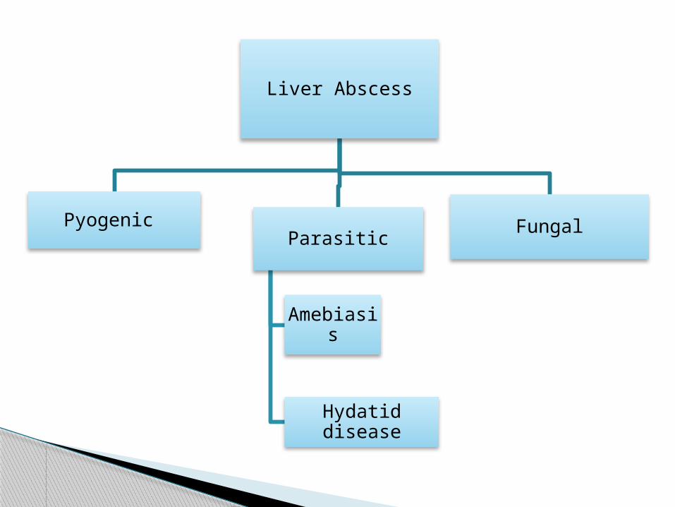
Liver Abscess
Pyogenic Parasitic
Amebiasis
Hydatid disease
Fungal

Liver is probably exposed to portal venous bacterial loads on a regular basis
Inoculum of bacteria exceeds the liver's ability to clear it Abscess
Potential routes of hepatic exposure to bacteria: ◦ Biliary tree ◦ Portal vein ◦ Hepatic artery ◦ Direct extension of a
nearby focus of infection ◦ Trauma
Pyogenic Liver Abscess
Sabiston Textbook of Surgery, 18th ed.

Etiology: Ascending cholangitis
◦ Enteric Gram Negative aerobic Bacilli and Enterococci
Infection from the pelvis and other intraperitoneal sources◦ Mixed infection with aerobic and anaerobic
species is common◦ Bacteroides fragilis- species most frequently
isolated Hematogenous spread- S. aureus, S. milleri
Harrison’s Principles of Internal Medicine, 17th ed

• Extraintestinal infection by E. histolytica• Trophozoites invade veins to reach the liver
through the portal venous system • Travelers of endemic areas - more
susceptible • Young patients- present w/ acute phase with
symptoms of <10 days duration• Older patients - subacute course of 6
months with weight loss and hepatomegaly
Amebic Liver Abscess
Harrison’s Principles of Internal Medicine, 17th ed

CLINICAL FEATURES AMEBIC ABSCESS PYOGENIC ABSCESS
Age (yr) 20-40 >50
Male-to-female ratio ≥10:1 1.5:1
Solitary vs. multiple Solitary 80%[*] Solitary 50%
Location Usually right liver Usually right liver
Travel in endemic area Yes No
Diabetes Uncommon (∼2%) More common (∼27%)
Alcohol use Common Common
Jaundice Uncommon Common
Elevated bilirubin Uncommon Common
Elevated alkaline phosphatase
Common Common
Positive blood culture No Common
Positive amebic serology
Yes No
Table 52-5 -- Features of Amebic Versus Pyogenic Liver Abscess
Sabiston Textbook of Surgery, 18th ed.

caused by the larval/cyst stage of Echinococcus granulosus, in which humans are an intermediate host
In the human duodenum, the parasitic embryo releases an oncosphere containing hooklets that penetrate the mucosa, allowing access to the bloodstream
In the blood, the oncosphere reaches the liver (most commonly) or lungs, where the parasite develops its larval stage known as the hydatid cyst
Hydatid Disease
Sabiston Textbook of Surgery, 18th ed.

Candida spp. Follow fungemia in patients receiving
chemotherapy from cancer Often present when PMNs return after a
period of neutropenia
Fungal Liver Abscess
Harrison’s Principles of Internal Medicine, 17th ed

• Fever - most common presenting sign • Pain, guarding, punch and rebound
tenderness localized to the right upper quadrant *
• Hepatomegaly *• Jaundice *Non-specific symptoms: • Chills• Anorexia • Vomiting
CLINICAL FEATURES
Harrison’s Principles of Internal Medicine, 17th ed

Patient Liver Abscess
Vague RUQ pain – 3 months RUQ pain
Low-grade fever Fever – most common presenting sign
Weight loss Weight loss in older patients with a chronic subacute course
Past Medical History•PTB•Acute Viral Hepatitis
Biliary tract diseaseRuptured appendicitisPylephlebitis
Personal, Family History• Smoker• Half a bottle of gin everyday since age 30
• Mother died of HCC
Travel to an endemic area
PE findings•Pale palpebral conjunctivae•Icteric sclerae•Spider angiomas, palmar erythema•Slightly distended abdomen•Liver palpable with a span of 14cm, tender, nodular
JaundiceTenderness over the liverHepatomegaly

DIAGNOSIS

DIAGNOSIS Laboratory work-up Amebic serologic testing (positive in 95% of
cases) ELISA test for Echinoccocal antigens ( positive for
85% of infected patients) Imaging studies
◦ Ultrasound◦ CT scan

LABORATORY FINDINGS
Elevated serum concentration of Alkaline Phosphatase
• Single most reliable laboratory finding• Documented in 70% of patients with liver abscesses
Other tests of liver function may yield normal results
• 50% of patients have elevated serum levels of bilirubin• 48% have elevated concentrations of aspartate aminotransferase
Other laboratory findings
• Leukocytosis in 77% of patients• Anemia (usually normochromic, normocytic) in 50%• Hypoalbuminemia in 33%
Concomitant bacteremia is found in one-third of patients
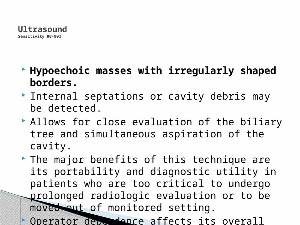
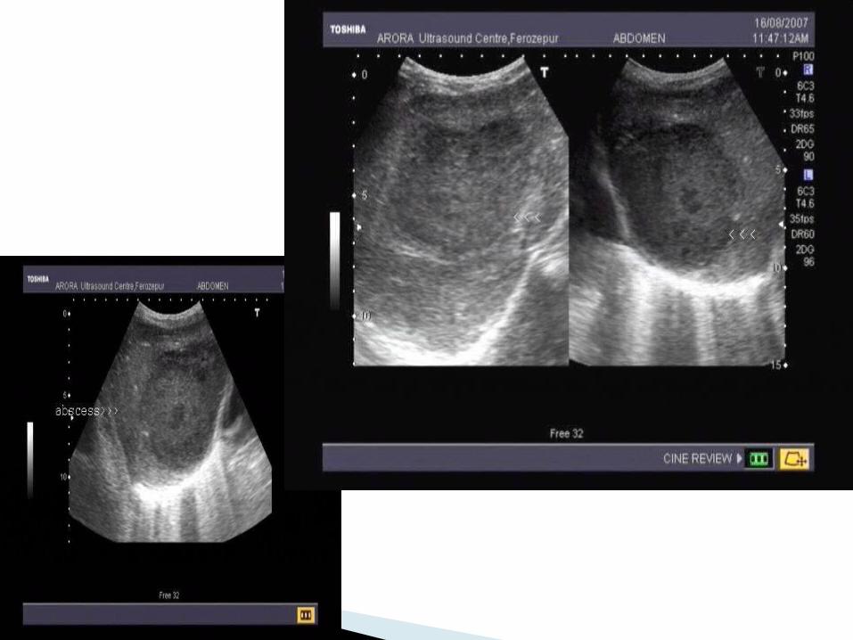
UltrasoundSensitivity 80-90%
Hypoechoic masses with irregularly shaped borders.
Internal septations or cavity debris may be detected.
Allows for close evaluation of the biliary tree and simultaneous aspiration of the cavity.
The major benefits of this technique are its portability and diagnostic utility in patients who are too critical to undergo prolonged radiologic evaluation or to be moved out of monitored setting.
Operator dependence affects its overall sensitivity.

Computed Tomographic Scan(Sensitivity 95%-100%) Well-demarcated areas hypodense to the
surrounding hepatic parenchyma. Peripheral enhancement is seen when IV
contrast is administered. Gas can be seen in as many as 20% of lesions. CT scan is superior in its ability to detect lesions
less than 1 cm. This technique also enables the evaluation for an
underlying concurrent pathology throughout the abdomen and pelvis. Indium-labeled WBC scans are somewhat more sensitive in this regard.


CT examination: Unenhanced axial scan: Round-shaped, hypodense masses
of 5-6 cm of diameter, with isodense wall, are visible in both liver lobes (arrows). A small amount of hypodense fluid is
observed within the liver capsule

CT examination: Postcontrast axial scan
The irregular hypodens lesions of variable sizes (arrows) are better
visualized in the contrast-enhancing liver parenchyma.

Chest X-ray
Basilar atelectasis Right hemidiaphragm elevation Right pleural effusion are present in
approximately 50% of cases Before advancements in radiologic
technique, these served as diagnostic clues.


MANAGEMENT

Drainage, either percutaneous or surgical, is the mainstay of therapy for intraabdominal abscess◦ Percutaneous needle aspiration◦ Percutaneous catheter drainage◦ Surgical drainage (open or laparoscopic)◦ Medical therapy

Percutaneous needle aspiration
Solitary dominant abscess Under CT scan or ultrasound guidance, needle
aspiration of cavity material can be performed. Needle aspiration enables rapid recovery of
material for microbiologic and pathologic evaluation.◦ Gram’s stain and culture
Needle aspiration can be performed with the initial diagnostic procedure.


Percutaneous catheter drainage
• Complex abscess or an abscess containing particularly thick fluid
• Small cysts A catheter is placed under ultrasound or CT guidance using
the Seldinger technique The catheter is flushed daily until output is less than 10
cc/d or cavity collapse is documented by serial CT scanning.
Multiple abscesses have been drained successfully by this method.
Failure to respond to catheter drainage is the main reported complication and is also an indication for surgical intervention.

Surgical drainage• Was the standard of care until the introduction of
percutaneous drainage techniques in the mid 1970s• For cysts greater than 5 cm • Ruptured cysts• Multiloculated cysts• Failure of percutaneous drianage
Lack of response in 4-7 days

Medical Therapy Diagnostic aspirate of abscess should be
obtained before initiation of empirical therapy◦ Empiric drug therapy – covering gram negative
aerobic, facultative and anaerobic organisms◦ Adjusted to specific antibiotic when results for
Gram’s stain and culture become available
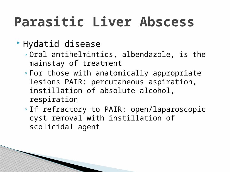
Parasitic Liver Abscess Hydatid disease
◦ Oral antihelmintics, albendazole, is the mainstay of treatment
◦ For those with anatomically appropriate lesions PAIR: percutaneous aspiration, instillation of absolute alcohol, respiration
◦ If refractory to PAIR: open/laparoscopic cyst removal with instillation of scolicidal agent

Parasitic Liver Abscess Amebiasis
◦ Metronidazole for at least 1 week◦ Most patients will respond rapidly with complete
defervescence within 3 days. ◦ Aspiration of the abscess is rarely necessary and
should be avoided, except in patients in whom secondary infection from pyogenic organisms is suspected.

THANK YOU


![PRIMARY DISTAL ESOPHAGEAL MALIGNANCY PRESENTING AS … · adjacent primaries, a hematogenous pathway and lymphatic route for metastatic spread to the thyroid have been suggested [8]](https://img.dokumen.tips/doc/110x75/5e53ac8c30394239ea461534/primary-distal-esophageal-malignancy-presenting-as-adjacent-primaries-a-hematogenous.jpg)