Embed Size (px)
Citation preview
1
財團法人明日醫學基金會專題研究計畫申請書
一、基本資料: 申請條碼:
本申請案所需經費(單選) ■A 類(執行計畫所需經費)
B類(研究主持費,限人文處計畫,不須填寫表 C002 及 C004 至 C009)
計 畫 類 別 ( 單 選 ) ■一般型研究計畫
新進人員研究計畫
特約研究計畫
其他
研 究 型 別 ■個別型計畫 整合型計畫
申請機構/系所(單位) 長庚大學/醫學院微生物及免疫學科
本計畫主持人姓名 賴志河 職 稱 教授 身 分 證 號 碼
本計畫名稱
中 文 探討降血脂藥物對於抑制胃癌之機制
英 文 The mechanism of cholesterol-lowering agents in the inhibition of gastric cancer
整合型總計畫名稱
整合型總計畫主持人 身 分 證 號 碼
全 程 執 行 期 限 自民國 105 年 1 月 1 日起至民國 105 年 12 月 31 日
研究學門(請參考本申
請書所附之學門專長
分 類 表 填 寫 )
學 門 代 碼 名 稱(如為其他類,請自行填寫學門)
研 究 性 質 █純基礎研究 導向性基礎研究 應用研究 技術發展
本計畫是否為國際合作計畫 █否; 是,合作國家: ,請加填表 I001~I003
本計畫是否申請海洋研究船 █否; 是,請務必填寫表 C014。
計 畫 連 絡 人 姓名: 賴志河 電話:(公) 03-2218800 ext. 5116 (宅/手機) 0937-936212
通 訊 地 址 桃園市龜山區文化一路 259 號
傳 真 號 碼 03-2118700 E-MAIL [email protected]

2
二、申請補助經費: 金額單位:新台幣元
執行年次
補助項目
第一年
(105年 1月~
105年 12月)
第二年
(___年___月~
___年___月)
第三年
(___年___月~
___年___月)
第四年
(___年___月~
___年___月)
第五年
(___年___月~
___年___月)
業 務 費 300,000
研 究 人 力 費
耗材、物品及雜項費用
國際合作研究計畫國外學 者 來 臺 費 用
研 究 設 備 費
國 外 差 旅 費
國外或大陸地區差旅費
出席國際學術會議差旅費
國際合作研究計畫
出國差旅費
管 理 費
合 計 300,000
貴重儀器中心使用額度
博士後研究
國 內 、 外 地 區
共 名 共 名 共 名 共 名 共 名
大 陸 地 區 共 名 共 名 共 名 共 名 共 名
申請機構或其他單位(含產業界)提供之配合項目(無配合補助項目者免填)
配 合 單 位 名 稱 配合補助項目 配合補助金額 配 合 年 次 證明文件

3
三、主要研究人力:
(一)請依照「主持人」、「共同主持人」、「協同研究人員」及「博士後研究」等類別
之順序分別填寫。
類 別 姓名 服務機構/系所 職稱 在本研究計畫內擔任之具
體工作性質、項目及範圍
*每週平均投入
工作時數比率 (%)
主持人 賴志河 長庚大學/微生
物及免疫學科
教授 統籌及推動本研究計畫、參與主
要實驗工作、整理文獻背景
及撰寫研究成果
70%
※註:每週平均投入工作時數比率係填寫每人每週平均投入本計畫工作時數佔其每週全部
工作時間之比率,以百分比表示(例如:50%即表示該研究人員每週投入本計畫研
究工作之時數佔其每週全部工時之百分五十)。
(二)如申請博士後研究,請另填表 CIF2101及 CIF2102(若已有人選者,請務必填註人選
姓名,並將其個人資料表併同本計畫書送本會)。
4
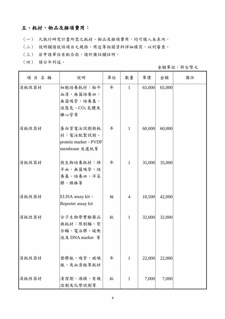
五、耗材、物品及雜項費用:
(一) 凡執行研究計畫所需之耗材、物品及雜項費用,均可填入本表內。
(二) 說明欄請就該項目之規格、用途等相關資料詳細填寫,以利審查。
(三) 若申請單位有配合款,請於備註欄註明。
(四) 請分年列述。 金額單位:新台幣元
項 目 名 稱 說明 單位 數量 單價 金額 備註
消耗性器材
消耗性器材
消耗性器材
消耗性器材
消耗性器材
消耗性器材
消耗性器材
細胞培養耗材︰胎牛
血清、無菌培養皿、
無菌吸管、培養基、
液態氮、CO2氣體及
離心管等
蛋白質電泳試劑與耗
材︰電泳配製試劑、
protein marker、PVDF
membrane 及濾紙等
微生物培養耗材︰綿
羊血、無菌吸管、培
養基、培養皿、洋菜
膠、棉棒等
ELISA assay kit、
Reporter assay kit
分子生物學實驗藥品
與耗材︰限制酶、聚
合酶、電泳膠、緩衝
液及 DNA marker 等
塑膠瓶、吸管、玻璃
瓶、及血清瓶等耗材
清潔劑、酒精、有機
溶劑及化學試劑等
年
年
年
組
批
年
批
1
1
1
4
1
1
1
65,000
60,000
35,000
10,500
32,000
22,000
7,000
65,000
60,000
35,000
42,000
32,000
22,000
7,000

5
資訊設備費
論文投稿費
碳粉夾、紙張、光碟
片等資訊耗材
期刊雜誌投稿
年
篇
1
2
8,000
15,000
8,000
30,000
合 計 300,000

6
Abstract
Gastric cancer is the second leading cause of cancer-related deaths in the world.
Cholesterol-enriched microdomains (also called lipid rafts), which provide platforms for signaling,
are thought to be associated with H. pylori-induced pathogenesis and thus lead to gastric cancer
progression. Several population-based case-control studies demonstrated that patients treated with
statins, which inhibited 3-hydroxy-3-methyl glutaryl coenzyme A (HMG-CoA) reductase, and
reduced the risk for several types of cancers. Therefore, we sought to investigate the molecular
mechanism of H. pylori hijacks membrane cholesterol and novel approach to eradicate H.
pylori-related gastric cancer cells. Accordingly, we will analyze the data from National Health
Insurance Research Database (NHIRD) and explore whether prescription of statins prevents gastric
carcinogenesis and gastritis in H. pylori-infected patients. Moreover, we will investigate the role of
cholesterol in H. pylori virulence factor-induced pathogenesis and how statins reduce H.
pylori-induced gastric disorders. The results from this study will not only promote the basic and
clinical-collaborations but also elucidate the scientific issues related to H. pylori pathogenesis and
explore statins to therapy the gastric cancer.
Keywords: Helicobacter pylori, cytotoxin-associated gene A, gastric cancer, statin

7
A. BACKGROUND AND SIGNIFICANCE
Involvement of cholesterol in microbial infection
Lipid rafts are not only a dynamic structure on cell membrane, but also provide an amplified
signaling for the activation of the cells [1]. Several studies have demonstrated that lipid rafts
might serve as platforms for entry portals of pathogens, including bacteria [2-5] and viruses [6-8].
There are two possible benefits for pathogens entry through rafts: one is prevention of
intracellular degradation, and the other is triggering of signaling that causes membrane fission and
cytoskeleton rearrangement, which are both required for infection of bacteria [9]. Therefore,
suggesting that pathogens might be favor to interact with lipid rafts where provide potential
gateways to enter host cells.
Infection of H. pylori
H. pylori can evade host immune responses by utilizing its particular strategies to manipulate
immune cells in harsh environment of the stomach [10,11]. Additionally, H. pylori penetrate
across the mucosal layer and that may enable the bacteria to survive in the gastric epithelial cells
[12]. Numerous reports have focused on identification of H. pylori virulence factors involved in
its pathogenesis and the underlying mechanisms that lead to different clinical sequelea in a
specific host niche [13-15]. It is interesting that mounting evidence suggested that H. pylori
exploits cholesterol-rich microdomains (also called lipid rafts) for their internalization of cells, as
many other pathogens.
Cholesterol-rich microdomains
The major composition of lipid rafts includes cholesterol, sphingolipids, and phospholipids
which interact tightly and create rigid microdomains in the cell membrane [16]. The structure of
lipid rafts is known to be stabilized in the cold in non-ionic detergents such as Triton X-100 [17].
After treatment of membrane with cold Triton X-100, insoluble components including lipids and
proteins remain in the composition of detergent-resistant membrane (DRM), that were considered
to be in the lipid rafts [17]. Several raft usurping or disruption agents including
methyl-β-cyclodextrin (MβCD), filipin, lovastatin, and nystatin have been extensively employed
in the investigation of their particular functions and compositions [18]. After depletion of
membrane cholesterol by MβCD or filipin, the raft-associated proteins and lipids can be
dissociated and rendered the structure becoming nonfunctional [19].
H. pylori virulence factors
H. pylori contains a set of virulence factors that enable it to survive, multiply, escape from
immune surveillance, and eventually lead to persistent infection in a particular niche of host.
Although gastric mucosa is well protected against other bacterial infection, H. pylori is highly
adapted to its ecological niche. These fashions that support the colonization and persistence of H.

8
pylori in the gastric mucus including polar flagella, urease, adhesins, and two major virulence
factors: vacuolating cytotoxin A (VacA) and cytotoxin-associated gene A (CagA) [20]. In addition
to VacA and CagA, an important study by Wunder et al. who revealed that the H. pylori enzyme,
cholesterol-α-glucosyltransferase, which is responsible for cholesterol glucosylation in
macrophages and is thought to be modulated the innate immunity [10].
H. pylori hijacks membrane cholesterol for their benefits
VacA was the first toxin that reported to utilize raft-microdomains for its assembly on the
cell membrane and intracellular delivery [21]. Several studies revealed that depletion of
membrane cholesterol significantly reduces the entry of VacA into target cells [21-23]. Our
previous study had been found that GPI-anchor protein, fasciclin I, was required for
internalization of VacA, but did not affect the binding of VacA to lipid rafts [23]. It has also
demonstrated that receptor-dependent translocation of VacA to lipid rafts is critical for signaling
pathways leading to p38 MAP kinase/ATF-2 activation and vacuolation [24]. We further showed
that depletion of cholesterol and mutation of VacA significantly reduced H. pylori internalization
in gastric epithelial cells [4]. Disruption of lipid rafts attenuates CagA translocation, hummingbird
phenotype, and IL-8 secretion, suggesting that the delivery of CagA into epithelial cells is
mediated through a cholesterol-dependent manner [4]. Recently, Murata-Kamiya et al. reported
that the initial contact of H. pylori with cells induced the phosphotidylserine externalization from
inner leaflet to outer leaflet of cell membrane, thus facilitated the translocation of CagA into
cytoplasm [25]. Moreover, the CagA C-terminal domain-containing EPIYA regions directly
targeted to lipid rafts of gastric epithelial cells was further determined by our recent study [26].
Wunder et al. have been showed that H. pylori follow a cholesterol gradient and extracts the
lipid from cytoplasmic membranes of epithelial cells for subsequent glucosylation [10].
Subsequently, this group identified the gene HP0421 (capJ) as encoding the enzyme
cholesterol-- glucosyltransferase responsible for cholesterol glucosylation [27]. These evidences
suggested that H. pylori harbor a delicate mechanism for the orchestration between activating
macrophages and protecting the bacteria from immune attack. However, the association of H.
pylori exploits lipid rafts and triggers autophagy, as well as how this bacterium inhibits innate
immunity in such interactions have not yet been studied extensively.
B. Specific aims
Gastric cancer is the fourth most common cancer, and the second leading cause of
cancer-related deaths, in the world [10]. Cholesterol-enriched microdomains (also called lipid
rafts), which provide platforms for signaling, are thought to be associated with the development
of various types of cancer [11]. Recently, a population-based case-control study demonstrated
that patients treated with statins, which inhibit 3-hydroxy-3-methyl glutaryl coenzyme A
(HMG-CoA) reductase, have reduced risk for gastric cancer [12]. These results suggest that
cholesterol-enriched rafts play a crucial role in H. pylori-induced pathogenesis and thus lead to

9
gastric cancer progression. Therefore, we sought to investigate the molecular mechanism of H.
pylori hijacks membrane cholesterol and novel approach to eradicate cholesterol-enriched gastric
cancer cells.
In this proposal, we intend to employ systematic review and meta-analysis studies along with
genetic, biochemical, and animal studies to further reveal molecular mechanisms underlying the
function of cholesterol, which may serve as a platform for the H. pylori-induced host
pathogenesis. The Specific Aims of this integral proposal are listing as below:
1. To analyze the correlation between H. pylori and cancer risk.
2. To compared the age, sex, and comorbidity of cancer patients with both infected and not
infected by H. pylori.
3. To compare the prescription of statins in a comorbidity cohort and an H. pylori cohort, both
taken from the National Health Insurance Research Database (NHIRD).
4. To investigate whether cholesterol involved in response to H. pylori infection.
5. To functionally characterize the molecular mechanisms of cholesterol involvement in H.
pylori-induced pathogenesis.
6. To validate the mobilization of cholesterol to signal H. pylori infection using animal study.

10
C. Preliminary results
1. Comparison between the control and the H. pylori cohorts
In this study, we first established a 6022 H. pylori cohort and a 24088 comparison cohort
with the same average age (mean=51.0 years) and sex ratio (54.7% male; Table 1). In the H.
pylori cohort, 10.7 % of the patients were without comorbidity; in the comparison cohort, 57.4 %
of the patients were without comorbidity. The incidence of peptic ulcers in the H. pylori cohort
was nearly 4-times higher than in the comparison cohort.
2. Statins decreases gastric cancer risk and H. pylori infection
Statin, a cholesterol-lowing agent, inhibited HMG-CoA reductase has been found to reduce
the risk for several types of cancers [28,29]. In this study by using case-control analysis, we
found statin use was associated with decreased risk of gastric cancer in patients adjusted for age,
sex, H. pylori infection, gastric diseases, gastroesophageal reflux disease, gastric polyp, cirrhosis,
and gastritis (Table 2). The adjusted odds ratios of simvastatin and lovastatin were 0.76 (95% CI
0.70 to 0.83, P < 0.0001) and 0.79 (95% CI 0.72 to 0.86, P < 0.0001), respectively. These results
reveal that the prescription of statins is associated with gastric cancer risk of patients with H.
pylori-infection.

11
Table 2. Odds ratios and 95% confidence intervals of gastric cancer associated with
aimvastatin, lovastatin and covariates
Crude Adjusted†
Variable OR (95%CI) OR (95%CI)
Medications
Simvastatin 0.89 (0.82, 0.96)** 0.76 (0.70, 0.83)***
Lovastatin 0.97 (0.90, 1.04) 0.79 (0.72, 0.86)***
Baseline co-morbidities
H. pylori infection 9.38 (7.37, 11.9)*** 5.09 (3.98, 6.51)***
Gastric diseases 4.49 (4.30, 4.69)*** 4.00 (3.82, 4.19)***
Gastroesophageal reflux disease 3.24 (3.00, 3.49)*** 2.13 (1.97, 2.31)***
Gastric polyp 7.32 (5.73, 9.36)*** 5.14 (3.98, 6.62)***
Cirrhosis 1.29 (1.24, 1.35)*** 0.95 (0.90, 1.00)
Gastritis 1.72 (1.65, 1.79)*** 1.15 (1.10, 1.20)***
† Adjusted for age, sex, helicobacter infection, gastric diseases, gastroesophageal reflux
disease, gastric polyp, cirrhosis and gastritis.
**, P < 0.001; ***, P < 0.0001.
3. H. pylori CagA-induced IL-8 promoter activity requires cholesterol
We first evaluated whether the level of endogenous cholesterol influenced the IL-8
transcriptional activation using a human IL-8 promoter construct (IL8-Luc) which contains AP-1
and NF-κB sites, fused with a luciferase reporter gene (Fig. 3A) [30]. Following transfection with
the IL8-Luc, AGS cells were treated with lovastatin to reduce the level of endogenous cholesterol
and then infected with wild-type, ΔCagA, or ΔCagE H. pylori. Our data show that a significant
attenuation in the stimulation of IL-8 promoter activity in cells infected with the wild-type strain,
but not with ΔCagA or ΔCagE H. pylori (Fig. 3B). These results suggest that CagA-mediated IL-8
promoter activity was dependent on host endogenous cholesterol in epithelial cells.
Fig. 3. Cholesterol is required for H. pylori
CagA-induced IL-8 promoter activity. (A) A schematic
representation of IL8-Luc construct. AP-1, Activator
protein-1; NF-κB, nuclear factor-kappaB; Luc,
luciferase reporter. In all numbering, the transcription
initiation site is denoted by +1. (B) AGS cells were
transfected with IL8-Luc vector. After 24 h
transfection, the cells were then treated with lovastatin
prior H. pylori infection. Cells were infected with
wild-type (WT 26695), ∆CagA, or ∆CagE mutant H.
pylori for 6 h and then subjected to luciferase activity
assays. The significant of the difference was assessed
by Student’s t-test. *, P < 0.05.

12
D. RESEARCH DESIGN AND METHODS
It is now evident that several virulence factors from H. pylori are able to exploit or modulate
cholesterol to gain a foothold in the host niche. Those molecules distributing in the
cholesterol-rich microdomains sense and respond to H. pylori via an orchestrated manner during
the persistent infection, which together play a role in disease progression. Previous study revealed
that depletion of cholesterol is found to be successful in anti-HIV activity, particularly in
decreasing viral replication and production [31]; these results shed light on the new therapeutic
approach that inhibition of cholesterol-enriched binding sites for microbial infection. Therefore, it
is worthy to investigate whether statin use, which may lead to failure of H. pylori infection in the
initial step. In parallel, understanding the molecular mechanism for pathogen-host interaction may
provide an insight into development of novel strategies that target cholesterol to control the
infection of these pathogens.
In this proposal, we will examine whether the recruitment of cholesterol is triggered by H.
pylori which engaged the signaling pathways. We then intend to test whether depletion of
cholesterol reduce H. pylori-induced pathogenesis of host. The following methods will be carried
out:
Cell culture
AGS cells (human gastric epithelial cells) will be cultured in F12 (GibcoBRL, NY). Ten
percent of de-complement FBS (Hyclone UT, USA) will add in all cultures. Penicillin and
streptomycin (GibcoBRL) will be used if needed. In bacteria internalization assay, cell culture
medium will not supplemented with antibiotic reagents.
Construction of H. pylori isogenic mutants
In this proposal, all experiments will be carried out with 26695 (ATCC 700392). An isogenic
mutant H. pylori vacA::cat will be generated by insertion of the cat fragment derived from
pUOA20 [32] into vacA gene of H. pylori through allelic replacement and selection of
chloramphenicol-resistant clones [33]. All isogenic mutants of H. pylori will be obtained by
following the natural transformation protocol [33]. The genomic DNA of H. pylori mutants will
be used to check inserted of antibiotics cassette into a target gene. Western blot analysis will be
carried out to the abolished expression of each protein.
Bacterial survival assay
To assess the intracellular survival, AGS cells will be infected with H. pylori wild type or
cagA mutant at an MOI of 100:1. One hour after infection, cells will be centrifuged at 350 g for
3 min and supernatant was discarded. Subsequently, cells will be washed twice with PBS,
re-suspended in medium containing 100g/ml gentamicin (Sigma) and seeded at 1 × 10⁶ /ml.
Cells will be lysed 1, 4 or 8 hr after infection. Diluted cell lysates will be plated on Brucella blood

13
agar plates. Colonies will be counted after 4–5 days. Experiments will be performed at least three
times in duplicates.
Flow cytometry analysis
It remains unclear, however, as to whether ceramide acts as a TLR4 signaling agonist upon H.
pylori infection. Details of the connection between H. pylori-induced ceramide/TLR4 and
cholesterol require further research. To this end, the determination of ceramide and TLR4
expression will be analyzed using flow cytometry. AGS cells were pretreated with imipramine (10
μM) which is an inhibitor for acid sphingomyelinase, MβCD (2.5 μM), or lovatatin (10 μg/ml) for
1 h, followed by infection with H. pylori at an MOI of 100 for 6 h. The cells will fix and stain
with anti-ceramide (Sigma-Aldrich) or anti-TLR4 (Santa Cruz), the fluorescence intensities will
then determine by flow cytometry.
Immunofluorescence labeling and confocal microscopy
The localization of cholesterol in H. pylori-infected AGS cells will be visualized by confocal
microscopy. AGS cells were infected with H. pylori (or not infected) at 37°C for 6 h. Cells will be
fixed and stained CTX-B, or DAPI to visualize bacteria and the cell nucleus, and then will be
analyzed by confocal microscopy. The adhered H. pylori (stained with DAPI) co-localized with
ceramide and TLR4 images will obtain using confocal microscopy z-section analysis.
Transient transfection of NF-B reporter gene
To investigate the involvement of cholestrol and ceramide in H. pylori-induced IL-8
activation, the human IL-8 promoter-Luc construct, IL-8/wt containing both AP-1 and NF-κB
sites, will be transfected into AGS cells. Following transfection, the cells will infect with H. pylori
and then subject to luciferase activity assays. Luciferase activity will be normalized to
transfection efficiency, which will be determined by the β-galactosidase activity generated from a
co-transfected β-galactosidase expression vector (Promega).
Cytokine assay
Culture supernatants of H. pylori-infected cells will be harvested for IL-8 secretion analysis.
The concentration of IL-8 will determine by enzyme-linked immunosorbent assay (ELISA). AGS
cells will be pre-treated with simvastatin, lovastatin, anti-TLR4, or anti-ceramide and then
infected with H. pylori at a MOI of 100 for 24 h. The IL-8 concentration in AGS cell culture
supernatants will be determined using a sandwich ELISA kit (R&D systems) according to the
manufacturer's instructions [4].
Animal study and histological examination
Six-week-old male BALB/c mice will obtain from the National Laboratory Animal Center of
Taiwan. The mice will be cared for in accordance with the Animal Care and Use Guidelines under

14
a protocol approved by the Institutional Animal Care Use Committee. Mice will feed a high
cholesterol diet (HCD) (60% kcal from fat, TestDiet 58Y1; Purina, Richmond, IN) or a normal
diet (ND) for 35 days starting from 11 weeks of age. Their body weight was recorded at the
beginning of the study period. For experiments, HCD and ND mice will be randomly divided into
two groups (five mice each) that received either an oral injection with the vehicle alone (PBS) or
H. pylori once every 3 days, for a total of five injections. The mice will then sacrifice after 14 h of
fasting. Gastric tissues from mice will formalin-fix and then will subject to hematoxylin-eosin
(H&E) or IHC staining. Briefly, tissue sections will be de-paraffinized, rehydrated, blocked with
3% bovine serum albumin, and then stained with rabbit monoclonal antibodies against interleukin
(IL)-1β (H-153) (Santa Cruz, CA) for 24 h at 4 °C. After washing, the samples will probe with a
peroxidase-labeled goat anti-rabbit secondary antibody (Epitomics, Burlingame, CA) and will
develop with an ABC kit (Vector Laboratories, Burlingame, CA).

15
References
1. Bini L, Pacini S, Liberatori S, Valensin S, Pellegrini M, Raggiaschi R, et al. Extensive
temporally regulated reorganization of the lipid raft proteome following T-cell antigen
receptor triggering. Biochem J. 2003;369: 301-309.
2. Duncan MJ, Li G, Shin JS, Carson JL, Abraham SN. Bacterial penetration of bladder
epithelium through lipid rafts. J Biol Chem. 2004;279: 18944-18951.
3. Kowalski MP, Pier GB. Localization of cystic fibrosis transmembrane conductance regulator
to lipid rafts of epithelial cells is required for Pseudomonas aeruginosa-induced cellular
activation. J Immunol. 2004;172: 418-425.
4. Lai CH, Chang YC, Du SY, Wang HJ, Kuo CH, Fang SH, et al. Cholesterol depletion reduces
Helicobacter pylori CagA translocation and CagA-induced responses in AGS cells. Infect
Immun. 2008;76: 3293-3303.
5. Wang M, Hajishengallis G. Lipid raft-dependent uptake, signalling and intracellular fate of
Porphyromonas gingivalis in mouse macrophages. Cell Microbiol. 2008;10: 2029-2042.
6. Favoreel HW, Mettenleiter TC, Nauwynck HJ. Copatching and lipid raft association of
different viral glycoproteins expressed on the surfaces of pseudorabies virus-infected cells. J
Virol. 2004;78: 5279-5287.
7. Chung CS, Huang CY, Chang W. Vaccinia virus penetration requires cholesterol and results in
specific viral envelope proteins associated with lipid rafts. J Virol. 2005;79: 1623-1634.
8. Bremer CM, Bung C, Kott N, Hardt M, Glebe D. Hepatitis B virus infection is dependent on
cholesterol in the viral envelope. Cell Microbiol. 2009;11: 249-260.
9. Lafont F, van der Goot FG. Bacterial invasion via lipid rafts. Cell Microbiol. 2005;7: 613-620.
10. Wunder C, Churin Y, Winau F, Warnecke D, Vieth M, Lindner B, et al. Cholesterol
glucosylation promotes immune evasion by Helicobacter pylori. Nat Med. 2006;12:
1030-1038.
11. Lu DY, Tang CH, Chang CH, Maa MC, Fang SH, Hsu YM, et al. Helicobacter pylori
attenuates lipopolysaccharide-induced nitric oxide production by murine macrophages. Innate
Immun. 2012;18: 406-417.
12. Necchi V, Candusso ME, Tava F, Luinetti O, Ventura U, Fiocca R, et al. Intracellular,
intercellular, and stromal invasion of gastric mucosa, preneoplastic lesions, and cancer by
Helicobacter pylori. Gastroenterology. 2007;132: 1009-1023.
13. Gebert B, Fischer W, Weiss E, Hoffmann R, Haas R. Helicobacter pylori vacuolating
cytotoxin inhibits T lymphocyte activation. Science. 2003;301: 1099-1102.
14. Ramarao N, Gray-Owen SD, Backert S, Meyer TF. Helicobacter pylori inhibits phagocytosis
by professional phagocytes involving type IV secretion components. Mol Microbiol. 2000;37:
1389-1404.
15. Mahdavi J, Sonden B, Hurtig M, Olfat FO, Forsberg L, Roche N, et al. Helicobacter pylori
SabA adhesin in persistent infection and chronic inflammation. Science. 2002;297: 573-578.

16
16. Ikonen E. Roles of lipid rafts in membrane transport. Curr Opin Cell Biol. 2001;13: 470-477.
17. Brown DA, London E. Structure and function of sphingolipid- and cholesterol-rich membrane
rafts. J Biol Chem. 2000;275: 17221-17224.
18. Simons K, Toomre D. Lipid rafts and signal transduction. Nat Rev Mol Cell Biol. 2000;1:
31-39.
19. Simons M, Kramer EM, Macchi P, Rathke-Hartlieb S, Trotter J, Nave KA, et al.
Overexpression of the myelin proteolipid protein leads to accumulation of cholesterol and
proteolipid protein in endosomes/lysosomes: implications for Pelizaeus-Merzbacher disease. J
Cell Biol. 2002;157: 327-336.
20. Amieva MR, El-Omar EM. Host-bacterial interactions in Helicobacter pylori infection.
Gastroenterology. 2008;134: 306-323.
21. Ricci V, Galmiche A, Doye A, Necchi V, Solcia E, Boquet P. High cell sensitivity to
Helicobacter pylori VacA toxin depends on a GPI-anchored protein and is not blocked by
inhibition of the clathrin-mediated pathway of endocytosis. Mol Biol Cell. 2000;11:
3897-3909.
22. Schraw W, Li Y, McClain MS, van der Goot FG, Cover TL. Association of Helicobacter
pylori vacuolating toxin (VacA) with lipid rafts. J Biol Chem. 2002;277: 34642-34650.
23. Kuo CH, Wang WC. Binding and internalization of Helicobacter pylori VacA via cellular
lipid rafts in epithelial cells. Biochem Biophys Res Commun. 2003;303: 640-644.
24. Nakayama M, Hisatsune J, Yamasaki E, Nishi Y, Wada A, Kurazono H, et al. Clustering of
Helicobacter pylori VacA in lipid rafts, mediated by its receptor, receptor-like protein tyrosine
phosphatase beta, is required for intoxication in AZ-521 Cells. Infect Immun. 2006;74:
6571-6580.
25. Murata-Kamiya N, Kikuchi K, Hayashi T, Higashi H, Hatakeyama M. Helicobacter pylori
exploits host membrane phosphatidylserine for delivery, localization, and pathophysiological
action of the CagA oncoprotein. Cell Host Microbe. 2010;7: 399-411.
26. Lai CH, Wang HJ, Chang YC, Hsieh WC, Lin HJ, Tang CH, et al. Helicobacter pylori
CagA-mediated IL-8 induction in gastric epithelial cells is cholesterol-dependent and requires
the C-terminal tyrosine phosphorylation-containing domain. FEMS Microbiol Lett. 2011;323:
155-163.
27. Lebrun AH, Wunder C, Hildebrand J, Churin Y, Zahringer U, Lindner B, et al. Cloning of a
cholesterol-alpha-glucosyltransferase from Helicobacter pylori. J Biol Chem. 2006;281:
27765-27772.
28. Platz EA, Leitzmann MF, Visvanathan K, Rimm EB, Stampfer MJ, Willett WC, et al. Statin
drugs and risk of advanced prostate cancer. J Natl Cancer Inst. 2006;98: 1819-1825.
29. Chiu HF, Ho SC, Chang CC, Wu TN, Yang CY. Statins are associated with a reduced risk of
gastric cancer: a population-based case-control study. Am J Gastroenterol. 2011;106:
2098-2103.
30. Chang YJ, Wu MS, Lin JT, Pestell RG, Blaser MJ, Chen CC. Mechanisms for Helicobacter

17
pylori CagA-induced cyclin D1 expression that affect cell cycle. Cell Microbiol. 2006;8:
1740-1752.
31. Adamson CS, Freed EO. Novel approaches to inhibiting HIV-1 replication. Antiviral Res.
2010;85: 119-141.
32. Wang Y, Taylor DE. Chloramphenicol resistance in Campylobacter coli: nucleotide sequence,
expression, and cloning vector construction. Gene. 1990;94: 23-28.
33. Wang Y, Roos KP, Taylor DE. Transformation of Helicobacter pylori by chromosomal
metronidazole resistance and by a plasmid with a selectable chloramphenicol resistance
marker. J Gen Microbiol. 1993;139: 2485-2493.