Embed Size (px)
Citation preview

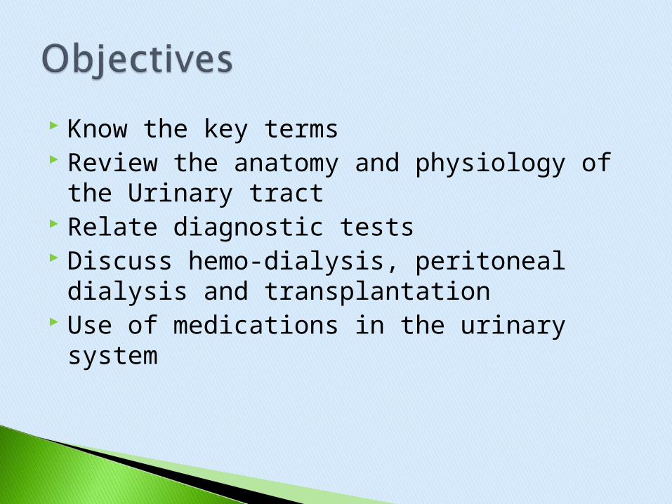
Know the key terms Review the anatomy and physiology of the
Urinary tract Relate diagnostic tests Discuss hemo-dialysis, peritoneal dialysis
and transplantation Use of medications in the urinary system

State changes in urinary system related to aging
Compare and contrast acute and chronic renal failure
The urinary system and care plans
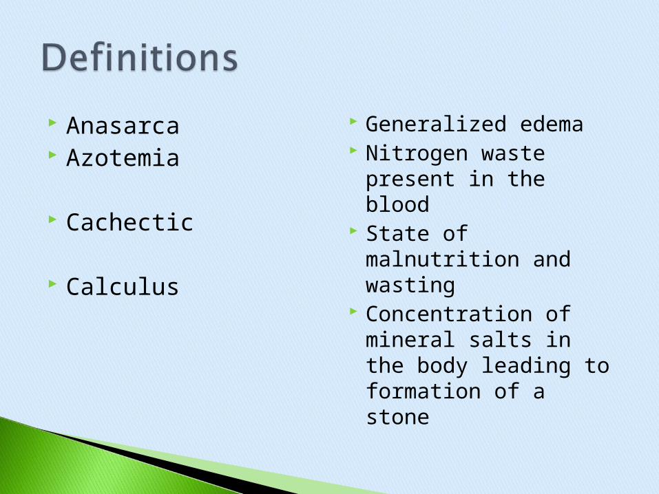
Anasarca Azotemia
Cachectic
Calculus
Generalized edema Nitrogen waste
present in the blood State of malnutrition
and wasting Concentration of
mineral salts in the body leading to formation of a stone

Cystitis
Dialysate
Dysuria
Inflammation of the urinary bladder
Solution used in dialysis, designated to approximate the normal electrolyte structure of plasma and extracellular fluid
Difficult or painful urination

Erythropoiesis
Fulguration
Ileal conduit
Production of red blood cells and release by the red bone marrow
Procedure to destroy tissue with long high-frequency electric sparks
Implantation of the ureters into a piece of ileum, which is attached to the abdominal wall as a stoma so urine can be removed from the body.
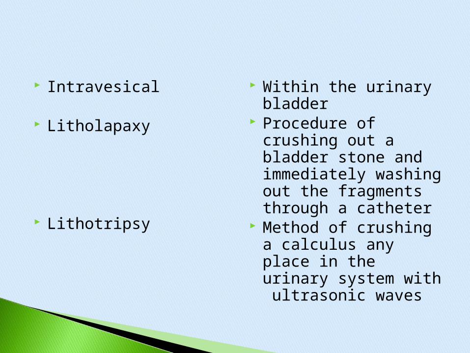
Intravesical
Litholapaxy
Lithotripsy
Within the urinary bladder
Procedure of crushing out a bladder stone and immediately washing out the fragments through a catheter
Method of crushing a calculus any place in the urinary system with ultrasonic waves

Consists of two kidneys, two ureters, urinary bladder and a urethra
The kidneys manufacture urine - Micturation is the act of expelling urine from the bladder

The waste products of the urine are water (95%), nitrogenous waste products of protein which are:
Urea, uric acid, creatinine, excessive electrolytes of sodium, calcium.
potassium phosphates, bile pigments hormones metabolized drugs and toxins

http://www.youtube.com/watch?v=XXxPPID5ge4&list=PLgaA42aWtrQv3a1j6GQHjbYo0nVDPO6WW&index=11
Kidney Function II http://www.youtube.com/watch?v=Ni_LO8x
aVTE&list=PLgaA42aWtrQv3a1j6GQHjbYo0nVDPO6WW

Urine moves steadily by peristalsis through the ureters into the urinary bladder.
Remains until capacity reached @ 500mL or until a desire to urinate is felt @250mL
Urine is then expelled from the bladder to the urethra
Micturition

http://www.youtube.com/watch?v=A55yBKNin3g

Urination: As the bladder fills: -Stretch receptors in it’s wall send impulses to the lower spinal cord -Motor impulses from this center stimulate bladder wall contraction which forces urine outward as both sphincters are made to relax
-In the infant, this is a simple reflex
-Toilet training – learning to control urination; comes from the higher centers on the brain

-The impulses to urinate will override conscious controls if the bladder becomes too full -Voluntary Emptying- relaxing the muscles of the pelvic floor and increasing pressure in the bladder triggers the spinal reflex which causes urination
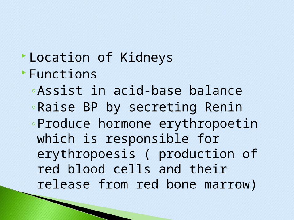
Location of Kidneys Functions
◦Assist in acid-base balance◦Raise BP by secreting Renin◦Produce hormone erythropoetin which is responsible for erythropoesis ( production of red blood cells and their release from red bone marrow)

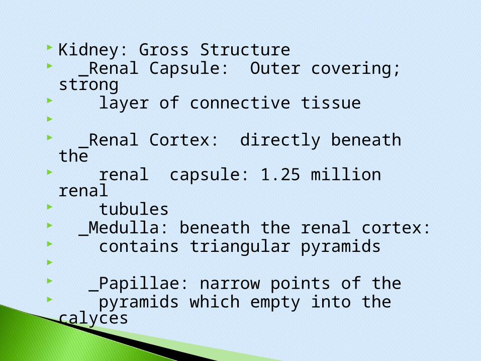
Kidney: Gross Structure _Renal Capsule: Outer covering;
strong layer of connective tissue _Renal Cortex: directly beneath the renal capsule: 1.25 million renal tubules _Medulla: beneath the renal cortex: contains triangular pyramids _Papillae: narrow points of the pyramids which empty into the
calyces

_Calyces: cup-like extensions of the renal pelvis: -guide urine into the renal pelvis _Renal Pelvis: an expansion of the upper end of the ureter. The ureter drains the finished product, urine, into the bladder.
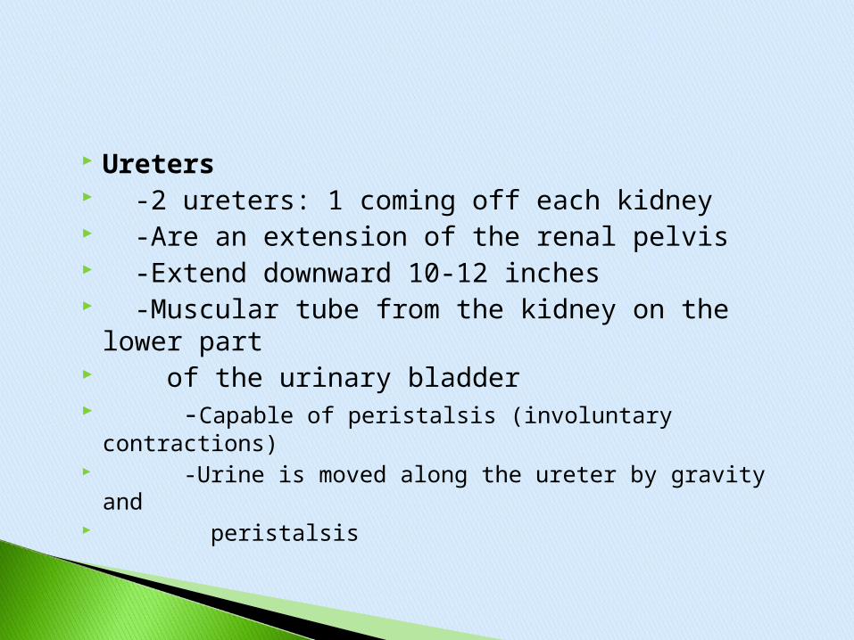
Ureters -2 ureters: 1 coming off each kidney -Are an extension of the renal pelvis -Extend downward 10-12 inches -Muscular tube from the kidney on the
lower part of the urinary bladder -Capable of peristalsis (involuntary contractions) -Urine is moved along the ureter by gravity and peristalsis

-As the ureters enter the bladder, the
mucous membrane folds and acts
as a valve to prevent backflow of urine

Urinary Bladder -A temporary pouch for urine -Composed of a collapsible muscle -Located anterior to the abdominal
cavity; posterior to the symphysis pubis -Can hold 750-1000 mL of urine -A moderately full bladder holds 450-500 mL of urine

Urethra -The terminal portion of the urinary system -Carries urine by peristalsis form the bladder to the external opening, the urinary meatus -In females- it is embedded in the anterior wall of the vaginal vestibule and exits between the clitoris and vaginal opening -Approximately ¼ inch in diameter and 1 ½ inches long

-In the male the urethra is approximately 8 inches long; passing through the prostate gland and extending the length of the glans penis -In the male, serves 2 functions: passage way for urine and semen
Nephrons◦Microscopic units: responsible for urine
formation◦Winds into the cortex and medulla of the
kidney◦Each contains renal corpuscle which
consists of a glomerulus (ball like network of capillaries formed from an arteriole) and held in Bowman’s Capsule.

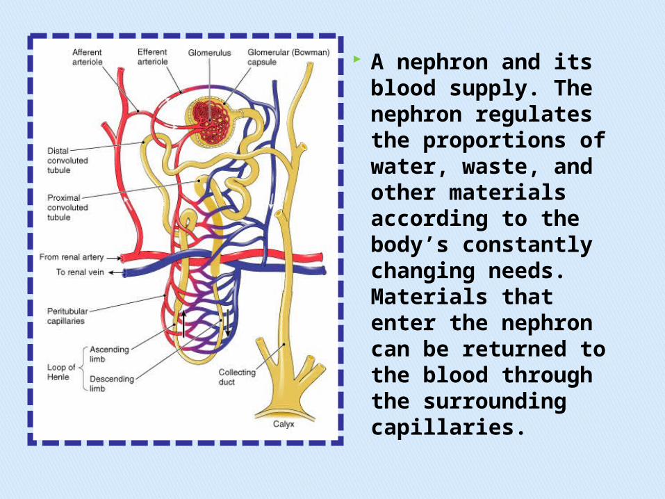
A nephron and its blood supply. The nephron regulates the proportions of water, waste, and other materials according to the body’s constantly changing needs. Materials that enter the nephron can be returned to the blood through the surrounding capillaries.

Structure of the juxtaglomerular (JG) apparatus. Note how the distal convoluted tubule contacts the afferent arteriole (right). Cells
in these two structures make up the JG
.
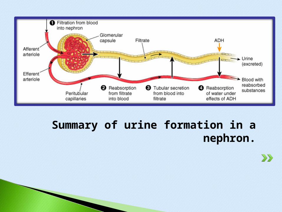
Summary of urine formation in a nephron.


•GlomerulusGlomerulus
•Bowman’s CapsuleBowman’s Capsule
Included in the baseline data for all clients.
Subjective questions
Objective questions
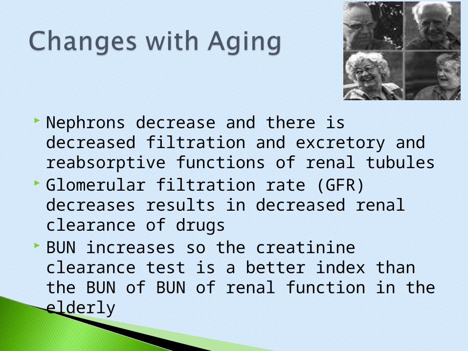
Nephrons decrease and there is decreased filtration and excretory and reabsorptive functions of renal tubules
Glomerular filtration rate (GFR) decreases results in decreased renal clearance of drugs
BUN increases so the creatinine clearance test is a better index than the BUN of BUN of renal function in the elderly
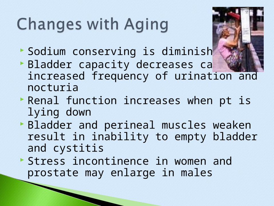
Sodium conserving is diminished Bladder capacity decreases causing
increased frequency of urination and nocturia
Renal function increases when pt is lying down
Bladder and perineal muscles weaken result in inability to empty bladder and cystitis
Stress incontinence in women and prostate may enlarge in males