Embed Size (px)
Citation preview
7
CHAPTER 2
REVIEW OF LITERATURE
Natural products serve as a consistent source of drug lead with diverse
chemical entities being derived from plants (Newman and Cragg, 2006), microbes
(Zhang et al., 2005), invertebrates (McCarthy and Pomponi, 2004), etc., Most of the
drugs in current use are natural products or natural product based. Microbes respond
to their environment quickly and compete for defense and survival by production of
unique secondary metabolites, which have been shown to possess biotechnological
or pharmaceutical applications. The marine realm which covers 70 % of the earth’s
surface provides the largest inhabitance for living organisms, particularly microbes
among the major habitats of the biosphere. Marine organisms have been shown to
produce numerous novel compounds with multiple pharmacological properties
(Fuesetani, 2000; Procsch et al., 2002; Haefner, 2003; Blunt et al., 2004). Marine
species comprises approximately a half of the total global biodiversity and hence the
sea offers an enormous resource for novel compounds (de Vries and Beart, 1995),
and it has been classified as the largest remaining reservoir of natural molecules to
be evaluated for drug activity (Gerwick, 1987). In recent years, many bioactive
compounds have been extracted from various marine animals like tunicates,
sponges, soft corals, sea hares, nudibranchs, bryozoans, sea slugs and marine
organisms (Donia and Hamann, 2003; Haefner, 2003). The search for new
metabolites from marine organisms has resulted in the isolation of more or less
30,000 metabolites, many of which are endowed with pharmacodynamic properties.
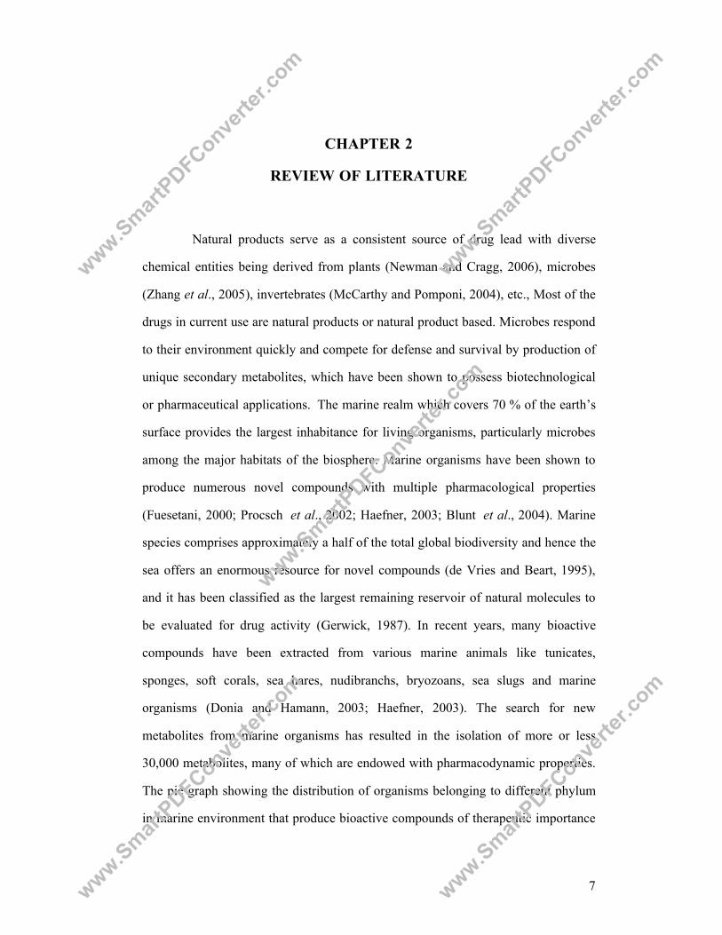
The pie graph showing the distribution of organisms belonging to different phylum
in marine environment that produce bioactive compounds of therapeutic importance
www.SmartPDFConve
rter.c
om
www.SmartPDFConve
rter.c
om
www.SmartPDFConve
rter.c
om
www.SmartPDFConve
rter.c
om
www.SmartPDFConve
rter.c
om
8
is shown in Fig 1. Sponges often have associated symbiotic microbial populations
(Lee et al., 2001; Richelle-Maurer et al., 2003). Symbionts include archaea, bacteria,
cyanobacteria, and microalgae. In some cases, these microorganisms and not sponge
cells are the likely source of the secondary metabolites of interest (Bewley and
Faulkner, 1998; Lee et al., 2001; Proksch et al., 2002).
Coelenterates21%
Sponges38%
Green algae2%
Red algae5%
Brown algae2%
Tunicates6%
Echinoderms6%
Microorganisms17%
Molluscs2% Bryozoans
1%
Fig 1 Distribution of marine natural products by Phylum, 2002 (Blunt et al., 2004)
The anti inflammatory activity of a new sphingosine derivative and
cembrenoid diterpene (lobohedleolide) isolated from the soft corals of Sinularia
crassa and Lobophytum sp. respectively, collected on the coasts of Andaman and
Nicobar Islands was reported by Radhika et al., (2005).
2.1 Microbes in Marine ecosystems
Marine microbes thrive not only on the surface of the sea but also in the
lower and abyssal depths, offshore to coastal regions. Microbial life is widely
distributed although they are rarely conspicuous; but Whitman et al., (1998) have
reported that about half the biomass of earth is microbial and is found in all
www.SmartPDFConve
rter.c
om
www.SmartPDFConve
rter.c
om
www.SmartPDFConve
rter.c
om
www.SmartPDFConve
rter.c
om
www.SmartPDFConve
rter.c
om

9
ecosystems. Marine microbes live in a biologically competitive environment with
unique conditions of salinity, pH, temperature, pressure, oxygen, light and nutrients.
Hence the marine microbial metabolites exhibit wide diversity both in chemical
composition and biological activities compared to terrestrial bacteria (Blunt et al.,
2004; Berdy 2005). Serious attempts to tap the vast potential of marine organisms as
sources of bioactive metabolites that may be directly utilized as drugs or serve as
lead structures for drug development started in the late 1960s. Realization of the
tremendous biological reserve of marine microbes has resulted in increasing interest
in the exploitation of microbes in search of new chemical entities.
2.1.1 Mangrove ecosystem
Mangroves are unique inter-tidal ecosystems, which support the growth of
genetically diverse groups of aquatic and terrestrial organisms. This ecosystem
situated at the inter-phase between the terrestrial and marine environment help to
sustain a rich and diverse group of microorganisms. Holguin et al., (2001) reported
the presence of different groups of bacteria that get nourished by detritus which in
turn help the mangrove ecosystem in different ways. The mangrove environment has
become a potent source for the isolation of antibiotic-producing actinomycetes and
marine fungal diversity is also best-studied in the mangrove ecosystem (Hyde et al.,
2000; Cheng et al., 2009).
2.1.2 Deep sea
The deep sea is characterized by unique and extreme environment like high
pressure, low temperature, lack of light, variable salinity and oxygen concentration.
In spite of the vast geographical area of deep sea, scant information is available
about the deep sea microorganisms. However, Bull et al., (2000) have reported deep
www.SmartPDFConve
rter.c
om
www.SmartPDFConve
rter.c
om
www.SmartPDFConve
rter.c
om
www.SmartPDFConve
rter.c
om
www.SmartPDFConve
rter.c
om

10
sea as a good source of novel organisms for microbiologists and biotechnologists.
Sterling efforts of Zobell and co-workers, who initiated work on the effect of
hydrostatic pressure on bacterial activity (Zobell and Johnson, 1949), have brought
improvements in the knowledge of deep-sea biology. The deep-sea habitat remained
untouched for a long time. Colquhoun et al., (1998) have isolated a large number of
actinomycetes from deep-sea sediments. Actinomycetes species isolated from deep
sea are mostly novel with potent sources of antibiotics. Two new genera and species
of Ascomycetes and two new Deuteromycetes from 1615 and 5315 m depth were
described by Kohlmeyer (1977). Gautschi et al., (2004) isolated Penicillium sp from
deep water sediment at 4380 ft and evaluated the cytotoxic activity.
2.1.3 Extreme environments
Any environmental condition that can be perceived as beyond the normal
acceptable range is an extreme condition (Satyanarayana et al., 2005). Extreme
environments can be found in all ecosystem and surprisingly almost all these
environments are colonized by microorganisms well adapted to the extreme
conditions and the microorganisms inhabiting such environments are termed as
‘extremophiles’. Highly saline conditions in water and soil exert a strong selective
pressure on the biota, favouring the development of halotolerant and halophilic
forms. Marine saltern is the common habitat of halophiles and the most common
genera are Halococcus and Halobacterium. Reports on fungi living in the extreme
environments were rare except the report on the diversity of fungi from the
hypersaline Dead Sea environment, which is one of the most extreme environments
for microorganisms on the earth (Molitoris et al., 2000; Kis-Papo et al., 2003).
Archaeglobus is found in hot sediments of marine hydrothermal vents with optimum
growth at 83 oC (Burggraf et al., 1990). A methanogenic bacterium Methanopyrus
www.SmartPDFConve
rter.c
om
www.SmartPDFConve
rter.c
om
www.SmartPDFConve
rter.c
om
www.SmartPDFConve
rter.c
om
www.SmartPDFConve
rter.c
om
11
capable of growth at 100 oC has also been recorded (Huber, 1989; Huber et al.,
1995).
2.2 Screening of marine bacteria for secondary metabolites
Secondary metabolites are compounds with varied and sophisticated
chemical structures, produced during the growth of the microbes (Barrios-Gonzalez
and M ejia, 1996; Barrios-Gonzalez and Mejia, 2008). The first document on
antibiotic-producing marine bacteria was by Rosenfeld and Zobell, (1947). Since
then, there are several reports of antibiotic-producing marine bacteria (Hanefeld and
Laatsch, 1991; Jensen and Fenical, 1994 and James et al., 1996). It is critical to have
efficient system to identify uninteresting or already known antibiotics as early as
possible, in order to focus the resources on the important ones. Buss and Butler,
(2004) state the importance of the use of secondary assays or counter-screens that
help to eliminate the uninteresting biological activities. However, the use of
secondary assays at an early stage may yield misleading results, since the extract
may contain several active compounds, and they may camouflage each other in the
secondary assays. Instead all the selected extracts from primary screening can be
subjected to some type of fractionation; however this technique is expensive and
time-consuming. The chromatographic techniques and other separation technologies
are employed for retrieving the active compound. Spectroscopic techniques such as
UV, FTIR, NMR and M ass used for structure elucidation, are obviously an
enormous improvement in the process isolation of active lead (Koehn and Carter,
2005). However, the complexity of the process, which involves not only bioassay
guided isolation chemistry, but also re-supply of extract in volume to obtain
sufficient pure material, still recognized as the rate limiting step of natural products
lead discovery.
www.SmartPDFConve
rter.c
om
www.SmartPDFConve
rter.c
om
www.SmartPDFConve
rter.c
om
www.SmartPDFConve
rter.c
om
www.SmartPDFConve
rter.c
om
12
Marine bacteria are capable of producing unusual bioactive compounds
that are not observed in terrestrial sources (Fenical, 1993; Fenical and Jensen, 1993).
Thermo-stable proteases, lipases, esterases, and starch and xylan degrading enzymes
have been found in bacterial and archaeal hyperthermophilic marine microorganisms
(Bertoldo and Antranikian, 2003). The first marine antibiotic isolated was
pentabromopseudilin (Burkholder et al., 1966; Hanessian et al., 1966; Lovell, 1966).
Yoshikawa et al., (1997) have isolated Korormycin from a marine Vibrio
alginolyticus, which is a specific inhibitor of the enzyme sodium dependent
NADH:quinone reductase making this organism halotolerant (Yoshikawa et al.,
1999). Alteromonas sp produces isatine, a long-known synthetic product, which
showed a reasonable anti fungal activity and protects the eggs of shrimp Palaemon
macrodactylus successfully against the pathogenic fungus Lagenidium callinectes
(Gil-Turnes et al., 1989). Streptomycetes are especially rich in biologically highly
active quinones; Parimycin, C-glycosides himalomycins and anthraquinones with
the rare fridamycin E chromophor, a precursor of the anthracycline antibiotics were
isolated from Streptomyces sp B6921 (Maskey et al., 2003). Korormicins have been
isolated from a marine Pseudoalteromonas sp. F- 420 (Yoshikawa et al., 2003),
which are selective inhibitors of the primary sodium pump. Jeong et al., (2003) have
isolated bacillamide from Bacillus sp which is active against dinoflagellate.
In our lab, potential marine Pseudomonas sp strains PS3 and PS7
producing anti inflammatory compounds (3S, 8aS)-3-Isobutyl-hexahydro-pyrrolo [1,
2-a] pyrazine-1,4-dione and (3S, 8aS)-3-(4-Hydroxy-benzyl)-hexahydro-pyrrolo [1, 2-
a] pyrazine-1,4-dione respectively have been isolated, characterized and structurally
elucidated by Rupesh (2007). Secondary metabolites from cultured microbes have
been a major source of clinically useful chemotherapeutics. Identification of
www.SmartPDFConve
rter.c
om
www.SmartPDFConve
rter.c
om
www.SmartPDFConve
rter.c
om
www.SmartPDFConve
rter.c
om
www.SmartPDFConve
rter.c
om
13
compounds that interact specifically with specific molecular targets will be useful in
controlling inflammation and cancer.
2.3 Inflammation
Inflammation is a process of body’s defense mechanism which recognizes
the danger signals that activate intracellular signaling cascades leading to increased
expression of pro inflammatory cytokine genes. Human diseases with acute and
chronic inflammation such as rheumatoid arthritis, inflammatory bowel disease,
multiple sclerosis, psoriasis, etc., still demand better therapies. Inflammation occurs
as a result of complex series of events that follow tissue damage caused by trauma,
or by microbial products. It is basically a protective and restorative reaction but due
to increased vasopermeability caused by local mediators liberated by damaged
tissue, granulocytes actively extravasate and accumulate at the site of injury
attracted by leucotactic factors of various origins (lymphocyte products, bacterial
metabolites, and fibrin split products). Monocytes and macrophages allured by
similar mechanism reach the inflammatory site within few hours and later become
predominating cells during healing or in chronic inflammation.
2.3.1 Activation of cells with mitogen
Exposure of immune-related cells to mitogen such as
Phytohaemeagglutinin (PHA), Lipopolysaccharide (LPS), etc., results in the intense
production of inflammatory cytokines, including TNF-α, IL-1β and other
inflammatory mediators, such as arachidonic acid (AA), thereby leading to septic
shock in mammals (Sweet and Hume, 1996) LPS, a component of the cell envelope
of Gram-negative bacteria, was shown by Glauser et al., (1991) to cause septic
shock. Lymphocyte proliferation assay (LPA) is used to assess the proliferation of
www.SmartPDFConve
rter.c
om
www.SmartPDFConve
rter.c
om
www.SmartPDFConve
rter.c
om
www.SmartPDFConve
rter.c
om
www.SmartPDFConve
rter.c
om
14
peripheral blood mononuclear cells (PBMC) in response to various non specific
(mitogen) and specific stimuli (antigen) that trigger a complicated series of events
that ultimately leads to cell division, which is measured by the incorporation of
[3H] labeled thymidine into newly synthesized DNA. This method reported by Sitz
and Birx, (1999) has become a standard surrogate measurement of proliferation.
2.3.2 Mediators of inflammation
Inflammation is a complex phenomenon involving multiple cellular and
molecular interactions which must be tightly regulated to avoid different pathology
and disorders (Balkwill et al., 2005; Consilvio et al., 2004). Activated mononuclear
cells that migrate by the way of PBMC, to bone, muscle, liver and other tissues
release inflammatory mediators, including TNF-α, Prostaglandin E2 (PGE2) and
interferon gamma (IFN-γ). Nitric oxide (NO) and PGs have been reported to play an
important role in inflammation and the role of phospholipase and cyclooxygenase in
production of prostaglandin is shown in Fig 2. The discovery of sizeable quantities
of PGs, which had been discovered as important mediator involved in inflammatory
diseases, fever and pain is usually, considered as the “take-off point” of any serious
search for “drugs from the sea”.
Membrane phospholids are converted into arachidonic acid by
phospholipase A2 (PLA2) and the PGs are generated from AA via cyclooxygenase
pathway (Funk, 2001). Cyclooxygenases are endogenous enzymes which exist in
two isoforms a house keeping enzymes, COX-1 (Vane et al., 1998) and an inducible
enzyme, COX-2, expressed in patho-physiological condition such as inflammation
due to pro inflammatory stimulus (Seibert et al., 1994; Needleman and Isakson,
1997; Nantel et al., 1999).
www.SmartPDFConve
rter.c
om
www.SmartPDFConve
rter.c
om
www.SmartPDFConve
rter.c
om
www.SmartPDFConve
rter.c
om
www.SmartPDFConve
rter.c
om

15
Cyclooxygenases (COX-1 and COX-2)
PGE2
Prostaglandins (PG) Thromboxanes (TX)
Fig 2 Role of phospholipase and cyclooxygenase in production of PGs
COX-2 is not only elevated in inflammatory conditions but also in many
human malignancies (Eberhart et al., 1994). Reports by Shiotani et al., (2001) and
Tiano et al., (2002) have shown that pharmacological inhibition of COX-2 reduces
the formation of various tumours in animal models. PG is a major cyclooxygenase
product at inflammatory sites where it contributes to increase in local blood flow,
edema formation, and pain sensitization (Williams and Peck, 1977).
Cyclooxygenase is the key enzyme that catalyses the two sequential steps in the
biosynthesis of PGs from AA (Smith et al., 1991). COX-2 is regulated by LPS,
IL-1β, TNF and active oxygen derivatives according to various cells (Martin-Sanz
et al., 1998).
NO, like PG forms an important mediator of inflammatory processes
(Moncada, 1999). NO is synthesized by nitric oxide synthase and three NOS
www.SmartPDFConve
rter.c
om
www.SmartPDFConve
rter.c
om
www.SmartPDFConve
rter.c
om
www.SmartPDFConve
rter.c
om
www.SmartPDFConve
rter.c
om

16
isoforms have been charecterised: eNOS and nNOS, calmodulin dependent or
constitutive and calmodulin independent and inducible iNOS (Moncada et al.,
1991). Reviews by Nathan, (1992), Moncada and Higgs, (1993) and Beckman and
Crow, (1993) have revealed the multiple patho-physiological functions of NO. NO
has many actions appropriate for a pro inflammatory agent, like, it is made by
numerous cell types in sites of inflammation, it increases blood flow and vascular
permeability, cell/tissue destructive abilities, and it may induce cyclooxygenase,
cause pain, destroy certain protease inhibitors, and enhance production of IL-1 and
TNF, and NADPH oxidase activity in myeloid cells (Magrinet et al., 1992, Clancy
and Abramson, 1995). NO production may result from the action of several
substances including cytokines, immune complexes, bacterial products, and
mechanical stress. NO production was determined by measuring the amount of
nitrite in the culture media using Griess reagents (Na et al., 2004).
2.3.3 Role of cytokines in inflammation
Cytokines mediates and control immune and inflammatory responses and
complex interactions between cytokines, inflammation and the adaptive responses
helps in maintaining homeostasis, health, and well-being (Elenkov et al., 2005).
Dysregulation of the pro versus anti inflammatory and Th1 versus Th2 cytokine
balance results in common human diseases such as atopy/allergy, autoimmunity,
chronic infections and sepsis.
The pro inflammatory cytokine TNF-α, controls inflammatory cell
populations as well as mediates many of the other aspects of the inflammatory
process. TNF-α is a potent inflammatory cytokine that is important for host defense
and is expressed at sites of inflammation where it activates resident tissue cells,
infiltrating immune cells and endothelial cells in surrounding blood vessels. The
www.SmartPDFConve
rter.c
om
www.SmartPDFConve
rter.c
om
www.SmartPDFConve
rter.c
om
www.SmartPDFConve
rter.c
om
www.SmartPDFConve
rter.c
om

17
significant role of TNF-α in chronic inflammatory disease is well established (Lin
and Yeh, 2005), TNF-α has been implicated in the pathogenesis of many acute and
chronic inflammatory conditions, including septic shock, rheumatoid arthritis, and
inflammatory bowel disease(Feldmann et al., 1996; Dinarello 1997; van Heel et al.,
2002). TNF-α production require tight control to avoid excessive inflammation,
tissue damage, and toxicity. Moldawer, (2003) have reported NF-κB as an important
inducer of TNF-α transcription, and activation of the p38 MAPK is required for
stabilization and efficient translation of TNF-α mRNA.
IL-1 β is thought to be the primary cytokine and the main regulator of T
cell proliferation during inflammation and is involved in the initiation and
persistence of inflammation. IL-1β is synthesized as 31 kD precursors, proIL-1β
which lacks biological effects. The active, mature IL-1β is produced upon cleavage
of proIL-1β by a specific IL-1β-converting enzyme (ICE or caspase-1). IL-1β is a
typical pro inflammatory cytokine and induces the expression of a variety of genes
and the synthesis of several proteins that in turn induce acute and chronic
inflammatory changes and can be provoked by infectious agents, products of
activated lymphocytes and complement (Dinarello, 1996). TNF-α and IL-1β
stimulate the production of secondary cytokine IL-6, which is a major inducer of
acute phase proteins. Combination of IL-1β, TNF-α, and/or IFN-γ have often been
found to be co-stimulatory (Sozzani et al., 1996; Proost et al., 1996). IFN-γ, the
principal Th1 effector cytokine, has been shown to be crucial for the development of
inflammation.
IL-10 is a Th2 cytokine also called as a cytokine synthesis inhibitory factor
inhibits the LPS induced synthesis of a number of cytokines implicated in the
regulation of T cells, including IL-1γ, IL-6, IL-8 and TNF-alpha (Fiorentino et al.,
1991). Elevated plasma IL-10 levels are detected in septicemia (Marchant et al.,
www.SmartPDFConve
rter.c
om
www.SmartPDFConve
rter.c
om
www.SmartPDFConve
rter.c
om
www.SmartPDFConve
rter.c
om
www.SmartPDFConve
rter.c
om
18
1994) and the protective effects of IL-10 against sepsis, morbidity and mortality are
well recognized, correlating in many cases with the inhibition of TNF-α production
(Marchant et al., 1994; Pajkrt et al., 1997; Cartmell et al., 2003). IL-10 as
immunosuppressive and anti inflammatory cytokine has been well explained by
Pestka et al., (2004) and inhibition of production of pro inflammatory cytokines,
including TNF-α, IL-6, and IL-12 were reported by Moore et al., (2001). Reverse-
transcriptase-polymerase chain reaction (RT-PCR) has become one of the most
widely applied techniques in biomedical research. The ease with which the
technique permits specific mRNA to be detected and quantified has been a major
asset in the molecular investigation of disease pathogenesis. Disease related
imbalances in the expression of specific mRNAs can be sensitively and
quantitatively determined by RT-PCR. Lee et al., (2008) found that CT20126
inhibited the expression iNOS, COX-2, cytokines like TNF-α and IL-1β and
chemokines, which are NF-kB regulated inflammation-associated genes, in immune
activated primary macrophages and human synoviocytes in vitro as well as in vivo
animal models. NF-kB is an important transcription factor for the induction of
various inflammation associated genes including iNOS, cytokines, and chemokines
in response to LPS and TNF-α (Tak and Firestein, 2001; Ahmed et al., 2006).
2.3.4 MAP kinases in inflammation
Immune responses involve a number of cell types that functions as
initiators, regulators, and effectors. These cells interact with and cross-regulate each
other, and the target cells respond using signal transduction pathways to mediate
gene expression and immune function. Many of the mediators of inflammation are
regulated by MAPK pathways and downstream transcription factors. There are three
major groups of MAP kinases in mammalian cells, viz ERK (Schaeffe and Weber,
1999), the p38 MAP kinases (Han and Ulevitch, 1999) and the JNK/SAPK (Davis
www.SmartPDFConve
rter.c
om
www.SmartPDFConve
rter.c
om
www.SmartPDFConve
rter.c
om
www.SmartPDFConve
rter.c
om
www.SmartPDFConve
rter.c
om
19
2000; Chang and Karin, 2001). In general ERK are activated by growth factors and
hormones, whereas both JNK and p38 MAPK are activated by environmental stress
and inflammatory cytokines. The p38 MAP kinases are a family of serine/threonine
protein kinases that play important roles in cellular responses to external stress
signals. In general, p38 MAPK is activated by many stimuli, including cytokines,
hormones, ligands for G protein-coupled receptors, and stresses such as osmotic
shock and heat shock and elevated levels of these cytokines are associated with
several chronic inflammatory diseases. p38 MAPK inhibitors have been shown to be
efficacious in several disease models, including those for asthma and chronic
obstructure pulmonary disease (COPD) (Adams et al., 2001).
2.4 Cancer
Cancer is a hyper proliferative disorder that involves morphological
cellular transformation, dysregulation of apoptosis, uncontrolled cellular
proliferation, invasion, angiogenesis, and metastasis (Hanahan and Weinberg, 2000).
It has been established that cancer can be promoted and/or exacerbated by
inflammation and infections. Lin and Karin, (2007) elaborated the link between
cancer and inflammation. COX-2 has been shown to be associated in a wide variety
of cancers, including that of the colon (Fournier and Gordon, 2000), lung (Hida
et al., 1998), and breast (Harris et al., 2000) cancers. Celecoxib, a specific inhibitor
of COX-2, has been shown to suppress colon carcinogenesis in animals (Reddy
et al., 2000). Colorectal cancer represents a major public health problem accounting
for over 1 million cases and about half a million deaths worldwide (Chau and
Cunningham, 2006). Survival from colon cancer has been found to vary
demographically and estimated to be 65 % in North America, 54 % in Western
Europe, 34 % in Eastern Europe, and 30 % in India (Parkin et al., 2005).
www.SmartPDFConve
rter.c
om
www.SmartPDFConve
rter.c
om
www.SmartPDFConve
rter.c
om
www.SmartPDFConve
rter.c
om
www.SmartPDFConve
rter.c
om

20
The uncontrolled cell proliferation in malignancy could be due to an
increased proliferation or decreased cell death. A block in apoptosis has been
implicated in cancer (Barr and Tomei, 1994). Since cancer cells often have an
immature phenotype representing a block in the normal differentiation pathway,
restoration of the normal differentiation program in cancer cells appears to activate
an apoptotic mechanism similar to the normal physiological process (Ohasi et al.,
1992). The proposition that apoptosis is a discrete phenomenon that is
fundamentally different from degenerative cell death or necrosis, is based on its
morphology, bio-chemistry and incidence. A variety of anti cancer drugs have been
shown to induce extensive apoptosis in rapidly proliferating normal cell population,
lymphoid tissues, and tumors (Kerr et al., 1994; Chintamani et al., 2004).
2.4.1 Programmed cell death
Apoptosis or programmed cell death is a critical determinant of tissue mass
homeostasis and may play a role in carcinogenesis. Apoptotic cell death occurs in
two phases: an initial commitment phase followed by an execution phase which is
characterized by a series of changes including cell shrinkage, plasma membrane
alterations, and condensation and fragmentation of chromatin (Earnshaw, 1995). In
the nucleus, chromatin is disrupted and endonucleases are activated, resulting in
cleavage of DNA. Apoptotic death is known to involve a cascade of proteolytic
events driven mainly by activation of family of aspartate-specific cysteine proteases
(Martin and Green, 1995) such as caspase-3, the major executioner of apoptosis.
Studies by Boldin et al., (1996), Muzio et al., (1996) and Sun et al., (1999),
suggests that the activator caspase-8 is an integral component of the cell death-
inducing mechanism and in receptor-mediated apoptosis, activation of caspase-8
www.SmartPDFConve
rter.c
om
www.SmartPDFConve
rter.c
om
www.SmartPDFConve
rter.c
om
www.SmartPDFConve
rter.c
om
www.SmartPDFConve
rter.c
om
21
represents a commitment point to cell death. Dimerization of the initiator caspases
was proposed to be the driving force for their activation (Renatus et al., 2001;
Boatright and Salvesan, 2003; Donepudi et al., 2003). Janicke et al., (1998) have
shown that caspase-3 is required for certain distinctive biochemical and
morphological changes during apoptosis and they have also demonstrated that
caspase-3 is essential for fragmentation of MCF-7 cell chromosomal DNA during
TNF induced apoptosis. Reyes et al., (2006) have reported caspase dependent DNA
fragmentation leading to apoptosis of HT 29 cells. Agents that suppress the
proliferation of malignant cells, by inducing apoptosis may represent a useful
mechanistic approach to both chemoprevention and chemotherapy of cancer.
Marine natural products have been prominently featured in the area of
cancer research mainly because of underlying preponderance of anti tumor agents
produced by marine organisms. There are many compounds such as didemnin B
(Rinehart et al., 1981), bryostatin 1 (Pettit et al., 1982), jaspamide (Crews et al.,
1986), dolastatin 10 (Pettit et al., 1987), ecteinascidin 743 (Rinehart et al., 1990),
curacin A (Gerwick et al., 1994) and thiocoraline (Baz et al., 1997), which were
discovered using traditional screening methods.
2.5 Docking
Three-dimensional molecular structure is one of the foundations of
structure-based drug design. Molecular docking and virtual screening based on
molecular docking have become an integral part of many modern structure based
drug discovery efforts. Docking is a term used for computational schemes that
attempts to find the “best” matching between two molecules: a receptor and a ligand.
The molecular docking can be defined as the prediction of correct bound association
www.SmartPDFConve
rter.c
om
www.SmartPDFConve
rter.c
om
www.SmartPDFConve
rter.c
om
www.SmartPDFConve
rter.c
om
www.SmartPDFConve
rter.c
om
22
between two molecules whose atomic co-ordinates are known. Molecular docking
involves the prediction of ligand, conformation and orientation within the active site
of the molecular target. Docking program has two key parts – a search for
conformational degrees of freedom and the evaluation or scoring function. The
search algorithm finds the potential energy landscape with sufficient details to
explore the global energy minimum. In rigid docking, the search algorithm explores
the different positions of the ligand in the receptor active site by translational and
rotational degrees of freedom whereas in addition torsional degree of freedom is also
included in flexible ligand docking. The scoring function gives importance to the
experimentally determined complex which assesses both the steric and the chemical
complementarities (Brooijmans and Kuntz, 2003). The fundamental goal of protein
docking is to determine whether two molecules interact and if so, the orientation that
maximizes this interaction as well as minimizing the total energy of the resulting
complex.
2.5.1 Types of docking:
Molecular docking process can be broadly categorized into three different
types:
2.5.1.1 Protein-Protein docking:
In these docking situations, the protein molecules are usually considered
rigid bodies. Thus, one can approximate the problem with 6 degrees of freedom: the
3 possible translations and rotations in the x, y and z axes. To further reduce the
search space of the problem, steric constraints (i.e. no overlap in van der Waal
envelope, geometric constraints) are applied. Then energies of the resulting binding
www.SmartPDFConve
rter.c
om
www.SmartPDFConve
rter.c
om
www.SmartPDFConve
rter.c
om
www.SmartPDFConve
rter.c
om
www.SmartPDFConve
rter.c
om

23
confirmations are examined. In essence this type of docking consists of constructing
the 6 dimensional confirmation spaces of all geometrically/physically allowable
configurations, then finding the best configuration by comparing energy scores of
the resulting binding complexes.
2.5.1.2 Protein-Ligand docking:
This type of docking is much more complex because of the flexibility of
the ligand. The ligand introduces this complexity because of rotatable bonds that
exist within the molecule. As the name suggests, these bonds allow parts of the
molecule to rotate, thus increasing the dimensionality of the problem with each
rotatable bond. To model the complex protein-ligand docking, it is usually common
to either reduce the flexibility of the ligand or search the conformational space using
either Monte Carlo methods or molecular dynamics.
2.5.1.3 Blind docking
Blind docking was introduced for the detection of possible binding sites
and modes of peptide ligands by scanning the entire surface of protein targets. Blind
docking can be used for unbiased mapping of the binding patterns of drug candidates
using AUTODOCK (Hetenyi and van der Spoel, 2006)
2.5.2 Computational receptor docking studies
Docking simulations are widely used for lead enhancement, using models
to analyze the atomic interactions between inhibitors and target molecules. A
detailed three- dimensional 3D picture of interactions between the ligands and the
www.SmartPDFConve
rter.c
om
www.SmartPDFConve
rter.c
om
www.SmartPDFConve
rter.c
om
www.SmartPDFConve
rter.c
om
www.SmartPDFConve
rter.c
om
24
active sites of COX-1, COX-2 and p38 MAPK would help us to determine the
mechanism of action.
2.5.3 Structure of COX-1 and COX-2
Molecular cloning and sequencing of pure preparations of COX-1 enzyme
from sheep and bovine seminal vesicles by different laboratories elucidated the
complete primary structure of COX-1 enzyme (Dewitt & Smith, 1988; Merlie et al.,
1988; Yokoyama et al., 1988; Yokoyama and Tanabe, 1989). The predicted amino
acid sequence of COX-2 cloned in chicken and mammals showed to posses
approximately 60% amino acid identity with COX-1 (Simmons et al., 1991). COX-1
and COX-2 were found to be approximately 600 aminoacid in size in all the species.
Mammalian COX sequences are represented in Fig 3 and the domains and residues
essential for their function are designated.
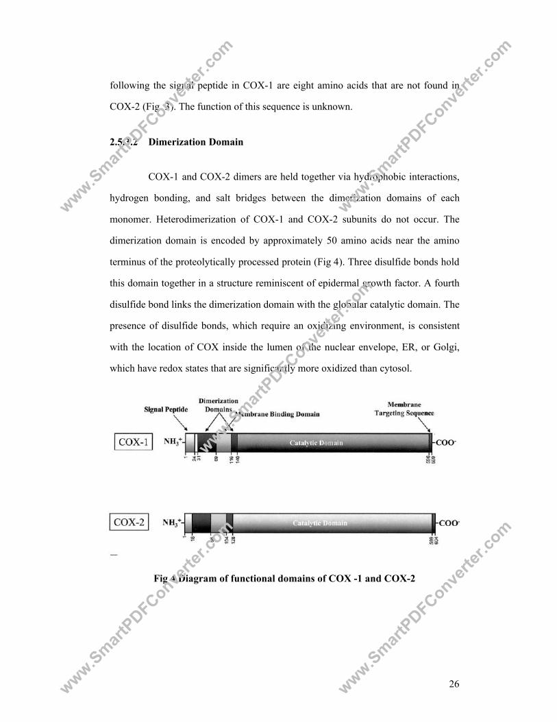
COX-1 has four distinct domains viz., amino terminal signal peptide,
dimerization, membrane binding and catalytic domains (Fig 4). Crystallographic
structure of COX-2 show striking similarity with COX-1 (Kurumbail et al., 1996,
Bayley et al., 1999). The structures of both the enzymes COX-1 and COX-2 predict
that both the enzymes are located in the lumen of the nuclear envelope and
endoplasmic reticulum (Simmons et al., 2004).
www.SmartPDFConve
rter.c
om
www.SmartPDFConve
rter.c
om
www.SmartPDFConve
rter.c
om
www.SmartPDFConve
rter.c
om
www.SmartPDFConve
rter.c
om

25
Fig 3 Amino acid sequences of human COX enzyme isoforms. Signal peptide
sequences, potential glycosylation sites and some important residues are in bold. Dimerization and membrane binding domains are denoted with a heavy underline. All sequence downstream to dimerization domain 2 constitutes catalytic domain. (Adopted from Simmon et al., 2004)
2.5.3.1 Amino-Terminal Signal Peptide
Nascent COX-1 and COX-2 polypeptides are directed into the lumen of
the endoplasmic reticulum by amino-terminal signal peptides. The signal peptide for
COX-1 is always 22 to 26 amino acids in length with a large hydrophobic core
comprised of four or more leucines or isoleucines (Fig 3). COX-2 signal peptide is
17 amino acids long in all species and appears to be less hydrophobic. Immediately
www.SmartPDFConve
rter.c
om
www.SmartPDFConve
rter.c
om
www.SmartPDFConve
rter.c
om
www.SmartPDFConve
rter.c
om
www.SmartPDFConve
rter.c
om
26
following the signal peptide in COX-1 are eight amino acids that are not found in
COX-2 (Fig. 3). The function of this sequence is unknown.
2.5.3.2 Dimerization Domain
COX-1 and COX-2 dimers are held together via hydrophobic interactions,
hydrogen bonding, and salt bridges between the dimerization domains of each
monomer. Heterodimerization of COX-1 and COX-2 subunits do not occur. The
dimerization domain is encoded by approximately 50 amino acids near the amino
terminus of the proteolytically processed protein (Fig 4). Three disulfide bonds hold
this domain together in a structure reminiscent of epidermal growth factor. A fourth
disulfide bond links the dimerization domain with the globular catalytic domain. The
presence of disulfide bonds, which require an oxidizing environment, is consistent
with the location of COX inside the lumen of the nuclear envelope, ER, or Golgi,
which have redox states that are significantly more oxidized than cytosol.
Fig 4 Diagram of functional domains of COX -1 and COX-2
www.SmartPDFConve
rter.c
om
www.SmartPDFConve
rter.c
om
www.SmartPDFConve
rter.c
om
www.SmartPDFConve
rter.c
om
www.SmartPDFConve
rter.c
om

27
2.5.3.3 Membrane Binding Domain
COX isozymes associate with the intra luminal surface of microsomal
membranes in an unusual fashion. COX isozymes contain a tandem series of four
amphipathic helices, which creates a hydrophobic surface that penetrates into the
upper portion of the luminal side of the hydrophobic core of the lipid bilayer. These
helices are encoded by approximately 50 amino acids found immediately to
carboxy-terminal of the dimerization domain (Fig 3). The helices allow COX dimers
to float on the surface of the lumen of the endoplasmic recticulum/nuclear envelope,
with the majority of the protein protruding into the luminal space of these
compartments. The membrane binding domain also forms the mouth of a narrow
hydrophobic channel that is the cyclooxygenase active site.
2.5.3.4 Catalytic Domain
Carboxy-terminal to the membrane binding domain in COX primary
structures is the catalytic domain, which comprises 80% (approximately 480 amino
acids) of the protein and contains two distinct enzymatic active sites.
a. Peroxidase Active Site: The catalytic domain is globular with two distinct
intertwining lobes. The interface of these lobes creates a shallow cleft on the upper
surface of the enzyme, where the peroxidase active site is located and where heme is
bound.
b. Cyclooxygenase Active Site: The cyclooxygenase active site is a long,
narrow, dead-end channel of largely hydrophobic character whose entrance is
framed by the four amphipathic helices of the membrane binding domain. The
channel extends approximately 25 Å into the globular catalytic domain and is on
average about 8 Å wide (Picot et al., 1994). However, significant narrowing of the
www.SmartPDFConve
rter.c
om
www.SmartPDFConve
rter.c
om
www.SmartPDFConve
rter.c
om
www.SmartPDFConve
rter.c
om
www.SmartPDFConve
rter.c
om

28
channel is observed where arginine 120, one of only two ionic residues found in the
COX active site, protrudes into the channel and forms a hydrogen bonded network
with glutamate 524 and tyrosine 355. Arg 120 is essential for binding substrates and
carboxylate-containing NSAIDs in COX-1. In contrast, this residue is unessential in
binding substrate in COX-2 (Rieke et al., 1999).
A crucial structural difference between the active sites of COX-1 and
COX-2 is a substitution of isoleucine 523 in COX-1 for a valine in COX-2 (Fig 3).
This single difference opens a hydrophobic out pocketing in COX-2 that can be
accessed by some COX-2-selective drugs (Kurumbail et al., 1996). The evolutionary
conservation of an enlarged cyclooxygenase active site in COX-2 relative to COX-1
may be essential to the recognition of bulkier substrates by COX-2.
2.5.4 Synthetic cyclooxygenase inhibitors- NSAIDs
NSAIDs are widely used for reducing pain and swelling associated with
inflammation. The known NSAIDs can be broadly classified into four types as
follows.
2.5.4.1 Irreversible inhibitors of COX -1 and COX -2 (aspirin)
Of the NSAIDs in medical use, only aspirin is a covalent modifier of COX-
1 and COX-2. Like other NSAIDs, aspirin diffuses into the COX active site through
the mouth of the channel and traverses up the channel to the constriction point
formed by Arg 120, Tyr 355, and Glu 524. At this point in the channel, the carboxyl
of aspirin forms a weak ionic bond with the side chain of Arg 120, positioning
aspirin only 5 Å below Ser 530 and in the correct orientation for transacetylation
(Loll et al., 1995).
www.SmartPDFConve
rter.c
om
www.SmartPDFConve
rter.c
om
www.SmartPDFConve
rter.c
om
www.SmartPDFConve
rter.c
om
www.SmartPDFConve
rter.c
om
29
2.5.4.2 Reversible, competitive inhibitors of both isoforms (mefenamate and
ibuprofen)
Other NSAIDs besides aspirin inhibit COX-1 and COX-2 by competing
with AA for binding in the COX active site. However, NSAIDs significantly differ
from each other in whether they bind the COX active site in a time-dependent or
independent fashion.
2.5.4.3 Slow, time dependent inhibitors of COX-1 and 2 (flurbiprofen and
indomethacin)
Time-dependent NSAIDs bind the COX active site first in a loose
interaction and then in a productive tight complex. The rate-limiting step in drug
binding is the formation of the tight binding conformation of the NSAID within the
COX channel. Second step in NSAID binding is the constriction point created by the
hydrogen bonding network of Arg 120, Tyr 355, and Glu5 24 and the proposed
difficulty for some NSAIDs to traverse it. The interactions results in tight binding of
many NSAIDs at the constriction point of the channel, where they totally block
entry of AA.
2.5.4.4 Selective inhibitors of COX-2 (SC-558 and Celecoxib)
The discovery of COX-2, which is expressed in inflammatory cells and
central nervous system, but not in the gastric mucosa, offers the impetus to develop
anti inflammatory and analgesic agents that is devoid of gastrointestinal toxicity
where they spare mucosal prostaglandin synthesis. Celecoxib and rofecoxib were
marketed in 1999 as the first NSAIDs developed as selective COX-2 inhibitors.
Other NSAIDs including meloxicam, nimesulide, and etodolac, which were
marketed earlier as safer NSAIDs, were found after the discovery of COX-2 to be
www.SmartPDFConve
rter.c
om
www.SmartPDFConve
rter.c
om
www.SmartPDFConve
rter.c
om
www.SmartPDFConve
rter.c
om
www.SmartPDFConve
rter.c
om

30
preferential inhibitors of this enzyme. Currently, second generation COX-2
inhibitors, such as valdecoxib (Smith and Baird, 2003) and etoricoxib (Hunt et al.,
2003) are other selective COX-2 agents in use.
2.5.5 Cyclooxygenase isoenzymes in human diseases
Several COX products like PGE2, PGI2 have been found in the synovial
fluid from knee joints of arthritic patients (Bombardier et al., 1981) and in
inflammatory conditions in animal models (Portanova et al., 1996; Zhang et al.,
1997; Rossi et al., 2000). COX-2 is primarily involved in the fever response to LPS
(Zhang et al., 2003). Large amounts of PGs production mediated by COX-2 by
certain tumour cells (Bennet et al., 1997) which induce a generalized state of
immune deficiency (Plescia et al., 1975). COX-2 induction by tumour cells
contributes to tumour angiogenesis and ultimately the growth of the tumours
(Williams et al., 2000). COX-2 can be induced in neurons, microglia and astrocytes
by a variety of neurotoxic stimuli including hypoxia and excitotoxins (Adams et al.,
1996; Tocco et al., 1997; Tomimoto et al., 2000). Elevated COX-2 is found in
neurogenerative diseases such as sporadic amyotrophic lateral sclerosis (Almer
et al., 2001). A number of excellent reviews have explored the complex and yet
unclear roles of COX enzymes in alzheimer’s disease (O’Banion, 1999; McGeer,
2000; Pasinetti, 2001).
2.5.5.1 COX-2 in inflammation
NSAIDs are currently used as first line therapeutics in the treatment of
osteoarthritis (OA), rheumatoid arthritis (RA), systemic lupus erythematosis (SLS)
and other inflammatory syndromes. Treatment of inflammation with NSAID is
palliative than disease modifying. They reduce inflammation and pain and hence
www.SmartPDFConve
rter.c
om
www.SmartPDFConve
rter.c
om
www.SmartPDFConve
rter.c
om
www.SmartPDFConve
rter.c
om
www.SmartPDFConve
rter.c
om

31
COX-2 inhibitors are used as pain relievers. In 1980s, anti inflammatory drugs with
less gastric injury were developed. Nimesulide, ecodolac and meloxicam emerged
from pre clinical studies as less toxic drugs and later proved to be selective inhibitor
of COX-2. Selectivity for the inducible isoform was established by comparing the
inhibitory potency against COX-1 measured as IC50 with inhibition of COX-2 in
isolated enzymes, cultured cells or in the whole blood assay.
Nimesulide did not have any effect on gastric PG levels and did not cause
bleeding in gastric mucosa. The therapeutic efficacy of nimesulide has been
demonstrated in clinical trials for inflammation and pain (Huskisson et al., 1999;
Bennett, 2001). However it was withdrawn in 2002 due to hepatic toxicity. Etodolac
has a pyranocarboxylic acid structure showed anti inflammatory effect without
gastric injury in a range of preclinical trials (Jones, 2001). The renal toxicity of
etodolac was also demonstrated to be minimal (Shand et al., 1986). Meloxicam also
emerged as anti inflammatory drug with low GI toxicity. Its chemical structure is
that of an enolcarboxamide and has been reported as selective COX-2 inhibitor.
2.5.5.2 COX-2 in cancer and angiogenesis
Many human malignancies results in production of increased amounts of
prostaglandins than in the normal tissues, a consequence of enhanced COX-2
expression (Eberhart et al., 1994; Sano et al., 1995). PGs are important in
pathogenesis of cancer due to the effects on cell proliferation, angiogenesis, immune
surveillance and apoptosis (Tsujii et al., 1998; Willams et al., 2000). Shiotani et al.,
2001 and Tiano et al., 2002 reported that pharmacologic inhibition of COX-2
reduces the formation of various tumour in animal models, suggesting selective
COX-2 inhibitors might be useful for prevention of cancer.
www.SmartPDFConve
rter.c
om
www.SmartPDFConve
rter.c
om
www.SmartPDFConve
rter.c
om
www.SmartPDFConve
rter.c
om
www.SmartPDFConve
rter.c
om

32
The role of COX products in the formation of new blood vessels, a process
commonly referred to as angiogenesis (Hla et al., 1993) has received attention in
recent years. The induction of angiogenesis by the COX derived PGE2 may be
potentially involved in colon cancer (Tsujii et al., 1998; Hansen-Petrik et al., 2002)
and the angiogenic factors like VEGF was modulated by COX-2 over expression.
Hence use of COX-2 inhibitors may exert anti angiogenic effect.
2.5.5.3 COX-2 in Alzheimer’s disease
Epidemiologic evidence indicates that NSAID use is associated with a
lower incidence of Alzheimer’s disease (McGeer et al., 1996; Hendrie, 1997).
Proinflammatory cytokines, acute phase proteins, prostaglandins and other
mediators of inflammation are elevated in and around the senile plaques present in
Alzheimer’s disease brains (Oka and Takashima, 1997; Kitamura et al., 1999) at the
same time it is important to note that COX-2 is also normally expressed in neurons
of neocortex and hippocampus (Kaufmann et al., 1996; Yasojima et al., 1999; Ho
et al., 2001). However the normal function of COX-2 in brain neurons is not known.
The current debate is whether COX-2 induction after neuronal insult serves to
protect against cell death or promote apoptosis. Findings using animal models and
in vitro systems support both protective (Kunz and Oliw, 2001) and pro apoptotic
roles (Iadecola et al., 2001). Findings of Baik et al., (1999) suggests that inhibition
of COX-2 induced by excitotoxins may be neuroprotective, but inhibition of
constitutive COX-2 expression may lead to be deleterious. Many question remained
to be answered regarding the use of selective COX-2 inhibitors in neurodegenerative
diseases. The motivation for research in this area is, of course, to develop more
selective anti inflammatory drugs, but recent research also points to possible
applications for selective COX-2 inhibitors as drugs for cancer and Alzheimer’s
disease (Vane and Botting, 1998).
www.SmartPDFConve
rter.c
om
www.SmartPDFConve
rter.c
om
www.SmartPDFConve
rter.c
om
www.SmartPDFConve
rter.c
om
www.SmartPDFConve
rter.c
om

33
Two major approaches are currently in use for discovery of lead molecules:
one uses the chemical diversity of nature and the other applies combinatorial
chemistry. Knowledge of the structural features that explain the deferential
inhibitory profile can be useful for the design of new selective COX-2 inhibitors.
Nimesulide is one of the first NSAID marketed with a preferential COX-2 inhibition
(Cullen et al., 1998). Analysis of the crystal structures of COX inhibitors (Picot
et al., 1994; Kurumbail et al., 1996) reveals a network of hydrogen bonds involving
Arg 120, Tyr 355, His 513 and Glu 524 that are thought to act as a gate for ligand
entrance to the COX active site.
2.5.6 p38 MAPK structure and function
p38 have typical kinase fold with a small N- terminal domain dominated by
β-strands, a larger C-terminal domain consisting of α-helices and an ATP binding
site in the interface between the two domains. The ATP binding site is formed by the
flexible glycine rich loop, that connects the N- terminal domain with the large α-
helical domain and the catalytic loop. The kinase domains of p38 have a substrate
binding groove which is at the interface of the N and C terminal domains. p38 has an
additional docking groove for activating kinases, substrate kinases and inactivating
phosphatases. The docking groove contains glutamate-aspartate and common
docking regions and is conserved among MAP kinases. The p38 activation loop
contain two phosphorylating sites (Thr 180 and Tyr 182) which when modified
stabilize its activated conformation and open the substrate binding groove. p38 have
a docking groove for binding with their activating kinases, inactivating phosphatases
and substrates. This docking groove is different from the substrate binding groove at
which the phosphorylation takes place. The p38 docking groove contains CD region
is part of a shallow groove formed by the acidic residues Asp313, Asp316,Glu81
and the aromatic residues Phe 129 and Tyr 311. The phosphorylation lip in p38
www.SmartPDFConve
rter.c
om
www.SmartPDFConve
rter.c
om
www.SmartPDFConve
rter.c
om
www.SmartPDFConve
rter.c
om
www.SmartPDFConve
rter.c
om
34
consists of 13 residues; Gly 170 to Thr 185. p38 is activated by phosphorylation on
Thr and Tyr within a Thr-X-Tyr motif where X is Gly or p38 and pro and Gly for
ERK and JNK respectively (Wilson et al., 1996).
2.5.7 Role of p38 MAP kinase in diseases
2.5.7.1 Inflammatory disease
p38 kinase is involved in regulation of inflammatory cytokines and
enzymes responsible for inflammatory mediators like COX-2, iNOS and MMP
(Dean et al., 1999; Underwood et al., 2000). Many MAPK inhibitors have
progressed to testing in clinical trials, however some of them failed due to safety and
other reasons. Many of the p38 inhibitor work through inhibition of TNF-α
production which in turn inhibits proinflammatory mediators such as COX-2 and
iNOS (Lee et al., 1994; Foey et al., 1998).
2.5.7.2 Pulmonary disease
Inflammatory cytokines play an important role in airways inflammation.
Cytokines such as TNF-α, IFN-γ, IL-4, IL-5 and chemokines such as IL-8 and
RANTES have been shown to regulate or support chronic airway inflammation
(Barnes et al., 1998). The production and action of these potential mediators have
been shown to be dependent upon the p38 MAPK cascade. The major pulmonary
diseases such as chronic obstructure pulmonary disease (COPD) and asthma may
result from chronic hypoxia which is mediated by p38 MAPK pathway (Scott et al.,
1998).
www.SmartPDFConve
rter.c
om
www.SmartPDFConve
rter.c
om
www.SmartPDFConve
rter.c
om
www.SmartPDFConve
rter.c
om
www.SmartPDFConve
rter.c
om
35
2.5.7.3 Neurodegenerative disease
ATF 2, a substrate for p38 MAP kinase is highly expressed in neurons
(Martin-villalba et al., 1998), which play a vital role in neuronal development and
survival. The expression and phosphorylation of ATF-2 increased with various
neurodegeneration insults TNF-α and iNOS gene expression in LPS stimulated
microglia and astrocyte correlated with activation of p38 (Bhat et al., 1998).
2.5.7.4 p38 and cancer
p38 MAPK is not only involved in regulation of cytokine expression but
also plays important role in apoptosis, cell cycle regulation, proliferation,
development and differentiation of immune cells (Aouadi et al., 2006; Khiem et al.,
2008). Multiple myeloma is a B-cell malignancy where overproduction of IL-6,
TNF-α and IL-1 can be observed. Nikas and Drogog, (2004) reported that SC10-
469, a potential anticancer agent inhibited p38 MAPK activation and cytokine
production in multiple myeloma cells. SC10-469 in combination with proteasome
inhibitor enhanced the reduction in multiple myeloma cell proliferation and
increased protease inhibition induced apoptosis. Pre clinical studies with p38
inhibitors have demonstrated significant efficacy in many diseases but most of them
failed due to side effects. Hence there is a need for identification of newer class of
compounds with specificity and less toxicity.
www.SmartPDFConve
rter.c
om
www.SmartPDFConve
rter.c
om
www.SmartPDFConve
rter.c
om
www.SmartPDFConve
rter.c
om
www.SmartPDFConve
rter.c
om