Embed Size (px)
Citation preview
O
�pa
MIGa
b
c
a
ARAA
K�ACR
I
nsaro
0c
Revista Brasileira de Farmacognosia 28 (2018) 344–351
ww w . elsev ier .com/ locate /b jp
riginal Article
-Hydroxyisovalerylshikonin promotes reactive oxygen speciesroduction in HCT116 colon cancer cells, leading to caspase-mediatedpoptosis
atharage Gayani Dilsharaa, Wisurumuni Arachchilage Hasitha Maduranga Karunarathnea,landarage Menu Neelaka Molagodaa, Chang-Hee Kangb, Jin-Woo Jeongb, Yung Hyun Choic,i-Young Kima,∗
Department of Marine Life Sciences, Jeju National University, Jeju-si, Republic of KoreaNakdonggang National Institute of Biological Resource, Sangju-si, Gyeongsangbuk-do, Republic of KoreaDepartment of Biochemistry, College of Oriental Medicine, Dong-Eui University, Busan, Republic of Korea
r t i c l e i n f o
rticle history:eceived 19 December 2017ccepted 19 March 2018vailable online 5 April 2018
eywords:-Hydroxyisovalerylshikoninpoptosisaspaseeactive oxygen species
a b s t r a c t
Although �-hydroxyisovalerylshikonin is suggested as a potential therapeutic agent for preventing var-ious cancers, the underlying molecular mechanisms are not completely understood. In the presentstudy, we investigated whether �-hydroxyisovalerylshikonin enhances apoptosis by triggering reac-tive oxygen species production in colon cancer HCT116 cells. �-Hydroxyisovalerylshikonin significantlyinhibited the viability of HCT116 cells with maximum inhibition at 4 �M. Furthermore, treatmentwith �-hydroxyisovalerylshikonin subsequently increased sub-G1 cells and annexin-V+ cell popula-tion. Additionally, pretreatment with the caspase-8 inhibitor, z-IETD-fmk, and the caspase-9 inhibitor,z-LETD-fmk, significantly decreased �-hydroxyisovalerylshikonin-induced apoptosis, suggesting that �-hydroxyisovalerylshikonin promotes apoptosis through both the intrinsic and the extrinsic apoptoticpathways by activating caspase-8 and caspase-9. We also found that mitochondria played an impor-tant role in �-hydroxyisovalerylshikonin-mediated apoptosis via the intrinsic pathway. Accordingly,�-hydroxyisovalerylshikonin-induced reactive oxygen species production was evident after treat-ment with �-hydroxyisovalerylshikonin, and pretreatment with reactive oxygen species inhibitors,N-acetyl-l-cysteine and glutathione, significantly decreased �-hydroxyisovalerylshikonin-induced reac-tive oxygen species production, resulting in inhibition of apoptosis, which suggests that ROS generation
is required for �-hydroxyisovalerylshikonin-mediated apoptosis. Taken together, these results demon-strated that the apoptotic effect of �-hydroxyisovalerylshikonin is enhanced in colon cancer HCT116cells via reactive oxygen species generation and triggering of the caspase pathways, indicating that�-hydroxyisovalerylshikonin has potential as a therapeutic in the treatment of colon cancers.© 2018 Sociedade Brasileira de Farmacognosia. Published by Elsevier Editora Ltda. This is an openhe CC
access article under tntroduction
Reactive oxygen species (ROS) are natural byproducts of theormal metabolism of oxygen and play important roles in cellignaling, immunity, and homeostasis (West et al., 2011). ROS
re classified into two groups; free-oxygen radicals and non-adical ROS. Free-oxygen radicals include superoxide (O2•−), nitricxide (NO•), hydroxyl radical (•OH), and organic radicals (R•),
∗ Corresponding author.E-mail: [email protected] (G. Kim).
https://doi.org/10.1016/j.bjp.2018.03.003102-695X/© 2018 Sociedade Brasileira de Farmacognosia. Published by Elsevier Editreativecommons.org/licenses/by-nc-nd/4.0/).
BY-NC-ND license (http://creativecommons.org/licenses/by-nc-nd/4.0/).
whereas hydrogen peroxide (H2O2), singlet oxygen (1O2), andhighly reactive lipid- or carbohydrate-derived carbonyl compoundsare included in the non-radical ROS group (Birben et al., 2012).Normally, redox status is strongly balanced by the enzyme andnon-enzyme systems (Wagener et al., 2013). However, redoxbalance is frequently disrupted by excessive ROS productionand/or anti-oxidant depletion, leading to oxidative stress (Poljsaket al., 2013). In particular, aberrant ROS production is knownas a potent mediator of inflammation, resulting in tissue injuryand diseases such as cancer and neuronal disorder (Kehrer and
Klotz, 2015). Thus, targeting ROS production is a promisingtherapeutic approach for inflammatory diseases and cancers. How-ever, ROS also play an important role in apoptosis under bothora Ltda. This is an open access article under the CC BY-NC-ND license (http://
ra de F
pEaswm2d
ct2siaiaakac2eswr
iwfHLBcthicmH
M
P
iKi(i
M.G. Dilshara et al. / Revista Brasilei
hysiologic and pathologic conditions (Wagener et al., 2013).nhanced production of intracellular ROS triggered apoptosis byctivating the mitochondrial-dependent cell death pathway viatimulation of the mitogen-activated protein kinase (MAPK) path-ays and proapoptotic signals, and thus subsequently stimulateditochondrial membrane potentials, resulting in cell death (Li et al.,
011). Therefore, ROS are crucial messengers in determining celleath or cell survival.
Apoptosis was described in terms of characteristic changes inell morphology, including cell shrinkage, chromatin condensa-ion, nuclear fragmentation, and membrane blebbing (Indran et al.,011). Apoptosis is implicated in a variety of biological processes,uch as embryogenesis, regulation of the immune system, and elim-nation of damaged cells (Guicciardi et al., 2013). The importance ofpoptosis has been emphasized by recent demonstrations involv-ng various chemotherapeutic anti-cancer agents. Indeed, currentnti-cancer therapy using many chemotherapeutic agents as wells ionizing radiation therapy activated the apoptotic machinery toill cancer cells (Zhang et al., 2015). The last decade has shownn extraordinary development in investigation of apoptosis andancer treatments by regulating the redox system (Circu and Aw,010). Furthermore, the molecular mechanisms that control andxecute apoptotic cell death are being identified. In the future, iteems likely that rational strategies to manipulate cell apoptosisill be focused novel therapies that are more beneficial than cur-
ent treatment regimens.To date, a series of novel shikonin-derivative analogs bear-
ng oxygen-containing substituents were investigated, amonghich, �-hydroxyisovalerylshikonin (1, HIVS) has been highlighted
or exhibiting the strongest apoptosis-inducing activity. �-ydroxyisovalerylshikonin isolated from traditional Asian species,
ithospermum radix (Lithospermum erythrorhizon Siebold & Zucc.,oraginaceae), induced apoptosis in various types of human cancerells (Kajimoto et al., 2008; Komi et al., 2009). It was also reportedhat HIVS showed great promise as a potent apoptotic agent inuman leukemia cells (Masuda et al., 2003). So far, only a few stud-
es have reported that HIVS induced apoptosis in various cancerell lines. Therefore, in this study, we investigated the apoptosisechanism of HIVS regulation via ROS generation in colon cancerCT116 cells.
aterials and methods
lant material and ˇ-hydroxyisovalerylshikonin
The roots of Lithospermum erythrorhizon Siebold & Zucc., Borag-naceae, were purchased in Jecheon Market (Jecheon, Republic of
orea). A voucher specimen has been deposited in Wood Chem-stry & Microbiology Department, Korea Forest Research InstituteSeoul, Republic of Korea). HIVS (1) was isolated and characterizedn our previous study (Jayasooriya et al., 2014).
armacognosia 28 (2018) 344–351 345
Antibodies and reagents
Antibodies against caspase-3, caspase-8, caspase-9, Bad, Bcl-2,Bid, poly(ADP-ribose) polymerase (PARP), cytochrome c, and �-actin were purchased from Santa Cruz Biotechnology (Santa Cruz,CA). A caspase-8 inhibitor, z-IETD-fmk, and a caspase-9 inhibitor,z-LETD-fmk, were purchased from Calbiochem (San Diego, CA).Peroxidase-labeled donkey anti-rabbit and sheep anti-mouseimmunoglobulins were purchased from KOMA Biotechnology(Seoul, Republic of Korea). 6-Carboxy-2′,7′-dichlorofluoresceindiacetate (DCFDA) and 3,3′-dihexyloxacarbocyanine iodide (DiOC6)were purchased from Molecular Probes (Eugene, OR). Glu-tathione (GSH), N-acetyl-l-cysteine (NAC), and 3-(4,5-dimethyl-2-thiazolyl)-2,5-diphnyl-2H-tetrazolium bromide (MTT) were pur-chased from Sigma (St. Louis, MO) and Roswell Park MemorialInstitute Medium (RPMI), antibiotic mixture, and fetal bovineserum (FBS) were obtained from WelGENE Inc. (Daegu, Republicof Korea).
Cell line and growth assay
Human colon cancer HCT116 cells (American Type CultureCollection, Manassas, VA) were cultured in RPMI (WelGENE Inc.,Daegu, Republic of Korea) supplemented with 10% FBS and antibi-otics (WelGENE Inc.) at 37 ◦C in a 5% CO2-humidified incubator.The cells were seeded at 1 × 10⁵ cells/ml and then treated with theindicated concentrations of HIVS for 24 h in the presence of vari-ous inhibitors. MTT assay was performed to determine relative cellviability.
DNA fragmentation
HCT116 cells were treated with various concentrations of HIVSfor 24 h and then lysed on ice in a buffer containing 10 mMTris–HCl (pH 7.4), 150 mM NaCl, 5 mM EDTA, and 0.5% TritonX-100 for 30 min. Lysates were vortexed and cleared by cen-trifugation at 10,000 × g for 20 min. Fragmented DNA in thesupernatant was extracted with an equal volume of neutralphenol:chloroform:isoamylalcohol (25:24:1, v/v/v) and analyzedelectrophoretically on a 1.5% agarose gel containing ethidium bro-mide.
Flow cytometry analysis
HCT116 cells were treated with various concentrations of HIVSfor 24 h in the presence of NAC and GSH. The cells (1 × 10⁶) werefixed in 70% ethanol overnight at 4 ◦C and washed in phosphate-buffered saline (PBS) with 0.1% BSA. Then, the cells were incubatedwith 1 U/ml RNase A (DNase free) and 10 �g/ml propidium iodide(PI, Sigma) for 30 min in the dark. The level of apoptotic cells con-taining sub-G1 DNA content was determined as a percentage ofthe total number of cells. For annexin-V staining, live cells werewashed with PBS and then incubated with annexin-V fluoresceinisothiocyanate (R&D Systems, Minneapolis, MN) for 30 min, untilthe cells were analyzed using flow cytometry. A FACSCalibur flowcytometer (Becton Dickinson, San Jose, CA) was used to determinethe number of apoptotic cells, i.e., cells with sub-G1 DNA that wereannexin-V+.
Western blot analysis
HCT116 cells were treated with various concentrations of HIVS
for 24 h in the absence and the presence of NAC and GSH. The cellswere lysed in buffer containing complete protease inhibitor mix(PRO-PREP) (iNtRON Biotechnology, Sungnam, Republic of Korea).After lysis for 30 min on ice, lysates were centrifuged at 14,000 × g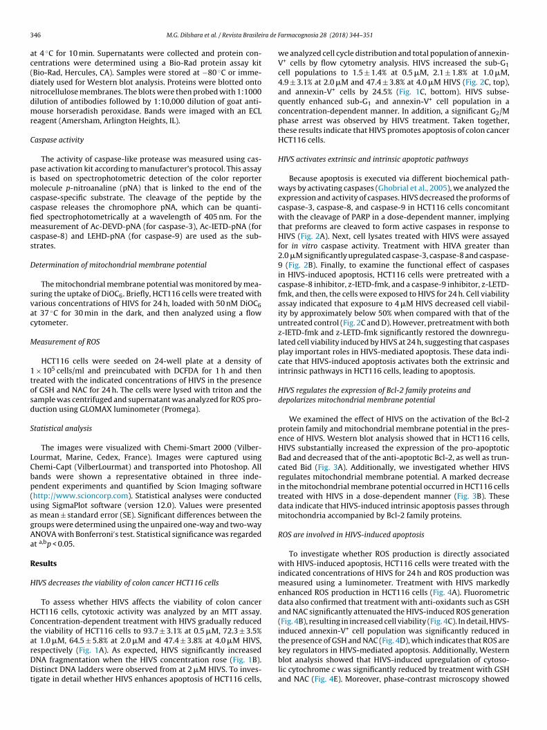
3 ra de
ac(dndmr
C
pimccfimcs
D
svac
M
1tosd
S
LCbp(uagAa
R
H
HCtarDDt
46 M.G. Dilshara et al. / Revista Brasilei
t 4 ◦C for 10 min. Supernatants were collected and protein con-entrations were determined using a Bio-Rad protein assay kitBio-Rad, Hercules, CA). Samples were stored at −80 ◦C or imme-iately used for Western blot analysis. Proteins were blotted ontoitrocellulose membranes. The blots were then probed with 1:1000ilution of antibodies followed by 1:10,000 dilution of goat anti-ouse horseradish peroxidase. Bands were imaged with an ECL
eagent (Amersham, Arlington Heights, IL).
aspase activity
The activity of caspase-like protease was measured using cas-ase activation kit according to manufacturer’s protocol. This assay
s based on spectrophotometric detection of the color reporterolecule p-nitroanaline (pNA) that is linked to the end of the
aspase-specific substrate. The cleavage of the peptide by theaspase releases the chromophore pNA, which can be quanti-ed spectrophotometrically at a wavelength of 405 nm. For theeasurement of Ac-DEVD-pNA (for caspase-3), Ac-IETD-pNA (for
aspase-8) and LEHD-pNA (for caspase-9) are used as the sub-trates.
etermination of mitochondrial membrane potential
The mitochondrial membrane potential was monitored by mea-uring the uptake of DiOC6. Briefly, HCT116 cells were treated witharious concentrations of HIVS for 24 h, loaded with 50 nM DiOC6t 37 ◦C for 30 min in the dark, and then analyzed using a flowytometer.
easurement of ROS
HCT116 cells were seeded on 24-well plate at a density of × 105 cells/ml and preincubated with DCFDA for 1 h and thenreated with the indicated concentrations of HIVS in the presencef GSH and NAC for 24 h. The cells were lysed with triton and theample was centrifuged and supernatant was analyzed for ROS pro-uction using GLOMAX luminometer (Promega).
tatistical analysis
The images were visualized with Chemi-Smart 2000 (Vilber-ourmat, Marine, Cedex, France). Images were captured usinghemi-Capt (VilberLourmat) and transported into Photoshop. Allands were shown a representative obtained in three inde-endent experiments and quantified by Scion Imaging softwarehttp://www.scioncorp.com). Statistical analyses were conductedsing SigmaPlot software (version 12.0). Values were presenteds mean ± standard error (SE). Significant differences between theroups were determined using the unpaired one-way and two-wayNOVA with Bonferroni′s test. Statistical significance was regardedt a,bp < 0.05.
esults
IVS decreases the viability of colon cancer HCT116 cells
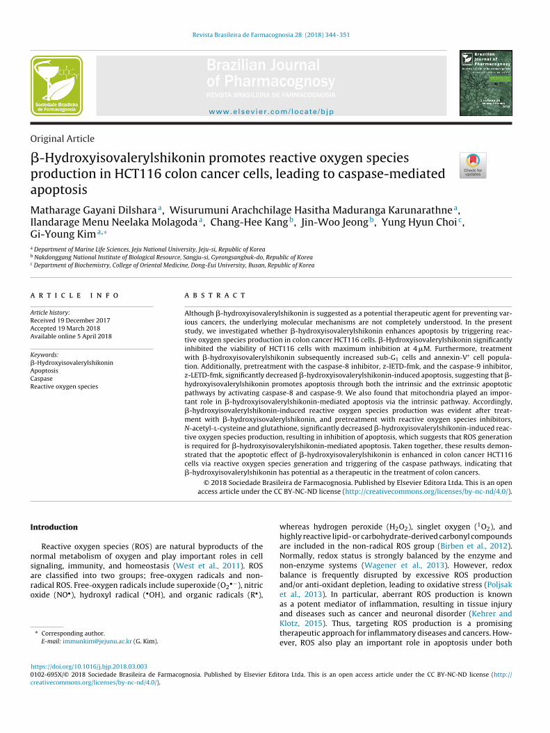
To assess whether HIVS affects the viability of colon cancerCT116 cells, cytotoxic activity was analyzed by an MTT assay.oncentration-dependent treatment with HIVS gradually reducedhe viability of HCT116 cells to 93.7 ± 3.1% at 0.5 �M, 72.3 ± 3.5%t 1.0 �M, 64.5 ± 5.8% at 2.0 �M and 47.4 ± 3.8% at 4.0 �M HIVS,
espectively (Fig. 1A). As expected, HIVS significantly increasedNA fragmentation when the HIVS concentration rose (Fig. 1B).istinct DNA ladders were observed from at 2 �M HIVS. To inves-igate in detail whether HIVS enhances apoptosis of HCT116 cells,
Farmacognosia 28 (2018) 344–351
we analyzed cell cycle distribution and total population of annexin-V+ cells by flow cytometry analysis. HIVS increased the sub-G1cell populations to 1.5 ± 1.4% at 0.5 �M, 2.1 ± 1.8% at 1.0 �M,4.9 ± 3.1% at 2.0 �M and 47.4 ± 3.8% at 4.0 �M HIVS (Fig. 2C, top),and annexin-V+ cells by 24.5% (Fig. 1C, bottom). HIVS subse-quently enhanced sub-G1 and annexin-V+ cell population in aconcentration-dependent manner. In addition, a significant G2/Mphase arrest was observed by HIVS treatment. Taken together,these results indicate that HIVS promotes apoptosis of colon cancerHCT116 cells.
HIVS activates extrinsic and intrinsic apoptotic pathways
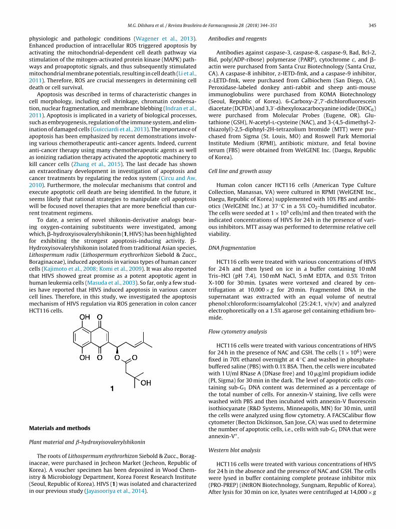
Because apoptosis is executed via different biochemical path-ways by activating caspases (Ghobrial et al., 2005), we analyzed theexpression and activity of caspases. HIVS decreased the proforms ofcaspase-3, caspase-8, and caspase-9 in HCT116 cells concomitantwith the cleavage of PARP in a dose-dependent manner, implyingthat preforms are cleaved to form active caspases in response toHIVS (Fig. 2A). Next, cell lysates treated with HIVS were assayedfor in vitro caspase activity. Treatment with HIVA greater than2.0 �M significantly upregulated caspase-3, caspase-8 and caspase-9 (Fig. 2B). Finally, to examine the functional effect of caspasesin HIVS-induced apoptosis, HCT116 cells were pretreated with acaspase-8 inhibitor, z-IETD-fmk, and a caspase-9 inhibitor, z-LETD-fmk, and then, the cells were exposed to HIVS for 24 h. Cell viabilityassay indicated that exposure to 4 �M HIVS decreased cell viabil-ity by approximately below 50% when compared with that of theuntreated control (Fig. 2C and D). However, pretreatment with bothz-IETD-fmk and z-LETD-fmk significantly restored the downregu-lated cell viability induced by HIVS at 24 h, suggesting that caspasesplay important roles in HIVS-mediated apoptosis. These data indi-cate that HIVS-induced apoptosis activates both the extrinsic andintrinsic pathways in HCT116 cells, leading to apoptosis.
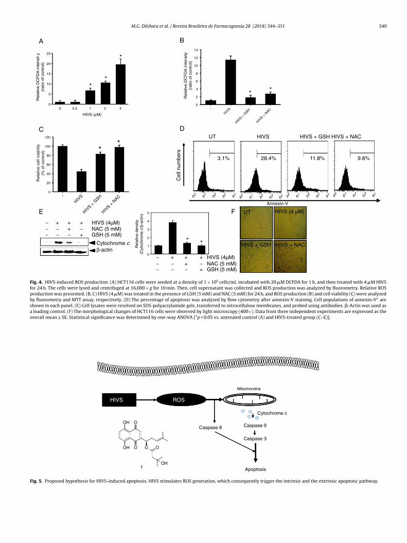
HIVS regulates the expression of Bcl-2 family proteins anddepolarizes mitochondrial membrane potential
We examined the effect of HIVS on the activation of the Bcl-2protein family and mitochondrial membrane potential in the pres-ence of HIVS. Western blot analysis showed that in HCT116 cells,HIVS substantially increased the expression of the pro-apoptoticBad and decreased that of the anti-apoptotic Bcl-2, as well as trun-cated Bid (Fig. 3A). Additionally, we investigated whether HIVSregulates mitochondrial membrane potential. A marked decreasein the mitochondrial membrane potential occurred in HCT116 cellstreated with HIVS in a dose-dependent manner (Fig. 3B). Thesedata indicate that HIVS-induced intrinsic apoptosis passes throughmitochondria accompanied by Bcl-2 family proteins.
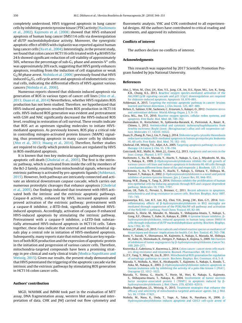
ROS are involved in HIVS-induced apoptosis
To investigate whether ROS production is directly associatedwith HIVS-induced apoptosis, HCT116 cells were treated with theindicated concentrations of HIVS for 24 h and ROS production wasmeasured using a luminometer. Treatment with HIVS markedlyenhanced ROS production in HCT116 cells (Fig. 4A). Fluorometricdata also confirmed that treatment with anti-oxidants such as GSHand NAC significantly attenuated the HIVS-induced ROS generation(Fig. 4B), resulting in increased cell viability (Fig. 4C). In detail, HIVS-induced annexin-V+ cell population was significantly reduced inthe presence of GSH and NAC (Fig. 4D), which indicates that ROS are
key regulators in HIVS-mediated apoptosis. Additionally, Westernblot analysis showed that HIVS-induced upregulation of cytoso-lic cytochrome c was significantly reduced by treatment with GSHand NAC (Fig. 4E). Moreover, phase-contrast microscopy showed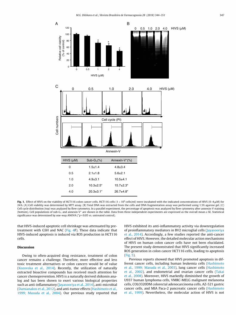
M.G. Dilshara et al. / Revista Brasileira de Farmacognosia 28 (2018) 344–351 347
Cell cycle (PI)
Cel
l num
bers
0 1.0 2.0 4.0
Annexin-V
C
Sub-G1(%) Annexin-V+(%)
HIVS (μM)
Rel
ativ
e ce
ll vi
abili
ty(%
of c
ontr
ol)
0
20
40
60
80
100
120
A
* *
*
B
0.5 HIVS (μM)
HIVS (μM)
HIVS (μM)
4.8±3.4
5.6±2.1
10.5±4 .1
15.7±2 .3*
26.7±4 .8*
1.5±1.4
2.1±1.8
4.9±3.1
10.3±2 .5*
20.3±3 .1*
0
0.5
1.0
2.0
4.0
4.0 2.0 1.0 0.5 0
4210.50
Fig. 1. Effect of HIVS on the viability of HCT116 colon cancer cells. HCT116 cells (1 × 10⁵ cells/ml) were incubated with the indicated concentrations of HIVS (0–4 �M) for24 h. (A) Cell viability was determined by MTT assay. (B) Total DNA was extracted from the cells and DNA fragmentation assay was performed using 1.5% agarose gel. (C)C nt, the( from
s
ttHc
D
ct(ecls(1
ell cycle distribution (top) was analyzed by flow cytometry. In a parallel experimebottom). Cell populations of sub-G1 and annexin-V+ are shown in the table. Data
ignificance was determined by one-way ANOVA (*p < 0.05 vs. untreated control).
hat HIVS-induced apoptotic cell shrinkage was attenuated by pre-reatment with GSH and NAC (Fig. 4F). These data indicate thatIVS-induced apoptosis is induced via ROS production in HCT116ells.
iscussion
Owing to often-acquired drug resistance, treatment of colonancer remains a challenge. Therefore, more effective and lessoxic treatment alternatives or colon cancers would be of valueKozovska et al., 2014). Recently, the utilization of naturallyxtracted bioactive compounds has received much attention forancer chemoprevention. HIVS is a naturally derived shikonin ana-
og and has been shown to exert various biological propertiesuch as anti-inflammatory (Jayasooriya et al., 2014), anti-microbialDamianakos et al., 2012), and anti-tumor effects (Hashimoto et al.,999; Masuda et al., 2004). Our previous study reported thatpercentage of apoptosis was analyzed by flow cytometry after annexin-V stainingthree independent experiments are expressed as the overall mean ± SE. Statistical
HIVS exhibited its anti-inflammatory activity via downregulationof proinflammatory mediators in BV2 microglial cells (Jayasooriyaet al., 2014). Accordingly, a few studies reported the anti-cancereffect of HIVS. However, the detailed molecular action mechanismsof HIVS on human colon cancer cells have not been elucidated.The present study demonstrated that HIVS significantly increasedROS generation in colon cancer HCT116 cells, leading to apoptosis(Fig. 5).
Previous reports showed that HIVS promoted apoptosis in dif-ferent cancer cells, including human leukemia cells (Hashimotoet al., 1999; Masuda et al., 2003), lung cancer cells (Hashimotoet al., 2002), and endometrial and ovarian cancer cells (Takaiet al., 2008). Moreover, HIVS markedly diminished the growth of
U937 human lymphoma cells, VMRC-MELG malignant melanomacells, COLO320DM colorectal adenocarcinoma cells, AZ-521 gastriccancer cells, and MIA Paca-2 pancreatic cancer cells (Hashimotoet al., 1999). Nevertheless, the molecular action of HIVS is not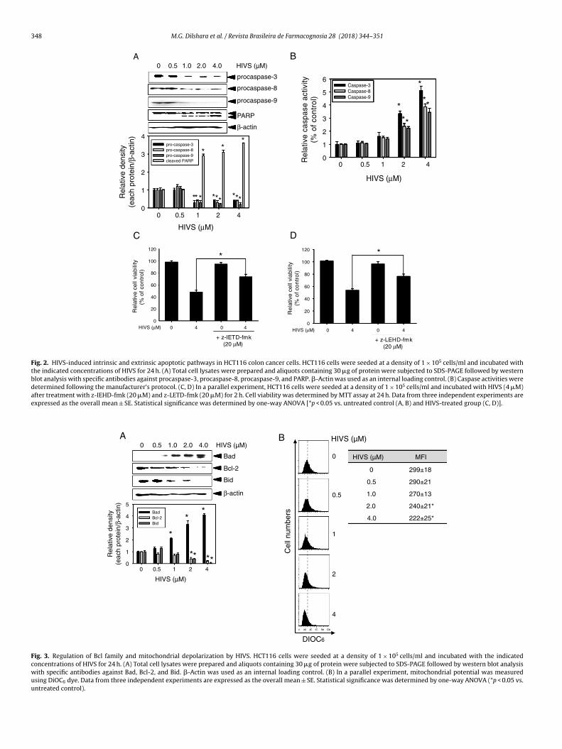
348 M.G. Dilshara et al. / Revista Brasileira de Farmacognosia 28 (2018) 344–351
HIVS (μM)
Rel
ativ
e ca
spas
e ac
tivity
(% o
f con
trol
)
0
1
2
3
4
5
6Caspase-3Caspase-8Caspase-9
***
*
**
HIVS (μM)
Rel
ativ
e ce
ll vi
abili
ty(%
of
con
tro
l)
0
20
40
60
80
100
120
+ z-IETD-f mk(20 μM)
+ z-LEHD-f mk(20 μM)
Rel
ativ
e ce
ll vi
abili
ty(%
of
con
tro
l)
0
20
40
60
80
100
120
HIVS (μM)
DHIVS (μM)
Rel
ativ
e de
nsity
(eac
h pr
otei
n/β-
actin
)
0
1
2
3
4pro-caspase-3pro-caspase-8pro-caspase-9cleaved PARP
C
* *
***
*
**
*
**
*
procaspase-9
procaspase-3
procaspase-8
β-actin
A
PAR P
B
* *
HIVS (µ M)4.0 2.0 1.0 0.5 0
4210.50
4210.50
44 00 44 00
Fig. 2. HIVS-induced intrinsic and extrinsic apoptotic pathways in HCT116 colon cancer cells. HCT116 cells were seeded at a density of 1 × 10⁵ cells/ml and incubated withthe indicated concentrations of HIVS for 24 h. (A) Total cell lysates were prepared and aliquots containing 30 �g of protein were subjected to SDS-PAGE followed by westernblot analysis with specific antibodies against procaspase-3, procaspase-8, procaspase-9, and PARP. �-Actin was used as an internal loading control. (B) Caspase activities weredetermined following the manufacturer’s protocol. (C, D) In a parallel experiment, HCT116 cells were seeded at a density of 1 × 10⁵ cells/ml and incubated with HIVS (4 �M)after treatment with z-IEHD-fmk (20 �M) and z-LETD-fmk (20 �M) for 2 h. Cell viability was determined by MTT assay at 24 h. Data from three independent experiments areexpressed as the overall mean ± SE. Statistical significance was determined by one-way ANOVA [*p < 0.05 vs. untreated control (A, B) and HIVS-treated group (C, D)].
B
DIO C6
Cel
l num
bers
0
HIVS (µ M)
HIVS (µM) MFI
0.5
1
2
4
Rel
ativ
e de
nsity
(eac
h pr
otei
n/β-
actin
)
0
1
2
3
4
5BadBcl-2Bid
*
**
****
A
Bcl-2
Bad
Bid
β-actin
HIVS (µ M)
HIVS (µ M)
4.0
4.0
2.0
2.0
1.0
1.0
0.5
0.5
0
0
4 2 1 0.5 0
222±25*
240±21*
270±13
290±21
299±18
Fig. 3. Regulation of Bcl family and mitochondrial depolarization by HIVS. HCT116 cells were seeded at a density of 1 × 10⁵ cells/ml and incubated with the indicatedconcentrations of HIVS for 24 h. (A) Total cell lysates were prepared and aliquots containing 30 �g of protein were subjected to SDS-PAGE followed by western blot analysiswith specific antibodies against Bad, Bcl-2, and Bid. �-Actin was used as an internal loading control. (B) In a parallel experiment, mitochondrial potential was measuredusing DiOC6 dye. Data from three independent experiments are expressed as the overall mean ± SE. Statistical significance was determined by one-way ANOVA (*p < 0.05 vs.untreated control).
M.G. Dilshara et al. / Revista Brasileira de Farmacognosia 28 (2018) 344–351 349
-
HIVS
HIVS +
GSH
HIVS +
NAC
Rel
ativ
e D
CF
DA
inte
nsity
(rat
io o
f co
ntro
l)
0
2
4
6
8
10
12
14
B
-
HIVS
HIVS +
GSH
HIVS +
NAC
Rel
ativ
e ce
ll vi
abili
ty(%
of
con
tro
l)
0
20
40
60
80
100
120
DCUT HIVS
Cel
l num
bers
Annexin-V
3.1% 28.4% 11.8% 9.8%
FE
−−− −
−
−−−−−
− −
−−
HIVS (4 µM) NAC (5 mM)
+ + +
+
+
+ + + +
+
GSH (5 mM)
Cytochrome c β-actin
UT HIVS (4 µ M)
HIVS + GSH HIVS + NA C
HIVS (μM)
Rel
ativ
e D
CF
DA
inte
nsit
y(r
atio
of co
ntr
ol)
0
5
10
15
20
25*
**
* *
**
A
Rel
ativ
e de
nsity
(Cyt
ochr
ome
c/β-
actin
)
0
1
2
3
4
5
HIVS (4µ M) NAC (5 mM)GSH (5 mM)
* *
4210.50
HIVS + GSH HIVS + NAC
Fig. 4. HIVS-induced ROS production. (A) HCT116 cells were seeded at a density of 1 × 10⁵ cells/ml, incubated with 20 �M DCFDA for 1 h, and then treated with 4 �M HIVSfor 24 h. The cells were lysed and centrifuged at 16,000 × g for 10 min. Then, cell supernatant was collected and ROS production was analyzed by fluorometry. Relative ROSproduction was presented. (B, C) HIVS (4 �M) was treated in the presence of GSH (5 mM) and NAC (5 mM) for 24 h, and ROS production (B) and cell viability (C) were analyzedby fluorometry and MTT assay, respectively. (D) The percentage of apoptosis was analyzed by flow cytometry after annexin-V staining. Cell populations of annexin-V+ areshown in each panel. (E) Cell lysates were resolved on SDS-polyacrylamide gels, transferred to nitrocellulose membranes, and probed using antibodies. �-Actin was used asa loading control. (F) The morphological changes of HCT116 cells were observed by light microscopy (400×). Data from three independent experiments are expressed as theoverall mean ± SE. Statistical significance was determined by one-way ANOVA [*p < 0.05 vs. untreated control (A) and HIVS-treated group (C–E)].
HIVS ROS
Caspase 9
Mitochond ria
Caspase 3
Apoptosis1
Cytochrome c
Caspase 8
Fig. 5. Proposed hypothesis for HIVS-induced apoptosis. HIVS stimulates ROS generation, which consequently trigger the intrinsic and the extrinsic apoptotic pathway.
3 ra de
cceaoalw25waGimc
g2pHcwltmii(ai
aste2vnevCpaiHPttnStimeWtii
A
ap
50 M.G. Dilshara et al. / Revista Brasilei
ompletely understood. HIVS triggered apoptosis in lung cancerells by inhibiting protein tyrosine kinase (PTK) activity (Hashimotot al., 2002). Kajimoto et al. (2008) showed that HIVS enhancedpoptosis of human lung cancer DMS114 cells via downregulationf dUTP nucleotidohydrolase activity. Moreover, the synergisticpoptotic effect of HIVS with cisplastin was reported against humanung cancer cells (Xu et al., 2004). Interestingly, in the present study,
e found that colon cancer HCT116 cells treated with 4 �M HIVS for4 h showed significant reduction of cell viability of approximately0%, whereas the percentage of sub-G1 phase and annexin-V+ cellsas approximately 20% each, suggesting that HIVS gently enhances
poptosis, resulting from the induction of cell stagnation or weak2/M phase arrest. Nishida et al. (2006) previously found that HIVS
nduced G0/G1 cell cycle arrest and apoptosis of endometriotic stro-al cells, indicating the differential effects of HIVS against various
ancers (Nishida et al., 2006).Numerous reports showed that shikonin induced apoptosis via
eneration of ROS in various types of cancer cell lines (Ahn et al.,013; Duan et al., 2014) Nevertheless, whether HIVS regulates ROSroduction has not been studied. Therefore, we hypothesized thatIVS-induced apoptosis resulted from ROS production in HCT116ells. HIVS-induced ROS generation was evident and pretreatmentith GSH and NAC significantly decreased the HIVS-induced ROS
evel, resulting in restoration of cell survival. These results indicatehat ROS act as upstream signaling molecules to initiate HIVS-
ediated apoptosis. As previously known, ROS play a critical rolen controlling mitogen-activated protein kinases (MAPK) signal-ng, thus promoting apoptosis in many cancer cells by shikoninAhn et al., 2013; Huang et al., 2014). Therefore, further studiesre required to clarify which protein kinases are regulated by HIVSn ROS-meditated apoptosis.
It is known that two key molecular signaling pathways governpoptotic cell death (Ghobrial et al., 2005). The first is the intrin-ic pathway, which is activated from inside the cell by members ofhe Bcl-2 family, resulting from mitochondrial signals, whereas thextrinsic pathway is activated by pro-apoptotic ligands (Ashkenazi,015). However, both pathways are intricately connected and acti-ate an identical downstream caspase cascade, which carries outumerous proteolytic cleavages that enhance apoptosis (Ghobrialt al., 2005). Our findings indicated that treatment with HIVS acti-ated both the intrinsic and the extrinsic apoptotic pathways.aspase-8 activity, enhanced by HIVS, increased apoptosis androved activation of the extrinsic pathway; pretreatment with
caspase-8 inhibitor, z-IETD-fmk, significantly inhibited HIVS-nduced apoptosis. Accordingly, activation of caspase-9 enhancedIVS-induced apoptosis by stimulating the intrinsic pathway.retreatment with a caspase-9 inhibitor, z-LETD-fmk substan-ially attenuated HIVS-induced apoptosis in HCT116 cells. Takenogether, these data indicate that external and mitochondrial sig-als play a central role in initiation of HIVS-mediated apoptosis.ubsequently, many reports state that mitochondria are key regula-ors of both ROS production and the expression of apoptotic proteinn the initiation and progression of various cancer cells. Therefore,
itochondria-targeted compounds have been a promising strat-gy in pre-clinical and early clinical trials (Modica-Napolitano andeissig, 2015). Given our results, the present study demonstrated
hat HIVS potentiated the triggering of the apoptotic cascade via thentrinsic and the extrinsic pathways by stimulating ROS generationn HCT116 colon cancer cells.
uthors’ contribution
MGD, WAHMK and IMNM took part in the evaluation of MTTssay, DNA fragmentation assay, western blot analysis and inter-retation of data. CHK and JWJ carried out flow cytometry and
Farmacognosia 28 (2018) 344–351
fluorometic analysis. YHC and GYK contributed to all experimen-tal designs. All the authors have contributed to critical reading andcomments, and approved its submission.
Conflicts of interest
The authors declare no conflicts of interest.
Acknowledgments
This research was supported by 2017 Scientific Promotion Pro-gram funded by Jeju National University.
References
Ahn, J., Won, M., Choi, J.H., Kim, Y.S., Jung, C.R., Im, D.S., Kyun, M.L., Lee, K., Song,K.B., Chung, K.S., 2013. Reactive oxygen species-mediated activation of theAkt/ASK1/p38 signaling cascade and p21 (Cip1) downregulation are requiredfor shikonin-induced apoptosis. Apoptosis 18, 870–881.
Ashkenazi, A., 2015. Targeting the extrinsic apoptotic pathway in cancer: lessonslearned and future directions. J. Clin. Invest. 125, 487–489.
Birben, E., Sahiner, U.M., Sackesen, C., Erzurum, S., Kalayci, O., 2012. Oxidative stressand antioxidant defense. World Allergy Organ. J. 5, 9–19.
Circu, M.L., Aw, T.Y., 2010. Reactive oxygen species, cellular redox systems, andapoptosis. Free Radic. Biol. Med. 48, 749–762.
Damianakos, H., Kretschmer, N., Syklowska-Baranek, K., Pietrosiuk, A., Bauer, R.,Chinou, I., 2012. Antimicrobial and cytotoxic isohexenylnaphthazarins fromArnebia euchroma (Royle) Jonst. (Boraginaceae) callus and cell suspension cul-ture. Molecules 17, 14310–14322.
Duan, D., Zhang, B., Yao, J., Liu, Y., Fang, J., 2014. Shikonin targets cytosolic thioredoxinreductase to induce ROS-mediated apoptosis in human promyelocytic leukemiaHL-60 cells. Free Radic. Biol. Med. 70, 182–193.
Ghobrial, I.M., Witzig, T.E., Adjei, A.A., 2005. Targeting apoptosis pathways in cancertherapy. CA Cancer J. Clin. 55, 178–194.
Guicciardi, M.E., Malhi, H., Mott, J.L., Gores, G.J., 2013. Apoptosis and necrosis in theliver. Compr. Physiol. 3, 977–1010.
Hashimoto, S., Xu, M., Masuda, Y., Aiuchi, T., Nakajo, S., Cao, J., Miyakoshi, M., Ida,Y., Nakaya, K., 1999. �-Hydroxyisovalerylshikonin inhibits the cell growth ofvarious cancer cell lines and induces apoptosis in leukemia HL-60 cells througha mechanism different from those of Fas and etoposide. J. Biochem. 125, 17–23.
Hashimoto, S., Xu, Y., Masuda, Y., Aiuchi, T., Nakajo, S., Uehara, Y., Shibuya, M.,Yamori, T., Nakaya, K., 2002. �-Hydroxyisovalerylshikonin is a novel and potentinhibitor of protein tyrosine kinases. Jpn. J. Cancer Res. 93, 944–951.
Huang, W.R., Zhang, Y., Tang, X., 2014. Shikonin inhibits the proliferation of humanlens epithelial cells by inducing apoptosis through ROS and caspase-dependentpathway. Molecules 19, 7785–7797.
Indran, I.R., Tufo, G., Pervaiz, S., Brenner, C., 2011. Recent advances in apoptosis,mitochondria and drug resistance in cancer cells. Biochim. Biophys. Acta 1807,735–745.
Jayasooriya, R.G., Lee, K.T., Lee, H.J., Choi, Y.H., Jeong, J.W., Kim, G.Y., 2014. Anti-inflammatory effects of �-hydroxyisovalerylshikonin in BV2 microglia aremediated through suppression of the PI3K/Akt/NF-�B pathway and activationof the Nrf2/HO-1 pathway. Food Chem. Toxicol. 65, 82–89.
Kajimoto, S., Horie, M., Manabe, H., Masuda, Y., Shibayama-Imazu, T., Nakajo, S.,Gong, X.F., Obama, T., Itabe, H., Kakaya, K., 2008. A tyrosine kinase inhibitor, �-hydroxyisovalerylshikonin, induced apoptosis in human lung cancer DMS114cells through reduction of dUTP nucleotidohydrolase activity. Biochim. Biophys.Acta 1782, 41–50.
Kehrer, J.P., Klotz, L.O., 2015. Free radicals and related reactive species as mediators oftissue injury and disease: implications for health. Crit. Rev. Toxicol. 45, 765–798.
Komi, Y., Suzuki, Y., Shimamura, M., Kajimoto, S., Nakajo, S., Masuda, M., Shibuya,M., Itabe, H., Shimokado, K., Oettgen, P., Nakaya, K., Kojima, S., 2009. Mechanismof inhibition of tumor angiogenesis by �-hydroxyisovalerylshikonin. Cancer Sci.100, 269–277.
Kozovska, Z., Gabrisova, V., Kucerova, L., 2014. Colon cancer: cancer stem cells mark-ers, drug resistance and treatment. Biomed. Pharmacother. 68, 911–916.
Li, Z.Y., Yang, Y., Ming, M., Liu, B., 2011. Mitochondrial ROS generation for regulationof autophagic pathways in cancer. Biochem. Biophys. Res. Commun. 414, 5–8.
Masuda, Y., Nishida, A., Hori, K., Hirabayashi, T., Kajimoto, S., Nakajo, S., Kondo, T.,Aska, M., Nakaya, K., 2003. �-Hydroxyisovalerylshikonin induces apoptosis inhuman leukemia cells by inhibiting the activity of a polo-like kinase 1 (PLK1).Oncogene 22, 1012–1023.
Masuda, Y., Shima, G., Aiuchi, T., Horie, M., Hori, K., Nakajo, S., Kajimoto,S., Shibayama-Imazu, T., Nakaya, K., 2004. Involvement of tumor necrosisfactor receptor-associated protein 1 (TRAP1) in apoptosis induced by �-hydroxyisovalerylshikonin. J. Biol. Chem. 279, 42503–42515.
Modica-Napolitano, J.S., Weissig, V., 2015. Treatment strategies that enhance theefficacy and selectivity of mitochondria-targeted anticancer agents. Int. J. Mol.Sci. 16, 17394–17421.
Nishida, M., Nasu, K., Ueda, T., Yuge, A., Takai, N., Narahara, H., 2006. �-Hydroxyisovalerylshikonin induces apoptosis and G0/G1 cell-cycle arrest of

ra de F
P
T
W
lung cancer DMS114 cells via a tyrosine kinase-dependent pathway. Oncology
M.G. Dilshara et al. / Revista Brasilei
endometriotic stromal cells: a preliminary in vitro study. Hum. Reprod. 21,2850–2856.
oljsak, B., Suput, D., Milisav, I., 2013. Achieving the balance between ROS andantioxidants: when to use the synthetic antioxidants. Oxid. Med. Cell Longev.,http://dx.doi.org/10.1155/2013/956792.
akai, N., Ueda, T., Nishida, M., Nasu, K., Narahara, H., 2008. �-Hydroxyisovalerylshikonin has a profound anti-growth activity in humanendometrial and ovarian cancer cells. Gynecol. Oncol. 109, 107–114.
agener, F.A., Carels, C.E., Lundvig, D.M., 2013. Targeting the redox balance ininflammatory skin conditions. Int. J. Mol. Sci. 14, 9126–9167.
armacognosia 28 (2018) 344–351 351
West, A.P., Shadel, G.S., Ghosh, S., 2011. Mitochondria in innate immune responses.Nat. Rev. Immunol. 11, 389–402.
Xu, Y., Kajimoto, S., Nakajo, S., Nakaya, K., 2004. �-Hydroxyisovalerylshikonin andcisplatin act synergistically to inhibit growth and to induce apoptosis of human
66, 67–75.Zhang, L., Wang, K., Lei, Y., Li, Q., Nice, E.C., Huang, C., 2015. Redox signaling: potential
arbitrator of autophagy an apoptosis in therapeutic response. Free Radic. Biol.Med. 89, 452–465.