Embed Size (px)
Citation preview

Foreign bodies are commonly encountered in emergency departments.
Children between the ages of 1 and 4 years sometimes insert foreign bodies into one or both nostrils , but this also affects adults especially those with mental retardation or psychiatric illness.
Children interest in exploring their bodies make them more prone to insert foreign bodies in their nostrils ,in addition they may also insert foreign bodies to relieve preexisting nasal mucosal irritation or epistaxis .
A benign nasal foreign body may seem, it harbors the potential for morbidity or even mortality if the object is dislodged into the airway.

FOREIGN BODY NOSE,RHINOLITH,NASAL MYIASIS

The most common location for nasal foreign bodies to lodge are just anterior to the middle turbinate or below the inferior turbinate.


Unilateral foreign bodies affect the right side about as twice the as often compared to the left. This may be due to a preference of right handed individuals to insert objects in their right naris .

Foreign bodies can be classified into either inorganic or organic. Inorganic materials are typically plastic or metal;common examples include beads and small parts from toys. These materials are often asymptomatic and may be discovered incidentally. Organic foreign bodies, including food, wood and sponge ,tend to be more irritating to the nasal mucosa and thus may produce earlier symptoms.
Another classification is : Hard(such as buttons,beads,or ball bearings) or soft (such as paper,cotton,wool,rubber,or other vegetable materials); the latter ,being as a rule more irritating give rise to symptoms more quickly.

It could be : 1. inorganic , inert (plastic). 2. organic (bean). 3. battery. 4. insect. 5. toxic material (naphthalene).

Injury from clumsy attempts at removal by unskilled persons.
Local spread of infection- sinusitis or meningitis.
Inhalation of foreign bodies leading to lung collapse and infection.
All foreign bodies harbor the potential for swallowing or airway obstruction if they are displaced posteriorely.


Nasal foreign bodies can cause damage to the nasal cavity and the surrounding structures. They can produce local inflammation which may result in pressure necrosis. This in turn can cause mucosal ulceration and erosion into blood vessels producing epistaxis.
The swelling can cause obstruction to sinus drainage and lead to secondary sinusitis.

Firmly impacted and unrecognized foreign bodies can in time become coated with calcium,magnesium,phosphate or carbonate and become a rhinolith.Rhinoliths are radio-opaque and are typically found on the floor of the nasal cavity.Rhinoliths can remain undetected for years and only upon growth do they produce symptoms that lead to their discovery.


The foreign body itself may cause irritation to the patient; however morbidity is primarily caused by the resulting inflammation ,mucosal damage and extension into adjucent structures.
Reported complications include sinusitis, acute otitis media, nasal septal perforation, periorbital cellulitis, meningitis, acute epiglottitis, diphtheria, and tetanus.

1. A fearful child ;irritable, crying. 2. Unilateral foul smelling nasal
discharge, sometimes blood-stained. 3. Excoriation around the nostril. 4. Occasionally, X-Ray evidence.


In most cases, the insertion of the nasal foreign body (NFB) is witnessed, and the dilemma of diagnosis is eliminated. In one study, presentations over 48 hours after the time of insertion accounted for 14% of all cases. In addition to obtaining a thorough history from the patient and his or her primary guardian(s), it is important to interview all caretakers that have recently spent time with the patient (i.e., babysitters, counselors). Once the diagnosis is missed, the foreign body may not be detected for days, weeks, or even years.

Among the delayed presentations, the most common clinical scenario is unilateral nasal discharge .
Nevertheless, clinicians must entertain the diagnosis of NFB in all patients with nasal irritation, epistaxis, sneezing, snoring, sinusitis, stridor, wheezing, or fever.
Some authors even report discovering NFBs as the etiology of more unusual patient presentations, such as irritability, halitosis, or generalized bromhidrosis (body malodor).

The physical examination is the main diagnostic tool, and a cooperative patient is essential for success. Parents and staff may be needed to comfort and immobilize a child to allow for a thorough otorhinolaryngologic examination. Sedation is often helpful in the pediatric population.
Maximal visualization of the nasal cavity is obtained by wearing a head-lamp.
Some authors recommend positioning children younger than 5 years in a supine lying position and older children in a sitting "sniffing" position to allow optimal visualization. A nasal speculum may also help to view the nasal cavity, although some authors report less patient anxiety and equally good visualization by using one's thumb to pull the nose upward.

In addition to adequate inspection of the nasal cavity, assessing for complications of the nasal foreign body is important. Visualize the tympanic membranes for signs of acute otitis media, assess for sinusitis, check for nuchal rigidity in the toxic child, and auscultate the chest and neck for wheezing or stridor, which may be a clue of foreign body aspiration.
Lastly, looking for additional foreign bodies, whether they are in the nose or other body cavities, is important.

Sinusitis . Polyps. Tumor. Upper respiratory infection (URI). Unilateral choanal atresia.

In the case of a cooperative child it may be possible with head mirror or lamp and Thudicum’s speculum, to see and, with small nasal forceps or blunt hooks to remove the foreign body without general anesthetic.
Local analgesia and decongestion are helpful and may be applied in the form of a small cotton-wool swab wrung out in Lidocaine or Phenelephrine solution. Extreme care is necessary.


If a child came with an inert inorganic foreign body stuck in his nose and we couldn’t remove it from the first trial we do the following :
1. send him home and ask the parents to bring him back the next day to remove it under general anesthesia .
2. give him decongestant. 3. give him antibiotics.

The following should be removed immediately from the nose and can’t wait till the next day :
1. insect. 2. bean. 3. battery: the alkaline material inside it
can burn the nose and cause severe adhesions.
4. toxic materials ( naphthalene).

A refractory child should, from the onset , be regarded as a case necessitating general anesthesia . This must be administered by an experienced anesthetist , and it is usual to employ an endotracheal tube. The surgeon may then remove the foreign body and need have no fear that it will enter the trachea.
Rarely an adult complaining of nasal obstruction is found to have a large concretion blocking one side of the nose. This is a rhinolith and it consists of many layers of calcium and magnesium salts that have formed around a small central nucleus. The latter often contain a foreign body.

REMOVAL WITH EUSTACHIAN CATHETER

REMOVAL WITH FOGARTY CATHETER
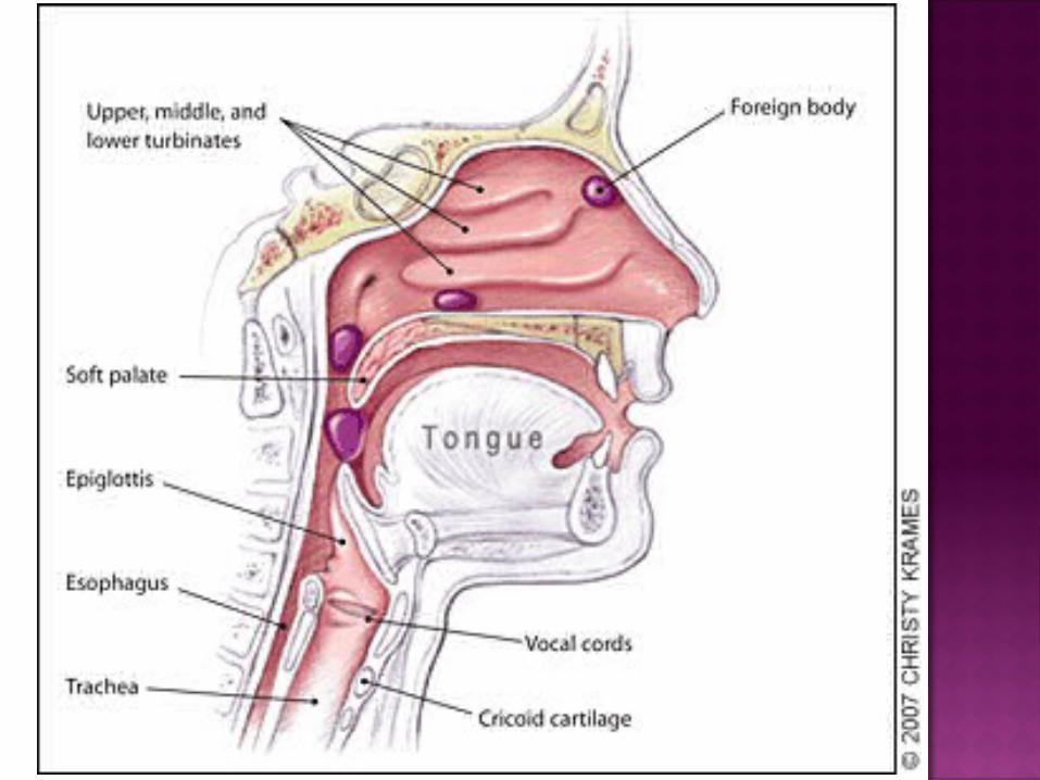
MYIASIS MAGGOTS ARE LARVAL FORM OF BLUE BOTTLE
FLY (CHRYSOMYIA) ATTRACTED BY FOUL SMELLING NASAL
DISCHARGE IN ATROPHIC RHINITIS, SYPHILIS, LEPROSY PURULENT SINUSITIS POST RADIOTHERAPY CA MAXILLA
FLY EGGS HATCH INTO 200 LARVAE WITHIN 24 HOURS
LARVAE CAUSE DESTRUCTION OF NOSE, NASOPHARYN, PALATE, PARANASAL SINUSES, ORBIT, SOFT TISSUE OF FACE
CASES MOSTLY SEEN BETWEEN AUGUST AND OCTOBER


CLINICAL FEATURES
INTENSE NASAL IRRITATION, SNEEZING, LACRIMATION, HEADACHE FOR FIRST THREE DAYS AFTER INFESTATION
NASAL OBSTRUCTION AND FOUL SMELLING BLOOD STAINED NASAL DISCHARGE
FACIAL PAIN AND PUFFY EYE LIDS AND LIPS CRAWLING SENSATION IN NOSE
(FORMICATION) MAGGOTS COMING OUT OF NOSE IN 3 TO 4
DAYS FISTULA ON NOSE/PALATE DEATH MAY OCCUR FROM MENINGITIS


TREATMENT ENDOSCOPIC REMOVAL OF MAGGOTS WITH
FORCEPS NASAL IRRIGATION WITH DILUTE CHLOROFORM
OR TUPENTINE OIL NASAL DOUCHING WITH NORMAL SALINE TO
REMOVE SLOUGH, CRUSTS AND DEAD MAGGOTS NASAL INSTILLATION OF LIQUID PARAFFIN FOR
LUBRICATION ISOLATION WITH MOSQUITO NET TO AVOID
CONTACT WITH FLIES ANTIBIOTICS TO CONTROL SECONDARY
INFECTIONS MAINTENANCE OF NASAL HYGIENE


QUESTIONS