Embed Size (px)
Citation preview
A Prototype of a 3D Bioprinter
Jakub Mielczarek1,a*, Grzegorz Gazdowicz1,b, Jakub Kramarz2,c,
Piotr Łątka3,d, Marcin Krzykawski4,e, Artur Miroszewski1,f, Paweł Pieczarko5,g,
Renata Szczelina4,h, Piotr Warchoł1,i, Sonia Wróbel1,j
1Garage of Complexity, Institute of Physics of the Jagiellonian University, ul. Łojasiewicza 11, Cracow, Poland
2Hackerspace Krakow, ul. Radziwiłłowska 20/2, 31-026 Cracow, Poland
3Faculty of Chemistry of the Jagiellonian University, ul. R. Ingardena 3, 30-060 Cracow, Poland
4Department of Immunology, Jagiellonian University College of Medicine, Czysta 18,
31-121 Cracow, Poland
5AGH University of Science and Technology, 30 Mickiewicza Av., 30-059 Cracow, Poland
[email protected], [email protected], [email protected],
[email protected], [email protected], [email protected], [email protected], [email protected], [email protected],
Keywords: 3D bioprinting, 3D printing, biomaterials
Abstract. 3D bioprinting is an innovative method of manufacturing three-dimensional tissue-like
structures. The method is based on a layer-by-layer deposition of biocompatible materials
successively forming a scaffold for living cells. The technology allows to fabricate complicated
tissue morphology, including vascular-like networks. The range of potential applications of 3D
bioprinting is immense: from drug testing, across regenerative medicine, to organ transplantation.
In this paper, we describe a prototype of a 3D bioprinter utilizing gelatin methacrylate (GelMA)
doped with a photoinitiator as the printing substance. Biological requirements for the material, its
synthesis and application adequacy for the bioprinting process are discussed. Technical details of
the mechanical construction of the bioprinter and its control system are presented.
Introduction. 3D bioprinting is a science fiction technology becoming a reality before our very
eyes. Successfully applied to print organs such as hearts, kidneys or livers, this technology has
potential to influence many people’s lives. Moreover, augmenting the usual 2D cell cultures (used
in pre-clinical drug testing) with a third dimension is predicted to improve the effectiveness of
pharmaceutics and reduce the overall cost of their development. Finally, the only limit for creating
various 3D bioconstructs, with different purposes in mind, is the human imagination. The
technology is not sufficiently mature yet, however the presently observed boom in the field inclines
one to make optimistic predictions for a not so distant future.
While artificial manufacturing of complex living organs is still a distant goal, 3D printing
techniques are already widely used in medicine. In particular, 3D printing has various applications
in the area of prosthetics. Materials such as polylactic acids (PLA), hydroxyapatite and bioactive
glasses are in common use [1]. Furthermore, custom, tomographic and MRI scan based, 3D printed
inorganic models of organs, turn out to be very helpful for surgeons, who practice with them before
complicated operations (especially of children) [2].
The 3D bioprinting goes beyond the standard 3D printing methods employing the living
cells as a part of the printing material [3]. Sensitivity of the biological component on the
environment and necessity of precise depositing of the `bio-ink’ makes the 3D bioprinting
Solid State Phenomena Vol 237 (2015) pp 221-226 Submitted: 2015-03-19© (2015) Trans Tech Publications, Switzerland Revised: 2015-04-14doi:10.4028/www.scientific.net/SSP.237.221 Accepted: 2015-05-06
All rights reserved. No part of contents of this paper may be reproduced or transmitted in any form or by any means without the written permission of TransTech Publications, www.ttp.net. (ID: 193.48.83.127-28/05/15,18:20:06)
technology challenging. Various types of cells and components forming a scaffold for them were
considered so far [3]. Exploiting stem cells is especially promising as it makes the implants less
likely to be rejected by the organism [4]. Such cells specialize their function in a given 3D
environment, potentially simplifying 3D printing of organs. Because of that number of different
types of `bio-inks’ needed is reduced. For reasons explained later, in our investigations we focus on
the use of somatic as well as cancer cells.
The version of 3D bioprinting considered in this paper does not concern printing with cells
themselves but with the suitable material forming a scaffold for the former. Such approach has been
studied in various configurations in the literature (Refs. [3,5]). Our objective is to be able to 3D
print a precisely specified scaffolding, equipped with a vascular system allowing a distribution of
nutrition to the whole bulk of the obtained biomimetic composite. To this end, we constructed a
prototype printer, which, layer-by-layer, deposits liquid hydrogel. The material is cured by the UV
light radiation, which stiffens the structure, reproducing a given 3D morphology. The fabricated
model is a base for the 3D cell culture. The technique employed for the project is analogous to the
Fused Deposition Modeling (FDM) method used in commercial 3D printers. Note however, that
different bio-printing methods can be considered as well, especially those utilizing piezoelectricity
(often used in engineering of bio-hybrid materials [6]) and lasers.
Our longterm goal is to develop a technology allowing the 3D bioprinting of custom scaffolds
with the precision reaching 10 microns and the volume of the diameter of a typical Petri dish (60
mm) cubed. We envision many different applications of the obtained 3D bio-constructs, such as:
pre-clinical drug testing, investigation of bio-toxicity of chemical components and studying various
biological mechanism and processes in simplified and reproducible configurations.
The Biological Aspects. The main challenge of the presented study was to find a material optimal
for 3D bioprinting. We have found that none of the commercially available materials fulfilled the
following requirements: affordable, easy to process, accessible, biocompatible, flexible, biomimetic
and easy to manipulate in print (fluid during the process but stiff in the cell culture). To resolve this
problem we have selected agar, agarose, cellulose, silicone beads and gelatin on the basis of their
non-toxicity, accessibility and low price. These were subsequently tested in various configurations
and concentrations as candidates for playing the role of the scaffold for the cell cultures.
The first criterion we tested was whether the material forms a stable skeleton for cells to
attach. To this end, we used the following cell culture facilities: incubation chamber, hood, cell
culture plates and culture media. We chose cancer cells (murine pancreatic cancer model: Pan02) as
a research model but we also selectively employed healthy renal, skin, aortic, blood, lung and brain
cells.
All cells were cultured in RPMI (Roswell Park Memorial Institute) medium with 10% FBS
(fetal bovine serum). The tests proved that only agar and gelatin are suitable for cell culture - only
these formed a stable skeleton for cells to grow. Therefore, the other candidate materials were
investigated as additives to the main substance.
Left with agar and gelatin, we begun experiments with the former. In particular, we looked
for cell division and cells growing on the surface of agar [7]. What we found was that cells can
grow in agar but do not attach to the agar surface. Cancer cells will create spheres but would not
grow as attached cells. Therefore, healthy cells would not be able to proliferate in the agar gel.
Hereby, we also confirmed that agar is not toxic to the cells. To further investigate the possibilities
of agar as a main constituent of the material for 3D cell culture development, we added portions of
cellulose, silicone beads and gelatin to the mixture. We hoped to create a material in which cells
floating in suspension could attach to charged surfaces inside the gel. These attempts also failed -
the cells didn't attach to any of the added substances. We therefore concluded that agar can be used
for culturing cells in suspension and thus mostly to investigate cancer cells. Unfortunately it also
means that agar cannot be used in 3D printing where we want to culture healthy cells together with
cancer cells to investigate intricate biological effects.
222 Advances in Manufacturing Engineering II

The material we were left with was gelatin. Its main disadvantage comes from the fact that
the cell cultures need to be kept in 37 deg C and gelatin is a fluid at this temperature. We decided
to test two chemical modifications of gelatin which allow to support the whole net structure of the
gel at higher temperatures: glutaraldehyde [8] and anhydrous methacrylamide [9]. Both of these
substances are chemically modifying the structure of gelatin making it insoluble (in case of
anhydrous methacrylamide additional initiation is needed).
First experiments showed that cells don't attach to the surface of gelatin. We started to work
on different modifications of gelatin surfaces (The details are not to be revealed due to the
intellectual property issues). These attempts led to the discovery of a modification procedure which
gave satisfactory results. We found that cancer cells and healthy cells can grow on the modified
surfaces. Thus, gelatin is suitable for 3D printing because it can be flexible during the printing
process and can be also harden with a chemical reaction or a physical processes. In the prototype of
the 3D bioprinter discussed below, the hydrogel network fromed by addition of the anhydrous
methacrylamide photoinitiated by exposition on the UV radiation.
The Material. Having the gelatin chosen to form the 3D scaffold, we have to introduce some
modifications to apply it in the printing procedure. The 3D printing requires from the material a
possibility of a sudden phase change from the liquid to the solid state while the material is
deposited. It is well known that the way to obtain this quality of the material one can modify gelatin
by doping it with methacrylic anhydride. The suitability of the so-called gelatin methacrylate
(GelMA) in fabrication of the 3D scaffolds for the biological purposes has been presented in Ref.
[10]. In our case, the synthesis of GelMA followed a modified procedure, reported previously in
Ref. [11].
In order to use GelMA as the printing material the following steps have to be made: 1)
Photoinitiator, which induces formation of the hydrogel network while exposing the material onto
UV radiation has to be added (See Fig. 1a). This step is crucial for the purpose of forming the solid
structure in the 3D printing procedure. In our studies, the material was doped with the
Irgacure2959 photo-initiator. The Irgacure2959 has been mixed with GelMA in the 1/10 proportion.
2) The 10 % solution of GelMA+Irgacure2959 with PBS has to be prepared. 3) Stirring (at the
temperature around 50 deg C) for 15-20 min is required to obtain a homogenous solution.
The process of formation of the hydrogel networks has been tested with use of the UV LED
source Nichia NSHU591B - UV 365 nm intended to be employed in the prototype of the 3D
bioprinter. The power emitted by the diode with the 10 degrees viewing angle is around 3 mW. The
drops (0,05 mL) of the solution have been exposed onto the UV light source placed at the distance
of 4 mm. The solution has been therefore exposed to approximately 750 mW/cm2
UV light flux. In
has been verified that material forms the hydrogel network after exposition times greater than
around 120 s. After this time the material became insoluble. The obtained times of curing have to be
reduced by at least by the factor of 100 in order to meet expectations of the final application. This
presumably can be achieved by increasing concentration of the photoinitiator and value of UV light
flux. Moreover, it is worth noticing that application of the UV irradiation in the process of
formation of the hydrogel network has significant restriction while applied to bioprinting. Namely,
living cells cannot be deposited simultaneously with the formation of scaffold due to destructive
impact of the UV radiation.
Construction of the 3D Bioprinter Prototype. The mechanical construction of the proposed 3D
bioprinter is similar to the standard FDM 3D printers. In case of the FDM method, the filament is
deposited layer-by-layer forming a topologically nontrivial 3D structure. The materials typically
used in the FDM approach are thermoplasitc polymers such as ABS and PLA. The materials
requires adequate heating of the extruder. In case of the later, the melting temperature is around
180 deg C, 105 deg C for the former.
The main difference between our construction and the FDM 3D printers is at the level of the
extruder. The printing material is now the methacrylated gelatin (GelMA). To form the 3D solid
Solid State Phenomena Vol. 237 223
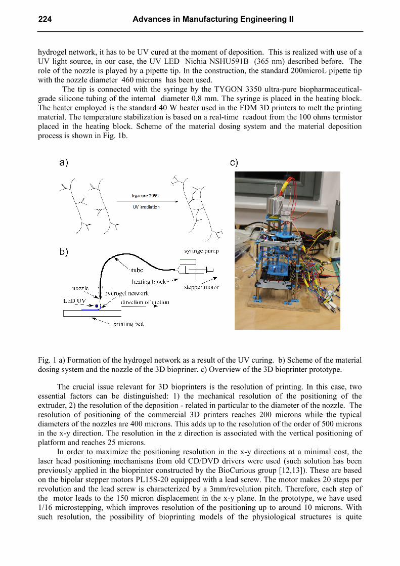
hydrogel network, it has to be UV cured at the moment of deposition. This is realized with use of a
UV light source, in our case, the UV LED Nichia NSHU591B (365 nm) described before. The
role of the nozzle is played by a pipette tip. In the construction, the standard 200microL pipette tip
with the nozzle diameter 460 microns has been used.
The tip is connected with the syringe by the TYGON 3350 ultra-pure biopharmaceutical-
grade silicone tubing of the internal diameter 0,8 mm. The syringe is placed in the heating block.
The heater employed is the standard 40 W heater used in the FDM 3D printers to melt the printing
material. The temperature stabilization is based on a real-time readout from the 100 ohms termistor
placed in the heating block. Scheme of the material dosing system and the material deposition
process is shown in Fig. 1b.
Fig. 1 a) Formation of the hydrogel network as a result of the UV curing. b) Scheme of the material
dosing system and the nozzle of the 3D biopriner. c) Overview of the 3D bioprinter prototype.
The crucial issue relevant for 3D bioprinters is the resolution of printing. In this case, two
essential factors can be distinguished: 1) the mechanical resolution of the positioning of the
extruder, 2) the resolution of the deposition - related in particular to the diameter of the nozzle. The
resolution of positioning of the commercial 3D printers reaches 200 microns while the typical
diameters of the nozzles are 400 microns. This adds up to the resolution of the order of 500 microns
in the x-y direction. The resolution in the z direction is associated with the vertical positioning of
platform and reaches 25 microns.
In order to maximize the positioning resolution in the x-y directions at a minimal cost, the
laser head positioning mechanisms from old CD/DVD drivers were used (such solution has been
previously applied in the bioprinter constructed by the BioCurious group [12,13]). These are based
on the bipolar stepper motors PL15S-20 equipped with a lead screw. The motor makes 20 steps per
revolution and the lead screw is characterized by a 3mm/revolution pitch. Therefore, each step of
the motor leads to the 150 micron displacement in the x-y plane. In the prototype, we have used
1/16 microstepping, which improves resolution of the positioning up to around 10 microns. With
such resolution, the possibility of bioprinting models of the physiological structures is quite
224 Advances in Manufacturing Engineering II

realistic. In particular, capillaries, the thinest body’s blood vessels have diameter of the order of 10
microns. The resolution of positioning in the z direction is even higher. In this direction the bipolar
stepper motor PG1521-0504B equipped with a gear giving 0,175 deg per step (around 2057 steps
per revolution) has been used. The motor is connected with the Tamiya 70171 3 mm diameter shalf
having the 0,5 mm/revolution pitch. This gives an incredibly low displacement of around 0,3
microns/step, which has a chance to be reduced even further, by an order of magnitude in the
microsteping mode. Due to backlashes in various parts of the mechanical construction, such
resolution is, however, not possible to achieve in the current prototype. Overviev of the prototype
is presented in Fig. 1c. It is worth stressing that in construction the rapid prototyping (3D printing
technique based on PLA) has been broadly applied. The disadvantage is, however, that precision
and quality of the 3D printed elements was not always sufficiently high.
The electronics employed in the constructions of the bioprinter have been adopted from those
used in many DIY 3D printers. Namely, the central control unit is the Arduino Mega development
board. The board is equipped with the ATmega2560 microcontroller (clocked at 16 MHz) and the
256 KB Flash memory. The board is connected to a computer via a USB port. The RAMPS 1.4
board with the Pololu stepper drivers is designed to work with the 12 V bipolar stepper motors. The
nominal voltages for the x-y motors PL15S-20 and the PG1521-0504B z-direction motor are 5 V
(the coil resistance is 10 ohms). Because the Pololu stepper drivers at 12 V, the voltage has been
sufficiently reduced by 15 ohms resistors.
At the software level, the suitably configured Marlin firmware [14] was used. In particular, it
turned to be essential to adjust number of steps per seconds the z-direction as well implement
geometry of the printing region. The Marlin firmware reads G-codes received from the slicer
software. The free open source Cura [15] slicer has been used during the tests. The Cura software
plays a role of the graphical interface with the device as well. 3D models in the stl format are
readed.
The Current State of the Prototype and Printing Results. So far only test with water and liquid
gelatine as the printing material have been executed. The tests proved efficiency of the positioning
as well as the material dosing systems. It has been shown that while the nozzle is placed close to the
bottom of the printing bed (Petri dish), the material can be smoothly distributed across the 2D
surface. Tests with arranging the material in the z-direction have not been performed yet.
Summary. This paper summarizes progress in the construction of a 3D bioprinter and the
development of the material it will use. The project described is undertaken at an interdisciplinary
laboratory under the name Garage of Complexity [16]. Our prototype is not at the operational state
yet, however numerous obstacles at various levels of the project have been overcome. In particular,
the material allowing for formation of the 3D cell cultures have been developed. Furthermore, the
sufficient precision of the nozzle positioning system has been achieved. The current UV curing
times of the photoactivated material are, however, insufficiently low for rapid 3D printing. With the
measured curing time of the order of 100 s, printing of 1 cm3 sample with the resolution of 100
microns would take around 3 years! In turn, the 1 cm3 sample printed with the target resolution of
10 microns corresponds to 109 3D pixels. The deposition+curing time has to be therefore reduced
below 0,1 millisecond in order to print the sample within a reasonable period of one day. Achieving
such values of times is a highly non-trivial task in the constructions adopting the FDM 3D printing
technology combined with UV curing. The alternative methods, such as those based on digital
optical projection stereolitography (DOPsL) [17], may be therefore more adequate for high
precision 3D bioprinting. Nevertheless, 3D biological structures printed with the resolution of the
order of 100 microns may still have broad applicability in medicine, pharmaceutics and
biotechnology.
Solid State Phenomena Vol. 237 225

Acknowledgements. Special thanks go to Krzysztof Stanik from PIRX for many valuable
suggestions at the early stage of the project. The research was supported from the FOCUS KNOW
grants 59/F/JM/2014 and 41/F/PW/2014.
References
[1] Sumita Bose, Sahar Vahabzadeh and Amit Bandyopadhyay, Bone tissue engineering using 3D
printing, Materials Today, Volume 16, Number 12 (2013) 496-504.
[2] Childrens Hospital Los Angeles. 3D printing makes heart surgery safer for children.
ScienceDaily. ScienceDaily, 29 January (2015).
[3] Sean V Murphy, Anthony Atala, 3D bioprinting of tissues and organs, Nature Biotechnology 32
(2014) 773–785.
[4] Savas Tasoglu and Utkan Demirci, Bioprinting for stem cell research, Vol. 31, 1 (2013) 10-19.
[5] Jordan S. Miller, et al. Rapid casting of patterned vascular networks for perfusable engineered
three-dimensional tissues, Nature Materials 11 (2012) 768–774.
[6] Ying Lie et al. Engineering of bio-hybrid materials by electrospinning polymer-microbe fibres,
PANS, vol. 106, no. 34 (2009) 14201-14206.
[7] Steven N. Anderson et al., A High-Throughput Soft Agar Assay for Identification of Anticancer
Compound, Journal of Biomolecular Screening (2007) 938-945.
[8] Yu-Cheng Ou et al., Attachment of Tumor Cells to the Micropatterns of Glutaraldehyde (GA)-
Crosslinked Gelatin, Sensors and Materials, Vol. 20, No. 8 (2008) 435–446.
[9]Thomas Billiet et al., The 3D printing of gelatin methacrylamide cell-laden tissue-engineered
constructs with high cell viability, Biomaterials 35 (2014) 49-62.
[10] Nichl JW et al. Cell-laden microengineered gelatin methacrylate hydrogels, Biomaterials
31, 21 (2010) 5536-5544.
[11] Van DenBulcke AI, Bogdanov B, De Rooze N, Schacht EH, Cornelissen M, Berghmans H.
Structural and rheological properties of methacrylamide modified gelatin hydrogels.
Biomacromolecules 1, 1 (2000) 31-28.
[12] Information on http://biocurious.org/projects/bioprinter/
[13] Information on http://www.wired.com/2013/01/diy-bio-printer/
[14] Information on http://reprap.org/wiki/Marlin
[15] Information on http://reprap.org/wiki/Cura
[16] Information on http://th-www.if.uj.edu.pl/ztuz/users/garage/
[17] A. Ping Zhang et al. Rapid fabrication of complex 3D extracellular microenvironments by
dynamic optical projection stereolithography, Adv Mater. 24, 31 (2012) 4266-4270.
226 Advances in Manufacturing Engineering II