Embed Size (px)
Citation preview
Zebrafish chemical screening reveals an inhibitor of Dusp6 thatexpands cardiac cell lineages
Gabriela Molina1,7,8, Andreas Vogt2,6,8, Ahmet Bakan3,8, Weixiang Dai4, Pierre Queiroz deOliveira2, Wade Znosko1, Thomas E. Smithgall1,6, Ivet Bahar3,6, John S. Lazo2,6, Billy W.Day4,5, and Michael Tsang1,9
1Department of Microbiology and Molecular Genetics, University of Pittsburgh, 3501 Fifth Ave,BST3-5062. Pittsburgh, PA 15213, USA.2Department of Pharmacology and Chemical Biology, University of Pittsburgh, 3501 Fifth Ave,BST3-5062. Pittsburgh, PA 15213, USA.3Department of Computational Biology, University of Pittsburgh, 3501 Fifth Ave, BST3-5062.Pittsburgh, PA 15213, USA.4Department of Pharmaceutical Sciences, University of Pittsburgh, 3501 Fifth Ave, BST3-5062.Pittsburgh, PA 15213, USA.5Department of Chemistry, University of Pittsburgh, 3501 Fifth Ave, BST3-5062. Pittsburgh, PA15213, USA.6Pittsburgh Molecular Libraries Screening Center, University of Pittsburgh, 3501 Fifth Ave,BST3-5062. Pittsburgh, PA 15213, USA.
AbstractThe dual specificity phosphatase 6 (Dusp6) functions as a feedback regulator of fibroblast growthfactor (FGF) signaling to limit the activity of extracellular signal regulated kinase (ERK) 1 and 2.We have identified a small molecule inhibitor of Dusp6, (E)-2-benzylidene-3-(cyclohexylamino)-2,3-dihydro-1H-inden-1-one (BCI), using a transgenic zebrafish chemicalscreen. BCI treatment blocked Dusp6 activity and enhanced FGF target gene expression inzebrafish embryos. Docking simulations predicted an allosteric binding site for BCI within thephosphatase domain. In vitro studies supported a model that BCI inhibits Dusp6 catalyticactivation by ERK2 substrate binding. A temporal role for Dusp6 in restricting cardiac progenitorsand controlling heart organ size was uncovered with BCI treatment at varying developmentalstages. This study highlights the power of in vivo zebrafish chemical screens to identify novelcompounds targeting Dusp6, a component of the FGF signaling pathway that has eludedtraditional high-throughput in vitro screens.
Fibroblast Growth Factors (FGFs) are members of a large family of secreted glycoproteinsthat serve important functions in development, proliferation and cellular homeostasis1.These ligands bind to single-pass transmembrane proteins of the receptor tyrosine kinaseclass to activate multiple signaling pathways including the rat sarcoma homologue (RAS)/mitogen-activated protein kinase (MAPK) cascade2. The wide-ranging biological roles of
9Correspondence should be addressed to M.T. ([email protected]) .7Current address: SRI, Center for Advanced Drug Research, Harrisonburg, VA, USA.8These authors contributed equally to this work.AUTHOR CONTRIBUTIONS G.M., A.V., A.B., P.Q., W.D., W.A.Z., and M.T. performed experiments. G.M., A.V. A.B., T.E.S.,J.S.L., I.B., B.W.D. and M.T., designed experiments and analyzed data. M.T. wrote the paper with help from A.V., A.B., T.E.S.,J.S.L., B.W.D., and I.B.
NIH Public AccessAuthor ManuscriptNat Chem Biol. Author manuscript; available in PMC 2010 March 1.
Published in final edited form as:Nat Chem Biol. 2009 September ; 5(9): 680–687. doi:10.1038/nchembio.190.
NIH
-PA Author Manuscript
NIH
-PA Author Manuscript
NIH
-PA Author Manuscript
FGFs and the multitude of signaling pathways activated by this family of ligands suggestthat FGF signaling must be tightly regulated. Dual specificity phosphatase 6 (Dusp6) (alsonamed MAPK Phosphatase 3), Sproutys (Spry1-4) and Sef (similar expression to FGFs)proteins function as RAS/MAPK pathway feedback attenuators1,3. Through their concertedactivities FGF signaling is adjusted to optimal levels in embryogenesis1,3. Sef and Spryproteins suppress RAS/MAPK signaling at multiple points within the pathway, while Dusp6inhibits the pathway only by dephosphorylation of one class of the MAPK family,extracellular signal-regulated kinase (ERK)1. Sef, Dusp6 and Sprouty depletion in zebrafishor gene knock-out in mice have revealed the requirement for these proteins to limit FGFsignaling during development and homeostasis1,4-6. The identification of small moleculesthat can reversibly modulate FGF signaling would provide useful tools to dissect the rolesfor this pathway in development that are not feasible with current genetic methods.
The zebrafish embryo is a vertebrate animal model well-suited for high-content smallmolecule screening7,8. Due to its small size, rapid development and ease of handling it ispossible to identify compounds that affect developmental processes and chemicalmodulators of signaling pathways in vivo8,9. Previous zebrafish chemical screens haverelied on the observations of phenotypes generated by exposure to small molecules. In onephenotypic screen, Dorsomorphin was identified as an inhibitor of Bone MorphogeneticProtein (BMP) as embryos exhibited axial patterning defects upon chemical treatment10.Subsequent studies utilizing Dorsomorphin in mice have revealed the importance of theBMP pathway in regulating iron metabolism10. Another example of the relevance ofzebrafish screens was the discovery that Prostaglandin E2 is a key regulator ofhaematopoietic stem cell (HSC) homeostasis11. Their studies have shown that this pathwayis conserved in vertebrates and provide the potential for using molecules to expand HSC torestore blood deficiencies in patients11.
The generation of transgenic reporter lines in zebrafish offers alternative in vivo tools forchemical screening. Reporters for FGF signaling have been generated and allow for the livevisualization of signaling activity during early development12. In this study, we performed achemical screen with an FGF reporter transgenic line and identified a small molecule, (E)-2-benzylidene-3-(cyclohexylamino)-2,3-dihydro-1H-inden-1-one (BCI, 1), that hyperactivatedFGF signaling. BCI was first described as a compound that suppressed NF-kB in cell-basedluciferase reporter assays13. Our analyses revealed that BCI blocked Dusp6 activity inzebrafish embryos and in cultured cells. Molecular modeling predicted an energeticallyfavorable site for BCI binding on the Dusp6 phosphatase domain and suggested a plausibleallosteric mechanism of action, which was supported by in vitro assays. Using BCI as achemical probe, we revealed that inhibition of Dusp6 activity during somitogenesisexpanded cardiac progenitors at the expense of endothelial lineages. These studies suggestthat Dusp6 functions as an attenuator of FGF signaling in the cardiac field to regulate heartorgan size.
ResultsA zebrafish chemical screen identifies a modulator of FGF signaling
We previously described the generation of a transgenic zebrafish line, (Tg(dusp6:EGFP)pt6)that expresses destabilized green fluorescent protein (d2EGFP) under the control of FGFsignaling12. Using Tg(dusp6:EGFP) embryos as a biosensor for FGF signaling, we screenedover 5000 diverse compounds assembled from chemical libraries for small moleculemodulators of this pathway. Five transgenic embryos at 24 hours post fertilization (hpf)were arrayed into each well of a 96-well plate containing test compounds at 10 μM.d2EGFP intensity in treated embryos was visually analyzed and compared to vehicle control(0.5% DMSO) after 6-8 hours. BCI enhanced d2EGFP fluorescence in a concentration-
Molina et al. Page 2
Nat Chem Biol. Author manuscript; available in PMC 2010 March 1.
NIH
-PA Author Manuscript
NIH
-PA Author Manuscript
NIH
-PA Author Manuscript
dependent manner and was detected as early as 2 hours post treatment (Fig. 1a-c, BCIchemical structure shown in Fig. 1l). To confirm that BCI hyperactivated FGF signaling, wetreated embryos prior to gastrulation (5hpf), and analyzed by whole mount in situhybridization the expression of ntl (zebrafish brachyury), a known FGF target gene14. Theexpression of ntl was greatly expanded within the notochord and the tailbud at the 6-somitestage in BCI-treated embryos (Fig. 1d-f). Similarly, BCI treatment from the 1- to 10-somitestage resulted in a marked increase in expression of another FGF target gene, dusp6, asshown by the expansion of prospective mid-hindbrain boundary (MHB), rhombomere4 (r4)and the tailbud (Fig. 1g,h). The expanded brain structures were confirmed as BCI increasedexpression of engrailed3 (eng3), which labels MHB, and krox20, which demarcates r3 andr5 identity, consistent with previous observations from FGF bead implantation studies (Fig.1i,j)15. To further demonstrate that BCI treatment hyperactivated FGF signaling, wemeasured an increase in the expression of sef and spry4 by semi-quantitative RT-PCR (Fig.1k; n=3 for each gene)16-18. These results confirmed that BCI enhanced FGF signaling inthe zebrafish embryo, resulting in the increased transcription of several FGF target genes.
We next determined the BCI structural features required to enhance FGF signaling. Twoanalogs, (E)-2-benzylidene-2,3-dihydro-1H-inden-1-one (BI, 2) lacking thecyclohexylamino group, and 3-(cyclohexylamino)-2,3-dihydro-1H-inden-1-one (ICD, 3)lacking the benzylidene group were synthesized (see Supplementary Materials forsynthesis). The cyclohexylamino and benzylidene substituents were both required inenhancing d2EGFP fluorescence, as analogs lacking either group were inactive (Fig. 2c,d).
BCI inhibits Dusp6To determine the mechanism for BCI’s activity and to identify a potential target, we probedwhere this compound acts within the RAS/MAPK pathway. In BCI-treated transgenicembryos, increased d2EGFP expression was restricted to embryonic regions where FGFs areexpressed (Fig 1b,c). Furthermore, BCI treatment did not induce d2EGFP expression in theMHB of Tg(dusp6:EGFP);ace mutant embryos, which are deficient in Fgf8 signaling (Fig.3d)19. Thus BCI did not enhance FGF signaling in the absence of ligand. We reasoned thatBCI could block a feedback attenuator of the FGF pathway, thereby resulting in a netincrease in transcription of target genes (Fig 3e). To test this model, we determined if BCIcould rescue phenotypes generated by ectopic expression of FGF inhibitors, Spry4, Dusp6and a dominant negative receptor, XFD in zebrafish (Supplementary Fig. 1a online)16,18.Injection of mRNA encoding dusp6, spry4, or XFD into 1-cell stage zebrafish embryosdecreased sef expression (Fig. 3g,j,o and Supplementary Fig. 1c). The addition of 5 μM BCIto dusp6-injected embryos rescued sef expression to control levels or higher (Fig. 3h,o). Incontrast, BCI treatment did not reverse the effects of spry4 or XFD mRNA, suggesting thatBCI directly inactivated Dusp6 (Fig. 3k,o and Supplementary Fig. 1d). To determine if BCIcould inhibit other Dusps, we first characterized zebrafish dusp5 and asked whether it couldsuppress FGF signaling similar to dusp620,21. Dusp5 has been shown to dephosphorylateactivated ERK (p-ERK) and ectopic expression of zebrafish dusp5 inhibited sef transcription(Fig. 3m,o)22. In contrast to observations with Dusp6 mRNA microinjections, BCI had littleor no effect in reversing the phenotype caused by Dusp5 over-expression (Fig. 3n,o). Theseobservations indicated that BCI was specific for Dusp6. Although both Dusp6 and Dusp5can dephosphorylate p-ERK and are highly conserved, their catalytic activities are quitedifferent22,23. Dusp6 phosphatase activity is subject to substrate binding and can becatalytically stimulated by ERK interaction22,23. This substrate-induced catalytic activityhas been described for several members of the Dusp family including Dusp1 (which issensitive to BCI, as shown below), and Dusp423-25. In contrast, Dusp5 is constitutivelyactivate and substrate binding has little consequence on catalytic rate22. Thus the differencewe noted with the ability of BCI to rescue Dusp6 but not Dusp5 over-expression in vivo
Molina et al. Page 3
Nat Chem Biol. Author manuscript; available in PMC 2010 March 1.
NIH
-PA Author Manuscript
NIH
-PA Author Manuscript
NIH
-PA Author Manuscript
suggested that BCI might suppress the activation of Dusp6 associated with substratebinding.
Since Dusp6 directly dephosphorylates p-ERK, BCI should restore p-ERK levels in Dusp6overexpressing cells. We tested this hypothesis in a cell-based chemical complementationassay26,27 in which HeLa cells were transiently transfected with Myc-tagged human Dusp6(Dusp6-Myc), stimulated with 12-O-tetradecanoylphorbol-13-acetate (TPA, 4), andimmunostained with anti-c-Myc (Fig. 4a,b) and anti-p-ERK antibodies (Fig. 4c,d),respectively. Upon TPA treatment, the RAS/MAPK pathway was activated leading to strongp-ERK staining in non-transfected cells, while in cells expressing Dusp6-Myc (Fig. 4a), p-ERK staining was abolished (Fig. 4c, Dusp6-Myc cells traced in yellow). BCI treatment ofDusp6-Myc transfected cells restored p-ERK levels after TPA addition, suggesting that BCIdirectly suppressed Dusp6-Myc function (Fig. 4d, Dusp6-Myc traced in yellow). In thisassay, BCI also inhibited human Dusp1, whose catalytic activity, like Dusp6, is induced bysubstrate binding (Fig. 4e)25. IC50 values for DUSP6 and DUSP1 inhibition from sixindependent experiments were 12.3 ± 4.0 μM and 11.5 ± 2.8 μM, respectively, consistentwith hyperactivation of FGF signaling and d2EGFP expression at these concentrations in thezebrafish embryo (Fig. 4e). In contrast, treatment with ICD did not block Dusp6 or Dusp1activity in the chemical complementation assays (Fig. 4e). Taken together, we have shownin biological systems BCI specifically inhibited Dusp1 and Dusp6, but not Dusp5.
We next addressed if BCI could directly inhibit Dusp6 activity in an in vitro pERK2dephosphorylation assay. Recombinant Dusp6 completely dephosphorylated pERK2 in vitroas determined by immunoblotting with pERK specific antibodies (Fig. 4f, lane 3). Additionof BCI prevented Dusp6-mediated pERK2 dephosphorylation as effectively as the generictyrosine phosphatase inhibitor sodium orthovanadate (Fig. 4f, lane 4 and 6, respectively).ICD did not block Dusp6 activity supporting the conclusion that BCI directly inhibitedDusp6 (Fig. 4f, lane 5). Since many known small molecule phosphatase inhibitors exhibitlow selectivity we determined whether BCI could suppress the phosphatase activity ofseveral related phosphatases. BCI did not block Cdc25B (Cell division cycle 25B), PTP1B(Protein Tyrosine Phosphatase 1B) or Dusp3/VHR activity, implicating specificity of BCI islimited to a set of MAPK Phosphatases (Supplementary Fig. 2 online)
Computational modeling predicts a BCI binding site within Dusp6Crystal structures of several Dusp catalytic domains have been determined 28-31. In eachcase, the phosphatase domain encompasses a five/six-stranded β-sheet surrounded by fiveα-helices. These structures enabled us to perform unbiased docking simulations32 toidentify potential BCI binding sites (Supplementary Methods online). BCI was dockedonto two different conformations of Dusp6 (MKP3): the low-activity form determined by X-ray crystallography (PDB ID: 1MKP)31 and the high-activity form obtained by homologymodeling using ORCHESTRAR (Tripos, Inc., St. Louis, MO). From cluster analysis of theresulting BCI-bound conformations, we identified a number of potential binding sites onboth the low- and high- activity forms (Fig. 5a, Supplementary Fig. 3 online). The mostfavorable site among them was further assessed by flexible docking33 using multiple Dusp6conformations generated by anisotropic network model (ANM) analysis34 and homologymodeling (Supplementary Methods online). BCI was predicted to preferentially fit withina crevice between the general acid loop and helix α7, rather than interacting directly withthe catalytic residues Asp262, Cys293, or Arg299. At this putative binding site, a closeinteraction of BCI with the backbone of the general acid loop and the side-chains of Trp264,Asn335 and Phe336 was predicted (Fig. 5b). Further docking simulations using a homologymodel of Dusp1 showed that BCI-Dusp1 interactions were comparable to those with Dusp6(Supplementary Fig. 4a online) rationalizing our observed activity data (Fig. 4e).
Molina et al. Page 4
Nat Chem Biol. Author manuscript; available in PMC 2010 March 1.
NIH
-PA Author Manuscript
NIH
-PA Author Manuscript
NIH
-PA Author Manuscript
In the zebrafish microinjection assays, BCI inhibited ectopic expression of dusp6 but notdusp5, exhibiting specificity toward certain members of this phosphatase family (Fig. 3). Wecompared the two-phosphatase crystal structures to understand how BCI can block Dusp6but not Dusp5. Structural superposition of Dusp5 and Dusp6 displayed the particular crevicein Dusp6 that accommodates BCI binding is not accessible in Dusp5 (Fig. 5c)22,23,29. As aresult, docking of BCI onto the same region of Dusp5 phosphatase domain resulted inenergetically less favorable interactions (Supplementary Fig. 4b online). The relativepositions of Asp262 and Asp232 in the respective phosphatases Dusp6 and Dusp5 differ by5 Å after optimal superposition of the two structures, suggesting that their basal activities aredetermined by the relative location of these catalytic residues (Fig. 5c)29. It was postulatedthat substrate binding to Dusp6 induces a conformational shift that reorients Asp262 towardsthe phosphatase loop, thereby creating a high activity enzyme35. In support of this model,mutation of Asp262 to asparagine did not abolish basal phosphatase activity, but suppressedcatalytic activation upon ERK binding31. To further understand BCI action on Dusp6mechanistically, we explored Dusp6 dynamics by particularly focusing on the ANM modesthat induce conformational changes at the general acid loop. Our analysis showed thatDusp6 possesses an intrinsic, structure-induced tendency to suitably reorient its general acidloop to position Asp262 closer to the phosphatase loop (Movie 1 and Movie 2 online)36.Therefore, we proposed that BCI binding to the accessible crevice in the low-activity formeffectively blocks the flexibility of this loop, thereby preventing the interaction of Asp262with the other catalytic residues. Such constraints on functional motions are likely to inhibitDusp6 activation induced by ERK binding.
BCI inhibits ERK2-mediated activation of Dusp6To test these modeling predictions, we measured the dephosphorylation of a small moleculephosphatase substrate, 3-O-methylfluorescein phosphate (OMFP, 5), by Dusp6 in thepresence or absence of ERK2. Docking simulations predicted that BCI and OMFP couldsimultaneously bind within the phosphatase active site with OMFP interfacing with the corecatalytic residues (Supplementary Fig. 5a online). This suggests that BCI would not blockbasal Dusp6 phosphatase activity toward OMFP. Indeed, at a concentration that inhibitedERK dephosphorylation in vitro (Fig. 4f), BCI did not inhibit basal Dusp6 activity(Supplementary Fig. 5b). Addition of ERK2 protein stimulated Dusp6 dephosphorylation ofOMFP three-fold and this enhancement was significantly inhibited in the presence of BCI(57% inhibition) (Fig. 5d). Increasing the ratio of ERK2 to Dusp6 (10:1) in the activationassay resulted in a 7-fold enhancement that was also suppressed by the addition of BCI(30% inhibition) (Supplementary Fig. 5c). These data suggest that BCI is an allostericinhibitor of Dusp6 that prevents the catalytic stimulation of phosphatase activity induced bysubstrate binding.
The role of Dusp6 and FGF in regulating heart sizeThe identification of a small molecule that blocks the biologically relevant activity of Dusp6and Dusp1 allowed us to probe the requirement for these enzymes in later developmentalprocesses. Given that BCI could potentially block related members of the Dusp family, weexamined the expression of other dusps in zebrafish. Detailed expression analyses of severaldusps have been described and include dusp4, dusp1, dusp7, dusp5, and dusp22a (seeSupplementary Methods and Supplementary Fig. 6 online) (Thisse, B. & Thisse, C (http://zfin.org))20,21,37,38. Of these only Dusp6 functions as a feedback regulator of FGF/MAPK/ERK signaling and is expressed within the anterior lateral plate mesoderm,supporting the idea that this phosphatase plays a role in heart development. In earlyembryogenesis, Dusp6 is an important regulator of FGF signaling; knock-down withantisense morpholinos results in embryo polarity defects, which precludes the study of
Molina et al. Page 5
Nat Chem Biol. Author manuscript; available in PMC 2010 March 1.
NIH
-PA Author Manuscript
NIH
-PA Author Manuscript
NIH
-PA Author Manuscript
Dusp6’s role in later development18. In contrast, small molecules permit the analysis at laterstages of development due to rapid and transient perturbation of their biological targets.
Using BCI as a chemical probe, we asked how inhibiting Dusp6 activity would alterpatterning and formation of the heart. The zebrafish heart develops from a small group ofcardiac progenitor cells that can be identified by 5hpf within the mesodermal layer of theblastula stage embryo39,40. During gastrulation, cardiac progenitor cells undergo cellularmigration to form two bilateral populations known as the anterior lateral plate mesoderm(ALPM) located just behind the MHB, and begin to express the transcription factors nkx2.5and gata440,41. Studies have described a role for Fgf8 in zebrafish heart development. Inembryos harboring an fgf8 mutation, both atria and ventricular cells are reduced42,43. Inagreement with the notion that FGF signaling plays a role in stipulating heart size, ectopicexpression of a constitutively activated FGF receptor (Fgfr1) during somitogenesis stagesexpanded cardiac tissue42. Therefore we used BCI to test if Dusp6 limits FGF signaling andrestricts cardiac progenitors and heart organ size. In BCI-treated embryos a caudalexpansion of gata4 in the ALPM was observed (compare Fig. 6a to 6b; 81%, n=16). Thegata4 caudal expansion of the ALPM corresponds to where cardiac progenitors are situatedat the 10-somite stage. Examination of nkx2.5 expression in BCI-treated embryos showedexpanded cardiac progenitor pools as compared to DMSO-treated embryos, confirming aspecific effect on heart precursors (compare Fig. 6d to c; 91%, n=11). While we noted anexpansion of cardiac progenitors, it was not clear if this event was at the expense of otherlineages. Recent studies have shown that there exists a repressive interaction between thevascular and hematopoietic precursors on cardiomyocyte progenitors that determine heartorgan size44. We analyzed expression of scl/tal1, a gene that is expressed in endothelial andblood lineages located within the rostral domain of the ALPM in BCI-treated embryos fromthe 1-somite stage. Inhibition of Dusp6 resulted in a marked reduction in scl expression,suggesting that activation of FGF signaling expanded cardiac tissue at the expense of bloodor endothelial progenitors (compare Fig. 6f to 6e; 93%, n=15). Likewise, etsrp, a marker forvascular fate was also reduced in BCI-treated embryos (data not shown). The loss ofendothelial and hematopoietic lineages was coupled with the concomitant expansion ofcardiac hand2 expression at the 10-somite stage (Fig. 6f; 32%, n=19). This surplus ofcardiac progenitors was also noted at the 18-somite stage by an increase in cells positive forcardiac myosin light chain 2 (cmlc2), which specifically labels differentiatedcardiomyocytes (Fig. 6h; 81% n=16). To test whether the expansion of cardiac progenitorsresulted in an increase in heart tissue, we analyzed treated embryos at larval stage. Embryoswere treated at 40% epiboly with BCI or DMSO, followed by compound washout the nextday and further incubated until the embryos reached 56hpf. In BCI-treated larvae, we noteda marked expansion in cardiac tissue (Fig. 6j,o). To define the critical period as to whenDusp6 activity limits heart organ size, we treated embryos at the 1- and 8-somite stages. Weobserved larger hearts at both time points, however the frequency was reduced in embryostreated at the later stage (Fig. 6o). In situ analysis with probes for ventricular myosin heavychain (vmhc) and cmlc2 confirmed that treated embryos exhibited enlarged hearts (Fig. 6l,n,and Supplementary Fig. 7 online). Expansion was particularly notable for ventricular tissue,known to be sensitive to Fgf8 signaling (Fig. 6l)42,43. These results indicate that inhibitionof Dusp6 by a small molecule inhibitor can induce an expansion of myocardial progenitorsthat ultimately increase heart size.
DiscussionThe zebrafish embryo offers distinct advantages over traditional in vitro and cell-basedchemical screens. With the generation of transgenic FGF reporter lines, it is possible toscreen for novel compounds that modulate this pathway in vivo. In addition, live embryoscreens allow for the elimination of toxic compounds and molecules that evoke non-specific
Molina et al. Page 6
Nat Chem Biol. Author manuscript; available in PMC 2010 March 1.
NIH
-PA Author Manuscript
NIH
-PA Author Manuscript
NIH
-PA Author Manuscript
effects on embryo differentiation. From a modest screen of approximately 5000 compounds,we identified BCI, a small molecule that enhanced FGF signaling. Subsequent in vitrophosphatase assays and docking simulations provided strong evidence that BCI suppressedthe ERK-induced activation of Dusp6.
The identification of BCI allowed us to directly probe the role of Dusp6 in heart formationduring a critical period when cardiac specific transcription factors begin to be expressed.The effects of BCI were consistent with studies when global activation of FGF signaling,which resulted in increased cardiac progenitors42. Treatment with BCI expanded the cardiacfield at the expense of endothelial lineages. The increase in cardiac progenitors resulted inenlarged hearts, suggesting that FGF signaling must be tightly regulated during this periodto allow for proper cardiac morphogenesis to occur. The role for Dusp6 in controlling heartorgan size is likely conserved with other vertebrates as disruption of Dusp6 was recentlyfound to cause enlarged hearts in mice6.
Previous large scale high-throughput screens for Dusp6 and Dusp1 inhibitors employed invitro assays with artificial substrates. Because these assays do not faithfully recapitulatephosphatase activity in a biological context, no specific Dusp6 inhibitors with in vivoactivity have been identified45. The phosphatase catalytic site is highly conserved across alltyrosine phosphatases and crystal structures have revealed shallow catalytic pockets. Thesestructural features have further hampered the identification of specific small moleculephosphatase inhibitors46. Small molecules targeting Dusp1 identified from in vitro screenshave exhibited promiscuous activity or lack biological activity47. However, with theidentification of a chemical Dusp inhibitor, it should be possible to rationally design newmolecules based on BCI to block substrate-induced Dusp function. This should offer highlyspecific compounds to probe the role of Dusps in development, and potentially providenovel agents for treatment of diseases that are dependent on FGF signaling such as woundrepair and regeneration48,49.
METHODSZebrafish chemical screens
All procedures involving zebrafish were reviewed and approved by the University ofPittsburgh Institutional Animal Care and Use Committee. Tg(dusp6:EGFP)pt6 embryos wereobtained by natural crossings and incubated at 28.5°C until they reached 24 hpf. Fivetransgenic embryos were placed into each well of a 96-well plate in 200 μl of E3, and a0.5% DMSO solution was added along with compound from each library at 10 μM. TheNCI diversity set (NCI/NIH), the Natural Products library (MicroSource Discovery SystemsInc.) and Phosphatase targeted set (ChemDiv Inc.) were screened in this study. (E)-2-Benzylidene-3-(cyclohexylamino)-2,3-dihydro-1H-inden-1-one (BCI; also known asNSC150117) was identified as a compound that enhanced fluorescence in treated transgenicembryos. Treated embryos were photographed under the same settings for exposure, gainand magnification for each picture using a MZFLIII (Leica) microscrope and fluorescentillumination for GFP using endow cube (Chroma Technology Corp.). Qimaging softwareand the Retiga Exi camera (Qimaging) were used to capture the images. Each experimentwas repeated three times to show reproducibility of the assay and at least four of the fivetreated embryos exhibited the same phenotype.
Zebrafish mRNA microinjectiondusp6 and XFD mRNA for microinjection studies were generated as previouslydescribed18. Both Dusp5 and Spry4 ORFs were amplified by RT-PCR from 24 hpfzebrafish with the following primers:
Molina et al. Page 7
Nat Chem Biol. Author manuscript; available in PMC 2010 March 1.
NIH
-PA Author Manuscript
NIH
-PA Author Manuscript
NIH
-PA Author Manuscript
Dusp5 Forward: 5′-AACTCGAGGCCATGAAGGTCTCCAGCATAGATTGCCG-3′
Dusp5 Reverse: 5′-AATCTAGATTAAGGCAGCGCAGTTATTGGACTC-3′
Spry4 Forward: 5′-ACTCGAGCCATGGAGTCAAGGGTTCCTCACCACATTC-3′
Spry4 Reverse: 5′-AATCTAGATCATGAGGCTTGTTTTTCTGGCTGAC-3′
Amplified PCR products were subcloned into pCS2+, sequenced verified and mRNAs weresynthesized as described previously. Embryos were injected with 500 pg mRNA at the 1-2cell stage, treated with 5 μM BCI at the 1000-cell stage and fixed at shield stage for in situhybridization.
Chemical complementation assays in HeLa cellsThese experiments were carried out essentially as described27. HeLa cells were obtainedfrom ATCC (Manassas, VA) and maintained in a humidified atmosphere of 5% CO2 at37°C, Dulbecco’s Minimum Essential Medium (DMEM) supplemented with 10% fetalbovine serum (FBS, HyClone), and 1% penicillin-streptomycin (Life Technologies, Inc.).Human c-Myc-Dusp6 (pSG5-PYST1) and Dusp1 (also known as CL100) were both kindlyprovided by Dr. Stephen Keyse (Cancer Research, UK)50. Full length Dusp1 was subclonedinto pcDNA3.1 for ectopic expression in mammalian cells50. HeLa cells (2,000) were platedin the wells of a collagen-coated 384-well plate (Falcon Biocoat) in the presence of FuGene6 (Roche Biosciences) and c-Myc-Dusp6 or c-Myc-Dusp1 as described27. After 20 h inculture, cells were treated in quadruplicate wells for 15 min with ten two-fold concentrationgradients of BCI or ICD and stimulated for 15 min. with TPA (500 ng/ml). Cells were fixedand stained with Hoechst 33342 in 4% formaldehyde, permeabilized, and immunostainedwith a mixture of anti-pERK (1:200 dilution, Cell Signaling Technology) and anti-c-Myc(1:100 dilution, Santa Cruz Biotechnology) antibodies. Positive pERK and c-Myc-DUSPsignals were visualized with AlexaFluor-594 (pERK) and Alexa-488 (c-Myc) conjugatedsecondary antibodies, respectively. Plates were analyzed by three-channel multiparametricanalysis for p-ERK and c-Myc-DUSP intensities in an area defined by nuclear staining usingthe Compartmental Analysis Bioapplication on an ArrayScan II high-content reader(Cellomics). Restoration of ERK phosphorylation by BCI in Dusp6 overexpressing cells wasquantified by Kolmogorov-Smirnov (KS) statistics as described previously using DUSP-transfected and vehicle treated control wells27. One thousand individual cells were gated forDusp-Myc expression based on c-Myc immunostaining and analyzed for ERKphosphorylation. A pERK cumulative distribution function (cdf) was established for eachcondition and compared to a reference cdf from Dusp-Myc expressing and vehicle-treatedcells. High KS values denote large differences in ERK phosphorylation levels comparedwith vehicle control and indicate suppression of Dusp activity. To quantify restoration ofErk phosphorylation in the Dusp expressing cells after compound treatment, KS values foreach condition were normalized to the average KS value from four wells transfected withDusp1 or Dusp6 and treated with vehicle.
Detailed Material and Methods on in vitro phosphatase assays, molecular modeling, andchemical synthesis of BCI and related analogs are listed in Supplementary Methods online.
Supplementary MaterialRefer to Web version on PubMed Central for supplementary material.
AcknowledgmentsThe project described was supported in part by Award Number R01HL088016 to M.T. from the National Heart,Lung, and Blood Institute. The content is solely the responsibility of the authors and does not necessarily represent
Molina et al. Page 8
Nat Chem Biol. Author manuscript; available in PMC 2010 March 1.
NIH
-PA Author Manuscript
NIH
-PA Author Manuscript
NIH
-PA Author Manuscript
the official views of the National Heart, Lung, And Blood Institute or the National Institutes of Health. This workwas also supported by NIH grants HD053287, CA52995, MH074411, and CA78039 and the Fiske Drug DiscoveryFund. We thank N. Hukriede, M. Rebagliati and I. Dawid for critical reading of the manuscript. Michael S.Poslusney for assistance in the syntheses. The authors thank Dr Robert Schultz, Developmental TherapeuticsProgram, NCI, NIH for providing the NCI diversity set and samples of individual compounds.
Reference1. Thisse B, Thisse C. Functions and regulations of fibroblast growth factor signaling during
embryonic development. Dev Biol. 2005; 287:390–402. [PubMed: 16216232]
2. Dailey L, Ambrosetti D, Mansukhani A, Basilico C. Mechanisms underlying differential responsesto FGF signaling. Cytokine Growth Factor Rev. 2005; 16:233–47. [PubMed: 15863038]
3. Tsang M, Dawid IB. Promotion and attenuation of FGF signaling through the Ras-MAPK pathway.Sci STKE. 2004; 2004:pe17. [PubMed: 15082862]
4. Abraira VE, et al. Changes in Sef levels influence auditory brainstem development and function. JNeurosci. 2007; 27:4273–82. [PubMed: 17442811]
5. Li C, Scott DA, Hatch E, Tian X, Mansour SL. Dusp6 (Mkp3) is a negative feedback regulator ofFGF-stimulated ERK signaling during mouse development. Development. 2007; 134:167–76.[PubMed: 17164422]
6. Maillet M, et al. DUSP6 (MKP3) Null Mice Show Enhanced ERK1/2 Phosphorylation at Baselineand Increased Myocyte Proliferation in the Heart Affecting Disease Susceptibility. J Biol Chem.2008; 283:31246–55. [PubMed: 18753132]
7. Vogt A, et al. Automated image-based phenotypic analysis in zebrafish embryos. Dev Dyn. 2009;238:656–63. [PubMed: 19235725]
8. Zon LI, Peterson RT. In vivo drug discovery in the zebrafish. Nat Rev Drug Discov. 2005; 4:35–44.[PubMed: 15688071]
9. Peterson RT, Link BA, Dowling JE, Schreiber SL. Small molecule developmental screens reveal thelogic and timing of vertebrate development. Proc Natl Acad Sci U S A. 2000; 97:12965–9.[PubMed: 11087852]
10. Yu PB, et al. Dorsomorphin inhibits BMP signals required for embryogenesis and iron metabolism.Nat Chem Biol. 2008; 4:33–41. [PubMed: 18026094]
11. North TE, et al. Prostaglandin E2 regulates vertebrate haematopoietic stem cell homeostasis.Nature. 2007; 447:1007–11. [PubMed: 17581586]
12. Molina GA, Watkins SC, Tsang M. Generation of FGF reporter transgenic zebrafish and theirutility in chemical screens. BMC Dev Biol. 2007; 7:62. [PubMed: 17553162]
13. Callahan, JF.; Chabot-Fletcher, MC. Inhibitors of Transcription Factor NF-kB. US PatentApplication. WO 99/65495. 1999.
14. Latinkic BV, et al. The Xenopus Brachyury promoter is activated by FGF and low concentrationsof activin and suppressed by high concentrations of activin and by paired-type homeodomainproteins. Genes Dev. 1997; 11:3265–76. [PubMed: 9389657]
15. Maves L, Jackman W, Kimmel CB. FGF3 and FGF8 mediate a rhombomere 4 signaling activity inthe zebrafish hindbrain. Development. 2002; 129:3825–37. [PubMed: 12135921]
16. Furthauer M, Reifers F, Brand M, Thisse B, Thisse C. sprouty4 acts in vivo as a feedback-inducedantagonist of FGF signaling in zebrafish. Development. 2001; 128:2175–86. [PubMed: 11493538]
17. Tsang M, Friesel R, Kudoh T, Dawid IB. Identification of Sef, a novel modulator of FGFsignalling. Nat Cell Biol. 2002; 4:165–9. [PubMed: 11802164]
18. Tsang M, et al. A role for MKP3 in axial patterning of the zebrafish embryo. Development. 2004;131:2769–79. [PubMed: 15142973]
19. Reifers F, et al. Fgf8 is mutated in zebrafish acerebellar (ace) mutants and is required formaintenance of midbrain-hindbrain boundary development and somitogenesis. Development.1998; 125:2381–95. [PubMed: 9609821]
20. Qian F, et al. Microarray analysis of zebrafish cloche mutant using amplified cDNA andidentification of potential downstream target genes. Dev Dyn. 2005; 233:1163–72. [PubMed:15937927]
Molina et al. Page 9
Nat Chem Biol. Author manuscript; available in PMC 2010 March 1.
NIH
-PA Author Manuscript
NIH
-PA Author Manuscript
NIH
-PA Author Manuscript
21. Sumanas S, Jorniak T, Lin S. Identification of novel vascular endothelial-specific genes by themicroarray analysis of the zebrafish cloche mutants. Blood. 2005; 106:534–41. [PubMed:15802528]
22. Mandl M, Slack DN, Keyse SM. Specific inactivation and nuclear anchoring of extracellularsignal-regulated kinase 2 by the inducible dual-specificity protein phosphatase DUSP5. Mol CellBiol. 2005; 25:1830–45. [PubMed: 15713638]
23. Camps M, et al. Catalytic activation of the phosphatase MKP-3 by ERK2 mitogen-activatedprotein kinase. Science. 1998; 280:1262–5. [PubMed: 9596579]
24. Chen P, et al. Discordance between the binding affinity of mitogen-activated protein kinasesubfamily members for MAP kinase phosphatase-2 and their ability to activate the phosphatasecatalytically. J Biol Chem. 2001; 276:29440–9. [PubMed: 11387337]
25. Slack DN, Seternes OM, Gabrielsen M, Keyse SM. Distinct binding determinants for ERK2/p38alpha and JNK map kinases mediate catalytic activation and substrate selectivity of map kinasephosphatase-1. J Biol Chem. 2001; 276:16491–500. [PubMed: 11278799]
26. Vogt A, Lazo JS. Chemical complementation: a definitive phenotypic strategy for identifying smallmolecule inhibitors of elusive cellular targets. Pharmacol Ther. 2005; 107:212–21. [PubMed:15925410]
27. Vogt A, Lazo JS. Implementation of high-content assay for inhibitors of mitogen-activated proteinkinase phosphatases. Methods. 2007; 42:268–77. [PubMed: 17532514]
28. Almo SC, et al. Structural genomics of protein phosphatases. J Struct Funct Genomics. 2007;8:121–40. [PubMed: 18058037]
29. Jeong DG, et al. Crystal structure of the catalytic domain of human DUSP5, a dual specificityMAP kinase protein phosphatase. Proteins. 2007; 66:253–8. [PubMed: 17078075]
30. Jeong DG, et al. Crystal structure of the catalytic domain of human MAP kinase phosphatase 5:structural insight into constitutively active phosphatase. J Mol Biol. 2006; 360:946–55. [PubMed:16806267]
31. Stewart AE, Dowd S, Keyse SM, McDonald NQ. Crystal structure of the MAPK phosphatasePyst1 catalytic domain and implications for regulated activation. Nat Struct Biol. 1999; 6:174–81.[PubMed: 10048930]
32. Morris GM, et al. Automated docking using a lamarckian genetic algorithm and an empiricalbinding free energy functions. Journal of Computational Chemistry. 1998; 19:1639–1662.
33. Jones G, Willett P, Glen RC, Leach AR, Taylor R. Development and validation of a geneticalgorithm for flexible docking. J Mol Biol. 1997; 267:727–48. [PubMed: 9126849]
34. Atilgan AR, et al. Anisotropy of fluctuation dynamics of proteins with an elastic network model.Biophys J. 2001; 80:505–15. [PubMed: 11159421]
35. Owens DM, Keyse SM. Differential regulation of MAP kinase signalling by dual-specificityprotein phosphatases. Oncogene. 2007; 26:3203–13. [PubMed: 17496916]
36. Bahar I, Chennubhotla C, Tobi D. Intrinsic dynamics of enzymes in the unbound state and relationto allosteric regulation. Curr Opin Struct Biol. 2007; 17:633–40. [PubMed: 18024008]
37. Brown JL, et al. Transcriptional profiling of endogenous germ layer precursor cells identifiesdusp4 as an essential gene in zebrafish endoderm specification. Proc Natl Acad Sci U S A. 2008;105:12337–42. [PubMed: 18719100]
38. Kudoh T, et al. A gene expression screen in zebrafish embryogenesis. Genome Res. 2001;11:1979–87. [PubMed: 11731487]
39. Keegan BR, Meyer D, Yelon D. Organization of cardiac chamber progenitors in the zebrafishblastula. Development. 2004; 131:3081–91. [PubMed: 15175246]
40. Yelon D. Cardiac patterning and morphogenesis in zebrafish. Dev Dyn. 2001; 222:552–63.[PubMed: 11748825]
41. Chen JN, Fishman MC. Genetics of heart development. Trends Genet. 2000; 16:383–8. [PubMed:10973066]
42. Marques SR, Lee Y, Poss KD, Yelon D. Reiterative roles for FGF signaling in the establishment ofsize and proportion of the zebrafish heart. Dev Biol. 2008; 321:397–406. [PubMed: 18639539]
Molina et al. Page 10
Nat Chem Biol. Author manuscript; available in PMC 2010 March 1.
NIH
-PA Author Manuscript
NIH
-PA Author Manuscript
NIH
-PA Author Manuscript
43. Reifers F, Walsh EC, Leger S, Stainier DY, Brand M. Induction and differentiation of the zebrafishheart requires fibroblast growth factor 8 (fgf8/acerebellar). Development. 2000; 127:225–35.[PubMed: 10603341]
44. Schoenebeck JJ, Keegan BR, Yelon D. Vessel and blood specification override cardiac potential inanterior mesoderm. Dev Cell. 2007; 13:254–67. [PubMed: 17681136]
45. Ducruet AP, Vogt A, Wipf P, Lazo JS. Dual specificity protein phosphatases: therapeutic targetsfor cancer and Alzheimer’s disease. Annu Rev Pharmacol Toxicol. 2005; 45:725–50. [PubMed:15822194]
46. Bakan A, Lazo JS, Wipf P, Brummond KM, Bahar I. Toward a molecular understanding of theinteraction of dual specificity phosphatases with substrates: insights from structure-basedmodeling and high throughput screening. Curr Med Chem. 2008; 15:2536–44. [PubMed:18855677]
47. Lazo JS, et al. Novel benzofuran inhibitors of human mitogen-activated protein kinasephosphatase-1. Bioorg Med Chem. 2006; 14:5643–50. [PubMed: 16698271]
48. Gurtner GC, Werner S, Barrandon Y, Longaker MT. Wound repair and regeneration. Nature. 2008;453:314–21. [PubMed: 18480812]
49. Lepilina A, et al. A dynamic epicardial injury response supports progenitor cell activity duringzebrafish heart regeneration. Cell. 2006; 127:607–19. [PubMed: 17081981]
50. Dowd S, Sneddon AA, Keyse SM. Isolation of the human genes encoding the pyst1 and Pyst2phosphatases: characterisation of Pyst2 as a cytosolic dual-specificity MAP kinase phosphataseand its catalytic activation by both MAP and SAP kinases. J Cell Sci. 1998; 111:3389–99.[PubMed: 9788880]
Molina et al. Page 11
Nat Chem Biol. Author manuscript; available in PMC 2010 March 1.
NIH
-PA Author Manuscript
NIH
-PA Author Manuscript
NIH
-PA Author Manuscript
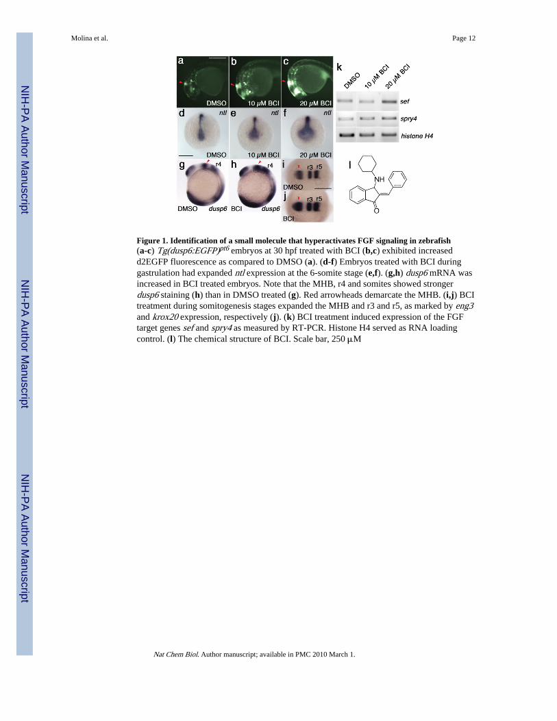
Figure 1. Identification of a small molecule that hyperactivates FGF signaling in zebrafish(a-c) Tg(dusp6:EGFP)pt6 embryos at 30 hpf treated with BCI (b,c) exhibited increasedd2EGFP fluorescence as compared to DMSO (a). (d-f) Embryos treated with BCI duringgastrulation had expanded ntl expression at the 6-somite stage (e,f). (g,h) dusp6 mRNA wasincreased in BCI treated embryos. Note that the MHB, r4 and somites showed strongerdusp6 staining (h) than in DMSO treated (g). Red arrowheads demarcate the MHB. (i,j) BCItreatment during somitogenesis stages expanded the MHB and r3 and r5, as marked by eng3and krox20 expression, respectively (j). (k) BCI treatment induced expression of the FGFtarget genes sef and spry4 as measured by RT-PCR. Histone H4 served as RNA loadingcontrol. (l) The chemical structure of BCI. Scale bar, 250 μM
Molina et al. Page 12
Nat Chem Biol. Author manuscript; available in PMC 2010 March 1.
NIH
-PA Author Manuscript
NIH
-PA Author Manuscript
NIH
-PA Author Manuscript
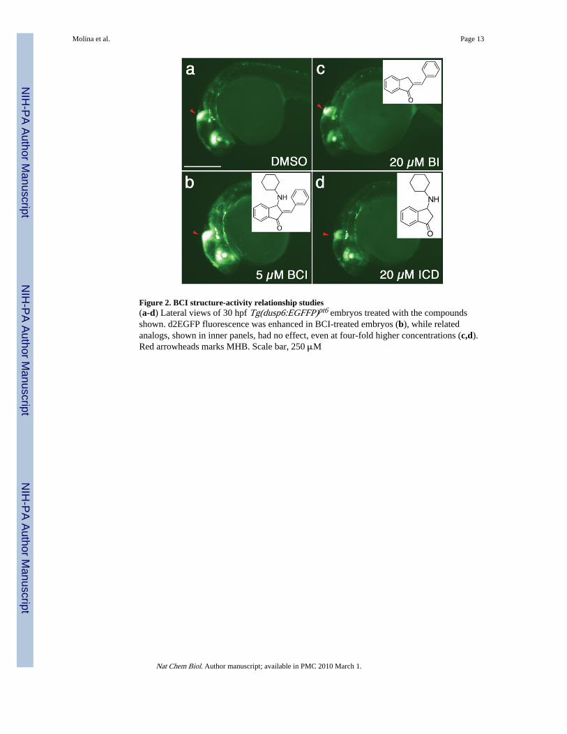
Figure 2. BCI structure-activity relationship studies(a-d) Lateral views of 30 hpf Tg(dusp6:EGFFP)pt6 embryos treated with the compoundsshown. d2EGFP fluorescence was enhanced in BCI-treated embryos (b), while relatedanalogs, shown in inner panels, had no effect, even at four-fold higher concentrations (c,d).Red arrowheads marks MHB. Scale bar, 250 μM
Molina et al. Page 13
Nat Chem Biol. Author manuscript; available in PMC 2010 March 1.
NIH
-PA Author Manuscript
NIH
-PA Author Manuscript
NIH
-PA Author Manuscript
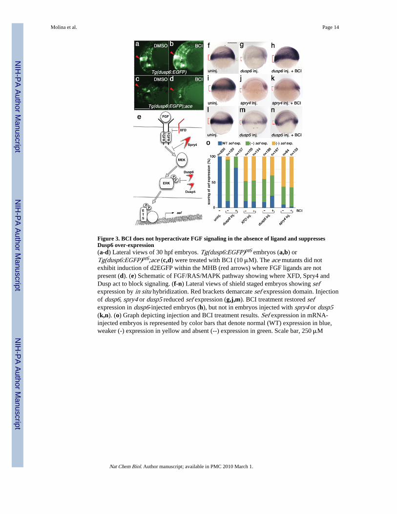
Figure 3. BCI does not hyperactivate FGF signaling in the absence of ligand and suppressesDusp6 over-expression(a-d) Lateral views of 30 hpf embryos. Tg(dusp6:EGFP)pt6 embryos (a,b) orTg(dusp6:EGFP)pt6;ace (c,d) were treated with BCI (10 μM). The ace mutants did notexhibit induction of d2EGFP within the MHB (red arrows) where FGF ligands are notpresent (d). (e) Schematic of FGF/RAS/MAPK pathway showing where XFD, Spry4 andDusp act to block signaling. (f-n) Lateral views of shield staged embryos showing sefexpression by in situ hybridization. Red brackets demarcate sef expression domain. Injectionof dusp6, spry4 or dusp5 reduced sef expression (g,j,m). BCI treatment restored sefexpression in dusp6-injected embryos (h), but not in embryos injected with spry4 or dusp5(k,n). (o) Graph depicting injection and BCI treatment results. Sef expression in mRNA-injected embryos is represented by color bars that denote normal (WT) expression in blue,weaker (-) expression in yellow and absent (--) expression in green. Scale bar, 250 μM
Molina et al. Page 14
Nat Chem Biol. Author manuscript; available in PMC 2010 March 1.
NIH
-PA Author Manuscript
NIH
-PA Author Manuscript
NIH
-PA Author Manuscript
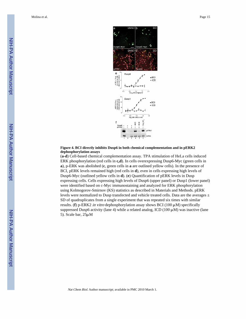
Figure 4. BCI directly inhibits Dusp6 in both chemical complementation and in pERK2dephosphorylation assays(a-d) Cell-based chemical complementation assay. TPA stimulation of HeLa cells inducedERK phosphorylation (red cells in c,d). In cells overexpressing Dusp6-Myc (green cells ina), p-ERK was abolished (c, green cells in a are outlined yellow cells). In the presence ofBCI, pERK levels remained high (red cells in d), even in cells expressing high levels ofDusp6-Myc (outlined yellow cells in d). (e) Quantification of pERK levels in Duspexpressing cells. Cells expressing high levels of Dusp6 (upper panel) or Dusp1 (lower panel)were identified based on c-Myc immunostaining and analyzed for ERK phosphorylationusing Kolmogorov-Smirnov (KS) statistics as described in Materials and Methods. pERKlevels were normalized to Dusp transfected and vehicle treated cells. Data are the averages ±SD of quadruplicates from a single experiment that was repeated six times with similarresults. (f) p-ERK2 in vitro dephosphorylation assay shows BCI (100 μM) specificallysuppressed Dusp6 activity (lane 4) while a related analog, ICD (100 μM) was inactive (lane5). Scale bar, 25μM
Molina et al. Page 15
Nat Chem Biol. Author manuscript; available in PMC 2010 March 1.
NIH
-PA Author Manuscript
NIH
-PA Author Manuscript
NIH
-PA Author Manuscript
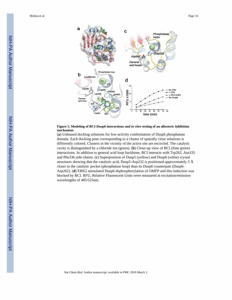
Figure 5. Modeling of BCI-Dusp6 interactions and in vitro testing of an allosteric inhibitionmechanism(a) Unbiased docking solutions for low-activity conformation of Dusp6 phosphatasedomain. Each docking pose corresponding to a cluster of spatially close solutions isdifferently colored. Clusters in the vicinity of the active site are encircled. The catalyticcavity is distinguished by a chloride ion (green). (b) Close-up view of BCI (lime green)interactions. In addition to general acid loop backbone, BCI interacts with Trp262, Asn335and Phe336 side-chains. (c) Superposition of Dusp5 (yellow) and Dusp6 (white) crystalstructures showing that the catalytic acid, Dusp5-Asp232 is positioned approximately 5 Åcloser to the catalytic pocket (phosphatase loop) than its Dusp6 counterpart (Dusp6-Asp262). (d) ERK2 stimulated Dusp6 dephosphorylation of OMFP and this induction wasblocked by BCI. RFU, Relative Fluorescent Units were measured at excitation/emissionwavelengths of 485/525nm.
Molina et al. Page 16
Nat Chem Biol. Author manuscript; available in PMC 2010 March 1.
NIH
-PA Author Manuscript
NIH
-PA Author Manuscript
NIH
-PA Author Manuscript
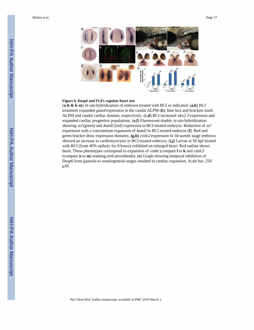
Figure 6. Dusp6 and FGFs regulate heart size(a-h & k-m) In situ hybridization of embryos treated with BCI as indicated. (a,b) BCItreatment expanded gata4 expression in the caudal ALPM (b); blue box and brackets markALPM and caudal cardiac domain, respectively. (c,d) BCI increased nkx2.5 expression andexpanded cardiac progenitor populations. (e,f) Fluorescent double in situ hybridizationshowing scl (green) and hand2 (red) expression in BCI-treated embryos. Reduction of sclexpression with a concomitant expansion of hand2 in BCI treated embryos (f). Red andgreen bracket show expression domains. (g,h) cmlc2 expression in 18-somite stage embryosshowed an increase in cardiomyocytes in BCI-treated embryos. (i,j) Larvae at 56 hpf treatedwith BCI (from 40% epiboly for 8 hours) exhibited an enlarged heart. Red outline showsheart. These phenotypes correspond to expansion of vmhc (compare l to k and cmlc2(compare n to m) staining (red arrowheads). (o) Graph showing temporal inhibition ofDusp6 from gastrula to somitogenesis stages resulted in cardiac expansion. Scale bar, 250μM
Molina et al. Page 17
Nat Chem Biol. Author manuscript; available in PMC 2010 March 1.
NIH
-PA Author Manuscript
NIH
-PA Author Manuscript
NIH
-PA Author Manuscript
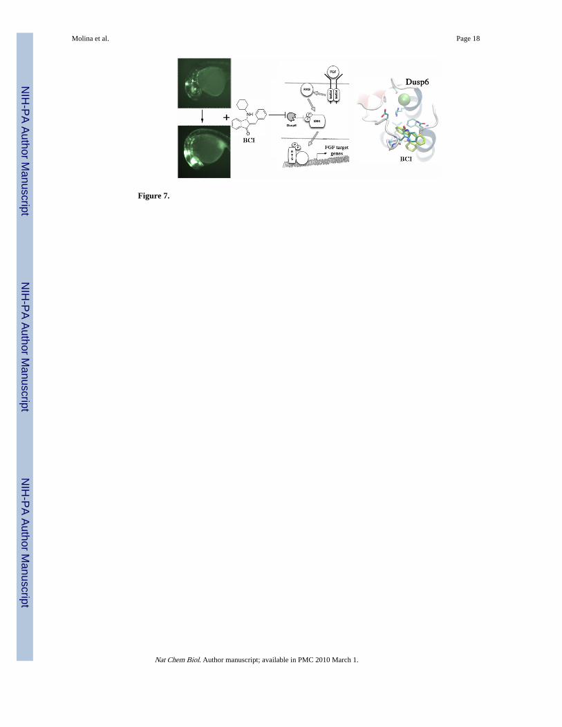
Figure 7.
Molina et al. Page 18
Nat Chem Biol. Author manuscript; available in PMC 2010 March 1.
NIH
-PA Author Manuscript
NIH
-PA Author Manuscript
NIH
-PA Author Manuscript