Embed Size (px)
Citation preview

University of Warwick institutional repository: http://go.warwick.ac.uk/wrap
A Thesis Submitted for the Degree of PhD at the University of Warwick
http://go.warwick.ac.uk/wrap/74324
This thesis is made available online and is protected by original copyright.
Please scroll down to view the document itself.
Please refer to the repository record for this item for information to help you to cite it. Our policy information is available from the repository home page.

STUDIES CONCERNING THE PIERICIDINS
by
C. J. COlJ:!)
A dissertation submitted to theUNIVERSITY OF WARWICK
for the degree of
DOCTOR OF PHILOSOPHY
Coventry, 1970

BEST COpy
AVAILABLE
Variable print quality

PREB'ACE
The work described in this thesis was carried out in the School
of Molecular Sciences, University of \iarwick, Coventry, betweenNovember 1966 and October 1969. It is the original work of theauthor, except where specific acknowledgement is made, and has notbeen submitted for a degree at any other University.
The author wishes to make the following acknowledgements:Professor V. M. Clark, who directed this work, andDr. D. W. Hutchinson for their constant advice and encouragement.The Department of Radiotherapeutics, University of Cambridge,
for the use of a tritium line.The Chemical Society for the use of a computerised information
retrieval service for current literature.The Science Research Council for the award of a Research
Student ship.

CONTENTS
Abbreviations and terminologySummary
TIlTRODUCTION
BIOCmUCAL INTRODUCTION
General IntroductionOxidative phosphorylationTheories of oxidative phosphorylationThe function of ubiquinoneInhibition by piericidin A
CIIDHCAL INTRODUCTION
The possible contrasting involvement of piericidin Ain a scheme analogous to that f:lrubiquinoneThe tautomerism c£ piericidin AThe properties of hydroxypyridinesThe properties of piericidin A compared with those ofother hydroxypyridines
Evidence concerning the structure of the pyridine ringof piericidin A
The side-chain of piericidin A
RESULTS AND DISCUSSION
Metabolites of Streptomyces mobaraensis and their
derivatives
Experiments of biochemical significanceThe 2,4-dinitrophenylhydrazones of methyl pyruvate
Paee
1
4
7
9
131920
2628
3344
48
52
53
64
69

The synthesis of 4,6-dimethoxy-2,3- dimetbyl-5-bydroxypyridine and of 4,6-dimethoxy-2,5-dimethyl-3-hydroxypyridineThe synthesis of compounds tested for theirinhibition of NAnH-linked oxidationThe synthesis of other pyridines and experiments 93
73
83
concerned with the synthesis of pyridinesExperimental comparisons involving substituents in 95the (X,~ and 't positions of pyridinesSynopsis ooncerning the structure of piericidin A 125
EXPERIM:Ji1ITALSECTIONMetabolites of Streptomyces mobaraensis and their 130
derivativesExperiments of biochemical signi~icance
.The 2,4-dinitrophenylydrazones o~ methyl pyruvateThe synthesis of 4,6-dimethoxy-2,3-dimethy1-5-hydroxypyridine and of 4,6-dimethoxy-2,5-dimethyl-3-hydroxypyridineThe synthesis of oompounds tested for their inhibition 180
146
157160
of NAnH-linked oxidationThe synthesis of other pyridines and experiments 199concerned with the synthesis of pyridines
Experimental comparisons involving substituents in 203the (X,p and ~ positions of pyridines
Mass Epectral Appendix 218

1
ABBlb""VIATIONS .AIm TERNINOLOGY
The following abbreviations are used in the text.NAD
NADH
ATPADPp.1.
cyt.EPR
R
T
g., mg.1., mI., pl.
1<1, rrl·t·
mm, - Hg
c
mC, J.1C
c.p.m.
M.Pt.B.Pt.
nicotinamide adenine dinucleotide
reduced nicotinamide adenine dinucleotideadenosine-S·l--triphosphateadenosine-51-diphosphateinorganic phosphateflavoprotein
protein containing non-heme ironcytochromeelectron paramagnetic resonance-loglO (dissociation constant)difference in Gibbs free energygas constanttemperature in degrees absolutegram, milligram
litre, millitre, microlitre (10-6 of a litre)moles per litre, millimoles per litre
millimetres of mercury (unit of pressure)( 10curie unit of radioactivity • 3.7x10
disintegrations per second)millicurie, microcurie(10-6 of a curie)
counts per minute
melting pointboiling point
degrees centigrade

2
T.L.C. thin layer chromatographyrate of flow of samplerate of flow of solvent
2,4-DNP 2,4-dinitrophenylhydrazonemISO dimethylsulphoxide
HMPA hexamethylphosphoramideTAl trichloroacecetylisocyanateDDQ 2,3-dichloro-5,6-dicyano-l,4-benzoquin-
one
TCNE tetracyanoethylene
lit. literature valueThe term "hydroxypyridine" is used in this dissertation to
describe a compound in which the predominant tautomer is notspecified. The tautomer of a hydroxypyridine bearing a proton onoxygen is referred to as a "pyridinol". The tautomer of a 2- or4-hydroxypyridine bearing a proton on nitrogen is referred to as a"l-(H)-2-pyridone" or a "l-(H)-4-pyridone", and in general as a"l-(H)-pyridone". The tautomer of a 3-hydroxypyridine bearing aproton on nitrogen is referred to as the zwitterionic form of the3-hydroxypyridine. The terms "pyridinol","l-(H)-2-pyridone" and"l-(H)-4-pyridone" are also used to describe hydroxypyridinesexisting predominant~ in the tautomeric form specified.ultra-violet spectra
uv ultra violet~max maximum of absorbance
nanometre (10-9 of a metre)run
€max molar extinction coefficient at

3
maximum of absorbance
E molar extinction coefficient atgiven wavelength
infra-red spectrainfra-redwavenumber of absorption (centimetre-I)
v.st., st., med., w ver.y strong, strong, medium, weaknuclear magnetic resonance spectra
Tnuclear magnetic resonance
1tau value of peak in H mm spectrums., d., t., m singlet, doublet, triplet, multiplet
spin-spin coupling constant (Ue~~~)megacycles per second
J CH!.)
It-C/S
ppm parts per millionst., med , , w strong, medium, weakmass spectra
m/e atomic mass units per unit of positivecharge
(M)+
(rf.)
molecular ion
molecular peak (corresponding tomolecular ion)
base peak most intense peak of mass spectrum
I intensity of peak in mass spectrumexpressed as percentage of intensityof base peak
1>•• ks 4e. ~. ~et"t.Ue t,..uifiou & .. ~.l:ecl I;, I:wo
.,l..ce, 6~ tL&ei",o.l$. 1l.ese vcc.l"c, ...tt. oA(elAl ..~f.J..

4
The chemical similarities and contrasting biochemical propertiesof piericidin A and ubiquinone are presented and discussed. Theformer, a penta-substituted pyridine and a metabolite of streptomycesmobaraensis, is an inhibitor of mitochondrial electron transport, whilstthe latter is an electron carrier. A scheme to account for thisdifference is proposed involving an olefinic linkage of the longhydrocarbon side-chain. This \-lasshown not to be responsible for theinhibitor,y action of piericidin A, since hydrogenation of the olefinicfunction did not noticeab~ reduce inhibitory potency.
Piericidin A was labelled with tritium in order to investigateits binding to mitochondria. It was fOQ~d to be bound unspecificallyin a linear manner up to 10 m~ mOles/mgm. protein, but washing withbovine serum albumen removed most of this piericidin A. However aquantity correspondinc to that required for the inhibition of NAnHoxidation (0.02 m~ moles/mgm. protein) could not be removed in this way.It is concluded that this piericidin A was bound in a site specific
manner. This specifically bound inhibitor was recovered unchanged on
extraction of the mitochondrial lipids ",ith acetone. Hence it isconcluded that tho inhibitor is not covalently bound to the mitochondria.
A number of analogues of piericidin A were synthesised. None ofthese compounds approached the potency of piericidin A as inhibitors,
"although those with lipophilic and phenolic properties were more
effective in this respect than those without, suggesting the possibleinvolvement of hydrophobic interactions and of a phenolic function duringinhibition.

5
The possible tautomers of the proposed structure of piericidin Aare considered in relation to its spectroscopic and chemical properties.Contrary to the behaviour expected from this structure, the moleculeappears to exist as a pyridinol, rather than the 1-(H)-4-pyridone tautomer.
The evidence for the proposed structure of piericidin A iscritically discussed. That for the arrangement of substituents on thepyridine nucleus depends partly on ultraviolet spectroscopic data, andmore significantly, on two fragnlents arising from ozonolysis. Theidentity of one of these fragments (the 2,4-DlrP of methyl pyruvate) isin doubt, since its properties differ from those of either of two fullycharacterised isomers of authentic material synthesised specially.It is concluded that piericidin A may be a p-hydroxypyridine, ratherthan the proposed ~ -hydroxypyridine, since the remaining evidence isno longer conclusive. The spectroscopic properties of piericidin Amentioned above also favour this proposal.
~lO fully substituted p-hydroxypyridines, isomeric with the proposedpyridine nucleus of piericidin A, were synthesised. Neither hadproperties corresponding to piericidin A. With these and otherhydroxypyridines, the comparative studies pertaining to piericidin A
were extended, using the techniques of DV, IR, mn.m and mass spectros-copy. A particularly simple diagnosis of hydroxypyridines by IE NMR
spectroscopy was developed, involving the use of trichloroacetyliso-cyanate. In addition base catalysed deuterium exchange of protons in
the methyl groups of substituted pyridines was studied.

6
As a result of these studies, distinctions can be made between~, and a or t substituted metbyl- and bydroxy-pyridines. Piericidin Aappears to be a ~-hydroxypyridine, its most likely structure being2-alkyl-3,6-dimethoxy-5-hydroxy-4-methylpyridine.
It is also concluded from the mass spectrum of piericidin A thatthere is an olefinic bond between carbon atoms 5 and 6 of the side-chain, rather than between carbon atpms 4 and 5 as published.

TIITRODUCTIOlI

FIGURE I
OH
PIERICIDIN A
o
CH,o
H
o CH3
n
UBIQUINONE

7
BIOCHEliICAL INTRODUCTION
GENERAL INTRODUCTION
Piericidin A is an insecticidal metabolite of streptomycesmobaraensis, and was first isolated in 19631• The chemical structureof piericidin A has been proposed as a penta-substituted pyridine ring2;~in which the substitution pattern closely resembles that of theubiquinones (see figure 1). The possibility of tautomerism to a l-(H)-4-pyridone enhances the similarity. The differences between the twostructures can be considered as twofold.
(i) Piericidin A is a hydroxypyridine rather than a quinone.(ii) In contrast to the ubiquinones, piericiclin A has a non-
isoprenoid side chain.It was found that piericidin A inhibits",itochondria1 reactions in
the region where ubiquinone is involved. Inhibition of succinatedehydrogenase appeared to be competitive with ubiquinone, and potentinhibition of NADH dehydrogenase, which was not reversible by additions
of ubiquinone, was also found4• These structural and biochemical
contrasts initiated the present investigation.The theories of electron transport and oxidative phosphorylation in
mitochondria have been reviewed5-7• Of particular interest to thisthesis are the possible involvements of ubiquinone8-10, and of piericidin
A and related inhibitors, in these processes.The energy generated in respiring cells during the oxidation of
substrates, principally of fatty acids, can be coupled to the synthesis
of ATP from ADP and Pd.. This conservational transduction of oxidativeenergy into the chemical energy of the triphosphor,yl group of ATP is

FIGURE 2
THE MITOCHONDRIAL RESPIRATORYCHAIN
r
NAD-linked-----NAD-Fp--+F
lubotrGtc - ~ XA, Site 2 •
r ua 1 I ' Site 3 ..1.Lcy<bJTcyt c,_Cytc_CytO,G, I 0,
~cSUCCinate Fp--Fep B
Site I
o
F~ flavoprotein
Fep protein containing non-hcmc Iron
ua ubiquinone
Cyt cytochrome
Sites 1,2 and 3 ,Itel of oxidative phosphorylation
A,B,C and0 Iitel of action of partlc:ular Inhibitors
SITE INHIBITORS
A pl«rleldln A , rot.enone , amytal
B theonyltrlfluoroacctone
C antimyclnAt 2-hcptyl-4-hydroxyqulnolinc-l-oxldc
o cyanide Ion, carbon monoxide

known as oxidative phosphorylation. In most types of cell, other than•bacteria, the enzymes concerned with the generation of this oxidativeenergy, and the enzymes of oxidative phosphorylation, are, a cloEelyassociated part of the imler membrane. of highly organised cytoplasmicparticles known as mitochondria. In bacteria, which have no mitochondria,they are bound to the cell membrane. The associated enzymes of thetricarboxylic acid cycle, and certain other oxidative functions, alsoappear to be localised in the mitochondria.
On the molecular level, most schemes for substrate oxidation inmitochondria postulate sequential electron flow through a series ofcarriers of increasing redox potential, terminating in molecular oxygen.
During the passage of a pair of electrons from substrate to oxygen,one molecule of ATP may be synthesised from ADP and Pi at a maximum ofthree coupling sites. A simple representation of the sequence ofcomponen~s of the mitochondrial respiratory chain appropriate to manysystems examined, is made in figure 2. The approximate locations ofcoupling to oxidative phosphorylation, and of interruption of electronflow by inhibitors are also shown. The experimental evidence for allthese assignments is reasonably consistent and will not be dealt withhere. This discussion will concentrate on experiments dealing with thefollowing topicsl
(i)
(ii)
(iii)
Oxidative phosphorylation
The funotion of ubiquinone
Inhibition by piericidin A

9
OXIDATIVE PHOSPHORYLATIONThe problem of oxidative phosphorylation should first be considered
from its standpoint as a biochemical phenomenon. The native mitochondrialprocess has not yet been observed in a soluble system, free from membrane.structure. A reasonable conclusion from this is that a membrane structureis an inherent part of oxidative phosphorylation. The three couplingsites have not yet been assigned to specific redox reactions. This
•makes chemical formulation of the phenomenon difficult. There is goodevidence that oxidative phosphorylation proceeds!!! a high energyintermediate.
(i) The existence of other energy dependent proeessesNeither the respiration dependent accumulation of inorganio
cationsll,12, nor the succinate linked reduction of acetylaoetate13, anda-ketoglutarate14, are inhibited by oligomycin, an antibiotic whichblocks ATP Synthesis15,16. Furthermore certain submitochondrialparticles, low in adenine nucleotide and Pi content, are able to utiliserespiratory energy for other energy dependent reductions, although of
. 17 18course they are not able to synthesise ATP w1thout added ADP and Pi ' •It can also be concluded from this result that any primar,y couplingreaction does not involve Pi- The above results, together with theinformation that under certain conditions competition between ATPsynthesis and other energy dependent reactions is observedl9-21 s~geststhe existence of a common hieh energy intermediate. Suppression of any
of these processes in mitochondria causes an inhibition of substrateoXidation22• This so-called "respiratory control" is believed to becaused by a builk up of the high energy intermediate.

10
(ii) Loss of respiratory oontrolA diverai tY'of ohemioals, known as tlunoouplera", cause
inhibition of all energy dependent reaotions, without stoppingrespiration23-25• The same effeot may be brought about bY'struoturaldamage to the mitoohondrion. The oause of this "unooupling" isoonsistent with the spontaneous deoomposition of a high energyintermediate. Unoouplers are proposed to act on the respirator,y chainside of the rate determining step of oxidative phosphorylation, followingstudies on the "ATP jump,,26. This is observed, after the addition of
ADP to respiring mitoohondria, preincubated with Pi' as an initialfast synthesis of ATP superseded bY' a slower steady state rate. Theinitial "ATP jump" is not effected bY'unoouplers, whereas the steadystate synthesis is abolished. FUrthermore, competition between ATPsynthesis and the unooupling reaotion has been shown, bY'measuringphosphorylation rates at different respiratory rates in the presenoe offixed oonoentrations of unooupler27• The'oomp~tition is believed to befor a common high energy intermediate, and also explains the observationthat phosphorylating efficiency inoreases with respiratory rate.
(iii) The chemistry of ATP synthesis and exohangeThe following isotopic exchanges have been observed in
intaot mitoohondria28•
(a) The radioaotive label of (~~)-Pi is found in ATP after
inoubation with mitoohondria in the presence of Pi' ADP
and ATP.
(b) The radioaotive label of (14C)_ADP or(3~)-ADP is found
in ATP after inoubation with mitochondria, without the

11
addition of Pi.(c) The radioactive label of, (18o)_water is found in Pi and
ATP after incubation with mitochondria. Similarly the18radioactive label of ( O)-Pi is found in water after
incubation with mitochondria.(d) The triphosphoryl oxygen bridge in ATP is supplied by ADP.These observations are consistent with the following equilibrias
ATP + I I_'P + ADP
*l-ThereI, I and I-P are respectively non-energised, high energy and
phosphorylated states of an undefined intermediate. Since theoxygen of Pi appears to originate from ~O, and that of the triphosphoryloxygen bridge of ATP from ADP, rather than from the intermediate ineither ease, no isotopic exchange into the intermediate would bedetectable, except of course in its phosphorylated state.
(iv) Coupling factors5
Certain isolable mitochondrial proteins, when added to
specifically depleted mitochondrial or sub-rnitochondrial preparations,increase their capacity to earry out oxidative phosphorylation. The
identity of these "coupling factors" is diverse. The ability ofcertain of these preparations to catalyse the ATP exchange reaction~ vitro can be chemically destroyed, without loss of the property of
restoring mitochondrial oxidative phoBPhorylation29• They are thus
believed to aot indireotly via a struotural modification of the mito-chondrial membrane, rather than in a direot catalytio ma.nner on a

FIGURE 3
A SCHEME OF OXIDATIVE PHOSPHORYLATION
1
ADP ADP ATP
or more conventionally
B + I A + 1*
other propolall
I) the reaction 1---- I II catalYled by uncouplers.
II) I* Is the ener~y-Iourc:e for other cncr~y-linked reactions.
III) Oll~omyc:lnInhibits further reaction of I""P.
A, AHa' Band BHa
1,1- and I....P
unspecified respiratory carriers in oxldiscd and reduced forms
non-energised,energised and phosphorylatcd forms respectively
of an unspecified Intermediate

12
reaction of oxidative phosphor,ylation. The existence of these proteincoupling factors emphasises the structural and sterie sensitivity ofthe reactions and presumed intermediates of oxidative phosphor,ylation.
It isvconcluded that the isoenergetic sequence presented infigure 3 is a minimum working hypothesis for the observed reactions ofoxidative phosphor,ylation. It should be emphasised that the involvementof a respirator,y carrier in ~ of the proposed intermediates, and thePossible existence of more than two such intermediates (the two being*I and I~P) is neither precluded nor proven.

13
TmDRIES OF OXIDATIVE PHOSPHORYLATION
Whilst the evidence for the existence of high energy intermediates
is convincing, that concerning their molecular nature is divergent and
elusive.Briefly there are three hypothetical treatments:(i) Involving conformational changes of the mitochondrial membrane.
(ii) Involving electrochemical potential difference across themitochondrial membrane.
(iii) Involving discrete chemical intermediates.(i) The conformational treatment
Of the three theories this is the least amenable to ~vitro experimentation. The original postulate, by Boyer30, proposesas intermediates a high energy conformation of a protein, with thefurther possibility of a sulphur-acyl bond. Three clearly distinguish-able configurations of the mitochondrial inner membrane have beenequated with energy states by Green31, who also proposes that there areno other intermediates. Such a clear-cut relationship between theconfiguration of the membrane and biochemical state is not found byother wOrkers32. It is difficult to test such a proposal further.
The involvement of the sulphydryl groups of proteins in the
formation of a phosphorylated intermediate has been suggested, following
mitochondrial studies involving sulphydryl reagents33• The phosphor,y-
lation of ADP has also been observed !u vitro during the oxidation of
several biochemically significant systems oontaining sulphur b.1 brominein pyridine34•

14
Intermediates involving protein sulphydr,yl or sulphur-acylfunctions should strictly be considered as chemical intermediates.Even the high energy conformation of a protein can be considered to be
the macro-molecular effect of discrete molecular-interations, and henceto be strictly a chemical intermediate. However this point of viewdoes not reveal any easier experimental approach.
The effect of coupling factors, mentioned previously (page " ),lends support to the theor,r of different conformers as intermediates.
(ii) Membrane potential treatment
In the "chemiosmotic theory" of };l1tchell,respirationcauses proton transport'across a membrane35• Evidence exists to thiseffect35,36• Thence a pH gradient and electrochemical potentialdifference is produced across the membrane. Reversal of this process
,'"f·'
is said to dri~e the formation of the anhydride between ADP and Pi'via a vectorial ATPase bound to the membrane. The mechanism of this-process is an unsatisfactor,r part of the theory. Two strong points ofthe same are firstly the r~ explanation of the necessity for amembrane, and secondly the explanation of the mechanism of action ofuncouplers37,38. There is evidence that these chemicals increase the
permeability of the membrane to protons, and hence collapse any membrane
potential. The chemical diversity of uncouplers is not easilyaccounted for by other theories. Conversely the action of couplingfactors may be explained by proposing that they decrease the permeabilityof the membrane to protons, stabilising any membrane potential.

FIGURE 4
A SCHEME INVOLVING PROTEINS IN OXIDATIVE PHOSPHORYLATION
unsp~cified respiratory carriers In oxidised and reduced forms]
==»ATP ADP
P.I

15
(iii) Chemical intermediate treatments*In chemical hypotheses the intermediate I (figure 3)
is conceived as a relatively stable chemical substance having a highenergy bond. The intermediate I-P is considered to have similarstability and energy, and to be capable of phosphorylating water or
ADP.
Three different chemical speoies have been put forward as chemical
intermediates at various times.(a) Proteins30
(b) NAD39(c) Quinones 40
(a) ProteinsThe idea of the involvement of the sulpbydr,yl or
sulphur-acyl functions of proteins as chemical intermediates has alreadybeen mentioned30,33,34,(pagesI3·'~). One such scheme is presented in
figure 4. The observed isotopic exchanges between ATP, ADP, Pi andwater (pages 10-11) may all be explained on this scheme. Phosphorylationby the proposed acyl phosphate would result in the retention of theanhydride oxygen by the acyl function4l• The other reactions,involving sulphur, are chemically demonstrable34,42. The difference
between the generalised scheme in figure 3 and that in figure 4 liesin the sequence of the dehydration reaction.
In figure 3.
In figure 4:
Thus in the latter case the observed isotopio exchanges between

FIGURE 5
A SCHEME INVOLVING NAD IN OXIDATIVE PHOSPHORYLATION
H H
R
R R= CONH2
R'= remainder of NAD molecule
R
NAO
NAOH H HR
R ATP ADP H
~H
I,R

16
water, and P. and ATP are less readily explained although they are1
perfectly feasible. The implied exchange of oxygen into the carboxylfunction of the protein would be difficult to detect isotopically,since exchange into such functions will be considerable, irrespective
Iof any oxidative phosphorylation.Some phosphorylated proteins have been isolated, possessing many
of the expected properties of a phosphorylated energy-transportintermediate43,44. These include:
(i) the ability to phosphorylate ADP,
(ii) the ability to catalyse the ATP ~ A~~exchange reaction,and
(iii) the ability to act as coupling factors.One author has even tentative~ suggested that the phosphate
function is an acyl phosphate44•It thus appears that this is one approach where chemical and
biochemical deductions merge.
The participation of NAn derivatives in oxidativephosphorylation has been suggested39• The scheme proposed is presented
in figure 5.Although NAD derivatives of unusual nature have been iBolated45,46,
their chemical identity remains obscure. FUrthermore,the existence
of a high energy intermediate not involving adenine nucleotides or Pi
has been inferredl1,l8, (page ~). This is divergent with the proposedscheme.

FIGURE 6
A SCHEME INVOLVING QUINONES IN OXIDATIVE PHOSPHORYLATION
-HOa 1
reductIon
OH
Io
nuclcophllc
~_,09~jP'cJ9
~ .~CH oxidation
m
1
Ill:

17(c) Quinones
Following the observation that the oxidation ofhydroquinone phosphates ~ vitro can lead to phosphor,ylation47, schemeswere suggested involving quinones in oxidative PhosPhorylation40,48.Of these the most chemically consistent is presented in figure 649• An
~ vitro study of this scheme has been made50, in the course of whichevidence for the equilibrium I.~ II has been found5l• No evidence
for the reactions II~ III, III~IV and IV~V was found, andcompound III proved to be very unstable. Considered from a biochemicalangle, it should be pointed out that the primary high energy intermediateof this scheme, IV is phosphorylated. This is not very satisfactory,since it has already been deduced that it is unlikelyl7,l8(page 1 ).The direct involvement of ubiquinone in oxidative phosphorylation (asdistinct from its involvement as a respiratory carrier) bas never beendemonstrated. Experiments in which the involvement of vitamin K inoxidative phospho~Jlation in Mycobacterium phlei bas been demonstrat~&,53
are the only direct evidences for such an involvement for quinones ingeneral. Indeed many experiments designed to detect the existence ofquinone intermediates, using the technique of isotopio labelling,suggest their paucit,54,56• The scheme presented in figure 6 is
considered later in relation to a possible mechanism of action of
In concluding this consideration of chemical intermediates inpiericidin A (page l').
bas yet been identified.
oxidative phosphorylation, the following points should be considered.(1) No chemical intermediate corresponding to 1* (figure 3)

13
(ii) Only in the case of proteins is there consistentevidence for the existence of an intermediate correspondingto I-P, and even in that case its chemical nature is
obscure.(iii) It might be expected, from the chemical standpoint, that
the three "coupling sites" would possess intermediatesof different chemical identity. No intrinsicdifferences in the three coupling sites have been
established.Thus the precise nature of the molecular processes of oxidative
phosphorylation remains unsolved and is the focus of controversy.

19THE FUNCTION OF UBIQUINONE
The function of ubiquinone as a mitochondrial electron carrierin the respiratory chain (figure 2)-is established by the following
observations.(i) It is found in relatively high concentrations in all aerobic
tissues tested57-59•(ii) Succinate dehydrogenase and NADH dehydrogenase activities
lost following the extraction of ubiquinone from mitochondrialpreparations, can be restored by the addition of ubiquinone6O-64•
. 59,65-68(iii) Ubiquinone is reduced and oxidised during electron transport
Some discrepancy has been observed between the rate of turnover of thequinone and the respiratory rate68-70. However other experiments
•
show this rate to be consistent with the oentral position of ubiquinonein the respiratory cbain7l.
(iv) Three of four isolated complexes of the mitochondrialrespiratory chain react specifically with ubiquinone72• These fourcomplexes can be reconstructed into a complete respiratory chain.
As mentioned previously (page:~ 17 ), there is no direct evidenceinvolving ubiquinone in mitochondrial phosphorylation reactions.
Such an involvement may be argued by analogy with vitamin K in bacterial~systems52,53. The necessity for an aromatio methyl group,!an adjacent
pt unsaturated carbon side ohain, has also been inferred, as a pre-
requisite for quinones involved in oxidative phosphorylation, from such
bacterial studies52b• However this may be disputed from the negativeresults of certain experiments involving isotopic exchange, designedto detect any quinone energy-transport ";1li'ermediates54-56•

20
INHIBITION BY PIERICIDIN AIt was found that piericidin A inhibits the oxidation of NADH and
of succinate in mitochondria, but that the NADH dehydrogenase systemis several orders of magnitude more sensitive than the succinatedehydrogenase system4, 73-75. NADH dehydrogenase is inhibited by
concentrations of piericidin A of the order of 3xlO-5 ~ moles/mg. ofprotein, and the inhibition is not reversed by the addition ofubiquinone. Succinate dehydrogenase is inhibited by concentrations ofpiericidin A of the order of 10-7 or 10-9M, and the inhibition isreversed by the addition of ubiquinone, suggesting competition for acommon reaction site. Evidence has also been produced to the effectthat piericidin A can act as an uncoupler74,75, and that at highconcentrations it may inhibit cytochrome C oXidase46,74,76~
Studies with radioactively labelled piericidin A77-79, inagreeement with results presented in this thesis and elsewhere80, haveshown that an amount of the inhibitor, equivalent to the minimum
required for complete inhibition of NADH dehydrogenase, is stronglybound to mitochondria. Concentrations above this may be easilyremoved by washing with bovine serum albumen. This removal is also
consistent with the reversible inhibition of succinate dehydrogenase
activity at higher concentrations. The binding of piericidin A to
mitochondria bears an approximately linear relationship to the
concentration of piericidin A added, ,over-all concentrations tested78-80.Between one and two molecules of piericidin A per unit of NADHdehydrogenase become strongly bound to mitoohondrial preparations78-81,
82more recent results favouring the higher figure •

FI UR 7
THE PIERICIDINS AND RELATED INHIBiTORS
CHP
H,CH,o
pier-lcidin A R=R=H·,. t dlocetcte R= R=COCH'. )
monoocctot~ R,=H, R=COCH,.
pie ric idin 8 R,= H t R,= CH, monooccta tc
The octahydro dt:rivatlvt:~ of the above flv~ compounds cr e those In which tht: ol~inic
bonch ore saturated.
CHProt~l\one Only the box ed part 01 the molecule may be
chemlcaJly modified without ot1tttl~ the potency
of the IMition of NADH dehydrogenasc.
o"'ytol

21
The constitution of NADH dehydrogenase, and the nature of theinteractions of inhibitors with it, are complementar.y subjects whichhave received a great deal of attention recently.
(i) Inhibitors related to piericidin AThe inhibition of NADH dehydrogenase b,y piericidin A
is ver,y sensitive to chemical modification of the inhibitor.Octahydropiericidin A (figure 7) has been shown to inhibit MAnHdehydrogenase at similar concentrations to piericidin A73,Bo, but
substitution of the phenolic function or loss of the hydrocarbon sidechain dissipates the potency of the molecule in this respect. On theother hand, the less potent inhibition of succinate dehydrogenase isnot subject to such variations, and is inhibited at ~ concentrations
by all these compounds.Rotenone92, and several rotenoids87, inhibit NADH dehydrogenase
at concentrations almost as low as those of piericidin A. Certainfeatures of the rotenone molecule have been deemed essential for suchinhibition87, (see figure 7). Binding and inhibition studies usingradioactive rotenone give ver,y similar results to those alreadymentioned for piericidinA9l,93 (page; 10 ). One significant
difference is the observation tbat tightly bound rotenone is slowly
removed by washing with boVine serum albumen, at a rate much fasterthan 1s tightly bound piericidin A93. Piericidin A must therefore
interact more strong~ with NADH dehydrogenase than does rotenone.
A number of other minor discrepancies between the behaviour of
piericidin A and rotenone have been observed74•

22
Certain other compounds have been found to inhibit NADHdehydrogenase at higher concentrations that those of piericidin A and
rotenone. Amongst these are amytal (figure 7) and otherbarbiturates93,94, certain steroids95, and a series of analogues ofubiquinone in which the methyl function of quinonoid ring is replacedby a hydroxyl group96.
Amytal, piericidin A and some rotenoids have been found todecrease the specific binding of radioactive rotenone91• Analogously,rotenone and a.rnytalhave a similar, but less pronounced effect on thebinding of radioactive piericidin A79,80. It is concluded that allthese compounds can compete for the same binding site, but thatpiericidin A has the strongest binding affinity.
Of the above compounds, neither the rotenoids nor the barbiturates,both structurally dissimilar to ubiquinone, inhibit sucoinatedehydrogenase. On the other hand piericidin A and its derivatives,and the series of 2,3-dimethoxy-5-hydroxy-6-alkyl-l,4-benzoquinones,all structurally resembling ubiquinone, 22 inhibit succinate oxidation.That this inhibition is relieved by the addition of ubiquinone isconsistent with the theory that these compounds inhibit succinatedehydrogenase (but not lULDH dehydrogenase) by competing with ubiquinone
for a common reaotion site.(ii) The constitution of NADH dehydrogenase
There is some controversy over the oonstitution of the
electron transport chain between NADH and ubiquinone. This is chieflyconcerned with differences between particulate and other preparatio~~-85.

23
The following scheme is consistent with the majority of current
opinion',NADH # Flavoprotein ~ Non-heme iron protein~ ubiquinone
(F )P
(Fe)pInhibition by piericidin A, rotenone and amytal is proposed, by
Sl-93Singer et al , to be on the oxygen side of Fep, following EPR
studies on their own soluble preparation. It is known that non-heme iron can be detected in its reduced form by a characteristicEPR signa186• On the other hand, lIatefi et alS4 have proposed thatthe inhibition by these compounds is between Fp and Fep. This isbased on spectrophotometric studies on their own "conplex I", aparticulate preparation.
The proximity of none-heme iron to the proposed common bindingsite for piericidin A, rotenone and amytal is implied by thefollowing observations.
(a) A characteristic EPR signal, attributed to non-heme iron,
together with certain optical changas , both remaining after the 1310"1
reoxidation of inhibited particles, have recently been put forward
as evidence for the involvement of non-heme iron in the binding of, "di AS8p~erlc~ n •
(b) Loss of sensitivity tovlards piercidin A and rotenone (and
loss of site 1 phosphorylation) has been observed in mitochondria
deficient in iron, from Torulopsis utilis89• (l1anyyeasts normally
lack both of these functions90). In similar observations with

24
preparations of Candida uti lis staryed of iron, no direct correlationis found between loss of sensitivity towards piericidin A, and lossof both the non-heme iron content and the EPR signal attributed to
non-heme iron85b. It is suggested that the majority of the
non-heme iron of NAnH dehydrogenase is not concerned with the
binding of piericidin A.Similar circumstantial evidence exists for the involvement
of sulphydryl groups in the proposed common binding site.(a) Sulphydryl reagents (i.e. reagents reacting with
sulphydryl groups) have a sunergistic effect when used withpiericidin A and rotenone, whereas, 2,3-dimercaptopropanol canactually relieve inhibition of NADH oxidase by piericidin A74,87.
(b) Sulphydryl reagents added before, but not after, the
addition of piericidin A decreaee the amount of i~hibitorspecifically bound to NADH dehydrogenase82•
NADH dehydrogenase is bound to a membrane, and hence is
closely associated with lipids. The possible involvement ofhydrophobic interactions with lipids was recognised early as a
factor in its inhib~tion87.Reagents which release proteins, such as phosPholipase82,
release tightly bound inhibitor from the specific binding site of
NADH dehydrogenase. Solvent extraction of the mitochondrial
lipids also releases tightly bound inhibitors from themitochondria70,80,9l• It follows that the interaction between
tightly bound ibhibitors and the enzyme is unlikely to involvecovalent linkages, such as the formation of a Schiff's base.

25In conclusion, proteins, lipids, non-heme iron and sulphydr,yl
groups are all inferred to be in the vicinity of the inhibition/bindingsite of piericidin A, rotenone and arnytal on NADH dehydrogenase.The interactions of these inhibitors do not appear to be covalent, andhence must be of a hydrophobio or ionic nature, or possibly involveligand interactions with a metal. This latter possibility is one of
the few ways in which the inhibitors might be considered to have
chemical similarities.An indirect attempt to reveal more information concerning the
interaction of piericidin A in particular with NADH dehydrogenaseis one concern of the present work. This is attempted b.1 extending thenumber of analogues of piericidin A whose effect on the NADHdehydrogenase region of the respirator,y chain has been studied, and byinvestigating the specific binding of radioactive piericidin A tomitochondria.

f IGUHE 8
A POSSIBLE REACTION SEQUENCE INVOLVING PIERICID1N A
o
CH,
C~\,
CHO,
HIt..H-~
H

26
CID1ITCAL INTRODUCTION
THE POSSIBLE CONTRASTING INVOLVEll1ENTOF PIERICIDIN A
IN A SCHU1E ANAWGOUS TO THAT FOR UBIQUINONE
The suggestion that ubiquinone may be involved in oxidative
phosphorylation bas already been/discussed (see figure 6 andaccompanying text). Piericidin A may be considered to act in areaction sequence of parallel nature. This is presented in figure 8.The chief difference would be that nucleophilic attack by phosphate onthe methide intermediate would be preceded b.1 an oxidative stepinstead of followed by one. Furthermore the aralkyl phosphate soformed would be unable to be oxidised in the aame sense as ubiquinone,and consequently would be unable to take part in further reactions.Although suoh a scheme does not aocount for all the biochemicalproperties of piericidin A, it has the advantage of acoommodatingexperimental investigation.
Chemically there are two points at which the above scheme may be
tested on model systems. The first is the viability of thecyclisation in step A, and secondly, if this is found, the detectionunder oxidising conditions of hydrogen loss from the ~methyl
f\Ulction.A simpler, and perhaps more definitive approach is to investigate
the biochemical properties of octahydropierioidin A, in which the
proposed cyclisation is not possible. Should there be a considerable
change-from the behaviour of the parent compound, then the proposedscheme could be responsible for the biochemical action of piericidin A.

27
On the other hand little or no change would indicate thatthe mechanism of inhibition is other than that proposed in
figure 8.

CHP
OH
CHO)
I
FIGURE 9
THE TAUTOMERS OF PIERICIDIN A
CH)
CH2R
CHO3
CH,
II
CHR2
o
mCHR
2

23
THE TAUTOMERISM OF PIERICIDIN AThe unique feature of piericidin A is its resemblance to the
ubiquinones. It may be an oversimplification to postulate that thearomatic ring of piericidin A is responsible for its ability toinhibit mitochondrial reactions, but the chemistry of such a penta-substituted pyridine certainly deserves due consideration in this
context.Neither the isolation nor properties of a 2,3-dialkyl-5,6-
dialkoxy-4-hydroxypyridine have been previously reported. Howeverthe properties of such a system can be deduced from a considerationof those of simpler analogues.
As with all 4-bydroxypyridines the molecule may exist in oitherof two tautomeric forms. These are represented in figure 9. Thetwo forms are clearly distinguishable from each other spectrally,and this aspect will be discussed shortly.
It is possible to calculate the basic pK values in aqueousasolution of species I and II (figure 9). These will be referred toas pKaI and pKaII• Such calculations agree reasonably well with themeasured values of corresponding O-rnethyl and N-metlzyl derivatives97•From the two pK values the relative abundance of the pyridone anda
pyridinol tautorners may be estimated, in agreement with observations97•
where XT = P idone tautomerpyridinol tautomer

29From KT' the tautomeric equilibrium constant, the difference
oof free energy between the tautomers, ~G T' may in turn becalculated, using the standard free energy relationship:
-Such calculations are made below for the proposed aromatic
nucleus of piericidin A.(a) Calculation of the basic pK value of species I,a
considered as a pyridine
pKaI = 5.25-5.90 E6 98
Values of 6are as quoted in reference 98.
erortho (for simple pyridines) methyl - -0.13- methoxyl - +0.34
(j" meta - methyl - -0.07methoxyl - +0.12
()para - hydroxyl'_ -0.37••• pKaI = 5.25-5.90(-0.11)
= 5.90(b) Calculation of the basic pKa value of species II,
considered as a. phenolate'ion
pKaII I: 10.00-2.11 LE)Values of 0 originate from the references shown belowl
99

6ortho - methyl- metho:x:yl
6meta - methyl
~para- metho:x:yl
+~N-H
30
"" -0.10)"" ZERO ~
"" -0.05 ~"" +0.16 ~...+3.1997
Caloulated from the pKavalues of oorrespondingly
100 'substituted phenols ,.
••• pKaII"" 10.00-2.11 (3.20)""3.25
(0) Caloulation of ~ and AG~
loglO KT - 5.90-3.25""2.65
••• K.r = 450
• •• - ~ G~ "" RI' Ln 450
•• • ~G~OOc - -2x293x5.12 oals. per mole
= -3 Koals. per moleHenoe in aqueous solution the pyridone tautomer is expeoted
to predominate, and to be of lower free energy.The only recorded oases of 4-hydroxypyridines existing as
pyridinols rather than l-(H)-pyridones are those in which one orboth of the u substituents is a halogen atom97,lOl,102. This issaid to be due to a preferential decrease in the basicity of nitrogencompared to oJtYgencaused by the electron withdrawing substituentin the u position97,lOl, and 1s consistent with the above pK
aevaluations.

31
The case of 2-hydroxypyridines is more diverse. Thepyridinol form is found in some halogenated compounds101a, 103,
presumably for the same reason as that mentioned above. Inaddition 2-hydroxy-6-methoxypyridine and two comparable 6-a1koxycompounds have been found to exist partly as their pyridino1tautomersl04, 105, although not to an appreciable extent in
aqueous solution.As a general observation, 2-hydroxypyridines would be expected
to exist as l-(H)-pyridones more readily than their 4-substitutedcounterparts, since the charge separation in the zwitterionicresonrulce form (see page 33 ) of the fonner is less tnan in that
of the latter. This is evident from studies on 2,4-dihydroxy-pyridine in \1Thichthe 2-pyridone form "1as fotmd to predominate,although both 2-and 4-pyridone forms would be possiblel06•Similar results are found in the cases of cyclic keto-lnctones and
107B-ketoesters • Thus the observation that 4-hydroxy-2-methoxy-pyridine exists principally as a 1_(H)_pyridoncl06, whereas2_hydroxy-6-methoxypyridine can exist partly as a pyridinol104 is
perfectly plausible.
2 Hyd 'd' 105 3 ~.~ 'd' 108,109 d·- roxypyrl anes , -~'\l,""roxypyrlanea an4_hydroxypyridines97,102 which have any appreciable tendency to
exist as their pyridinol rather than their pyridone or zwitterionic
tautomers are encouraged to do so by solvents of lm"lpolarity.
This is because the more dipolar pyridone or z'tV'itterionic tautomers
are energetically favoured by interactions with dipolar solvents,which are reduced in solvents of low polarity.

32
An examination of the speotral data of pierioidin A in the lightof these deduotions is preceded by considerations of the spectraland chemical properties of 2-, 3- and 4-hydroxypyridines in theirvarious tautomerio forms.

FIGURE 10
TAUTOMERISM IN HYDROXYPYRIDINES
N~utral Moltculu
phenolicform
2-pyridinol ():::~::
~AOH
(yaH3-pyrldinol v:::::::=::
zwltttrlonicform
oml~-liktketo form
Cl c,...-_- 1-[~-2-pyridont
~ ° 0I IeH H
(y,"'" oe~ zwittcrionlc form of 3-hydroxypyridiM
~
I-H-4-pyridonc
Predominant For",. of Anl~. and Coth.;n\
Q& VO-
~OH H
~IH

33
TH8 PROPTmIES OF HYDROXYFYRIDIIITS
All 2-, 3- and 4-hydroxypyridines may exist in two tautomericforms, representations of which are shown in figure 10. Thephenolic form, with a proton on oxygen, is referred to in this
thesis as a pyridinol. The other tautomer, lvith a proton onnitrogen, can be fairly represented by a zwitterionic structureonly in the case of a 3-hydroxypyridine. vlith 2- and 4-hydroxypyridines this tautomer is known as a l-(H)- pyridone, andis best thought of,as intermediate between the zwitterionic and
amide-like keto structures shown. Such a structure is of course
precluded by the rules of valency from,being dra~m in the caoe ofa 3-hydroxypyridine.
The chemistry and spectroscopy of the three parent hydroxy-pyridines is often related in text books to this latterdiffercnce157• 3-HYdroxypyridine has many typical phenolicproperties, whereas the other tl'lOisomers are f'ound to exist
predominantly as l-(H)-pyridones and behave differently. l1any
~ore highly substituted hydroxypyridines reflect the chemistr,r ofthese parent compounds, but the existence of a small number of2- and 4-hydroxypyridines in which the phenolic pyridi~?l tautomer
predominates (see pages 30 ..J( ) necessitates the subdivision ofgeneralisations under these headings.
Despite some initial dOUbtlIO, the anions and cations of all
three hydroxypyridines are now believed to exist substantially inKekule forms as represented in figure 10110-114.

34
The ultra-violet absorption spectra of hydroxYpYTidines in aqueous
solution(1') 2 hyd 'd' 102,104-106,111,115-111- roxypyr1 1nes
(a) 1-(H)-2-pyridones
2-hydroxypyridine 116 A max (run.) Emax
Neutral molecule 293 5,890Anion 291 5,010Cation 211 6,950
1.1anysubstituted 2-by"d.ro:x;ypyridinesexist predominantly as1-(H)-2-pyridones and have U.V. spectra reminiscent of 2-hydroxy-
111pyridine itself • The additional substituents frequentlyproduce a bathochromic shift of the absorption maximum of the neutral
molecule compared to that of the parent compound. The absorptionmaximum in basic solution is usually of a slightly lower wavelengthand extinction coeffieient than that in neutra~ solution, that inacidic solution being usually of lower wavelength but higher
extinction coefficient.(b) 2-pyridinolsTetrach1oro-2-hydroWyridine 102,
Cation
Amax (riWi.) (ma.x
305 5,400
330 4,500
244 7,000
321 6,400
327 6,900
Neutral molecule
Anion

35
Too few examples of 2-pyridinols are known to makegeneralisations concerning their UV. spectra, but the spectra of
'di b bo • • dl02,111,116,117 In aCl.·dic2-alko:xypyrl. nes may e rne an man •
solution these often have absorption maxima of higher wavelength andextinction coefficient that those in neutral solution.
(l.'l.')3 H;yd 'd' 108,109,115,116,118_- __roxypyr1 l.nes3_hydro;ypyridinel16 ~ ma~ (nm) Emax
Neutral molecule (pyridinol) 278 2,320 Wariable)(zwitterion) 315
298283
3,060 (variable)
4,9605,840
AnionCation
Additional substituents frequently produce bathochromic shirtsof the absorption maxima of the neutral species compared to thoseof the parent compound. Two neutral species, one a pyridinol,theother a zwitterion, are often detected, the latter having anabsorption maximum at higher wavelength. The extinotion coeffioientof eaoh will var,y since it is related to the proportion of theparticular speoies present, which in turn depends on the polarity
of the solvent. The absorption maxima in acidic and basicsolutions are usually of greater wavelength than tnos'eof the neutralpyridinol species.' The absorption maximum in acidic solution alsocommonly has a greater extinction coefficient thak the combined
values of those of the two neutral speoies.

36
(iii) ~hydroxYpY!idines(a) 1_(H)_4-PYridones2b,97,102,106,115,116
4-h.ydro:x;ypYl'idinel16 A max (nm.) E max
Neutral molecule 253239260
(shoulder)234
Anion
Cation
14,80014,150(2,200)9,800
Many substituted 4-bydroxypyridines exist predominantly as
l-(H).;"4-pyridones and have UV spectra reminiscent of 4-bydroxy-pyridine itself. Owing to their considerable dipolar nature thesemolecules have extinction coefficients greater than other comparablepyridines. Additional substituents usually produce a bathochromicshift of the absorption maximum in neutral solution compared to that
of the parent compound. In acidic and basic solutions a hypsoohromicshift of the absorption maximum compared to that in neutral solutionis commonly, but not exclusively, found.
(b) 4-pyridinols97,101b,102
Too few examples of 4-pyridinols are known to enable
generalisations concerning their UV spectra to be made. These
examples are shown below.
Tetrafluoro-4=hYdroxypyridine max (nm, )
Neutral molecule
Anion
max
243
255 1,790
233 11,450

3'7
2,6-dichloro-4=hydroxypyridine
,\ max (run.) Emax
235 7,900212 2,200
253 5,900218 3,000
253 4,200287 5,000
A max (nm.) €max260 1,900
244 10,400240 1,400262 6,800
Tetrach1oro-3-hydrocypyridine11eutra.1molecule
Anion
Cation
Neutral molecule
AnionCation
The infra-red absorption spectra of hydroxypyridinesPyridinols and l-(H)-pyridones can be clearly distinguished
from each other by their IR spectra. In solution, 3-hydro:x:y-121 102 91,101,102pyridines ,and such 2-hydro:x:ypyridines and 4-hydroxypyridinea
that exist as pyridino1s exhibit a strong sharp absorption in the6 -1 t .region of 3,500-3, 00 cm , yp1ca1 of free O-H stretching vibration.
This is not found with l-(H)-pyridones, which additionally absorb103,117,121,122
in regions where pyridino1s do not. 1-(H)-2-Pyridonesabsorb strongly within the range 1635-1100 cm-1, and also lessstrongly, often with more than one peak and in a very broad. mannerwithin the range 2700-3420 om-le 1_(H)_4-Pyridones97,121,122C,Similarly, absorb with the ranges 1620-1685 cm-1 and 2500-3445 cm-1•The higher wavenumber absorption in each case is attributed toN-H stretch:iAt vibrations. The lower waven'Wllberabsorption has been

attributed to C=O stretching vibrations12l,122a, and to vibrationswithin the pyridine ringl19,120. Studies using compounds enrichedwith 180, l5N or 2H l22a,b,c, lead to the conclusion122b,c that
this absorption is due to a vibration of a mixed nature, since,although its frequency is reduced in compounds containing 180 in
16 .place of 0, this reduction is less than would be expected from therelationship commonly used to estimate the stretching frequency,
'J~:t, of a chemical bond.
'Jst~-~2ftC..:.l- k[~+ ~]
(~lhere c is the velocity of light, k the force constant ofthe bond and l>~land r~2 the masses of the two bonded atoms).
Moreover the frequency of absorption is effected by changingthe isotope of nitrogen or hydrogen present, leading to the sameconclusion.
lH nuclear magnetic resonance spectra of hydroxypyridinesThelH NMR studies on 2-hydroxypyridine 123 and 4-hydroxypyridine 124,
together with the corresponding l-methylpyridones, have establisheddifferences in the chemic~l shifts of their nuclear protons
compared to those of the co~espondine methoxY compounds. Theresonance frequencies of the protOl'$of the hydroxypyridines(and l-rnetbylpyridones) are found at higher T values, implying a.
reduced aromatio ring-current effect in these molecules. The
degree of this increase in T value depends on the position ofsubstitution of the nuclear proton, in the following manner.
2-nvdroxYPyridine: 6>5>4>3

39
4-hydroxypyridine: 2.~and 6>3 and 5
The general effect of additional substituents on the chemical
shifts of existing ones, particular~ protons, in pyridines maybe summarised by saying that electron-withdrawing substituentsdeshield existing substituents (lowering their I values), whilstelectron-releasing substituents have the opposite effect91,l23,l28.
Studies concerning changes of coupling constants with substituentl23,or on protonation of nitrogenl23,l25, have been made, but these arehardly relevent to a penta-substituted pyridine such as that present inpiericidin A. As mentioned earlier (page JJ ), the cations of 2- and
14_hydroxypyridine have been confirmed by H Nt,DR spectroscopy to existsubstantially in Kekule formsll2,l13,124,126. The resonance frequencies
of protonic substituents are usually shifted to lower 1rvalues on theprotonation of nitrogen, this again being an effect of the withdrawal ofelectrons from the aromatic ring. The effect is greater in the ~ and~ positions of the pyridine ring thah in the a positions123,124,127.
As a final observation it might be expected, from a considerationof the NVLR spectrum of pyridine itself129, that the protons of a
particular substituent in ana position in a pyridine would resonate at
a lower 't'value than those of the sarne substituent in a p or ~ positionprovided that the remainder of the molecule was oomparable.The mass spectrometry of bydroxypyridinesl30
The mass spectra. of 2,-3- and 4-hydroxypyridine have been published13:

FIGURE II
FRAGMENTAT IONS IN THE MASS SPECTROMETER
McLafferty rearrangement ~
~ bond cleavage ...
't bond cleavage ~
McLafferty rearrangement likely
McLofferty reorrC'''gemcnt unlikely
~
~~ACH01 'H
~ .. ~~~A• <;OH,
~,.__..___.,...0o Cl Hz \ /CH
CH CH'o , ,

40
l,~olecularions of 2-hydro:xypyridine decompose principally by eliminatingcarbon monoxide, whereas those of 3- and 4-hydroxypyridine also eliminatehydrogen cyanide with comparable" frequency. The principal fragmentsof primary decomposition of a'large number of 2-hydroxypyridines andl-methyl-2-pyridones appear to be formed by the elimination of eithercarbon monoxide or a formyl radical132-l35. l-rnethyl-4-pyridone also
136decomposes by elininating carbon monoxide • Other examples of simple3- and 4-oxygenated pyridines have not been reported.
A study of the mass spectra of the seven hydroxyquinolines andtheir methoxy and l-methyl derivatives, relevent to the analogous
137pyridinic examples, has been made • The major primary fragmentationprocess of all the hydro~uinolines and of the two l-methyl derivativesis the elimination of carbon monoxide. The metho~ derivatives eliminateeither formaldehyde or a neutral species of mass 43, identified as anacetyl radical, in their major initial fragmentation.
One further piece of information of particular relevence to themass spectra of the octahydropiericidins is included here, although itdoes not concern hydroxypyridines in general. Any pyridine, substitutedin an a position with a saturated carbon chain of at least three carbon
atoms, possessing a hydrogen atom on the third carbon atom, will·prefere~tially fragment!!! a NcLafferty rearrangement in the mass
138 Vspectrometer ,rather than by ~ or 0 bond cleavage (see figure 11).
A molecule with the same 'substituent in a p, or r ring position will
fragment principally by p or l bond cleavage, and not !!! a ~1cLa.ffertyrearrangement.

Hydroxypyridines are both weak acids and weak bases.
values of the three simplest exampl~s have been measured139•
The pKa
41
The ionisation constants of hydroxypyridines
Basic pKa Acidic pKa
3-hydroxypyridine4-bydroxypyridine
11.62
8.7211.09
2-hydroxypyridine
In addition the basic and acidic pK values of each extremea
tautomeric form have been estimatedl40•Basic pK Acidic pKa a
2-pyridinol 3.71 8.663-pyridinol 5.22 8.364-pyridino1 6.56 7.801-(H)-2-pyridone 0.75 11.62zwitterion of 3-hydroxypyridine 5.12 8.461-(H)-4-pyridone 3.27 11.09
It is clear that 2- and 4-hydroxypyridines existing predominantlY
as l-(H)-pyridones can be expected to be weakly phenolic and very weaklybasic. Most 3-hydroxypyridines can be expected to be slightly moreacidic than a corresponding phenol and at the same time approach the
basio strength of pyridine. However any 2- or 4- hydroxypyridines
existing predominantly as pyridinols are likely to be quite strong acids.
For example tetrachloro-4-pyridinol has an acidic pK value as low asa2.0102, and that of tetrafluoro-4-pyridinol is 3.21101b• Eoth compounds
are also ~ weak bases.

42
Chemical differences between hydro;rRyridines of relevence to
piericidin A(i) Spray tests
The possibility of a facile colour test, applicable as aspray, to provide some distinction between 2-, 3- and 4-hydroxypyridineshas not escaped the imagination of chemists. However, neither ferric-chloride solutionl4l,l42, nor the Folin-Denis reagentl4l,l43,l44, have
proved foolproof in this respect.(ii) Acyl derivativesl47
2-, 3- and 4-Hydroxypyridines are all acylated on oxygenrather than nitrogen. 3-Acetoxypyridine behaves most like a phenylacetate, but 2- and 4-acetoxypyridines are unstable and are easilyhydrolysed, and need to be prepared under anhydrous conditionsl45•
(iii) Alkyl derivatives3-HYdroxypyridines, which in many respects behave as
typical phenols, may be alkylated on nitrogen by methyl iodide, dimethylsulphate and diazomethane. However diazomethane will methylate 3-hydroxypyridine in homogeneous solution to give 3-methoxypyridine.2- and 4-hydroxypyridine are alkylated on both oxygen and nitrogen, andit is quite usual to isolate both products. Basic conditions favouralkylation on nitrogen. In connection with the preparation of
"Q-methyloctahydropiericidin A" 2a, it should be noted that 4-hydroxy-\\Ipyridine is methylated exclusively on nitrogen in the presence of methyl \
iodide and silver oxidel46• 1
A brief summary of this section is inoluded below.
(i) l-(H)-2- and l-(H)-4-Pyridones, and 3-hydroxypyridines behave

43
in neutral, basic and acidic solutions. No such generalisation ispredictably and differently in their absorption of ultra-violet radiation
evident in the case of 2- and 4-pyridinols.(ii) It is possible to distinguish between 1-(n)-2-and l-(H)-4-
pyridones, on the one hand, and 2-,3- and 4-pyridinols on the other, byinfra-red spectroscopy
(iii) It may be possible to distinguish close~ related poly-substituted hydroxypyridines by In m.m spectroscopy, particularly with
reference to the location of sUbstituents. Too few examples arereported to enable a more precise statement to be made at this stage.
(iv) Mass spectrometr,y is unlikely to distinguish between isomeriopoly substituted hydroxypyridines.
(v) There are noticeable differences between the pK values ofal_(n)-pyridones and pyridinols. pyridinols are uBual~ more acidic.
'.(vi) 2- and 4-Acetoxypyridines are much less stable and more easilyhydrolysised than are 3-acetoxypyridines. All three hydroxypyridinesare acylated on oxygen, but may be alkylated on either o~gen or nitrogen.They cannot be reliably distinguished by a siMple spray test.

44THE PROPERrIES OF PIERICIDIN A CmiPARED 1'lITHTHOSE
OF CYrHER HYDROXYPYRIDINTIS
(i) Ultra-violet absorption BPectrum2a
The UV spectral absorptions of octahydropiericidin A arequoted, since in this compound they can only be attributed to the pyridine
Octahydropiericidin A A max (run.) trnaxNeutral molecule 267 5,300Anion No peak above 230 Approx. 3,000 at 270 run.
Cation 275 8,500The bathochromic shift in acidic solution is typical of a 3-
nucleus, whereas in the case of piericidin A dienic absorptions mayinterfere. The spectra were recorded in aqueous methanolic solution.
bydroxypyridine, but not of 1-(H)-2-or l-(H)-4-pyridone (see pages 34--37).The behaviour in basic solution is atypical of any hydroxypyridine.
(ii) Infra-red absorption spectrurn2a
No significant absorption of radiation is found with
piericidin A between 1630 and 1700 cm-I. The weak absorption of
piericidin A occurring at 1620 cms-l, and not found in octa~ropiericidinA, is attributed to olefinio C=C stretching vibrations. On the other
-1 6-1hand absorptions at 3470 em and 35 0 ,cf' typical of hydrogen bonded
piericidin A is a pyridinol and not a l-(H)-pyridone (see pages 31 "38 ).
(iii) ~ mm spectra of piericidin A and octa.hydropiericidin A are
and "free" Q-H stretching vibrations are found. It is concluded that
consistent with the proposed substituents of the pyridine ring of thetwo compounds. However no conclusion as to the location of these

45
substituents in particular positions in the ring can be made from thespectra, without recourse to the spectra of appropriate analogues. ~
Insufficient of these have been reported. (See pages 3B·3' ).
(iv) Mass SpectrumNeither the mass spectrum of piericidin A nor that of
octahydropiericidin A have been reported. The mass spectra of threeacety1ated derivatives of octahydropiericidin A are however avai1ab1e2e•
,They are consistent with the proposed structure of octahydropierioidin A,and indioate that the side-ohain is substituted in an « position, since,in eaoh oase, a Mclafferty rearrangement!!!. observed (see page 4- 0 ).
It seems reasonable to suppose that, in the case of a 1-(H)-4-pyridone suoh as may exist in piericidin A, a MoLafferty rearrangement
would be less faoi1e on e1eotronio grounds than in the case of a 3- or4-pyridino1 (see figure 11). Such a supposition needs verification, butnevertheless, sinoe the rearrangement is demonstrable in the octahydro-piericidins,this may prove to be evidence for the presence of a pyridinol
(see pages 114- -II' ).No cono1usion ooncerning the location of other ring substituents
can be made from the mass speotra.
(v) pK valuea
The acidic pK value of piericidin A has been measured as 10 ina5~ metbanolic solution2a• I.lost2- and 4-pyridinols are more acidio
than this (see page. 4-1 ). 1-(H)-2- and 1-(H)-4-pyridones, havingtwo alkyl and two methoxyl substituents, would be expeoted to be slightly

46
less acidic than the unsubst Ituted compound.s (see page.' 4- r ), and
.would probably have acidic pKa values of about 12. 3-hydroxypyridines,
having two alkyl and two methoxyl substituents, would likewise beexpected to be slightly less acidic than the parent compound, and mightver,y well have acidic pKa values of about 9.5. This corresponds to thevalue observed in the case of piericidin A, suggesting that thiscompound is a 3-hydroxypyridine.
(Vi) Chemical observationsThe unquestioned stability of piericidin A diacetate and·
of octahydropiericidin A mono- and di-acetates2a, implies that thesecompounds are 3-acetoxypyridines rather than 2- or 4-acetoxypyridines
(see page 4-l).The reported "O-methylation" of octahydropiericidin A with methyl
iodide and silver-oxide2a is inconsistent with the behaviour of 4-
bydroxypyridine which is methylated on nitrogen under these conditions146•Furthermore no change of the UV spectrwn of ~'O-methyloctabydropiericidin A"is observed in acidic and neutral solutions2a• This is unusual for amethoxypyridinel16, and suggests that the compound may be a dimethylated
quaternary salt.In conclusion, it can be said that under conditions where distinctions
have been observed between the behaviour of pyridinols and l-(U)-
pyridones, piericidin A behaves as a pyridinol and not as a l-(H)-
pyridone. It was concluded from an earlier calculation (pageJO )
that a 2,3-dialkyl-5,6-dimethoxy-4-hydroxypyridine, such as is proposed

47
for the structure of piericidin A2,3, should exist as a l-(H)-4-pyridone and not as a 4-pyridinol.
This dichotomy has two solutions:(i) Piericidin A is a 3-hydroxypyridine and not a 4-hydroxypyridine.
(ii) The proposed 2,3-dialkyl-5,6-dimethoxy-4-hydroxypyridinemoiety of piericidin A2,3, has the unique and unexpected property ofexisting as a pyridinol and not as a l-(H)-pyridone.
It is clearly of interest to consider the evidence put forward by
Takahashi et ~2,3 for the proposed substitution of the pyridine ring
of piericidin A.

4'3
EiTIDENCE CONCERNING THE STRUCTURE OF THE PTIUDINERING OF PIERICIDIN A
The presence of a pyridine ring in piericidin A bearing the
following substituents (methyl, two methoxyl, hydroxyl and a methylenegroup bearing a C16H250 side chain) is consistent with all the reportedDV, IR, IH mm and mass spectral observation~a,2e,3. Zeisel methoxyl
determinations, spray tests and an acidic pK value of 10, togethera
with the isolation of a phenolic acetate, are also in line with such2aa structure •
The proposed arrangement of substituents in the pyridine ring2,3is shown in figure 1, and has been deduced from two lines of
investigation:(i) The identification of fragments arising from the ozonolysis
of octahydropiericidin A and its derivatives, attributed to thepyridine ring of these compounds.
(ii) Comparisons of the DV spectra of piericidin A, and ofoctahydropiericidin A, with those of two similar synthetiC pyridines.
(i) Fragments arising from ozonolysis
(a) The isolation and identification of CH30.co.~1I.CO.CH2.R
'(where BPC16H32(OCO.CH3)) from the ozonolysis of octahydropiericidin Adiacetate 2b is convincing evidence for the presence of the two proposed
ex substituents of the pyridine ring (a methoxyl group and a long
hydrocarbon side-chain). As already mentioned (page 4S"), massspectral data confirms the presence of the long hydrocarbon side-chainin an exposition in the ring2e.

49
(b) The isolation of the 2,4-dinitrophenylbydrazone of methylpyruvate after the ozonolysis of "O-methyloctabydropiericidin A", butnot after the ozonolysis of octahydropiericidin A, is cited as evidencefor the adjacency of hydroxyl and methyl functions in the pyridine ringof the parent compound. Identification of the derivative was bymixed melting-point and IR spectroscopy. The recorded melting-pointis 140-1430c2b• Literature values for the melting point of the2,4-DNP of methyl pyruvate are inconsistent, being l42_144oc148,186.5_181.5oc149 and 184-185oc150• In addition the 2,4-DNP derivativesof a-keto esters150, and a-keto aCids151, have been shown to exist intwo forms. This ambiguity throws doubt on the identity of the compoundisolated by Takahashi2b• Even if this compound has been identified
i
I,
correctly, its isolation only proves the adjacency of methoxyl and methylfunctions in "O-methyloctahydropiericidin A". Failure to isolate thesame compound from the phenolic parent compound is not ~ factoevidence for the adjacency of hydroxyl and methyl functions in thismolecule and piericidin A. The deception of this evidence appears to
have passed unnoticed.(ii) Ultra-violet spectral comparisons
Comparisons of the UV spectra of piericidin A, and of
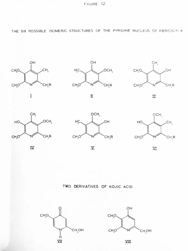
octahydropiericidin A, with those of compo~ds VII and VIII (figure 12),
both derived from kojic acid, are furnished as conclusive proof of theproposed structure of the pyridine ring of piericidin A2b, 2d.
Octab.ydropiericidin A A max (nm.) €ma.xNeutral MoleculeAnion No peak above 230
5,300Approx.3,ooo at 210
cation 215 8,500

Cation
50
A max (nm.) €max pH;;
l74 11,500250 8,500270 (shoulder) 5,300244 5,150270 5,300
)" max (nm.) E.max262 3,700241 8,300268 7,200
Compound VIINeutral moleculeAnion
Cation
Compound VIIINeutral moleculeAnion
The identity of compound VII is firmly established152, howeverits UV spectra are not very similar to those of octahydropiericidin A.
The UV spectra of compound VIII, synthesised by Takahashi2d,resemble those of ootahydropierioidin muoh more close~. On the otherhand their dissimilarity from those of compound VII and other 1-(H)-4-pyridones (see page Jb ) is surprising. The only other spectral dataof compound VIII ~uoted are some IR absorptionsJ name1ys vnuj013,200
-1 2d max1,620, 1,600, 1,125 cm • The first two of these figures suggestthat the compound exists as a l-(H)-pyridone (see page 37 ), andcertainly no equivalent absorptions are found in the IR spectrum ofoctahydropiericidin A, which appears to exist as a pyridinol (see page 4.4-).Bearing in mind this difference, and the fact that no other spectralcomparisons are made between the above-mentioned compounds, thesimilarity of the UV spectra of ootahydropiericidin A and of compound VIIIcannot be regarded as irre~utable evidence for the proposed location of
FI UR 12
THE SIX POSSIBLE ISOMERIC STRUCTURES OF THE PYRIDINE NUCLEUS OF PIERIClul N A
OH
I
OH
OCH,
]I m
OCH. OCH,
CH1
TWO DERIVATIVES Of KOJIC ACID
CHOJ
OH
CHOH• CH H~

51substituents on the pyridine ring of octahydropiericidin A (and of
piericidin A).It is a contention of this thesis that the published structure
of piericidin A2,3 may be incorrect. The reviewed evidence allowsthat any one of five other isomeric structures of the pyridine nucleuscould also be that of piericidin A. The six possible isomericstructures are shown in figure 12 (I-VI). Of these compounds I and IIwould be expected to exist as l-(H)-4-pyridones (see pages !li-30 ),and the remainder as pyridinols. Since piericidin A behaves as apyridinol (see page 4-b ), it is likely that the correct structure isone of these latter four isomers.
One of the aims of the present work is the identification of thecorrect structure of the pyridine nucleus of piericidin A, by extensionof both the number and the kind of comparisons between it .an appropriatepyridinic analogues. Sone of these will need to be specially
~.
synthesised, since they~~ve not/previoUSly'reported.

52
TI-m SIDE-CHAnr OF PIERICIDIN A
A simple experiment designed to confirm the location of olefinicbonds in the sias-chain of piericidin A has been undertaken as part ofthe present work. The mass spectra of octahydropiericidin A
derivatives contain peaks attributable to ILons.. formed by the cleavageof sequential C-C bonds of the side chain2e• In this way the carbonframework of the side-chain is reliably revealed, with the exceptionof the location of the olefinic bonds of piericidin A. The massspectrum of (?~~)-octahydrOPiericidin A should reveal these latterfeatures on comparison of the two spectra. The synthesis of such acompound by catalytic ~H)-hydrogenation of piericidin A should be easy.
The above experiment is prompted by the unusual DV absorptions
attributed to the diene system of piericidin A2a, namely the maximaat 232 nm (€max ...39,500) and 239 nm. (€max ... 40,500). A single
do h ° d2c Id b ttrans-trans 1ene, suc as 1S propose ,wou e expec ed to have asingle absorption maximum in this region, having an extinctioncoefficient not exceeding a limit of 30,000153• The doublet, ofextinction coefficient 40,000, implies the presence of two independent
° t d2adien1c sys ems, as was once propose •The structure of piericidin B154a,155, in which the alcoholic
function of piericidin A is methylated, and a detailed investigationof the stereochemistr,y of the side-chain of piericidin A154b,155, have
been published.The incorporation of propionate and acetate, rather than of
m~alonate, during the biosynthesis of piericidin A, and of piericidin Bthas been proved, although the origin of the nitrogen atom of thepyridine ring of these compounds remains obscure 156.

RISULTS AND DISCUSSIOn

53
!.~AOOLITZ3 OF STREPTOI.rrC:ES J.:OBARAIJ~SIS AIm THEIR DERIVATIVES
Piericidin A and piericidin B were isolated from the mycelia1
of streptomyces mobaraensis in the manner described by Takahashi et al.The fungus was grown on two media, one very richl~and the other
relatively simplel56 (see pagesr~O-I). The yield of piericidin A
was lower than that reported by Takahashil in both cases, the simplermedium giving a slightly higher yield of 11 mg./l. of medium. Theyield of piericidin B was 3.6 mg./l. of medium in both cases. Duringthe isolation procedure, relatively large quantities of fattysubstances were recovered from the early column eluents, but were notinvestigated further. Two other metabolites, both crystalline andreferred to as C and D, were isolated from fermentations using thesimple medium, but not from those using the ri~h medium. The yieldsof these, neither of which have been previously reported, wereapproximately 2 and 1mg./l. of medium respectively. The IR (thinfilm), DV and ~ mm spectrum of piericidin A and piericidin B weresimilar to those reported by Takahashil,2a l54a (see pages 131-r34-
and figure 35). In addition the IR spectrum of piericidin A in
solution revealed a strong and sharp absorption at 3505 cm-l typical ofa pyridinol, and no absorptions attributable to a l-(H)-pyridone(See pages 31-38) 13:t ). ~ }n.m spectra of piericidin A andpiericidin B after reaction with trichloroacetylisocyana~e were also
recorded and are discussed later (pages loa -I o~ ~ and figure 36.)
Pierioidin A has a very low vapour pressure at room temperature,however a mass spectrum was obtained at high gain and an ionisation
chamber temperature of 20000 (see figure 40 of the mass spectral

FIGURE 13
THE MASS SPECTRAL BEHAVIOUR OF PIERICIDIN A
~Pyr. I mjc =t..I~
+
~.IU II ~.33'
$Pyr.
Imfi-4lS
KEV -CH,-OC~-OH
= '~'CH,~~
19Pyr.
·O~H.
m;'= 14
I mic= LIS~~

54
appendix). A molecular ion at m/e - 415was detected, with a muchmore prominent ion at m/e = 397, presumably formed by the loss of waterfrom the parent. Accurate mass measurements of six other prominentpeaks of the spectrum were made (see page 1.33 ). As a result a schemefor the behaviour of piericidin A is postulated in figure 13. Inorder to account for the intense ions at m/e - 161 and m/e - 236, achange in the location of one olefinic bond in the side chain, from thatof the published structure2e, is necessary.
1 2 345 678pyr.- CH2-CH=~-CH;CH-CH2~=CH-
CH3 CH3becomes
1 234 567 8Pyr.- CH2-CH=~-CH2-CHiiiCH~=CH-
CH3 CII3On inspection the two structures are seen to be very similar, and
would not be expected to have markedly different UV, IR and ~I mmspectra. Indeed, the possibility that either structure may be correcthas already been recognised2e• It is now proposed that the lowerstructure is correct on the basis of the mass spectrum of piericidin A.
The molecular ion, I (figure 13), may decompose in a variety ofways depending, presumably, on the orientation of the molecule and onthe location of positive charge. The elimination of either water
(I-"'_~~"'''IV) or of 2-methy1butandl (I~II) gives rise to iona
at m/e ..3~7 and 331 respectively. 2-methy1butanal itself also
features prominently as an ion at m/e. 84. Fission of ion IVbetween C4 and C5 produces prominent ions at m/e - 236 and m/e. 161.The stability of this latter hydrocarbon fragment, borne out by the--

55
intensity of the ion, is to be expected from a highly conjugated system158such as the one proposed • Fission of species I, II or IV between
Cl and C2, C3
and C4 and C4 and C5 can be considered to be the origin
of ions at m/e s 182, 222 and 236 respectively. Ions at m/e = 330and 55 can result from fission of the molecular ion (I) on either sideof CIO' the carbon atom bearing the alcoholic function. Less intenseions at m/e - 262 and 302 correspond to fission principally of Ibetween C6 and C
7, and C8 and C9 respectively. An ion of similarly
low intensity at m/e - 282 may originate from IV1by the loss of an
unspecified methyl radical.The intense peak at m/e ..183 corresponding to
would be
-CH3 -CH3-OCH
3-OCH3
-OH -on
CllaJ;ic~·o(-~~C~~~W3expected &:toarise from a 1.1cLaffertyrearrangement, which is
unlikely since there is no proton attached to C3• Although the origin
of this species remains obscure, it appears to decompose further bylosing a methyl radical producing an ion at m/ e Cl 168.
Thus the principal ionic fragments of the mass spectrum of
piericidin A can all be given identity, assuming that the side chain of
the molecule has the structure proposed on page 5"4-.The 1I11J.IR spectrum of piericidin A diacetate (page 137-13B and
figure 35) confirms the presence of phenolic and alcoholic acetatefunctions in the molecule by the singlet peaks a.t 7.67 r and 8.08 '(
respectively. As expected, the doublet attributed to the proton

FIGURE 14
THE MASS SPECTRAL BEHAVIOUR OF OCTAHYDROPIERICIDIN A
I mic = 423
CHi. CHLOC~ OC~OH ~
r0---.... .~ - -tCHJ ..,A. IU
CH~~ o~oAofi~l~CHaI o~ \....~a ...
H.....'C?' a H
~\ II"A = 113
CH8~l-i1~
~I-:-. - ["CH)A~A -...;----:....--1..... 'Y'. 1&9
CHP 0, CHa
D
II'm;'= 181,.

56attached to C10' exto the alcoholic acetate group, is found at 5.06 T ,
whereas in unacetylated piericidin A it appears at 6.36 i . Thisshift is similar to that observed after the reaction of piericidin A
with trichloracetylisocyanate (see page 10& ,13l: and figure 35).Small changes in the chemical shifts of substituents attached to thepyridine ring are also evident after acetylation as shown below.
Substituent -0CH3 -OCH -CH3 -CH-3 2
Piericidin A (,) 6.08 6.18 7.91 6.63
Piericidin A 6.05 6.20 7.99 6.60diacetate (,)Shift on acetylation -0.03 +0.02 +0.08 -0.03(T)
Similar shifts are observed after the reaction of piericidin Aand piericidin B with trichloracetylisocyanate (see pages 101 120, ),but as in those cases no clear deductions can be made regarding theproximity of other substituents to the phenolic function.
The rn, UV and lH Nl\'iR spectra of octahydropiericidin A (page 13'
and figure 36) were similar to those reported by Takahashi2a• In
addition the mass spectrum of the compound (figure 40 of the massspectral appendix) fully confirmed th~ proposed structure of its side-
hai 2c,2ec n • The molecular ion, I (figure 14), at m/e. 423 gives
rise to ions at m/e = 408, 394, 366 and 57, 335 and 87, 308, 294, 266,252, 238, 224 and 196 by fragmentation between consecutive carbon atoms
of the side-chain, aa indicated. The ion at m/e. 337 may be formed. -
by the loss of a formylradical from that at m/e. 366, or by fission of
the molecular ion between C9 and C10 together with the transfer of a
drogen atom from the alcoholio function.

FIGURE 15
THE ISOTOPIC DISTRIBUTION OF MOLECULAR IONS IN 11-£ MASS SPECTROMETER
OCTAHYOROPIERICIOIN A
21
PEROEUTEROPIERICDIN' A
EXPB:TED SPECTRUM
• PERDEUTEROPIERICK* A
OBSERVED SPECTRUM
I I I I I I I I I I I
00 ~ - M/•

57
The ion at m/e = 183 can be considered to originate from aJ.:cLaffertyrearrangement (I--;,..II,figure 14) confirming the locationof the hydrocarbon side-chain in an ct position in the heterocycle(.C~ page 4.S), giving rise in turn to that at m/e = 168 by the Loas
at m/e = 154.23. (
of a methyl radical. This latter process is confirmed by theexistence of a peak due to the correspondine Detastable transition
Piericidin A wac reduced with deuterium in an attempt toprepare (2Ha)-oct,:"hYdrOPiericidin A, by analogy to octahydropiericidinA. The purpose of the attempt was to confirm the location of theolefinic bonds of piericidin A by a comparison of the mass spectraof its two reduction products (see page ~l). Deuterium gas,prepared from deuterimfi oxide, was found to have an isotopicabundance of 95;1. (page 13' ). The expected and actual isotopicdistributions of the molecular ion of (2HS)-octahydrOPiericidin A,prepared using such gas, are shown in figure 15, together with thatobserved in the case of octahydropiericidin A. Clearly there hasbeen considerable incorporation of deuterium in excess of the eieht
\atoms expected from the reduction of olefinic bonds.The mass spectrum of the perdeutero compound (figure 40 of
the mass spectral appendix) reveals a similar fragmentation pattern
to that of octahydropiericidin A. Estimation of the vectorial
mean of corresponding groups of spectral lines for each compound
revealed that deuterium was randomly incorporated into the
side-chain, apparently

FIGURE 16
THE DISTRIBUTION OF DEUTERIUM IN PERDEUTEROPI ER.ICI DIN A
'!_~:J "e 0
01
01 <C c=6 :g
Z c 'u 10
0 &. ·c~ ~ .9!
Q. -c.. ea: S0 i ,6D- O ~ ,§ 'Ec v ,~
0 Q.~"e ....
~0 !,
:::I '6 0 0t: a ..ffi s u
l 0
i =c =4
" 0
I Ev
.0 -5§ .E
"" .E~ '0.0 10
'" 0-;
~.. " '" . .... '" .. !! .. ~01 , ~ , , , ~ , ,
Q, ~~ .. .. '" .. Cl co
SUCCESSIVE IONIC FRAGMENTS
M = Molecular Ion
n -(MO = Ion formed by fission bctwccn carbon atoms nand
(0+0 of the side chain
MeL = Ion formed by McLafferty rearrangement
MeL-IS = Ion formed by 1055 of methyl radical from Mc:L

on all carbon atoms except that adjacent to the pyridine ring. Therewas no incorporation of deuterium into other substituents of thepyridine ring. These results are represented graphically in figure 16.
Confirmation is provided for the position of the first olefinicbond of the side-chain by the ions at m/e. 169, 184, 197 and 226(see figure 14, Il~IIl etc.). Above m/e. 226 the isotopicdistribution of deuterium obscures any such observations. Oneexplanation of the random incorporation of excess deuterium is thatdouble-bond migration, with concurrent incorporation of the isotope,took place on the surface of the 'catalyst (platinUm oxide) before or (during reduction. This possibility has been recognised in other
systems159•The mass spectrum of a sample of perdeuteropiericidin A which had
been dissolved in (2Hl)-methanOl revealed ions at m/e • 170, 185,198 and 227 in place of those of one mass unit less (see figure 40of the mass spectral appendix). These confirm the location of aphenolic function on the pyridine nucleus of the piericidins.
In the mass spectra of both octahydropiericidin A and perdeutero-piericidin A, peaks due to a corresponding metastable transition are
detected at m/e. 154.23, 155.22 and 156.22. This corresponds in each
case to the loss of a neutral fragment of mass 15 from the ion resultinGfrom the'l-1cLaffertyrearrangement at m/e 1:1 183, 184 and 185 respectively.
160It has been suggested that the neutral fra£~ent is a methyl radical
originating from a methoxyl group. The above evidence is consistentwith this theory.

59
The preparation of (3H)-piericidin A by the Wilzbach gas exposuretechnique161, and of (3H)-octahydropiericidin A from it, were
straightforward (pages 14-3 -/4.4-). The use of these compounds is
discussed later (page {,b -, g ).The structures of metabolites C and D, whose isolation was
mentioned earlier, and of the hydrogenation product of each have notbeen determined. However the available spectroscopic evidence ofthese compounds does provide some structural information (pagesI34-f37,
14-0-14-3). The UV spectra of the two metabolites are strikinglysimilar both before and after hydrogenation (see figure 37), suggestingthat they are structurally closely related. The IR spectrum insolution, of each, exhibits no absorption attributable to the O-H
stretching vibration of a free hydroxyl group (3,500-3,600 cm-l); bothhave strong absorptions in the regions of 1685 cm-l and 1645 cm-l,possibly due to the presence of an aP-unsaturated ester or lactonechelated to an enolic group162. On hydrogenation a single carbonyl
absorption in the region of 1665 cm-l is observed.The mass spectra of metabolites C and D have parent ions at
m/e - 236 and m/e = 206 respectively, accurate mass measurements
revealing that their elemental compositions, CIOHION304 andC9HaU303 respectively, differ by CH20. Both molecular ions may )(.
-,"-~-.--....decompose by the loss of 15 or 29 mass units, corresponding to methyl
and formyl.·radica.l:.. In the case of metabolite C, but not of
metabolite D, the ion formed by the loss of a methyl radical from themolecular ion at m/e D 221 decomposes further ~ the expulsion of

60
carbon monoxide, producine an ion at m/e = 193. A peak due to the
metastable tr,msi tion cor-reapondi.ngto this process occurs at m/ e =
168.55. The mass spectra of sarap Lea of metabolites e and D, whi.chr~d been dissolved in deuterium oxide, revealed increases of one massunit in all the above mentioned principal ions in both cases. Thisindicates the presence of a single exchangeable proton in bothcompounds, which is not lost in any of the decomposition processes
mentioned above.The molecular ions of the mass spectra of both metabolites
increased by two mass units after hydroGenation, implying thereduction of a single olefinic bond in both compounds. Althoue;hit appears that both molecular ions may decompose by the loss of a
formyl radical (29 mass units), the spectra are ncre complicatedtr~n those of the original metabolites, and preclude reliableinterpretation in the absence of further corroborative evidence.The elemental compositions of the molecular ion and base peak, inthe mass spectrum of reduced metabolite e, have been sho~m todiffer by e2II502by accurate mass measurements. A sample of
{
I
revealed an increase of one mass unit in the molecular ion of its
reduced metabolite e, which had been dissolved in deuterium oxide,
mass spectrum, indicating the presence of a single cxchanGe~ble
proton as in the case of the parent compound.
lUlunusual feature of the mass spec~ra of all four compounds
mentioned above concerns their molecular ions. These are of even Jm/e value in each case, and yet accurate mass measurements show thatthey contain three nitrogen atoms. The rules of valency require

61an uncharged tri-aza molecule, containing carbon, hydrogen and oxygen as
the only other elements, to be of odd molecular weight. One possibleexplanation of this phenomenon is that the four compounds are in fact
salts, and that the supposed molecular ions of the spectra correspond
to the cations of the salts, which are charged species before entryinto the ionisation chamber of the mass spectrometer. This possibilityis also consistent with their observed solubility in water, theirrelatively high melting points and the possibility that they may containbasic nitrogen atoms capable of forming quaternary salts.
The In m~R spectra of metabolites C and D and of hydrogenatedmetabolite C are recorded (pages 135",'3'J'4-I). In all three cases there
I spectra·appear from the H m~alto be more protons than are indicated by accuratemass measurements of the molecular ions in the mass spectra. This isconsistent with the possibility that the compounds are salts, of whichonly the cations contribute to the supposed molecular ions of the
mass spectra.All three compounds exhibit a sharp singlet corresponding to one
proton at -1.11i, independent of concentration and undergoing exchangeon the addition of deuterium oxide. This is probably due to thepresence of an intramolecularly hydrogen-bonded phenolic or enolicproton, and may also be responsible for the observed mass spectral
increases of samples dissolved in deuterium oxide. In addition
metabolite C has a singlet at 8.45 r corresponding to two protons, which
moves to higher field on dilution and is exchanged by the addition ofdeuterium oxide. This behaviour 1s typical of the protons of aprimary amine, and, since it clearly does not feature in the supposedmolecular ion of the mass spectrum of metabolite e, may be a feature
of the suggested anion of this compound.

62
The ~ 11m spectrum of metabolite C also has the following
features:(i) Two olefinic singlets each corresponding to one proton
at 3.67 and 3.82 r.(ii) Two singlets each corresponding to three protons at
6.05 and 6.09 1', typical of the protons of methoxyl groups attachedto sp2 hybridised carbon atoms.
(iii) One singlet, corresponding to three protons at 7.74 1r,typical of the protons of a methyl group attached to an sp2 hybridised
carbon atom.The lH Nr.1Rspectrum of metabolite D differs slightly from these
observations in that it has one more signal due to an olefinic protonand one less attributable to a methoxyl group. This coincidessatisfactorily with the difference of CH20 observed in the elementalcomposition of the molecular ions in the mass spectra of the twocompounds (see page ~'). The additional olefinic proton ofmetabolite D with a resonance a.bsorption at 3.51 ; is spin-spin
coupled to the olefinic proton detected at 3.67 1r t the couplingconstant being 2.6 H. Thus it may be that the methoxyl groupz
present in metabolite C, but absent in metabolite D, is adjacent to
a single isolated olefinic proton.
On hydrogenation, two signals in the In mm spectrum of
metabolite Ct namely the olefinic singlet at 3.821i and the singlet
at 7.74 7' , disappear. Correspondingly, new signals are found at7.13, 8.51 and 9.1 7' , implying the reduction of an olefinic bondbearing a methyl group and a proton.

63
Thus the initial suspicion that metabolite C might be closelyrelated to the piericidins, based on the observation that it produced
IH l~m signals attributable to two methoxyl groups and a methyl groupattached to a pyridine ring, similar to those observed in the caseof the piericidins, is incorrect.
In conclusion it can be said that metabolites C and D appear tobe salts. If this is the case then each has a similar ioniccomponent, differing only in that metabolite C possesses a methoxylgroup in place of an olefinic proton present in metabolite D.This common component appears to be an unsaturated nitrogen heterocycle,which, since the species was not completely hydrogenated using aplatinum oxide catalyst, may possess aromatic character.
The investigation into the properties of metabolites C and Dwas not extended, once that it was established that they did notpossess the pyridine nucleus present in the piericidins as seemedpossible at one stage. The small quantities in which they wereobtained also restricted their investigation.

EXPERIMJiNTS OF BIOCHD·1CIAL SIGNIFICANCE
The potent inhibition of NADH dehydrogenase by piericidin A is
s subject of considerable chemical interest (see page 25"). It
features of piericidin A, during inhibitory studies might lead tom
seems possible that the use of analogues, possessing selected chemical
understanding of the function, or lack of function, of these featuresduring inhibition. Piericidin A has been shown to be a phenolicpyridine having a long hydrocarbon side-chain, a methyl group and twomethoxyl groups as the remaining sUbstituents2• A number ofcompounds possessing a variety of these features were tested for theirinhibition of NADH linked oxidation in mitochondria (pages 14-' -/4-1).
Some of these were synthesised (pages 180 -" i ).
Piericidin Band octahydropiericidin A were found to be almostas potent as inhibitors as piericidin A, indicating that the side-chainneed not possess an alcoholic function nor be unsaturated in order to
achieve complete inhibition.Thus the scheme shown in figure 8 (see also pages 2. & ...27 )
involving an olefinic linkage of the side-chain, does not account forthe inhibition by these piericidins, and consequently the proposedchemical tests of this scheme involving model systems are unnecessary.
Piericidin A diacetate was found to be three orders of
magnitude less inhibitory than piericidin A, suggesting that the
phenolic function of piericidin A is of considerable importance
during inhibition.Of the remaining compounds all were more than 105 times less
effective as inhibitors than piericidin A. However, ot these,

compounds having a lipophilic side-chain ~ a phenolic functionwere the most effective. This provides some confirmation for thefunctional importance of the phenolic moiety suggested above in thecase of the piericidins. The importance of lipid solubility hasalready been recognised (page 2 ~ ) •
The significance of these two features is exemplified by thethree compounds 2-(1-n-hexyl)-4-hydroxypyridine, 2-(1-n-hexyl)pyridineand 4-hydroxy-2-methylpyridine. The first is required in aconcentration 3.5 times less than the second and 50 times less thanthe third in order to achieve the same degree of inhibition.FUrthermore 2-(1-n-hexyl)-4-methoxypyridine and 4-chloro-2-(l-n-hexyl) pyridine are both less inhibitory than 2-(1-n-hexyl(-4-hydroxypyridine in accordance with the suggested importance of thephenolic function during inhibition. Considering the similarity ofthis latter compound to the published struoture of pierioidin A2,3,its oomparative impotenoy as an inhibitor is a matter for some
surprise.The importanoe of lipid solubility concerning these inhibitors
is again illustrated by the examples of 4-(1-n-hexyl)-2-rnethoxyphenoland vanillin, the former being 66 times more potent than the latter.
4_(1-n-hexyl)-2-methoxypheno1 is also more potent than 4-(1-n-pentyl)phenol, a faot whioh may be due to the presence of either the
methoxyl group or the longer side-ohain in the former.
It is conoluded from these studies using analogues that the
phenolic function and the long side-chain of the piericidins are
both essential in order to aohieve potent inhibition of NAnH

FIGURE 17
THE BINDING OF ['H]- PIERICIDIN A TO MITOCHONDRIA
160
14<)
110
'20
40
20
o t.I' Ii' I'JI 120 I~, r-r- ~ ;:':">"
"'f mole M picrlcldln A addcd/g. protein
Curve A; bindin9 in sucrose-tria medium.
Curvll A ; binding in sucrose-tria - 8S'; medium.
Curve C. binding in sucro5e-tris-BSA mediumafter preincuba~ion with
unlabelkd piericidin A.

FIGURE 18
THE EFFECT OF REPEATED WASHING ON THE BINDING OF [3H]-PIERICIDIN A
TO MITOCHONDRIA
,0
>:J
c:.~2 H"Q.
..st"c:~0.t:l 50
-cj.~'" .0III'ii-
I"0 lOet-
20
o 110
mf mole tHl picrlcidln A addcdAl·protein
Curve A Ibinding In sucro5e-tris medium.
Curves 0,1,3,5,71 binding, after washing in sucrOM-tris- BSA medium the
indicated no. of times.

66
dehydrogenase.A sample of piericidin A labelled IIIth tritiurn (page 14-3 )
NADH oxidation (page (~8and curve A figure 17). Hence, in
was used to investigate the binding of piericidin A to mitochondria.The binding of (3H)-piericidin A to mitochondria extende~ toconcentrations above those required for the maximal inhibition of
agreement with earlier observations (see page 10 ), piericidin Ais not a specific inhibitor of NADH dehydrogenase. In the presence
of 2% bovine serum albumen the binding curve became clearly biphasic/
(curve B figure 17). Furthermore the rapidly rising portion of thiscurve could be abolished by titrating the mitochondria to maximalinhibition with unlabelled piericidin A before the addition of any(3H)-piericidin A (curve C figure 17). A measure of theconcentration of piericidin A involved at the sensitive site wasobtained by subtracting curve C from curve B and was in the region
0.02 mu moles/mg. of protein. Repeated washing of the mitochondria
with 2% bovine serum albumen also indicated that a similar amount
of (3H)-piericidin A was tightly bound (figure 18). Acomparison of the amount of (3H)-piericidin A bound after repeated
washing and the degree of inhibition obtained showed a direct
correlation (see page 14-').It was shown that piericidin A "Tas not removed from the
specific binding site by equilibration withunspecifically bound piericidin A, since the addition of excess

67
unlabelled piericidinA to mitochondria previously treated witheH)-piericidin A resulted in no displacement of (3H) piericidin A
after repeated washing (page ISf). Conversely when excess (3H)_piericidin A was added to mitochondria pretreated with unlabelledpiericidin A, the (3H)-piericidin A was removed by repeated
washing (page lrO ).
Pretreatment of mitochondria with rotenone, amytal or unlabelledpiericidin A decreased the binding of (3H)-piericidin A (paee I~I ),indicatine that all three compounds compete for a common binding site.This is consistent with the knowledge that they all inhibitrespiration in the sarne region of the respiratory chain (seepages ~'''ll). Antimycin A on the other hand, an inhibitoracting elsewhere in the respiratory chain (see figure 2), did notdecrease the binding of (3H)-Piericidin A.
The extraction of mitochondria, treated with (3H)-piericidin A,with acetone resulted in complete removal of the tightly boundradioactivity. The extract contained the expected mitochondriallipidsl63 together ~ith unchanged (3H)-Piericidin A identified
chromatographically (page I ~ 3 )• Hence althoueh piericidin Amay interact with mitochondrial lipids, and with other mitochondrial
components, it does not become covalently bound to them.The possibility that piericidin A might interact with the
functional groups of proteins was investigated in a rudimentary
manner by heating (3H)-piericidin A with appropriate amino acids insolution (page 154-), and examining the products by thin layer

63
chromatography. In all cases about 80% of the original radio-
activity was recovered at the Rf corresponding to piericidin A andat the origin, negligible amounts being found elsewhere. Theradioactivity detected at the origin was assumed to be due todecomposed piericidin A, since it was found in a blank experiment.Only in the case of cysteine hydrochloride, and to a lesser extentof cysteine itself, was the ratio of radioactivity at the origin tothat corresponding to unchanged piericidin A significantly differentfrom the blank experiment. Thus despite the fact that piericidin Amight ~e expected to behave either as a weak acid or a weak base, nointeraction between it and free amino or carboxyl functions wasdetected. The results with cysteine and its hydrochloride probablyindicate that oxidation of the sulphydryl groups in these compoundshas involved the olefinic lirucages of piericidin A in a destructivemanner, rather than any reaction of biochemical significance.
Following the observation that 2,5-diethoxy-l,4-benzoquinonecan act as a charge transfer donor164 towards 2,3-dich1oro-5,6-
. 165dicyano-l,4-benzoquinone (DDQ) ,and towards tetracyanoethylene(TmrE)166, analogous experiments were performed with ubiquinone
and piericidin A (page IS r), in the hope of finding a property
common to the two compounds of some biochemical significance.Charge transfer was not detected in either case, nor surprisingly
was it detected between tetramethoxy-l,4-benzoquinone and DDQ or
TCNE. It is concluded that piericidin A and ubiquinone are not
likely to act as charge trans~er donors in biochemical systems.

FIGURE 19
2t4-DNP DERIVATIVES OF METHYL PYRUVATE
"-N-H-O
lo
I
o
~
_A'~C?.--.~
II
THE CHROMOPHORIC ANION OF 2t4-DNP DERIVATIVESy~~

69THE 2! 4-DINITROPIIDITLHYDRAZONFS OF l.11!rr'HYLPYRUVATE
The disorepanoy in the reported melting point 148,149,150 of
the 2,4-dinitrophenylhydrazone of methyl pyruvate was resolved bythe isolation and characterisation of two distinct isomers of this
compound.Corresponding isomers of 2,4-DlrP derivatives of other pyruvic
acid esters have previously been assigned the structures I and II(figure 19), and referred to as« and ~ formslSO• These assignmentswere based on comparisons of the UV spectra with compounds in which
the proposed isomerisation'is not possible.Isomerism has also been observed in the 2,4-DlrP derivatives oft od 151«-ke 0 ac~ s • In these cases the IR spectra indicated hydrogen
bonding to the carbonyl fUnction in one isomer, but not in the other.Furthermore the addition of soditun hydroxide solution to the "hydrogen-bonded" isomer had. very little effect, whereas in the other case a~ed colour was immediately produced.
Methyl pyruvate was produced from pyruvic acid by the action of
diazomethanel67 (page I~7 ). A 2,4-DNP derivative of methylpyruvate was then prepared by reaction with 2,4-dinitrophenylhydrazineaccording to a standard method168 (page I S"7). It was identifiedas the « form by comparison of its melting point and ultra violet
150absorption spectrum with reported values • In dilute, slightly
acidic, methanolic solution this isomer became partially converted
to a seoond isomer on prolonged exposure to sunlight (page ,,,').
The second, or ~, isomer was separated chromatographioally.

70
Some of the chief differences in the properties of the twoisomers are shovm in Table 1, however considerable similarities,notably in the mass spectra and parts of the :mm and IR spectra,
aleo exist between them •..Table 1 Some properties of the two 2z4-DNP derivatives of methyl pyruvate
ex learner ~ Isomer
Appearance yellow crystalline orange crystalline
M.Pt. 186-181 164.5-165.53310 (med. st.)
1730 (st.)341 (22,800)
VN-H (nujol mull)cm-l 3150 (med. st.)\1(,:0 (nujol mull)cm-l 1700 (st.)
A max {methanol)nm. 361 ( ::11,400)'r NH (CDC13, 1%)
T.L.C. (System)
-1.0
Effect of NaOH onmethanolic solution
No colour changeRf • 0.16Red colour produced
It appears that hydrogen bonding between the proton attached to
nitrogen and the ester carbonyl function is present in the ex, butnot the ~ isomer. This specifically accounts fora
1) the C=O stretching vibration frequency being 1100 cm-l
-1inetead of 1730 cm • (In methyl pyruvate a value of 1135 cm-l
is observed).2) the N-H stretching vibration frequency being 3150 cm-l
-1instead of 3310 cm •3) the nuclear magnetic resonance of the proton attached to
nitrogen being at -4.1 ,..instead of - 1.0 i .The difference in behaviour towards base is less readily
explained. The red colour is reversibly produced, and is also

71observed with the 2,4-Dl~ derivative of acetone, suggesting thatthis latter compow1u structurally resembles the ~ rather than the aisomer. The chromophone may simply be produced by loss of theproton attached to nitrogen. Such an anion could have azaquinonoidcharacter responsible for the chromophone, (see figure 19). Inthe case of the "hydrogen bonded" ex isomer the loss of the protonmight be expected to be less facile, accounting for the difference.m-Dinitrobenzene produces no colour under these conditions, shouingthat the effect is not solely connected with the aromatic ring.
It is concluded that the ex isomer of the 2,4-DlrP derivativeof methyl pyruvate has a structure represented by II (figure 19),and the ~ isomer a structure represented by I (figure 19). Thisconclusion is contrar,y to the previous report150, and can beextended to include the whole series of pyruvic acid ester derivativesmentioned in this report.
It can be seen from the above results that neither of theisolated 2,4-dinitrophenylhydrazones of methyl pyruvate have amelting point corresponding to that of the compound reported byTakahashi2b, obtained on treatment of an ozonolysis product of
"O-methylhydropiericidin A" with Brady's reagent. Thus thiscompotmd was not a 2,4-dinitrophenylhydrazone of methyl pyruvate as
was claimed. The conclusions made from this claim, concerning the
relative positions of substituents in the pyridine ring of piericidin
At must therefore be discounted. It is proposed that the correct

72
structure of this pyridine ring may be any one of the six isomers
represented in figure 12, but that it is more likely as mentioned
earlier to be a 3-hydroxypyridine than a 4-hydroxypyridine.

FIGURE 20
SYNTHETIC ROUTES
NO. Cl
f\)O 8.j, :')0 7S:l! "H,CJ¢j 08'. H~H.C ~ H.C ~
-HC ClH. •I II m
rz 1'"OCH, OCH3 E: H,C:6:H,)¢(H H,~NH, H.C O.
•1\ 95'• SI,.
H,C 0 CH,H. CH, H,C N OCH, H3C CH,
El[ ::sm: JZI :sz:
overall yield I-:::2lII = HZ (7 stages)
I'
nNO. ClrAr'CH, CH. ~O~CH,
80\ AQ _.;.;_77\;:__~ ---~ J..V~"H,C~ H, iii HF Cl
][' D[' "-" .... ! nz:'
rA(CH.
H.C¥
OCH.
H)¢iCH.o 5,\
H,C OCH.
:szm'
H.Nij(CH... 96'1.
H.C OCH,
E' ::szr'
X;CH,
H.cMoCH.
'7,'
overall yield I~:sz:m:' = ',2Z f{ ,tag«s)

73
THE SYNTHESISOF 4, 6-DnmrHoxy -2, 3-DU'lErIITL-5-
HYDROXYPYRIDINE AND OF 4, 6-DIMEl'HOXY-2,5-DTMErHYL-3-
HYDROXYPYRIDINE
The synthesis of the two above named ~ -hydroxypyridines, bothpossible candidates for the correct structure of the pyridine nucleusof piericidin A (see page ~I and figure 12, V and VI:" where R - H),has been achieved. The synthetic routes, which are from thecorresponding 'lu/t.lcrin~sand are analogous, have the advantage ofbeing based on known reaetions which do not accommodate ambiguousproducts, but they are not the only possible synthetic pathways andare not necessarily the shortest. They are summarised in figure 20.
The first two stages in each ease were by standard procedures169
(Bee pages r ~ 0 , 173 ....174- ), since both dimethyl-4-ni tropyridine-1 no f' Il-oxides (III and III figure 20) are reported in the literature, J •
2,4-Dichloropyridine has been prepared from 4-nitropyridine-l-oxide by the action of sulphuryl chloride in a sealed tube172• Thismethod was repeated using phosphorus oxychloride under reflux with an
almost identical yield of the dich1oropyridine (page "'). It wastherefore decided to adopt the latter technica1~ safer procedure in
analogous reactions involving the two dimethyl-4-nitropyridine-1-
oxides (III and 1111, figure 20).The reaction product of 2,3-dimethyl-4-nitropyridine-1-oxide
was examined in detail (pages ", -I' t,.. ), three compounds beingisolated. Each was identified as a dichloropyridine from the isotopicdistribution of the molecular ion in its mass spectrum. The two
major components, 4,6-dichloro-2,3-dimethylpyridine and 4-chloro-2-

74
ch1oromethyl-3-methy1pyridine isolated in the ratio 2·6:1, wereisomeric and were distinguished principally by their 1H NlIR
spectra. These are interpreted in figure 23 (I and II). An
interpretation of their mass spectra is presented in figure 46 ofthe mass spectral appendix. The molecular ion of 4,6-dich1oro-2·,3-dimethyl-pyridine is also the base peak of the spectrum, but in thecase of 4-ch1oro-2-ch1oromethyl-3-rnethy1pyridine the base peakcorresponds to an ion formed by the facile elimination of a chlorine
atom from the molecular ion. Presumably this is the chlorine atomof the 2-ch1oromethy1 group. The stability of the resultant ionmay be explained as indicated in figure 46.
The third component, 2-oyano-4,6-dich1oro-3-methy1pyridinepossessed an IR absorption at 2240 cm-l, typical of the C~~stretching vibration of an aromatic nitrile. Since the !r NMRspectrum indicated the presence of only one aromatic proton and onemethyl group in the molecule (interpretation in figure 23, III), theother carbon substituent, formerly a methyl group, had clearly beenconverted to a cyano group, as detected independently. It wasconcluded that this cyano group was in the a rather than the ~position for two reasons. Firstly, by comparison with the assignments
given to compounds I and II in figure 23, the protons of an a-methyl
group in a dichlorocyanopyridine might be expected to have a resonance
frequency below that of 7.46 1i observed. Secondly on chemicalgrounds (see below) an a-cyano group is more likely. The massspectrum of this compound is interpreted in figure 46 of themass spectral appendix.

FIGURE 21
, MECHANISMS OF REACTION
NO. Cl Cl
H.JS POC:'. H. H,::Q H,~. I fil~ orHC ~I
H.C ( H~ ~ 3 I
C~'{I
0..0me CI/~CIm
mA
1H+ + [HaPOCIJ~N,lci•
ClH.C~
eHc)lN'Acl•
mc
- HP
12:e

75The initial step in the production of the above mentioned
products is probably the formation of a salt with phosphorusoxychloridel73 (figure 21, IlIA). Nucleophilic displacement of thenitro group by chloride ion will release nitrite ion producing IIIB.Nucleophilic attack by chloride ion on this species at theunsubstituted a position, followed by elimination of the elements ofphosphorodichloridic acid may be considered to produce the desiredproduct IV. On the other hand, loss of a proton from III B to produce
the methide, III e, may then give 4-chloro-2-chloromethyl-3-methyl-pyridine, IVA, by nucleophilic attack of chloride ion on the methide.
The chlorination of ~ or a methyl groups in pyridine-I-oxidesusing phosphorus oxychloride has been observed before174, however theformation of a cyanopyridine is more unusual. The additionalnitrogen atom can only be derived from the nitro group present inthe starting material III (figures 20 and ~). It is proposed thatnitrite ions produced during the reaction are in equilibrium withnitrosonium ions (NO+), the equilibrium being in favour of the lattersince the medium is acidic. Nitrosation of the most acidio methylgroup may then occur, followed by the loss of water in the hygroscopio.
medium, produoing the product IV B. It is assumed that the a methyl
group is more aoidio than the ~ methyl group, in line witll generalobservations on methyl pyridinesl75,l76• It is oonceivable that
the methide, III e, might aot in a nucleophilic manner towards
nitrosonium ions, producing the same product, IV B, but this isconsidered less likely since it requires the methide to aot in anambident manner under a single set of experimental oonditions.

76
Clearly the proposed nitrosation cannot occur before the replacementof the nitro group in III A, a process which concurrently reducesthe acidity of the ~-methyl group. The possibility of an intra-molecular reaction involving the ~-methyl and 4-nitro groups isconsidered unlikely, since nitrous acid is quite clearly present insolution, as apparent from the observation of oxides of nitrogen intowhich it decomposes. An intermolecular mechanism is also favouredby the fact that on using a greater proportion of phosphorus oxychloridefor the reaction, negligible amounts of the cyano compound were
obtained (see note on page "1 ).The reaction of 2,5-dimethyl-4-nitropyridine-l-oxide with
phosphorus oxychloride was not examined in detail, the crude reaction
product being continued to the next stage (page 114-). However theproducts of this next reaction stage indicated that 4,6-dichloro-2,5-dimethyl pyridine and 4-chloro-2-chloromethyl-5-methylpyridine hadbeen produced in a ratio of 4:1. No cyano compound was detected inthis case, probably on account of the greater proportion of phosphorusoxychloride used during the reaction.
Mixtures of 4,6-dichlo~2,3-dimethylpyridine with 4-chloro-2-
chloromethyl-3-methylpyridine and of 4,6-dichloro-2,5-dimethylpyridine
with 4-chloro-2-chloromethyl-5-methylpyridine were treated with sodium
methoxide in methanol, in a manner similar to procedures recorded
in the literature112a,177 (pages 1'4-)114-). The expected products,
corresponding to nucleophilic displacement of chlorine by methoxide1ion (including V and V, figure 20), were isolated in each case.
, '

FIGURE 22
POSSIBLE STRUCTURE OF A PRODUCT OF RE,6CTlON
OCH, o
IH
o
I 11
OH
][
R,· CHI. Ra- H or R,= H. Ra= CHJ

71Their 1aln~R spectra were completely consistent with the proposed
structures and are interpreted in figure 23 (IV to VII). Theirmass spectra (see mass spectral appendix, figure 44) are interpretedin terms of their proposed structures (see mass spectral appendix
figure 47). The ease with which a hydrogen radical is lost fromthe molecular ions of 4,6-dimethoxy-2,3-dimethylpyridine and 4,6-dimethoxy-2,5-dimethylpyridine appears to be typical of a-methoxy-pyridines (page 111). The facile loss of 30 mass units from themolecular ions of 4-methoxy-2-methoxymethyl-3-methylpyridine and4_methoxy-2-methoxymethyl-5-methylpyridine is most probably due tothe elimination of formaldehyde ~ a Mclafferty rearrangement (seemass spectral appendix figure 41).
In addition to the two·eA~ected compounds mentioned above afurther product was isolated in each case, the two products being ofequivalent structure, judging from their spectral similarities
(pages "').1" ). Each possessed a strong IR absorption inthe 1630-1660 cm-l region indicative of a pyridone (see page 37 ).
In addition the 1I1~~R spectra showed peaks attributable to oneexchangeable proton, two methyl groups and a proton each attached toan sp2 hybridised carbon atom, and either a methoxyl group attached
to an sp2 hybridised carbon atom or an N-methyl group of I-methyl-
pyridone. This information can be accommodated by three different
structures (see figure 22, I, II and III), each consistent with the
elemental analyses and the mass spectra. Since the UV spectraremain unchanged in noutral and basic solution and exhibit ahyprochromic shift in acid solution, neither compound is likely to be

18
a 1-(H)-2-pyridone or a 1-(H)-4-pyridone (see pages 34) 3' ).
A compound with structure III (figure 22) might conceivably have UVabsorptions of the type observed, although in the absence of anyanalogy no such assumption can be made. That this third structurewas correct·was deduced from two further experiments with the 2,3-dimethylpyridine derivative. Firstly reaction with trichlo!acety-lisocyanate revealed two products by h mm spectroscopy (page r' 1),the homogeneity of the starting material being well established. Itappears from other experiments (page 110 ) that trichloroacety-lisocyanate reacts with tautomeric hydroxypyridines predominantlyat the oxygen atom, producing a single product. It is therefore
concluded that in this case, since there are two products, reactionhas taken place at two oxygen atoms rather than at an oxygen and anitrogen atom. This favours structure III (figure 22). Secondly,if the compound contained a methoxyl group, then it should be possibleto hydrolyse this function.producing a dihydroxypyridine. Three
10680, 178 179different attempts at ether cleavage based on established proceduresfailed to produce any such product, substantial recoveries ofunchanged starting material being achieved;. in each case
It is therefore proposed that the two compounds are 4-hydroxy-
1,2,3-trimethyl-6-pyridone and 4-hydroxy-l,3,6-trimethyl-2-pyridone,existing as such in their principal tautomeric formsl06 (page 31 ).The hydrolysis implied in their formation probably occurs duringthe experimental work-up, the rearrangement to l-methylpyridonestructures. being more likely to occur thermally during the reaction.

79The desired products V and VI (figure 20) of the above
reactions were easily separated in quantity chromatographically.Both of these compounds were readily nitrated using mixed acidsl80
producing over 50% of the desired products VI and VII (pages('9,'17).The identity of the products was consistent with their lunflR spectra,which are interpreted in figure 23 (VIII and IX), and with theirelemental compositions and the molecular ions of their mass spectra.No starting material was recovered in either case, and only in thecase of the reaction involving 4,6-dimethoxy-2,3-dimethylpyridinewas any other product isolated, even that being only in a 0.5~ yield(page "'). This compound was tentatively identified as 2-cyano-4,6-dimethoxy-3-methylpyridine from its lR absorption at 2240 em-I,typical of the C-N stretching vibration of an aromatic nitrile, andfrom its nmm spectrum,. which is interpreted in figure 23 (x),
The ease of the above nitration reactions (30 mins. at 0-20oC),compared to that of pyridinel8l, is a measure of the change incharacter of the pyridine nucleus brought about by four electrondonating substi tuents. The remaining unrecovered ma.terial:·inboth of the nitration reactions w~s water soluble. This may
indicate that some cleavage of the methoxyl groups of the aromationucleus, only to be expected in acid solution, has occurred.
Catalytic hydrogenation of the two nitro-compounds prepared
above resulted in the isolation of the corresponding amino-compounds
in essentially quantitative yields. 5-amino-4,6-dimethoxy-2,3-dimethylpyridine and 3-amino-4,6-dimethoxy-2,5-dimethylpyridine
1(VII and VII in figure 20) were clearly identified from their

80
spectral properties (pages 11r )f18). Both possessed IRabsorptions in the region of 3350 cm-l and of 3450 cm-l attributableto the N-H stretching vibrations of a primary amino group. Theexchangeable singlet between 5.7 and 5.9 7' corresponding to two
1protons, found in the H 1,r.Rspectrum of each compound in solutionin d6-DJ.1S0 can similarly be attributed to the protons of the aminogroup. Interpretations of the lH mm spectra of each compound indeuterochloroform are shown in figure 23 (XI and XII). Themolecular ions of each compound in the mass spectrometer decomposedprincipally by the elimination of 1, 15, 43 or 30 mass units,corresponding to hydrogen, methyl or acetyl radicals, or formaldehyderespectively. Possible structures of the resultant ionic species
are shown in figure 48 of the mass spectral appendix.The final stage in the synthesis of the two ~hydroxypyridines
related to the piericidins (VIII and VIlll, figure 20) involved thehydrolysis of the diazonium salts derived from the above amines.
The initial attempt to do this, based on the procedure of ananalogous reaction, itself of low yield182, produced none of thedesired product (page n3 ). Instead a bright red oil was isolated,
60 -1having a strong IR absorption at 21 cm ,typical of the N~1
stretching vibration of a diazonium salt. Since no hydrolysis orother change occurred in boiling D1 sulphuric acid, the compound is
more likely to have been an azo dye.

81
The experiment was repeated at a lower temperature using TIlt
sulphuric acid, w~th the reactants in much more dilute solution and
allowing a longer time for reaction. The reaction was followedspectrophotometrically, and using a spot test for diazoniurn compounds
(see figure 39). It appeared to be completed after 20 hours. In
both cases, yields of the desired bydroxypyridine (VIII and VIII1,
figure 20) were near 5~ (pages 11 r ) 11<0. The improvement may
have been due both to the stronger acidity of the solution, suggesting
the involvement of a protonated species in the reactlonl83, and tothe reduction of by~products as a result of dilution.
In chloroform solution both bydroxypyridines absorbed IRradiation strongly at 3550 cm-l, a wavenumber typical of the O-II
stretching frequency of free phenolic groups inlpyridinols (see
pages 31- 38 ). Both displayed variations of their UV spectra inacidic, basic and neutral solutions analogous to those of 3-bydroxy-pyridine itself (see pages 35" ) l f0 ). Their IIImm spectra(interpreted in figure 23, XIII and XIV), mass spectra (see figures45 and 49 of the mass spectral appendix, and page 1.10· for aninte.pretation) and elemental analyses were entirely consistent with
their proposed structures.The yields of each stage in the two synthetic sequences are
shown in figure 20, the overall yield after seven stages being very
similar in each case at just over 5% and just over 6%.It is interesting to note that, using the available spectral
information, it would have been practically impossible to distineuishthe isomeric compounds IV and IVl, V and Vl, VI and VIl, VII and VI!

82
and VIII and VIllI (figure 20) without knowing from which lutidine
they had been synthesised. lr.inordifferences were observed, forinstance, in the DV, ~IN'MR and mass spectra of the two hydroxy-pyridines VIII and VIllI (figure 20). However these do not lendthemselves to interpretation in a manner leading to predictionsconcerning the spectra of similar isomers. In particular it wouldbe difficult to forecast the spectral differences by which compoundshaving the four remaining possible structures of the pyridine nucleusof the piericidins (I-IV, figure 12) could be distinguished,assuming that they all existed predominantly as pyridinols.However, it seems likely that two of these, I and II, would existpredominantly as 1-(H)-4-pyridones (page ~I ), whereas III and IVwould be expected to exist as pyridinols.pyridinols are easily distinguished spectrally (pages 4.2 - 4-3 ).
Since piericidin A behaves as a pyridinol (page ~, ), anddoes not correspond to either of the synthesised isomeric hydroxypyrid~ines, V and VI (figure l2,R=H), it may be deduced that either structure
III or structure IV (figure 12) corresponds to that of the pyridine
nucieus of piericidin A.Comparisons between the two synthesised hydroxypyridines V and
VI (figure l2,R=H), piericidin A and other hydroxypyridines are
considered more fully on pages 9S'-flo t,.. •

FIGURE 23
T le DCI,)
'H NMR ASSIGNMENTS
I IT
6-19 6'"OCHj OCH,
~c'A, ~,~o'I c~tt , ~" :::o;__ ;__v~
CH,OCH, H~ N OCH,S'4-:> r,.,2 7'61
][
H!£ ~£CH'N CHPCH, CHP N CH,
SOSO &51 6'OJ 7'5~
:w :wI
.... 614 62" .2e£~ii'" x5(~' XX'"O~ CH, CH, ~N CH) H~ CH
CH,o'''O CN CH,O Q CH, H,C 0 O~HHJC OCH,7.$1 6D4 &'0 ~09 '73 '·71 e I.j ~
:IX: X :xI: Xl[

FIGURE 24
SYNTHETIC ROUTES
I ~.
1·,n~H.
1-'
._...J:H.]2A
Clt~\ l~'ll:A . H
•
f' 1·'EA
CH, H,
II I - overall yield I - 177.Cs stages)
H,

FIGURE 25
'H NMR ASSIGNMENTS
T
0('","',
"7. "':.,. N CH~CH-Cl+CH-crl~cH
) :2 ) .2 ) J
., 28 '
I
ill
'.6c0CH~,:- SOlO,
I'?',J t I )~I J;.Il,
N CH;CH;CH;CH;-CH;CH]
g...
allN, J"",II,
CH;CH;C H;CH;C HiCH,
."m'
JOt",2 ,. :.1 '",3".03')4
,.PIf. J~'~,_CH0 N CH-CH-CH-CH-CH-CH
J 222'22 J(HI 7]1 ......
11-01,
;'I.nu.. 't "0:..
CH;CH;CH;CH;CH;-CH 1
y'

83
THE SYNTHF.SIS OF CONPOUNDSTESTED FOR THEIR
INHIBITION OF NADH-LINKED OXIDATION
Piericidin A has been shown to be a phenolic pyridine having along hydrocarbon side-chain, a methyl group and two methoxyl groupsas the remaining substituents of the pyridine ring2. The compoundswhose synthesis is described below possess certain of these features,and were ·tested for their inhibition of NADH linked oxidation inmitochondria in comparison to piericidin A.
In addition, the hydroxypyridines amongst these compounds wereused for the purpose of spectral comparisons pertaining to thepiericidins described later (pages qs - 121 ).
(i) The Synthesis of 2-(l-n-hexyl)-4-~ydroxypyridine and6-(1-n-hexyl)-2.-hydroxypyridine
Both compounds were synthesised in five stages from a-pico1ineas indicated in figure 24.
The reaction of a-picolyl lithium184 with l-bromopenta~e yielded2_(1-n-hexy1)pyridine (II figure 24) as the sole isolated product(page Ise ), having UV and lH mm spectra (interpreted in figure 25,I)reminiscent of a picoline (see figure 32). Its mass spectrum (seemass spectral appendix figure 42) is interpreted later (paee \1"., ) ,
and is entirely consistent with the proposed structure.
In order to prepare the N-oxide of 2-(l-n-hexyl)pyridine it was
necessar,y to use 35% peracetic acid as an oxidant, the in situ
production of a dilute solution of this acid using hydrogen peroxideand acetic acid169 affecting no reaction (page ,~1). Clearly the
hexyl substituent is responsible for the difference (er page \<l~ ),

the most likely reason for this being that heterocyclic nitrogenatom is sterica11y less accessible to peracetic acid. Anotherpossibility is that the ionisation of acetic acid"necessary to formperacetic acid by reaction with hydrogen peroxide, may be reducedby the presence of the long hydrocarbon substituent to such an extentthat very little oxidant is present at any one time. The productobtained using 35~ peracetic acid was identified as the desired N-Oxide,III (figure 24), by its strong IR absorption at 1245 cm-1 attributedto the N-O stretching vibration185, and by its 1H mm spectrum whichis interpreted in figure 25 (II).
All attempts to nitrate 2-(1-n-hexy1)pyridine-1-oxide failed,the original intention being to prepare a 4-hydroxy derivative ~2_(l_n_hexy1)-4-nitropyridine-1-oxide.
Using mixed acids under conditions where pyridine-1-oxides are
normally nitrated169 (pages" O)I74-}I'4- )"'), a considerable quantityof starting material was the only recovered material (page II' ).The failure of the reaction can hardly be put down to steric factorsin this case. A reduction ionisation of the acids, and hence ofthe concentration of nitronium ions, in the presence of the hexy1substituent might once again be the cause of failure. The use of
nitronium tetraf1uoroborate, a very hygroscopic but otherwise stablesalt186, appeared to offer a solution to this problem. Its use in
the nitration of pyridine on the heterocyclic nitrogen atom, and of
pyridinium f1uoroborate on the ~ carbon atom has been reported187•
B'-J ana.logy, a:.nitration on carbon was attempted using the f1uoroboricacid salt of 2-(1-n-hexy1)pyridine-l-oxide and nitronium tetra-

85
fluoroborate in acetonitrile or nitromethane at room temperature.Despite an exothermic reaction, in both solvents, only starting
material was recovered after hydrolysis (page 13~ ). It is
conceivable that this exothermic reaction corresponds to a nitrationon oxygen, which, as in the pyridine example181b, effectivelyprevents any further nitration due to the withdra\,lalof eLect rors fromthe pyridine ring. Any O-ni tro compound produced 'tolouldof course
be easily hydrolysed.Reaction of 2-(l-n-hexyl)pyridine-l-oxide with phosphorus
oxychloride yielded 4-chloro-2-(l-n-hexyl)pyridine and 6-chloro-2-(l-n-hexyl)pyridine as the only products (page"3). The reactionwas analogous to that involving 2-methyl-pyridine-l_oxide174b, thenotable difference beinc that no compound corresponding to 2-chloro-methylpyridine, which was isolated in that case, was found. Itseems likely that such a compound would be formed by nucleophilicattack of a chloride ion on a methide intermediate (Cf figure 21,II1c), and that its absence in the case of the reaction involving2_(1_n_hexyl)pyridine~1-oxide implies that the loss of a proton
required for the formation of a methide is less facile on account of
the inductive effect of the alkyl substituent. Furthermore
nucleophilic attack on such a methide would be sterically hindered.
The two isomeric chloropyridines isolated were distinguished by the
differences in the lH mm signals of their aromatic protons. Those
lH mm spectra are interpreted in fi~e 25 (III and 1111). Thedistinction was based on the following observationst

FIGURE 26
REACTION OF PYRIDIN£-t-OXIDES WITH PHOSPHORUS~HLORICE
.~.",•••,
••••••I
HO'• III•,,,,,.,
'" ,_ .....
.'
oI
CI,9/p=oCl

86
(i) et protons resonate at lower ,values than do ~ or t protons
in comparable pyridinesl29, and pages ~5"-10'0(ii) The spin-spin coupling constant between et and ~ protons in
pyridines is typically in the region of %-5 ~z'" whereas that betweenV ,188~ and 0 protons is around 8~: •
(iii) The triplet signal at 2.3l/with J=7.7 Hz.~ can only be
accounted for by the 2,6-disubstituted derivative.A.s mentioned earlier (page 75'" ) , it has been suggested that the
initial step in the reaction between phosphorus oxychloride andpyridine-l-oxides is the formation of a salt, the chlorinationoccurring later (see figure 26).
The mixing of pyridine-l-oxides and phosphorus oxychloride at oOeis exothermic (page 181 ), however the pyridine-l-oxide may berecovered unchanged on hydrolysis if the mixture is not heated(page f 8S" ) • It therefore seems likely that the proposed salt wasformed on mixing, since this is the only stage of the reaction which
is likely to be reversible.Some confirmation for this suspicion was gained by an examination
of the 3lp Nl·!Rspectra of mixtures of pyridine-l-oxides with
phosphorus oxychloride (page 18S-). A. signal attributable to
phosphorus oxychloride (from-2 to -4 ppm relative to that of 85~phosphoric acid) was only found when that material was present in
an excess molar ratio. Two signals were found in all the mixtures.One, very weak, in the region of +8 to +10 ppm was attributed to
pyrophosphoryl chloridel89 produced by traces of water.

37
H2O POC13 0
~Cl'P~O~ Cl ......~-o_p.';lPOC1) Cl/ ,,'Cl
(Cy'OH) 0
+HCl +HCl
The other signal, in the region of +3 to +4 p.p.m., \-lasthought
to be of the proposed salt (figure 26).OThe only other likelyintermediate, having the structure ~ , would
1+oCl"P - Cl
Cl"°ebe expected to have a 3lp resonance at much higher fieldl90• Thesmall signal present when the pyridine-l-oxide was present in an excessmolar ratio may be attributed to the following species:
The above spectral results, together with the experiment
mentioned earlier in which the pyridine-l-oxides were hydrolytically Irecovered, support the proposed mechanism of chlorination (figure 26),and indicate that the intermediate salt is quite stable in the
absence of water.The two chloropyridines, IV and IVA (figure 24), were converted
to the corresponding methoxypyridines, V and VA, on treatment with
sodium methoxide in methanol (pages Ig, - (87 ) by analogy to177 .]__published procedures • The products were identified by their ~
Nl,1R spectra, which are interpreted in figure 25 (IV and IVl), and.

FIGUR 27
SYNTHE TIC ROUTE
CH,O CHO
! '00." Cotter recovery of atortlng motcriol)
CH,O CHO
1 ...~
Phenyl - C H;-O
CH,
1 loa.
HO
CH

83
their mass spectra (figure 42 of the mass spectral appendix, and ~114- ), and by the correspondence of their UV spectra to those
of the unsubstituted 2-methoxypyridine and 4-methoxypyridine
appropriately (see pages ,,'0 - '-." ).Acidic hydrolysis of these methoxypyridines191 produced 2-(1-n-
hexyl)-4-hydroxypyridine and 2-(1-n-hexyl)-6-bydroxypyridine, VI andVIA (figure 24) respectively, in high yields. These compounds wereidentified by their lH N1~ spectra, which are interpreted in figure
25 (V and vl), their mass spectra (figure 42 of the mass spectralappendix, and page I/~), and by their elemental analyses (pages, S 1 - 188 ). Their strong IR absorptions at 1628 cm-1 (4-hydroxyderivative) and 1640 cm-l (6-hydroxy derivative) are typical ofl_(H)-pyridone tautomers (page 37). Striking similarities of theirUV spectra.·and parts of their lH NMR spectra with those of 2-hydroxy-pyridine and 4-hydroxypyridine as appropriate (page ;1.10 -~, I andfigure 32) confirm that the products exist predominantly as l-(H)-
pyridones and not as pyridinols. Such spectral comparisons are
extended later (pages CfS--l:t1 ).
The yield of each stage in the two synthetio routes is shown in
figure 24, overall yields for the five stage syntheses being 21%
for 2_(1_n_hexyl)-4-hydroxypyridine and 11% for 2-(1-n-hexyl)-6-
hydroxypyridine.(ii) The synthesis of 4-(1-n-he;xyl)-2-metho:x;yphenol•
This compound was synthesised from vanillin in three
stages as indicated in figure 27 (see pages 18'-1").The phenolic function of vanillin was protected in the form of

FIGURE 28
IH NMR ASSIGNMENTS
HO0·22
2·55 ""4
Phcnyl-CH;-O
"11 CHO00'606
2". +12
Phcnyl -CH;-
612
l".ll .. N&l7.'''N.. 1'1.'·"U&
CH- CH- CH,- CH,- CHa-CHIhO •.•• H, H ..to
1.."111. 'h ,·rII.H CHa'" CH;-CHa-CH;-CH,-CH1
7045 '.t2Hr

89
its benzyl ether prepared ~ a ~lilliamson ether synthesisl92•A Wittig salt, formed from l-bromopentane and triphenyl phosphine,was then employed in the alkylation of the benzyl vanillin to
produce benzyl-(4-(I-n-hex-I-enyl)-2-methoxyphenyl)ether. Catalytichydrogenation of this gave the desired 4-(I-n-hexyl)-2-methoxy-phenol.
All four compounds shown in figure 27 were chromatographicallydistinguishable, their reported structures being clearly indicatedby their In NMR spectra, which are interpreted in figure 28.
(iii) The synthesis of 4-hydroxy-2-hydroxyrnethyl-5-methoxy_pyridine
This compound was prepared by the successive
methylation and ammonolysis of kojic acid according to standa~lproceduresI93,194, (pages Itt 1...t,3 ).
Its strong IR absorption at 1621 cm-l is typical of a 1-(H)-4-pyridone (page 37 ), as is the behaviour of its tN absorption inacidic, neutral and, basic solutions (pages 3' and "I( ). Its
lH 1'~R spectrum is consistent with the proposed structure and also
favours the 1-(H)-4-pyridone tautomer (see pages ~S-IOI ).
An interpretation of the mass spectrum is given in figure 50 ofthe mass spectral appendix.
(iv) The synthesis of 2,6-dimethyl-4-hydroxypyridine_l_oxide
This compound was prepared in four stages from2,6-1utidine as indicated in figure 29 (pages "4- -('IS- ).
oxidation of the starting material with hydrogen peroxide in aceticacid, followed by nitration using mixed acids was according to the

I(.UR£ 29
SYNTHETIC ROUTES
~CH"""J --
H

90
standard procedures of OChiai169, yielding 2,6-dimethyl-4-nitro-pyridine-I-oxide. Nucleophilic displacement of the nitro groupof this compound using sodium benzyl oxide in benzyl alcohol wasaccording to the method used for the same reaction on 4-nitropyridine-l-oxide, again by Ochiai195• Catalytic hydrogenation of 4-benzyloxy-2,6-dimethylpyridine-l-oxide, using 5% palladium on carbon in ethanol,gave 2,6-dimethyl-4-hydroxypyridine-l-oxide as the sole product.The fact that no 2,6-dimethyl-4-bydroxypyridine was isolated even
after prolonged hydrogenation was not anticipated from the reportof similar reactions employing these conditions196• It isconcluded that the 2,6-disubstitution drastically retards the rateof reduction of the N-oxide function under these conditions, probablyon account of steric hindrance of the same towards hydrogen bound
to the catalyst.(v) The synthesis of 4-hydroxy-2-methylpyridine
This compound was prepared in four stages from a-picoline
as indicated in figure 29 (pages ler 6 - I, 7 ).,2-Metbyl-4-
nitropyridine was prepared according to the standard procedures ofochiai169 in three stages. This was converted to 4-hydroxy-2-
methylpyridine by prolonged heating with water (page' 'I' ), in amanner similar to, but slower than, the preparation of 4-hydroxy-
pyridine from 4_nitropyridine197• If the reaction involves
nucleophilic displacement of the nitro group by water, then the
electron donation of the additional alkyl substituent would beexpected to reduce the rate of reaction.

91
4-Hydroxy-2-methylpyridine was identified. as a l-(II)-pyridonefrom the changes of its DV spectrum in acidic, neutral and basicsolution (er page 36 ), from its IR absorption at 1630 cm-l
(er page 37 ), and from its III mm spectrum (see pages CfG"-IO( ).
It was discovered that 2-methyl-4-nitropyridine became convertedto 4-hydroxy-2-methylpyridine nitrate in high yield on prolongedatmospheric exposure (pagel'7). The melting point of the productwas in agreement with a reported value198, but its identity was
also assured by the chemical detection of nitrate ions and theexistence of ions at m/e = 30 and 46 in the mass spectrum correspondingto NO+ and N02+ presumably derived from the nitrate moiety, and at
m/e = 109, 94, and 8o, corresponding to the molecular ion of 4-hydroxy-2-methylpyridine and ions derived from it by the loss of a methylradical or of a formyl radical. Furthermore the h NIwlR spectrumof the salt resembles that of 4-hydroxy-2-methylpyridine except thatthe resonance frequencies of the protons attached to carbon atomsare all found at lower field. This is only to be expected on
128protonation of the heterocycle •The isolation of this salt in high yield, with none of the
compound corresponding to 1-(4-pyridyl)-4-pyridone, reported in thecase of the exposure of 4-nitropyridine to the atmosphere191, is
surprising. It is possible that the formation of the salt involves
the nucleophilic displacement of the nitro group by atmospheric
water, the nitrite salt so formed being afterwards oxidised to anitrate by atmospheric oxygen (Mechanism 1, figure 30). Howeverthe possibility of an intramolecular rearrangement of the nitro

TWO DIFFERENT MECHANISMS OF REACTION
IQ
N CHI 'H
e'"cos:u
~
J:. NO
lN~ ·I CH,H

92group, proposed in a similar example199 is equally feasible(t:echanism 2, figure 30). The distinctive difference lies in thesource of oxygen present in the pyridine moiety of the product.

93
THE SYNTHESIS OF OTHER PYRIDIN:ES AND EXPERIJUll"TS
CONCmrED \ITTH THE SYNTHE3IS OF PYRIDIN"ES
3-l:1ethoxypyridine and 5-methoxy-2-methylpyridine were prepared
by methylation of the corresponding hydroxypyridines with diazo-methane in homogeneous solution (pages ICJ, -lOO) •
4-Methoxypyridine was prepared from 4-chloropyridine by the200action of sodium methoxide according to a standard procedure ,
(page ,"00).
l-rnethyl-4-pyridone was prepared by methylation of the potassiumsalt of 4-hydroxypyride with methyl iodide, the method being derivedfrom a similar alkylation of 4-hydroxypyridine201• ~e lao)
I-methyl-2-pyridone was prepared by the thermal isomerisationof 2-methoxypyridine at 230°0, there being no conversion at 180°C,and was a solid at room temperature.
1The UV, lR, H mm and mass spectra of the above compounds areconsidered later in comparisons with related pyridines (pages <fS-"U.9.They are all consistent with the proposed structures.
The facile substitution of hydroxyl or methoxyl groups into the
pyridine nucleus is clearly a subject of interest in the preparation
of compounds related to the piericidins. Nucleophilic substitution202areactions of this type are well .known ,some of them being
employed in syntheses described in this thesis (pages 7',101 r~)'ofA.tJ,. ",trove. ). On the other hand comparatively little is known
about the electrophilic oxygenation of pyridines202b, 203. Elbs

94
oxidation has been employed in the preparation of 2,5-dihydroxy-pyridine204, but the reagent has the disadvantage of being apowerful oxidant. Aromatic hydroxylations with Fenton's reagentand other reagen~ involving hydroxyl radicals which are characteristic-ally electrophilic, are established in other systems205• The slowoxidation of pyridine to 3-hydroxypyridine, which was reported someyears ag0206, using hydrogen peroxide as the sole reagent at 500C,
may be a reaction of this type. This experiment was repeated(page :1.01), but no 3-hydroxypyridine or even pyridine-I-oxide, wasisolated. The somewhat unstable solid produced, which was watersoluble and oxidised starch/iodide paper, may have been a peroxide
salt.The mild hydroxylation of nicotine involving enzymic hydration
followed by oxidation, reminiscent of the older Decker oxidation ofl_methylpyridinium iodide to l-methyl-2-pyridone207, does not appearto offer general applicability to other pyridines208•
It is concluded that the direct introduction of hydroxyl or
alkoxyl groups into pyridine nuclei is usually most conveniently
achieved by nucleophilic substitution.

FIGI.AE 31
IH NMR ASSIGNMENTS
-r (C'JlCl,)
HI
H,cHI
NO. NO.,...,H.C
:loO -f." II_1.....
la
CH.~
CH.1'47 NI
- MIl
M·exOCHaPhcnyl OH
~.........1'33
CH, HaC CH, ~C CHI,·u "50 7'.6

FIGURE 32
'H NMR ASSIGNMENTS
0: :.7.40[a.n H.24':.1...
1-14 ...
,."707. ..& H~ 106. :.71... 2·71'·~oc· )Cl.02 lOt ,,....,... J.J...~., ... ..., H. CH, H CH,7012 7·12 Ha H'
,."It
CH,
H.
6: CH.,.,. ,."
HAH.H'H~CH. 'JO(H.~, ...,'.4·....
,."
,.,. 12 1'7'
H.C CH.,..,"56 ,...
WI .t5lH':!l;cOCH. HIT'l~~.071 ".5 6·..1.64'. '.6oS,. .: ,,,,Ho. :.r04""~a 0 a... 0 I~ 1·72
~ ~ A
~OH
H~~CHaOH7'10 7'Sl •

95EXPERJ1.w.tITALCOl>lPARISONSINVOLVING SUl3STITU:ElITS
.IN .TIDTIf!., @ and ~ POSITIOl:\OF PYRIDnrrs
(i) The interpret~,tion of IE htlc1ear maenetic resonance spectraDuring the course of work described in this thesis the
IH 1"m spectra of a number of pyridine compounds having alkyi,methoxyl and hydroxyl (or tautomeric hydroxyl) functions wererecorded. These spectra are interpreted in figures 23, 25 and 31.Additionally the spectra of a number of similar readily availablepyridines were recorded (pages l 03 "104- ) • These are interpretedin figure 32. All the spectra were of 10% w/v solutions of thecompounds in deuterochloroform whenever- possible.
Careful comparisons of all these revealed a number ofsimilarities by which a degree of predictability may be conferredt~lardB the spectra of any other,pyridine bearing exclusively suchsubstituents.
(a) Chemical Shifts
There were differences in the chemical shifts ofnuclear protons in the spectra of 2,3- and 4-methyl-, methoxy- andhydroxy-pyridines compared to those in the spectrum of pyridine
itself. These are summarised in figure 33. The comparativelygreater changes in the cases of 2- and 4-hydroxypyridine areaccounted for by the predominance of pyridone structures in these
compounds. In all nine compounds the I values of exprotons, and
of all protons ortho or para to the additional substituent, weregreater than in pyridine itself, presumably due in part to greaterlocal electron density (i.e. shielding) around these 'protons.

FIGURE 33
DIFFERENCES OF CHEMICAL SHIFT COMPARED TO THE PROTONS OF PYRIDINE
(expressed os r value differences)
.!.O'41~~(J'52+O'50~~IA
N OCH3
_O'''2CX+U'~OCH3
<0-» I /.,,,N
+0·15
tOIO I+0-13 / +0·13
N
OCH3

96~ or t protons which were meta to the additional substituentshowed variable shifts much smaller than those observed elsewherein the particular molecule. This may be accounted for by theabsence of any mesomeric effect due to the additional substituenton these protons, and their consequent dependence on the inductiveeffect of the same. The fact that protons meta to the additionalsubstituent but in an a position behaved differently indicates thatthey are considerably affected by the electron attracting demandsof the heterocyclic nitrogen atom. The presence of an electrondonating substituent elsewhere in the molecule may partially satisf,ythese demands, and in so doing reduce the attraction of electronsfrom the rest of the nucleus in general, the effect being particularly
noticeable in the a position.The chemical shifts of nuclear protons in more highly substituted
pyridines containing methyl, methoxyl and hydroxyl substituents, ortheir equivalents, can be estimated from the data in figure 33.The sum of the differences of chemical shift shown in figure 33, of
relevence to a particular nuclear proton, attributable to eachadditional substituent considered in isolation, gives a moderatelyreliable estimate of the actual difference between the observed
chemical shift of the proton and that of the equivalent proton inpyridine itself. Two examples of this calculation are shown belm,r,
the results of others being listed in Table 2. It can be seen that
the estimates are frequently, but not always, slightly too small.
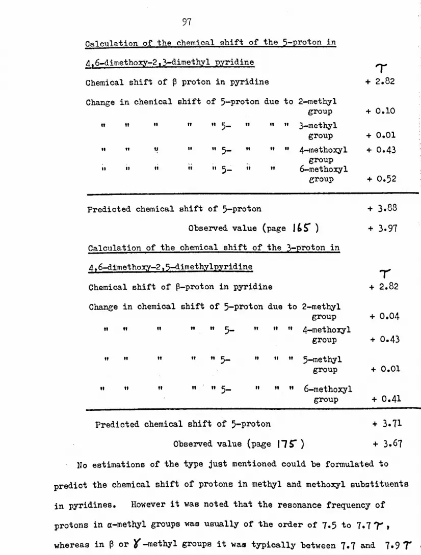
97Calculation of the chemical shift of the 5-proton in4,6-dimethoxy-2,3-dimethyl pyridineChemical shift of P proton in pyridineChange in chemical shift of 5-proton due to 2-methyl
group" " It " " 5- " " " 3-methyl
group" " u " " 5- " " " 4-methoxyl
groupII It It It II 5- It " 6-methoxyl
group
'I+ 2.82
+ 0.10
+ 0.01+ 0.43
Predicted chemical shift of 5-proton
+ 0.52
Observed value (page 1'5")Calculation of the chemical shift of the 3-proton in4,6-dlmethoxy-2,5-dimet~ylpyridineChemical shift of p-proton in pyridineChange in chemical shift of 5-proton due to 2-methyl
group4-methoxyl" " " " 5- " " "" group
" " " " 5- 5-methylgroup
" " " "
" " " " " 5- " " 6-methoxylgroup"
+ 3.88
+ 3.97
,+ 2.82
+ 0.04
+ 0.43
+ 0.01
Predicted chemical shift of 5-proton
+ 0.41
Observed value (page 11~ )
No estimations of the type just mentioned could be formulated to
predict the chemical shift of protons in methyl and methoxyl substituents
in pyridines. However it was noted that the resonance frequency of
protons in a-methyl groups was usually of the order of 7.5 to 7.7 'r' t
whereas in p or '(-methyl groups it was typically between 7.7 and 7.9 T '

98I
TABLE 2
PREDICTION OF THE CHU!ICAL SHIFTS OF NUCLEAR
PROTons Dr PYRIDINIlS BEARING ALKYL, ALKOXYL AND
HYDROXYL (OR .TAUTOMERIC HYDROXYL) FUNCTIons EXCLUSIVELY
The observed '-values of the nuclear protons of each molecule,numbering the positions of substitution 2-6 in a clockwise direction,are listed. Under each of.these values, the figure in brackets isthat of the predicted ~ value of the proton.
POSITION OF SUBSTITUTION OF NUCLEAR PRarOU~pound 2 3 4 5 6
3.02(2.93)
3.03(2.96)
3.14(3.02)
1.68(1.61)
~ChClfJ
2.99 2.64 1.69(2.87) (2.53) (1.59)
3.21 2.71 3.21
I)C~CHJ(2.96) (2.42) (2.96)
~~
1.72 3.03 1.75(1.67) (2.93) (1.67)

99TABLE 2 corrt ' d
Compound2 3 4 5 6
H,.ClOfCH• 1.79 2.76 1.79(1.65) (2.64) (1.65)
CH3
",cAcH3
3.29 3.29
"(3.06) (3.06)
)~(:., 3.21 3.03
~JC N C"'3(2.97) (2.54)
CIf,OJOlOCH3
3.76 2.56 3.76(3.75) (2.66) (3.75)
...II£."JOlOCH,3.34 2.56 3.49(3.27) (2.55) (3.44)
·OeilJ
".II••yrJO3.4 3.4 1.70(3.29) (3.35) (1.6G)
JQr0CIU 2.95 2.95 1.8(2.CO) (2.D1) (1.75)
HJC
ii'H~3.37 1.72
e,\o-Uf(3.36) (1.79)

100TABLE 2,cont'd
Compound 2 3 4 5 6
sS 3.13 1.86CH~
(3.30) (1.79)u,co-~ N
~e,/0,Hf-j CH, .2.00~:1&c~O" (1.99)
HchoH2.86 2.86 1.87(2.76) (2.76) (1.77)
J
0 . 3.78
·~c~
3.71 2.45(3.61) (3.67) (2.40)
H0 3.6
...,.."D3.6 2.33
(3.61) (3.67) (2.40)~0f.r0C~ 3.50 in D20 2.42 in D20
UO ..~c N(3.59) (2.67)
~
f)_ 3.70 2.73 4.05(3.55) (2.56) (3.79)o ~ I\.uc~,r
R
C'""'0t:) 3.31 3.87 3.02(2.95) (3.73) (2.90)o N
IH

101Similarly the resonance frequency of protons in a-methoxy1 groupswas usually found near 6.1 l' , whereas in ~-or 't-metho:xyl groups.a value close to 6.2 7' was typical. A greater number of alkyl,.alkoxy1 or hydroxyl functions in the pyridine nucleus usually resultedin higher jfva1ues for the resonance frequency of protons in aparticular methyl group, but the effect on the resonance frequency of
protons in methoxyl groups was variable. Finally the 'r values of the
absorptions of protons in methyl and methoxyl substituents attachedto pyridones were often higher than might be expected from the aboveremarks. This is accounted for by the reduced aromatic ring-current
effect likely in these compounds.The 7' values of the two methoxy1 substi tuents and of the methyl
substi tuent of the pyridine ring in piericidin A are 6.087' , 6.181
and 7.91 7' respectively. It is deduced from the above considerationsthat one of the methoxyl ~ubstituents is in an a - position in thepyridine ring, whilst the other met~o%lsubstituent and the methyl
substituent are each in either a ~- or a t -position. It has been
deduced independently (pa-ge toa ) that the hydroxyl group of thepyridine nucleus of piericidin A is ~ in an a-position, and ipso
facto it must be in a ~-or t -position. It therefore follows thatthe long side-chain of piericidin A is in an a-position in the
pyridine ring. The same conclusion is arrived at from mass
spectral studies (pages 4- ~ J ~7', I" I ).
Thus the contention on page ~I that piericidin A is a pyridinecompound, substituted in the a-positions with a methoxyl group and along hydrocarbon side-chain, and in the {3- and '(-positions· ':,.

102
with a methyl group, a methoxyl group and a hydroxyl groupin an unspecified manner, is completely in line with its In:NIm -, spectrum.
(b) Coupling constants
The spin-spin coupling constants between nuclearprotons in pyridines are reported to be in the region of'41-5 Hz
J 188between ~ and~ protons • These observations agree well withmost of the values recorded in this thesis. However the cases of
2- and 4-hydroxypyridines and of 4-nitropyridine-I-oxides appear tobe significant exceptions. Their coupling constants are listedbelow.
4-b.Ydroxypyridine
I-methyl-2-pyridone
J 6.9 Hz(Y,~
Ja~ 6.9 Hz
Jci{3 6.9 Hz
Ja~ 7.7 HzJ 6.5 Hza~
J~~ 6.5 Hz
Jplr 8.8 Hz
J~ ~ 6.9 HzI 8.8 HzJ~l
J 6.5 Hzal3
J~r 6.5 Hz
JaB 6.5 Hz
J~r 6.5 Hz1
J~ r 9.5 Hz
4-hydroxy-2-methylpyridine2-(I-n-hexyl)-4-hydroxypyridine
l-methyl-4-pyridone2-hydroxypyridine
2-hydroxy-3-methoxy-pyridine
6-(1-n-hexyl)2~hydroxy-pyridine

103
4-nitropyridine-1-oxide2_methyl-4-nitropyridine-I-oxide2,3-dimethyl-4-nitropyridine-l-oxide
J<X~ 8.8 JIz (A2B2 system)
Jo:~ 6.9 <Hz;
J B' 6.9 'liz;0:, :
The values of these coupling constants can be attributed to
the predominance of structures of the type
6I
Cl1
in these compounds, rather than the more common form of the pyridine
nucleus represented by
OJN(ii) IH nuclear magnetic resonance spectra in hexamethy1-
phosphoramideThe use of hexamethylphosphoramide (m'iPA) in the
examination of mixtures of phenols has been reported recently209•In this solvent the resonancesof phenolic protons, which in many
solvents are normally broad, are considerably sharpened and usually
appear in the region from -0.3 T to + 1.1 1', conveniently apart from
aromatic protons. \'lithin this range a distinction has been drawn
between various types of alkylated phenols. 1.1oreover,electron-
withdrawing substituents were seen to produce a lowering of the 1r
value of the phenolic signal, the effect of electron-donating

104
substituents being variable. The signals were independent ofconcentration in the range of molarity from 0.05 to 1.0.
These studies have been extended in the hop~'of finding afacile distinction between 2-,3- and 4-hydro:xypyridines (pages 204--~OS-).
The phenolic (or amidic) signals of 2-hydroxypyridine, 2- and4-hydro:xycruino1ineand 2-hydro:xy-1-naptha1dehyde were all be1m'1-2 7 , probably indicating a strong interaction with the solvent ofthe type shown below.
Uracil, which exhibited a doublet at -1.6 and -1.77 , may interactsimilarly with two solvent molecules.
The remaining examples are best considered in two groups; firstlythose having a phenolic signal with the breadth at half height lessthan 30 ,HJ., and secondly those with very broad flat phenolic
signals.The resonances of phenolic protons in the first group were all
in the region from -11"to 0 ,. • Little distinction apparently canbe made between 3- and 4-bydroxypyridines and other phenols of thisgroup.
The breadth of the signals in the second group seems attributable
to more than one factor. The suspicion that the phenolic groups of
4,6-dimethoxy-2,3-dimethyl-5-bydroxypyridine and its isomer might be
sterically hindered from interaction with the solvent was supported
by the equally broad signal found in the case of 2,6-di-t-butyl-4-
105
methylphenol. The line broadening observed in the cases of 4-hydroxy-2-hydroxymethyl-5-methoxypyridine, 2,3-dihydroxypyridine and 8-hydroxy-quinoline is of a less obvious origin. Possib~ it is evidence offaster chemical exchange caused by the interaction of the solvent atalternate localities of the molecules.
It is thus concluded that H!.J'Acannot be used as a solvent todistinguish between 3- and 4-bydroxypyridines by IIIm:n spectroscopy,but that it may interact with 2-hydroxypyridines to produce acharacteristic signal below -2 , • In the case of hydroxypyridinesin l-lhichthe phenolic group is sterical~ hindered from interactionwith the solvent, a broad lH mm signal of no value in characterisationis likely to be produced. The use of m:FA as a solvent for thecharacterisation of the phenolic pyridine nucleus of piericidin A byIIIm.:R spectroscopy is therefore valueless.
Interactions of E,:FA with alcohol, amine and amide functionswere observed in the course of this investigation.
The signal at + 2.5 , in the spectrum of l-hydroxymethyl-2-naphthol \-lasattributed to the alcoholic proton of this molecule oncomparison with the spectrwn of l-methyl-2-naphthol.
The amino protons of aniline were found at 4.6 ~as a singlet.
Two equally sized singlets, equivalent to one proton each, werefound in the spectra of acetamide and benzamide at 1.95 and 2.95 l' ,
and at 1.14 and 2.25~ respectively. Presumably the lower field
signal in each case corresponds to an amidic proton involved in asolvent interaction of the type mentioned earlier, whereas the higherfield signal corresponds to the other amidic proton which is relativ~ly

106
uninvolved in such interactions. The slow rates of isotopic
exchange observed in the cases of these latter amidic protons is inline with the weak basicity of amides compared to amines.
These latter results indicate that mlPA may be used as a solventfor In mm spectroscopy) to distinguish betvleen phenols, alcohols,
amides and amines.(iii) The use of trichloroacety1isocyanate in lITnuclear
mq~etic resonance spectroscopyThe in situ reaction of trichloroacetylisocyanate. '
(TAl) with a.lcoho1s and phenols for 1H m::R studies has been described
t1 210,211recen y • The product of reaction, the carbamate (II), was
found to exhibitoIIC13C-C-N=C=0
an U-lI signal within the range -1 to +
ROH ?t ~OC13C-C-H-C~1\ on
2 T •
TAI (I) II1In the case of primary and secondary alcohols, the IIHIm
signals of protons a to the.alcoholic function shifted appreciablydownfield on reaction, typical of their behaviour on esterification212•No such effects on the substituents of phenols after reaction are
reported.These studies have been extended in the hope of discoverine a
facile distinction between 2-,3-and 4-hydroxypyridinea (pages lOS- -lo<l ,!
and Table 13.) The solvent of choice was deuterochloroform for the
reasons described on page ~O,.(a) The carbamate N-H signal
All the a-hydroxypyridines examined exhibited carbamate

101
N-H signals betl'leen-4 and -5 i . Signals at such a low fieldimply hydrogen-bonding of the carbamate as indicated below.
00\ ,\""C::o-C'3C-CO
The corresponding signals of all the 4-hydroxypyridinesexamined, and of 4-hydroxyquinoline, occurred between -2.3 and + 0.8;,l'Thereasthose of the ~-hydroxypyridines examined "Tere within the muchsmaller range f_z'om+0.4 to + 1.1 ; , with the exception of 3-hydroxypyri-,
dine itself in which the resonance was at -1.1 r .Of the remaining compounds, 8-hydroxyquinoline produced a
carbamate N-H signal at -0.1 i',typical of a phenol. 2-HYdroxyquinoline,which was only sparingly soluble in chloroform, did not appear toreact at all, possibly on account of strong intermolecular hydrogen-bonding. Uracil and 2,6-dihydroxy-4-methylpyrimidine were completelyinsoluble in chloroform and also gave no detectable sign of reaction.
Signals attributable to reaction of the alcoholic functions of 3-
bydroxy-2-hydroxymetl~1-6-rnethylpyridine and of 4-hydroxy-2-hydroxy-
methyl-5-rnethoxypyridine were found at + 1.1 and + 3.3 i respectively.~10 distinct carbamate N-II signals were found in all the caseo "Theredihydroxy-compounds reacted with TAl.
It appears from the above results that 2-hydroxypyridines are
clearly distinguishable from 3- and 4-hydroxypyridines usine TAl.
The distinction between 3- and 4-bydroxypyridines is less clcarcut,but generally the carbamate N-H signals o£ 4-hydroxypyridines are

103
found at lower field than those of 3-hydroxypyridines after reaction
\-lithTAl.The corresponding signals derived from alcoholic functions appear
to be at higher field than those derived from any of the hydro~-
pyridines.From the results obtained with 4,6-dimetho~-2t3-dimethyl-5-
hydroxypyridine (carbamate N-H signal at + 1.06 'r) and with 4,6-dimetho~-2 ,5-dimethyl-3-hydroxypyridine (carbamate signal at 1.01 'r ),it is concluded that the presence of electron-donating substitucntsin a phenol raises the carbamate NH signal to higher field than would
otherwise be the case.Piericidins A and B both react with TAl· (see figure 3(, and
pages 13:2. .. 134-) • The alcoholic carbamate NU signal ofpiericidin A appears at 1.77?- whilst in both cases the phenoliccarba.mate 1m signal is between 0.9 and 1.Oj, very close to thoseof the isomeric dimetho~imethylhydroxypyridines mentioned above.The obvious inference is that, although the pyridine nucleus of the
piericidins does not correspond to either of these isomers, it is veryclosely related to them, and may well be a ~-hydroxypyridine.· Itcan be confidently asserted that piericidins A and B are not a-hydro~-
pyridines.The signal at 6.361' (doublet, J .. 8.9 Hz.~)in the 19 m:'R
spectrum of piericidin A is attributed to the proton a to the alcoholic
function. After reaction with TAl the resonance of this proton is
found at 4.99 i (doublet, J - 8.9 Hi.:) (page 13l, figure 36r in210-211
accordance vlith the observations mentioned earlier (page 10').

109
Similarly in the case of piericidin A diacetate the corresponding"( . 212signal is found at 5.06 i doublet, J == 8.9 <Hz) as ezpec'ted .
(see figure 36 and pages S'6 ) (37 ). The comparable signals inthe spectrum of piericidin B are found at 6.7 and 6.6 (' , before andafter reaction with TAl (see figures 35 and 36 and page 13~). Theseresults confirm that piericidin A possesses a secondar.1 alcoholfunction which is methylated in piericidin B.
(b) The chemical shift of substituents in hydroxypyridinesAfter reaction with TAl the 1H m.m signals attributable
to the Eubstituents of a particular hydroxypyridine were usual~observed at lower field (see Table 13). The magnitude of thischange l'laShighly variable, showing some dependence on the positionof the substituent relative to the heterocyclic nitrogen atom and tothe phenolic group, as well on the nature of other substituents.
The biggest change of chemical shift was frequently observedfor a substituent in an a-position in the heterocycle. (Cor.lpounds1, 2, 3, 4, 8, la, 11 in Table 13). On the other hand in the casesof 4,6-dimetho:xy-2,3-dimethyl-5-hydroJCYPyridine and its ieornerupfie1d shifts were observed in the IIIm:.n signals of a substi tuent.sadjacent to the reacting phenolic group.
Upfield shifts l'lerealso observed in the signal of the hot crc--cyclic methyl group of piericidins A and B after re~ction with TAl.
No conclusion wae dro"m from this observation on account of the
diversity and lack of predictability apparent from the other examples.
Hith many of the hydroxypyridines whose spectra were recorded,alternative reaction of TAl at either oxygen or nitrogen would seem

110
possible. An example lias mentioned earlier (pace 1g ) in 1'lhichtwoproducts resulted from reaction with TAl, although in that casereaction appeared to be at alternative oxygen atoms. Since l>1ithoutexception only one product is apparent from the IIINl,::a spectra ofthe above hydroxypyridines after reaction with TAl, it is assumedthat this is formed by reaction at the oxygen atom, in accord withgeneral observations on the acylation of hydroxypyridines147•
(iv) Comparis~ns of infra-red spectraThe distinction between pyridinols and l-(H)-pyridones
mentioned earlier (page 37 ) was borne out by the IR spectra ofseven further hydroxypyridines.
4,6-Dimethoxy-2,3-dimethyl-5-hydroxypyridine (page Ill),4,6-d.imethoxy-2 ,5-dimethyl- 3-hydroxypyridine (page 171 ), and2,4-dimethyl-3-hydroxy-6-methoxypyridinc213 exhibited strong, sllarpabsorptions in solution at 3550, 3550 and 3610 cm-l respectively,typical of the O-H stretching vibrations of free hydroxyl groupo in
pyridinols (page "37 ). In addition each compound absorbed strongly
6 -1at 1610, 1609 and 1 15 cm respectively. These absorptionc are
attributed to ring vibrations although they are a little higher than
1 ° °dO 214is usua 1n pyr1 1nes •In contrast 4-hydroxy-2-rncthylpyridine (page 1'7 ), 4-hydro::cy-
2_hydro:xymetbyl-5-metho:rypyridine (page 112. ) t 2-(1-n-hcxyl)-4-
bydro::zypyridine (page 187 ) and 6-(1-n-hexyl)-2-hydroxypyridinc
(page lai) had very strong absorptions at 1630,1621,1628 and1640 cm-l respectively, typical of' l-(H)-pyridones (page 37 ),wi th weaker absorptions in the 3200-3400 cm-1 regian attributable to

III
r:-H stretching vibrations.Piericidin A in solution exhibited strong absorptions at-1 -13505 ern and 1595 em attributed to the free O-II stretching
vibration and a ring vibration respectively of a pyridinol. Asalready mentioned (page 4-.4-) the \-leakabsorption at 1620 cm-l inthe III spectrum of piericidin A is attributed to olefinic C=C
stretching vibrations.Since it appears, as expected (page 30 ), that the attachment
of aly~l and methoxyl groups to the nucleus of 4-hydroxypyridine doesnot lead to a predominance of the pyridinol tautomcr in preferenceto the l-(H)-pyridone tautorlcr in these compounds, and that 3-hydroxy-pyridines bearing such substituents are typically pyridinols, it isconcluded that piericidin A, clearly a pyridinol, is itself a 3-
hydroxypyridine.(v) Comparisons of ultra-violet spectra
The ulT spectra of hydroxypyridines and related
compounds in acidic, basic and neutral solutions containing 75i~
methanol (pages ,.,O~_.'"II ) indicated that the observations madeearlier (pages 34- -37 ) concerning the spectra. of such compoundsin \-lhollyaqueous solutions could be safely extended to this
.methanolic solvent. It was necessary to use such a solvent in order
to make comparisons with the piericidins, which were not sufficiently
soluble in water alone to obtain their ulT spectra.

112
6-(1-n-Hexyl)-2-hydroxypyridine had very similar absorptionsin acidic, basic and neutral solutions, typical of a 1-(H)-2-pyridone.The alkyl substituent produced a bathochromic shift relative to the
spectrum of 2-hydroxypyridine.Four substituted 3-hydroxypyridines (5-hydroxy-2-methylpyridine,
4,6-dimethoxy-2,3-dimethyl-5-hydroxypyridine, 4,6-dimethoxy-2,5-dimethyl-3-hydroxypyridine and 2,4-dimethyl-3-hydroxy-6-methoxypyridinc)all displayed bathochroraic shifts of their absorption maxima inacidic and basic solutions compared to those in neutral solution. Theshifts vlere accompanied by increases of extinction coefficient, thesebeing most marked in acidic solution. 3-hydroxypyridine itselfbehaved-similarly to the above mentioned compounds, no absorption
attributable to the zWitterion (see pageJS) being observed. Theeffect of substituents on the absorption maximum obeyed no simplerule, although all the substituted compounds exhibited a bathochromicshift relative to 3-hydroxypyridine. This unpredictability iswell illustrated by the hypsochromic shift in tho absorption maximum
observed on increasing the substitution of 5-hydroxy-2-methylpyridineto that of 4,6-dimethoxy-2,3-dimethyl-5-hydroxypyridine.
4-HYdroxy-2-methylpyridine, 2-(1-n-hexyl)-4-hydroxypyridinc and4_hydroxy-2-hydroxymethyl-5-methoxypyridine all displayed hyprochromic
shifts in acidic and basic solution relative to their absorptions in
neutral solution, typical of l-(H)-4-pyridones (paee J/:, ).
The absorption maximum at 267 nm in the spectra of the
unacety1ated piericidins in neutral solution undergoes a bathochromicshift with an increase of extinction coefficient in acidic solution.

113This is typical of a 3-hydroxypyridine but not of a 1-(H)-4-pyridone.On the other hand the absence of any absorption maximum above 220 nmin the spectrum of octahydropiericidin A in basic solution is atypical,of any known hydroxypyridine.
Thus it is concluded that, although the DV spectra of theI
piericidins are not completely understood and the possibility thatthey are 4-pyridinol compounds cannot be excluded, their behaviour
is most like that of 3-hydroxypyridines.(vi) l'ass spectral compC'.risons
During the course of work described in this thesis themass spectra of a number of pyridine compounds excl~Gively substituted\-lithalkyl, methoxyl and hydroxyl (or t;:>.utomerichydroxyl) groupswere recorded. Additionally the mass spectra of a number of similarcommercially available pyridines were recorded. These spectra arepresented in the mass spectral appendix in four groups:
(a) Pyridines possessing methyl substituents only (figure 41)(b) Pyridines possessing an a-(l-n-hexyl)substituent (figure 42)
(c) pyridines possessinc methoxyl (or lJ-methyl)functionsexcept those \-lhichalso possess hydroxyl (or l-(U)-pyridone)
functions (figures 43 and 44)
(d) pyridines possessine hydroxyl (or l-(II)-pyridone) functions
(figure 45)
(a) Pyridines possessine methyl substituents only (fieure 41)The spectra of all these compounds shoH peaks
attributable to the loss of hydroeen and methyl radicals from themolecular ion, (E)~ at m/e = (1:-1) and (1.1-15).

114
The spectra of all three pico1ines have peaks at m/e = (H-27),(n-28) and (r.I-42)corresponding to the elimination of hydrogencyanide from the ions (1-1)+,(11-1)+ and (1,:-15)+. It is alsoapparent that the loss of a methyl radical from the molecular ion ismost favoured in the case of a-picoline, and of a hydrogen radical
I
in the case of ~-picoline.The four lutidines and the collidine examined show an increased
tendency to lose a hydrogen radical from the molecular ion, particularly
vrhen this has a p-methyl grO'l.'_p.The spectra of all five compounds
have peaks at m/e = (H-27) and (1I-42) as in the case of the picolinesand presumably of a similar origin, but those at m/e - (11-27) are ofcomparatively low intensity. However although the loss of hydrogen
cyanide from the molecular ion appears to be less frequent anoccurrence, a series of peaks at m/e = (J.I-4l)indicate that theelimination of methyl cyanide from the same is much more common than
in the case of the pico1ines.In all the spectra,peaks of 10v1 intensity due to the doubly
charged molecular ion, (n) 2+, were detected at m/ e = t¥).(b) Pyridines possessing an o:-(l-n-he:zyl) substituent (fic;ure 42)
All five compounds examined exhibited a base peak at
m/ e = (I·:-70),corresponding to the elimination of n-pent-I-ene fromtheir molecular ions Yl!! a J.1cLaffertyrearrangement. This was
expected in the cases of 2-(1-n-hexyl)pyridine, 2-(1-n-hexyl)-4-
methoxypyridine and 2-(1-n-hexyl)-6-methoxypyridine, but it was
supposed (see page 4-~ and figure 11) that 2-(I-n-hexyl)-4-hydroxy-pyridine and 6-(l-n-hexi1)-2-hydroxypyridine, being l-(II)-pyridones,

115
might behave differently. The fact tr4t they did not may indicatethat the moLecul.ea exist as pyridinols in the vapour phase. Atany rate it can definitely be said that no distinction, besed onthe absence or presence of ions resulting from a EcLaffertyrearrangement in the mass spectrometer, can be made between a, ~- and~ -hydroxypyridines bearing a long hydrocarbon side-chain in an
(X-position._ In addition to the base peak mentioned above, and the molecular
peru(, the spectrum of each compound showed peaks equivalent to thelOBS of methyl, ethyl, n-propyl and n-butyl radicals from the
molecular ion. Ions due to the loss of n-pentyl radicals, at m/e ...(1-1-71),were of negligible intensity compared to those at m/e - (}!~7())
fomed :ti.! a Mclafferty rearrangementThe breakdown of ~hese ions at m/e = (U-70) appeared typical
of the pyridine nucleus bearing the substitucnts peculiar to eachcompound, supposing that the residue of the hexyl substituent \-las
present as a methyl function. For instance, that of 2-(1-n-hexyl)pyridinec" appeared to lose a methyl radical (peale at m/e. 78) orhydrogen cyanide (peak at m/e = 66); those of the two methoxy-pyridines appeared to lose formaldehyde (peak at m/e. 93) or an
acetyl radical (peak at m/e III 80); those of the t\OlOhydro:xypyridines
appeared to lose carbon monoxide or a formyl radical (peako at m/e =81 and 80).
No peaks corresponding to doubly chareed molecular ions, (M)2+,
or even doubly charged ions of the base peak, (t':-70)2+,...rer-edetected in the spectra of any of these compounds. This vlas in

116'contrast to most of the spectra of other pyridine compounds recorded,with the exception of the piericidins which also have a longhydrocarbon side-chain and a molecular peak of correspondingly low
intensity.(c) Pyridines possessing methoxyl (or N-methyl)functions but
not hydroxyl (or l-(H)-pyridone) functions (ficures 43'and 44)The mass spectra of 2-,3-,4-methoxypyridines differ
considerably. The molecular ion of 2-methoxypyridine readilyeliminates a hydrogen radical, giving a second base peak at m/e ; 108,and formaldehyde giving an intense peak at m/e = 79. In contrast
the ions at m/e = 108 and 79 in the mass spectrum of 3-methoxypyridineare of low intensity, the principal frD~entation of the molecularion being the elimination of 43 maso units, corresponding to an ~,cetylradical (Cf. the methoxyquinolines,137 page ~O ),producing a perucat m/e = 66. The ion at m/e a 108 in the mass spectrum of 4-methoxy-pyridine is of low intensity. The molecular ion appears to decompose
chiefly by the elimination of formaldehyde, and less frequently bythe elimination of an acetyl radical producing peaks at m/e a 79 and
66 respectively. In all three compounds the elimination of a methyl
radical from the molecular ion is infrequent.
The molecular ions of l-methyl-2-pyridone and l-mcthyl-4-pyridonedecompose principally by the elimination of carbon monoxide or of a
fornyl radical.
The peak at m/e = 80 in the n~ss spectruo of 5-methoxy-2-methylpyridine is attributed to the elimination of an acetyl radical
from the molecular ion, similar to the case of 3-methoxypyridine.

117
Additional peaks at m/e = 108 and 95 correspond to the loss of amethyl radical (presumably from the 2-methyl group) and of carbonmonoxide respectively from the molecular ion.
The mass spectra of 2,6-dimethoxypyridine, 4,6-dimethoxy-2,3-dimethy1pyridine and 4,6-dimethoxy-2,5-diroethylpyridine all have
.intense peaks at m/e = (n-l), corresponding to the loss of a hydroe-enradical from the molecular ion. This appears to be typical of a-methoxypyridines. Peaks are found in the mass spectra of all threecompounds at m/ e = 0·;-30) and (r.I-45),attributed respectively to theloss of fonnaldehyde and to the consecutive loss of formaldehyde anda methyl radical from the molecular ions. Peaks of low intensityat m/e = 121 and 124 in the mass spectrum of 2,6-dinlethoxypyridincare probably due to the elimination of water and of a methyl radicalfrom the molecular ion of this compound. The mass spectra of thetwo isomeric dimethoxydimethy1pyridines differ radically in one
respect. In the case of the 2,5-dimethyl compound the elimin~tion ofa methyl radical from the molecular ion producing an intense peak
at m/e = 152 is highly favoured. The same procesc is almoct absentin the 2,3-dimethyl isomer, the intensity of the peru~ at m/c. 152being very lm-l. Possibly due to the absence of a competitive po.th-Hay of decorr.positionthe ion at role • 166 in the spectrum of this
compound , due to the elimination of a hydrogen radical from themolecular ion, is relatively more intense. The essential differencebetween these two compounds lies in the location of the ~-methylgr::>up. It is tempting to suppose that it is this ~-methyl croup,,;hichis lost from the r.:olecularion of the 2,5-dimethyl isoner,

11'3
although ",by this should be so much more facile than in the case of
the 2,3-dimetbyl isomer is not apparent.The mass spectra of the tW9 remaining compounds, 4-methoxy-2-
methoxymetbyl-3-metbylpyridine and 4-methoxy-2-methoxymethyl-5-methylpyridine, illustrate the ease with which the 1.:cLaffertyrearrangement takes place. The elimination of formaldehyde by thismechanism from the molecular ion of each accounts for the base peaksat m/e = 137. The elimination of a methyl radical appears to takeplace from both the molecular ion and the base peak ion of eachcompound producing peaks at m/e = 152 and 122. The peak at m/e =107 in the spectrum of the 3-methyl isomer is probably due to thefurther loss of formaldehyde from the ion at m/e - 137, although whythe intensity of the same ion is so much lower in the spectrum of the
5-rnethyl isomer is not readily explained.In all the spectra of this group, peaks of low intensity due to
doubly charged molecular ions, (H)2+, or in the case of the la.ttertwo compounds due to doubly charged base peak ions, were detected
M -at m/e = (2) or 67.5 respectively.(d) pyridines possessing hydro:x;yl(or l-(II)-pyridone) functions
(fip;ure 45)
The mass spectra of many hydroxypyridinea have intense
molecular'peaks, with peaks of relatively low intensity elsewhere,
indicating that the molecular ions are particularly stable. The mass
spectra of 2-,3- and 4-hydroxypyridine have been reported131,(page.3' ). All three compounds eliminate carbon monoxide fromtheir molecular ions, although the elimination of hydrogen cyanide is

119
of comparable importance in the cases of the 3- and 4-isomers.This elimination of hydrogen cyanide appears to be peculiar
to these two hydroxypyridines, since it is of negligible importance
in the cases of 4-hydroxy-2-methylpyridine and 5-hydroxY-2-methyl-pyridine. The molecular ions of these two compounds appear toeliminate formyl radicals (peaks at m/e ...(M-29)) as the majorprocess of decomposition. The elimination of methyl radicals fromthe molecular ions of both compounds also occurs to a lesser extent.
The mass spectra of the other four a- and ~-hydroxy pyridinesconsidered have peaks at m/e .. (M-28), (1·:-30)and (1<I-29)correspondingto the elimination of carbon monoxide, formaldehyde and formylradicals from the molecular ions. The elimination of hydrocen
cyanide from the same is of relative~ little importance since nopeaks at m/e = (N-21) were detected.
A peak: at m/e ...82 in the mass spectrum of 2-hydroxy-3-methoxy-
pyridine is thought to be due to the loss of an acetyl radical fromthe molecular ion, in comparison with the case of 3-methoxypyridine
(page "' ).B::ruivalentpeaks, at m/e = (1,!-43),are also observed in the mass
spectra of 4,6-dimethoxy-2,3-dimethyl-5-hydroxy pyridine and 4,6-
dimethoxy-2,5-dimethyl-3-hydroxypyridine. Both mass spectra are very
similar, displaying a large number of Common ions with similar
intensities. Those aro listed below, tocether with an interpretation
of their origin.

Peak at m/_e =183 (M)
182 (r.l-1)
163 (M-15)
165 (1,1-18)
.164 (M-19)
154 0·:-29)
153 (!;1-30)
152 (r:'-31)
150 (M-33)
140 (1;1-43)
139 (1"-44)
133131
(11-45)
(1.1-45)
120
Species lost from molecular ionNone
tt.
(H·)
(·CH3)(H20)
(H·) and (H20) consecutively(·CHO)
(CH20) or (a') and (·CHO) consecutively
(H·) and (CH20) consecutively
C-CH3) and (H20) consecutively(·CO.C1I3)
(H·) and (·CO.CH3) consecutively
or CC1l3) and (·CHO) consecutivelyCCH3) and (CH20) consecutively(H·), (·CH3) and (CH20) consecutively
In the mass spectra of all the hydroxypyridines examined,peaks .of 10'1.1 intensity corresponding to doubly charGed molecular ions, (u) 2+
were detected at m/e = (~).2
It is concluded from the above examples t~t no distinction canbe expected between the mass spectra of isomeric pyridines possessine
in the a-positions a methoxyl group and a long hydrocarbon side-chain,
and in the p- and ¥positions a methoxyl group, a methyl group and
a hydroxyl group. This means that the location of the p- andY
Bubstitucnts of the pyridine nucleus of the piericidins cannot bededuced from the mass spectra of the same. As mentioned earlier

121
(pages ), mass spectrometr,y confirms thelocation of the hydrocarbon side-chain to be in an a-position inthe heterocycle, since only with the side-chain in this position arethe observed KcLafferty rearrangements possible.
(vii) The base-catalysed deuterium exchance of protons in "the methyl grOUpS of pyridines
studies of base-catalysed deuterium exchange ofprotons in the methyl groups of picolines in alcoholic solution givethe sequence of reactivity as ~-pico1in~_picoline>~-picoline176.These results have been extended to include the methyl groups of morecomplex pyridines using aqueous media (pages 2.13 "1'~). Apreliminary experiment performed at 1000C gave no de1ectable isotopic
exchange. It was therefore decided to employ a temperature of lSOoC,having first ascertained that the isomerisation of methoxypyridines atthis temperature \-laS not excessive (page.: ), sincesome of the compounds to be examined ,·rereof this type.
The greater reactivity of protons in the methyl groups of a-
picoline and r -picoline, compared to those: of ~ picoline, "raoconfirmed, isotopic scrambling being essentially complete in theformer two cases, but only 18% in the latter. Under the same
conditions 5-methoxy-2-methylpyridine exhibited only 67% scr~mbling
in the protons of its a-methyl group. Presumably the decrcaco in
reactivity is caused by the electron donating effect of the methoxyl
group. In contrast nearly complete isotopic scrambling was observedin the «-methyl group of 5-hydroxy-2-methylpyridine under the same

122
conditions. This result is unexpected since the compound \-raspresumably present predominantly as its phenolate anion, from l-rhichproton loss would be unlikely. The exchange must have occurred ontraees of the neutral molecule present, the apparently greaterreactivity in comparison with the corresponding methoxy-compoundpossibly being on account of the greater homogeneity of the reaction
mixture.Once again under the same conditions, f!I;~ of the protons in the ex-
methyl group of 4,6-dimethoxy-2,3-dimethylpyridine were found to beisotopically exchanged, the marked reduction in their reactivitybeing attributed to the presence of electron donating substituents.Increasing the time of reaction and the strength of the base raisedthis percentage to 75, the exchange of protons in the ~-methyl groupremaining essentially zero. The additional presence of dioxan wasin order to achieve a more homogeneous reaction mixture. Theabove result (mUCh greater isotopic exchange in an ex-methyl groupthan a p-methyl group in the same compound) is reinforced by the
results obtained with 4,6-dimethoxy-2,5-dimethylpyridine, 4,6-dimethoxy-2,3-dimethyl-5-hydroxypyridine and 4,6-dimethoxy-2,5-:dimethyl-3-hydroxypyridine, although the apparent variations in the
degree of exchange of the protons in the ex-rnethylgroups of each,and hence in their reactivities, is not readily explained.
It is concluded that the base-catalysed deuterium exchange of
protons in the methyl groups of comparable pyridine compounds is
more rapid when these are in an ex or a 'I-position than when they arein a ~ position.

123
.In the examples of 4,6-dimethoxy-2,3-dimethyl-5-hydroxypyridineand 4,6-dimetk1:xy-2,5-dimethyl-3-hydroxypyridine, \"lhichare isomericwith the pyridine nucleus of the piericidins, virtually no exchaneewas observed in the protons of the ~-metby,l groups, ,,:hereasconsiderable exchange took place in those of the a-methyl groups.
An analogous experiment was performed with octahydropiericidin A,which is believed to contain either a., K or a ~-metlzyl group(page S-I and figure 12), in which no isotopic exchange was detected.This was unfortunately a negative result, and although it woul.d beexpected were the methyl group in a ~ position, it cannot be excludedassuming that the methyl group is in the ((position. For instance itis conceivable that a ~-methy1 group might be forced out of the planeof the pyridine ring by adjacent substituents, decreasing its reactivityand l~ihood of taking part in isotopic exchange. It is alsopossible that the long hydrocarbon side-chain might affect the reactivityof the medium (Cf. pages
It is concluded tr~t the heterocyclic methyl group of the pierici-dins ma.y be in a.: '( or a P-position, and that the result of the
attempted isotopic exchange reaction on octahydropiericidin A does notexclude either possibility.
(Viii) Spray tests
Colour reactions to the Dragendorff reagent215 were
highly varied (page :til, ). A positive reaction of some sort was
usually found with aromatic and aliphatic amines, although three
exceptions were found. Piericidin A and other hydroxypyridines all
gave colours 'with the reagent. Amongst the compounds giving no

124
visible reaction were amides and amino-acids (except for glycine) •.In both of these cases the basicity of nitrogen is less than in mostamines, probably accotUlting for the lack of reaction. Clearly thereagent does not distin~guish different hydroxypyridines and is there-fore of no use in the characterisation of piericidin A in this respect.
Both the Folin-Dellis reagent143, and the "enol" spray216 gaveblue colours with all phenols tested, including pyridinols and
piericidin A (pages ~(' -117 ). l-(H)-pyridones gave no colour
reaction or only a ver,y slowly produced one in both cases. Aspointed out earlier14l,l44 (page ~1),a positive reaction to areagent of this type does not exclude the possibility of the compoundbeing a 2- or 4-hydroxypyridine if such a compound should possess an
enolic hydroxyl group (e.g. should it be a 4-pyridinol). It followsthat piericidin A behaves as a pyridinol towards both reagents, althoughthe position of substitution of the hydroxyl group in the heterocycle
cannot be deduced.

125
SYNOPSIS CONCERNING THE STRUCTUREOF PIERICIDIN A
It was concluded that there were six possibilities for thecorrect structure of the pyridine nucleus of piericidin A
(page )1 and figure 12, I-VI). Of these, I and II are
4-hydroxypyridines and would be expected to exist as l-(H)-
4-pyridones. Since piericidin A behaves as a pyridinol
(page 4.' ), it is unlikely that either I or II is the correct
structure. Of the remaining four possibilities, V and VI,where R=H, have been synthesised and do not correspond topiericidin A, the correct structure of which must consequently
be represented by III or IV.In a parallel investieation213,the synthesis of 2,4-
dimethyl-3-hydroxy-6-methoxypyridine has been achieved. Thiscorresponds to IIIexcept that it lacks a methoxyl group. ItsDV spectrum has been recorded and does not resemble piericidin A
closely (page 110).
1 . d2l3was a so exam1ne •
The ozonolysis of piericidin A diacetate
A compound identified as the pyridinc-
2-aldehyde corresponding to piericidin A, but lacking the side-
chain, was isolated. In particular it exhibited an aldehydic
absorption in its IR spectrum at 1715 cm-l• From this it
was concluded that the aldehydic function (and therefore the side-
chain) vTaS not adjacent to a phenolic function, since if this

126
were the case the aldehydic absorption would be expected to be-1 224about 50 cm less • Both of the above pieces of evidence ~ .'
suggest that the structureII]lOes not correspond to that of
piericidin A.It is therefore concluded, by elimination, that piericidin A
is a 2_alkyl_3,6-dinethoxy-5-hydroxy-4-methylpyridine
corresponding to structure IV of figure 12.It is also concluded from the mass spectrum of piericidin A
(page ~4-) that there is an olefinic bond between carbon atoms
5 and 6 of the side-chain, rather than betvleen carbon atoms
4 and 5 as published2e•
REV. SEl> STRUCTU~E OF ""ERIC/7)/N A

Jro'ERn:n'JTAL SECTION

127
Solvents were purified and dried by conventional means.Commercially available startinrr materials were either redistilled or
recr,ystallsed before use.
ChromatographyVapour phase chromatography was carried out using a Perkin-Elmer
FIl instrument having a flame ionisation detector.Thin layer chromatography was carried out using the following
systems:Plate Eluent (v/v)
System A Silica \'letbenzene/acetone, 2511
B Silica chloroform/acetone, 713
C Silica chloroform/methanol/water, 65:25:4D Silica dry benzene
E Silica dry benz~e/acetone, 1911F Silica ethyl acetate
G alumina dry benzene
H alumina. dry benzene/acetone, 19:1The detection of materials on developed T.L.C. plates was by
exposure to iodine vapour unless otherwise stated.
SpectrometersUltra-violet spectra were recorded using a Unicam SP 800 instrument.
EXtinction coefficients at the maximum of absorbance were checked usine
either a Uniearn SP 500 or a Carey 14 instrument.
Infra-red spectra were reoorded using a Perkin-Elmer 257instrument.

12]
IH NMR spectra were recorded at 60 Mcl s and 14,100 gauss usine a
Perkin-Elmer RIO instrument.1.1assspectra were recorded using an A.E.I. M.S. 902 mass
spectrometer at the University of Hull, Oh. 0& ,e~vice. 6.';6. 1>i...e. c.1- it\4e ...b~"o~ bite. '_""ple£ 1oJ~ e....-plo~.
The radioactivity of samples containing tritium was measured usingMeltinrr Points are given uncorrected.
a Packard Tri-Carb liquid scintillation spectrometer.

FIGURE 34
APPARATUS FOR SHORT PATH DISTILLATION
VAtWMT.,
• 14 OfllOUHD GUM
CONDINII.
CONTAINING
,eITONI
•• D
DRY ICI
_==>W'GMETtc ITI,",INI)

129
Short path distillationThe glass apparatus indicated in figure 34 was developed for the
purification of liquids having an appreciable vapour pressure at roomtemperature.
The best results were achieved by a process of slow evaporationof the distilland, at room temperature and low pressure (0.0.01 mm IIg),
rather than of boiling. It was important to keep the surface of thedistilland fresh by stirring.
In practice the distillate often sol~ified on the condenser, onlyrunning into the collection cup after the apparatus, isolated from thepump, had equilibrated to room temperature.
A smaller version was used for milligram quantities.This apparatus may be adapted for the direct recover.y of oily
residues from a reaction vessel, eliminating an operation of transfer.

130
METABOLITES OF STREPTQl.1YCm lTOBARAlil~SIS
AND TlOOR DEnIVATIV'FS
Streptomyces mobaraensisl was grown on two different culture
media.Rich medium1
Composition: glucose 20 gIl
starch 10 "soya peptone 25 "meat extracts 10 "yeast e:-..-tract 4 "sodium chloride 2 "dipotassium hydrogen 0.5"phosphate10% sodium hydroxide 3 mlsolution
The conditions of the fermentation and of the extraction of
containing 3% ethyl acetate in benzene. Piericidins A and B 80 isolated
piericidins A and B were substantially the same as those of Takahashi
et all. After a typical 51. fermentation, silicic acid (I,1allinckrodt):column chromatography yielded piericidin B (18 mg) in the eluent containing
1?~~ethyl acetate in benzene, and piericidin A (65 mg) in the eluent
were ascertained by T.L.C. (System A) to be sinele compounds.,
Apart from fatty substances, eluted early from the column, no
other metabolites \'lereisolated from mycelia of S.mobaraensis grown onthis medium.

131
d. 156Sir.mle me J.um
Composition: glucose 20 ellb~cteriological peptone 5 It
sodium chloride 2 "dipotassium hydrogenphosphate
2 It
other conditions of the fermentation and of the extraction ofmetabolites vlere the same as those mentioned above. Piericidin B
(18 mg) and piericidin A (85 mg) were isolated as above from a typical
5 1. fermentation. In addition tl"TOnew non-fatty metabolites wereisolated in e1uents from the silicic acid co Lumn , f·letaboliteC (20 mg)"Tas found in that containing 2% ethylacetate in benzene (that is,between piericidins A and B), and metabolite D (10 mg) was eluted
earlier with benzene. Both were contaminated with other materialsand were purified by preparative T.L.C. (System A) yielding 9 mg and5 me respectively.Piericidin A
A viscous pale yellow oilChromatographic homogeneity
TLC System A One spot, Rf = 0.22
sensitive to "enol" spray
DV Spectra
(75% methanol, 25j~ water)
Amax (nm)
232
273
39,50040,5005,600
39,0008,200
(75~methanol, 25% O.lNHCl)
239267
236

FIGURE 35
'H NMR SPECTRA
PIERICIDIN A
o
PlERICIDIN A DIACETATE
PIERICIDIN B
o 1 3.,.. ··--~-r--~··--l----~1 S 9 ~y

PIERICIDIN A plus TAl
o
PIERICIDIN B plus TAl
o
OCTAHVDROPIERICIOIN A
FIGURE 36
rH NMR SPECTRA
v
o 10
"_s

132
(75/t methanol, 25% o.m NaOH)
IR Spectra\l -1V(CHC13) cm
\\ -1Y (thin film) cm
i~"~RSpectra.
T (CDC13 plus TAI)
231 40,000Shoulder 3,200
at 210 nm.
3505 (st. and sharp), 2925, 2810 (med)1620 (w), 1595, 1475, 1420 (st.)1390,1360, l330,(w), 1240, 1190 (med.)1128 (st.), 1042, 1005, 968 (med.) r ••.
3380 (broad st.), 2025, 2865 (med.), 1620 (w),15951475,1420 (st.), 1390, 1360, 1330 (w)1252,1193 (med.), 1129 (st.), 1050,1008.968. 040, 914, 882, 829, 760 (all med.)
..
See figure 353.87 (d,J==15.5Hz)lH:J
4.19-4.29-4.45-4.56-4.60-4.86 (m.) 4II,
6.03 (s) 3H, 6.18 (s)31I,6.36 (d,J:c8.9IIJUI, ...6.63 (d,J-6.9 II )2H, 7.20 (d, J...6.5 H and m)z z3H, 7.91 (s)3H, 8.15-8.45 (m)12H,9.19 (d, J:c6.8H )3HzSee figure 360·93 (s)UI, 1.77 (s) IH, 3.94(d,J=15.5H )111,z4.26-4.35-4.47-4.63-4.75-4.90 (obscured),(m)4H, 4.99(d,J-8.9 H )lH, 6.04 (s)3H,z

133
~~assSpectrum
7.25 (m.and d., J= 6.5 H )3H,z7.94 (s)3H, 8.20-8.45 (m)12H,9.14 (d,J=6.8 H )3Hz
See figure 40 of the mass spectralappendixNo peaks due to metastabletransitions detectedAccurate mass measurements I
331.213 (C20H29N03 is 331.2147)236.130 (C13H18N03 is 236.1286)222.113 (C12H16N03 is 222.1130)183.089 (C9H13N03 is 183.0895)161.133 (C12H17 is 161.1330)84.057 (C5H80 is 85.0575)
Piericidin B
A viscous pale yellow oilChromator.raphichomogeneity
T.L.C. System A One spot, Rf=0.33sensitive to enol sprayu.V. Spectra
(75% methanol, 25% water) A max (nm.) €~
(75~ methanol, 25% O.lNHCl)
232 39,500239 40,500267 5,600236 39,000273 8,200231 40,000
Shoulder = 3,200at 270 nm.
(7510 methanol, 25% O.IN N~OH)

IR_El'!:_C?t~. -1~ (thin film) cm
1r(CDC13 ~lUS TAl)
134
3370 (broad st.), 2925, 2870 (med.)1620 (w), 1595, 1475, 1420 (st.)1390, l360,1330(w),1250,1195 (med.)1128 (st.) 1100 (Med.) 1050, 957, 830
.770 (med.) ..:-.See figure 35
Hz3.90 (d,J=15.5)lH, 4.29-4.40-4.54-4.66-4.79 (m)4H, 6.08 (s)3H, 6.18 (s)3H,
Hz6.64 (d,J=6.9) and 6.7 (d, obs1Y:fed)3H,6.90 (s)3H, 7.23 (m. and e, J=6.5)311, Hz
7.29 (s)3H, 8.20-8.50 (m)12H, 9.22 (d,J=6.8)3H
See figure 36Hz
0.97 (s)ia, 3.93 (d,J=15.5) lII,4.31-4.43-4.54-4.64-4.75-4.82 (m)~IHz6.03 (s)3H, 6.17 (s)3H, 6.59 (d,J=6.9)3H,6.89 (s)3H, 7.22 {m. and d.)3H, 7.92 (s)3H,Hz8.20-8.45 (m)12H, 9.20 (d,J=6.8)3H
Metabolite CI'lhitecrystalline ~l.Pt. 199-200oCSoluble in water
Chromatographic HomogeneityT.L.C. System A One spot Rf • 0.27 (sensitive to enol spray)

25
15
10
5
15 "
IIIIIIII
IIIIII
!,,,:I,,I
!
FIGURE 37
UY SPECTRAI:
..',-',,,
IIIII
METABOLITE C
METABOLITE 0
,I
\,,.\\...\\\\,'..._ .....- ..
2.'50 ,,,,,,
REDUCED METABOLITE C
REDUCED METABOLITE 0
r>,r ," \10 .' \
I \.f .......I 'I •, ., \
f \f •, \
f \,5 :
2..00 "'00 '''"

IN Sp~_ctra(methanol)
. (and acidified methanol)
(methanol plus 2 drops~IN NaOID
IR Spectra\J (CHC13)
-1cm
V (nujo1) -1cm
1H NER Spectrum7' (CDC13, 2'}~)
}f;assSpectrum
135
A max (nm)
See figure 37242279334234356
30,5005,100 ba.sed3,100 on
1II.W.
30,000 of 236
5,000
2920, 2850 (med.), 1682, 1646(v.st.)1562, 1460, 1376, 1340, 1290, 1112,1096, 1002, 979 (all st.)3400 (broad w.), 1680, 1644, 1561,15~0, 1433, 1355, 1290, 1262, 1210,1167, 1108, 1020, 1005, 976, 849,792, 746 (all st.)
-1.12 (s)lH, exchanged byaddition of D203.67 (s)lH, 3.82 (s)lll,6.05 (s) 3H,6.09 (s)3H, 7.74 (s)3H, 8.45 (s) 2H,exchanged by addition of D20
mIc I mle I236 (97) and after shaking 237 (100)221 (100) with D20--' 222 (97)
207 (17) 208 (13)193 (76) 194 (73)150 (19) 151 (16)

136
Peak due to metastable transition
Accurate mass determinations236.0672 (C10HION304 = 236.0671)221.0436 (C9H~304 = 221.0436)
193.0499 (CsH7U303 = 193.0487)
r"etabolite D
vlhitecrystalline, M.Pt. 123-125°Csoluble-.in water
Chromatoeraphic HomoeeneitlT.L.C. System A
DV Spectrum(methanol)One spot, ~f = 0.56 (sensitive to enol spray)See figure 37
A max (nm) t~238 (shoulder) 28,500 ~244 32,500 based277 4,700 on
N.U.287 3,200 of 206
326 3,700-1cm 3400 (broad w.), 2920, 2850 (med.)
1685 (v.st.) 1648,1623,1573 (all st.),1490, 1465 (med.) 1372,1351, 1334 (st.),1163, 1149 (v. st.), 1099, 1069 (med.),1004, 971, 950, 888, 844 ( all med.) ,:-.-1.12 (8)111 exchanged by addition of D20,3.51 (d,J=2.6 H )lB, 3.67 (d,J=2.6 H llB,Z z
In mm ~ectrumI (CDC13,1~)

131
3.80 (s)lH, 6.15 (s)3H, 1.16 (s)3H8.25 (s)lH
Mass Snectrum Mle (1) 206 (100) and after 201 (100)191 (35) shaking 192 (36)111 (12) with 118 (14)135 (32) D20 _.,. 136 (36)Peaks due to metastable transitions206~ 111+ + 20 @ 152.08201+~ 118+ + 29 @ 153.06Accurate mass determination206.0561 (C9H8N303 = 206.0566)
Piericidin A diacetate was prepared in 55% yield by,the action ofacetic anhydride in pyridine on piericidin A as described byTakahashi et a12a•
A viscous pale yellow oilChromatoeraphic homoeeneity
T.L.C. System A One spot, Rf = 0.14 sensitive toenol spray
tN Spectra(15% methanol, 25% water)(15% methanol, 25~ O.lNHC1)(15% methanol, 25% O.IN NaOH)
~N'l.Trt SpectrumT(CDC13)
A max nm €~218 6,800280 6,400210 (shoulder) 3,000See figure 35
3.94 (d,J=15.5 H )lH, 4.21-4.31-4.49-z4.62-4.16"-4.95 (m)4H, 5.06(d,J=8.9H )lHz

133
6.05 (s)3H, 6.20 (s)3H, 6.61 (d,J=6.9Hz)2H7.24 (m and d, J=6.5 H )3H, 7.67 (s)3H,z7.99 (s)3H, 8.08 (s)3H, 8.30-8.50 (m)12H,9.15 (d,J=6.8 H )3Hz
Octahydropiericidin A was prepared in 90% yield by the hydrogenationof piericidin A in methanolic solution, using a platinum catalyst, asdescribed by Takahashi et a12a•
A viscous colourless oilChromatoeraphic homoeeneity
T.L.C. System A One spot Rf = 0.26 sensitive to enol sprayA max (nm) €~W Spect.ra
(75% methanol, 255bwater)(15% methanol, 25% O.IN HOI)
267 5,600233 5,600214 8,200240 (shoulder) 9,000
and 210 2,7003360 (broad st.), 2925, 2870 (med.)1595,1415,1420 (st.), 1390, 1365
(75% methanol, 25% O.UT NaOH)IR S'10.ctrum~-.-~---
'J (thin film) cm-l
1330 (w), 1250, 1186 (med.), 1129 (~t.),1048, 976 (med.), 868,829,758 (all
III mm sp~med.) "T(CD013) 6.07 (s)3H, 6.17 (s)3H, 6.85 (m)lH,
7.40 (t ,J=1.5 H ) 2H, 7.91 (s)3H,, z.8.55-8.80 (m)16H, 9.00-9.20 (m)15H
I,rassspectru!'ll See figure 40 of the mass spectral

139
appendix.Peak due to metastable transition
DeuteriumZinc containing 10% tin (3 ) was added to deuterium oxidee
(lOg, 99.7%) acidified with redistilled sulphuryl chloride (2g)contained in an evacuated vessel at room temperature. Deuterium gas( 1 litre at S.T.P.) was slowly evolved and collected over natural
water.Nass spectrometric analysis
2
Relative fCorrected for]Int ens ity,Lbackground J
10
Percentage
3
4
1901920
0.478.9690.57
Isotopic abundance of deuterium =__{ill_ x 100u>J+rH)
1:29.• 1920 + 2 x 1002120
Perdeuteropiericidin A was prepared in 90% yield by the hydroeenation
of piericidin A in dr-methanolic solution with deuterium gas, ucine a
platinum oxide catalyst. The method was analogous to that described2a
by Takahashi et nl for octahydropiericidin A •
Chromatographic homogeneityT.L.C. System ADV Spectrum(methanol)
One spot, Rf • 0.26
A max (run)267
E!!JE:!5,400

140IH HT,iRSpect~
'/ (CDC13) 6.07 (s)3H, 6.17 (s)3H, 6.85 {m)lH7.41 {d,J 7.5 H )2H, 7.91 (s)3H,z
8.55-8.80 (m)8H, 9.00-9.20 (m)15HMass spectra
(1) From (~4)-methanoliC solution: see figure 40 of M.S.A.
(l:SA= r:aslo~~l~~~:ee~i;)2(2) From ( Hl)-methanolic solution: see figure 40 of M.S.A.,
prominent differences from the preceeding spectrum arerecorded.Peaks due to metastable transitions:
Cl) l84+~ 169+ + 15 @ 155.22(2) l85+~ 170+ + 15 @ 156.22
Reduction of metabolite CPlatinum oxide (2 mg) was added to metabolite C (10 mg) in
methanol (25 ml), and the mixture hydrogenated for 24 hours at oneatmosphere pressure and 20°C. Crude hydro-metabolite C (9.2 mg)
i'laSrecovered after the removal of catalyst and solvent.Chromatographic homoeeneity
T.L.C. System A Rf a 0.19 strongRf D 0.27 faint-starting material (bothsensitive to enol spray)
Pure hydro-metabolite C (8.5 mg) was recovered after preparativeT.L.C.

141
(methanol) See figure 37Amax (nm) E~
221 15,800273 7,500305 (shoulder) 1,800 based on221 15,400 1-1.U. of
275 9,500~
238
305 (shoulder) 2,200 l270 (shoulder) 7,500340 4,000 )
(methanol + 2 dropsHe1 concv]
(methanol + 2 dropsIN.NaO~H~_~
III :mm Spe9tI'U!,!!T(CDCl3' 1%)
2920, 2850 (med.), 1668 (v.st.)1619, 1579, 1453, 1355, 1265 (all st.)1120 (v.st.) , 975, 940 (med.) ( ..-1.14 (s)lH exchanged bY,addition of D203.64 (d,J=4.3 II )111, 6.10 (s)3II,6.12(s)3H,z
7.13 (d,J~7.0 II)2H, 8.51 (d,J-7.0 H )2H. z z9.1 (d,J=7.0 II)3IIz

142""It. (1) ... [& (1)
Mass s12ectrum 238 (73) and after 239 (100)223 (27) shaking 224 (19)219 (9) with 220 (14)209 (23) D20~ 210 (19)205 (37) 205 (51)195 (50) 196 (19)191 (28) 192 (11)179 (47) 180 (26)177 (100) 177 (72)163 (30) 164 (43)151 (37) 152 (16)Accurate mass determinations,
238.0828 (C10H12N304 = 238.0327)177.0546 (C8Hi1302 - 177.0538)
Reduction of metabolite D
Platinum oxide (0.5 me) was added to metabolite D (2 me) inmethanol (5 ml), and the mixture hydrogenated for 24 hours at one
atmosphere pressure and 200C. Crude hydro-metabolite D (1.8 me) "rasrecovered after the removal of catalyst and solvent. Examination
of this material by TLC (System A) revealed four products (Rf values =
0.10, 0.46, 0.70,0.75). Hydro-metabolite D (0.5 me) was recoveredfrom the band at Rf = 0.70 (sensitive to enol spray) after preparative
T.L.C.

1431iaxysolid (amorphous)
Amax (nm) RelativeIntensities
217 1.00 8-16,000 ~CIP.,.d 0"-
(methanol) See figure 37 1w\.1.c:.~r CII.~
267 0.63 5-10,000we.i,kt o~
2081R Sl?~c~ 301 0.26 2-4,000
-··~l .~~ (CRC13) C1';1 2920, 2850, 1664, 1636 (all st.)
1580 (med.), 1365, 1154 ( .':Nass s;Eectrum:tnle (r) 208 (55)
190 (15)179 (12)164 (100)
(3H)-Piericidin A
Platinum oxide (700 ~) was added to piericidin A (10 mg) incyclohexane rO.5 m1) and the mixture stirred under tritium cas(1 Curie) for 24 hours at 20°C in a tritium vacuum line. (3R)_Piericidin A (8.5 mg) was recovered by preparative T.L.C. (System A),Rf = 0.22.
m 9l?~gJ~l!l}(cyc1ohexane)
A max (nm)232239
~~
39,10040,000
Specific activity 56.5 mC/mmole (corrected for backgroundradiation and self quenching effect).(3H)-Octahydropiericidin A
Platinum oxide (4 mg) was added to (3H)-Piericidin A (4 mg) in

144
methanol (2 ml), and the mixture hydrogenated at one atmospherepressure for 24 hours at 20oC. (3H)-octahydroPiericidin A (3.6 me)was recovered by preparative T.L.C. (System A), Rf = 0.26:gv Spectrum
(methanol) A max Cnm)
267 5,400Specific activity 56.5 mC/mmole (corrected for background
radiation and self quenching effect).Scintillating fluid for use in the Packard Tri-Carb liquid
scintillation spectrometer2,5-diphenyloxazole (pPO, 8g) and 1,4-bis(2-(5-phenyloxazoYI~
benzene (POPOP, 0.5e) were dissolved in toluene (700 ml) and methanol(300 ml). The solution 'Has stored in the dark.
The guenchine effects of piericidin A and of octahydropiericidin Aon the scintillation counts observed in the liquid scintillationspectrometer
Aliquots of a methanolic solution of (3H)-Piericidin.A (70 ~~/ml)\-Tereadded to a series of Packard Tri-Carb vials, each containingscintillating fluid (10 ml.}, The actual counts of each vial "leremeasured after the vials had been left in the dark for a fe,,,,hoursin order to reach equilibrium.
The experiment "laS repeated using a methanolic solution of
(3H)-octahydroPiericidin A (100 ~/ml).
The results are shown below, and the quenching curves in
figure 38.

145
A: (3H)-piericidin A (70~/m1 of methanol)
tJl of (3H)-Piericidin A .apparent apparentsolution added radioactivity specific activityadded "B' m'lmo1es cpm uC mC/mmo1e
0 0 0 100
5 0.35 0.835 103,300 0.046 55.910 0.70 1.685 201,700 0.091 53.9
25 1.75 4.22 472,200 0.213 50.4
50 3.5 8.35 845,100 0.381 45.7100 7.0 16.85 1,320,000 0.595 35.3
B: (3H)-octahydropiericidin A (100 ~/ml of methanol)
tJl of (3II)-octahydro- apparent apparentsolution piericidin A radioactivity specific activityadded added
!J€ mpmo l.ea cpm !lC mC/mmole
° 0 0 100
5 0.5 1.18 147,000 0.066 56.110 1.0 2.36 290,600 0.131 55.525 5.0 11.8 1,410,000 0.635 53.8
100 10.0 23.6 2,659,000 1.198 50.7
Beef Heart latochondria l-lereprepared by the method of Sanadi217and F1uarty •

10
rlGURC 38
QUENCHING CURVES
........ _--- _._-- ------. ---- - _-_ _-._- _ _._-- ~
10
,10
" I" '. '.CONCENTRATION
m} moln/IO ml of .clntillQtln~ fluld

146EXPE..iIYUTTS OF BIOCl-D'!J·:ICALSIGnIFICAHCE
The inhibition of NADH-linked oxidationBeef heart mitochondria (0.1 ml, 2-3 mg of protein) were
suspended in 0.25M sucrose and 50 mr~ tris lie!" pH 7.6, (2.8 ml.) at300C in the cell of an oxygen electrode. After 2-3 minutes, \-Thena steady rate of oxygen uptake was established, O.lM sodium pyruvateand O.Ul sodium malate, pH 7.6, (0.1 ml) was added, causing a markedincrease in respiration as measured by the rate of oxygen uptake.
Inhibitors were added in ethanolic solution, the degree of
inhibition being measured by the change in the rate of oxygenuptake, taking into account the slight inhibition caused by ethanolalone.
The results are shown below.Compound Concentration required for 5~~
inhibition K moles/me. of protein1.1:x:10-51.2xlO-2
2.5xlO-5
2.5xlO-5
piericidin Apiericidin A diacetateoctahydropiericidin A
piericidin B
4-hydroxypyridine
4-hydroxy-2-methylpyridine
4-hydroxy-2-hydroxymethyl-5-rnethoxypyridine 12.52,6-dirnethyl-4-hydroxypyridine-l-oxide 6.0vanillin 3.04-(1-n-hexyl)-2-rnethoxyphenol 0.045

147
4-(1-n-pentyl) phenol2-(1-n-hexyl) pyridine
2-(1-n-hexy1)-4-hydroxypyridine6-(1-n-hexyl)-2-hydroxypyridine2-(1-n-hexyl)-4-methoxypyridine2-(1-n-hexyl)-6-methoxypyridine4-chloro-2-(1-n-hexyl) pyridine6-chloro-2-(1-n-hexy1) pyridine
0.11
0.350.100.12
1.2
0.90.6
0.8

143The bindillg.of (3H)_ piericidin A to mitochondria
(performed by Mr. A. J. Sweetman)A. The binding of (3H)_ piericidin A to Ilitochondria in the absence of
bovine serum albullen, and the corresponding inhibition of NiDH -linked oxidationBeef heart Ilitochondria (1.0 ml, 20 mg. of protein) were suspended
in 0.25M sucrose and 50 mM tria HCl, pH7.6, (25ml) at 30oC. (3H)_piericidin A was added in ethanolic solution. After 8 minutes themitochondria were collected by centrifugation at 30,000xg, washed withthe sucrose-tris medium, and resuspended in more of the same (4m1).!liquots of this suspension (x O.lml) were added to scintillating fluid(10m1), and the radioactivity measured in a Packard Tri-Carb liquidscintillation spectrometer.
The inhibition of NADH-linked oxidation by (3H)_ piericidin A,
added at corresponding concentrations, was measured as described in theexperiment immediately above.
The results of both the binding and the inhibition experiments are
shown in table 4.TABLE 4
IDlI. ·mo1es of (3H)-piericidin A/g of protein Inhibitionadded bound !.
10.4 8.3 42.526 22 68.552 40 88
104 83 96156 121 100208 160 100
1040 680 100

------_ ..-
149
B. The binding of (3H)_ piericidin A to mitochondria in the presenceof bovine serum albumen, and the effect of repeated washing withbovine serum albumen.The procedure of the experiment described immediately above was
repeated using 0.25M sucrose and 50mM tris RCl,pH 7.6, containing 2%bovine serum albumen in place Of the sucrose-tris medium.
The effect of repeated washing with the sucrose-tris-bovine serumalbumen medium, by the process of suspension and recollection of themitochondrial pellets, was also investigated.
The results are shown in table 5.TABLE 5
fo binding(after 7washes)
fo inhibition(from table 4)
10.4 8.4 8.2 7.4 6.9 6.8 35 42.526 18.2 17.7 15.8 14.8 13.4 71 68.552 34.3 27.8 22.8 21.2 19.2 100 88
104 51.8 31.4 24.1 20.8 1911 100 96260 98
520 1701040 350
C. The binding of (3H)-piericidin A to mitochondria pretreated withunlabelled piericidin A in the presence of bovine serum albumen
Beef heart mitochondria (l.Oml, 2Omg. of protein) were suspended
in 0.25M sucrose and 50mM tria CC1, pR 7.6, containing 2% bovine serum
albumen (25ml) at 30oC, and incubated for 8 minutes with unlabelled
piericidin A (0.7 "'rlft.les). (3H)-Piericidin J.. was added in ethanolic
solution and the suspension incubated for a further 8 minutes. The

150
mitochondria were collected by centrifugation at 30,000 xg, washed with
sucrose-tris-bovine serum albumen medium, and resuspended in more of thesame (4ml). Aliquots of this suspension (xO.lml) were added to scintill-ating fluid (lOml), and the radioactivity measured in a Packard Tri-Carb liquid scintillation spectrometer.
The results are shown in table 6.TABLE 6
m", moles of (3H)-piericidin,J../g of protetinadded bound
10.4 3.926 8.052 16.4
104 34260 80520 165
D. The effect of repeated washing with bovine serum albumen onspe~ifi'call1 and unspecifically bound (3H)-piericidin A
(i) Beef heart mitochondria (I.Oml, 20mg of protein) were suspended
in 0.25M sucrose and 50mM tria HCI, pH 7.6, containing 2% bovine serumalbumen (25ml) at 30oC, and incubated for 8 minutes with unlabelledpiericidin A (0.15 "'fMoles ). (3H)-Piericidin A was added in ahanolicsolution and the suspension incubated for a further 8 minutes. Themitochondria were collected and their radioactivity was measured asdescribed in the experiment immediately above. Once again the effect of
repeated washing with the sucrose-tria-bovine serum albumen medium was
investigated by the process of suspension and recollection of the
mitochondrial pellets.
The results are shown in table 7.

151
TABLE 7
mitmoles (3H -piericidin g of proteinbound after number of washes down
added o 1 2 3
1.32.6
0.390.83
0.160.31
0.100.14
0.080.10
(ii) The experiment described immediately above was repeated using thesame quantities of materials, but reversing the additions of unlabelledpiericidin A and (3H)-piericidin A. The results are shown in table 8.
TABLE 8
IDllmoles downadded 0 1 2 3
1.3 0.65 0.37 0.30 0.282.6 1.18 0.56 0.42 0.35
E. The effect of other inhibitions on the bindine of (3H)-pierioidin Amitochondria.Beef heart mitochondria. (l.Oml, 20mg of protein) were suspended in
0.25M sucrose and 50mM tria HC1, pH 7.6, containing ~ bovine serumalbumen (25ml), at 30oC, and incubated for 8 minutes with ranges ofconcentrations of unlabelled piericidin A, rotenone, amytal and
antimycin A. (3H)-Piericidin A (0.5 Mr"".lu ) was added in ethanolicsolution, and the suspension incubated for a.further 8 minutes. The
mitochondria were collected and their radioactivity was measured as in
the experiments described above.
The results are shown in Table 9.

152TABLE 9
m
3 J 3 . amytal(3H)-pier antimycin(3H)-unlabelled (H)- rotenone ( H)-pier-p1er1cidin A piericidin A added icidin A added icidin A A pieri-added bound bound bound added cidin .A."lJJnolerig milmoles Ig r/ mu mole1-g mM mumoleig mg/g boundmu mole g mnmolel1' g0 16.2 0 16.2 0 16.3 0 16.27.15 14.6 5.5 14.1 1.0 15.0 2.75 16.814.3 13.2 13.75 13.5 3.0 10.6 5.5 16.135.8 10.1 , 27.5 11.5 5.0 8.571.5 8.2 55 8.4
The Recovery of (3H)-piericidin A from Mitochondria~performed in conjunction with Mr. A.J.Sweetman)
Beef heart mitochondria (25ml) (equivalent to 0.5g of mitochondrial. 0 .protein) were incubated at 30 C for 8 minutes with 0.25M-sucrose/50mM-
tris-HOl (100m1), pH 7.6, containing 2% w/v bovine serum albumen (BBA).(3H)-piericidint .A. (0~02 u mole;, 1.13 uol in ethanol CO.l ml) wasadded. The mitochondria were cOQ1ected by centrifugation at 30,000 xg
and then washed in sucrose-tris-BSA medium. The pellets were suspendedin sucrose-tria-medium (10Oml). An aliquot (O.1ml) of this suspension wasadded to scintillating fluid (10ml), and the radioactivity measured in aPackard Tri-Oarb liquid scintil1~tion spectrometer.
Radioactivity (observed-background). 1,800 c.p.m.(uncorrected for self absorption)
1,800 x 103 x 109of piericidin A bound. 3.7 x 1010 x 60 x 56.5Apparent total
• 14.4 m u moles
The mitochondria were again collected by centrifugation. The
pellets were extracted with acetone (10Om1 for 5 ~ins) by a process of
suspension and centrifugation yielding an oily solid (78mg.), which 1n

TEXT BOUND INTO
THE SPINE

153
turn yielded a yellow oil (23.3mg) on elution from a short silicic acid(Mallinckrodt) column with acetone. The oil was dissolved in 50% v/vmethanol/chloroform mixture (4ml) and an aliquot (O.Olml) counted asbefore.
Radioactivity (observed-background) - 4,750 c.p.m.
Apparent total ~f pier1c1din A bound -(uncorrected for self absorption)
4,750 x 400 x 109 mU moles103.7 x 10 x 60 x 56.5
• 15.1 mu mdllesThe remaining material was examined chromatographically (t.l.c.
system C) for phospholipids with reference to samples of authenticmaterials. The results are shown in table 10, together with thedifferential radioactivity of the chromatogram, measured after extracting
its fraction with chloroform.TABLE 10
IThromatogram Rf 0-0.1 0.1-0.20.~.3 O.}-o.4 04-0.5 0.5-0.6 0.6-0.7 0.7-ClS 0.8-0.9 0.9-1.0~ Fractions
tadioactivity, Content c.p.m 0~hromatogram R.. Spots f
0 0 , 0 0 0 0 0 2'10,(l))1,3JO,fIIJ
0.41 0.55 0.64 0.93
0.27
0.41
0.550.64
0.93
Chromatography of Mitochondrial ExtI~Q~
hospbati-!ll Inositol RfJ'oosphati-.2z1 Choline Rfittosphatid-,1 Ethanola.- Rf'I mine.~U'd101ipin Rf'....
~ricidin A Rf'

154
The fractions containing the radioactivity (RfJ.8-1.0) were combinedto yield a yellow oil (2.3mg). The Rf value of the radioactive componentof this oil was the same as piericidin A on two other chromoatographiosystems.
t.l.c. (S~stem A)t.l.c. (System B)
Rf ' ';"0.22
Rf = 0.57No spectroscopic absorption attd butable to piericidin J. (eg.). max at232 and 239 nm) was detected in any of the above preparations.Attempted detection of Interaction between Piericidin A and Amino Acids
A sealed ampoule containing 0.25mM-(3H)-Piericidin A in methanol(SOul, 0.7051.1C) and O.lML-lyeine in water (50 «i) together with a3.1 v/v methanol/water mixture (5ml) was heated at 85°C for 12 hours.At the end of this the solution was reoovered and the solvent removedon the rotary evaporator. The residue was examined chromatographicallyfor radioaotivity (T.L.C. system A).
The experiment was repeated using other amino acids in place of lysineand also with no amino acid.
The results are shown in table 11.Under the chromatographic conditions used it was shown independently
that all the amino acids remained at the origin. They were detectedusing a 2% ninhydrin spray.

155T.A:BLE11
Radioactivity of Chloroform % recovery* RadioactivityAmino Acid Extracts (c.p.m.) in these Ratio RfO-01.RfO - 0.1 Rf 0.2 - 0.33 two Rf 0.2 - 0.33
extra.ctsL-Lysine 77,000 1,230,000 83 16L-Lysine HCl 95,000 1,190,000 82 12.5L-Glutamine 90,000 1,270,000 87 14L-CHutamic Acid 87,000 1,190,000 82 13.5L-Cystine 78,000 1,150,000 79 14.5L-Cysteine 114,000 1,140,000 80 10L-Cysteine HCI 380,000 830,000 78 2.2None 93,000 1,250,000 86 13.5
3* 0.105 UC of ( H)-piericidin A were used for each experiment
(1,560,000 c.p.m.)
Spectroscopic Investigation of Fiericidin A and iUbiguinone as Charge-Transfer Donors
The absorption maxima of the following compounds were measuredin freshly prepared Di?hloromethane solution in the range 24O-777nm.
2,3-dich1oro-5,6-dicyano-1,4-benzoquinone(DDQ)
Ubiquinone(UQ)
Bericidin A(PA)
Tetramethoxy-1,4-benzoquinone(TMQ)
.AVax.nm fMax266 15,000276 13,400285 11,600387 955275 13,600400 480267 5,300
298 17,600408 330
Tetracyanoethylene(TCn)
The absorption maxima of equimo1ar mixtures of TCNE or DDQ withU~,FA or TMQ were measured as before. No absorptions unattributab1e
to the individual compounds of each pair were deteoted.

156
Charge transfer colours were obtained with the followingequinolar mixtures in dichloromethane, in order to evidence thesuitability of the conditions.
(i) Hexarnethylbenzene with TCNE or DDQ (blue)(ii) :Benzidine with TCNE or DDQ (ma.uve)

157THE_?.,A-DINITROPHENYTlfYDRAZONES OF l~ImIYL PYRlNATE
~iazQ~~!~~~_in athereal solution was prepared from. 21'Jp-to1y1s~lphony1methy1nitrosamideas described by Vogel
Methyl pyruvate was prepared in 85% yield by the reaction ofdiazomethanewith pyruvic acid (167)
°13.~.. 133-137 CIR Sneetrum-~. -1\l (thin film) em 2960 (med.st.) 1275
1735 tv.st.) 11951440 st.j 11401362 st. 10021307 st. 838
IE mm spe~ 740T (CDC1 3) 6.14 (s) 3R, 7.54 (s) 3R
st.)st.)v;st.)st.)med , st.)med.at.)
. . preparedMethyl Pyruvate - ~,4-dinitrophenylhydrazone was/in 9~ yield, asyellow cystals, according to the general method of Smith and Jones (168)M.·lP;t~. 186 - 187°C (lit. (148j142 - 144°C)
(lit. (149 186.5 - l87.50C)Chromatog~h;c Hornoe~it. (150 184 - 185°C)T••Q~L~ (SYstem D) One component, Rf • 0.43, yellow_u1J,j'p!'c..!.~
A max (methanol) 361 run (E"'AX - 17,400)(lit(150) 361 nm!B....§.P.e et rum
'J ( nujol mull) 3 50 !m8d. st•)3090 w)1700 st.)1618 II
1592 II
1574 "1510 "1415 II
( £MA)(. 17, 200 ))1338 (st.)1303 II
1205 "1167 II
1140 et1102 It
844 ..744 ..144 " •

153
Mass Spectrum m/e (1)
-4.1 (broad s.)lHt O.90(d,Jc~ HZ)1H,1.8(m)2H, 6.06(S)3H,7.69(S)3H
282 79) 152~15)266 II 135 6)251 1 122 6)222 8 43(100)181 15)
Peaks due to metastable transitions282+-4181+181+--+ 122+ + 101
+ 59@ 115.82C 82.24
Analysis IFound e, 42.61,
e10HlON406 requires et 42.55;Ht 3.67,Ht 3.55;
The addition of IN Sodium hydroxide solution to a methano10 solutionof the above compound produced no colour change.Methyl PYruvate - ff,2,4-dinitrophenylhydrazoneMethyl Pyruvate-a, 2,4-dinitrophenylhydrazone (5Omg), dissolved inmethanol (150 m1) aoidified with sulphury1chloride (2 drops), wasexposed to sunlight for one month in a silica flask. Chromatographioexamination of the resulting solution (T.L.C.System D) showed the presenceof starting material (Rf • 0.43,ye11ow) and a new compound (Rt. 0.16,yellow) as spots of similar intensity. The residue left Gn eVaporationof methanol was separated into its two components by elution trom asilicic aoid (Mal1inckrodt) column with hexane containing an increasingproportion of benzene. The new compound was further ~fied bypreparative T.L.C., followed by recrysta111a&tion from methano1,yie1d1ngorange crystals (7mg, 14~ yield).
M.~... 164.5 - l65.5°CT.L.C. (System D) One component, Rt - 0.16, 18lloy

IN Spectrum
A max (methanol)]R Spect~
'Y ( nujol mUll) -1cm
Mass Spectrum role (r)
159
347 run(ft'\AX- 22,800)
Peaks due to metastable transitions:
3310 (med.st.)3080 (med.st.)1730 (st.)1619 It
1592 et1501 tt
1342 It
1 ," ~ 1310"_IU}T.·R ~P.£.<?~ ~
(CDC13,1%) -1.0 (broad S),lHtO.83(d,J.2_lIz)lH,1.7(m)2H, 6.09(S)3H,7.71(S)3H '
282 (65) 152 l15)266 (ll 135 6)251 5 122 6)222 ~8 43 100)18a,(15
282+--+181+ + 101181+ )122+ + 59
1262 (dt)1225 ( tt)
1200 It
1136 et1102 It
835 It
731 tt
717 It
The addition of IN Sodium Hydroxide solution to a methanolic eolutionof the above compound produced a red colour, dischargab1e onacidification.Acetone-2.4-dlnitropheny1gydrazone
T~~ ~initrophenylhydrazone was prepared in 9~ yield, ae orange crystals,according to the general method of Smith and Jones (168).M.F~~. 12600 sharp (lit (168) 12600)
!:l!_Spect~Amax (methanol) 356nm
The addition ot LN Sodium Hydroxide solution to a methano1c solution ofthe above compound produced a red colour, dischargab1e on acidification.

160_j'lI?_._~XlIT!.I_0)J~. .9F_..4.,6-Dll'IP.rJIO~J-2.J.3:-~.I!1p_r_HYL-5-HYDnO_xJPIRID.gr:~
.AITDOF 4 t 6-DlnmHOXY -2, 5-DIKBrlIYL- 3-HYDROXYPYRIDllJE-~..-•.-"." ..-.-" ......-.-~ .-.-.- ...-......--~--.~.---..-..--~------.-2.3-aimethYlpyridine~xide was prepared in 84%yield from 2,3-
lutidine by a method analogous to that desoribed for pyridine-l-orlde
by Oohia! (169)
BoPt 90%.5 mm HgUV ~peet~
Xmax Jmethanol)]n_..?'p.~gj;~~ (thin film)
(lit (170) 11~oO/4 mm. Hg)
259 nm (En~ 9,300)
l2650m-l (vost.)
2,3-dimethyl-4""llitropyridine-l-orlde was prepared in 75% y~eld from
2,3-dimethylpyridine-l-oxide by a method analagous to that desoribed
for 4-ni tropyridine-l-orlde by Oohia! (169). The rea.otion temperature
o 0was 90 0 instead of 130 o.Yellow, oysta11ine M.Pt:~.
!IV ~J>2~.~!;JIllA max (methanol):------~.Ll}_.§I>_9 et .;:tun
'Y (nujolmu11)l}!...rmX{ S~et?t~
'r (eWI 3)
92-93°0 (lit(170) 91.5-93°0)
24lnm (€~x-9,800)316 nm (£nA~-9,900)1278 om-l (v.st.)
-1.82 (d,J • 609?z )lH,
2033(d,J - 609 Hi~)lH,7.52 (s) 6Il
4-nitropyridine-I-oxide was prepared in 8~ yield from pyridine-l-..oxide aocording to the method of Oohiai (169)
~R Speet.I"Uffi
'Y ( nuj_ol pmll)1H N1:~Rspee{'~rUl=;1~--
r (ena 3)
Yellow, orystalline, M.it;:.
gvS;pec_~~
A max (methanol)
16105 -162.5°e)
(lit (169) 159°)
233 nm (lM~ 9,300)328 run (~~ 14100)
1265 cm-l (v.st.)
1.19 (m,AZB2 system with JAB
- 8.0 Hi ..'. .

161
2,4-dich1oropyridine was prepared in 49% yield by the aotion of.sulphuryl chloride on 4-nitropyridine-1-oxide, at 1100C in a sealedtube, as desoribed by Kolder and den Hertog (172)0
258 run, 265 nm, 272 nm
1.63 (d,5.5 ~z)L~,2.60(m)lH,2.70·(double d., J-Sp5 nz and
1.1 qrz)lH.
2,4-dichloropyridine was prepared in 4~ yield by the aotion ofredistilled phosphorus oxyohloride under reflux, in plaoe of sulphurylchloride, on 4-nitropyridine-l-oxide, in a modifioation of theexperiment described immediately above. The produot was identioal to
that described aboveo~,6-dioh1oro-2,3-dimetby1pYridine2,3-dimethyl-4-nitro-pyridine-l-oxide (50g) was dissolved in redistilledphosphorus oxyohloride (200 mI.) On raising the temperature to 65°C a~gorous reaction commenoed, oxides of nitrogen being evolved. When
o .this abated the mixture was heated under reflux (oiroa 110 C) for three
hours. On oooling, the exoess phosphorus oxyohloride was removed by
distillationo The reaotion mixture was poured onto ioe, adjusted to
pH 8 with ammonium hydroxide solution, and extracted three times withchloroform. Evaporation of solvent from the oombined dried extraots
'yielded an oil (53g), which was examined directly by vapour phase
chromatography (using a Perkin Elmer Fll instrument, having a flame. 0ionisation deteotor,and a 50ft. LAC Capillary Column at 160 C,with all
gas inlet pressures at 25 psL)

162
This showed the presence of three products, A,E and C, referred to intheir order of elution, in apparent proportions of 13, 5 and 1.
*(corresponding to apparent gravimetric yields of 72,28 and 6%).Preliminary separation was by elution from a Woelm cationio aluminacolumn with dry benzene, the order of elution being C, E, A.Components A and E were further purified by preparative T.L.C.(System E A, Rf - 005J E, Rf - 0075). Component C was recrystallisedfrom methanol. The purity of all three samples was shown by vapour
phase chromatography to be more than 98%
* The proportion of oomponent C produced became negligable (i.e.-< 1% gravimetric yield) when the experiment was repeated using 20g
of 2,3-dimethyl-4-ni trop ~idine-N-orlde and 250 m1 of phosphorusoxychloride, other oonditions remaining unchanged.
A, 4 6-dichloro-2,3-dimethylpYridine
46 - 47°C
Mass Spectrum m/e (1)
272 run
2.80 (S)lH, 7.49(S)3H, 70,l(S)3H
179 (11) (M+)177 (60) (M.)175 (100) (M+)162 (4)160 (6)140 (15)139 (17)123 (22)109 (46)99 (26)

163AnalysisFound:01H1N 012 requires:
0,47061J H,4015; N,8009J 01,40.24%0,470151 H,3098J N,7096; 01,40034%
B, 4-ch10ro-2-ch10romethyl-3-methy1pyridine
Mass Speotru~ m/e (r)
.62°0/0.3mm. Hg267 run1.80 (d, J - 502 )H,2.81 (d, J - 9.2 )lH5.35 (S)2H, 7.55 (S)3H119 (6) (M+)171 (30) (M+)115 (43) (M+)142 (35)140 (100)105 (21)
Peak due to Metastable 'hansition175+ .140+ + 35 0 112000
Colourless oil, l1",Pt~.UV spectrum
A max (methanol)li,nm Spectrllm- T.(ODCa3)
Found: 0, 47.871 H, 4.33. N, 70667.07H1N 012 requires: 0, 47075. H, 3.98J N, 7096"Ot 2-cyano-4t6-dlch1oro-3-metby1pYridine
oWhite,tW~t~~\~, MoPt. 94-95 0Amax\;~th~~~rum 284, 293 run
~ ( ---n"U.Jol mull) 3070 (med.st.), 2240 (w) cm-1IH mm T(OD01 3) 2.46 (S)1H, 7046(S)3H.Spectrum
:Mass Spemr:um. rn/e (r)

164Peaks due to metastable transitions188+~161+ + 27 @ 137.87186+-4159+ + 27 @ 135092188+___'15l + + 37 @ 121.28l8~~151+ + 35 @ 122.59188+~150+ + 38 @ 119069
, l86+~150+ + 36 @ 120097
Analysis.Found: C, 44080 H, 2021 N,1407l Cl, 370644C7R7N2C12requirestC, 4409aJ H, 2014J N,14.97J Cl, 37097%4,6-dimethoxy-2,3-dimethylpyridine~O,.diummethoxide (40g) dissolved in dry, redistilled, methanol(200ml)was added to a mirlure of'4,6-dichloro,..2,3-dimethyl~ l'yridine (14.5g)together with 4-chloro-2;- chloromethyl-3-methylpyridine (5.5g) in anautoclave, reaction being at 130°0 for three hours. On cooling)excessmethanol was removed by distillation)and water (100 ml) was added.The solution was extracted three times with chloroform, the combinedextracts after drying and removal of solvent, yielding an oil (18g).
I
Short path distillation (see page '11 ) of this at toom temperature,and a l'ressure of 0001 mm Hg, yielded an oily distillate (12.1g) and a
solide residue (508g).The oil was separated into two coml'onents by elution from a Woalm
neut~ alumina column. The first oomponent, A (7.2)g, 52% partial,
yield) was eluted with ~enzene, the second, B (403g) (8~ partial yield)
with a 50% v/v benzene/ethyl acetate mixture.

165*Their homogeneity was established by t.l.c. (System D, A Rf - 0.80,
B, Rf - 0.23)The solid distillation residue e was reorystallised from methanol(4.3g, 34% partial yield).* This compound produced no colour with the Dragendorff reagent.A, 4,6-dimethoxy-2,}-dimethylpyridineColourless crystals
DV SpectrumAmax (methanol)
~R S~e._ctrum -1~(thin film) cm
1\r~Pt31-320e268 run
1II mm Spectrum; (CD Cl 3)
3010 (med), 2950 (med), 1599 (v.st),1479 (v.st.), 1451 (st.), 1405 (med.),1387 (med.), 1373 (st.), 1347 (v.st.)1268 (med.), 1208 (v.st.), 1185 (med.)1155 (v.st.), 1221 (v.st.), 1064 (st.)·998 (med.), 904 (med.), 817 (st.)756 (med). ,;...3091(S)lH, 6.14(S)3H, 6.24(S)3H,7.64(S)3H, 7.98(S)3H
Mass Speotrum See figure 44 of the mass speotral appendixPeak due to metastable transition
167+ ~ 166+ + 1 @ 165.01Analysis'Found: e,64.51, H 7.78, N, 8.57~C9H1302N requires: e, 64.671 H, 7.78, N, 8.387.

166
B 4-methoxy~2-methoxymethyl-3-methylEYridineM.Pt.
-1 .IR - Speotrum (thin film) om 2940 (med.), 1585 (v.st.), 1480(st.)1440 (st.), 1377 (st.), 1291 (vost.),1192 (med.), 1130 (st.), 1087 (v.st.),1014, 984, 956, 915 (all.med),828-818 (st.)
lH Nlv1R Speotrum, (CDC13)
1072 (d,J- 6.0 Hz'1)lH,3.37 (d,J - 6.0 HZ1) in,5.47 (s) 2H, 6.19 (s) 3H, 6.62 (s)3H,
7.81 (s)3H.Mass Speotrum See figure 44 of the mass speotral
appendixPeak due to metastable transition137+ .122+ + 15 @ 108.64
c 4-hydroxy-l,2,3-trimethyl-6-pyridbneWhiteJorystalline, ., .Chromatographio HomogehityTLC System F
System COne Spot, Rt - 0.05One Spot, Rt - 0.90
UV Speotrum
~ethano1' water, 311~1ethanol•• 11~aOH, 3&lJ'
methanol •• 1111C1,3a1)
~286 •
~MAX6,7007,200272
IR Speotrum~ (nujol mUll) -1cm 3410 (med), 3110 (med.), 1658 (shoulder),
1638 ~v.st.), 1550 (st.), 1412 (st.)1253 med.), 1238 (at.), 1187(st.),1131 st.) ,1040 (st.), 1007(med),970, 940 (med.), 817,767 (st.)

167
IH mm Spectra1r (CDC13, saturated)
T (CDC13, with T.A.I)
4023 (s)lH, 6.25 (s)3H,7.72 (s)3H, 8.07 (s)3H
- 1.9 (s)lH,3.57 (s)O.4H, 3.78 (s)0.6H,6014 (s)lo2H, 6.17(s)lo8H,7056 (s)lo2H, 7066(s)108H,7.8S (s)lo2H, 7099(s)10SH,
M assSpectrum m/e I
153 100 (M+)138 15125 9124 9110 38Peak due to metastable transition
•153+ 138+ + 15 @ 124047
AnalysislFound ICSHII02N requires:
0,63.05. H,7.13J0,62075. H,7.~9J
The Stability of 4=hydroxy-1,2,3-trimethyl-6-pyridmne to conditionsunder which aromatic methyl ethers cleave.
A 48% Hydrobromio Acid4_Hydroxy-l,2,3-trimethyl-6-pyridone (50Omg) in 48% w/v hydrobromio aoia(25 ml) was heated under reflux and the produot extraoted aocording to
the method described for the hy~olysis of 2-(1-n-hexyl)-4-, and 2-
(1-n-hexy1)-6- methoxypyridines (page J 17 ). Starting material
(420 mg) was recovered, identified by its melting point (259-261°0) and
infra red spectrum. No other material was isolatedo
:5 Hydriodic Acid Cd • 1.7)
4-Hydroxy-1,2,3-trimetbyl-6-pyridone (50Omg) was heated under reflux with
red hosphorus 4g), aoetio anhydride 10 ml and h driodio acid, d • 1.7,

168(10 ml), analogously to the method described in organic syntheses (179)for the hpJ~olysis of N-methyl-4-hyd~oxy-3-methoxyphenylalanine. Theproduct was isolated by extraction with chloroform instead of bypreoipitation. Starting material (397 mg) was recovered as the onlyisolated material, identified by its melting point (259-261°0) andinfra red speotrumoo Boron TribromideBoron tribromide (205g) in dichloromethane (lOml) was slowly added to
a slurry of 4-hyrdoxy-l,2,3-trimethyl-6-pyridone (50Omg) indichloromethane (2Oml) at - 80°C, produoing a Eolution. This was allowedto warm up to room temperature and stirred for 16 hours, whereupon itwas shaken with water, the pH of which was adjusted to 6 with ammonmumhydroxide solution. The diohloromethane layer was separated and combinedwith two further chloroform extracts. These extracts were washed w~water, dried, and the solvent evaporated yielding starting material(474mg), identified by.its melting point (260-2620C) and infra redspectrum.
4,6-dimethoxy-2,3-dimethyl-5-nitropyridine was prepared from 4,6-dimethoxy-2,3-dimethylpyridine in 51% yield by a method analogous to thatdescribed for 2,4-dimethoxy-3-nitropyridine by Johnson, KatritZkyt 1 ·180ea,. • The product was purified by elution from a Woelm ne~tral
alumina column with dry benzene.
Yellow, crystalline,

169
Chromatograp~c homogeneityTLC System G One spot Rf - 0.85(this compound produced no colour with the Dragendorff reagent)UV Spectrum \ .,.275 nm
A max
lR Spectrum'J (thin film) -1cm
2960, 2930, 2855 (all med.) 1598 (v.st.),1585 (sh), (1538 (v.st.), 1462, 1407,
1391, 1372 (all st.), 1343, 1279, 1259,(all med.), 1212 (st.) 1123, 1013 (all st.),910 (med), 793 (st.)
lH NMR Spectrum7(CDC13)
6.03 (s)3H, 6.13(s)3H, 7.59 (s)3H,7.88 (s)3H
Mass Spectrum m/e212182165151
137
I10029171729
Analysis. by accurate mass determination of molecular ion of massspectrum.Found:C9H12N204 requires:
212.078621200797." .t .,
was obtainedA second compound (24 mg, equivalent to 005% yield) hrom a later columneluent, and was purified by preparative TLC (System G R
f• 0.70)
(this compound produced no colour with the Dragendorff reagent)Colourless crystalslR Spectrum
~ (nujol mUll) cm-l 2240 (med), 1608 (v.st.), 1565 (st.)t1428 (st.), 1275 (med.), 1220 (v.sto)1189, 1165, 1102, 1050 (all st.), 1012948, 895 (all med.), 851 (st.)

170
IH 11.lR SpectrumT (CDC13) 3.68 (s)lH, 6.10(s)3H, 6.14(s)3H,
1.12(s)3HThis compo~nd was not investigated further, but the spectroscopic
didata is consistent with the struoture 2-cyano-4,6-methoxy-3-
methylpyridine.5_amino-4,6-dimetho!y-2,3-dimethl!~ridineRaney nickel (100 mg) was added to 4,6-dimethoxy-2,3-dimethyl-5-nitropyridine (600 mg) in methanol (50 ml) and the mixturehydrogenated at one atmosphere pressure. After three hours the catalyst
was removed magnetically, and the solvent by distillation on the rotaryevaporator, yielding an oil (510 mg). Short path distillation (see page11' ) of this at room temperature and 0.01 mm Hg gave colourless crystals
(490 mg) (95% yield), darkening on exposure to light.
M. Pt. 31 - 33°CChromatographic homogeneity
UV Speotra(methanol)
(methanol plus 2 drops IN N~OH)(methanol plus 2 drops IN Hal)
One Spot Rf • 0.55One Spot Rf • 0.12(sensitive to enol spray)
A maxnm A max t\""
240 289240 289212 319
TLC System E
TLC System H
IR Spectrum~ (thin film) -1cm 3460, 3360, 2950 (ell st.), 2865 (med.)
1598, 14~O (all v.st.), 1427, 1414,1381, 1349,1283, 1258, 1191, 1160,1090, 1029, 1007 (all st.) 891, 873 (med.)

FIGUR 39
SPECTROPHOTOMETRIC COURSE OF REACTION
OCH) OCH,
H'NJ6(Cti, HONON:='nCH
•HP.
CHO ¥ CH CHO ¥ CH) )' 1
10
UV Spectra
aoo 110 )OQ
Telt 101'dlazoniumlalt using resorcinol Imprc9natcd paper
Time Rtlult
0 -vc15mM. +yt
Ihr. +ye
IOh,... faintly +Vt
20hr. -YC

171IH }1MRSpeotra7' (CDCl) )
r (D6IJMSO)
6009 (s)3H, 6028 (s)3Hp 7073(a)3H,7.9)(s)3H, amino ~rotons not visible5072{s)2H, slowly exohanged by addedD20, 6.17(s)3H, 6037(s)3H, 7077(s)3H,7.~6(s)3R.
Mass Speotrum m1.e I182 100 (M+)167 12153 3152 3149 3139 42124 6Peaks due to metastable transitions182+---+167+ + 15 @ 153.24182+---.139+ + 43 @ 106016
Analysis.Found: C, 59.63J H,7.75; N,15025 ~
c, 59.34i H, 7.69; IT, 15.3&,t
4,6-dimethoxy-2,3-dimetbyl-2=hydroxypyridineSodium nitrite (96mg) in water (5 ml) was added to 5-arr~no-4,6-dimethoxy-2,3-dimethylpyridine (250 mg) inIM-sulphurio acid (250 ml),initiallyat OoC. The reaotion was followed s~ectrophotometrically, and usinC aspot test for diazonium salts (Resorcinol impregnated paper), and wascomplete after 20 hours at room temperature (see figure 39)0 Thesolution was adjusted to pH7 with potassium hydroxide solution, andextracted three times with chloroform. The combined extracts, afterdrying and removal of solvent on the rotary evaporator, yielded anoil (130 mg.) This was purified by TLC (System E Rf • 0050) followedby short path distillation, at room temperature and 0001 mm Hg, yieldinglong colourless birefringent crystals (120 mg, 48% yield)

M.Pt.Chromatogra~hic homogeneityTLC SysthemETLC System H
DV Spectra(methanol)
(methanollwater,311)(methanol:011TNaOH,3Il)(methanoll.INECl,3Il)IR Spectra
~ (thin film) cm-l
\)(CRC13) cm-11H :NMR Spectra
1'" (CDC13)
,(HMPA)
Mass Spectrum
Analysis'Found IC9H13N03 requires:
172
One Spot Rf - 0050One Spot Rf - 0022(sensitive to Enol Spray)
A nm £maxmax281 6,000281 6,000247 7,400297 7,100291 7,800
3420 (broad st.), 2975 (st.), 1605 (v.st.)1463, 1413 (all vost.), 1383, 1348, 12981247, 1192(all st.)t 1120 (v.st.), 1033,1006, 903, 869 (sto)3550, 1610, 1460, 1295, 1115 (all v.st)1003, 902 (all st.)6.08(s)3H, 6.10(s)3H, 7070(s)3H,7.93(s)3H, hydroxy proton not visible1.06(s)lH, 6.11(s)3H, 6.l5(s)3H,7065(s)3H, 7092(s)3H5-7 (v.broad flat signal exchanged byaddition of D20 - presumably hydroxyproton)6.17(s)3H, 6.23(s)3H, 7.81(s)3H, 8.03(s)311See figure 45 of the mass speotral appendixNo peaks due to metastable transitionsdeteoted.
C, 59.03, H ,6.97 J N, 7.9 &foe, 59.02J H,7.10; N, 7.65%

173
Note-Using a method based on that for the preparation of 5-hydroxy-2-metholypyridine from 5-amino-2-methoxypyridine}but usingM/10 sulphuricacid,by Adams and Govindachari 182, no phenolic product was isolated
from 5-amino-4,6-dimethoxy-2,3-dimethy1pyridine.Indeed,at the point where the temperatu~e was raised ~o 80oC,a red
compound was suddenly formed, having a strong infrared absorption at.V (CHC13) 60 -121 cm
This may have been a diazonium compound, or an azo-dye. On
boiling with lM-sulphurio acid for 3 hours, in the presence of a traoe
of copper sulphate,no ohange could be detected. The compound was
therefore not investigated further.2,5-d1methylEYridine-l-oxide was prepared in 80% yield from 2,5-1utidine by .a method analogous to that described for pyridine-l-oxide by
OChiai169•B. Pt.
DV Spectrum\ (methanol)A max
IR Spectrum259 run (~"A)C- 9,100)
'J(thin film) -1cm 1275 (vost.)
1H 11m Speotrum7'(CIXn3)
•lo88(s}lH, 2.9(m}2H, 7.52(a}3H, 7.73{s)3H

1742,5=dimethy1=4-nitropyridine-l-oxide was prepared in 11% yield from 2,5-dimethy1pyridine-l-oxide by a method analogous to that described for 4-nitropyridine-l-oxide by OChiai169• The reaction temperature was 900C
oinstead of 130 C.
DV Spectru~(methanol), __
\ nmAm~239329
(lito170 151-1520C)(lito171 114-115°C)
tm~1,950
11,200
Yellow crystalline, M.Pt.
IR Spectrum~(nujol mull) cm-l 1298 (vost)
IIINMR Spectrum~(CDC13) 1.80(s)lH, 1.91(s)lH, 7042(s)3H,
7047(s)3H4,6-dimetho~~imethylpyridine
A 2,5-dimethyl-4-nitropyridine-l-oxide (2C,) was treated with redistilledphosphorus oxychloride (250 ml) in the same manner described earlier forthe case of 2~3-dimet~-4-nitropyridine-l-oxide (page '" ).B The crude product (20g) of the above reaotion was reacted with sodium
methoxide, in a manner similar to that described earlier (page "~ ) inan analogous experiment, yielding an oil (1105g). Short path
distillation of this, at room temperature and a pressure of 0.01 mm Hg,yielded an oily distillate (12g) and a solid residue (5g).
The oil was separated into two components by elution from a Woelm
neutral alumina column, monitored by TLC (System E)o Elution with benzene
gave low melting point crystals (7.9g, 39% yield over two stages), A,
a second eomponent, B, (204g, 12% yield over two stages) being eluted
later with a 50% v/v benzene/ethyl acetate mixture. The solid distillation

·'175
residue was recrystalised from methanol; e, (301g, 20% yield over two\
stages)0
A. 4,6-dimethoxy-2,5-dimethylpsridineColourless, crystalline, M.Pt. 2l-220eChromatographic homogeneityTLC System E One Spot Rf - 0.84
(This compound produced no colou~ with the Dragendorff reagent andwas detected with iodine vapour)
tJV Spectrum}.. (methanol)max
IR SpectrumV (thin film)
266 nm (~,r 4,400)
-1cm 3010, 2950, 2810 (all med.) 1600-1588(v.st.), 1485, 1455, 1365t 1338, 1293,1219, 11Sl, 1161 (all st.), 1142 (v.sto), 1043, 919, S15 (all st.)
lH 11MR Spectrum"r (CDca3) 3061(s)lH, 6.Q9(s)3H, 6.21(s)3H,
1061(s)3H, 8001(s)3H.Mass Spectrum See figure 44 of the Mass spectral
appendix.B ~-methoxy-2-methoxymethyl-5-methylpyridineColourless oil, solidifying in iceo M.Pt. 0_30CChromatographic homogeneity
, TLC System E One Spot, Rf• 0012
IR Spectrum~ (thin film) -1cm 2935, 2865, 2830 (all med.) 1603
(v.st.), 1575, 1502, 1467, 1388, 1377t1311, 1250, 1198, 1157, 1110 (all st.) ,1039 (Vest.) 953, 853, 753, 133 (all sv.) !
I
II1II
1.86(s)lH, 3013(e)lH, 5.50(s)2H,6015(e)3H, 6057(s)3H, 7e88(s)3H
IH mlR Spectrumr(CDCl)

176Mass Spectl'Um See figure 44 of the mass spectral
ElPpendix.Analysis'Found: C, 64039, H,7067; N, 8088%
C, 64067; H, 1078; N, 8.38%C. 4_hydroxy-l,3,6-trimethyl-2-pYridmneWhite, crystalline. M.Pto l85-187°CUV Spectr~ A nmmax tmax
[methanol: Jmethanol: water, )11)methanell 011TNaOH,311(methanoll ol1THCI,311)
292 7,300
277 7,800
IR Spectrum~ (nuj01 mull) cm-l 3280 (w), 1630 (v.st.) 1563, 1339
(med), 12(6 (et), 12l0(med), 1145(v.st.), 1042, 950, 906, 765 (all med)
lE l<1~R Speotl'UmT (CDC13) - 303(broad s)lR, (exchanged by addition of Dr~)'
40l3(a)lH, 6.l9(s)3H , 7.64(s)3H, 8.03(s)3H.Mass Speotl'Um- m/e I
153 100 (M+)138 39124 17123 28110 1394 19
Peak due to metastable transitionl53+~l38+ + 15 @ 124047
AnalysisFound:C8H1102N requires:
C, 62097.C, 62075J
H, 70 22JH, 7.19;
N, 9.53%N, 9.15%

1774,6-dimethoxy-2,5=dimethyl-3-nitropyridine was prepared from 4,6-dimethoxy-2,5-dimethylpyridine in 51% yield by a method analogous to
. 180that described by Johnson, KatritZky et al for 2,4-dimethoxy-3-nitropyridineo The product was puri~ied by elution from a Woelm neutralalumina column with dry benzeneoYellow, crystalline, M.Pte 69-71°0Chromatographic homogeneityTLC System H One spot, Rf - 0085UV Spectrum" A ma:x:(methanol)
IR Spectrum'J (nujol mull) cm-l 1580, 1525, 1342, 1195, 1132, 1077
995, 916 (all ste) 874 (med), 800 (st.)782, 739, 707 (all med.)
IH NMR SpeotrumT (CDCn) 6004(s)3H, 6.14(s)3H, 7057(s)3H,
7.90(s)3H.Mass Spectrum m/e
212197195182167165140137
I10016204418241424
Peak due to metastable transition212+~182+ + 30 C 156025
Ana}rsisaFound~C9H12N204 requires:
C, 50077. H, 5.66,C, 50094J H, 5.66J
N, 13.0C;;N, 13021%

1783-amin0-4,6-dimethoxy-2,3-dimethylpyridine was prepared in 9~ yield,by h~drogenation of the corresponding 3-nitro compound, according tothe method described earlier for the preparation of 5-amino-4,6-dimetho~-2,3-dimethylpyridine (see page 110 )
Colourless, crystals, M.Pt.Chromatographic homoeeneit~TLC System E Rne spot, R - 0065
(sensitive to enol spray)IR Spectrum
~ (thin film) -1cm 3440, 3355, 2950, 2855 (all med.)1590, 1460, 1412, 1384 1344, 1216,1199, 1133, 1076, 1022, 995 (all at.)904, ,865 (msd},
lH NMR Spectrar (CWl3)
T (D6DMSO)
6.l4(a)3H, 6.26(a)3H, 6.1(broad s),2H,exchanged by addition of D20, 7.7l(s)3H, 7.93(s)3H5.87(a)2H, exchanged b~ addition ofD20. 6027(a)3H, 6034(s)3H, 7.18{e)3H,8.0l{s)3H
Mass Spectrum m/e182181167152139124
I1003630392912
Analysis.Found: e, 59033J H, 7074; N, 15.74%
e, 590341 H,7.69; N, 15.38%C H N 09 14 2 2 requires:
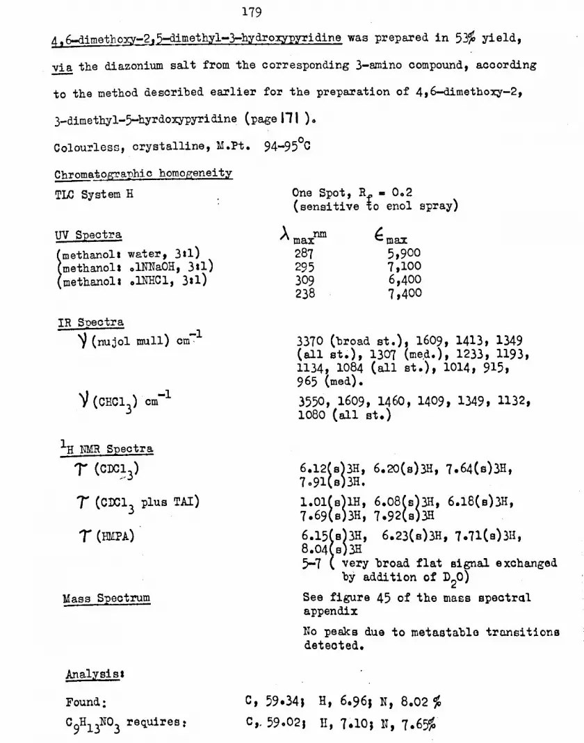
1794,6-dimetbo~-2J5-dimethyl-3-hydroxypyridine was prepared in 53% yield,!i! the diazonium salt from the corresponding 3-smino compound, acoordingto the method described earlier for the preparation of 4,6-dimethoxy-2,3-dimethyl-5-hyrdoxypyridine (page 111 ) 0Colourless, crystalline, M.Pt. 94-95°CChromatographic homogeneityTLC System H One Spot, R - 002
(sensitive io enol spray)UV Spectra
~
methano11 water, 311)methanol. olNNaOH, 3.1)methanol. olNHCl, 3.1)
A maXnIn
287295309238
~max5,9007,1006,4007,400
IR Suectra~ (nujol mull) -1cm, 3370 (broad st.), 1609, 1413, 1349
(all st.), 1307 (me,d.),1233, 1193,1134, 1084 (all st.), 1014, 915,965 (med).3550, 1609, 1460, 1409, 1349, 1132,1080 (all st.)
-1cm
IH NMR Spectra, (CDC;3)
I (CIX:a3plus TAl)
T (EMPA)
Mass Spectrum
6.12(s)3H, 6.20(s)3H, 7.64(a)3H,7091{s}3H.1.01(s)lH, 6.08(a)3H, 6.18(s)3H,7.69(s)3H, 7.92(s)3H6.15~B)3H' 6.23(s)3H, 7.71(s)3H,8.04 s)3H5-7 very broad flat signal exchanged
by addition of D20)See figure 45 of the mass spectralappendixno peaks due to metastable tro.neitionadetected.
Analysis.
Found:C9H13N03 requires:
C, 59.34, H, 6096J N, 8.02 %C,.59.02. II,1.10; U, 1.65%

ISO
TIm SYNTh~IS OF COiIPOUlIDS TESTED FOR THEIR
INHIBITION OF NADH-LINKEDOXIDATIOH
2_(1-n-hexyl)pyridine2-picolyl lithium (9g) in ether was prepared according to the methodof Organic S~theses 184. l-Eromopentane (14g) in ether (5Oml) was
oslowly added at 0 C. After 15 minutes, when the red colour of ~icolyl
lithium was discharged, water (20ml)lfollowed by hydrochloric acid(38% w/v, 20ml), was slowly added. The pH of the mixture was adjustedto 8 with sodium carbonate solution. The ethereal layer was separated,
washed with water and dried, yielding an oil (14.5g). On distillationthis ~lded a clear colourless oil of 2-(1-n-hexyl)pyridine, B. Pt.
90-92~/6mm, (1102g, 16% yield), (lit.220 E.Pt. 810/6mm)
(methanol)Amaxnm
265262269
fmax3,1503,5002,600
UV Speotrum
IH 1"MR SpectrUm1(CC14)
1.56 (double d,Ja4.1Hi· and 2.0Hz)lH,2.51 (double t,Jt - 1.5Hz and Jd - 2.0Hz)lH, 3.0 (m., and dou1:le d r • ,
superim~osed)2H, 7.28 (t, J. 7.8Hz)2H,8.66 (m)8H, 9.12(t,J- 6Hz)3H
Mass Speotrum See figure 42 of the mass speotralappendix.
2-(1-n-hexyl) pyridine-I-oxide
2-(1-n-hexyl)ppridine (1g) and peraoetic acid in acetio aoid (3~ w/v,
50 ml) were mixed carefully at ooc. The temperature was slowly raised
to 800C, and maintained at suca , After 16 hours the excess acetio acid
was removed on the rotary evaporator and the residue diluted with an
equal quantity of water. After adjuating the pH to 8 withsodium

131
oarbonate solution, the liquid was extracted with chloroform •. Thedried extract,after removal of solvent, yielded an oil (7.1 g, 92%) which
was distilled (94-96°/5 mm).uv SpectrumA (methanol)
max261 nm (~A)(. 10,200)
IR Spedtrum'Y (thin film)
lH NMR Spectrum1"' (eIC13)
1245 cm-1 (vost.)
1.70 (m)1H, 2.75(m)3H, 7.03(t, J.7.4Hz)za, 8.62(m)8H, 9.11(t,J - 6Hz)3H
After several attempts,starting material was reoovered quantitativelyon treating 2-(1-n-hexyl)pyridine with hydrogen peroxide(lee volume)and acetic aoid, according to the method of Ochiai169 for the preparationof pyridine-I-oxide from pyridine.
Attempted Nitration of 2-(1-n-hexyl)pyridine-l-oxideA. Following the procedure of OChia1169for the nitration of pyridine-l-oxide with nitric aoid (d - 1.48) in sulphurio acid, only startingmaterial (72%) was recovered from 2-(1-n-hexyl) pyridine-I-oxide,being identified from its infra red spectrum. Unidentified, water
soluble,·highly coloured products were also produced.
On the substitution of fuming nitrio acid for nitric acid (d - 1.48)in the same procedure, the reaction ~ecame Violent and no starting
material was recovered. Water soluble, highly coloured materials were
again produced, but were not investigated.
E (i) 2-(1-n-hexyl)pYridine-l-oxide fluroborate
2-(1-n-hexyl)pyridine-l-oxide (5g) was dissolved in aqueous fluoroborio

182
acid (40% w/v, 20m1). The solution was extracted with chloroformthree times. The combined extraots were dried, firstly over anhydroussodium sulphate and secondly over molecular sieves (type 3A). Chloroformwas removed by distillation ,yielding a colourless oil of 2..(1-n-hexy1)pyridine-1-oxide f1uoroborate (704g, quantitative yield).
rR Spectrum~ (thin film) 1060 cm-1 (broad v.st.)(ii) Attempted nitration with nitroniu~ tetra fluoroborate
Ni troniumtetraf1uoroborate (1.7 g) in d:ry redistilled acetonitrile ~(lO.ml)
was slowly added to 2-(1-n-hexylJ~yridine-1-oxide fluoroborate (3g)in dry redistilled acetonitri~e (lOml) at ~oom temperature in a dry box.The addition was exothermic,and the temperature was not allowed to riseabove 30°C. After stirring for 12 hours, the solution was poured ontoice, adjusted to pH 8 with ammonium hydroxide, and extracted threetimes w1t~ chloroform. The combined extracts, after drying, and removalof solvent by disti11ationjyie1ded 2-(1-n-hexy1)pyridine-l-oxide(2g, quantitative recovery), identified from its infra red s~eotrum.
The experiment was repeated, using nitromethane in place of
acetoni tril e, with the same result.
Reaotion of 2-(1-n-hexy1)pyridine-l-oxide with Phosphor~B Oxyohloride
2-(1-n-hexy1)pyridine-l-oxide (5g) and redistilled phosphorus oxychlorideowere carefully mixed at 0 C, the process being exothermio. The tem~erature
of the mixture was slowly raised,and it was heated under reflux
(collOOC). After 12 hours,exoesB phosphorus oxychloride was removed
by distillation, and the mixture pouted onto ice. After adjusting thepI! to 8 with ammonium ~hydroxide solution, the liquid was extracted

l8}
with chloroform three times. The combined extracts were dried, and thesolvent removed on the rotary evaporator, yielding a dark oil (4.8g),which was examined directly by vapour phase chromatography (using a
Perkin Elmer Fll instrument,having a flame ionisation detector,and a50ft·LAC Capillary Column,at l80oC,with all gas inlet pressures 25 pBi~.This showed the presence of two products, A and E, together with
starting material, in apparent proportions,respeotively, of 23, 18 and 10(corresponding to apparent gravimetrio yields of 43, 34 and 19,%).Product B was eluted from a Woelm neutral alumina column with benzene,
followed later by product A. 2-(1-n-hexyl)pyridine-l-oxide was only
eluted a~ter products A and B, with 10% v/v methanol in benzene, and wasidentified from its infra red speotrum.A. 4-ohloro-2-(1-n-he_NlJpyridine
An oil.UV Spectrum(methanol)
\. nmA max
261269
f.max
3,3002,700
IR Snectrum~ (thin film) cm-l 3050(w), 2960, 2930, 2860, 1577,
1555, 1468, 1393, 1101, 823, 706(all st.)
IH NMR Spectrumi (CDen}) 1.39 (d, J • 5.0 Hz)lH,
2.7 (rn,s. and d. superimposed)2H,7.16 (t, J • 7.7 Hi)~q,8.63(m)8H, 9.10(t, J • 6Hz)3H.

184
B 6-chloro-2-(I-n-hexyl)pyridine . '
An Oil.UV Spectrum
A (methanol)maxIR Spectrum
V (thin fi1m)cm-1 3060(w), 2960, 2930, 2860(st.)1587, 1560, 1439, 1410 (et),1380(w), 1163, 1140, 1101, 989,789 (st.)
IH NMR SpectrumT (CDC13) 2.31
2076208270198.64
t, J - 7.•7 Hz)lH,a, J - 707 HZ)lHtd, J .. 707Hz)lH,tl J - 707 Hz)2H,m)8H, 9.11 (t, J - 6 Hz)3H.
2-methyl-5:ethylpyridine-1-oxide was prepared in 8~ yield from~-fIfrl;hyl-5-:,ethylpyridinebya ,method analogous to that desoribed forpyridine -l-oxide by OChiai16~
IR Spectrum)l (thin film) 1265 cm-l (v.st.)
IE NMR SpectrumT(CDC13) 1.81(e)lH, 2.05(s)IH, 7.06(q,J-
7.0Hz)2H, 7.51(0)3H, 8.71(t,J- 7.0Hz)3H

l8S
Interactiop_of_y~idine-l-oxides and phosphorus oxyohloride at 330C I31an investigation by P 1~R Spectroscopy
{Performed in conjunotion with Dr. K. Dillon)2_(1_n_hexyl)pyridine-l-oxide (I,tab1e 12) and 2-methyl-5-ethylpyridine-l-oxide(II,table 12) used in this e~eriment were dried by passage throughmolecular sieves (type 3A). Phosphorus oxychloride (POC13, table 12)and dichloroethane (S,table 12) were redistilled, and manipulation
was in a dry box.The compounds were mixed at room temperature in the volumetric
proportions shown in table 12. The 3~ ~ spectra of the mixtures are
also shown in table 12.Hydrolysis of the mixtures containing pyridine-l-orldes on ice,
within 1 hour of mixing, followed by extraction of the bydrolyoatewith chloroform, resulted in essentially quantitative recovery of the
pyridine-I-oxides. They were identified by their IR speotra.TABLE 12
Volumetric Proportions 31 to 85%P NMR signals in p.p.m.relativeI II POC13 S phosphoric acid
0 0 1 1 -2.2 st.1 0 1 1 -3.95 (st); + 3.2(med.)J +8.3(w)0 1 1 1 -3.95(st)J + 3.9 (med) J +8.15(w)0 4 1 2.5 +4.05(st); +5.0S(w), +lO.05(w)

186
2 - (l_n_he~lJ~-methoxypYridine4-chloro-2-(1-n-hexyl)pyridine (150 mg) and sodium methoxide (250mg)in metheno~ (5ml) were heated in a sealed ampoule at 140oCo After16 hours the mixture was recovered, and the methanol removed bydistillation. The residue was extracted with hot chloroform,and theextract washed with water. On drying the solution and removing thechloroform by distillation an oil was obtained. After short pathdistillation at reom temperature and a pressure of 0001 mm Hg,2_(1_n_hezyl)-4-methoxypyridine (11Omg,75% yield), an oil, was
recoveredoPurity was ascertained by vapour phase chromatography.
UV· Spectrum\ (methanol)"'max
IR Spectrum~ (thin film) cm-l 3010(w), 2960, 2930, 2860 (st)
1595, 1567, 1482, 1460, 1308,1195, 1160, 1042(all st.) 993,825, 810 {w}
IH NMR Spectrumi (CDC:l3) 1070 (d, J - 5Hz)lH,
3.4 (6 and d superimposed)2H,6.21(s)3H, 7.27 (t, J - 7.7Ez)2H,8.66 (m)8R, 9.11 (t, J • 6nz)3H
Mass Spectrum See figure 42 in the mass 6pectra~appendix.
2_(1-n-hex;yl)-6-metho:xypyril1ine, an oil, was prepared in 7;n, yield from
6-chloro-2-(1-n-hexyl)pyridine in a manner analogous to that described in
the experiment immediately above.

187UV Spectrum
A (methanol)max
IR Spectrum'Y (thin film) -1cm 3060(w), 2960, 2930, 2860(sto)
1598, 1578, 1466, 1438, 1414, 13401317 (all st.), 1279 (med), 1188(med),'1148 (sto), 1101,1073 (med)1038, 986, 795, 735 (st.)
lH mm Spectrum; (Clxa3) 2056 t, J • 7.7 Hz)lH
3.34 d, J • 7.7 Hz)lH3.49 d J. 7.7 Hz)IH6.11 s~3H, 7.33 (t, J • 7.7 Hz)2H,8.66 m)8H, 9.12(t, J • 6 Hz)3H
Mass Speotrum See figure 42 in the mass speotr~lappendix.
2-(1-n-hcxyl)-4-hydroxypYridine2-(1-n-hexyl)-4-methoxypyridine (100 mg) in 48% w/v hydrobromie aoid(5 ml) was heated under refulx. After 16 hours,the pH of the solutionwas adjusted to 8, and it was extracted with chloroform. The extractwas dried,and the chloroform removed by distillation. Short pathdistillation of the residual oil, at room temperature and a DreBsure of0.01 mm Hg, yielded 2-(1-n-hexy1)-4-bydroxypyridine (85 mg, 92% yield),as a waxy solid.
oM. Pt. 24-28 CUV Spectrum
~ (methanol)maxIR·Spectrum
~ (thin film) -1om 3220, 3050 (medo) 2930, 2855 (st.)1628, 1515 (v.st.), 1385, 1231,862(med),
1HNMR Spectrum'T (eDCn3) -0.2 (broad s.)lH, exchanged by

183
the addition of D20, 2.35 (d,J •609 Hz)lH, 3.6 (d and B superimposed)2Ht 7035 (t, J - 707Hz)2H, 8.72(m)8H, 9012 (t, J - HZ)3H
Mass STlectrum See figure 42 in the mass speotrdlappendix
Analysis IFound r
CIIH17NO requires:e,73071, H, 9.66; N,7.66%C, 73.75; H, 9050 J N, 7.8zfo
6-{1-n-hexyl)-2-bydro:xy:pyridine,a wax;'"solid, was prepared ;in9% yieldfrom 2-(1-n-hexyl)-6-metho:xypyridine in a manner analogous to thatdesoribed in the experiment immediately aboveo
oM. Pt. 46-47 eUV Speotrum
_.-X ' (methanol)max
IR Speotrum~ (thin film) om-l 3270, 3120 (med)~ 2920, 2850(st.)
1640(broad v.sto), 1547, 1468(st.)1416, 1377, 1228, 1211 (med). 1165,1000, 950, 798, 788 (sto)
~ lIMRSpeotrumJ (CDC13) -3.1 (broad s)lH exchanged by addition
of D 0t 2.73 (double do J • 609 and8.8 ~z)lH, 3.70(dt J • 8.8 Hz)lH,4.05 (d, J - 609Hz)lH, 7.43 (t, J •7.7 Hz), 8.72 (m)8H, 9012 (t, J •6 Hz)3H.
Mass Spectrum See figure 42 of the mass speotralappendix.
Analysis
requires S
C, 73051, .H, 9048J N, 7.25%C, 73.75J H, 9.501 ~,7.82%
Found:

139:Benzyl VanillinBenzyl chloride (12.5g) in methano1(50ml) was slowly added to thesodium salt of vanillin (17.5g) in methanol {lOOml), and the mixtureheated under~flux. After 16 hours,the solvent was removed bydistillationJand the residue dissolved in chloroform and washed withwater three times. Drying and eva~oration of the chloroform solutionyielded crude benzyl vanillin. (14.5g, 60% yield). On aoidifioation ofthe aqueous washings followed by a similar chloroform extraction,vanillin (6g, 40% recovery, Mopto 80-81°0 (lit.222 80-81°0) was
obtainedoOn elution from a Woelm neutral alumina column with benzene the
crude product, mentioned above, yielded ohromatographically pure
benzyl vanillin as an oil.Chromatographio HomogeneityTLC System G One spot, ~ - 0040
(Vanillin on the same system has an Rf of Zero)
IR Speotrum\' (nujol)'co~ (nujol) VanillinJco
-11680 cm-11658 cm
III W.JR Spectrumi (CnC;13) 0.05(s)lH, 2.55(m)lH, 2094
(a, J - 8.6 Hz)1H, 4.14(e)2H,6.06(s)3H.
Triphenyl-Ul-n-he;yl)phosphonium bromide (Wittig Salt)Tri~henylphosphine (2603g) and l-bromopentane (1805g) in toluene .(lOOml)
were heated under reflux for 16 hours. After this time,the mixture was
cooled and the precipitated Wittig salt isolated by filtration and dried
under vacuum at 100e• (Yield- 26.8g white, crystalline, 6~)M.Pt. l65-l66OC (11t.223 165-165.50C)

190Note-
T~"se. o~ benzene instead of toluene in the above reaction reduoed theyield of the Wittig salt to zero. Presumably this is because thetemperature of boiling benzene is not sufficient to activate/the/,
reaction.Benzy1-(4-(1-n-hex-1-eny1)-2-methoxyphenyl) etherTripheny1-(1-n-hexy1) phosphonium bromide (4.lg) and potassium tertiarybutoxide (1.1g) in dry ether (100 m1) were mixed together in a slurrygenerating an orange ylid. Benzyl vanillin (2.4 g) in dry ether (5Oml)was added slowly, after which the mixture was heated under reflux
for 24 hours 0
After washing th~ cooled mixture with water, evaporation of thedried ether solution yielded an oil (2.3g)o Short path distillation ofthis at room temperature gave benzyl -(4-(1-n-hex-l-enyl)-2-methoxphenyl)
ether (lo9g, 64~ yield) as an oil.Chromatographic homogeneity
TLC System G One Spot, Rf • 0.90
In NMR Snectruml' (CDC,13) 2054(m)5H, 3oll(s)3H, 3060(d, J •
12.0 HZ}lH, 4.3a (double tJ d, J •12.0 HZJ tl J • 6.9 Hz}lH, 4.82(8)2H, 6ol2(s)3H, 7.65(Q,J .7Hz)2II,8.5 (m)4H, 9ol0(t, J • 605Rz}3H
4_(1-n-hexyl)-2-methoxyphenol10% Palladium on oarbon (10 mg) was added to benzyl-(4-(1-n-hex-l-enyl)-
2-metho:x:yphenyl)ether (100 mg) 11m methanol( 25ml), and the mixture
hydrogenated at one atmosphere pressure.
After the theoretical quantity of hydrogen was absorbed (l~ ml
over 30 minutes), further absorption oeased. The oatalyst was removed

191by filtration,and the solvent on the rotary evaporator. Short pathdistillation, at room temperature and a pressure of 0001 mm Hg, of the
residual oil gava4-(~n-haJQ'1 )-2-methol:Yphenol (7Omg, quantitativayield).
Chromatographic homogeneitlTLC System G One Spot, Rf • 0.10
3.2 (m, AB s~stem and singletsuperimposed)}H, 4.4 (broad s)1H,exchanged by addition of D~O, 6014(s)3H, 7045(t, J • 703 HZJ2H~8.65(m)8H, 9012 (t, J • 605Hz)~1

192
2_hydro:xymetbyl-5=rnethox;y=4-pyronawas prepared in 72% yield, by theaction of dimethylsul;phateon kojio aoid, aocording to the method ofCampoell et al 193
o' 193M. Pt. 163-164 C (lit. 161°C)
tmetharlOl)methanoll water, 311)methanol 1 .1NNaOH, 311)methanol •• lNHCl, 311)
AmaxnIn
265268268268
~max
10,30010,2009,400
11,700
DV Speotrum
IR Speotrum~ (nujol mUll) -1cm 3180 (v.broad, st.), 1635, 1582
1256, 1217, 1144, 1080, 1050, 998949, 866, 838, 716 (all st.)
lE NMR SpeotrumT(D20) 1.85(s)lH, 3.35(s)lH, 5.4l(s)2H,
6.16(s)3H.4-hydro:x;y-2-h.ydro:x;ymethyl-5-methoxywridinewas prepared in 8~ yield by
, .the action of B!nmoniaon 2-bydroxymethyl-5-methoxy-4-pyrone, aooordingto the method of Armi t and Nolan194• The ;produotwas rasublimed as whiteorystals.
IR Speotrum~(nujol mull) om-l 340O-2500(v.broad,st.), 1621 (v.st.)
1540(st.), 1268, 1250, 1168, 1153,1081, 1017, 855, 765, 717 (all st.)
UV Speotrum
~
methanol)methanol. water, 3.1)methanol. .11TNaOH,311)(methanol•• lNHC1, 311)
AmaxnIn273275250275 (shoulder)245270
~max11,30011,5008,5004,7005,1505,300

1931H NMR Spectra-r (D20)
'T (raso)
Mass Spectrum
2042~B)lH' 3050(s)lH, 5.39(s)lH,6.20 s)3H .2054 s)1H, 3069(s)lH, 4.7 (broad s.)2H,exchanged by addition of D20,5.58(s)2H, 6028(s)3H.mle155154140139126III
I73
10020174232
Peaks due to metastable tranaitions+ +155+---+154+ + 1 @ 153.01
155 ---+140 + 15 @ 126045

1942,6-dimethyl=4-nitropyridine-l-oxide was prepared in 62% yield over twostages from 2,6-dimetbylpyridine by a method analogous to that describedfor 4-nitropyridine-l-oxide-l-oxide by OChiai169
M.Pto 163-l64°C (lit.169 1630C)UV Spectrum(methanol)
Amaxnm239232
tmax9,6009,700
IR Spectrum~ (nujol)
lH mm Spectrum T (C.XI3)
4_benzyloxy-2,6-dimethylpyridine-l-oxide was prepared in 6~ yield trom2,6-dlmethyl-4-nitropyridine-l-oxide by a method analogous to thatdescribed for 4-benzyloxypyridine-l-oxide by Ocbiai195
oM.Pto 135 C
uv Spectrum~MA)t(methano1)IR Spectrum
~ (nujo1)
262 nm (~~- 16,800)
1230 cm-1 (vost.)
~ NMR Spe!6trum, (CDC:1
3) 2065(s}5H, 3026(s}2H,
4098(s}2H, 7050(s)6H2,6-dimetbyl-4-hydroxypyridine-1-oxiderffi· palladium on carbon (30 mg) was added to 4-benzyloxy 2,6-dimethyl-pyridine-l-oxide (230 mg) in ethano1(10 ml) and the mixture hydrogenatedat one atmosphere pressure, analogous to the standard conditions of
196KatritZky and Monro for the reduotion of pyridine-I-oxides.
After 24 hours,the oatalyst was removed by filtration and the solventby d1st11lat1on/y1e1ding white orystals of 2,6-dimethyl-4-hydroxypyridine-

195
-l-oxide (130 mg. 92% yield). These were purified by reprecipitation,from methano1io solution, in cychlohexane, since recrysta11isationproved to be difficult.
M. Pt.UV Spectrum
A (methanol)max 259 run (~t1AX - 12,800)
~ NMR SpeotrumT (eDC13) 3.35(s)2H, 7.56(s)6H
~ Prolonged hydrogenation under the above conditions did not affeotthe course of the reaction.

1961~etbyl-4-nitropyridine-1-oxide was ~re~ared in 65% yield over two stagesfrom 2-methy1pyridine by a method analogous to that desoribed for 4-nitro~yridine-l-oxide by OOhiai169
Mo Pt. 157-159°C (lito169153-4°C)
uv Speotrum\ (methanol)"max
234 run (~t1AX - 9,200)329 nm (e"A)(. 14,300)
IR Spectrum~ (nujol) 1285 (st.) -1om
IH mm Speotrum1" (COO13) 1.75 (d,J • 6.9 Hz)lH, 200 (m)2H,
7.47(s)3H ..2-methyl=4-nitropyridine was ~re~ared in 70% yield from 2-methyl-4-nitropyridine-l-oxide by a method analogous to that described for 4-
. . 169nitropyridine by Oohia!° ~ 225a ° ~MoPto 40-42 C lito225b 32-34 C
lit. 42-45°C
uv Speotrum(methanol)
Amaxnm227295
emu:6,3002,000
~ NMR Speotrum'r (CDC13) 1033 (d,J • 408Hz}lH, 2.25(m)~1
7.32(s)3H
4-hydroxy-2-methylpyridine was prepared in 8~ yield from 2-methyl-~5C ...i"eaf
4-nitropyridine by a method analogous to that~for the preparationof 4-hydroxypyridine by den Hertog et al 191. The time of the reaotion
was three days instead of 12 hours. Oxides of nitrogen were produoed
during the reaotion.
(lit.226o
174-116 C)

197
UV Speotrum AmaxntD €. max
~
methanOl)methanol plus 2 drops .11~aOH)methanol plus 2 drops .1NHCl)
256234240265 (shoulder)
13,8009,600
11,4002,800
IR Speotrum~ (nujol)om-l 3400,,3210(medo)~630(st.) l535(st.)
1230, 1180, 1095, 1048~ 942, 924,891, 829, J30 (all st.)3240, 3030, 2940 (med) 1631,1508 (v.st.), 1395, 1386(st.)
IH NMR Spectrumi (CDC13) -1.6 (broad s)lH,
2041 (d, J - 6.9Hz)lH, 3.7(m)2H,7.69(s)3H .
Mass Spectrum See figure 45 of the mass speotralappendix
Pe~ due to metastable transition109 ---.. 80+ + 29 @ 58.72
4-hydroxy-2-methylpYridine nitrate
2-methyl-4-nitror,ridine (M.Pt. 40-4200), exposed to the atmosphereofor two months,was found to have a melting range of 161-165 C. White
crystals of 4-hydroxy-2-methylpyridine nitrate were recovered in ·9~gravimetrio yield on recrystallisation from acetone.Effeot of dry heat (i) melting
(ii) violent decomposition. no oxides of nitrogendetected, strong smell typical of a pyridine base.
Test for nitrite and nitrate ions using sulphanilic aoid and
J_napthylamine,with the addition of zino dust later as a reduoing agent.Nitrite Test. NegativeNitrate Testa Positive (red colour of azo dye)

UV Speotrum A maxnm €max(methanol) 238 9,900
243 (shoulder) 9,600248 " 9,400252 II 9,100257 II 7,900
IR spectrumV (nujol) cm-l 3100 (med,), 1641, 1628, 1613 (v.sto)
1500, 1315, 1250, 1214, 1185, 1040,995, 869, 838, 710 (st.)
IE NMR Spectrum't (D20)
, (DMSO)
1071 (d, J - 702Hz)lH, 209(m)2H,7.37(s)3E1052 (d, J - 702Hz)lH,109 (broads.)2H exchanged by additionof D20, 209(m)2H, Signal from methylgroup obscured by solvent.
Mass Speotrum m/e109948070463930
I100
5922
2188
Peak due to metastable transition
Analysis'Found:
C 6E804N2 require s:0,.42003%; H,4.74%;e, 41.8~J H, 4065~J

199Tm~ SYIJTlIESIS OF OTHER PYRIDINJiS AIID EXPERH1EHTS
CONCERHJiiO \elITE TI-rE: SYNT~IS OF PYRIDINES
3-methoxypYridine
Diazomethane (0064)g in ether (100 ml) was slowly added to 3-
hydroxypyridine (l.44g) im methanol (25ml) at OOCo After 30 mins.the solvent was removed by disti1latlon,and the residue purifiedby short path distillation. 3-methoxypyridine (0.9g, 55.%yield) wasobtained as the distillate. The involatile residue (Oo7g) was notidentifiedo
oB. Pt. 180 CUV SpectrumA"')l(methanol)IR Spectrum
~ (thin film)cm-l 3010, 2960, 2840 (med.), 15761480, 1460, 1426, 1283, 1218, 1232,1192, 1108~ 1054, 1018, 797, 705(all v.sto)
l.HNMR SpectrumT (CDC13) 1.7m(2H), 208(m)2H, 6.18(s)3H.
Ma.ss Spectrum See figure 43 of the mass speotralappendix.
5-methoxy-2-methylpyridine was prepared in 52% yield from 5-hydroxy-2-
methylpyridine by a method analogous to that descrmbed immediatelyabove for the preparation of }-methoxypyridine.
oB. Pt. 190 CUV Spectrum
A (methanol)maxIR Spectrum
V (thin film)cm-1 3010, 2940, 28401, 1600 (all med.)1575, 1499, 1486, 1463, 1441, 1390,1268,1241,1213,1181,1134,1120,1030,822,722 (all st.)

200
108(m)lH, 2.95(m)2H, 6020(s)3H,7051(s)3Ho
Mass Speotrum See figure 44 of the mass speotralappendix.
4-methoxyEYridine was prepared in 8~ yield, from 4-chloropyridine,. 200according to the method of Spinner and White 0
B. Pt.. 86°/15mm (lit.200 l07°/50mm)(lito200 4°C)oMoPto 2-4 C.
UV Spectrum tmax(methanol) 220 - 9,300
245 (shoulder)' 1,200)IR Speotrum
~ (thin film) om-l 2960, 2840 (med, ) 1597(v.st.),1570,1501(st.), 1460, 1441, 1423 (all med.)1289 (v.st.) 1212 (st.) 1029 (st.)995 (med.), 820, 805(med.)
1063 (d, J • 500 Hz)2H,3.25 (d, J • 500 Hz)2H, 6.23{e)3H
Mass Speotrum See figure 43 of the mass speotralappendix.
1-methyl=4-pyridone
The potassium salt of 4-hydroxypyridine (2.7g), methyl iodide (2.9g)and ethanol (5 ml) were heated at 1000C for 105 days in a sealed
ampoule. After the removal of solvent from the recovered mixture,
extraction with hot acetone yielded a semi-crysta~line solid (1.8g)o. 0Short path distillation at 150 C and 0001 mm Hg yielded colourlees
hygroscopio crystals (1.4g, 65% yield)o,i'
. \

201UV Spectrum
AMAA(methanol)...~t1AX:J263 nm (18,200)
IR SpectrumV (thin film)cm-l 2960(m), 1648, l5~0 (v.st.), 1440(m),
1397 (st) ~364 (m), 1203 (v.st.),1254, 1024 (m), 853 (st.)
~ N1'~R SpectrumT(CIXH3)
Mass Spectrum See ~igure 43 of the mass spectralappendix.
l-methyl-2-pyridone was prepared in 98% yield from 2-methoxypyridine.oby isomerisation in a seald ampoule at 230 C for 2.5 days. The product
was distilled at reduced pressure.
The distillate crystallised, with some difficulty in initiation, undernitrogen.
Colourless CEystals
UV Spectrum€nethanol)
\ nmAomax229302
tmax7,4006,000
IR Spectrum -1V (thin film)cm 3080, 3030, 2850 (med.), 1657, 1583(v.st.), 1542,(st). 1470,1440, 1416,1386 (med.), 1320 (st.) l242'(med),1156, 1064, 875, 843, 763, 726(all st.)
~ NMR Spectruml' (eDC1) 207(m, double do and ~uartet of
doublets superimposed)3.43(double d, J - 9.5,1.4Hz)IH,3.83(triplet of doublets, J - 6.5lo4Hz)lH, 6.46(s)3H
Mass Spectrum See figure 43 of the mass speotralappendix.

202
Attempted Hydroxylation of Pyridines with Hydrogen peroxide
A 2% v/v solution of pyridine in 100 voaume hydrogen peroxide (5Oml)was maintained at 500Co After one week volatile material was removedon the rotary evaporator, the last traces being evaporated on the
freeze dryer, yielding a semi-crystalline oil (0054 g). This materialwas not appreciably soluble in acetone, methanol or other organicsolvents, but was soluble in water, the solution colouring starch-iodide paper blueo Recrystallisation or reprecipitation of thematerial proved impracticable, but it was examined chromatographicallyoTLC System F Rf = 0005 (faint) and 0060 (strong and tailing)
-detected using the enol Bpr~(Rf value of authentic 3-hydro~pyridine under these conditions. 0020)
the oilbIt 110 C under an air condenser for one week, aOn maintaining~qR~i~ of material (11 mg) remained as an oily residue.The experiment was performed concurrently with 2-,3- and 4-lutidinein place of pyridine, analogous results being obtained in each oasso

203
EXPERIKENTAL COr'IPARISONS INVOLVING
SUBSTITUENTS Dr THE rx , @ AND t PO~ITIONS OF PYRIDIN"ES
IH Nuclear magnetic resonance spectra of readily available pyridines
;pyridine2-'Dicoline
3-;picoline
4-;picoline
2,3-1utidine
2,4-lutidine
2,5-1utidine
2,6-lutidine
3,4-1utidine
3,5-1utidine
2,4,6-collidine2,3,6-collidine
2-hydroxypyridine
3-hydro~;pyridine4-hydroxypyridine
7' (CIXJl3)
1043(m)~~, 2040(m)lH, 2.82(m)2Hl048~d' m - 5.lHz)IH, 2041(t, J - 609Hz)lH,2.86 dt J - 609Hz)lH, 2092(t, J - 6Hz)lH,7045 s)3Ho1054(m)2H, 2052(d, J m 704Hz}lH, 2083(doub1e d,J ...704 and 500 Hz)lH, 7069(s)311.1056(d~ J ...5.0Hz)2H, 2092(d, J ...600Hz)2H,7069(S)3H1069(d, J ...501Hz)lH, 2073 (d,J ...707Hz)lH,3002 (double d, J ...707 and 50lHz}lH, 7052(s)3H,7075(s)3H.1068 (d,J ...500Hz)lH, 3.08(s)lH, 3014(d, J - 5.0Hz)IH, 7.52(s)3H, 7.73(s)3H.lo69(s)lH, 2.64(d, J - 1.9Hz)lH, 2.99(d, J - 1.9Hz)IH, 7052(s)3H, 1015(s)3H.2.71(t~ J - 107Hz)lH, 3021 (d,J - 107Hz)2H,1051(s)6Ho1072(s)lH, lo15(d, J - 4.8Hz)lH, 3.03(d,J-4.8Hz)lB,1019(a)6Ho~19(d, J - 200Ha)2H, 2076(d, J - 200Hz)lH,1078(s)6Ho3029(s)2H, 7051(s)6H, 7.79(a)3Ho3.08(d~ J ...709Hz)lH, 3021(d, J ...709Hz)lH,1.56(s)6H, 7081(3)311.2.63, (d, J ... 605Hz)lH, 2.55(doub1e d, J ...605 and905Hz)lH, .3045(d, J - 905Hz)lH, 3075 Ct, J ...605Hz}lH.lo72(s)lH, 1.94(d, J - 5.0Hz)lHp 2075(m}~q.2035 (d,J - 609Hz)2H, 3057(d,J ...6.9Hz)2H

204
2-methoxypyridine lo93(d, J - 500Hz)IH, 2.54(t, J - 705Hz)1H, 3023 (double d, J - 5.0 and 702Hz)lH, 3034 (d,J = 800Hz)lH, 6.l2(s)3H.2056 (t,J = 8.2Hz)IH, 3.76(d, J _ 8.2HZ)2H, 6.13(s)6H.
3.02(d, J - 6.5Hz)IH, 3031(d, J - 6.5Hz)IE, 3.81(t, J = 605Hz)IH, 6028(s)3H.1.81(s)1H, 2.86(m)2H, 7053(s)3Ho4.95(s), 7050(s)
2,6-dimethoxypyridine
2-hYdroxy-3-methoxypyridine
5-hydroxy-2-methylpyridine
O,O-iso-propylidinepyridoxineThe use of Hexamethyl Phosphoramide in the Proton NMR study ofHydro:xypyridines
The ~ 1lliffi spectra of a series of compounds were recorded as their 10%solutions in redistilled hexamethylphosphoramidoo The chemical shiftsof protons attached to oxygen or nitrogen atoms in the speotra of thesecompounds, confirmed by their rea4y isotopio e~change with deuteriumoxide, is shown belowo
Compound (1~7A) (Singlet)
2-hydroxyprridine3-hydroxypyridine4-hydroxypyridine (anhydrous)4-hydroxy-2-methylpyridine (anhydrous)4-hydroxy-2-hydroxymethyl-5-methoxypyridine
-203 (broad)-102 (sharp)-006 (broad)-004 (broad)+2.5 to + 4 (v.broad + flat)
,5-hydroxy-2-methylpyridine2,3-dihydroxypyridine4,6-dimethoxy-2,3-dimethyl-5-hydroxypyridine
-0067 (sharp)o to +2 (vobroad + flat)+5 to +7 (vobroad + flat)

205
4,6-dimethoxy-2,5-dimethyl-3-hydroxypyridine4-methyl-2,6-di-t-butylphenol2-hydroxyquinoline4-hydroxyquinoline8-hydroxyquinoline
+ 5 to +7 (vobroed + flat)
Uracil
+2 to +3 (vobroad + flat)-205 (broad)
-2.8 (broad)-005 (vobroad + flat)-1.6 ) ( )-1.7 ) broad doublet-0078 (sharp)-0065 (sharp)
at -O~57 (sharp)at +2.5 (broad)
-2.6 (broad)2H at 406 (broad)
2-naphthol (J.G.C)l-methyl-2-naphthol (J.G.C)
2_hydroxy-l-naphthaldehydeAniline
(J .G.c)~i~(J .G.C)
l_hydroxymethyl-2-naphthol
330CAcetamide(slow isotopic exchange, t ~ = 12 mins)2 '
Benzamide(slow isotopic exchange,
(IH at(1l.H at
330Ct t =(IH at(lH at
.1095 (sharp)2.95 (sharp)2 mins)
1014 (sharp)2.25 (sharp)
J.G.Ca compounds prepared and spectra recorded by Dr. J.G.Catterall.1The use of Trichloroace]3.isocyanate in the H l~R study of Hydrory-
I?yridinesThe ~ NMR spectra of a series of compounds were recorded in 10%
solution in deuterochloroform, or in the case of lens E:oluble compounds
as their saturated solutions. After the addition of one molarequivalent of trichloroacetylisocyanate (two molar equivalents in thecase of a compound with two potentially reactive protons) the spectra
were again recorded. The results are shown in table 13.

206
Deuterochloroform was chosen as the most suitable solvent after rejectingother possibilities on the following grounds.
Acetone and Carbon Tetrachloride: hydroxypyridines are relativelyinsoluble in these solvents.
Acetonitrile, mitromethane, dimethylformamide and prototropicsolvents react with trichlo~acetylisocyanateo
Dimethylsulphoxide and Hexamethylllhosphoramide& although both
of these have good solvating properties towards hydroxypyridines,and neither appear to react with trichlo~acetylisocyanate, thesolutions after the addition of trichlo~acetylisocyanate becomeviscous, producing a line broadening effect on the lMR signals to
such an extent, in some cases, as to m~ce the spectra uninterpretable.KEY TO TABLE 13
Number Compound2-hydroxypyridine1
2
345678
9101112131415
3-4-
It
tt
4-hydroxy~2-methylpyridine5- " -2- "3-hydroxy-2-hydroxymethyl-6-methylpyridine2,3-dihydroxypyridine2-hydroxy-3-methoxypyridine4-hydroxy-2-hydroxymethyl-5-methoxypyridine2-(1-n-hexy1)-4-hydroxypyridine6-(1-n-hexyl)-2-hydroxypyridine4,6-dimethoxy-2,3-dimethyl-5-hydrozypyridine4, 6- " -2, 5- " - 3- "Uracil2,6-dihYdroXY-4-methylpyridine

207
1617181920
2-hydroxyquinoline4-8-
""
Fiericidin AFiericidin B
Symbols
CAT Spectrum composed with the aid of a computer of averagetransients (CAT).
* Compound insufficiently soluble to produce a spectrum,even withthe aid of CAT.The arrangements of substituents on the pyridine nucleus ofthese compounds is not certain.
TABLE 13A = T(CrQ13); B • r(CDC13 with TAl); C - A-13
Substituent Positions -OH,-NH -NH withCompound 2 3 4 5 6 wi thout TAI; TAl
1 A - 3.45 2055 3.75 2.63 llitobservedB - 3030 2145 3.50 1.64 -5.0C - 0.15 0.10 0.25 0099
2 A 1.72 - 2.75 2.75 1.94 2.3B 1.40 - 2.40 2040'1040 -1.1C 0.32 - 0035 0.35 0.54
3 A 2..35 3.57 - 3057 2.35 Not observedCAT"B 1035 2.60 - 2060 1035 -1.4
0 1.00 0.97 - 0097 1000
5
A 7.67 3.78 - 3.71 2045 109:8 7.32 2.86 - 2086 1036 -C 0.35 0092 - 0.85 1.09 -A 7053 2086 2086 - 1.87 Not observed:8 7.44 2082 2.50 - 1.64C 0.09 0004 0036 - 0.23 -
-2.21 (sha.l'p)4
-00-1

203
Substituent Positions -CH,-1"'H -NB withCompound 2 3 4 5 6 without TAIa TAI
6 A 4.95 * * 7050 *:B 4055 -(2.27 or 2.71)7.37 007 and 101C 0.40 0.13
7 A ~ ... * * * *:B ... 2.47 3052 1.72 0.90 and -4.25
(both sharp)C
8 a 6.28 3.31 3.87 3002 -305 -:B 6016 3.22 3.62 2.08 -4.85 (sharp)C 0.12 0.09 0025 0094
9 A * * * * * -] 4.66 2.64 - 6002 1.61 0.8 and 303C -
10 A 7035 3.6 306 2.35 -Co2:B 7.02 2.7 207 1.31 1.8C 0033 009 ... 009 1004
11 A 7.42 4.05 2.73 3.70 - -3.1] 7.07 3.83 2.64 3.55 - -4.8C 0035 0022 0.09 0.15 -
12 A 7070 7093 6.10 - 6.08 Not observed -:B 7.65 7092 6.16 - 6011 1.06 (sharp)C 0.05 0001-0.05 - -0.03
13 A 7064 ... 6.20 7091 6012 Not observed:B 7.69 - 6.187.92 6.08 1.01 (sharp)C -0005 - 0.02 -0.010.04
14 & A * * - * -15 B * * - - *C Apart from their insolubility neither compound appeared to
react with TAI even at 80°C

209
Substituent Positions -OH,-lUI -NH withCompound ~ 3 4- 5 6 without TAIl TAl
2 3 4 others16 A 3.26 2016 2.3-3.0 -2.6
B 3026 2016 203-3.0 -2.6C No reaction appeared to take place in this case even at 800C
17 A * * * * *B 2059 4.18 302-401 000C -
18 A 1020 (2. 3-2.9) 1086 2.3-2.9 Not observed -B 1007 (2.1:-207)1.81 2.1-207 0.1c 0.13 002 0005 002
19 A 6008 6.18 6063 7·91 Not observede B 6004 6.18 6.60 '·94 0.93 and 1.77
(both sharp)C 0004 zero 0003 -0.03
20 A 6008 6.18 6064 7091 Not observed(Z) B 6003 6.17 6059 7092 0.97 (sharp)C 0005 0001 0005 -0001
An ultraviolet speotroscopic investigation of a seriee ofhydrox;y;pyridines and related oompounds in neutral, o.cidic and basiosolutions was undertaken. The solvents contained methanol in order toextend comparisons of these compounds to the pericidins, which are notsufficiently soluble in completely aqueous media to produce detectableultraviolet absorptionso
2dNeutral Solvent 0
Acidic SOlvent2do2dBasic Solvent •
75% methanol, 2~ water (v/v)
75% methanol, 23% OolN Hydrochlorio acid
7~ methanol, 25% O.lN sodium hydroxide Bolutiom.The results are presented in table 140

210
TABLE 14A) 2-Hydroxypyridines and Related Compounds
- --~--.-~~--.-.
CompoundNeutral SolverrtAcidic Solvent Basic Solvent
2-hydroxypyridine1-methyl-2-pyridone2-methoxypyridine2-\I-n-hexyl )-6-
hydroxypyridine2-(1-n-hexyl)-6-
methoxypyridine
298(5,200)297(6,000)271(3,000)306(5,700)
274(3,100 )
29415,300l294 5,000295 5,200300 5,900
301(5,600)
303(5,500)
B) 3-hydroxypyridines and Related Compounds
CompoundNeutral Solvent Acidic Solvent Basic Solvent
3-hydroxypyridine3-methoxypyridine5-hydroxy~2-rhethyl:pyridlne
5-methoXY~2;...fncthylpyrid.1he4,6-dimethoxy-2,3-dimethyl-
5-hydroxypyridine4,6-dimethoxy-2,5-dimethy1-
3~hydroxypyridine2,4-dimethyl-3-hydroxy-
~etho:xypyridine (mm)Phenol
277(4,200)278(4,100)283(4,100)
28413'900)281 6,000226 4,800)287 5,900)290(4,500)272(1,560)
··~~~ll:6gg~286 7,500292 7,200292(7,100)291(7,800)
~ 295(7,100)306(7,200)271(1,440)
300(4,700)238(11,100)303(4,800)24°(9,900)297 7,100248 7,400307 6l4oo238 7,400316 5,000289(2,700)
(HMO). Compound prepared and spectra recorded by Mr. H.M.Okely.

211
c) 4-hydroxypyridines and related compounds
A maxnm ( EHAX)CompoundNeutral Solvent Acidic Solvent Basic Solvent
4-hydroxypyridine 256(14,800) 234(9,900) 240(14,500)and shoulder
263~18,200) 240~13,000)265(3,200)1-methyl-4-pyridone -4-methoxypyridine 220 9,300) 235 9,700)
and shoulder245(1,200)
240(11,400)'2-methyl-4-hydroxypyridine 256(13,800) 234(9,600)and shoulder
2-(1-n-hexy1)-4-hydroxy 256(13,500) 234(9,700)265~2,800)239 12,100)pyridine and shoulder
2-(1-n-hexyfu)-4-methoxy 235(9,400)265(2,800)
220~9'OOOl -pyridine 247 1,7004-hydroxy-2-hydroxymethy1- 214 11,500) 245(5,100) 250(8,500)5-methoxypyridine 270(5,300) and shoulder
267(10,200) 267 (11,700) 210~4,9oo~2-hydroxymethyl-5-methoxy- 267 9,4004-pyrone
D) The Piericidins
Piericidins A and B 267(5,600) 273(8,200) Shoulder267(5,600)
270(3,200)Ootahydropierioidin A 274~8,200~ Shoulder233 5,600 240~9,OOO~
218(6,500) 280(6,400)270 2,700Piericidin A diacetate Shoulder210(3,000)

•
212
Thermal Stability of 4,6-dimethoxy-2,3-dimethylpyridineA 4,6-dimetboxy-2,3-dimethylpyridine was heated at 1800c for 3 daysin a sealed ~ule. 8~ of the starting material was recovered byshort path distillation at room temperature and 0001 mm Hg after
this time.
The infra red speotra of the starting material and distillate were
identicalo
B After heatir€ 4,6-dimethoxy-2,3-d1methy1pyridine at 2300C for 3days in a sealed ampoule, no starting material or volatile produotwas recovered. The solid residue, corresponding gravimetrically toquantitative isomerisation of the starting materialt was not
investigated further.Thermal Stability of 2-rneth0xYpYTidineA 2-methoxypyridine was heated at 1800C for 3 days in a sealedempoule. 98% of the starting material was recovered by distillation
after this time.
The infra red spectra of starting material and distillate were
identioal.B 2-methoxypyridine was converted in 9~ yield to I-methyl-2-pyridone
o .on heating at 230 C for 2.5 days in a sealed ampoule. (see thepreparation of l-methyl-2-pyridone, page 101 )

213
The base catalysed deuterium exchange of protons in the methyl grOUpS
of pYXidines was investigated under three sets of conditions. Thedegree of exchange was caloulated from a oompariocn of the integratedproton NMR spectra of the individual compounds before and afterreac~n, taking into account the estimated isotopio ratio of deuteriumto hydrogen in themedium after reaotion. An example of this oalculationis shown below for the case of 2-methylpyridine. The results of this,and other experiments are shovin in Table l5,together with the conditions,A, ::B or C used in each case, and the percentage of starting material
recovered"Errerimental ConditionsA The pyridine base (100 mg) in 4N Sodium Deuteroxide in deuterium-oxide (9907% ?s 5ml), was maintained at 1000°.. After 7 days the pH
of the solution was adjusted to 7 with deuterochloric acid, and thesolution extracted with chloroform. The extract was dried, the solventevaporated and the reoovered material purified by short pathdistillation at room temperature and a pressure of 0001 mm HgoB The pyridine base (500 mg) in lli sodium deuterorlde in deuterium-oxide (1.5 ml) was maintained at 180°C in a sealed ampoule for 1 day.
theThe recovery of reaction product was by the method described
immediately above.
C The pyridine base (200 mg) in 4N sodium deuteroxide in deuterium-oxide (4 ml),with redistilled dry dioxan.(2ml),was maintained at l800Cin a sealed ampoule for 3 d~s. The reaotion product was reoovered bythe method described above, except in the case of cctahydropierioldin Awhich was reoovered by preparative TLC (System A).

F,I 214
Calculation of deuterium exchange of protons in 2-rnethylpyridine
subjected to the experimental conditions described in Et aboveo
2.40( t ) 1.46(s)
Assignment to position ofsubstitution in pyridine ring 6 4 3 and 5 2
Ratio of pe~ areas beforereaction * (~.05) 0091 1.00 1093
Ration of p~ak areas afterreaction* (~.05) 0.61 1.00 1.65 0.39
% Incorporation ofDeuterium10 Sorambling x* measured gravimetrically on scale exp~~ed spectraox % deuterium in medium after reaction
(2 x 10⁵ _ 303 x 005) x 100
CD) x 100 20 93"(D) ... (H) =( 2 ~1.5 - 3.3 ~30.5) + [3.3 ~30.5)
- 89%KEY TO TABLE 15NUMBER1234'5678910
Compound2-methylpyridine3-methylpyridine4-methylpyridine5-metho~~2-methylpyridin.5-hydro~?~ethy.lpyridine(sodium salt}4,6-dimethoxy-2,3-dimethylpyridine4,6-dimethoxy-2,5-dimetb.ylpyridine4,6-dimethoxy~2,3-dimethyl-5-hydroxypyridine(sodium salt}4,6~dimethoxy-2,5-cimethyl-3-hydroxypyridine(sodlum salt}Octahydropierioidin A

215
TAIlLE 1:2Number ofConditions I" Reoovery of % Sorambling in pyridine ringCompound of Exchange .'Starting positions
Material 2 3 4 5 61 E 92 98%* (1&,t) R (16%) 4~2 E 96 (29%) 18%* R 2~ (2~)3 E 96 21% R 103%* R 21%4 ] 96 67%* (28%) (28%) R 98%5 E 86 95%* R R (-OH) 9f!1,5 l 95 No-isotopio exchange deteoted6 B 93 &/0* ~* R None R
6 C 81 1~* None* R 2910 R
7 C 81 1~* None R None* R
8 C 59 561# . 50?". R (0.;..11) R-.. 79
9 C 52 5O;t* (-OH) R :Hone* R
10 C 51 .No- isotopio exchange deteotedExperiment 10 was performed on 75 mgo of starting materialoR: A substituent whose :mm .signal was used as a referenoe, on the
assumption that no isotopio exohange had taken place on ito( ):Scrambling percentages whioh are braoketted are of substituents whioh
have NMR signals ooincident with those of other substitucntso* Methyl substituents.Exchange of phenolic protons (OH) was instantaneouBoAs in the oase of 2-methyl pyridine (see above),the error in the
. + ~quoted ~ scrambling· is estimat ed as - 4", for methyl sub sti+uent B and
± 12% for single proton substituents.

216
Colour Reactions215Dragendorff Reagent
Application was as a spray on dried spots of lq% solutions of test
compounds. The resultant colour was estimated after 15 minuteso
Blue. diethylaniline
Reda 2-hydrazinoq~inolineN-methylpyrolidine8-hydroxyquinoline
:Brown. 2-hydroxypyridine, 3-hydroxypyridine,4-hydroxypyridine4-hydro~-2-metbylpyridine,2-bydroxyquinoline3-aminoquinoline, anisole, quinoline, isoquinoline
Orange. 4-hydroxy-2-hydroxymetbyl-5-methoxypyridine,2-(1-n-hexyl) pyridine2-(l-n-hexyl)pyridine-l-oxide, glycine,piericidin AYellow.
No Colour a 4,6-dimethoxy-1,·3-dimethylpyridine4,6-dimethoxy~2,3-dimethyl-5-nitropyridineAcetamide, beazarnide, urea, uracil, L - alanine,L - valine, L-leucine, 4-nitrobcnzoic acid, adenineo
Folin-Denis Rear,ent143
Applicati.on was as a spray to dried spots of 10% solutions of test compounds.~I All :phenols tested including 3-bydroxypyridine and piericidin A
4,6-dimethoxy-2,3-dimetbyl-5-bydroxypyridine,4,6- " -2 ,5- ,," -3- "
No Colourl 2-and 4-hydroxypyridine, 4-hydroxy~2-metby1pyridine,4-hydroxy-2-bydroxymethyl-5-methoxypyridine

217216Enol S;pra;L
4~ solutions of ferric chloride and potassium ferricyanide were
mixed in equal proportions,and immediately applied as a spray to dried
spots of 10% solutions of test compoundso
Rapidly ;produced blue colour reactionAll phenols tested,inoluding 3-hydroxypyridine and ~iericidin Ao
4,6-dimethoxy-2,3-dimethyl-5-hydroxypyridine4,6- " -2,5- "-3- "Some aromatic amines;e.g. 5-amino-4,6-dimethoxy-2,3-dimethylpyridine
3- " -4,6- " -2,5- "Brown or Green Colour Reaotion turning blue only ve~y slowly2-, and 4-hydroxypyridine, 4-hydroxy-2-methylpy~idine,4_hydroxy_2-hyd~oxymethyl-5-methoxypyridine, anisole,
1_(2_pyridyl)propan-2-o1.

MASS SPECTRAL APPENDIX

MASS SPECTRA.,--i0 (:)! :::
0•!! · .
"
:;
~:;: § i ,4(4(. Zz15CS .Q0Cl:a: w~ UJ 0:.0::O~4( 0ffi!Cl:z 0 I-~CS >- ::>a J: UJ
~ 0t(a:UJ g UJ0: Cl.
.l$~~
· .....~....... - .....-~-: t;-i~ ; i;
,IIL_
"·
IGUR 40
o "'" ...¥ 0o .r:." ..0. _ ~
'"0 0!2 " .Q- V 0
! ;.="g..,_.... ..... ..
-cj"8~e 0 -~ .. c- I: ::J
-oe::J!; 0u c:~~: ~'"g.II-

FIGURE 41
MASS SPECTRA
;(~ ,L1-, 11 r
0 ,(;)'10so
Il'd:(' 79
d , II ,HeN CHJ
,hJ100 1)0
1 11
10 ')$0
l
Sb (,.0J;<:>
100
I I~b7~! 1 - 0
,r
""/a.II 1
0
I 1
10.-,
be To

~---------------~--------------------~\~FIGURE 42
MASS SPECTRA
o~~ C))
lOb140
L__.r 14-% 11o~
r__ L
'bO ~I~~ '!SO110 1').0 I~"'0 50 100 "10
10C)
&_ 12.~
r ISO
J 11..'t '''5 193I I I I II I -,tL 0'0 qo ,Jo 110 '20 '30 litO ISO 'bo '10 ''le> ''JO ""'la.50 (,,0 -'0
100
1
"YeO10C'
0
~~'YI10'3 I
I~1:2.1.
150'79II,,-r
Ij.Q 60 <co ~o 'OQ 110 lao I!IQ ''1-0 1.::s0 lbe 110 ""'Ict_0\
M 109I Il'htx ylIH
III
lLt I~b '7jI II I II II I 150IbL;.I
I •"0 50 60 -ro T"",,0Wo 90 'oQ IIQ 120 l'!le 1*0 160 '1.0 '10 I'll

FIGURE 43MASS SPECTRA------------------------------n---_ lOO
log 109
(ROCH,
JiL,- -r- • -, ~ - -5('" t 7()
I,.----r- -...LI....I~_. 0'00 '10 ""11
-------- .-.--~ 100
- 050 bo 70 ?'O 90 lDO rUto
•6' 79 III
bb
I III I I I I \ 10LtO 50 be) 70 ~o <)0 lOO \10 ""10'
lOO
Cl II7 0'01CH"
I
bO 70 0
0 lOO
6N ~I IICH3
~o so eo -'0 0lOO 110 n..../<L

FIGURE 4.4
MASS SPECTRA\00
jQJOCH, 12.'!>
\0$ IHf N
11 ,III I .i _tIl 0..-0 50 .., ,. 80 90 .00 "0 lao ~100
CHOMeCHl
13q
leA I,
0""Iq.
\00-s.Hf1~7
Hf HOi1"7 1
1510
,..a 50 60 70 iO ISO IQO a,
~H.\00
1~2 IHf N OCH;\ '~1
ltD 60 100 IJO -.0 150
0c' l~?
ICHpcH" 1Ci7
'r II _1 1.1 .III II 1~1
Ib'!II ,II Ll1 1 C)If-O 60 Qo "10 80 ~O 100 110 12.1) 130 1"0 ISO IE>O ",:/Q.
\00HlctiQ Hf", m
I, It I 1"1 J 'J1. ,II In ~..1····_1'11.. , , , , It" • I I , ---r-1---,-- 0~ se tlo -'0 %0 ~o 100 110 110 '30 I~J 15;) 160 rf'I'
• 01.

FIGURE 45
MASS SPECTRA100
~H'Ice, I
H~'tIII I l1 0
"" 60 100 70 So '0 loo ft\/elOO
DOH 109 IHaC N
lOO ~or-------------------------------,---.IOO10
I
C(CH. 12.5~
I 1j:t
II.. I
H,III .1 III II. "' II
I
try'a.0----------------------------------------------------------------r---'/OO.so '0 lOO 110 I~O
I_ ",I~O.so bO (V le) ~ 100 ,"0 I~------------~~-------------------------------------------------~~~IOO
OCH;a
HohCH3
H;QAOCH .bf>
,II ,I' .I ~.--~t--- _.Id.- .. J,,~4-.Jl__~Jllso bO 70 So ~o ,oC> 110 uo I~ ''1-,) 1:)"0 IbQ ITO
1l1 0
110 .,k

218
The structures of the ionic species represented in the followingfigures areproposed for the convenience of interpretation of thecorresponding mass spectrao The ions are not necessarily present
in such a form in the mass spectrometero

FIGURE46
MASS SPECTRAL INTERPRETATIONS
ClHyYH.
-CH~CI~Cl ~
HX;C~ H, -'Cl,.<:
Cl H, .
mic. 100,162
~ = 175,177, 179
ClH~H,
~c "Vc. u..oCH,
«.m/c.1399CI HI.
Cl
Cl :ryCH.
~+H~CH. _-...;:'C;,.;..I_. ~,t Hi _-..;;;'C~I_
~J H)lFrAcH Cl e" H~CHl
... ."- .175, 171, 179 ..."p H ,. CH
• i
rnA· 11.O.1t..l
H~·H.CI~\:
'I
Cl ~HL~Hx)CH. CI~[~·t I ~ = 1£11,63Cl ' CN~ I 7m/c.lab.18S." ~ H~CH.
Y-CN~mic -151
~CH'
~.121., 12~
~H.~
cIAN.Jl...CNm;.~ISO

FIGURE47
MASS SPECTRAL INTERPRETATIONS
7OCH,
R'~R. -c~o.~tl~
H~ ~ OCH,
OCH,R'Y;R.
HJcA~H.-·CH
1 ..
OCH,~R.
~OCH'~.ISl
--------~(~R~I·C_H~,~.R~:_H__ o_r__ R~I·_H_.~R~i_C_H~!)~------------------------
)
OCH
::¢(R\ ....CH,o
ny'c.ISl
-CH.O
H
-CHO .... R.~R.
HA~AcH,'1H
"V'W -107

FIGURE 48
MASS SPECTRAL INTERPRETATIONS
~, OCH,
H::ONH.I .~
HS: • OCH,
m/c. 112 ~o.CHI H
[Mf Hf: ~ H
~X:XOC~m/c • 13'
OCH,
Hi='(xNH.
N OCH,nytal67
-C'-IVOCH. /'H»NH
2I m/ca 152
H H.
m/e al5'3
m/C.IU
OCH.
H,N0(CH.
Hr~• " 'H
~-III ""
mAt -IS2
OCH, ;::/I\N~H'HC)l~CHI• •
rnA • liZ '....-'CO,CH,
[M)+ '" HH~CH,
H~,Jl..~OCH,
"Vc al39

FIGURE 49
MASS SPECTRAL INTERPRETATIONS
OCH,
HC'¢(OH, I mA-IU< OCH.>:" ,
OCH,
Hct{oHm/~ =182.! I
IilhHC N
3 \~O ~-'HC, z__
H::C0 H) "'y mA=140H ~OCH1 1
-CHOI
mic =15~O~H. _. -_·..,;;.C...;..HL.- -._ H_,.l~~H.C • H
CH•ny{ -13'
H

FIGURE 50
MASS SPECTRAL INTERPRETATIONS
O~
mft -140
o
-co
Cl
9;CH.~"'III


219
1. S. Tamura, N. Takahashi, S. rayamoto, R. Mori, S. SV2.'ukiandJ. Nagatsu, Agr. Biril.Chem. (Japan) 1963, RI, 576.
2a. N. Takahashi, A. Suzuki, S. Miyamoto, R. 1110riand S. Tamura,Ibid. 583.
2b. N. Takahashi, A. Suzuki and S. Tamura, Ibid. 798.2c. Eadem, ibid, 1966, ~, 1.2d. Eadem, ibid, 13.2e. Eadem, ibid, 18·3. Eadem, J. !mer. Chern. Soc., 1965, §It 2066.4a. C. Hall, M. Hu, F.L. Crane, N. Takahashi, S. Tamura and
K. Folkers, Fed. Prac. 1966, ~, 530,4b. Eadem, Biochem. Biophys. Res. Comm., 1966, £2,373.5. M.E. Pullman and G. Schatz, Ann. Rev. Biochem., 1967, ~, 539.6. G. Schatz, Aneew.Chem.(Int.) 1967, ~, 1035.7. D.\~.Deamer, J. Chern.E1uc., 1969, ~, 198,8. F.L. Crane and H. Low, Physio1.Rev., 1966, ~, 662.9. E.R. Redfearn, Vitamins and Hormones, 196G, £1, 464.
10. V.1II.Clark and D.\-J.Hutchinson, Progr.Org.Chem., 19G8, 1, 75.11. F.D. Vasington and J.V. Murphy, J. :Bioa..Chem.1962, ill, 2670.12. G.P. Brier1ey,.E. lilurerand D.E. Green, SCience, 1963, 140, 60.13. L.Er~ster, N.M. Sissakiam and E.C. Slater, Proc.Fifth Int.
Congress Biochemistry, Moscow, 1961, Pergamon Press, Oxford,I.U.B. 25, Vol. V 1963, p.115.
14. J.M. Taeer and E.C. Slater, Biochim. Biophys.Acta, 1963,11.227.15. H.A. Lardy. D. Johnson and H.C.McMurray, Arch. Biochem. Biophys.
1558, Jil, 587 •

220
16. F. Hujlng and E.C. Slater, J. Biochem. (Japan) 1961, .12, 493.17. I. Va11in and H. LOw, Biochim. Biophys.Acta, 1964, ~, 446.18. L. Daniels)n and L.Ernster, Biochem. Z., 1963, 338, 188.19. B. Chance, J. Bioi.Chem. 1964, 240, 2729·20. C.S. Rossi and A.H. L~ruaager,J. Bio1.Chem., 1964, ~, 3971,21. C.P. Lee and L.Ernster, Biochem. Biophys.Res. Comm., 1966,
.u, 176.22. n.A. Lardy and H. Hellman, J. Bio1.Chem. 1952, l2.2., 215.23. H.F. Loomis and F. Li.pmann, J. Bio1. Chern. 1948, ill, 807 •24. R.K. Crane and F. Liprnann,J. Bio1. Chern. 1953, 201, 235.25. P.G. Ueyther, Biochemistr,rACS, 1963, g, 357.26. R.U. Eisenhardt and O. Rosenthal, Biochemistry ACS 1968, 1, 1327.27. C.S. Trou and K. Van Darn,Biochim. Biophys. Acta, 1969, l]R, 174.28. A.L. Lehninger and C.L. Wadkins, Ann. Rev. Biochem. 1962, ~, 47.29. H.S. Penefsky, unpublished experiment cited in G. Schatz
H.S. Penefsky and E. Backer, J. Bio1. Chern.,1967, 242, 2552.30. P.D. Boyer in T.E. King, H.S. l,~asonand It. MorrisonJ
(J Oxidases and Related Redox Systems. \Ii1ey, New York, 1965, p.994.31a. D.E. Green, J.Asai, R.A. lsrris a.ndJ.T. Penniston, Arch.
Biochem. Biophys. 1968, ]12, 684.3lb. R.A. lsrris, J.T. Penniston, J. Asai, D.E. Green, Proc. Nat.
Acad. Sci. U.S.A., 1968,22, 830.32. L.A. Sordah1, Z.R. B1ai10ck, G.W. Kraft and A. SCh\o1artz,
Arch. Biochem. Biophys. 1969, 132, 404.33. N. Haugaard , N.H. Lee, R. Kostrewa., R.S. Horn and E.S. lIaucaard,
Biochim. Biophys. Acta, 1969, 172,198.

22134. T. Wieland and E. Bauerlein, Angew. Chern. (Int.), 1968,1, 893,
and references therein.35. P. lilitche1l,Nature, 1961, ill, 144.36. P. Mitchell and J. 1110y1e,Nature, 1965,~, 1205.37. J.B. C~~ll and A.R. Crofts, Biochem. J. 1965, 2i, 393.38. P. latchel1 and J. 1-1oyle,Biochem. J., 1967, 104, 588•39. J.A. Barltrop, P.vl. Grubb.akd,B. Hesp , Nature, 1963, l-.2.2, 759.40. M. Vilkas and E. Lederer, Experientia, 1962, 18, 546.41. G. Di Sabato and W.P. Jencks, J. Amer. Chern.Soc., 1961, §l,4400•42. E.E. Reid,"Organic Chemistry of Bivalent Sulfur" Vol IV,
Chemical Pub. Co., New York, 1962, p.24.43. M.G. Colomb, J.G.Laturage, P.V. Vignais, Biochem. Biophys,
Res. Comm., 1966, ~, 909.44. R.E. Beyer, Arch. Biochem. Biophys. 1968, 112, 834.45. J.L. Purvis, Biochim. Biophys. Acta. 1960, ~, 435.46. D.E. Griffiths, Fed. Proc., 1963, ~, 1064·47a. V.M. Clark, D.\<l.Hutchinson, G.~l.Kirby and Sir A. Todd,
J. Chern.Soc., 1961, 715.47b. V.1·1.Clark, D.\i.Hutchinson and Sir A. Todd, J. Chern.Soc.,
1961, 722 •48. A.F. Brodie in R.A. l.~orton"Biochemistry of Quinones",
Academic Press, New York, 1965, p. 356.49. R.E. Erickson, A.F. ,\'lagnerand K. Folkers, J. Arner.Chem.
Soc., 1963, ~, 1535.50. R.G. Wilson, "Methyl Quinones in Oxidative Phosphorylation",
PhD Thesis, University of Warwick, 1968.

222
51. V.~1.Clark, D.~'1.Hutchinson and R.G. Hilson, Chern.Comma
1958, 52.
52a. A.F. Brodie and J. Ballantine, J. Bio1. Chern., 1960,~,226.
52b. D.L. Gutnick and A.F. Brodie, J. Bio1. Chem., 1965, 240, 3698.
52c. D. Gutnick, T. llatanabe and A.F. Brodie, Fed. Proc., 1966,
~, 530.
53. E. Lederer and 1.1. Vilkas, Vitamins and Honnones, 1966, 11, 409.
54. C.E. Horth, D. JlcHa1e, I.R. Jeffries, S.A. Price, A.T. Dip10ck
and J. Green, Biochem. J., 1966, !Q£, 424.
55. D.L. Gutnick and A.F. Brodie, J. Bio1. Chern., 1966, ~, 255.
56. U.ll. Parson and H. Rudney, Biochemistry AC3, 1966, .2" 1013.
57. R.L. Lester and F.L. Crane, Biochim. Biophys.Acta, 1961,
.u, 358.
58. R.A.l'!orton, Nature, 1958, 182, 1764.
59. L. Szarkowska and H. K1ingenburg, Biochem. Z., 1963, .ll§., 674.
60. F.L. Crane, C. \Udner, R.L. Lester and. Y. lIa.tef1, Biochim.
Biophys. Acta, 1959, .ll, 476 •
61. R.L. Lester and S. Fleischer, Biochim. Biophys Acta, 1961, ~,
358 •
62. E.R. Redfearn and J. Burgos, Nature, 1966, 209, 711.
63. J.A. And.erson, Biochim. Biophys. Acta, 1964, Q2, 540.
64. L. Szarkowska, Arch. Biochem. Biophys. 1966, !ll, 519.
65a. A.}!. Pumphrey, E.R. Redfearn and. R.A. Morton, Chem. and Ind.
1958, 978.
65b. E.R. Redfearn and A.M. Pumphrey, Biochem. Biophys. Ree. Comma

223
65c. E.R. Redfearn and P.A. Uhittaker, Biochim. Biop!1ys.Acta,1966, 118, 413.
66a. Y. Hatefi, R. Lester, P.L. Crane and C. Widner, Biochim.,Biophys. Acta, 1959, .ll, 490.
66b. Y. Hatefi, ibid, 502.67. B.T. Storey, Arch. Biochern. Biophys, 1966, 114, 431 and 438.68. A. Kroger and N.Klingenburg, Biochem. Z, 1966, ~, 317.69. E.R. Redfearn and A.l,l.Pumphrey, Biochem. J., 1960, li, 64.70. B. Chance and E.R. Redfearn, Biochem. J., 1961, ~, 632.71. D.E. Green, Y. Hatefi and U.F. Fechner, Biochem. Biophys.
Res. Comm., 1959, 1, 45.72. Y. Imtefi, A.G. Rearick, L.R. Fowler and D.E. Griffiths,
J. Bio1 Chern., 1962, 231, 2661.73. 1-1. Jong, C. Hall, F.L. Crane, N. Takahas"l" S. Tamura and
K.FollCers;,Biochemistry ACS, 1968, 1, 1311.74. I. Va11in and H. LOw, Europ. J. Biochern., 1968, 2, 402 ,75. T. Kosaka and S. Ishikawa, J. Biochem (Japan) 1968, 2l' 506.76. lil.E. Teeter, M.L. Baginsky and Y. Hatefi, Biochim. Biop!1ys.
Acta, 1969, 172, 331 •77. D. J. Horgan, J.E. Caaida and T.P. Singer, Fed. Proc., 19G8,
li, 527 •78. D.J. Morgan and J.E. Casida, Biochern. J., 1968, loB, 153,79. D.J. Morgan, H. Ohno and T.P. Singer, J. Bio1 Chern., 1968, ill
5967.80. C.J. Coles, D.E. Griffiths, D.W. Hutchinson and A.J. Sweotman,
Biochern. Biophys. Rea. Comm. 1968 31 983.,--'

22481. T.P. Singer, D.J. l,lorganand J.E. Casida in K. Yagi (m),
"Symposium'on F1avoproteins", Tokyo University Press, 1968, 192.82. T.P. Singer and 1.1. Gutman, "The NADH Dehydrogenase of the
Respiratory Chain", Symposium, Konstanz, 1969.83. G. Palmer, D.J. l.lorganand H. Tisdale, T.P. Singer, H. Beinert,
J. Bio1.Chem. 1968, 243, 844.84. Y. lstefi, Proe. Nat. Aead. Sci. U.S.A., 1968, £Q, 133.85a. B.Chanee, L.Ernster, P.B. Garland, q.P.Lee, P.A. Licht,
T. Ohnishi:,C ,I. Ragan and D. v·long,Proc , nat. Acad. Sci.U.S.A., 1961,21,1498.
85b. T. Ohnishi, II. Schleyer and B. Chance, Biochem. Biophys •.,'Res. Comm., 1969, ~, 481.
86. J.S. Rieske, R.E. Hansen and W.S. Zaugg, J. Bio1. Chern.,1964, ~, 3011 and 3023.
81. J. Burgos and E.R. Redfearn, Biochim. Biophys. Acta., 1965,!!Q., 415.
88. R. Bois and R.i".Estabrook" Arch. Bioehem. Biophys. 1969,129, 362.
89. P.A. Light, C.l. Ragan, R.A. Clege and P.B. Garland, F.E.B.S.Letts. 1968, 1, 4.
90. T. Ohnishi, G. Sottocasa and L.Ernster, Bull. Soc. Chim. Biol.1966, .1§" 1189.
91. D. J ~ J;!organand T.P. Sineer, J. Bio1. Chern., 1968, ill, 834 •92. L.Ernster, G. Da11ner and G.F. Azzone, J. BioI. Chern. 1963, 2~3t
1124.93. o. Ja11ing, O. Lindberg ani L.Ernstcr, Acta, Chern.Scand.,
1955, 2" 198 •

225
94. L.Ernster, o. Jalling, H. LOw and O. Lindberg, Exptl.CellResearch Supple 1955, l, 124.
·95. K.L. Yielding, G.1.1. TomTcins, J.S. lliundayand r.r, Covlley,J. Biol. Chern.,1960, ~, 3413.
96a. J.C. Catlin, R.S. Pardini, G.D. Daves, J.C. Heidker andK. Folkers, J. Arner.Chern.Soc., 1968, ~, 3572.
96b. F.S. Skelton, R.S. Pardini, J.C. Heidker, and K. Folkers,J. Amer. Chern.Soc., 1968, ~, 5334 •
97. A. Gordon, A.R. Katritzky and S.K. Roy, J. Chern.Soc. (B)1968, 556.
98. J. Clark and D.D. Perrin, Quart. Rav., 1964, ~, 295.99. H.H. Jaf~, Chern.Rev., 1953, 2l, ·191 •100. G. KortUin, \-l. Voga1 and K. Andusso\." "Dissociation Constants of
Organic Acids in Aqueous Solution", Butterworths, London, 1961.lOla. R.D. Chamber's, J. Hutchinson and \~.K.R. Musgrave, J. Chern.Soo.,
1964, 5634.10lb. R.E. Banks, J.E. Burgess, W.I.!. Chene and R.N. Haszeldin~,
J. Chern.Soc., 1965, 575.102. A.R. Katritzky, J.D. Rowe and S.K. Roy, J. Chern. Soc. (B),
1967, 758.103. r.r, 1,10rtirner,Austral. J. Chern., 1968, ll, 461 •
104. A.R. Katritzky, F.D. Popp and J.D. Rowe, J. Chern.Soc. (B)1966, 562 •
105. E. Spinner and G.B. Yeah, Tetrahedron Letters, 1968, 5691 •106a. H.J. den Hertog and D.J. Buurman, Rec. Trav. Chim. 1956, ]2,
257 •

106b.107.
226C.R. Ko1der and H.J.den Hertog, Rec. Trav. Chim., 1960, 12, 474.F. Arndt, L. Loewe, R. nn and E. Ayca, Chern.Ber., 1951, §1, 319 •
108. D.E. Metzler and E.E. Snell, J. Amer. Chern.Soc, 1955, 11, 2431.109a. K. Nakamoto and A.E. M.arte11, J. Amer.Chem. Soc., 1959, .§l, 5857 •109b. Eadem, ibid, 5863 .110a. E. Spinner, J. Chern.Soc., 1960, 1226 •110b. Idem, ibid, 1232.111. E. Spinner and J.C.B. \ihite, J. Chern.Soc. (B), 1966, 996.112. A.R. Katritzky and R.E. Reavi11, J. Chern. Soc., 1963, 753.113. P.J. van der Haak and T.J. de Boer, Rec. Trav. Chim., 1964,
§l, 186.114. D. Cook, Canad. J. Chern., 1963, ~, 2575.115. H. Specker and H. Gawzasch,Chem. BeT., 1942, ]2, 1338.116. S.F. Mason, J. Chern.Soc., 1959, 1253 •117. E. Spinner and J.C.B. \-lhite,J. Chern. Soc. (B), 1966, 991.118. J.A. Berson and T. Cohen, J. Amer. Chern. Soc., 1955, 11, 1281.119. A.R. Katritzky and R.A. Jones, J. Chern. Soc., 1960, 2947.120. L.J. Bellamy and P.E. Roeaech, Spectrochim. Acta, 1960, 1£, 30.121. S.F. J.iason,J. Chern.sse., 1957, 4874.122a. C.H. Keller, L. Bauer and C.L. Bell, Canad. J. Chern., 1968
!§., 2475 •l22b. R.A. Coburn and G.O. Dudek, J. Phye. Chern., 1968, ~, 1177.122c. Eadem, ibid, 3681.123. R.H. Cox and A.A. Bothner-By, J. Phya. Chern., 1969, 73, 2465.124. R.A.Y. Jones, A.R. Katritzky and J.M. Lagowski, Chern. and
Ind., 1960, 870 •

227
125. M.H. Palmer and B. Semple, Chern.and Ind., 1965, 1766.126. A.R. Katritzky and R.A.Y. Jones, Proc. Chern.Soc., 1960,313.127. I.C. Smithand\'l.G. Schneider, Oanad , J. Chern.,1961,~, 1158•128. A.R. Katritzky and J .l.!.Lagowski, J. Chern.Soc., 1961, 43.129. W.G. Schneider, II.G.Bernstein and J.A. Pop1e, Canad. J. Chern.,
1957, ~, 1487·130. G. Spite11er, Adv. Het. Chern.,1966, 1, 301.131. G. Spite11er and l~.Spitel1er-Friedmann, It.onatsch.,1952, 21,
1395.132. W.A. Ager, T.E. l~bgood, v. Deu10feu and H.R. Ju1iani,
Tetrahedron, 196B, ~, 2169.133. A.M. Duffield and C. Djerassi, Acta Chern.Seand. 1966, m, 361,134. G.II.Keller, L. Bauer and C.L. Bell, J. moterocyc1ic Chern.,
1968,2" 647·135•. R. Lawrence and E.S. \'laight,J. Chern.Soc. (n), 1968, 1 ,136. J. Bonham, E. Mc1eister and P. Deak, J. Org. Chern., 1967,
~, 639.137. D.l.i.Clugston and D.B. liiaclean,Canada J. Chern., 1966, ia" 781.138. II.Budzikiewi~, C. Djerassi and D.H. vli11iams, "t·1ass
Spectrometry of Or~nic Compounds", Holden-Day Inc.,San Francisco, 1967, p. 570 •
139. A. Albert and J.N. Phillips, J. Chern.Soc., 1956, 1294.140. S.F. Mason, J. Chern. Soc., 1958, 674 •
141. R.J. den lIertog,J.P. Uibaut, F.R. Schepman and A.A. vander Hal, Rec. Trav. Chim., 1950, .2,2, 700.

223
142. A. Albert, ":HeterocyclicChemistry, an Introduction",Athlone Press, London, 2nd Ed. 1968 p. 88.
143. 0. Folin and W. Denis, J. Biol.·Chem., 1912, ~, 239,Chern.Abs. 1912, 6, 3434·
144. R.J.C. Kleipool and J.P. ~ibaut, Rec. Trav. Chim., 1950,
2.2., 59.145a. F. Arndt; and A. Kalischek, Chern.EeT., 1930, 2l, 587 •145b. C.J. Cavallito and T.R. Haskell, J. Amer. Chern.Soc. 1944,
§.§" 1166.146. T. Takahashi and F. Yoneda, Chern.Pharm. Bull., 1958, ~, 365.147. R. 1,~eislichin E. Klinesberg, "Pyridine and its Derivatives",
Part Three, Interscience, NellYork, 1962, p. 50?148. R. Kuhn and K. Dury, Annalen, 1951, 21!, 44.
l49a. R.H. Strain, J. Amer. Chern.Soc., 1935, 21, 758149b. J.R.A. Pol1uck and R. Stevens (Eds.)"Dietionary of
Oreanie Compounds"~e and Spottiswoode, London, 4th Di.1965, p. 2829.
150. H. Van Duin, Rec. Trav. Chim., 1954, 1lL 78.151. H.B. Stei'1art,Eioehem. J., 1953, 22" XXVI •
152a. T. Yabuta and S. Kaube, J. Aerie. Chern.Soc. Japan, 1930,,2, 516.
l52b. A. Be~lik, Adv. Carbohydrate Chern., 1956,11, 145.153. ~H.H. Jaffe and M. Orehin, "Theory and Applications of
Ultraviolet Spectroscopy", John \<liley,New York, 1962, p.201 •

229
154a. U. Takahashi, A. Suzuki, Y. Kimur~,S. lUyamoto, S. Tar:lura,
T. l~itBUi and J. Fukami, Agr. Bio1. Chern. (Japan), 1968,
2., 1115.
154b. N. Takahashi, S. Yoshida, A. Suzuki and S. Ta~ura,
ibid, 1108.
155. N. Takahashi, A. Suzuki, Y. Kimura, S. !Uyamotoand S. Tamura.,
Tetrahedron Letters, 1997, p. 1961.
156. N. Takahashi, Y. Kimura and S. Tamura, Tetrahedron Letters,
1968, 4659.
157a. R.I.l. Acheson, "An Introduction to the Chemistry of
Heterocyclic Compounds",Interscience, Ne,.,York, 2nd Bi.,1967, pps, 218-222 .
157b. A. Albert, "Heterocyclic Chemistry", Athlone Press, London,
2nd Ed. 1968, pps. 88-101 •
157c. A. Albert in A.R. Katritzky "Physical Illethods in Organio
Chemistry", Vol. I 36-39, AcademicPress, NewYork, 1963t'
157d. G.t.1.Badeer, "The Chemistry of Heterocyclic Compounds",
AcademicPress, NewYork, 1961, pps. 261-263.
157e. A.R. Katritzky and J .~1.L"{;O\'lSki,Adv. Heterocyclic Chern.
1963, !" 311 and 339 •
15?f. K. Schofield, "Heteroaromatic nitrogen CompoundsPyrro1eo
and Pyridines", Butterworths, London, 1967, ppo. 373-373.
158. Reference 138, page 56 .
159. P.N. Rylander, "Catalytic Hydroeenation over Platinum Natala"
AcademicPreas, NewYork, 1967, p. 97 .
160. Reference 138, page 571.

230
161. R.F. Nystrom, Atorn1ight, 1962, !I?23, 5 •162. L.J. Bellamy, "The Infra-red Spectra of Complex 1·:01ecu1es",
Hethuen, London, 1958, 2nd ra, p. 184'163. S. Fleischer, II.K10uwen and G. Brierley, J. Bio1. Chern.,
1961, 236, 2936.164. R. L~ Hansen, J. Org. Chern., 1968, ~, 3968 .165. D. Halker and J.D. Hiebert, Chern. Rev. 1961, §l, 153.166. N. Dhar, Chern.Rev., 1961, §I, 611 •
•• ••F. Arndt ~! 1.1.Ozansoy and II. Ustunyar, Chern.Abs. 1939, .ll,
86246C •
168. F.J. Smith and E. Jones, "A Scheme of Qualitative OrganicAnalysis", B1ackie, London, 1953, p. 31.
169. E. Ochiai, J. Org. Ohems , 1953, 1&, 534 •110. R.F. Evans and W.Kynaston, J. Chern. Soc., 1961, 5556.111. J.l·l.Essery and K. Schofield, J. Chern. Soc., 1960, 4953.112a. H.J. den Hertog and J. Overhoff, Rec. Trav. Chim., 1950,
22, 468.112b. C.R. Ko1der and H.J.den Hertog, Rec. Trav. Chim., 1953,
E, 285.113. K. Schofield, "lIeteroaromatic Nitrogen Compounds, Pyrro1eD
and Pyridinea", Butterwortha, London, 1961 p. 340.114a. L. Bower and L.A. Gardella, J. Org. Chern., 1963, ~, 1323 •114b. T. Kato, J. Pharm. Soc. Japan, 1955,]2,1236 and 1239.115. Reference 113, p. 324.116. N.N. Zatsepina, I.F. Tupitsyn and L.S. ErroD, J. Gen.Chem.
U.S~S.R., 1964, ~, 4124 and 4130 •

231
177. H.J. den Hertog, Rec. Trav. Chim., 1948, 67, 381 •
118. V. Deu10feu and T.J. Guerrero, Org. Synth. Co11.11I, 1955, 588 •
179a. N.F.lI. NcOmie, lvl.L. '-latts and D.E. West, Tetrahedron, 1968,
£1, 2289.
179b. R.Yossefyeh and Mazur, Chern. Ind. !2§.l, 609.
179c. ll.l-l. Ayer, lI.R. Bowman,T.C. Joseph and P. Smith,
J. Amer. Chern. Soc. 1968, 2l, 1648.
180. C.D. Johnson, A.R. Katritzky, :B.J. Rideewe11 and H. Viney,
J. Chern. Soc., 1961 (B), 1204.
181. R.J. den Hertog and J. Overhorr, Rec. Trav. Chim., 1930,
~, 552.
182. R. Adams and T.R. GoV'indachari, J. Amer. Chern. Soc.,
1941, §2, 1806 .
183. E. Ka1atzis, J. Chern. Soc., 1961 (Bl 213 •
184. L.A. l'la1ter, Org. Synth. Co11. III, 1955, 751.
185. E. Ochiai, "Aromatic Amine Oxides", Elsevier, Amsterdam,
1961, p. 114·
186. L.F. Fieser and 1·1. Fieser, "Reagents for Orc;anic Synthesia",
Wiley, NewYork, 1961, p. 142 •
187a. J. Jonca and J. Jones, Tetrahedron Letters, 1964, 2111.
187b. G.A. Cl.ah, J .A. Olah and N.A. Overchuk, J. Org. Chern.,
1965, JQ, 3373.
188. Y. Sasaki and lot. Suzuki, Chern. Pharrn. Bull., 1969, U, 1104.
189. K. Dillon, personal communication

232
190. l·!.M.Crutchfield, C.R. Dungan, J.H. Letcher, V. l:ark, and
J.R. van Hazer in l.~.Grayson and E.J. Griffith, "Topics in
Phosphorus Chemistry", Vol 5, tt3~ Nuclear l.iagnetic
Resonance", Uiley, 1967, p. 49.
191. Reference 173, p. 247.
192. A.vI. v1111ia.mson,J. Chern.Soc. 1852, ~, 229.
193. K.N. Campbell, J. F. Ackermanand 13.K.Campbell,
J. Org. Chern., 1950, 12, 221.
194. J.ll. Annit and T.J. Nolan, J. Chern.Soc., 1931, 3023.
195. E. Ochiai and T. Teshigawara, YakugakuZasshi, 1952, ~, 767.
196. A.R. Katritzky and A.l·l. l.!onro, J. Chern.Soc., 1958, 1263 •
197. II.J. den Hertoe', F.U. Broekmanand lI.P. Comb~,Rec. Trov.
Chim., 1951, lQ, 105.
198. H. Dojarska-Dahlig and I. Guda, Roczniki, Chern., 1957, ~,
1147·
199. . . " ItI.D.S. 13owering,V.N. Clark, R.S. Thakur and Lord Todd,
Annalen, 1963, 669, 106.
200. E. SpinnerandJ.C.B.lIhite, Chern.Ind. 1967, 1784.
201. A.F. Bickel, J. Amer. Chern.Soc., 1948, 70, 328.
202a. Reference 173, p. 233.
202b. Reference 173, p, 170.
203. J.B. Lee and B.C. Uff, Quar.t. Rev. 1967, ~ pago 249.
204a. E.J. Behrman and B.I.I. Pitt, J. Amer. Chern.Soc. 1958, QQ,3717 •
204b. E.J. Behrmanand P.P. ilalker, J. Amer.Chern.Soc., 19G2, Q.1,
3455.

233
205a. S. Udenfriend, and B.D. Bro~ie, J. BioI. Chern., 1954, 208,
731.
205b.J.Srnith and R.O.C. lIorman, J. Chern.Soc., 1963,2897.
206. l.i. Fischer, T. Yoshioka and P. Hartmann, Z. physiol Chern.
1932, 212, 146 •
207a. H. Decker, Ber. 1892, E2, 443 •
207b. E.A. Prill and S.1·1. 1.lcElvain, Org. Synth. ceri, II, 1943, 419.
208. L.I. Hochstein and B.P. Dalton, Biochem. Diophys. Reo. Comm.,
1965, ll, 644.
209. M.'il. Dietriech, J.S. nash and A.E. Keller, Anal. Chems ,
1966, ~, 1479.
210. V .1"1. Goodlett, Anal. Chern.1965, :.ll, 431.
211. I.R. Trehan, C. l>ionderand A.K. Bose, Tetrahedron Lotters,
1968, 67.
212. L.N. Jackman, "Applications of Nuclear J.laenetio Resonance
Spectroscopy in Organic Chemistry", P_ereaoonPrcse , N e\>1York,
1959, p, 55.
213. n.n, Okely, M.Sc. Thesis, University of Harwick, 1969.
214. A.R. Katritzky and A.P. Ambler in A.R. Katritzky
"physical 1.lethodsin Heterocyclio Cherniotry ", Vol. II,
AcademioPress, Ne"TYork, 1963, p. 274.
215. l:argasinski, Szymanska and Uasi1c\'1ska, Acta. Pol'bn.
Pharm. 1955, ~, 65 (Chern. Abs. 1955, !2, 16349F).
216a. F. Fiegl, "spot Tests in Inorga.nic Analysis", Elsevier,
Amsterdam, 1958, 5th Ed. p. 363.
216b. E. Heftmann, "Chromatography", Reinhold, NeHYork, 1961,p.610.

234
217. D.R. Sanadi and A.L. Fluarty, Eiochemistr,yA.C.S.,1963, £, 523.
218. A.I. Vogel, "A Textbook of Practical Organic Chemistry",Longmans, Green and Co., London, 1956, 3rd Bd., p.971.
219. Ref. 149b, page 2829.220. R.R. Linnell, J. Org. Chern.,1960, ~, 290.221. T.B. Lee and G.A. Swan, J. Chern.Soc., 1956, 771.222. Ref. 149b, page 3237.223. J.M. Brovm, M.Sc. Thesis, Victoria.University of }~anchestert
1961.224. Z. Fleming and D.H. Williams, "Spectroscopic I.:ethodain
Organic Chemistry", l!:cGrawHill, 1961, p. 61.225a. E.V. Brown, J. Amer. Chern.Soc., 1957, ]2,3565.225b. E.Ochia.iand I. Suzuki, Pharm. Dull (Japan), 1954, ~, 247.226. T. Kato, J. Pharm. Soc. Ja.pan,1955, ~, 1239.227. Not adopted.
228. Ref. 149b page 1803.229. Ref. 149b, page 1742 •230. A.E. Tschitschibabin and E.D.Ossetrowa, n€~.,1925, ~, 1708 •231. A. Bing and C. l~th, Anna1an. 1931, ~, 107 •

),.,- -.~ ~Wli!n'J iU:~'U,~l(f , '1 ,. '.0,_ '"1101101!JO1
1"11631&116)1621
l'1,11
1Tli.116,11114t173,.1 't .,'1,1
4-. l'I ..4, ~
"4,,·. ~
(, ...( f'

H -1,2.3-tl.1-L1Q- iOVln.~h -1,3,6-
l.\\iftiQDO- lJl:tc C~1QbQ. ltt .:D
/IoIIh'ln[l!~~l-7 yr1Jino~~.,.. ~pa,nrJ.
4,~~j~ ~:th.'\'··lDyrltU.nO
4-4-4-
192192
1%1911661'7613411'1751661"1'920019611
4-,_
uatltl'llJ. ~~ :8;lit2.h"'aw (J 'OJ
.~W" (~It: )"11'.1'413913113113)11t'


