Embed Size (px)
Citation preview
World Journal of CardiologyWorld J Cardiol 2014 April 26; 6(4): 115-215
ISSN 1949-8462 (online)
www.wjgnet.com
EDITORS-IN-CHIEFJian-Jun Li, BeijingGiuseppe De Luca, NovaraNathan D Wong, Irvine
GUEST EDITORIAL BOARD MEMBERSShih-Tai Chang, PutzMien-Cheng Chen, KaohsiungJuei-Tang Cheng, TainanWoei-Jer Chuang, TainanShih-Hung Hsiao, KaohsiungWei-Chun Huang, KaohsiungTsung-Ming Lee, TainanTzong-Shyuan Lee, TaipeiJiun-Yi Li, TaipeiGen-Min Lin, HualienPing-Yen Liu, TainanKou-Gi Shyu, TaipeiChin-Hsiao Tseng, Taipei
MEMBERS OF THE EDITORIAL BOARD
Argentina
Mariano Falconi, Buenos AiresRicardo R Forastiero, Buenos AiresGaston A Rodriguez-Granillo, Buenos Aires
Australia
Christoph E Hagemeyer, MelbourneChristian Hamilton-Craig, BrisbaneKwok Ming Ho, PerthTin Kyaw, MelboruneKazuko Masuo, MelbourneHamish C Prosser, Sydney
Zhonghua Sun, Perth
AustriaAlexander Binder, GrazMariann Gyongyosi, ViennaRudolf Kirchmair, InnsbruckDeddo Moertl, ViennaGert Reiter, GrazIoannis Tentzeris, Vienna
BelgiumBSN Alzand, RonsePaul Vermeersch, Antwerpen
Brazil
Edimar A Bocchi, Sao PauloAntonio CC de Carvalho, Rio de JaneiroGuilherme V Guimaraes, Sao PauloRonaldo Lima, Rio de JaneiroChristiane Malfitano, Sao PauloAntonio P Mansur, Sao PauloGilberto De Nucci, CampinasAndre Talvani, Ouro Preto
Canada
Rodrigo Bagur, QuebecJagdish Butany, TorontoMohamed Chahine, QuébecPaul Farand, SherbrookeMichael E Farkouh, TorontoRobert Gros, LondonJoseph F Ndisang, SaskatoonSimon W Rabkin, Vancouver
Jacqueline WL Saw, VancouverCaroline Sirois, LevisSara S Nunes Vasconcelos, Toronto
China
Feng Cao, Xi'anXiao-Shu Cheng, NanchangJie Du, BeijingJun-Bao Du, BeijingDeng-Feng Gao, Xi'anChang-Qing Gao, Kai-Zheng Gong, YangzhouKai Huang, WuhanBin Jiang, BeijingZhi-Yong Li, NanjingTong Liu, TianjinJing-Ping Sun, Hong KongJun Tao, GuangzhouMalcolm J Underwood, Hong KongSong Wan, Hong KongYi Wan, Xi'anChi-Ming Wong, Hong KongJian-Bo Wu, LuzhouHai-Wei Wu, NanjingYong Xu, NanjingChen-Jiang Ying, WuhanHong-Kun Zhang, HangzhouJiu-Chang Zhong, Shanghai
Croatia
Viktor Culic, Split
Cuba
Fidel M Caceres-Loriga, Havana
Editorial Board2014-2017
The World Journal of Cardiology Editorial Board consists of 410 members, representing a team of worldwide experts in cardiology. They are from 46 countries, including Argentina (3), Australia (7), Austria (6), Belgium (2), Brazil (8), Canada (11), China (37), Croatia (1), Cuba (1), Cyprus (1), Czech Repoublic (2), Denmark (3), Egypt (1), Finland (3), France (3), Germany (32), Greece (10), Hungary (5), India (4), Iran (2), Ireland (1), Israel (4), Italy (61), Japan (32), Kosovo (1), Malaysia (1), Mexico (1), Morocco (1), Netherlands (9), New Zealand (1), Nigeria (2), Norway (2), Poland (7), Portugal (2), Saudi Arabia (2), Singapore (3), Slovenia (1), South Korea (9), Spain (14), Switzerland (2), Thailand (3), Turkey (13), United Arab Emirates (1), United Kingdom (20), United States (72), Uruguay (2), and Venezuela (1).
World Journal of CardiologyW J C
March 26, 2014IWJC|www.wjgnet.com
Cyprus
Christos Eftychiou, Nicosia
Czech RepoublicPavel Osmancik, PragueJan Sochman, Prague
Denmark
Louise L Schierbeck, Copenhagen NVJacob Tfelt-Hansen, CopenhagenBo G Winkel, Copenhagen
Egypt
Mohamed E Fawzy, Cairo
Finland
Fausto Biancari, OuluKjell Nikus, TampereJani T Tikkanen, Oulu
France
Dominique Charron , ParisJoao C Das-Neves-Pereira, ParisGuillaume Leurent, Rennes
Germany
Helmut Acker, EssenRalf A Benndorf, Halle (Saale)Niyazi Cebi, StadeEmmanuel Chorianopoulos, HeidelbergIwona Cicha, ErlangenUlrich H Frey, EssenAlexander Ghanem, BonnMichael Gotzmann, BochumTakahiro Higuchi, WürzburgThomas W Jax, NeussChristoph J Jensen, EssenBeate E Kehrel, MuensterKlaus Kettering, FrankfurtKorff Krause, HamburgArnt V Kristen, HeidelbergPhilipp C Lurz, LeipzigThomas Muenzel, MainzUlrich Nellessen, Stendal Peter E Ong, StuttgartGuenter Pilz, HaushamTienush Rassaf, DüsseldorfBernhard Rauch, Ludwigshafen am RheinSonja Schrepfer, HamburgAndreas Schuster, GoettingenGuiscard Seebohm, MuensterHans-Jürgen Seyfarth, LeipzigErik Skobel, AachenDirk Skowasch, BonnGustav Steinhoff, RostockMichael Steinmetz, GoettingenTheodor Tirilomis, Goettingen
Rainer Wessely, Cologne
GreeceDimitrios Farmakis, AthensIgnatios Ikonomidis, AthensTheofilos M Kolettis, IoanninaAntigone Lazou, ThessalonikiKonstantinos Letsas, AthensKosmas I Paraskevas, LarissaElias Rentoukas, AthensGeorgios Tagarakis, ThessalonikiTheodoros Xanthos, AthensMichael Zairis, Piraeus
HungaryGergely Feher, PecsAndrás Komócsi, PécsBéla Merkely, BudapestAttila Nemes, SzegedAlbert Varga, Szeged
IndiaAmitesh Aggarwal, DelhiDebasis Das, KolkataYatin Mehta, GurgaonNikhil Sikri, Bangalore
IranFarid Najafi, KermanshahMahdi Najafi, Tehran
Ireland Timothy M McGloughlin, Abu Dhabi
IsraelRobert Dragu, HaifaEhud Goldhammer, HaifaAviv Mager, Petah TikvaDavid Rott, Tel Hashomer
ItalyRomualdo Belardinelli, AnconaMatteo Bertini, FerraraRiccardo Bigi, MilanCarlo Bonanno, VicenzaGiuseppe Boriani, BolognaNatale D Brunetti, FoggiaGiuseppe Bruschi, MilanAlida LP Caforio, PadovaCorrado Carbucicchio, MilanOronzo Catalano, PaviaMassimo Chello, RomeQuirino Ciampi, BeneventoAntonio Cittadini, NaplesAnca I Corciu, PisaMichele Correale, FoggiaMichele D'Alto, NaplesFabrizio D'Ascenzo, Turin
Giuseppe De Luca, NovaraRoberto De Ponti, VareseFabio Esposito, MilanPompilio Faggiano, BresciaKhalil Fattouch, PalermoAmalia Forte, NaplesChiara Fraccaro, RovigoMario Gaudino, RomeSandro Gelsomino, FlorenceMassimo Iacoviello, BariMassimo Imbriaco, NapoliCiro Indolfi, CatanzaroMaurizio E Landolina, PaviaChiara Lazzeri, FlorenceJacopo M Legramante, RomeAntonio Loforte, BolognaRosalinda Madonna , ChietiOlivia Manfrini, BolognaGiancarlo Marenzi, MilanRaffaele Marfella, NaplesGiovanni Mariscalco, VareseFranca Di Meglio, NaplesPietro A Modesti, FlorenceMassimo Napodano, PaduaDaria Nurzynska, NaplesClaudio Passino, PisaSalvatore Patanè, Taormina Francesco Perticone, CatanzaroNunzia R Petix, EmpoliFrancesco Petrella, MilanMario Petretta, NaplesCarmine Pizzi, BolognaMarco Pocar, MilanRoberto Pola, RomeFrancesco Prati, RomeFabio M Pulcinelli, RomeAndrea Rossi, VeronaAndrea Rubboli, BolognaGiovanni Di Salvo, NaplesGiuseppe M Sangiorgi, RomeCarlo Setacci, SienaImad Sheiban, VeronaGiuseppe Stabile, NapoliLuca Testa, Milan
Japan
Eisuke Amiya, TokyoRyuichiro Anan, MiyakonojoXian Wu Cheng, NagoyaIkuo Fukuda, AomoriShin-ichiro Hayashi, SuitaAtsushi Hirohata, OkayamaToru Hosoda, IseharaKazuhiro P Izawa, KawasakiTakatoshi Kasai, TokyoHajime Kataoka, OitaMasaya Kato, HiroshimaTomoko S Kato, TokyoAtsuhiko Kawamoto, KobeZhong-Fang Lai, KumamotoSeiichiro Matsuo, TokyoShin-ichiro Miura, FukuokaSachio Morimoto, FukuokaToshiya Muramatsu , YokohamaKoichi Sakabe, TokyoHiroyuki Sakurai, Chuo-kuAkira Sato, TsukubaShinji Satoh, FukuokaHiroshi Satoh, Hamamatsu
March 26, 2014IIWJC|www.wjgnet.com
Akira Sugawara, SendaiIsao Taguchi, TochigiMasamichi Takano, InzaiHiroki Teragawa, HiroshimaHiroyasu Ueda, OsakaTadayuki Uetani, NagoyaSho-ichi Yamagishi, KurumeHideya Yamamoto, Hiroshima Hiroshi Yoshida, Kashiwa
Kosovo
Gani Bajraktari, Prishtina
Malaysia
Harris A Ngow, Kuantan
Mexico
Erick Alexanderson, Mexico City
Morocco
Abdenasser Drighil, Casablanca
Netherlands
Pierfrancesco Agostoni, UtrechtChristos V Bourantas, RotterdamJasper J Brugts, RotterdamFilippo Cademartiri, RotterdamHenricus J Duckers, UtrechtGuido Krenning, GroningenFrans L Moll, UtrechtMartijn C Post, NieuwegeinSalah AM Said, Hengelo
New Zealand
Barry Palmer, Christchurch
Nigeria
Rufus A Adedoyin, Ile-IfeOkechukwu S Ogah, Ibadan
Norway
Jonas Hallen, OsloSerena Tonstad, Oslo
Poland
Maciej Banach, LodzGrzegorz Gajos, KrakowPiotr Jankowski, KrakówMaciej K Kurpisz, PoznanKatarzyna M Mizia-Stec, Katowice
Jerzy Sacha, OpoleSebastian Szmit, Warsaw
Portugal
Rui A Providência, CoimbraFernando Ribeiro, Aveiro
Saudi Arabia
T Albacker, RiyadhMouaz H Al-Mallah, Riyadh
Singapore
Koon-Hou Mak, SingaporeKian Keong Poh, SingaporeSamuel SW Tay, Singapore
Slovenia
Mitja Lainscak, Golnik
South Korea
Kyung-Mook Choi, SeoulYoung-Hoon Jeong, Jinju-siHyo-Soo Kim, SeoulCheorl-Ho Kim, SuwonSeong Hwan Kim, AnsanYoung-Guk Ko, SeoulGi-Byoung Nam, SeoulJong-Min Song, SeoulDarren R Williams, Gwangju
Spain
Ezequiel Alvarez, Santiago de CompostelaMiguel A Arias, ToledoAlberto B Berenguer, ValenciaAlberto Dominguez-Rodriguez, TenerifeJulio J Ferrer-Hita, La LagunaJoaquin De Haro, MadridRaul Moreno, MadridIvan J Nunez-Gil, MadridJesus Millan Nuuez-Cortes, MadridJesus Peteiro, A CorunaAurelio Quesada, ValenciaManel Sabate, BarcelonaRocio Toro, CadizJose M Valdivielso, Lleida
Switzerland
Paul Erne, ZurichRichard Kobza, Luzern
Thailand
Nipon Chattipakorn, Chiang MaiRungroj Krittayaphong, Bangkok
Yaowapa Maneerat, Bangkok
Turkey
Bahri Akdeniz, Izmir Ismail Biyik, UsakMurat Can, ZonguldakTurgay Celik, AnkaraYengi U Celikyurt, KocaeliOmer F Dogan, AdanaDursun Duman, IstanbulNihan Erdogan, IstanbulTevfik F Ilgenli, KonyaFehmi Kacmaz, SanliurfaKaan Kirali, IstanbulMehmet Ozaydin, IspartaMurat Ozeren, Mersin
United Arab Emirates
Nicolas Christoforou, Abu Dhabi
United Kingdom
Suneil K Aggarwal, LondonAbdallah Al-Mohammad, Sheffield Umberto Benedetto, PapworthChristopher J Boos, Poole Geoffrey Burnstock, LondonHalina Dobrzynski, ManchesterLyndon M Evans, CardiffMatthew Ginks, OxfordCathy M Holt, ManchesterJamie Y Jeremy, BristolMuhammed Z Khawaja, LondonBabu Kunadian, LiverpoolNajma Latif, HarefieldSaagar Mahida, leedsMamas Mamas, ManchesterPankaj K Mishra, WolverhamptonShahzad G Raja, LondonSudhir Rathore, CamberleyGanesh N Shivu, RavensheadNeil A Turner, Leeds
United States
Ola Akinboboye, New YorkArshad Ali, North PlattePiero Anversa, BostonEhrin J Armstrong, DenverWilbert S Aronow, ValhallaBasem Azab, Staten IslandAlison E Baird, BrooklynSaravanan Balamuthusamy, TucsonHendrick B Barner, Saint Louis Marion A Hofmann Bowman, ChicagoDanny Chu, PittsburghUndurti N Das, Federal WayJose M Dizon, New YorkKhalid M Elased, DaytonSammy Elmariah, BostonJames D Fett, LaceyDon A Gabriel, Chapel HillNisha J Garg, GalvestonCynthia J Girman, North WalesMardi Gomberg-Maitland, Chicago
March 26, 2014IIIWJC|www.wjgnet.com
March 26, 2014IVWJC|www.wjgnet.com
Robert G Gourdie, RoanokeAbdul Hakeem, Little RockM Brennan Harris, WilliamsburgRobert C Hendel, MiamiGang Hu Baton, RougeAntony Innasimuthu, PittsburghSabzali Javadov, San JuanShahrokh Javaheri, MasonKai Jiao, BirminghamPaul Kurlansky, New YorkYulong Li, OmahaJi Li, BuffaloZhongmin Li, SacramentoJoseph R Libonati, PhiladelphiaSteven E Lipshultz, DetroitYi-Hwa Liu, New HavenSuvitesh Luthra, BostonAnastasios Lymperopoulos, Fort LauderdaleShingo Maeda, PhiladelphiaJawahar L Mehta, Little RockJeffrey W Moses, New York
Jamal S Mustafa, MorgantownHiroshi Nakagawa, Oklahoma CityNavin C Nanda, BirminghamSurya Nauli, ToledoSiyamek Neragi-Miandoab, New YorkTien MH Ng, Los AngelesChee Yuan Ng, Loma LindaGustavo S Oderich, RochesterJin O-Uchi, PhiladelphiaMohammed S Razzaque, BostonJun Ren, LaramieRahman Shah, MemphisNian-Qing Shi, MadisonBoris Z Simkhovich Los, AngelesPhilippe Sucosky, Notre DameJunhui Sun, BethesdaTahir Tak, RochesterGeorge W Vetrovec, RichmondJiang W, DurhamMingyi Wang, BaltimoreLu Wang, Boston
Howard S Weber, HersheyGiora Weisz, New YorkMonte S Willis, Chapel HillMichael S Wolin, ValhallaNathan D Wong, IrvineLai-Hua Xie, NewarkMeifeng Xu, CincinnatiZequan Yang, CharlottesvilleMidori A Yenari, San FranciscoLi Zhang, Wynnewood
Uruguay
Victor Dayan, MontevideoJuan C Grignola, Montevideo
Venezuela
Diego F Davila, Merida
World Journal of CardiologyW J C
115 Coronaryarterycalcificationinchronickidneydisease:Anupdate
Stompór T
130 Myocardialischemiaisakeyfactorinthemanagementofstablecoronary
arterydisease
Iwasaki K
140 ClinicalsignificanceofglycatedhemoglobinintheacutephaseofST
elevationmyocardialinfarction
Lazzeri C, Valente S, Chiostri M, D'Alfonso MG, Gensini GF
148 Durationofdualantiplatelettreatmentintheeraofnextgeneration
drug-elutingstents
Rha SW
154 Arrhythmogenicventricularcardiomyopathy:Aparadigmshiftfromrightto
biventriculardisease
Saguner AM, Brunckhorst C , Duru F
175 High-sensitivitycardiactroponinsineverydayclinicalpractice
Mair J
183 MolecularphenotypesofhumanparvovirusB19inpatientswithmyocarditis
Bock CT, Düchting A, Utta F, Brunner E, Sy BT, Klingel K, Lang F, Gawaz M, Felix SB, Kandolf R
196 Coronaryarterydiseaseincongenitalsinglecoronaryarteryinadults:A
Dutchcaseseries
Said SAM, de Voogt WG, Bulut S, Han J, Polak P, Nijhuis RLG, op den Akker JW, Slootweg A
205 Prognosticvalueofincreasedcarbohydrateantigeninpatientswithheart
failure
Méndez AB, Ordoñez-Llanos J, Ferrero A, Noguero M, Mir T, Mora J, Bayes-Genis A, Mirabet S, Cinca J, Roig E
213 Cardiacembolismafterimplantablecardiacdefibrillatorshockinnon-antico-
agulatedatrialfibrillation:Theroleofleftatrialappendageocclusion
Freixa X, Andrea R, Martín-Yuste V, Fernández-Rodríguez D, Brugaletta S, Masotti M, Sabaté M
Contents MonthlyVolume6Number4April26,2014
IWJC|www.wjgnet.com April 26, 2014|Volume 6|Issue 4|
TOPIC HIGHLIGHT
REVIEW
MINIREVIEWS
ORIGINAL ARTICLE
RETROSPECTIVE STUDY
CASE REPORT
ContentsWorld Journal of Cardiology
Volume6Number4April26,2014
EDITORS FOR THIS ISSUE
Responsible Assistant Editor: Xiang Li Responsible Science Editor: Xiu-Xia Song Responsible Electronic Editor: Su-Qing Liu Proofing Editor-in-Chief: Lian-Sheng Ma
sion of Cardiology, Department of Medicine, Univer-sity of California, Irvine, CA 92629, United States
EDITORIALOFFICEJin-Lei Wang, DirectorXiu-Xia Song, Vice DirectorWorld Journal of CardiologyRoom 903, Building D, Ocean International Center, No. 62 Dongsihuan Zhonglu, Chaoyang District, Beijing 100025, ChinaTelephone: +86-10-85381891Fax: +86-10-85381893E-mail: [email protected]://www.wjgnet.com
PUBLISHERBaishideng Publishing Group Co., LimitedFlat C, 23/F., Lucky Plaza, 315-321 Lockhart Road, Wan Chai, Hong Kong, ChinaFax: +852-65557188Telephone: +852-31779906E-mail: [email protected]://www.wjgnet.com
PUBLICATIONDATEApril 26, 2014
COPYRIGHT© 2014 Baishideng. Articles published by this Open Access journal are distributed under the terms of the Creative Commons Attribution Non-commercial License, which permits use, distribution, and repro-duction in any medium, provided the original work is properly cited, the use is non commercial and is otherwise in compliance with the license.
SPECIALSTATEMENTAll articles published in this journal represent the viewpoints of the authors except where indicated otherwise.
INSTRUCTIONSTOAUTHORSFull instructions are available online at http://www.wjgnet.com/1949-8462/g_info_20100316161927.htm.
ONLINESUBMISSIONhttp://www.wjgnet.com/esps/
IIWJC|www.wjgnet.com
APPENDIX
ABOUT COVER
AIM AND SCOPE
FLYLEAF
April 26, 2014|Volume 6|Issue 4|
NAMEOFJOURNALWorld Journal of Cardiology
ISSNISSN 1949-8462 (online)
LAUNCHDATEDecember 31, 2009
FREQUENCYMonthly
EDITORS-IN-CHIEFJian-Jun Li, MD, PhD, Professor, Center for Coro-nary Artery Disease, Fu Wai Cardiovascular Hospital, Chinese Academy of Medical Science, Beijing 100037, China
Giuseppe De Luca, PhD, Assistant Professor, De-partment of Cardiology, Piedmont University, Novara 28100, Italy
Nathan D Wong, FACC, FAHA, PhD, Director, Professor, Heart Disease Prevention Program, Divi-
I-V Instructionstoauthors
EditorialBoardMemberofWorld JournalofCardiology ,NiyaziCebi,MD,DivisionforVascularandThoracicSurgery,ElbeklinikenStade,Stade21682,Germany
World Journal of Cardiology (World J Cardiol, WJC, online ISSN 1949-8462, DOI: 10.4330) is a peer-reviewed open access journal that aims to guide clinical practice and improve diagnostic and therapeutic skills of clinicians. WJC covers topics concerning arrhythmia, heart failure, vascular disease, stroke, hypertension, prevention and epidemiology, dyslipidemia and metabolic disorders, cardiac imaging, pediatrics, nursing, and health promotion. Priority publication will be given to articles concerning diagnosis and treatment of cardiology diseases. The following aspects are covered: Clinical diagnosis, laboratory diagnosis, differential diagnosis, imaging tests, pathological diagnosis, molecular biological diagnosis, immunological diagnosis, genetic diagnosis, functional diagnostics, and physical diagnosis; and comprehensive therapy, drug therapy, surgical therapy, interventional treatment, minimally invasive therapy, and robot-assisted therapy. We encourage authors to submit their manuscripts to WJC. We will give priority to manuscripts that are supported by major national and international foundations and those that are of great basic and clinical significance.
World Journal of Cardiology is now indexed in PubMed Central, PubMed, Digital Object Identifier, and Directory of Open Access Journals.
I-IV EditorialBoard
INDEXING/ABSTRACTING
Tomasz Stompór
Tomasz Stompór, Department of Nephrology, Hypertension and Internal Medicine, University of Warmia and Mazury, 10-561 Olsztyn, PolandAuthor contributions: Stompór T solely contributed to this paper.Correspondence to: Tomasz Stompór, MD, Department of Nephrology, Hypertension and Internal Medicine, University of Warmia and Mazury, 18 Zolnierska Str., 10-561 Olsztyn, Poland. [email protected]: +48-89-5386219 Fax: + 48-89-5337882Received: December 18, 2013 Revised: February 10, 2014Accepted: March 13, 2014Published online: April 26, 2014
AbstractArterial calcification is a well-recognized complication of advanced atherosclerosis. Chronic kidney disease (CKD) is characterized by significantly more pronounced, dis-seminated and fast-progressing calcification of the vascular system, including the coronary arteries. New computed tomography-based imaging techniques al-low for the noninvasive assessment and monitoring of calcification in different vascular sites. Coronary artery calcification (CAC) develops early in the course of CKD and is tightly associated with mineral and bone disor-ders, which include but are not limited to secondary hyperparathyroidism. In this review, recent data on the pathogenesis of CAC development and progression are discussed, with a special emphasis on fibroblast growth factor 23 and its co-receptor, klotho. The prevalence, progression and prognostic significance of CAC are reviewed separately for patients with end-stage renal disease treated with dialysis, kidney transplant recipi-ents and patients with earlier stages of CKD. In the last section, therapeutic considerations are discussed, with special attention paid to the importance of treatment that addresses mineral and bone disorders of CKD.
© 2014 Baishideng Publishing Group Co., Limited. All rights reserved.
Key words: Chronic kidney disease; Dialysis; Kidney transplantation; Vascular calcification; Coronary artery calcification; Coronary artery calcification score; Ag-atston units
Core tip: Vascular calcification, a common feature of advanced atherosclerosis in the general population, is extremely advanced in patients with chronic kidney dis-ease (CKD). CKD is associated with very fast progres-sion of vascular (and in particular coronary) calcifica-tion. Pathogenetic aspects, clinical consequences and prognostic significance of coronary artery calcification in different CKD populations are discussed in this re-view. Therapeutic strategies used to limit the extent of vascular calcification and to improve the prognosis of patients with CKD are also discussed.
Stompór T. Coronary artery calcification in chronic kidney dis-ease: An update. World J Cardiol 2014; 6(4): 115-129 Available from: URL: http://www.wjgnet.com/1949-8462/full/v6/i4/115.htm DOI: http://dx.doi.org/10.4330/wjc.v6.i4.115
INTRODUCTIONThe importance of pathological calcification of soft tis-sue in chronic uremia has been recognized for a long time. The new era of research is associated with the introduction of new tools, allowing for noninvasive, quantitative assessment of mineral depositions in soft tis-sues, and electron-beam computed tomography (CT) and multi-slice CT (MSCT). A milestone study in the field was published in 1996 by Braun et al[1] which documented an extremely high coronary artery calcium score (CACS) of 4290 ± 1509 Agatston units in patients on long-term hemodialysis (for comparison, a value of 400 Agatston units is associated with an extremely high risk of coro-nary artery disease in a general population). Many stud-
TOPIC HIGHLIGHT
Online Submissions: http://www.wjgnet.com/esps/[email protected]:10.4330/wjc.v6.i4.115
115 April 26, 2014|Volume 6|Issue 4|WJC|www.wjgnet.com
World Journal of CardiologyW J C
World J Cardiol 2014 April 26; 6(4): 115-129ISSN 1949-8462 (online)
© 2014 Baishideng Publishing Group Co., Limited. All rights reserved.
Coronary artery calcification in chronic kidney disease: An update
WJC 6th Anniversary Special Issues (2): Coronary artery disease
ies that followed this seminal paper reported advanced coronary and other cardiovascular calcification in patients with chronic kidney disease (CKD) in the pre-dialysis period, on hemodialysis, peritoneal dialysis and following kidney transplantation. Several studies also documented progression of arterial calcification in patients who re-mained on dialysis or progressed from earlier to more advanced stages of CKD. We were among the first who demonstrated such a progression in patients treated with peritoneal dialysis and attenuation of progression fol-lowing kidney transplantation[2-4]. Several experimental and clinical studies attempted to highlight mechanisms of development and progression of vascular calcification under the setting of chronic uremia. In this review, the pathophysiological background of coronary artery calci-fication (CAC) is discussed and the recent literature in the field of CAC in CKD reviewed.
CURRENT UNDERSTANDING OF PATHOPHYSIOLOGY OF CAC IN CKDCalcium and phosphateMineral and bone disorders of CKD (CKD-MBD) de-velop early in the course of CKD. The hallmark of these disorders is hyperphosphatemia; levels of calcium and parathyroid hormone (PTH) are variable, i.e., decreased, normal or elevated. Phosphate plays two important roles in the development of artery mineralization. It certainly serves as a substrate that is deposited within the tunica media or intimal layer of the vessel. It also acts as a mediator activating transcription of certain genes in vas-cular smooth muscle cells (VSMC) and pericytes which results in their transformation into osteoblast-like cells. The term “ossification” used sometimes with regards to pathological calcification is fully justified since this is not just a passive deposition of minerals within the vessel wall, but a precisely regulated process that mirrors bone formation. Macrophages resembling osteoclasts can also be found in an area of vascular mineralization; they be-come silenced upon challenge with phosphates, so the process of “bone formation” within the blood vessel is not counterbalanced with “bone resorption”[5,6]. It should be emphasized that phosphate, considered a uremic toxin responsible for several adverse effects on cardiovascular system (CVS) in CKD, now has also been identified as such a toxin in the general population. Several popula-tion-based studies (such as the Framingham Offspring Study) showed that a high-normal serum phosphate level is also associated with a worse outcome and a higher risk of CV end-points[7-9]. Low normal serum phosphorus in patients with normal renal function is associated with less calcification within coronary arteries[10].
PTHChanges in plasma PTH are linked to poor survival of patients with CKD, although the normal PTH level for a given level of glomerular filtration rate (GFR) is the matter of ongoing debate. Although recently published
Kidney Disease: Improving Global Outcomes (KDIGO) guidelines on CKD-MBD expanded the upper acceptable value in CKD stage 5 to as high as nine times above the reference value for normal subjects, recent studies indi-cate that mortality increases markedly when plasma PTH decreases below 150 or exceeds 300 pg/mL (according to most laboratories, the upper normal level for a healthy population oscillates around 70 pg/mL)[11,12]. It seems that low plasma PTH is even more significantly associ-ated with progression of vascular calcification than high PTH. Low bone turnover resulting from low PTH leads to decreased ability of bone to uptake calcium and phos-phate delivered with diet since renal function is severely compromised and there is no “safety valve” by means of hypercalciuria and hyperphosphaturia; excess minerals activate pathological calcification and serve as substrates to this process[13].
As in the case of phosphates, PTH is also considered cardiotoxic in uremia[14,15]. High-normal plasma PTH is also considered a risk factor for increased CV morbidity in patients with normal renal function[16,17].
Calcium sensing receptorThe discovery of calcium sensing receptor (Ca-SR) al-lowed for a more precise understanding of regulation of PTH synthesis and release in the course of calcium-phos-phate metabolism disorders. Although its expression was originally thought to be limited to parathyroid cells, now it has become apparent that Ca-SR is present in several cell types. These include endothelial cells, cardiomyocytes and VSMC. Stimulation of Ca-SR on parathyroid gland cells strongly suppresses PTH synthesis and release. Ca-SR located in cardiovascular (CVS) structures seems to protect against their pathological calcification, decreased expression of this receptor observed in chronic uremia promotes osteoblastic transformation of VSMC and ac-celerates vessel wall calcification. Drugs designed to sen-sitize Ca-SR (i.e., to enhance the receptor response even in lower serum calcium level, calcimimetics) were demon-strated to limit development and progression of vascular calcification in several experiments[5,18-20]. This is in agree-ment with observations made in a general population suggesting that a high calcium diet is cardioprotective[20]. Two distinct protective mechanisms of these drugs can be considered: better control of hyperparathyroidism and direct interaction with the vessel wall. Data from clinical studies using calcimimetics to control secondary (renal) hyperparathyroidism are equivocal, although these drugs tend to slow down the progression of coronary artery and heart valve calcification[21].
Fibroblast growth factor 23 and klothoThe current era of investigation on vascular mineraliza-tion can be called the “era of Fibroblast growth factor (FGF)23 and klotho”. FGF23 was recently described as the hormone that acts as a strong phosphaturic agent in line with PTH. This protein is synthesized and released by osteocytes and represents the family of proteins re-
116 April 26, 2014|Volume 6|Issue 4|WJC|www.wjgnet.com
Stompór T. Coronary artery calcification in kidney disease
ferred to as phosphatonins. Both PTH and FGF23 are released upon stimulation by a high serum phosphate level. Although PTH and FGF23 act synergistically on the proximal tubular epithelial cells where they limit phosphate reabsorption (and thus enhance phosphaturia), their effects in other pathways is rather opposite. PTH enhances renal activation of active vitamin D (calcitriol) and thus increases intestinal absorption of calcium and phosphate; FGF23 decreases calcitriol synthesis and stimulates its degradation, in turn resulting in decreased GI absorption of calcium and phosphate[22,23].
FGF23 starts to increase much earlier than PTH in the course of CKD. Its increase can already be noticed when the GFR decreases from 90 to 60 mL/min per 1.73 m2; thereafter, this increase is even steeper. Changes in serum calcitriol level follow FGF23. It starts to decrease when GFR falls below 60-70 mL/min per 1.73 m2. PTH elevation is a rather late event; it occurs in the GFR range between 45 and 50 mL/min per 1.73 m2. Increased se-rum phosphate can be noticed usually when GFR drops below 40 mL/min per 1.73 m2[24]. This sequence of events indicates the efficacy of phosphaturic agents in elimination of phosphate via the kidney (they significantly increase single nephron phosphaturia which is sufficient to keep a normal serum phosphate level despite progres-sive loss of the total nephron number).
FGF23 has been identified as a very powerful predic-tor of poor prognosis, both all-cause and cardiovascular mortality. This predictive value applies to the whole population with CKD, including end-stage renal disease (ESRD), CKD stages 2-4 and kidney transplant recipi-ents[24-30]. FGF23 remains an independent predictive factor after correction for possible confounders, such as plasma phosphate, calcitriol or PTH. As in the case of high normal phosphate and PTH, borderline elevated or high normal FGF23 is also associated with a worse CV prognosis (this has been demonstrated, for example, in the Heart and Soul Study)[31]. An association between CV outcome and plasma FGF23 can at least in part be explained by stimulation of vascular calcification; some data may indicate that this phosphatonin stimulates more tunica media calcification (Monckeberg calcification or arteriosclerosis that translates into increased arterial stiff-ness, left ventricular hypertrophy and heart failure) rather than intimal calcification (localized mostly within athero-sclerotic lesions, atherosclerosis)[32-35]. A predominance of Monckeberg-like lesions may in general explain why advanced CAC does not directly translate into coronary events (linked rather to calcification of lumen-narrowing atherosclerotic plaques). FGF23 was found to predict the severity of coronary artery disease in a large group of 1263 males and 813 females patients subjected to coro-nary angiography due to an acute coronary syndrome. FGF23 was an independent and strong predictor of ste-nosis score (that combined both severity of stenosis of an individual vessel and the number of vessels involved) and was also correlated with the extent of atherosclerosis and plaque calcification, as assessed with IVUS and vir-
tual histology. There were 368 patients with eGFR < 60 mL/min per 1.73 m2. FGF23 appeared to predict the ex-tent of stenosis and number of stenotic vessels (integrated together into stenosis score) in the whole study group and separately in patients with normal (> 60 mL/min per 1.73 m2) and reduced eGFR. FGF23 was inversely corre-lated with eGFR, but remained an independent predictor of coronary artery disease severity on angiography and the extent of atherosclerosis and plaque calcification on IVUS and virtual histology[36].
Klotho is one of the most fascinating proteins dis-covered in relation to vascular calcification and FGF23 function. This protein is considered to have an impor-tant anti-aging potential and to protect against CVS disease[37,38]. Since klotho is expressed mostly in renal tubular cells and parathyroid glands, this emphasizes the paramount importance of phosphate balance for cardiovascular health. Klotho facilitates normal phos-phaturic function of FGF23 in the kidney and acts as its co-receptor. In experimental models of klotho, knock-out FGF23 loses its phosphaturic potential even if renal function is preserved. Renal content of klotho possibly decreases early in the course of CKD and triggers up-regulation of FGF23, even when other abnormalities of mineral balance (such as hyperphosphaturia) are not yet apparent[39]. It is important to mention that several tissue receptors for FGF23 can be localized without klotho co-expression, possibly elevated FGF23 overstimulates these receptors leading to adverse CVS effects. Indeed, recep-tors for FGF23 can be found in cardiomyocytes and ex-perimental studies demonstrate that FGF23 leads to left ventricular hypertrophy. This may suggest a direct cardio-toxic effect of FGF23[32,33]. Klotho deficiency leads to in-creased expression of sodium-phosphate co-transporters Pit1 and Pit2 which facilitate phosphate transport into VSMC and stimulate their osteoblastic transformation. Runx2, a transcription factor that governs this transfor-mation, is also upregulated in klotho deficiency[40,41].
Vitamin D and vitamin K; matrix Gla proteinIn many experiments, very high doses of vitamin D were shown to induce disseminated vascular calcification; these doses are never used in humans[42]. Vitamin D receptor deficiency and a low vitamin D diet stimulate vascular calcification in mice[43]. Experiments also demonstrated that vitamin D analogues [vitamin D receptor agonists (VDRA) modified in order to decrease their hypercalce-mic effect] may protect against pathological calcification. Patients with CKD (and especially those with end-stage renal disease) suffer from profound vitamin D deficiency. Dietary regimes, lack of skin exposure to sun, failure to hydroxylate vitamin D in 1α-position in failing kidneys, as well as the impact of high serum FGF23 contribute to such a deficiency[44]. Low plasma level of 25-hydroxy-vitamin D is associated with poor survival in patients with ESRD and CKD, as well as with the risk of progres-sion to ESRD[45-47]. An association between low vitamin D status and adverse outcome in CKD may possibly be
117 April 26, 2014|Volume 6|Issue 4|WJC|www.wjgnet.com
Stompór T. Coronary artery calcification in kidney disease
118 April 26, 2014|Volume 6|Issue 4|WJC|www.wjgnet.com
The anti-inflammatory potential of human serum seems to be essential in protecting patients against vas-cular calcification. One of the best recognized protec-tive mechanisms is serum fetuin A. This is a “negative” (anti-inflammatory) acute phase protein synthesized by hepatocytes. It was hypothesized some years ago that fe-tuin A prevents precipitation of calcium and phosphate in serum. Uremic serum is supersaturated with calcium and phosphate, which suggests their ability to precipi-tate spontaneously in the absence of inhibitors. Fetuin A forms colloidal complexes with calcium apatite and other crystals (called calciprotein particles), thus prevent-ing from their precipitation within soft tissues[61]. Serum fetuin A was shown to predict prognosis in patients with advanced CKD; patient survival was inversely correlated with serum fetuin A[62]. Recent years have brought new insight into the role of fetuin A in vascular calcification. Data concerning the association between serum fetuin A and soft tissue calcification are equivocal: some stud-ies reported such an association, whereas others failed to demonstrate it[63,64]. Hamano et al[65] found, in an animal model of uremia and in humans with CKD, that cen-trifugation of serum at 16000 g can separate fetuin A into two fractions: pellets in sediment, containing fetuin A, fibronectin-1, albumin, fibrinogen, Igκ light chains and Igμ heavy chains; and apolipoprotein A-Ⅰ and “free” fe-tuin fraction in supernatant. The pellets are also enriched with calcium. The authors found that the serum level of fetuin A before centrifugation is higher compared to supernatant fetuin A after centrifugation in patients with different stages of CKD (including ESRD and dialysis); such a difference was not observed in healthy controls. CACS did not correlate with fetuin A; however, it was correlated with the reduction ratio of fetuin A (i.e., reduc-tion in fetuin A level in supernatant after sedimentation, reflecting the amount of fetuin complexed with calcium and other proteins in the calciprotein particle). These re-sults were confirmed and extended by Smith et al[66], who also identified two fractions of fetuin in sera of patients with CKD, free and contributing to calciprotein particle formation. They found that high fetuin A in the calcipro-tein complex was positively associated with aortic pulse wave velocity, which reflects media calcification of arter-ies. In addition, they highlighted the importance of fetuin A molecule phosphorylation as a prerequisite to form calciprotein particles.
Epicardial fat as a new factor regulating CACObesity and body mass index (BMI) were identified as important predictors of CAC both in the general popula-tion and in patients with CKD. Several cytokines such as TNFα that were implied in the development of CAC can be synthesized in adipose tissue; in addition, adipose tissue may be the source of more specific mediators (adipocytokines). The most important include leptin, adi-ponectin, visfatin and resistin. They were also shown to correlate with the degree and progression of CAC[3,58,67]. Recently, a fascinating observation has been made, name-
explained in part by the risk of vascular calcification, inversely associated with plasma vitamin D (calcidiol)[48]. Multiple clinical observational or registry studies dem-onstrated that supplementing 1α-hydroxy-vitamin D is beneficial for the outcome of patients with end-stage re-nal disease; even better results can be achieved with novel analogues, such as paricalcitol. Unfortunately, these trials do not allow a conclusion of what the impact of vitamin D and other VDRA on vascular calcification in the clini-cal setting is.
Disseminated calcification of microcirculation that leads to necrotic lesions of skin and subcutaneous tissue, and ultimately to a fatal outcome has been well docu-mented in ESRD (mostly on the level of case reports or case series) and is called calciphylaxis or calcifying uremic arteriolopathy (CUA). This phenomenon was demon-strated mostly in patients using warfarin and other drugs that antagonize vitamin K[49,50]. Vitamin K is responsible for γ-carboxylation of several proteins, not only those of the clotting cascade. It contributes to post-translational modification of matrix Gla protein (MGP), a protein synthesized by VSMC which acts as a potent inhibitor of vascular calcification. This biochemical pathway was supposed to link development of CUA and the use of warfarin[51,52]. Based on these observations, it has been hypothesized that vitamin K may have certain cardio-protective effects. The data from observational studies suggested a relationship between a higher intake of vita-min K (or biochemical measures suggesting high intake of this vitamin) and better CVS outcome, although a direct cardioprotective effect of vitamin K has not been proven to date[53]. A high percentage of ESRD patients suffer from vitamin K deficiency; supplementing them with menaquinone 7 (vitamin K2) decreases the level of circulating uncarboxylated MGP. This observation may provide a rationale for the therapeutic use of vitamin K in order to prevent cardiovascular disease (possibly by limiting advancement of vascular calcification)[54]. Low levels of carboxylated MGP were shown to predict a poor outcome in patients on maintenance dialysis[55].
InflammationChronic inflammation is a well-recognized factor that accelerates atherosclerosis and vascular calcification. Chronic inflammation is one of the hallmarks of ure-mia. It is triggered by the uremic status itself but also results from multiple co-morbid conditions activating inflammation (such as periodontal disease, activity of autoimmune systemic diseases, infection of vascular ac-cess for hemodialysis, presence of other foci of infec-tion, etc.)[56]. Several proinflammatory cytokines, such as interleukin 1, interleukin 6 or tumor necrosis factor alpha (TNFα), were shown to promote vascular calcification in experimental models of uremia and in uremic patients. C-reactive protein, the marker most commonly measured to assess inflammation, also correlated with the advance-ment of vascular and coronary calcification in patients with CKD[3,4,57-60].
Stompór T. Coronary artery calcification in kidney disease
119 April 26, 2014|Volume 6|Issue 4|WJC|www.wjgnet.com
ly, that similar to fat present in other body regions, epi-cardial fat is also characterized with certain metabolic and proinflammatory functions and the hormonal cross-talk between epicardial adipose tissue (EAT), myocardium and coronary artery exists[68-73]. It is important to empha-size that adipose tissue in this location can be assessed quantitatively using similar techniques that are used to identify CAC (for example MSCT). Studies revealed an association between the amount of epicardial fat and the presence of CAC in post-menopausal women[74]. Re-cently, the series of studies on such a link was published in CKD patients. Kerr et al[75] searched for a correlation between CAC and epicardial fat volume in 94 stage 4-5 (pre-dialysis) CKD patients and found that CAC strongly and independently correlates with epicardial fat volume in this patient group. In addition, the amount of EAT was correlated with plasma interleukin 6, which confirms its inflammatory activity. A similar association was found in ESRD patients. Recent publications from the Turkish study group indicated that both CAC and EAT deposits were significantly more prevalent and more advanced in patients on renal replacement therapy compared to con-trols. These studies revealed an independent relationship between EAT and advancement of malnutrition, inflam-mation, atherosclerosis-calcification (MIAC) syndrome. MIAC integrates signs of malnutrition, enhanced “non-specific” inflammation of uremia, accelerated athero-sclerosis and the presence of arterial calcification in one score. It cannot be concluded from the manuscript if there was a correlation between the amount of EAT and CACS[76].
PREVALENCE AND PROGRESSION OF CAC IN DIFFERENT GROUPS OF CKD PATIENTS AND ITS ASSOCIATION WITH OUTCOMEIn this part of the review, the recent, most important publications dealing with CAC and its clinical and labora-tory associations in different groups of renal patients are discussed.
Dialysis patientsAs mentioned previously, the phenomenon of an ex-tremely advanced CAC was first identified and explored in patients treated with hemodialysis; these publications were followed by investigation in the field of peritoneal dialysis. In recent years, a series of publications were issued by the Italian independent study group. These authors aimed to analyze if randomization to different types of phosphate binders (sevelamer HCl vs aluminum or calcium-containing salts) have any impact on the pro-gression of CAC. The study was performed in patients new to hemodialysis (which is important, since previ-ously many were performed in prevalent patients, i.e., with different dialysis vintage before inclusion). The 24
mo observation period was completed by 132 patients (23% diabetics); 70.4% had evidence of CAC at the study entry (although the initial CAC score was relatively low and equaled 286 ± 744 Agatston units). About 61% of patients experienced progression in CACS; it was independently and positively associated with the pres-ence of diabetes, increasing serum LDL-cholesterol and C-reactive protein; randomization to sevelamer decreased the risk of progression by 34% (P < 0.001). This study also demonstrated that an increment in CACS correlates with progression of pulse wave velocity and worsening in cardiac repolarization, as measured with QT dispersion. As in most of the previous studies, it was also shown that baseline CACS is an important predictor of CACS pro-gression; in contrast to several other studies, age did not predict the progression[77,78].
High prevalence and fast progression of CAC were also identified in children and young adults with advanced CKD[79,80]. This issue was analyzed recently by Srivaths et al[81], who examined the relationship between CAC and FGF23, discussed above as one of the key predictors of cardiovascular outcome in renal patients. Sixteen patients aged 16 ± 3.3 years were involved in this study; they were on dialysis for quite a long period of time given their young age, i.e., for 27.3 ± 19.3 mo. Compared to ear-lier reports on young patients, CACS was relatively low (median, 19; range 1-49 Agatston units) and present in only 5. FGF23 and serum phosphate were identified as being independently associated with CACS, although the statistical power in this small sized study must be consid-ered very low. It should be emphasized that mean serum FGF23 level equaled 4024 pg/mL (in one of the recently published studies, the lowest quartile of FGF23 in pa-tients with normal renal function was as low as < 40 pg/mL)[36,81]. Pencak et al[82], who recently analyzed correla-tions between CAC and a broad spectrum of calcification and bone turnover parameters (including FGF23, osteo-calcin, osteoprotegerin, MGP, fetuin A, C-reactive pro-tein, interleukin 6 and TNFα) in a large group of patients on hemodialysis, failed to reveal any association between CAC and any of the listed markers. Multiple logistic re-gression analysis allowed identification only of “classical” risk factors, namely age and time, on HD as independent predictors of CAC. FGF23 was not associated with the risk of CAC in the group of CKD patients (in stages 1-5) included in a recent Turkish study, although phosphato-nin was related to valvular (aortic valve) calcification[83].
The impact of CAC on survival was analyzed in he-modialysis patients included into the prospective Nutri-tional and Inflammatory Evaluation of Dialysis Patients study that comprised of 166 subjects on hemodialysis (51% diabetics) who were followed prospectively and all-cause mortality was analyzed according to baseline CACS. More than 80% of patients were Hispanic or black and the majority was dialyzed for more than 2 years. Patients were divided according to baseline CACS into four groups (0, 1-100, 101-400, 400+ Agatston units). There was a statistically significant trend towards increasing
Stompór T. Coronary artery calcification in kidney disease
120 April 26, 2014|Volume 6|Issue 4|WJC|www.wjgnet.com
age, percentage of diabetics and value of the Charlson Comorbidity score with increasing CACS category; no differences in serum calcium, phosphate, cytokine profile or BMI were observed between the groups. Fifty deaths occurred during follow-up: 30 in 400+ CACS group and only 2 in patients with CACS 0 at baseline. This trans-lated into 88.9% event-free survival rate in patients with-out CACS compared to 58.3% in those with CACS 400+. Cox proportional regression analysis with adjustment for case-mix variables has shown that the hazard ratio of death in three CASC groups (1-100, 101-400 and 400+ Agatston units) equaled 2.9, 8.5 and 13.3 compared to the reference group (CACS = 0). This analysis also revealed that CACS measured for each coronary artery (individual CACS) was also predictive for all-cause mortality (with significance decreasing from the left main through left anterior and left circumflex to right coronary artery)[84].
The predictive value of CAC for survival was also analyzed by the Italian group led by Prof. Gorgio Coen. 81 patients on maintenance hemodialysis for a very long time (82.5 ± 99.5 mo) at the time of baseline CAC as-sessment were included. In most of them (71 out of 81) CAC was found at baseline; the median value increased after one year from 481 to 528 Agatston units. Age and dialysis vintage were found to predict baseline CAC. A strong positive association was found between the baseline CAC and CAC increment over 12-18 mo obser-vation period. In addition, calcium and PTH predicted the increment in CAC over this period of time, whereas fetuin A was shown to be protective. A total of 11 pa-tients died during follow-up; mortality among those who progressed in terms of CACS increment equaled 72.7%. Agatston score was found to predict mortality during the follow-up[85].
In many previously published studies, a fascinating link between CAC and bone turnover was postulated: in clinical circumstances with excess bone resorption, a cer-tain amount of mineral content from the skeletal system may deposit within soft tissues, including the vessel wall. The inverse relationship between vascular calcification, vascular stiffness and bone mineral density was described in the general population[86]. In CKD, characterized with bone and mineral disorders that are far more complicated that in osteoporosis, such a relationship was also docu-mented[87]. So called “adynamic” bone disease (low bone turnover) was postulated to be a form of bone mineral disorders that is frequently associated with advanced and progressing vascular calcification in CKD patients[88]. Osteoprotegerin/receptor activator of NF-κB ligand (OPG/RANKL) axis, crucial in regulation of bone re-sorption, was also postulated to be involved in pathologi-cal soft tissue calcification in uremia. The possible link between this axis and CAC was recently addressed in a group of 78 HD patients, 44 CKD stage 4 subjects and 42 healthy volunteers in a prospective manner. Serum OPG was significantly higher in HD patients compared to stage 4 CKD or healthy controls; an opposite trend could be seen for RANKL and resulted in a significantly
higher osteoprotegerin/RANKL ratio in HD patients compared to CKD stage 4 and healthy controls. Serum OPG and OPG/RANKL ratio were correlated with CAC at baseline and after one year; patients who progressed in CAC after one year (at least 10% and 50 Agatston units vs baseline) were characterized with a higher baseline and follow-up OPG and an increase in OPG during the one year observation period. Multivariate analysis confirmed an independent relationship between CAC progression and increase in serum OPG; high baseline CAC was also identified as another significant predictor of CAC pro-gression. In the cited study, femoral bone mineral density was also measured but no correlation of BMD with base-line CAC or CAC progression was found[89].
Pre-dialysis patientsThe burden of CAC in CKD subjects not yet on dialysis is also significant, although generally less advanced com-pared to dialysis patients. The prognostic significance of CAC in pre-dialysis, however, was not known until re-cently. Russo et al[90] analyzed the impact of baseline CAC and CAC progression on cardiac events in CKD patients not yet on dialysis (the study group comprised of the patients with CKD stages 2-5). They identified 181 pa-tients with baseline CAC assessment who were followed prospectively and 54.7% of subjects were found to have CAC at baseline. The authors divided them into those with baseline CACS ≤ 100 and > 100 Agatston units and followed them until a cardiac event or end of the study, for a median period of 689 and 820 d, respectively (cardiac event was defined as cardiac death or myocar-dial infarction). Patients with higher baseline CACS were older, more frequently diabetic and had a longer duration of hypertension; interestingly, they did not differ in terms of GFR, mineral metabolism parameters, lipid profile or inflammatory markers. After adjustment for baseline differences, CACS > 100 Agatston units at the start of observation and accelerated progression of CAC (defined as annualized increment of CACS exceeding 75th percen-tile) were shown to predict cardiac events.
Another recent study addressed the issue of CAC pro-gression in CKD patients not yet on dialysis. This study comprised of 103 CKD stage 3 and 4 patients with a baseline CAC assessment and who were then followed for 2 years. CAC was repeated after this period of time. Many other parameters, including a broad panel of bio-chemical markers and bone mineral density, were moni-tored. The study demonstrated that baseline CAC was higher in diabetic patients with CKD stage 3-4 compared to those without diabetes. Patients with diabetes were also more likely to progress in CAC compared to non-diabetics. The rate of progression was also faster among diabetics (although the increment in CAC was statisti-cally significant within both groups). The prevalence of CAC greater than zero was also higher in diabetic CKD patients at baseline and follow-up (73% and 80%, respec-tively) compared to non-diabetics (46% and 60%). As in many previous reports, the most important predictors of
Stompór T. Coronary artery calcification in kidney disease
121 April 26, 2014|Volume 6|Issue 4|WJC|www.wjgnet.com
CAC progression were baseline CAC, BMI and serum phosphate level[91].
Proteinuric patientsProteinuria is considered a powerful predictor of car-diovascular events (CVEs) and mortality due to CVS disease. To the best of my knowledge, no study has been performed to analyze the prevalence or extent of CAC among patients with proteinuria in the course of primary kidney disease (primary glomerulopathy). However, a study was performed in diabetic patients with CKD and overt proteinuria (mean eGFR 52 ± 26 mL/min per 1.73 m2 and median urine protein loss 2.7 g/g of creatinine, i.e., close to nephrotic). No correlation was found between CAC and proteinuria, or eGFR; there was also no asso-ciation between CAC and parameters of mineral metabo-lism, including calcium, phosphate, PTH or 25-hydroxy-vitamin D. Only age, male gender and ethnicity (being non-Latino white) were independently associated with advancement of CAC. In this study that involved 225 patients, 54 deaths occurred over the period of 39 ± 25 mo. CAC was an independent predictor of death in dif-ferent statistical models and the hazard ratio of death equaled 1.49, 2.2 and 4.32 in patients with baseline CACS of 1-99, 100-399 and ≥ 400 Agatston units, respectively, compared to patients with CACS = 0[92,93].
Renal transplant recipientsSeveral papers demonstrated that CAC is highly prevalent in transplant recipients and that successful kidney trans-plantation attenuates the rate of progression in CAC and mineralization within other vascular sites[2,4,94,95]. Papers that were published recently expand our knowledge of CAC after kidney transplantation.
Shu et al[96] analyzed the prevalence of CAC in a group of 99 renal transplant recipients from Taiwan. In 60% of patients CACS exceeded 10 Agatston units (mean and median values were not provided). CASC was indepen-dently associated with age and the presence of hyper-tension; female gender and high HDL-cholesterol were identified as protective factors in multivariate analysis.
Roe et al[97] were among the first who analyzed the im-pact of CAC on CVEs and mortality in renal transplant recipients. These authors selected a broad spectrum of inflammatory markers in addition to other “classical” clinical and biochemical risk factors of CVEs. The study group consisted of 112 renal transplant recipients (31.5% diabetics, 61% received kidney from a deceased donor) with age a mean 48.8 ± 12.5 years. Dialysis vintage before transplantation was relatively short (3 ± 2.7 years). Mean calcification score equaled 367.7 ± 682.3 Agatston units (median 70.5 units, no CAC found in 38 patients). These results correspond with values expected in wait-listed di-alysis patients (usually healthier compared to non-selected dialysis population). The patients (n = 87) had CAC as-sessment repeated after the median period of 1.7 years; in 25.9% CAC progression was noted and 95.1% of patients with CAC < 100 units survived, whereas survival
rate among those with CAC > 100 units was 82.3% (P = 0.03). The probability of remaining CVS event-free in re-spective CAC groups equaled 90.2% and 70.6%. Baseline CAC and CAC increments were shown to predict CVEs and mortality (depending on applied statistical approach, time spent on dialysis and if the presence of diabetes was predictive for CVS events or death).
Nguyen et al[98] recently published the observation of 281 renal transplant recipients in whom initial CAC and aortic calcification were measured and the predictive value of arterial calcification in these two localizations on development of CVE was analyzed. The patients had a very long history of ESRD since the main dialysis vin-tage before transplantation was 2.4 ± 2.4 years and the time between transplantation and baseline CAC analysis equaled 8.3 ± 6.9 years. They were much younger than an “average” dialysis cohort (53 ± 13 years). Higher CACS and previously experienced CVE were identified as in-dependent predictors of future CVEs during the mean observation period of 2.3 ± 0.5 years. These two factors combined significantly decreased the chance of remain-ing CVE-free during the follow-up. Interestingly, in this study, “classical” factors such as age, male gender, obesity, lipid profile disorders and smoking, did not predict the onset of CVE.
Seyahi et al[99] analyzed the prevalence and progression of CAC in the group of renal transplant recipients a long time after transplantation (99.5 ± 54 mo) with well-pre-served graft function (mean eGFR of 63.9 ± 18.1 mL/min per 1.73 m2), who were earlier treated with dialysis for a mean period of two years. This Turkish population was much younger compared to an “average” Western di-alysis or transplant cohort (38.7 ± 11.2 years) and, prob-ably due to the young age, the prevalence and advance-ment of CAC was relatively low, despite a long history of renal replacement therapy (mean CACS 60 ± 174.8 Agatston units; median 0, range 0-1350; CAC present in 35.6% of patients). A very high percentage of patients (84%) received the kidney from a living donor. There were different methods of CAC progression defined in this study; depending on definition, progression in CAC was observed in 28%-38% of patients and prevalence of CAC-positive patients increased to 64.6% after 3 years. Baseline CAC and serum triglycerides were identified as independent predictors of CAC progression; in addition, bisphosphonate use was also independently associated with a 2.64-fold increased risk of CAC progression. The latter observation is very interesting and has been re-ported previously for other populations, for example, in a population-based Multi-Ethnic Study on Atherosclerosis. This study demonstrated that using bisphosphonates in post-menopausal osteoporotic women is associated with an increased risk of calcification in the aortic valve, aortic valve ring, mitral annulus, thoracic aorta and coronary arteries, especially in patients younger than 65 years[100].
One of the most interesting studies in the field is the paper reporting prevalence and progression of CAC in transplant recipients who were on dialysis due to lupus
Stompór T. Coronary artery calcification in kidney disease
122 April 26, 2014|Volume 6|Issue 4|WJC|www.wjgnet.com
nephritis. Systemic lupus erythematosus (SLE) is one of the most important causes of “secondary” glomerular diseases, especially among young females, and certain types of lupus nephritis are associated with poor renal outcome and a need for renal replacement therapy. SLE is a systemic inflammatory disease with a very high risk of atherosclerosis and CVS disease[101]. This includes a high prevalence of CAC in this patient group[102]. Patients with SLE on dialysis are excellent candidates for kidney transplantation (unless no disease activity is observed at the time of transplantation) and the outcome after trans-plantation is comparable with non-SLE subjects. Hence the importance of study performed by Norby et al[103] on CAC in renal transplant recipients should be acknowl-edged. These authors included 39 young renal transplant recipients with SLE (aged 34.1 ± 12.1 years, 74% female) in the study and identified a very high prevalence of CAC in MSCT (82%) and high mean and median CAC (894 ± 1679 and 135 Agatston units, respectively, with 36% of subjects with CAC exceeding 400 units). This important study identified the duration of SLE and BMI as inde-pendent predictors of CAC advancement; CAC was high-ly correlated with aortic pulse wave velocity (the measure of arterial stiffness and tunica media calcification). It should be emphasized that, in contrast to other papers in the field, the impact of dialysis on CAC in these patients was almost negligible: average time on dialysis was very short (13.2 ± 14.7 mo) and almost half of the recipients obtained a graft from a living donor[103]. Given the fact that CAC was shown to predict cardiovascular outcome in transplant patients, it is, however, sad to say that these young people (predominantly women) can be considered as high-risk patients.
THERAPEUTIC PERSPECTIVEThere are only a few prospective randomized trials avail-able in the literature with therapeutic interventions aimed at controlling cardiovascular disease and improving sur-vival in patients with advanced CKD. Their general mes-sage is rather pessimistic since most of the trials failed to prove that therapeutic interventions really change outcome (exceptions include one small study with carvedilol in pa-tients with ESRD and heart failure, and another large trial demonstrating benefits of combined treatment with sim-vastatin and ezetimibe vs placebo in advanced CKD)[104,105]. Since there is an association between CKD-MBD, vascular calcification and mortality, mineral balance abnormalities became an obvious target for therapeutic interventions. Unfortunately, none of the interventions available in the field (including older and new phosphate binders, vita-min D and other VDRA, calcimimetics, low phosphate diet) was demonstrated to change patient prognosis and improve survival. This rather pessimistic notion was also upheld and emphasized by the most complex and com-prehensive document in the field, namely, KDIGO clini-cal practice guidelines on CKD-MBD[106]. Unfortunately, since publication of the KDIGO guidelines, no additional
data have been published to change this perspective. Probably the most disappointing news was the results of the EVOLVE trial; 3883 HD patients in this study were randomized to cinacalcet or placebo to test the hypothesis that treatment with cinacalcet would reduce the risks of death and nonfatal CVEs in this population. Unfortu-nately, no benefit was demonstrated from using the calci-mimetic drug[107]. Several other studies were performed to demonstrate the usefulness of certain drugs to reduce the advancement of vascular (and coronary) calcification or at least to slow down the progression over time.
Phosphate bindersThe most obvious therapeutic intervention in CKD-MBD is using phosphate-binding agents to reduce ab-sorption of calcium and phosphate from GI (and thus limit the availability of substrates and stimulating agents for vascular calcification). Since the drugs tradition-ally used for this purpose, namely calcium containing phosphate binders (usually calcium carbonate, calcium acetate and citrate), may be the source of additional and unwanted calcium supply (which may promote vascular calcification, limit possibilities of using vitamin D and lead to parathyroid gland oversuppression)[108], most of the studies focused on the comparison between calcium-containing and calcium-free phosphate binders. The most important preparations in the field include lanthanum carbonate and synthetic compounds, sevelamer hydro-chloride and sevelamer carbonate.
First, it is important to mention that in agreement with the KDIGO statement, other meta-analyses did not show survival benefit or attenuation in vascular calcifica-tion in patients using non-calcium containing phosphate binders vs those treated with calcium-based drugs[109]. Thus, early enthusiastic reports on the positive impact of sevelamer on CAC progression or even mortality could not be confirmed; they were also criticized as be-ing underpowered to detect any outcome differences and influenced by the pharmaceutical industry[110-112]. In addi-tion, other trials demonstrated similar efficacy of calcium acetate combined with a statin and sevelamer in control of CAC progression in patients on hemodialysis[113]. The newer studies in the field point on the higher efficacy of sevelamer in limiting the progression of CAC compared to calcium-containing phosphate binders, although these publications are also statistically underpowered due to small study samples and relatively short observation pe-riods. Shantouf et al[114] found in a cross-sectional study that long-term sevelamer users on hemodialysis display lower values of CACS compared to those treated exclu-sively with calcium-containing phosphate binders. Bar-reto et al[115] assigned treatment with sevelamer or calcium acetate to 101 HD patients and followed them for one year, with baseline and follow-up bone biopsy and CAC assessment. They failed to demonstrate any difference both in terms of changes in bone turnover and CACS progression over 12 mo between the two treatment groups. A randomized study completed recently in Japan
Stompór T. Coronary artery calcification in kidney disease
123 April 26, 2014|Volume 6|Issue 4|WJC|www.wjgnet.com
included 183 HD patients with a relatively long (118 ± 89 mo) history of dialysis. They were randomly assigned in a 1:1 ratio to sevelamer or calcium carbonate. CACS increased significantly in both treatment arms after one year (in both groups with P value of < 0.001 vs baseline), although the increase of CACS was significantly lower in patients using sevelamer after adjustment for base-line differences between groups[116]. Similar results were also demonstrated for earlier stages of CKD. Russo et al[117] randomized 100 patients with CKD 3-5 (in stage 5 patients not yet on dialysis) to low-phosphate diet only, sevelamer or calcium carbonate. A significant increase of CACS was noted after an average observation period of two years in patients randomized to diet only and calcium carbonate (in both groups with P < 0.001 vs baseline), whereas it remained stable in those using sevelamer hy-drochloride. An annualized progression in CACS equaled 205 ± 82 Agatston units in controls, 178 ± 40 units in the calcium carbonate group and 36 ± 32 units in the sevelamer group[117].
Sevelamer interacts with bile acid recirculation in the gut and may also influence lipid profile (with LDL-cho-lesterol lowering effect); some benefits of this polymer referred to this mode of action.
Lanthanum carbonate is a phosphate binder intro-duced to replace aluminum hydroxide in the treatment of hyperphosphatemia. In contrast to aluminum, GI absorp-tion of lanthanum, a rare earth element, is considered negligible and thus it has been accepted as an effective phosphate binder without noticeable toxicity. In a recent study, it has been demonstrated that treatment with lan-thanum carbonate is more effective compared to calcium carbonate in preventing the progression of CAC in pa-tients on hemodialysis; in fact, regression by 6.4% was noticed in lanthanum-treated group vs 41.2% progression in those receiving calcium carbonate[118].
StatinsThe above mentioned study of Qunibi[119] combined a statin with calcium acetate and demonstrated a similar efficacy in controlling CKD-MBD and CACS progres-sion, as in the case of sevelamer. Lipid disorders are well-recognized triggers of atherosclerosis and they also con-tribute to arterial calcification[119,120]. There were attempts to control CACS progression with statins, although the results are equivocal and today there is no scientific back-ground to conclude that these drugs really stop CAC pro-gression[121-125]. Recently, Lemos et al[126] randomized 117 patients with CKD stage 3 and 4 (eGFR 36 ± 16.5 mL/min) to treatment with rosuvastatin, sevelamer or control group and found no difference between the three groups in terms of CACS progression vs baseline after two years. Statins are widely used in the general population in both primary and secondary prevention. Data on the beneficial influence of statins on cardiovascular health in non-renal patients are extrapolated to CKD patients and most of them are treated with these drugs; there are also some preliminary data on the usefulness of the benefits
of statins in CKD[105,127]. Hence, preventing CAC would probably not be the primary indication to commence these drugs in CKD patients since they are already widely used.
VDRA, vitamin K, cinacalcet Although there is some pathological background to be-lieve that low vitamin D status is associated with CAC progression, there are no clinical trials on the therapeutic role of vitamin D (native, calcidiol, calcitriol) in the pre-vention of CAC progression. The same holds true for paricalcitol, the leading vitamin D analogue which con-trols hyperparathyroidism with a less pronounced action on calcium and phosphate absorption from the gastro-intestinal tract. The results of the most important recent trial testing the impact of cinacalcet on CAC progression are somewhat inconclusive. A total of 360 patients in this study (known as ADVANCE) were randomized to cina-calcet with vitamin D or to vitamin D alone. After 5 wk, CACS increased by 24% as measured in Agatston units and by 22% as measured using the volume method in cinacalcet users, whereas in the vitamin D group the re-spective increases equaled 31% and 30%. The difference between treatment arms was non-significant when values in Agatston units were compared but became significant (P = 0.009) when the volume scoring was applied. Cina-calcet significantly attenuated the progression of aortic valve calcification but had no influence on mitral valve and thoracic aorta[21]. The results of this trial are difficult to interpret since VDRA were used in both treatment arms. ADVANCE was followed by publication of the EVOLVE trial, which demonstrated no impact of cina-calcet compared to placebo on mortality and major CVEs in the group of 3883 patients on maintenance dialysis[107]. As mentioned above, although there are multiple publica-tions on the role of vitamin K-dependent proteins in the development of vascular calcification, to date no inter-ventional study has been performed to show the benefit of vitamin K treatment in slowing down the progression of CAC.
BisphosphonatesThe role of bisphosphonates in the treatment of CKD-MBD is unknown since classical osteoporosis is not in-cluded in the classification of this disease[128]. In addition, a low value of GFR is a generally accepted contraindica-tion for using these drugs. Small sample size trials per-formed in Japan some years ago suggested benefits asso-ciated with bisphosphonate use on CAC progression but it seems that this idea was abandoned since no further papers have emerged recently[129,130]. As mentioned in this review, there is a link between bone metabolism and soft-tissue calcification. Osteoporosis as a main indication for bisphosphonates may per se promote vascular calcifica-tion since calcium and phosphate mobilized from bone may serve as a source of substrates. Bisphosphonates in-teract with vitamin K metabolism and thus may decrease γ-carboxylation of MGP, a well-recognized inhibitor of
Stompór T. Coronary artery calcification in kidney disease
124 April 26, 2014|Volume 6|Issue 4|WJC|www.wjgnet.com
pathological calcification. Specifically in patients with CKD (including moderate CKD after kidney transplan-tation), low-turnover bone disease develops which may be additionally worsened with bisphosphonates. These mechanisms may explain why the increased risk of cal-cification in the aortic valve, aortic valve ring, mitral an-nulus, thoracic aorta and coronary arteries was found in a substantial percentage of post-menopausal women using bisphosphonates to treat or prevent osteoporosis[100]. On the other hand, bisphosphonates decrease expression of TNFα, down-regulate the inflammatory process and decrease the uptake of LDL-cholesterol by macrophages within atherosclerotic plaque; all these effects may poten-tially protect from calcification[131].
CONCLUSIONSoft tissue and especially arterial calcification is a danger-ous process which may affect patients from the general population but poses a special threat to subjects with chronic (advanced) kidney disease. Although many risk factors of the development and progression of arte-rial calcification were identified, they are not universally confirmed across studies; only age seems to determine CAC in all studies and baseline CAC usually determines its progression over time. The extremely complex nature of uremic toxicity, additionally complicated by treatment (dialysis or transplantation), makes the identification of a single or main modifiable risk factor extremely difficult. In an attempt to prevent the development and progres-sion of CAC, several pathological pathways (mostly related to mineral and bone disorders) are targeted but due to multi-factorial etiology many others remain unad-dressed. This results in a very high prevalence and fast progression of CAC in patients with CKD, with potential consequences in terms of increased cardiovascular mor-bidity and mortality.
REFERENCES1 Braun J, Oldendorf M, Moshage W, Heidler R, Zeitler E, Luft
FC. Electron beam computed tomography in the evaluation of cardiac calcification in chronic dialysis patients. Am J Kid-ney Dis 1996; 27: 394-401 [PMID: 8604709]
2 Stompór T, Pasowicz M, Sulowicz W, Dembinska-Kiec A, Tracz W. Trends in coronary artery calcification in peritoneal dialysis and transplant patients. Nephrol Dial Transplant 2004; 19: 3205-3206; author reply 3206 [PMID: 15575016]
3 Stompór TP, Pasowicz M, Sułowicz W, Dembińska-Kieć A, Janda K, Wójcik K, Tracz W, Zdzienicka A, Konieczyńska M, Klimeczek P, Janusz-Grzybowska E. Trends and dynamics of changes in calcification score over the 1-year observation period in patients on peritoneal dialysis. Am J Kidney Dis 2004; 44: 517-528 [PMID: 15332225]
4 Stompór T, Rajzer M, Kawecka-Jaszcz K, Dembińska-Kieć A, Janda K, Wójcik K, Tabor B, Zdzienicka A, Grzybowska EJ, Sulowicz W. Renal transplantation ameliorates the progres-sion of arterial stiffness in patients treated with peritoneal dialysis. Perit Dial Int 2005; 25: 492-496 [PMID: 16178484]
5 Torres PA, De Broe M. Calcium-sensing receptor, calcimi-metics, and cardiovascular calcifications in chronic kidney disease. Kidney Int 2012; 82: 19-25 [PMID: 22437409 DOI:
10.1038/ki.2012.69]6 Kendrick J, Chonchol M. The role of phosphorus in the de-
velopment and progression of vascular calcification. Am J Kidney Dis 2011; 58: 826-834 [PMID: 21956015 DOI: 10.1053/j.ajkd.2011.07.020]
7 Cancela AL, Santos RD, Titan SM, Goldenstein PT, Rochitte CE, Lemos PA, dos Reis LM, Graciolli FG, Jorgetti V, Moysés RM. Phosphorus is associated with coronary artery disease in patients with preserved renal function. PLoS One 2012; 7: e36883 [PMID: 22590632 DOI: 10.1371/journal.pone.0036883]
8 Dhingra R, Sullivan LM, Fox CS, Wang TJ, D’Agostino RB, Gaziano JM, Vasan RS. Relations of serum phosphorus and calcium levels to the incidence of cardiovascular disease in the community. Arch Intern Med 2007; 167: 879-885 [PMID: 17502528]
9 Dhingra R, Gona P, Benjamin EJ, Wang TJ, Aragam J, D’Agostino RB, Kannel WB, Vasan RS. Relations of serum phosphorus levels to echocardiographic left ventricular mass and incidence of heart failure in the community. Eur J Heart Fail 2010; 12: 812-818 [PMID: 20675668 DOI: 10.1093/eurjhf/hfq106]
10 Park KS, Chang JW, Kim TY, Kim HW, Lee EK, Kim HS, Yang WS, Kim SB, Park SK, Lee SK, Park JS. Lower concen-trations of serum phosphorus within the normal range could be associated with less calcification of the coronary artery in Koreans with normal renal function. Am J Clin Nutr 2011; 94: 1465-1470 [PMID: 22030227 DOI: 10.3945/ajcn.110.001974]
11 Naves-Díaz M, Passlick-Deetjen J, Guinsburg A, Marelli C, Fernández-Martín JL, Rodríguez-Puyol D, Cannata-Andía JB. Calcium, phosphorus, PTH and death rates in a large sample of dialysis patients from Latin America. The CORES Study. Nephrol Dial Transplant 2011; 26: 1938-1947 [PMID: 20513773 DOI: 10.1093/ndt/gfq304]
12 Floege J, Kim J, Ireland E, Chazot C, Drueke T, de Francisco A, Kronenberg F, Marcelli D, Passlick-Deetjen J, Scherntha-ner G, Fouqueray B, Wheeler DC. Serum iPTH, calcium and phosphate, and the risk of mortality in a European haemodi-alysis population. Nephrol Dial Transplant 2011; 26: 1948-1955 [PMID: 20466670 DOI: 10.1093/ndt/gfq219]
13 Braun J. Extraosseous calcification in patients with chronic renal failure--no escape? Nephrol Dial Transplant 2005; 20: 2054-2059 [PMID: 16077145]
14 Custódio MR, Koike MK, Neves KR, dos Reis LM, Graciolli FG, Neves CL, Batista DG, Magalhães AO, Hawlitschek P, Oliveira IB, Dominguez WV, Moysés RM, Jorgetti V. Para-thyroid hormone and phosphorus overload in uremia: im-pact on cardiovascular system. Nephrol Dial Transplant 2012; 27: 1437-1445 [PMID: 21825304 DOI: 10.1093/ndt/gfr447]
15 Cha H, Jeong HJ, Jang SP, Kim JY, Yang DK, Oh JG, Park WJ. Parathyroid hormone accelerates decompensation following left ventricular hypertrophy. Exp Mol Med 2010; 42: 61-68 [PMID: 19887893 DOI: 10.3858/emm.2010.42.1.006]
16 Hagström E, Ingelsson E, Sundström J, Hellman P, Larsson TE, Berglund L, Melhus H, Held C, Michaëlsson K, Lind L, Arnlöv J. Plasma parathyroid hormone and risk of conges-tive heart failure in the community. Eur J Heart Fail 2010; 12: 1186-1192 [PMID: 20797986 DOI: 10.1093/eurjhf/hfq134]
17 Hagström E, Hellman P, Larsson TE, Ingelsson E, Berglund L, Sundström J, Melhus H, Held C, Lind L, Michaëlsson K, Arnlöv J. Plasma parathyroid hormone and the risk of car-diovascular mortality in the community. Circulation 2009; 119: 2765-2771 [PMID: 19451355 DOI: 10.1161/CIRCULA-TIONAHA]
18 Koleganova N, Piecha G, Ritz E. Vasculotropic effects of calcimimetics. Curr Opin Nephrol Hypertens 2010; 19: 32-36 [PMID: 19816173 DOI: 10.1097/MNH.0b013e328332fbcf]
19 Lopez I, Mendoza FJ, Aguilera-Tejero E, Perez J, Guerrero F, Martin D, Rodriguez M. The effect of calcitriol, paricalcitol, and a calcimimetic on extraosseous calcifications in uremic rats. Kidney Int 2008; 73: 300-307 [PMID: 18004298]
Stompór T. Coronary artery calcification in kidney disease
125 April 26, 2014|Volume 6|Issue 4|WJC|www.wjgnet.com
20 Smajilovic S, Yano S, Jabbari R, Tfelt-Hansen J. The calcium-sensing receptor and calcimimetics in blood pressure modu-lation. Br J Pharmacol 2011; 164: 884-893 [PMID: 21410453 DOI: 10.1111/j.1476-5381.2011.01317.x]
21 Raggi P, Chertow GM, Torres PU, Csiky B, Naso A, Nossuli K, Moustafa M, Goodman WG, Lopez N, Downey G, Dehm-el B, Floege J. The ADVANCE study: a randomized study to evaluate the effects of cinacalcet plus low-dose vitamin D on vascular calcification in patients on hemodialysis. Nephrol Dial Transplant 2011; 26: 1327-1339 [PMID: 21148030 DOI: 10.1093/ndt/gfq725]
22 Jüppner H. Novel regulators of phosphate homeostasis and bone metabolism. Ther Apher Dial 2007; 11 Suppl 1: S3-S22 [PMID: 17976082 DOI: 10.1038/ki.2011.27]
23 Wolf M, Molnar MZ, Amaral AP, Czira ME, Rudas A, Ujsza-szi A, Kiss I, Rosivall L, Kosa J, Lakatos P, Kovesdy CP, Muc-si I. Elevated fibroblast growth factor 23 is a risk factor for kidney transplant loss and mortality. J Am Soc Nephrol 2011; 22: 956-966 [PMID: 21436289 DOI: 10.1681/ASN.2010080894]
24 Nakano C, Hamano T, Fujii N, Matsui I, Tomida K, Mikami S, Inoue K, Obi Y, Okada N, Tsubakihara Y, Isaka Y, Rakugi H. Combined use of vitamin D status and FGF23 for risk stratification of renal outcome. Clin J Am Soc Nephrol 2012; 7: 810-819 [PMID: 22362065 DOI: 10.2215/CJN.08680811]
25 Isakova T, Wahl P, Vargas GS, Gutiérrez OM, Scialla J, Xie H, Appleby D, Nessel L, Bellovich K, Chen J, Hamm L, Ga-degbeku C, Horwitz E, Townsend RR, Anderson CA, Lash JP, Hsu CY, Leonard MB, Wolf M. Fibroblast growth factor 23 is elevated before parathyroid hormone and phosphate in chronic kidney disease. Kidney Int 2011; 79: 1370-1378 [PMID: 21389978 DOI: 10.1038/ki.2011.47]
26 Nakano C, Hamano T, Fujii N, Obi Y, Matsui I, Tomida K, Mikami S, Inoue K, Shimomura A, Nagasawa Y, Okada N, Tsubakihara Y, Rakugi H, Isaka Y. Intact fibroblast growth factor 23 levels predict incident cardiovascular event before but not after the start of dialysis. Bone 2012; 50: 1266-1274 [PMID: 22425694 DOI: 10.1016/j.bone.2012.02.634]
27 Gutiérrez OM, Mannstadt M, Isakova T, Rauh-Hain JA, Tamez H, Shah A, Smith K, Lee H, Thadhani R, Jüppner H, Wolf M. Fibroblast growth factor 23 and mortality among patients undergoing hemodialysis. N Engl J Med 2008; 359: 584-592 [PMID: 18687639 DOI: 10.1056/NEJMoa0706130]
28 Isakova T, Xie H, Yang W, Xie D, Anderson AH, Scialla J, Wahl P, Gutiérrez OM, Steigerwalt S, He J, Schwartz S, Lo J, Ojo A, Sondheimer J, Hsu CY, Lash J, Leonard M, Kusek JW, Feldman HI, Wolf M. Fibroblast growth factor 23 and risks of mortality and end-stage renal disease in patients with chronic kidney disease. JAMA 2011; 305: 2432-2439 [PMID: 21673295 DOI: 10.1001/jama.2011.826]
29 Kendrick J, Cheung AK, Kaufman JS, Greene T, Roberts WL, Smits G, Chonchol M. FGF-23 associates with death, cardio-vascular events, and initiation of chronic dialysis. J Am Soc Nephrol 2011; 22: 1913-1922 [PMID: 21903574 DOI: 10.1681/ASN.2010121224]
30 Zoccali C, Yilmaz MI, Mallamaci F. FGF23: a mature renal and cardiovascular risk factor? Blood Purif 2013; 36: 52-57 [PMID: 23735695 DOI: 10.1159/000351001]
31 Gutiérrez OM, Wolf M, Taylor EN. Fibroblast growth fac-tor 23, cardiovascular disease risk factors, and phosphorus intake in the health professionals follow-up study. Clin J Am Soc Nephrol 2011; 6: 2871-2878 [PMID: 22034506 DOI: 10.2215/CJN.02740311]
32 Wolf M. Update on fibroblast growth factor 23 in chronic kidney disease. Kidney Int 2012; 82: 737-747 [PMID: 22622492 DOI: 10.1038/ki.2012.176]
33 Faul C, Amaral AP, Oskouei B, Hu MC, Sloan A, Isakova T, Gutiérrez OM, Aguillon-Prada R, Lincoln J, Hare JM, Mun-del P, Morales A, Scialla J, Fischer M, Soliman EZ, Chen J, Go AS, Rosas SE, Nessel L, Townsend RR, Feldman HI, St John Sutton M, Ojo A, Gadegbeku C, Di Marco GS, Reuter S,
Kentrup D, Tiemann K, Brand M, Hill JA, Moe OW, Kuro-O M, Kusek JW, Keane MG, Wolf M. FGF23 induces left ven-tricular hypertrophy. J Clin Invest 2011; 121: 4393-4408 [PMID: 21985788 DOI: 10.1172/JCI46122]
34 Heine GH, Seiler S, Fliser D. FGF-23: the rise of a novel cardiovascular risk marker in CKD. Nephrol Dial Transplant 2012; 27: 3072-3081 [PMID: 22851630 DOI: 10.1093/ndt/gfs259]
35 Udell JA, O’Donnell T, Morrow D, Jarolim P, Omland T, Sloan S, Sabatine M. Association of fibroblast growth factor (FGF)-23 levels with risk of cardiovascular events inpatients with stable coronary artery disease. J Am Coll Cardiol 2012; 59: E1480 [DOI: 10.1016/S0735-1097(12)61481-8]
36 Xiao Y, Peng C, Huang W, Zhang J, Xia M, Zhang Y, Ling W. Circulating fibroblast growth factor 23 is associated with angiographic severity and extent of coronary artery disease. PLoS One 2013; 8: e72545 [PMID: 24015259 DOI: 10.1371/journal.pone.0072545]
37 Kuro-o M, Matsumura Y, Aizawa H, Kawaguchi H, Suga T, Utsugi T, Ohyama Y, Kurabayashi M, Kaname T, Kume E, Iwasaki H, Iida A, Shiraki-Iida T, Nishikawa S, Nagai R, Nabeshima YI. Mutation of the mouse klotho gene leads to a syndrome resembling ageing. Nature 1997; 390: 45-51 [PMID: 9363890]
38 Kurosu H, Yamamoto M, Clark JD, Pastor JV, Nandi A, Gurnani P, McGuinness OP, Chikuda H, Yamaguchi M, Kawaguchi H, Shimomura I, Takayama Y, Herz J, Kahn CR, Rosenblatt KP, Kuro-o M. Suppression of aging in mice by the hormone Klotho. Science 2005; 309: 1829-1833 [PMID: 16123266]
39 Kuro-O M. Phosphate and Klotho. Kidney Int Suppl 2011; (121): S20-S23 [PMID: 21346722 DOI: 10.1038/ki.2011.26]
40 Hu MC, Shi M, Zhang J, Quiñones H, Griffith C, Kuro-o M, Moe OW. Klotho deficiency causes vascular calcification in chronic kidney disease. J Am Soc Nephrol 2011; 22: 124-136 [PMID: 21115613 DOI: 10.1681/ASN.2009121311]
41 Shroff R. Phosphate is a vascular toxin. Pediatr Nephrol 2013; 28: 583-593 [PMID: 23161206 DOI: 10.1007/s00467-012-2347-x]
42 Mizobuchi M, Finch JL, Martin DR, Slatopolsky E. Differen-tial effects of vitamin D receptor activators on vascular cal-cification in uremic rats. Kidney Int 2007; 72: 709-715 [PMID: 17597697]
43 Schmidt N, Brandsch C, Kühne H, Thiele A, Hirche F, Stangl GI. Vitamin D receptor deficiency and low vitamin D diet stimulate aortic calcification and osteogenic key factor ex-pression in mice. PLoS One 2012; 7: e35316 [PMID: 22536373 DOI: 10.1371/journal.pone.0035316]
44 Barreto DV, Barreto FC, Liabeuf S, Temmar M, Boitte F, Choukroun G, Fournier A, Massy ZA. Vitamin D affects sur-vival independently of vascular calcification in chronic kid-ney disease. Clin J Am Soc Nephrol 2009; 4: 1128-1135 [PMID: 19443628 DOI: 10.2215/CJN.00260109]
45 Kendrick J, Cheung AK, Kaufman JS, Greene T, Roberts WL, Smits G, Chonchol M. Associations of plasma 25-hydroxyvi-tamin D and 1,25-dihydroxyvitamin D concentrations with death and progression to maintenance dialysis in patients with advanced kidney disease. Am J Kidney Dis 2012; 60: 567-575 [PMID: 22621970 DOI: 10.1053/j.ajkd.2012.04.014]
46 Pilz S, Iodice S, Zittermann A, Grant WB, Gandini S. Vita-min D status and mortality risk in CKD: a meta-analysis of prospective studies. Am J Kidney Dis 2011; 58: 374-382 [PMID: 21636193 DOI: 10.1053/j.ajkd.2011.03.020]
47 Drechsler C, Verduijn M, Pilz S, Dekker FW, Krediet RT, Ritz E, Wanner C, Boeschoten EW, Brandenburg V. Vitamin D status and clinical outcomes in incident dialysis patients: results from the NECOSAD study. Nephrol Dial Transplant 2011; 26: 1024-1032 [PMID: 20947538 DOI: 10.1093/ndt/gfq606]
48 Naves-Díaz M, Cabezas-Rodríguez I, Barrio-Vázquez S,
Stompór T. Coronary artery calcification in kidney disease
126 April 26, 2014|Volume 6|Issue 4|WJC|www.wjgnet.com
Fernández E, Díaz-López JB, Cannata-Andía JB. Low cal-cidiol levels and risk of progression of aortic calcification. Osteoporos Int 2012; 23: 1177-1182 [PMID: 21308362 DOI: 10.1007/s00198-011-1550-0]
49 Saifan C, Saad M, El-Charabaty E, El-Sayegh S. Warfarin-induced calciphylaxis: a case report and review of litera-ture. Int J Gen Med 2013; 6: 665-669 [PMID: 23966800 DOI: 10.2147/IJGM.S47397]
50 Yalin AS, Altiparmak MR, Trabulus S, Yalin SF, Yalin GY, Melikoglu M. Calciphylaxis: a report of six cases and review of literature. Ren Fail 2013; 35: 163-169 [PMID: 23151146 DOI: 10.3109/0886022X.2012.741426]
51 Krüger T, Oelenberg S, Kaesler N, Schurgers LJ, van de Sandt AM, Boor P, Schlieper G, Brandenburg VM, Fekete BC, Veulemans V, Ketteler M, Vermeer C, Jahnen-Dechent W, Floege J, Westenfeld R. Warfarin induces cardiovascular damage in mice. Arterioscler Thromb Vasc Biol 2013; 33: 2618-2624 [PMID: 23990204 DOI: 10.1161/AT-VBAHA.113.302244]
52 Koos R, Krueger T, Westenfeld R, Kühl HP, Brandenburg V, Mahnken AH, Stanzel S, Vermeer C, Cranenburg EC, Floege J, Kelm M, Schurgers LJ. Relation of circulating Matrix Gla-Protein and anticoagulation status in patients with aortic valve calcification. Thromb Haemost 2009; 101: 706-713 [PMID: 19350115]
53 Shea MK, Holden RM. Vitamin K status and vascular cal-cification: evidence from observational and clinical studies. Adv Nutr 2012; 3: 158-165 [PMID: 22516723 DOI: 10.3945/an.111.001644]
54 Westenfeld R, Krueger T, Schlieper G, Cranenburg EC, Magdeleyns EJ, Heidenreich S, Holzmann S, Vermeer C, Jahnen-Dechent W, Ketteler M, Floege J, Schurgers LJ. Ef-fect of vitamin K2 supplementation on functional vitamin K deficiency in hemodialysis patients: a randomized trial. Am J Kidney Dis 2012; 59: 186-195 [PMID: 22169620 DOI: 10.1053/j.ajkd.2011.10.041]
55 Schlieper G, Westenfeld R, Krüger T, Cranenburg EC, Magdeleyns EJ, Brandenburg VM, Djuric Z, Damjanovic T, Ketteler M, Vermeer C, Dimkovic N, Floege J, Schurgers LJ. Circulating nonphosphorylated carboxylated matrix gla protein predicts survival in ESRD. J Am Soc Nephrol 2011; 22: 387-395 [PMID: 21289218 DOI: 10.1681/ASN.2010040339]
56 Stenvinkel P, Alvestrand A. Inflammation in end-stage re-nal disease: sources, consequences, and therapy. Semin Dial 2002; 15: 329-337 [PMID: 12358637]
57 Haydar AA, Covic A, Colhoun H, Rubens M, Goldsmith DJ. Coronary artery calcification and aortic pulse wave veloc-ity in chronic kidney disease patients. Kidney Int 2004; 65: 1790-1794 [PMID: 15086918]
58 Stompór T, Pasowicz M, Sulłowicz W, Dembińska-Kieć A, Janda K, Wójcik K, Tracz W, Zdzienicka A, Klimeczek P, Janusz-Grzybowska E. An association between coronary artery calcification score, lipid profile, and selected mark-ers of chronic inflammation in ESRD patients treated with peritoneal dialysis. Am J Kidney Dis 2003; 41: 203-211 [PMID: 12500238]
59 Stompór T. An overview of the pathophysiology of vascular calcification in chronic kidney disease. Perit Dial Int 2007; 27 Suppl 2: S215-S222 [PMID: 17556308]
60 Jung HH, Kim SW, Han H. Inflammation, mineral metabo-lism and progressive coronary artery calcification in patients on haemodialysis. Nephrol Dial Transplant 2006; 21: 1915-1920 [PMID: 16554319]
61 Heiss A, DuChesne A, Denecke B, Grötzinger J, Yamamoto K, Renné T, Jahnen-Dechent W. Structural basis of calcifica-tion inhibition by alpha 2-HS glycoprotein/fetuin-A. Forma-tion of colloidal calciprotein particles. J Biol Chem 2003; 278: 13333-13341 [PMID: 12556469]
62 Ketteler M, Bongartz P, Westenfeld R, Wildberger JE, Mahn-ken AH, Böhm R, Metzger T, Wanner C, Jahnen-Dechent W,
Floege J. Association of low fetuin-A (AHSG) concentrations in serum with cardiovascular mortality in patients on dialy-sis: a cross-sectional study. Lancet 2003; 361: 827-833 [PMID: 12642050]
63 El-Shehaby AM, Zakaria A, El-Khatib M, Mostafa N. As-sociation of fetuin-A and cardiac calcification and inflamma-tion levels in hemodialysis patients. Scand J Clin Lab Invest 2010; 70: 575-582 [PMID: 20964498 DOI: 10.3109/00365513.2010.528445]
64 Jung HH, Baek HJ, Kim SW. Fetuin-A, coronary artery calci-fication and outcome in maintenance hemodialysis patients. Clin Nephrol 2011; 75: 391-396 [PMID: 21543017]
65 Hamano T, Matsui I, Mikami S, Tomida K, Fujii N, Imai E, Rakugi H, Isaka Y. Fetuin-mineral complex reflects extraos-seous calcification stress in CKD. J Am Soc Nephrol 2010; 21: 1998-2007 [PMID: 20947626 DOI: 10.1681/ASN.2009090944]
66 Smith ER, Ford ML, Tomlinson LA, Rajkumar C, McMahon LP, Holt SG. Phosphorylated fetuin-A-containing calcip-rotein particles are associated with aortic stiffness and a procalcific milieu in patients with pre-dialysis CKD. Nephrol Dial Transplant 2012; 27: 1957-1966 [PMID: 22105144 DOI: 10.1093/ndt/gfr609]
67 Register TC, Divers J, Bowden DW, Carr JJ, Lenchik L, Wa-genknecht LE, Hightower RC, Xu J, Smith SC, Hruska KA, Langefeld CD, Freedman BI. Relationships between serum adiponectin and bone density, adiposity and calcified ath-erosclerotic plaque in the African American-Diabetes Heart Study. J Clin Endocrinol Metab 2013; 98: 1916-1922 [PMID: 23543659 DOI: 10.1210/jc.2012-4126]
68 Luo XH, Zhao LL, Yuan LQ, Wang M, Xie H, Liao EY. De-velopment of arterial calcification in adiponectin-deficient mice: adiponectin regulates arterial calcification. J Bone Miner Res 2009; 24: 1461-1468 [PMID: 19257834 DOI: 10.1359/jbmr.090227]
69 Hirata Y, Kurobe H, Akaike M, Chikugo F, Hori T, Bando Y, Nishio C, Higashida M, Nakaya Y, Kitagawa T, Sata M. Enhanced inflammation in epicardial fat in patients with coronary artery disease. Int Heart J 2011; 52: 139-142 [PMID: 21646734]
70 Sacks HS, Fain JN. Human epicardial fat: what is new and what is missing? Clin Exp Pharmacol Physiol 2011; 38: 879-887 [PMID: 21895738 DOI: 10.1111/j.1440-1681.2011.05601.x]
71 Sacks HS, Fain JN, Cheema P, Bahouth SW, Garrett E, Wolf RY, Wolford D, Samaha J. Inflammatory genes in epicardial fat contiguous with coronary atherosclerosis in the metabolic syndrome and type 2 diabetes: changes associated with pio-glitazone. Diabetes Care 2011; 34: 730-733 [PMID: 21289232 DOI: 10.2337/dc10-2083]
72 Shibasaki I, Nishikimi T, Mochizuki Y, Yamada Y, Yoshi-tatsu M, Inoue Y, Kuwata T, Ogawa H, Tsuchiya G, Ishim-itsu T, Fukuda H. Greater expression of inflammatory cyto-kines, adrenomedullin, and natriuretic peptide receptor-C in epicardial adipose tissue in coronary artery disease. Regul Pept 2010; 165: 210-217 [PMID: 20691218 DOI: 10.1016/j.regpep.2010.07.169]
73 Verhagen SN, Visseren FL. Perivascular adipose tissue as a cause of atherosclerosis. Atherosclerosis 2011; 214: 3-10 [PMID: 20646709 DOI: 10.1016/j.atherosclerosis.2010.05.034]
74 de Vos AM, Prokop M, Roos CJ, Meijs MF, van der Schouw YT, Rutten A, Gorter PM, Cramer MJ, Doevendans PA, Rensing BJ, Bartelink ML, Velthuis BK, Mosterd A, Bots ML. Peri-coronary epicardial adipose tissue is related to cardio-vascular risk factors and coronary artery calcification in post-menopausal women. Eur Heart J 2008; 29: 777-783 [PMID: 18156138]
75 Kerr JD, Holden RM, Morton AR, Nolan RL, Hopman WM, Pruss CM, Garland JS. Associations of epicardial fat with coronary calcification, insulin resistance, inflammation, and fibroblast growth factor-23 in stage 3-5 chronic kidney disease. BMC Nephrol 2013; 14: 26 [PMID: 23351146 DOI:
Stompór T. Coronary artery calcification in kidney disease
127 April 26, 2014|Volume 6|Issue 4|WJC|www.wjgnet.com
10.1186/1471-2369-14-26]76 Tonbul HZ, Turkmen K, Kayıkcıoglu H, Ozbek O, Kayrak M,
Biyik Z. Epicardial adipose tissue and coronary artery calci-fication in diabetic and nondiabetic end-stage renal disease patients. Ren Fail 2011; 33: 770-775 [PMID: 21770856 DOI: 10.3109/0886022X.2011.599913]
77 Di Iorio B, Nargi O, Cucciniello E, Bellizzi V, Torraca S, Russo D, Bellasi A. Coronary artery calcification progression is associated with arterial stiffness and cardiac repolarization deterioration in hemodialysis patients. Kidney Blood Press Res 2011; 34: 180-187 [PMID: 21502766 DOI: 10.1159/000325656]
78 Di Iorio B, Bellasi A, Russo D. Mortality in kidney dis-ease patients treated with phosphate binders: a random-ized study. Clin J Am Soc Nephrol 2012; 7: 487-493 [PMID: 22241819 DOI: 10.2215/CJN.03820411]
79 Oh J, Wunsch R, Turzer M, Bahner M, Raggi P, Querfeld U, Mehls O, Schaefer F. Advanced coronary and carotid arteri-opathy in young adults with childhood-onset chronic renal failure. Circulation 2002; 106: 100-105 [PMID: 12093777]
80 Goodman WG, Goldin J, Kuizon BD, Yoon C, Gales B, Sider D, Wang Y, Chung J, Emerick A, Greaser L, Elashoff RM, Salusky IB. Coronary-artery calcification in young adults with end-stage renal disease who are undergoing dialysis. N Engl J Med 2000; 342: 1478-1483 [PMID: 10816185]
81 Srivaths PR, Goldstein SL, Silverstein DM, Krishnamurthy R, Brewer ED. Elevated FGF 23 and phosphorus are associated with coronary calcification in hemodialysis patients. Pediatr Nephrol 2011; 26: 945-951 [PMID: 21359960 DOI: 10.1007/s00467-011-1822-0]
82 Pencak P, Czerwieńska B, Ficek R, Wyskida K, Kujawa-Sze-wieczek A, Olszanecka-Glinianowicz M, Więcek A, Chudek J. Calcification of coronary arteries and abdominal aorta in relation to traditional and novel risk factors of atherosclero-sis in hemodialysis patients. BMC Nephrol 2013; 14: 10 [PMID: 23317172 DOI: 10.1186/1471-2369-14-10]
83 Unsal A, Kose Budak S, Koc Y, Basturk T, Sakaci T, Ahbap E, Sinangil A. Relationship of fibroblast growth factor 23 with left ventricle mass index and coronary calcificaton in chronic renal disease. Kidney Blood Press Res 2012; 36: 55-64 [PMID: 22854270 DOI: 10.1159/000339026]
84 Shantouf RS, Budoff MJ, Ahmadi N, Ghaffari A, Flores F, Gopal A, Noori N, Jing J, Kovesdy CP, Kalantar-Zadeh K. Total and individual coronary artery calcium scores as independent predictors of mortality in hemodialysis pa-tients. Am J Nephrol 2010; 31: 419-425 [PMID: 20389057 DOI: 10.1159/000294405]
85 Coen G, Pierantozzi A, Spizzichino D, Sardella D, Man-tella D, Manni M, Pellegrino L, Romagnoli A, Pacifici R, Zuccaro P, Digiulio S. Risk factors of one year increment of coronary calcifications and survival in hemodialysis patients. BMC Nephrol 2010; 11: 10 [PMID: 20565936 DOI: 10.1186/1471-2369-11-10]
86 Lampropoulos CE, Papaioannou I, D’Cruz DP. Osteoporosi-s-a risk factor for cardiovascular disease? Nat Rev Rheumatol 2012; 8: 587-598 [PMID: 22890244 DOI: 10.1038/nrrheum.2012.120]
87 Aoki A, Kojima F, Uchida K, Tanaka Y, Nitta K. Associations between vascular calcification, arterial stiffness and bone mineral density in chronic hemodialysis patients. Geriatr Gerontol Int 2009; 9: 246-252 [PMID: 19702934 DOI: 10.1111/j.1447-0594.2009.00528.x]
88 Barreto DV, Barreto Fde C, Carvalho AB, Cuppari L, Draibe SA, Dalboni MA, Moyses RM, Neves KR, Jorgetti V, Mi-name M, Santos RD, Canziani ME. Association of changes in bone remodeling and coronary calcification in hemodialy-sis patients: a prospective study. Am J Kidney Dis 2008; 52: 1139-1150 [PMID: 18824289 DOI: 10.1053/j.ajkd.2008.06.024]
89 Ozkok A, Caliskan Y, Sakaci T, Erten G, Karahan G, Ozel A, Unsal A, Yildiz A. Osteoprotegerin/RANKL axis and progression of coronary artery calcification in hemodialy-
sis patients. Clin J Am Soc Nephrol 2012; 7: 965-973 [PMID: 22490874 DOI: 10.2215/CJN.11191111]
90 Russo D, Corrao S, Battaglia Y, Andreucci M, Caiazza A, Carlomagno A, Lamberti M, Pezone N, Pota A, Russo L, Sacco M, Scognamiglio B. Progression of coronary artery calcification and cardiac events in patients with chronic renal disease not receiving dialysis. Kidney Int 2011; 80: 112-118 [PMID: 21451461 DOI: 10.1038/ki.2011.69]
91 Stavroulopoulos A, Porter CJ, Pointon K, Monaghan JM, Roe SD, Cassidy MJ. Evolution of coronary artery calcifica-tion in patients with chronic kidney disease Stages 3 and 4, with and without diabetes. Nephrol Dial Transplant 2011; 26: 2582-2589 [PMID: 21224493 DOI: 10.1093/ndt/gfq751]
92 Chiu YW, Adler SG, Budoff MJ, Takasu J, Ashai J, Mehrotra R. Coronary artery calcification and mortality in diabetic pa-tients with proteinuria. Kidney Int 2010; 77: 1107-1114 [PMID: 20237457 DOI: 10.1038/ki.2010.70]
93 Haas MH. The risk of death in patients with a high coronary calcification score: does it include predialysis patients? Kid-ney Int 2010; 77: 1057-1059 [PMID: 20508663 DOI: 10.1038/ki.2010.92]
94 Moe SM, O’Neill KD, Reslerova M, Fineberg N, Persohn S, Meyer CA. Natural history of vascular calcification in dialy-sis and transplant patients. Nephrol Dial Transplant 2004; 19: 2387-2393 [PMID: 15252163]
95 Oschatz E, Benesch T, Kodras K, Hoffmann U, Haas M. Changes of coronary calcification after kidney transplanta-tion. Am J Kidney Dis 2006; 48: 307-313 [PMID: 16860198]
96 Shu KH, Tsai IC, Ho HC, Wu MJ, Chen CH, Cheng CH, Yu TM, Chuang YW, Huang ST. Coronary artery calcifica-tion in kidney transplant recipients with long-term follow-up. Transplant Proc 2012; 44: 687-690 [PMID: 22483469 DOI: 10.1016/j.transproceed.2011.11.031]
97 Roe P, Wolfe M, Joffe M, Rosas SE. Inflammation, coronary artery calcification and cardiovascular events in incident renal transplant recipients. Atherosclerosis 2010; 212: 589-594 [PMID: 20934074 DOI: 10.1016/j.atherosclerosis.2010.05.016]
98 Nguyen PT, Henrard S, Coche E, Goffin E, Devuyst O, Jadoul M. Coronary artery calcification: a strong predictor of cardiovascular events in renal transplant recipients. Nephrol Dial Transplant 2010; 25: 3773-3778 [PMID: 20501456 DOI: 10.1093/ndt/gfq268]
99 Seyahi N, Cebi D, Altiparmak MR, Akman C, Ataman R, Pekmezci S, Serdengecti K. Progression of coronary artery calcification in renal transplant recipients. Nephrol Dial Trans-plant 2012; 27: 2101-2107 [PMID: 21965591 DOI: 10.1093/ndt/gfr558]
100 Elmariah S, Delaney JA, Bluemke DA, Budoff MJ, O’Brien KD, Fuster V, Kronmal RA, Halperin JL. Associations of LV hypertrophy with prevalent and incident valve calcifica-tion: Multi-Ethnic Study of Atherosclerosis. JACC Cardiovasc Imaging 2012; 5: 781-788 [PMID: 22897991 DOI: 10.1016/j.jcmg.2011.12.025]
101 Knight JS, Kaplan MJ. Cardiovascular disease in lupus: insights and updates. Curr Opin Rheumatol 2013; 25: 597-605 [PMID: 23846339 DOI: 10.1097/BOR.0b013e328363eba3]
102 Asanuma Y, Oeser A, Shintani AK, Turner E, Olsen N, Fazio S, Linton MF, Raggi P, Stein CM. Premature coronary-artery atherosclerosis in systemic lupus erythematosus. N Engl J Med 2003; 349: 2407-2415 [PMID: 14681506]
103 Norby GE, Günther A, Mjøen G, Andersen R, Dolgos S, Hartmann A, Holdaas H. Prevalence and risk factors for cor-onary artery calcification following kidney transplantation for systemic lupus erythematosus. Rheumatology (Oxford) 2011; 50: 1659-1664 [PMID: 21624893 DOI: 10.1093/rheuma-tology/ker186]
104 Cice G, Ferrara L, D’Andrea A, D’Isa S, Di Benedetto A, Cit-tadini A, Russo PE, Golino P, Calabrò R. Carvedilol increases two-year survivalin dialysis patients with dilated cardio-myopathy: a prospective, placebo-controlled trial. J Am Coll
Stompór T. Coronary artery calcification in kidney disease
128 April 26, 2014|Volume 6|Issue 4|WJC|www.wjgnet.com
Cardiol 2003; 41: 1438-1444 [PMID: 12742278]105 Baigent C, Landray MJ, Reith C, Emberson J, Wheeler DC,
Tomson C, Wanner C, Krane V, Cass A, Craig J, Neal B, Jiang L, Hooi LS, Levin A, Agodoa L, Gaziano M, Kasiske B, Walker R, Massy ZA, Feldt-Rasmussen B, Krairittichai U, Ophascharoensuk V, Fellström B, Holdaas H, Tesar V, Wiecek A, Grobbee D, de Zeeuw D, Grönhagen-Riska C, Dasgupta T, Lewis D, Herrington W, Mafham M, Majoni W, Wallendszus K, Grimm R, Pedersen T, Tobert J, Armitage J, Baxter A, Bray C, Chen Y, Chen Z, Hill M, Knott C, Parish S, Simpson D, Sleight P, Young A, Collins R. The effects of lowering LDL cholesterol with simvastatin plus ezetimibe in patients with chronic kidney disease (Study of Heart and Renal Protection): a randomised placebo-controlled trial. Lancet 2011; 377: 2181-2192 [PMID: 21663949 DOI: 10.1016/S0140-6736(11)60739-3]
106 Kidney Disease: Improving Global Outcomes (KDIGO) CKD-MBD Work Group. KDIGO clinical practice guideline for the diagnosis, evaluation, prevention, and treatment of Chronic Kidney Disease-Mineral and Bone Disorder (CKD-MBD). Kidney Int Suppl 2009; (113): S1-S130 [PMID: 19644521 DOI: 10.1038/ki.2009.188]
107 Chertow GM, Block GA, Correa-Rotter R, Drüeke TB, Floege J, Goodman WG, Herzog CA, Kubo Y, London GM, Mahaffey KW, Mix TC, Moe SM, Trotman ML, Wheeler DC, Parfrey PS. Effect of cinacalcet on cardiovascular disease in patients undergoing dialysis. N Engl J Med 2012; 367: 2482-2494 [PMID: 23121374 DOI: 10.1056/NEJMoa1205624]
108 Asmus HG, Braun J, Krause R, Brunkhorst R, Holzer H, Schulz W, Neumayer HH, Raggi P, Bommer J. Two year comparison of sevelamer and calcium carbonate effects on cardiovascular calcification and bone density. Nephrol Dial Transplant 2005; 20: 1653-1661 [PMID: 15930018]
109 Jamal SA, Fitchett D, Lok CE, Mendelssohn DC, Tsuyuki RT. The effects of calcium-based versus non-calcium-based phosphate binders on mortality among patients with chronic kidney disease: a meta-analysis. Nephrol Dial Transplant 2009; 24: 3168-3174 [PMID: 19622572 DOI: 10.1093/ndt/gfp350]
110 Block GA, Spiegel DM, Ehrlich J, Mehta R, Lindbergh J, Dreisbach A, Raggi P. Effects of sevelamer and calcium on coronary artery calcification in patients new to hemodialysis. Kidney Int 2005; 68: 1815-1824 [PMID: 16164659]
111 Block GA, Raggi P, Bellasi A, Kooienga L, Spiegel DM. Mor-tality effect of coronary calcification and phosphate binder choice in incident hemodialysis patients. Kidney Int 2007; 71: 438-441 [PMID: 17200680]
112 Friedman EA. Calcium-based phosphate binders are appro-priate in chronic renal failure. Clin J Am Soc Nephrol 2006; 1: 704-709 [PMID: 17699276]
113 Qunibi W, Moustafa M, Muenz LR, He DY, Kessler PD, Di-az-Buxo JA, Budoff M. A 1-year randomized trial of calcium acetate versus sevelamer on progression of coronary artery calcification in hemodialysis patients with comparable lipid control: the Calcium Acetate Renagel Evaluation-2 (CARE-2) study. Am J Kidney Dis 2008; 51: 952-965 [PMID: 18423809 DOI: 10.1053/j.ajkd.2008.02.298]
114 Shantouf R, Ahmadi N, Flores F, Tiano J, Gopal A, Kalantar-Zadeh K, Budoff MJ. Impact of phosphate binder type on coronary artery calcification in hemodialysis patients. Clin Nephrol 2010; 74: 12-18 [PMID: 20557861]
115 Barreto DV, Barreto Fde C, de Carvalho AB, Cuppari L, Draibe SA, Dalboni MA, Moyses RM, Neves KR, Jorgetti V, Miname M, Santos RD, Canziani ME. Phosphate binder im-pact on bone remodeling and coronary calcification--results from the BRiC study. Nephron Clin Pract 2008; 110: c273-c283 [PMID: 19001830 DOI: 10.1159/000170783]
116 Kakuta T, Tanaka R, Hyodo T, Suzuki H, Kanai G, Nagaoka M, Takahashi H, Hirawa N, Oogushi Y, Miyata T, Kobayashi H, Fukagawa M, Saito A. Effect of sevelamer and calcium-based phosphate binders on coronary artery calcification
and accumulation of circulating advanced glycation end products in hemodialysis patients. Am J Kidney Dis 2011; 57: 422-431 [PMID: 21239096 DOI: 10.1053/j.ajkd.2010.10.055]
117 Russo D, Miranda I, Ruocco C, Battaglia Y, Buonanno E, Manzi S, Russo L, Scafarto A, Andreucci VE. The progres-sion of coronary artery calcification in predialysis patients on calcium carbonate or sevelamer. Kidney Int 2007; 72: 1255-1261 [PMID: 17805238]
118 Ohtake T, Kobayashi S, Oka M, Furuya R, Iwagami M, Tsu-tsumi D, Mochida Y, Maesato K, Ishioka K, Moriya H, Hida-ka S. Lanthanum carbonate delays progression of coronary artery calcification compared with calcium-based phosphate binders in patients on hemodialysis: a pilot study. J Cardio-vasc Pharmacol Ther 2013; 18: 439-446 [PMID: 23615577 DOI: 10.1177/1074248413486355]
119 Qunibi WY. Dyslipidemia and progression of cardiovas-cular calcification (CVC) in patients with end-stage renal disease (ESRD). Kidney Int Suppl 2005; (95): S43-S50 [PMID: 15882313]
120 Greif M, Arnoldt T, von Ziegler F, Ruemmler J, Becker C, Wakili R, D’Anastasi M, Schenzle J, Leber AW, Becker A. Lipoprotein (a) is independently correlated with coronary artery calcification. Eur J Intern Med 2013; 24: 75-79 [PMID: 23021791 DOI: 10.1016/j.ejim.2012.08.014]
121 Tenenbaum A, Shemesh J, Koren-Morag N, Fisman EZ, Adler Y, Goldenberg I, Tanne D, Hay I, Schwammenthal E, Motro M. Long-term changes in serum cholesterol level does not influence the progression of coronary calcification. Int J Cardiol 2011; 150: 130-134 [PMID: 20350769 DOI: 10.1016/j.ijcard.2010.03.001]
122 Callister TQ, Raggi P, Cooil B, Lippolis NJ, Russo DJ. Effect of HMG-CoA reductase inhibitors on coronary artery dis-ease as assessed by electron-beam computed tomography. N Engl J Med 1998; 339: 1972-1978 [PMID: 9869668]
123 Houslay ES, Cowell SJ, Prescott RJ, Reid J, Burton J, North-ridge DB, Boon NA, Newby DE. Progressive coronary calcification despite intensive lipid-lowering treatment: a randomised controlled trial. Heart 2006; 92: 1207-1212 [PMID: 16449511]
124 Saremi A, Bahn G, Reaven PD. Progression of vascular cal-cification is increased with statin use in the Veterans Affairs Diabetes Trial (VADT). Diabetes Care 2012; 35: 2390-2392 [PMID: 22875226 DOI: 10.2337/dc12-0464]
125 Mulders TA, Sivapalaratnam S, Stroes ES, Kastelein JJ, Guerci AD, Pinto-Sietsma SJ. Asymptomatic individuals with a posi-tive family history for premature coronary artery disease and elevated coronary calcium scores benefit from statin treat-ment: a post hoc analysis from the St. Francis Heart Study. JACC Cardiovasc Imaging 2012; 5: 252-260 [PMID: 22421169 DOI: 10.1016/j.jcmg.2011.11.014]
126 Lemos MM, Watanabe R, Carvalho AB, Jancikic AD, Sanch-es FM, Christofalo DM, Draibe SA, Canziani ME. Effect of rosuvastatin and sevelamer on the progression of coronary artery calcification in chronic kidney disease: a pilot study. Clin Nephrol 2013; 80: 1-8 [PMID: 23442255 DOI: 10.5414/CN107630]
127 Ridker PM, MacFadyen J, Cressman M, Glynn RJ. Efficacy of rosuvastatin among men and women with moderate chronic kidney disease and elevated high-sensitivity C-reactive pro-tein: a secondary analysis from the JUPITER (Justification for the Use of Statins in Prevention-an Intervention Trial Evalu-ating Rosuvastatin) trial. J Am Coll Cardiol 2010; 55: 1266-1273 [PMID: 20206456 DOI: 10.1016/j.jacc.2010.01.020]
128 Stompór T, Zabłocki M, Łesiów M. Osteoporosis in mineral and bone disorders of chronic kidney disease. Pol Arch Med Wewn 2013; 123: 314-320 [PMID: 23711558]
129 Ariyoshi T, Eishi K, Sakamoto I, Matsukuma S, Odate T. Effect of etidronic acid on arterial calcification in dialysis pa-tients. Clin Drug Investig 2006; 26: 215-222 [PMID: 17163254]
130 Nitta K, Akiba T, Suzuki K, Uchida K, Watanabe R, Majima
Stompór T. Coronary artery calcification in kidney disease
129 April 26, 2014|Volume 6|Issue 4|WJC|www.wjgnet.com
K, Aoki T, Nihei H. Effects of cyclic intermittent etidronate therapy on coronary artery calcification in patients receiving long-term hemodialysis. Am J Kidney Dis 2004; 44: 680-688 [PMID: 15384019]
131 Toussaint ND, Elder GJ, Kerr PG. Bisphosphonates in chron-ic kidney disease; balancing potential benefits and adverse effects on bone and soft tissue. Clin J Am Soc Nephrol 2009; 4: 221-233 [PMID: 18987295 DOI: 10.2215/CJN.02550508]
P- Reviewers: Cebi N, Rabkin SW S- Editor: Zhai HH L- Editor: Roemmele A E- Editor: Liu SQ
Stompór T. Coronary artery calcification in kidney disease
Kohichiro Iwasaki
Kohichiro Iwasaki, Department of Cardiology, Okayama Kyo-kuto Hospital, Okayama 703-8265, JapanAuthor contributions: Iwasaki K contributed to the concept, design, and analysis and interpretation of the data; Iwasaki K also drafted the article, revised it critically for important intellectual content, and approved the final version to be published.Correspondence to: Kohichiro Iwasaki, MD, Department of Cardiology, Okayama Kyokuto Hospital, 567-1 Kurata, Naka-ku, Okayama 703-8265, Japan. [email protected]: +81-86-2763231 Fax: +81-86-2741028Received: September 2, 2013 Revised: November 16, 2013Accepted: March 3, 2014Published online: April 26, 2014
AbstractPrevious studies demonstrated that coronary revascu-larization, especially percutaneous coronary interven-tion (PCI), does not significantly decrease the incidence of cardiac death or myocardial infarction in patients with stable coronary artery disease. Many studies using myocardial perfusion imaging (MPI) showed that, for patients with moderate to severe ischemia, revascu-larization is the preferred therapy for survival benefit, whereas for patients with no to mild ischemia, medical therapy is the main choice, and revascularization is as-sociated with increased mortality. There is some evi-dence that revascularization in patients with no or mild ischemia is likely to result in worsened ischemia, which is associated with increased mortality. Studies using fractional flow reserve (FFR) demonstrate that isch-emia-guided PCI is superior to angiography-guided PCI, and the presence of ischemia is the key to decision-making for PCI. Complementary use of noninvasive MPI and invasive FFR would be important to compensate for each method’s limitations. Recent studies of ap-propriateness criteria showed that, although PCI in the acute setting and coronary bypass surgery are properly performed in most patients, PCI in the non-acute set-
ting is often inappropriate, and stress testing to identify myocardial ischemia is performed in less than half of patients. Also, some studies suggested that revascular-ization in an inappropriate setting is not associated with improved prognosis. Taken together, the presence and the extent of myocardial ischemia is a key factor in the management of patients with stable coronary artery disease, and coronary revascularization in the absence of myocardial ischemia is associated with worsened prognosis.
© 2014 Baishideng Publishing Group Co., Limited. All rights reserved.
Key words: Coronary artery bypass surgery; Coronary revascularization; Fractional flow reserve; Myocardial ischemia; Myocardial perfusion imaging; Percutaneous coronary intervention
Core tip: Studies of myocardial perfusion imaging dem-onstrate that, for patients with moderate to severe ischemia, revascularization is the preferred therapy for survival benefit. For patients with no to mild ischemia, medical therapy is the main choice, and revasculariza-tion is associated with increased mortality probably because of worsened ischemia. Studies using fractional flow reserve demonstrate that ischemia-guided per-cutaneous coronary intervention (PCI) is superior to angiography-guided PCI, and the presence of ischemia is the key factor in decision-making for PCI. Thus, myo-cardial ischemia is a key factor in the management of patients with stable coronary artery disease.
Iwasaki K. Myocardial ischemia is a key factor in the man-agement of stable coronary artery disease. World J Cardiol 2014; 6(4): 130-139 Available from: URL: http://www.wjg-net.com/1949-8462/full/v6/i4/130.htm DOI: http://dx.doi.org/10.4330/wjc.v6.i4.130
TOPIC HIGHLIGHT
Online Submissions: http://www.wjgnet.com/esps/[email protected]:10.4330/wjc.v6.i4.130
130 April 26, 2014|Volume 6|Issue 4|WJC|www.wjgnet.com
World Journal of CardiologyW J C
World J Cardiol 2014 April 26; 6(4): 130-139ISSN 1949-8462 (online)
© 2014 Baishideng Publishing Group Co., Limited. All rights reserved.
Myocardial ischemia is a key factor in the management of stable coronary artery disease
WJC 6th Anniversary Special Issues (2): Coronary artery disease
INTRODUCTIONCoronary artery disease is a leading cause of mortality and morbidity in developing and developed countries[1-5]. In approximately half of patients with newly diagnosed coronary artery disease, the first presentation is either acute myocardial infarction or sudden cardiac death[6,7].
The development of percutaneous coronary inter-vention (PCI) has enhanced the management of patients with acute coronary syndrome, and the prognosis of these patients has been considerably improved[8-15]. How-ever, in patients with stable coronary artery disease, coro-nary revascularization decreases angina symptoms but does not significantly prevent cardiac death or myocardial infarction[16-21]. Recent studies suggest that the presence and extent of myocardial ischemia determine the prog-nosis of patients with stable coronary artery disease. Coronary revascularization is associated with improved prognosis in patients with moderate or severe ischemia, but is associated with worsened prognosis in patients with no or mild ischemia[22,23]. In this article, studies with myocardial perfusion imaging (MPI) and fractional flow reserve (FFR) on the effects of coronary revasculariza-tion on prognosis are reviewed.
CLINICAL OUTCOMES UTILIZING REVASCULARIZATION AND AGGRESSIVE DRUG EVALUATION TRIALSPrevious studies demonstrated that coronary revascular-ization does not significantly decrease the incidence of cardiac death and myocardial infarction in patients with stable coronary artery disease[16-21]. In particular, the Clini-cal Outcomes Utilizing Revascularization and Aggressive Drug Evaluation (COURAGE) study had a tremendous impact on our management of patients with stable coro-nary artery disease[24]. COURAGE trial is a randomized trial involving 2287 patients who had objective evidence of myocardial ischemia and significant coronary artery disease. The investigators assigned 1149 patients to un-dergo PCI with optimal medical therapy (PCI group) and 1138 to receive optimal medical therapy (OMT group) alone. The 4.6-year cumulative primary outcome (death from any cause and nonfatal myocardial infarction) rates were 19.0% in the PCI group and 18.5% in the OMT group (HR for the PCI group: 1.05; 95%CI: 0.87-1.27; P = 0.62). There were no significant differences between the PCI group and the OMT group in the composite of death, myocardial infarction, and stroke (20.0% vs 19.5%, HR = 1.05; 95%CI: 0.87-1.27; P = 0.62); hospitalization for acute coronary syndrome (12.4% vs 11.8%, HR = 1.07; 95%CI: 0.84-1.37; P = 0.56); or myocardial infarction (13.2% vs 12.3%, HR = 1.13; 95%CI: 0.89-1.43; P = 0.33). They concluded that as an initial management strategy in patients with stable coronary artery disease, PCI did not reduce the risk of death, myocardial infarction, or other major cardiovascular events when added to OMT.
However, the COURAGE Trial Nuclear Substudy tells another story[25]. This study enrolled 314 patients who underwent MPI performed before treatment and 6 to 18 mo after randomization. At follow-up, the reduc-tion in ischemic myocardium was greater with PCI than with OMT (-2.7% vs -0.5%; P < 0.0001). More PCI pa-tients exhibited significant ischemia reduction (33% vs 19%; P = 0.0004), especially patients with moderate to severe pretreatment ischemia (78% vs 52%; P = 0.007). Patients with ischemia reduction had lower ischemia-un-adjusted risk of death or myocardial infarction (P = 0.037; risk-adjusted P = 0.26), particularly if baseline ischemia was moderate to severe (P = 0.001; risk-adjusted P = 0.08). Death or myocardial infarction rates ranged from 0% to 39% for patients with no residual ischemia to ≥ 10% residual ischemia on follow-up MPI (P = 0.002; risk-adjusted P = 0.09). Thus this study showed that adding PCI to OMT resulted in a greater reduction in ischemia compared with OMT alone, although the effect of PCI on death or myocardial infarction was borderline signifi-cant probably because of the small number of patients.
MPIMPI is the most commonly used test to assess the pres-ence and the extent of myocardial ischemia. Many studies demonstrated that the presence and extent of myocardial ischemia was closely related to adverse cardiac events[26-36]. Hachamovitch et al[36] identified 5183 patients who un-derwent MPI and were followed up for the occurrence of cardiac death or myocardial infarction. Over a mean follow-up of 642 ± 226 d, 119 cardiac deaths and 158 myocardial infarctions occurred, giving an annual cardiac death rate of 3.0% and annual myocardial infarction rate of 2.3%. In patients with no [summed stress score (SSS) 0-3], mild (SSS 4-8), moderate (SSS 9-13), and severe (SSS > 13) ischemia, the annual cardiac death rate was 0.3%, 0.8%, 2.3%, and 2.9%, respectively. Similarly, in patients with no, mild, moderate, and severe ischemia, the annual myocardial infarction rate was 0.5%, 2.7%, 2.9%, and 4.2%, respectively. Thus increased myocardial ischemia is associated with more frequent cardiac events.
Many studies also showed that coronary revascular-ization has a beneficial effect in patients with moderate to severe ischemia[22,23,37]. Hachamovitch et al[22] studied 10627 patients without known coronary artery disease who underwent MPI and were followed up for 1.9 ± 0.6 years. Within 60 d after MPI, 671 patients underwent revascularization therapy and 9956 patients underwent medical therapy (MT). On the basis of the Cox propor-tional hazards model predicting cardiac death, patients undergoing MT demonstrated a survival advantage over patients undergoing revascularization in the setting of no or mild ischemia (% total myocardial ischemia less than 10%), whereas patients undergoing revascularization had an increasing survival benefit over patients undergoing MT when moderate ischemia (% total myocardial isch-emia 11%-20%) to severe ischemia (% total myocardial ischemia more than 20%) was present. In 2011, the same
131 April 26, 2014|Volume 6|Issue 4|WJC|www.wjgnet.com
Iwasaki K. Myocardial ischemia in stable coronary disease
authors expanded their sample to 12329 patients and studied the interaction between the extent of ischemia and myocardial scar after revascularization on patient sur-vival[23]. In the absence of prior coronary artery disease, increasing amounts of ischemia were associated with lower HRs with early revascularization. In the setting of little or no ischemia, early revascularization was associ-ated with an approximately 50% greater risk than MT, whereas, with increasing ischemia, a progressive improve-ment in risk with early revascularization compared with MT was found. In the setting of extensive ischemia (> 20% myocardium), a 30% reduction in risk of all-cause death was present with the use of early revascularization compared with MT. Equipoise between the two strate-gies was present with approximately 10%-15% of the myocardium ischemic. As for patients with < 10% fixed defect, the risk reduction was 12.5% with MT and for pa-tients with prior revascularization but no prior myocardial infarction it was 7.5%. Thus, these studies demonstrate that for patients with moderate to severe ischemia, revas-cularization is the preferred therapy for survival benefit, whereas for patients with no to mild ischemia MT is the main choice and revascularization is associated with in-creased mortality.
WHY IS CORONARY REVASCULARIZATION IN PATIENTS WITH NO OR MILD ISCHEMIA ASSOCIATED WITH INCREASED MORTALITY?There is some evidence that revascularization in patients with no or mild ischemia is not associated with improved ischemia, but rather associated with worsened ischemia. Safley et al[38] identified 301 patients who underwent PCI for chronic total occlusion and in whom MPI was performed within 12 ± 3 mo before PCI and a follow-up study within 12 ± 3 mo after PCI. The change in % ischemia was +5.39% (P = 0.006), -1.70% (P = 0.008), -6.32% (P < 0.001), and -16.26% (P < 0.001) in patients with no/minimal (< 5% ischemic myocardium), mild (5%-9.9%), moderate (10%-16%), and severe (> 16%) ischemia, respectively. The percentage of patients with improved ischemic myocardium ≥ 5% was 0%, 34.7%, 68.5%, and 86.7% in patients with no/minimal, mild, moderate, and severe ischemia, respectively (P < 0.001). The percentage of patients with worsened ischemic myo-cardium ≥ 5% was 87.3%, 34.7%, 19.2%, and 9.2% in patients with no/minimal, mild, moderate, and severe ischemia, respectively (P < 0.001). Kaplan-Meier survival in patients with vs without improvement in ischemia showed a survival advantage in patients with improved ischemic myocardium ≥ 5% (87% vs 78%, P = 0.018). Receiver operating characteristics curve (ROC) analysis identified a 12.5% ischemic burden as the optimal cut-point to predict improvement in ischemia following PCI (sensitivity 80%, specificity 80%). This 12.5% ischemic
burden is almost the same as that in the 2011 study by Hachamovitch et al[23]. Also ROC analysis identified a 6.25% ischemic burden as the optimal cut-point to pre-dict worsening in ischemia following PCI (sensitivity 75%, specificity 80%). Thus, this study demonstrated that revascularization had no survival benefit and harms patients with no to mild ischemia, although the study was limited to patients who underwent PCI for chronic total occlusion.
Myocardial infarction associated with PCI (peripro-cedural myocardial infarction) is classified as type 4a by the third universal definition of myocardial infarction[39]. The prevalence of periprocedural myocardial infarction is 7.3% to 17.9% defined by CK-MB isoenzyme elevation > 3x upper limit of normal (ULN) and 15.0% to 44.2% defined by cardiac troponin > ULN[40-55]. The results of several studies suggested that any elevation in CK-MB was associated with reduced long-term survival and that there was a direct correlation between the magnitude of myonecrosis and mortality. Other studies have shown that only large myocardial infarctions were predictive of a poor long-term outcome[40-46]. Similarly, some studies showed that the serum concentration of cardiac tropo-nin was an independent predictor of survival, others did not[47-55]. However two recent meta-analyses concluded that an elevated cardiac troponin levels after PCI does provide prognostic information[56,57]. Risk factors of peri-procedural myocardial infarction are those which identify patients with increasing atherosclerotic disease burden, increased thrombotic risk, and with neurohormonal activation that predispose to either macrovascular com-plications (side branch occlusion or macroembolization) or microvascular obstruction (distal embolization of mic-roparticles)[58].
In the era of coronary angioplasty, many studies re-ported that numerous “false positive” reversible perfu-sion defects occurred early after angioplasty, possibly as a result of inadequate early vessel remodeling or sustained abnormalities of coronary vasomotor tone. However, a significant percentage of patients showed persistent ab-normalities in the later period[59-66]. In one study, 76% of patients without prior myocardial infarction showed im-provement in perfusion abnormalities after angioplasty, but only 34% had completely reversible ischemia[60]. In the other study of 15 patients 1 to 2 wk after angioplasty, 7 had a reversible perfusion defect, of whom only 4 subsequently normalized by 4 to 6 wk[61]. These stud-ies suggested that an improved or normalized perfusion abnormality does not necessarily occur after coronary an-gioplasty in every patient. Taken together, revasculariza-tion in patients with no or mild ischemia is likely to result in worsened ischemia, which is associated with increased mortality.
ISCHEMIA-GUIDED REVASCULARIZATIONThere are some studies which showed that the ischemia-
132 April 26, 2014|Volume 6|Issue 4|WJC|www.wjgnet.com
Iwasaki K. Myocardial ischemia in stable coronary disease
133 April 26, 2014|Volume 6|Issue 4|WJC|www.wjgnet.com
vascularization at 1 year. The number of indicated lesions per patient was 2.7 ± 0.9 in the angiography group and 2.8 ± 1.0 in the FFR group (P = 0.34). The number of stents used per patient was 2.7 ± 1.2 and 1.9 ± 1.3, respectively (P < 0.001). The 1-year event rate was 18.3% in the angi-ography group and 13.2% in the FFR group (P = 0.02). The rate of death and myocardial infarction was 11.1% in the angiography group and 7.3% in the FFR group (P = 0.04). Pijls et al[82] reported the 2-year follow-up results of the FAME study. The 2-year rates of mortality or myocardial infarction were 12.9% in the angiography-guided group and 8.4% in the FFR-guided group (P = 0.02). Combined rates of death, nonfatal myocardial infarction, and revascularization were 22.4% and 17.9%, respectively (P = 0.08). For lesions deferred on the basis of FFR > 0.80, the rate of myocardial infarction was 0.2% and the rate of revascularization was 3.2% after 2 years, which is a very low rate. Thus, routine measurement of FFR in patients with multivessel coronary artery disease who undergo PCI with drug-eluting stents significantly reduced the rate of death, nonfatal myocardial infarction, and repeat revascularization for up to 2 years.
Tonino et al[83] studied the angiographic vs functional severity of coronary artery stenosis in the FAME study. Of the 1414 lesions (509 patients) in the FFR-guided arm of the FAME study, 1329 were successfully as-sessed by the FFR. Before FFR measurement, these le-sions were categorized into 50%-70%, 71%-90%, and 91%-99% diameter stenosis by visual assessment. In the category 50%-70% stenosis, only 35% were functionally significant. In the category 71%-90% stenosis, 80% were functionally significant and in the category of subtotal stenoses, 96% were functionally significant. Of all 509 patients with angiographically defined multivessel disease, only 235 (46%) had functional multivessel disease.
In FAME 2 study, investigators enrolled patients with stable coronary artery disease for whom PCI was being considered, and assessed all stenoses by measur-ing FFR[84]. Patients in whom at least one stenosis was functionally significant (FFR ≤ 0.80) were randomly assigned to FFR-guided PCI plus the best available MT (PCI group), or the best available MT alone (MT group). Patients in whom all stenoses had an FFR of more than 0.80 were entered into a registry and received the best available MT. The primary endpoint was a composite of death, myocardial infarction, or urgent revascularization. Recruitment was halted prematurely after enrollment of 1220 patients (888 who underwent randomization and 332 enrolled in the registry) because of a significant between-group difference in the percentage of patients who had a primary endpoint event: 4.3% in the PCI group and 12.7% in the MT group (HR with PCI: 0.32; 95%CI: 0.19-0.53; P < 0.001). The difference was driven by a lower rate of urgent revascularization in the PCI group than in the MT group (1.6% vs 11.1%; HR = 0.13; 95%CI: 0.06-0.30; P < 0.001). Among patients in the registry, 3.0% had a primary endpoint event, which was not significantly different from the PCI group. Thus, in
guided (IG) strategy resulted in a better prognosis[67-70]. Farzaneh-Far et al[67] identified 1425 consecutive patients with coronary artery disease who underwent two serial MPI. They were followed for a median of 5.8 years after the second MPI. Patients were included in the PCI or coronary artery bypass graft (CABG) group on the basis of the first revascularization procedure occurring within 60 d of the first MPS scan. Thus patients were divided into a MT group, PCI group, and CABG group. The in-cidence of patients with worsening of the ischemic myo-cardium by ≥ 5% was more frequent in the MT group (15.6%) compared with the PCI (6.2%) and CABG groups (6.7%) (P < 0.001). After adjustment for estab-lished predictors, ≥ 5% ischemia worsening remained a significant independent predictor of death or myocardial infarction (HR = 1.634; P = 0.0019). Thus, this study showed that ischemia worsening was an independent pre-dictor of death or myocardial infarction, and revascular-ization was associated with more frequent improvement in myocardial ischemia compared with MT.
Kim et al[68] studied the importance of IG revascular-ization. From a registry of 5340 patients with multivessel coronary artery disease, comprising 2587 PCI and 2753 CABG. MPI was performed in 42.3% of patients and IG revascularization was performed in 17.3%. The MPI was defined as abnormal if the SSS was 3 or greater. The incidence of major adverse cardiac and cerebrovascular events (MACCE) was significantly lower in the IG group than in the non-IG group [16.2% vs 20.7%, adjusted HR (aHR) = 0.73; 95%CI: 0.60-0.88; P = 0.001], primarily driven by the lower repeat revascularization rate (9.9% vs 22.8%, aHR = 0.66; 95%CI: 0.49-0.90; P = 0.009). Subgroup analysis showed that IG reduced the risk of MACCE in PCI patients (17.4% vs 22.8%, aHR = 0.59; 95%CI: 0.43-0.81; P = 0.001) but not in CABG patients (16.0% vs 18.5%, aHR = 0.87; 95%CI: 0.67-1.14; P = 0.31). Thus IG revascularization with MPI, particularly in PCI-treated patients, seems to decrease the risk of repeat revascularization and MACCE in patients with multives-sel disease. Taken together, these studies suggest that the IG strategy is associated with improved prognosis.
FFRFFR (the ratio of maximal blood flow in a stenotic ar-tery to normal maximal flow), is now a gold standard for invasive assessment of coronary artery stenosis[71-80]. In Fractional Flow Reserve vs Angiography in Multives-sel Evaluation (FAME) study, investigators randomly assigned 1005 patients with multivessel coronary artery disease to PCI with implantation of drug-eluting stents guided by angiography alone or guided by FFR measure-ments in addition to angiography[81]. Patients assigned to angiography-guided PCI underwent stenting of all indi-cated lesions, whereas those assigned to FFR-guided PCI underwent stenting of all indicated lesions only if the FFR was 0.80 or less. The primary endpoint was the rate of death, nonfatal myocardial infarction, and repeat re-
Iwasaki K. Myocardial ischemia in stable coronary disease
134 April 26, 2014|Volume 6|Issue 4|WJC|www.wjgnet.com
patients with stable coronary artery disease and function-ally significant stenoses, FFR-guided PCI plus the best available MT, as compared with the best available MT alone, decreased the need for urgent revascularization. In patients without ischemia, the outcome appeared to be favorable with the best available MT alone. The main reason why there was no significant difference in death and myocardial infarction between the PCI group and MT group seems to be the relatively small number of patients and short-term follow-up period (mean duration of follow-up was 213 ± 128 d in the PCI group and 214 ± 127 d in the MT group).
Pijls et al[80] explain why FFR-guided PCI decreases the rate of death and myocardial infarction in the FAME study. From many studies it is known that the death and myocardial infarction rates are less than 1% per year for a functionally nonsignificant stenosis if treated appropri-ately by medication, between 5% and 10% per year for a functionally significant stenosis if only treated by medica-tion, and approximately 3% per year for a stented lesion whether it was functionally significant or not. Thus, stent-ing a functionally significant stenosis improves outcome, but stenting a functionally nonsignificant stenosis wors-ens outcome. Taken together, these studies suggest that IG PCI is superior to angiography-guided PCI, and the presence of ischemia is the key to the decision-making for PCI.
APPROPRIATENESS CRITERIAFor many years, the American College of Cardiology (ACC) and American Heart Association (AHA) have jointly published and updated guidelines for PCI and CABG[85,86]. Recently, the ACC Foundation/Society for Cardiovascular Angiography and Interventions/Soci-ety for Thoracic Surgeons/American Association for Thoracic Surgery/AHA/American Society of Nuclear Radiology released appropriateness criteria for coronary revascularization to serve as a supplement to the ACC/AHA guideline documents[87].
Hannan et al[88] studied the appropriateness of PCI and CABG performed in New York for patients without acute coronary syndrome or previous CABG. Of the 8168 patients undergoing CABG, 90.0% were appropri-ate for revascularization, 1.1% were inappropriate, and 8.6% were uncertain. Of the 33970 PCI patients, 28% lacked sufficient information to be rated. Of the patients who could be rated, 36.1% were appropriate, 14.3% were inappropriate, and 49.6% were uncertain. A total of 91% of the patients undergoing PCI who were classified as inappropriate had one- or two-vessel disease without proximal left anterior descending artery disease, and had no or minimal anti-ischemic MT. Chan et al[89] studied 500154 patients enrolled in the National Cardiovascular Data Registry. For 355417 patients with acute indications, 98.6% were classified as appropriate, 1.1% as inappro-priate, and 0.3% as uncertain. For 144737 patients with nonacute indications, 50.4% were classified as appropri-
ate, 11.6% as inappropriate, and 38.0% as uncertain. The majority of inappropriate PCIs for nonacute indications were performed in patients with no angina (53.8%), low-risk ischemia on noninvasive stress testing (71.6%), or suboptimal (≤ 1 medication) antianginal therapy (95.8%). Furthermore, although variation in the proportion of inappropriate PCI across hospitals was minimal for acute procedures, there was substantial hospital variation for nonacute procedures (mean hospital rate for inappropri-ate PCI, 10.8%; interquartile range, 6.0%-16.7%).
Lin et al[90] studied the frequency and predictors of stress testing prior to elective PCI in a Medicare popu-lation of 23887 patients. Only 44.5% of patients un-derwent stress testing within 90 d prior to elective PCI. There were wide regional variations among the hospital referral regions, with stress testing ranging from 22.1% to 70.6% (mean, 44.5%, interquartile range 39.0%-50.9%). Female sex [adjusted OR (aOR) = 0.91; 95%CI: 0.86-0.97], age 85 years or older (aOR = 0.83; 95%CI: 0.72-0.95), a history of congestive heart failure (aOR = 0.85; 95%CI: 0.79-0.92), and prior cardiac catheterization (aOR = 0.45; 95%CI: 0.38-0.54) were associated with a decreased likeli-hood of prior stress testing. Thus, these studies demon-strated that, although PCI in the acute setting and CABG are properly performed in most patients, PCI in the nonacute setting is often inappropriate, and stress testing to identify myocardial ischemia is performed in less than half of patients.
Some studies also showed that revascularization in an inappropriate setting is not associated with improved prognosis. Ko et al[91] assessed the appropriateness of cor-onary revascularization (PCI or CABG) and examined its association with longer-term outcomes. In 1625 patients with stable coronary artery disease, coronary revascular-ization was performed in only 69% in the appropriate category, 45% in the inappropriate category, and 54% in the uncertain category. In patients in the appropri-ate category, coronary revascularization was associated with a lower adjusted hazard of death or acute coronary syndrome (aHR = 0.61; 95%CI: 0.42-0.88; P = 0.0087) at 3 years compared with MT. No significant differences in death or acute coronary syndrome were observed between coronary revascularization and MT in the inap-propriate category (aHR = 0.99; 95%CI: 0.48-2.02) and the uncertain category (aHR = 0.57; 95%CI: 0.28-1.16; P = 0.12).
FUTURE PERSPECTIVEBoth MPI and FFR clearly identify the presence or ab-sence of myocardial ischemia, and IG revascularization is associated with improved prognosis. However, the FFR value which is concordant with a 10% ischemic myocardium by MPI remains to be determined. A cut-off value of 0.75 was determined by the positive or negative results of three noninvasive stress tests; bicycle exercise test, thallium scintigraphy, and stress echocar-diography with dobutamine[92]. A FFR value between
Iwasaki K. Myocardial ischemia in stable coronary disease
135 April 26, 2014|Volume 6|Issue 4|WJC|www.wjgnet.com
0.75 and 0.80 is deemed to be in the gray zone. MPI has limitation in identification of the highest risk subsets, left main coronary artery disease and three-vessel coronary artery disease, because of “balanced ischemia”[93-98]. One study showed that in patients with left main coronary artery disease, MPI results were normal in 5% and low-risk in 10% of patients[93]. The other study showed that in patients with triple-vessel coronary artery disease, MPI results were normal in 12% and single-vessel in 28% of patients[94].
Some studies compared MPI and FFR in patients with multivessel coronary artery disease. Ragosta et al[99] performed angiography, FFR, and MPI in 36 patients (88 arteries), and determined the association between FFR and perfusion for each vascular zone. Concordance be-tween angiography, FFR, and MPI was seen in 61 of 88 zones (69%). Discordance was seen in the remaining 27 zones (31%), and was predominantly related to the find-ing of a FFR < 0.75 or total occlusion despite no defect on MPI. Melikian et al[100] performed MPI and FFR in 67 patients (201 vessels) with angiographic two- or three-vessel coronary artery disease. In 42% of patients, MPI and FFR detected identical ischemic areas (mean number of areas 0.9 ± 0.8 for both, P = 1.00). In the remaining 36% MPI underestimated the number (MPI = 0.46 ± 0.6, FFR = 2.0 ± 0.6, P < 0.001) and in 22% overestimated the number (MPI = 1.9 ± 0.8, FFR = 0.5 ± 0.8, P < 0.001) in comparison with FFR. Thus, MPI has poor concor-dance with FFR and tends to underestimate or overesti-mate the functional importance of coronary stenosis in comparison with FFR in patients with multivessel disease. In patients with multivessel coronary artery disease, FFR is the preferred method to identify myocardial ischemia. Therefore, complementary use of noninvasive MPI and invasive FFR would be important to compensate for each method’s limitations.
CONCLUSIONMPI studies demonstrate that for patients with moder-ate to severe ischemia, revascularization is the preferred therapy for survival benefit. For patients with no to mild ischemia, MT is the main choice and revascularization is associated with increased mortality probably because of worsened ischemia. FFR studies demonstrate that IG PCI is superior to angiography-guided PCI, and the presence of ischemia is the key to decision-making for PCI. Studies of appropriateness criteria demonstrate that, although CABG and emergency PCI are appropriately performed in most patients, use of elective PCI is often inappropriate. Some studies also suggest that revascular-ization in an inappropriate setting is not associated with improved prognosis. Taken together, myocardial ischemia is a key factor in the management of patients with stable coronary artery disease.
REFERENCES1 National Cholesterol Education Program (NCEP) Expert
Panel on Detection, Evaluation, and Treatment of High Blood Cholesterol in Adults (Adult Treatment Panel III). Third Report of the National Cholesterol Education Program (NCEP) Expert Panel on Detection, Evaluation, and Treat-ment of High Blood Cholesterol in Adults (Adult Treatment Panel III) final report. Circulation 2002; 106: 3143-3421 [PMID: 12485966]
2 Ohira T, Iso H. Cardiovascular disease epidemiology in Asia: an overview. Circ J 2013; 77: 1646-1652 [PMID: 23803294 DOI: 10.1253/circj.CJ-13-0702]
3 Hata J, Kiyohara Y. Epidemiology of stroke and coronary artery disease in Asia. Circ J 2013; 77: 1923-1932 [PMID: 23842096 DOI: 10.1253/circj.CJ-13-0786]
4 WHO. Statistical Information System. Causes of death: Mor-tality and health statsu. WHO data and statistics. Accessed Novomber 12, 2013. Available from: URL: http: //www.who.int/research/en/
5 WHO. The global burden of disease. Accessed Novomber 12, 2013. Available from: URL: http: //www.who.int/top-ics/global_burden_of_disease/en/
6 Schatzkin A, Cupples LA, Heeren T, Morelock S, Kannel WB. Sudden death in the Framingham Heart Study. Dif-ferences in incidence and risk factors by sex and coronary disease status. Am J Epidemiol 1984; 120: 888-899 [PMID: 6239541]
7 Gibbons RJ, Jones DW, Gardner TJ, Goldstein LB, Moller JH, Yancy CW. The American Heart Association’s 2008 Statement of Principles for Healthcare Reform. Circulation 2008; 118: 2209-2218 [PMID: 18820173 DOI: 10.1161/CIRCU-LATIONAHA.108.191092]
8 Keeley EC, Boura JA, Grines CL. Primary angioplasty versus intravenous thrombolytic therapy for acute myocardial in-farction: a quantitative review of 23 randomised trials. Lancet 2003; 361: 13-20 [PMID: 12517460 DOI: 10.1016/S0140-6736(03)12113-7]
9 Indications for fibrinolytic therapy in suspected acute myo-cardial infarction: collaborative overview of early mortal-ity and major morbidity results from all randomised trials of more than 1000 patients. Fibrinolytic Therapy Trialists’ (FTT) Collaborative Group. Lancet 1994; 343: 311-322 [PMID: 7905143]
10 Hochman JS, Sleeper LA, Webb JG, Sanborn TA, White HD, Talley JD, Buller CE, Jacobs AK, Slater JN, Col J, McKin-lay SM, LeJemtel TH. Early revascularization in acute myocardial infarction complicated by cardiogenic shock. SHOCK Investigators. Should We Emergently Revascular-ize Occluded Coronaries for Cardiogenic Shock. N Engl J Med 1999; 341: 625-634 [PMID: 10460813 DOI: 10.1056/NEJM199908263410901]
11 Zahn R, Schiele R, Schneider S, Gitt AK, Wienbergen H, Seidl K, Voigtländer T, Gottwik M, Berg G, Altmann E, Rosahl W, Senges J. Primary angioplasty versus intrave-nous thrombolysis in acute myocardial infarction: can we define subgroups of patients benefiting most from primary angioplasty? Results from the pooled data of the Maxi-mal Individual Therapy in Acute Myocardial Infarction Registry and the Myocardial Infarction Registry. J Am Coll Cardiol 2001; 37: 1827-1835 [PMID: 11401118 DOI: 10.1016/S0735-1097(01)01264-5]
12 Schömig A, Neumann FJ, Kastrati A, Schühlen H, Blasini R, Hadamitzky M, Walter H, Zitzmann-Roth EM, Richardt G, Alt E, Schmitt C, Ulm K. A randomized comparison of antiplatelet and anticoagulant therapy after the placement of coronary-artery stents. N Engl J Med 1996; 334: 1084-1089 [PMID: 8598866 DOI: 10.1056/NEJM199604253341702]
13 Kastrati A, Mehilli J, Dirschinger J, Schricke U, Neverve J, Pache J, Martinoff S, Neumann FJ, Nekolla S, Blasini R, Sey-farth M, Schwaiger M, Schömig A. Myocardial salvage after coronary stenting plus abciximab versus fibrinolysis plus abciximab in patients with acute myocardial infarction: a randomised trial. Lancet 2002; 359: 920-925 [PMID: 11918909
Iwasaki K. Myocardial ischemia in stable coronary disease
136 April 26, 2014|Volume 6|Issue 4|WJC|www.wjgnet.com
DOI: 10.1016/S0140-6736(02)08022-4]14 Stone GW. Angioplasty strategies in ST-segment-elevation
myocardial infarction: part I: primary percutaneous coronary intervention. Circulation 2008; 118: 538-551 [PMID: 18663102 DOI: 10.1161/CIRCULATIONAHA.107.756494]
15 Stone GW. Angioplasty strategies in ST-segment-elevation myocardial infarction: part II: intervention after fibrinolytic therapy, integrated treatment recommendations, and future directions. Circulation 2008; 118: 552-566 [PMID: 18663103 DOI: 10.1161/CIRCULATIONAHA.107.739243]
16 Parisi AF, Folland ED, Hartigan P. A comparison of angio-plasty with medical therapy in the treatment of single-vessel coronary artery disease. Veterans Affairs ACME Investiga-tors. N Engl J Med 1992; 326: 10-16 [PMID: 1345754 DOI: 10.1056/NEJM199201123260102]
17 Hueb WA, Bellotti G, de Oliveira SA, Arie S, de Albuquer-que CP, Jatene AD, Pileggi F. The Medicine, Angioplasty or Surgery Study (MASS): a prospective, randomized trial of medical therapy, balloon angioplasty or bypass surgery for single proximal left anterior descending artery stenoses. J Am Coll Cardiol 1995; 26: 1600-1605 [PMID: 7594092 DOI: 10.1016/0735-1097(95)00384-3]
18 Coronary angioplasty versus medical therapy for angina: the second Randomised Intervention Treatment of Angina (RITA-2) trial. RITA-2 trial participants. Lancet 1997; 350: 461-468 [PMID: 9274581 DOI: 10.1016/S0140-6736(97)07298-X]
19 Folland ED, Hartigan PM, Parisi AF. Percutaneous trans-luminal coronary angioplasty versus medical therapy for stable angina pectoris: outcomes for patients with double-vessel versus single-vessel coronary artery disease in a Vet-erans Affairs Cooperative randomized trial. Veterans Affairs ACME InvestigatorS. J Am Coll Cardiol 1997; 29: 1505-1511 [PMID: 9180111 DOI: 10.1016/S0735-1097(97)00097-1]
20 Pitt B, Waters D, Brown WV, van Boven AJ, Schwartz L, Title LM, Eisenberg D, Shurzinske L, McCormick LS. Aggressive lipid-lowering therapy compared with angioplasty in stable coronary artery disease. Atorvastatin versus Revasculariza-tion Treatment Investigators. N Engl J Med 1999; 341: 70-76 [PMID: 10395630 DOI: 10.1056/NEJM199907083410202]
21 TIME Investigators. Trial of invasive versus medical therapy in elderly patients with chronic symptomatic coronary-artery disease (TIME): a randomised trial. Lancet 2001; 358: 951-957 [PMID: 11583747 DOI: 10.1016/S0140-6736(01)06100-1]
22 Hachamovitch R, Hayes SW, Friedman JD, Cohen I, Berman DS. Comparison of the short-term survival benefit associated with revascularization compared with medical therapy in patients with no prior coronary artery disease undergoing stress myocardial perfusion single photon emission com-puted tomography. Circulation 2003; 107: 2900-2907 [PMID: 12771008 DOI: 10.1161/CIRC.0000072790.23090.41]
23 Hachamovitch R, Rozanski A, Shaw LJ, Stone GW, Thomson LE, Friedman JD, Hayes SW, Cohen I, Germano G, Berman DS. Impact of ischaemia and scar on the therapeutic ben-efit derived from myocardial revascularization vs. medical therapy among patients undergoing stress-rest myocardial perfusion scintigraphy. Eur Heart J 2011; 32: 1012-1024 [PMID: 21258084 DOI: 10.1093/eurheartj/ehq500]
24 Boden WE, O’Rourke RA, Teo KK, Hartigan PM, Maron DJ, Kostuk WJ, Knudtson M, Dada M, Casperson P, Harris CL, Chaitman BR, Shaw L, Gosselin G, Nawaz S, Title LM, Gau G, Blaustein AS, Booth DC, Bates ER, Spertus JA, Berman DS, Mancini GB, Weintraub WS. Optimal medical therapy with or without PCI for stable coronary disease. N Engl J Med 2007; 356: 1503-1516 [PMID: 17387127 DOI: 10.1056/NEJ-Moa070829]
25 Shaw LJ, Berman DS, Maron DJ, Mancini GB, Hayes SW, Hartigan PM, Weintraub WS, O’Rourke RA, Dada M, Sper-tus JA, Chaitman BR, Friedman J, Slomka P, Heller GV, Germano G, Gosselin G, Berger P, Kostuk WJ, Schwartz RG, Knudtson M, Veledar E, Bates ER, McCallister B, Teo KK,
Boden WE. Optimal medical therapy with or without percu-taneous coronary intervention to reduce ischemic burden: re-sults from the Clinical Outcomes Utilizing Revascularization and Aggressive Drug Evaluation (COURAGE) trial nuclear substudy. Circulation2008; 117: 1283-1291 [PMID: 18268144 DOI: 10.1161/CIRCULATIONAHA.107.743963]
26 Brown KA, Boucher CA, Okada RD, Guiney TE, Newell JB, Strauss HW, Pohost GM. Prognostic value of exercise thal-lium-201 imaging in patients presenting for evaluation of chest pain. J Am Coll Cardiol 1983; 1: 994-1001 [PMID: 6833659 DOI: 10.1016/S0735-1097(83)80100-4]
27 Ladenheim ML, Pollock BH, Rozanski A, Berman DS, St-aniloff HM, Forrester JS, Diamond GA. Extent and severity of myocardial hypoperfusion as predictors of prognosis in patients with suspected coronary artery disease. J Am Coll Cardiol 1986; 7: 464-471 [PMID: 3950226 DOI: 10.1016/S0735-1097(86)80454-5]
28 Machecourt J, Longère P, Fagret D, Vanzetto G, Wolf JE, Polidori C, Comet M, Denis B. Prognostic value of thal-lium-201 single-photon emission computed tomographic myocardial perfusion imaging according to extent of myo-cardial defect. Study in 1,926 patients with follow-up at 33 months. J Am Coll Cardiol 1994; 23: 1096-1106 [PMID: 8144775 DOI: 10.1016/0735-1097(94)90597-5]
29 Iskander S, Iskandrian AE. Risk assessment using single-photon emission computed tomographic technetium-99m sestamibi imaging. J Am Coll Cardiol 1998; 32: 57-62 [PMID: 9669249 DOI: 10.1016/S0735-1097(98)00177-6]
30 Berman DS, Hachamovitch R, Kiat H, Cohen I, Cabico JA, Wang FP, Friedman JD, Germano G, Van Train K, Diamond GA. Incremental value of prognostic testing in patients with known or suspected ischemic heart disease: a basis for opti-mal utilization of exercise technetium-99m sestamibi myo-cardial perfusion single-photon emission computed tomog-raphy. J Am Coll Cardiol 1995; 26: 639-647 [PMID: 7642853 DOI: 10.1016/0735-1097(95)00218-S]
31 Hachamovitch R, Berman DS, Kiat H, Cohen I, Cabico JA, Friedman J, Diamond GA. Exercise myocardial perfusion SPECT in patients without known coronary artery disease: incremental prognostic value and use in risk stratifica-tion. Circulation 1996; 93: 905-914 [PMID: 8598081 DOI: 10.1161/01/CIR.93.5.905]
32 Hachamovitch R, Berman DS, Kiat H, Bairey CN, Cohen I, Cabico A, Friedman J, Germano G, Van Train KF, Diamond GA. Effective risk stratification using exercise myocardial perfusion SPECT in women: gender-related differences in prognostic nuclear testing. J Am Coll Cardiol 1996; 28: 34-44 [PMID: 8752792 DOI: 10.1016/0735-1097(96)00095-2]
33 Marwick TH, Shaw LJ, Lauer MS, Kesler K, Hachamovitch R, Heller GV, Travin MI, Borges-Neto S, Berman DS, Miller DD. The noninvasive prediction of cardiac mortality in men and women with known or suspected coronary artery disease. Economics of Noninvasive Diagnosis (END) Study Group. Am J Med 1999; 106: 172-178 [PMID: 10230746]
34 Chatziioannou SN, Moore WH, Ford PV, Fisher RE, Lee VV, Alfaro-Franco C, Dhekne RD. Prognostic value of myocardi-al perfusion imaging in patients with high exercise tolerance. Circulation 1999; 99: 867-872 [PMID: 10027807 DOI: 10.1016/S0002-9343(98)00388-X]
35 O’Keefe JH, Bateman TM, Ligon RW, Case J, Cullom J, Barnhart C, Spertus J. Outcome of medical versus invasive treatment strategies for non-high-risk ischemic heart disease. J Nucl Cardiol 1998; 5: 28-33 [PMID: 9504870 DOI: 10.1016/S1071-3581(98)80007-X]
36 Hachamovitch R, Berman DS, Shaw LJ, Kiat H, Cohen I, Cabico JA, Friedman J, Diamond GA. Incremental prognostic value of myocardial perfusion single photon emission com-puted tomography for the prediction of cardiac death: differ-ential stratification for risk of cardiac death and myocardial infarction. Circulation 1998; 97: 535-543 [PMID: 9494023 DOI:
Iwasaki K. Myocardial ischemia in stable coronary disease
137 April 26, 2014|Volume 6|Issue 4|WJC|www.wjgnet.com
10.1161/01/CIR.97.6.535]37 Moroi M, Yamashina A, Tsukamoto K, Nishimura T; J-AC-
CESS Investigators. Coronary revascularization does not decrease cardiac events in patients with stable ischemic heart disease but might do in those who showed moderate to severe ischemia. Int J Cardiol 2012; 158: 246-252 [PMID: 21342709 DOI: 10.1016/j.ijcard.2011.01.040]
38 Safley DM, Koshy S, Grantham JA, Bybee KA, House JA, Kennedy KF, Rutherford BD. Changes in myocardial isch-emic burden following percutaneous coronary intervention of chronic total occlusions. Catheter Cardiovasc Interv 2011; 78: 337-343 [PMID: 21413136 DOI: 10.1002/ccd.23002]
39 Thygesen K, Alpert JS, Jaffe AS, Simoons ML, Chaitman BR, White HD, Thygesen K, Alpert JS, White HD, Jaffe AS, Katus HA, Apple FS, Lindahl B, Morrow DA, Chaitman BA, Clemmensen PM, Johanson P, Hod H, Underwood R, Bax JJ, Bonow RO, Pinto F, Gibbons RJ, Fox KA, Atar D, Newby LK, Galvani M, Hamm CW, Uretsky BF, Steg PG, Wijns W, Bassand JP, Menasché P, Ravkilde J, Ohman EM, Antman EM, Wallentin LC, Armstrong PW, Simoons ML, Januzzi JL, Nieminen MS, Gheorghiade M, Filippatos G, Luepker RV, Fortmann SP, Rosamond WD, Levy D, Wood D, Smith SC, Hu D, Lopez-Sendon JL, Robertson RM, Weaver D, Tendera M, Bove AA, Parkhomenko AN, Vasilieva EJ, Mendis S; ESC Committee for Practice Guidelines (CPG). Third universal definition of myocardial infarction. Eur Heart J 2012; 33: 2551-2567 [PMID: 22922414 DOI: 10.1093/eurheartj/ehs184]
40 Ellis SG, Chew D, Chan A, Whitlow PL, Schneider JP, Topol EJ. Death following creatine kinase-MB elevation after coronary intervention: identification of an early risk period: importance of creatine kinase-MB level, completeness of revascularization, ventricular function, and probable benefit of statin therapy. Circulation 2002; 106: 1205-1210 [PMID: 12208794 DOI: 10.1161/01/CIR.0000028146.71416.2E]
41 Tardiff BE, Califf RM, Tcheng JE, Lincoff AM, Sigmon KN, Harrington RA, Mahaffey KW, Ohman EM, Teirstein PS, Blankenship JC, Kitt MM, Topol EJ. Clinical outcomes after detection of elevated cardiac enzymes in patients undergo-ing percutaneous intervention. IMPACT-II Investigators. In-tegrilin (eptifibatide) to Minimize Platelet Aggregation and Coronary Thrombosis-II. J Am Coll Cardiol 1999; 33: 88-96 [PMID: 9935014 DOI: 10.1016/S0735-1097(98)00551-8]
42 Stone GW, Mehran R, Dangas G, Lansky AJ, Kornowski R, Leon MB. Differential impact on survival of electrocardio-graphic Q-wave versus enzymatic myocardial infarction after percutaneous intervention: a device-specific analysis of 7147 patients. Circulation 2001; 104: 642-647 [PMID: 11489768 DOI: 10.1161/hc3101.093902]
43 Jeremias A, Baim DS, Ho KK, Chauhan M, Carrozza JP, Cohen DJ, Popma JJ, Kuntz RE, Cutlip DE. Differential mor-tality risk of postprocedural creatine kinase-MB elevation following successful versus unsuccessful stent procedures. J Am Coll Cardiol 2004; 44: 1210-1214 [PMID: 15364321 DOI: 10.1016/j.jacc.2004.06.051]
44 Ioannidis JP, Karvouni E, Katritsis DG. Mortality risk con-ferred by small elevations of creatine kinase-MB isoenzyme after percutaneous coronary intervention. J Am Coll Cardiol 2003; 42: 1406-1411 [PMID: 14563583 DOI: 10.1016/S0735-1097(03)01044-1]
45 Ghazzal Z, Ashfaq S, Morris DC, Douglas JS, Marshall JJ, King SB, Weintraub WS. Prognostic implication of creatine kinase release after elective percutaneous coronary interven-tion in the pre-IIb/IIIa antagonist era. Am Heart J 2003; 145: 1006-1012 [PMID: 12796756 DOI: 10.1016/S0002-8703(03)00095-4]
46 Brener SJ, Ellis SG, Schneider J, Topol EJ. Frequency and long-term impact of myonecrosis after coronary stenting. Eur Heart J 2002; 23: 869-876 [PMID: 12042008 DOI: 10.1053/euhj.2001.2976]
47 Ricciardi MJ, Davidson CJ, Gubernikoff G, Beohar N, Eck-
man LJ, Parker MA, Bonow RO. Troponin I elevation and cardiac events after percutaneous coronary intervention. Am Heart J 2003; 145: 522-528 [PMID: 12660677 DOI: 10.1067/mhj.2003.2]
48 Nallamothu BK, Chetcuti S, Mukherjee D, Grossman PM, Kline-Rogers E, Werns SW, Bates ER, Moscucci M. Prognos-tic implication of troponin I elevation after percutaneous cor-onary intervention. Am J Cardiol 2003; 91: 1272-1274 [PMID: 12745120 DOI: 10.1016/S0002-9149(03)00283-2]
49 Gruberg L, Fuchs S, Waksman R, Pichard AD, Kent KM, Laird JR, Wu H, Elsayyad S, Allen CM, Satler LF. Prognostic value of cardiac troponin I elevation after percutaneous cor-onary intervention in patients with chronic renal insufficien-cy: a 12-month outcome analysis. Catheter Cardiovasc Interv 2002; 55: 174-179 [PMID: 11835642 DOI: 10.1002/ccd.10081]
50 Fuchs S, Kornowski R, Mehran R, Lansky AJ, Satler LF, Pich-ard AD, Kent KM, Clark CE, Stone GW, Leon MB. Prognos-tic value of cardiac troponin-I levels following catheter-based coronary interventions. Am J Cardiol 2000; 85: 1077-1082 [PMID: 10781755 DOI: 10.1016/S0002-9149(00)00699-8]
51 Bertinchant JP, Polge A, Ledermann B, Genet L, Fabbro-Peray P, Raczka F, Brunet J, Poirey S, Wittenberg O, Pernel I, Nigond J. Relation of minor cardiac troponin I elevation to late cardiac events after uncomplicated elective successful percutaneous transluminal coronary angioplasty for angina pectoris. Am J Cardiol 1999; 84: 51-57 [PMID: 10404851 DOI: 10.1016/S0002-9149(99)00191-5]
52 Garbarz E, Iung B, Lefevre G, Makita Y, Farah B, Michaud P, Graine H, Vahanian A. Frequency and prognostic value of cardiac troponin I elevation after coronary stenting. Am J Cardiol 1999; 84: 515-518 [PMID: 10482147 DOI: 10.1016/S0002-9149(99)00369-0]
53 Kini AS, Lee P, Marmur JD, Agarwal A, Duffy ME, Kim MC, Sharma SK. Correlation of postpercutaneous coronary intervention creatine kinase-MB and troponin I elevation in predicting mid-term mortality. Am J Cardiol 2004; 93: 18-23 [PMID: 14697460]
54 Natarajan MK, Kreatsoulas C, Velianou JL, Mehta SR, Peri-cak D, Goodhart DM. Incidence, predictors, and clinical sig-nificance of troponin-I elevation without creatine kinase el-evation following percutaneous coronary interventions. Am J Cardiol 2004; 93: 750-753 [PMID: 15019884 DOI: 10.1016/j.amjcard.2003.11.069]
55 Cavallini C, Savonitto S, Violini R, Arraiz G, Plebani M, Olivari Z, Rubartelli P, Battaglia S, Niccoli L, Steffenino G, Ardissino D; Italian 'Atherosclerosis, Thorombosis, and Vascular Biology' and 'Society for Invasive Cardiology-GISE' Investigators. Impact of the elevation of biochemical markers of myocardial damage on long-term mortality after percuta-neous coronary intervention: results of the CK-MB and PCI study. Eur Heart J 2005; 26: 1494-1498 [PMID: 15741227 DOI: 10.10931/eurheartj/ehi173]
56 Nienhuis MB, Ottervanger JP, Bilo HJ, Dikkeschei BD, Zijlstra F. Prognostic value of troponin after elective per-cutaneous coronary intervention: A meta-analysis. Catheter Cardiovasc Interv 2008; 71: 318-324 [PMID: 18288753 DOI: 10.1002/ccd.21345]
57 Testa L, Van Gaal WJ, Biondi Zoccai GG, Agostoni P, Latini RA, Bedogni F, Porto I, Banning AP. Myocardial infarction after percutaneous coronary intervention: a meta-analysis of troponin elevation applying the new universal definition. QJM 2009; 102: 369-378 [PMID: 19286891 DOI: 10.1093/qjmed/hcp005]
58 Lansky AJ, Stone GW. Periprocedural myocardial infarction: prevalence, prognosis, and prevention. Circ Cardiovasc Interv 2010; 3: 602-610 [PMID: 21156928 DOI: 10.1161/CIRCINTER-VENTIONS.110.959080]
59 Hirzel HO, Nuesch K, Gruentzig AR, Luetolf UM. Short- and long-term changes in myocardial perfusion after per-cutaneous transluminal coronary angioplasty assessed by
Iwasaki K. Myocardial ischemia in stable coronary disease
138 April 26, 2014|Volume 6|Issue 4|WJC|www.wjgnet.com
thallium-201 exercise scintigraphy. Circulation 1981; 63: 1001-1007 [PMID: 6781790 DOI: 10.1161/01.CIR.63.5.1001]
60 Cloninger KG, DePuey EG, Garcia EV, Roubin GS, Robbins WL, Nody A, DePasquale EE, Berger HJ. Incomplete redis-tribution in delayed thallium-201 single photon emission computed tomographic (SPECT) images: an overestimation of myocardial scarring. J Am Coll Cardiol 1988; 12: 955-963 [PMID: 2971086 DOI: 10.1016/0735-1097(88)90461-5]
61 Miller DD, Verani MS. Current status of myocardial perfu-sion imaging after percutaneous transluminal coronary an-gioplasty. J Am Coll Cardiol 1994; 24: 260-266 [PMID: 8006276 DOI: 10.1016/0735-1097(94)90572-X]
62 Kanemoto N, Hör G, Kober G, Kaltenbach M. Quantitative evaluation of exercise T1-201 myocardial scintigraphy before and after transluminal coronary angioplasty. A preliminary report. Jpn Heart J 1983; 24: 891-907 [PMID: 6231390 DOI: 10.1536/ihj.24.891]
63 Lim YL, Okada RD, Chesler DA, Block PC, Boucher CA, Pohost GM. A new approach to quantitation of exercise thallium-201 scintigraphy before and after an intervention: application to define the impact of coronary angioplasty on regional myocardial perfusion. Am Heart J 1984; 108: 917-925 [PMID: 6237567 DOI: 10.1016/S0002-8703(84)90455-1]
64 Miller DD, Liu P, Strauss HW, Block PC, Okada RD, Bouch-er CA. Prognostic value of computer-quantitated exercise thallium imaging early after percutaneous transluminal cor-onary angioplasty. J Am Coll Cardiol 1987; 10: 275-283 [PMID: 2955023 DOI: 10.1016/S0735-1097(87)80008-6]
65 Manyari DE, Knudtson M, Kloiber R, Roth D. Sequential thallium-201 myocardial perfusion studies after successful percutaneous transluminal coronary artery angioplasty: delayed resolution of exercise-induced scintigraphic abnor-malities. Circulation 1988; 77: 86-95 [PMID: 2961482 DOI: 10.1161/01.CIR.77.1.86]
66 Kostkiewicz M, Jarosz W, Tracz W, Przewłocki T, Pieniazek P, Podolec P, Wójcik J. Thallium-201 myocardial perfusion imaging in patients before and after successful percutane-ous transluminal coronary angioplasty. Int J Cardiol 1996; 53: 299-304 [PMID: 8793585 DOI: 10.1016/0167-5273(05)02552-9]
67 Farzaneh-Far A, Phillips HR, Shaw LK, Starr AZ, Fiuzat M, O’Connor CM, Sastry A, Shaw LJ, Borges-Neto S. Ischemia change in stable coronary artery disease is an independent predictor of death and myocardial infarction. JACC Cardio-vasc Imaging 2012; 5: 715-724 [PMID: 22789940 DOI: 10.1016/j.jcmg.2012.01.019]
68 Kim YH, Ahn JM, Park DW, Song HG, Lee JY, Kim WJ, Yun SC, Kang SJ, Lee SW, Lee CW, Moon DH, Chung CH, Lee JW, Park SW, Park SJ. Impact of ischemia-guided revascu-larization with myocardial perfusion imaging for patients with multivessel coronary disease. J Am Coll Cardiol 2012; 60: 181-190 [PMID: 22789882 DOI: 10.1016/j.jacc.2012.02.061]
69 Hachamovitch R, Rozanski A, Hayes SW, Thomson LE, Germano G, Friedman JD, Cohen I, Berman DS. Predicting therapeutic benefit from myocardial revascularization pro-cedures: are measurements of both resting left ventricular ejection fraction and stress-induced myocardial ischemia necessary? J Nucl Cardiol 2006; 13: 768-778 [PMID: 17174808 DOI: 10.1016/j.nuclcard.2006.08.017]
70 Aldweib N, Negishi K, Hachamovitch R, Jaber WA, Seicean S, Marwick TH. Impact of repeat myocardial revasculariza-tion on outcome in patients with silent ischemia after previ-ous revascularization. J Am Coll Cardiol 2013; 61: 1616-1623 [PMID: 23500275 DOI: 10.1016/j.jacc.2013.01.043]
71 Kern MJ, Lerman A, Bech JW, De Bruyne B, Eeckhout E, Fearon WF, Higano ST, Lim MJ, Meuwissen M, Piek JJ, Pijls NH, Siebes M, Spaan JA. Physiological assessment of coro-nary artery disease in the cardiac catheterization laboratory: a scientific statement from the American Heart Association Committee on Diagnostic and Interventional Cardiac Cath-eterization, Council on Clinical Cardiology. Circulation 2006;
114: 1321-1341 [PMID: 16940193 DOI: 10.1161/CIRCULA-TIONAHA.106.177276]
72 Kern MJ, Meier B. Evaluation of the culprit plaque and the physiological significance of coronary atherosclerotic nar-rowings. Circulation 2001; 103: 3142-3149 [PMID: 11425782 DOI: 10.1161/01.CIR103.25.3142]
73 Bishop AH, Samady H. Fractional flow reserve: critical re-view of an important physiologic adjunct to angiography. Am Heart J 2004; 147: 792-802 [PMID: 15131533 DOI: 10.1016/j.ahj.2003.12.009]
74 Pijls NH. Optimum guidance of complex PCI by coronary pressure measurement. Heart 2004; 90: 1085-1093 [PMID: 15310716 DOI: 10.1136/hrt.2003.032151]
75 Tobis J, Azarbal B, Slavin L. Assessment of intermediate severity coronary lesions in the catheterization laboratory. J Am Coll Cardiol 2007; 49: 839-848 [PMID: 17320741 DOI: 10.1016/j.jacc.2006.10.055]
76 Spaan JA, Piek JJ, Hoffman JI, Siebes M. Physiological basis of clinically used coronary hemodynamic indices. Circulation 2006; 113: 446-455 [PMID: 16432075 DOI: 10.1161/CIRCU-LATIONAHA.105.587196]
77 De Bruyne B, Sarma J. Fractional flow reserve: a review: invasive imaging. Heart 2008; 94: 949-959 [PMID: 18552231 DOI: 10.1136/hrt.2007.122838]
78 Kern MJ, Samady H. Current concepts of integrated coro-nary physiology in the catheterization laboratory. J Am Coll Cardiol 2010; 55: 173-185 [PMID: 20117397 DOI: 10.1016/j.jacc.2009.06.062]
79 Park SJ, Ahn JM, Kang SJ. Paradigm shift to functional angioplasty: new insights for fractional flow reserve- and intravascular ultrasound-guided percutaneous coronary intervention. Circulation 2011; 124: 951-957 [PMID: 21859982 DOI: 10.1161/CIRCULATIONAHA.110.012344]
80 Pijls NH, Sels JW. Functional measurement of coronary ste-nosis. J Am Coll Cardiol 2012; 59: 1045-1057 [PMID: 22421298 DOI: 10.1016/j.jacc.2011.09.077]
81 Tonino PA, De Bruyne B, Pijls NH, Siebert U, Ikeno F, van’ t Veer M, Klauss V, Manoharan G, Engstrøm T, Oldroyd KG, Ver Lee PN, MacCarthy PA, Fearon WF. Fractional flow reserve versus angiography for guiding percutaneous coro-nary intervention. N Engl J Med 2009; 360: 213-224 [PMID: 19144937 DOI: 10.1056/NEJMoa0807611]
82 Pijls NH, Fearon WF, Tonino PA, Siebert U, Ikeno F, Born-schein B, van’t Veer M, Klauss V, Manoharan G, Engstrøm T, Oldroyd KG, Ver Lee PN, MacCarthy PA, De Bruyne B. Fractional flow reserve versus angiography for guiding per-cutaneous coronary intervention in patients with multivessel coronary artery disease: 2-year follow-up of the FAME (Frac-tional Flow Reserve Versus Angiography for Multivessel Evaluation) study. J Am Coll Cardiol 2010; 56: 177-184 [PMID: 20537493 DOI: 10.1016/j.jacc.2010.04.012]
83 Tonino PA, Fearon WF, De Bruyne B, Oldroyd KG, Leesar MA, Ver Lee PN, Maccarthy PA, Van’t Veer M, Pijls NH. An-giographic versus functional severity of coronary artery ste-noses in the FAME study fractional flow reserve versus angi-ography in multivessel evaluation. J Am Coll Cardiol 2010; 55: 2816-2821 [PMID: 20579537 DOI: 10.1016/j.jacc.2009.11.096]
84 De Bruyne B, Pijls NH, Kalesan B, Barbato E, Tonino PA, Piroth Z, Jagic N, Möbius-Winkler S, Rioufol G, Witt N, Kala P, MacCarthy P, Engström T, Oldroyd KG, Mavromatis K, Manoharan G, Verlee P, Frobert O, Curzen N, Johnson JB, Jüni P, Fearon WF. Fractional flow reserve-guided PCI versus medical therapy in stable coronary disease. N Engl J Med 2012; 367: 991-1001 [PMID: 22924638 DOI: 10.1056/NEJ-Moa1205361]
85 Levine GN, Bates ER, Blankenship JC, Bailey SR, Bittl JA, Cercek B, Chambers CE, Ellis SG, Guyton RA, Hollenberg SM, Khot UN, Lange RA, Mauri L, Mehran R, Moussa ID, Mukherjee D, Nallamothu BK, Ting HH. 2011 ACCF/AHA/SCAI Guideline for Percutaneous Coronary Intervention:
Iwasaki K. Myocardial ischemia in stable coronary disease
139 April 26, 2014|Volume 6|Issue 4|WJC|www.wjgnet.com
a report of the American College of Cardiology Founda-tion/American Heart Association Task Force on Practice Guidelines and the Society for Cardiovascular Angiography and Interventions. Circulation 2011; 124: e574-e651 [PMID: 22064601 DOI: 10.1161/CIR.0b013e31823ba622]
86 Hillis LD, Smith PK, Anderson JL, Bittl JA, Bridges CR, Byrne JG, Cigarroa JE, Disesa VJ, Hiratzka LF, Hutter AM, Jessen ME, Keeley EC, Lahey SJ, Lange RA, London MJ, Mack MJ, Patel MR, Puskas JD, Sabik JF, Selnes O, Shahian DM, Trost JC, Winniford MD. 2011 ACCF/AHA Guideline for Coronary Artery Bypass Graft Surgery: a report of the American College of Cardiology Foundation/American Heart Association Task Force on Practice Guidelines. Circu-lation 2011; 124: e652-e735 [PMID: 22064599 DOI: 10.1161/CIR.0b013e31823c074e]
87 Patel MR, Dehmer GJ, Hirshfeld JW, Smith PK, Spertus JA. ACCF/SCAI/STS/AATS/AHA/ASNC 2009 Appropriate-ness Criteria for Coronary Revascularization: A Report of the American College of Cardiology Foundation Appropri-ateness Criteria Task Force, Society for Cardiovascular An-giography and Interventions, Society of Thoracic Surgeons, American Association for Thoracic Surgery, American Heart Association, and the American Society of Nuclear Cardiol-ogy: Endorsed by the American Society of Echocardiogra-phy, the Heart Failure Society of America, and the Society of Cardiovascular Computed Tomography. Circulation 2009; 119: 1330-1352 [PMID: 19131581 DOI: 10.1161/CIRCULA-TIONAHA.108.191768]
88 Hannan EL, Cozzens K, Samadashvili Z, Walford G, Jacobs AK, Holmes DR, Stamato NJ, Sharma S, Venditti FJ, Fergus I, King SB. Appropriateness of coronary revascularization for patients without acute coronary syndromes. J Am Coll Cardiol 2012; 59: 1870-1876 [PMID: 22595405 DOI: 10.1016/j.jacc.2012.01.050]
89 Chan PS, Patel MR, Klein LW, Krone RJ, Dehmer GJ, Kenne-dy K, Nallamothu BK, Weaver WD, Masoudi FA, Rumsfeld JS, Brindis RG, Spertus JA. Appropriateness of percutane-ous coronary intervention. JAMA 2011; 306: 53-61 [PMID: 21730241 DOI: 10.1001/jama.2011.916]
90 Lin GA, Dudley RA, Lucas FL, Malenka DJ, Vittinghoff E, Redberg RF. Frequency of stress testing to document isch-emia prior to elective percutaneous coronary intervention. JAMA 2008; 300: 1765-1773 [PMID: 18854538 DOI: 10.1001/jama.300.15.1765]
91 Ko DT, Guo H, Wijeysundera HC, Natarajan MK, Nagpal AD, Feindel CM, Kingsbury K, Cohen EA, Tu JV. Assessing the association of appropriateness of coronary revasculariza-tion and clinical outcomes for patients with stable coronary artery disease. J Am Coll Cardiol 2012; 60: 1876-1884 [PMID:
23062534 DOI: 10.1016/j.jacc.2012.06.056]92 Pijls NH, De Bruyne B, Peels K, Van Der Voort PH, Bonnier
HJ, Bartunek J Koolen JJ, Koolen JJ. Measurement of fraction-al flow reserve to assess the functional severity of coronary-artery stenoses. N Engl J Med 1996; 334: 1703-1708 [PMID: 8637515 DOI: 10.1056/NEJM199606273342604]
93 Berman DS, Kang X, Slomka PJ, Gerlach J, de Yang L, Hayes SW, Friedman JD, Thomson LE, Germano G. Underestima-tion of extent of ischemia by gated SPECT myocardial perfu-sion imaging in patients with left main coronary artery dis-ease. J Nucl Cardiol 2007; 14: 521-528 [PMID: 17679060 DOI: 10.1016/j.nuclcard.2007.05.008]
94 Lima RS, Watson DD, Goode AR, Siadaty MS, Ragosta M, Beller GA, Samady H. Incremental value of combined per-fusion and function over perfusion alone by gated SPECT myocardial perfusion imaging for detection of severe three-vessel coronary artery disease. J Am Coll Cardiol 2003; 42: 64-70 [PMID: 12849661 DOI: 10.1016/S0735-1097(03)00562-X]
95 Zaacks SM, Ali A, Parrillo JE, Barron JT. How well does radionuclide dipyridamole stress testing detect three-vessel coronary artery disease and ischemia in the region supplied by the most stenotic vessel? Clin Nucl Med 1999; 24: 35-41 [PMID: 9890491 DOI: 10.1097/00003072-1999901000-00008]
96 Beller GA. Underestimation of coronary artery disease with SPECT perfusion imaging. J Nucl Cardiol 2008; 15: 151-153 [PMID: 18371582 DOI: 10.1016/j.nuclcard.2008.01.012]
97 Shiba C, Chikamori T, Hida S, Igarashi Y, Tanaka H, Hirose K, Ohtaki Y, Usui Y, Miyagi M, Hatano T, Yamashina A. Im-portant parameters in the detection of left main trunk disease using stress myocardial perfusion imaging. J Cardiol 2009; 53: 43-52 [PMID: 19167637 DOI: 10.1016/j.jjcc.2008.08.010]
98 Potter BJ, Dorais M, Mansour S, Orlicka K, Gobeil F, Rinfret S. Effectiveness of myocardial perfusion scintigraphy to predict coronary anatomy in patients with non-ST elevation acute coronary syndrome. Am J Cardiol 2009; 104: 644-647 [PMID: 19699338 DOI: 10.1016/j.amjcard.2009.04.051]
99 Ragosta M, Bishop AH, Lipson LC, Watson DD, Gimple LW, Sarembock IJ, Powers ER. Comparison between angiog-raphy and fractional flow reserve versus single-photon emis-sion computed tomographic myocardial perfusion imaging for determining lesion significance in patients with multives-sel coronary disease. Am J Cardiol 2007; 99: 896-902 [PMID: 17398179 DOI: 10.1016/j.amjcard.2006.11.035]
100 Melikian N, De Bondt P, Tonino P, De Winter O, Wyffels E, Bartunek J, Heyndrickx GR, Fearon WF, Pijls NH, Wijns W, De Bruyne B. Fractional flow reserve and myocardial perfu-sion imaging in patients with angiographic multivessel coro-nary artery disease. JACC Cardiovasc Interv 2010; 3: 307-314 [PMID: 20298990 DOI: 10.1016/j.jcin]
P- Reviewers: Goldhammer E, Maurizio T, Skowasch D S- Editor: Ma YJ L- Editor: A E- Editor: Liu SQ
Iwasaki K. Myocardial ischemia in stable coronary disease
has several advantages over fasting plasma glucose or an oral glucose tolerance test in an acute setting. The test can be performed in the non-fasting state and reflects average glucose concentration over the pre-ceding 2-3 mo. We therefore proposed an algorithm based on pragmatic grounds which could be applied in STEMI patients without known diabetes in order to detect glucose intolerance abnormalities from the early phase. The main advantage of this algorithm is that it may help in tailoring the follow-up program, by help-ing in identifying patients at risk for the development of glucose intolerance after MI. Further validation of this algorithm in prospective studies may be required in the contemporary STEMI population to resolve some of these uncertainties around HbA1c screening cutoff points.
© 2014 Baishideng Publishing Group Co., Limited. All rights reserved.
Key words: Glycated hemoglobin; ST-elevation myo-cardial infarction; Prognosis; Hyperglycemia; Glucose intolerance
Core tip: Data on the prognostic role of glycated hemo-globin A1c (HbA1c) in patients with acute myocardial infarction (MI) are not univocal since they stem from studies which mainly differ in patients' selection crite-ria, therapy (thrombolysis vs mechanical revasculariza-tion) and number consistency. According to available evidence, in contemporary cohorts of ST-elevation myocardial infarction (STEMI) patients submitted to mechanical revascularization, HbA1c does not seem to be associated with short and long term mortality. How-ever, in STEMI patients, HbA1c, even measured in the early phase, may represent a screening tool for glucose intolerance since its measurement can be performed in the non-fasting state and reflects average glucose concentration over the preceding 2-3 mo. We therefore proposed an algorithm based on pragmatic grounds
Chiara Lazzeri, Serafina Valente, Marco Chiostri, Maria Grazia D’Alfonso, Gian Franco Gensini
Chiara Lazzeri, Serafina Valente, Marco Chiostri, Maria Gra-zia D'Alfonso, Gian Franco Gensini, Intensive Cardiac Care Unit, Heart and Vessel Department, Azienda Ospedaliero, Uni-versitaria Careggi, 50134 Florence, ItalyAuthor contributions: Lazzeri C and Valente S designed the study and wrote the manuscript; Chiostri M and D'Alfonso MG collected the data; Gensini GF designed the study.Correspondence to: Chiara Lazzeri, MD, Intensive Cardiac Care Unit, Heart and Vessel Department, Azienda Ospedaliero, Universitaria Careggi, Viale Morgagni 85, 50134 Florence, Italy. [email protected]: +39-55-7947518 Fax: +39-55-7947518Received: November 25, 2013 Revised: March 5, 2014Accepted: March 13, 2014Published online: April 26, 2014
AbstractIn population-based studies, including diabetic and nondiabetic cohorts, glycated hemoglobin A1c (HbA1c) has been reported as an independent predictor of all-cause and cardiovascular disease mortality. Data on the prognostic role of HbA1c in patients with acute myo-cardial infarction (MI) are not univocal since they stem from studies which mainly differ in patients’ selection criteria, therapy (thrombolysis vs mechanical revascu-larization) and number consistency. The present review is focused on available evidence on the prognostic significance of HbA1c measured in the acute phase in patients with ST-elevation myocardial infarction (STEMI) submitted to primary percutaneous coronary intervention (PCI). We furthermore highlighted the role of HbA1c as a screening tool for glucose intolerance in patients with STEMI. According to available evidence, in contemporary cohorts of STEMI patients submitted to mechanical revascularization, HbA1c does not seem to be associated with short and long term mortal-ity rates. However, HbA1c may represent a screening tool for glucose intolerance from the early phase on in STEMI patients. On a pragmatic ground, an HbA1c test
TOPIC HIGHLIGHT
Online Submissions: http://www.wjgnet.com/esps/[email protected]:10.4330/wjc.v6.i4.140
140 April 26, 2014|Volume 6|Issue 4|WJC|www.wjgnet.com
World Journal of CardiologyW J C
World J Cardiol 2014 April 26; 6(4): 140-147ISSN 1949-8462 (online)
© 2014 Baishideng Publishing Group Co., Limited. All rights reserved.
Clinical significance of glycated hemoglobin in the acute phase of ST elevation myocardial infarction
WJC 6th Anniversary Special Issues (5): Myocardial infarction
which could be applied in STEMI patients without known diabetes in order to detect glucose intolerance abnormalities from the early phase. The main advan-tage of this algorithm is that it may help in tailoring the follow-up program, by helping in identifying patients at risk for the development of glucose intolerance after MI.
Lazzeri C, Valente S, Chiostri M, D'Alfonso MG, Gensini GF. Clinical significance of glycated hemoglobin in the acute phase of ST elevation myocardial infarction. World J Cardiol 2014; 6(4): 140-147 Available from: URL: http://www.wjg-net.com/1949-8462/full/v6/i4/140.htm DOI: http://dx.doi.org/10.4330/wjc.v6.i4.140
INTRODUCTIONDiscovered more than forty years ago by Rahbar et al[1], the breakthrough for glycated hemoglobin A1c (HbA1c) was achieved when it was discovered in the Diabetes Control and Complications Trial in 1993 that the concen-tration of HbA1c was an excellent predictor of diabetes-related long-term complications[2].
In population-based studies[3], including diabetic and nondiabetic cohorts, HbA1c has been reported as an in-dependent predictor of all-cause and cardiovascular dis-ease (CDV) mortality[4-6]. Among individuals with diabetes, every 1% rise in HbA1c is associated with a 30% increase in all-cause mortality and a 40% increase in CVD mor-tality[7]. In the Reykjavik Study and in a meta-analysis of other Western prospective studies, fasting and post-load glucose levels were modestly associated with coronary heart disease (CHD) risk in people without diabetes[8], while associations of HbA1c with CHD risk in such peo-ple appeared somewhat stronger (a RR for CHD of 1.20 per 1% higher HbA1c). In a community-based popula-tion study, elevated HbA1c has been recently reported to be predictive for CDV and mortality in patients without diabetes mellitus, regardless of fasting glucose levels[9].
Data on the prognostic role of HbA1c in patients with acute myocardial infarction (AMI) stem from studies which mainly differ for patients’ selection criteria, therapy (thrombolysis vs mechanical revascularization) and num-ber consistency.
The present review is focused on available evidence on the prognostic significance of HbA1c measured in the acute phase in patients with ST-elevation myocardial infarction (STEMI) submitted to primary percutaneous coronary intervention (PCI). We furthermore highlighted the role of HbA1c as a screening tool for glucose intoler-ance in these patients.
GLYCATED HEMOGLOBIN AS A PROGNOSTIC TOOL IN STEMI PATIENTSGlycated hemoglobin and patients without known diabetes and with ST elevation myocardial infarctionOnly small studies assessed the prognostic role of HbA1c
in STEMI patients without a history of diabetes and results are not univocal due to differences in patients’ se-lection criteria and methods[10-13]. In 150 non diabetic pa-tients with myocardial infarction (MI), mortality rate and the risk of cardiogenic shock increased with HbA1c[10]. In a high-risk MI population[12]. HbA1c was a risk mark-er of death at follow-up in patients without a history of diabetes and not in diabetic patients, while, in a small group of MI patients (diabetic and not diabetic) treated with thrombolysis[11], there were significant relationships between admission glucose, HbA1c level and mortality at follow-up. Similarly, in 374 STEMI patients (diabetic and not diabetic), after adjusting for baseline characteris-tics, HbA1c remained a strong independent predictor of in-hospital mortality (OR = 1.412; 95%CI: 1.031-1.935, P = 0.03)[14].
On the other hand, in 504 unselected, consecutive non diabetic STEMI patients submitted to PCI, hypergly-cemia (not glycated hemoglobin) was a predictor of 30-d outcome[13]. We recently[15] assessed the prognostic role of HbA1c for mortality at short and long terms in 518 consecutive STEMI patients without previously known diabetes, all submitted to mechanical revascularization. Patients with HbA1c ≥ 6.5% showed higher values of admission, peak and discharge glucose (P < 0.001, P < 0.001 and P < 0.001, respectively) and a higher incidence of acute insulin resistance [as inferred by the Homeo-static Model Assessment index (HOMA)] (P = 0.001) as well as higher values of fibrinogen (P < 0.001) and triglycerides (P = 0.001) and lower values of HDL (P = 0.018). No differences in short and long-term mortality rates and in the use of devices were detectable between patients with HbA1c < 6.5% and those with HbA1c ≥ 6.5%. At multivariate backward logistic regression analy-sis HbA1c was not associated with in-hospital death (OR = 7.210, 95%CI: 0.75-69.69, P = 0.088). At follow-up [median 39.7 (22.2-57.1) mo], a Kaplan-Meier survival curve documented no significant differences between patients with HbA1c < 6.5% and those with HbA1c ≥ 6.5%. In our study population, patients with HbA1c lev-els higher than 6.5% did not show a higher infarct size (as indicated by TnI and left ventricular ejection fraction) or a more critical illness (as inferred by the use of devices). Discrepancies with previous papers are mainly related to number consistency[10], population selection criteria[11] and type of revascularization[13]. As a difference from previ-ous studies[10,11,13], we observed that higher HbA1c values help in identifying a subset of patients who, in the early phase of STEMI, show an abnormal glucose response to stress as indicated by higher values of glucose, worse glycemic control during Intensive Cardiac Care Union (ICCU) stay (peak glycemia) and a higher incidence of acute insulin resistance (HOMA index). All these factors have been associated with increased risk of early death as reported by Deedwania et al[16] and by us in previous reports[17-21]. Patients with HbA1c > 6.5% also showed an increased inflammatory activation (increased values of fibrinogen), suggesting a link between acute glucose
141 April 26, 2014|Volume 6|Issue 4|WJC|www.wjgnet.com
Lazzeri C et al . Glycated hemoglobin in STEMI
dysmetabolism and inflammatory activation in the early phase of STEMI[16].
Similar results were recently reported by Tian et al[22] in an observational multicenter study performed in 608 STEMI patients submitted to primary PCI. The study population was stratified according to the new American Diabetes Association criteria, into three groups: Ⅰ, HbA1c 5.6% or less (n = 262); Ⅱ, HbA1c 5.7%-6.4% (n = 182); and Ⅲ, HbA1c at least 6.5% (n = 164). The 7-d mortality was similar (P = 0.179) between groups Ⅰ (1.9%), Ⅱ (2.2%), and Ⅲ (0.0%) as well as the 30-d mortality (P = 0.241) between groups Ⅰ (3.8%), Ⅱ (2.2%), and Ⅲ (1.2%). Major adverse cardiac events at the 7-d and 30-d follow-up were not significantly dif-ferent between the three groups either (P > 0.05). After adjusting the baseline characteristics, HbA1c was not an independent predictor of short-term outcomes (HR = 0.431; 95%CI: 0.175-1.061, P = 0.067).
Glycated hemoglobin and patients with known diabetes and with STEMIIn patients with AMI and diabetes, the two Diabetes In-sulin Glucose in AMI studies both showed that increas-ing HbA1c levels increased mortality in diabetic patients with MI[23,24]. Conversely[12], in Optimal Trial in Myo-cardial Infarction with the Angiotensin Ⅱ Antagonist Losartan trial (including patients with MI complicated by heart failure) the level of HbA1c had no impact on mor-tality among the patients with well-known diabetes. Simi-larly, in consecutive diabetic patients undergoing PCI[25], HbA1c was not a predictor of cardiac events at one-year follow-up.
In a recent investigation[26], which includes the largest series of consecutive STEMI patients with known diabetes submitted to mechanical revascularization, we observed that HbA1c was not associated with mortality in either the short or the long term. Nevertheless, higher HbA1c values (which were detectable in about half of the entire popu-lation) helped to identify a subset of patients who, in the early phase of STEMI, showed an abnormal glucose re-sponse to stress as indicated by higher values of glucose, a worse glycemic control during ICCU stay (as inferred by peak glycemia) and a higher incidence of acute insulin resistance (as indicated by HOMA index). This subset of patients may deserve a more aggressive treatment for glucose management, since previous studies performed by other intestigators[16] and by us[17-21,26,27] showed that admission glycemia and peak glycemia are independent predictors for in-hospital mortality in STEMI patients.
Glycated hemoglobin and long term mortality in STEMI patientsIn the thrombolytic era, in two small studies both ex-cluding patients with newly diagnosed diabetes[28,29], an independent effect on mortality of HbA1c was reported in nondiabetic patients with MI. HbA1c levels higher than 6.5% were associated with higher ischemic score
in patients with MI (diabetic and non diabetic) submit-ted to thrombolysis[13], and significant relationships were observed between admission glucose, HbA1c level and mortality at follow-up. Glycated hemoglobin was a potent risk marker of death at follow-up only in MI patients without a history of diabetes but not in diabetic patients[12]. Conversely, elevated admission glucose (and not glycated hemoglobin) was an important predictor of 30-d outcome after STEMI in 504 unselected, con-secutive non diabetic patients with STEMI submitted to PCI[11]. Chan et al[30] reported, in a small cohort of 317 diabetic patients with acute coronary syndrome, that HbA1c levels before admission were not associated with short-term cardiovascular outcome (all-cause mortality, cardiovascular mortality, symptom driven revasculariza-tion, rehospitalization for angina, and hospitalization for heart failure).
On the other hand, Timmer et al[31] observed that increasing quartiles of HbA1c (even below the diagnos-tic threshold for diabetes mellitus) were associated with increased mortality rates over an average 3.3 years of follow-up in 4176 consecutive STEMI patients without known diabetes submitted to PCI. This finding was par-tially related to the fact that increasing HbA1c levels were associated with adverse baseline characteristics such as a higher cardiovascular risk profile.
In a large contemporary cohort of 1205 consecutive patients with STEMI submitted to PCI, we recently[32] as-sessed the impact of increased HBA1c (≥ 6.5%) on long term mortality. In our series 276 patients with previously diagnosed diabetes (276/1205, 22.9%, Group A), 78 patients without previously known diabetes and HbA1c ≥ 6.5% (78/1205, 6.5%, Group B) and 851 patients without previ-ously known diabetes and HbA1c < 6.5% (851/1205, 70.1%, Group C). At Cox regression analysis, HbA1c ≥ 6.5% was not related to 1-year post discharge mortality in patients with previously diagnosed diabetes (Group A) nor in those with-out previously known diabetes (Group B and C). Kaplan-Meier survival curve analysis showed that patients in Group A exhibited the lowest survival rate, while patients in Group B (that is patients without previously known diabetes and with HbA1c ≥ 6.5%) showed a significant reduction in their survival rate since 6-mo after discharge. In conclusion, in our investigation HBA1c levels were not related with outcomes at multivariable analysis in a large cohort of unselected STEMI patients submitted to PCI.
GLYCATED HEMOGLOBIN AS A SCREENING TOOL FOR GLUCOSE INTOLERANCE IN STEMI PATIENTS IN THE ACUTE PHASEMore than 18 million people in the United States have diabetes mellitus, and approximately 35% of the popula-tion is prediabetic[33]. Another 7 million Americans have undiagnosed diabetes and are at high risk of developing
142 April 26, 2014|Volume 6|Issue 4|WJC|www.wjgnet.com
Lazzeri C et al . Glycated hemoglobin in STEMI
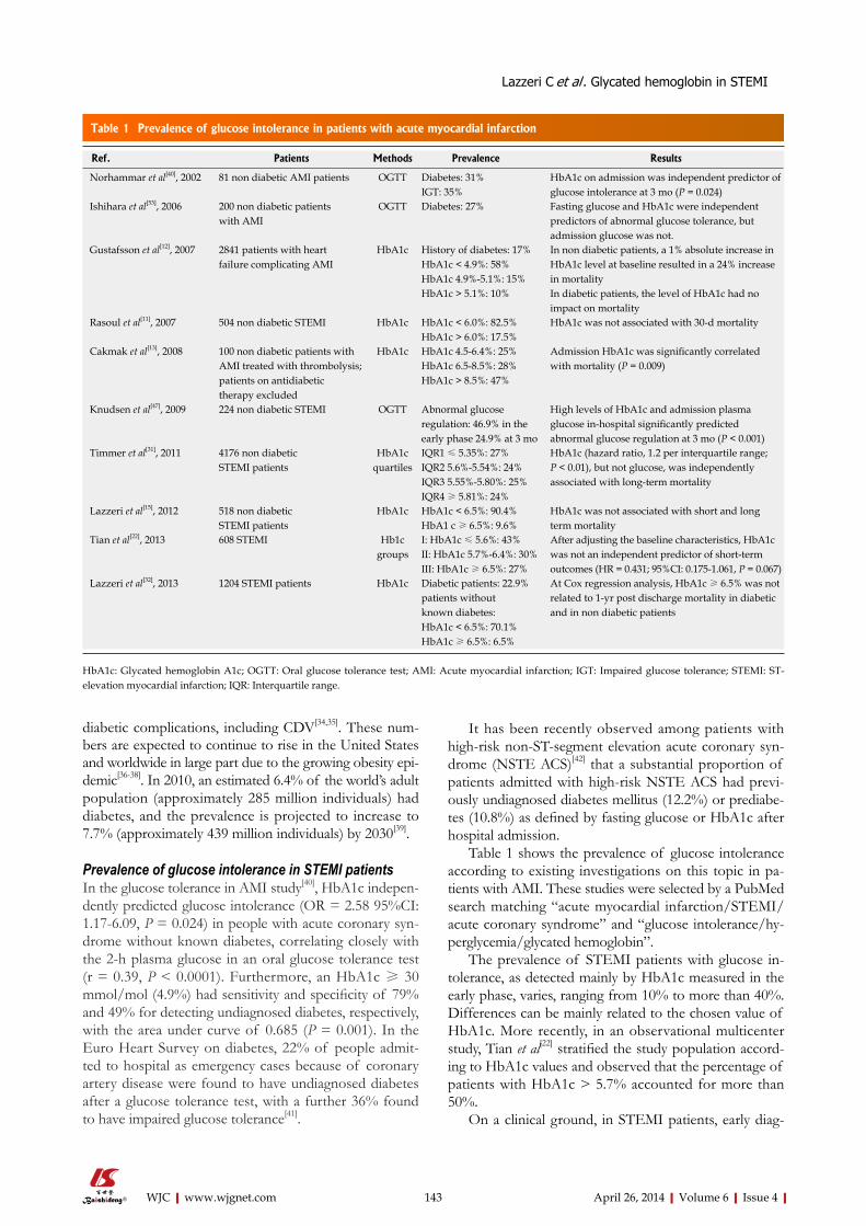
143 April 26, 2014|Volume 6|Issue 4|WJC|www.wjgnet.com
It has been recently observed among patients with high-risk non-ST-segment elevation acute coronary syn-drome (NSTE ACS)[42] that a substantial proportion of patients admitted with high-risk NSTE ACS had previ-ously undiagnosed diabetes mellitus (12.2%) or prediabe-tes (10.8%) as defined by fasting glucose or HbA1c after hospital admission.
Table 1 shows the prevalence of glucose intolerance according to existing investigations on this topic in pa-tients with AMI. These studies were selected by a PubMed search matching “acute myocardial infarction/STEMI/acute coronary syndrome” and “glucose intolerance/hy-perglycemia/glycated hemoglobin”.
The prevalence of STEMI patients with glucose in-tolerance, as detected mainly by HbA1c measured in the early phase, varies, ranging from 10% to more than 40%. Differences can be mainly related to the chosen value of HbA1c. More recently, in an observational multicenter study, Tian et al[22] stratified the study population accord-ing to HbA1c values and observed that the percentage of patients with HbA1c > 5.7% accounted for more than 50%.
On a clinical ground, in STEMI patients, early diag-
diabetic complications, including CDV[34,35]. These num-bers are expected to continue to rise in the United States and worldwide in large part due to the growing obesity epi-demic[36-38]. In 2010, an estimated 6.4% of the world’s adult population (approximately 285 million individuals) had diabetes, and the prevalence is projected to increase to 7.7% (approximately 439 million individuals) by 2030[39].
Prevalence of glucose intolerance in STEMI patientsIn the glucose tolerance in AMI study[40], HbA1c indepen-dently predicted glucose intolerance (OR = 2.58 95%CI: 1.17-6.09, P = 0.024) in people with acute coronary syn-drome without known diabetes, correlating closely with the 2-h plasma glucose in an oral glucose tolerance test (r = 0.39, P < 0.0001). Furthermore, an HbA1c ≥ 30 mmol/mol (4.9%) had sensitivity and specificity of 79% and 49% for detecting undiagnosed diabetes, respectively, with the area under curve of 0.685 (P = 0.001). In the Euro Heart Survey on diabetes, 22% of people admit-ted to hospital as emergency cases because of coronary artery disease were found to have undiagnosed diabetes after a glucose tolerance test, with a further 36% found to have impaired glucose tolerance[41].
Ref. Patients Methods Prevalence Results
Norhammar et al[40], 2002 81 non diabetic AMI patients OGTT Diabetes: 31%IGT: 35%
HbA1c on admission was independent predictor of glucose intolerance at 3 mo (P = 0.024)
Ishihara et al[53], 2006 200 non diabetic patients with AMI
OGTT Diabetes: 27% Fasting glucose and HbA1c were independent predictors of abnormal glucose tolerance, but admission glucose was not.
Gustafsson et al[12], 2007 2841 patients with heart failure complicating AMI
HbA1c History of diabetes: 17%HbA1c < 4.9%: 58%HbA1c 4.9%-5.1%: 15%HbA1c > 5.1%: 10%
In non diabetic patients, a 1% absolute increase in HbA1c level at baseline resulted in a 24% increase in mortalityIn diabetic patients, the level of HbA1c had no impact on mortality
Rasoul et al[11], 2007 504 non diabetic STEMI HbA1c HbA1c < 6.0%: 82.5%HbA1c > 6.0%: 17.5%
HbA1c was not associated with 30-d mortality
Cakmak et al[13], 2008 100 non diabetic patients withAMI treated with thrombolysis;patients on antidiabetic therapy excluded
HbA1c HbA1c 4.5-6.4%: 25%HbA1c 6.5-8.5%: 28%HbA1c > 8.5%: 47%
Admission HbA1c was significantly correlated with mortality (P = 0.009)
Knudsen et al[47], 2009 224 non diabetic STEMI OGTT Abnormal glucoseregulation: 46.9% in theearly phase 24.9% at 3 mo
High levels of HbA1c and admission plasma glucose in-hospital significantly predicted abnormal glucose regulation at 3 mo (P < 0.001)
Timmer et al[31], 2011 4176 non diabetic STEMI patients
HbA1c quartiles
IQR1 ≤ 5.35%: 27%IQR2 5.6%-5.54%: 24%IQR3 5.55%-5.80%: 25%IQR4 ≥ 5.81%: 24%
HbA1c (hazard ratio, 1.2 per interquartile range; P < 0.01), but not glucose, was independently associated with long-term mortality
Lazzeri et al[15], 2012 518 non diabetic STEMI patients
HbA1c HbA1c < 6.5%: 90.4%HbA1 c ≥ 6.5%: 9.6%
HbA1c was not associated with short and long term mortality
Tian et al[22], 2013 608 STEMI Hb1c groups
I: HbA1c ≤ 5.6%: 43%II: HbA1c 5.7%-6.4%: 30%III: HbA1c ≥ 6.5%: 27%
After adjusting the baseline characteristics, HbA1c was not an independent predictor of short-term outcomes (HR = 0.431; 95%CI: 0.175-1.061, P = 0.067)
Lazzeri et al[32], 2013 1204 STEMI patients HbA1c Diabetic patients: 22.9%patients without known diabetes:HbA1c < 6.5%: 70.1%HbA1c ≥ 6.5%: 6.5%
At Cox regression analysis, HbA1c ≥ 6.5% was not related to 1-yr post discharge mortality in diabetic and in non diabetic patients
Table 1 Prevalence of glucose intolerance in patients with acute myocardial infarction
HbA1c: Glycated hemoglobin A1c; OGTT: Oral glucose tolerance test; AMI: Acute myocardial infarction; IGT: Impaired glucose tolerance; STEMI: ST-elevation myocardial infarction; IQR: Interquartile range.
Lazzeri C et al . Glycated hemoglobin in STEMI
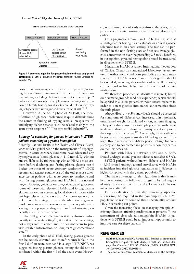
144 April 26, 2014|Volume 6|Issue 4|WJC|www.wjgnet.com
nosis of unknown type 2 diabetes or impaired glucose regulation allows initiation of treatment or lifestyle in-terventions, including diet and exercise to prevent type 2 diabetes and associated complications. Gaining informa-tion on family history for diabetes could help in identify-ing subjects with undiagnosed diabetes or at risk[43,44].
However, in the acute phase of STEMI, the iden-tification of glucose intolerance is quite difficult since the common finding of hyperglycemia, irrespective of underlying diabetic status, is to be related mainly to the acute stress response[16-21,26] to myocardial ischemia[45].
Strategy for screening for glucose intolerance in STEMI patients according to glycated hemoglobinRecently, National Institute for Health and Clinical Excel-lence (NICE) guidelines on the management of hypergly-caemia in acute coronary syndrome have advocated any hyperglycaemia (blood glucose > 11.0 mmol/L) without known diabetes be followed up with an HbA1c measure-ment before discharge and fasting plasma glucose test 4 d after the onset of acute coronary syndrome[46]. NICE recommend against routine use of the oral glucose toler-ance test in patients with acute coronary syndrome and with fasting plasma glucose and HbA1c in the normal range. However, guidance on categorization of glycaemic status of those with elevated HbA1c and fasting plasma glucose, as well as screening for diabetes in those with-out hyperglycaemia, is less clear. As a consequence, the lack of simple strategy for early identification of glucose intolerance in acute coronary syndrome is potentially leaving many people undiagnosed and under-treated, es-pecially after the cardiac event.
The oral glucose tolerance test is performed infre-quently in the acute setting[41], since it is time consuming, not always well tolerated and it does not seem to pro-vide reliable information on long-term glucometabolic state[47].
In the early phase of STEMI, fasting plasma glucose can be acutely elevated and therefore unreliable in the first 2 d of an acute event and in a large MI[48]. NICE has suggested fasting plasma glucose testing should not be conducted within the first 4 d of the acute event. Howev-
er, in the current era of early reperfusion therapies, many patients with acute coronary syndrome are discharged earlier.
On a pragmatic ground, an HbA1c test has several advantages over fasting plasma glucose or an oral glucose tolerance test in an acute setting. The test can be per-formed in the non-fasting state and reflects average glu-cose concentration over the preceding 2-3 mo. Therefore, in our opinion, glycated hemoglobin should be measured in all patients with STEMI.
Measuring HbA1c assumes International Federation of Clinical Chemistry standardized laboratory assays are used. Furthermore, conditions precluding accurate mea-surement of HbA1c concentration for diagnosis should be excluded, including abnormalities of red cell turnover, chronic renal or liver failure and chronic use of certain medications.
We therefore proposed an algorithm (Figure 1) based on pragmatic grounds (and our experience) which should be applied in STEMI patients without known diabetes in order to detect glucose intolerance abnormalities since the early phase.
Above HbA1c > 6.5%, individuals should be assessed for symptoms of diabetes (i.e., increased thirst, polyuria, unexplained weight loss, blurred vision, extreme fatigue), ruling out other causes, for example polyuria attributable to diuretic therapy. In those with unequivocal symptoms the diagnosis is confirmed[49]. Conversely, those with am-biguous or absent symptoms should undergo a confirma-tory HbA1c measurement 4-8 wk post-discharge for con-sistency and to counteract any potential laboratory errors on the first occasion.
Patients with HbA1c between 6.0% and < 6.4% should undergo an oral glucose tolerance test after 6-8 wk.
STEMI patients without known diabetes and HbA1c < 6.0% should undergo annual surveillance with HbA1c as incident impaired glucose regulation and diabetes is higher compared with the general population[50].
The main advantage of this algorithm is that it may help in tailoring the follow-up program, by helping to identify patients at risk for the development of glucose intolerance after MI.
Further validation of this algorithm in prospective studies may be required in the contemporary STEMI population to resolve some of these uncertainties around HbA1c screening cut points.
Given the increasing focus on managing multiple co-existing illnesses affecting cardiovascular patients[51], the assessment of glycosylated hemoglobin (HbA1c) in pa-tients with STEMI could be an important opportunity to improve care for these patients[52].
REFERENCES1 Rahbar S, Blumenfeld O, Ranney HM. Studies of an unusual
hemoglobin in patients with diabetes mellitus. Biochem Bio-phys Res Commun 1969; 36: 838-843 [PMID: 5808299 DOI: 10.1016/0006-291X(69)90685-8]
2 The effect of intensive treatment of diabetes on the develop-
STEMI patients without previously known diabetes
Annual monitoringwith HbA1c
HbA1c > 6.5% HbA1c 6.0%-6.4% HbA1c > 6.0%
Symptoms absentRepeat HbA1c after 4-8 wk
Symptoms presentDiabetes
Oral glucose Tolerance testafter 6-8 wk
Figure 1 A screening algorithm for glucose intolerance based on glycated hemoglobin. STEMI: ST-elevation myocardial infarction; HbA1c: Glycated he-moglobin A1c.
Lazzeri C et al . Glycated hemoglobin in STEMI
145 April 26, 2014|Volume 6|Issue 4|WJC|www.wjgnet.com
ment and progression of long-term complications in insulin-dependent diabetes mellitus. The Diabetes Control and Complications Trial Research Group. N Engl J Med 1993; 329: 977-986 [PMID: 8366922 DOI: 10.1056/NEJM199309303291401]
3 Kishore P, Kim SH, Crandall JP. Glycemic control and car-diovascular disease: what’s a doctor to do? Curr Diab Rep 2012; 12: 255-264 [PMID: 22467273 DOI: 10.1007/s11892-012-0268-5]
4 Gall MA, Borch-Johnsen K, Hougaard P, Nielsen FS, Parv-ing HH. Albuminuria and poor glycemic control predict mortality in NIDDM. Diabetes 1995; 44: 1303-1309 [PMID: 7589828 DOI: 10.2337/diab.44.11.1303]
5 Agewall S, Wikstrand J, Ljungman S, Fagerberg B. Useful-ness of microalbuminuria in predicting cardiovascular mor-tality in treated hypertensive men with and without diabetes mellitus. Risk Factor Intervention Study Group. Am J Cardiol 1997; 80: 164-169 [PMID: 9230153 DOI: 10.1016/S0002-9149(97)00312-3]
6 Goff DC, Gerstein HC, Ginsberg HN, Cushman WC, Mar-golis KL, Byington RP, Buse JB, Genuth S, Probstfield JL, Simons-Morton DG. Prevention of cardiovascular disease in persons with type 2 diabetes mellitus: current knowledge and rationale for the Action to Control Cardiovascular Risk in Diabetes (ACCORD) trial. Am J Cardiol 2007; 99: 4i-20i [PMID: 17599424 DOI: 10.1016/j.amjcard.2007.03.002]
7 Khaw KT, Wareham N, Luben R, Bingham S, Oakes S, Welch A, Day N. Glycated haemoglobin, diabetes, and mor-tality in men in Norfolk cohort of european prospective in-vestigation of cancer and nutrition (EPIC-Norfolk). BMJ 2001; 322: 15-18 [PMID: 11141143 DOI: 10.1136/bmj.322.7277.15]
8 Sarwar N, Aspelund T, Eiriksdottir G, Gobin R, Seshasai SR, Forouhi NG, Sigurdsson G, Danesh J, Gudnason V. Markers of dysglycaemia and risk of coronary heart disease in people without diabetes: Reykjavik prospective study and system-atic review. PLoS Med 2010; 7: e1000278 [PMID: 20520805 DOI: 10.1371/journal.pmed.1000278]
9 Selvin E, Steffes MW, Zhu H, Matsushita K, Wagenknecht L, Pankow J, Coresh J, Brancati FL. Glycated hemoglobin, dia-betes, and cardiovascular risk in nondiabetic adults. N Engl J Med 2010; 362: 800-811 [PMID: 20200384 DOI: 10.1056/NEJ-Moa0908359]
10 Oswald GA, Corcoran S, Yudkin JS. Prevalence and risks of hyperglycaemia and undiagnosed diabetes in patients with acute myocardial infarction. Lancet 1984; 1: 1264-1267 [PMID: 6144976 DOI: 10.1016/S0140-6736(84)92447-4]
11 Rasoul S, Ottervanger JP, Bilo HJ, Timmer JR, van ‘t Hof AW, Dambrink JH, Dikkeschei LD, Hoorntje JC, de Boer MJ, Zijlstra F. Glucose dysregulation in nondiabetic patients with ST-elevation myocardial infarction: acute and chronic glucose dysregulation in STEMI. Neth J Med 2007; 65: 95-100 [PMID: 17387235]
12 Gustafsson I, Kistorp CN, James MK, Faber JO, Dickstein K, Hildebrandt PR. Unrecognized glycometabolic disturbance as measured by hemoglobin A1c is associated with a poor outcome after acute myocardial infarction. Am Heart J 2007; 154: 470-476 [PMID: 17719292 DOI: 10.1016/j.ahj.2007.04.057]
13 Cakmak M, Cakmak N, Cetemen S, Tanriverdi H, Enc Y, Teskin O, Kilic ID. The value of admission glycosylated hemoglobin level in patients with acute myocardial infarc-tion. Can J Cardiol 2008; 24: 375-378 [PMID: 18464942 DOI: 10.1016/S0828-282X(08)70600-7]
14 Cicek G, Uyarel H, Ergelen M, Ayhan E, Abanonu GB, Eren M, Gibson CM. Hemoglobin A1c as a prognostic marker in patients undergoing primary angioplasty for acute myo-cardial infarction. Coron Artery Dis 2011; 22: 131-137 [PMID: 21394027 DOI: 10.1097/MCA.0b013e328342c760]
15 Lazzeri C, Valente S, Chiostri M, Picariello C, Attanà P, Gen-sini GF. Glycated hemoglobin in ST-elevation myocardial infarction without previously known diabetes: its short and
long term prognostic role. Diabetes Res Clin Pract 2012; 95: e14-e16 [PMID: 22056718 DOI: 10.1016/j.diabres.2011.09.028]
16 Deedwania P, Kosiborod M, Barrett E, Ceriello A, Isley W, Mazzone T, Raskin P. Hyperglycemia and acute coronary syndrome: a scientific statement from the American Heart Association Diabetes Committee of the Council on Nutri-tion, Physical Activity, and Metabolism. Circulation 2008; 117: 1610-1619 [PMID: 18299505 DOI: 10.1161/CIRCULA-TIONAHA.107.188629]
17 Lazzeri C, Chiostri M, Sori A, Valente S, Gensini GF. Post-procedural hyperglycemia in ST elevation myocardial in-farction submitted to percutaneous coronary intervention: a prognostic indicator and a marker of metabolic derange-ment. J Cardiovasc Med (Hagerstown) 2010; 11: 7-13 [PMID: 19829142 DOI: 10.2459/JCM.0b013e32832d83b3]
18 Lazzeri C, Sori A, Chiostri M, Gensini GF, Valente S. Prog-nostic role of insulin resistance as assessed by homeostatic model assessment index in the acute phase of myocardial infarction in nondiabetic patients submitted to percutaneous coronary intervention. Eur J Anaesthesiol 2009; 26: 856-862 [PMID: 19367169 DOI: 10.1097/EJA.0b013e32832a235c]
19 Lazzeri C, Valente S, Chiostri M, Picariello C, Gensini GF. Correlates of acute insulin resistance in the early phase of non-diabetic ST-elevation myocardial infarction. Diab Vasc Dis Res 2011; 8: 35-42 [PMID: 21262869]
20 Lazzeri C, Valente S, Chiostri M, Picariello C, Gensini GF. Acute glucose dysmetabolism in the early phase of ST-elevation myocardial infarction: the age response. Diab Vasc Dis Res 2010; 7: 131-137 [PMID: 20382776 DOI: 10.1177/1479164109353369]
21 Lazzeri C, Valente S, Chiostri M, Picariello C, Gensini GF. In-hospital peak glycemia and prognosis in STEMI pa-tients without earlier known diabetes. Eur J Cardiovasc Prev Rehabil 2010; 17: 419-423 [PMID: 20517158 DOI: 10.1097/HJR.0b013e328335f26f]
22 Tian L, Zhu J, Liu L, Liang Y, Li J, Yang Y. Hemoglobin A1c and short-term outcomes in patients with acute myocardial infarction undergoing primary angioplasty: an observational multicenter study. Coron Artery Dis 2013; 24: 16-22 [PMID: 23168569 DOI: 10.1097/MCA.0b013e32835b3971]
23 Malmberg K, Rydén L, Wedel H, Birkeland K, Bootsma A, Dickstein K, Efendic S, Fisher M, Hamsten A, Herlitz J, Hildebrandt P, MacLeod K, Laakso M, Torp-Pedersen C, Waldenström A. Intense metabolic control by means of insu-lin in patients with diabetes mellitus and acute myocardial infarction (DIGAMI 2): effects on mortality and morbidity. Eur Heart J 2005; 26: 650-661 [PMID: 15728645 DOI: 10.1093/eurheartj/ehi199]
24 Malmberg K, Norhammar A, Wedel H, Rydén L. Glycomet-abolic state at admission: important risk marker of mortality in conventionally treated patients with diabetes mellitus and acute myocardial infarction: long-term results from the Diabetes and Insulin-Glucose Infusion in Acute Myocardial Infarction (DIGAMI) study. Circulation 1999; 99: 2626-2632 [PMID: 10338454 DOI: 10.1161/01.CIR.99.20.2626]
25 Lemesle G, Bonello L, de Labriolle A, Maluenda G, Syed AI, Collins SD, Ben-Dor I, Torguson R, Kaneshige K, Xue Z, Sud-dath WO, Satler LF, Kent KM, Lindsay J, Pichard AD, Waks-man R. Prognostic value of hemoglobin A1C levels in patients with diabetes mellitus undergoing percutaneous coronary intervention with stent implantation. Am J Cardiol 2009; 104: 41-45 [PMID: 19576319 DOI: 10.1016/j.amjcard.2009.02.060]
26 Lazzeri C, Valente S, Chiostri M, Picariello C, Attanà P, Gensini GF. The prognostic impact of glycated hemoglo-bin in diabetic ST-elevation myocardial infarction. Int J Cardiol 2011; 151: 250-252 [PMID: 21723626 DOI: 10.1016/j.ijcard.2011.06.077]
27 Lazzeri C, Valente S, Chiostri M, Picariello C, Gensini GF. Predictors of the early outcome in elderly patients with ST
Lazzeri C et al . Glycated hemoglobin in STEMI
146 April 26, 2014|Volume 6|Issue 4|WJC|www.wjgnet.com
elevation myocardial infarction treated with primary angio-plasty: a single center experience. Intern Emerg Med 2011; 6: 41-46 [PMID: 20853070]
28 Bartnik M, Malmberg K, Norhammar A, Tenerz A, Ohrvik J, Rydén L. Newly detected abnormal glucose tolerance: an important predictor of long-term outcome after myocardial infarction. Eur Heart J 2004; 25: 1990-1997 [PMID: 15541834 DOI: 10.1016/j.ehj.2004.09.021]
29 Kostis WJ, Deng Y, Pantazopoulos JS, Moreyra AE, Kostis JB. Trends in mortality of acute myocardial infarction after discharge from the hospital. Circ Cardiovasc Qual Outcomes 2010; 3: 581-589 [PMID: 20923995 DOI: 10.1161/CIRCOUT-COMES.110.957803]
30 Chan CY, Li R, Chan JY, Zhang Q, Chan CP, Dong M, Yan BP, Lam YY, Yu CM. The value of admission HbA(1c) level in diabetic patients with acute coronary syndrome. Clin Cardiol 2011; 34: 507-512 [PMID: 21717470 DOI: 10.1002/clc.20915]
31 Timmer JR, Hoekstra M, Nijsten MW, van der Horst IC, Ottervanger JP, Slingerland RJ, Dambrink JH, Bilo HJ, Zi-jlstra F, van ‘t Hof AW. Prognostic value of admission gly-cosylated hemoglobin and glucose in nondiabetic patients with ST-segment-elevation myocardial infarction treated with percutaneous coronary intervention. Circulation 2011; 124: 704-711 [PMID: 21768543 DOI: 10.1161/CIRCULA-TIONAHA.110.985911]
32 Lazzeri C, Valente S, Chiostri M, Attanà P, Mattesini A, Nesti M, Gensini GF. Glycated hemoglobin and long term mortality in STEMI patients. JCM; 2013 (in press)
33 Roger VL, Go AS, Lloyd-Jones DM, Adams RJ, Berry JD, Brown TM, Carnethon MR, Dai S, de Simone G, Ford ES, Fox CS, Fullerton HJ, Gillespie C, Greenlund KJ, Hailpern SM, Heit JA, Ho PM, Howard VJ, Kissela BM, Kittner SJ, Lack-land DT, Lichtman JH, Lisabeth LD, Makuc DM, Marcus GM, Marelli A, Matchar DB, McDermott MM, Meigs JB, Moy CS, Mozaffarian D, Mussolino ME, Nichol G, Paynter NP, Rosamond WD, Sorlie PD, Stafford RS, Turan TN, Turner MB, Wong ND, Wylie-Rosett J. Heart disease and stroke statistics--2011 update: a report from the American Heart Association. Circulation 2011; 123: e18-e209 [PMID: 21160056 DOI: 10.1161/CIR.0b013e3182009701]
34 Cowie CC, Rust KF, Ford ES, Eberhardt MS, Byrd-Holt DD, Li C, Williams DE, Gregg EW, Bainbridge KE, Saydah SH, Geiss LS. Full accounting of diabetes and pre-diabetes in the U.S. population in 1988-1994 and 2005-2006. Diabetes Care 2009; 32: 287-294 [PMID: 19017771 DOI: 10.2337/dc08-1296]
35 Olshansky SJ, Passaro DJ, Hershow RC, Layden J, Carnes BA, Brody J, Hayflick L, Butler RN, Allison DB, Ludwig DS. A potential decline in life expectancy in the United States in the 21st century. N Engl J Med 2005; 352: 1138-1145 [PMID: 15784668 DOI: 10.1056/NEJMsr043743]
36 Wild S, Roglic G, Green A, Sicree R, King H. Global preva-lence of diabetes: estimates for the year 2000 and projections for 2030. Diabetes Care 2004; 27: 1047-1053 [PMID: 15111519 DOI: 10.2337/diacare.27.5.1047]
37 Danaei G, Finucane MM, Lu Y, Singh GM, Cowan MJ, Pa-ciorek CJ, Lin JK, Farzadfar F, Khang YH, Stevens GA, Rao M, Ali MK, Riley LM, Robinson CA, Ezzati M. National, regional, and global trends in fasting plasma glucose and diabetes prevalence since 1980: systematic analysis of health examination surveys and epidemiological studies with 370 country-years and 2·7 million participants. Lancet 2011; 378: 31-40 [PMID: 21705069 DOI: 10.1016/S0140-6736(11)60679-X]
38 King H, Aubert RE, Herman WH. Global burden of diabe-tes, 1995-2025: prevalence, numerical estimates, and projec-tions. Diabetes Care 1998; 21: 1414-1431 [PMID: 9727886 DOI: 10.2337/diacare.21.9.1414]
39 Shaw JE, Sicree RA, Zimmet PZ. Global estimates of the prevalence of diabetes for 2010 and 2030. Diabetes Res Clin Pract 2010; 87: 4-14 [PMID: 19896746 DOI: 10.1016/j.dia-
bres.2009.10.007]40 Norhammar A, Tenerz A, Nilsson G, Hamsten A, Efendíc
S, Rydén L, Malmberg K. Glucose metabolism in patients with acute myocardial infarction and no previous diagno-sis of diabetes mellitus: a prospective study. Lancet 2002; 359: 2140-2144 [PMID: 12090978 DOI: 10.1016/S0140-6736(02)09089-X]
41 Bartnik M, Rydén L, Ferrari R, Malmberg K, Pyörälä K, Si-moons M, Standl E, Soler-Soler J, Ohrvik J. The prevalence of abnormal glucose regulation in patients with coronary artery disease across Europe. The Euro Heart Survey on dia-betes and the heart. Eur Heart J 2004; 25: 1880-1890 [PMID: 15522466 DOI: 10.1016/j.ehj.2004.07.027]
42 Giraldez RR, Clare RM, Lopes RD, Dalby AJ, Prabhakaran D, Brogan GX, Giugliano RP, James SK, Tanguay JF, Pollack CV, Harrington RA, Braunwald E, Newby LK. Prevalence and clinical outcomes of undiagnosed diabetes mellitus and prediabetes among patients with high-risk non-ST-segment elevation acute coronary syndrome. Am Heart J 2013; 165: 918-925.e2 [PMID: 23708162]
43 Pannacciulli N, De Pergola G, Ciccone M, Rizzon P, Giorgi-no F, Giorgino R. Effect of family history of type 2 diabetes on the intima-media thickness of the common carotid artery in normal-weight, overweight, and obese glucose-tolerant young adults. Diabetes Care 2003; 26: 1230-1234 [PMID: 12663602 DOI: 10.2337/diacare.26.4.1230]
44 De Pergola G, Ciccone M, Pannacciulli N, Modugno M, Sciaraffia M, Minenna A, Rizzon P, Giorgino R. Lower in-sulin sensitivity as an independent risk factor for carotid wall thickening in normotensive, non-diabetic, non-smoking normal weight and obese premenopausal women. Int J Obes Relat Metab Disord 2000; 24: 825-829 [PMID: 10918528 DOI: 10.1038/sj.ijo.0801239]
45 Gholap N, Davies MJ, Mostafa SA, Squire I, Khunti K. A simple strategy for screening for glucose intolerance, using glycated haemoglobin, in individuals admitted with acute coronary syndrome. Diabet Med 2012; 29: 838-843 [PMID: 22417234 DOI: 10.1111/j.1464-5491.2012.03643.x]
46 Senthinathan A, Kelly V, Dzingina M, Jones D, Baker M, Longson D. Hyperglycaemia in acute coronary syndromes: summary of NICE guidance. BMJ 2011; 343: d6646 [PMID: 22031910 DOI: 10.1136/bmj.d6646]
47 Knudsen EC, Seljeflot I, Abdelnoor M, Eritsland J, Mang-schau A, Arnesen H, Andersen GO. Abnormal glucose regulation in patients with acute ST- elevation myocardial infarction-a cohort study on 224 patients. Cardiovasc Diabetol 2009; 8: 6 [PMID: 19183453 DOI: 10.1186/1475-2840-8-6]
48 Hage C, Malmberg K, Rydén L, Wallander M. The impact of infarct type on the reliability of early oral glucose toler-ance testing in patients with myocardial infarction. Int J Cardiol 2010; 145: 259-260 [PMID: 19850366 DOI: 10.1016/j.ijcard.2009.09.469]
49 World Health Organization. Use of Glycated Haemoglobin (HbA1c) in the Diagnosis of Diabetes Mellitus. Abbreviated Report of a WHO Consultation. Geneva: World Health Or-ganization, 2011: 1-25
50 Mozaffarian D, Marfisi R, Levantesi G, Silletta MG, Tavazzi L, Tognoni G, Valagussa F, Marchioli R. Incidence of new-onset diabetes and impaired fasting glucose in patients with recent myocardial infarction and the effect of clinical and lifestyle risk factors. Lancet 2007; 370: 667-675 [PMID: 17720018 DOI: 10.1016/S0140-6736(07)61343-9]
51 Werner RM, Greenfield S, Fung C, Turner BJ. Measuring quality of care in patients with multiple clinical conditions: summary of a conference conducted by the Society of Gen-eral Internal Medicine. J Gen Intern Med 2007; 22: 1206-1211 [PMID: 17516106 DOI: 10.1007/s11606-007-0230-4]
52 Stolker JM, Sun D, Conaway DG, Jones PG, Masoudi FA, Peterson PN, Krumholz HM, Kosiborod M, Spertus JA. Im-
Lazzeri C et al . Glycated hemoglobin in STEMI
147 April 26, 2014|Volume 6|Issue 4|WJC|www.wjgnet.com
portance of measuring glycosylated hemoglobin in patients with myocardial infarction and known diabetes mellitus. Am J Cardiol 2010; 105: 1090-1094 [PMID: 20381658 DOI: 10.1016/j.amjcard.2009.12.010]
53 Ishihara M, Inoue I, Kawagoe T, Shimatani Y, Kurisu S,
Hata T, Nakama Y, Kijima Y, Kagawa E. Is admission hyper-glycaemia in non-diabetic patients with acute myocardial infarction a surrogate for previously undiagnosed abnormal glucose tolerance? Eur Heart J 2006; 27: 2413-2419 [PMID: 17000629]
P- Reviewers: Ciccone MM, Hwang KC, Liu PY, Loffredo L, Sethi A, Wan Y
S- Editor: Wen LL L- Editor: O’Neill M E- Editor: Liu SQ
Lazzeri C et al . Glycated hemoglobin in STEMI
Seung-Woon Rha
Seung-Woon Rha, Cardiovascular Center, Korea University Guro Hospital, Seoul 152-703, South KoreaAuthor contributions: Rha SW solely contributed to this paper.Correspondence to: Seung-Woon Rha, MD, PhD, FACC, FAHA, FESC, FSCAI, Cardiovascular Center, Korea University Guro Hospital, 80, Guro-dong, Guro-gu, Seoul 152-703, South Korea. [email protected]: +82-2-26263020 Fax: +82-2-8643062Received: October 10, 2013 Revised: December 15, 2013Accepted: January 6, 2014Published online: April 26, 2014
AbstractCurrent percutaneous coronary intervention guidelines recommend dual antiplatelets (aspirin 100 mg + clopi-dogrel 75 mg daily) for at least 12 mo following drug-eluting stent (DES) implantation if patients are not at high risk of bleeding. Several reports have tried to shorten the dual antiplatelet therapy to 3-6 mo, espe-cially following next-generation DES implantation, for cost-effectiveness. However, the clinical results are in-consistent and the data regarding next-generation DESs limited. In this report, recently published important piv-otal reports regarding the optimal duration of dual anti-platelets following DES implantation are summarized.
© 2014 Baishideng Publishing Group Co., Limited. All rights reserved.
Key words: Drug-eluting stent; Dual antiplatelet treat-ment; Percutaneous coronary intervention
Core tip: Recently published important pivotal reports regarding the optimal duration of dual antiplatelets fol-lowing drug-eluting stent implantation are summarized.
Rha SW. Duration of dual antiplatelet treatment in the era of next generation drug-eluting stents. World J Cardiol 2014; 6(4): 148-153 Available from: URL: http://www.wjgnet.com/1949-8462/full/v6/
i4/148.htm DOI: http://dx.doi.org/10.4330/wjc.v6.i4.148
INTRODUCTIONMultiple randomized clinical trials have shown the effi-cacy of drug-eluting stents (DES) in reducing restenosis and the need for target lesion revascularization (TLR) compared with bare-metal stents (BMS)[1,2]. Despite the reduced incidence of recurrence, safety issues related to DESs, such as stent thrombosis, late stent malapposition, aneurysm, stent fracture, endothelial dysfunction and re-stenosis, have been reported elsewhere, particularly with first-generation DESs. Furthermore, some observational studies have shown that the risk of death or myocardial infarction was even higher with DESs than BMSs, pos-sibly due to a higher incidence of late or very late stent thrombosis[3].
Early or premature discontinuation of dual antiplate-let therapy has been reported as an important risk factor for late stent thrombosis following DES implantation[4,5]. Thus, current percutaneous coronary intervention (PCI) guidelines recommend dual antiplatelets (aspirin + clopi-dogrel 75 mg daily) for at least 12 mo following DES implantation if patients are not at high risk of bleeding[6]. Several reports have tried to address this issue but the results are inconsistent and the data regarding second-generation DESs limited. In this report, the important pivotal reports regarding the optimal duration of dual antiplatelets following DES implantation, particularly in patients who underwent PCI with next generation DESs, are summarized.
OPTIMAL DURATION OF DUAL ANTIPLATELET THERAPY WITH DESsMajor clinical trials for duration of dual antiplatelets after DES implantationREAL-LATE and ZEST-LATE trial: (Aspirin +
TOPIC HIGHLIGHT
Online Submissions: http://www.wjgnet.com/esps/[email protected]:10.4330/wjc.v6.i4.148
148 April 26, 2014|Volume 6|Issue 4|WJC|www.wjgnet.com
World Journal of CardiologyW J C
World J Cardiol 2014 April 26; 6(4): 148-153ISSN 1949-8462 (online)
© 2014 Baishideng Publishing Group Co., Limited. All rights reserved.
Duration of dual antiplatelet treatment in the era of next generation drug-eluting stents
WJC 6th Anniversary Special Issues (5): Myocardial infarction
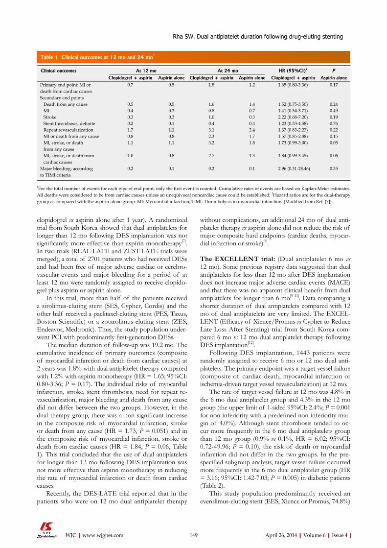
clopidogrel vs aspirin alone after 1 year). A randomized trial from South Korea showed that dual antiplatelets for longer than 12 mo following DES implantation was not significantly more effective than aspirin monotherapy[7]. In two trials (REAL-LATE and ZEST-LATE trials were merged), a total of 2701 patients who had received DESs and had been free of major adverse cardiac or cerebro-vascular events and major bleeding for a period of at least 12 mo were randomly assigned to receive clopido-grel plus aspirin or aspirin alone.
In this trial, more than half of the patients received a sirolimus-eluting stent (SES, Cypher, Cordis) and the other half received a paclitaxel-eluting stent (PES, Taxus, Boston Scientific) or a zotarolimus-eluting stent (ZES, Endeavor, Medtronic). Thus, the study population under-went PCI with predominantly first-generation DESs.
The median duration of follow-up was 19.2 mo. The cumulative incidence of primary outcomes (composite of myocardial infarction or death from cardiac causes) at 2 years was 1.8% with dual antiplatelet therapy compared with 1.2% with aspirin monotherapy (HR = 1.65; 95%CI: 0.80-3.36; P = 0.17). The individual risks of myocardial infarction, stroke, stent thrombosis, need for repeat re-vascularization, major bleeding and death from any cause did not differ between the two groups. However, in the dual therapy group, there was a non-significant increase in the composite risk of myocardial infarction, stroke or death from any cause (HR = 1.73, P = 0.051) and in the composite risk of myocardial infarction, stroke or death from cardiac causes (HR = 1.84, P = 0.06, Table 1). This trial concluded that the use of dual antiplatelets for longer than 12 mo following DES implantation was not more effective than aspirin monotherapy in reducing the rate of myocardial infarction or death from cardiac causes.
Recently, the DES-LATE trial reported that in the patients who were on 12 mo dual antiplatelet therapy
without complications, an additional 24 mo of dual anti-platelet therapy vs aspirin alone did not reduce the risk of major composite hard endpoints (cardiac deaths, myocar-dial infarction or stroke)[8].
The EXCELLENT trial: (Dual antiplatelet 6 mo vs 12 mo). Some previous registry data suggested that dual antiplatelets for less than 12 mo after DES implantation does not increase major adverse cardiac events (MACE) and that there was no apparent clinical benefit from dual antiplatelets for longer than 6 mo[9-11]. Data comparing a shorter duration of dual antiplatelets compared with 12 mo of dual antiplatelets are very limited. The EXCEL-LENT (Efficacy of Xience/Promus vs Cypher to Reduce Late Loss After Stenting) trial from South Korea com-pared 6 mo vs 12 mo dual antiplatelet therapy following DES implantation[12].
Following DES implantation, 1443 patients were randomly assigned to receive 6 mo or 12 mo dual anti-platelets. The primary endpoint was a target vessel failure (composite of cardiac death, myocardial infarction or ischemia-driven target vessel revascularization) at 12 mo.
The rate of target vessel failure at 12 mo was 4.8% in the 6 mo dual antiplatelet group and 4.3% in the 12 mo group (the upper limit of 1-sided 95%CI: 2.4%; P = 0.001 for non-inferiority with a predefined non-inferiority mar-gin of 4.0%). Although stent thrombosis tended to oc-cur more frequently in the 6 mo dual antiplatelets group than 12 mo group (0.9% vs 0.1%, HR = 6.02; 95%CI: 0.72-49.96; P = 0.10), the risk of death or myocardial infarction did not differ in the two groups. In the pre-specified subgroup analysis, target vessel failure occurred more frequently in the 6 mo dual antiplatelet group (HR = 3.16; 95%CI: 1.42-7.03; P = 0.005) in diabetic patients (Table 2).
This study population predominantly received an everolimus-eluting stent (EES, Xience or Promus, 74.8%)
149 April 26, 2014|Volume 6|Issue 4|WJC|www.wjgnet.com
Rha SW. Dual antiplatelet duration following drug-eluting stenting
Clinical outcomes At 12 mo At 24 mo HR (95%CI)2 PClopidogrel + aspirin Aspirin alone Clopidogrel + aspirin Aspirin alone Clopidogrel + aspirin Aspirin alone
Primary end point: MI or death from cardiac causes
0.7 0.5 1.8 1.2 1.65 (0.80-3.36) 0.17
Secondary end points Death from any cause 0.5 0.5 1.6 1.4 1.52 (0.75-3.50) 0.24 MI 0.4 0.3 0.8 0.7 1.41 (0.54-3.71) 0.49 Stroke 0.3 0.3 1.0 0.3 2.22 (0.68-7.20) 0.19 Stent thrombosis, definite 0.2 0.1 0.4 0.4 1.23 (0.33-4.58) 0.76 Repeat revascularization 1.7 1.1 3.1 2.4 1.37 (0.83-2.27) 0.22 MI or death from any cause 0.8 0.8 2.3 1.7 1.57 (0.85-2.88) 0.15 MI, stroke, or death from any cause
1.1 1.1 3.2 1.8 1.73 (0.99-3.00) 0.05
MI, stroke, or death from cardiac causes
1.0 0.8 2.7 1.3 1.84 (0.99-3.45) 0.06
Major bleeding, according to TIMI criteria
0.2 0.1 0.2 0.1 2.96 (0.31-28.46) 0.35
Table 1 Clinical outcomes at 12 mo and 24 mo1
1For the total number of events for each type of end point, only the first event is counted. Cumulative rates of events are based on Kaplan-Meier estimates. All deaths were considered to be from cardiac causes unless an unequivocal noncardiac cause could be established; 2Hazard ratios are for the dual-therapy group as compared with the aspirin-alone group. MI: Myocardial infarction; TIMI: Thrombolysis in myocardial infarction. (Modified from Ref. [7]).
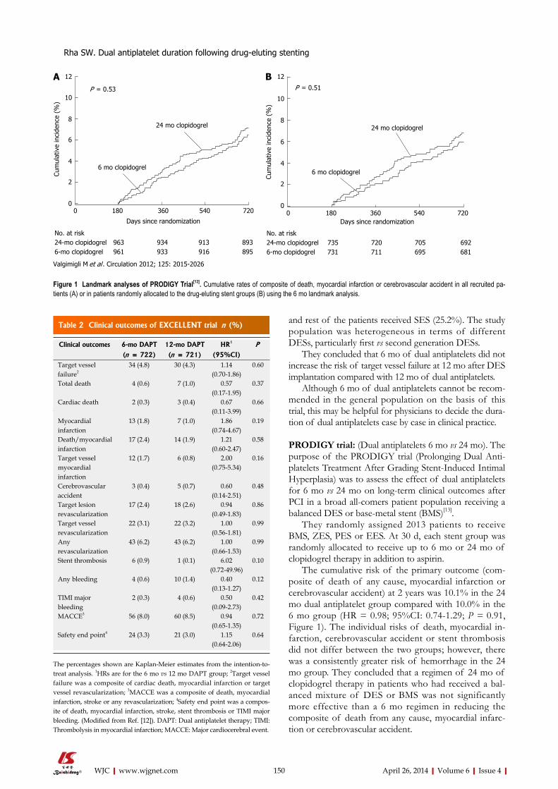
and rest of the patients received SES (25.2%). The study population was heterogeneous in terms of different DESs, particularly first vs second generation DESs.
They concluded that 6 mo of dual antiplatelets did not increase the risk of target vessel failure at 12 mo after DES implantation compared with 12 mo of dual antiplatelets.
Although 6 mo of dual antiplatelets cannot be recom-mended in the general population on the basis of this trial, this may be helpful for physicians to decide the dura-tion of dual antiplatelets case by case in clinical practice.
PRODIGY trial: (Dual antiplatelets 6 mo vs 24 mo). The purpose of the PRODIGY trial (Prolonging Dual Anti-platelets Treatment After Grading Stent-Induced Intimal Hyperplasia) was to assess the effect of dual antiplatelets for 6 mo vs 24 mo on long-term clinical outcomes after PCI in a broad all-comers patient population receiving a balanced DES or base-metal stent (BMS)[13].
They randomly assigned 2013 patients to receive BMS, ZES, PES or EES. At 30 d, each stent group was randomly allocated to receive up to 6 mo or 24 mo of clopidogrel therapy in addition to aspirin.
The cumulative risk of the primary outcome (com-posite of death of any cause, myocardial infarction or cerebrovascular accident) at 2 years was 10.1% in the 24 mo dual antiplatelet group compared with 10.0% in the 6 mo group (HR = 0.98; 95%CI: 0.74-1.29; P = 0.91, Figure 1). The individual risks of death, myocardial in-farction, cerebrovascular accident or stent thrombosis did not differ between the two groups; however, there was a consistently greater risk of hemorrhage in the 24 mo group. They concluded that a regimen of 24 mo of clopidogrel therapy in patients who had received a bal-anced mixture of DES or BMS was not significantly more effective than a 6 mo regimen in reducing the composite of death from any cause, myocardial infarc-tion or cerebrovascular accident.
150 April 26, 2014|Volume 6|Issue 4|WJC|www.wjgnet.com
Clinical outcomes 6-mo DAPT 12-mo DAPT HR1 P(n = 722) (n = 721) (95%CI)
Target vessel failure2
34 (4.8) 30 (4.3) 1.14 (0.70-1.86)
0.60
Total death 4 (0.6) 7 (1.0) 0.57 (0.17-1.95)
0.37
Cardiac death 2 (0.3) 3 (0.4) 0.67 (0.11-3.99)
0.66
Myocardial infarction
13 (1.8) 7 (1.0) 1.86 (0.74-4.67)
0.19
Death/myocardial infarction
17 (2.4) 14 (1.9) 1.21 (0.60-2.47)
0.58
Target vessel myocardial infarction
12 (1.7) 6 (0.8) 2.00 (0.75-5.34)
0.16
Cerebrovascular accident
3 (0.4) 5 (0.7) 0.60 (0.14-2.51)
0.48
Target lesion revascularization
17 (2.4) 18 (2.6) 0.94 (0.49-1.83)
0.86
Target vessel revascularization
22 (3.1) 22 (3.2) 1.00 (0.56-1.81)
0.99
Any revascularization
43 (6.2) 43 (6.2) 1.00 (0.66-1.53)
0.99
Stent thrombosis 6 (0.9) 1 (0.1) 6.02 (0.72-49.96)
0.10
Any bleeding 4 (0.6) 10 (1.4) 0.40 (0.13-1.27)
0.12
TIMI major bleeding
2 (0.3) 4 (0.6) 0.50(0.09-2.73)
0.42
MACCE3 56 (8.0) 60 (8.5) 0.94(0.65-1.35)
0.72
Safety end point4 24 (3.3) 21 (3.0) 1.15(0.64-2.06)
0.64
Table 2 Clinical outcomes of EXCELLENT trial n (%)
The percentages shown are Kaplan-Meier estimates from the intention-to-treat analysis. 1HRs are for the 6 mo vs 12 mo DAPT group; 2Target vessel failure was a composite of cardiac death, myocardial infarction or target vessel revascularization; 3MACCE was a composite of death, myocardial infarction, stroke or any revascularization; 4Safety end point was a compos-ite of death, myocardial infarction, stroke, stent thrombosis or TIMI major bleeding. (Modified from Ref. [12]). DAPT: Dual antiplatelet therapy; TIMI: Thrombolysis in myocardial infarction; MACCE: Major cardiocerebral event.
P = 0.53
24 mo clopidogrel
6 mo clopidogrel
Cum
ulat
ive
inci
denc
e (%
)
Days since randomization
0 180 360 540 720
12
10
8
6
4
2
0
No. at risk24-mo clopidogrel 963 934 913 8936-mo clopidogrel 961 933 916 895
Days since randomization
Cum
ulat
ive
inci
denc
e (%
)
P = 0.51
24 mo clopidogrel
6 mo clopidogrel
No. at risk24-mo clopidogrel 735 720 705 6926-mo clopidogrel 731 711 695 681
0 180 360 540 720
12
10
8
6
4
2
0
A B
Figure 1 Landmark analyses of PRODIGY Trial[13]. Cumulative rates of composite of death, myocardial infarction or cerebrovascular accident in all recruited pa-tients (A) or in patients randomly allocated to the drug-eluting stent groups (B) using the 6 mo landmark analysis.
Valgimigli M et al . Circulation 2012; 125: 2015-2026
Rha SW. Dual antiplatelet duration following drug-eluting stenting
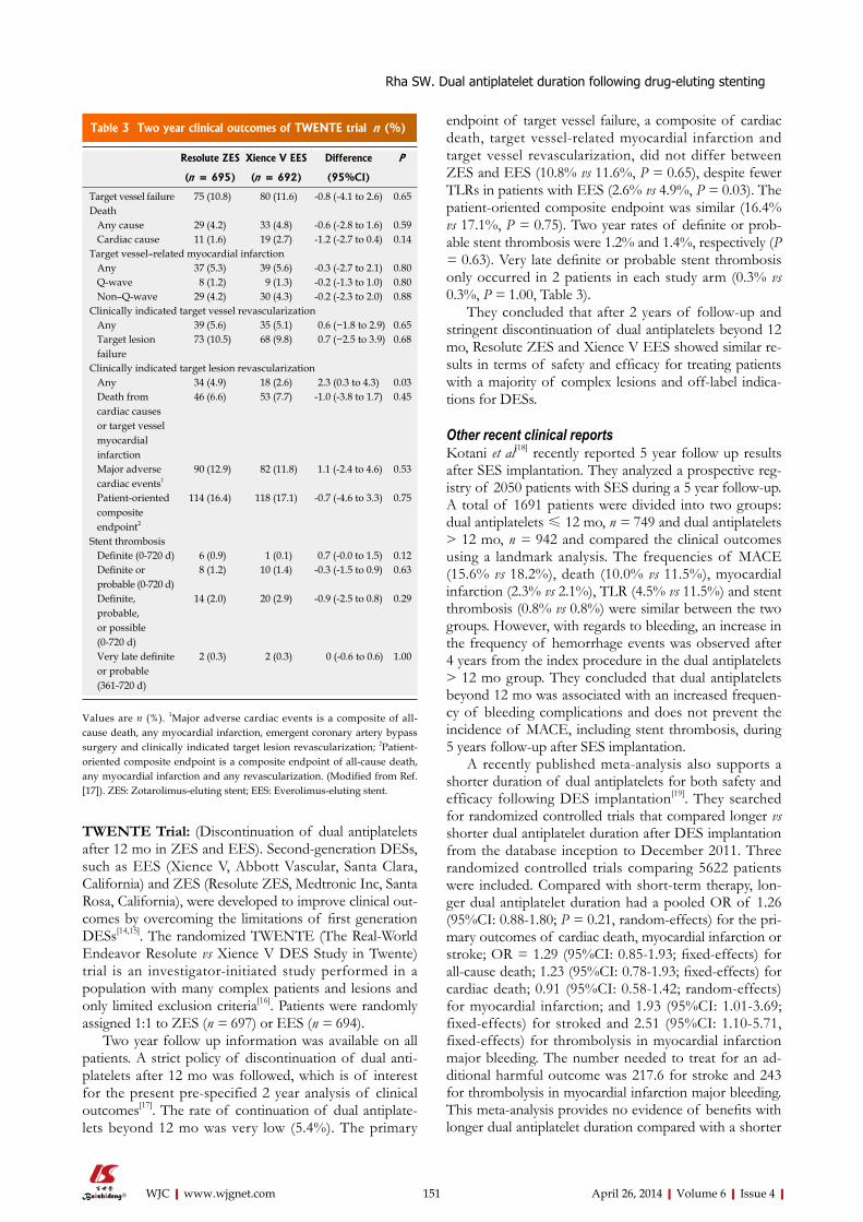
151 April 26, 2014|Volume 6|Issue 4|WJC|www.wjgnet.com
endpoint of target vessel failure, a composite of cardiac death, target vessel-related myocardial infarction and target vessel revascularization, did not differ between ZES and EES (10.8% vs 11.6%, P = 0.65), despite fewer TLRs in patients with EES (2.6% vs 4.9%, P = 0.03). The patient-oriented composite endpoint was similar (16.4% vs 17.1%, P = 0.75). Two year rates of definite or prob-able stent thrombosis were 1.2% and 1.4%, respectively (P = 0.63). Very late definite or probable stent thrombosis only occurred in 2 patients in each study arm (0.3% vs 0.3%, P = 1.00, Table 3).
They concluded that after 2 years of follow-up and stringent discontinuation of dual antiplatelets beyond 12 mo, Resolute ZES and Xience V EES showed similar re-sults in terms of safety and efficacy for treating patients with a majority of complex lesions and off-label indica-tions for DESs.
Other recent clinical reportsKotani et al[18] recently reported 5 year follow up results after SES implantation. They analyzed a prospective reg-istry of 2050 patients with SES during a 5 year follow-up. A total of 1691 patients were divided into two groups: dual antiplatelets ≤ 12 mo, n = 749 and dual antiplatelets > 12 mo, n = 942 and compared the clinical outcomes using a landmark analysis. The frequencies of MACE (15.6% vs 18.2%), death (10.0% vs 11.5%), myocardial infarction (2.3% vs 2.1%), TLR (4.5% vs 11.5%) and stent thrombosis (0.8% vs 0.8%) were similar between the two groups. However, with regards to bleeding, an increase in the frequency of hemorrhage events was observed after 4 years from the index procedure in the dual antiplatelets > 12 mo group. They concluded that dual antiplatelets beyond 12 mo was associated with an increased frequen-cy of bleeding complications and does not prevent the incidence of MACE, including stent thrombosis, during 5 years follow-up after SES implantation.
A recently published meta-analysis also supports a shorter duration of dual antiplatelets for both safety and efficacy following DES implantation[19]. They searched for randomized controlled trials that compared longer vs shorter dual antiplatelet duration after DES implantation from the database inception to December 2011. Three randomized controlled trials comparing 5622 patients were included. Compared with short-term therapy, lon-ger dual antiplatelet duration had a pooled OR of 1.26 (95%CI: 0.88-1.80; P = 0.21, random-effects) for the pri-mary outcomes of cardiac death, myocardial infarction or stroke; OR = 1.29 (95%CI: 0.85-1.93; fixed-effects) for all-cause death; 1.23 (95%CI: 0.78-1.93; fixed-effects) for cardiac death; 0.91 (95%CI: 0.58-1.42; random-effects) for myocardial infarction; and 1.93 (95%CI: 1.01-3.69; fixed-effects) for stroked and 2.51 (95%CI: 1.10-5.71, fixed-effects) for thrombolysis in myocardial infarction major bleeding. The number needed to treat for an ad-ditional harmful outcome was 217.6 for stroke and 243 for thrombolysis in myocardial infarction major bleeding. This meta-analysis provides no evidence of benefits with longer dual antiplatelet duration compared with a shorter
TWENTE Trial: (Discontinuation of dual antiplatelets after 12 mo in ZES and EES). Second-generation DESs, such as EES (Xience V, Abbott Vascular, Santa Clara, California) and ZES (Resolute ZES, Medtronic Inc, Santa Rosa, California), were developed to improve clinical out-comes by overcoming the limitations of first generation DESs[14,15]. The randomized TWENTE (The Real-World Endeavor Resolute vs Xience V DES Study in Twente) trial is an investigator-initiated study performed in a population with many complex patients and lesions and only limited exclusion criteria[16]. Patients were randomly assigned 1:1 to ZES (n = 697) or EES (n = 694).
Two year follow up information was available on all patients. A strict policy of discontinuation of dual anti-platelets after 12 mo was followed, which is of interest for the present pre-specified 2 year analysis of clinical outcomes[17]. The rate of continuation of dual antiplate-lets beyond 12 mo was very low (5.4%). The primary
Resolute ZES Xience V EES Difference P
(n = 695) (n = 692) (95%CI)
Target vessel failure 75 (10.8) 80 (11.6) -0.8 (-4.1 to 2.6) 0.65 Death Any cause 29 (4.2) 33 (4.8) -0.6 (-2.8 to 1.6) 0.59 Cardiac cause 11 (1.6) 19 (2.7) -1.2 (-2.7 to 0.4) 0.14 Target vessel–related myocardial infarction Any 37 (5.3) 39 (5.6) -0.3 (-2.7 to 2.1) 0.80 Q-wave 8 (1.2) 9 (1.3) -0.2 (-1.3 to 1.0) 0.80 Non–Q-wave 29 (4.2) 30 (4.3) -0.2 (-2.3 to 2.0) 0.88 Clinically indicated target vessel revascularization Any 39 (5.6) 35 (5.1) 0.6 (−1.8 to 2.9) 0.65 Target lesion failure
73 (10.5) 68 (9.8) 0.7 (−2.5 to 3.9) 0.68
Clinically indicated target lesion revascularization Any 34 (4.9) 18 (2.6) 2.3 (0.3 to 4.3) 0.03 Death from cardiac causes or target vessel myocardial infarction
46 (6.6) 53 (7.7) -1.0 (-3.8 to 1.7) 0.45
Major adverse cardiac events1
90 (12.9) 82 (11.8) 1.1 (-2.4 to 4.6) 0.53
Patient-oriented composite endpoint2
114 (16.4) 118 (17.1) -0.7 (-4.6 to 3.3) 0.75
Stent thrombosis Definite (0-720 d) 6 (0.9) 1 (0.1) 0.7 (-0.0 to 1.5) 0.12 Definite or probable (0-720 d)
8 (1.2) 10 (1.4) -0.3 (-1.5 to 0.9) 0.63
Definite, probable, or possible (0-720 d)
14 (2.0) 20 (2.9) -0.9 (-2.5 to 0.8) 0.29
Very late definite or probable (361-720 d)
2 (0.3) 2 (0.3) 0 (-0.6 to 0.6) 1.00
Table 3 Two year clinical outcomes of TWENTE trial n (%)
Values are n (%). 1Major adverse cardiac events is a composite of all-cause death, any myocardial infarction, emergent coronary artery bypass surgery and clinically indicated target lesion revascularization; 2Patient-oriented composite endpoint is a composite endpoint of all-cause death, any myocardial infarction and any revascularization. (Modified from Ref. [17]). ZES: Zotarolimus-eluting stent; EES: Everolimus-eluting stent.
Rha SW. Dual antiplatelet duration following drug-eluting stenting
152 April 26, 2014|Volume 6|Issue 4|WJC|www.wjgnet.com
course of therapy. It also reports significant harm with respect to major bleeding and stroke associated with pro-longed dual antiplatelet use.
Another new clinical trial (OPTIDUAL; OPTImal DUAL antiplatelet therapy trial) is ongoing to assess the efficacy and safety of 12 vs 48 mo of dual antiplatelet therapy after DES implantation[20].
Lastly, regarding clinical events associated with stent thrombosis, P2Y12 and thromboxane receptor are not the sole therapeutic measure to prevent the thrombotic risk. There must be different pathways leading to thrombotic events, including hypersensitivity reactions[21,22].
CONCLUSIONDespite the latest PCI guidelines recommending at least 1 year of dual antiplatelet therapy, recent randomized clini-cal trials, registries and meta-analysis data have shown that a shorter duration of dual antiplatelet therapy is as effective as a longer duration of dual antiplatelets, re-gardless of DES type (whether first-generation or next generation). Furthermore, a shorter duration of dual an-tiplatelets was associated with less bleeding complications without increasing the incidence of stent thrombosis. Currently, at least 6 mo of dual antiplatelets following next-generation DES implantation appears to be safe and effective, even with the expanded indication in the con-temporary PCI setting. However, caution should be exer-cised until enough clinical data is obtained, in particular in the subset of higher risk patients, including diabetes, aspirin and clopidogrel resistance or the very complex lesion subset expecting a vulnerability to stent throm-bosis. In this review, we focused only on classical dual antiplatelets, aspirin and clopidogrel. However, more data is needed to define the role of newer generation P2Y12 inhibitors, including ticagrelor and prasugrel, especially in the acute coronary syndrome setting in the future.
REFERENCES1 Moses JW, Leon MB, Popma JJ, Fitzgerald PJ, Holmes DR,
O’Shaughnessy C, Caputo RP, Kereiakes DJ, Williams DO, Teirstein PS, Jaeger JL, Kuntz RE. Sirolimus-eluting stents versus standard stents in patients with stenosis in a native coronary artery. N Engl J Med 2003; 349: 1315-1323 [PMID: 14523139 DOI: 10.1056/NEJMoa035071]
2 Stone GW, Ellis SG, Cox DA, Hermiller J, O’Shaughnessy C, Mann JT, Turco M, Caputo R, Bergin P, Greenberg J, Popma JJ, Russell ME. A polymer-based, paclitaxel-eluting stent in patients with coronary artery disease. N Engl J Med 2004; 350: 221-231 [PMID: 14724301 DOI: 10.1056/NEJMoa032441]
3 Pfisterer M, Brunner-La Rocca HP, Buser PT, Rickenbacher P, Hunziker P, Mueller C, Jeger R, Bader F, Osswald S, Kai-ser C. Late clinical events after clopidogrel discontinuation may limit the benefit of drug-eluting stents: an observational study of drug-eluting versus bare-metal stents. J Am Coll Cardiol 2006; 48: 2584-2591 [PMID: 17174201 DOI: 10.1016/j.jacc.2006.10.026]
4 Iakovou I, Schmidt T, Bonizzoni E, Ge L, Sangiorgi GM, Stankovic G, Airoldi F, Chieffo A, Montorfano M, Carlino M, Michev I, Corvaja N, Briguori C, Gerckens U, Grube E, Colombo A. Incidence, predictors, and outcome of throm-
bosis after successful implantation of drug-eluting stents. JAMA 2005; 293: 2126-2130 [PMID: 15870416 DOI: 10.1001/jama.293.17.2126]
5 Park DW, Park SW, Park KH, Lee BK, Kim YH, Lee CW, Hong MK, Kim JJ, Park SJ. Frequency of and risk factors for stent thrombosis after drug-eluting stent implantation during long-term follow-up. Am J Cardiol 2006; 98: 352-356 [PMID: 16860022 DOI: 10.1016/j.amjcard.2006.02.039]
6 King SB, Smith SC, Hirshfeld JW, Jacobs AK, Morrison DA, Williams DO, Feldman TE, Kern MJ, O’Neill WW, Schaff HV, Whitlow PL, Adams CD, Anderson JL, Buller CE, Creager MA, Ettinger SM, Halperin JL, Hunt SA, Krumholz HM, Kushner FG, Lytle BW, Nishimura R, Page RL, Riegel B, Tarkington LG, Yancy CW. 2007 Focused Update of the ACC/AHA/SCAI 2005 Guideline Update for Percutaneous Coronary Intervention: a report of the American College of Cardiology/American Heart Association Task Force on Practice Guidelines: 2007 Writing Group to Review New Evidence and Update the ACC/AHA/SCAI 2005 Guideline Update for Percutaneous Coronary Intervention, Writing on Behalf of the 2005 Writing Committee. Circulation 2008; 117: 261-295 [PMID: 18079354 DOI: 10.1161/CIRCULA-TIONAHA.107.188208]
7 Park SJ, Park DW, Kim YH, Kang SJ, Lee SW, Lee CW, Han KH, Park SW, Yun SC, Lee SG, Rha SW, Seong IW, Jeong MH, Hur SH, Lee NH, Yoon J, Yang JY, Lee BK, Choi YJ, Chung WS, Lim DS, Cheong SS, Kim KS, Chae JK, Nah DY, Jeon DS, Seung KB, Jang JS, Park HS, Lee K. Duration of dual antiplatelet therapy after implantation of drug-eluting stents. N Engl J Med 2010; 362: 1374-1382 [PMID: 20231231 DOI: 10.1056/NEJMoa1001266]
8 Lee CW, Ahn JM, Park DW, Kang SJ, Lee SW, Kim YH, Park SW, Han SB, Lee SG, Seong IW, Rha SW, Jeong MH, Lim DS, Yoon JH, Hur SH, Choi YS, Yang JY, Lee NH, Kim HS, Lee BK, Kim KS, Lee SU, Chae JK, Cheong SS, Suh IW, Park HS, Nah DY, Jeon DS, Seung KB, Lee K, Jang JS, Pakr SJ. Optimal duration of dual antiplatelet therapy after drug-eluting stent implantation: A Randomized Controlled Trial. Circulation 2013 (In Press)
9 Airoldi F, Colombo A, Morici N, Latib A, Cosgrave J, Buelles-feld L, Bonizzoni E, Carlino M, Gerckens U, Godino C, Melzi G, Michev I, Montorfano M, Sangiorgi GM, Qasim A, Chieffo A, Briguori C, Grube E. Incidence and predictors of drug-elut-ing stent thrombosis during and after discontinuation of thi-enopyridine treatment. Circulation 2007; 116: 745-754 [PMID: 17664375 DOI: 10.1161/CIRCULATIONAHA.106.686048]
10 Kimura T, Morimoto T, Nakagawa Y, Tamura T, Kadota K, Yasumoto H, Nishikawa H, Hiasa Y, Muramatsu T, Meguro T, Inoue N, Honda H, Hayashi Y, Miyazaki S, Oshima S, Honda T, Shiode N, Namura M, Sone T, Nobuyoshi M, Kita T, Mitsudo K. Antiplatelet therapy and stent thrombosis after sirolimus-eluting stent implantation. Circulation 2009; 119: 987-995 [PMID: 19204304 DOI: 10.1161/CIRCULA-TIONAHA.108.808311]
11 Hahn JY, Song YB, Choi JH, Choi SH, Lee SY, Park HS, Hur SH, Lee S, Han KR, Rha SW, Cho BR, Park JS, Yoon J, Lim do S, Lee SH, Gwon HC. Three-month dual antiplatelet therapy after implantation of zotarolimus-eluting stents: the DATE (Duration of Dual Antiplatelet Therapy AfterImplantation of Endeavor Stent) registry. Circ J 2010; 74: 2314-2321 [PMID: 20938098 DOI: 10.1253/circj.CJ-10-0347]
12 Gwon HC, Hahn JY, Park KW, Song YB, Chae IH, Lim DS, Han KR, Choi JH, Choi SH, Kang HJ, Koo BK, Ahn T, Yoon JH, Jeong MH, Hong TJ, Chung WY, Choi YJ, Hur SH, Kwon HM, Jeon DW, Kim BO, Park SH, Lee NH, Jeon HK, Jang Y, Kim HS. Six-month versus 12-month dual antiplatelet ther-apy after implantation of drug-eluting stents: the Efficacy of Xience/Promus Versus Cypher to Reduce Late Loss After Stenting (EXCELLENT) randomized, multicenter study.
Rha SW. Dual antiplatelet duration following drug-eluting stenting
153 April 26, 2014|Volume 6|Issue 4|WJC|www.wjgnet.com
Circulation 2012; 125: 505-513 [PMID: 22179532 DOI: 10.1161/CIRCULATIONAHA.111.059022]
13 Valgimigli M, Campo G, Monti M, Vranckx P, Percoco G, Tumscitz C, Castriota F, Colombo F, Tebaldi M, Fucà G, Kubbajeh M, Cangiano E, Minarelli M, Scalone A, Cavazza C, Frangione A, Borghesi M, Marchesini J, Parrinello G, Fer-rari R. Short- versus long-term duration of dual-antiplatelet therapy after coronary stenting: a randomized multicenter trial. Circulation 2012; 125: 2015-2026 [PMID: 22438530 DOI: 10.1161/CIRCULATIONAHA.111.071589]
14 Stone GW, Midei M, Newman W, Sanz M, Hermiller JB, Williams J, Farhat N, Mahaffey KW, Cutlip DE, Fitzgerald PJ, Sood P, Su X, Lansky AJ. Comparison of an everolimus-eluting stent and a paclitaxel-eluting stent in patients with coronary artery disease: a randomized trial. JAMA 2008; 299: 1903-1913 [PMID: 18430909 DOI: 10.1001/jama.299.16.1903]
15 Meredith IT, Worthley S, Whitbourn R, Walters DL, Mc-Clean D, Horrigan M, Popma JJ, Cutlip DE, DePaoli A, Ne-goita M, Fitzgerald PJ. Clinical and angiographic results with the next-generation resolute stent system: a prospective, multicenter, first-in-human trial. JACC Cardiovasc Interv 2009; 2: 977-985 [PMID: 19850258 DOI: 10.1016/j.jcin.2009.07.007]
16 von Birgelen C, Basalus MW, Tandjung K, van Houwelin-gen KG, Stoel MG, Louwerenburg JH, Linssen GC, Saïd SA, Kleijne MA, Sen H, Löwik MM, van der Palen J, Verhorst PM, de Man FH. A randomized controlled trial in second-generation zotarolimus-eluting Resolute stents versus evero-limus-eluting Xience V stents in real-world patients: the TWENTE trial. J Am Coll Cardiol 2012; 59: 1350-1361 [PMID: 22341737 DOI: 10.1016/j.jacc.2012.01.008]
17 Tandjung K, Sen H, Lam MK, Basalus MW, Louwerenburg JH, Stoel MG, van Houwelingen KG, de Man FH, Lins-sen GC, Saïd SA, Nienhuis MB, Löwik MM, Verhorst PM, van der Palen J, von Birgelen C. Clinical outcome follow-
ing stringent discontinuation of dual antiplatelet therapy after 12 months in real-world patients treated with second-generation zotarolimus-eluting resolute and everolimus-eluting Xience V stents: 2-year follow-up of the randomized TWENTE trial. J Am Coll Cardiol 2013; 61: 2406-2416 [PMID: 23602769 DOI: 10.1016/j.jacc.2013.04.005]
18 Kotani J, Ikari Y, Kyo E, Nakamura M, Yokoi H. Consider-ation of dual anti-platelet therapy duration after drug-elut-ing stent implantation in a Japanese population: a five-year follow-up after sirolimus-eluting stent implantation. Intern Med 2013; 52: 703-711 [PMID: 23545663 DOI: 10.2169/inter-nalmedicine.52.8205]
19 Valgimigli M, Park SJ, Kim HS, Park KW, Park DW, Tri-coci P, Ferrante G. Benefits and risks of long-term duration of dual antiplatelet therapy after drug-eluting stenting: a meta-analysis of randomized trials. Int J Cardiol 2013; 168: 2579-2587 [PMID: 23590932 DOI: 10.1016/j.ijcard.2013.03.047]
20 Helft G, Le Feuvre C, Georges JL, Carrie D, Leclercq F, Eltchaninoff H, Furber A, Prunier F, Sebagh L, Cattan S, Cayla G, Vicaut E, Metzger JP. Efficacy and safety of 12 versus 48 months of dual antiplatelet therapy after implanta-tion of a drug-eluting stent: the OPTImal DUAL antiplatelet therapy (OPTIDUAL) trial: study protocol for a randomized controlled trial. Trials 2013; 14: 56 [PMID: 23433461 DOI: 10.1186/1745-6215-14-56]
21 Kounis NG, Grapsas N. Pathways of platelet activation and unexplained clopidogel variability: causes of poor response to clopidogrel. Thromb Res 2013; 132: 312 [PMID: 23714177 DOI: 10.1016/j.thromres.2013.04.031]
22 Kounis NG, Giannopoulos S, Tsigkas GG, Goudevenos J. Eosinophilic responses to stent implantation and the risk of Kounis hypersensitivity associated coronary syndrome. Int J Cardiol 2012; 156: 125-132 [PMID: 21700348 DOI: 10.1016/j.ijcard.2011.05.052]
P- Reviewers: Kounis GN, Rassaf T, Sochman S S- Editor: Wen LL L- Editor: Roemmele A E- Editor: Liu SQ
Rha SW. Dual antiplatelet duration following drug-eluting stenting
Arrhythmogenic ventricular cardiomyopathy: A paradigm shift from right to biventricular disease
Ardan M Saguner, Corinna Brunckhorst , Firat Duru
Ardan M Saguner, Corinna Brunckhorst, Firat Duru, Depart-ment of Cardiology, University Heart Center, CH-8091 Zurich, SwitzerlandAuthor contributions: Saguner AM, Brunckhorst C and Duru F equally contributed to this article.Supported by The Georg and Bertha Schwyzer-Winiker Foun-dation, Zurich, SwitzerlandCorrespondence to: Firat Duru, Professor, Department of Cardiology, University Heart Center, Rämistrasse 100, CH-8091 Zurich, Switzerland. [email protected]: +41-44-2553565 Fax: +41-44-2554401Received: December 17, 2013 Revised: February 25, 2014Accepted: March 13, 2014Published online: April 26, 2014
AbstractArrhythmogenic ventricular cardiomyopathy (AVC) is generally referred to as arrhythmogenic right ventricu-lar (RV) cardiomyopathy/dysplasia and constitutes an inherited cardiomyopathy. Affected patients may succumb to sudden cardiac death (SCD), ventricular tachyarrhythmias (VTA) and heart failure. Genetic studies have identified causative mutations in genes encoding proteins of the intercalated disk that lead to reduced myocardial electro-mechanical stability. The term arrhythmogenic RV cardiomyopathy is somewhat misleading as biventricular involvement or isolated left ventricular (LV) involvement may be present and thus a broader term such as AVC should be preferred. The di-agnosis is established on a point score basis according to the revised 2010 task force criteria utilizing imaging modalities, demonstrating fibrous replacement through biopsy, electrocardiographic abnormalities, ventricu-lar arrhythmias and a positive family history including identification of genetic mutations. Although several risk factors for SCD such as previous cardiac arrest, syncope, documented VTA, severe RV/LV dysfunction and young age at manifestation have been identified, risk stratification still needs improvement, especially in
asymptomatic family members. Particularly, the role of genetic testing and environmental factors has to be further elucidated. Therapeutic interventions include re-striction from physical exercise, beta-blockers, sotalol, amiodarone, implantable cardioverter-defibrillators and catheter ablation. Life-long follow-up is warranted in symptomatic patients, but also asymptomatic carriers of pathogenic mutations.
© 2014 Baishideng Publishing Group Co., Limited. All rights reserved.
Key words: Arrhythmogenic right ventricular dyspla-sia/cardiomyopathy; Arrhythmias; Ventricular tachy-cardia; Sudden cardiac death; Implantable cardioverter defibrillator
Core tip: This manuscript constitutes an updated over-view about arrhythmogenic ventricular cardiomyopathy (AVC) and describes well the paradigm shift in the un-derstanding of AVC from an isolated right-sided entity to biventricular disease that can present with multiple facets. The most recent advances in molecular and clinical research are discussed, with particular focus on genetic novelties and risk stratification. We believe that this review will help clinicians to better understand the pathomechanisms that lead to AVC, its diagnosis and state-of-the-art therapeutic decision making.
Saguner AM, Brunckhorst C , Duru F. Arrhythmogenic ventricu-lar cardiomyopathy: A paradigm shift from right to biventricular disease. World J Cardiol 2014; 6(4): 154-174 Available from: URL: http://www.wjgnet.com/1949-8462/full/v6/i4/154.htm DOI: http://dx.doi.org/10.4330/wjc.v6.i4.154
INTRODUCTIONArrhythmogenic ventricular cardiomyopathy (AVC), as
REVIEW
Online Submissions: http://www.wjgnet.com/esps/[email protected]:10.4330/wjc.v6.i4.154
154 April 26, 2014|Volume 6|Issue 4|WJC|www.wjgnet.com
World Journal of CardiologyW J C
World J Cardiol 2014 April 26; 6(4): 154-174ISSN 1949-8462 (online)
© 2014 Baishideng Publishing Group Co., Limited. All rights reserved.
recently re-named by the Heart Rhythm Society (HRS) and the European Heart Rhythm Association (EHRA) consensus statement paper[1], is generally referred to as arrhythmogenic right ventricular cardiomyopathy/dys-plasia (ARVC/D), constituting a hereditary cardiomy-opathy usually with an autosomal-dominant inheritance pattern. Its first description by Giovanni Maria Lancisi, the Pope’s physician, dates back to 1736 in his book “De Motu Cordis et Aneurysmatibus”[2]. The first comprehen-sive description of ARVC/D by Guy Fontaine in 1978 marks a milestone for our current understanding of this heterogeneous disease[3]. Initially, ARVC/D was thought to be an embryological aberration, such as Uhl’s anomaly leading to the original designation of dysplasia[4]. How-ever, further research shed light on the pathophysiology of ongoing genetically determined myocardial atrophy that did not support the theory of a congenital myocar-dial absence. Thus, in 1995, ARVC/D was assigned to the World Health Organization’s definition and classifica-tion of primary cardiomyopathies[5]. Autopsy studies have been crucial in understanding AVC. Progressive atrophy of the ventricular musculature due to cumulative myocyte loss and infiltration by fibrous and adipose tissue can be observed.
The right ventricle (RV) is primarily affected in AVC, representing the most common form known as ARVC/D, and thus can be referred to as classic AVC[6]. At a later stage, the left ventricle (LV) can also be involved and is often associated with severe disease and a worse progno-sis[7]. Advanced molecular genetic studies have identified causative mutations in genes encoding proteins of the intercalated disk, mainly desmosomal proteins[8] that lead to reduced electrical and mechanical stability of the myo-cardium[9,10]. Subsequent myocardial inflammation, apop-tosis and necrosis may occur. Some of these histological changes are currently discussed as potential cases of myocarditis mimicking AVC[11-14]. Because of the genetic basis and the many facets of the disease, the term “ARVC” is somewhat misleading. Particularly as biventricular in-volvement and less often isolated LV involvement may be present in a substantial proportion of patients[15], a broader term such as “arrhythmogenic cardiomyopathy” should be preferred, as already suggested by Gallo et al[16] almost 20 years ago, and as recently proposed by the HRS and the EHRA[1]. However, the cardiology community is still reluctant to accept the proposed new nomenclature, probably because RV involvement constitutes a hallmark of the disease and non-classic forms are difficult to dis-tinguish from non-ischemic dilated cardiomyopathies.
EPIDEMIOLOGYIn most parts of the world, phenotypic expression is more common in men than in women (2-3:1)[17,18]. AVC commonly manifests during late childhood or adoles-cence but can also emerge in the elderly[19,20]. With a gen-eral prevalence of 1:2000, which can be higher in certain geographical regions with enhanced genetic prevalence
such as the Veneto region or the Greek island Naxos, it is not so rare[21,22]. Recent data indicates that the preva-lence is even higher than initially estimated[23]. AVC is recognized as a leading cause of sudden cardiac death (SCD) in young adults ≤ 35 years of age and may ac-count for up to 10% of cardiovascular deaths in the < 65 age group[24,25]. Of note, in one series from northern Italy, AVC accounted for up to 22% of SCD in all young adults ≤ 35 years of age[26-29]. AVC usually first manifests with ventricular tachyarrhythmias (VTA) or SCD. In its most common form ARVC/D, ventricular arrhythmias originate in the RV and thus have left bundle branch block (LBBB) morphology[28,30]. Less often, the primary manifestation can be heart failure without symptomatic arrhythmias. As LV function is often preserved at early stages, ventricular tachycardia (VT) may be asymptomatic as far as it does not degenerate into ventricular fibril-lation (VF)[29]. An early concealed phase without gross structural abnormalities is unique among the primary cardiomyopathies. On the contrary, in hypertrophic cardiomyopathy, arrhythmic risk can be ascribed to the underlying myocardial disarray. In dilated cardiomyopathy (DCM), arrhythmias generally concur with significant LV systolic dysfunction[31]. Of note, early AVC may resemble myocardial channelopathies, such as Brugada syndrome (Bs)[32], thus making correct diagnosis and risk stratifica-tion difficult.
DISEASE SUBTYPESClassification of AVC into three different subtypes is evolving. AVC in its classic right-dominant form is the most common and best known and referred to as ARVC/D. The non-classic forms were first described by patholo-gists on autopsy studies and in isolated clinical case re-ports[33,34]. Through intensive in vivo characterization of affected families, a link to hereditary mutations of the intercalated disk was established[35-37]. LV involvement is increasingly described with a prevalence of up to 76% of cases, which may be attributed to improved diagnostic methods such as genetic testing, high-resolution contrast-enhanced cardiac magnetic resonance tomography (CMR), and recently the new technology of echocardiographic strain imaging[38]. The proposed classification below is simplistic since due to genetic heterogeneity and epigen-etic factors, a phenotypic continuum with right- and left-dominant subtypes at opposite ends has to be assumed.
In classic right-dominant ARVC, a dilated RV with fibro-fatty infiltration with no or only minimal LV in-volvement can be found at autopsy (Figure 1). This fibro-fatty infiltration typically begins subepicardially and may expand transmurally over time[39]. Papillary muscles and trabeculae are generally not involved in this process[25]. Yet, fatty infiltration alone does not constitute a pathog-nomonic sign of AVC, as a certain amount of epicardial and intramyocardial fat without an increase in fibrous tis-sue is present in both ventricles, more commonly in the RV, of persons without cardiovascular disease, particu-
155 April 26, 2014|Volume 6|Issue 4|WJC|www.wjgnet.com
Saguner AM et al . Arrhythmogenic ventricular cardiomyopathy
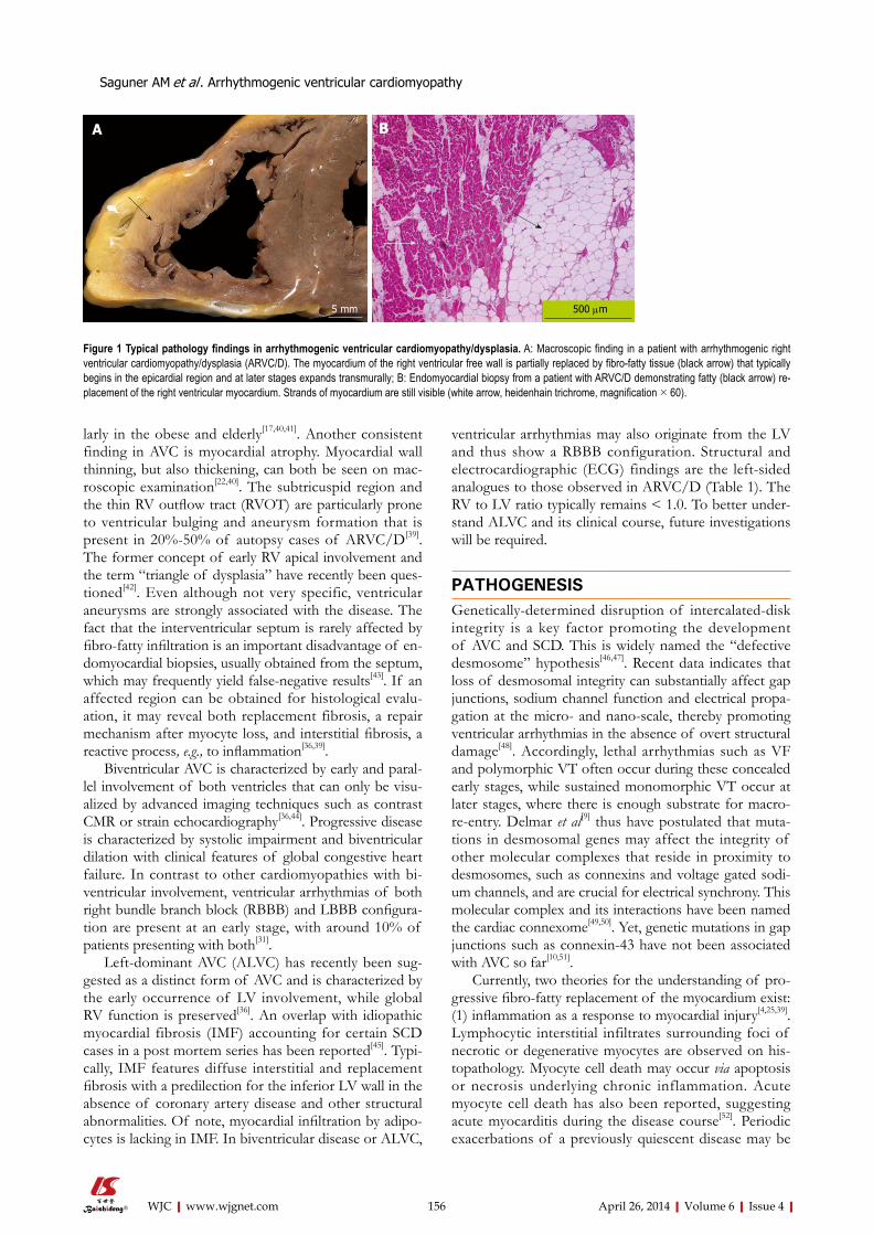
larly in the obese and elderly[17,40,41]. Another consistent finding in AVC is myocardial atrophy. Myocardial wall thinning, but also thickening, can both be seen on mac-roscopic examination[22,40]. The subtricuspid region and the thin RV outflow tract (RVOT) are particularly prone to ventricular bulging and aneurysm formation that is present in 20%-50% of autopsy cases of ARVC/D[39]. The former concept of early RV apical involvement and the term “triangle of dysplasia” have recently been ques-tioned[42]. Even although not very specific, ventricular aneurysms are strongly associated with the disease. The fact that the interventricular septum is rarely affected by fibro-fatty infiltration is an important disadvantage of en-domyocardial biopsies, usually obtained from the septum, which may frequently yield false-negative results[43]. If an affected region can be obtained for histological evalu-ation, it may reveal both replacement fibrosis, a repair mechanism after myocyte loss, and interstitial fibrosis, a reactive process, e.g., to inflammation[36,39].
Biventricular AVC is characterized by early and paral-lel involvement of both ventricles that can only be visu-alized by advanced imaging techniques such as contrast CMR or strain echocardiography[36,44]. Progressive disease is characterized by systolic impairment and biventricular dilation with clinical features of global congestive heart failure. In contrast to other cardiomyopathies with bi-ventricular involvement, ventricular arrhythmias of both right bundle branch block (RBBB) and LBBB configura-tion are present at an early stage, with around 10% of patients presenting with both[31].
Left-dominant AVC (ALVC) has recently been sug-gested as a distinct form of AVC and is characterized by the early occurrence of LV involvement, while global RV function is preserved[36]. An overlap with idiopathic myocardial fibrosis (IMF) accounting for certain SCD cases in a post mortem series has been reported[45]. Typi-cally, IMF features diffuse interstitial and replacement fibrosis with a predilection for the inferior LV wall in the absence of coronary artery disease and other structural abnormalities. Of note, myocardial infiltration by adipo-cytes is lacking in IMF. In biventricular disease or ALVC,
ventricular arrhythmias may also originate from the LV and thus show a RBBB configuration. Structural and electrocardiographic (ECG) findings are the left-sided analogues to those observed in ARVC/D (Table 1). The RV to LV ratio typically remains < 1.0. To better under-stand ALVC and its clinical course, future investigations will be required.
PATHOGENESISGenetically-determined disruption of intercalated-disk integrity is a key factor promoting the development of AVC and SCD. This is widely named the “defective desmosome” hypothesis[46,47]. Recent data indicates that loss of desmosomal integrity can substantially affect gap junctions, sodium channel function and electrical propa-gation at the micro- and nano-scale, thereby promoting ventricular arrhythmias in the absence of overt structural damage[48]. Accordingly, lethal arrhythmias such as VF and polymorphic VT often occur during these concealed early stages, while sustained monomorphic VT occur at later stages, where there is enough substrate for macro-re-entry. Delmar et al[9] thus have postulated that muta-tions in desmosomal genes may affect the integrity of other molecular complexes that reside in proximity to desmosomes, such as connexins and voltage gated sodi-um channels, and are crucial for electrical synchrony. This molecular complex and its interactions have been named the cardiac connexome[49,50]. Yet, genetic mutations in gap junctions such as connexin-43 have not been associated with AVC so far[10,51].
Currently, two theories for the understanding of pro-gressive fibro-fatty replacement of the myocardium exist: (1) inflammation as a response to myocardial injury[4,25,39]. Lymphocytic interstitial infiltrates surrounding foci of necrotic or degenerative myocytes are observed on his-topathology. Myocyte cell death may occur via apoptosis or necrosis underlying chronic inflammation. Acute myocyte cell death has also been reported, suggesting acute myocarditis during the disease course[52]. Periodic exacerbations of a previously quiescent disease may be
156 April 26, 2014|Volume 6|Issue 4|WJC|www.wjgnet.com
Figure 1 Typical pathology findings in arrhythmogenic ventricular cardiomyopathy/dysplasia. A: Macroscopic finding in a patient with arrhythmogenic right ventricular cardiomyopathy/dysplasia (ARVC/D). The myocardium of the right ventricular free wall is partially replaced by fibro-fatty tissue (black arrow) that typically begins in the epicardial region and at later stages expands transmurally; B: Endomyocardial biopsy from a patient with ARVC/D demonstrating fatty (black arrow) re-placement of the right ventricular myocardium. Strands of myocardium are still visible (white arrow, heidenhain trichrome, magnification × 60).
5 mm 500 μm
A B
Saguner AM et al . Arrhythmogenic ventricular cardiomyopathy
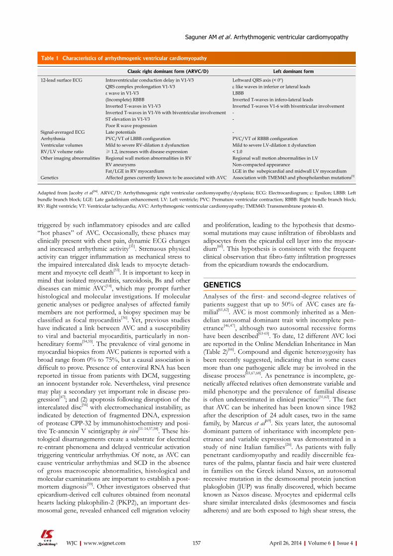
157 April 26, 2014|Volume 6|Issue 4|WJC|www.wjgnet.com
and proliferation, leading to the hypothesis that desmo-somal mutations may cause infiltration of fibroblasts and adipocytes from the epicardial cell layer into the myocar-dium[60]. This hypothesis is consistent with the frequent clinical observation that fibro-fatty infiltration progresses from the epicardium towards the endocardium.
GENETICSAnalyses of the first- and second-degree relatives of patients suggest that up to 50% of AVC cases are fa-milial[61,62]. AVC is most commonly inherited as a Men-delian autosomal dominant trait with incomplete pen-etrance[46,47], although two autosomal recessive forms have been described[63-65]. To date, 12 different AVC loci are reported in the Online Mendelian Inheritance in Man (Table 2)[66]. Compound and digenic heterozygosity has been recently suggested, indicating that in some cases more than one pathogenic allele may be involved in the disease process[65,67,68]. As penetrance is incomplete, ge-netically affected relatives often demonstrate variable and mild phenotype and the prevalence of familial disease is often underestimated in clinical practice[31,62]. The fact that AVC can be inherited has been known since 1982 after the description of 24 adult cases, two in the same family, by Marcus et al[69]. Six years later, the autosomal dominant pattern of inheritance with incomplete pen-etrance and variable expression was demonstrated in a study of nine Italian families[26]. As patients with fully penetrant cardiomyopathy and readily discernible fea-tures of the palms, plantar fascia and hair were clustered in families on the Greek island Naxos, an autosomal recessive mutation in the desmosomal protein junction plakoglobin (JUP) was finally discovered, which became known as Naxos disease. Myocytes and epidermal cells share similar intercalated disks (desmosomes and fascia adherens) and are both exposed to high shear stress, the
triggered by such inflammatory episodes and are called “hot phases” of AVC. Occasionally, these phases may clinically present with chest pain, dynamic ECG changes and increased arrhythmic activity[31]. Strenuous physical activity can trigger inflammation as mechanical stress to the impaired intercalated disk leads to myocyte detach-ment and myocyte cell death[53]. It is important to keep in mind that isolated myocarditis, sarcoidosis, Bs and other diseases can mimic AVC[14], which may prompt further histological and molecular investigations. If molecular genetic analyses or pedigree analyses of affected family members are not performed, a biopsy specimen may be classified as focal myocarditis[36]. Yet, previous studies have indicated a link between AVC and a susceptibility to viral and bacterial myocarditis, particularly in non-hereditary forms[54,55]. The prevalence of viral genome in myocardial biopsies from AVC patients is reported with a broad range from 0% to 75%, but a causal association is difficult to prove. Presence of enteroviral RNA has been reported in tissue from patients with DCM, suggesting an innocent bystander role. Nevertheless, viral presence may play a secondary yet important role in disease pro-gression[47]; and (2) apoptosis following disruption of the intercalated disc[56] with electromechanical instability, as indicated by detection of fragmented DNA, expression of protease CPP-32 by immunohistochemistry and posi-tive Tc-annexin V scintigraphy in vivo[11-14,57,58]. These his-tological disarrangements create a substrate for electrical re-entrant phenomena and delayed ventricular activation triggering ventricular arrhythmias. Of note, as AVC can cause ventricular arrhythmias and SCD in the absence of gross macroscopic abnormalities, histological and molecular examinations are important to establish a post-mortem diagnosis[59]. Other investigators observed that epicardium-derived cell cultures obtained from neonatal hearts lacking plakophilin-2 (PKP2), an important des-mosomal gene, revealed enhanced cell migration velocity
Table 1 Characteristics of arrhythmogenic ventricular cardiomyopathy
Classic right dominant form (ARVC/D) Left dominant form
12-lead surface ECG Intraventricular conduction delay in V1-V3 Leftward QRS axis (< 0°)QRS complex prolongation V1-V3 ε like waves in inferior or lateral leadsε wave in V1-V3 LBBB(Incomplete) RBBB Inverted T-waves in infero-lateral leadsInverted T-waves in V1-V3 Inverted T-waves V1-6 with biventricular involvementInverted T-waves in V1-V6 with biventricular involvement -ST elevation in V1-V3 -Poor R wave progression
Signal-averaged ECG Late potentials -Arrhythmia PVC/VT of LBBB configuration PVC/VT of RBBB configurationVentricular volumes Mild to severe RV-dilation ± dysfunction Mild to severe LV-dilation ± dysfunctionRV/LV volume ratio ≥ 1.2, increases with disease expression < 1.0Other imaging abnormalities Regional wall motion abnormalities in RV Regional wall motion abnormalities in LV
RV aneurysms Non-compacted appearanceFat/LGE in RV myocardium LGE in the subepicardial and midwall LV myocardium
Genetics Affected genes currently known to be associated with AVC Association with TMEM43 and phospholamban mutations[1]
Adapted from Jacoby et al[84]. ARVC/D: Arrhythmogenic right ventricular cardiomyopathy/dysplasia; ECG: Electrocardiogram; ε: Epsilon; LBBB: Left bundle branch block; LGE: Late gadolinium enhancement; LV: Left ventricle; PVC: Premature ventricular contraction; RBBB: Right bundle branch block; RV: Right ventricle; VT: Ventricular tachycardia; AVC: Arrhythmogenic ventricular cardiomyopathy; TMEM43: Transmembrane protein 43.
Saguner AM et al . Arrhythmogenic ventricular cardiomyopathy
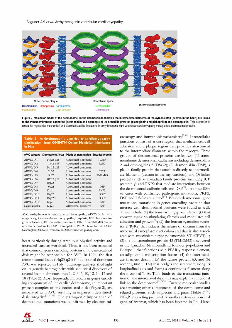
158 April 26, 2014|Volume 6|Issue 4|WJC|www.wjgnet.com
heart particularly during strenuous physical activity and increased cardiac workload. Thus, it has been assumed that common genes encoding proteins of the intercalated disk might be responsible for AVC. In 1994, the first chromosomal locus (14q23-q24) for autosomal dominant AVC was reported in Italy[47]. Linkage analyses shed light on its genetic heterogeneity with sequential discovery of several loci on chromosomes 1, 2, 3, 6, 10, 12, 14, 17 and 18 (Table 2). Most frequently, mutations in genes encod-ing components of the cardiac desmosome, an important protein complex of the intercalated disk (Figure 2), are associated with AVC, resulting in impaired intercalated-disk integrity[62,67,68]. The pathogenic importance of desmosomal mutations was confirmed by electron mi-
croscopy and immunohistochemistry[2,56]. Intercellular junctions consist of a core region that mediates cell-cell adhesion and a plaque region that provides attachment to the intermediate filaments within the myocyte. Three groups of desmosomal proteins are known: (1) trans-membrane desmosomal cadherins including desmocollins 2 and desmogleins 2 (DSG2); (2) desmoplakin (DSP), a plakin family protein that attaches directly to intermedi-ate filaments (desmin in the myocardium); and (3) linker proteins such as armadillo family proteins including JUP (catenin-γ) and PKP2 that mediate interactions between the desmosomal cadherin tails and DSP[70]. In about 80% of cases with confirmed pathogenic mutations, PKP2, DSP and DSG2 are altered[22]. Besides desmosomal gene mutations, mutations in genes encoding proteins that interact with desmosomal proteins were found as well. These include: (1) the transforming growth factor β3 that conveys cytokine-stimulating fibrosis and modulates cell adhesion and growth[52]; (2) the human ryanodine recep-tor 2 (RyR2) that induces the release of calcium from the myocardial sarcoplasmic reticulum and that is also associ-ated with catecholaminergic polymorphic VT (CPVT)[71]; (3) the transmembrane protein 43 (TMEM43) discovered in the Canadian Newfoundland founder population and Europe[72] that functions as a PPAR-γ response element, an adipogenic transcription factor; (4) the intermedi-ate filament desmin; (5) the tumor protein 63; and (6) recently, titin (TTN) that bridges the sarcomere along its longitudinal axis and forms a continuous filament along the myofibril[66]. As TTN binds to the transitional junc-tion of the intercalated disk, this may explain a functional link to the desmosome[66,73,74]. Current molecular studies are screening other components of the desmosome and related proteins, such as plectin and pinin (Table 3)[75]. NFκB interacting protein-1 is another extra-desmosomal gene of interest, which has been isolated in Poll-Here-
Table 2 Arrhythmogenic ventricular cardiomyopathy classification, from OMIMTM Online Mendelian inheritance in Man
AVC subtype Chromosome/locus Mode of transmission Encoded protein
ARVC/D 1 14q23-q24 Autosomal-dominant TGFβ3ARVC/D 2 1q42-q43 Autosomal-dominant RyR2ARVC/D 3 14q12-q22 Autosomal-dominant -ARVC/D 4 2q32 Autosomal-dominant TTNARVC/D 5 3p23 Autosomal-dominant TMEM43ARVC/D 6 10p12-p14 Autosomal-dominant -ARVC/D 7 10q22 Autosomal-dominant -ARVC/D 8 6p24 Autosomal-dominant DSPARVC/D 9 12p11 Autosomal-dominant PKP2ARVC/D 10 18q12 Autosomal-dominant DSG2ARVC/D 11 18q12.1 Autosomal-dominant DSC2ARVC/D 12 17q21 Autosomal-dominant JUPNaxos disease 17q21 Autosomal-recessive JUP
AVC: Arrhythmogenic ventricular cardiomyopathy; ARVC/D: Arrhyth-mogenic right ventricular cardiomyopathy/dysplasia; TGF: Transforming growth factor; RyR2: Ryanodine receptor 2; TTN: Titin; TMEM43: Trans-membrane protein 43; DSP: Desmoplakin; PKP2: Plakophilin-2; DSG2: Desmoglein-2; DSC2: Desmocollin-2; JUP: Junction plakoglobin.
Outer dense plaque
DesmoplakinPlakoglobin
Plakophilins SarcolemmaGap junction
Intercellular space
DesmocollinDesmoglein
Intermediate filaments
Figure 2 Molecular model of the desmosome: in the desmosomal complex the intermediate filaments of the cytoskeleton (desmin in the heart) are linked to the transmembranous cadherins (desmocollin and desmoglein) via armadillo proteins (plakoglobin and plakophilin) and desmoplakin. This interaction is crucial for myocardial mechanical and electrical stability. Mutations in arrhythmogenic right ventricular cardiomyopathy mostly affect desmosomal proteins.
Saguner AM et al . Arrhythmogenic ventricular cardiomyopathy
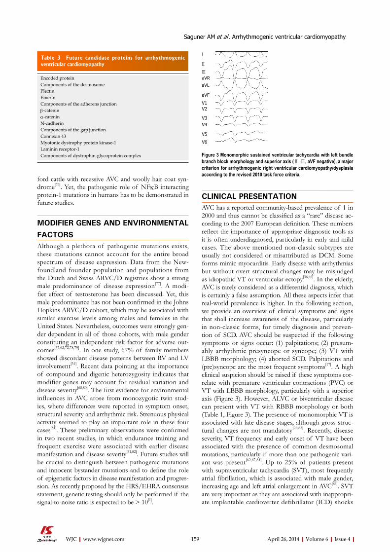
159 April 26, 2014|Volume 6|Issue 4|WJC|www.wjgnet.com
ford cattle with recessive AVC and woolly hair coat syn-drome[76]. Yet, the pathogenic role of NFκB interacting protein-1 mutations in humans has to be demonstrated in future studies.
MODIFIER GENES AND ENVIRONMENTAL FACTORSAlthough a plethora of pathogenic mutations exists, these mutations cannot account for the entire broad spectrum of disease expression. Data from the New-foundland founder population and populations from the Dutch and Swiss ARVC/D registries show a strong male predominance of disease expression[77]. A modi-fier effect of testosterone has been discussed. Yet, this male predominance has not been confirmed in the Johns Hopkins ARVC/D cohort, which may be associated with similar exercise levels among males and females in the United States. Nevertheless, outcomes were strongly gen-der dependent in all of those cohorts, with male gender constituting an independent risk factor for adverse out-comes[37,62,72,78,79]. In one study, 67% of family members showed discordant disease patterns between RV and LV involvement[31]. Recent data pointing at the importance of compound and digenic heterozygosity indicates that modifier genes may account for residual variation and disease severity[68,80]. The first evidence for environmental influences in AVC arose from monozygotic twin stud-ies, where differences were reported in symptom onset, structural severity and arrhythmic risk. Strenuous physical activity seemed to play an important role in these four cases[81]. These preliminary observations were confirmed in two recent studies, in which endurance training and frequent exercise were associated with earlier disease manifestation and disease severity[31,82]. Future studies will be crucial to distinguish between pathogenic mutations and innocent bystander mutations and to define the role of epigenetic factors in disease manifestation and progres-sion. As recently proposed by the HRS/EHRA consensus statement, genetic testing should only be performed if the signal-to-noise ratio is expected to be > 10[1].
CLINICAL PRESENTATIONAVC has a reported community-based prevalence of 1 in 2000 and thus cannot be classified as a “rare” disease ac-cording to the 2007 European definition. These numbers reflect the importance of appropriate diagnostic tools as it is often underdiagnosed, particularly in early and mild cases. The above mentioned non-classic subtypes are usually not considered or misattributed as DCM. Some forms mimic myocarditis. Early disease with arrhythmias but without overt structural changes may be misjudged as idiopathic VT or ventricular ectopy[36,46]. In the elderly, AVC is rarely considered as a differential diagnosis, which is certainly a false assumption. All these aspects infer that real-world prevalence is higher. In the following section, we provide an overview of clinical symptoms and signs that shall increase awareness of the disease, particularly in non-classic forms, for timely diagnosis and preven-tion of SCD. AVC should be suspected if the following symptoms or signs occur: (1) palpitations; (2) presum-ably arrhythmic presyncope or syncope; (3) VT with LBBB morphology; (4) aborted SCD. Palpitations and (pre)syncope are the most frequent symptoms[17]. A high clinical suspicion should be raised if these symptoms cor-relate with premature ventricular contractions (PVC) or VT with LBBB morphology, particularly with a superior axis (Figure 3). However, ALVC or biventricular disease can present with VT with RBBB morphology or both (Table 1, Figure 3). The presence of monomorphic VT is associated with late disease stages, although gross struc-tural changes are not mandatory[28,83]. Recently, disease severity, VT frequency and early onset of VT have been associated with the presence of common desmosomal mutations, particularly if more than one pathogenic vari-ant was present[62,67,84]. Up to 25% of patients present with supraventricular tachycardia (SVT), most frequently atrial fibrillation, which is associated with male gender, increasing age and left atrial enlargement in AVC[85]. SVT are very important as they are associated with inappropri-ate implantable cardioverter defibrillator (ICD) shocks
Table 3 Future candidate proteins for arrhythmogenic ventricular cardiomyopathy
Encoded proteinComponents of the desmosomePlectinEmerinComponents of the adherens junctionβ-cateninα-cateninN-cadherin Components of the gap junction Connexin 43Myotonic dystrophy protein kinase-1Laminin receptor-1Components of dystrophin-glycoprotein complex
Ⅰ
Ⅱ Ⅲ aVR
aVL
aVF
V1 V2
V3 V4
V5
V6
Figure 3 Monomorphic sustained ventricular tachycardia with left bundle branch block morphology and superior axis (Ⅱ, Ⅲ, aVF negative), a major criterion for arrhythmogenic right ventricular cardiomyopathy/dysplasia according to the revised 2010 task force criteria.
Saguner AM et al . Arrhythmogenic ventricular cardiomyopathy
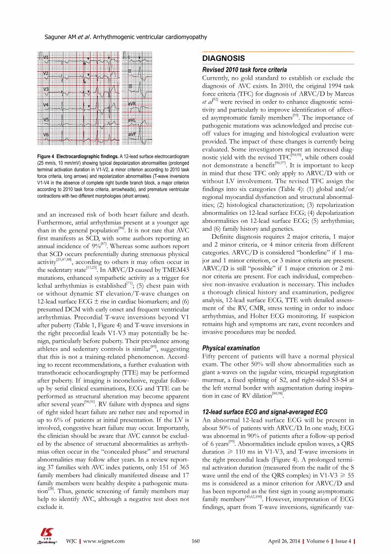
160 April 26, 2014|Volume 6|Issue 4|WJC|www.wjgnet.com
and an increased risk of both heart failure and death. Furthermore, atrial arrhythmias present at a younger age than in the general population[86]. It is not rare that AVC first manifests as SCD, with some authors reporting an annual incidence of 9%[87]. Whereas some authors report that SCD occurs preferentially during strenuous physical activity[25,87,88], according to others it may often occur in the sedentary state[13,25]. In ARVC/D caused by TMEM43 mutations, enhanced sympathetic activity as a trigger for lethal arrhythmias is established[71]; (5) chest pain with or without dynamic ST elevation/T-wave changes on 12-lead surface ECG ± rise in cardiac biomarkers; and (6) presumed DCM with early onset and frequent ventricular arrhythmias. Precordial T-wave inversions beyond V1 after puberty (Table 1, Figure 4) and T-wave inversions in the right precordial leads V1-V3 may potentially be be-nign, particularly before puberty. Their prevalence among athletes and sedentary controls is similar[89], suggesting that this is not a training-related phenomenon. Accord-ing to recent recommendations, a further evaluation with transthoracic echocardiography (TTE) may be performed after puberty. If imaging is inconclusive, regular follow-up by serial clinical examinations, ECG and TTE can be performed as structural alteration may become apparent after several years[90,91]. RV failure with dyspnea and signs of right sided heart failure are rather rare and reported in up to 6% of patients at initial presentation. If the LV is involved, congestive heart failure may occur. Importantly, the clinician should be aware that AVC cannot be exclud-ed by the absence of structural abnormalities as arrhyth-mias often occur in the “concealed phase” and structural abnormalities may follow after years. In a review report-ing 37 families with AVC index patients, only 151 of 365 family members had clinically manifested disease and 17 family members were healthy despite a pathogenic muta-tion[28]. Thus, genetic screening of family members may help to identify AVC, although a negative test does not exclude it.
DIAGNOSISRevised 2010 task force criteriaCurrently, no gold standard to establish or exclude the diagnosis of AVC exists. In 2010, the original 1994 task force criteria (TFC) for diagnosis of ARVC/D by Marcus et al[92] were revised in order to enhance diagnostic sensi-tivity and particularly to improve identification of affect-ed asymptomatic family members[93]. The importance of pathogenic mutations was acknowledged and precise cut-off values for imaging and histological evaluation were provided. The impact of these changes is currently being evaluated. Some investigators report an increased diag-nostic yield with the revised TFC[94,95], while others could not demonstrate a benefit[96,97]. It is important to keep in mind that these TFC only apply to ARVC/D with or without LV involvement. The revised TFC assign the findings into six categories (Table 4): (1) global and/or regional myocardial dysfunction and structural abnormal-ities; (2) histological characterization; (3) repolarization abnormalities on 12-lead surface ECG; (4) depolarization abnormalities on 12-lead surface ECG; (5) arrhythmias; and (6) family history and genetics.
Definite diagnosis requires 2 major criteria, 1 major and 2 minor criteria, or 4 minor criteria from different categories. ARVC/D is considered “borderline” if 1 ma-jor and 1 minor criterion, or 3 minor criteria are present. ARVC/D is still “possible” if 1 major criterion or 2 mi-nor criteria are present. For each individual, comprehen-sive non-invasive evaluation is necessary. This includes a thorough clinical history and examination, pedigree analysis, 12-lead surface ECG, TTE with detailed assess-ment of the RV, CMR, stress testing in order to induce arrhythmias, and Holter ECG monitoring. If suspicion remains high and symptoms are rare, event recorders and invasive procedures may be needed.
Physical examinationFifty percent of patients will have a normal physical exam. The other 50% will show abnormalities such as giant a-waves on the jugular veins, tricuspid regurgitation murmur, a fixed splitting of S2, and right-sided S3-S4 at the left sternal border with augmentation during inspira-tion in case of RV dilation[88,98].
12-lead surface ECG and signal-averaged ECGAn abnormal 12-lead surface ECG will be present in about 50% of patients with ARVC/D. In one study, ECG was abnormal in 90% of patients after a follow-up period of 6 years[99]. Abnormalities include epsilon waves, a QRS duration ≥ 110 ms in V1-V3, and T-wave inversions in the right precordial leads (Figure 4). A prolonged termi-nal activation duration (measured from the nadir of the S wave until the end of the QRS complex) in V1-V3 ≥ 55 ms is considered as a minor criterion for ARVC/D and has been reported as the first sign in young asymptomatic family members[45,62,100]. However, interpretation of ECG findings, apart from T-wave inversions, significantly var-
Figure 4 Electrocardiographic findings. A 12-lead surface electrocardiogram (25 mm/s, 10 mm/mV) showing typical depolarization abnormalities (prolonged terminal activation duration in V1-V2, a minor criterion according to 2010 task force criteria, long arrows) and repolarization abnormalities (T-wave inversions V1-V4 in the absence of complete right bundle branch block, a major criterion according to 2010 task force criteria, arrowheads), and premature ventricular contractions with two different morphologies (short arrows).
V1
V2
V3
V4
V5
V6
aVR
Ⅰ
Ⅱ
Ⅲ
aVL
aVF
Saguner AM et al . Arrhythmogenic ventricular cardiomyopathy
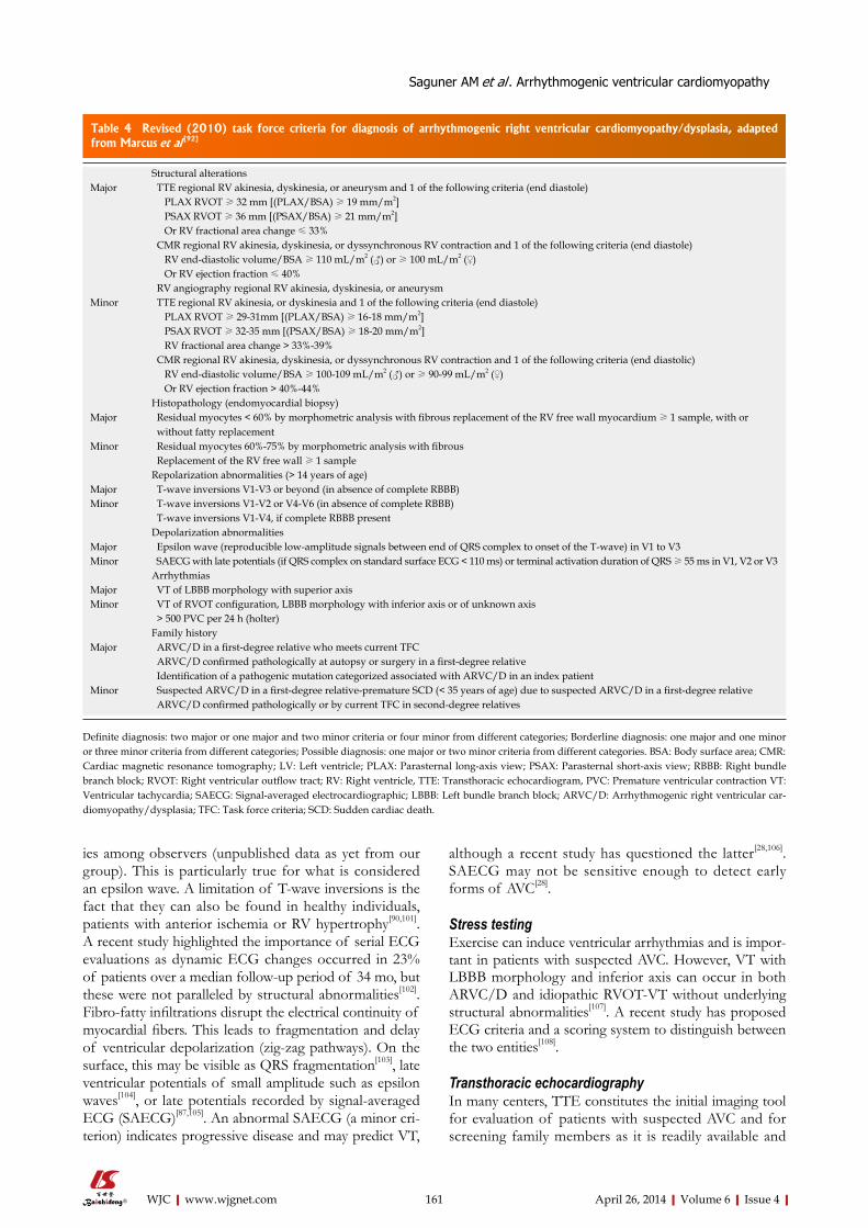
161 April 26, 2014|Volume 6|Issue 4|WJC|www.wjgnet.com
ies among observers (unpublished data as yet from our group). This is particularly true for what is considered an epsilon wave. A limitation of T-wave inversions is the fact that they can also be found in healthy individuals, patients with anterior ischemia or RV hypertrophy[90,101]. A recent study highlighted the importance of serial ECG evaluations as dynamic ECG changes occurred in 23% of patients over a median follow-up period of 34 mo, but these were not paralleled by structural abnormalities[102]. Fibro-fatty infiltrations disrupt the electrical continuity of myocardial fibers. This leads to fragmentation and delay of ventricular depolarization (zig-zag pathways). On the surface, this may be visible as QRS fragmentation[103], late ventricular potentials of small amplitude such as epsilon waves[104], or late potentials recorded by signal-averaged ECG (SAECG)[87,105]. An abnormal SAECG (a minor cri-terion) indicates progressive disease and may predict VT,
although a recent study has questioned the latter[28,106]. SAECG may not be sensitive enough to detect early forms of AVC[28].
Stress testingExercise can induce ventricular arrhythmias and is impor-tant in patients with suspected AVC. However, VT with LBBB morphology and inferior axis can occur in both ARVC/D and idiopathic RVOT-VT without underlying structural abnormalities[107]. A recent study has proposed ECG criteria and a scoring system to distinguish between the two entities[108].
Transthoracic echocardiographyIn many centers, TTE constitutes the initial imaging tool for evaluation of patients with suspected AVC and for screening family members as it is readily available and
Saguner AM et al . Arrhythmogenic ventricular cardiomyopathy
Table 4 Revised (2010) task force criteria for diagnosis of arrhythmogenic right ventricular cardiomyopathy/dysplasia, adapted from Marcus et al [92]
Structural alterationsMajor TTE regional RV akinesia, dyskinesia, or aneurysm and 1 of the following criteria (end diastole)
PLAX RVOT ≥ 32 mm [(PLAX/BSA) ≥ 19 mm/m2] PSAX RVOT ≥ 36 mm [(PSAX/BSA) ≥ 21 mm/m2] Or RV fractional area change ≤ 33% CMR regional RV akinesia, dyskinesia, or dyssynchronous RV contraction and 1 of the following criteria (end diastole) RV end-diastolic volume/BSA ≥ 110 mL/m2 (♂) or ≥ 100 mL/m2 (♀) Or RV ejection fraction ≤ 40% RV angiography regional RV akinesia, dyskinesia, or aneurysm
Minor TTE regional RV akinesia, or dyskinesia and 1 of the following criteria (end diastole) PLAX RVOT ≥ 29-31mm [(PLAX/BSA) ≥ 16-18 mm/m2] PSAX RVOT ≥ 32-35 mm [(PSAX/BSA) ≥ 18-20 mm/m2] RV fractional area change > 33%-39% CMR regional RV akinesia, dyskinesia, or dyssynchronous RV contraction and 1 of the following criteria (end diastolic) RV end-diastolic volume/BSA ≥ 100-109 mL/m2 (♂) or ≥ 90-99 mL/m2 (♀) Or RV ejection fraction > 40%-44%Histopathology (endomyocardial biopsy)
Major Residual myocytes < 60% by morphometric analysis with fibrous replacement of the RV free wall myocardium ≥ 1 sample, with or without fatty replacement
Minor Residual myocytes 60%-75% by morphometric analysis with fibrous Replacement of the RV free wall ≥ 1 sampleRepolarization abnormalities (> 14 years of age)
Major T-wave inversions V1-V3 or beyond (in absence of complete RBBB)Minor T-wave inversions V1-V2 or V4-V6 (in absence of complete RBBB)
T-wave inversions V1-V4, if complete RBBB presentDepolarization abnormalities
Major Epsilon wave (reproducible low-amplitude signals between end of QRS complex to onset of the T-wave) in V1 to V3Minor SAECG with late potentials (if QRS complex on standard surface ECG < 110 ms) or terminal activation duration of QRS ≥ 55 ms in V1, V2 or V3
ArrhythmiasMajor VT of LBBB morphology with superior axisMinor VT of RVOT configuration, LBBB morphology with inferior axis or of unknown axis
> 500 PVC per 24 h (holter)Family history
Major ARVC/D in a first-degree relative who meets current TFC ARVC/D confirmed pathologically at autopsy or surgery in a first-degree relative Identification of a pathogenic mutation categorized associated with ARVC/D in an index patient
Minor Suspected ARVC/D in a first-degree relative-premature SCD (< 35 years of age) due to suspected ARVC/D in a first-degree relative ARVC/D confirmed pathologically or by current TFC in second-degree relatives
Definite diagnosis: two major or one major and two minor criteria or four minor from different categories; Borderline diagnosis: one major and one minor or three minor criteria from different categories; Possible diagnosis: one major or two minor criteria from different categories. BSA: Body surface area; CMR: Cardiac magnetic resonance tomography; LV: Left ventricle; PLAX: Parasternal long-axis view; PSAX: Parasternal short-axis view; RBBB: Right bundle branch block; RVOT: Right ventricular outflow tract; RV: Right ventricle, TTE: Transthoracic echocardiogram, PVC: Premature ventricular contraction VT: Ventricular tachycardia; SAECG: Signal-averaged electrocardiographic; LBBB: Left bundle branch block; ARVC/D: Arrhythmogenic right ventricular car-diomyopathy/dysplasia; TFC: Task force criteria; SCD: Sudden cardiac death.
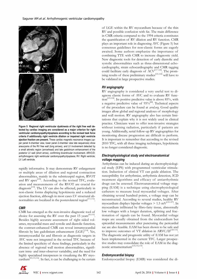
162 April 26, 2014|Volume 6|Issue 4|WJC|www.wjgnet.com
rapidly informative. It may demonstrate RV enlargement or multiple areas of dilation and regional contraction abnormalities, mainly in the subtricuspid region, RVOT and RV apex[109]. According to the revised TFC, evalu-ation and measurements of the RVOT are crucial for diagnosis[110]. The LV can also be affected, particularly in non-classic forms displaying hypokinesia and a reduced ejection fraction, although in most cases LV structural ab-normalities are localized in the posterolateral region[111,112].
CMRCMR has emerged as the non-invasive diagnostic tool of choice for assessing the RV over the past 15 years[45,113]. Besides highly accurate assessment of right sided vol-umes, myocardial mass and systolic and diastolic function, the contrast-enhanced CMR can reveal intramyocardial fibrosis by late gadolinium enhancement (LGE)[114]. Yet, intramyocardial fat and fibrosis as diagnostic targets in AVC were not integrated in the revised TFC because of the limited specificity of these findings, particularly in the absence of regional wall motion abnormalities, signifi-cant intra- and inter-observer variability, and the need for highly specialized interpreters in visualizing the RV myo-cardium[45,115,116]. In fact, it can be challenging to be certain
of LGE within the RV myocardium because of the thin RV and possible confusion with fat. The main difference in CMR criteria compared to the 1994 criteria constitutes the quantification of RV dilation and RV function. CMR plays an important role in diagnosing AVC (Figure 5) but consensus guidelines for non-classic forms are eagerly awaited. Some authors emphasize the importance of combining TTE with CMR to increase diagnostic yield. New diagnostic tools for detection of early diastolic and systolic abnormalities such as three-dimensional echo-cardiography, strain echocardiography and CMR tagging could facilitate early diagnosis of ACV[117-120]. The prom-ising results of these preliminary studies[121,122] will have to be validated in large prospective studies.
RV angiographyRV angiography is considered a very useful test to di-agnose classic forms of AVC and to evaluate RV func-tion[123,124]. Its positive predictive value is above 85%, with a negative predictive value of 95%[88]. Technical aspects of the procedure can be found at arvd.org. Good quality images allow global and regional analyses of morphology and wall motion. RV angiography also has certain limi-tations that explain why it is not widely used in clinical practice. Clinicians want to offer non-invasive strategies without ionising radiation, particularly if patients are young. Additionally, serial follow-up RV angiographies for monitoring disease progression are difficult to perform. It is important to remember that according to the revised 2010 TFC, with all three imaging techniques, hypokinesia is no longer considered diagnostic.
Electrophysiological study and electroanatomical voltage mappingArrhythmias can be induced during an electrophysiologi-cal study (EPS) with programmed ventricular stimula-tion. Induction of clinical VT can guide ablation. The susceptibility for arrhythmias, arrhythmia detection, ICD treatment algorithms and efficacy of antiarrhythmic drugs can be assessed. Electroanatomical voltage map-ping (EAM) is a technique using electrophysiological catheters to measure local myocardial voltages. After obtaining several hundred points, a voltage map can be reconstructed. According to several studies, healthy RV myocardium displays bipolar voltages > 1.5 mV[125-127]. In myocardium infiltrated by fibro-fatty tissue, abnormally low voltages with a longer duration, splitting and frac-tionation of signals can be found. Myocardial voltage maps are usually obtained from the endocardium but epicardial measurements after puncturing the pericardial sac are also feasible. EAM has been shown to be safe and to improve outcomes of VT ablation in ARVC/D[128-131]. The diagnostic and prognostic utility of EAM has not yet been implemented in the current TFC. Larger prospec-tive studies may consolidate the role of EAM in the diag-nostic armamentarium[125,132,133].
Endomyocardial biopsyEndomyocardial biopsy (EMB) was considered the di-
Saguner AM et al . Arrhythmogenic ventricular cardiomyopathy
Figure 5 Regional right ventricular dyskinesia of the right free wall de-tected by cardiac imaging are considered as a major criterion for right ventricular cardiomyopathy/dysplasia according to the revised task force criteria if additionally right ventricle dilation or impaired right ventricle ejection fraction are present. These cardiac magnetic resonance images (up-per panel 4-chamber view, lower panel 2-chamber view late sequences) show aneurysms of the RV free wall (long arrows), and LV involvement detected by a small akinetic region (arrowhead) and late gadolinium enhancement of the posterior LV wall (short arrow), confirming biventricular involvement. ARVC/D: arrhythmogenic right ventricular cardiomyopathy/dysplasia; RV: Right ventricle; LV: Left ventricle.
OS 2D GRS2
D GR
163 April 26, 2014|Volume 6|Issue 4|WJC|www.wjgnet.com
agnostic gold standard for AVC for a long time. It may allow confirmation of AVC in an index patient and ex-clude potential differential diagnoses such as sarcoidosis or Chagas disease. However, EMB are commonly taken from the thicker RV septum to assure a safe procedure. It was recognized that the septum is often spared by fibro-fatty infiltration and thus often yields false-negative results[132,134]. Nevertheless, septal EMB can identify other conditions such as sarcoidosis, myocarditis and IMF. EMB from diseased regions is problematic as these re-gions are often difficult to reach, very thin and sample acquisition carries an increased risk of perforation and tamponade[4]. Histological analysis should best be per-formed by an expert cardiac pathologist who judges the amount of surviving myocytes and fibro-fatty replace-ment. The results can be allocated as one major or one minor criterion according to the revised TFC. As AVC is patchy, several biopsies should be obtained. EAM-guided biopsies taken from low-voltage areas may improve di-agnostic yield and better distinguish between myocarditis or sarcoidosis[14,125,135]. Serious concerns remain about the hazards of sampling thin areas, although complication rates in preliminary studies were low[14]. Moreover, EAM-guided EMB may be of limited value in early stages of AVC when serious arrhythmias occur in the absence of gross structural abnormalities. Additional immunohisto-chemical staining of the intercalated disk, e.g., with plako-globin, may turn into a valuable tool for pathologists in the future but results very much depend on the protocols used[2]. Confirmation of typical histological changes by cardiac surgery or necropsy can help to confirm the diag-nosis and exclude differential diagnoses.
Genetic testingA consensus statement from the HRS and the EHRA regarding genetic testing in AVC was published recently[1]. The major purposes of genetic testing are to confirm AVC in probands with a high (Class Ⅱa recommenda-tion, level of evidence C) or intermediate (at least 1 major or 2 minor criteria; Class Ⅱb recommendation, level of evidence C) clinical suspicion and to identify genetically-affected relatives harboring the pathogenic mutation (Class Ⅰ recommendation, level of evidence C), particular-ly those without overt disease. Genetic testing in probands fulfilling only one minor criterion is not recommended. A family background and identification of a pathogenic mutation has been demonstrated in up to 50%, while in the remaining probands, an underlying familial disease with incomplete penetrance cannot be excluded. The most common mutations are found in PKP2 (80% of mutations in the Dutch and Northern American cohorts) and DSP (39% in the Italian cohort)[80], followed by DSG2[8,47,62,136,137]. It should be kept in mind that molecu-lar genetic testing may only support a clinical diagnosis or suspicion. A negative test does not rule out AVC, because other causal genetic mutations and unknown environ-mental factors may also cause the disease[47,138]. Pathogen-ic mutations do not make a diagnosis of AVC itself, as multiple sources of diagnostic information such as ECG
changes, ventricular arrhythmias and ventricular abnor-malities have to be considered[56]. Yet, the identification of pathogenic mutations may be useful in the differential diagnosis of AVC and phenocopies, such as myocarditis, idiopathic RVOT tachycardia, DCM, muscular dystro-phies, IMF or sarcoidosis[35]. Cascade genetic screening of relatives may offer another strategy to serial non-invasive cardiovascular evaluation of family members.
Current guidelines[1,87] do not recommend genetic testing for risk stratification and therapeutic decision making in AVC because study results regarding the ability of geno-typing to detect malignant mutations associated with an increased susceptibility to potentially lethal arrhythmias have been conflicting[8,62,64,90,139]. Recent large scale stud-ies[8,62] indicate an association between positive mutation carrier status and early disease onset. Thus, genotyping of younger family members should strongly be encouraged. This might be particularly important for patients carrying digenic or compound heterozygote mutations that are reported in up to 18% of the AVC population studied and have been associated with a stronger phenotype[1]. Issues such as the availability of genetic counselling in a multidisciplinary setting[140], low-probability mutations[136], genetic testing for “low-probability” AVC, psychological repercussions of young patients, and costs need to be considered before performing genetic screening[75].
DIFFERENTIAL DIAGNOSISIdiopathic RVOT-VT is a major non-hereditary differen-tial diagnosis that has to be distinguished from ARVC/D. This is often demanding, particularly in the early stages of AVC[141]. RVOT-VT is not associated with structural heart disease and thus has a more benign course. Its eti-ology is unclear, although in one study a somatic point mutation in the inhibitory G protein Gai2 was identified by EMB from the arrhythmic focus[142]. In RVOT-VT, 12-lead surface ECG and SAECG are normal during sinus rhythm. It is characterized by repetitive monomor-phic VT of a single morphology with LBBB morphology and an inferior axis. Similar VT morphologies can be found in patients with ARVC/D. 12-lead ECG scoring systems to differentiate both types of VT have recently been proposed[108]. In ARVC/D the duration of the QRS complex during VT is usually longer (≥ 120 ms in lead Ⅰ)[143]. Notching of the QRS and precordial transi-tion in lead V6 may exclusively be seen in ARVC/D[144]. RVOT-VT is difficult to induce by programmed ventricu-lar stimulation during EPS, particularly in the absence of isoproterenol[87]. It responds well to beta-blockers or verapamil and ablation after successful mapping is usually curative. EAM demonstrates normal voltages. CPVT is caused by mutations in the RyR2 gene, which has also been described in ARVC/D subtype 2. CPVT is characterized by effort-induced polymorphic VT in patients with structurally normal hearts. Genetic analysis, a positive family history, EAM and EMB can help to dif-ferentiate AVC and regional myocarditis[14]. Myocardial involvement in sarcoidosis can mimic ARVC/D and the
Saguner AM et al . Arrhythmogenic ventricular cardiomyopathy
164 April 26, 2014|Volume 6|Issue 4|WJC|www.wjgnet.com
current TFC do not reliably distinguish between them. In a prospective study of patients with suspected ARVC/D, evaluated by a protocol including EMB, a surprisingly high incidence (15%) of cardiac sarcoidosis was veri-fied[145]. Sarcoidosis with cardiac involvement thus always needs to be considered, particularly if respiratory and sys-temic symptoms, high-grade atrioventricular conduction block, and no family disease are present. Similar clinical presentations and imaging findings can pose a challenge in the absence of histological diagnosis. Features favor-ing cardiac sarcoidosis include early septal involvement, reduced LV function, a wide QRS during VT, right-sided apical VT and more inducible forms of monomorphic VT[146]. Diagnosis is usually confirmed by EMB[147]. In patients who survive SCD, ischemic heart disease and an anomalous origin of the coronary arteries have to be ex-cluded. DCM is particularly difficult to distinguish from non-classic forms of AVC. Palpitations, (pre)syncope and ventricular arrhythmias are present at an early stage in AVC, often in the absence of gross structural abnormali-ties, which is usually not the case in DCM. Subepicardial LGE on CMR, particularly in the posterobasal LV wall, also favors AVC[36]. Atrioventricular conduction block is more common in DCM, but mutations in lamin A/C can cause AVC with conduction defects[148]. Bs may mimic ARVC as RV conduction delay has been demonstrated in both and recently a genetic overlap between these two entities has been proposed[94,149]. The presence of gross structural abnormalities favors AVC and mutations in SCN5A are very rare in AVC. Further differential diag-noses include RV infarction, pulmonary hypertension, congenital left-to-right shunts, Chagas disease and Uhl’s disease (congenital hypoplastic RV).
DISEASE COURSE AND PROGNOSISAlthough AVC is a progressive disease, the individual disease course can vary considerably. The mortality rate is currently estimated to be around 1%-3% per year. In one study, after 8 years of mean follow-up, total mortality was approximately 20% and the mean age at death 54 ±19 years. Most patients died of progressive heart failure (59%) and VTA (29%)[150]. Embolic stroke may lead to death in a smaller proportion of patients.
AVC occurs in four phases[2]: (1) concealed phase, during which patients are asymptomatic and structural abnormalities are absent or subtle. Nevertheless, AVC can present with SCD as the primary manifestation; (2) oc-currence of symptomatic arrhythmias; (3) early heart fail-ure symptoms; and (4) end-stage heart failure necessitat-ing a ventricular assist device or cardiac transplantation. One study has shown that 7% of AVC patients received cardiac transplantation after a mean follow-up period of 10 years and severe LV involvement is often present in this population[7]. Strenuous physical activity often leads to early disease manifestation and rapid disease progres-sion. Young competitive athletes with AVC have a 5-fold increased risk of SCD compared to non-athletes and
identification of affected athletes by pre-participation screening has substantially reduced mortality in this co-hort[64,151]. Interestingly, in one study, mutation-carrying female relatives were less frequently affected than male relatives. This has been interpreted as prevention of apoptosis in cardiac myocytes by estradiol but could also be related to more life-long physical activity in men[152].
RISK STRATIFICATIONSCD in patients with AVC is difficult to predict and often occurs without alarming symptoms. The only reliable strategy for SCD prevention is the implantation of an ICD, with an annual incidence of appropriate ICD inter-ventions among AVC patients of 5%-22%, demonstrat-ing its importance for these patients. Thus, in secondary prevention after aborted SCD, VF or sustained VT, ICD implantation is recommended[87,147]. Besides aborted SCD, VF and sustained VT, other potential risk factors for SCD or appropriate ICD therapy (a surrogate marker for SCD) have been suggested: (1) syncope (DARVIN 2 study)[93]; (2) left ventricular dysfunction[7,56,153]; (3) young age at presentation[62,63,67] and young age per se[47,64]; (4) RV structural abnormalities fulfilling 2010 TFC[47,154]; (5) severe tricuspid regurgitation[7]; (6) particular genetic vari-ants[8,72]; (7) presence of non-sustained VT[155]; (8) male gender[79]; (9) proband status[79]; (10) frequent PVC[79]; and (11) presence of precordial T-wave inversions[79].
It is important to recognize that the use of appropri-ate ICD therapy due to sustained VT or VF as a surro-gate for SCD can result in an overestimation of this end-point. Whether in the absence of arrhythmic syncope or significant ventricular arrhythmias the other potential risk factors are consistently related to an adverse arrhythmic outcome and require prophylactic ICD therapy remains to be determined by future studies. Of note, young pa-tients may suffer from neurocardiogenic syncope, making differential diagnosis difficult and its prognostic value elusive. T-waves in the precordial and inferior leads often become negative with progression of AVC and a greater extent of precordial negative T-waves are associated with more severe RV dilation and dysfunction[100]. Recently, the Johns Hopkins group found that 88% of patients with documented sustained VTA exhibited an abnor-mal ECG. A total of 122 (84%) subjects demonstrated T-wave inversions in the precordial leads with 97 of them extending to lead V3 and beyond, while depolarization abnormalities such as epsilon waves were present only in a minority of patients[156]. The same group found that the presence of T-wave inversions in ≥ 3 precordial ECG leads was an independent predictor of adverse events during follow-up[79]. An Italian group has also demon-strated a link between the extent of negative T-waves and ventricular arrhythmic events during follow-up[157]. Although a class Ⅱb recommendation, the role of EPS with programmed ventricular stimulation for risk stratifi-cation in AVC is less well established and conflicting data about its prognostic significance exist[45,64,90,158]. Differ-
Saguner AM et al . Arrhythmogenic ventricular cardiomyopathy
165 April 26, 2014|Volume 6|Issue 4|WJC|www.wjgnet.com
ences in the studied patient population may be influenced by disease severity[159] and differences in study design may have led to discrepant results. A positive family history of SCD in asymptomatic patients does not seem to increase their individual risk for lethal arrhythmias. Guidelines do not support genetic testing for risk stratification in AVC[1] and genotype-phenotype correlation studies so far have not consistently been able to show that genotyping is able to detect mutations specifically associated with an increased susceptibility to life-threatening arrhythmic events. However, recent data indicates that certain patho-genic mutations (e.g., plakoglobin in Naxos disease, RyR2 and TME-43) may increase the risk for SCD[8,62,67]. These preliminary results have to be confirmed in larger studies and more precise risk stratification tools for asymptom-atic patients are needed. Novel imaging modalities such as strain and three-dimensional echocardiography could help to further improve risk stratification[160].
Based on the available data from observational stud-ies, we suggest classifying patients into three risk catego-ries[79,161]: (1) high risk: aborted SCD, sustained VT and VF, arrhythmic syncope; (2) moderate risk: non-sustained VT, severe structural abnormalities of RV and/or LV, presence of cardiac symptoms, ≥ 3 leads with T-wave inversions, frequent PVCs (i.e., > 760 PVC/24 h Holter) and severe disease onset age < 35 years; and (3) low risk: asymptomatic family members (also despite a positive family history of SCD), < 10 PVC/24 h Holter.
The risk factors listed here have focused largely on patients with right-dominant disease. Prognostic factors in non-classic disease still remain elusive. Patients should be astute for symptoms. Dynamic T-wave inversions, ST segment elevation and myocardial biomarker release mimicking myocardial infarction should alert the treating physicians to think of a “hot phase” of AVC. Clinical evaluation starting at age 10-12 is suggested for all first- and second-degree relatives of AVC index patients until age 60[140]. If SCD occurs at age < 35, a full postmortem autopsy by an expert cardiac pathologist including mo-lecular autopsy screening for genetic variants should be performed.
THERAPYPhysical activity restrictionIt is a general consensus that strenuous physical activity should be avoided in symptomatic patients with AVC. There is no consensus that physical activity should be avoided in asymptomatic healthy gene carriers. A recent study has shown that endurance exercise and frequent exercise increase the risk of VT/VF and heart failure in patients, but also in healthy family members carrying a pathogenic desmosomal mutation, supporting exercise restriction for these patients[82]. We prudently advise all symptomatic patients and healthy gene carriers to re-frain from practicing competitive sports and strenuous physical exercise, not only for reducing the risk of ven-tricular arrhythmias, but also to prevent disease onset
and progression.
Pharmacological therapyBeta-blockers, amiodarone and sotalol can be effective for treatment of sustained VT or VF in patients with AVC. However, they have no proven prognostic benefit such as ICD therapy. Wichter et al[107] proved that sotalol is highly effective to suppress VT by programmed ven-tricular stimulation with an efficacy of 68% and 83%, respectively, but had no effects on prognosis and SCD. Amiodarone was not superior to sotalol in this study and is not considered first-line therapy by many clinicians be-cause of frequent side effects during long term therapy, particularly in young patients. However, recent data from the Northern American ARVC registry demonstrated amiodarone to confer the greatest efficacy in prevent-ing ventricular arrhythmias when compared to sotalol or beta-blockers. However, mean sotalol doses were lower than in the study from Wichter et al[107] and only ten pa-tients were treated with amiodarone in the American study. In clinical practice, beta-blockers, sotalol or amio-darone are often used as an adjunctive therapy to reduce arrhythmia burden in patient with an ICD and amioda-rone is sometimes combined with beta-blockers in order to reduce sympathetic tone and mechanical wall stress[162]. Co-administration of sotalol and amiodarone is not recommended due to QT interval prolongation. Hiroi et al[163] suggest that carvedilol may control arrhythmias and improve LV function in some patients with biventricular AVC. Calcium antagonists such as verapamil and mexile-tin may be effective in some patients to suppress VT but data is anecdotal. If heart failure occurs, standard therapy with beta-blockers, angiotensin converting enzyme-inhibitors and a diuretic should be established, although there are no specific studies in patients with AVC[46]. Brain natriuretic peptide, C-reactive protein, IL-1β and TNF-α as surrogate biomarkers for disease activity, in-flammation and prognosis have been advocated in AVC but await further validation[3,58,164]. AVC patients at later stages have an increased risk for thromboembolism[43]. The annual incidence of thromboembolic complications, including pulmonary embolism, RVOT thrombosis and cerebrovascular events, was 0.5% in a retrospective study of 126 patients followed up for a mean period of 99 ± 64 mo[56]. Anticoagulation is often started by clinicians in the presence of severe ventricular dilation, dysfunction and aneurysm, although existing studies do not support prophylactic use in those with RV aneurysms. Data for the non-classic subtypes are lacking.
Implantable cardioverter-defibrillatorAccording to the ACC/AHA/HRS 2008 Guidelines for Device-Based Therapy of Cardiac Rhythm Abnormali-ties and its recent update[165,166], ICD implantation is indi-cated in patients with structural heart disease who have experienced a sustained VTA (secondary prevention, Class Ⅰ indication). It is also stated that ICD implanta-tion is reasonable in AVC patients who have at least one
Saguner AM et al . Arrhythmogenic ventricular cardiomyopathy
166 April 26, 2014|Volume 6|Issue 4|WJC|www.wjgnet.com
risk factor for SCD (ⅡA indication, level of evidence C). Thus, ICDs constitute a cornerstone for those patients and can prolong survival in this population. In fact, a large number of studies has demonstrated that patients with AVC who undergo ICD implantation have a high likelihood for appropriate ICD therapies[167]. However, many questions remain regarding AVC patients and their relatives who are at low to moderate risk for SCD. In these patients, a lifelong risk for lethal arrhythmias has to be weighed against the complication rates of ICDs, inadequate interventions (up to 24% within 5 years), psy-chological burden and economic costs of this therapy. However, complication rates seem to have declined since the use of third- or fourth-generation defibrilla-tors. Active and young patients are at particular risk of lead displacement and inappropriate discharges for sinus tachycardia, including painful shocks and multiple inva-sive procedures. Thus, indiscriminate device implantation cannot be endorsed. Instead, reliable risk stratification is of paramount importance. An ICD with dual chamber detection algorithms may be wise in young patients to discriminate VT or SVT from sinus tachycardia. The use of antiarrhythmic agents can also reduce the number of inadequate interventions due to supraventricular tachyar-rhythmias. Furthermore, programming of higher VT/VF cut-offs and longer detection intervals can avoid inap-propriate ICD shocks[168]. Complications of ICD therapy include a risk for perforation caused by thinning of the RV wall, lead dislodgement, R wave under-sensing and high pacing thresholds. As patients are young and mobile, these risks need particular consideration, although in one study, short and long-term risks of ICD therapy were similar to patients without AVC[45].
In our clinical routine, we recommend ICD im-plantation for all AVC patients who have experienced a sustained VTA but we also carefully evaluate ICD implantation for primary prevention in probands and family members without documented sustained VTA. Therefore, we evaluate whether a particular patient (1) has high-risk features for SCD during follow-up (see list above), (2) whether the patient is willing to take his medication regularly and to stop competitive sports (i.e., competitive individual events like triathlon or participa-tion in a competitive sports team), and (3) the patient’s preferences. Our threshold for ICD implantation is high-er in family members and asymptomatic patients owing to the fact that previous studies have consistently shown that family members are at lower risk of experiencing sustained VTA. A possible explanation for this finding is that diagnosis occurs earlier in the disease course and once diagnosed, family members are encouraged to give up competitive sports. However, more data obtained from different well characterized AVC cohorts are neces-sary to assist clinicians in guiding ICD therapy.
Catheter ablationCatheter ablation was first applied to treat drug-resistant VT. The application of direct current (DC) termed fulgu-
ration, used DC from a defibrillator to burn myocardial sites responsible for abnormal ventricular activation. The electric voltage was directly delivered through a catheter to the origins of VT. However, this procedure was as-sociated with a significant risk of complications and thus rapidly abandoned. Currently accepted indications for radiofrequency catheter ablation in patients with AVC include drug-refractory VT or incessant VT with fre-quent ICD shocks. It should be kept in mind that, unlike in patients with idiopathic VT where catheter ablation is curative, catheter ablation in patients with AVC can only improve quality of life by decreasing the number of VT episodes and PVCs[169]. Catheter ablation can follow a trial of beta-blocker therapy and antiarrhythmic therapy. In some patients who do not wish long-term therapy with beta-blockers, sotalol and particularly amiodarone, catheter ablation can be performed as first line therapy. Elimination of clinical tachycardia can relieve symptoms but may not prevent SCD.
Over the last years, mapping and ablation techniques have made outstanding progress and nowadays include activation, pace and entrainment mapping during VT and substrate-based ablation using EAM that can be performed via an endocardial and epicardial approach[170]. Substrate-based ablation of PVCs and VT is particularly important when conventional mapping during tachy-cardia is not possible due to hemodynamic instability or multiple VT morphologies[171]. Although the initial ap-proach involved extensive mapping to identify critical zones of slow conduction during VT, this approach has recently been replaced by a substrate-based approach. Preliminary studies have shown promising results regard-ing safety, arrhythmia-free survival and reduction of ICD discharges, particularly if an endocardial and epicardial approach are combined[128-131]. In one recent study from the Johns Hopkins cohort, the overall freedom from VT was 47%, 21% and 15% at 1, 5 and 10 years, respectively. Following epicardial VT ablation, the cumulative free-dom from VT was 64% and 45% at 1 and 5 years. Of note, the VT burden decreased from a median of 0.16 VT episodes per month pre ablation to 0.08 episodes per month post ablation[172]. Mid-term and long-term success and safety of these methods have to be demonstrated in future studies with larger cohorts.
Surgical methodsTotal surgical electrical RV disconnection carries an im-portant risk of postoperative RV failure and has been practically abandoned[173]. If severe therapy refractory heart failure occurs, ventricular assist devices or heart transplantation have to be considered for isolated LV or biventricular failure and less frequently isolated RV fail-ure. Some authors suggest that right heart catheterization should be performed in all cases with suspected severe RV dysfunction. If increased filling pressures suggest a Fontan-type physiology, the patient may be considered for heart transplantation[174].
Saguner AM et al . Arrhythmogenic ventricular cardiomyopathy
167 April 26, 2014|Volume 6|Issue 4|WJC|www.wjgnet.com
CONCLUSIONDuring the last three decades, our understanding of AVC from a developmental RV dysplasia with substitution by adipose tissue has remarkably changed to a mostly inherited polygenic disease of the intercalated disk with a broad phenotypic spectrum. Although AVC predomi-nantly affects the RV, non-classic forms affecting the LV or both ventricles are increasingly recognized. A hallmark is the early propensity to ventricular arrhythmias associ-ated with SCD at a young age. Enormous progress in un-ravelling the genetic and molecular basis of this complex disease, in which environmental factors seem to play a pivotal role, has been made in the last years. While prog-ress in imaging and device therapy has facilitated clini-cal diagnosis and prevention of SCD, today’s challenges include discovery of novel genetic and environmental factors, early detection of asymptomatic patients, im-proved risk stratification, catheter ablation strategies and causal therapies to cure the disease[175]. Multicenter, large, prospective follow-up studies are planned to improve our understanding of the complex underlying molecular mechanisms of AVC, which may facilitate diagnosis, risk stratification and causal therapy.
REFERENCES1 Ackerman MJ, Priori SG, Willems S, Berul C, Brugada R,
Calkins H, Camm AJ, Ellinor PT, Gollob M, Hamilton R, Hershberger RE, Judge DP, Le Marec H, McKenna WJ, Schulze-Bahr E, Semsarian C, Towbin JA, Watkins H, Wilde A, Wolpert C, Zipes DP. HRS/EHRA expert consensus state-ment on the state of genetic testing for the channelopathies and cardiomyopathies: this document was developed as a partnership between the Heart Rhythm Society (HRS) and the European Heart Rhythm Association (EHRA). Europace 2011; 13: 1077-1109 [PMID: 21810866 DOI: 10.1093/euro-pace/eur245]
2 Basso C, Corrado D, Marcus FI, Nava A, Thiene G. Arrhyth-mogenic right ventricular cardiomyopathy. Lancet 2009; 373: 1289-1300 [PMID: 19362677 DOI: 10.1016/S0140-6736(09)60256-7]
3 Frank R, Fontaine G, Vedel J, Mialet G, Sol C, Guiraudon G, Grosgogeat Y. [Electrocardiology of 4 cases of right ventricu-lar dysplasia inducing arrhythmia]. Arch Mal Coeur Vaiss 1978; 71: 963-972 [PMID: 102297]
4 Angelini A, Basso C, Nava A, Thiene G. Endomyocardial biopsy in arrhythmogenic right ventricular cardiomyopathy. Am Heart J 1996; 132: 203-206 [PMID: 8701870]
5 Richardson P, McKenna W, Bristow M, Maisch B, Mautner B, O’Connell J, Olsen E, Thiene G, Goodwin J, Gyarfas I, Martin I, Nordet P. Report of the 1995 World Health Organization/International Society and Federation of Cardiology Task Force on the Definition and Classification of cardiomyopa-thies. Circulation 1996; 93: 841-842 [PMID: 8598070]
6 Basso C, Calabrese F, Corrado D, Thiene G. Postmortem diagnosis in sudden cardiac death victims: macroscopic, microscopic and molecular findings. Cardiovasc Res 2001; 50: 290-300 [PMID: 11334833]
7 Pinamonti B, Dragos AM, Pyxaras SA, Merlo M, Pivetta A, Barbati G, Di Lenarda A, Morgera T, Mestroni L, Sinagra G. Prognostic predictors in arrhythmogenic right ventricular cardiomyopathy: results from a 10-year registry. Eur Heart J 2011; 32: 1105-1113 [PMID: 21362707 DOI: 10.1093/eur-heartj/ehr040]
8 Fressart V, Duthoit G, Donal E, Probst V, Deharo JC, Cheva-
lier P, Klug D, Dubourg O, Delacretaz E, Cosnay P, Scanu P, Extramiana F, Keller D, Hidden-Lucet F, Simon F, Bessirard V, Roux-Buisson N, Hebert JL, Azarine A, Casset-Senon D, Rouzet F, Lecarpentier Y, Fontaine G, Coirault C, Frank R, Hainque B, Charron P. Desmosomal gene analysis in ar-rhythmogenic right ventricular dysplasia/cardiomyopathy: spectrum of mutations and clinical impact in practice. Euro-pace 2010; 12: 861-868 [PMID: 20400443 DOI: 10.1093/euro-pace/euq104]
9 Delmar M, McKenna WJ. The cardiac desmosome and ar-rhythmogenic cardiomyopathies: from gene to disease. Circ Res 2010; 107: 700-714 [PMID: 20847325 DOI: 10.1161/CIR-CRESAHA.110.223412]
10 Sato PY, Coombs W, Lin X, Nekrasova O, Green KJ, Isom LL, Taffet SM, Delmar M. Interactions between ankyrin-G, Plakophilin-2, and Connexin43 at the cardiac intercalated disc. Circ Res 2011; 109: 193-201 [PMID: 21617128 DOI: 10.1161/CIRCRESAHA.111.247023]
11 Calabrese F, Basso C, Carturan E, Valente M, Thiene G. Ar-rhythmogenic right ventricular cardiomyopathy/dysplasia: is there a role for viruses? Cardiovasc Pathol 2006; 15: 11-17 [PMID: 16414451 DOI: 10.1016/j.carpath.2005.10.004]
12 Thiene G, Corrado D, Nava A, Rossi L, Poletti A, Boffa GM, Daliento L, Pennelli N. Right ventricular cardiomyopathy: is there evidence of an inflammatory aetiology? Eur Heart J 1991; 12 Suppl D: 22-25 [PMID: 1915454]
13 Chimenti C, Pieroni M, Maseri A, Frustaci A. Histologic findings in patients with clinical and instrumental diagnosis of sporadic arrhythmogenic right ventricular dysplasia. J Am Coll Cardiol 2004; 43: 2305-2313 [PMID: 15193698 DOI: 10.1016/j.jacc.2003.12.056]
14 Pieroni M, Dello Russo A, Marzo F, Pelargonio G, Casella M, Bellocci F, Crea F. High prevalence of myocarditis mim-icking arrhythmogenic right ventricular cardiomyopathy differential diagnosis by electroanatomic mapping-guided endomyocardial biopsy. J Am Coll Cardiol 2009; 53: 681-689 [PMID: 19232901 DOI: 10.1016/j.jacc.2008.11.017]
15 Corrado D, Basso C, Thiene G, McKenna WJ, Davies MJ, Fontaliran F, Nava A, Silvestri F, Blomstrom-Lundqvist C, Wlodarska EK, Fontaine G, Camerini F. Spectrum of clinico-pathologic manifestations of arrhythmogenic right ventricu-lar cardiomyopathy/dysplasia: a multicenter study. J Am Coll Cardiol 1997; 30: 1512-1520 [PMID: 9362410]
16 Gallo P, d’Amati G, Pelliccia F. Pathologic evidence of ex-tensive left ventricular involvement in arrhythmogenic right ventricular cardiomyopathy. Hum Pathol 1992; 23: 948-952 [PMID: 1644439]
17 Azaouagh A, Churzidse S, Konorza T, Erbel R. Arrhyth-mogenic right ventricular cardiomyopathy/dysplasia: a review and update. Clin Res Cardiol 2011; 100: 383-394 [PMID: 21360243 DOI: 10.1007/s00392-011-0295-2]
18 Fontaine G, Fontaliran F, Hébert JL, Chemla D, Zenati O, Lecarpentier Y, Frank R. Arrhythmogenic right ventricular dysplasia. Annu Rev Med 1999; 50: 17-35 [PMID: 10073261 DOI: 10.1146/annurev.med.50.1.17]
19 Koulouris S, Pastromas S, Sakellariou D, Kratimenos T, Mano-lis AS. Arrhythmogenic right ventricular cardiomyopathy in an octogenarian presenting with ventricular tachycardia. Pacing Clin Electrophysiol 2009; 32: e43-e47 [PMID: 19744268 DOI: 10.1111/j.1540-8159.2009.02540.x]
20 Abraham WT, Fisher WG, Smith AL, Delurgio DB, Leon AR, Loh E, Kocovic DZ, Packer M, Clavell AL, Hayes DL, Ellestad M, Trupp RJ, Underwood J, Pickering F, Truex C, McAtee P, Messenger J. Cardiac resynchronization in chron-ic heart failure. N Engl J Med 2002; 346: 1845-1853 [PMID: 12063368 DOI: 10.1056/NEJMoa013168]
21 Coonar AS, Protonotarios N, Tsatsopoulou A, Needham EWA, Houlston RS, Cliff S, Otter MI, Murday VA, Mattu RK, McKenna WJ. Gene for arrhythmogenic right ventricular cardiomyopathy with diffuse nonepidermolytic palmoplan-tar keratoderma and woolly hair (Naxos disease) maps to
Saguner AM et al . Arrhythmogenic ventricular cardiomyopathy
168 April 26, 2014|Volume 6|Issue 4|WJC|www.wjgnet.com
17q21. Circulation 1998; 97: 2049-205822 Herren T, Gerber PA, Duru F. Arrhythmogenic right ven-
tricular cardiomyopathy/dysplasia: a not so rare “disease of the desmosome” with multiple clinical presentations. Clin Res Cardiol 2009; 98: 141-158 [PMID: 19205777 DOI: 10.1007/s00392-009-0751-4]
23 La Gerche A, Robberecht C, Kuiperi C, Nuyens D, Willems R, de Ravel T, Matthijs G, Heidbuechel H. Lower than expected desmosomal gene mutation prevalence in endurance ath-letes with complex ventricular arrhythmias of right ventricu-lar origin. Heart 2010; 96: 1268-1274 [PMID: 20525856 DOI: 10.1136/hrt.2009.189621]
24 Basso C, Corrado D, Thiene G. Cardiovascular causes of sudden death in young individuals including athletes. Car-diol Rev 1999; 7: 127-135 [PMID: 10423663]
25 Tabib A, Loire R, Chalabreysse L, Meyronnet D, Miras A, Malicier D, Thivolet F, Chevalier P, Bouvagnet P. Circum-stances of death and gross and microscopic observations in a series of 200 cases of sudden death associated with arrhyth-mogenic right ventricular cardiomyopathy and/or dyspla-sia. Circulation 2003; 108: 3000-3005 [PMID: 14662701 DOI: 10.1161/01.CIR.0000108396.65446.21]
26 Nava A, Thiene G, Canciani B, Scognamiglio R, Daliento L, Buja G, Martini B, Stritoni P, Fasoli G. Familial occurrence of right ventricular dysplasia: a study involving nine families. J Am Coll Cardiol 1988; 12: 1222-1228 [PMID: 3170963]
27 Corrado D, Basso C, Buja G, Nava A, Rossi L, Thiene G. Right bundle branch block, right precordial st-segment el-evation, and sudden death in young people. Circulation 2001; 103: 710-717 [PMID: 11156883]
28 Basso C, Thiene G, Corrado D, Buja G, Melacini P, Nava A. Hypertrophic cardiomyopathy and sudden death in the young: pathologic evidence of myocardial ischemia. Hum Pathol 2000; 31: 988-998 [PMID: 10987261 DOI: 10.1053/hupa.2000.16659]
29 Basso C, Maron BJ, Corrado D, Thiene G. Clinical profile of congenital coronary artery anomalies with origin from the wrong aortic sinus leading to sudden death in young com-petitive athletes. J Am Coll Cardiol 2000; 35: 1493-1501 [PMID: 10807452]
30 Marcus FI, Fontaine G. Arrhythmogenic right ventricular dysplasia/cardiomyopathy: a review. Pacing Clin Electro-physiol 1995; 18: 1298-1314 [PMID: 7659585]
31 Sen-Chowdhry S, Morgan RD, Chambers JC, McKenna WJ. Arrhythmogenic cardiomyopathy: etiology, diagnosis, and treatment. Annu Rev Med 2010; 61: 233-253 [PMID: 20059337 DOI: 10.1146/annurev.med.052208.130419]
32 Towbin JA. Arrhythmogenic right ventricular cardiomy-opathy: a paradigm of overlapping disorders. Ann Nonin-vasive Electrocardiol 2008; 13: 325-326 [PMID: 18973488 DOI: 10.1111/j.1542-474X.2008.00241.x]
33 De Pasquale CG, Heddle WF. Left sided arrhythmogenic ventricular dysplasia in siblings. Heart 2001; 86: 128-130 [PMID: 11454821]
34 Michalodimitrakis M, Papadomanolakis A, Stiakakis J, Kanaki K. Left side right ventricular cardiomyopathy. Med Sci Law 2002; 42: 313-317 [PMID: 12487516]
35 Sen-Chowdhry S, Syrris P, McKenna WJ. Role of genetic analysis in the management of patients with arrhythmo-genic right ventricular dysplasia/cardiomyopathy. J Am Coll Cardiol 2007; 50: 1813-1821 [PMID: 17980246 DOI: 10.1016/j.jacc.2007.08.008]
36 Sen-Chowdhry S, Syrris P, Prasad SK, Hughes SE, Merri-field R, Ward D, Pennell DJ, McKenna WJ. Left-dominant ar-rhythmogenic cardiomyopathy: an under-recognized clinical entity. J Am Coll Cardiol 2008; 52: 2175-2187 [PMID: 19095136 DOI: 10.1016/j.jacc.2008.09.019]
37 Hodgkinson KA, Parfrey PS, Bassett AS, Kupprion C, Drenckhahn J, Norman MW, Thierfelder L, Stuckless SN, Dicks EL, McKenna WJ, Connors SP. The impact of implant-able cardioverter-defibrillator therapy on survival in autoso-
mal-dominant arrhythmogenic right ventricular cardiomy-opathy (ARVD5). J Am Coll Cardiol 2005; 45: 400-408 [PMID: 15680719 DOI: 10.1016/j.jacc.2004.08.068]
38 Abecasis J, Masci PG, Aquaro GD, Pingitore A, De Marchi D, Lombardi M. Arrhythmogenic biventricular dysplasia? Rev Port Cardiol 2009; 28: 1459-1463 [PMID: 20301991]
39 Corrado D, Basso C, Nava A, Rossi L, Thiene G. Sudden death in young people with apparently isolated mitral valve prolapse. G Ital Cardiol 1997; 27: 1097-1105 [PMID: 9419819]
40 Burke AP, Farb A, Tashko G, Virmani R. Arrhythmogenic right ventricular cardiomyopathy and fatty replacement of the right ventricular myocardium: are they different dis-eases? Circulation 1998; 97: 1571-1580 [PMID: 9593562]
41 Sen-Chowdhry S, Lowe MD, Sporton SC, McKenna WJ. Arrhythmogenic right ventricular cardiomyopathy: clini-cal presentation, diagnosis, and management. Am J Med 2004; 117: 685-695 [PMID: 15501207 DOI: 10.1016/j.am-jmed.2004.04.028]
42 Te Riele AS, James CA, Philips B, Rastegar N, Bhonsale A, Groeneweg JA, Murray B, Tichnell C, Judge DP, Van Der Heijden JF, Cramer MJ, Velthuis BK, Bluemke DA, Zim-merman SL, Kamel IR, Hauer RN, Calkins H, Tandri H. Mutation-positive arrhythmogenic right ventricular dyspla-sia/cardiomyopathy: the triangle of dysplasia displaced. J Cardiovasc Electrophysiol 2013; 24: 1311-1320 [PMID: 23889974 DOI: 10.1111/jce.12222]
43 Basso C, Corrado D, Thiene G. Arrhythmogenic right ven-tricular cardiomyopathy in athletes: diagnosis, management, and recommendations for sport activity. Cardiol Clin 2007; 25: 415-22, vi [PMID: 17961795 DOI: 10.1016/j.ccl.2007.08.009]
44 Kjaergaard J, Hastrup Svendsen J, Sogaard P, Chen X, Bay Nielsen H, Køber L, Kjaer A, Hassager C. Advanced quanti-tative echocardiography in arrhythmogenic right ventricular cardiomyopathy. J Am Soc Echocardiogr 2007; 20: 27-35 [PMID: 17218199 DOI: 10.1016/j.echo.2006.07.006]
45 Bomma C, Rutberg J, Tandri H, Nasir K, Roguin A, Tich-nell C, Rodriguez R, James C, Kasper E, Spevak P, Bluemke DA, Calkins H. Misdiagnosis of arrhythmogenic right ventricular dysplasia/cardiomyopathy. J Cardiovasc Elec-trophysiol 2004; 15: 300-306 [PMID: 15030420 DOI: 10.1046/j.1540-8167.2004.03429.x]
46 Gerull B, Heuser A, Wichter T, Paul M, Basson CT, Mc-Dermott DA, Lerman BB, Markowitz SM, Ellinor PT, Mac-Rae CA, Peters S, Grossmann KS, Drenckhahn J, Michely B, Sasse-Klaassen S, Birchmeier W, Dietz R, Breithardt G, Schulze-Bahr E, Thierfelder L. Mutations in the desmosomal protein plakophilin-2 are common in arrhythmogenic right ventricular cardiomyopathy. Nat Genet 2004; 36: 1162-1164 [PMID: 15489853 DOI: 10.1038/ng1461]
47 Bauce B, Rampazzo A, Basso C, Mazzotti E, Rigato I, Ste-riotis A, Beffagna G, Lorenzon A, De Bortoli M, Pilichou K, Marra MP, Corbetti F, Daliento L, Iliceto S, Corrado D, Thiene G, Nava A. Clinical phenotype and diagnosis of arrhythmogenic right ventricular cardiomyopathy in pedi-atric patients carrying desmosomal gene mutations. Heart Rhythm 2011; 8: 1686-1695 [PMID: 21723241 DOI: 10.1016/j.hrthm.2011.06.026]
48 Gomes J, Finlay M, Ahmed AK, Ciaccio EJ, Asimaki A, Saf-fitz JE, Quarta G, Nobles M, Syrris P, Chaubey S, McKenna WJ, Tinker A, Lambiase PD. Electrophysiological abnormali-ties precede overt structural changes in arrhythmogenic right ventricular cardiomyopathy due to mutations in desmoplakin-A combined murine and human study. Eur Heart J 2012; 33: 1942-1953 [PMID: 22240500 DOI: 10.1093/eurheartj/ehr472]
49 Rizzo S, Lodder EM, Verkerk AO, Wolswinkel R, Beekman L, Pilichou K, Basso C, Remme CA, Thiene G, Bezzina CR. In-tercalated disc abnormalities, reduced Na(+) current density, and conduction slowing in desmoglein-2 mutant mice prior to cardiomyopathic changes. Cardiovasc Res 2012; 95: 409-418 [PMID: 22764152 DOI: 10.1093/cvr/cvs219]
Saguner AM et al . Arrhythmogenic ventricular cardiomyopathy
169 April 26, 2014|Volume 6|Issue 4|WJC|www.wjgnet.com
50 Noorman M, Hakim S, Kessler E, Groeneweg JA, Cox MG, Asimaki A, van Rijen HV, van Stuijvenberg L, Chkourko H, van der Heyden MA, Vos MA, de Jonge N, van der Smagt JJ, Dooijes D, Vink A, de Weger RA, Varro A, de Bakker JM, Saffitz JE, Hund TJ, Mohler PJ, Delmar M, Hauer RN, van Veen TA. Remodeling of the cardiac sodium chan-nel, connexin43, and plakoglobin at the intercalated disk in patients with arrhythmogenic cardiomyopathy. Heart Rhythm 2013; 10: 412-419 [PMID: 23178689 DOI: 10.1016/j.hrthm.2012.11.018]
51 Green KJ, Gaudry CA. Are desmosomes more than teth-ers for intermediate filaments? Nat Rev Mol Cell Biol 2000; 1: 208-216 [PMID: 11252896 DOI: 10.1038/35043032]
52 Bauce B, Basso C, Rampazzo A, Beffagna G, Daliento L, Frigo G, Malacrida S, Settimo L, Danieli G, Thiene G, Nava A. Clinical profile of four families with arrhythmogenic right ventricular cardiomyopathy caused by dominant des-moplakin mutations. Eur Heart J 2005; 26: 1666-1675 [PMID: 15941723 DOI: 10.1093/eurheartj/ehi341]
53 Khan A, Mittal S, Sherrid MV. Arrhythmogenic right ven-tricular dysplasia: from genetics to treatment. Anadolu Kardi-yol Derg 2009; 9 Suppl 2: 24-31 [PMID: 20089484]
54 H Fischer A, van der Loo B, M Shär G, Zbinden R, Duru F, Brunckhorst C, Rousson V, Delacrétaz Y E, Stuber T, Oechs-lin EN, Follath F, Jenni R. Serological evidence for the asso-ciation of Bartonella henselae infection with arrhythmogenic right ventricular cardiomyopathy. Clin Cardiol 2008; 31: 469-471 [PMID: 18666174 DOI: 10.1002/clc.20269]
55 Bowles NE, Ni J, Marcus F, Towbin JA. The detection of cardiotropic viruses in the myocardium of patients with ar-rhythmogenic right ventricular dysplasia/cardiomyopathy. J Am Coll Cardiol 2002; 39: 892-895 [PMID: 11869858]
56 Basso C, Czarnowska E, Della Barbera M, Bauce B, Beffagna G, Wlodarska EK, Pilichou K, Ramondo A, Lorenzon A, Wozniek O, Corrado D, Daliento L, Danieli GA, Valente M, Nava A, Thiene G, Rampazzo A. Ultrastructural evidence of intercalated disc remodelling in arrhythmogenic right ventricular cardiomyopathy: an electron microscopy inves-tigation on endomyocardial biopsies. Eur Heart J 2006; 27: 1847-1854 [PMID: 16774985 DOI: 10.1093/eurheartj/ehl095]
57 Mallat Z, Tedgui A, Fontaliran F, Frank R, Durigon M, Fon-taine G. Evidence of apoptosis in arrhythmogenic right ven-tricular dysplasia. N Engl J Med 1996; 335: 1190-1196 [PMID: 8815941 DOI: 10.1056/NEJM199610173351604]
58 Campian ME, Tan HL, van Moerkerken AF, Tukkie R, van Eck-Smit BL, Verberne HJ. Imaging of programmed cell death in arrhythmogenic right ventricle cardiomyopathy/dysplasia. Eur J Nucl Med Mol Imaging 2011; 38: 1500-1506 [PMID: 21553091 DOI: 10.1007/s00259-011-1817-x]
59 Michaud K, Fellmann F, Abriel H, Beckmann JS, Mangin P, Elger BS. Molecular autopsy in sudden cardiac death and its implication for families: discussion of the practical, legal and ethical aspects of the multidisciplinary collaboration. Swiss Med Wkly 2009; 139: 712-718 [PMID: 20047134]
60 Matthes SA, Taffet S, Delmar M. Plakophilin-2 and the mi-gration, differentiation and transformation of cells derived from the epicardium of neonatal rat hearts. Cell Commun Adhes 2011; 18: 73-84 [PMID: 21985446 DOI: 10.3109/15419061.2011.621561]
61 Hamid MS, Norman M, Quraishi A, Firoozi S, Thaman R, Gimeno JR, Sachdev B, Rowland E, Elliott PM, McKenna WJ. Prospective evaluation of relatives for familial arrhythmo-genic right ventricular cardiomyopathy/dysplasia reveals a need to broaden diagnostic criteria. J Am Coll Cardiol 2002; 40: 1445-1450 [PMID: 12392835]
62 Rasmussen TB, Palmfeldt J, Nissen PH, Magnoni R, Dalager S, Jensen UB, Kim WY, Heickendorff L, Mølgaard H, Jensen HK, Baandrup UT, Bross P, Mogensen J. Mutated desmo-glein-2 proteins are incorporated into desmosomes and exhibit dominant-negative effects in arrhythmogenic right
ventricular cardiomyopathy. Hum Mutat 2013; 34: 697-705 [PMID: 23381804 DOI: 10.1002/humu.22289]
63 Protonotarios N, Tsatsopoulou A, Anastasakis A, Sevdalis E, McKoy G, Stratos K, Gatzoulis K, Tentolouris K, Spilio-poulou C, Panagiotakos D, McKenna W, Toutouzas P. Geno-type-phenotype assessment in autosomal recessive arrhyth-mogenic right ventricular cardiomyopathy (Naxos disease) caused by a deletion in plakoglobin. J Am Coll Cardiol 2001; 38: 1477-1484 [PMID: 11691526]
64 Basso C, Wichter T, Danieli GA, Corrado D, Czarnowska E, Fontaine G, McKenna WJ, Nava A, Protonotarios N, Anto-niades L, Wlodarska K, D’Alessi F, Thiene G. Arrhythmo-genic right ventricular cardiomyopathy: clinical registry and database, evaluation of therapies, pathology registry, DNA banking. Eur Heart J 2004; 25: 531-534 [PMID: 15039134 DOI: 10.1016/j.ehj.2003.12.025]
65 Kilic T, Babaoglu K, Aygün F, Vural A, Ural D, Agacdiken A, Anik Y, Komsuoglu B. Biventricular involvement in a Turk-ish boy with palmoplantar hyperkeratosis and curly hair, an unusual presentation of Naxos-Carvajal syndrome. Int J Cardiol 2007; 115: e122-e125 [PMID: 17125858 DOI: 10.1016/j.ijcard.2006.08.097]
66 Taylor M, Graw S, Sinagra G, Barnes C, Slavov D, Brun F, Pina-monti B, Salcedo EE, Sauer W, Pyxaras S, Anderson B, Simon B, Bogomolovas J, Labeit S, Granzier H, Mestroni L. Genetic vari-ation in titin in arrhythmogenic right ventricular cardiomyop-athy-overlap syndromes. Circulation 2011; 124: 876-885 [PMID: 21810661 DOI: 10.1161/CIRCULATIONAHA.110.005405]
67 den Haan AD, Tan BY, Zikusoka MN, Lladó LI, Jain R, Daly A, Tichnell C, James C, Amat-Alarcon N, Abraham T, Russell SD, Bluemke DA, Calkins H, Dalal D, Judge DP. Comprehensive desmosome mutation analysis in north americans with arrhythmogenic right ventricular dyspla-sia/cardiomyopathy. Circ Cardiovasc Genet 2009; 2: 428-435 [PMID: 20031617 DOI: 10.1161/CIRCGENETICS.109.858217]
68 Xu T, Yang Z, Vatta M, Rampazzo A, Beffagna G, Pilichou K, Scherer SE, Saffitz J, Kravitz J, Zareba W, Danieli GA, Lorenzon A, Nava A, Bauce B, Thiene G, Basso C, Calkins H, Gear K, Marcus F, Towbin JA. Compound and digenic het-erozygosity contributes to arrhythmogenic right ventricular cardiomyopathy. J Am Coll Cardiol 2010; 55: 587-597 [PMID: 20152563 DOI: 10.1016/j.jacc.2009.11.020]
69 Marcus FI, Fontaine GH, Guiraudon G, Frank R, Laurenceau JL, Malergue C, Grosgogeat Y. Right ventricular dysplasia: a report of 24 adult cases. Circulation 1982; 65: 384-398 [PMID: 7053899]
70 Huber O. Structure and function of desmosomal proteins and their role in development and disease. Cell Mol Life Sci 2003; 60: 1872-1890 [PMID: 14523549 DOI: 10.1007/s00018-003-3050-7]
71 Tiso N, Stephan DA, Nava A, Bagattin A, Devaney JM, Stan-chi F, Larderet G, Brahmbhatt B, Brown K, Bauce B, Muriago M, Basso C, Thiene G, Danieli GA, Rampazzo A. Identifica-tion of mutations in the cardiac ryanodine receptor gene in families affected with arrhythmogenic right ventricular cardiomyopathy type 2 (ARVD2). Hum Mol Genet 2001; 10: 189-194 [PMID: 11159936]
72 Merner ND, Hodgkinson KA, Haywood AF, Connors S, French VM, Drenckhahn JD, Kupprion C, Ramadanova K, Thierfelder L, McKenna W, Gallagher B, Morris-Larkin L, Bassett AS, Parfrey PS, Young TL. Arrhythmogenic right ventricular cardiomyopathy type 5 is a fully penetrant, lethal arrhythmic disorder caused by a missense mutation in the TMEM43 gene. Am J Hum Genet 2008; 82: 809-821 [PMID: 18313022 DOI: 10.1016/j.ajhg.2008.01.010]
73 Bennett PM, Maggs AM, Baines AJ, Pinder JC. The transi-tional junction: a new functional subcellular domain at the intercalated disc. Mol Biol Cell 2006; 17: 2091-2100 [PMID: 16481394 DOI: 10.1091/mbc.E05-12-1109]
74 Aiba T, Shimizu W, Hidaka I, Uemura K, Noda T, Zheng C,
Saguner AM et al . Arrhythmogenic ventricular cardiomyopathy
170 April 26, 2014|Volume 6|Issue 4|WJC|www.wjgnet.com
Kamiya A, Inagaki M, Sugimachi M, Sunagawa K. Cellular basis for trigger and maintenance of ventricular fibrillation in the Brugada syndrome model: high-resolution optical mapping study. J Am Coll Cardiol 2006; 47: 2074-2085 [PMID: 16697328 DOI: 10.1016/j.jacc.2005.12.064]
75 Norman M, Simpson M, Mogensen J, Shaw A, Hughes S, Syrris P, Sen-Chowdhry S, Rowland E, Crosby A, McKenna WJ. Novel mutation in desmoplakin causes arrhythmo-genic left ventricular cardiomyopathy. Circulation 2005; 112: 636-642 [PMID: 16061754 DOI: 10.1161/CIRCULA-TIONAHA.104.532234]
76 Simpson MA, Cook RW, Solanki P, Patton MA, Dennis JA, Crosby AH. A mutation in NFkappaB interacting protein 1 causes cardiomyopathy and woolly haircoat syndrome of Poll Hereford cattle. Anim Genet 2009; 40: 42-46 [PMID: 19016676 DOI: 10.1111/j.1365-2052.2008.01796.x]
77 Saguner AM, Medeiros-Domingo A, Schwyzer MA, On CJ, Haegeli LM, Wolber T, Hürlimann D, Steffel J, Krasniqi N, Rüeger S, Held L, Lüscher TF, Brunckhorst C, Duru F. Use-fulness of inducible ventricular tachycardia to predict long-term adverse outcomes in arrhythmogenic right ventricular cardiomyopathy. Am J Cardiol 2013; 111: 250-257 [PMID: 23103200 DOI: 10.1016/j.amjcard.2012.09.025]
78 Kannankeril PJ, Bhuiyan ZA, Darbar D, Mannens MM, Wilde AA, Roden DM. Arrhythmogenic right ventricular cardiomyopathy due to a novel plakophilin 2 mutation: wide spectrum of disease in mutation carriers within a fam-ily. Heart Rhythm 2006; 3: 939-944 [PMID: 16876743 DOI: 10.1016/j.hrthm.2006.04.028]
79 Bhonsale A, James CA, Tichnell C, Murray B, Madhavan S, Philips B, Russell SD, Abraham T, Tandri H, Judge DP, Calkins H. Risk stratification in arrhythmogenic right ven-tricular dysplasia/cardiomyopathy-associated desmosomal mutation carriers. Circ Arrhythm Electrophysiol 2013; 6: 569-578 [PMID: 23671136 DOI: 10.1161/CIRCEP.113.000233]
80 Rigato I, Bauce B, Rampazzo A, Zorzi A, Pilichou K, Maz-zotti E, Migliore F, Marra MP, Lorenzon A, De Bortoli M, Calore M, Nava A, Daliento L, Gregori D, Iliceto S, Thiene G, Basso C, Corrado D. Compound and digenic heterozygosity predicts lifetime arrhythmic outcome and sudden cardiac death in desmosomal gene-related arrhythmogenic right ventricular cardiomyopathy. Circ Cardiovasc Genet 2013; 6: 533-542 [PMID: 24070718 DOI: 10.1161/CIRCGENET-ICS.113.000288]
81 Wlodarska EK, Konka M, Zaleska T, Ploski R, Cedro K, Pucilowska B, Bekiesinska-Figatowska M, Rydlewska-Sadowska W, Ruzyllo W, Hoffman P. Arrhythmogenic right ventricular cardiomyopathy in two pairs of monozygotic twins. Int J Cardiol 2005; 105: 126-133 [PMID: 16243102 DOI: 10.1016/j.ijcard.2004.11.016]
82 James CA, Bhonsale A, Tichnell C, Murray B, Russell SD, Tandri H, Tedford RJ, Judge DP, Calkins H. Exercise increas-es age-related penetrance and arrhythmic risk in arrhythmo-genic right ventricular dysplasia/cardiomyopathy-associat-ed desmosomal mutation carriers. J Am Coll Cardiol 2013; 62: 1290-1297 [PMID: 23871885 DOI: 10.1016/j.jacc.2013.06.033]
83 Kaplan SR, Gard JJ, Protonotarios N, Tsatsopoulou A, Spiliopoulou C, Anastasakis A, Squarcioni CP, McKenna WJ, Thiene G, Basso C, Brousse N, Fontaine G, Saffitz JE. Re-modeling of myocyte gap junctions in arrhythmogenic right ventricular cardiomyopathy due to a deletion in plakoglobin (Naxos disease). Heart Rhythm 2004; 1: 3-11 [PMID: 15851108 DOI: 10.1016/j.hrthm.2004.01.001]
84 Jacoby D, McKenna WJ. Genetics of inherited cardiomy-opathy. Eur Heart J 2012; 33: 296-304 [PMID: 21810862 DOI: 10.1093/eurheartj/ehr260]
85 Tonet JL, Castro-Miranda R, Iwa T, Poulain F, Frank R, Fon-taine GH. Frequency of supraventricular tachyarrhythmias in arrhythmogenic right ventricular dysplasia. Am J Cardiol 1991; 67: 1153 [PMID: 2024612]
86 Camm CF, James CA, Tichnell C, Murray B, Bhonsale A, te Riele AS, Judge DP, Tandri H, Calkins H. Prevalence of atrial arrhythmias in arrhythmogenic right ventricular dysplasia/cardiomyopathy. Heart Rhythm 2013; 10: 1661-1668 [PMID: 23994726 DOI: 10.1016/j.hrthm.2013.08.032]
87 Zipes DP, Camm AJ, Borggrefe M, Buxton AE, Chaitman B, Fromer M, Gregoratos G, Klein G, Moss AJ, Myerburg RJ, Priori SG, Quinones MA, Roden DM, Silka MJ, Tracy C, Blanc JJ, Budaj A, Dean V, Deckers JW, Despres C, Dickstein K, Lekakis J, McGregor K, Metra M, Morais J, Osterspey A, Tamargo JL, Zamorano JL, Smith SC, Jacobs AK, Adams CD, Antman EM, Anderson JL, Hunt SA, Halperin JL, Nishimura R, Ornato JP, Page RL, Riegel B. ACC/AHA/ESC 2006 guidelines for management of patients with ventricular arrhythmias and the prevention of sudden cardiac death-executive summary: A report of the American College of Cardiology/American Heart Association Task Force and the European Society of Cardiology Committee for Practice Guidelines (Writing Committee to Develop Guidelines for Management of Patients with Ventricular Arrhythmias and the Prevention of Sudden Cardiac Death) Developed in col-laboration with the European Heart Rhythm Association and the Heart Rhythm Society. Eur Heart J 2006; 27: 2099-2140 [PMID: 16923744 DOI: 10.1093/eurheartj/ehl199]
88 Francés RJ. Arrhythmogenic right ventricular dysplasia/car-diomyopathy. A review and update. Int J Cardiol 2006; 110: 279-287 [PMID: 16099519 DOI: 10.1016/j.ijcard.2005.07.004]
89 Sharma S, Elliott P, Whyte G, Jones S, Mahon N, Whipp B, McKenna WJ. Utility of cardiopulmonary exercise in the as-sessment of clinical determinants of functional capacity in hypertrophic cardiomyopathy. Am J Cardiol 2000; 86: 162-168 [PMID: 10913477]
90 Basso C, Carturan E, Pilichou K, Rizzo S, Corrado D, Thiene G. Sudden cardiac death with normal heart: molecular au-topsy. Cardiovasc Pathol 2010; 19: 321-325 [PMID: 20381381 DOI: 10.1016/j.carpath.2010.02.003]
91 Uberoi A, Stein R, Perez MV, Freeman J, Wheeler M, Dewey F, Peidro R, Hadley D, Drezner J, Sharma S, Pelliccia A, Corrado D, Niebauer J, Estes NA, Ashley E, Froelicher V. Interpretation of the electrocardiogram of young athletes. Circulation 2011; 124: 746-757 [PMID: 21824936 DOI: 10.1161/CIRCULATIONAHA.110.013078]
92 Marcus FI, McKenna WJ, Sherrill D, Basso C, Bauce B, Bluemke DA, Calkins H, Corrado D, Cox MG, Daubert JP, Fontaine G, Gear K, Hauer R, Nava A, Picard MH, Proto-notarios N, Saffitz JE, Sanborn DM, Steinberg JS, Tandri H, Thiene G, Towbin JA, Tsatsopoulou A, Wichter T, Zareba W. Diagnosis of arrhythmogenic right ventricular cardiomy-opathy/dysplasia: proposed modification of the Task Force Criteria. Eur Heart J 2010; 31: 806-814 [PMID: 20172912 DOI: 10.1093/eurheartj/ehq025]
93 Corrado D, Calkins H, Link MS, Leoni L, Favale S, Bevi-lacqua M, Basso C, Ward D, Boriani G, Ricci R, Piccini JP, Dalal D, Santini M, Buja G, Iliceto S, Estes NA, Wichter T, McKenna WJ, Thiene G, Marcus FI. Prophylactic implant-able defibrillator in patients with arrhythmogenic right ven-tricular cardiomyopathy/dysplasia and no prior ventricular fibrillation or sustained ventricular tachycardia. Circulation 2010; 122: 1144-1152 [PMID: 20823389 DOI: 10.1161/CIRCU-LATIONAHA.109.913871]
94 Letsas KP, Efremidis M, Weber R, Korantzopoulos P, Proto-notarios N, Prappa E, Kounas SP, Evagelidou EN, Xydonas S, Kalusche D, Sideris A, Arentz T. Epsilon-like waves and ventricular conduction abnormalities in subjects with type 1 ECG pattern of Brugada syndrome. Heart Rhythm 2011; 8: 874-878 [PMID: 21315837 DOI: 10.1016/j.hrthm.2011.01.043]
95 Cox MG, van der Zwaag PA, van der Werf C, van der Smagt JJ, Noorman M, Bhuiyan ZA, Wiesfeld AC, Volders PG, van Langen IM, Atsma DE, Dooijes D, van den Wijngaard A, Houweling AC, Jongbloed JD, Jordaens L, Cramer MJ,
Saguner AM et al . Arrhythmogenic ventricular cardiomyopathy
171 April 26, 2014|Volume 6|Issue 4|WJC|www.wjgnet.com
Doevendans PA, de Bakker JM, Wilde AA, van Tintelen JP, Hauer RN. Arrhythmogenic right ventricular dyspla-sia/cardiomyopathy: pathogenic desmosome mutations in index-patients predict outcome of family screening: Dutch arrhythmogenic right ventricular dysplasia/cardiomyopa-thy genotype-phenotype follow-up study. Circulation 2011; 123: 2690-2700 [PMID: 21606396 DOI: 10.1161/CIRCULA-TIONAHA.110.988287]
96 Vermes E, Strohm O, Otmani A, Childs H, Duff H, Friedrich MG. Impact of the revision of arrhythmogenic right ven-tricular cardiomyopathy/dysplasia task force criteria on its prevalence by CMR criteria. JACC Cardiovasc Imaging 2011; 4: 282-287 [PMID: 21414577 DOI: 10.1016/j.jcmg.2011.01.005]
97 Szymański P, Klisiewicz A, Hoffman P. ARVC/D task force imaging criteria: it is difficult to get along with the guide-lines. JACC Cardiovasc Imaging 2011; 4: 686 [PMID: 21679906 DOI: 10.1016/j.jcmg.2011.04.002]
98 Ananthasubramaniam K, Khaja F. Arrhythmogenic right ventricular dysplasia/cardiomyopathy: review for the clini-cian. Prog Cardiovasc Dis 1998; 41: 237-246 [PMID: 9872609]
99 Piccini JP, Nasir K, Bomma C, Tandri H, Dalal D, Tichnell C, James C, Crosson J, Calkins H. Electrocardiographic find-ings over time in arrhythmogenic right ventricular dyspla-sia/cardiomyopathy. Am J Cardiol 2005; 96: 122-126 [PMID: 15979449 DOI: 10.1016/j.amjcard.2005.02.057]
100 Steriotis AK, Bauce B, Daliento L, Rigato I, Mazzotti E, Foli-no AF, Marra MP, Brugnaro L, Nava A. Electrocardiographic pattern in arrhythmogenic right ventricular cardiomyopa-thy. Am J Cardiol 2009; 103: 1302-1308 [PMID: 19406276 DOI: 10.1016/j.amjcard.2009.01.017]
101 Bauce B, Nava A, Rampazzo A, Daliento L, Muriago M, Basso C, Thiene G, Danieli GA. Familial effort polymorphic ventricular arrhythmias in arrhythmogenic right ventricular cardiomyopathy map to chromosome 1q42-43. Am J Cardiol 2000; 85: 573-579 [PMID: 11078270]
102 Quarta G, Ward D, Tomé Esteban MT, Pantazis A, Elliott PM, Volpe M, Autore C, McKenna WJ. Dynamic electrocar-diographic changes in patients with arrhythmogenic right ventricular cardiomyopathy. Heart 2010; 96: 516-522 [PMID: 20350987 DOI: 10.1136/hrt.2009.182949]
103 Canpolat U, Kabakçi G, Aytemir K, Dural M, Sahiner L, Yorgun H, Sunman H, Bariş Kaya E, Tokgözoğlu L, Oto A. Fragmented QRS complex predicts the arrhythmic events in patients with arrhythmogenic right ventricular cardiomyop-athy/dysplasia. J Cardiovasc Electrophysiol 2013; 24: 1260-1266 [PMID: 23845044 DOI: 10.1111/jce.12202]
104 Fontaine G, Frank R, Tonet JL, Guiraudon G, Cabrol C, Chomette G, Grosgogeat Y. Arrhythmogenic right ven-tricular dysplasia: a clinical model for the study of chronic ventricular tachycardia. Jpn Circ J 1984; 48: 515-538 [PMID: 6376841]
105 Kinoshita O, Fontaine G, Rosas F, Elias J, Iwa T, Tonet J, Lascault G, Frank R. Time- and frequency-domain analyses of the signal-averaged ECG in patients with arrhythmogenic right ventricular dysplasia. Circulation 1995; 91: 715-721 [PMID: 7828298]
106 Breithardt G, Borggrefe M. Pathophysiological mechanisms and clinical significance of ventricular late potentials. Eur Heart J 1986; 7: 364-385 [PMID: 3525166]
107 Wichter T, Borggrefe M, Haverkamp W, Chen X, Breithardt G. Efficacy of antiarrhythmic drugs in patients with arrhyth-mogenic right ventricular disease. Results in patients with inducible and noninducible ventricular tachycardia. Circula-tion 1992; 86: 29-37 [PMID: 1617780]
108 Hoffmayer KS, Bhave PD, Marcus GM, James CA, Tichnell C, Chopra N, Moxey L, Krahn AD, Dixit S, Stevenson W, Calkins H, Badhwar N, Gerstenfeld EP, Scheinman MM. An electrocardiographic scoring system for distinguishing right ventricular outflow tract arrhythmias in patients with arrhythmogenic right ventricular cardiomyopathy from
idiopathic ventricular tachycardia. Heart Rhythm 2013; 10: 477-482 [PMID: 23246596 DOI: 10.1016/j.hrthm.2012.12.009]
109 Baran A, Nanda NC, Falkoff M, Barold SS, Gallagher JJ. Two-dimensional echocardiographic detection of arrhyth-mogenic right ventricular dysplasia. Am Heart J 1982; 103: 1066-1067 [PMID: 7081018]
110 Yoerger DM, Marcus F, Sherrill D, Calkins H, Towbin JA, Zareba W, Picard MH. Echocardiographic findings in patients meeting task force criteria for arrhythmogenic right ventricular dysplasia: new insights from the multi-disciplinary study of right ventricular dysplasia. J Am Coll Cardiol 2005; 45: 860-865 [PMID: 15766820 DOI: 10.1016/j.jacc.2004.10.070]
111 Pinamonti B, Pagnan L, Bussani R, Ricci C, Silvestri F, Camerini F. Right ventricular dysplasia with biventricular involvement. Circulation 1998; 98: 1943-1945 [PMID: 9799217]
112 Lindström L, Nylander E, Larsson H, Wranne B. Left ven-tricular involvement in arrhythmogenic right ventricular car-diomyopathy - a scintigraphic and echocardiographic study. Clin Physiol Funct Imaging 2005; 25: 171-177 [PMID: 15888098 DOI: 10.1111/j.1475-097X.2005.00607.x]
113 Pennell D, Casolo G. Right ventricular arrhythmia: emer-gence of magnetic resonance imaging as an investigative tool. Eur Heart J 1997; 18: 1843-1845 [PMID: 9447306]
114 Dalal D, Tandri H, Judge DP, Amat N, Macedo R, Jain R, Tichnell C, Daly A, James C, Russell SD, Abraham T, Bluemke DA, Calkins H. Morphologic variants of familial ar-rhythmogenic right ventricular dysplasia/cardiomyopathy a genetics-magnetic resonance imaging correlation study. J Am Coll Cardiol 2009; 53: 1289-1299 [PMID: 19358943 DOI: 10.1016/j.jacc.2008.12.045]
115 Tandri H, Calkins H, Marcus FI. Controversial role of mag-netic resonance imaging in the diagnosis of arrhythmogenic right ventricular dysplasia. Am J Cardiol 2003; 92: 649 [PMID: 12943901]
116 Bluemke DA, Krupinski EA, Ovitt T, Gear K, Unger E, Axel L, Boxt LM, Casolo G, Ferrari VA, Funaki B, Globits S, Hig-gins CB, Julsrud P, Lipton M, Mawson J, Nygren A, Pennell DJ, Stillman A, White RD, Wichter T, Marcus F. MR Imaging of arrhythmogenic right ventricular cardiomyopathy: mor-phologic findings and interobserver reliability. Cardiology 2003; 99: 153-162 [PMID: 12824723 DOI: 10.1159/000070672]
117 Prakasa KR, Dalal D, Wang J, Bomma C, Tandri H, Dong J, James C, Tichnell C, Russell SD, Spevak P, Corretti M, Bluemke DA, Calkins H, Abraham TP. Feasibility and vari-ability of three dimensional echocardiography in arrhyth-mogenic right ventricular dysplasia/cardiomyopathy. Am J Cardiol 2006; 97: 703-709 [PMID: 16490442 DOI: 10.1016/j.amjcard.2005.11.020]
118 Prakasa KR, Wang J, Tandri H, Dalal D, Bomma C, Cho-jnowski R, James C, Tichnell C, Russell S, Judge D, Corretti M, Bluemke D, Calkins H, Abraham TP. Utility of tissue Doppler and strain echocardiography in arrhythmogenic right ventricular dysplasia/cardiomyopathy. Am J Cardiol 2007; 100: 507-512 [PMID: 17659937 DOI: 10.1016/j.amj-card.2007.03.053]
119 Teske AJ, Cox MG, De Boeck BW, Doevendans PA, Hauer RN, Cramer MJ. Echocardiographic tissue deformation im-aging quantifies abnormal regional right ventricular function in arrhythmogenic right ventricular dysplasia/cardiomyopa-thy. J Am Soc Echocardiogr 2009; 22: 920-927 [PMID: 19553080 DOI: 10.1016/j.echo.2009.05.014]
120 Jain A, Shehata ML, Stuber M, Berkowitz SJ, Calkins H, Lima JA, Bluemke DA, Tandri H. Prevalence of left ventricu-lar regional dysfunction in arrhythmogenic right ventricular dysplasia: a tagged MRI study. Circ Cardiovasc Imaging 2010; 3: 290-297 [PMID: 20197508 DOI: 10.1161/CIRCIMAG-ING.109.911313]
121 Teske AJ, Cox MG, Te Riele AS, De Boeck BW, Doevendans PA, Hauer RN, Cramer MJ. Early detection of regional func-
Saguner AM et al . Arrhythmogenic ventricular cardiomyopathy
172 April 26, 2014|Volume 6|Issue 4|WJC|www.wjgnet.com
tional abnormalities in asymptomatic ARVD/C gene carri-ers. J Am Soc Echocardiogr 2012; 25: 997-1006 [PMID: 22727198 DOI: 10.1016/j.echo.2012.05.008]
122 Vitarelli A, Cortes Morichetti M, Capotosto L, De Cicco V, Ricci S, Caranci F, Vitarelli M. Utility of strain echocar-diography at rest and after stress testing in arrhythmogenic right ventricular dysplasia. Am J Cardiol 2013; 111: 1344-1350 [PMID: 23411103 DOI: 10.1016/j.amjcard.2013.01.279]
123 Daubert C, Descaves C, Foulgoc JL, Bourdonnec C, Laurent M, Gouffault J. Critical analysis of cineangiographic criteria for diagnosis of arrhythmogenic right ventricular dysplasia. Am Heart J 1988; 115: 448-459 [PMID: 3341180]
124 Indik JH, Dallas WJ, Gear K, Tandri H, Bluemke DA, Mouk-abary T, Marcus FI. Right ventricular volume analysis by angiography in right ventricular cardiomyopathy. Int J Cardiovasc Imaging 2012; 28: 995-1001 [PMID: 21706146 DOI: 10.1007/s10554-011-9915-1]
125 Corrado D, Basso C, Leoni L, Tokajuk B, Bauce B, Frigo G, Tarantini G, Napodano M, Turrini P, Ramondo A, Daliento L, Nava A, Buja G, Iliceto S, Thiene G. Three-dimensional electroanatomic voltage mapping increases accuracy of diag-nosing arrhythmogenic right ventricular cardiomyopathy/dysplasia. Circulation 2005; 111: 3042-3050 [PMID: 15939822 DOI: 10.1161/CIRCULATIONAHA.104.486977]
126 Ejima K, Shoda M, Manaka T, Hagiwara N. Targeted endo-myocardial biopsy using electroanatomical voltage mapping in the early stage of arrhythmogenic right ventricular cardio-myopathy. Europace 2009; 11: 388-389 [PMID: 19168858 DOI: 10.1093/europace/eun357]
127 Santangeli P, Pieroni M, Dello Russo A, Casella M, Pelargo-nio G, Macchione A, Camporeale A, Smaldone C, Bartoletti S, Di Biase L, Bellocci F, Natale A. Noninvasive diagnosis of electroanatomic abnormalities in arrhythmogenic right ven-tricular cardiomyopathy. Circ Arrhythm Electrophysiol 2010; 3: 632-638 [PMID: 20937720 DOI: 10.1161/CIRCEP.110.958116]
128 Marchlinski FE, Callans DJ, Gottlieb CD, Zado E. Linear ablation lesions for control of unmappable ventricular tachy-cardia in patients with ischemic and nonischemic cardiomy-opathy. Circulation 2000; 101: 1288-1296 [PMID: 10725289]
129 Sosa E, Scanavacca M, d’Avila A, Pilleggi F. A new tech-nique to perform epicardial mapping in the electrophysiol-ogy laboratory. J Cardiovasc Electrophysiol 1996; 7: 531-536 [PMID: 8743758]
130 Bai R, Di Biase L, Shivkumar K, Mohanty P, Tung R, Santan-geli P, Saenz LC, Vacca M, Verma A, Khaykin Y, Mohanty S, Burkhardt JD, Hongo R, Beheiry S, Dello Russo A, Casella M, Pelargonio G, Santarelli P, Sanchez J, Tondo C, Natale A. Ablation of ventricular arrhythmias in arrhythmogenic right ventricular dysplasia/cardiomyopathy: arrhythmia-free survival after endo-epicardial substrate based mapping and ablation. Circ Arrhythm Electrophysiol 2011; 4: 478-485 [PMID: 21665983 DOI: 10.1161/CIRCEP.111.963066]
131 Garcia FC, Bazan V, Zado ES, Ren JF, Marchlinski FE. Epicar-dial substrate and outcome with epicardial ablation of ven-tricular tachycardia in arrhythmogenic right ventricular car-diomyopathy/dysplasia. Circulation 2009; 120: 366-375 [PMID: 19620503 DOI: 10.1161/CIRCULATIONAHA.108.834903]
132 Basso C, Ronco F, Marcus F, Abudureheman A, Rizzo S, Frigo AC, Bauce B, Maddalena F, Nava A, Corrado D, Grigo-letto F, Thiene G. Quantitative assessment of endomyocardial biopsy in arrhythmogenic right ventricular cardiomyopathy/dysplasia: an in vitro validation of diagnostic criteria. Eur Heart J 2008; 29: 2760-2771 [PMID: 18819962 DOI: 10.1093/eurheartj/ehn415]
133 Migliore F, Zorzi A, Silvano M, Bevilacqua M, Leoni L, Marra MP, Elmaghawry M, Brugnaro L, Dal Lin C, Bauce B, Rigato I, Tarantini G, Basso C, Buja G, Thiene G, Iliceto S, Corrado D. Prognostic value of endocardial voltage map-ping in patients with arrhythmogenic right ventricular car-diomyopathy/dysplasia. Circ Arrhythm Electrophysiol 2013; 6:
167-176 [PMID: 23392584 DOI: 10.1161/CIRCEP.111.974881]134 Asimaki A, Saffitz JE. The role of endomyocardial biopsy in
ARVC: looking beyond histology in search of new diagnostic markers. J Cardiovasc Electrophysiol 2011; 22: 111-117 [PMID: 21235662 DOI: 10.1111/j.1540-8167.2010.01960.x]
135 Avella A, d’Amati G, Pappalardo A, Re F, Silenzi PF, Lau-renzi F, DE Girolamo P, Pelargonio G, Dello Russo A, Barat-ta P, Messina G, Zecchi P, Zachara E, Tondo C. Diagnostic value of endomyocardial biopsy guided by electroanatomic voltage mapping in arrhythmogenic right ventricular car-diomyopathy/dysplasia. J Cardiovasc Electrophysiol 2008; 19: 1127-1134 [PMID: 18554207 DOI: 10.1111/j.1540-8167.2008.01228.x]
136 Kapplinger JD, Landstrom AP, Salisbury BA, Callis TE, Pollevick GD, Tester DJ, Cox MG, Bhuiyan Z, Bikker H, Wi-esfeld AC, Hauer RN, van Tintelen JP, Jongbloed JD, Calkins H, Judge DP, Wilde AA, Ackerman MJ. Distinguishing ar-rhythmogenic right ventricular cardiomyopathy/dysplasia-associated mutations from background genetic noise. J Am Coll Cardiol 2011; 57: 2317-2327 [PMID: 21636032 DOI: 10.1016/j.jacc.2010.12.036]
137 Bao J, Wang JZ, Yao Y, Wang YL, Fan XH, Sun K, Zhang S, Hui RT, Song L. Screening of pathogenic genes in Chinese patients with arrhythmogenic right ventricular cardio-myopathy. Chin Med J (Engl) 2013; 126: 4238-4241 [PMID: 24238504]
138 Awad MM, Calkins H, Judge DP. Mechanisms of disease: molecular genetics of arrhythmogenic right ventricular dys-plasia/cardiomyopathy. Nat Clin Pract Cardiovasc Med 2008; 5: 258-267 [PMID: 18382419 DOI: 10.1038/ncpcardio1182]
139 Dalal D, Molin LH, Piccini J, Tichnell C, James C, Bomma C, Prakasa K, Towbin JA, Marcus FI, Spevak PJ, Bluemke DA, Abraham T, Russell SD, Calkins H, Judge DP. Clinical features of arrhythmogenic right ventricular dysplasia/car-diomyopathy associated with mutations in plakophilin-2. Circulation 2006; 113: 1641-1649 [PMID: 16549640 DOI: 10.1161/CIRCULATIONAHA.105.568642]
140 Charron P, Arad M, Arbustini E, Basso C, Bilinska Z, Elliott P, Helio T, Keren A, McKenna WJ, Monserrat L, Pankuweit S, Perrot A, Rapezzi C, Ristic A, Seggewiss H, van Langen I, Tavazzi L. Genetic counselling and testing in cardiomy-opathies: a position statement of the European Society of Cardiology Working Group on Myocardial and Pericardial Diseases. Eur Heart J 2010; 31: 2715-2726 [PMID: 20823110 DOI: 10.1093/eurheartj/ehq271]
141 O’Donnell D, Cox D, Bourke J, Mitchell L, Furniss S. Clini-cal and electrophysiological differences between patients with arrhythmogenic right ventricular dysplasia and right ventricular outflow tract tachycardia. Eur Heart J 2003; 24: 801-810 [PMID: 12727147]
142 Lerman BB, Stein KM, Markowitz SM, Mittal S, Slotwiner DJ. Right ventricular outflow tract tachycardia: an update. Card Electrophysiol Rev 2002; 6: 68-71 [PMID: 11984021]
143 Ainsworth CD, Skanes AC, Klein GJ, Gula LJ, Yee R, Krahn AD. Differentiating arrhythmogenic right ventricular cardio-myopathy from right ventricular outflow tract ventricular tachycardia using multilead QRS duration and axis. Heart Rhythm 2006; 3: 416-423 [PMID: 16567288 DOI: 10.1016/j.hrthm.2005.12.024]
144 Hoffmayer KS, Machado ON, Marcus GM, Yang Y, Johnson CJ, Ermakov S, Vittinghoff E, Pandurangi U, Calkins H, Can-nom D, Gear KC, Tichnell C, Park Y, Zareba W, Marcus FI, Scheinman MM. Electrocardiographic comparison of ven-tricular arrhythmias in patients with arrhythmogenic right ventricular cardiomyopathy and right ventricular outflow tract tachycardia. J Am Coll Cardiol 2011; 58: 831-838 [PMID: 21835319 DOI: 10.1016/j.jacc.2011.05.017]
145 Vasaiwala SC, Finn C, Delpriore J, Leya F, Gagermeier J, Akar JG, Santucci P, Dajani K, Bova D, Picken MM, Basso C, Marcus F, Wilber DJ. Prospective study of cardiac sarcoid
Saguner AM et al . Arrhythmogenic ventricular cardiomyopathy
173 April 26, 2014|Volume 6|Issue 4|WJC|www.wjgnet.com
mimicking arrhythmogenic right ventricular dysplasia. J Cardiovasc Electrophysiol 2009; 20: 473-476 [PMID: 19017339 DOI: 10.1111/j.1540-8167.2008.01351.x]
146 Dechering DG, Kochhäuser S, Wasmer K, Zellerhoff S, Pott C, Köbe J, Spieker T, Piers SR, Bittner A, Mönnig G, Breithar-dt G, Wichter T, Zeppenfeld K, Eckardt L. Electrophysiologi-cal characteristics of ventricular tachyarrhythmias in cardiac sarcoidosis versus arrhythmogenic right ventricular cardio-myopathy. Heart Rhythm 2013; 10: 158-164 [PMID: 23070261 DOI: 10.1016/j.hrthm.2012.10.019]
147 Corrado D, Leoni L, Link MS, Della Bella P, Gaita F, Curnis A, Salerno JU, Igidbashian D, Raviele A, Disertori M, Zanotto G, Verlato R, Vergara G, Delise P, Turrini P, Basso C, Naccarella F, Maddalena F, Estes NA, Buja G, Thiene G. Implantable cardioverter-defibrillator therapy for prevention of sudden death in patients with arrhythmogenic right ventricular cardiomyopathy/dysplasia. Circulation 2003; 108: 3084-3091 [PMID: 14638546 DOI: 10.1161/01.CIR.0000103130.33451.D2]
148 Quarta G, Syrris P, Ashworth M, Jenkins S, Zuborne Alapi K, Morgan J, Muir A, Pantazis A, McKenna WJ, Elliott PM. Mu-tations in the Lamin A/C gene mimic arrhythmogenic right ventricular cardiomyopathy. Eur Heart J 2012; 33: 1128-1136 [PMID: 22199124 DOI: 10.1093/eurheartj/ehr451]
149 Cerrone M, Lin X, Zhang M, Agullo-Pascual E, Pfenniger A, Chkourko Gusky H, Novelli V, Kim C, Tirasawadischai T, Judge DP, Rothenberg E, Chen HV, Napolitano C, Priori SG, Delmar M. Missense mutations in plakophilin-2 can cause brugada syndrome phenotype by decreasing sodium current and nav1.5 membrane localization. Heart Rhythm 2013; 10: 1743
150 Hulot JS, Jouven X, Empana JP, Frank R, Fontaine G. Natu-ral history and risk stratification of arrhythmogenic right ventricular dysplasia/cardiomyopathy. Circulation 2004; 110: 1879-1884 [PMID: 15451782 DOI: 10.1161/01.CIR.0000143375.93288.82]
151 Corrado D, Basso C, Pavei A, Michieli P, Schiavon M, Thiene G. Trends in sudden cardiovascular death in young com-petitive athletes after implementation of a preparticipation screening program. JAMA 2006; 296: 1593-1601 [PMID: 17018804 DOI: 10.1001/jama.296.13.1593]
152 Pelzer T, Schumann M, Neumann M, deJager T, Stimpel M, Serfling E, Neyses L. 17beta-estradiol prevents programmed cell death in cardiac myocytes. Biochem Biophys Res Com-mun 2000; 268: 192-200 [PMID: 10652235 DOI: 10.1006/bbrc.2000.2073]
153 Lemola K, Brunckhorst C, Helfenstein U, Oechslin E, Jenni R, Duru F. Predictors of adverse outcome in patients with ar-rhythmogenic right ventricular dysplasia/cardiomyopathy: long term experience of a tertiary care centre. Heart 2005; 91: 1167-1172 [PMID: 16103549 DOI: 10.1136/hrt.2004.038620]
154 te Riele AS, Bhonsale A, James CA, Rastegar N, Murray B, Burt JR, Tichnell C, Madhavan S, Judge DP, Bluemke DA, Zimmerman SL, Kamel IR, Calkins H, Tandri H. Incremental value of cardiac magnetic resonance imaging in arrhythmic risk stratification of arrhythmogenic right ventricular dyspla-sia/cardiomyopathy-associated desmosomal mutation car-riers. J Am Coll Cardiol 2013; 62: 1761-1769 [PMID: 23810894 DOI: 10.1016/j.jacc.2012.11.087]
155 Bhonsale A, James CA, Tichnell C, Murray B, Gagarin D, Philips B, Dalal D, Tedford R, Russell SD, Abraham T, Tan-dri H, Judge DP, Calkins H. Incidence and predictors of im-plantable cardioverter-defibrillator therapy in patients with arrhythmogenic right ventricular dysplasia/cardiomyopa-thy undergoing implantable cardioverter-defibrillator im-plantation for primary prevention. J Am Coll Cardiol 2011; 58: 1485-1496 [PMID: 21939834 DOI: 10.1016/j.jacc.2011.06.043]
156 te Riele AS, James CA, Bhonsale A, Groeneweg JA, Camm CF, Murray B, Tichnell C, van der Heijden JF, Dooijes D, Judge DP, Hauer RN, Tandri H, Calkins H. Malignant ar-rhythmogenic right ventricular dysplasia/cardiomyopathy
with a normal 12-lead electrocardiogram: a rare but under-recognized clinical entity. Heart Rhythm 2013; 10: 1484-1491 [PMID: 23816439 DOI: 10.1016/j.hrthm.2013.06.022]
157 Zorzi A, Migliore F, Elmaghawry M, Silvano M, Marra MP, Niero A, Nguyen K, Rigato I, Bauce B, Basso C, Thiene G, Iliceto S, Corrado D. Electrocardiographic predictors of elec-troanatomic scar size in arrhythmogenic right ventricular cardiomyopathy: implications for arrhythmic risk stratifica-tion. J Cardiovasc Electrophysiol 2013; 24: 1321-1327 [PMID: 24016194 DOI: 10.1111/jce.12246]
158 Saguner AM, Duru F, Brunckhorst CB. Arrhythmogenic right ventricular cardiomyopathy: a challenging disease of the intercalated disc. Circulation 2013; 128: 1381-1386 [PMID: 24043146 DOI: 10.1161/CIRCULATIONAHA.112.001009]
159 Di Biase M, Favale S, Massari V, Amodio G, Chiddo A, Rizzon P. Programmed stimulation in patients with minor forms of right ventricular dysplasia. Eur Heart J 1989; 10 Suppl D: 49-53 [PMID: 2806304]
160 Sarvari SI, Haugaa KH, Anfinsen OG, Leren TP, Smiseth OA, Kongsgaard E, Amlie JP, Edvardsen T. Right ventricular mechanical dispersion is related to malignant arrhythmias: a study of patients with arrhythmogenic right ventricular cardiomyopathy and subclinical right ventricular dysfunc-tion. Eur Heart J 2011; 32: 1089-1096 [PMID: 21406439 DOI: 10.1093/eurheartj/ehr069]
161 Corrado D, Basso C, Pilichou K, Thiene G. Molecular biol-ogy and clinical management of arrhythmogenic right ven-tricular cardiomyopathy/dysplasia. Heart 2011; 97: 530-539 [PMID: 20930047 DOI: 10.1136/hrt.2010.193276]
162 Calkins H. Arrhythmogenic right-ventricular dysplasia/cardiomyopathy. Curr Opin Cardiol 2006; 21: 55-63 [PMID: 16355031 DOI: 10.1097/01.hco.0000198984.70884.4d]
163 Hiroi Y, Fujiu K, Komatsu S, Sonoda M, Sakomura Y, Imai Y, Oishi Y, Nakamura F, Ajiki K, Hayami N, Murakawa Y, Ohno M, Hirata Y, Ohtomo K, Nagai R. Carvedilol therapy improved left ventricular function in a patient with arrhyth-mogenic right ventricular cardiomyopathy. Jpn Heart J 2004; 45: 169-177 [PMID: 14973363]
164 Matsuo K, Nishikimi T, Yutani C, Kurita T, Shimizu W, Taguchi A, Suyama K, Aihara N, Kamakura S, Kangawa K, Takamiya M, Shimomura K. Diagnostic value of plasma levels of brain natriuretic peptide in arrhythmogenic right ventricular dysplasia. Circulation 1998; 98: 2433-2440 [PMID: 9832489]
165 Epstein AE, Dimarco JP, Ellenbogen KA, Estes NA, Freed-man RA, Gettes LS, Gillinov AM, Gregoratos G, Hammill SC, Hayes DL, Hlatky MA, Newby LK, Page RL, Schoenfeld MH, Silka MJ, Stevenson LW, Sweeney MO. ACC/AHA/HRS 2008 Guidelines for device-based therapy of cardiac rhythm abnormalities. Heart Rhythm 2008; 5: e1-62 [PMID: 18534360 DOI: 10.1016/j.hrthm.2008.04.014]
166 Epstein AE, DiMarco JP, Ellenbogen KA, Estes NA, Freed-man RA, Gettes LS, Gillinov AM, Gregoratos G, Hammill SC, Hayes DL, Hlatky MA, Newby LK, Page RL, Schoenfeld MH, Silka MJ, Stevenson LW, Sweeney MO. 2012 ACCF/AHA/HRS focused update incorporated into the ACCF/AHA/HRS 2008 guidelines for device-based therapy of cardiac rhythm abnormalities: a report of the American Col-lege of Cardiology Foundation/American Heart Association Task Force on Practice Guidelines and the Heart Rhythm So-ciety. Circulation 2013; 127: e283-e352 [PMID: 23255456 DOI: 10.1161/CIR.0b013e318276ce9b]
167 Schuler PK, Haegeli LM, Saguner AM, Wolber T, Tanner FC, Jenni R, Corti N, Lüscher TF, Brunckhorst C, Duru F. Predictors of appropriate ICD therapy in patients with ar-rhythmogenic right ventricular cardiomyopathy: long term experience of a tertiary care center. PLoS One 2012; 7: e39584 [PMID: 23028419 DOI: 10.1371/journal.pone.0039584]
168 Veltmann C, Kuschyk J, Schimpf R, Streitner F, Schoene N, Borggrefe M, Wolpert C. Prevention of inappropriate ICD
Saguner AM et al . Arrhythmogenic ventricular cardiomyopathy
174 April 26, 2014|Volume 6|Issue 4|WJC|www.wjgnet.com
shocks in patients with Brugada syndrome. Clin Res Cardiol 2010; 99: 37-44 [PMID: 19760052 DOI: 10.1007/s00392-009-0075-4]
169 Zou J, Cao K, Yang B, Chen M, Shan Q, Chen C, Li W, Haines DE. Dynamic substrate mapping and ablation of ventricular tachycardias in right ventricular dysplasia. J Interv Card Elec-trophysiol 2004; 11: 37-45 [PMID: 15273453 DOI: 10.1023/B:JICE.0000035928.54293.42]
170 Arbelo E, Josephson ME. Ablation of ventricular arrhyth-mias in arrhythmogenic right ventricular dysplasia. J Car-diovasc Electrophysiol 2010; 21: 473-486 [PMID: 20132399 DOI: 10.1111/j.1540-8167.2009.01694.x]
171 Verma A, Kilicaslan F, Schweikert RA, Tomassoni G, Ros-sillo A, Marrouche NF, Ozduran V, Wazni OM, Elayi SC, Saenz LC, Minor S, Cummings JE, Burkhardt JD, Hao S, Beheiry S, Tchou PJ, Natale A. Short- and long-term suc-cess of substrate-based mapping and ablation of ventricular tachycardia in arrhythmogenic right ventricular dysplasia. Circulation 2005; 111: 3209-3216 [PMID: 15956125 DOI: 10.1161/CIRCULATIONAHA.104.510503]
172 Philips B, Madhavan S, James C, Tichnell C, Murray B, Dalal D, Bhonsale A, Nazarian S, Judge DP, Russell SD, Abraham T, Calkins H, Tandri H. Outcomes of catheter ablation of ven-tricular tachycardia in arrhythmogenic right ventricular dys-plasia/cardiomyopathy. Circ Arrhythm Electrophysiol 2012; 5: 499-505 [PMID: 22492430 DOI: 10.1161/CIRCEP.111.968677]
173 Guiraudon GM, Klein GJ, Sharma AD, Yee R, Guiraudon CM. Surgical therapy for arrhythmogenic right ventricular adiposis. Eur Heart J 1989; 10 Suppl D: 82-83 [PMID: 2806309]
174 Gilljam T, Bergh CH. Right ventricular cardiomyopathy: timing of heart transplantation in Uhl’s anomaly and ar-rythmogenic right ventricular cardiomyopathy. Eur J Heart Fail 2009; 11: 106-109 [PMID: 19147464 DOI: 10.1093/eurjhf/hfn014]
175 Kim C, Wong J, Wen J, Wang S, Wang C, Spiering S, Kan NG, Forcales S, Puri PL, Leone TC, Marine JE, Calkins H, Kelly DP, Judge & Huei-Sheng Vincent Chen DP. Studying arrhythmogenic right ventricular dysplasia with patient-specific iPSCs. Nature 2013; 494: 105-110 [DOI: 10.1038/na-ture11799]
P- Reviewers: Ciampi Q, Nikus K, Nurzynska D S- Editor: Zhai HH L- Editor: Roemmele A E- Editor: Liu SQ
Saguner AM et al . Arrhythmogenic ventricular cardiomyopathy
High-sensitivity cardiac troponins in everyday clinical practice
Johannes Mair
Johannes Mair, Department of Internal Medicine Ⅲ, Cardiology and Angiology, Innsbruck Medical University, A-6020 Innsbruck, AustriaAuthor contributions: Mair J solely contributed to this paper.Correspondence to: Johannes Mair, MD, Associate Profes-sor, Department of Internal Medicine Ⅲ, Cardiology and Angi-ology, Innsbruck Medical University, Anichstrasse 35, A-6020 Innsbruck, Austria. [email protected]: +43-512-50481314 Fax: +43-512-50422767Received: November 10, 2013 Revised: February 15, 2014 Accepted: March 13, 2014Published online: April 26, 2014
AbstractHigh-sensitivity cardiac troponin (hs-cTn) assays are increasingly being used in many countries worldwide, however, a generally accepted definition of high-sen-sitivity is still pending. These assays enable cTn mea-surement with a high degree of analytical sensitivity with a low analytical imprecision at the low measuring range of cTn assays (coefficient of variation of < 10% at the 99th percentile upper reference limit). One of the most important advantages of these new assays is that they allow novel, more rapid approaches to rule in or rule out acute coronary syndromes (ACSs) than with previous cTn assay generations which are still more commonly used in practice worldwide. hs-cTn is also more sensitive for the detection of myocardial damage unrelated to acute myocardial ischemia. Therefore, the increase in early diagnostic sensitivity of hs-cTn assays for ACS comes at the cost of a reduced ACS specificity, because more patients with other causes of acute or chronic myocardial injury without overt myocardial isch-emia are detected than with previous cTn assays. As hs-cTn assays are increasingly being adopted in clinical practice and more hs-cTn assays are being developed, this review attempts to synthesize the available clinical data to make recommendations for their everyday clini-cal routine use.
© 2014 Baishideng Publishing Group Co., Limited. All rights
reserved.
Key words: Cardiac troponin; High-sensitivity; Diagno-sis; Acute myocardial infarction; Acute coronary syn-drome; Review
Core tip: High-sensitivity cardiac troponin (hs-cTn) as-says enable cardiac troponin measurement with a high degree of analytical sensitivity with a low analytical im-precision at the low measuring range. One of the most important advantages of these new assays is that they allow novel, more rapid approaches to rule in or rule out acute coronary syndromes (ACSs). The increase in early diagnostic sensitivity of hs-cTn assays for ACS comes at the cost of a reduced ACS specificity, because more patients with other causes of acute or chronic myocardial injury without overt myocardial ischemia are detected than with previous cTn assays.
Mair J. High-sensitivity cardiac troponins in everyday clinical practice. World J Cardiol 2014; 6(4): 175-182 Available from: URL: http://www.wjgnet.com/1949-8462/full/v6/i4/175.htm DOI: http://dx.doi.org/10.4330/wjc.v6.i4.175
INTRODUCTIONCardiac troponin I (cTnI) and cTnT are the biomark-ers of choice for the diagnosis of myocardial damage, because they are the most sensitive and cardiac-specific biomarkers currently available[1,2]. Recommendations for the use of cTn measurement in acute cardiac care[1] and practical clinical considerations in the interpretation of cTn elevations[2] have been published recently. Over the years the analytical sensitivity of cTn assays has been continuously improved, and more recently a new gen-eration of cTn assays, i.e., the high-sensitivity (hs)-cTn assays, have been introduced into routine clinical prac-tice[3]. It is important to note, that these assays measure the same analyte as previous assay generations but with
MINIREVIEWS
Online Submissions: http://www.wjgnet.com/esps/[email protected]:10.4330/wjc.v6.i4.175
175 April 26, 2014|Volume 6|Issue 4|WJC|www.wjgnet.com
World Journal of CardiologyW J C
World J Cardiol 2014 April 26; 6(4): 175-182ISSN 1949-8462 (online)
© 2014 Baishideng Publishing Group Co., Limited. All rights reserved.
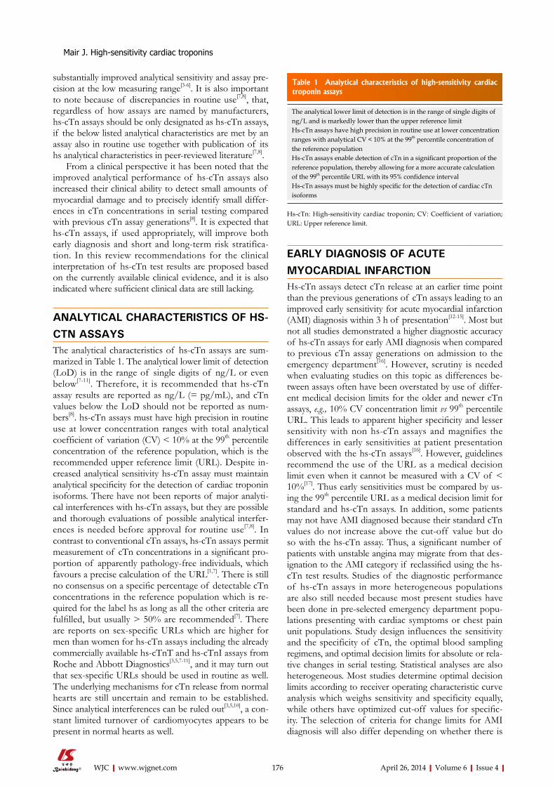
substantially improved analytical sensitivity and assay pre-cision at the low measuring range[3-6]. It is also important to note because of discrepancies in routine use[7,8], that, regardless of how assays are named by manufacturers, hs-cTn assays should be only designated as hs-cTn assays, if the below listed analytical characteristics are met by an assay also in routine use together with publication of its hs analytical characteristics in peer-reviewed literature[7,8].
From a clinical perspective it has been noted that the improved analytical performance of hs-cTn assays also increased their clinical ability to detect small amounts of myocardial damage and to precisely identify small differ-ences in cTn concentrations in serial testing compared with previous cTn assay generations[8]. It is expected that hs-cTn assays, if used appropriately, will improve both early diagnosis and short and long-term risk stratifica-tion. In this review recommendations for the clinical interpretation of hs-cTn test results are proposed based on the currently available clinical evidence, and it is also indicated where sufficient clinical data are still lacking.
ANALYTICAL CHARACTERISTICS OF HS-CTN ASSAYSThe analytical characteristics of hs-cTn assays are sum-marized in Table 1. The analytical lower limit of detection (LoD) is in the range of single digits of ng/L or even below[7-11]. Therefore, it is recommended that hs-cTn assay results are reported as ng/L (= pg/mL), and cTn values below the LoD should not be reported as num-bers[8]. hs-cTn assays must have high precision in routine use at lower concentration ranges with total analytical coefficient of variation (CV) < 10% at the 99th percentile concentration of the reference population, which is the recommended upper reference limit (URL). Despite in-creased analytical sensitivity hs-cTn assay must maintain analytical specificity for the detection of cardiac troponin isoforms. There have not been reports of major analyti-cal interferences with hs-cTn assays, but they are possible and thorough evaluations of possible analytical interfer-ences is needed before approval for routine use[7,8]. In contrast to conventional cTn assays, hs-cTn assays permit measurement of cTn concentrations in a significant pro-portion of apparently pathology-free individuals, which favours a precise calculation of the URL[1,7]. There is still no consensus on a specific percentage of detectable cTn concentrations in the reference population which is re-quired for the label hs as long as all the other criteria are fulfilled, but usually > 50% are recommended[7]. There are reports on sex-specific URLs which are higher for men than women for hs-cTn assays including the already commercially available hs-cTnT and hs-cTnI assays from Roche and Abbott Diagnostics[3,5,7-11], and it may turn out that sex-specific URLs should be used in routine as well. The underlying mechanisms for cTn release from normal hearts are still uncertain and remain to be established. Since analytical interferences can be ruled out[3,5,10], a con-stant limited turnover of cardiomyocytes appears to be present in normal hearts as well.
EARLY DIAGNOSIS OF ACUTE MYOCARDIAL INFARCTIONHs-cTn assays detect cTn release at an earlier time point than the previous generations of cTn assays leading to an improved early sensitivity for acute myocardial infarction (AMI) diagnosis within 3 h of presentation[12-15]. Most but not all studies demonstrated a higher diagnostic accuracy of hs-cTn assays for early AMI diagnosis when compared to previous cTn assay generations on admission to the emergency department[16]. However, scrutiny is needed when evaluating studies on this topic as differences be-tween assays often have been overstated by use of differ-ent medical decision limits for the older and newer cTn assays, e.g., 10% CV concentration limit vs 99th percentile URL. This leads to apparent higher specificity and lesser sensitivity with non hs-cTn assays and magnifies the differences in early sensitivities at patient presentation observed with the hs-cTn assays[16]. However, guidelines recommend the use of the URL as a medical decision limit even when it cannot be measured with a CV of < 10%[17]. Thus early sensitivities must be compared by us-ing the 99th percentile URL as a medical decision limit for standard and hs-cTn assays. In addition, some patients may not have AMI diagnosed because their standard cTn values do not increase above the cut-off value but do so with the hs-cTn assay. Thus, a significant number of patients with unstable angina may migrate from that des-ignation to the AMI category if reclassified using the hs-cTn test results. Studies of the diagnostic performance of hs-cTn assays in more heterogeneous populations are also still needed because most present studies have been done in pre-selected emergency department popu-lations presenting with cardiac symptoms or chest pain unit populations. Study design influences the sensitivity and the specificity of cTn, the optimal blood sampling regimens, and optimal decision limits for absolute or rela-tive changes in serial testing. Statistical analyses are also heterogeneous. Most studies determine optimal decision limits according to receiver operating characteristic curve analysis which weighs sensitivity and specificity equally, while others have optimized cut-off values for specific-ity. The selection of criteria for change limits for AMI diagnosis will also differ depending on whether there is
176 April 26, 2014|Volume 6|Issue 4|WJC|www.wjgnet.com
Mair J. High-sensitivity cardiac troponins
Table 1 Analytical characteristics of high-sensitivity cardiac troponin assays
The analytical lower limit of detection is in the range of single digits of ng/L and is markedly lower than the upper reference limitHs-cTn assays have high precision in routine use at lower concentration ranges with analytical CV < 10% at the 99th percentile concentration of the reference populationHs-cTn assays enable detection of cTn in a significant proportion of the reference population, thereby allowing for a more accurate calculation of the 99th percentile URL with its 95% confidence intervalHs-cTn assays must be highly specific for the detection of cardiac cTn isoforms
Hs-cTn: High-sensitivity cardiac troponin; CV: Coefficient of variation; URL: Upper reference limit.
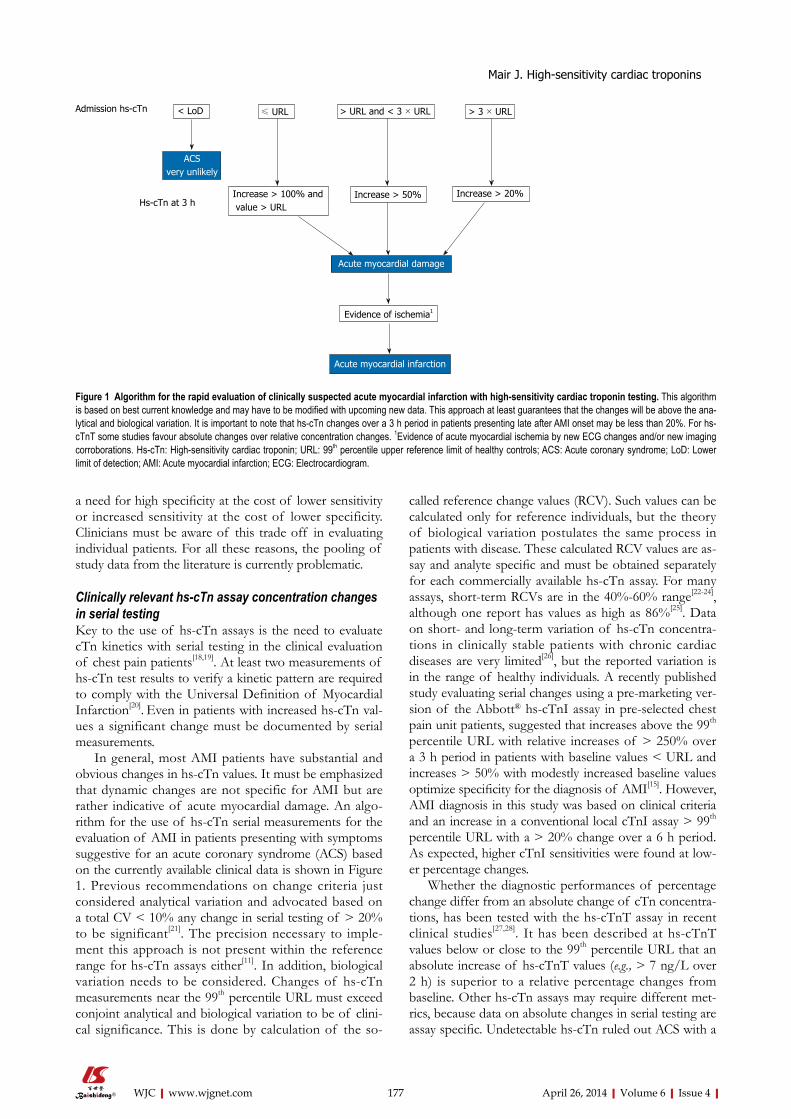
a need for high specificity at the cost of lower sensitivity or increased sensitivity at the cost of lower specificity. Clinicians must be aware of this trade off in evaluating individual patients. For all these reasons, the pooling of study data from the literature is currently problematic.
Clinically relevant hs-cTn assay concentration changes in serial testingKey to the use of hs-cTn assays is the need to evaluate cTn kinetics with serial testing in the clinical evaluation of chest pain patients[18,19]. At least two measurements of hs-cTn test results to verify a kinetic pattern are required to comply with the Universal Definition of Myocardial Infarction[20]. Even in patients with increased hs-cTn val-ues a significant change must be documented by serial measurements.
In general, most AMI patients have substantial and obvious changes in hs-cTn values. It must be emphasized that dynamic changes are not specific for AMI but are rather indicative of acute myocardial damage. An algo-rithm for the use of hs-cTn serial measurements for the evaluation of AMI in patients presenting with symptoms suggestive for an acute coronary syndrome (ACS) based on the currently available clinical data is shown in Figure 1. Previous recommendations on change criteria just considered analytical variation and advocated based on a total CV < 10% any change in serial testing of > 20% to be significant[21]. The precision necessary to imple-ment this approach is not present within the reference range for hs-cTn assays either[11]. In addition, biological variation needs to be considered. Changes of hs-cTn measurements near the 99th percentile URL must exceed conjoint analytical and biological variation to be of clini-cal significance. This is done by calculation of the so-
called reference change values (RCV). Such values can be calculated only for reference individuals, but the theory of biological variation postulates the same process in patients with disease. These calculated RCV values are as-say and analyte specific and must be obtained separately for each commercially available hs-cTn assay. For many assays, short-term RCVs are in the 40%-60% range[22-24], although one report has values as high as 86%[25]. Data on short- and long-term variation of hs-cTn concentra-tions in clinically stable patients with chronic cardiac diseases are very limited[26], but the reported variation is in the range of healthy individuals. A recently published study evaluating serial changes using a pre-marketing ver-sion of the Abbott® hs-cTnI assay in pre-selected chest pain unit patients, suggested that increases above the 99th percentile URL with relative increases of > 250% over a 3 h period in patients with baseline values < URL and increases > 50% with modestly increased baseline values optimize specificity for the diagnosis of AMI[15]. However, AMI diagnosis in this study was based on clinical criteria and an increase in a conventional local cTnI assay > 99th percentile URL with a > 20% change over a 6 h period. As expected, higher cTnI sensitivities were found at low-er percentage changes.
Whether the diagnostic performances of percentage change differ from an absolute change of cTn concentra-tions, has been tested with the hs-cTnT assay in recent clinical studies[27,28]. It has been described at hs-cTnT values below or close to the 99th percentile URL that an absolute increase of hs-cTnT values (e.g., > 7 ng/L over 2 h) is superior to a relative percentage changes from baseline. Other hs-cTn assays may require different met-rics, because data on absolute changes in serial testing are assay specific. Undetectable hs-cTn ruled out ACS with a
177 April 26, 2014|Volume 6|Issue 4|WJC|www.wjgnet.com
Admission hs-cTn
Hs-cTn at 3 h
< LoD ≤ URL > URL and < 3 × URL > 3 × URL
Increase > 100% and value > URL
Increase > 50% Increase > 20%
Evidence of ischemia1
Acute myocardial infarction
Acute myocardial damage
ACSvery unlikely
Figure 1 Algorithm for the rapid evaluation of clinically suspected acute myocardial infarction with high-sensitivity cardiac troponin testing. This algorithm is based on best current knowledge and may have to be modified with upcoming new data. This approach at least guarantees that the changes will be above the ana-lytical and biological variation. It is important to note that hs-cTn changes over a 3 h period in patients presenting late after AMI onset may be less than 20%. For hs-cTnT some studies favour absolute changes over relative concentration changes. 1Evidence of acute myocardial ischemia by new ECG changes and/or new imaging corroborations. Hs-cTn: High-sensitivity cardiac troponin; URL: 99th percentile upper reference limit of healthy controls; ACS: Acute coronary syndrome; LoD: Lower limit of detection; AMI: Acute myocardial infarction; ECG: Electrocardiogram.
Mair J. High-sensitivity cardiac troponins
178 April 26, 2014|Volume 6|Issue 4|WJC|www.wjgnet.com
not available. Thus, only the limits recommended by the universal definition of AMI can be currently used[20], i.e., increase > 5-times URL after PCI and > 10-times URL after CABG. However, these limits are still very contro-versially discussed in the communities of interventionists and cardiac surgeons, because it appears from the avail-able data that periprocedural cTn increases in clinically uncomplicated patients must be substantially higher to be of prognostic significance[29].
DO WE NEED ADDITIONAL BIOMARKERS FOR AMI DIAGNOSIS WHEN HS-CTN ASSAYS ARE USED?The most recently advertised markers for the early diag-nosis of AMI are heart-type fatty acid binding protein (H-FABP) and copeptin. However, in the vast majority of studies these markers were compared only with previ-ous, less sensitive cTn assays and comparative data with hs-cTn assays are still limited.
H-FABPDespite its name this protein is not a cardiac-specific marker as it is also expressed, although in much lower amounts, in several other tissues. It is cleared by the kidneys and thereby increased in case of renal failure[30]. H-FABP increases rapidly in ACS[31], but more recent data do not support a benefit when combined with hs-cTn[15,32].
CopeptinCopeptin is the 39 amino acids long c-terminal part of pro-arginine-vasopressin and a stable surrogate marker of vasopressin secretion[33]. It is a marker of stress[33] and has been proposed for early AMI diagnosis on emergency department admission[34]. More recent data do not sup-port a benefit when combined with hs-cTn (Figure 2)[15,32].
In summary, when hs-cTn assays are used instead of standard cTn assays both H-FABP and copeptin do not add to the early diagnosis of AMI, particularly, if the LoD is used as an AMI rule-out limit for hs-cTn in chest pain patients. However, in case of point-of-care testing where the criteria for hs are very difficult to be fulfilled for cTn assays a combination with these markers may be useful.
DISEASES WITH POTENTIAL HS-CTN ELEVATIONS OTHER THAN AMIGiven the high frequency of detectable and slightly el-evated hs-cTn values in the community[35-37], especially in patients with cardiovascular comorbidities, it is important to note that an increased hs-cTn concentration alone is not sufficient to make the diagnosis of AMI[20]. hs-cTn increases must, therefore, be interpreted in relation to the clinical presentation (Table 2). Thus, a recent publication suggested that it may be advisable to use a higher cut-point (about 3-fold the 99th percentile URL) as a decision
negative predictive value > 99% on ED admission[15,16].
Timing of hs-cTn measurements in serial testingAccording to the recent European guideline for the man-agement of ACS, blood samples should be obtained at the time of presentation and 3 h after admission when using hs-cTn assays[19]. There is recent evidence suggest-ing that many patients with an AMI can be reliably identi-fied within 3 h after admission with close to 100% sensi-tivity and negative predictive value using a hs-cTn assay, which indicates that observation time in the emergency department may be reduced for the rule out of AMI[12-15]. However, most of these studies based the diagnosis of AMI on the prior less sensitive cTn assays and ignored AMI only detected with hs-cTn assays. Thus, if the clini-cal situation is ambiguous and the pre-test likelihood of disease is high, additional subsequent sampling (e.g., at 6 h and even beyond) is still necessary in individual patients.
Myocardial infarction after percutaneous coronary interventions or aortocoronary bypass graftingThere are still no data on hs-cTn decision limits in these clinical settings. In acute percutaneous coronary inter-ventions (PCI) or nowadays rarely performed acute coronary artery bypass grafting (CABG) for evolving AMI acute myocardial damage is caused by AMI itself and the potential additional myocardial damage caused by PCI or CABG cannot be differentiated from cTn re-lease caused by ongoing AMI. In elective PCI or CABG, by contrast, baseline cTn values are usually within the normal range and potential myocardial damage caused by these interventions can be reliably detected by hs-cTn measurements. However, in these elective patients hs-cTn decision limits for periprocedural AMI are also still
100-specificity
Sens
itivi
ty
0 20 40 60 80 100
100
80
60
40
20
0
Copeptin
Hs-cTnT
Combined
Figure 2 Diagnostic performances of high-sensitivity troponin T and copeptin for the diagnosis of acute myocardial infarction in chest pain pa-tients. Own unpublished results, the area under receiver operator characteristic curves of the combination of copeptin with hs-cTnT (0.94) was not significantly different from the area under hs-cTnT curve (0.90). The worthless test is indi-cated as reference line. Hs-cTnT: High-sensitivity cardiac troponin T.
Mair J. High-sensitivity cardiac troponins
179 April 26, 2014|Volume 6|Issue 4|WJC|www.wjgnet.com
limit for AMI in > 70 year-old patients[38]. However, it is likely that most of elevations in the elderly are caused by comorbidities. Thus, the use of higher cut-off val-ues decreases early sensitivity for AMI in older patients without comorbidities. Regardless of the cut-off value used, the critical distinction that remains to be made is to determine whether there is a significant rising pattern of hs-cTn values in serial testing as an indicator of acute myocardial damage. Thus, clinical judgement still remains essential.
With hs-cTn assays, elevations above the 99th percen-tile URL are common in patients with structural heart disease (Table 2), including patients with stable coronary artery disease[39-43]. In patients with putative stable angina, a hs-cTnT value > 99th percentile URL is found in 37% of those with coronary plaques that are thought to be more labile or vulnerable[39,40]. In stable heart failure pa-tients, the median concentration for hs-cTnT is 12 ng/L, which is very close to the 99th percentile URL of 14 ng/L for this assay[42,43]. However, regardless of the cause, el-evations of hs-cTn values are associated with an adverse clinical outcome in most clinical conditions, as in patients with AMI, stable CAD, heart failure, pulmonary embo-lism or chronic pulmonary arterial hypertension[35,37,39,41-45].
Cardiac specificity of cTnT vs cTnIA recent report again raised concerns regarding the cardiac-specificity of the current generation cTnT assay in patients with chronic skeletal muscle disorders due to potential reexpression of cTnT isoforms or expres-sion of an immunoreactive protein in skeletal muscle myopathies. In patients without evidence of myocardial injury increases of creatine kinase MB (CKMB) isoen-
zyme and cTnT without concomitant increases in cTnI were found[46]. A potential release of cTnT from skeletal muscle with normal cTnI in patients with chronic skel-etal muscle damage is also highlighted by an own case in whom we measured cTnT and cTnI with hs assays (Figure 3). The most cardiac-specific marker in this rare patient population with chronic skeletal muscle damage (e.g., muscular dystrophies) is cTn. Based on our experience patients with unexplained increased cTnT with normal cTnI should be also evaluated for possible, clinically still asymptomatic chronic skeletal muscular diseases.
RISK STRATIFICATION BY HS-CTN TESTING-IS THERE ADDITIONAL VALUE COMPARED WITH HIGH-SENSITIVITY C-REACTIVE PROTEIN OR NATRIURETIC PEPTIDE TESTING?There are no studies to date evaluating hs-CRP or na-triuretic peptides together with hs-cTn assays for risk stratification in non-ST-segment elevation myocardial infarctions. Patients in the community who have elevated values of hs-cTn have underlying cardiovascular disease and thus are in the long run at increased risk for ischemic events and heart failure, and hs-cTn was also described as an independent risk marker in the general popula-tion[35-37,39,43,46,47]. However, despite robust statistical pre-dictive value, hs-cTn is similar to hs-CRP and natriuretic peptide testing in the sense that when added to traditional risk factors, it only modestly improves risk stratification and reclassification. There are still insufficient data to as-
Acute myocardial damage related to secondary myocardial ischemia (AMI type 2)
Tachycardia or bradycardia (e.g., rapid pacing during transcutaneous aortic valve replacement)Aortic dissection with involvement of coronary ostiaSevere aortic valve stenosisHypertrophic cardiomyopathyHypo- or hyper-tension (e.g., hemorrhagic shock, hypertensive emergency)Acute heart failure without significant concomitant CADSevere pulmonary embolism or pulmonary hypertensionCoronary vasculitis, e.g., systemic lupus erythematosusCoronary endothelial dysfunction (spasm) without significant CAD, e.g., cocaine abuseCoronary embolism
Acute myocardial damage not related to myocardial ischemia
Cardiac contusionCardiac incisions with surgeryRadiofrequency or cryoablation therapy for arrhythmiasRhabdomyolysis with cardiac involvementMyocarditisCardiotoxic agents, e.g., anthracyclines, CO poisoning, severe burns affecting > 30% of body surface
Indeterminate or multiform group Apical ballooning syndromeRenal failureSevere acute neurological diseases, e.g., stroke, traumaInfiltrative diseases, e.g., amyloidosis, sarcoidosisExtreme exertionSepsisAcute respiratory failureFrequent defibrillator shocks
Analytical interferences Rare, e.g., by high titres of auto- or hetero-philic antibodies
Table 2 Elevations of high-sensitivity cardiac troponin in the absence of significant coronary artery disease
AMI: Acute myocardial infarction; CO: Carbone monoxide; CAD: Coronary artery disease.
Mair J. High-sensitivity cardiac troponins
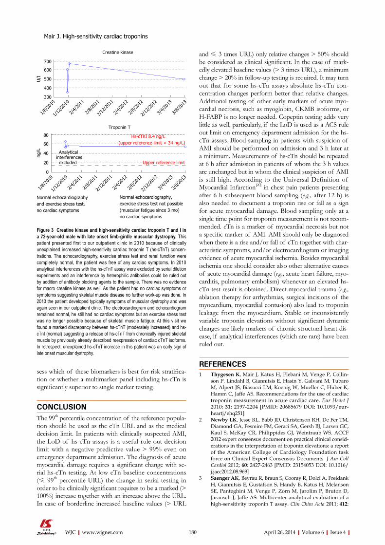
180 April 26, 2014|Volume 6|Issue 4|WJC|www.wjgnet.com
sess which of these biomarkers is best for risk stratifica-tion or whether a multimarker panel including hs-cTn is significantly superior to single marker testing.
CONCLUSIONThe 99th percentile concentration of the reference popula-tion should be used as the cTn URL and as the medical decision limit. In patients with clinically suspected AMI, the LoD of hs-cTn assays is a useful rule out decision limit with a negative predictive value > 99% even on emergency department admission. The diagnosis of acute myocardial damage requires a significant change with se-rial hs-cTn testing. At low cTn baseline concentrations (≤ 99th percentile URL) the change in serial testing in order to be clinically significant requires to be a marked (> 100%) increase together with an increase above the URL. In case of borderline increased baseline values (> URL
and ≤ 3 times URL) only relative changes > 50% should be considered as clinical significant. In the case of mark-edly elevated baseline values (> 3 times URL), a minimum change > 20% in follow-up testing is required. It may turn out that for some hs-cTn assays absolute hs-cTn con-centration changes perform better than relative changes. Additional testing of other early markers of acute myo-cardial necrosis, such as myoglobin, CKMB isoforms, or H-FABP is no longer needed. Copeptin testing adds very little as well, particularly, if the LoD is used as a ACS rule out limit on emergency department admission for the hs-cTn assays. Blood sampling in patients with suspicion of AMI should be performed on admission and 3 h later at a minimum. Measurements of hs-cTn should be repeated at 6 h after admission in patients of whom the 3 h values are unchanged but in whom the clinical suspicion of AMI is still high. According to the Universal Definition of Myocardial Infarction[20] in chest pain patients presenting after 6 h subsequent blood sampling (e.g., after 12 h) is also needed to document a troponin rise or fall as a sign for acute myocardial damage. Blood sampling only at a single time point for troponin measurement is not recom-mended. cTn is a marker of myocardial necrosis but not a specific marker of AMI. AMI should only be diagnosed when there is a rise and/or fall of cTn together with char-acteristic symptoms, and/or electrocardiogram or imaging evidence of acute myocardial ischemia. Besides myocardial ischemia one should consider also other alternative causes of acute myocardial damage (e.g., acute heart failure, myo-carditis, pulmonary embolism) whenever an elevated hs-cTn test result is obtained. Direct myocardial trauma (e.g., ablation therapy for arrhythmias, surgical incisions of the myocardium, myocardial contusion) also lead to troponin leakage from the myocardium. Stable or inconsistently variable troponin elevations without significant dynamic changes are likely markers of chronic structural heart dis-ease, if analytical interferences (which are rare) have been ruled out.
REFERENCES1 Thygesen K, Mair J, Katus H, Plebani M, Venge P, Collin-
son P, Lindahl B, Giannitsis E, Hasin Y, Galvani M, Tubaro M, Alpert JS, Biasucci LM, Koenig W, Mueller C, Huber K, Hamm C, Jaffe AS. Recommendations for the use of cardiac troponin measurement in acute cardiac care. Eur Heart J 2010; 31: 2197-2204 [PMID: 20685679 DOI: 10.1093/eur-heartj/ehq251]
2 Newby LK, Jesse RL, Babb JD, Christenson RH, De Fer TM, Diamond GA, Fesmire FM, Geraci SA, Gersh BJ, Larsen GC, Kaul S, McKay CR, Philippides GJ, Weintraub WS. ACCF 2012 expert consensus document on practical clinical consid-erations in the interpretation of troponin elevations: a report of the American College of Cardiology Foundation task force on Clinical Expert Consensus Documents. J Am Coll Cardiol 2012; 60: 2427-2463 [PMID: 23154053 DOI: 10.1016/j.jacc2012.08.969]
3 Saenger AK, Beyrau R, Braun S, Cooray R, Dolci A, Freidank H, Giannitsis E, Gustafson S, Handy B, Katus H, Melanson SE, Panteghini M, Venge P, Zorn M, Jarolim P, Bruton D, Jarausch J, Jaffe AS. Multicenter analytical evaluation of a high-sensitivity troponin T assay. Clin Chim Acta 2011; 412:
1/8/
2010
1/12
/201
0
2/4/
2011
2/8/
2011
2/12
/201
1
2/4/
2012
2/8/
2012
2/12
/201
2
3/4/
2013
3/8/
2013
700
600
500
400
300
U/I
Creatine kinase
Troponin T
80
60
40
20
0
1/8/
2010
1/12
/201
0
2/4/
2011
2/8/
2011
2/12
/201
1
2/4/
2012
2/8/
2012
2/12
/201
2
3/4/
2013
3/8/
2013
ng/L
Upper reference limit
Hs-cTnI 8.4 ng/L(upper reference limit < 34 ng/L)
Analytical interferences excluded
Normal echocardiographyand exercise stress test,no cardiac symptoms
Normal echocardiography, exercise stress test not possible (muscular fatigue since 3 mo)no cardiac symptoms
Figure 3 Creatine kinase and high-sensitivity cardiac troponin T and I in a 72-year-old male with late onset limb-girdle muscular dystrophy. This patient presented first to our outpatient clinic in 2010 because of clinically unexplained increased high-sensitivity cardiac troponin T (hs-cTnT) concen-trations. The echocardiography, exercise stress test and renal function were completely normal, the patient was free of any cardiac symptoms. In 2010 analytical interferences with the hs-cTnT assay were excluded by serial dilution experiments and an interference by heterophilic antibodies could be ruled out by addition of antibody blocking agents to the sample. There was no evidence for macro creatine kinase as well. As the patient had no cardiac symptoms or symptoms suggesting skeletal muscle disease no further work-up was done. In 2013 the patient developed typically symptoms of muscular dystrophy and was again seen in our outpatient clinic. The electrocardiogram and echocardiogram remained normal, he still had no cardiac symptoms but an exercise stress test was no longer possible because of skeletal muscle fatigue. At this visit we found a marked discrepancy between hs-cTnT (moderately increased) and hs-cTnI (normal) suggesting a release of hs-cTnT from chronically injured skeletal muscle by previously already described reexpression of cardiac cTnT isoforms. In retrospect, unexplained hs-cTnT increase in this patient was an early sign of late onset muscular dystrophy.
Mair J. High-sensitivity cardiac troponins
181 April 26, 2014|Volume 6|Issue 4|WJC|www.wjgnet.com
748-754 [PMID: 21219893 DOI: 10.1016/j.cca.2010.12.034]4 Zaninotto M, Mion MM, Novello E, Moretti M, Delprete E,
Rocchi MB, Sisti D, Plebani M. Precision performance at low levels and 99th percentile concentration of the Access AccuTnl assay on two different platforms. Clin Chem Lab Med 2009; 47: 367-371 [PMID: 19676150 DOI: 10.1515/CCLM.2009.080]
5 Todd J, Freese B, Lu A, Held D, Morey J, Livingston R, Goix P. Ultrasensitive flow-based immunoassays using single-molecule counting. Clin Chem 2007; 53: 1990-1995 [PMID: 17890441 DOI: 10.1373/clinchem.2007.091181]
6 van de Kerkhof D, Peters B, Scharnhorst V. Performance of the Advia Centaur second-generation troponin assay TnI-Ul-tra compared with the first-generation cTnI assay. Ann Clin Biochem 2008; 45: 316-317 [PMID: 18482922 DOI: 10.1258/acb.2007.007209]
7 Apple FS, Collinson PO. Analytical characteristics of high-sensitivity cardiac troponin assays. Clin Chem 2012; 58: 54-61 [PMID: 21965555 DOI: 10.1373/clinchem.2011.165795]
8 Thygesen K, Mair J, Giannitsis E, Mueller C, Lindahl B, Blankenberg S, Huber K, Plebani M, Biasucci LM, Tubaro M, Collinson P, Venge P, Hasin Y, Galvani M, Koenig W, Hamm C, Alpert JS, Katus H, Jaffe AS. How to use high-sensitivity cardiac troponins in acute cardiac care. Eur Heart J 2012; 33: 2252-2257 [PMID: 22723599 DOI: 10.1373/10.1093/eurheartj/ehs154]
9 Mingels A, Jacobs L, Michielsen E, Swaanenburg J, Wodzig W, van Dieijen-Visser M. Reference population and mara-thon runner sera assessed by highly sensitive cardiac tro-ponin T and commercial cardiac troponin T and I assays. Clin Chem 2009; 55: 101-108 [PMID: 18988757 DOI: 10.1373/clinchem.2008.106427]
10 Venge P, Johnston N, Lindahl B, James S. Normal plasma levels of cardiac troponin I measured by the high-sensitivity cardiac troponin I access prototype assay and the impact on the diagnosis of myocardial ischemia. J Am Coll Car-diol 2009; 54: 1165-1172 [PMID: 19761938 DOI: 10.1016/j.jacc.2009.05.051]
11 Apple FS, Ler R, Murakami MM. Determination of 19 car-diac troponin I and T assay 99th percentile values from a common presumably healthy population. Clin Chem 2012; 58: 1574-1581 [PMID: 22983113 DOI: 10.1373/clinchem.2012.192716]
12 Reichlin T, Hochholzer W, Bassetti S, Steuer S, Stelzig C, Hartwiger S, Biedert S, Schaub N, Buerge C, Potocki M, Noveanu M, Breidthardt T, Twerenbold R, Winkler K, Bingisser R, Mueller C. Early diagnosis of myocardial in-farction with sensitive cardiac troponin assays. N Engl J Med 2009; 361: 858-867 [PMID: 19710484 DOI: 10.1056/NEJ-Moa0900428]
13 Weber M, Bazzino O, Navarro Estrada JL, de Miguel R, Salzberg S, Fuselli JJ, Liebetrau C, Woelken M, Moellmann H, Nef H, Hamm C. Improved diagnostic and prognostic performance of a new high-sensitive troponin T assay in pa-tients with acute coronary syndrome. Am Heart J 2011; 162: 81-88 [PMID: 21742093 DOI: 10.1016/j.ahj.2011.04.007]
14 Keller T, Zeller T, Peetz D, Tzikas S, Roth A, Czyz E, Bickel C, Baldus S, Warnholtz A, Fröhlich M, Sinning CR, Elefthe-riadis MS, Wild PS, Schnabel RB, Lubos E, Jachmann N, Genth-Zotz S, Post F, Nicaud V, Tiret L, Lackner KJ, Münzel TF, Blankenberg S. Sensitive troponin I assay in early diag-nosis of acute myocardial infarction. N Engl J Med 2009; 361: 868-877 [PMID: 19710485 DOI: 10.1056/NEJMoa0903515]
15 Keller T, Zeller T, Ojeda F, Tzikas S, Lillpopp L, Sinning C, Wild P, Genth-Zotz S, Warnholtz A, Giannitsis E, Möckel M, Bickel C, Peetz D, Lackner K, Baldus S, Münzel T, Blanken-berg S. Serial changes in highly sensitive troponin I assay and early diagnosis of myocardial infarction. JAMA 2011; 306: 2684-2693 [PMID: 22203537 DOI: 10.1001/jama.2011.1896]
16 Hammerer-Lercher A, Ploner T, Neururer S, Schratzberger P, Griesmacher A, Pachinger O, Mair J. High-sensitivity cardiac troponin T compared with standard troponin T testing on
emergency department admission: how much does it add in everyday clinical practice? J Am Heart Assoc 2013; 2: e000204 [PMID: 23735897 DOI: 10.1161/JAHA.113.000204]
17 Jaffe AS, Apple FS, Morrow DA, Lindahl B, Katus HA. Being rational about (im)precision: a statement from the Biochemistry Subcommittee of the Joint European Society of Cardiology/American College of Cardiology Foundation/American Heart Association/World Heart Federation Task Force for the definition of myocardial infarction. Clin Chem 2010; 56: 941-943 [PMID: 20360122 DOI: 10.1373/clinchem.2010.143958]
18 Hammarsten O, Fu ML, Sigurjonsdottir R, Petzold M, Said L, Landin-Wilhelmsen K, Widgren B, Larsson M, Johanson P. Troponin T percentiles from a random population sample, emergency room patients and patients with myocardial in-farction. Clin Chem 2012; 58: 628-637 [PMID: 22258764 DOI: 10.1373/clinchem.2011.171496]
19 Hamm CW, Bassand JP, Agewall S, Bax J, Boersma E, Bueno H, Caso P, Dudek D, Gielen S, Huber K, Ohman M, Petrie MC, Sonntag F, Uva MS, Storey RF, Wijns W, Zahger D. ESC Guidelines for the management of acute coronary syndromes in patients presenting without persistent ST-segment eleva-tion: The Task Force for the management of acute coronary syndromes (ACS) in patients presenting without persistent ST-segment elevation of the European Society of Cardiology (ESC). Eur Heart J 2011; 32: 2999-3054 [PMID: 21873419]
20 Thygesen K, Alpert JS, Jaffe AS, Simoons ML, Chaitman BR, White HD, Thygesen K, Alpert JS, White HD, Jaffe AS, Katus HA, Apple FS, Lindahl B, Morrow DA, Chaitman BA, Clemmensen PM, Johanson P, Hod H, Underwood R, Bax JJ, Bonow RO, Pinto F, Gibbons RJ, Fox KA, Atar D, Newby LK, Galvani M, Hamm CW, Uretsky BF, Steg PG, Wijns W, Bassand JP, Menasché P, Ravkilde J, Ohman EM, Antman EM, Wallentin LC, Armstrong PW, Simoons ML, Januzzi JL, Nieminen MS, Gheorghiade M, Filippatos G, Luepker RV, Fortmann SP, Rosamond WD, Levy D, Wood D, Smith SC, Hu D, Lopez-Sendon JL, Robertson RM, Weaver D, Ten-dera M, Bove AA, Parkhomenko AN, Vasilieva EJ, Mendis S. Third universal definition of myocardial infarction. Eur Heart J 2012; 33: 2551-2567 [PMID: 22922414 DOI: 10.1093/eurheartj/ehs184]
21 Apple FS, Jesse RL, Newby LK, Wu AH, Christenson RH, Cannon CP, Francis G, Morrow DA, Ravkilde J, Storrow AB, Tang W, Jaffe AS, Mair J, Ordonez-Llanos J, Pagani F, Pan-teghini M, Tate J. National Academy of Clinical Biochemis-try and IFCC Committee for Standardization of Markers of Cardiac Damage Laboratory Medicine Practice Guidelines: analytical issues for biochemical markers of acute coronary syndromes. Clin Chem 2007; 53: 547-551 [PMID: 17384000 DOI: 10.1373/clinchem.2006.084715]
22 Wu AH, Shea E, Lu QT, Minyard J, Bui K, Hsu JC, Agee SJ, Todd J. Short- and long-term cardiac troponin I analyte stability in plasma and serum from healthy volunteers by use of an ultrasensitive, single-molecule counting assay. Clin Chem 2009; 55: 2057-2059 [PMID: 19729470 DOI: 10.1337/clinchem.2009.128611]
23 Vasile VC, Saenger AK, Kroning JM, Klee GG, Jaffe AS. Biologic variation of a novel cardiac troponin I assay. Clin Chem 2011; 57: 1080-1081 [PMID: 21519039 DOI: 10.1373/clinchem.2011.162545]
24 Frankenstein L, Wu AH, Hallermayer K, Wians FH, Giannit-sis E, Katus HA. Biological variation and reference change value of high-sensitivity troponin T in healthy individu-als during short and intermediate follow-up periods. Clin Chem 2011; 57: 1068-1071 [PMID: 21519037 DOI: 10.1373/clinchem.2010.158964]
25 Vasile VC, Saenger AK, Kroning JM, Jaffe AS. Biological and analytical variability of a novel high-sensitivity cardiac troponin T assay. Clin Chem 2010; 56: 1086-1090 [PMID: 20472824 DOI: 10.1373/clinchem.2009.140616]
26 Frankenstein L, Remppis A, Giannitis E, Frankenstein J,
Mair J. High-sensitivity cardiac troponins
182 April 26, 2014|Volume 6|Issue 4|WJC|www.wjgnet.com
Hess G, Zdunek D, Doesch A, Zugck C, Katus HA. Biologi-cal variation of high sensitive Troponin T in stable heart failure patients with ischemic or dilated cardiomyopathy. Clin Res Cardiol 2011; 100: 633-640 [PMID: 21327843 DOI: 10.1007/s00392-011-0285-4]
27 Reichlin T, Irfan A, Twerenbold R, Reiter M, Hochholzer W, Burkhalter H, Bassetti S, Steuer S, Winkler K, Peter F, Meiss-ner J, Haaf P, Potocki M, Drexler B, Osswald S, Mueller C. Utility of absolute and relative changes in cardiac troponin concentrations in the early diagnosis of acute myocardial in-farction. Circulation 2011; 124: 136-145 [PMID: 21709058 DOI: 10.1161/CIRCULATIONAHA.111.023937]
28 Mueller M, Biener M, Vafaie M, Doerr S, Keller T, Blan-kenberg S, Katus HA, Giannitsis E. Absolute and relative kinetic changes of high-sensitivity cardiac troponin T in acute coronary syndrome and in patients with increased troponin in the absence of acute coronary syndrome. Clin Chem 2012; 58: 209-218 [PMID: 22134520 DOI: 10.1373/clinchem.2011.171827]
29 Novack V, Pencina M, Cohen DJ, Kleiman NS, Yen CH, Saucedo JF, Berger PB, Cutlip DE. Troponin criteria for myo-cardial infarction after percutaneous coronary intervention. Arch Intern Med 2012; 172: 502-508 [PMID: 22371874 DOI: 10.1001/archinternmed.2011.2275]
30 Mair J. Progress in myocardial damage detection: new bio-chemical markers for clinicians. Crit Rev Clin Lab Sci 1997; 34: 1-66 [PMID: 9055056 DOI: 10.3109/1040836970903821]
31 Kilcullen N, Viswanathan K, Das R, Morrell C, Farrin A, Barth JH, Hall AS. Heart-type fatty acid-binding protein predicts long-term mortality after acute coronary syndrome and identifies high-risk patients across the range of troponin values. J Am Coll Cardiol 2007; 50: 2061-2067 [PMID: 18021874 DOI: 10.1016/j.jacc.2007.08.021]
32 Reiter M, Twerenbold R, Reichlin T, Mueller M, Hoeller R, Moehring B, Haaf P, Wildi K, Merk S, Bernhard D, Mueller CZ, Freese M, Freidank H, Campodarve Botet I, Mueller C. Heart-type fatty acid-binding protein in the early diagnosis of acute myocardial infarction. Heart 2013; 99: 708-714 [PMID: 23514979 DOI: 10.1136/heartjnl-2012-303325]
33 Land H, Schütz G, Schmale H, Richter D. Nucleotide se-quence of cloned cDNA encoding bovine arginine vaso-pressin-neurophysin II precursor. Nature 1982; 295: 299-303 [PMID: 6276766 DOI: 10.1038/295299a0]
34 Reichlin T, Hochholzer W, Stelzig C, Laule K, Freidank H, Morgenthaler NG, Bergmann A, Potocki M, Noveanu M, Breidthardt T, Christ A, Boldanova T, Merki R, Schaub N, Bingisser R, Christ M, Mueller C. Incremental value of co-peptin for rapid rule out of acute myocardial infarction. J Am Coll Cardiol 2009; 54: 60-68 [PMID: 19555842 DOI: 10.1016/j.jacc.2009.01.076]
35 Saunders JT, Nambi V, de Lemos JA, Chambless LE, Virani SS, Boerwinkle E, Hoogeveen RC, Liu X, Astor BC, Mosley TH, Folsom AR, Heiss G, Coresh J, Ballantyne CM. Cardiac troponin T measured by a highly sensitive assay predicts coronary heart disease, heart failure, and mortality in the Atherosclerosis Risk in Communities Study. Circulation 2011; 123: 1367-1376 [PMID: 21422391 DOI: 10.1161/CIRCULA-TIONAHA.110.005264]
36 de Lemos JA, Drazner MH, Omland T, Ayers CR, Khera A, Rohatgi A, Hashim I, Berry JD, Das SR, Morrow DA, Mc-Guire DK. Association of troponin T detected with a highly sensitive assay and cardiac structure and mortality risk in the general population. JAMA 2010; 304: 2503-2512 [PMID:
21139111 DOI: 10.1001/jama.2010.1768]37 deFilippi CR, de Lemos JA, Christenson RH, Gottdiener JS,
Kop WJ, Zhan M, Seliger SL. Association of serial measures of cardiac troponin T using a sensitive assay with incident heart failure and cardiovascular mortality in older adults. JAMA 2010; 304: 2494-2502 [PMID: 21078811 DOI: 10.1001/jama.2010.1708]
38 Reiter M, Twerenbold R, Reichlin T, Haaf P, Peter F, Meiss-ner J, Hochholzer W, Stelzig C, Freese M, Heinisch C, Bre-idthardt T, Freidank H, Winkler K, Campodarve I, Gea J, Mueller C. Early diagnosis of acute myocardial infarction in the elderly using more sensitive cardiac troponin assays. Eur Heart J 2011; 32: 1379-1389 [PMID: 21362702 DOI: 10.1093/eurheartj/ehr033]
39 Omland T, de Lemos JA, Sabatine MS, Christophi CA, Rice MM, Jablonski KA, Tjora S, Domanski MJ, Gersh BJ, Rouleau JL, Pfeffer MA, Braunwald E. A sensitive cardiac tropo-nin T assay in stable coronary artery disease. N Engl J Med 2009; 361: 2538-2547 [PMID: 19940289 DOI: 10.1056/NEJ-Moa0805299]
40 Korosoglou G, Lehrke S, Mueller D, Hosch W, Kauczor HU, Humpert PM, Giannitsis E, Katus HA. Determinants of tro-ponin release in patients with stable coronary artery disease: insights from CT angiography characteristics of atheroscle-rotic plaque. Heart 2011; 97: 823-831 [PMID: 20884786 DOI: 10.1136/hrt.2010.193201]
41 Ndrepepa G, Braun S, Mehilli J, Birkmeier KA, Byrne RA, Ott I, Hösl K, Schulz S, Fusaro M, Pache J, Hausleiter J, Laugwitz KL, Massberg S, Seyfarth M, Schömig A, Kastrati A. Prognostic value of sensitive troponin T in patients with stable and unstable angina and undetectable conventional troponin. Am Heart J 2011; 161: 68-75 [PMID: 21167336 DOI: 10.1016/j.ahj.2010.09.018]
42 Kawahara C, Tsutamoto T, Nishiyama K, Yamaji M, Sakai H, Fujii M, Yamamoto T, Horie M. Prognostic role of high-sensitivity cardiac troponin T in patients with nonischemic dilated cardiomyopathy. Circ J 2011; 75: 656-661 [PMID: 21178288 DOI: 10.1253/circj.CJ-10-0837]
43 Latini R, Masson S, Anand IS, Missov E, Carlson M, Vago T, Angelici L, Barlera S, Parrinello G, Maggioni AP, Tognoni G, Cohn JN. Prognostic value of very low plasma concentra-tions of troponin T in patients with stable chronic heart fail-ure. Circulation 2007; 116: 1242-1249 [PMID: 17698733]
44 Lankeit M, Friesen D, Aschoff J, Dellas C, Hasenfuss G, Ka-tus H, Konstantinides S, Giannitsis E. Highly sensitive tropo-nin T assay in normotensive patients with acute pulmonary embolism. Eur Heart J 2010; 31: 1836-1844 [PMID: 20584774 DOI: 10.1093/eurheartj/ehq234]
45 Filusch A, Giannitsis E, Katus HA, Meyer FJ. High-sensitive troponin T: a novel biomarker for prognosis and disease se-verity in patients with pulmonary arterial hypertension. Clin Sci (Lond) 2010; 119: 207-213 [PMID: 20412051 DOI: 10.1042/CS20100014]
46 Jaffe AS, Vasile VC, Milone M, Saenger AK, Olson KN, Apple FS. Diseased skeletal muscle: a noncardiac source of increased circulating concentrations of cardiac troponin T. J Am Coll Cardiol 2011; 58: 1819-1824 [PMID: 21962825 DOI: 10.1016/j.jacc.2011.08.026]
47 Beatty AL, Ku IA, Christenson RH, DeFilippi CR, Schiller NB, Whooley MA. High-sensitivity cardiac troponin T levels and secondary events in outpatients with coronary heart dis-ease from the Heart and Soul Study. JAMA Intern Med 2013; 173: 763-769 [PMID: 23568589]
P- Reviewers: Biondi-Zoccai G, Ghanem A, Hirohata S S- Editor: Zhai HH L- Editor: A E- Editor: Liu SQ
Mair J. High-sensitivity cardiac troponins
Molecular phenotypes of human parvovirus B19 in patients with myocarditis
C-Thomas Bock, Anja Düchting, Friederike Utta, Eva Brunner, Bui Tien Sy, Karin Klingel, Florian Lang, Meinrad Gawaz, Stephan B Felix, Reinhard Kandolf
C-Thomas Bock, Anja Düchting, Friederike Utta, Eva Brun-ner, Karin Klingel, Reinhard Kandolf, Department of Molecu-lar Pathology, University Hospital of Tuebingen, 72076 Tuebin-gen, GermanyC-Thomas Bock, Bui Tien Sy, Robert Koch Institute, 13353 Berlin, GermanyFlorian Lang, Department of Physiology, University of Tuebin-gen, 72070 Tuebingen, GermanyMeinrad Gawaz, Department of Cardiology and Cardiovascular Medicine, University Hospital of Tuebingen, 72076 Tuebingen,
GermanyStephan B Felix, Clinics for Internal Medicine B, Ernst-Moritz-Arndt-University, 17475 Greifswald, GermanyAuthor contributions: Bock CT, Gawaz M, Felix SB and Kan-dolf R conceived and designed the research; Bock CT, Düchting A, Utta F, Brunner E, Sy BT, Klingel K, Lang F and Kandolf R per-formed the experiments; Bock CT, Klingel K, Lang F, Gawaz M, Felix SB and Kandolf R analyzed the data; Klingel K, Gawaz M, Felix SB and Kandolf R contributed to reagents/materials/analy-sis tools; Bock CT and Kandolf R contributed to drafting of the manuscript; all authors read and approved the final manuscript.Supported by Grants of the Deutsche Forschungsgemeinschaft, Sonderforschungsbereich-Transregio 19 (project B5)Correspondence to: C-Thomas Bock, PhD, Director, Profes-sor, Department of Molecular Pathology, University Hospital of Tuebingen, Liebermeisterstr. 8, 72076 Tuebingen, Germany. [email protected]: + 49-30-187542379 Fax: + 49-30-187542617Received: October 11, 2013 Revised: January 16, 2014Accepted: February 18, 2014Published online: April 26, 2014
AbstractAIM: To investigate molecular phenotypes of myocar-dial B19V-infection to determine the role of B19V in myocarditis and dilated cardiomyopathy (DCM).
METHODS: Endomyocardial biopsies (EMBs) from 498 B19V-positive patients with myocarditis and DCM
were analyzed using molecular methods and functional experiments. EMBs were obtained from the University Hospitals of Greifswald and Tuebingen and addition-ally from 36 German cardiology centers. Control tissues were obtained at autopsy from 34 victims of accidents, crime or suicide. Identification of mononuclear cell infil-trates in EMBs was performed using immunohistological staining. Anti-B19V-IgM and anti-B19V-IgG were ana-lyzed by enzyme-linked immunosorbent assay (ELISA). B19V viral loads were determined using in-house quan-titative real-time polymerase chain reaction (PCR). For B19V-genotyping a new B19V-genotype-specific restric-tion fragment length polymorphism (RFLP)-PCR was es-tablished. B19V-genotyping was verified by direct DNA-sequencing and sequences were aligned using BLAST and BioEdit software. B19V P6-promoter and HHV6-U94-transactivator constructs were generated for cell culture experiments. Transfection experiments were conducted using human endothelial cells 1. Luciferase reporter assays were performed to determine B19V-replication activity. Statistical analysis and graphical representation were calculated using SPSS and Prism5 software.
RESULTS: The prevalence of B19V was significantly more likely to be associated with inflammatory car-diomyopathy (iCMP) compared to uninflamed DCM (59.6% vs 35.3%) (P < 0.0001). The detection of B19V-mRNA replication intermediates proved that rep-lication of B19V was present. RFLP-PCR assays showed that B19V-genotype 1 (57.4%) and B19V-genotype 2 (36.7%) were the most prevalent viral genotypes. B19V-genotype 2 was observed more frequently in EMBs with iCMP (65.0%) compared to DCM (35%) (P = 0.049). Although there was no significant difference in gender-specific B19V-loads, women were more fre-quently infected with B19V-genotype 2 (44.6%) than men (36.0%) (P = 0.0448). Coinfection with B19V and other cardiotropic viruses was found in 19.2% of tissue
ORIGINAL ARTICLE
Online Submissions: http://www.wjgnet.com/esps/[email protected]:10.4330/wjc.v6.i4.183
183 April 26, 2014|Volume 6|Issue 4|WJC|www.wjgnet.com
World Journal of CardiologyW J C
World J Cardiol 2014 April 26; 6(4): 183-195ISSN 1949-8462 (online)
© 2014 Baishideng Publishing Group Co., Limited. All rights reserved.
samples and was associated with higher B19V viral load compared to B19V-monoinfected tissue (P = 0.0012). The most frequent coinfecting virus was human herpes virus 6 (HHV6, 16.5%). B19V-coinfection with HHV6 showed higher B19V-loads compared to B19V-monoin-fected EMBs (P = 0.0033), suggesting that HHV6 had transactivated B19V. In vitro experiments confirmed a 2.4-fold increased B19V P6-promoter activity by the HHV6 U94-transactivator.
CONCLUSION: The finding of significantly increased B19V loads in patients with histologically proven cardiac inflammation suggests a crucial role of B19V-genotypes and reactivation of B19V-infection by HHV6-coinfection in B19V-associated iCMP. Our findings suggest that B19V-infection of the human heart can be a causative event for the development of an endothelial cell-me-diated inflammatory disease and that this is related to both viral load and genotype.
© 2014 Baishideng Publishing Group Co., Limited. All rights reserved.
Key words: Myocarditis; Dilated cardiomyopathy; Par-vovirus B19; B19V-genotypes; B19V co-infection
Core tip: Human parvovirus B19 (B19V) has recently been shown to be an emerging pathogen for inflam-matory cardiomyopathy (iCMP). We showed that B19V replication intermediates could be detected in acute and ongoing myocarditis. B19V-genotypes 1 and 2 were predominant although B19V-genotype 2 was more prevalent in iCMP. Further analyses revealed that B19V-coinfection with other cardiotropic viruses does occur, most frequently with human herpes virus 6 (HHV6). In vitro experiments showed that the HHV6 U94-transactivator element could transactivate the B19V-P6-promoter. We suggest that long-term per-sistence of B19V DNA in the human heart occurs and that active/reactivated B19V-replication can be associ-ated with iCMP in a viral load and genotype-dependent manner.
Bock CT, Düchting A, Utta F, Brunner E, Sy BT, Klingel K, Lang F, Gawaz M, Felix SB, Kandolf R. Molecular phenotypes of human parvovirus B19 in patients with myocarditis. World J Cardiol 2014; 6(4): 183-195 Available from: URL: http://www.wjgnet.com/1949-8462/full/v6/i4/183.htm DOI: http://dx.doi.org/10.4330/wjc.v6.i4.183
INTRODUCTIONIt has been shown that parvovirus B19 (B19V) infec-tion of the myocardium can cause potentially lethal acute myocarditis in infants and adults[1-3]. Acute B19V-infection of endothelial cells is accompanied by the intravascular accumulation, adhesion and penetration of
inflammatory cells in to vessel walls, leading to an impair-ment of the myocardial micro-circulation with secondary myocyte necrosis that can mimic myocardial infarction[1,2]. B19V has been found in high copy numbers in myocardi-al endothelial cells of small vessels but not in myocytes[1]. We recently reported that B19V-loads greater than 500 GE/μg of isolated nucleic acid identified in endomyocar-dial biopsies (EMBs) argue for virus-induced myocarditis. In contrast, a low viral load detected in uninflamed hearts has been associated with a latent-type of B19V-infec-tion[4]. As expected, high viral loads of approximately 3 × 10⁵ GE/μg were detected in acute myocardial B19V-infection, while approximately 700 GE/μg were found to be characteristically associated with chronic myocarditis[4]. Notably, a growing number of reports suggests an asso-ciation between B19V-infection and the development of chronic myocarditis, as well as isolated endothelial and/or diastolic dysfunction[5-8]. However, the frequent detection of B19V genomes in EMBs of patients clinically sus-pected of having myocarditis and dilated cardiomyopa-thy (DCM) and the potential pathogenic role of B19V remains controversial and warrants studies to differenti-ate viral pathogenic effects from harmless latent B19V-infection[9,10].
Infection with human parvovirus B19 (B19V) is common, with approximately 70% to 90% of adoles-cents having anti-B19V IgG detectable in serum[11]. B19V-infection is usually benign and in children it most commonly manifests with erythema infectiosum (fifth disease)[12]. The genome of B19V consists of a single stranded DNA molecule of approximately 5.5 kb that contains three major open reading frames coding for the two capsid proteins VP1 and VP2 and the nonstructural protein NS1. Genome diversity divides the genus eryth-rovirus of the parvoviridae family into three pathogenic human genotypes: PVBAu (genotype 1), Lali-like (geno-type 2) and V9-like (genotype 3) viruses[13]. The three erythrovirus genotypes show different geographical and temporal distributions. Whereas B19V-genotype 1 and 2 can be detected in most populations, genotype 3 seems to be prevalent only in Ghana, France and Brazil[13-15]. In-terestingly, the age distribution of B19V-genotype 1 and 2 infections is different, with B19V-genotype 1 occur-ring most frequently in individuals born after 1955 while B19V-genotype 2 is predominantly found in individuals older than 50 years[16,17].
Recent reports have indicated that coinfection with different cardiotropic viruses of the human heart is com-mon[7,8,18,19]. Human herpes virus 6 (HHV6) has been identified as an important coinfecting pathogen with B19V of the myocardium and resulting in fatal myocardi-tis in infants[1,20]. It has been reported that HHV6 is able to transactivate human immunodeficiency virus (HIV) and human cytomegalovirus (HCMV)[19,21].
In the present study, we explored molecular pheno-types of myocardial B19V-infection in association with patient age, gender, B19V replicative mRNA intermedi-ates, B19V genotype and B19V-coinfection to gain fur-
184 April 26, 2014|Volume 6|Issue 4|WJC|www.wjgnet.com
Bock CT et al . Parvovirus B19-associated myocarditis
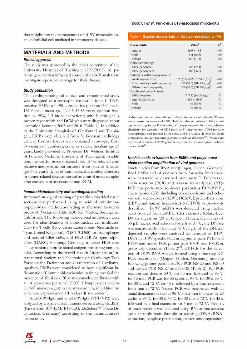
ther insight into the pathogenesis of B19V-myocarditis as an endothelial-cell mediated inflammatory disease.
MATERIALS AND METHODSEthical approvalThe study was approved by the ethics committee of the University Hospital of Tuebingen (297/2005). All pa-tients gave written informed consent for EMB analysis to investigate a possible etiology for their disease.
Study populationThis cardiopathological clinical and experimental study was designed as a retrospective evaluation of B19V-positive EMBs of 498 consecutive patients (341 male, 157 female, mean age 46.9 ± 15.85 years, ejection frac-tion < 45%, 2-3 biopsies/patient) with histologically proven myocarditis and DCM who were diagnosed at our institution between 2003 and 2010 (Table 1). In addition to the University Hospitals of Greifswald and Tuebin-gen, EMBs were obtained from 36 German cardiology centers. Control tissues were obtained at autopsy from 34 victims of accidents, crime or suicide (median age 29 years, kindly provided by Professor Dr. Wehner, Institute of Forensic Medicine, University of Tuebingen). In addi-tion, myocardial tissue obtained from 57 unselected con-secutive autopsies at our institute from patients (median age 67.2 years) dying of cardiovascular, cardiopulmonary or tumor-related diseases served as control tissue samples after exclusion of myocarditis and DCM.
Immunohistochemistry and serological testingImmunohistological staining of paraffin-embedded tissue sections was performed using an avidin-biotin-immu-noperoxidase method according to the manufacturer’s protocol (Vectastain Elite ABC Kit, Vector, Burlingame, California). The following monoclonal antibodies were used for identification of mononuclear cell infiltrates: CD3 for T cells (Novocastra Laboratories, Newcastle on Tyne, United Kingdom), PGM1 (CD68) for macrophages and natural killer cells, and HLA-DR-Antigen, alpha chain (DAKO, Hamburg, Germany) to assess HLA class Ⅱ expression on professional antigen-presenting immune cells. According to the World Health Organization/In-ternational Society and Federation of Cardiology Task Force on the Definition and Classification of Cardiomy-opathies, EMBs were considered to have significant in-flammation if immunohistochemical staining revealed the presence of focal or diffuse mononuclear infiltrates with > 14 leukocytes per mm2 (CD3+ T lymphocytes and/or CD68+ macrophages) in the myocardium, in addition to enhanced expression of HLA class Ⅱ molecules[1].
Anti-B19V-IgM and anti-B19V-IgG (VP1/VP2) were analyzed by enzyme-linked immunosorbent assay (ELISA) (Parvovirus B19-IgM; B19-IgG, DxSelect™ FocusDi-agnostics, Germany) according to the manufacturer’s instructions.
Nucleic acids extraction from EMBs and polymerase chain reaction amplification of viral genomesNucleic acids from RNAlater (Qiagen, Hilden, Germany) fixed EMBs and of controls from formalin fixed tissue were extracted as described previously[1,6]. Polymerase chain reaction (PCR) and reverse transcriptase (RT)-PCR was performed to detect parvovirus B19 (B19V), enteroviruses (EV) (including coxsackieviruses and echo-viruses), adenoviruses (ADV), HCMV, Epstein-Barr virus (EBV), and human herpesvirus 6 (HHV6) as previously described[1]. B19V mRNA was detected using nucleic acids isolated from EMBs. After extensive RNase-free-DNase digestion (20 U; Qiagen, Hilden, Germany) of 30 μL nucleic acid solution for 2 h at 37 ℃, the DNAse was inactivated for 15 min at 75 ℃. 5 μL of the DNAse-digested samples were analyzed for removal of B19V DNA by B19V-specific PCR using primer pairs PVB3 and PVB4 and nested PCR primer pairs PVB1 and PVB2 as previously described (Table 2)[1]. RT-PCR for the detec-tion of B19V-RNA was performed using a one-step RT-PCR reaction kit (Qiagen, Hilden, Germany) and the following primer pairs: first/RT-PCR NS-25 and NS-30 and nested PCR NS-27 and NS-32 (Table 2). RT-PCR reaction was done at 50 ℃ for 30 min followed by 95 ℃ for 15 min. PCR was for 35 cycles at 94 ℃ for 30 s, 53 ℃ for 30 s, and 72 ℃ for 45 s, followed by a final extension for 5 min at 72 ℃. Nested PCR was performed with an initial denaturation step at 95 ℃ for 2 min followed by 29 cycles at 95 ℃ for 30 s, 53 ℃ for 30 s, and 72 ℃ for 45 s, followed by a final extension for 5 min at 72 ℃. Five μL of each reaction was analyzed using RNase-free agarose gel electrophoresis. Sample processing (DNA/RNA-extraction, template preparation, master-mix preparation)
185 April 26, 2014|Volume 6|Issue 4|WJC|www.wjgnet.com
Table 1 Baseline characteristics of the study population n (%)
Characteristic Value1 n 3
Age, yr 46.9 ± 15.82 498 Male 341 (68.5) 498 Female 157 (31.5) 498Molecular findings B19V-genotype 1 286 (57.4) 498 B19V-genotype 2 183 (36.7) 498Endomyocardial biopsy results4
Acute myocarditis 25 (5.0) (3.2 × 10⁵ GE/μg)5 498 Inflammatory cardiomyopathy 297 (59.6) (709 GE/μg)5 498 Dilated cardiomyopathy 176 (35.3) (392 GE/μg)5 498Uninflamed control hearts B19V-detection 7 (7.7) (84 GE/μg)5 91 Age (at death), yr 48.1 ± 20.82 91 Male 49 (53.9) 91 Female 42 (46.1) 91
1Values are number, absolute and relative frequency of patients; 2Values are expressed as mean and ± SD; 3Total number of patients; 4Histopathol-ogy according to the Dallas criteria[41] supplemented by immunohisto-chemistry for detection of CD3-positive T-lymphocytes, CD68-positive macrophages and natural killer cells, and HLA class Ⅱ expression in professional antigen-presenting immune cells as described[18]; 5Values are expressed as mean of B19V-genome equivalents per microgram isolated nucleic acids[4].
Bock CT et al . Parvovirus B19-associated myocarditis

186 April 26, 2014|Volume 6|Issue 4|WJC|www.wjgnet.com
ment length polymorphism PCR (RFLP-PCR) was de-veloped using nested PCR. The following primer pairs were used for the first PCR, G2170F and NS-38, and for the second PCR, G2176F and NS-40 (Table 2, Figure 1). Reactions were initially denatured at 94 ℃ for 4 min fol-lowed by 35 cycles of 94 ℃ for 30 s, 52 ℃ for 30 s, and 72 ℃ for 30 s, followed by a final extension for 10 min at 72 ℃. Nested PCR was performed using the primer pair G2176F and NS-40 (Table 2). DNA amplicons (343 bp) were digested with the restriction enzymes HpaI and TaqI in separate reactions (New England Biolabs, Frankfurt, Germany) for 30 min at 37 ℃ and analyzed by agarose gel electrophoresis. The restriction recogni-tion site for HpaI is only present in B19V-genotype 2 and B19V-genotype 3, resulting in fragment sizes of 117 and 226 bp, respectively, while the TaqI restriction site is only present in B19V-genotype 1 and B19V-genotype 2 (re-sulting in fragment sizes of 158 and 185 bp) (Figure 1).
Luciferase reporter gene assayAfter transfection of human endothelial cells 1 cells with luciferase plasmid constructs containing the B19V P6-promoter region to a final mass of 5 μg per plasmid con-taining 0.2 μg of the β-galactosidase reporter pCMVb-Gal as an internal standard, cells were cultured for 48 h. Cells were co-transfected with an HHV6 U94 expressing vector construct (kindly provided by Dr. S. Aberle, Uni-versity of Tuebingen, Germany). To measure luciferase, activity cells were harvested, lysed and measured as de-scribed previously[23].
Statistical analysisStatistical analysis and graphical representation were per-formed by two-tailed T-test and non-parametric Mann-Whitney U-test using SPSS statistical software version 16.0 and GraphPad Prism, version 5 (GraphPad Software Inc, San Diego, United States). One-way ANOVA fol-lowed by post hoc testing and Tukey’s multiple compari-son test were also performed. The results were expressed as mean ± SD. Values below significance level of 0.05 were considered statistically significant.
and PCR were done in separate laboratory rooms, which are all certified for molecular diagnostics using standard precautions to prevent assay contamination.
Quantitative real-time PCRB19V viral load was determined using quantitative real-time PCR (qPCR) and calculated according to genome equivalents per microgram isolated myocardial nucleic acid (GE/μg) as described previously[1,4,6]. Dilutions of B19V plasmid DNA and the World Health Organiza-tion international B19V DNA standard (code 99/800) were included to standardize the assay. A qPCR of the adenosine triphosphate synthase-6 gene was performed as a control for the addition of equivalent amounts of human DNA as described previously[6]. All samples were analyzed in duplicate.
DNA sequence analysis and B19V genotype analysisDNA fragments spanning the B19V NS1/VP1/VP2 coding region (nt 602 to 5014; 4413 nt; numbering ac-cording to GenBank accession no. AF162273) were amplified by PCR using primer pairs as described previ-ously[22]. DNA sequencing was performed in duplicate with purified PCR products in 2 mL BigDye Terminator cycle sequencing mix (Perkin Elmer) and 15 pmol of forward and reverse primers as described previously[22]. B19V sequences were aligned using BLAST (National Center for Biotechnology Information; http://www.ncbi.nlm.nih.gov/ blast/blast.cgi). The reliability of align-ment was checked using the BioEdit program (http://www.mbio.ncsu.edu/BioEdit/bioedit.html). Prototype B19V sequences from GenBank were used as reference sequences (GenBank accession numbers: genotype 1: AB030694, AF113323, AF162273, M13178, DQ225148, DQ225149, DQ225150 and DQ225151; genotype 2: AY064476, AY044266, AY661663 and AY661664; geno-type 3: AX003421 and AY083234).
Restriction fragment length polymorphism-PCR for B19V genotypingIn order to determine B19V genotypes, a restriction frag-
Table 2 Primer sequences
No Primer name Sequences (5’ to 3’) Position (numbering according M13178) 1st, 2nd (RT/RFLP)-PCR
1 PVB1 GCTAACTCTGTAACTTGTAC 3221-3240 Sense B19V-VP2-PCR (2nd PCR)2 PVB2 AAATATCTCCATGGGGTTGAG 3373-3393 As B19V-VP2-PCR (2nd PCR)3 PVB3 AGCATGTGGAGTGAGGGGGC 3191-3210 Sense B19V-VP2-PCR (1st PCR)4 PVB4 AAAGCATCAGGAGCTATACTTCC 3458-3480 As B19V-VP2-PCR (1st PCR)5 NS-25 AAATGCGTGGAAGTGTAGCT 1628-1647 Sense B19V-NS1-PCR (RT/1st PCR)6 NS-27 ATGCGTGGAAGTGTAGCTGT 1630-1649 As B19V-NS1-PCR (2nd PCR)7 NS-30 CCAACTAACAGTTCACGAAAC 2172-2192 Sense B19V-NS1-PCR (RT/1st PCR)8 NS-32 TAACAGTTCACGAAACTGGTC 2168-2187 As B19V-NS1-PCR (2nd PCR)9 NS-38 ATTCCACAAATTGCTGATACAC 2498-2519 As RFLP-PCR (1st PCR)10 NS-40 AATTGCTGATACACAGCTTTAG 2490-2511 As RFLP-PCR (2nd PCR)11 G2170 CAGTTTCGTGAACTGTTAGT 2170-2189 Sense RFLP-PCR (1st PCR)12 G2176 CGTGAACTGTTAGTTGGGGTTGA 2176-2198 Sense RFLP-PCR (2nd PCR)
As: Antisense; RFLP: Restriction fragment length polymorphism; RT-PCR: Reverse transcriptase-polymerase chain reaction.
Bock CT et al . Parvovirus B19-associated myocarditis
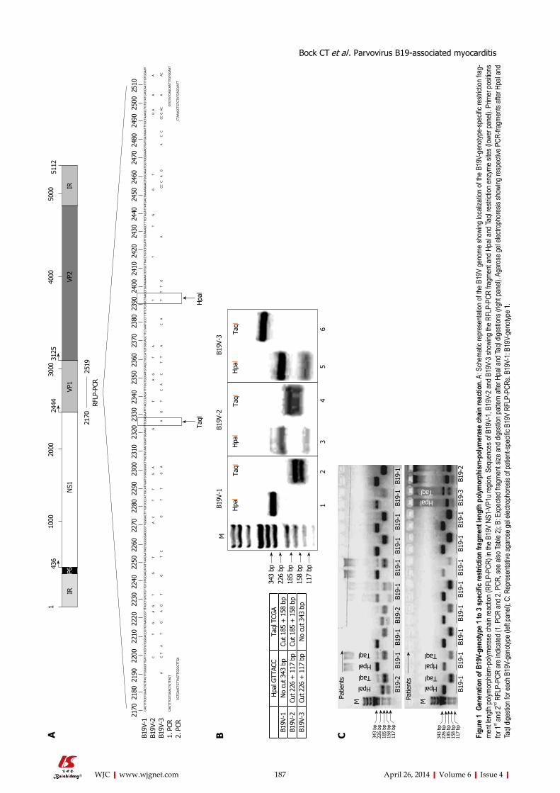
187 April 26, 2014|Volume 6|Issue 4|WJC|www.wjgnet.com
CAG
TTTC
GTG
AACT
GTT
AGTT
GG
GG
TTG
ATTA
TGTG
TGG
GAC
GG
TGTA
AGG
GG
TTTA
CCTG
TGTG
TTG
TGTG
CAAC
ATAT
TAAC
AATA
GTG
GG
GG
AGG
GTT
GG
GAC
TTTG
TCCC
CATT
GCA
TTAA
TGTA
GG
GG
CTTG
GTA
TAAT
GG
ATG
GAA
ATTT
CGAG
AATT
TACC
CCAG
ATTT
GG
TGCG
ATG
TAG
CTG
CCAT
GTG
GG
AGCT
TCTA
ATCC
CTTT
TCTG
TGCT
AACC
TGCA
AAAA
ATG
TGCT
TACC
TGTC
TGG
ATTG
CAAA
GCT
TTG
TAG
ATTA
TGAG
TAAA
AAAA
GTG
GCA
AATG
GTG
GG
AAAG
TGAT
GAT
AAAT
TTG
CTAA
AGCT
GTG
TATC
AGCA
ATTT
GTG
GAA
T
C
T
G
A
G
T
G
T
A
C
T
T
G A
G
T
A
G
T
A
T
T
T
G
G
T
G
A
A
A
A
C
T
A T
G
A
G
T
G
T
C
G
T
T
G
A A
G
T
C
A
C
T
T
A
C
A
T
T
T
G
A
CC C
A
G
A
C C
C
C G
AC
A
AC
CAG
TTTC
GTG
AACT
GTT
AGT
GTG
TATC
AGCA
ATTT
GTG
GAA
T
CG
TGAA
CTG
TTAG
TTG
GG
GTT
GA
C
TAAA
GCT
GTG
TATC
AGCA
ATT
B19V
-1B1
9V-2
B19V
-31.
PCR
2. P
CR
Taql
Hpa
l
2170
218
0 2
190
220
0 2
210
222
0 2
230
224
0 2
250
226
0 2
270
228
0 2
290
230
0 2
310
232
0 2
330
234
0 2
350
236
0 2
370
238
0 2
390
240
0 2
410
242
0 2
430
244
0 2
450
246
0 2
470
248
0 2
490
250
0 2
510
RFLP
-PCR
2170
2519
436
2444
3125
110
0020
0030
0040
0050
0051
12
IRP6
NS1
VP1
VP2
IR
A
Hpa
l GTT
ACC
Taql
TCG
AB1
9V-1
No
cut 3
43 b
pCu
t 185
+ 1
58 b
pB1
9V-2
Cut 2
26 +
117
bp
Cut 1
85 +
158
bp
B19V
-3Cu
t 226
+ 1
17 b
pN
o cu
t 343
bp
343
bp22
6 bp
185
bp15
8 bp
117
bp
MB1
9V-1
B19
V-2
B
19V-
3
Hpa
lTa
qlH
pal
Taql
Hpa
lTa
ql
1
2
3
4
5
6
B
Patie
nts
Hpal
Taql
Hpal
Taql
M
B19-
2
B19-
1
B19-
1 B
19-2
B
19-1
B
19-1
B1
9-1
B1
9-1
B1
9-1
B19
-1
Patie
nts
HpalTaql
Hpal
Taql
M
HpalTaql
B19-
1
B19-
1
B19-
1
B19-
1
B19-
1 B
19-1
B
19-1
B
19-1
B1
9-3
B1
9-2
343
bp22
6 bp
185
bp15
8 bp
117
bp
343
bp22
6 bp
185
bp15
8 bp
117
bp
C Figu
re 1
Gen
erat
ion
of B
19V-
geno
type
1 to
3 sp
ecifi
c res
trict
ion
fragm
ent l
engt
h po
lymor
phism
-pol
ymer
ase c
hain
reac
tion.
A: S
chem
atic r
epre
senta
tion o
f the B
19V
geno
me sh
owing
loca
lizati
on of
the B
19V-
geno
type-
spec
ific re
strict
ion fr
ag-
ment
length
polym
orph
ism-p
olyme
rase
chain
reac
tion (
RFLP
-PCR
) in th
e B19
V NS
1-VP
1u re
gion.
Sequ
ence
s of B
19V-
1, B1
9V-2
and B
19V-
3 sho
wing
the R
FLP-
PCR
fragm
ent a
nd H
paI a
nd Ta
qI re
strict
ion en
zyme
sites
(lowe
r pan
el). P
rimer
posit
ions
for 1st an
d 2nd
RFL
P-PC
R ar
e ind
icated
(1. P
CR an
d 2. P
CR, s
ee al
so Ta
ble 2)
; B: E
xpec
ted fr
agme
nt siz
e and
dige
stion
patte
rn af
ter H
paI a
nd Ta
qI dig
estio
ns (r
ight p
anel)
. Aga
rose
gel e
lectro
phor
esis
show
ing re
spec
tive P
CR-fr
agme
nts af
ter H
paI a
nd
TaqI
diges
tion f
or ea
ch B
19V-
geno
type (
left p
anel)
; C: R
epre
senta
tive a
garo
se ge
l elec
troph
ores
is of
patie
nt-sp
ecific
B19
V RF
LP-P
CRs.
B19V
-1: B
19V-
geno
type 1
.
Bock CT et al . Parvovirus B19-associated myocarditis
188 April 26, 2014|Volume 6|Issue 4|WJC|www.wjgnet.com
RESULTSStudy populationEMBs of 498 B19V-positive patients described in this retrospective study were obtained from 38 clinical centers in Germany between 2003 and 2010 for cardiopathologi-cal diagnosis of myocarditis and DCM. In addition, 91 uninflamed hearts without cardiac failure served as a con-trol group. The baseline data of the patients, molecular and cardiopathological findings of the EMBs are provid-ed in Table 1. Quantitative assessment of B19V loads has been described previously, with viral loads of more than 500 GE/μg in EMBs as a clinically relevant threshold for the maintenance of myocardial inflammation[4]. Patients were relatively young (mean age 46.9 ± 15.9 years) and approximately two thirds were men (68.5%) (Table 1). With regards to clinical history, the majority of patients had cardiac symptoms for longer than six months, con-sistent with chronic heart disease.
Figure 2 illustrates typical histological and immuno-histological findings in acute and chronic myocarditis, as well as in uninflamed DCM in B19V-positive human hearts. As expected, acute B19V-myocarditis had a low prevalence of 5% in our study (Figure 2A and 2B, Table 1), while the majority of patients (59.6%) had chronic B19V-myocarditis (Figure 2C and 2D, Table 1). By con-trast, chronic DCM without inflammation (Figure 2E and 2F) exhibiting a latent type of B19V-infection was found in 35.3% of our study population (Table 1). Only 7% of 91 uninflamed control hearts were found to harbor latent B19V genomes (Figure 2G and 2H, Table 1).
Replication activity of B19V in myocarditisIn order to determine replication activity of B19V in infected endothelial cells of the myocardium, we de-termined B19V RNA replication intermediates of 114 randomly selected patients of our cohorts using RT-PCR to amplify the NS1 and VP1 regions of the B19V genome (2 acute, 54 chronic myocarditis, 51 DCM and 7 control hearts). As expected, B19V replicative RNA intermediates could be confirmed in acute B19V-myocar-ditis as described[4] (2/2), but also in EMBs of patients with chronic myocarditis harboring viral loads greater than 500 GE/μg (16/51) (Figure 3). In contrast, B19V-mRNA intermediates were not observed in EMBs of DCM-patients with uninflamed hearts and viral loads < 100 GE/μg, indicating a latent-type of B19V-infection. In addition, B19V-mRNA intermediates were found to be absent in latent B19V-infected normal hearts without inflammation.
B19V-specific IgG and IgM antibodies were detected in the serum by ELISA almost exclusively in B19V-DNA positive EMBs of patients with acute myocarditis (11.4%) but not with DCM or in controls. Overall, B19V-IgG antibodies, but not IgM, were detected in the serum of 91% of patients with chronic myocarditis and in 60.9% with DCM.
Detection of different B19V genotypes in EMBsRecent reports have shown an association between B19V-
infection of myocardial endothelial cells and the develop-ment of viral myocarditis[1,4,24-26]. However, it is not clear whether B19V-genotypes modulate the outcome of the disease.
In order to determine the B19V genotype confirmed in B19V-positive EMBs, we developed a new RFLP-PCR method spanning the NS1/VP1u region (nt 2170 to 2519; Figure 1A). As shown in Figure 1B, the RFLP-PCR method is capable of discriminating between the known B19V genotypes 1 to 3 by using Hpa1 and Taq1 restriction enzyme digestions. RFLP-PCR patterns of representative patient-specific samples are shown in Fig-ure 1C. In addition, direct sequencing and phylogenetic analysis was performed to verify the specificity of the re-sults by RFLP-PCR. B19V-genotype 1 (286/498; 57.4%) and B19V-genotype 2 (183/498; 36.7%) were the most frequently detected genotypes in persistently infected patients, whereas twenty-five patients with acute B19V-infection showed B19V-genotype 1 (5.0%) (Table 1). In contrast, B19V-genotype 3 infection of the human heart was found to be rare in Germany and only detectable in 0.8% (4/498) of our patients.
B19V genotype-dependent correlation with inflammatory cardiomyopathyThe prevalence of B19V-genotype 1 and B19V-genotype 2 in EMBs was investigated in order to establish if a cor-relation exists between B19V-genotypes and the occur-rence of myocarditis and DCM. B19V-genotype 1 was detected in 176/286 of EMBs (61.5%) with iCMP and in 110/286 samples (38.5%) of DCM-patients without myocardial inflammation, respectively (Figure 4A). A strong trend was observed in the more frequent preva-lence of B19V-genotype 2 genomes (total 183/498) in EMBs with iCMP (119/183, 65.0%) compared to DCM (64/183, 35%; P = 0.048) (Figure 4A). We found compa-rable viral loads in B19V-positive EMBs for both B19V-genotype 1 (584 ± 695 GE/μg) and B19V-genotype 2 (613 ± 715 GE/μg) (P = ns; Figure 4B).
Determination of B19V-genotype specific viral loads in EMBs of patients with iCMP and DCM showed that B19V-genotype 1 load was significantly higher in iCMP samples (706 ± 821 GE/μg) compared to DCM (389 ± 343 GE/μg; P < 0.0002). In keeping with this finding, significantly higher viral loads were observed in B19V-genotype 2 positive EMBs with iCMP (723 ± 846 GE/μg) compared to DCM samples (408 ± 270 GE/μg; P = 0.0039) (Figure 4C).
Age-dependent distribution of B19V-genotype 1 and B19V-genotype 2Recent publications have shown an association between age and B19V genotype prevalence[16]. B19V-genotype 2 is considered to be an “older” B19V-variant which is more often found in tissues of patients over the age of 60 years, whereas B19V-genotype 1 is more frequently observed in younger people. In accordance with recent reports, we determined a comparable age-dependent dis-tribution of B19V-genotypes in human hearts[16,17]. B19V-
Bock CT et al . Parvovirus B19-associated myocarditis
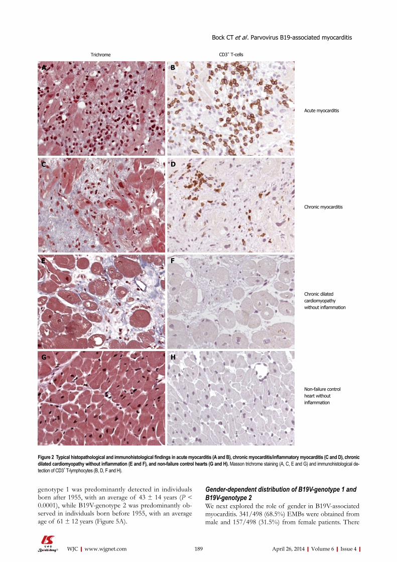
189 April 26, 2014|Volume 6|Issue 4|WJC|www.wjgnet.com
genotype 1 was predominantly detected in individuals born after 1955, with an average of 43 ± 14 years (P < 0.0001), while B19V-genotype 2 was predominantly ob-served in individuals born before 1955, with an average age of 61 ± 12 years (Figure 5A).
Gender-dependent distribution of B19V-genotype 1 and B19V-genotype 2We next explored the role of gender in B19V-associated myocarditis. 341/498 (68.5%) EMBs were obtained from male and 157/498 (31.5%) from female patients. There
A B
C D
E F
G H
Trichrome CD3+ T-cells
Acute myocarditis
Chronic myocarditis
Chronic dilated cardiomyopathy without inflammation
Non-failure controlheart without inflammation
Figure 2 Typical histopathological and immunohistological findings in acute myocarditis (A and B), chronic myocarditis/inflammatory myocarditis (C and D), chronic dilated cardiomyopathy without inflammation (E and F), and non-failure control hearts (G and H). Masson trichrome staining (A, C, E and G) and immunohistological de-tection of CD3+ T-lymphocytes (B, D, F and H).
Bock CT et al . Parvovirus B19-associated myocarditis
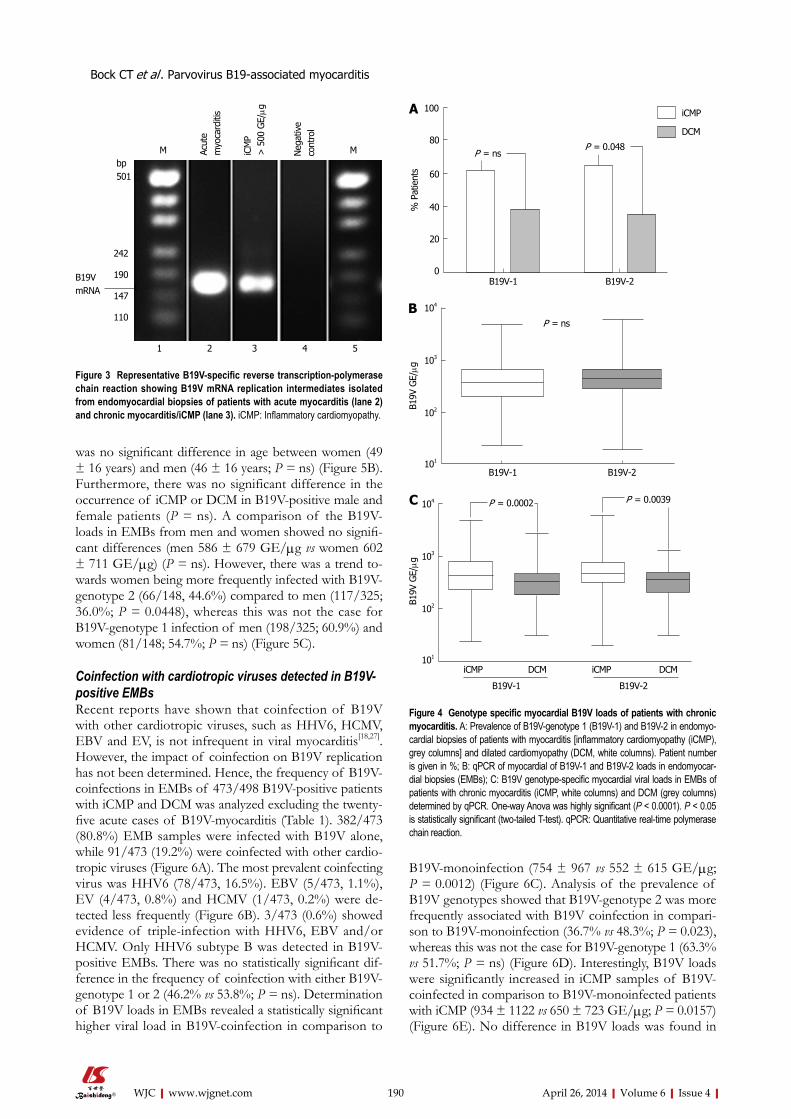
190 April 26, 2014|Volume 6|Issue 4|WJC|www.wjgnet.com
was no significant difference in age between women (49 ± 16 years) and men (46 ± 16 years; P = ns) (Figure 5B). Furthermore, there was no significant difference in the occurrence of iCMP or DCM in B19V-positive male and female patients (P = ns). A comparison of the B19V-loads in EMBs from men and women showed no signifi-cant differences (men 586 ± 679 GE/μg vs women 602 ± 711 GE/μg) (P = ns). However, there was a trend to-wards women being more frequently infected with B19V-genotype 2 (66/148, 44.6%) compared to men (117/325; 36.0%; P = 0.0448), whereas this was not the case for B19V-genotype 1 infection of men (198/325; 60.9%) and women (81/148; 54.7%; P = ns) (Figure 5C).
Coinfection with cardiotropic viruses detected in B19V-positive EMBsRecent reports have shown that coinfection of B19V with other cardiotropic viruses, such as HHV6, HCMV, EBV and EV, is not infrequent in viral myocarditis[18,27]. However, the impact of coinfection on B19V replication has not been determined. Hence, the frequency of B19V-coinfections in EMBs of 473/498 B19V-positive patients with iCMP and DCM was analyzed excluding the twenty-five acute cases of B19V-myocarditis (Table 1). 382/473 (80.8%) EMB samples were infected with B19V alone, while 91/473 (19.2%) were coinfected with other cardio-tropic viruses (Figure 6A). The most prevalent coinfecting virus was HHV6 (78/473, 16.5%). EBV (5/473, 1.1%), EV (4/473, 0.8%) and HCMV (1/473, 0.2%) were de-tected less frequently (Figure 6B). 3/473 (0.6%) showed evidence of triple-infection with HHV6, EBV and/or HCMV. Only HHV6 subtype B was detected in B19V-positive EMBs. There was no statistically significant dif-ference in the frequency of coinfection with either B19V-genotype 1 or 2 (46.2% vs 53.8%; P = ns). Determination of B19V loads in EMBs revealed a statistically significant higher viral load in B19V-coinfection in comparison to
B19V-monoinfection (754 ± 967 vs 552 ± 615 GE/μg; P = 0.0012) (Figure 6C). Analysis of the prevalence of B19V genotypes showed that B19V-genotype 2 was more frequently associated with B19V coinfection in compari-son to B19V-monoinfection (36.7% vs 48.3%; P = 0.023), whereas this was not the case for B19V-genotype 1 (63.3% vs 51.7%; P = ns) (Figure 6D). Interestingly, B19V loads were significantly increased in iCMP samples of B19V-coinfected in comparison to B19V-monoinfected patients with iCMP (934 ± 1122 vs 650 ± 723 GE/μg; P = 0.0157) (Figure 6E). No difference in B19V loads was found in
242
190
147
110
B19VmRNA
bp501
M MAcut
em
yoca
rditi
s
iCM
P>
500
GE/μg
Neg
ativ
eco
ntro
l
1 2 3 4 5
Figure 3 Representative B19V-specific reverse transcription-polymerase chain reaction showing B19V mRNA replication intermediates isolated from endomyocardial biopsies of patients with acute myocarditis (lane 2) and chronic myocarditis/iCMP (lane 3). iCMP: Inflammatory cardiomyopathy.
B19V-1 B19V-2
100
80
60
40
20
0
% P
atie
nts
iCMP
DCM
P = nsP = 0.048
A
B19V
GE/μg
B19V-1 B19V-2
P = ns
104
103
102
101
B
C
B19V
GE/μg
104
103
102
101
P = 0.0002 P = 0.0039
iCMP DCM iCMP DCM
B19V-1 B19V-2
Figure 4 Genotype specific myocardial B19V loads of patients with chronic myocarditis. A: Prevalence of B19V-genotype 1 (B19V-1) and B19V-2 in endomyo-cardial biopsies of patients with myocarditis [inflammatory cardiomyopathy (iCMP), grey columns] and dilated cardiomyopathy (DCM, white columns). Patient number is given in %; B: qPCR of myocardial of B19V-1 and B19V-2 loads in endomyocar-dial biopsies (EMBs); C: B19V genotype-specific myocardial viral loads in EMBs of patients with chronic myocarditis (iCMP, white columns) and DCM (grey columns) determined by qPCR. One-way Anova was highly significant (P < 0.0001). P < 0.05 is statistically significant (two-tailed T-test). qPCR: Quantitative real-time polymerase chain reaction.
Bock CT et al . Parvovirus B19-associated myocarditis
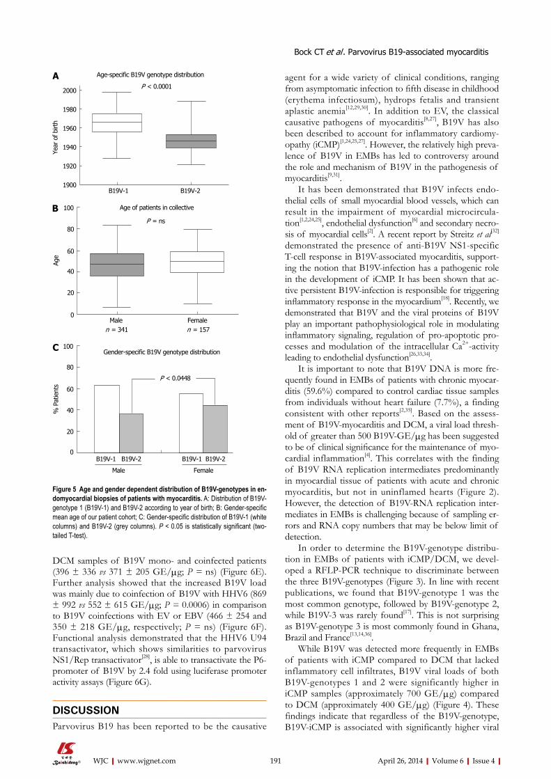
191 April 26, 2014|Volume 6|Issue 4|WJC|www.wjgnet.com
DCM samples of B19V mono- and coinfected patients (396 ± 336 vs 371 ± 205 GE/μg; P = ns) (Figure 6E). Further analysis showed that the increased B19V load was mainly due to coinfection of B19V with HHV6 (869 ± 992 vs 552 ± 615 GE/μg; P = 0.0006) in comparison to B19V coinfections with EV or EBV (466 ± 254 and 350 ± 218 GE/μg, respectively; P = ns) (Figure 6F). Functional analysis demonstrated that the HHV6 U94 transactivator, which shows similarities to parvovirus NS1/Rep transactivator[28], is able to transactivate the P6-promoter of B19V by 2.4 fold using luciferase promoter activity assays (Figure 6G).
DISCUSSIONParvovirus B19 has been reported to be the causative
agent for a wide variety of clinical conditions, ranging from asymptomatic infection to fifth disease in childhood (erythema infectiosum), hydrops fetalis and transient aplastic anemia[12,29,30]. In addition to EV, the classical causative pathogens of myocarditis[8,27], B19V has also been described to account for inflammatory cardiomy-opathy (iCMP)[1,24,25,27]. However, the relatively high preva-lence of B19V in EMBs has led to controversy around the role and mechanism of B19V in the pathogenesis of myocarditis[9,31].
It has been demonstrated that B19V infects endo-thelial cells of small myocardial blood vessels, which can result in the impairment of myocardial microcircula-tion[1,2,24,25], endothelial dysfunction[6] and secondary necro-sis of myocardial cells[2]. A recent report by Streitz et al[32] demonstrated the presence of anti-B19V NS1-specific T-cell response in B19V-associated myocarditis, support-ing the notion that B19V-infection has a pathogenic role in the development of iCMP. It has been shown that ac-tive persistent B19V-infection is responsible for triggering inflammatory response in the myocardium[18]. Recently, we demonstrated that B19V and the viral proteins of B19V play an important pathophysiological role in modulating inflammatory signaling, regulation of pro-apoptotic pro-cesses and modulation of the intracellular Ca2+-activity leading to endothelial dysfunction[26,33,34].
It is important to note that B19V DNA is more fre-quently found in EMBs of patients with chronic myocar-ditis (59.6%) compared to control cardiac tissue samples from individuals without heart failure (7.7%), a finding consistent with other reports[2,35]. Based on the assess-ment of B19V-myocarditis and DCM, a viral load thresh-old of greater than 500 B19V-GE/μg has been suggested to be of clinical significance for the maintenance of myo-cardial inflammation[4]. This correlates with the finding of B19V RNA replication intermediates predominantly in myocardial tissue of patients with acute and chronic myocarditis, but not in uninflamed hearts (Figure 2). However, the detection of B19V-RNA replication inter-mediates in EMBs is challenging because of sampling er-rors and RNA copy numbers that may be below limit of detection.
In order to determine the B19V-genotype distribu-tion in EMBs of patients with iCMP/DCM, we devel-oped a RFLP-PCR technique to discriminate between the three B19V-genotypes (Figure 3). In line with recent publications, we found that B19V-genotype 1 was the most common genotype, followed by B19V-genotype 2, while B19V-3 was rarely found[17]. This is not surprising as B19V-genotype 3 is most commonly found in Ghana, Brazil and France[13,14,36].
While B19V was detected more frequently in EMBs of patients with iCMP compared to DCM that lacked inflammatory cell infiltrates, B19V viral loads of both B19V-genotypes 1 and 2 were significantly higher in iCMP samples (approximately 700 GE/μg) compared to DCM (approximately 400 GE/μg) (Figure 4). These findings indicate that regardless of the B19V-genotype, B19V-iCMP is associated with significantly higher viral
Year
of b
irth
Age-specific B19V genotype distribution
P < 0.0001
B19V-1 B19V-2
2000
1980
1960
1940
1920
1900
Age of patients in collective
P = ns
Male Femalen = 341 n = 157
Age
100
80
60
40
20
0
A
B
100
80
60
40
20
0B19V-1 B19V-2 B19V-1 B19V-2
Male Female
% P
atie
nts
P < 0.0448
Gender-specific B19V genotype distributionC
Figure 5 Age and gender dependent distribution of B19V-genotypes in en-domyocardial biopsies of patients with myocarditis. A: Distribution of B19V-genotype 1 (B19V-1) and B19V-2 according to year of birth; B: Gender-specific mean age of our patient cohort; C: Gender-specific distribution of B19V-1 (white columns) and B19V-2 (grey columns). P < 0.05 is statistically significant (two-tailed T-test).
Bock CT et al . Parvovirus B19-associated myocarditis
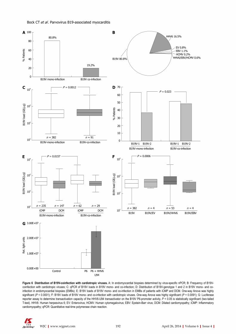
192 April 26, 2014|Volume 6|Issue 4|WJC|www.wjgnet.com
% P
atie
nts
100
80
60
40
20
0
80.8%
19.2%
B19V mono-infection B19V co-infection
HHV6 16.5%
EV 0.8%EBV 1.1%HCMV 0.2%
HHV6/EBV/HCMV 0.6%B19V 80.8%
B19V
load
(G
E/μg
)
104
103
102
101
B19V-mono-infection B19V-co-infection
P = 0.0012
n = 382 n = 91
P = 0.023
B19V-1 B19V-2 B19V-1 B19V-2
B19V-mono-infection B19V-co-infection
70
60
50
40
30
20
10
0
% P
atie
nts
B19V
load
(G
E/μg
)
104
103
102
101
P = 0.0157
iCMP DCM iCMP DCM
B19V-mono-infection B19V-co-infection
n = 235 n = 147 n = 62 n = 29
B19V
load
(G
E/μg
)
104
103
102
101
P = 0.0006
n = 382 n = 4 n = 53 n = 4
B19V B19V/EV B19V/HHV6 B19V/EBV
3.00E+07
2.00E+07
1.00E+07
0.00E+00Control P6 P6 + HHV6 U94
Rel.
light
uni
ts
Figure 6 Distribution of B19V-coinfection with cardiotropic viruses. A: In endomyocardial biopsies determined by virus-specific nPCR; B: Frequency of B19V-coinfection with cardiotropic viruses; C: qPCR of B19V loads in B19V mono- and co-infection; D: Distribution of B19V-genotype 1 and 2 in B19V mono- and co-infection in endomyocardial biopsies (EMBs); E: B19V loads of B19V mono- and co-infection in EMBs of patients with iCMP and DCM. One-way Anova was highly significant (P < 0.0001); F: B19V loads of B19V mono- and co-infection with cardiotropic viruses. One-way Anova was highly significant (P = 0.0091); G: Luciferase reporter assay to determine transactivation capacity of the HHV6-U94 transactivator on the B19V P6-promoter activity. P < 0.05 is statistically significant (two-tailed T-test). HHV6: Human herpesvirus 6; EV: Enterovirus; HCMV: Human cytomegalovirus; EBV: Epstein-Barr virus; DCM: Dilated cardiomyopathy; iCMP: Inflammatory cardiomyopathy; qPCR: Quantitative real-time polymerase chain reaction.
A B
C D
E F
G
Bock CT et al . Parvovirus B19-associated myocarditis
193 April 26, 2014|Volume 6|Issue 4|WJC|www.wjgnet.com
loads than B19V-DCM.As with the age-dependent distribution of B19V-
genotype 1 and 2 in various tissue samples (e.g., skin, synovium, liver, and heart)[15-17], we found that B19V-gen-otype 1 was predominantly detected in younger people (mean age 43 years), while B19V-genotype 2 was mostly observed in patients older than 60 years (Figure 5). This age-associated difference in frequency of genotypes is thought to be due to the reported lifelong persistence of viral genomes in humans[15,16,29]. Analysis of gender de-pendency of B19V genotype distribution and viral loads did not show any significant differences. However, a sig-nificantly higher proportion of women were found to be persistently infected with B19V-genotype 2 in contrast to men (P < 0.05) (Figure 5).
It can be hypothesized that B19V can be reactivated from long-term persistent or latent infection by viral and/or host-specific determinants. B19V-coinfection with other cardiotropic viruses like EV, ADV and HHV6 may contribute to the severity of B19V-myocarditis[4,7,8,20], possibly by reactivating B19V replication and thereby enhancing virus specific host immune responses, tissue inflammation and the progression to chronic heart fail-ure[37]. This is reminiscent of the increased replication of HIV caused by coinfection with HHV6 and HHV8[21,38]. Our finding show that coinfection with B19V and other cardiotropic viruses, particularly HHV6, is not infre-quent and is associated with higher B19V loads in EMBs. HHV6 cannot only infect and replicate in endothelial cells[39], but also encodes a viral transactivator[37] called U94 which is homologous to the B19V-NS1 transactiva-tor[28]. By using promoter activity assays, we have shown that the HHV6 U94 transactivator also transactivates the B19V-P6 promoter, resulting in enhanced B19V loads in B19V/HHV6 coinfection (Figure 6).
Our data shows that B19V is capable of long-term persistence in the human heart, even lasting for decades, and that active B19V replication is associated with the de-velopment of iCMP. To establish a clinically relevant link between B19V-infection and the development of iCMP, it is important to develop molecular diagnostic techniques to determine myocardial viral loads and sequence analysis to verify the association between myocardial disease and genotype-specific B19V isolates. Notably, persistently low replicating and latent B19V-infection may be reactivated by coinfecting cardiotropic viruses, especially HHV6. Our findings together with other recently published data[1,6,18,25,26,40] argue an important role for B19V in the development of iCMP.
ACKNOWLEDGMENTSWe appreciated the excellent technical assistance of Rosa Mammato, Gerd Janke, Isabell Haussmann and Silke Aberle. We wish to thank Dr. Bernhard Renard, Rob-ert Koch Institute, Germany, for his valuable help with the statistical analysis. The authors would like to thank Professor Joseph Torresi, Department of Infectious Diseases, Austin Hospital, The University of Melbourne,
Australia, for his valuable and critical reading of the manuscript.
COMMENTSBackgroundViral and post viral myocarditis is the major cause of acute and chronic dilated cardiomyopathy (DCM). Human parvovirus B19 has been identified as a new emerging pathogenic agent in the etiology of inflammatory cardiomyopathy (iCMP). However, the role of B19V-infection in the development of chronic myo-carditis is still unclear. The authors have recently demonstrated that endothelial cells but not cardiac myocytes are B19V-specific target cells in patients with B19V-associated myocarditis. Furthermore, B19V was not detected frequently in patients with unexplained isolated diastolic dysfunction. Molecular pheno-types such as patient age, gender, B19V replicative mRNA intermediates, B19V genotype and B19V-coinfection of myocardial B19V-infection contributing to the etiopathogenesis of B19V-myocarditis as an endothelial-cell mediated inflam-matory disease have not been described so far.Research frontiersThe identification of various viral nucleic acids by polymerase chain reaction in the myocardium of patients with suspected myocarditis led to the hypothesis that acute and chronic myocarditis may develop as a result of infection, not only with enteroviruses (Coxsackievirus B3), but also with other cardiotropic viruses, such as parvovirus B19. In this regard, the research hotspot is how molecular phenotypes can contribute to the pathogenic role of myocardial B19V-infection in myocarditis and DCM.Innovations and breakthroughsThe study results showed that B19V RNA replication intermediates could be determined mainly in acute and ongoing myocarditis, indicating active replica-tion of B19V. B19V-genotypes 1 and 2 were predominant with a more frequent prevalence of B19V-genotype 2 in iCMP. Furthermore, B19V-coinfections with other cardiotropic viruses can occur, most frequently with human herpes virus 6 (HHV6). Functional experiments showed that the HHV6 U94-transactivator could transactivate the B19V-P6-promoter. From these findings it is suggested that long-term persistence of B19V DNA in the human heart occurs and that active/reactivated B19V-replication can be associated with iCMP in a viral load and genotype-dependent manner.ApplicationsTo establish a clinically relevant link between B19V-infection and the develop-ment of iCMP, it is important to pursue molecular diagnostic techniques to de-termine myocardial B19V loads and to verify genotype-specific B19V isolates. Notably, persistent low replicating and even latent B19V-infection may be reac-tivated by coinfecting cardiotropic viruses, especially HHV6.TerminologyParvovirus B19 was assumed to be an agent of human disease when its as-sociation with erythema infectiosum (fifth disease), hydrops fetalis and transient aplastic anemia was demonstrated in the 1980s. During the last few years, a growing number of reports have been published demonstrating an association between B19V and many other clinical diseases, like arthritis, myocarditis, various vasculitis syndromes, hepatitis and neurological disorders. A growing number of reports have suggested an association between B19V infection with acute and chronic cardiac diseases. Myocarditis is the term used to indicate in-fectious, toxic or autoimmune processes causing inflammation of the heart and represents a non-ischemic inflammatory heart disease with a highly variable clinical outcome. In most cases this disease is self-limiting; however, it may also lead to acute heart failure, resulting in early death or heart transplantation. Myocarditis can also mimic acute myocardial infarction. ICMP was included as a subtype of the specific cardiomyopathies and defined “as myocarditis in as-sociation with cardiac dysfunction” by the World Health Organization.Peer reviewThe paper deals with the interesting subject of molecular phenotypes of human parvovirus B19 in patients with myocarditis. Both the specific virus, which is a cause of important pathologies, as well as myocarditis, an entity that can affect great portions of a population, among them young, otherwise healthy individu-als, are very interesting subjects with an impact on the general practice of internists, cardiologists, general physicians, pathologists, biologists and genetic scientists. The paper deals with the aforementioned interesting subject in a thorough way.
COMMENTS
Bock CT et al . Parvovirus B19-associated myocarditis
194 April 26, 2014|Volume 6|Issue 4|WJC|www.wjgnet.com
REFERENCES1 Bültmann BD, Klingel K, Sotlar K, Bock CT, Baba HA, Sau-
ter M, Kandolf R. Fatal parvovirus B19-associated myocardi-tis clinically mimicking ischemic heart disease: an endothe-lial cell-mediated disease. Hum Pathol 2003; 34: 92-95 [PMID: 12605372 DOI: 10.1053/hupa.2003.48]
2 Klingel K, Sauter M, Bock CT, Szalay G, Schnorr JJ, Kandolf R. Molecular pathology of inflammatory cardiomyopathy. Med Microbiol Immunol 2004; 193: 101-107 [PMID: 12920583 DOI: 10.1007/s00430-003-0190-1]
3 Mahfoud F, Gärtner B, Kindermann M, Ukena C, Gadom-ski K, Klingel K, Kandolf R, Böhm M, Kindermann I. Virus serology in patients with suspected myocarditis: utility or futility? Eur Heart J 2011; 32: 897-903 [PMID: 21217143 DOI: 10.1093/eurheartj/ehq493]
4 Bock CT, Klingel K, Kandolf R. Human parvovirus B19-associated myocarditis. N Engl J Med 2010; 362: 1248-1249 [PMID: 20357294 DOI: 10.1056/NEJMc0911362]
5 Ruppert V, Meyer T, Balbach A, Richter A, Müller HH, Maisch B, Pankuweit S. Genotype-specific effects on left ven-tricular function in parvovirus B19-positive patients with di-lated cardiomyopathy. J Med Virol 2011; 83: 1818-1825 [PMID: 21837800 DOI: 10.1002/jmv.22187]
6 Tschöpe C, Bock CT, Kasner M, Noutsias M, Westermann D, Schwimmbeck PL, Pauschinger M, Poller WC, Kühl U, Kan-dolf R, Schultheiss HP. High prevalence of cardiac parvo-virus B19 infection in patients with isolated left ventricular diastolic dysfunction. Circulation 2005; 111: 879-886 [PMID: 15710767 DOI: 10.1161/01.CIR.0000155615.68924.B3]
7 Kühl U, Pauschinger M, Noutsias M, Seeberg B, Bock T, Lassner D, Poller W, Kandolf R, Schultheiss HP. High preva-lence of viral genomes and multiple viral infections in the myocardium of adults with “idiopathic” left ventricular dysfunction. Circulation 2005; 111: 887-893 [PMID: 15699250 DOI: 10.1161/01.CIR.0000155616.07901.35]
8 Kandolf R, Bültmann B, Klingel K, Bock CT. [Molecular mechanisms and consequences of cardiac viral infections]. Pathologe 2008; 29 Suppl 2: 112-117 [PMID: 18820926 DOI: 10.1007/s00292-008-1027-x]
9 Schenk T, Enders M, Pollak S, Hahn R, Huzly D. High prevalence of human parvovirus B19 DNA in myocardial autopsy samples from subjects without myocarditis or dila-tive cardiomyopathy. J Clin Microbiol 2009; 47: 106-110 [PMID: 19005147 DOI: 10.1128/JCM.01672-08]
10 Koepsell SA, Anderson DR, Radio SJ. Parvovirus B19 is a bystander in adult myocarditis. Cardiovasc Pathol 2012; 21: 476-481 [PMID: 22425629 DOI: 10.1016/j.carpath.2012.02.002]
11 Cohen BJ, Buckley MM. The prevalence of antibody to hu-man parvovirus B19 in England and Wales. J Med Microbiol 1988; 25: 151-153 [PMID: 3339634 DOI: 10.1099/00222615-25-2-151]
12 Brown KE, Young NS. Human parvovirus B19 infections in infants and children. Adv Pediatr Infect Dis 1997; 13: 101-126 [PMID: 9544309]
13 Servant A, Laperche S, Lallemand F, Marinho V, De Saint Maur G, Meritet JF, Garbarg-Chenon A. Genetic diversity within human erythroviruses: identification of three geno-types. J Virol 2002; 76: 9124-9134 [PMID: 12186896 DOI: 10.1128/JVI.76.18.9124-9134.2002]
14 Sanabani S, Neto WK, Pereira J, Sabino EC. Sequence vari-ability of human erythroviruses present in bone marrow of Brazilian patients with various parvovirus B19-related hematological symptoms. J Clin Microbiol 2006; 44: 604-606 [PMID: 16455922 DOI: 10.1128/JCM.44.2.604-606.2006]
15 Ekman A, Hokynar K, Kakkola L, Kantola K, Hedman L, Bondén H, Gessner M, Aberham C, Norja P, Miettinen S, Hedman K, Söderlund-Venermo M. Biological and immu-nological relations among human parvovirus B19 genotypes
1 to 3. J Virol 2007; 81: 6927-6935 [PMID: 17409158 DOI: 10.1128/JVI.02713-06]
16 Norja P, Hokynar K, Aaltonen LM, Chen R, Ranki A, Partio EK, Kiviluoto O, Davidkin I, Leivo T, Eis-Hübinger AM, Schneider B, Fischer HP, Tolba R, Vapalahti O, Vaheri A, Söderlund-Venermo M, Hedman K. Bioportfolio: lifelong persistence of variant and prototypic erythrovirus DNA genomes in human tissue. Proc Natl Acad Sci USA 2006; 103: 7450-7453 [PMID: 16651522 DOI: 10.1073/pnas.0602259103]
17 Kühl U, Lassner D, Pauschinger M, Gross UM, Seeberg B, Noutsias M, Poller W, Schultheiss HP. Prevalence of erythro-virus genotypes in the myocardium of patients with dilated cardiomyopathy. J Med Virol 2008; 80: 1243-1251 [PMID: 18461615 DOI: 10.1002/jmv.21187]
18 Kindermann I, Kindermann M, Kandolf R, Klingel K, Bül-tmann B, Müller T, Lindinger A, Böhm M. Predictors of outcome in patients with suspected myocarditis. Circulation 2008; 118: 639-648 [PMID: 18645053 DOI: 10.1161/CIRCU-LATIONAHA.108.769489]
19 Pozzuto T, von Kietzell K, Bock T, Schmidt-Lucke C, Poller W, Zobel T, Lassner D, Zeichhardt H, Weger S, Fechner H. Transactivation of human parvovirus B19 gene expres-sion in endothelial cells by adenoviral helper functions. Virology 2011; 411: 50-64 [PMID: 21236463 DOI: 10.1016/j.virol.2010.12.019]
20 Rohayem J, Dinger J, Fischer R, Klingel K, Kandolf R, Reth-wilm A. Fatal myocarditis associated with acute parvovirus B19 and human herpesvirus 6 coinfection. J Clin Microbiol 2001; 39: 4585-4587 [PMID: 11724892 DOI: 10.1128/JCM.39.12.4585-4587.2001]
21 Caselli E, Galvan M, Cassai E, Caruso A, Sighinolfi L, Di Luca D. Human herpesvirus 8 enhances human immunodeficiency virus replication in acutely infected cells and induces reactiva-tion in latently infected cells. Blood 2005; 106: 2790-2797 [PMID: 15976177 DOI: 10.1182/blood-2005-04-1390]
22 Toan NL, Duechting A, Kremsner PG, Song le H, Ebinger M, Aberle S, Binh VQ, Duy DN, Torresi J, Kandolf R, Bock CT. Phylogenetic analysis of human parvovirus B19, indicating two subgroups of genotype 1 in Vietnamese patients. J Gen Virol 2006; 87: 2941-2949 [PMID: 16963753 DOI: 10.1099/vir.0.82037-0]
23 Bock CT, Kubicka S, Manns MP, Trautwein C. Two control elements in the hepatitis B virus S-promoter are important for full promoter activity mediated by CCAAT-binding fac-tor. Hepatology 1999; 29: 1236-1247 [PMID: 10094970 DOI: 10.1002/hep.510290426]
24 Bültmann BD, Klingel K, Sotlar K, Bock CT, Kandolf R. Parvovirus B19: a pathogen responsible for more than he-matologic disorders. Virchows Arch 2003; 442: 8-17 [PMID: 12536309]
25 Bock CT, Klingel K, Aberle S, Duechting A, Lupescu A, Lang F, Kandolf R. Human parvovirus B19: a new emerg-ing pathogen of inflammatory cardiomyopathy. J Vet Med B Infect Dis Vet Public Health 2005; 52: 340-343 [PMID: 16316397 DOI: 10.1111/j.1439-0450.2005.00867.x]
26 Duechting A, Tschöpe C, Kaiser H, Lamkemeyer T, Tanaka N, Aberle S, Lang F, Torresi J, Kandolf R, Bock CT. Human parvovirus B19 NS1 protein modulates inflammatory sig-naling by activation of STAT3/PIAS3 in human endothe-lial cells. J Virol 2008; 82: 7942-7952 [PMID: 18550668 DOI: 10.1128/JVI.00891-08]
27 Kandolf R. [Virus etiology of inflammatory cardiomyopa-thy]. Dtsch Med Wochenschr 2004; 129: 2187-2192 [PMID: 15457399 DOI: 10.1055/s-2004-831863]
28 Rapp JC, Krug LT, Inoue N, Dambaugh TR, Pellett PE. U94, the human herpesvirus 6 homolog of the parvovirus nonstructural gene, is highly conserved among isolates and is expressed at low mRNA levels as a spliced transcript. Virology 2000; 268: 504-516 [PMID: 10704358 DOI: 10.1006/viro.1999.0163]
Bock CT et al . Parvovirus B19-associated myocarditis
195 April 26, 2014|Volume 6|Issue 4|WJC|www.wjgnet.com
29 Heegaard ED, Brown KE. Human parvovirus B19. Clin Mi-crobiol Rev 2002; 15: 485-505 [PMID: 12097253 DOI: 10.1128/CMR.15.3.485-505.2002]
30 Young NS, Brown KE. Parvovirus B19. N Engl J Med 2004; 350: 586-597 [PMID: 14762186 DOI: 10.1056/NEJMra030840]
31 Lindner J, Noutsias M, Lassner D, Wenzel J, Schultheiss HP, Kuehl U, Modrow S. Adaptive immune responses against parvovirus B19 in patients with myocardial disease. J Clin Virol 2009; 44: 27-32 [PMID: 18980860 DOI: 10.1016/j.jcv.2008.09.007]
32 Streitz M, Noutsias M, Volkmer R, Rohde M, Brestrich G, Block A, Klippert K, Kotsch K, Ay B, Hummel M, Kühl U, Lassner D, Schultheiss HP, Volk HD, Kern F. NS1 specific CD8+ T-cells with effector function and TRBV11 dominance in a patient with parvovirus B19 associated inflammatory cardiomyopathy. PLoS One 2008; 3: e2361 [PMID: 18523634 DOI: 10.1371/journal.pone.0002361]
33 Lupescu A, Bock CT, Lang PA, Aberle S, Kaiser H, Kandolf R, Lang F. Phospholipase A2 activity-dependent stimulation of Ca2+ entry by human parvovirus B19 capsid protein VP1. J Virol 2006; 80: 11370-11380 [PMID: 16956939 DOI: 10.1128/JVI.01041-06]
34 Lupescu A, Geiger C, Zahir N, Aberle S, Lang PA, Kramer S, Wesselborg S, Kandolf R, Foller M, Lang F, Bock CT. In-hibition of Na+/H+ exchanger activity by parvovirus B19 protein NS1. Cell Physiol Biochem 2009; 23: 211-220 [PMID: 19255516 DOI: 10.1159/000204110]
35 Donoso Mantke O, Nitsche A, Meyer R, Klingel K, Niedrig M. Analysing myocardial tissue from explanted hearts of heart transplant recipients and multi-organ donors for the presence of parvovirus B19 DNA. J Clin Virol 2004; 31: 32-39
[PMID: 15288611 DOI: 10.1016/j.jcv.2003.12.013]36 Parsyan A, Szmaragd C, Allain JP, Candotti D. Identifica-
tion and genetic diversity of two human parvovirus B19 genotype 3 subtypes. J Gen Virol 2007; 88: 428-431 [PMID: 17251559 DOI: 10.1099/vir.0.82496-0]
37 Mahrholdt H, Wagner A, Deluigi CC, Kispert E, Hager S, Meinhardt G, Vogelsberg H, Fritz P, Dippon J, Bock CT, Klingel K, Kandolf R, Sechtem U. Presentation, patterns of myocardial damage, and clinical course of viral myocardi-tis. Circulation 2006; 114: 1581-1590 [PMID: 17015795 DOI: 10.1161/CIRCULATIONAHA.105.606509]
38 Isegawa Y, Katahira J, Yamanishi K, Sugimoto N. Reactiva-tion of latent human immunodeficiency virus 1 by human herpesvirus 6 infection. Acta Virol 2007; 51: 13-20 [PMID: 17432939]
39 Caruso A, Rotola A, Comar M, Favilli F, Galvan M, Tosetti M, Campello C, Caselli E, Alessandri G, Grassi M, Garrafa E, Cassai E, Di Luca D. HHV-6 infects human aortic and heart microvascular endothelial cells, increasing their ability to secrete proinflammatory chemokines. J Med Virol 2002; 67: 528-533 [PMID: 12115999 DOI: 10.1002/jmv.10133]
40 Vogelsberg H, Mahrholdt H, Deluigi CC, Yilmaz A, Kispert EM, Greulich S, Klingel K, Kandolf R, Sechtem U. Cardio-vascular magnetic resonance in clinically suspected cardiac amyloidosis: noninvasive imaging compared to endomyo-cardial biopsy. J Am Coll Cardiol 2008; 51: 1022-1030 [PMID: 18325442 DOI: 10.1016/j.jacc.2007.10.049]
41 Aretz HT, Billingham ME, Edwards WD, Factor SM, Fal-lon JT, Fenoglio JJ, Olsen EG, Schoen FJ. Myocarditis. A histopathologic definition and classification. Am J Cardiovasc Pathol 1987; 1: 3-14 [PMID: 3455232]
P- Reviewers: Robert KI, Tagarakis G S- Editor: Wen LL L- Editor: Roemmele A E- Editor: Liu SQ
Bock CT et al . Parvovirus B19-associated myocarditis
Coronary artery disease in congenital single coronary artery in adults: A Dutch case series
Salah AM Said, Willem G de Voogt, Suat Bulut, Jacques Han, Peter Polak, Rogier LG Nijhuis, Jeroen W op den Akker, Andries Slootweg
Salah AM Said, Rogier LG Nijhuis, Andries Slootweg, De-partment of Cardiology, Hospital Group Twente Almelo-Hengelo, 7555 DL Hengelo, The NetherlandsWillem G de Voogt, Department of Cardiology, St. Lucas-Andreas Hospital, 1061 AE Amsterdam, The NetherlandsSuat Bulut, Department of Cardiology, Gelre Hospital Zutphen, 7200 GZ Zutphen, The NetherlandsJacques Han, Department of Cardiology, Hospital De Sionsberg, 9101 DC Dokkum, The NetherlandsPeter Polak, Department of Cardiology, St. Anna Hospital, 5664 EH Geldrop, The NetherlandsJeroen W op den Akker, Department of Radiodiagnostics, Hospital Group Twente Almelo-Hengelo, 7555 DL Hengelo, The NetherlandsAuthor contributions: All authors contributed equally to the writing of the manuscript.Correspondence to: Dr. Salah AM Said, Department of Car-diology, Hospital Group Twente Almelo-Hengelo, Geerdinksweg 141, 7555 DL Hengelo, The Netherlands. [email protected]: +31-74-2905286 Fax: +31-74-2905289Received: September 15, 2013 Revised: November 6, 2013Accepted: March 13, 2014Published online: April 26, 2014
AbstractAIM: To assess the current diagnostic and therapeutic management and the clinical implications of congenital single coronary artery (SCA) in adults.
METHODS: We identified 15 patients with a SCA detected from four Dutch angiography centers in the period between 2010 and 2013. Symptomatic patients who underwent routine diagnostic coronary angiogra-phy (CAG) for suspected coronary artery disease and who incidentally were found to have isolated SCA were analyzed.
RESULTS: Fifteen (7 females) with a mean age of 58.5 ± 13.78 years (range 43-86) had a SCA. Conventional
CAG demonstrated congenital isolated SCA originating as a single ostium from the right sinus of Valsalva in 6 patients and originating from the left in 9 patients. Minimal to moderate coronary atherosclerotic changes were found in 4, and severe stenotic lesions in another 4 patients. Seven patients were free of coronary ath-erosclerosis. Runs of non-sustained ventricular tachy-cardia were documented in 2 patients, one of whom demonstrated transmural ischemic changes on presen-tation. Myocardial perfusion scintigraphic evidence of transmural myocardial ischemia was found in 1 patient due to kinking and squeezing of the SCA with an in-terarterial course between the aorta and pulmonary artery. Multi-slice computed tomography (MSCT) was helpful to delineate the course of the anomalous artery relative to the aorta and pulmonary artery. Percutane-ous coronary intervention was successfully performed in 3 patients. Eight patients were managed medically. Arterial bypass graft was performed in 4 patients with the squeezed SCA.
CONCLUSION: SCA may be associated with transient transmural myocardial ischemia and aborted sudden death in the absence of coronary atherosclerosis. The availability and sophistication of MSCT facilitates the delineation of the course of a SCA. We present a Dutch case series and review of the literature.
© 2014 Baishideng Publishing Group Co., Limited. All rights reserved.
Key words: Congenital heart disease; Coronary artery anomaly; Coronary angiography; Single coronary ar-tery; Coronary artery disease; Multi-slice computed tomography
Core tip: A Dutch case series of 15 adult patients with congenital isolated single coronary artery (SCA) are presented. Conventional coronary angiography demon-
ORIGINAL ARTICLE
Online Submissions: http://www.wjgnet.com/esps/[email protected]:10.4330/wjc.v6.i4.196
196 April 26, 2014|Volume 6|Issue 4|WJC|www.wjgnet.com
World Journal of CardiologyW J C
World J Cardiol 2014 April 26; 6(4): 196-204ISSN 1949-8462 (online)
© 2014 Baishideng Publishing Group Co., Limited. All rights reserved.
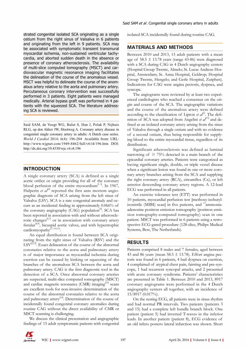
strated congenital isolated SCA originating as a single ostium from the right sinus of Valsalva in 6 patients and originating from the left in 9 patients. SCA may be associated with symptomatic transient transmural myocardial ischemia, non-sustained ventricular tachy-cardia, and aborted sudden death in the absence or presence of coronary atherosclerosis. The availability of multi-slice computed tomography (MSCT) and car-diovascular magnetic resonance imaging facilitates the delineation of the course of the anomalous vessel. MSCT was helpful to delineate the course of the anom-alous artery relative to the aorta and pulmonary artery. Percutaneous coronary intervention was successfully performed in 3 patients. Eight patients were managed medically. Arterial bypass graft was performed in 4 pa-tients with the squeezed SCA. The literature address-ing SCA is reviewed.
Said SAM, de Voogt WG, Bulut S, Han J, Polak P, Nijhuis RLG, op den Akker JW, Slootweg A. Coronary artery disease in congenital single coronary artery in adults: A Dutch case series. World J Cardiol 2014; 6(4): 196-204 Available from: URL: http://www.wjgnet.com/1949-8462/full/v6/i4/196.htm DOI: http://dx.doi.org/10.4330/wjc.v6.i4.196
INTRODUCTIONA single coronary artery (SCA) is defined as a single aortic orifice or origin providing for all of the coronary blood perfusion of the entire myocardium[1-3]. In 1967, Halperin et al[4] reported the first ante mortem angio-graphic diagnosis of SCA arising from the left sinus of Valsalva (LSV). SCA is a rare congenital anomaly and oc-curs as an incidental finding in approximately 0.066% of the coronary angiography (CAG) population[5]. SCA has been reported in association with and without atheroscle-rotic changes[6,7] or in association with coronary artery fistulas[8,9], bicuspid aortic valves, and with hypertrophic cardiomyopathy[1,7,10,11].
An equal distribution is found between SCA origi-nating from the right sinus of Valsalva (RSV) and the LSV[2,12]. Exact delineation of the course of the abnormal coronaries relative to the aorta and pulmonary artery is of major importance as myocardial ischemia during exertion can be caused by kinking or squeezing of the branches of the anomalous SCA between the aorta and pulmonary artery. CAG is the first diagnostic tool in the detection of a SCA. Once abnormal coronary arteries are suspected, multi-slice computed tomography (MSCT) and cardiac magnetic resonance (CMR) imaging[13] scans are excellent tools for non-invasive determination of the course of the abnormal coronaries relative to the aorta and pulmonary artery[14]. Determination of the course of incidentally found congenital coronary anomalies during routine CAG without the direct availability of CMR or MSCT scanning is challenging.
We discuss the clinical presentation and angiographic findings of 15 adult symptomatic patients with congenital
isolated SCA incidentally found during routine CAG.
MATERIALS AND METHODSBetween 2010 and 2013, 15 adult patients with a mean age of 58.5 ± 13.78 years (range 43-86) were diagnosed with a SCA during CAG in 4 Dutch angiography centers (Hospital Group Twente, Almelo; St. Lucas Andreas Hos-pital, Amsterdam; St. Anna Hospital, Geldrop; Hospital Group Twente, Hengelo; and Gerle Hospital, Zutphen). Indications for CAG were angina pectoris, dyspnea, and syncope.
The angiograms were reviewed by at least two experi-enced cardiologists who reached a consensus on the ori-gin and course of the SCA. The angiographic variations and the course of the anomalous artery were defined according to the classification of Lipton et al[1]. The defi-nition of SCA was adopted from Angelini et al[15] and de-fined as an isolated coronary artery arising from the sinus of Valsalva through a single ostium and with no evidence of a second ostium, thus being responsible for supply-ing blood to the entire myocardial tissue, regardless of its distribution.
Significant atherosclerosis was defined as luminal narrowing of ≥ 75% detected in a main branch of the epicardial coronary arteries. Patients were categorized as having significant single, double, or triple vessel disease when a significant lesion was found in one or more coro-nary artery branches arising from the SCA and supplying the right coronary artery (RCA), circumflex (Cx), or left anterior descending coronary artery regions. A 12-lead ECG was performed in all patients.
An exercise tolerance test (ETT) was performed in 10 patients, myocardial perfusion test [methoxy-isobutyl-isonitrile (MIBI) scan] in five patients, and 13ammonia-adenosine positron emission tomography (positron emis-sion tomography-computed tomography) scan in one patient. MSCT was performed in 6 patients using a retro-spective ECG-gated procedure (128-slice, Philips Medical Systems, Best, The Netherlands).
RESULTSPatients comprised 8 males and 7 females, aged between 43 and 86 years (mean 58.5 ± 13.78). Effort angina pec-toris was found in 6 patients, 4 had dyspnea on exertion, 4 complained of atypical chest pain, fainting and pre-syn-cope, 1 had recurrent syncopal attacks, and 2 presented with acute coronary syndrome. Patients’ characteristics are presented in Table 1. Between 2010 and 2013, 8917 coronary angiograms were performed in the 4 Dutch angiography centers all together, with an incidence of 15/8917 (0.017%).
On the resting ECG, all patients were in sinus rhythm and had normal PR intervals. Two patients (patients 3 and 15) had a complete left bundle branch block. One patient (patient 5) had inverted T-waves in the inferior leads. In another patient (patient 8), ECG evidence of an old infero postero lateral infarction was shown. Short
197 April 26, 2014|Volume 6|Issue 4|WJC|www.wjgnet.com
Said SAM et al . Congenital single coronary artery in adults
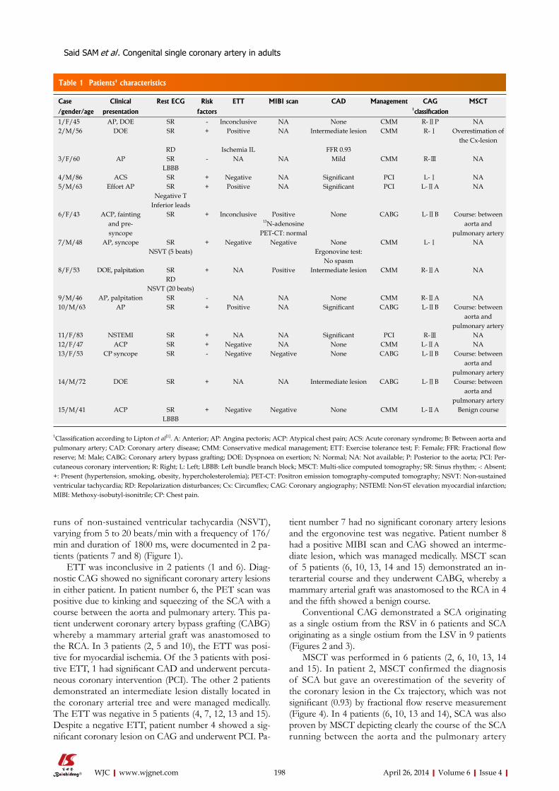
runs of non-sustained ventricular tachycardia (NSVT), varying from 5 to 20 beats/min with a frequency of 176/min and duration of 1800 ms, were documented in 2 pa-tients (patients 7 and 8) (Figure 1).
ETT was inconclusive in 2 patients (1 and 6). Diag-nostic CAG showed no significant coronary artery lesions in either patient. In patient number 6, the PET scan was positive due to kinking and squeezing of the SCA with a course between the aorta and pulmonary artery. This pa-tient underwent coronary artery bypass grafting (CABG) whereby a mammary arterial graft was anastomosed to the RCA. In 3 patients (2, 5 and 10), the ETT was posi-tive for myocardial ischemia. Of the 3 patients with posi-tive ETT, 1 had significant CAD and underwent percuta-neous coronary intervention (PCI). The other 2 patients demonstrated an intermediate lesion distally located in the coronary arterial tree and were managed medically. The ETT was negative in 5 patients (4, 7, 12, 13 and 15). Despite a negative ETT, patient number 4 showed a sig-nificant coronary lesion on CAG and underwent PCI. Pa-
tient number 7 had no significant coronary artery lesions and the ergonovine test was negative. Patient number 8 had a positive MIBI scan and CAG showed an interme-diate lesion, which was managed medically. MSCT scan of 5 patients (6, 10, 13, 14 and 15) demonstrated an in-terarterial course and they underwent CABG, whereby a mammary arterial graft was anastomosed to the RCA in 4 and the fifth showed a benign course.
Conventional CAG demonstrated a SCA originating as a single ostium from the RSV in 6 patients and SCA originating as a single ostium from the LSV in 9 patients (Figures 2 and 3).
MSCT was performed in 6 patients (2, 6, 10, 13, 14 and 15). In patient 2, MSCT confirmed the diagnosis of SCA but gave an overestimation of the severity of the coronary lesion in the Cx trajectory, which was not significant (0.93) by fractional flow reserve measurement (Figure 4). In 4 patients (6, 10, 13 and 14), SCA was also proven by MSCT depicting clearly the course of the SCA running between the aorta and the pulmonary artery
198 April 26, 2014|Volume 6|Issue 4|WJC|www.wjgnet.com
Table 1 Patients' characteristics
Case Clinical Rest ECG Risk ETT MIBI scan CAD Management CAG MSCT
/gender/age presentation factors 1classification1/F/45 AP, DOE SR - Inconclusive NA None CMM R-ⅡP NA2/M/56 DOE SR + Positive NA Intermediate lesion CMM R-Ⅰ Overestimation of
the Cx-lesionRD Ischemia IL FFR 0.93
3/F/60 AP SR - NA NA Mild CMM R-Ⅲ NALBBB
4/M/86 ACS SR + Negative NA Significant PCI L-Ⅰ NA5/M/63 Effort AP SR + Positive NA Significant PCI L-ⅡA NA
Negative T Inferior leads
6/F/43 ACP, fainting and pre-syncope
SR + Inconclusive Positive13N-adenosine
PET-CT: normal
None CABG L-ⅡB Course: betweenaorta and
pulmonary artery7/M/48 AP, syncope SR + Negative Negative None CMM L-Ⅰ NA
NSVT (5 beats) Ergonovine test:No spasm
8/F/53 DOE, palpitation SR + NA Positive Intermediate lesion CMM R-ⅡA NARD
NSVT (20 beats)9/M/46 AP, palpitation SR - NA NA None CMM R-ⅡA NA10/M/63 AP SR + Positive NA Significant CABG L-ⅡB Course: between
aorta and pulmonary artery
11/F/83 NSTEMI SR + NA NA Significant PCI R-Ⅲ NA12/F/47 ACP SR + Negative NA None CMM L-ⅡA NA13/F/53 CP syncope SR - Negative Negative None CABG L-ⅡB Course: between
aorta and pulmonary artery
14/M/72 DOE SR + NA NA Intermediate lesion CABG L-ⅡB Course: betweenaorta and
pulmonary artery15/M/41 ACP SR + Negative Negative None CMM L-ⅡA Benign course
LBBB
1Classification according to Lipton et al[1]. A: Anterior; AP: Angina pectoris; ACP: Atypical chest pain; ACS: Acute coronary syndrome; B: Between aorta and pulmonary artery; CAD: Coronary artery disease; CMM: Conservative medical management; ETT: Exercise tolerance test; F: Female; FFR: Fractional flow reserve; M: Male; CABG: Coronary artery bypass grafting; DOE: Dyspnoea on exertion; N: Normal; NA: Not available; P: Posterior to the aorta; PCI: Per-cutaneous coronary intervention; R: Right; L: Left; LBBB: Left bundle branch block; MSCT: Multi-slice computed tomography; SR: Sinus rhythm; -: Absent; +: Present (hypertension, smoking, obesity, hypercholesterolemia); PET-CT: Positron emission tomography-computed tomography; NSVT: Non-sustained ventricular tachycardia; RD: Repolarization disturbances; Cx: Circumflex; CAG: Coronary angiography; NSTEMI: Non-ST elevation myocardial infarction; MIBI: Methoxy-isobutyl-isonitrile; CP: Chest pain.
Said SAM et al . Congenital single coronary artery in adults
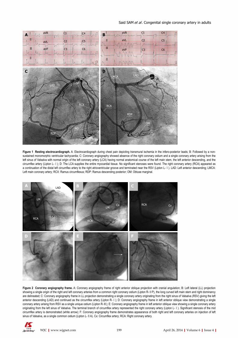
199 April 26, 2014|Volume 6|Issue 4|WJC|www.wjgnet.com
Ⅰ
Ⅱ
Ⅲ
Ⅱ
aVR
aVL
aVF
C1
C2
C3
C4
C5
C6
Ⅰ
Ⅱ
Ⅲ
Ⅱ
aVR
aVL
aVF
C1
C2
C3
C4
C5
C6
LAD
RCARCX
MO
RDP
LMCA LAD
RCA
RCX
OM
RDP
A B
C D
Figure 1 Resting electrocardiograph. A: Electrocardiograph during chest pain depicting transmural ischemia in the infero-posterior leads; B: Followed by a non-sustained monomorphic ventricular tachycardia; C: Coronary angiography showed absence of the right coronary ostium and a single coronary artery arising from the left sinus of Valsalva with normal origin of the left coronary artery (LCA) having normal anatomical course of the left main stem, the left anterior descending, and the circumflex artery (Lipton L-Ⅰ); D: The LCA supplies the entire myocardial tissue. No significant stenoses were found. The right coronary artery (RCA) appeared as a continuation of the distal left circumflex artery to the right atrioventricular groove and terminated near the RSV (Lipton L-Ⅰ). LAD: Left anterior descending; LMCA: Left main coronary artery; RCX: Ramus circumflexus; RDP: Ramus descending posterior; OM: Obtuse marginal.
Figure 2 Coronary angiography frame. A: Coronary angiography frame of right anterior oblique projection with cranial angulation; B: Left lateral (LL) projection showing a single origin of the right and left coronary arteries from a common right coronary ostium (Lipton R-ⅡP), the long curved left main stem and right dominancy are delineated; C: Coronary angiography frame in LL projection demonstrating a single coronary artery originating from the right sinus of Valsalva (RSV) giving the left anterior descending (LAD) and continued as the circumflex artery (Lipton R-Ⅰ); D: Coronary angiography frame in left anterior oblique view demonstrating a single coronary artery arising from RSV as a single unique ostium (Lipton R-Ⅲ); E: Coronary angiography frame in left anterior oblique view showing a single coronary artery originating from the left sinus of Valsalva. The terminal branch of circumflex artery represented the right coronary artery (Lipton L-Ⅰ). Significant stenosis of the mid circumflex artery is demonstrated (white arrow); F: Coronary angiography frame demonstrates appearance of both right and left coronary arteries on injection of left sinus of Valsalva, as a single common ostium (Lipton L-ⅡA). Cx: Circumflex artery; RCA: Right coronary artery.
LAD
RCA
LM
Cx
LAD
LM
RCA
Cx RCA Cx
A
D E F
B C
Said SAM et al . Congenital single coronary artery in adults
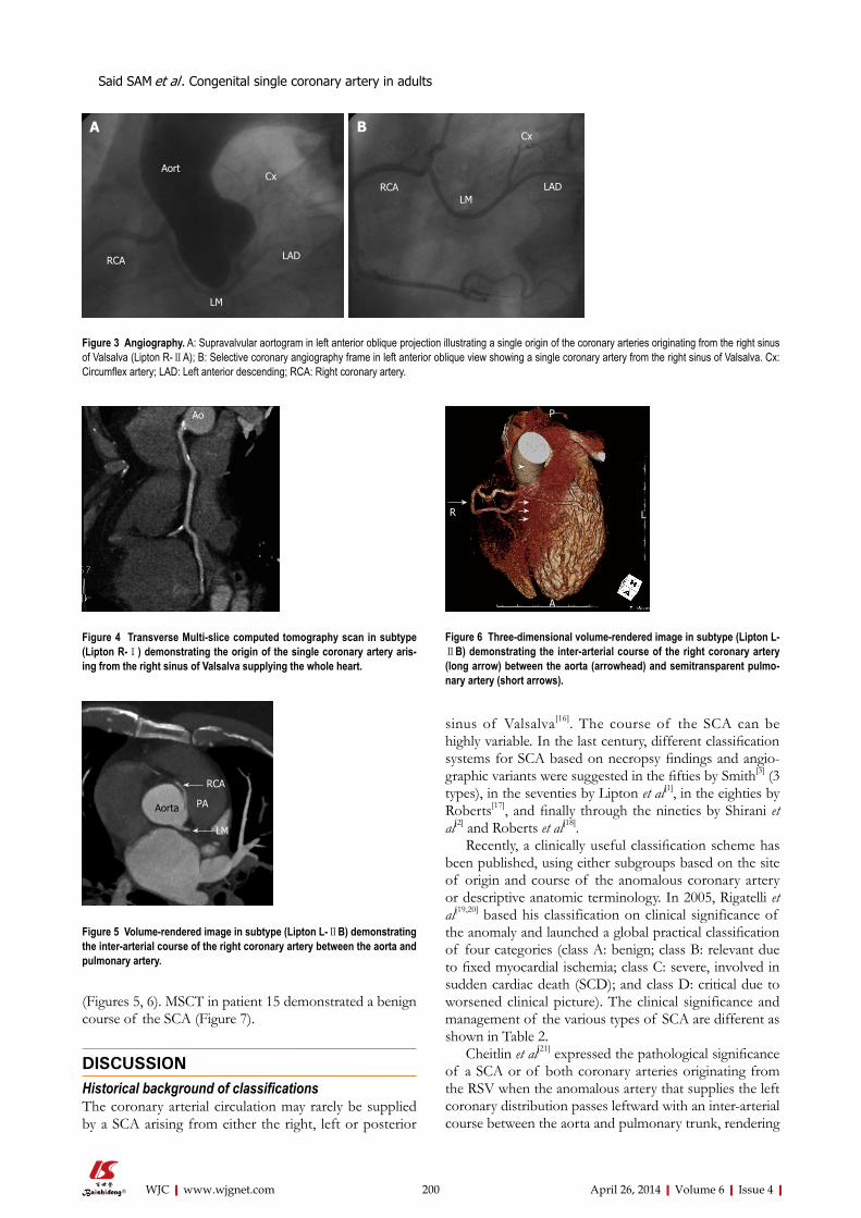
200 April 26, 2014|Volume 6|Issue 4|WJC|www.wjgnet.com
(Figures 5, 6). MSCT in patient 15 demonstrated a benign course of the SCA (Figure 7).
DISCUSSIONHistorical background of classificationsThe coronary arterial circulation may rarely be supplied by a SCA arising from either the right, left or posterior
sinus of Valsalva[16]. The course of the SCA can be highly variable. In the last century, different classification systems for SCA based on necropsy findings and angio-graphic variants were suggested in the fifties by Smith[3] (3 types), in the seventies by Lipton et al[1], in the eighties by Roberts[17], and finally through the nineties by Shirani et al[2] and Roberts et al[18].
Recently, a clinically useful classification scheme has been published, using either subgroups based on the site of origin and course of the anomalous coronary artery or descriptive anatomic terminology. In 2005, Rigatelli et al[19,20] based his classification on clinical significance of the anomaly and launched a global practical classification of four categories (class A: benign; class B: relevant due to fixed myocardial ischemia; class C: severe, involved in sudden cardiac death (SCD); and class D: critical due to worsened clinical picture). The clinical significance and management of the various types of SCA are different as shown in Table 2.
Cheitlin et al[21] expressed the pathological significance of a SCA or of both coronary arteries originating from the RSV when the anomalous artery that supplies the left coronary distribution passes leftward with an inter-arterial course between the aorta and pulmonary trunk, rendering
Aort
RCA
LM
LAD
CxRCA
LMLAD
CxA B
Figure 3 Angiography. A: Supravalvular aortogram in left anterior oblique projection illustrating a single origin of the coronary arteries originating from the right sinus of Valsalva (Lipton R-ⅡA); B: Selective coronary angiography frame in left anterior oblique view showing a single coronary artery from the right sinus of Valsalva. Cx: Circumflex artery; LAD: Left anterior descending; RCA: Right coronary artery.
Figure 4 Transverse Multi-slice computed tomography scan in subtype (Lipton R-Ⅰ) demonstrating the origin of the single coronary artery aris-ing from the right sinus of Valsalva supplying the whole heart.
Ao
Figure 5 Volume-rendered image in subtype (Lipton L-ⅡB) demonstrating the inter-arterial course of the right coronary artery between the aorta and pulmonary artery.
Aorta
RCA
PA
LM
Figure 6 Three-dimensional volume-rendered image in subtype (Lipton L-ⅡB) demonstrating the inter-arterial course of the right coronary artery (long arrow) between the aorta (arrowhead) and semitransparent pulmo-nary artery (short arrows).
P
LR
A
Said SAM et al . Congenital single coronary artery in adults
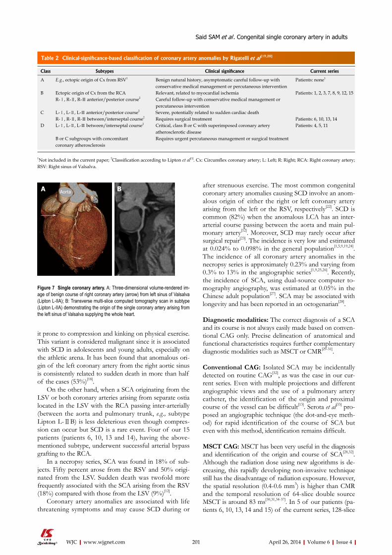
201 April 26, 2014|Volume 6|Issue 4|WJC|www.wjgnet.com
it prone to compression and kinking on physical exercise. This variant is considered malignant since it is associated with SCD in adolescents and young adults, especially on the athletic arena. It has been found that anomalous ori-gin of the left coronary artery from the right aortic sinus is consistently related to sudden death in more than half of the cases (53%)[18].
On the other hand, when a SCA originating from the LSV or both coronary arteries arising from separate ostia located in the LSV with the RCA passing inter-arterially (between the aorta and pulmonary trunk, e.g., subtype Lipton L-ⅡB) is less deleterious even though compres-sion can occur but SCD is a rare event. Four of our 15 patients (patients 6, 10, 13 and 14), having the above-mentioned subtype, underwent successful arterial bypass grafting to the RCA.
In a necropsy series, SCA was found in 18% of sub-jects. Fifty percent arose from the RSV and 50% origi-nated from the LSV. Sudden death was twofold more frequently associated with the SCA arising from the RSV (18%) compared with those from the LSV (9%)[12].
Coronary artery anomalies are associated with life threatening symptoms and may cause SCD during or
after strenuous exercise. The most common congenital coronary artery anomalies causing SCD involve an anom-alous origin of either the right or left coronary artery arising from the left or the RSV, respectively[22]. SCD is common (82%) when the anomalous LCA has an inter-arterial course passing between the aorta and main pul-monary artery[12]. Moreover, SCD may rarely occur after surgical repair[23]. The incidence is very low and estimated at 0.024% to 0.098% in the general population[1,5,9,19,24]. The incidence of all coronary artery anomalies in the necropsy series is approximately 0.23% and varying from 0.3% to 13% in the angiographic series[1,9,25,26]. Recently, the incidence of SCA, using dual-source computer to-mography angiography, was estimated at 0.05% in the Chinese adult population[27]. SCA may be associated with longevity and has been reported in an octogenarian[28].
Diagnostic modalities: The correct diagnosis of a SCA and its course is not always easily made based on conven-tional CAG only. Precise delineation of anatomical and functional characteristics requires further complementary diagnostic modalities such as MSCT or CMR[29-31].
Conventional CAG: Isolated SCA may be incidentally detected on routine CAG[32], as was the case in our cur-rent series. Even with multiple projections and different angiographic views and the use of a pulmonary artery catheter, the identification of the origin and proximal course of the vessel can be difficult[13]. Serota et al[33] pro-posed an angiographic technique (the dot-and-eye meth-od) for rapid identification of the course of SCA but even with this method, identification remains difficult.
MSCT CAG: MSCT has been very useful in the diagnosis and identification of the origin and course of SCA[28,32]. Although the radiation dose using new algorithms is de-creasing, this rapidly developing non-invasive technique still has the disadvantage of radiation exposure. However, the spatial resolution (0.4-0.6 mm3) is higher than CMR and the temporal resolution of 64-slice double source MSCT is around 83 ms[30,31,34-37]. In 5 of our patients (pa-tients 6, 10, 13, 14 and 15) of the current series, 128-slice
Figure 7 Single coronary artery. A: Three-dimensional volume-rendered im-age of benign course of right coronary artery (arrow) from left sinus of Valsalva (Lipton L-IIA); B: Transverse multi-slice computed tomography scan in subtype (Lipton L-IIA) demonstrating the origin of the single coronary artery arising from the left sinus of Valsalva supplying the whole heart.
AortaA B
Table 2 Clinical-significance-based classification of coronary artery anomalies by Rigatelli et al [19,20]
Class Subtypes Clinical significance Current series
A E.g., ectopic origin of Cx from RSV1 Benign natural history, asymptomatic careful follow-up with conservative medical management or percutaneous intervention
Patients: none1
B Ectopic origin of Cx from the RCA Relevant, related to myocardial ischemia Patients: 1, 2, 3, 7, 8, 9, 12, 15R-Ⅰ, R-Ⅱ, R-Ⅲ anterior/posterior course2 Careful follow-up with conservative medical management or
percutaneous interventionC L-Ⅰ, L-Ⅱ, L-Ⅲ anterior/posterior course2 Severe, potentially related to sudden cardiac death
R-Ⅰ, R-Ⅱ, R-Ⅲ between/interseptal course2 Requires surgical treatment Patients: 6, 10, 13, 14D L-Ⅰ, L-Ⅱ, L-Ⅲ between/interseptal course2 Critical, class B or C with superimposed coronary artery
atherosclerotic diseasePatients: 4, 5, 11
B or C subgroups with concomitant coronary atherosclerosis
Requires urgent percutaneous management or surgical treatment
1Not included in the current paper; 2Classification according to Lipton et al[1]. Cx: Circumflex coronary artery; L: Left; R: Right; RCA: Right coronary artery; RSV: Right sinus of Valsalva.
Said SAM et al . Congenital single coronary artery in adults
202 April 26, 2014|Volume 6|Issue 4|WJC|www.wjgnet.com
MSCT confirmed the diagnosis of a SCA with clear demonstration of the inter-arterial course of the RCA originating from the LSV in four (Figure 6) and a benign course of the RCA from LSV in one (Figure 7).
Cardiovascular MR imaging: This technique has the advantage of not using ionizing radiation and has no need for the use of iodinated nephrotoxic ionic or non-ionic contrast agents. Image acquisition occurs with fairly good spatial and temporal resolution, but acquisition and imaging time is long, which makes routine use difficult and time consuming. Cardiovascular magnetic resonance proved to be useful in determining the anatomy and functional significance of SCA[38]. Both the MSCT and the CMR imaging techniques have the additional advan-tage of 3-D reconstruction of the areas of the coronaries relative to the aorta and pulmonary artery. This makes a definitive diagnosis of squeezed aberrant coronary arter-ies between the great vessels feasible[13].
TreatmentThe detection of atherosclerotic coronary artery disease (CAD) in the presence of coronary anomalies is of prac-tical importance, especially when a decision between PCI and CABG has to be made. For diagnostic and therapeu-tic reasons, the knowledge of possible variations of the coronary anatomy, their different origin, and their course is of pivotal importance. Symptomatic patients with as-sociated significant CAD may be treated with routine interventions such as PCI or CABG[6,39]. Angiographic recognition of coronary artery anomalies prior to sur-gery is of great importance. During operation, surgical complications may occur if an unrecognized anomalous vessel is excluded from perfusion during cardiopulmo-nary bypass or if the surgeon inadvertently damages an artery with an anomalous pathway.
Because of the reported high mortality, the occur-rence of “symptomatic or asymptomatic” squeezing of SCA, regardless of the degree of atherosclerosis or site of origin, justifies arterial grafting, as was shown in 4 of our series (patients 6, 10, 13 and 14).
Significant atherosclerotic CAD[37] in association with coronary artery anomalies has been reported in 26%-60% of cases[1,2,40-42]. Rigatelli et al[43] suggested that benign coronary artery anomalies are not associated with or in-volved in the development of premature atherosclerotic CAD. Indeed the high percentage of coronary artery ste-nosis could be biased by the indication to perform CAG as SCA is mainly found during this diagnostic procedure. Only 4 of our 15 (27%) patients (patients 4, 5, 10 and 11) had significant CAD and 3 of them required percu-taneous intervention. When the SCA does not course between the aorta and pulmonary artery, it is not vulner-able to acute angulations or kinking of the coronaries. SCA may be associated with longevity and patients in the 7th and 8th decade of life have been reported[3,12,13,44-47], as was the case in 2 octogenarians from our current series (patients 4 and 11).
Although a SCA is often a benign congenital anomaly,
in which sudden death is a rare complication, different diagnostic modalities should be used to exclude an inter-arterial course between the aorta and pulmonary artery to detect patients at risk for serious complications.
Congenital coronary artery anomalies, detected at necropsy, associated with sudden death and without an-tecedent signs have been recognized in calves[48]. SCA is not limited to the human race, it has also been reported in other mammals such as horses[49], syrian hamsters[50] and minipigs[51].
As was shown in our patient’s population, SCA can be associated with longevity. It has been documented up till the 8th decade of life. In the adult population, SCA-isolated or in association with acquired atherosclerotic changes-may cause severe sequelae. In some cases with-out CAD, the course of the SCA may be malignant.
SCA may be associated with symptomatic transient transmural myocardial ischemia, NSVT, and aborted sud-den death in the absence or presence of coronary athero-sclerosis. The availability of MSCT and CMR facilitates the delineation of the course of the anomalous vessel. The accurate delineation of the course of the anomalous vessel is of great importance even in patients without CAD and in cases of surgical intervention where anatom-ic details of the course of the vessel are of importance.
ACKNOWLEDGMENTSThe authors appreciate the dedicated assistance of the personnel of the catheterization laboratories of Hospi-tal Group Twente, Almelo-Hengelo (Mr. M. Mintjens, Mrs. M. Holleman and Mrs. M. Rigterink-Hofman, Mr. H. Schutte, Mr. R. Heinen, Mr. E. IJspeerd, Mr. B. Ger-ing, Mr. F. van de Bosch, Mr. J. Oolderink and Mr. M. Wildemors) and St. Lucas-Andreas Hospital, Amsterdam; St. Anna Hospital, Geldrop; and Gelre Hospital, Zut-phen, the Netherlands. The assistance of the librarians of the medical library of Hospital Group Twente, Mrs. A. Geerdink and Mr. D. Maas during the preparation of the manuscript is highly appreciated.
COMMENTSBackgroundSingle coronary artery (SCA) is a rare congenital anomaly and occurs as an incidental finding in approximately 0.066% of the coronary angiography (CAG) population. SCA has been reported in association with and without athero-sclerotic changes or in association with coronary artery fistulas, bicuspid aortic valves, and with hypertrophic cardiomyopathy.Research frontiersCAG is the first diagnostic tool in the detection of a SCA. Once abnormal coro-nary arteries are suspected, multi-slice computed tomography (MSCT) and cardiac magnetic resonance (CMR) imaging scans are excellent tools for non-invasive determination of the course of the abnormal coronaries relative to the aorta and pulmonary artery. Determination of the course of incidentally found congenital coronary anomalies during routine CAG without the direct availability of CMR or MSCT scanning is challenging.Innovations and breakthroughsPercutaneous coronary intervention was successfully performed in 3 patients. Eight patients were managed medically. Arterial bypass graft was performed in 4 patients with the squeezed SCA. The literature addressing SCA is reviewed.
COMMENTS
Said SAM et al . Congenital single coronary artery in adults
203 April 26, 2014|Volume 6|Issue 4|WJC|www.wjgnet.com
ApplicationsCongenital coronary artery anomalies, detected at necropsy, associated with sudden death and without antecedent signs have been recognized in calves. SCA is not limited to the human race, it has also been reported in other mam-mals such as horses, syrian hamsters and minipigs.Peer reviewThis paper showed that the availability and sophistications of MSCT facilitated the delineation of the course of a SCA. The authors presented a Dutch case series and review of the literature. This is an interesting report for clinical prac-tice. Overall the report appears to be carefully examined and data adequately discussed.
REFERENCES1 Lipton MJ, Barry WH, Obrez I, Silverman JF, Wexler L. Iso-
lated single coronary artery: diagnosis, angiographic clas-sification, and clinical significance. Radiology 1979; 130: 39-47 [PMID: 758666]
2 Shirani J, Roberts WC. Solitary coronary ostium in the aorta in the absence of other major congenital cardiovascular anomalies. J Am Coll Cardiol 1993; 21: 137-143 [PMID: 8417054 DOI: 10.1016/0735-1097(93)90728-J]
3 Smith JC. Review of single coronary artery with report of 2 cases. Circulation 1950; 1: 1168-1175 [PMID: 15411739 DOI: 10.1161/01.CIR.1.5.1168]
4 Halperin IC, Penny JL, Kennedy RJ. Single coronary artery. Antemortem diagnosis in a patient with congestive heart failure. Am J Cardiol 1967; 19: 424-427 [PMID: 6020312 DOI: 10.1016/0002-9149(67)90456-0]
5 Desmet W, Vanhaecke J, Vrolix M, Van de Werf F, Piessens J, Willems J, de Geest H. Isolated single coronary artery: a review of 50,000 consecutive coronary angiographies. Eur Heart J 1992; 13: 1637-1640 [PMID: 1289093]
6 Türkay C, Gölbasi I, Bayezid O. A single coronary artery from the right sinus of valsalva associated with atheroscle-rosis. Acta Cardiol 2002; 57: 377-379 [PMID: 12405578 DOI: 10.2143/AC.57.5.2005457]
7 Hara H, Ishii K, Nakamura M. A case of hypertrophic ob-structive cardiomyopathy complicated by a single coronary artery treated by transcoronary septal ablation. J Invasive Cardiol 2006; 18: 234-238 [PMID: 16670451]
8 El-Menyar AA, Das KM, Al-Suwaidi J. Anomalous origin of the three coronary arteries from the right aortic sinus Val-salva: role of MDCT coronary angiography. Int J Cardiovasc Imaging 2006; 22: 723-729 [PMID: 16642404 DOI: 10.1007/s10554-005-9062-7]
9 Bolognesi R, Tsialtas D, Barbaresi F, Manca C. Single coro-nary artery-right ventricular fistula with a partially throm-bosed large aneurysm of its proximal tract in a 66-year-old man. Eur Heart J 1994; 15: 1720-1724 [PMID: 7698144]
10 Hillestad L, Eie H. Single coronary artery. A report of three cases. Acta Med Scand 1971; 189: 409-413 [PMID: 5578491]
11 Larsen AI, Ørn S, Barvik S, Nilsen DW. Anomalies of the coronary arteries originating from the right sinus of Val-salva. (1) Single coronary artery originating from the right sinus associated with fusion of the left and the non coronary cusp and atrophy of the left coronary ostium (2)Three sepa-rate coronary arteries originating from the right sinus of Val-salva. Int J Cardiol 2007; 115: e86-e89 [PMID: 17107719 DOI: 10.1016/j.ijcard.2006.08.042]
12 Taylor AJ, Rogan KM, Virmani R. Sudden cardiac death as-sociated with isolated congenital coronary artery anomalies. J Am Coll Cardiol 1992; 20: 640-647 [PMID: 1512344 DOI: 10.1016/0735-1097(92)90019-J]
13 Bunce NH, Lorenz CH, Keegan J, Lesser J, Reyes EM, Firmin DN, Pennell DJ. Coronary artery anomalies: assessment with free-breathing three-dimensional coronary MR angiography. Radiology 2003; 227: 201-208 [PMID: 12601193 DOI: 10.1148/radiol.2271020316]
14 Yucel EK, Anderson CM, Edelman RR, Grist TM, Baum RA, Manning WJ, Culebras A, Pearce W. AHA scientific state-ment. Magnetic resonance angiography : update on applica-tions for extracranial arteries. Circulation 1999; 100: 2284-2301 [PMID: 10578005 DOI: 10.1161/01.CIR.100.22.2284]
15 Angelini P, Velasco JA, Flamm S. Coronary anomalies: inci-dence, pathophysiology, and clinical relevance. Circulation 2002; 105: 2449-2454 [PMID: 12021235 DOI: 10.1161/01.CIR.0000016175.49835.57]
16 Virmani R, Burke AP, Farb A. Sudden cardiac death. Cardio-vasc Pathol 2001; 10: 211-218 [DOI: 10.1016/S1054-8807(01)00091-6]
17 Roberts WC. Major anomalies of coronary arterial origin seen in adulthood. Am Heart J 1986; 111: 941-963 [PMID: 3518378 DOI: 10.1016/0002-8703(86)90646-0]
18 Roberts WC, Shirani J. The four subtypes of anomalous origin of the left main coronary artery from the right aortic sinus (or from the right coronary artery). Am J Cardiol 1992; 70: 119-121 [DOI: 10.1016/0002-9149(92)91406-T]
19 Rigatelli G, Docali G, Rossi P, Bandello A, Rigatelli G. Validation of a clinical-significance-based classification of coronary artery anomalies. Angiology 2005; 56: 25-34 [PMID: 15678253 DOI: 10.1177/000331970505600104]
20 Rigatelli G, Rigatelli G. Congenital coronary artery anoma-lies in the adult: a new practical viewpoint. Clin Cardiol 2005; 28: 61-65 [PMID: 15757074 DOI: 10.1002/clc.4960280203]
21 Cheitlin MD, MacGregor J. Congenital anomalies of coro-nary arteries: role in the pathogenesis of sudden cardiac death. Herz 2009; 34: 268-279 [PMID: 19575157 DOI: 10.1007/s00059-009-3239-0]
22 Liberthson RR. Sudden death from cardiac causes in chil-dren and young adults. N Engl J Med 1996; 334: 1039-1044 [PMID: 8598843 DOI: 10.1056/NEJM199604183341607]
23 Nguyen AL, Haas F, Evens J, Breur JM. Sudden cardiac death after repair of anomalous origin of left coronary artery from right sinus of Valsalva with an interarterial course: Case report and review of the literature. Neth Heart J 2012; 20: 463-471 [PMID: 23055055 DOI: 10.1007/s12471-012-0324-4]
24 Yamanaka O, Hobbs RE. Coronary artery anomalies in 126,595 patients undergoing coronary arteriography. Cathet Cardiovasc Diagn 1990; 21: 28-40 [PMID: 2208265 DOI: 10.1002/ccd.1810210110]
25 Cieslinski G, Rapprich B, Kober G. Coronary anomalies: in-cidence and importance. Clin Cardiol 1993; 16: 711-715 [PMID: 8222383 DOI: 10.1002/clc.4960161005]
26 Kardos A, Babai L, Rudas L, Gaal T, Horvath T, Talosi L, Toth K, Sarvary L, Szasz K. Epidemiology of congenital cor-onary artery anomalies: a coronary arteriography study on a central European population. Cathet Cardiovasc Diagn 1997; 42: 270-275 [PMID: 9367100 DOI: 10.1002/(SICI)1097-0304(199711)42:3<270::AID-CCD8>3.0.CO;2-9]
27 Zhang LJ, Yang GF, Huang W, Zhou CS, Chen P, Lu GM. Incidence of anomalous origin of coronary artery in 1879 Chinese adults on dual-source CT angiography. Neth Heart J 2010; 18: 466-470 [PMID: 20978590 DOI: 10.1007/BF03091817]
28 Morimoto H, Mukai S, Obata S, Hiraoka T. Incidental single coronary artery in an octogenarian with acute type A aortic dissection. Interact Cardiovasc Thorac Surg 2012; 15: 307-308 [PMID: 22561294 DOI: 10.1093/icvts/ivs016]
29 Fernandes F, Alam M, Smith S, Khaja F. The role of trans-esophageal echocardiography in identifying anomalous coronary arteries. Circulation 1993; 88: 2532-2540 [PMID: 8252664 DOI: 10.1161/01.CIR.88.6.2532]
30 Soon KH, Selvanayagam J, Bell KW, Tang SH, Pereira J, Chan W, Lim YL. Giant single coronary system with coronary cameral fistula diagnosed on MSCT. Int J Cardiol 2006; 106: 276-278 [PMID: 16321707 DOI: 10.1016/j.ijcard.2004.12.093]
31 Datta J, White CS, Gilkeson RC, Meyer CA, Kansal S, Jani ML, Arildsen RC, Read K. Anomalous coronary arteries in adults: depiction at multi-detector row CT angiography. Radiology 2005; 235: 812-818 [PMID: 15833984 DOI: 10.1148/
Said SAM et al . Congenital single coronary artery in adults
204 April 26, 2014|Volume 6|Issue 4|WJC|www.wjgnet.com
radiol.2353040314]32 Liesting C, Brugts JJ, Kofflard MJ, Dirkali A. Acute coronary
syndrome in a patient with a single coronary artery aris-ing from the right sinus of Valsalva. World J Cardiol 2012; 4: 264-266 [PMID: 22953025 DOI: 10.4330/wjc.v4.i8.264]
33 Serota H, Barth CW, III, Seuc CA, Vandormael M, Aguirre F, Kern MJ. Rapid identification of the course of anomalous coronary arteries in adults: the “dot and eye” method. Am J Cardiol 1990; 65: 891-898 [PMID: 2321539 DOI: 10.1016/0002-9149(90)91432-6]
34 Kunimasa T, Sato Y, Ito S, Takagi T, Lee T, Saeki F, Moroi M. Absence of the right coronary artery detected by 64-detector-row multislice computed tomography. Int J Cardiol 2007; 115: 249-250 [PMID: 16757047 DOI: 10.1016/j.ijcard.2006.01.039]
35 Jessurun GA, Willemsen MH, Vercoutere RA, Boonstra PW, Tio RA. Single coronary artery: a reappraisal. J Invasive Car-diol 2004; 16: 40-41 [PMID: 14699223]
36 Hegde AN, Desai SB. Two cases of anomalous origins of left coronary artery with a course between the aortic root and the free standing subpulmonary infundibulum on CT coronary angiography. Interact Cardiovasc Thorac Surg 2005; 4: 297-298 [PMID: 17670416 DOI: 10.1510/icvts.2005.105965]
37 Schroeder S, Achenbach S, Bengel F, Burgstahler C, Ca-demartiri F, de Feyter P, George R, Kaufmann P, Kopp AF, Knuuti J, Ropers D, Schuijf J, Tops LF, Bax JJ. Cardiac com-puted tomography: indications, applications, limitations, and training requirements: report of a Writing Group deployed by the Working Group Nuclear Cardiology and Cardiac CT of the European Society of Cardiology and the European Council of Nuclear Cardiology. Eur Heart J 2008; 29: 531-556 [PMID: 18084017 DOI: 10.1093/eurheartj/ehm544]
38 Jahnke C, Nagel E, Ostendorf PC, Tangcharoen T, Fleck E, Paetsch I. Images in cardiovascular medicine. Diagnosis of a “single” coronary artery and determination of functional sig-nificance of concomitant coronary artery disease. Circulation 2006; 113: e386-e387 [PMID: 16520417 DOI: 10.1161/CIRCU-LATIONAHA.105.564260]
39 Zweiker R, Luha O, Klein WW. Rescue percutaneous trans-luminal coronary angioplasty in a patient with a single coronary artery arising from the right Sinus Valsalvae: previ-ously unreported scenario and review of literature. J Intern Med 2002; 252: 84-87 [PMID: 12074743 DOI: 10.1046/j.1365-2796.2002.01003.x]
40 Topaz O, DeMarchena EJ, Perin E, Sommer LS, Mallon SM, Chahine RA. Anomalous coronary arteries: angiographic findings in 80 patients. Int J Cardiol 1992; 34: 129-138 [DOI:
10.1016/0167-5273(92)90148-V]41 Garg N, Tewari S, Kapoor A, Gupta DK, Sinha N. Primary
congenital anomalies of the coronary arteries: a coronary: arteriographic study. Int J Cardiol 2000; 74: 39-46 [PMID: 10854679 DOI: 10.1016/S0167-5273(00)00243-6]
42 Tejada JG, Hernandez F, Sanchez I, Martin-Asenjo R. Stent-ing of anomalous left main coronary artery arising from the right sinus of Valsalva: a case report. Int J Cardiol 2007; 119: 266-267 [PMID: 17056136 DOI: 10.1016/j.ijcard.2006.07.149]
43 Rigatelli G, Gemelli M, Zamboni A, Docali G, Rossi P, Rossi D, Grazio M, Franco G, Rigatelli G. Are coronary artery anomalies an accelerating factor for coronary atherosclerosis development? Angiology 2004; 55: 29-35 [PMID: 14759087 DOI: 10.1177/000331970405500105]
44 Kuon E, Ropers D. Single coronary artery--a rarity in the catheterization laboratory: case report and current review. Can J Cardiol 2004; 20: 647-651 [PMID: 15152298]
45 Tejada JG, Albarran A, Velazquez MT, Sanz J, Pindado C, Tascon JC. Direct stenting in single coronary artery arising from the left sinus of Valsalva-a case report. Angiology 2002; 53: 733-736 [PMID: 12463629 DOI: 10.1177/000331970205300616]
46 Basalus M, Louwerenburg JW, van Houwelingen KG, Stoel MG, von Birgelen C. Primary percutaneous coronary inter-vention in the left main stem of a monocoronary artery. Neth Heart J 2009; 17: 274-276 [PMID: 19789693 DOI: 10.1007/BF03086264]
47 Cheitlin MD, De Castro CM, McAllister HA. Sudden death as a complication of anomalous left coronary origin from the anterior sinus of Valsalva, A not-so-minor congenital anomaly. Circulation 1974; 50: 780-787 [PMID: 4419670 DOI: 10.1161/01.CIR.50.4.780]
48 Shank AM, Bryant UK, Jackson CB, Williams NM, Janes JG. Anomalous origin of the left coronary artery from the pul-monary artery (ALCAPA) in four calves. Vet Pathol 2008; 45: 634-639 [PMID: 18725466 DOI: 10.1354/vp.45-5-634]
49 Rooney JR, Franks WC. Congenital cardiac anomalies in horses. Path Vet 1964; 1: 454-464
50 Sans-Coma V, Arque JM, Duran AC, Cardo M. Origin of the left main coronary artery from the pulmonary trunk in the Syrian hamster. Am J Cardiol 1988; 62: 159-161 [DOI: 10.1016/0002-9149(88)91388-4]
51 Matthews KA, Gogas BD, Sumida A, Nagai H, King Iii SB, Chronos N, Hou D. Anomalous right coronary artery origi-nating from the left sinus of Valsalva in a Yucatan minipig. Comp Med 2012; 62: 127-130 [PMID: 22546919]
P- Reviewers: Letsas K, Sakabe K S- Editor: Ma YJ L- Editor: O’Neill M E- Editor: Liu SQ
Said SAM et al . Congenital single coronary artery in adults
Prognostic value of increased carbohydrate antigen in patients with heart failure
Ana B Méndez, Jordi Ordoñez-Llanos, Andreu Ferrero, Mariana Noguero, Teresa Mir, Josefina Mora, Antoni Bayes-Genis, Sònia Mirabet, Juan Cinca, Eulàlia Roig
Ana B Méndez, Andreu Ferrero, Mariana Noguero, Sò-nia Mirabet, Juan Cinca, Eulàlia Roig, Cardiology Depart-ment, Hospital de la Santa Creu i Sant Pau, Institut de Recerca Biomèdica Sant Pau and Universitat Autònoma, 08041 Barce-lona, SpainJordi Ordoñez-Llanos, Teresa Mir, Josefina Mora, Biochem-istry Department, Hospital de la Santa Creu i Sant Pau, Institut de Recerca Biomèdica Sant Pau and Universitat Autònoma, 08041 Barcelona, SpainAntoni Bayes-Genis, Cardiology Department, Hospital Germans Trias I Pujol, 08916 Badalona, SpainAuthor contributions: Mendez AB contributed to design of the study, analysis and interpretation of data, manuscript writing and final approval of manuscript; Ordoñez-Llanos J contributed to analysis of data, review of CA125 and NT-proBNP determina-tions and substantial contribution to manuscript review; Ferrero A contributed to statistical assessment and substantial contribution to manuscript review; Noguero M contributed to patient selection and follow-up, Serotec and analysis of data; Mir T and Mora J contributed to CA125 and NT-proBNP determinations, analysis of data, substantial contribution to manuscript; Bayes-Genis A and Mirabet S contributed to patient selection and follow-up, analysis of data, substantial contribution to manuscript; Cinca J contributed to design of the study and substantial contribution to manuscript review; Roig E contributed to design of the study, selection of patients, analysis of data, and substantial contribution to manuscript review.Supported by Ministerio Español de Salud, Redes de Investig-ación del Instituto de Salud Carlos Ⅲ (RIC, RD12/0042) y Fondo Europeo de Desarrollo Regional (FEDER)Correspondence to: Eulàlia Roig, MD, Cardiology Depart-ment, Hospital de la Santa Creu i Sant Pau, Institut de Recerca Biomèdica Sant Pau and Universitat Autònoma, C/ Mas Casano-vas 90, 08041 Barcelona, Spain. [email protected]: +34-93-5565958 Fax: +34-93-5565604 Received: October 30, 2013 Revised: March 7, 2014Accepted: March 13, 2014Published online: April 26, 2014
AbstractAIM: To study the prognostic value of carbohydrate
antigen 125 (CA125) and whether it adds prognostic information to N-terminal pro-brain natriuretic peptide (NT-proBNP) in stable heart failure (HF) patients.
METHODS: The predictive value of CA125 was ret-rospectively assessed in 156 patients with stable HF remitted to the outpatient HF unit for monitoring from 2009 to 2011. Patients were included in the study if they had a previous documented episode of HF and received HF treatment. CA125 and NT-proBNP concen-trations were measured. The independent association between NT-proBNP or CA125 and mortality was as-sessed with Cox regression analysis, and their com-bined predictive ability was tested by the integrated discrimination improvement (IDI) index.
RESULTS: The mean age of the 156 patients was 72 ± 12 years. During follow-up (17 ± 8 mo), 27 patients died, 1 received an urgent heart transplantation and 106 required hospitalization for HF. Higher CA125 val-ues were correlated with outcomes: 58 ± 85 KU/L if hospitalized vs 34 ± 61 KU/L if not (P < 0.05), and 94 ± 121 KU/L in those who died or needed urgent heart transplantation vs 45 ± 78 KU/L in survivors (P < 0.01). After adjusting for propensity scores, the highest risk was observed when both biomarkers were elevated vs not elevated (HR = 8.95, 95%CI: 3.11-25.73; P < 0.001) and intermediate when only NT-proBNP was el-evated vs not elevated (HR = 4.15, 95%CI: 1.41-12.24; P < 0.01). Moreover, when CA125 was added to the clinical model with NT-proBNP, a 4% (P < 0.05) im-provement in the IDI was found.
CONCLUSION: CA125 > 60 KU/L identified patients in stable HF with poor survival. Circulating CA125 level adds prognostic value to NT-proBNP level in predicting HF outcomes.
© 2014 Baishideng Publishing Group Co., Limited. All rights reserved.
RETROSPECTIVE STUDY
Online Submissions: http://www.wjgnet.com/esps/[email protected]:10.4330/wjc.v6.i4.205
205 April 26, 2014|Volume 6|Issue 4|WJC|www.wjgnet.com
World Journal of CardiologyW J C
World J Cardiol 2014 April 26; 6(4): 205-212ISSN 1949-8462 (online)
© 2014 Baishideng Publishing Group Co., Limited. All rights reserved.
Key words: Heart failure; Prognosis; Carbohydrate anti-gen 125; Brain natriuretic peptides; Survival
Core tip: Increased carbohydrate antigen 125 (CA125) has prognostic implications in acute heart failure (HF). The aim of this study was to assess the prognostic value of increased CA125 and whether it adds prognos-tic information to N-terminal pro-brain natriuretic pep-tide (NT-proBNP) in stable HF patients. Higher CA125 values correlated with outcomes. The highest risk was observed when both biomarkers CA125 and NT-proBNP were elevated vs not elevated (HR = 8.95, 95%CI: 3.11-25.73; P < 0.001). CA125 > 60 KU/L identified patients in stable HF with very poor survival. Circulating CA125 level adds prognostic value to NT-proBNP level in predicting HF outcomes.
Méndez AB, Ordoñez-Llanos J, Ferrero A, Noguero M, Mir T, Mora J, Bayes-Genis A, Mirabet S, Cinca J, Roig E. Prognostic value of increased carbohydrate antigen in patients with heart failure. World J Cardiol 2014; 6(4): 205-212 Available from: URL: http://www.wjgnet.com/1949-8462/full/v6/i4/205.htm DOI: http://dx.doi.org/10.4330/wjc.v6.i4.205
INTRODUCTIONHeart failure (HF) is a disease with high mortality[1] and an estimated prevalence up to 3% in the European popu-lation[2]. This prevalence increases exponentially with age, especially in patients older than 75 years. As the main cause of hospitalization, in patients over 65, HF is also associated with high costs. Despite recent advances in treatment, mortality is still high (12% per year) in stable patients, and there is a high rate of hospital readmissions due to worsening HF[3,4]. Even though a high number of clinical parameters have been associated with poor out-come in patients with stable HF, the assessment of prog-nosis is still a challenge[5,6]. Multiple biomarkers have been suggested to identify patients with worse prognosis, such as some interleukins, natriuretic peptides, endothelin, ST2, and several fibrosis markers[7-11]. Although most of these effectively select patients with high risk of death, only circulating levels of natriuretic peptides have proven useful in clinical practice. Natriuretic peptides are released when ventricular wall stress and ventricular end-diastolic pressure increase. Thus, an elevated level of either brain natriuretic peptide (BNP) or the amino terminal portion of N-terminal pro-BNP (NT-proBNP) in peripheral blood is associated with decompensated HF. The nega-tive and positive predictive values of these peptides to diagnose HF have been widely studied[12-14]. Moreover, they are useful in the assessment of prognosis after a HF admission[8]. However, certain limitations affect the more extensive use of natriuretic peptides[15].
Carbohydrate antigen 125 (CA125) is a biomarker previously used in the detection and monitoring of some cancers, especially ovarian cancer[16]. It is a high-
molecular-weight glycoprotein synthesized by epithelial cells of the serosa when there is inflammation and in-creased interstitial fluid; therefore, its level is elevated in the presence of pleural or pericardial effusion and ascites. Elevated CA125 has also been found in patients with HF, with or without fluid retention[16-19]. Although its release mechanisms are not yet well understood, they correlate with increased left ventricular end-diastolic pressure, higher BNP level, and worse New York Heart Association (NYHA) functional class[20,21]. Moreover, previous studies of BNP and CA125 in acute HF have demonstrated an additive prognostic value of CA125; therefore, the determination of both biomarkers would improve risk stratification[22]. The advantages of CA125 determination with respect to natriuretic peptides are its higher stability in the circulation and lower cost[23]. The usefulness of CA125 measurement to assess prognosis in patients admitted with acute decompensated HF has been previously demonstrated[19-22]. However, informa-tion is lacking on its value in assessing the prognosis of patients with stable chronic HF. The aim of this study was to analyze the prognostic implications of increased CA125 concentration in peripheral blood and whether it adds prognostic information to NT-proBNP in stable HF patients.
MATERIALS AND METHODSStudy populationThe population was a prospective cohort of 156 patients diagnosed with HF and remitted to the outpatient HF unit for monitoring from 2009 to 2011. Patients were included in the study if they had a previous documented episode of HF and received HF treatment. The diagnosis of HF was made following the Clinical Practice Guide-lines of the European Society of Cardiology[24]. HF treat-ment included angiotensin-converting enzyme inhibitors (ACEIs) or angiotensin Ⅱ receptor blockers (ARBs) if ACEIs were contraindicated, beta blockers, diuretics, and aldosterone antagonists, with individualized assessment of treatment indication and optimized doses per recom-mendations of the Clinical Practice Guidelines of the European Society of Cardiology[24].
We excluded patients in unstable HF (NYHA func-tional class Ⅳ), patients with hemodynamic instability, or those diagnosed with cancer or systemic diseases that could shorten life expectancy. Patients with valve heart disease waiting for surgery repair were also excluded from the study. A 12-lead electrocardiogram (ECG) chest radi-ography and echocardiogram were performed in all pa-tients to establish the HF etiology. Left ventricular systolic dysfunction was considered when ejection fraction was < 50%[25] and ventricular hypertrophy when ventricular sep-tum or posterior left ventricular wall thickness was > 11 mm. Ischemic heart disease was diagnosed if pathologic Q waves were observed on the ECG or significant coro-nary lesions on the coronary angiography. Valve heart dis-ease was diagnosed when a biological or mechanic valve prosthesis had been implanted or if the valve dysfunction
206 April 26, 2014|Volume 6|Issue 4|WJC|www.wjgnet.com
Méndez AB et al . CA125 concentration in HF prognosis
was considered significant by echocardiography.The hospital ethics committee approved the study,
patients gave informed consent to participate, and the protocol conformed to the principles outlined in the Declaration of Helsinki.
Laboratory measurementsTo obtain routine laboratory determinations and NT-proBNP concentrations, fasting serum blood samples were obtained during the first visit to the HF unit. CA125 and NT-proBNP concentrations were measured with commercial electrochemiluminescence assays in a Modu-lar E170 analyzer (both from Roche Diagnostics, Basel, Switzerland). The CA125 assay used a pair of monoclo-nal anti-CA125 antibodies, one labeled with biotin, the other linked to ruthenium chelates. The analyzer had a sensitivity of 0.60 KU/L, and the total imprecision at dif-ferent concentrations was ≤ 2.5%, expressed as a coef-ficient of variation.
Statistical analysisDuring follow-up, mortality or need for urgent heart transplantation and new hospital admissions due to wors-ening HF were assessed. The primary end-point was all-cause mortality, which included need for urgent heart transplantation. The secondary end-point was worsening HF requiring hospital admission.
Continuous variables are expressed as mean ± SD and categorical variables as percentages. Continuous variables were compared using Student’s t test. The χ 2 or Fisher exact test was used for categorical variables. The Kruskal-Wallis test was used when necessary. Variables without homogeneous distribution were analyzed with the non-parametric Mann-Whitney test. To normalize CA125 and NT-proBNP measurements, the neperian logarithm of CA125 and NT-proBNP values was used, creating two new variables: ln-CA125 and ln-NT-proBNP. Without a clear cut-off for NT-proBNP in the literature, 3100 ng/L was selected because it was close to the mean NT-proBNP value of our population and it had the best cut-off according to area under the curve (AUC) calculation, with a sensitivity of 62% and a specificity of 82% to pre-dict mortality. A CA125 cut-off of 60 KU/L was used, in keeping with the best predictor of mortality identified by a previous study in patients with acute HF[22]. Based on these data, a new dichotomous variable was created for a CA125 value of 60 KU/L. Also, both variables were cat-egorized for clinical practice and better interpretation.
To avoid overfitting multivariate models, two propen-sity score models[26,27] were used to adjust for confound-ing factors. Propensity score analysis was performed us-ing logistic regression for computing probability to fall in NT-proBNP 3100 ng/L or CA125 60 KU/L categories. This analysis was based on seven variables associated to end-points with the objective of eliminating differences in baseline patient characteristics that might affect event comparison. The AUC was 0.75 (0.66-0.84); P < 0.001 for NT-proBNP categories and 0.70 (0.61-0.80); P < 0.001 for CA125. Variables included in the propensity
score were left atrial diameter, age, atrial fibrillation, left ventricular ejection fraction, estimated glomerular filtrate rate (eGFR), hemoglobin and interventricular septum thickness.
The independent associations between CA125 and NT-proBNP and survival were assessed with the Cox regression analysis. The models were adjusted for the two propensity scores performed previously.
Two Cox models were compared to test improvement in risk stratification: Cox-model 1 (adjusted model with NT-proBNP 3100 ng/L) vs Cox-model 3 (adjusted model with 4 groups according to the cut-off concentrations of CA125 and NT-proBNP) (Table 1): The four groups in Cox model 3 were patients with both markers below the cut-off values, with only one elevated level (assessed for each biomarker), and with both markers above the cut-offs. The increase in the prognostic utility of NT-proBNP and NT-proBNP combined with CA125 when adding sequentially to the model was evaluated by the in-tegrated discrimination improvement (IDI) index. When two nested models are compared, IDI quantifies the increment in the predicted probabilities for the subset of patients experiencing the event and the decrease for those not experiencing the event. In simpler terms, it reflects an improvement in the average of the true positive rate without sacrificing its average true negative rate[28]. The proportionality assumption for the hazard function over time was tested by means of the Schoenfeld residuals. The discriminative ability of the final model was assessed by Harrell’s C-statistic, and the calibration ability was as-sessed by the Gronnesby and Borgan test[29]. A two-sided P < 0.05 was considered statistically significant for all analyses. All analyses were performed using SPSS version 19.0 (Statistical Package for the Social Sciences, Chicago, Illinois) and R (R: A Language and Environment for Sta-tistical Computing at http://www.R-project.org).
RESULTSBaseline clinical characteristicsThe mean age of the studied population was 72 ± 12 years; 63% were male. Clinical characteristics of the studied population and HF treatments according to the specified 4 groups are shown in Table 1. Mean ejection fraction was 48% ± 17%, and half (77) of the patients had preserved ejection fraction. Ischemic heart disease was the most frequent etiology of HF, followed by hy-pertension and valve disease. The percentage of patients with HFrEF treated with ACEI/ARB was 85%, with beta-blockers 76% and with mineral corticoid receptor antagonists 56%, while in the HFpEF group the percent-age of patients treated with ACEI/ARB was 69%, with beta-blockers 42% and with mineral corticoid receptor antagonists 39%. The percentage of patients treated with furosemide was similar in both groups, 84%.
OutcomesNo patient was lost during the mean follow-up of 17 ± 8 mo (range 2 to 32 mo). Monitoring of patients was blind-
207 April 26, 2014|Volume 6|Issue 4|WJC|www.wjgnet.com
Méndez AB et al . CA125 concentration in HF prognosis
208 April 26, 2014|Volume 6|Issue 4|WJC|www.wjgnet.com
for decompensated HF) and 3 to cerebrovascular acci-dent. The remaining patient had a diagnosis of ischemic heart disease and was lost to sudden death while being treated with chemotherapy for rectal cancer that appeared during follow-up. Mortality was similar for women (19%) and men (18%). There was a high incidence of worsen-ing, new HF episodes: 106 patients required an admis-sion for decompensated HF (68%), and nearly half (50 patients) were admitted more than once for HF during follow-up.
Prognostic implications of CA125Older age, increased ventricular septum thickness and left atrial diameter, low eGFR and hemoglobin concentra-tion, higher CA125 and NT-proBNP levels, the presence of atrial fibrillation, and worse NYHA functional class were all associated with mortality or need for urgent heart transplantation by univariate analyses (Table 2). Hospital-ization for worsening HF was associated with low eGFR, higher CA125 and NT-proBNP values, presence of atrial fibrillation, and worse NYHA functional class by univari-ate analyses (Table 3). Patients who died and those who required admission for decompensated HF had signifi-cantly lower eGFR, had higher CA125 and NT-proBNP, and more frequently had atrial fibrillation and NYHA functional class Ⅲ. In univariate analyses, ln-CA125 was positively associated with ln-NT-proBNP (β = 0.39, P =
ed to CA125 values. During follow-up, 27 patients died (17%), and 1 had progressive HF deterioration requiring urgent heart transplantation at one year. All 27 deaths were due to cardiovascular causes, 23 (85%) of them to progressive HF (3 of which required hospital admission
Table 1 Baseline characteristics by carbohydrate antigen 125 and N-terminal pro-brain natriuretic peptide class
Variable CA125 < 60 CA125 > 60 CA125 < 60 CA125 > 60 PNT-proBNP < 3100 NT-proBNP < 3100 NT-proBNP > 3100 NT-proBNP > 3100
(n = 95, 61%) (n = 17, 11%) (n = 24, 15%) (n = 20, 13%)Male 55 (58%) 11 (65%) 19 (79%) 14 (70%) 0.240Age 71 ± 12 69 ± 10 76 ± 12 73 ± 15 0.309Hypertension 73 (77%) 10 (59%) 18 (75%) 14 (70%) 0.471Diabetes 38 (40%) 7 (41%) 11 (46%) 8 (40%) 0.964Dyslipidemia 46 (48%) 5 (29%) 11 (46%) 7 (35%) 0.406Atrial fibrillation 47 (49%) 9 (53%) 17 (71%) 11 (55%) 0.316NYHA class Ⅱ 85 (89%) 7 (41%) 13 (54%) 11 (55%) < 0.001NYHA class Ⅲ 10 (11%) 10 (59%) 11 (46%) 9 (45%) < 0.001HF Etiology Hypertension 26 (27%) 1 (6%) 5 (21%) 3 (15%) 0.212 Ischemic heart disease 30 (32%) 7 (41%) 8 (33%) 9 (45%) 0.641 Dilated cardiomyopathy 8 (8%) 2 (12%) 1 (4%) 2 (10%) 0.768 Valve heart disease 15 (16%) 5 (29%) 4 (17%) 5 (25%) 0.439 Congenital 0 (0%) 1 (6%) 1 (4%) 1 (5%) 0.058 Others 16 (17%) 1 (6%) 5 (21%) 0 (0%) 0.110Systolic BP (mmHg) 129 ± 18 118 ± 20 119 ± 21 114 ± 12 0.001Diastolic BP (mmHg) 74 ± 12 67 ± 8 73 ± 14 71 ± 9 0.156LVEF (%) 51 ± 16 46 ± 16 46 ± 22 38 ± 18 0.015LVDD (mm) 54 ± 8 52 ± 9 56 ± 13 57 ± 11 0.290LAD (mm) 49 ± 9 49 ± 9 51 ± 9 54 ± 12 0.184IVS (mm) 12 ± 3 12 ± 2 13 ± 3 11 ± 3 0.158LVPW (mm) 11 ± 2 10 ± 2 11 ± 2 10 ± 2 0.461Na+ (mEq/dL) 140 ± 3 138 ± 4 140 ± 4 139 ± 5 0.227K+ (mEq/dL) 4.2 ± 0.5 4.3 ± 0.6 4.4 ± 0.7 4.2 ± 0.5 0.453GF (mL/min per 1.73 m2) 61 ± 20 62 ± 16 49 ± 22 53 ± 20 0.029Hemoglobin (g/dL) 131 ± 20 118 ± 25 126 ± 21 129 ± 24 0.111
LVEF: Left ventricular ejection fraction; NYHA: New York Heart Association; HF: Heart failure; LVDD: Left ventricular diastolic diameter; LAD: Left au-ricular diameter; IVS: Interventricular septum; LVPW: Left ventricular posterior wall; BP: Blood pressure; Na: Sodium; K: Potassium; GF: Glomerular filtra-tion; CA125: Carbohydrate antigen 125; NT-proBNP: N-terminal pro-B-type natriuretic peptide.
Table 2 Univariate analysis, variables significantly associated with mortality or need for urgent cardiac transplantation
Variable Deaths Alive P
Age 78 ± 12 70 ± 12 0.004LVEF (%) 53 ± 16 47 ± 17 0.08IVS (mm) 13.7 ± 3.4 11.9 ± 2.5 0.005LA (mm) 54 ± 12 49 ± 9 0.01GF (MDRD) 46 ± 18 61 ± 19 0.0001Hemoglobin (g/L) 121 ± 21 130 ± 21 0.02Na+ (mEq/mL) 140 ± 4 139 ± 3 0.54K+ (mEq/mL) 4.3 ± 0.6 4.2 ± 0.5 0.54Systolic BP (mmHg) 120 ± 19 125 ± 19 0.24Diastolic BP (mmHg) 70 ± 12 73 ± 12 0.25CA125 (KU/L) 94 ± 121 45 ± 78 0.01NT-proBNP (pg/dL) 6613 ± 8437 2326 ± 2823 0.02AF (%) 71 50 0.05NYHA class Ⅲ (%) 70 30 0.03
LVEF: Left ventricular ejection fraction; IVS: Interventricular septum; LA: Left atrial; GF: Glomerular filtration; Na: Sodium; K: Potassium; BP: Blood pressure; CA125: Carbohydrate antigen 125; NT-proBNP: N-terminal pro-B-type natriuretic peptide; AF: Atrial fibrillation; NYHA: New York Heart Association; MDRD: Modification of diet in renal disease.
Méndez AB et al . CA125 concentration in HF prognosis
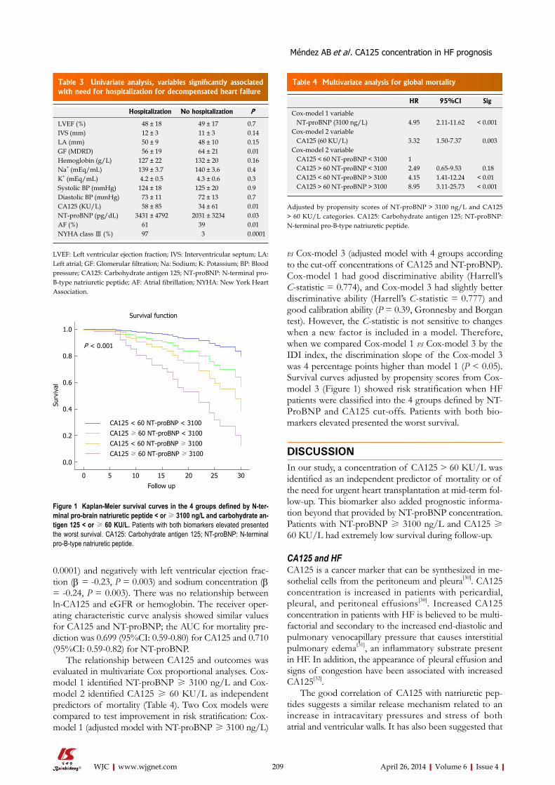
209 April 26, 2014|Volume 6|Issue 4|WJC|www.wjgnet.com
0.0001) and negatively with left ventricular ejection frac-tion (β = -0.23, P = 0.003) and sodium concentration (β = -0.24, P = 0.003). There was no relationship between ln-CA125 and eGFR or hemoglobin. The receiver oper-ating characteristic curve analysis showed similar values for CA125 and NT-proBNP; the AUC for mortality pre-diction was 0.699 (95%CI: 0.59-0.80) for CA125 and 0.710 (95%CI: 0.59-0.82) for NT-proBNP.
The relationship between CA125 and outcomes was evaluated in multivariate Cox proportional analyses. Cox-model 1 identified NT-proBNP ≥ 3100 ng/L and Cox-model 2 identified CA125 ≥ 60 KU/L as independent predictors of mortality (Table 4). Two Cox models were compared to test improvement in risk stratification: Cox-model 1 (adjusted model with NT-proBNP ≥ 3100 ng/L)
vs Cox-model 3 (adjusted model with 4 groups according to the cut-off concentrations of CA125 and NT-proBNP). Cox-model 1 had good discriminative ability (Harrell’s C-statistic = 0.774), and Cox-model 3 had slightly better discriminative ability (Harrell’s C-statistic = 0.777) and good calibration ability (P = 0.39, Gronnesby and Borgan test). However, the C-statistic is not sensitive to changes when a new factor is included in a model. Therefore, when we compared Cox-model 1 vs Cox-model 3 by the IDI index, the discrimination slope of the Cox-model 3 was 4 percentage points higher than model 1 (P < 0.05). Survival curves adjusted by propensity scores from Cox-model 3 (Figure 1) showed risk stratification when HF patients were classified into the 4 groups defined by NT-ProBNP and CA125 cut-offs. Patients with both bio-markers elevated presented the worst survival.
DISCUSSIONIn our study, a concentration of CA125 > 60 KU/L was identified as an independent predictor of mortality or of the need for urgent heart transplantation at mid-term fol-low-up. This biomarker also added prognostic informa-tion beyond that provided by NT-proBNP concentration. Patients with NT-proBNP ≥ 3100 ng/L and CA125 ≥ 60 KU/L had extremely low survival during follow-up.
CA125 and HFCA125 is a cancer marker that can be synthesized in me-sothelial cells from the peritoneum and pleura[30]. CA125 concentration is increased in patients with pericardial, pleural, and peritoneal effusions[30]. Increased CA125 concentration in patients with HF is believed to be multi-factorial and secondary to the increased end-diastolic and pulmonary venocapillary pressure that causes interstitial pulmonary edema[31], an inflammatory substrate present in HF. In addition, the appearance of pleural effusion and signs of congestion have been associated with increased CA125[32].
The good correlation of CA125 with natriuretic pep-tides suggests a similar release mechanism related to an increase in intracavitary pressures and stress of both atrial and ventricular walls. It has also been suggested that
Table 3 Univariate analysis, variables significantly associated with need for hospitalization for decompensated heart failure
Hospitalization No hospitalization P
LVEF (%) 48 ± 18 49 ± 17 0.7IVS (mm) 12 ± 3 11 ± 3 0.14LA (mm) 50 ± 9 48 ± 10 0.15GF (MDRD) 56 ± 19 64 ± 21 0.01Hemoglobin (g/L) 127 ± 22 132 ± 20 0.16Na+ (mEq/mL) 139 ± 3.7 140 ± 3.6 0.4K+ (mEq/mL) 4.2 ± 0.5 4.3 ± 0.6 0.3Systolic BP (mmHg) 124 ± 18 125 ± 20 0.9Diastolic BP (mmHg) 73 ± 11 72 ± 13 0.7CA125 (KU/L) 58 ± 85 34 ± 61 0.01NT-proBNP (pg/dL) 3431 ± 4792 2031 ± 3234 0.03AF (%) 61 39 0.01NYHA class Ⅲ (%) 97 3 0.0001
LVEF: Left ventricular ejection fraction; IVS: Interventricular septum; LA: Left atrial; GF: Glomerular filtration; Na: Sodium; K: Potassium; BP: Blood pressure; CA125: Carbohydrate antigen 125; NT-proBNP: N-terminal pro-B-type natriuretic peptide; AF: Atrial fibrillation; NYHA: New York Heart Association.
Table 4 Multivariate analysis for global mortality
HR 95%CI Sig
Cox-model 1 variable NT-proBNP (3100 ng/L) 4.95 2.11-11.62 < 0.001Cox-model 2 variable CA125 (60 KU/L) 3.32 1.50-7.37 0.003Cox-model 2 variable CA125 < 60 NT-proBNP < 3100 1 CA125 > 60 NT-proBNP < 3100 2.49 0.65-9.53 0.18 CA125 < 60 NT-proBNP > 3100 4.15 1.41-12.24 < 0.01 CA125 > 60 NT-proBNP > 3100 8.95 3.11-25.73 < 0.001
Adjusted by propensity scores of NT-proBNP > 3100 ng/L and CA125 > 60 KU/L categories. CA125: Carbohydrate antigen 125; NT-proBNP: N-terminal pro-B-type natriuretic peptide.
Survival function
P < 0.001
Surv
ival
Follow up
0 5 10 15 20 25 30
1.0
0.8
0.6
0.4
0.2
0.0
CA125 < 60 NT-proBNP < 3100
CA125 ≥ 60 NT-proBNP < 3100
CA125 < 60 NT-proBNP ≥ 3100
CA125 ≥ 60 NT-proBNP ≥ 3100
Figure 1 Kaplan-Meier survival curves in the 4 groups defined by N-ter-minal pro-brain natriuretic peptide < or ≥ 3100 ng/L and carbohydrate an-tigen 125 < or ≥ 60 KU/L. Patients with both biomarkers elevated presented the worst survival. CA125: Carbohydrate antigen 125; NT-proBNP: N-terminal pro-B-type natriuretic peptide.
Méndez AB et al . CA125 concentration in HF prognosis
210 April 26, 2014|Volume 6|Issue 4|WJC|www.wjgnet.com
mesothelial cells can secrete CA125 in response to activa-tion of cytokines. Thus, the activation of either TNF-alpha or interleukin-6 has been associated with elevated CA125, and the increase of these cytokines correlates with the severity of HF[7,18]. It has been suggested that increased CA125 can be a sign of both congestion and inflammation; both parameters have been associated with poor prognosis in HF patients. Furthermore, due to the long half-life of CA125 (about 12 d) it can be used as an indirect marker of fluid retention in the weeks preceding blood extraction, in both acute and chronic HF. Especial-ly in patients with stable HF, CA125 ≥ 60 KU/L can be an early sign of congestion that would identify patients with worse prognosis.
Previous studiesNumerous studies have examined the value of natriuretic peptides to assess the prognosis of HF[33-36], most of them performed in the emergency room or in patients with acute decompensated HF requiring hospitaliza-tion. Few of the studies focus on stable chronic HF[37,38]. Earlier studies in acute HF patients associated increased CA125 with worse NYHA functional class, greater left atrial size, higher BNP, and poor prognosis at 6 mo follow-up[19,20,22]. A recent study by Ordu et al[37] reports that increased CA125 and NT-proBNP were similar in their capacity to predict prognosis in patients with stable HF[38]. Although our population differed from theirs, which included only patients with systolic dysfunction and elevated NT-proBNP, the CA125 concentrations were similar in both studies. Our patients were better controlled, with higher proportions receiving ACEI/ARB, beta-blockers and spironolactone therapy. Both studies report a significant correlation between CA125 and NT-proBNP. However, Ordu et al[37] used a higher NT-proBNP cut-off value for patient stratification than previous studies[11,15]. It is possible that with this high cut-off value the addition of CA125 did not improve upon the predictive value obtained with only NT-proBNP. However, lower NT-proBNP is frequently found in stable HF patients. In our study, CA125 ≥ 60 KU/L added prognostic information to that obtained from the ≥ 3100 ng/L NT-proBNP cut-off. In fact, when both biomarkers were elevated, the prognosis was very poor. In another recent study performed in stable patients with left ventricular dysfunction, a value of CA125 ≥ 60 KU/L was associated with an increased risk of cardiovas-cular death and hospitalization for HF[22]. Our study did not select patients according to the degree of left ventric-ular dysfunction; all patients diagnosed with HF were in-cluded. A high level of CA125 allowed the identification of patients with worse prognosis independently of their ejection fraction. This is important because almost half of all patients diagnosed with HF in the majority of epi-demiological studies have preserved ejection fraction[39]. Furthermore, new biomarkers are under development to improve diagnosis and prognosis assessment in HF. Re-cently, experimental studies have suggested that changes in circulating microRNAs can be used as a biomarker of
disease[40,41]. However, these new molecular markers are still under investigation, On the contrary, more than 10 years of experience with natriuretic peptides and CA125 have already been reported[12,20].
Although the value of natriuretic peptides for the diagnosis of HF has been widely demonstrated, its usefulness to assess prognosis in some cases remains controversial. Thus, in advanced HF, there is contro-versy whether BNP concentration helps identify patients requiring heart transplantation[42,43]. Similarly, in asymp-tomatic individuals with HF and those in pre-HF stages, NT-proBNP failed to predict the patients who presented with clinical HF during follow-up[44]. Other limitations to the use of natriuretic peptides exist, particularly with NT-proBNP since cut-off points for assessing prognosis have not been clearly established. Therefore, the addition of CA125 may help to improve prognosis assessment in patients with stable HF.
LimitationsIn this study the inclusion criterion was the diagnosis of HF. The predictive value of CA125 possibly could have been even greater if left ventricular dysfunction had been a selection criterion. Although the cohort was relatively small, mortality and hospitalization rates were similar to previous studies analyzing stable HF patients. When the study population was divided according to CA125 and NT-proBNP values, some subgroups were quite small. Nonetheless, increased CA125 concentration was effec-tive in identifying stable HF patients at high risk of death and new admissions for worsening HF, and despite hav-ing a relatively small sample size the results are consistent with those obtained in previous studies in acute HF.
In conclusion, CA125 is an excellent marker of prog-nosis in patients with stable HF. CA125 ≥ 60 KU/L identified patients at high risk of death or need for ur-gent heart transplantation. CA125 added prognostic in-formation to the predictive value of NT-proBNP. Its easy determination and low cost may encourage its expanded use.
ACKNOWLEDGMENTSThe authors are especially grateful to Elaine Lilly, Ph.D. for English language revision of the manuscript.
COMMENTSBackgroundHeart failure (HF) is the final phase of many heart diseases. The estimated prevalence is up to 3% in the European population. This prevalence increases exponentially with age. As the main cause of hospitalization, in patients over 65, HF is also associated with high costs. Despite recent advances in treat-ment, mortality is still high (12% per year) in stable patients, and there is a high rate of hospital readmissions due to worsening HF. Even though a high number of clinical parameters have been associated with poor outcome in patients with stable HF, the assessment of prognosis is still a challenge.Research frontiersSeveral biomarkers have been used to assess the prognosis of patients with HF. Although the natriuretic peptides are considered the most widely used bio-
COMMENTS
Méndez AB et al . CA125 concentration in HF prognosis
211 April 26, 2014|Volume 6|Issue 4|WJC|www.wjgnet.com
markers, they still have some limitations. Increased concentrations of natriuretic peptides have been associated with worse prognosis in patients hospitalized for worsening HF. However, its utility to identify patients at high risk of death in sta-ble HF has been less studied. Furthermore, although new molecular biomarkers are under development to improve prognosis assessment in HF, they are not ready for clinical use. On the other hand, more than 10 years of experience with natriuretic peptides and carbohydrate antigen 125 (CA125) have already been reported.Innovations and breakthroughsThis study provides the results of a clinical investigation demonstrating that the combination of two well-known biomarkers, CA125 and N-terminal pro-brain natriuretic peptide (NT-proBNP), is useful to select patients with stable HF and poor outcome. CA125 concentration, when added to NT-proBNP, provides rel-evant prognostic information.ApplicationsCA125 concentration can be routinely added to NT-proBNP assessment in stable HF patients, at a low cost, to identify patients at high risk of death or a worsening HF episode.TerminologyCA125 is a biomarker previously used in the detection and monitoring of some cancers. It is a high-molecular-weight glycoprotein synthesized by epithelial cells of the serosa when there is inflammation and increased interstitial fluid. Elevated CA125 has also been found in patients with HF, with or without fluid retention.Peer reviewThe authors report on the prognostic valve of CA125 in patients with stable chronic HF. This biomarker has proven its validity in previous studies, in this manuscript has additive value, due to the combination of the new biomarker with the established brain natriuretic peptide-value.
REFERENCES1 Heidenreich PA, Sahay A, Kapoor JR, Pham MX, Massie B.
Divergent trends in survival and readmission following a hospitalization for heart failure in the Veterans Affairs health care system 2002 to 2006. J Am Coll Cardiol 2010; 56: 362-368 [PMID: 20650356]
2 Santulli G. Epidemiology of Cardiovascular Disease in the 21 century: update numbers and update facts. J Cardiovas Dis 2013; 1: 1-2
3 Cleland JG, Khand A, Clark A. The heart failure epidemic: exactly how big is it? Eur Heart J 2001; 22: 623-626 [PMID: 11286518 DOI: 10.1053/euhj.2000.2493]
4 Adams KF, Fonarow GC, Emerman CL, LeJemtel TH, Costanzo MR, Abraham WT, Berkowitz RL, Galvao M, Hor-ton DP. Characteristics and outcomes of patients hospital-ized for heart failure in the United States: rationale, design, and preliminary observations from the first 100,000 cases in the Acute Decompensated Heart Failure National Registry (ADHERE). Am Heart J 2005; 149: 209-216 [PMID: 15846257]
5 Santulli G. Coronary heart disease risk factors and mortali-ty. JAMA 2012; 307: 1137; author reply 1138 [PMID: 22436947 DOI: 10.1001/jama.2012.323]
6 Ather S, Chan W, Chillar A, Aguilar D, Pritchett AM, Rama-subbu K, Wehrens XH, Deswal A, Bozkurt B. Association of systolic blood pressure with mortality in patients with heart failure with reduced ejection fraction: a complex relation-ship. Am Heart J 2011; 161: 567-573 [PMID: 21392613 DOI: 10.1016/j.ahj.2010.12.009]
7 Orús J, Roig E, Perez-Villa F, Paré C, Azqueta M, Filella X, Heras M, Sanz G. Prognostic value of serum cytokines in pa-tients with congestive heart failure. J Heart Lung Transplant 2000; 19: 419-425 [PMID: 10808148]
8 Bettencourt P, Azevedo A, Pimenta J, Friões F, Ferreira S, Ferreira A. N-terminal-pro-brain natriuretic peptide predicts outcome after hospital discharge in heart failure patients. Circulation 2004; 110: 2168-2174 [PMID: 15451800]
9 Bartunek J. Biomarkers for coronary artery disease: mission impossible? Biomark Med 2010; 4: 339-340 [PMID: 20550466]
10 Kalogeropoulos A, Georgiopoulou V, Psaty BM, Rodondi N, Smith AL, Harrison DG, Liu Y, Hoffmann U, Bauer DC, Newman AB, Kritchevsky SB, Harris TB, Butler J. Inflamma-tory markers and incident heart failure risk in older adults: the Health ABC (Health, Aging, and Body Composition) study. J Am Coll Cardiol 2010; 55: 2129-2137 [PMID: 20447537 DOI: 10.1016/j.jacc.2009.12.045]
11 Bayes-Genis A, de Antonio M, Galán A, Sanz H, Urrutia A, Cabanes R, Cano L, González B, Díez C, Pascual T, Elosúa R, Lupón J. Combined use of high-sensitivity ST2 and NTproB-NP to improve the prediction of death in heart failure. Eur J Heart Fail 2012; 14: 32-38 [PMID: 22179033 DOI: 10.1016/j.jacc.2009]
12 Maisel AS, Krishnaswamy P, Nowak RM, McCord J, Hol-lander JE, Duc P, Omland T, Storrow AB, Abraham WT, Wu AH, Clopton P, Steg PG, Westheim A, Knudsen CW, Perez A, Kazanegra R, Herrmann HC, McCullough PA. Rapid measurement of B-type natriuretic peptide in the emergency diagnosis of heart failure. N Engl J Med 2002; 347: 161-167 [PMID: 12124404]
13 Bayés-Genís A, Santaló-Bel M, Zapico-Muñiz E, López L, Cotes C, Bellido J, Leta R, Casan P, Ordóñez-Llanos J. N-terminal probrain natriuretic peptide (NT-proBNP) in the emergency diagnosis and in-hospital monitoring of patients with dyspnoea and ventricular dysfunction. Eur J Heart Fail 2004; 6: 301-308 [PMID: 14987580]
14 Maisel A, Hollander JE, Guss D, McCullough P, Nowak R, Green G, Saltzberg M, Ellison SR, Bhalla MA, Bhalla V, Clopton P, Jesse R. Primary results of the Rapid Emergency Department Heart Failure Outpatient Trial (REDHOT). A multicenter study of B-type natriuretic peptide levels, emer-gency department decision making, and outcomes in pa-tients presenting with shortness of breath. J Am Coll Cardiol 2004; 44: 1328-1333 [PMID: 15364340]
15 Januzzi JL, Camargo CA, Anwaruddin S, Baggish AL, Chen AA, Krauser DG, Tung R, Cameron R, Nagurney JT, Chae CU, Lloyd-Jones DM, Brown DF, Foran-Melanson S, Sluss PM, Lee-Lewandrowski E, Lewandrowski KB. The N-ter-minal Pro-BNP investigation of dyspnea in the emergency department (PRIDE) study. Am J Cardiol 2005; 95: 948-954 [PMID: 15820160]
16 O’Brien TJ, Tanimoto H, Konishi I, Gee M. More than 15 years of CA 125: what is known about the antigen, its struc-ture and its function. Int J Biol Markers 1998; 13: 188-195 [PMID: 10228899]
17 Yilmaz MB, Nikolaou M, Cohen Solal A. Tumour biomark-ers in heart failure: is there a role for CA-125? Eur J Heart Fail 2011; 13: 579-583 [PMID: 21525015 DOI: 10.1093/eurjhf/hfr022]
18 Kosar F, Aksoy Y, Ozguntekin G, Ozerol I, Varol E. Relation-ship between cytokines and tumour markers in patients with chronic heart failure. Eur J Heart Fail 2006; 8: 270-274 [PMID: 16309955]
19 Núñez J, Núñez E, Consuegra L, Sanchis J, Bodí V, Martínez-Brotons A, Bertomeu-González V, Robles R, Bosch MJ, Fácila L, Darmofal H, Llàcer A. Carbohydrate antigen 125: an emerging prognostic risk factor in acute heart failure? Heart 2007; 93: 716-721 [PMID: 17164487]
20 D’Aloia A, Faggiano P, Aurigemma G, Bontempi L, Ruggeri G, Metra M, Nodari S, Dei Cas L. Serum levels of carbohy-drate antigen 125 in patients with chronic heart failure: rela-tion to clinical severity, hemodynamic and Doppler echocar-diographic abnormalities, and short-term prognosis. J Am Coll Cardiol 2003; 41: 1805-1811 [PMID: 12767668]
21 Duman D, Palit F, Simsek E, Bilgehan K. Serum carbohy-drate antigen 125 levels in advanced heart failure: relation to B-type natriuretic peptide and left atrial volume. Eur J Heart Fail 2008; 10: 556-559 [PMID: 18501671]
22 Núñez J, Sanchis J, Bodí V, Fonarow GC, Núñez E, Ber-tomeu-González V, Miñana G, Consuegra L, Bosch MJ,
Méndez AB et al . CA125 concentration in HF prognosis
212 April 26, 2014|Volume 6|Issue 4|WJC|www.wjgnet.com
Carratalá A, Chorro FJ, Llàcer A. Improvement in risk strati-fication with the combination of the tumour marker antigen carbohydrate 125 and brain natriuretic peptide in patients with acute heart failure. Eur Heart J 2010; 31: 1752-1763 [PMID: 20501480 DOI: 10.1093/eurheartj/ehq142]
23 Vizzardi E, Nodari S, D’Aloia A, Chiari E, Faggiano P, Metra M, Dei Cas L. CA 125 tumoral marker plasma levels relate to systolic and diastolic ventricular function and to the clinical status of patients with chronic heart failure. Echocardiography 2008; 25: 955-960 [PMID: 18771557 DOI: 10.1111/j.1540-8175.2008]
24 McMurray JJ, Adamopoulos S, Anker SD, Auricchio A, Böhm M, Dickstein K, Falk V, Filippatos G, Fonseca C, Gomez-Sanchez MA, Jaarsma T, Køber L, Lip GY, Maggioni AP, Parkhomenko A, Pieske BM, Popescu BA, Rønnevik PK, Rutten FH, Schwitter J, Seferovic P, Stepinska J, Trindade PT, Voors AA, Zannad F, Zeiher A, Bax JJ, Baumgartner H, Ceconi C, Dean V, Deaton C, Fagard R, Funck-Brentano C, Hasdai D, Hoes A, Kirchhof P, Knuuti J, Kolh P, McDonagh T, Moulin C, Popescu BA, Reiner Z, Sechtem U, Sirnes PA, Tendera M, Torbicki A, Vahanian A, Windecker S, Mc-Donagh T, Sechtem U, Bonet LA, Avraamides P, Ben Lamin HA, Brignole M, Coca A, Cowburn P, Dargie H, Elliott P, Flachskampf FA, Guida GF, Hardman S, Iung B, Merkely B, Mueller C, Nanas JN, Nielsen OW, Orn S, Parissis JT, Poni-kowski P. ESC guidelines for the diagnosis and treatment of acute and chronic heart failure 2012: The Task Force for the Diagnosis and Treatment of Acute and Chronic Heart Fail-ure 2012 of the European Society of Cardiology. Developed in collaboration with the Heart Failure Association (HFA) of the ESC. Eur J Heart Fail 2012; 14: 803-869 [PMID: 22828712 DOI: 10.1093/eurjhf/hfs105]
25 Nguyen PK, Schnittger I, Heidenreich PA. A comparison of echocardiographic measures of diastolic function for predict-ing all-cause mortality in a predominantly male population. Am Heart J 2011; 161: 530-537 [PMID: 21392608 DOI: 10.1016/j.ahj.2010.12.010]
26 Pattanayak CW, Rubin DB, Zell ER. [Propensity score meth-ods for creating covariate balance in observational studies]. Rev Esp Cardiol 2011; 64: 897-903 [PMID: 21872981]
27 Núñez E, Steyerberg EW, Núñez J. [Regression modeling strategies]. Rev Esp Cardiol 2011; 64: 501-507 [PMID: 21531065 DOI: 10.1016/j.recesp.2011.01.019]
28 Pencina MJ, D’Agostino RB, D’Agostino RB, Vasan RS. Evaluating the added predictive ability of a new marker: from area under the ROC curve to reclassification and be-yond. Stat Med 2008; 27: 157-172; discussion 207-212 [PMID: 17569110]
29 May S, Hosmer DW. Hosmer and Lemeshow type goodness-of-fit statistics for the Cox proportional hazards model. Ad-vances in Survival Analysis 2003; 23: 383-394 [DOI: 10.1016/S0169-7161(03)23021-2]
30 Epiney M, Bertossa C, Weil A, Campana A, Bischof P. CA125 production by the peritoneum: in-vitro and in-vivo studies. Hum Reprod 2000; 15: 1261-1265 [PMID: 10831552]
31 Sevinc A, Buyukberber S, Sari R, Kiroglu Y, Turk HM, Ates M. Elevated serum CA-125 levels in hemodialysis patients with peritoneal, pleural, or pericardial fluids. Gynecol Oncol 2000; 77: 254-257 [PMID: 10785474]
32 Turk HM, Pekdemir H, Buyukberber S, Sevinc A, Camci C, Kocabas R, Tarakcioglu M, Buyukberber NM. Serum CA 125 levels in patients with chronic heart failure and accom-
panying pleural fluid. Tumour Biol 2003; 24: 172-175 [PMID: 14654710]
33 Pascual-Figal DA, Domingo M, Casas T, Gich I, Ordoñez-Llanos J, Martínez P, Cinca J, Valdés M, Januzzi JL, Bayes-Genis A. Usefulness of clinical and NT-proBNP monitoring for prognostic guidance in destabilized heart failure outpa-tients. Eur Heart J 2008; 29: 1011-1018 [PMID: 18263871 DOI: 10.1093/eurheartj/ehn023]
34 Tang WH. Biomarkers of risk stratification in congestive heart failure: North American view. Biomark Med 2009; 3: 443-452 [PMID: 20477515 DOI: 10.2217/bmm]
35 Bayes-Genis A, Vazquez R, Puig T, Fernandez-Palomeque C, Fabregat J, Bardají A, Pascual-Figal D, Ordoñez-Llanos J, Valdes M, Gabarrús A, Pavon R, Pastor L, Gonzalez Juanatey JR, Almendral J, Fiol M, Nieto V, Macaya C, Cinca J, Bayes de Luna A. Left atrial enlargement and NT-proBNP as predictors of sudden cardiac death in patients with heart failure. Eur J Heart Fail 2007; 9: 802-807 [PMID: 17569580]
36 Berger R, Huelsman M, Strecker K, Bojic A, Moser P, Stanek B, Pacher R. B-type natriuretic peptide predicts sudden death in patients with chronic heart failure. Circulation 2002; 105: 2392-2397 [PMID: 12021226]
37 Ordu S, Ozhan H, Alemdar R, Aydin M, Caglar O, Yuksel H, Kandis H. Carbohydrate antigen-125 and N-terminal pro-brain natriuretic peptide levels: compared in heart-failure prognostication. Tex Heart Inst J 2012; 39: 30-35 [PMID: 22412224]
38 Vizzardi E, D’Aloia A, Pezzali N, Bugatti S, Curnis A, Dei Cas L. Long-term prognostic value of CA 125 serum levels in mild to moderate heart failure patients. J Card Fail 2012; 18: 68-73 [PMID: 22196844 DOI: 10.1016/j.cardfail.2011.09.012]
39 Yancy CW, Lopatin M, Stevenson LW, De Marco T, Fonarow GC. Clinical presentation, management, and in-hospital outcomes of patients admitted with acute decompensated heart failure with preserved systolic function: a report from the Acute Decompensated Heart Failure National Registry (ADHERE) Database. J Am Coll Cardiol 2006; 47: 76-84 [PMID: 16386668 DOI: 10.1016/j.jacc.2005.09.022]
40 Dickinson BA, Semus HM, Montgomery RL, Stack C, Lat-imer PA, Lewton SM, Lynch JM, Hullinger TG, Seto AG, van Rooij E. Plasma microRNAs serve as biomarkers of thera-peutic efficacy and disease progression in hypertension-induced heart failure. Eur J Heart Fail 2013; 15: 650-659 [PMID: 23388090 DOI: 10.1093/eurjhf/hft018]
41 Santulli G, Campanile A, Spinelli L, Assante di Panzillo E, Ciccarelli M, Trimarco B, Iaccarino G. G protein-coupled receptor kinase 2 in patients with acute myocardial infarc-tion. Am J Cardiol 2011; 107: 1125-1130 [PMID: 21296320 DOI: 10.1016/j.amjcard.2010.12.006]
42 Pirracchio R, Salem R, Mebazaa A. Use of B-type natriuretic peptide in critically ill patients. Biomark Med 2009; 3: 541-547 [PMID: 20477521 DOI: 10.2217/bmm.09.45]
43 Vidal B, Roig E, Pérez-Villa F, Orús J, Pérez J, Jiménez V, Leivas A, Cuppoletti A, Roqué M, Sanz G. [Prognostic value of cytokines and neurohormones in severe heart failure]. Rev Esp Cardiol 2002; 55: 481-486 [PMID: 12015927]
44 McKie PM, Cataliotti A, Lahr BD, Martin FL, Redfield MM, Bailey KR, Rodeheffer RJ, Burnett JC. The prognostic value of N-terminal pro-B-type natriuretic peptide for death and cardiovascular events in healthy normal and stage A/B heart failure subjects. J Am Coll Cardiol 2010; 55: 2140-2147 [PMID: 20447539 DOI: 10.1016/j.jacc.2010.01.031]
P- Reviewers: Deshpande SR, Ghanem A, Panduranga P, Santulli G
S- Editor: Song XX L- Editor: A E- Editor: Liu SQ
Méndez AB et al . CA125 concentration in HF prognosis
Cardiac embolism after implantable cardiac defibrillator shock in non-anticoagulated atrial fibrillation: The role of left atrial appendage occlusion
Xavier Freixa, Rut Andrea, Victoria Martín-Yuste, Diego Fernández-Rodríguez, Salvatore Brugaletta, Mónica Masotti, Manel Sabaté
Xavier Freixa, Rut Andrea, Victoria Martín-Yuste, Diego Fernández-Rodríguez, Salvatore Brugaletta, Mónica Masotti, Manel Sabaté, Department of Interventional Cardiology, Thorax Institute, Hospital Clinic of Barcelona, University of Barcelona, 08036 Barcelona, SpainAuthor contributions: Freixa X, Andrea R, Martín-Yuste V and Fernández-Rodríguez D contributed to the conception and design of the paper; Freixa X, Andrea R and Fernández-Rodríguez D drafted the article; Freixa X, Andrea R, Martín-Yuste V, Fernán-dez-Rodríguez D, Brugaletta S, Masotti M and Sabaté M contrib-uted to the final review and manuscript approval of the version to be published.Correspondence to: Xavier Freixa, MD, Department of Inter-ventional Cardiology, Thorax Institute, Hospital Clinic of Barce-lona, University of Barcelona, c/Villarroel 170, 08036 Barcelona,Spain. [email protected]: +34-93-2275519 Fax: +34-93-2275505Received: October 18, 2013 Revised: December 6, 2013Accepted: February 18, 2014Published online: April 26, 2014
AbstractCardioembolic events are one of the most feared com-plications in patients with non-valvular atrial fibrillation (NVAF) and a formal contraindication to oral anticoagu-lation (OAC). The present case report describes a case of massive peripheral embolism after an implantable cardiac defibrillator (ICD) shock in a patient with NVAF and a formal contraindication to OAC due to previous intracranial hemorrhage. In order to reduce the risk of future cardioembolic events, the patient underwent percutaneous left atrial appendage (LAA) occlusion. A 25 mm Amplatzer™ Amulet was implanted and the patient was discharged the following day without com-plications. The potential risk of thrombus dislodgement after an electrical shock in patients with NVAF and no anticoagulation constitutes a particular scenario that might be associated with an additional cardioembolic
risk. Although LAA occlusion is a relatively new tech-nique, its usage is rapidly expanding worldwide and constitutes a very valid alternative for patients with NVAF and a formal contraindication to OAC.
© 2014 Baishideng Publishing Group Co., Limited. All rights reserved.
Key words: Left atrial appendage; Implantable cardiac defibrillator; Defibrillator; Atrial fibrillation
Core tip: The present case report discusses the treat-ment of a patient with atrial fibrillation and contraindi-cation to anticoagulation who presented with a massive peripheral embolism after an implantable cardiac defi-brillator shock. The manuscript describes the successful management of the patient and discusses a clinical set-ting that might be associated with an increased cardio-embolic risk.
Freixa X, Andrea R, Martín-Yuste V, Fernández-Rodríguez D, Brugaletta S, Masotti M, Sabaté M. Cardiac embolism after im-plantable cardiac defibrillator shock in non-anticoagulated atrial fibrillation: The role of left atrial appendage occlusion. World J Cardiol 2014; 6(4): 213-215 Available from: URL: http://www.wjgnet.com/1949-8462/full/v6/i4/213.htm DOI: http://dx.doi.org/10.4330/wjc.v6.i4.213
INTRODUCTIONCardioembolic events are one of the most feared com-plications in patients with non-valvular atrial fibrillation (NVAF) and a formal contraindication to oral antico-agulation (OAC). In these patients, the risk of stroke can generally be predicted using the CHA2DS2VASc score[1]. However, factors not contemplated in the CHA2DS2-
CASE REPORT
Online Submissions: http://www.wjgnet.com/esps/[email protected]:10.4330/wjc.v6.i4.213
213 April 26, 2014|Volume 6|Issue 4|WJC|www.wjgnet.com
World Journal of CardiologyW J C
World J Cardiol 2014 April 26; 6(4): 213-215ISSN 1949-8462 (online)
© 2014 Baishideng Publishing Group Co., Limited. All rights reserved.
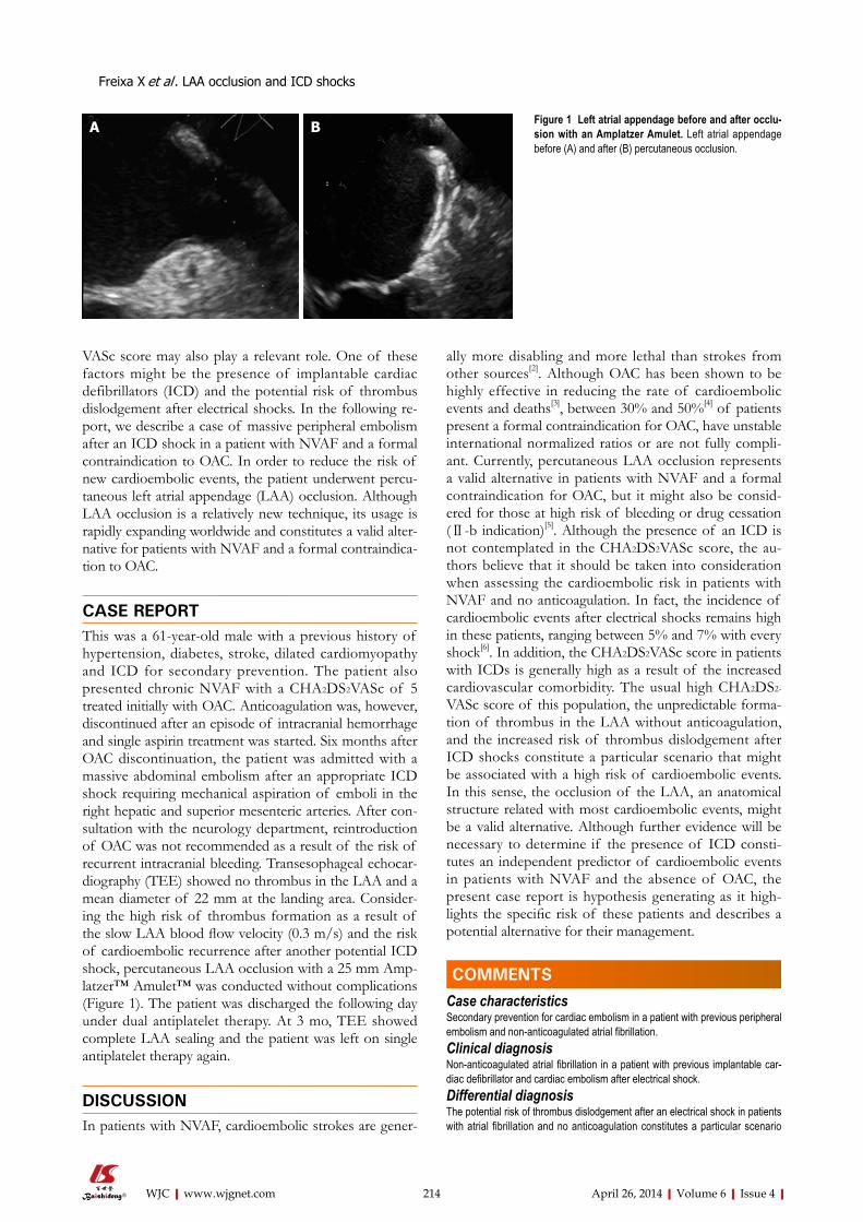
VASc score may also play a relevant role. One of these factors might be the presence of implantable cardiac defibrillators (ICD) and the potential risk of thrombus dislodgement after electrical shocks. In the following re-port, we describe a case of massive peripheral embolism after an ICD shock in a patient with NVAF and a formal contraindication to OAC. In order to reduce the risk of new cardioembolic events, the patient underwent percu-taneous left atrial appendage (LAA) occlusion. Although LAA occlusion is a relatively new technique, its usage is rapidly expanding worldwide and constitutes a valid alter-native for patients with NVAF and a formal contraindica-tion to OAC.
CASE REPORTThis was a 61-year-old male with a previous history of hypertension, diabetes, stroke, dilated cardiomyopathy and ICD for secondary prevention. The patient also presented chronic NVAF with a CHA2DS2VASc of 5 treated initially with OAC. Anticoagulation was, however, discontinued after an episode of intracranial hemorrhage and single aspirin treatment was started. Six months after OAC discontinuation, the patient was admitted with a massive abdominal embolism after an appropriate ICD shock requiring mechanical aspiration of emboli in the right hepatic and superior mesenteric arteries. After con-sultation with the neurology department, reintroduction of OAC was not recommended as a result of the risk of recurrent intracranial bleeding. Transesophageal echocar-diography (TEE) showed no thrombus in the LAA and a mean diameter of 22 mm at the landing area. Consider-ing the high risk of thrombus formation as a result of the slow LAA blood flow velocity (0.3 m/s) and the risk of cardioembolic recurrence after another potential ICD shock, percutaneous LAA occlusion with a 25 mm Amp-latzer™ Amulet™ was conducted without complications (Figure 1). The patient was discharged the following day under dual antiplatelet therapy. At 3 mo, TEE showed complete LAA sealing and the patient was left on single antiplatelet therapy again.
DISCUSSIONIn patients with NVAF, cardioembolic strokes are gener-
ally more disabling and more lethal than strokes from other sources[2]. Although OAC has been shown to be highly effective in reducing the rate of cardioembolic events and deaths[3], between 30% and 50%[4] of patients present a formal contraindication for OAC, have unstable international normalized ratios or are not fully compli-ant. Currently, percutaneous LAA occlusion represents a valid alternative in patients with NVAF and a formal contraindication for OAC, but it might also be consid-ered for those at high risk of bleeding or drug cessation (Ⅱ-b indication)[5]. Although the presence of an ICD is not contemplated in the CHA2DS2VASc score, the au-thors believe that it should be taken into consideration when assessing the cardioembolic risk in patients with NVAF and no anticoagulation. In fact, the incidence of cardioembolic events after electrical shocks remains high in these patients, ranging between 5% and 7% with every shock[6]. In addition, the CHA2DS2VASc score in patients with ICDs is generally high as a result of the increased cardiovascular comorbidity. The usual high CHA2DS2-
VASc score of this population, the unpredictable forma-tion of thrombus in the LAA without anticoagulation, and the increased risk of thrombus dislodgement after ICD shocks constitute a particular scenario that might be associated with a high risk of cardioembolic events. In this sense, the occlusion of the LAA, an anatomical structure related with most cardioembolic events, might be a valid alternative. Although further evidence will be necessary to determine if the presence of ICD consti-tutes an independent predictor of cardioembolic events in patients with NVAF and the absence of OAC, the present case report is hypothesis generating as it high-lights the specific risk of these patients and describes a potential alternative for their management.
COMMENTSCase characteristicsSecondary prevention for cardiac embolism in a patient with previous peripheral embolism and non-anticoagulated atrial fibrillation.Clinical diagnosisNon-anticoagulated atrial fibrillation in a patient with previous implantable car-diac defibrillator and cardiac embolism after electrical shock.Differential diagnosisThe potential risk of thrombus dislodgement after an electrical shock in patients with atrial fibrillation and no anticoagulation constitutes a particular scenario
214 April 26, 2014|Volume 6|Issue 4|WJC|www.wjgnet.com
Freixa X et al . LAA occlusion and ICD shocks
Figure 1 Left atrial appendage before and after occlu-sion with an Amplatzer Amulet. Left atrial appendage before (A) and after (B) percutaneous occlusion.
A B
COMMENTS
that might be associated with an additional cardioembolic risk.Imaging diagnosisPrevious intracranial hemorrhage.Pathological diagnosisNon-valvular atrial fibrillation (NVAF) with formal contraindication to anticoagu-lation due to previous intracranial bleeding.TreatmentPercutaneous left atrial appendage (LAA) occlusion.Experiences and lessonsAlthough LAA occlusion is a relatively new technique, its usage is rapidly expanding worldwide and constitutes a valid alternative for patients with atrial fibrillation and a formal contraindication to oral anticoagulation (OAC).Peer reviewThe authors reported a case with non-valvular atrial fibrillation and a formal con-traindication to OAC, who had undergone percutaneous LAA occlusion after the occurrence of massive abdominal embolism. This case report is interesting and suggestive but there are several questions to be solved.
REFERENCES1 CammAJ, Kirchhof P, Lip GY, Schotten U, Savelieva I, Ernst
S, Van Gelder IC, Al-Attar N, Hindricks G, Prendergast B, Heidbuchel H, Alfieri O, Angelini A, Atar D, Colonna P, De Caterina R, De Sutter J, Goette A, Gorenek B, Heldal M, Hohloser SH, Kolh P, Le Heuzey JY, Ponikowski P, Rutten FH. Guidelines for the management of atrial fibrillation: the
Task Force for the Management of Atrial Fibrillation of the European Society of Cardiology (ESC). Eur Heart J 2010; 31: 2369-2429 [PMID: 20802247 DOI: 10.1093/eurheartj/ehq278]
2 LinHJ, Wolf PA, Kelly-Hayes M, Beiser AS, Kase CS, Benja-min EJ, D’Agostino RB. Stroke severity in atrial fibrillation. The Framingham Study. Stroke 1996; 27: 1760-1764 [PMID: 8841325 DOI: 10.1161/01.STR.27.10.1760]
3 HartRG, Pearce LA, Aguilar MI. Meta-analysis: antithrom-botic therapy to prevent stroke in patients who have non-valvular atrial fibrillation. Ann Intern Med 2007; 146: 857-867 [PMID: 17577005 DOI: 10.7326/0003-4819-146-12-200706190-00007]
4 BungardTJ, Ghali WA, Teo KK, McAlister FA, Tsuyuki RT. Why do patients with atrial fibrillation not receive warfa-rin? Arch Intern Med 2000; 160: 41-46 [PMID: 10632303 DOI: 10.1001/archinte.160.1.41]
5 CammAJ, Lip GY, De Caterina R, Savelieva I, Atar D, Hohn-loser SH, Hindricks G, Kirchhof P. 2012 focused update of the ESC Guidelines for the management of atrial fibrillation: an update of the 2010 ESC Guidelines for the management of atrial fibrillation. Developed with the special contribution of the European Heart Rhythm Association. Eur Heart J 2012; 33: 2719-2747 [PMID: 22922413 DOI: 10.1093/eurheartj/ehs253]
6 BjerkelundCJ, Orning OM. The efficacy of anticoagulant therapy in preventing embolism related to D.C. electrical conversion of atrial fibrillation. Am J Cardiol 1969; 23: 208-216 [PMID: 4180019 DOI: 10.1016/0002-9149(69)90068-X]
P-Reviewers: Nakos G, Teragawa H S-Editor:Ma YJ L-Editor: Roemmele A E-Editor:Liu SQ
215 April 26, 2014|Volume 6|Issue 4|WJC|www.wjgnet.com
Freixa X et al . LAA occlusion and ICD shocks
GENERAL INFORMATIONWorld Journal of Cardiology (World J Cardiol, WJC, online ISSN 1949-8462, DOI: 10.4330) is a peer-reviewed open access (OA) academic journal that aims to guide clinical practice and improve diagnostic and therapeutic skills of clinicians.
Aim and scopeWJC covers topics concerning arrhythmia, heart failure, vascular disease, stroke, hypertension, prevention and epidemiology, dyslipi-demia and metabolic disorders, cardiac imaging, pediatrics, nursing, and health promotion. Priority publication will be given to articles concerning diagnosis and treatment of cardiology diseases. The fol-lowing aspects are covered: Clinical diagnosis, laboratory diagnosis, differential diagnosis, imaging tests, pathological diagnosis, molecular biological diagnosis, immunological diagnosis, genetic diagnosis, functional diagnostics, and physical diagnosis; and comprehensive therapy, drug therapy, surgical therapy, interventional treatment, min-imally invasive therapy, and robot-assisted therapy. We encourage authors to submit their manuscripts to WJC. We will give priority to manuscripts that are supported by major national and international foundations and those that are of great basic and clinical significance. WJC is edited and published by Baishideng Publishing Group (BPG). BPG has a strong professional editorial team composed of science editors, language editors and electronic editors. BPG current-ly publishes 42 OA clinical medical journals, including 41 in English, has a total of 15 471 editorial board members or peer reviewers, and is a world first-class publisher.
ColumnsThe columns in the issues of WJC will include: (1) Editorial: The editorial board members are invited to make comments on an im-portant topic in their field in terms of its current research status and future directions to lead the development of this discipline; (2) Frontier: The editorial board members are invited to select a highly cited cutting-edge original paper of his/her own to summarize ma-jor findings, the problems that have been resolved and remain to be resolved, and future research directions to help readers understand his/her important academic point of view and future research dir-ections in the field; (3) Diagnostic Advances: The editorial board members are invited to write high-quality diagnostic advances in their field to improve the diagnostic skills of readers. The topic covers general clinical diagnosis, differential diagnosis, pathological diagnosis, laboratory diagnosis, imaging diagnosis, endoscopic diag-nosis, biotechnological diagnosis, functional diagnosis, and physical diagnosis; (4) Therapeutics Advances: The editorial board members are invited to write high-quality therapeutic advances in their field to help improve the therapeutic skills of readers. The topic covers medication therapy, psychotherapy, physical therapy, replacement therapy, interventional therapy, minimally invasive therapy, endo-scopic therapy, transplantation therapy, and surgical therapy; (5) Field of Vision: The editorial board members are invited to write commentaries on classic articles, hot topic articles, or latest articles to keep readers at the forefront of research and increase their levels of clinical research. Classic articles refer to papers that are included in Web of Knowledge and have received a large number of citations (ranking in the top 1%) after being published for more than years, reflecting the quality and impact of papers. Hot topic articles refer
to papers that are included in Web of Knowledge and have received a large number of citations after being published for no more than 2 years, reflecting cutting-edge trends in scientific research. Latest articles refer to the latest published high-quality papers that are included in PubMed, reflecting the latest research trends. These commentary articles should focus on the status quo of research, the most important research topics, the problems that have now been resolved and remain to be resolved, and future research directions. Basic information about the article to be commented (including authors, article title, journal name, year, volume, and inclusive page numbers; (6) Minireviews: The editorial board members are invited to write short reviews on recent advances and trends in research of molecular biology, genomics, and related cutting-edge technologies to provide readers with the latest knowledge and help improve their diagnostic and therapeutic skills; (7) Review: To make a systematic review to focus on the status quo of research, the most import-ant research topics, the problems that have now been resolved and remain to be resolved, and future research directions; (8) Topic Highlight: The editorial board members are invited to write a series of articles (7-10 articles) to comment and discuss a hot topic to help improve the diagnostic and therapeutic skills of readers; (9) Medical Ethics: The editorial board members are invited to write articles about medical ethics to increase readers’ knowledge of med-ical ethics. The topic covers international ethics guidelines, animal studies, clinical trials, organ transplantation, etc.; (10) Clinical Case Conference or Clinicopathological Conference: The editorial board members are invited to contribute high-quality clinical case confer-ence; (11) Original Articles: To report innovative and original find-ings in cardiology; (12) Brief Articles: To briefly report the novel and innovative findings in cardiology; (13) Meta-Analysis: Covers the systematic review, mixedtreatment comparison, meta-regression, and overview of reviews, in order to summarize a given quantitative effect, e.g., the clinical effectiveness and safety of clinical treatments by combining data from two or more randomized controlled trials, thereby providing more precise and externally valid estimates than those which would stem from each individual dataset if analyzed separately from the others; (14) Case Report: To report a rare or typical case; (15) Letters to the Editor: To discuss and make reply to the contributions published in WJC, or to introduce and comment on a controversial issue of general interest; (16) Book Reviews: To introduce and comment on quality monographs of cardiology; and (17) Autobiography: The editorial board members are invited to write their autobiography to provide readers with stories of suc-cess or failure in their scientific research career. The topic covers their basic personal information and information about when they started doing research work, where and how they did research work, what they have achieved, and their lessons from success or failure.
Name of journalWorld Journal of Cardiology
ISSNISSN 1949-8462 (online)
Launch dateDecember 31, 2009
FrequencyMonthly
World Journal of CardiologyW J C
Online Submissions: http://www.wjgnet.com/esps/[email protected]
IWJC|www.wjgnet.com April 26, 2014|Volume 6|Issue 4|
World J Cardiol 2014 April 26; 6(4): I-VISSN 1949-8462 (online)
© 2014 Baishideng Publishing Group Co., Limited. All rights reserved.
INSTRUCTIONS TO AUTHORS
Instructions to authors
Editors-in-ChiefJian-Jun Li, MD, PhD, Professor, Center for Coronary Artery Disease, Fu Wai Cardiovascular Hospital, Chinese Academy of Medical Science, Beijing 100037, China
Giuseppe De Luca, PhD, Assistant Professor, Department of Cardiology, Piedmont University, Novara 28100, Italy
Nathan D Wong, FACC, FAHA, PhD, Director, Professor, Heart Disease Prevention Program, Division of Cardiology, De-partment of Medicine, University of California, Irvine, CA 92629, United States
Editorial officeJin-Lei Wang, DirectorXiu-Xia Song, Vice DirectorWorld Journal of CardiologyRoom 903, Building D, Ocean International Center,No. 62 Dongsihuan Zhonglu, Chaoyang District,Beijing 100025, ChinaTelephone: +86-10-59080039Fax: +86-10-85381893E-mail: [email protected]://www.wjgnet.com
PublisherBaishideng Publishing Group Co., LimitedFlat C, 23/F., Lucky Plaza, 315-321 Lockhart Road, Wan Chai, Hong Kong, ChinaFax: +852-65557188Telephone: +852-31779906E-mail: [email protected]://www.wjgnet.com
Production centerBeijing Baishideng BioMed Scientific Co., LimitedRoom 903, Building D, Ocean International Center,No. 62 Dongsihuan Zhonglu, Chaoyang District,Beijing 100025, ChinaTelephone: +86-10-85381892Fax: +86-10-85381893
Representative officeUSA Office8226 Regency Drive, Pleasanton, CA 94588-3144, United States
Instructions to authorsFull instructions are available online at http://www.wjgnet.com/1949-8462/g_info_20100316161927.htm.
Indexed and Abstracted inPubMed Central, PubMed, Digital Object Identifier, and Direc-tory of Open Access Journals.
SPECIAL STATEMENTAll articles published in this journal represent the viewpoints of the authors except where indicated otherwise.
Biostatistical editingStatistical review is performed after peer review. We invite an ex-pert in Biomedical Statistics to evaluate the statistical method used in the paper, including t-test (group or paired comparisons), chi-squared test, Ridit, probit, logit, regression (linear, curvilinear, or stepwise), correlation, analysis of variance, analysis of covariance, etc. The reviewing points include: (1) Statistical methods should be described when they are used to verify the results; (2) Whether the statistical techniques are suitable or correct; (3) Only homogeneous data can be averaged. Standard deviations are preferred to standard errors. Give the number of observations and subjects (n). Losses
in observations, such as drop-outs from the study should be re-ported; (4) Values such as ED50, LD50, IC50 should have their 95% confidence limits calculated and compared by weighted probit analysis (Bliss and Finney); and (5) The word ‘significantly’ should be replaced by its synonyms (if it indicates extent) or the P value (if it indicates statistical significance).
Conflict-of-interest statementIn the interests of transparency and to help reviewers assess any po-tential bias, WJC requires authors of all papers to declare any compet-ing commercial, personal, political, intellectual, or religious interests in relation to the submitted work. Referees are also asked to indi-cate any potential conflict they might have reviewing a particular paper. Before submitting, authors are suggested to read “Uniform Requirements for Manuscripts Submitted to Biomedical Journals: Ethical Considerations in the Conduct and Reporting of Research: Conflicts of Interest” from International Committee of Medical Journal Editors (ICMJE), which is available at: http://www.icmje.org/ethical_4conflicts.html.
Sample wording: [Name of individual] has received fees for serv-ing as a speaker, a consultant and an advisory board member for [names of organizations], and has received research funding from [names of organization]. [Name of individual] is an employee of [name of or-ganization]. [Name of individual] owns stocks and shares in [name of organization]. [Name of individual] owns patent [patent identification and brief description].
Statement of informed consentManuscripts should contain a statement to the effect that all human studies have been reviewed by the appropriate ethics committee or it should be stated clearly in the text that all persons gave their informed consent prior to their inclusion in the study. Details that might disclose the identity of the subjects under study should be omitted. Authors should also draw attention to the Code of Ethics of the World Med-ical Association (Declaration of Helsinki, 1964, as revised in 2004).
Statement of human and animal rightsWhen reporting the results from experiments, authors should follow the highest standards and the trial should conform to Good Clinical Practice (for example, US Food and Drug Administration Good Clinical Practice in FDA-Regulated Clinical Trials; UK Medicines Research Council Guidelines for Good Clinical Practice in Clinical Trials) and/or the World Medical Association Declaration of Hel-sinki. Generally, we suggest authors follow the lead investigator’s na-tional standard. If doubt exists whether the research was conducted in accordance with the above standards, the authors must explain the rationale for their approach and demonstrate that the institutional review body explicitly approved the doubtful aspects of the study.
Before submitting, authors should make their study approved by the relevant research ethics committee or institutional review board. If human participants were involved, manuscripts must be accom-panied by a statement that the experiments were undertaken with the understanding and appropriate informed consent of each. Any per-sonal item or information will not be published without explicit con-sents from the involved patients. If experimental animals were used, the materials and methods (experimental procedures) section must clearly indicate that appropriate measures were taken to minimize pain or discomfort, and details of animal care should be provided.
SUBMISSION OF MANUSCRIPTSManuscripts should be typed in 1.5 line spacing and 12 pt. Book Antiqua with ample margins. Number all pages consecutively, and start each of the following sections on a new page: Title Page, Abstract, Introduction, Materials and Methods, Results, Discus-sion, Acknowledgements, References, Tables, Figures, and Figure Legends. Neither the editors nor the publisher are responsible for the opinions expressed by contributors. Manuscripts formally ac-cepted for publication become the permanent property of Baish-ideng Publishing Group Co., Limited, and may not be reproduced by any means, in whole or in part, without the written permission
IIWJC|www.wjgnet.com April 26, 2014|Volume 6|Issue 4|
Instructions to authors
of both the authors and the publisher. We reserve the right to copy-edit and put onto our website accepted manuscripts. Authors should follow the relevant guidelines for the care and use of laboratory ani-mals of their institution or national animal welfare committee. For the sake of transparency in regard to the performance and report-ing of clinical trials, we endorse the policy of the ICMJE to refuse to publish papers on clinical trial results if the trial was not recorded in a publicly-accessible registry at its outset. The only register now available, to our knowledge, is http://www.clinicaltrials.gov spon-sored by the United States National Library of Medicine and we en-courage all potential contributors to register with it. However, in the case that other registers become available you will be duly notified. A letter of recommendation from each author’s organization should be provided with the contributed article to ensure the privacy and secrecy of research is protected.
Authors should retain one copy of the text, tables, photo-graphs and illustrations because rejected manuscripts will not be returned to the author(s) and the editors will not be responsible for loss or damage to photographs and illustrations sustained dur-ing mailing.
Online submissionsManuscripts should be submitted through the Online Submis-sion System at: http://www.wjgnet.com/esps/. Authors are highly recommended to consult the ONLINE INSTRUCTIONS TO AUTHORS (http://www.wjgnet.com/1949-8462/g_info_ 20100316161927.htm) before attempting to submit online. For assist-ance, authors encountering problems with the Online Submission System may send an email describing the problem to [email protected], or by telephone: +86-10-85381892. If you submit your manuscript online, do not make a postal contribution. Repeated online submission for the same manuscript is strictly prohibited.
MANUSCRIPT PREPARATIONAll contributions should be written in English. All articles must be submitted using word-processing software. All submissions must be typed in 1.5 line spacing and 12 pt. Book Antiqua with ample mar-gins. Style should conform to our house format. Required informa-tion for each of the manuscript sections is as follows:
Title pageTitle: Title should be less than 12 words.
Running title: A short running title of less than 6 words should be provided.
Authorship: Authorship credit should be in accordance with the standard proposed by ICMJE, based on (1) substantial contribu-tions to conception and design, acquisition of data, or analysis and interpretation of data; (2) drafting the article or revising it critically for important intellectual content; and (3) final approval of the ver-sion to be published. Authors should meet conditions 1, 2, and 3.
Institution: Author names should be given first, then the complete name of institution, city, province and postcode. For example, Xu-Chen Zhang, Li-Xin Mei, Department of Pathology, Chengde Med-ical College, Chengde 067000, Hebei Province, China. One author may be represented from two institutions, for example, George Sgourakis, Department of General, Visceral, and Transplantation Surgery, Essen 45122, Germany; George Sgourakis, 2nd Surgical Department, Korgialenio-Benakio Red Cross Hospital, Athens 15451, Greece
Author contributions: The format of this section should be: Author contributions: Wang CL and Liang L contributed equally to this work; Wang CL, Liang L, Fu JF, Zou CC, Hong F and Wu XM designed the research; Wang CL, Zou CC, Hong F and Wu XM performed the research; Xue JZ and Lu JR contributed new reagents/analytic tools; Wang CL, Liang L and Fu JF analyzed the
data; and Wang CL, Liang L and Fu JF wrote the paper.
Supportive foundations: The complete name and number of sup-portive foundations should be provided, e.g., Supported by National Natural Science Foundation of China, No. 30224801
Correspondence to: Only one corresponding address should be provided. Author names should be given first, then author title, af-filiation, the complete name of institution, city, postcode, province, country, and email. All the letters in the email should be in lower case. A space interval should be inserted between country name and email address. For example, Montgomery Bissell, MD, Professor of Medicine, Chief, Liver Center, Gastroenterology Division, Uni-versity of California, Box 0538, San Francisco, CA 94143, United States. [email protected]
Telephone and fax: Telephone and fax should consist of +, coun-try number, district number and telephone or fax number, e.g., Tele-phone: +86-10-85381892 Fax: +86-10-85381893
Peer reviewers: All articles received are subject to peer review. Normally, three experts are invited for each article. Decision on acceptance is made only when at least two experts recommend publication of an article. All peer-reviewers are acknowledged on Express Submission and Peer-review System website.
AbstractThere are unstructured abstracts (no less than 200 words) and struc-tured abstracts. The specific requirements for structured abstracts are as follows:
An informative, structured abstract should accompany each manuscript. Abstracts of original contributions should be struc-tured into the following sections: AIM (no more than 20 words; Only the purpose of the study should be included. Please write the Aim in the form of “To investigate/study/…”), METHODS (no less than 140 words for Original Articles; and no less than 80 words for Brief Articles), RESULTS (no less than 150 words for Original Articles and no less than 120 words for Brief Articles; You should present P values where appropriate and must provide relevant data to illustrate how they were obtained, e.g., 6.92 ± 3.86 vs 3.61 ± 1.67, P < 0.001), and CONCLUSION (no more than 26 words).
Key wordsPlease list 5-10 key words, selected mainly from Index Medicus, which reflect the content of the study.
Core tip Please write a summary of less than 100 words to outline the most innovative and important arguments and core contents in your paper to attract readers.
TextFor articles of these sections, original articles and brief articles, the main text should be structured into the following sections: INTRO-DUCTION, MATERIALS AND METHODS, RESULTS and DISCUSSION, and should include appropriate Figures and Tables. Data should be presented in the main text or in Figures and Tables, but not in both.
IllustrationsFigures should be numbered as 1, 2, 3, etc., and mentioned clearly in the main text. Provide a brief title for each figure on a separate page. Detailed legends should not be provided under the figures. This part should be added into the text where the figures are applicable. Keep-ing all elements compiled is necessary in line-art image. Scale bars should be used rather than magnification factors, with the length of the bar defined in the legend rather than on the bar itself. File names should identify the figure and panel. Avoid layering type directly over shaded or textured areas. Please use uniform legends for the same subjects. For example: Figure 1 Pathological changes in atrophic gas-
IIIWJC|www.wjgnet.com April 26, 2014|Volume 6|Issue 4|
tritis after treatment. A: ...; B: ...; C: ...; D: ...; E: ...; F: ...; G: …etc. It is our principle to publish high resolution-figures for the E-versions.
TablesThree-line tables should be numbered 1, 2, 3, etc., and mentioned clearly in the main text. Provide a brief title for each table. Detailed legends should not be included under tables, but rather added into the text where applicable. The information should complement, but not duplicate the text. Use one horizontal line under the title, a second under column heads, and a third below the Table, above any footnotes. Vertical and italic lines should be omitted.
Notes in tables and illustrationsData that are not statistically significant should not be noted. aP < 0.05, bP < 0.01 should be noted (P > 0.05 should not be noted). If there are other series of P values, cP < 0.05 and dP < 0.01 are used. A third series of P values can be expressed as eP < 0.05 and fP < 0.01. Other notes in tables or under illustrations should be expressed as 1F, 2F, 3F; or sometimes as other symbols with a superscript (Arabic numerals) in the upper left corner. In a multi-curve illustration, each curve should be labeled with ●, ○, ■, □, ▲, △, etc., in a cer-tain sequence.
AcknowledgmentsBrief acknowledgments of persons who have made genuine con-tributions to the manuscript and who endorse the data and conclu-sions should be included. Authors are responsible for obtaining written permission to use any copyrighted text and/or illustrations.
REFERENCESCoding systemThe author should number the references in Arabic numerals ac-cording to the citation order in the text. Put reference numbers in square brackets in superscript at the end of citation content or after the cited author’s name. For citation content which is part of the narration, the coding number and square brackets should be typeset normally. For example, “Crohn’s disease (CD) is associated with increased intestinal permeability[1,2]”. If references are cited directly in the text, they should be put together within the text, for example, “From references[19,22-24], we know that...”
When the authors write the references, please ensure that the order in text is the same as in the references section, and also ensure the spelling accuracy of the first author’s name. Do not list the same citation twice.
PMID and DOIPleased provide PubMed citation numbers to the reference list, e.g., PMID and DOI, which can be found at http://www.ncbi.nlm.nih.gov/sites/entrez?db=pubmed and http://www.crossref.org/Sim-pleTextQuery/, respectively. The numbers will be used in E-version of this journal.
Style for journal referencesAuthors: the name of the first author should be typed in bold-faced letters. The family name of all authors should be typed with the in-itial letter capitalized, followed by their abbreviated first and middle initials. (For example, Lian-Sheng Ma is abbreviated as Ma LS, Bo-Rong Pan as Pan BR). The title of the cited article and italicized journal title (journal title should be in its abbreviated form as shown in PubMed), publication date, volume number (in black), start page, and end page [PMID: 11819634 DOI: 10.3748/wjg.13.5396].
Style for book referencesAuthors: the name of the first author should be typed in bold-faced letters. The surname of all authors should be typed with the initial letter capitalized, followed by their abbreviated middle and first initials. (For example, Lian-Sheng Ma is abbreviated as Ma LS, Bo-Rong Pan as Pan BR) Book title. Publication number. Publication place: Publication press, Year: start page and end page.
FormatJournals English journal article (list all authors and include the PMID where applicable)1 Jung EM, Clevert DA, Schreyer AG, Schmitt S, Rennert J,
Kubale R, Feuerbach S, Jung F. Evaluation of quantitative con-trast harmonic imaging to assess malignancy of liver tumors: A prospective controlled two-center study. World J Gastroenterol 2007; 13: 6356-6364 [PMID: 18081224 DOI: 10.3748/wjg.13. 6356]
Chinese journal article (list all authors and include the PMID where applicable)2 Lin GZ, Wang XZ, Wang P, Lin J, Yang FD. Immunologic
effect of Jianpi Yishen decoction in treatment of Pixu-diar-rhoea. Shijie Huaren Xiaohua Zazhi 1999; 7: 285-287
In press3 Tian D, Araki H, Stahl E, Bergelson J, Kreitman M. Signature
of balancing selection in Arabidopsis. Proc Natl Acad Sci USA 2006; In press
Organization as author4 Diabetes Prevention Program Research Group. Hyperten-
sion, insulin, and proinsulin in participants with impaired glu-cose tolerance. Hypertension 2002; 40: 679-686 [PMID: 12411462 PMCID:2516377 DOI:10.1161/01.HYP.0000035706.28494. 09]
Both personal authors and an organization as author 5 Vallancien G, Emberton M, Harving N, van Moorselaar RJ;
Alf-One Study Group. Sexual dysfunction in 1, 274 European men suffering from lower urinary tract symptoms. J Urol 2003; 169: 2257-2261 [PMID: 12771764 DOI:10.1097/01.ju. 0000067940.76090.73]
No author given6 21st century heart solution may have a sting in the tail. BMJ
2002; 325: 184 [PMID: 12142303 DOI:10.1136/bmj.325. 7357.184]
Volume with supplement7 Geraud G, Spierings EL, Keywood C. Tolerability and safety
of frovatriptan with short- and long-term use for treatment of migraine and in comparison with sumatriptan. Headache 2002; 42 Suppl 2: S93-99 [PMID: 12028325 DOI:10.1046/j.1526-4610.42.s2.7.x]
Issue with no volume8 Banit DM, Kaufer H, Hartford JM. Intraoperative frozen
section analysis in revision total joint arthroplasty. Clin Orthop Relat Res 2002; (401): 230-238 [PMID: 12151900 DOI:10.1097/00003086-200208000-00026]
No volume or issue9 Outreach: Bringing HIV-positive individuals into care. HRSA
Careaction 2002; 1-6 [PMID: 12154804]
BooksPersonal author(s)10 Sherlock S, Dooley J. Diseases of the liver and billiary system.
9th ed. Oxford: Blackwell Sci Pub, 1993: 258-296Chapter in a book (list all authors)11 Lam SK. Academic investigator’s perspectives of medical
treatment for peptic ulcer. In: Swabb EA, Azabo S. Ulcer disease: investigation and basis for therapy. New York: Marcel Dekker, 1991: 431-450
Author(s) and editor(s)12 Breedlove GK, Schorfheide AM. Adolescent pregnancy.
2nd ed. Wieczorek RR, editor. White Plains (NY): March of Dimes Education Services, 2001: 20-34
Conference proceedings13 Harnden P, Joffe JK, Jones WG, editors. Germ cell tumours V.
Proceedings of the 5th Germ cell tumours Conference; 2001 Sep 13-15; Leeds, UK. New York: Springer, 2002: 30-56
Conference paper14 Christensen S, Oppacher F. An analysis of Koza's computa-
tional effort statistic for genetic programming. In: Foster JA, Lutton E, Miller J, Ryan C, Tettamanzi AG, editors. Genetic programming. EuroGP 2002: Proceedings of the 5th Euro-
IVWJC|www.wjgnet.com
Instructions to authors
April 26, 2014|Volume 6|Issue 4|
VWJC|www.wjgnet.com
Instructions to authors
April 26, 2014|Volume 6|Issue 4|
pean Conference on Genetic Programming; 2002 Apr 3-5; Kinsdale, Ireland. Berlin: Springer, 2002: 182-191
Electronic journal (list all authors)15 Morse SS. Factors in the emergence of infectious diseases.
Emerg Infect Dis serial online, 1995-01-03, cited 1996-06-05; 1(1): 24 screens. Available from: URL: http://www.cdc.gov/ncidod/eid/index.htm
Patent (list all authors)16 Pagedas AC, inventor; Ancel Surgical R&D Inc., assignee.
Flexible endoscopic grasping and cutting device and position-ing tool assembly. United States patent US 20020103498. 2002 Aug 1
Statistical dataWrite as mean ± SD or mean ± SE.
Statistical expressionExpress t test as t (in italics), F test as F (in italics), chi square test as χ2 (in Greek), related coefficient as r (in italics), degree of freedom as υ (in Greek), sample number as n (in italics), and probability as P (in italics).
UnitsUse SI units. For example: body mass, m (B) = 78 kg; blood pres-sure, p (B) = 16.2/12.3 kPa; incubation time, t (incubation) = 96 h, blood glucose concentration, c (glucose) 6.4 ± 2.1 mmol/L; blood CEA mass concentration, p (CEA) = 8.6 24.5 mg/L; CO2 volume fraction, 50 mL/L CO2, not 5% CO2; likewise for 40 g/L formal-dehyde, not 10% formalin; and mass fraction, 8 ng/g, etc. Arabic numerals such as 23, 243, 641 should be read 23243641.
The format for how to accurately write common units and quantums can be found at: http://www.wjgnet.com/1949-8462/g_info_20100312200347.htm.
AbbreviationsStandard abbreviations should be defined in the abstract and on first mention in the text. In general, terms should not be abbrevi-ated unless they are used repeatedly and the abbreviation is helpful to the reader. Permissible abbreviations are listed in Units, Symbols and Abbreviations: A Guide for Biological and Medical Editors and Authors (Ed. Baron DN, 1988) published by The Royal Society of Medicine, London. Certain commonly used abbreviations, such as DNA, RNA, HIV, LD50, PCR, HBV, ECG, WBC, RBC, CT, ESR, CSF, IgG, ELISA, PBS, ATP, EDTA, mAb, can be used directly without further explanation.
ItalicsQuantities: t time or temperature, c concentration, A area, l length, m mass, V volume.Genotypes: gyrA, arg 1, c myc, c fos, etc.Restriction enzymes: EcoRI, HindI, BamHI, Kbo I, Kpn I, etc.Biology: H. pylori, E coli, etc.
Examples for paper writingAll types of articles’ writing style and requirement will be found in the
link: http://www.wjgnet.com/esps/NavigationInfo.aspx?id=15
RESUBMISSION OF THE REVISED MANUSCRIPTSAuthors must revise their manuscript carefully according to the revision policies of Baishideng Publishing Group Co., Limited. The revised version, along with the signed copyright transfer agreement, responses to the reviewers, and English language Grade A certifi-cate (for non-native speakers of English), should be submitted to the online system via the link contained in the e-mail sent by the editor. If you have any questions about the revision, please send e-mail to [email protected].
Language evaluation The language of a manuscript will be graded before it is sent for revision. (1) Grade A: priority publishing; (2) Grade B: minor lan-guage polishing; (3) Grade C: a great deal of language polishing needed; and (4) Grade D: rejected. Revised articles should reach Grade A.
Copyright assignment formPlease download a Copyright assignment form from http://www.wjgnet.com/1949-8462/g_info_20100312200118.htm.
Responses to reviewersPlease revise your article according to the comments/suggestions provided by the reviewers. The format for responses to the reviewers’ comments can be found at: http://www.wjgnet.com/1949-8462/g_info_20100312195923.htm.
Proof of financial supportFor papers supported by a foundation, authors should provide a copy of the approval document and serial number of the foundation.
STATEMENT ABOUT ANONYMOUS PUBLICATION OF THE PEER REVIEWERS' COMMENTSIn order to increase the quality of peer review, push authors to carefully revise their manuscripts based on the peer reviewers' com-ments, and promote academic interactions among peer reviewers, authors and readers, we decide to anonymously publish the review-ers’ comments and author’s responses at the same time the manu-script is published online.
PUBLICATION FEEWJC is an international, peer-reviewed, OA online journal. Articles published by this journal are distributed under the terms of the Cre-ative Commons Attribution Non-commercial License, which per-mits use, distribution, and reproduction in any medium and format, provided the original work is properly cited. The use is non-com-mercial and is otherwise in compliance with the license. Authors of accepted articles must pay a publication fee. Publication fee: 698 USD per article. All invited articles are published free of charge.

Published by Baishideng Publishing Group Co., LimitedFlat C, 23/F., Lucky Plaza,
315-321 Lockhart Road, Wan Chai, Hong Kong, ChinaFax: +852-65557188
Telephone: +852-31779906E-mail: [email protected]
http://www.wjgnet.com
© 2014 Baishideng Publishing Group Co., Limited. All rights reserved.


