Embed Size (px)
Citation preview

Experimental Neurology xxx (2011) xxx–xxx
YEXNR-10980; No. of pages: 9; 4C: 3, 5, 6, 7
Contents lists available at SciVerse ScienceDirect
Experimental Neurology
j ourna l homepage: www.e lsev ie r .com/ locate /yexnr
Regular Article
Widespread suppression of huntingtin with convection-enhanced delivery of siRNA
David K. Stiles a, Zhiming Zhang b, Pei Ge c, Brian Nelson a, Richard Grondin b, Yi Ai b, Peter Hardy b,Peter T. Nelson d, Andrei P. Guzaev e, Mark T. Butt f, Klaus Charisse c, Verbena Kosovrasti c,Lubomir Tchangov c, Michael Meys c, Martin Maier c, Lubomir Nechev c, Muthiah Manoharan c,William F. Kaemmerer a, Douglas Gwost a, Gregory R. Stewart a, Don M. Gash b, Dinah W.Y. Sah c,⁎a Medtronic Neuromodulation, Medtronic, Minneapolis, MN 55432, USAb Department of Anatomy and Neurobiology, University of Kentucky College of Medicine, Lexington, KY 40536, USAc Alnylam Pharmaceuticals Inc., Cambridge, MA 02142, USAd Sanders Brown Center on Aging, University of Kentucky College of Medicine, Lexington, KY 40536, USAe AM Chemicals LLC, Oceanside, CA 92056, USAf Tox Path Specialists, LLC, Walkersville, MD 21793, USA
Abbreviations: (CED), convection enhanced delivery;siRNA; (cs), threshold tissue concentration for suppressiosion rate; (RNAi), RNA interference; (siRNA), small intsuppression; (Vs,Str), volume of suppression within the⁎ Corresponding author at: AlnylamPharmaceuticals In
MA 02142, USA. Fax: +1 617 551 8102.E-mail address: [email protected] (D.W.Y. Sah).
0014-4886/$ – see front matter © 2011 Elsevier Inc. Alldoi:10.1016/j.expneurol.2011.11.020
Please cite this article as: Stiles, D.K., et al.,(2011), doi:10.1016/j.expneurol.2011.11.02
a b s t r a c t
a r t i c l e i n f oArticle history:Received 24 June 2011Revised 3 November 2011Accepted 10 November 2011Available online xxxx
Keywords:Huntington's diseaseSite-specific deliveryConvection enhanced deliveryPutamenStriatum
Huntington's disease is an autosomal dominant neurodegenerative disease caused by a toxic gain of functionmutation in the huntingtin gene (Htt). Silencing of Htt with RNA interference using direct CNS delivery in ro-dent models of Huntington's disease has been shown to reduce pathology and promote neuronal recovery. Akey translational step for this approach is extension to the larger non-human primate brain, achieving suffi-cient distribution of small interfering RNA targeting Htt (siHtt) and levels of Htt suppression that may havetherapeutic benefit. We evaluated the potential for convection enhanced delivery (CED) of siHtt to providewidespread and robust suppression of Htt in nonhuman primates. siHtt was infused continuously for 7 or28 days into the nonhuman primate putamen to analyze effects of infusion rate and drug concentration onthe volume of effective suppression. Distribution of radiolabeled siHtt and Htt suppression were quantifiedby autoradiography and PCR, respectively, in tissue punches. Histopathology was evaluated and Htt suppres-sion was also visualized in animals treated for 28 days. Seven days of CED led to widespread distribution ofsiHtt and significant Htt silencing throughout the nonhuman primate striatum in an infusion rate and dosedependent manner. Htt suppression at therapeutic dose levels was well tolerated by the brain. A model de-veloped from these results predicts that continuous CED of siHtt can achieve significant coverage of the stri-atum of Huntington's disease patients. These findings suggest that this approach may provide an importanttherapeutic strategy for treating Huntington's disease.
© 2011 Elsevier Inc. All rights reserved.
Introduction
Huntington's disease is caused by an autosomal dominant mutationconsisting of an expansion in the CAG repeat region in the huntingtin(Htt) gene (The Huntington's Disease Collaborative Research Group,1993), with 36 or more repeats conferring a gain of functional toxicity(Walker, 2007). The resulting underlying pathology is characterizedby atrophy of the striatum, including profound degeneration ofmediumspiny neurons, as well as atrophy of the cortex and other regions of thebrain. Clinical manifestations include progressive involuntary
(cinf), concentration of infusedn; (Htt), huntingtin; (Q), infu-erfering RNA; (Vs), volume ofstriatum.c., 300 Third Street, Cambridge,
rights reserved.
Widespread suppression of h0
movement, cognitive dysfunction and behavioral changes. Currently,there are no therapies that modify the underlying progression ofdisease.
RNA interference (RNAi) is a naturally occurring cellularmechanismof gene regulation that can be leveraged for selectively suppressing Httexpression. Studies in rodentmodels of Huntington's disease have dem-onstrated the clinical potential of Htt lowering strategies using RNAi forreducing neuropathology, improving motor behavior and extendingsurvival time (Boudreau et al., 2009; DiFiglia et al., 2007; Drouet et al.,2009). Because of the blood–brain barrier, one of the major challengesin translating RNAi treatments into clinical treatments for neurodegen-erative diseases is delivery. Theoretically, convection enhanced delivery(CED) which entails infusing molecules under positive pressure to in-crease the volume of distribution in target tissues should be an effectivemeans for delivering small interfering RNA (siRNA) targeting Htt intoselected brain sites. The term “convection” in the context of CED refersto bulk flow in tissue that occurs as a result of pressure gradients, whichgreatly enhances the distribution of both small and large molecules
untingtin with convection-enhanced delivery of siRNA, Exp. Neurol.

2 D.K. Stiles et al. / Experimental Neurology xxx (2011) xxx–xxx
(Bobo et al., 1994). CED can occur with either acute or chronic infusiondosing paradigms ofmolecules, at increasing or constant flow rates; themolecules are “carried” by the movement of the effluent through theinterstitium. Thismethod has beenused successfully for deliveringmol-ecules in the siRNA size range into the brain in animals and humans,resulting in far greater distribution than possible by passive diffusion(Bobo et al., 1994; Fiandaca et al., 2009; Song and Lonser, 2008). Clinicalprecedent for direct delivery of drugs into the brain with CED includes~3 hour infusion at 0.5–10 μL/min of glucocerebrosidase in a Gaucher'spatient (Lonser et al., 2007), and continuous infusion of Trabedersen forup to 22 weeks at 1 and 4 μL/min in patients with glioblastoma or ana-plastic astrocytoma (Bogdahn et al., 2011).
Here, 7 days of CED of a radiolabeled siRNA (14C-siHtt) targetingboth wild type and mutant Htt was used to evaluate siRNA distributionand Htt suppression after infusion into the non human primate puta-men. The study was designed to enable development of an empiricalmodel of the Volume of Suppression (Vs) by choosing concentrationsof siRNA and infusion rates that span a range of clinically relevantsteady-state dosing regimens. Seven days of CED led to widespread dis-tribution of siRNA and silencing of Htt mRNA and protein. These datawere then used to model the dependence of the volume of effectivesuppression (volume over which effective siRNA concentrations areattained) on infusion rate and concentration of siRNA. In addition, a sec-ond study was conducted to analyze histopathology following 28 daysof continuous CED in the putamen to assess brain tissue responses to si-lencing Htt mRNA and lowering protein levels. Collectively, the resultsshow that 7 or 28 days of CED of siRNA leads to widespread and robustsuppression of Htt which is well tolerated in the nonhuman primatebrain. The results support the clinical potential of this approach forthe treatment of Huntington's disease.
Materials and methods
Animal surgery and siRNA delivery
All animal procedures were approved by the Animal Care and UseCommittee of the University of Kentucky, which is accredited by the As-sociation for Assessment and Accreditation of Laboratory Animal Care.Female rhesus monkeys (Macaca mulatta), approximately 7–16 yearsold, weighing 4.4–8.1 kgwere obtained fromCovance Research Products(Alice, Texas). MRI-guided stereotaxic surgery (Grondin et al., 2001;Heiss et al., 2010) was conducted to implant an intraparenchymal cath-eter with a titanium needle tip into the right putamen. The catheterwas connected to a SynchroMed® II Pump (Medtronic Neurological,Minneapolis, MN) subcutaneously implanted in the abdomen. After a7 day infusion of phosphate buffered saline at 6 μL/day, pumps werere-filled with test article and programmed for continuous delivery for7 days. The conditions chosen for this experiment (Table 1 plus
Table 1Amount of siRNA infused per day (mg/d) and corresponding relative (%) Htt/GAPDHmRNA suppression obtained in putamenal tissues punches, at different infusion ratesand concentrations of siRNA infused.
Infusion Rate(µL/min)
0.1 0.3 0.5
16 2.30 mg/d28% N = 3
11.52 mg/d 42% N = 3
12 5.18 mg/d 44% N = 5
8 3.46 mg/d
39% N = 5
Concentration
of siRNA
Infused
(mg/mL)
4 0.58 mg/d
16% N = 3
2.9 mg/d
16% N = 2
Please cite this article as: Stiles, D.K., et al., Widespread suppression of h(2011), doi:10.1016/j.expneurol.2011.11.020
phosphate buffered saline group, 0.3 μL/min, n=5) were designed tooptimize modeling distribution at varying flow rates and drug concen-trations. At the end of test article infusion, animals were euthanizedusing American Veterinary Medical Association approved methods. Tis-sue was evaluated from animals that met pre-determined criteria of ac-curate catheter placement and patency. The first criterion comprisedcatheter tip placement of ≤2 mm radial distance from the intended tar-get position, as determined by post-operativeMRI; for this study, all non-human primates implanted met this criterion. The second criterioncomprised catheter patency as determined by post-necropsy quantita-tive autoradiography (qAR) evaluation of brain tissue sections (seebelow); if no qAR signal was present, then this nonhuman primate wasnot included for further analysis. The catheter patency criterion wasnot met in only one animal (from the 0.5 μL/min, 4 mg/mL group) inthis study.
siRNA
The radiolabeled siRNA (Supplemental Fig. 1 online) was synthe-sized and purified according to standard procedures with minor mod-ifications as follows. A deoxythymidine phosphoramidite buildingblock, 14C-labeled at C-4, was synthesized and coupled to a solidsupport-bound, protected oligonucleotide precursor followed bydeprotection and anion-exchange HPLC purification of the radiola-beled antisense strand. The radiolabeled antisense strand was thenannealed to the unlabeled sense strand, and diluted in PBS. The si-lencing activity of this modified siRNA was found to be unchangedcompared to the parent unlabeled siRNA. The final specific activityof the 14C-siHtt was 2.0 μCi/mg, and osmolarity and endotoxin levelswere confirmed to be within acceptable levels.
Htt mRNA quantitation
For each animal, the infusion point in the brain was identified withMRI and visual inspection of the 2 mm coronal brain sections at nec-ropsy, prior to storage at −80 °C. After partial thawing on ice, tissuepunches of 1.2 mm diameter were taken using a pre-defined punchtemplate (Supplemental Fig. 2), and stored at−80 °C prior to blindedanalysis for Htt and GAPDHmRNA by RT-qPCR at QPS PharmaceuticalServices, using standard methods. To normalize data across plates, a“common control” derived from naïve rhesus monkey brain tissuewas included on each RT-qPCR plate. The qPCR data were unblindedafter all data were obtained.
Western blot analysis
Additional tissue punches, located 4 mmfrom the infusion site, wereused for Western blot analysis with mouse anti-Huntingtin (MAB2166,Millipore, Billerica, MA) and rabbit anti-β-catenin (Abcam, Cambridge,MA) primary antibodies, followed by fluorescently-labeled secondaryantibodies (anti-rabbit 680 nm and anti-mouse 800 nm, LI-COR Biosci-ence) for detection. Standard curves were generated from pooled stria-tal tissue samples from phosphate buffered saline-treated animals,using 5, 10 or 15 μg protein per lane. For test samples, 15 μg proteinper lane was evaluated for a tissue punch located within the putamenfromeachof 3 animals from the 12 mg/mL 14C-siHtt and control groups,after 7 days of infusion at 0.3 μL/min.
Determination of tissue concentration of 14C-siHtt
Photographs were obtained of the 2 mm coronal brain sectionsafter 40 μm thick coronal sections and tissue punches had beentaken for quantitative autoradiography (qAR) imaging and RT-qPCRprocessing, respectively. The spatial location of the punches was de-termined by calibrating the image files to a known length scale andcompared to the qAR image. The 14C-siHtt concentration at the
untingtin with convection-enhanced delivery of siRNA, Exp. Neurol.
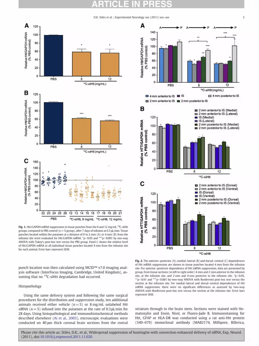
Fig. 1. Htt/GAPDHmRNA suppression in tissue punches from the8 and 12 mg/mL 14C-siHttgroups, compared to PBS control (n=5/group), after 7 days of infusion at 0.3 μL/min. Tissuepunches located within the putamen at a distance of 0 to 2 mm (A) or 4 mm (B) from theinfusion site were evaluated for Htt/GAPDH mRNA. *pb0.05 and ***pb0.001 by one-wayANOVA with Tukey's post-hoc test versus the PBS group. Panel C shows the relative levelof Htt/GAPDH mRNA in all individual tissue punches located 4 mm from the infusion sitefor each animal. Error bars represent SEM.
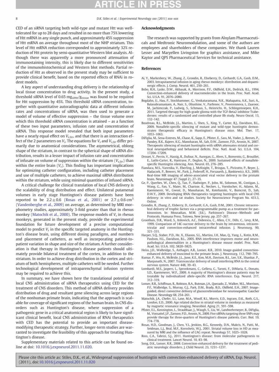
Fig. 2. The anterior–posterior (A),medial–lateral (B) and dorsal–ventral (C) dependencesof Htt mRNA suppression are shown in tissue punches located 4 mm from the infusionsite. For anterior–posterior dependence of Htt mRNA suppression, data are presented bygroup, from tissue sections (in left to right order) 4 mm and 2 mmanterior to the infusionsite, at the infusion site, and 2 mm and 4 mm posterior to the infusion site. *pb0.05,**pb0.01 and ***pb0.001 by two-way ANOVA with Bonferroni post-hoc test versus thesection at the infusion site. For medial–lateral and dorsal–ventral dependences of HttmRNA suppression, there were no significant differences as assessed by two-wayANOVA with Bonferroni post-hoc test versus the section at the infusion site. Error barsrepresent SEM.
3D.K. Stiles et al. / Experimental Neurology xxx (2011) xxx–xxx
punch location was then calculated using MCID™ v7.0 imaging anal-ysis software (InterFocus Imaging, Cambridge, United Kingdom), as-suming that no 14C-siHtt degradation had occurred.
Histopathology
Using the same delivery system and following the same surgicalprocedures for the distribution and suppression study, ten additionalanimals received either vehicle (n=5) or 8 mg/mL unlabeled HttsiRNA (n=5) infused into the putamen at the rate of 0.3μL/min for28 days. Using histopathological and immunohistochemical methodsdescribed elsewhere (Ai et al., 2003), microscopic evaluations wereconducted on 40 μm thick coronal brain sections from the rostral
Please cite this article as: Stiles, D.K., et al., Widespread suppression of h(2011), doi:10.1016/j.expneurol.2011.11.020
striatum through to the brain stem. Sections were stained with He-matoxylin and Eosin, Nissl, or Fluoro-Jade B. Immunostaining forHtt, GFAP or HLA-DR was conducted using a rat anti-Htt protein(549–679) monoclonal antibody (MAB2174, Millipore, Billerica,
untingtin with convection-enhanced delivery of siRNA, Exp. Neurol.

4 D.K. Stiles et al. / Experimental Neurology xxx (2011) xxx–xxx
MA), a mouse anti-GFAP monoclonal antibody (MAB360, Chemicon,Temecula, CA), or a mouse anti-HLA-DR monoclonal antibody(Clone L243, BD Biosciences, San Jose, CA), respectively.
Calculation of Vs and determination of empirical function for Vs (Q, cinf)
The Vs in each nonhuman primate was obtained by spatiallyorienting the autoradiographic images in Amira® v5.2.0 (Visage Im-aging, Inc., San Diego, CA). Using the known spacing between eachcoronal brain section and the pixel size calibrated using MCID, thepixel volume was calculated. All pixels with concentrations greaterthan ce were then summed to determine Vs. The response model de-termining the empirical function for Vs as a function of infusion rate(Q) and the concentration of the infused siRNA (cinf) was generatedusing Design-Expert® (v. 8.0.1, Stat-Ease, Inc., Minneapolis, MN).
Statistics
To determine the significance of Htt mRNA suppression versus acontrol or reference condition, one-way or two-way ANOVA withTukey's or Bonferroni post-hoc test was used. For Figs. 1A and B, one-way ANOVAs were used for statistical analysis because the study wasintended to address 2 separate questions: 1) how the response (HttmRNA reduction) was affected by dose near the infusion site (i.e. at a0–2 mm radial distance from the infusion site) after siRNA treatment,in order to demonstrate siRNA activity in NHP, and 2) how the response(Htt mRNA reduction) was affected by dose at a 4 mm radial distancefrom the infusion site after siRNA treatment, in order to demonstratesignificant siRNA activity at a distance consistent with CED. In addition,all punches at either a 0–2 mm or 4 mm radial distance from the infu-sion site, irrespective of anatomical location within the putamen, weregrouped together for the analysis, rather than comparing each treat-ment sample to its anatomical counterpart in the PBS group. We didnot have a large enough sample size with the current data set to beable to resolve the data analysis at this level, with a number of punch lo-cations having 2 or fewer data points for at least one treatment group.The lower confidence interval of the suppression of Htt mRNA in thepunches used to calculate ce, was determined using the T distribution(Minitab, Inc., State College, PA).
Results
CED of siRNA suppresses Htt mRNA throughout the striatum and lowersHtt protein levels
Previous studies have demonstrated silencing of Htt throughoutthe rat striatum with intrastriatal infusion of siRNA targeting Htt(Ge et al., 2009). Here, the translational potential of direct CNS deliv-ery of siRNA for silencing Htt was evaluated in the significantly largernonhuman primate striatum, a critical step in developing this poten-tial therapy for Huntington's disease. This study tested varying infu-sion rates (0.1, 0.3 and 0.5 μL/min) and siRNA concentrations (4, 8,12, and 16 mg/mL), with Htt mRNA suppression measured for alltest conditions. The boundary conditions were based on preliminaryexperiments suggesting that they represented appropriate limits forthe study. The center point conditions of 0.3 μL/min infusion rateand either 8 or 12 mg/mL siRNA concentration were used as themain independent variable conditions for which to study the effectson Htt mRNA suppression. From preliminary experiments, 7 dayswas found to be a sufficient period of time for siRNA delivery toreach steady state; thus, the results of this study represent the extentof Htt suppression achievable under these delivery conditions. ThesiRNA was radiolabeled (14C-siHtt) to facilitate correlation of Htt si-lencing with spatial distribution of siRNA within the same animal(see below). In vitro studies had demonstrated that radiolabelinghad no effect on Htt suppression with the siRNA sequence used in
Please cite this article as: Stiles, D.K., et al., Widespread suppression of h(2011), doi:10.1016/j.expneurol.2011.11.020
these experiments (data not shown). Silencing of Htt mRNA wasassessed in tissue punches taken from specific locations in the puta-men and caudate using a template placed over 2 mm thick serial cor-onal sections of the brain, extending 6 mm anterior to 6 mm posteriorfrom the infusion site (Supplemental Fig. 2 online). In addition, sup-pression of Htt protein was evaluated by semi-quantitative Westernblot analysis using tissue punches from the putamen.
For all tested combinations of infusion rate and 14C-siHtt concentra-tion, significant (15.5 to 44.4%) reduction of Htt mRNA was observedthroughout the putamen and caudate relative to vehicle infusion. Sup-pression of mRNA was dose-dependent for a given infusion rate. Forthe boundary conditions (4 or 16 mg/mL 14C-siHtt at 0.1 or 0.5 μL/min),Htt mRNA suppression in the putamen was more pronounced with in-creased siRNA concentration than with increased infusion rate(Table 1), although both variables and their interaction are importantin the response model that was developed from these data (see below).Of particular interest were the center point conditions of 8 and12 mg/mL at 0.3 μL/min, where more extensive spatial analysis was per-formed. For both groups, there was statistically significant reduction ofHtt mRNA of approximately 40–45% (Table 1). The level of Htt mRNAsuppression in the putamen at a 0 to 2 mm distance from the infusionsite (Fig. 1A) was similar to that at 4 mm from the infusion site(Fig. 1B), consistent with the principle of CED that drug is distributedmore uniformly with positive pressure than with diffusion which resultsin exponential decline of drug concentrationwith distance from the infu-sion site (Groothuis et al., 1999). Consistent Htt mRNA reduction wasobtained across animals within the same treatment group with low var-iability across punches from the same animal (Fig. 1C).
To assess directional dependence, Htt mRNA suppression in tissuepunches at an approximately 4 mm distance from the infusion sitewas evaluated along each axis: anterior–posterior (Fig. 2A), medial–lateral (Fig. 2B) and dorsal–ventral (Fig. 2C). While there was sub-stantial suppression of Htt mRNA throughout almost all of the puta-men after 7 days of infusion of 8 or 12 mg/mL 14C-siHtt, punchestaken from locations 4 mm posterior to the infusion site showed sig-nificantly less suppression on average than those taken from coronalsections at or anterior to the infusion site (Fig. 2A). In the coronal sec-tion 4 mm posterior to the infusion site, the reduction in Htt mRNAwas only 21% (pb0.05 vs PBS) at 8 mg/mL, and the reductionobtained using 12 mg/mL 14C-siHtt was not significantly differentfrom vehicle controls. In contrast, in the coronal section 4 mm anteri-or to the infusion site, there was a substantial 38% and 45% reductionof Htt mRNA with 8 and 12 mg/mL 14C-siHtt, respectively, relative tothe corresponding vehicle group, that was significantly different(pb0.01 and pb0.001, respectively) from the coronal section 4 mmposterior to the infusion site.
Htt mRNA suppression in tissue punches within the putamen wasconsistent across the medial–lateral (Fig. 2B) and dorsal–ventral(Fig. 2C) axis. Significant reduction of Htt mRNA extended into theadjacent caudate with intraputamenal infusion. CED of 8 or 12 mg/mL 14C-siHtt at 0.3 μL/min resulted in 17% or 19% suppression of HttmRNA, respectively (pb0.01 or pb0.001 versus vehicle controlgroup), in the caudate.
Suppression of Htt protein in tissue punches from the putamen, asassessed by semi-quantitative Western blot analysis, correspondedqualitatively to mRNA changes (data not shown). CED of 12 mg/mL14C-siHtt at 0.3 μL/min resulted in a reduction of Htt protein by 32%on average compared with a 44% average reduction of Htt mRNA, rel-ative to the vehicle control group.
Correlation of siHtt tissue concentration with Htt mRNA suppression anddetermination of threshold tissue concentration for suppression
To correlate tissue concentration of siRNA with extent of HttmRNA suppression, quantitative autoradiographic determinations of14C-siHtt concentration in coronal brain sections were co-registered
untingtin with convection-enhanced delivery of siRNA, Exp. Neurol.

5D.K. Stiles et al. / Experimental Neurology xxx (2011) xxx–xxx
with tissue punch locations (Fig. 3A). Radioactivity counts were as-sumed to represent parent 14C-siHtt; this is a conservative assump-tion for calculating the threshold 14C-siHtt tissue concentrationneeded for a certain level of Htt mRNA suppression. These data,from 422 tissue punches from putamen and caudate, are shown inFig. 3B. From RNAi studies reported to-date in animal models of Hun-tington's disease, Htt mRNA suppression of approximately 45% orgreater has resulted in meaningful normalization of neuropathologyand behavior (Harper et al., 2005). The subset of tissue punches inthis study that demonstrated at least 45% reduction of Htt mRNA,was therefore considered to have siRNA concentrations that areequal to or greater than the threshold suppression concentration, cs.In these 244 tissue punches from the striatum, the measured cs of14C-siHtt was 0.65 mgsiHtt/gtissue which corresponded to an averageHtt mRNA suppression of 46.1% with a 90% lower confidence intervalof 45.1%.
As described above, there was directionality to mRNA suppres-sion within the putamen along the anterior–posterior axis, withpunches from the coronal section located 4 mm posterior to the
Fig. 3. Tissue concentrations of 14C-siHtt and correlation with suppression of HttmRNA. Panel A is a heat map representation of quantitative autoradiography of a cor-onal section encompassing the infusion site after 7 days of 12 mg/mL 14C-siHtt infusionat 0.3 μL/min. The logarithmic color scale indicates the concentration of 14C-siHtt in thetissue. The white circles represent the locations of tissue punches taken for mRNAquantitation. The inset comprises a photograph of a corresponding coronal brain sec-tion with the caudate and putamen labeled. The calibration bar is 1 cm in length,with mm subdivisions. Panel B shows the correlation of percent Htt/GAPDH mRNAsuppression, relative to PBS control, with tissue concentration of 14C-siHtt from indi-vidual tissue punches taken from all animals in the study. Inset: the same data isshown with the tissue concentration plotted on a semi-logarithmic scale. Open andfilled squares represent data from tissue punches taken from putamen and caudate,respectively.
Please cite this article as: Stiles, D.K., et al., Widespread suppression of h(2011), doi:10.1016/j.expneurol.2011.11.020
infusion site exhibiting significantly less Htt silencing. To assesswhether tissue concentrations of siRNA were also lower in this loca-tion, levels of 14C-siHtt were determined. In the 8 mg/mL 14C-siHttgroup, punches taken from the section located 4 mm posterior tothe infusion site exhibited significantly lower levels of 14C-siHttthan those from the section located 4 mm anterior to the infusionsite (0.25 mg/g versus 1.54 mg/g, respectively, p=0.029), consis-tent with the reduced effect on mRNA suppression (21 vs 38% sup-pression, respectively, pb0.01).
Volume of suppression — dependence on concentration of siRNA infusedand infusion rate
For local drug delivery to the CNS, it is critical to ensure that suffi-cient drug distribution in the brain is achieved for the intended ther-apeutic effect. The volume of suppression, Vs, is defined as the region inthe brain where the siRNA has a meaningful biological effect; everypoint in this volume has a tissue concentration equal to or greaterthan the target threshold tissue concentration for suppression, cs. Twokey factors that impact Vs (mm3) are infusion rate (Q, μL/min) andconcentration of drug infused (cinf, mg/mL). In the present study, Vs,as measured by quantitative autoradiography, increased with increas-ing infusion rates, or with increasing concentrations of siRNA infused(Table 2). With 4 or 16 mg/mL 14C-siHtt, an infusion rate of 0.5 μL/min resulted in a larger Vs in the brain and striatum, compared withan infusion rate of 0.1 μL/min. A 14C-siHtt concentration of 16 mg/mL resulted in a larger Vs in the brain and striatum, compared with4 mg/mL 14C-siHtt, whether at an infusion rate of 0.1 or 0.5 μL/min.At an infusion rate of 0.3 μL/min, a 14C-siHtt concentration of12 mg/mL resulted in a larger Vs in the brain and striatum, comparedwith 8 mg/mL 14C-siHtt.
Table 2Measured Vs (brain) and Vs, Str (striatum) in individual NHPs at different infusion rates(Q) and concentrations of siRNA infused (cinf) for ≥45% suppression.
Monkey no. Infusion rate(μL/min)
Concentration(mg/mL)
Vs
(mm3)Vs, Str
(mm3)
1 0.1 4 232.4 13.72 0.1 4 75.2 28.13 0.1 4 156.6 156.6
Meanvolume±SEM 154.7±45.3 66±45.44 0.1 16 1160.8 539.25 0.1 16 936.6 555.56 0.1 16 519 454.3
Meanvolume±SEM 872.1±188.1 516.3±31.47 0.5 4 748.1 1888 0.5 4 655.4 309.3
Meanvolume±SEM 701.8 248.79 0.5 16 3819.2 1543.810 0.5 16 2468.4 1182.111 0.5 16 3515.5 1058.9
Meanvolume±SEM 3267.7±409.2
1261.6±145.5
12 0.3 8 563.2 506.513 0.3 8 987.2 348.114 0.3 8 781.2 530.915 0.3 8 720.2 302.816 0.3 8 812.1 667.9
Meanvolume±SEM 772.8±68.7 471.2±66.017 0.3 12 1813.5 1110.618 0.3 12 1961.3 1240.719 0.3 12 922.5 470.420 0.3 12 820.6 740.921 0.3 12 1468.2 779.3
Meanvolume±SEM 1397.2±229.6
868.4±137.8
untingtin with convection-enhanced delivery of siRNA, Exp. Neurol.

6 D.K. Stiles et al. / Experimental Neurology xxx (2011) xxx–xxx
For a target cs corresponding to 45% Htt mRNA suppression, andthe obtained Vs measured from quantitative autoradiography datafor a given infusion rate Q and concentration of drug infused cinf(Table 2), a response surface model describing Vs as a function of Qand cinf was generated. A regression analysis was performed tocurve-fit the data with a second order polynomial function of theform: Vs(Q,cinf)=a0+a1Q+a2cinf+a3Qcinf+a4Q
2+a5cinf2 (1). For
this experiment, the curvature was not found to be statistically signif-icant within the ranges tested, so only the terms a1, a2 and a3 wereused in the regression model for Vs. The empirical function obtainedfor Vs (adjusted R2 of 0.85) demonstrates a linear dependence on Qand cinf as well as the interaction term Qcinf.: Vs(Q,cinf)=−139.0−304.7Q+21.9cinf+393.4Qcinf (2). With this response model, it is pos-sible to estimate the Vs as a function of any value of Q and cinf withinthe experimentally-tested design space (Fig. 4).
In addition, Vs observed in the striatum only (Vs, Str) was measured(Table 2), and with cs corresponding to 45% Htt mRNA suppression,the resultant empirical function (adjusted R2 of 0.69) demonstratesa linear dependence on Q and cinf only, with the interaction termQcinf being negligible: Vs, Str(Q,cinf)=−389.0+1258.2Q+61.2cinf (3).
Histopathology
Infusion of vehicle and siRNA for 28 days was well tolerated; nobehavioral changes were observed and tissue responses to catheterimplantation and CED infusion were restricted to the catheter trackand adjacent tissue in the putamen. Changes in tissue adjacent tothe catheter track in both vehicle and siRNA recipients includedsome circumscribed neuronal loss, mild astrocytosis as assessed byan increase in GFAP-immunoreactive cells, and reactive microgliosis,as assessed by an increase in HLA-DR-immunoreactive cells. The cir-cumscribed neuronal loss appeared to be associated with catheter im-plantation. The relatively high infusion rate may have alsocontributed to the pathological changes at the injection site. Fluoro-Jade B staining did not reveal any continuing neuronal necrosis, eitherat the catheter dosing site or in brain regions away from the cathetersite, in any of the 28 day control and siRNA treated animals. De-creased neuronal levels of Htt protein were indicated by a pro-nounced attenuation of immunostaining intensity in the putamen ofsiRNA recipients, while Nissl-staining was retained (Fig. 5). Decreasesin immunostaining were evident for up to 11.5 mm in rostral–caudal
Fig. 4. Color contour plot of the predicted total Vs as a function of infusion rate (Q, μL/min)and concentration of siRNA infused (cinf, mg/mL), for cs corresponding to 45% suppression.The predicted total Vs (mm3) is represented as colors from 100 (dark blue, lower left) to3300 mm3 (bright red, upper right). Vs isocontours are shown as solid dark lines for 500,1000, 1500, 2000, 2500 and 3000 mm3. The boundary and center design points of the ex-periment are shown as filled red circles.
Please cite this article as: Stiles, D.K., et al., Widespread suppression of h(2011), doi:10.1016/j.expneurol.2011.11.020
extent. In addition to the putamen, the volume of distribution of thesiRNA as indicated by immunostaining spread into the adjacent cau-date nucleus and nucleus accumbens. Attenuated Htt immunostain-ing was not seen in vehicle recipients.
Discussion
A key remaining challenge for achieving the full therapeutic po-tential of siRNAs for CNS disorders is in vivo delivery to an area ofbrain sufficient to obtain a clinically meaningful effect. To date, localdelivery of siRNA has yielded limited spatial distribution and silenc-ing in neurons (DiFiglia et al., 2007; Kumar et al., 2007; Thakker etal., 2004). Here, we demonstrate that 7 days of CED of siRNA targetingHtt provides distribution of drug and Htt lowering in a large region ofthe nonhuman primate brain. Moreover, a response model developedwith these data provides a way to scale continuous intraparenchymalCED for delivery of efficacious levels of siRNA throughout sufficientlylarge regions of the human brain for clinical benefit in CNS disorderssuch as Huntington's disease.
Acute CED has shown experimental success for intraparenchymalCNS administration of molecules ranging from nanoparticles to pro-teins (Voges et al., 2003), with distribution throughout regions ofthe nonhuman primate and human brain that are much larger thanachievable with passive diffusion, and good tolerability. An importantdistinction in the present study is that continuous CED infusion over 7or 28 days was used, versus the previously reported CED infusion du-rations of minutes to hours. Consequently, the typical measure of CEDperformance (Song and Lonser, 2008) – the ratio of volume of distri-bution to volume of infusion (Vd/Vi) – is not appropriate here sincethe volume of infusion continues to increase with time, while themeasured volume of distribution has reached steady-state. Concen-tration–distance profiles show approximately constant drug concen-trations over substantial distances, beyond which concentrationsdecline exponentially. In the present study, with the infusion site cen-tered in the putamen, spatial profiles of siRNA concentration demon-strated approximately constant siRNA levels over an 8 mm distancewithin the putamen in the coronal plane, and substantially lowerlevels in surrounding tissues with increasing distance from the infu-sion site. The observed 8 mm distribution of Htt lowering was in thelarge central region of the roughly football-shaped putamen, repre-senting over 50% of the putamenal volume. Along the anterior–posterior axis, both siRNA level and effect were greater at more ante-rior locations. Further experimentation would be needed to assesswhether factors such as asymmetry of anatomical structures or pref-erential transport paths may result in directional preference. None-theless, the results from the present study suggest that CED intogray matter is an effective delivery method for distributing siRNA inthe brain.
Despite the identification of mutant Htt as causal for HD in 1993,there are currently no therapies that impact the underlying progres-sion of disease. With the advent of oligonucleotide approaches togene suppression, Htt lowering strategies have been used across mul-tiple studies assessing approaches that simultaneously lower wildtype and mutant Htt expression (Boudreau et al., 2009; Drouetet al., 2009) and that provide allele-specific silencing of mutant Httby targeting associated single nucleotide polymorphisms (Lombardiet al., 2009; Pfister et al., 2009). Both general approaches can be effec-tive therapeutic strategies, each with its own relative merits. Shorthairpin RNAs and siRNAs targeting wild-type and/or mutant Htthave been evaluated in rodent models of Huntington's disease. Con-tinuous, partial suppression of Htt in adult rodent models is effectivein reducing neuropathology, improving motor behavior and prolong-ing survival (Boudreau et al., 2009; Drouet et al., 2009). Importantly,these studies showed striatal suppression of up to 75% of both wild-type and mutant Htt to be not only efficacious against HD pathology,but also well-tolerated for at least 9 months without overt toxicity or
untingtin with convection-enhanced delivery of siRNA, Exp. Neurol.

Fig. 5. Normal patterns of Nissl-stained neurons are retained in the putamenwhile Huntingtin (Htt) immunostaining is suppressed. Htt immunostaining intensity increasedwith increas-ing distance from the catheter track (small arrows). A, and insets below: IntenseHtt immunostaining of neurons overlappedwith normal patterns of Nissl-stained neurons in the putamenbeyond the immunostaining penumbra. B, and insets below: In the penumbra, moderately and weakly stained Htt positive cells are evident while a normal pattern of Nissl-stained neu-rons was retained. C, and insets below: Closer to the catheter track, Htt immunostaining was reduced to background levels while Nissl staining of neurons was retained.
7D.K. Stiles et al. / Experimental Neurology xxx (2011) xxx–xxx
an increase in striatal vulnerability in rats. Therefore, while WT-Httplays a role in multiple cellular processes, including axonal transport,neuronal development, transcriptional regulation, and protection
Please cite this article as: Stiles, D.K., et al., Widespread suppression of h(2011), doi:10.1016/j.expneurol.2011.11.020
from cell death (Ross and Tabrizi, 2011), it is anticipated based onthese studies that partial Htt lowering (of both WT and mutant Htt)in the adult may be both safe and effective. In the present study,
untingtin with convection-enhanced delivery of siRNA, Exp. Neurol.
8 D.K. Stiles et al. / Experimental Neurology xxx (2011) xxx–xxx
CED of an siRNA targeting both wild-type and mutant Htt was well-tolerated for up to 28 days and resulted in no more than 75% loweringof Htt mRNA in any single punch, and approximately 45% suppressionof Htt mRNA on average throughout most of the NHP striatum. Thislevel of Htt mRNA reduction corresponded to approximately 32% re-duction of Htt protein by semi-quantitative Western blot analysis. Al-though there was apparently a more pronounced attenuation ofimmunostaining intensity, this is likely due to different sensitivitiesof the immunohistochemical and Western blot methods. Partial re-duction of Htt as observed in the present study may be sufficient toprovide clinical benefit, based on the reported effects of RNAi in ro-dent models.
A key aspect of understanding drug delivery is the relationship oflocal tissue concentration to drug activity. In the present study, athreshold siRNA level of 0.65 mgsiHtt/gtissue was found to be requiredfor Htt suppression by 45%. This threshold siRNA concentration, to-gether with quantitative autoradiographic data at different infusionrates and concentrations of siRNA, was then used to generate amodel of volume of effective suppression – the tissue volume overwhich this threshold siRNA concentration is attained – as a functionof these two input parameters, infusion rate and concentration ofsiRNA. This response model revealed that both input parametershave a nearly equal effect on Vs,Str, and that there is an interaction ef-fect of the 2 parameters on Vs. The equations for Vs and Vs,Str differ pri-marily due to anatomical considerations. The asymmetrical, oblongshape of the striatum, in contrast to the spherical shape of siRNA dis-tribution, results in a lesser impact of infusion rate and concentrationof infusate on volume of suppression within the striatum (Vs,Str) thanwithin the entire brain (Vs). This finding has important implicationsfor optimizing catheter configuration, including catheter placementand use of multiple catheters, to achieve maximal siRNA distributionwithin the striatum, while using minimal quantities of infused siRNA.
A critical challenge for clinical translation of local CNS delivery isthe scalability of drug distribution and effect. Unilateral putamenalvolumes in early stage Huntington's disease patients have beenreported to be 2.2±0.6 (Rosas et al., 2001) or 2.7±0.6 cm3
(Vandenberghe et al., 2009) on average, as determined by MRI mor-phometry studies, approximately 3-fold greater than that in rhesusmonkey (Matochik et al., 2000). The response models of Vs in rhesusmonkeys, generated in the present study, provide the experimentalfoundation for future development of a precise computationalmodel to predict Vs in the specific targeted anatomy in the Hunting-ton's disease brain, using different dosing paradigms, and numbersand placement of catheters, as well as incorporating patient-to-patient variation in shape and size of the striatum. A further consider-ation is that therapy in Huntington's disease patients should ulti-mately provide bilateral treatment of the cortex, in addition to thestriatum. In order to achieve drug distribution in the cortex and stri-atum of both hemispheres, multiple catheters will be needed. Furthertechnological development of intraparenchymal infusion systemsmay be required to achieve this.
In summary, we have shown here the translational potential oflocal CNS administration of siRNA therapeutics using CED for thetreatment of CNS disorders. This method of siRNA delivery providesdistribution of drug and resultant gene silencing across large regionsof the nonhuman primate brain, indicating that the approach is scal-able for coverage of significant regions of the human brain. In CNS dis-orders such as Huntington's disease, where suppression of apathogenic gene in a critical anatomical region is likely to have signif-icant clinical benefit, local CNS administration of RNAi therapeuticswith CED has the potential to provide an important disease-modifying therapeutic strategy. Further, longer-term studies are war-ranted to investigate the feasibility of this approach for treating Hun-tington's disease.
Supplementary materials related to this article can be found on-line at doi: 10.1016/j.expneurol.2011.11.020.
Please cite this article as: Stiles, D.K., et al., Widespread suppression of h(2011), doi:10.1016/j.expneurol.2011.11.020
Acknowledgments
The research was supported by grants from Alnylam Pharmaceuti-cals and Medtronic Neuromodulation, and some of the authors areemployees and shareholders of these companies. We thank LaurenLesser and Maryellen Livingston for graphics assistance, and MikeKaytor and QPS Pharmaceutical Services for technical assistance.
References
Ai, Y., Markesbery, W., Zhang, Z., Grondin, R., Elseberry, D., Gerhardt, G.A., Gash, D.M.,2003. Intraputamenal infusion in aging rhesus monkeys: distribution and dopami-nergic effects. J. Comp. Neurol. 461, 250–261.
Bobo, R.H., Laske, D.W., Akbasak, A., Morrison, P.F., Oldfield, E.H., Dedrick, R.L., 1994.Convection-enhanced delivery of macromolecules in the brain. Proc. Natl. Acad.Sci. U.S.A. 91, 2076–2080.
Bogdahn, U., Hau, P., Stockhammer, G., Venkataramana, N.K., Mahapatra, A.K., Suri, A.,Balasubramaniam, A., Nair, S., Oliushine, V., Parfenov, V., Poverennova, I., Zaaroor,M., Jachimczak, P., Ludwig, S., Schmaus, S., Heinrichs, H., Schlingensiepen, K.H.,2011. Targeted therapy for high-grade glioma with the TGF-Beta2 inhibitor Trabe-dersen: results of a randomized and controlled phase IIb study. Neuro Oncol. 13,132–142.
Boudreau, R.L., McBride, J.L., Martins, I., Shen, S., Xing, Y., Carter, B.J., Davidson, B.L.,2009. Nonallele-specific silencing of mutant and wild-type huntingtin demon-strates therapeutic efficacy in Huntington's disease mice. Mol. Ther. 17,1053–1063.
DiFiglia, M., Sena-Esteves, M., Chase, K., Sapp, E., Pfister, E., Sass, M., Yoder, J., Reeves, P.,Pandey, R.K., Rajeev, K.G., Manoharan, M., Sah, D.W., Zamore, P.D., Aronin, N., 2007.Therapeutic silencing of mutant huntingtin with siRNA attenuates striatal and cor-tical neuropathology and behavioral deficits. Proc. Natl. Acad. Sci. U.S.A. 104,17204–17209.
Drouet, V., Perrin, V., Hassig, R., Dufour, N., Auregan, G., Alves, S., Bonvento, G., Brouillet,E., Luthi-Carter, R., Hantrave, P., Deglon, N., 2009. Sustained effects of nonallele-specific Huntingtin silencing. Ann. Neurol. 65, 276–285.
Fiandaca, M.S., Varenika, V., Eberling, J., McKnight, T., Bringas, T., Pivirotto, P., Beyer, J.,Hadaczek, P., Bowers, W., Park, J., Federoff, H., Forsaueth, J., Bankiewicz, K.S., 2009.Real-time MR imaging of adeno-associated viral vector delivery to the primatebrain. Neuroimage 47 (Suppl 2), 27–35.
Ge, P., Querbes, W., Chen, Q., Tan, P., Hadwiger, P., John, M., Solomon, M., Costigan, J.,Wang, G., Fan, Y., Maier, M., Charisse, K., Nechev, L., Vornlocher, H., Adams, M.,Kaemmerer, W., Gwost, D., Manoharan, M., Kotelianski, V., Bumcrot, D., Sah,D.W.Y., 2009. Developing RNAi therapeutics targeting huntingtin with local CNSdelivery: in vitro and rat studies. Society for Neuroscience Program No. 433.3,Online.
Grondin, R., Zhang, Z., Elsberry, D., Gerhardt, G.A., Gash, D.M., 2001. Chronic intracere-bral delivery of trophic factors via a programmable pump as a treatment for par-kinsonism. In: Mouradian, M.M. (Ed.), Parkinson's Disease—Methods andProtocols. Humana Press, Totowa, New Jersey, pp. 257–267.
Groothuis, D.R., Ward, S., Itskovich, A.C., Dobrescu, C., Allen, C.V., Dills, C., Levy, R.M.,1999. Comparison of 14C-sucrose delivery to the brain by intravenous, intraven-tricular and convection-enhanced intracerebral infusion. J. Neurosurg. 90,321–331.
Harper, S.Q., Staber, P.D., He, X., Eliason, S.L., Martins, I.H., Mao, Q., Yang, L., Kotin, R.M.,Paulson, H.L., Davidson, B.L., 2005. RNA interference improves motor and neuro-pathological abnormalities in a Huntington's disease mouse model. Proc. Natl.Acad. Sci. U.S.A. 102, 5820–5825.
Heiss, J.D., Walbridge, S., Asthagiri, A.R., Lonser, R.R., 2010. Image-guided convection-enhanced delivery of muscimol to the primate brain. J. Neurosurg. 112, 790–795.
Kumar, P., Wu, H., McBride, J.L., June, K.E., Kim, M.H., Davison, B.L., Lee, S.K., Shankar, P.,Manjunath, N., 2007. Transvascular delivery of small interfering RNA to the centralnervous system. Nature 448, 39–43.
Lombardi, M.S., Jaspers, L., Spronkmans, C., Gellera, C., Taroni, F., DiMaria, E., Donato,S.D., Kaemmerer, W.F., 2009. A majority of Huntington's disease patients may betreatable by individualized allele-specific RNA interference. Exp. Neurol. 217,312–319.
Lonser, R.R., Schiffman, R., Robison, R.A., Butman, J.A., Quezado, Z., Walker, M.L., Morrison,P.F., Walbridge, S., Murray, G.J., Park, D.M., Brady, R.O., Oldfield, E.H., 2007. Image-guided, direct convective delivery of glucocerebrosidase for neuronopathic GaucherDisease. Neurology 68, 254–261.
Matochik, J.A., Chefer, S.I., Lane, M.A., Woolf, R.I., Morris, E.D., Ingram, D.K., Roth, G.S.,London, E.D., 2000. Age related decline in striatal volume in monkeys as measuredby magnetic resonance imaging. Neurobiol. Aging 21, 591–598.
Pfister, E.L., Kennington, L., Straubhaar, J., Waugh, S., Liu, W., Landwehrmeyer, B., DiFiglia,M., Vonsattel, J.P., Zamore, P.D., Aronin, N., 2009. Five siRNAs targeting three SNPsmayprovide therapy for three-quarters of Huntington's disease patients. Curr. Biol. 19,774–778.
Rosas, H.D., Goodman, J., Chen, Y.I., Jenkins, B.G., Kennedy, D.N., Makris, N., Patti, M.,Seidman, L.J., Beal, M.F., Koroshetz, W.J., 2001. Striatal volume loss in HD as mea-sured by MRI and the influence of CAG repeat. Neurology 57, 1025–1028.
Ross, C.A., Tabrizi, S.J., 2011. Huntington's disease: from molecular pathogenesis toclinical treatment. Lancet Neurol. 10, 83–98.
Song, D.K., Lonser, R.R., 2008. Convection-enhanced delivery for the treatment of pedi-atric neurologic disorders. J. Child Neurol. 23, 1231–1237.
untingtin with convection-enhanced delivery of siRNA, Exp. Neurol.

9D.K. Stiles et al. / Experimental Neurology xxx (2011) xxx–xxx
Thakker, D.R., Natt, F., Hüsken, D., Maier, R., Müllen, M., van der Putten, H., Hover, D.,Cryan, J.F., 2004. Neurochemical and behavioral consequences of widespreadgene knockdown in the adult mouse brain by using nonviral RNA interference.Proc. Natl. Acad. Sci. U.S.A. 101, 17270–17275.
The Huntington's Disease Collaborative Research Group, 1993. A novel gene containinga trinucleotide repeat that is expanded and unstable on Huntington's disease chro-mosomes. Cell 72, 971–983.
Please cite this article as: Stiles, D.K., et al., Widespread suppression of h(2011), doi:10.1016/j.expneurol.2011.11.020
Vandenberghe, W., Demaerel, P., Dom, R., Maes, F., 2009. Diffusion-weighted versusvolumetric imaging of the striatum in early symptomatic Huntington disease. J.Neurol. 256, 109–114.
Voges, J., Reszka, R., Gossmann, A., Dittmar, C., Richter, R., Garlip, G., Kracht, L., Coenen, H.H.,Sturm, V., Wienhard, K., Heiss, W.D., Jacobs, A.H., 2003. Imaging-guided convection-enhanced delivery and gene therapy of glioblastoma. Ann. Neurol. 54, 479–487.
Walker, F.O., 2007. Huntington's disease. Lancet 369, 218–228.
untingtin with convection-enhanced delivery of siRNA, Exp. Neurol.


