Embed Size (px)
Citation preview
Makara Journal of Science, 20/1 (2016), 7-16 doi: 10.7454/mss.v20i1.5656
March 2016 | Vol. 20 | No. 1 7
Vinblastine and Vincristine Production on Madagascar Periwinkle (Catharanthus roseus (L.) G. Don) Callus Culture Treated with
Polethylene Glycol
Nisa Nur Iskandar*and Iriawati
School of Life Sciences and Technology, Institut Teknologi Bandung, Bandung 40526, Indonesia
*E-mail: [email protected]
Received June 18, 2014 | Accepted October 23, 2015
Abstract
Vinblastine and vincristine are secondary metabolites from Madagascar periwinkles that have a very high economic value as chemotherapy drugs. These compounds are naturally produced in a very low quantity in planta. One promising alternative method for vinblastine and vincristine production is to use a treatment that can trigger plant stress response in vitro. This study has been done to evaluate the effect of drought stress using polyethylene glycol (PEG) on vinblastine and vincristine production in the C. roseus callus culture, which were grown on medium Zenk supplemented with plant growth regulators (PGR) 1 µM NAA + 10 µM Kinetin to induce laticifer and idioblast differentiation. 13-week-old callus cultures were then treated with 0%, 6%, 9%, and 12% (w/v) PEG4000 each for 0, 24, 48, and 72 hours. Biochemical analysis was performed using HPLC to determine the levels of vinblastine and vincristine, while the presence of differentiated cells (idioblasts and laticifers) was determined using a histochemical method. Protein profiles of the culture were determined by SDS-Page. The results showed that drought treatment with PEG4000, until the concentration was 12% (w/v), did not significantly affect the production of vinblastine and vincristine, but might affect terpenoid production. Histochemical analysis confirmed the presence of idioblasts, non-elongated laticifers, and laticifers that were producing and accumulating terpenoids highest in the 12% PEG treatment. PEG treatments also did not change the protein profile of callus.
Abstract
Produksi Senyawa Viblastin dan Vincristin pada Kultur Kalus Tanaman Tapak Dara (Catharanthus roseus (L.) G. Don) dengan Pemberian Cekaman Menggunakan Polietilena Glikol (PEG). Vinblastin dan vincristine merupakan senyawa metabolit sekunder dari tanaman Tapak Dara yang memiliki nilai ekonomi tinggi sebagai obat kemoterapi kanker. Kedua senyawa ini hanya diproduksi dalam jumlah yang sangat sedikit pada tumbuhan. Salah satu metode alternatif yang menjanjikan untuk meningkatkan produksinya adalah dengan pemberian cekaman pada kultur in vitro. Telah dilakukan penelitian dengan tujuan untuk menentukan pengaruh pemberian cekaman kekeringan menggunakan polyethylene glycol (PEG) terhadap produksi senyawa vinblastin dan vincristine pada kultur agregat C. roseus yang ditanam pada medium Zenk dengan penambahan zat pengatur tumbuh (ZPT) NAA 1 µM + Kinetin 10 µM untuk menginduksi pembentukan kelenjar latisifer dan sel idioblas. Kalus yang berusia 13 minggu kemudian diberi perlakuan cekaman kekeringan menggunakan PEG4000 0%, 6%, 9%, dan 12% (w/v) masing-masing selama 0, 24, 48, dan 72 jam. Analisis dilakukan secara histokimia untuk menguji keberadaan latisifer pada kalus dan biokimiawi untuk menentukan kadar vinblastin dan vincristine dengan menggunakan HPLC. Selain itu dilakukan pula analisis profil protein dengan SDS-Page. Hasil penelitian menunjukkan bahwa pemberian cekaman kekeringan dengan menggunakan PEG4000 hingga konsentrasi 12% (w/v) tidak berdampak signifikan terhadap produksi senyawa vinblastin dan vincristine namun diduga berdampak pada pembentukan senyawa terpenoid. Sel idioblas dan latisifer yang mengandung terpenoid dalam jumlah relatif banyak ditemukan pada perlakuan PEG 12%. Perlakuan dengan PEG juga tidak berdampak pada perubahan profil protein kalus. Analisis protein menunjukkan bahwa seluruh perlakuan memiliki profil protein yang sama. Keywords: madagascar periwinkle, peg, vinblastine, vincristine Introduction Madagascar periwinkle (Catharanthus roseus (L.) G. Don) is a medicinal plant that can produce indole alkaloid
(TIAs) compound. Some TIAs have a physiological effect on humans [1]. Two of these compounds, vinblastine and vincristine, have a very high economic value as the main active compound in various types of chemotherapy drugs
Iskandar, et al.
Makara J. Sci. March 2016 | Vol. 20 | No. 1
8
[1-4], including leukemia, lymphosarcoma, choriokarsi-noma, neuroblastoma, breast cancer, lung cancer, Hodgkin's disease, Wilkin’s tumors and reticulum cell sarcoma [5]. The demand for vinblastine and vincristine is increasing year to year [5], but the industrialization process of these two compounds is still encountering many obstacles [1]. Naturally, both vinblastine and vincristine only produce in very low quantities, about 0.0003% from the 2.56% total alkaloid content in C. roseus [3]. It takes a large biomass and an inefficient extraction process to industrialize these two compounds [6]. One promising alternative method for secondary metabolite production, including vinblastine and vincristine, is to use the in vitro culture technique for developing cells or tissues that play a role in the biosynthesis of these secondary metabolites [6,7]. By using in vitro culture, the growth of cells or tissues and their metabolism can be controlled and directed to produce the desired secondary metabolites. The biosynthesis of vinblastine and vincristine naturally occurs through a complex process involving various types of proteins and enzymes that are synthesized specifically in certain parts of the plant and cell [1,5,8]. Based on the study of activities and transport mechanisms of the enzymes involved in the biosynthesis of vinblastine and vincristine, it can be concluded that these enzymes are not fully expressed and synthesized in the cell culture of C. roseus [1]. Enzymes that are responsible for the last step of vindolin biosynthesis, desacetoxyvindoline 4-hydroxylase (D4H) and deacetylvindoline-4-O-acetyl-transferase (DAT), only expressed in the differentiated cells, laticifer, and idioblast [1,8]. This may explain why bisindole alkaloids such as vinblastine and vincristine cannot be produced in the desired amount within the cell culture which mostly contains undifferentiated cells [1]. Based on previous research by Darsini [7] and Iskandar [9], cells in the C. roseus callus can directly differentiate into laticifer, non-elongated laticifer, and idioblast. These cells can also produce secondary metabolites such as alkaloids and terpenoids. It can be concluded that the callus from an aggregate culture has the potential to be used in producing bisindole alkaloids such as vinblastine and vincristine. Over the years, researchers have been using manipulated media, combinations of phytohormones, precursor compounds, mutation, transformation, and hairy root development for increasing secondary metabolite production, but those methods are still not able to meet the desired quantity of secondary metabolites in the in vitro culture of C. roseus. One of the promising alternative solutions for improving the production of
secondary metabolites is the use of elicitors [6]. Abiotic elicitors such as jasmonate, abscisic acid, salicylic acid, and ethylene were able to increase the production of some important TIAs compounds in C. roseus, such as ajmalicine, catharanthine, serpentine, vindoline, vinblastine and vincristine, as well as the precursors of terpenoid indole alkaloid compounds, such as tryptamine and strictosidine [1,6,10]. However, those elicitors can naturally be produced by plant cells when subjected to drought stress [11]. In this study, polyethylene glycol (PEG) was used within the culture medium as an osmotic agent to induce drought stress in the callus culture. Drought stress leads to the production of the elicitors and finally the desired secondary metabolites vinblastine and vincristine. Some of the parameters that were analyzed are: the production of vinblastine and vincristine within the callus, the existence of differentiated cells, and the pattern of protein in the C. roseus callus culture treated with PEG.
Materials and Methods Plant material. 13-week-old Catharanthus roseus callus culutures were used as the explant’s source. The callus was derived from the young leaves of non-flowering shoots of C. roseus. The first subculture performed 4 weeks after initiation and subsequently every 3 weeks on the same medium. The initiation and subculture was conducted at the Laboratory of Structural Analysis and Development of Plant, School of Life Sciences and Technology ITB. Culture media. The medium used was a Zenk medium that was mixed with 50% sucrose (%V) and 2.5% gelrite for the callus culture, and 0% gelrite for the treatment medium. Growth regulator substances, were used to induce the callus culture and for the treatment medium, is 1 µM NAA and 10 µM Kinetin [9]. Tools and media were sterilized using an autoclave at 121 oC, 15 psi. Sterilization was done for 20 minutes for the glass and metal instrument and 15 minutes for the culture media. Sterilization of laminar airflow was achieved by UV light exposure for at least 12 hours. All of the equipment that was put into the laminar was sterilized using 70% alcohol. Drought stress treatment. Drought stress treatment was performed by using the PEG with a molecular weight of 4000. Prior to use, the PEG was sterilized using a 0.45 µM PTFE membrane filter and put into a 100 ml Erlenmeyer flask containing 25 ml of sterile media (Zenk liquid medium). PEG concentration 0%, 6%, 9% and 12% was used, each for 0, 24, 48 and 72 hours of treatment. Before placing it into the treatment medium, the compact callus culture was dismembered into a ±5x5x5 mm3 aggregate form and weighed. 1 gram of the aggregate was then put into a 100 ml Erlenmeyer
Vinblastine and Vincristine Production on Madagascar
Makara J. Sci. March 2016 | Vol. 20 | No. 1
9
flask containing 25 ml of treatment media. After treatment within the media containing PEG, the aggregate cultures were subcultured into media containing solid Zenk with 1 µM NAA and 10 Μm Kinetin. The callus was then isolated on day 8 and 11 after treatment for analysis. Secondary metabolites extraction. The callus that was isolated on day 8 and 11 was dried using a freeze dryer for 32 hours until the dry weight became constant. 100 mg of dry sample was then crushed using a mortar and extracted by 1 ml methanol (absolute). Crude extract was then sonicated for 60 minutes and centrifuged for 5 minutes at 15,000 rpm, at room temperature. The supernatant was then separated from the pellet, and filtered. Metabolite extracts were then stored at -20 oC. HPLC analysis. Vinblastine-sulfate (V1377) and Vincristine-sulfate (V8879) powders were purchased from SIGMA©, USA. A total of 1 mg of each powder was dissolved in 1 ml of bidistilled water for vinblastine, and absolute methanol for vincristine. The stock solution was then diluted to 0, 2, 4, 6, 8 and 10 ppm. Each solution was then analyzed by High-Performance Liquid Chromatography (HPLC). The column used was a C18 silica column, 25 cm long, with a temperature set at 35 oC. The mobile phase used was 5 mM Na2HPO4 (pH 6 adjusted with H3PO4) (Solution A) and acetonitrile (Solution B) [12] with a ratio of solution A: solution B = 50:50; the rate of the mobile phase was 1 ml min-1. The wavelength used was 205 nm. Vinblastine and Vincristine content analysis of the samples was done by injecting 5 µL sample extract into the column. Concentration measurement was repeated three times. Histochemical analysis. Callus treated with 0%, 6%, 9% and 12% PEG each for 24, 48 and 72 hours was collected on day 0, 5, 8 and 11 after treatment. The callus was then sliced using a thin blade and placed on an object glass that had been soaked with distilled water. The sample was then closed with a cover glass and the distilled water replaced slowly using a Jeffrey reagent for alkaloid analysis and Neutral Red for terpenoid analysis. The preparation was then observed using an inverted microscope at 400x magnification. Catharanthus roseus Protein Analysis using TCA-SDS Page. Protein analysis was performed to determine the quality of putative enzymes involved in the final stages of the biosynthesis of vinblastine and vincristine. The intentioned enzymes are DAT (50 kDa), D4H (45.5 kDa), and PRXI (37.43 kDa). Protein extraction. 1 gram of callus treated with 0%, 6%, 9% and 12% PEG each for 24, 48 and 72 hours was collected on day 8 and 11 after treatment. The callus was then ground into fine powder using liquid nitrogen.
The sample was then precipitated within 15 ml of 10% TCA (trichloroacetic acid) and 2% β-mercaptoethanol for 16 hours at -20 oC. Samples were then centrifuged for 30 minutes at 5000 g at 4°C. The supernatant was discarded slowly, then replaced with 10 ml acetone (ice cold) and centrifuged for 10 minutes at 5000 g at 4 °C (this step was repeated 3 times). The supernatant was removed and a 250 ml lysis buffer was added [13, 14]. Protein concentration measurement. 0.1 ml of the protein sample was added to 1 ml of Bradford reagent and then incubated for 2 minutes at room temperature. the OD (optical density) of the sample was then measured using a spectrophotometer at a 595 nm wavelength. The protein concentration of the samples was known by interpolating the sample absorbance with a standard curve of bovine serum albumin (BSA) protein [15]. SDS-Page electrophoresis. A loading buffer was added to 0.5 mg of protein samples with a ratio of 1:1 (V/V). The sample was then heated for 10 minutes at 100 oC and cooled until the temperature of the sample reached 4 oC. A 10% SDS-Page gel that had been made was placed into the electrophoresis chamber, which was filled with an electrophoresis buffer. 12 µ of protein samples were then inserted into the wells on SDS-page gel. 6 µl of protein ladder (10-260 kDa broad range) was inserted beside the sample. The SDS-Page was performed at 100 V for 1.5 hours. Protein staining and analysis. The gel was immersed in a dye solution containing 0.05% Coomassie Brilliant Blue R-250 for 4 hours. The gel was then inserted into a destaining solution for 12 hours and shook (50 rpm) to remove the excess dye. The molecular weight of the protein bands was determined by interpolating the sample with the ladder.
Results and Discussion Characteristics of the callus culture. Callus was used so that the differentiation process of idioblast and laticifer that play a key role in the production of secondary metabolites would still occur. A study conducted by several researchers has concluded that idioblast cells can be formed in vitro, either in callus cultures or in cell suspension cultures. For example, research conducted by Khafagi (2007) [16] showed that the cell suspension culture of Peganum harmala L. can be differentiated into idioblasts that have the ability to produce alkaloid compounds, albeit in limited sizes and quantities. But the phenomenon can not be generalized. Other studies of cell cultures of Catharanthus roseus, conducted by Rischer et al. (2006) [17] and Zhou et al. (2010) [1] have shown that the cell suspension culture cannot produce the desired amount of alkaloids. Both of these studies concluded that the gene that encodes
Iskandar, et al.
Makara J. Sci. March 2016 | Vol. 20 | No. 1
10
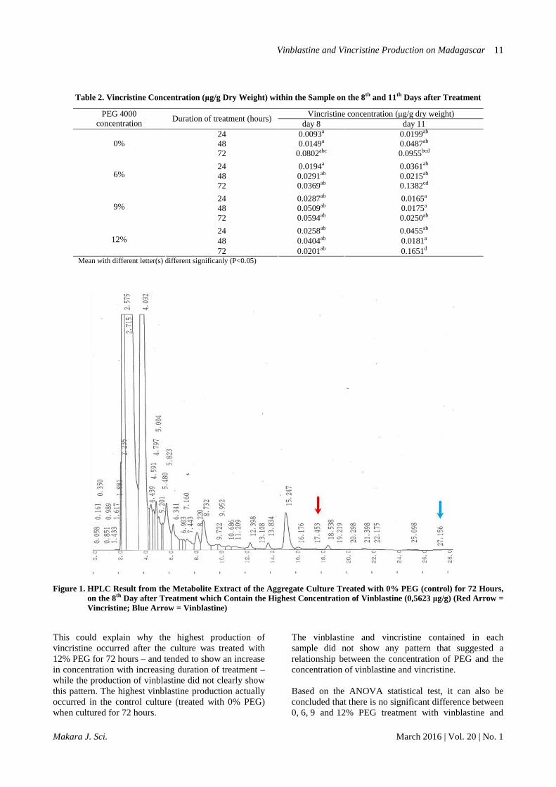
enzymes D4H and DAT, which is involved in the final stages of terpenoid indole alkaloid compounds production, are not expressed in cell cultures of C. roseus. Naturally, these two genes are only expressed in the idioblast and laticifer. The studies showed that the idioblast and laticifer may not form on the C. roseus cell suspension culture – or might form, but in a very small quantity. This might explain why vindolin compounds cannot be produced in cell cultures. Thus, in this study, callus cultures were used to induce idioblast and laticifer development as the only site for vindolin, a major precursor of vinblastine and vincristine biosynthesis. The decision of a cell to proliferate, elongate, and differentiate is influenced by the interaction and communication between cells, either through long-distance communication or across short distances [18, 19]. Adjacent cells can transport signals more efficiently through simple mechanisms [20]. Callus, which consists of a clump of cells, allows that phenomenon to happen. In addition to the process of differentiation, the ability of cell-to-cell coordination is needed to deal with changing environmental conditions [19]. Thus, aggregate cultures will be more resistant than the cell cultures when treated by stress. The aggregate culture that used was ± 0.5 cm in size. The culture shape was compact and greenish. The characteristics of aggregate culture were not morphologically different from the characteristics of callus culture. Effect of PEG concentration and duration of treatment on the production of vinblastine and vincristine. Based on the HPLC analysis, the retention time of vinblastine in acetonitrile:Na2HPO4 eluent is ±27 minutes and vincristine, ±17 minutes (Figure 1). When the HPLC results of sample extracts were interpolated with the standard solution of vinblastine and vincristine, it was concluded that all of the cultures
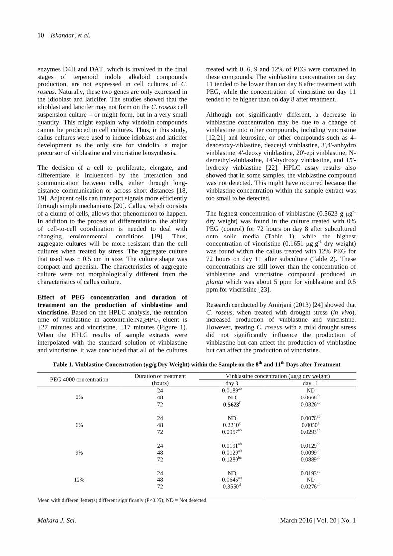
treated with 0, 6, 9 and 12% of PEG were contained in these compounds. The vinblastine concentration on day 11 tended to be lower than on day 8 after treatment with PEG, while the concentration of vincristine on day 11 tended to be higher than on day 8 after treatment. Although not significantly different, a decrease in vinblastine concentration may be due to a change of vinblastine into other compounds, including vincristine [12,21] and leurosine, or other compounds such as 4-deacetoxy-viblastine, deacetyl vinblastine, 3',4'-anhydro vinblastine, 4'-deoxy vinblastine, 20'-epi vinblastine, N-demethyl-vinblastine, 14'-hydroxy vinblastine, and 15'-hydroxy vinblastine [22]. HPLC assay results also showed that in some samples, the vinblastine compound was not detected. This might have occurred because the vinblastine concentration within the sample extract was too small to be detected. The highest concentration of vinblastine (0.5623 g µg-1 dry weight) was found in the culture treated with 0% PEG (control) for 72 hours on day 8 after subcultured onto solid media (Table 1), while the highest concentration of vincristine (0.1651 µg g-1 dry weight) was found within the callus treated with 12% PEG for 72 hours on day 11 after subculture (Table 2). These concentrations are still lower than the concentration of vinblastine and vincristine compound produced in planta which was about 5 ppm for vinblastine and 0.5 ppm for vincristine [23]. Research conducted by Amirjani (2013) [24] showed that C. roseus, when treated with drought stress (in vivo), increased production of vinblastine and vincristine. However, treating C. roseus with a mild drought stress did not significantly influence the production of vinblastine but can affect the production of vinblastine but can affect the production of vincristine.
Table 1. Vinblastine Concentration (µg/g Dry Weight) within the Sample on the 8th and 11th Days after Treatment
Vinblastine concentration (µg/g dry weight) PEG 4000 concentration
Duration of treatment (hours) day 8 day 11
24 0.0189ab ND 48 ND 0.0668ab 0% 72 0.5623f 0.0326ab
24 ND 0.0076ab 48 0.2210c 0.0050a 6% 72 0.0957ab 0.0293ab
24 0.0191ab 0.0129ab 48 0.0129ab 0.0099ab 9% 72 0.1280bc 0.0889ab
24 ND 0.0193ab 48 0.0645ab ND 12% 72 0.3550d 0.0276ab
Mean with different letter(s) different significanly (P<0.05); ND = Not detected
Vinblastine and Vincristine Production on Madagascar
Makara J. Sci. March 2016 | Vol. 20 | No. 1
11
Table 2. Vincristine Concentration (µg/g Dry Weight) within the Sample on the 8th and 11th Days after Treatment
Vincristine concentration (µg/g dry weight) PEG 4000 concentration
Duration of treatment (hours) day 8 day 11
24 0.0093a 0.0199ab 48 0.0149a 0.0487ab 0% 72 0.0802abc 0.0955bcd
24
0.0194a
0.0361ab 48 0.0291ab 0.0215ab 6% 72 0.0369ab 0.1382cd
24
0.0287ab
0.0165a 48 0.0509ab 0.0175a 9% 72 0.0594ab 0.0250ab
24
0.0258ab
0.0455ab 48 0.0404ab 0.0181a 12%
72 0.0201ab 0.1651d Mean with different letter(s) different significanly (P<0.05)
Figure 1. HPLC Result from the Metabolite Extract of the Aggregate Culture Treated with 0% PEG (control) for 72 Hours, on the 8th Day after Treatment which Contain the Highest Concentration of Vinblastine (0,5623 µg/g) (Red Arrow = Vincristine; Blue Arrow = Vinblastine)
This could explain why the highest production of vincristine occurred after the culture was treated with 12% PEG for 72 hours – and tended to show an increase in concentration with increasing duration of treatment – while the production of vinblastine did not clearly show this pattern. The highest vinblastine production actually occurred in the control culture (treated with 0% PEG) when cultured for 72 hours.
The vinblastine and vincristine contained in each sample did not show any pattern that suggested a relationship between the concentration of PEG and the concentration of vinblastine and vincristine. Based on the ANOVA statistical test, it can also be concluded that there is no significant difference between 0, 6, 9 and 12% PEG treatment with vinblastine and
Iskandar, et al.
Makara J. Sci. March 2016 | Vol. 20 | No. 1
12
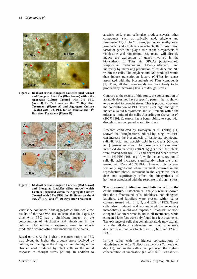
Figure 2. Idioblast or Non-elongated Laticifer (Red Arrow) and Elongated Laticifer (Blue Arrow) within the Aggregate Culture Treated with 0% PEG (control) for 72 Hours on the 8th Day after Treatment (Figure A) and Aggregate Culture Treated with 12% PEG for 72 Hours on the 11th Day after Treatment (Figure B)
Figure 3. Idioblast or Non-elongated Laticifer (Red Arrow)
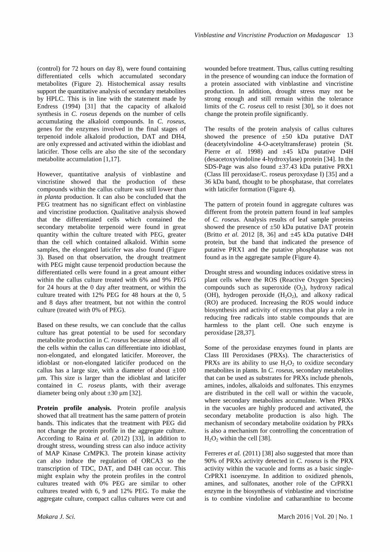
and Elongated Laticifer (Blue Arrow) which Contain Terpenoid within the Aggregate Culture Treated with 12% PEG for 48 Hours on the 0 (A), 5th (B,C) and 8th (D) Days after Treatment
vincristine contained in the aggregate culture, while the results of the ANOVA test indicate that the exposure time with PEG had a significant impact on the concentration of vinblastine and vincristine in the culture. The optimum exposure time to induce production of vinblastine and vincristine is 72 hours. Based on theory, the higher the concentration of PEG was given, the higher the drought stress received by culture; and the higher the drought stress, the higher the abscisic acid produced by plant cells as the initial response to drought stress [25-28]. In addition to
abscisic acid, plant cells also produce several other compounds, such as salicylic acid, ethylene and jasmonate [11,29]. In C. roseus, jasmonate, methyl ester jasmonate, and ethylene can activate the transcription factor of genes that play a role in the biosynthesis of vinblastine and vincristine. Jasmonate will directly induce the expression of genes involved in the biosynthesis of TIAs via ORCAs (Octadecanoid Responsive Catharanthus AP2/ERF-domain) and indirectly by increasing production of ethylene and NO within the cells. The ethylene and NO produced would then induce transcription factors (CrTFs) for genes associated with the biosynthesis of TIAs compounds [1]. Thus, alkaloid compounds are more likely to be produced by increasing levels of drought stress.
Contrary to the results of this study, the concentration of alkaloids does not have a specific pattern that is shown to be related to drought stress. This is probably because the concentration of PEG given is not high enough to induce alkaloid biosynthesis and still remain within the tolerance limits of the cells. According to Osman et al. (2007) [30], C. roseus has a better ability to cope with drought stress compared to salinity stress. Research conducted by Hamayun et al. (2010) [11] showed that drought stress induced by using 16% PEG can increase the biosynthesis of jasmonate compound, salicylic acid, and abscisic acid in soybeans (Glycine max) grown in vivo. The jasmonate concentration increased dramatically (204.9 ng g-1) when the plants were treated with 8% PEG and decreased when treated with 16% PEG (108 ng g-1 ), while the concentration of salicylic acid increased significantly when the plant treated with 8% and 16% PEG. However, this increase was only significant when treatment occurred in the reproductive phase. Treatment in the vegetative phase does not significantly affect the biosynthesis of hormones associated with the response to drought stress. The presence of idioblast and laticifer within the callus culture. Histochemical analysis results showed that the differentiated cells, idioblasts, non-elongated laticifers, and laticifers were present within callus cultures treated with 0, 6, 9, and 12% of PEG. Those cells also produced and accumulated the secondary metabolites alkaloid and terpenoid. Idioblasts or non-elongated laticifers were found in all treatments, while elongated laticifers were only found in a few treatments. The existence of cells that contain alkaloids may explain why the alkaloids vinblastine and vincristine were detected in all cultures treated with 0, 6, 9 and 12% of PEG. In the callus with the highest concentrations of vincristine (i.e. at 12 % PEG treatment for 72 hours on day 11), and in the callus that produced the highest concentration of vinblastine (i.e. at 0 % PEG treatment
Vinblastine and Vincristine Production on Madagascar
Makara J. Sci. March 2016 | Vol. 20 | No. 1
13
(control) for 72 hours on day 8), were found containing differentiated cells which accumulated secondary metabolites (Figure 2). Histochemical assay results support the quantitative analysis of secondary metabolites by HPLC. This is in line with the statement made by Endress (1994) [31] that the capacity of alkaloid synthesis in C. roseus depends on the number of cells accumulating the alkaloid compounds. In C. roseus, genes for the enzymes involved in the final stages of terpenoid indole alkaloid production, DAT and DH4, are only expressed and activated within the idioblast and laticifer. Those cells are also the site of the secondary metabolite accumulation [1,17]. However, quantitative analysis of vinblastine and vincristine showed that the production of these compounds within the callus culture was still lower than in planta production. It can also be concluded that the PEG treatment has no significant effect on vinblastine and vincristine production. Qualitative analysis showed that the differentiated cells which contained the secondary metabolite terpenoid were found in great quantity within the culture treated with PEG, greater than the cell which contained alkaloid. Within some samples, the elongated laticifer was also found (Figure 3). Based on that observation, the drought treatment with PEG might cause terpenoid production because the differentiated cells were found in a great amount either within the callus culture treated with 6% and 9% PEG for 24 hours at the 0 day after treatment, or within the culture treated with 12% PEG for 48 hours at the 0, 5 and 8 days after treatment, but not within the control culture (treated with 0% of PEG). Based on these results, we can conclude that the callus culture has great potential to be used for secondary metabolite production in C. roseus because almost all of the cells within the callus can differentiate into idioblast, non-elongated, and elongated laticifer. Moreover, the idioblast or non-elongated laticifer produced on the callus has a large size, with a diameter of about ±100 µm. This size is larger than the idioblast and laticifer contained in C. roseus plants, with their average diameter being only about ±30 µm [32].
Protein profile analysis. Protein profile analysis showed that all treatment has the same pattern of protein bands. This indicates that the treatment with PEG did not change the protein profile in the aggregate culture. According to Raina et al. (2012) [33], in addition to drought stress, wounding stress can also induce activity of MAP Kinase CrMPK3. The protein kinase activity can also induce the regulation of ORCA3 so the transcription of TDC, DAT, and D4H can occur. This might explain why the protein profiles in the control cultures treated with 0% PEG are similar to other cultures treated with 6, 9 and 12% PEG. To make the aggregate culture, compact callus cultures were cut and
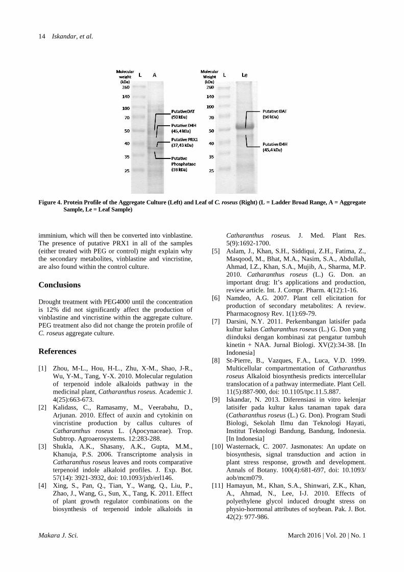
wounded before treatment. Thus, callus cutting resulting in the presence of wounding can induce the formation of a protein associated with vinblastine and vincristine production. In addition, drought stress may not be strong enough and still remain within the tolerance limits of the C. roseus cell to resist [30], so it does not change the protein profile significantly. The results of the protein analysis of callus cultures showed the presence of ±50 kDa putative DAT (deacetylvindoline 4-O-acetyltransferase) protein (St. Pierre et al. 1998) and ±45 kDa putative D4H (desacetoxyvindoline 4-hydroxylase) protein [34]. In the SDS-Page was also found ±37.43 kDa putative PRX1 (Class III peroxidase/C. roseus peroxydase I) [35] and a 36 kDa band, thought to be phosphatase, that correlates with laticifer formation (Figure 4). The pattern of protein found in aggregate cultures was different from the protein pattern found in leaf samples of C. roseus. Analysis results of leaf sample proteins showed the presence of ±50 kDa putative DAT protein (Britto et al. 2012 [8, 36] and ±45 kDa putative D4H protein, but the band that indicated the presence of putative PRX1 and the putative phosphatase was not found as in the aggregate sample (Figure 4). Drought stress and wounding induces oxidative stress in plant cells where the ROS (Reactive Oxygen Species) compounds such as superoxide (O2), hydroxy radical (OH), hydrogen peroxide (H2O2), and alkoxy radical (RO) are produced. Increasing the ROS would induce biosynthesis and activity of enzymes that play a role in reducing free radicals into stable compounds that are harmless to the plant cell. One such enzyme is peroxidase [28,37]. Some of the peroxidase enzymes found in plants are Class III Peroxidases (PRXs). The characteristics of PRXs are its ability to use H2O2 to oxidize secondary metabolites in plants. In C. roseus, secondary metabolites that can be used as substrates for PRXs include phenols, amines, indoles, alkaloids and sulfonates. This enzymes are distributed in the cell wall or within the vacuole, where secondary metabolites accumulate. When PRXs in the vacuoles are highly produced and activated, the secondary metabolite production is also high. The mechanism of secondary metabolite oxidation by PRXs is also a mechanism for controlling the concentration of H2O2 within the cell [38]. Ferreres et al. (2011) [38] also suggested that more than 90% of PRXs activity detected in C. roseus is the PRX activity within the vacuole and forms as a basic single-CrPRX1 isoenzyme. In addition to oxidized phenols, amines, and sulfonates, another role of the CrPRX1 enzyme in the biosynthesis of vinblastine and vincristine is to combine vindoline and catharanthine to become
Iskandar, et al.
Makara J. Sci. March 2016 | Vol. 20 | No. 1
14
Figure 4. Protein Profile of the Aggregate Culture (Left) and Leaf of C. roseus (Right) (L = Ladder Broad Range, A = Aggregate Sample, Le = Leaf Sample)
imminium, which will then be converted into vinblastine. The presence of putative PRX1 in all of the samples (either treated with PEG or control) might explain why the secondary metabolites, vinblastine and vincristine, are also found within the control culture. Conclusions Drought treatment with PEG4000 until the concentration is 12% did not significantly affect the production of vinblastine and vincristine within the aggregate culture. PEG treatment also did not change the protein profile of C. roseus aggregate culture. References [1] Zhou, M-L., Hou, H-L., Zhu, X-M., Shao, J-R.,
Wu, Y-M., Tang, Y-X. 2010. Molecular regulation of terpenoid indole alkaloids pathway in the medicinal plant, Catharanthus roseus. Academic J. 4(25):663-673.
[2] Kalidass, C., Ramasamy, M., Veerabahu, D., Arjunan. 2010. Effect of auxin and cytokinin on vincristine production by callus cultures of Catharanthus roseus L. (Apocynaceae). Trop. Subtrop. Agroaerosystems. 12:283-288.
[3] Shukla, A.K., Shasany, A.K., Gupta, M.M., Khanuja, P.S. 2006. Transcriptome analysis in Catharanthus roseus leaves and roots comparative terpenoid indole alkaloid profiles. J. Exp. Bot. 57(14): 3921-3932, doi: 10.1093/jxb/erl146.
[4] Xing, S., Pan, Q., Tian, Y., Wang, Q., Liu, P., Zhao, J., Wang, G., Sun, X., Tang, K. 2011. Effect of plant growth regulator combinations on the biosynthesis of terpenoid indole alkaloids in
Catharanthus roseus. J. Med. Plant Res. 5(9):1692-1700.
[5] Aslam, J., Khan, S.H., Siddiqui, Z.H., Fatima, Z., Masqood, M., Bhat, M.A., Nasim, S.A., Abdullah, Ahmad, I.Z., Khan, S.A., Mujib, A., Sharma, M.P. 2010. Catharanthus roseus (L.) G. Don. an important drug: It’s applications and production, review article. Int. J. Compr. Pharm. 4(12):1-16.
[6] Namdeo, A.G. 2007. Plant cell elicitation for production of secondary metabolites: A review. Pharmacognosy Rev. 1(1):69-79.
[7] Darsini, N.Y. 2011. Perkembangan latisifer pada kultur kalus Catharanthus roseus (L.) G. Don yang diinduksi dengan kombinasi zat pengatur tumbuh kinetin + NAA. Jurnal Biologi. XV(2):34-38. [In Indonesia]
[8] St-Pierre, B., Vazques, F.A., Luca, V.D. 1999. Multicellular compartmentation of Catharanthus roseus Alkaloid biosynthesis predicts intercellular translocation of a pathway intermediate. Plant Cell. 11(5):887-900, doi: 10.1105/tpc.11.5.887.
[9] Iskandar, N. 2013. Diferensiasi in vitro kelenjar latisifer pada kultur kalus tanaman tapak dara (Catharanthus roseus (L.) G. Don). Program Studi Biologi, Sekolah Ilmu dan Teknologi Hayati, Institut Teknologi Bandung, Bandung, Indonesia. [In Indonesia]
[10] Wasternack, C. 2007. Jasmonates: An update on biosynthesis, signal transduction and action in plant stress response, growth and development. Annals of Botany. 100(4):681-697, doi: 10.1093/ aob/mcm079.
[11] Hamayun, M., Khan, S.A., Shinwari, Z.K., Khan, A., Ahmad, N., Lee, I-J. 2010. Effects of polyethylene glycol induced drought stress on physio-hormonal attributes of soybean. Pak. J. Bot. 42(2): 977-986.
Vinblastine and Vincristine Production on Madagascar
Makara J. Sci. March 2016 | Vol. 20 | No. 1
15
[12] Tikhomiroff, C., Joliceur, M. 2002. Screening of Catharanthus roseus secondary metabolites by high-performance liquid chromatoghraphy. J. Chromatogr. 955(1):87-93, doi: 10.1016/S0021-9673(02)00204-2.
[13] Bhardwaj, J., Yadav, S.K. 2013. A common protein extraction protocol for proteomic analysis: Horse gram a case study. Am. J. Agric. Biol. Sci. 8(4): 293-310, doi: 10.3844/ajabssp.2013.293.301.
[14] Cilia, M., Yang, X., McLaughlin, M., Thannhauser, T.W., Gray, S. 2009. A comparison of protein extraction methods suitable for gel-based proteomic studies of aphid proteins. J. Biomol. Tech. 20(4):201-215, PMID: 19721822.
[15] Bradford, M.M. 1976. A rapid method for the quantitation of microgram quantities of protein utilizing the princilple of protein-dye binding. Anal. Biochem. 72(1-2):248-254, doi: 10.1016/ 0003-2697(76)90527-3.
[16] Khafagi, I.K. 2007. Generation of alkaloid-containing idioblast during cellular morphogenesis of Peganum harmala L. cell suspension cultures. Am. J. Plant Physiol. 2(1):17-26, doi: 10.3923/ ajpp.2007.17.26.
[17] Rischer, H., Oresic, M., Seppanen-Laakso, T., Katajamaa, M., Lammertyn, F., Ardiles-Diaz, W., Montagu, M.C.E., Inze, D., Oksman-Caldentey, K-M., Goossens, A. 2006. Gene-to-metabolite networks for terpenoid indole alkaloid biosynthesis in Catharanthus roseus cells. PNAS. 103(14): 5614-5619, doi: 10.1073/pnas.0601027103.
[18] Dornelas, M.C. 2003. Signal transduction, cell division, differentiation and development: Towards unifying mechanism for pattern formation in plants, Minireview. Braz. J. Plant Physiol. 15(1):1-8, http://dx.doi.org/10.1590/S1677-042020030001 00001.
[19] Van Norman, J.M., Breakfield, N.W., Benfey, P.N. 2011. Intercellular communication during plant development. The Plant Cell 23(3):855-864, doi: 10.1105/tpc.111.082982.
[20] Taiz, L., Zeiger, E. 2002. Plant physiology, 3rd ed. Sinauer Associates. USA. pp 283-307; 423-457; 493-515.
[21] O’Connor, S.E., Maresh, J.J. 2006. Chemistry and biology of monoterpenoid indole alkaloid biosynthesis, a review. National Product Report 23:532-547, doi: 10.1093/B512615K.
[22] Heijden, R., Jacobs, D.I., Snoeijer, W., Hallard, D., Verpoorte, R. 2004. The Catharanthus alkaloids: pharmacognosy and biotechnology. Curr. Med. Chem. 11(5):607-628, doi: 10.2174/0929867043 455846.
[23] Poutrain, P., Mazars, C., Thiersault, M., Rideau, M., Pichon. 2009. Two distinct intracellular Ca2+ -Release components act in opposite ways in the regulation of the Auxin-Dependent MIA
Biosynthesis in Catharanthus roseus Cells. J. Exp. Bot. 60(4):1387-1398, doi: 10.1093/jxb/erp017.
[24] Amirjani, M.R. 2013. Effects of drought stress on the alkaloid contents and growth parameters of Catharanthus roseus. ARPN J. Agric. Biol. Sci. 8(11):745-750.
[25] Farooq, M., Hussain, M., Wahid, A., Siddique, K.H.M. 2012. Plant Responses to Drought Stress, Chapter 1. Drought Stress in Plants: An Overview. Springer Verlag. Berlin. Heidelberg, doi: 10.1007/978-3-642-32653-0_1.
[26] Peleg, Z., Blumwald, E. 2011. Hormone balance and abiotic stress tolerance in crop plants. Curr. Opin. Plant. Biol. 14(3):290-295, doi: 10.1016/j. pbi.2011.02.001.
[27] Okamoto, M., Peterson, F.C., Defries, A., Park, S-Y., Endo, A., Nambara, E., Volkman, B.F., Cutler, S.R. 2013. Activation of dimeric ABA receptors elicits guard cell closure, ABA-regulated gene expression and drought tolerance. PNAS. 110(29):12132-12137, doi:10.1073/pnas.1305919 110/-/DCSupplemental.
[28] Xiong, L., Schumaker, K.S., Zhu, J-K. 2002. Cell signaling during cold, drought, and salt stress. The Plant Cell. 14(1):165-183, doi: 10.1105/tpc.000596.
[29] Daszkowska-Golec, A., Szarejko, I. 2013. Chapter 4. The Molecular Basis of ABA-Mediated Plant Response to Drought. InTech.
[30] Osman, M.E.H., Elfeky, S.S., El-Soud, K.A., Hasan, A.M. 2007. Response of Cathanranthus roseus shoot to salinity and drought in relation to vincristine alkaloid content. Asian J. Plant Sci. 6(8): 1223-1228, doi: 10.3923/ajps. 2007.1223.1228.
[31] Endress, R. 1994. Plant Cell Biotechnology. Springer-Verlag. Berlin. Heidelberg. pp. 173, 257.
[32] Hagel, J.M., Yeung, E.C., Facchini, P.J. 2008. Got milk? The secret life of laticifers, A review. Trends in Plant Sci. 13(12):631-639, doi: 10.1016/j.tplants. 2008.09.005.
[33] Raina, S.K., Wankhede, D.P., Jaggi, M., Singh, P., Jalmi, S.K., Raghuram, B., Sheikh, A.H., Sinha, A.K. 2012. CrMPK3 a mitogen activated protein kinase from Catharanthus roseus and its possible role in stress induced biosynthesis of monoter-penoid indole alkaloids. BMC Plant Biol. 12(134): 1-13, doi: 10.1186/1471-2229-12-134.
[34] Vaquez-Flota, F.A., De Luca, V. 1998. Developmental and light regulation of desacetoxyvindoline 4-hydroxylase in Catharanthus roseus (L.) G. Don. Plant Physiol. 117: 1351-1361, doi: 10.1104/pp.117.4.1351.
[35] Kumar, S., Dutta, A., Sinha, A.K., Sen, J. 2007. Cloning, characterization and locallization of a novel basic peroxidase gene from Catharanthus roseus. The FEBS Journal 274:1290-1303, doi: 10.1111/j.1742-4658.2007.05677.x.
[36] Britto, A.J., Kumar, P.B.J.R., Gracelin, D.H.S. 2012. Studies on protein profile of some
Iskandar, et al.
Makara J. Sci. March 2016 | Vol. 20 | No. 1
16
medicinally important species of Apocynaceae family using SDS-Page. J. Chem. Biol. Physic. Sci. 2(2): 792-796.
[37] Jaleel, C.D., Sankar, B., Murali, P.V., Gomathinayagam, M., Lakshmanan, G.M.A., Panneerselvam, R. 2007. Water deficit stress effects on reactive oxygen metabolism in Catharanthus roseus; Impacts on ajmalicine accumulation. Colloid Surf. B-Biointerfaces 62: 105-111, doi: 10.1016/j.colsurfb.2007.09.026.
[38] Ferreres, F., Figueiredo, R., Bettencourt, S., Carqueijeiro, I., Oliviera, J., Gil-Izquierdo, A., Pereira, D., Valentao, P., Andrade, P.B., Duarte, P., Barcelo, A.R., Sottomayor, M. 2011. Identification of phenolic compounds in isolated vacuoles of the medicinal plant Catharanthus roseus and their interaction with vacuolar class III peroxidase: an H202 affair? J. Exp. Biol. 62(8):2841-2854, doi: 10.1093/jxb/erq458.


