Embed Size (px)
Citation preview
SPINE Volume 34, Number 19, pp E682–E688©2009, Lippincott Williams & Wilkins
Vibration Modes of Injured Spine at ResonantFrequencies Under Vertical Vibration
Li-Xin Guo, PhD,*† Ming Zhang, PhD,† Yi-Min Zhang, PhD,* and Ee-Chon Teo, PhD‡
Study Design. A detailed three-dimensional finite el-ement model of the spine segment T12-Pelvis was de-veloped to investigate dynamic characteristics of wholelumbar spine with injured cases.
Objective. This study investigates the motion mecha-nism of the human lumbar spine and the effect of com-ponent injuries on adjacent spinal components underwhole body vibration.
Summary of Background Data. Several investigationshave analyzed the influence of injured spines on adjacentspinal components under static loadings. However, it isnot clear how the spine injury affects dynamic character-istics of whole lumbar spine and adjacent components ofthe injured segment under vibration.
Methods. The T12-Pelvis model was used to obtain themodal vibration modes of the spine at resonant frequen-cies. Injury conditions of the spine were simulated andtested, including denucleation and/or facetectomy withremoval of capsular ligaments.
Results. The results indicate the first-order vertical res-onant frequency of the intact model is 7.21 Hz. After thedenucleation at L4–L5, it decreases by more than 4%compared with the intact condition. All the injured condi-tions including disc injury and ligament injury decreasethe resonant frequency of the spine. Due to the denucle-ation at L4–L5 the anteroposterior displacements of thevertebrae from L2 to L5 decrease and the vertical dis-placements of the vertebrae from L1 to L4 increase undervibration. The denucleation also decreases the rotationaldeformations of the vertebrae from L1 to L5. The materialproperty sensitivity analysis shows intervertebral discshave a dominating effect on variation of vertical resonantfrequency of the spine.
Conclusion. The denucleation may decrease cush-ioning effects of adjacent motion segments at the in-jured level under vibration. The injured condition mayincrease the vertical displacement amplitudes of the spineabove the injured level. All the injured conditions maydecrease the resonant frequency of the spine system.
Key words: lumbar spine, finite element model, modalanalysis, injury, vibration. Spine 2009;34:E682–E688
Low back pain and degenerative diseases of the spineoccur more frequently among vehicle drivers and othervibrational mechanical operators.1–4 Long-term whole-body vibration (WBV) had been found to cause healthrisks for the lumbar spine, especially for the lower lum-bar motion segment L3–L5.5–7 Experimental studies haddemonstrated that the prolonged vibration of the humanbody might lead to muscle fatigue and reduce its adjust-ment function to the spine.8 The reduction of adjustmentcapability may lead to large deformation and high stressin some regions of the spine. In vitro experimental stud-ies have demonstrated that long-term exposure to vibra-tion decreases the proteoglycan content and eventuallyresults in disruption of matrix integrity and cause degen-eration of intervertebral discs.9,10 The experimentsfound that morphologic changes of intervertebral discsunder repetitive loading and implied that failure and de-generation or instability was strongly linked.11 How-ever, it is difficult to investigate quantitively the dynamicmechanical behavior under chronic WBV. Therefore, thefinite element (FE) models were used to investigate thedynamical characteristics of the human spine.12–14
Recently, with consideration of the effect of multiseg-ments on frequency characteristics of the spine, the FEmodels with much more spinal segments had been devel-oped. Kong and Goel15 built a detailed H–S1 (from headto S1) FE model of the human spine and conducted har-monic analyses to study the resonant frequencies of thewhole spine, in which the transmissibility was reportedat different spine vertebrae with respect to the sinusoidalvertical vibration. Guo and Teo16 used a FE model oflumbar spine and performed modal analyses to studyvibrational characteristics of the lumbar spine. In addi-tion, there have been a lot of FE studies delineating theinfluence of the injured or degenerated intervertebraldiscs on the biomechanical responses of the lumbarspine17–19 using short lumbar motion segments. Manyall-around FE models20–23 of the lumbar spine were usedto analyze the biomechanical responses of large compres-sion loading and instrument implant on the adjacent spi-nal components under static loading conditions. TheseFE models of lumbar spine have made possible for moreunderstanding in detail the static biomechanical charac-teristics of the lumbar spine.
In this study, we investigated how the injury condi-tions (including denucleation and facetectomy) of thespine affect the vibrational trend of whole lumbar spine,e.g., the motion relation of spine motion segments. At thesame time, this study is also aim to investigate the influ-ence of 1 level injury of the spine motion segment on its
From the *School of Mechanical Engineering and Automation, North-eastern University, China; †Department of Health Technology andInformatics, The Hong Kong Polytechnic University, Hong Kong,China; and ‡School of Mechanical and Aerospace Engineering,Nanyang Technological University, Singapore.Acknowledgment date: May 7, 2008. First revision date: December 15,2008. Second revision date: February 21, 2009. Acceptance date: Feb-ruary 23, 2009.The manuscript submitted does not contain information about medicaldevice(s)/drug(s).Other funds were received in support of this work. No benefits in anyform have been or will be received from a commercial party relateddirectly or indirectly to the subject of this manuscript.Address correspondence and reprint requests to Li-Xin GUO, PhD,School of Mechanical Engineering and Automation, Northeastern Uni-versity, Shenyang, 110004, China; E-mail: [email protected]
E682
adjacent spinal components under WBV. The hypothesisof this study is the spinal joint injury might change thedynamic characteristics of whole human lumbar spineand also give a bad influence on adjacent motion seg-ments of the injured spinal segment under WBV. Tocomplete these studies, a FE model of a whole lumbarspine including its adjacent motion segments was devel-oped and used to explore the dynamic characteristics ofthe human spine using FE modal analyses. The findingsin this study might help people to further understand themotion mechanism of the lumbar spine and appreciatethe effect of a component injury on its adjacent spinalcomponents under WBV.
Materials and Methods
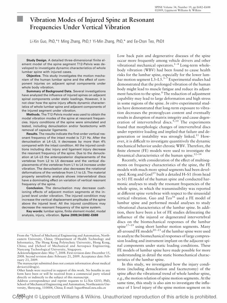
The human spine structure is complicated so many investiga-tions were carried out to construct three-dimensional (3D)models of the human spine based on different modeling meth-ods.13,24,25 In this study, a mechanical arm flexible digi-tizer14,26 was used to acquire the point-cloud data of the spinalvertebrae obtained from cadaveric vertebrae of an adult man(59 years old) with no physical abnormalities.
After obtaining the geometrical data of the vertebrae, thepoint data were input into ANSYS 10.0 (ANSYS, Inc.) to createlines, areas, and volumes. The 3D CAD model of a vertebramay consist of several volumes and the CAD model of an in-tervertebral disc includes the volumes of anulus fibrosus andnucleus pulpous. The configuration of the intervertebral discswas obtained from Gilad and Nissan.27 After finishing CADmodeling of the vertebrae and intervertebral discs, the CADmodels of vertebrae and intervertebral discs were formed into awhole CAD model of the T12-Pelvis spine segment. After pa-rameters of material property assigned, the T12-Pelvis CADmodel was meshed. In the end, the 3D FE model of the humanspinal motion segment T12-Pelvis (Figure 1) with a seatingposture was generated. The T12-Pelvis model mainly includes7 vertebral bodies (including the sacrum) and 6 intervertebraldiscs. Each spinal motion segment consists of cortical bone,cancellous bone, posterior bony element, anulus fibrosus, nu-cleus pulpous, anulus fibers, ligaments, and facet articulations.The vertebral body was modeled as cancellous core with theperiphery composed of cortical bone. The intervetebral discswere modeled with the nucleus pulpous encased in the anulusfibrosus and the anulus fibrosus was constructed as a matrix ofhomogenous ground substance reinforced by anulus fibers. Thevertebral body and the intervertebral disc except of anulus fi-bers and ligaments were assumed homogeneous and linear andwere modeled using isotropic brick elements. The anulus fibersand ligaments were simulated by 3D cable elements (tensilestress only). The modeling methods of intervertebral disc andthe material property of all the spinal components are mainlyobtained by reviewing the materials from the refer-ence13,24,28–31 as shown in Table 1. The anulus fibrosus wasassumed to consist of 3 consecutive laminar layers. In eachlayer, anulus fibers were oriented on average at �30° to theendplate. The total volume of anulus fibers was assumed as19% of the anulus volume.24,28 For more details about thedevelopment process for one motion segment including verte-bral body (including cortical bone, cancellous bone, and bonyposterior element) and intervertebral disc (including anulussubstance ground, nucleus pulpous, and anulus fiber) have
been reported elsewhere.14 The modeling process of every ver-tebra and intervertebral disc is similar, and in the end, the FEmodel of T12-Pelvis was finished.
The facet contact surface areas were developed and con-formed to their respectively actual geometrical curvatures. Sur-face-to-surface contact element types were applied to mimicsliding conditions for facet articulations. For the considerationof the small motion of sacroiliac joints,32–34 a cartilage-like softtissue material with embedded fibers was assumed to mimic thesoft tissue between the sacrum and the ilium including the car-tilage, the joint gap, and the synovia in the sacroiliac joint usingisotropic brick elements. In this study, the pelvis was assumedto fix on the ground, and the surfaces of pelvis near sacroiliacjoints were modeled and fixed in following analyses.
The mass of human upper body has an important role onextracting the resonant frequency and the vibration mode ofthe human spine. Many simulations to the spine have consid-ered the effect of the upper body weight.12,13,35 In this study, apoint mass of 40 kg was also fixed on the top vertebra of theT12-Pelvis FE model. The mass point was assigned to locate onthe top of T12 by 1.0 cm anterior to the L3–L4 vertebral cen-troid.36 In this study, 3 injury conditions of the spine at theL4–L5 segment were assumed, i.e., case 1 is to represent thecondition of denucleation at the L4–L5 intervertebral disc, case2 is the condition of removal of facet joints and their capsularligaments at the L4–L5 segment, and case 3 is the condition ofdenucleation and removal of facet articulations and their cap-sular ligaments at the L4–L5 segment.
After modeling, the FE modal analysis was conducted toanalyze the dynamic characteristics of the human spine. Thenthe resonant frequencies and the vibration modes were ob-tained for further analysis. In this study, only the first-ordervertical resonant frequency (FOVRF) and the first-order verti-
Figure 1. The FE model of the spine T12-Pelvis motion segment.
E683Vibration Modes of Injured Spine • Guo et al
cal vibration mode (FOVVM) of T12-Pelvis will be specificallydescribed for different injury conditions in view of the vibrationcharacteristics of the human body. For the consideration ofvalidation and comparison with other published results, addi-tional analyses were carried out on the specific segments of theT12-Pelvis model, such as T12–L1, L4–S1, L1–L5, etc.
In addition, to well-understand dynamic characteristics ofthe human spine, the material property sensitivity analysis ofthe spinal components was conducted in this study. For thecomparison convenience, the elastic modulus of each spinalcomponent (including endplate, vertebra, intervertebral disc,and ligament) is assumed to vary by �30% against the basicmodel in this study. After modal analysis, we can understandhow the variation of material property influences the resonantfrequency of the spine, and which component of the spine hasthe primary effect on the resonant frequency of the humanspine system.
Results
Resonant FrequenciesThe FOVRF of the intact T12-Pelvis model is 7.21 Hz.For the injured conditions, the FOVRFs of the T12-Pelvis model are 6.89 Hz, 7.17 Hz, and 6.83 Hz for case1, case 2, and case 3, respectively. The results indicate allthe injured conditions including the disc injury and theligament injury may decrease the resonant frequency ofwhole spine system. The FOVRF decreases by 5.3% ifthe spine suffers a denucleation and a facetectomy withcapsular ligament removal together. In addition, for val-idation consideration and well-understanding the dy-namic characteristics of the human spine, additionalanalyses were also conducted to several shorter segmentsof the T12-Pelvis model. For one motion segment, theFOVRF of T12–L1 is 25.7 Hz. For 2 motion segments,the FOVRF of L4–S1 was 16.4 Hz. The FOVRFs ofL1–L5 and L1–S1 are 11.5 Hz and 9.12 Hz, respectively.
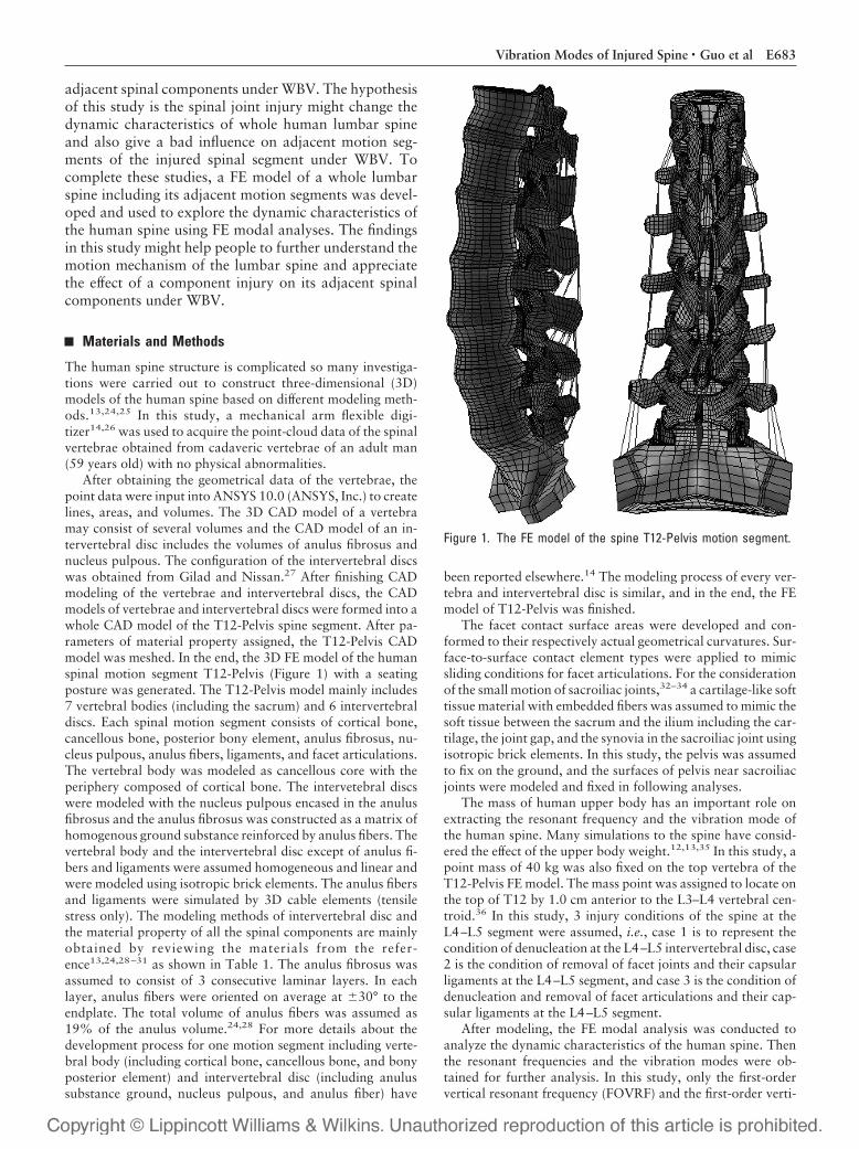
Vibration ModesFigure 2 shows the anteroposterior (AP) deformation(for the 4 model conditions) of all the vertebrae from T12to S1 for the FOVVM of the model. For the same ques-
tion, we also give a curve description of the spine defor-mation in the sagittal plane for the FOVVM in Figure 3and the marked points in the curves mean the vertebraeand the intervertebral discs. Figure 4 exhibits the defor-mations in the vertical direction. From Figures 2 and 4, itcan be seen that the human upper body mainly performsthe vertical vibration with a small motion in the A–Pdirection during WBV. Figure 5 indicates the rotationalangles, in the sagittal plane, of each vertebra for the FO-VVM. From the figures, it can be seen that the lumbarspine segments not only show vertical vibration but alsoexhibit A–P motions (including the sagittal plane trans-lation and flexion-extension rotation) during WBV. Fig-ure 6 shows the A–P displacements of anterior points ofeach disc anulus in the sagittal plane for the FOVVM. InFigure 5, the symbols D12–1, D1–2, D2–3, D3–4, D4–5, andD5–1 represent the disc between of every 2 vertebrae, e.g.,D1–2 means the disc between of L1 and L2.
From the above simulations, it can also be seen thatthe A–P deformations of the vertebrae L3 and L4 and theintervertebral disc between them are maximal (Figures 6,2). In addition, it can be found that although the facet-ectomy with removal of capsular ligaments has slightlyinfluence on the lumbar spine, the denucleation makes anobvious influence (variations of vibration amplitudes) onthe adjacent discs of the injured spinal motion segment.
Table 1. Material Properties of Spinal Components in the FE Model
ComponentsElementTypes
Young’sModulus (MPa)
Poisson’sRatio
Cross–SectionalArea (mm2)
Density(kg/mm3) References
Cortical bone 8-node 1200 0.30 7 � 10�6 13, 24, 28, 29, 30, 31Cancellous bone Solid 100 0.2 1.1 � 10�6
Bony posterior element 3500 0.25 1.4 � 10�6
Annulus 4.2 0.45 1.05 � 10�6
Endplate 500 0.25 1.2 � 10�6
Nucleus pulpous 1.0 0.49 1.02 � 10�6
Annulus fiber 3D-Cable 500 1.0 � 10�6 7, 13Capsular ligaments 7.5 30 1.0 � 10�6
Intertransverse ligaments 10 1.8 1.0 � 10�6
Supraspinous ligaments 8 30 1.0 � 10�6
Interspinous ligaments 10 40 1.0 � 10�6
Ligamentum flavum 15 40 1.0 � 10�6
Anterior longitudinal ligaments 7.8 63.7 1.0 � 10�6
Posterior longitudinal ligaments 10 20 1.0 � 10�6
Iliolumbar ligaments 10 26.4 1.0 � 10�6
Anterior sacroiliac ligaments 20 160 1.0 � 10�6
Posterior sacroiliac ligaments 20 300 1.0 � 10�6
Figure 2. Deformation of vertebrae in the A–P direction for theFOVVM.
E684 Spine • Volume 34 • Number 19 • 2009
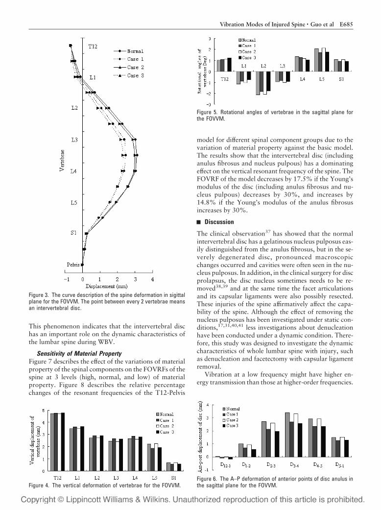
This phenomenon indicates that the intervertebral dischas an important role on the dynamic characteristics ofthe lumbar spine during WBV.
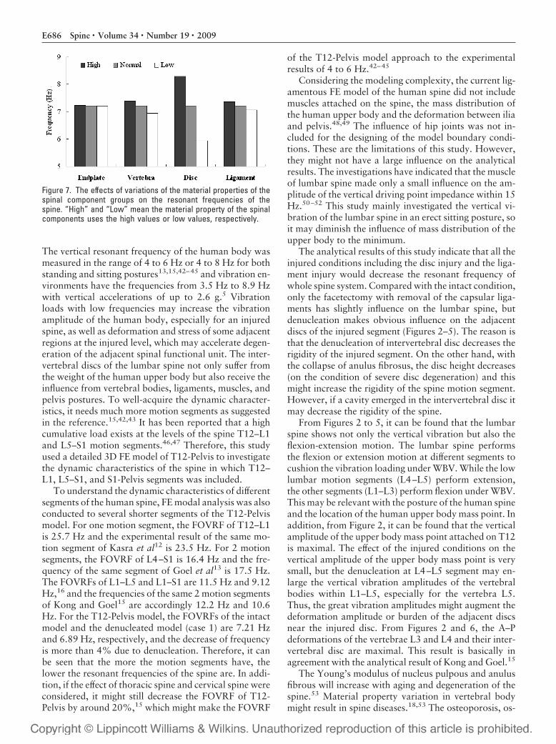
Sensitivity of Material PropertyFigure 7 describes the effect of the variations of materialproperty of the spinal components on the FOVRFs of thespine at 3 levels (high, normal, and low) of materialproperty. Figure 8 describes the relative percentagechanges of the resonant frequencies of the T12-Pelvis
model for different spinal component groups due to thevariation of material property against the basic model.The results show that the intervertebral disc (includinganulus fibrosus and nucleus pulpous) has a dominatingeffect on the vertical resonant frequency of the spine. TheFOVRF of the model decreases by 17.5% if the Young’smodulus of the disc (including anulus fibrosus and nu-cleus pulpous) decreases by 30%, and increases by14.8% if the Young’s modulus of the anulus fibrosusincreases by 30%.
Discussion
The clinical observation37 has showed that the normalintervertebral disc has a gelatinous nucleus pulposus eas-ily distinguished from the anulus fibrosus, but in the se-verely degenerated disc, pronounced macroscopicchanges occurred and cavities were often seen in the nu-cleus pulposus. In addition, in the clinical surgery for discprolapsus, the disc nucleus sometimes needs to be re-moved38,39 and at the same time the facet articulationsand its capsular ligaments were also possibly resected.These injuries of the spine affirmatively affect the capa-bility of the spine. Although the effect of removing thenucleus pulposus has been investigated under static con-ditions,17,31,40,41 less investigations about denucleationhave been conducted under a dynamic condition. There-fore, this study was designed to investigate the dynamiccharacteristics of whole lumbar spine with injury, suchas denucleation and facetectomy with capsular ligamentremoval.
Vibration at a low frequency might have higher en-ergy transmission than those at higher-order frequencies.
Figure 3. The curve description of the spine deformation in sigittalplane for the FOVVM. The point between every 2 vertebrae meansan intervertebral disc.
Figure 4. The vertical deformation of vertebrae for the FOVVM.
Figure 5. Rotational angles of vertebrae in the sagittal plane forthe FOVVM.
Figure 6. The A–P deformation of anterior points of disc anulus inthe sagittal plane for the FOVVM.
E685Vibration Modes of Injured Spine • Guo et al
The vertical resonant frequency of the human body wasmeasured in the range of 4 to 6 Hz or 4 to 8 Hz for bothstanding and sitting postures13,15,42–45 and vibration en-vironments have the frequencies from 3.5 Hz to 8.9 Hzwith vertical accelerations of up to 2.6 g.5 Vibrationloads with low frequencies may increase the vibrationamplitude of the human body, especially for an injuredspine, as well as deformation and stress of some adjacentregions at the injured level, which may accelerate degen-eration of the adjacent spinal functional unit. The inter-vertebral discs of the lumbar spine not only suffer fromthe weight of the human upper body but also receive theinfluence from vertebral bodies, ligaments, muscles, andpelvis postures. To well-acquire the dynamic character-istics, it needs much more motion segments as suggestedin the reference.15,42,43 It has been reported that a highcumulative load exists at the levels of the spine T12–L1and L5–S1 motion segments.46,47 Therefore, this studyused a detailed 3D FE model of T12-Pelvis to investigatethe dynamic characteristics of the spine in which T12–L1, L5–S1, and S1-Pelvis segments was included.
To understand the dynamic characteristics of differentsegments of the human spine, FE modal analysis was alsoconducted to several shorter segments of the T12-Pelvismodel. For one motion segment, the FOVRF of T12–L1is 25.7 Hz and the experimental result of the same mo-tion segment of Kasra et al12 is 23.5 Hz. For 2 motionsegments, the FOVRF of L4–S1 is 16.4 Hz and the fre-quency of the same segment of Goel et al13 is 17.5 Hz.The FOVRFs of L1–L5 and L1–S1 are 11.5 Hz and 9.12Hz,16 and the frequencies of the same 2 motion segmentsof Kong and Goel15 are accordingly 12.2 Hz and 10.6Hz. For the T12-Pelvis model, the FOVRFs of the intactmodel and the denucleated model (case 1) are 7.21 Hzand 6.89 Hz, respectively, and the decrease of frequencyis more than 4% due to denucleation. Therefore, it canbe seen that the more the motion segments have, thelower the resonant frequencies of the spine are. In addi-tion, if the effect of thoracic spine and cervical spine wereconsidered, it might still decrease the FOVRF of T12-Pelvis by around 20%,15 which might make the FOVRF
of the T12-Pelvis model approach to the experimentalresults of 4 to 6 Hz.42–45
Considering the modeling complexity, the current lig-amentous FE model of the human spine did not includemuscles attached on the spine, the mass distribution ofthe human upper body and the deformation between iliaand pelvis.48,49 The influence of hip joints was not in-cluded for the designing of the model boundary condi-tions. These are the limitations of this study. However,they might not have a large influence on the analyticalresults. The investigations have indicated that the muscleof lumbar spine made only a small influence on the am-plitude of the vertical driving point impedance within 15Hz.50–52 This study mainly investigated the vertical vi-bration of the lumbar spine in an erect sitting posture, soit may diminish the influence of mass distribution of theupper body to the minimum.
The analytical results of this study indicate that all theinjured conditions including the disc injury and the liga-ment injury would decrease the resonant frequency ofwhole spine system. Compared with the intact condition,only the facetectomy with removal of the capsular liga-ments has slightly influence on the lumbar spine, butdenucleation makes obvious influence on the adjacentdiscs of the injured segment (Figures 2–5). The reason isthat the denucleation of intervertebral disc decreases therigidity of the injured segment. On the other hand, withthe collapse of anulus fibrosus, the disc height decreases(on the condition of severe disc degeneration) and thismight increase the rigidity of the spine motion segment.However, if a cavity emerged in the intervertebral disc itmay decrease the rigidity of the spine.
From Figures 2 to 5, it can be found that the lumbarspine shows not only the vertical vibration but also theflexion-extension motion. The lumbar spine performsthe flexion or extension motion at different segments tocushion the vibration loading under WBV. While the lowlumbar motion segments (L4–L5) perform extension,the other segments (L1–L3) perform flexion under WBV.This may be relevant with the posture of the human spineand the location of the human upper body mass point. Inaddition, from Figure 2, it can be found that the verticalamplitude of the upper body mass point attached on T12is maximal. The effect of the injured conditions on thevertical amplitude of the upper body mass point is verysmall, but the denucleation at L4–L5 segment may en-large the vertical vibration amplitudes of the vertebralbodies within L1–L5, especially for the vertebra L5.Thus, the great vibration amplitudes might augment thedeformation amplitude or burden of the adjacent discsnear the injured disc. From Figures 2 and 6, the A–Pdeformations of the vertebrae L3 and L4 and their inter-vertebral disc are maximal. This result is basically inagreement with the analytical result of Kong and Goel.15
The Young’s modulus of nucleus pulpous and anulusfibrous will increase with aging and degeneration of thespine.53 Material property variation in vertebral bodymight result in spine diseases.18,53 The osteoporosis, os-
Figure 7. The effects of variations of the material properties of thespinal component groups on the resonant frequencies of thespine. “High” and “Low” mean the material property of the spinalcomponents uses the high values or low values, respectively.
E686 Spine • Volume 34 • Number 19 • 2009
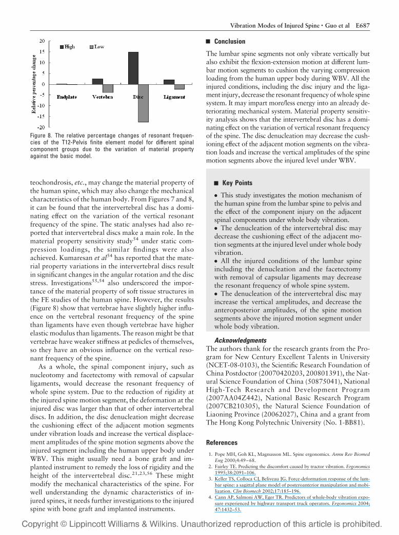
teochondrosis, etc., may change the material property ofthe human spine, which may also change the mechanicalcharacteristics of the human body. From Figures 7 and 8,it can be found that the intervertebral disc has a domi-nating effect on the variation of the vertical resonantfrequency of the spine. The static analyses had also re-ported that intervertebral discs make a main role. In thematerial property sensitivity study54 under static com-pression loadings, the similar findings were alsoachieved. Kumaresan et al54 has reported that the mate-rial property variations in the intervertebral discs resultin significant changes in the angular rotation and the discstress. Investigations55,54 also underscored the impor-tance of the material property of soft tissue structures inthe FE studies of the human spine. However, the results(Figure 8) show that vertebrae have slightly higher influ-ence on the vertebral resonant frequency of the spinethan ligaments have even though vertebrae have higherelastic modulus than ligaments. The reason might be thatvertebrae have weaker stiffness at pedicles of themselves,so they have an obvious influence on the vertical reso-nant frequency of the spine.
As a whole, the spinal component injury, such asnucleotomy and facetectomy with removal of capsularligaments, would decrease the resonant frequency ofwhole spine system. Due to the reduction of rigidity atthe injured spine motion segment, the deformation at theinjured disc was larger than that of other intervertebraldiscs. In addition, the disc denucleation might decreasethe cushioning effect of the adjacent motion segmentsunder vibration loads and increase the vertical displace-ment amplitudes of the spine motion segments above theinjured segment including the human upper body underWBV. This might usually need a bone graft and im-planted instrument to remedy the loss of rigidity and theheight of the intervertebral disc.21,23,56 These mightmodify the mechanical characteristics of the spine. Forwell understanding the dynamic characteristics of in-jured spines, it needs further investigations to the injuredspine with bone graft and implanted instruments.
Conclusion
The lumbar spine segments not only vibrate vertically butalso exhibit the flexion-extension motion at different lum-bar motion segments to cushion the varying compressionloading from the human upper body during WBV. All theinjured conditions, including the disc injury and the liga-ment injury, decrease the resonant frequency of whole spinesystem. It may impart more/less energy into an already de-teriorating mechanical system. Material property sensitiv-ity analysis shows that the intervertebral disc has a domi-nating effect on the variation of vertical resonant frequencyof the spine. The disc denucleation may decrease the cush-ioning effect of the adjacent motion segments on the vibra-tion loads and increase the vertical amplitudes of the spinemotion segments above the injured level under WBV.
Key Points
● This study investigates the motion mechanism ofthe human spine from the lumbar spine to pelvis andthe effect of the component injury on the adjacentspinal components under whole body vibration.● The denucleation of the intervertebral disc maydecrease the cushioning effect of the adjacent mo-tion segments at the injured level under whole bodyvibration.● All the injured conditions of the lumbar spineincluding the denucleation and the facetectomywith removal of capsular ligaments may decreasethe resonant frequency of whole spine system.● The denucleation of the intervertebral disc mayincrease the vertical amplitudes, and decrease theanteroposterior amplitudes, of the spine motionsegments above the injured motion segment underwhole body vibration.
AcknowledgmentsThe authors thank for the research grants from the Pro-gram for New Century Excellent Talents in University(NCET-08-0103), the Scientific Research Foundation ofChina Postdoctor (20070420203, 200801391), the Nat-ural Science Foundation of China (50875041), NationalHigh-Tech Research and Development Program(2007AA04Z442), National Basic Research Program(2007CB210305), the Natural Science Foundation ofLiaoning Province (20062027), China and a grant fromThe Hong Kong Polytechnic University (No. 1-BB81).
References
1. Pope MH, Goh KL, Magnusson ML. Spine ergonomics. Annu Rev BiomedEng 2000;4:49–68.
2. Fairley TE. Predicting the discomfort caused by tractor vibration. Ergonomics1995;38:2091–106.
3. Keller TS, Colloca CJ, Beliveau JG. Force-deformation response of the lum-bar spine: a sagittal plane model of posteroanterior manipulation and mobi-lization. Clin Biomech 2002;17:185–196.
4. Cann AP, Salmoni AW, Eger TR. Predictors of whole-body vibration expo-sure experienced by highway transport truck operators. Ergonomics 2004;47:1432–53.
Figure 8. The relative percentage changes of resonant frequen-cies of the T12-Pelvis finite element model for different spinalcomponent groups due to the variation of material propertyagainst the basic model.
E687Vibration Modes of Injured Spine • Guo et al
5. Frymoyer JW, Pope MH, Costanza MC, et al. Epidemiologic studies oflow-back pain. Spine 1980;5:419–23.
6. Barry M, Livesley P. Facet joint hypertrophy: the cross-sectional area of thesuperior articular process of L4 and L5. Eur Spine J 1997;6:121–4.
7. Pankoke S, Hofmann J, Wolfel HP. Determination of vibration-related spi-nal loads by numerical simulation. Clin Biomech 2001;16:S45–56.
8. Broman H, Pope MH, Benda M. The impact response of the seated subject.J Orthop Res 1991;9:150–4.
9. Ishihara H, Tsuji H, Hirano N, et al. Effects of continuous quantitativevibration on rheologic and biological behaviors of the intervertebral disc.Spine 1992;17:S7–12.
10. Benneker LM, Heini PF, Anderson SE, et al. Correlation of radiographic andMRI parameters to morphological and biochemical assessment of interver-tebral disc degeneration. Eur Spine J 2005;14:27–35.
11. Yu CY, Tsai KH, Hu WP, et al. Geometric and morphological changes of theintervertebral disc under fatigue testing. Clin Biomech 2003;18:S3–9.
12. Kasra M, Shirazi-Adl A, Drouin G. Dynamics of human lumbar interverte-bral joints: experimental and finite-element investigations. Spine 1992;17:93–102.
13. Goel VK, Park H, Kong W. Investigation of vibration characteristics of theligamentous lumbar spine using the finite element approach. J Biomech Eng1994;116:377–83.
14. Guo LX, Teo EC, Lee KK, et al. Vibration characteristics of human spineunder axial cyclic loads: effect of frequency and damping. Spine 2005;30:631–7.
15. Kong WZ, Goel VK. Ability of the finite element models to predict responseof the human spine to sinusoidal vertical vibration. Spine 2003;28:1961–7.
16. Guo LX, Teo EC. Prediction of the modal characteristics of the human spineat resonant frequency using finite element models. Proc Inst Mech Eng H.2005;219:277–84.
17. Kim YE, Goel VK, Weinstein JN, et al. Effect of disc degeneration at one levelon the adjacent level in axial mode. Spine 1991;16:331–5.
18. Polikeit A, Nolte LP, Ferguson SJ. Simulated influence of osteoporosis anddisc degeneration on the load transfer in a lumbar functional spinal unit.J Biomech 2004;37:1061–9.
19. Guo LX, Teo EC. Influence prediction of injury and vibration on adjacentcomponents of spine using finite element methods. J Spinal Disord Tech2006;19:118–24.
20. Shirazi-Adl A, Parnianpour M. Load-bearing and stress analysis of the hu-man spine under a novel wrapping compression loading. Clin Biomech 2000;15:718–25.
21. Chen CS, Cheng CK, Liu CL, et al. Stress analysis of the disc adjacent tointerbody fusion in lumbar spine. Med Eng Phys 2001;23:483–91.
22. Goel VK, Grauer JN, Patel TCh, et al. Effects of charite artificial disc on theimplanted and adjacent spinal segments mechanics using a hybrid testingprotocol. Spine 2005;30:2755–64.
23. Rohlmann A, Zander T, Bergmann G. Comparison of the biomechanicaleffects of posterior and anterior spine-stabilizing implants. Eur Spine J 2005;14:445–453.
24. Shirazi-Adl A, Drouin G. Load-bearing role of facets in a lumbar segmentunder sagittal plane loadings. J Biomech 1987;20:601–13.
25. Wang ZL, Teo JC, Chui CK, et al. Computational biomechanical modellingof the lumbar spine using marching-cubes surface smoothened finite elementvoxel meshing. Comput Methods Programs Biomed 2005;80:25–35.
26. Teo EC, Lee KK, Qiu TX, et al. The biomechanics of lumbar graded facet-ectomy under anterior-shear load. IEEE Trans Biomed Eng 2004;51:443–9.
27. Gilad I, Nissan M. A study of vertbra and disc geometric relations of thehuman cervical and lumbar spine. Spine 1985;11:154–7.
28. Shirazi-Adl SA, Shrivastava SC, Ahmed AM. Stress analysis of the lumbardisc-body unit in compression: a three-dimensional nonlinear finite elementstudy. Spine 1984;9:120–34.
29. Goel VK, Monroe BT, Gilbertson LG, et al. Interlaminar shear stresses andlaminae separation in a disc. Spine 1995;20:689–98.
30. Sharma M, Langrana NA, Rodriguez J. Role of ligaments and facets inlumbar spinal stability. Spine 1995;20:887–900.
31. Kumaresan S, Yoganandan N, Pintar FA, et al. Contribution of disc degen-eration to osteophyte formation in the cervical spine: a biomechanical inves-tigation. J Orthop Res 2001;19:977–84.
32. Skalak R, Shu CE. Handbook of Bioengineering. New York, NY: McGraw-Hill Book Company; 1987.
33. Sturesson B, Uden A, Vleeming A. A radiostereometric analysis of the move-ments of the sacroiliac joints in the reciprocal straddle position. Spine 2000;25:214–7.
34. Sturesson B, Uden A, Vleeming A. A radiostereometric analysis of move-ments of the sacroiliac joints during the standing hip flexion test. Spine2000;25:364–8.
35. Rohlmann A, Zander T, Bergmann G. Spinal loads after osteoporotic verte-bral fractures treated by vertebroplasty or kyphoplasty. Eur Spine J 2006;15:1255–64.
36. Shirazi-Adl A, Parnianpour M. Role of posture in mechanics of the lumbarspine in compression. J Spinal Disord 1996;9:277–86.
37. Shirado O, Kaneda K, Tadano S, et al. Influence of disc degeneration onmechanism of thoracolumbar burst fractures. Spine 1992;17:286–92.
38. Frei H, Oxland TR, Rathonyi GC, et al. The effect of nucleotomy on lumbarspine mechanics in compression and shear loading. Spine 2001;26:2080–9.
39. Putzier M, Schneider SV, Funk JF, et al. The surgical treatment of the lumbardisc prolapse: nucleotomy with additional transpedicular dynamic stabiliza-tion versus nucleotomy alone. Spine 2005;30:1109–14.
40. Meakin JR, Hukins DW. Effect of removing the nucleus pulposus on thedeformation of the annulus fibrosus during compression of the intervertebraldisc. J Biomech 2000;33:575–80.
41. Meakin JR, Redpath TW, Hukins DW. The effect of partial removal of thenucleus pulposus from the intervertebral disc on the response of the humanannulus fibrosus to compression. Clin Biomech (Bristorl, Avon) 2001;16:121–8.
42. Sandover J, Dupuis H. A reanalysis of spinal motion during vibration.Ergonomics 1987;30:975– 85.
43. Sandover J. Behavior of the spine under shock and vibration: a review. ClinBiomech 1988;3:249–56.
44. Panjabi MM, Andersson GB, Jorneus L, et al. In vivo measurements of spinalcolumn vibrations. J Bone Joint Surg Am 1986;68:695–702.
45. Pope MH, Wilder DG, Jorneus L, et al. The response of the seated human tosinusoidal vibration and impact. J Biomech Eng 1987;109:279–84.
46. Kumar S. Theories of musculoskeletal injury causation. Ergonomics 2001;44:17–47.
47. Cheng CK, Chen HH, Kuo HH, et al. A three-dimensional mathematicalmodel for predicting spinal joint force distribution during manual liftings.Clin Biomech 1998;13:S59–64.
48. Vukicevic S, Marusic A, Stavljenic A, et al. Holographic analysis of thehuman pelvis. Spine 1991;16:209–14.
49. Vleeming A, Van Wingerden JP, Dijkstra PF, et al. Mobility in the sacroiliacjoints in the elderly: a kinematic and radiological study. Clin Biomech 1992;7:170–6.
50. Buck B, Woelfel HP. Dynamic three-dimensional finite element model of asitting man with a detailed representation of the lumbar spine and muscle. In:Middletion J, Jones ML, Pande GN, eds. Computer Methods in Biomechan-ics and Biomedical Engineering-2. Amsterdam, The Netherland: Gordonand Breachs; 1998:379–86.
51. Guo LX, Zhang M, Teo EC. Influences of denucleation on contact force offacet joints under whole body vibration. Ergonomics 2007;50:967–78.
52. Guo LX, Zhang M, Wang ZW, et al. Influence of anteroposterior shifting oftrunk mass centroid on vibrational configuration of human spine. ComputBiol Med 2008;38:146–51.
53. Keller TS, Holm SH, Hansson TH, et al. The dependence of intervertebraldisc mechanical properties on physiologic conditions. Spine 1990;15:751– 61.
54. Kumaresan S, Yoganandan N, Pintar FA. Finite element analysis of thecervical spine: a material property sensitivity study. Clin Biomech 1999;14:41–53.
55. Rao AA, Dumas GA. Influence of material properties on the mechanicalbehaviour of the L5–S1 intervertebral disc in compression: a nonlinear finiteelement study. J Biomech Eng 1991;13:139–51.
56. Goel VK, Kim YE, Lim TH, et al. An analytical investigation of the mechanics ofspinal instrumentation. Spine 1988;13:1003–11.
E688 Spine • Volume 34 • Number 19 • 2009


