Embed Size (px)
Citation preview
ELSEVIER Antiviral Research 27 (1995) 375-388
Q Antiviral Research
Vertebrate brains contain a broadly active antiviral substance
Indra P. Singh *, Ashok K. Chopra, Dorian H. Coppenhaver, Edna Smith, Joyce Poast, Samuel Baron
Department of Microbiology and lmmunology, University of Texas Medical Branch, Galveston, TX 77555-1019, USA
Received 27 September 1994; accepted 22 February 1995
Abstract
Brain tissue extracts from vertebrates were examined for non-specific, broad-spectrum virus inhibitors, previously identified and characterized from other body tissues and fluids. An antiviral activity found in human, bovine, ovine, porcine, lapine, murine and piscine brain tissues shares some properties with a contact blocking-virus inhibitor, which was previously found only in cell culture supernatants. The inhibitor was active against (in order of sensitivity to inhibitor) Banzi, Sindbis, Bunyamwera, Newcastle disease, herpes simplex I, Semliki forest, polio I, mengo, vaccinia and vesicular stomatitis viruses. It is approximately 4000 kDa and possesses a complex structure containing protein, carbohydrate and lipid moieties. The inhibitor does not directly neutralize virus or induce an antiviral state in cells, but appears to act early in the replication cycle, most likely by preventing virus attachment to target cells. Its occurrence in concentrations sufficient to reduce virus yield in cell cultures at least 30-fold may indicate a role in limiting viral infections of the central nervous system.
Keywords: Virus inhibitor; Host defense; Antiviral agent; Sindbis virus
1. Introduction
The b l o o d - b r a i n barrier excludes from the central nervous system (CNS) most of the b o d y ' s host defenses, inc luding systemic antibodies, i m m u n e cells and cytokines (Baron,
* Corresponding author. Fax: + 1 (409) 772 5065.
0166-3542/95/$09.50 © 1995 Elsevier Science B.V. All rights reserved SSDI 0166-3542(95)00021-6
376 LP. Singh et al. /Antiviral Research 27 (1995) 375-388
1963; Cathala and Baron, 1970; Griffin, 1991). Non-specific antiviral molecules within the brain may be candidate defenses against CNS infections, and thus we examined brain tissue extracts for their ability to inhibit virus infections.
In this paper, we describe isolation and characterization of a broadly active antiviral agent from brain tissue extracts of several vertebrate species, which shares characteris- tics with the high molecular weight inhibitor (CVI) previously identified only in cell culture supernatants (Baron and McKerlie, 1981; Hughes et al., 1981; Sullivan et al., 1987).
2. Materials and methods
2.1. Virus inhibitor preparation
Brain tissue containing both gray and white matter of different vertebrates (human, bovine, porcine, ovine, lapine, murine and piscine) was mixed with twice the volume of Hank's balanced salt solution (HBSS) and homogenized. The homogenate was cen- trifuged at 10,000 rpm for 15 rain, and the supernatant was extensively dialyzed, first against 10 mM phosphate buffer, pH 7.0, and then against Eagle's minimum essential medium (EMEM). The dialyzed homogenate was centrifuged, the supernatant filtered through a 0.22-/xm filter, aliquoted in 5-ml amounts, and frozen at -20°C until use.
2.2. Antiviral assay
Antiviral activity was titered by a previously described plaque reduction assay (Baron and McKerlie, 1981). We used a poxvirus, vaccinia virus (VV) strain IHDE; a herpes virus, herpes simplex type I (HSV-I) strain Heitzman; a paramyxovirus, Newcastle disease virus (NDV) strain B-I; enteroviruses, poliomyelitis virus type I (PV) strain Mahoney and mengo virus (MV); alphaviruses, Semliki Forest virus (SFV) strain original, and Sindbis virus (SBV) strain EgAr 339; a flavivirus, Banzi virus (BZV) strain SAH 336; a bunyavirus, Bunyamwera virus (BWV) strain original; and a rhabdovirus, vesicular stomatitis virus (VSV) strain Indiana. The cells used to cultivate these viruses were Vero (African green monkey kidney cell, ATCC CRL 1587) for SBV, SFV, BZV, BWV and VSV; HEp-2 (human larynx epidermoid carcinoma cell, ATCC CCL 23) for PV; RS (rabbit skin cell, provided by Dr. Jack Stevens, School of Medicine, University of California, Los Angeles) for HSV-I; and CER (chicken embryo reticulocytes, provided by Dr. Robert Shope, Yale University, New Haven, CT) for MV and NDV. Routinely, SBV on Vero cells was used for antiviral assays, unless otherwise stated. Duplicate, serial two-fold dilutions of inhibitor preparations were titered in each assay. One unit (U) of activity was defined as the reciprocal of the highest dilution of brain preparation showing 50% inhibition of the 30-40 plaque-forming units (PFU) used for challenge. A reference UTI-a standard used in each assay as a positive control; this was a commercial, partially purified preparation from milk (Neuramide, DIFA-Cooper, Milan, Italy) (Albrecht et al., 1980; Albrecht et al., 1983; Antonelli et al., 1986). Placebo controls such as human serum albumin (30 mg/ml) , transferrin (30 m g / m l )
l.P. Singh et al. /Antiuiral Research 27 (1995) 375-388 377
and human plasma low density lipoprotein (5.4 m g / m l ) were also examined. In duplicate experiments using two replicates in each assay, titers that are >~ 3-fold different are significant at the P ~< 0.05 level using the Student's t-test (Grossberg et al., 1973; Singh et al., 1993). All antiviral titers represent averages of at least two experiments involving two replicates each. Since brain tissue extracts from various vertebrates examined possessed similar antiviral activity, an inhibitor preparation from lamb brain was used for physicochemical characterization in this study.
2.3. Molecu lar size
Size exclusion chromatography of the brain tissue extract was initially performed on Superdex 200 16/60 and Superdex 75 10/30 FPLC columns (Pharmacia Biotech Inc., Piscataway, NJ) using phosphate-buffered saline (PBS), pH 7.4. Samples (0.5-1 ml) were applied to the column and fractions (1 ml /min) were collected for antiviral plaque reduction assays.
For sizing by sucrose gradient centrifugation, 5 ml of the neural extract in EMEM was concentrated 10-fold in a speed vac (Savant Instruments, Inc., Hicksville, NY) and mixed with an equal volume of 50% sucrose solution. Eight hundred microliters of this solution was layered onto a discontinuous sucrose gradient, which consisted of 6 5 - 6 0 - 5 5 - 5 0 - 4 5 - 4 0 - 3 0 - 2 5 % sucrose, and centrifuged in a SW-55 rotor at 31,000 rpm for 90 min at 10°C, as described previously (Sullivan et al., 1987). Fractions were collected and dialyzed against 70 vols. of PBS and assayed for antiviral activity.
The sizes of the active antiviral moieties produced by butanol-ether extraction of the crude brain tissue preparations were determined by size exclusion HPLC. Material (1 ml) was initially applied to a series of 4 calibrated size exclusion columns (1 GPC-500, Synchrom, Lafayette, IN; 2 TSK-250 and 1 TSK-125, Bio-Rad, Richmond, CA) connected in tandem, and developed with 100 mM sodium phosphate buffer, pH 7.3. The extracted material (1 ml) was also applied to two tandemly connected Synchrom GPC-peptide columns (7.8 x 300 mm) and eluted with a buffer consisting of KH2PO4, pH 7, 0.1% TFA, and 35% MeOH (Singh et al., 1992). The columns were calibrated with synthetic peptide standards (Synchrom). Antiviral titers were determined for each fraction eluted from the column, after removal of MeOH and TFA.
2.4. Chemica l composi t ion
To determine the effect of lipid solvents on the antiviral activity, an inhibitor sample was extracted 5 times with equal volumes of n-butanol and twice with equal volumes of ethyl ether. Organic and aqueous phases were separated and the residual organic solvents were removed from the aqueous phase by evaporation in a speed vac before assaying for antiviral activity.
Susceptibility of the antiviral activity to degradation by proteolysis was determined using immobilized proteinase K (Singh et al., 1993). Briefly, 1 ml of brain tissue virus inhibitor preparation, preadjusted to pH 7.5, was incubated with the hydrated immobi- lized proteinase K at 37°C for 24 h in a rotary shaker. The enzyme beads were removed by centrifugation and the supernatant was assayed for antiviral activity.
378 LP. Singh et al. /Antiviral Research 27 (1995) 375-388
The possible role of carbohydrate structure in viral inhibition was investigated by periodate oxidation and enzymatic deglycosylation. The former was achieved by adding 0.35 M NalO 4 to the inhibitor sample (pH 7.5) and incubating at 37°C for 24 h. The sample was dialyzed to remove NalO 4 and assayed for antiviral activity (Singh et al., 1992). Deglycosylation was carried out by a modified technique of Bose et al., 1976 (Singh et al., 1992) by treating the inhibitor with a mixture of carbohydrases, consisting of o~-galactosidase (Boehringer Mannheim, Indianapolis, IN); fl-galactosidase (Boeh- ringer Mannheim); /3-glucosidase (Boehringer Mannheim); /3-N-acetylglucosaminidase (Sigma); endoglycosidase (Sigma); neuraminidase (Sigma); and a-mannosidase (Sigma).
The inhibitor preparation was diluted 1 :3 in EMEM without fetal bovine serum (FBS) and pH adjusted to 6.0. To 1 ml of this sample was added 100 /xl of the glycosidase mixture prior to incubation at 37°C in a rotary shaker. After 24 h incubation a sulfhydryl reagent, dithiothreitol (DTT), was added to a 0.01 M final concentration, and the mixture was further incubated for 6 h, then dialyzed against PBS to remove DTT, and assayed for residual antiviral activity (Singh et al., 1992)
2.5. Mechanism of inhibition
The ability of the inhibitor to irreversibly neutralize virus was evaluated as previously described (Hughes et al., 1981; Kumar et al., 1984). Approximately 60 U / m l of inhibitor and 10 3.3 PFU virus were mixed and incubated at 37°C for 2 h. The mixture was diluted beyond the inhibitor level to test for recovery of infectious virus.
The possible effect of the inhibitor preparation on the prevention of attachment of SBV, VSV and MV to target cells was determined at 4°C, as previously described (Kumar et al., 1984; Singh et al., 1992). In this experiment virus multiplication after adsorption to cells at 4°C was compared with that at 37°C in a parallel assay.
The possibility of an interferon (IFN)-like induction of a cellular antiviral state was evaluated using SBV, VSV and HSV-I, as described by Singh et al., 1993. Briefly, confluent monolayers of appropriate cells were preincubated with serial two-fold dilutions of the inhibitor overnight, washed 3 times with EMEM to remove the inhibitor, challenged with 30-40 PFU of virus, and overlaid with 0.5% carboxymethylcellulose after 2 h of incubation. The 50% plaque reduction end point was calculated.
To determine the stage at which virus replication was inhibited, we added inhibitor at various times and examined virus yield in a single cycle growth curve. To obtain a synchronized initiation of replication (designated 0 h), we first cooled cell monolayers in 96-well culture plates to 4°C in a refrigerator for 30 min and then treated them with either 50 ~1 precooled inhibitor or EMEM alone. Each monolayer was infected with approximately 6000 PFU of test virus and incubated on ice in a refrigerator for 2 h. The cells were then washed 3 times with cold EMEM to remove unadsorbed virus and inhibitor and refed with warm inhibitor or EMEM containing 2% FBS. Next, for the interval study, we incubated the plate at 37°C and added 20-40 U / m l inhibitor, representing a dilution of 1 :6 of the brain tissue preparation, to infected cells at predetermined time intervals relative to 0 h. After 8 -10 h the plate was frozen at -70°C. The cells were thawed and medium from wells for each timed addition of inhibitor was harvested and pooled. Each pool was titered to determine virus yield.
LP. Singh et al. /Antiviral Research 27 (1995) 375-388 379
2.6. Inhibitor levels during virus infections
To determine the effect on inhibitor levels of neurotropic BZV, which spreads by viremia to the brain resulting in encephalitis and death (Singh et al., 1989), we used three-week-old female outbred (ICR) mice weighing 14-15 g (Harlan Sprague Dawley, Houston, TX). We maintained them in a pathogen-free facility for 1-2 weeks and monitored their health by recording core body temperatures and weights. A 3LD75 challenge of BZV, previously passaged in 2- to 3-day-old suckling mice, was injected intraperitoneally (i.p.) into groups of 4- to 5-week-old mice. Five to 8 mice were sacrificed daily from day 3 postinfection (p.i.) and their brains were harvested asepti- cally, pooled, homogenized in twice the volume of HBSS, and dialyzed. A few drops of ether were added to brain homogenates to inactivate infectious BZV. The homogenate samples were aliquoted and frozen at -70°C until assayed for antiviral titers.
3. Results
3.1. Antiviral activity
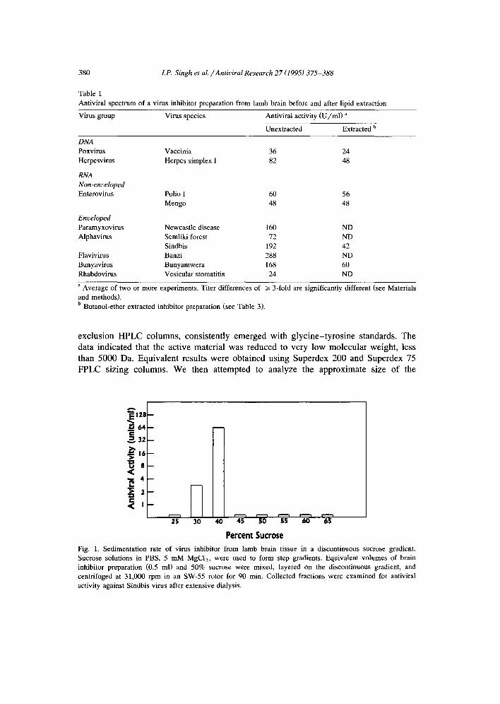
The inhibitor in lamb brain tissue extract exhibited broad antiviral activity against DNA viruses and enveloped and non-enveloped RNA viruses (Table 1). The inhibitory titers showed an 8-fold variation against different viruses. Virus sensitivity to inhibitor showed a rough rank order of (NDV, SBV, BZV, BWV) > (SFV, HSV-I, PV) > (VV, MV, VSV). Human, bovine, ovine, porcine, lapine, murine and piscine brains all possessed similar levels of antiviral activity against SBV and were broadly active (data not shown). The antiviral activity was not due to cell toxicity as no difference in Vero and CER cell growth over 48 h was detected in the absence or presence of up to 100 U of the inhibitor. Furthermore, albumin, transferrin and low density lipoprotein used as controls did not exhibit any antiviral activity.
3.2. Molecular size
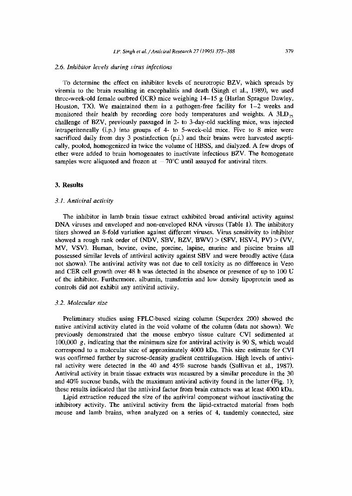
Preliminary studies using FPLC-based sizing column (Superdex 200) showed the native antiviral activity eluted in the void volume of the column (data not shown). We previously demonstrated that the mouse embryo tissue culture CVI sedimented at 100,000 g, indicating that the minimum size for antiviral activity is 90 S, which would correspond to a molecular size of approximately 4000 kDa. This size estimate for CVI was confirmed further by sucrose-density gradient centrifugation. High levels of antivi- ral activity were detected in the 40 and 45% sucrose bands (Sullivan et al., 1987). Antiviral activity in brain tissue extracts was measured by a similar procedure in the 30 and 40% sucrose bands, with the maximum antiviral activity found in the latter (Fig. 1); these results indicated that the antiviral factor from brain extracts was at least 4000 kDa.
Lipid extraction reduced the size of the antiviral component without inactivating the inhibitory activity. The antiviral activity from the lipid-extracted material from both mouse and lamb brains, when analyzed on a series of 4, tandemly connected, size
380 LP. Singh et al. /Antiviral Research 27 (1995) 375-388
Table 1 Antiviral spectrum of a virus inhibitor preparation from lamb brain before and after lipid extraction
Virus group Virus species Antiviral activity (U/ml) a
Unextracted Extracted b
DNA Poxvirus Herpesvirus
RNA Non-enveloped Enterovirus
Vaccinia 36 24 Herpes simplex I 82 48
Polio I 60 56 Mengo 48 48
Enveloped Paramyxovirus Newcastle disease 160 ND Alphavirus Semliki forest 72 ND
Sindbis 192 42 Flavivirus Banzi 288 ND Bunyavirus Bunyamwera 168 60 Rhabdovirus Vesicular stomatitis 24 ND
a Average of two or more experiments. Titer differences of >/3-fold are significantly different (see Materials and methods). b Butanol-ether extracted inhibitor preparation (see Table 3).
exclusion HPLC columns, consistently emerged with glycine-tyrosine standards. The data indicated that the active material was reduced to very low molecular weight, less than 5000 Da. Equivalent results were obtained using Superdex 200 and Superdex 75 FPLC sizing columns. We then attempted to analyze the approximate size of the
e- ~= 32
' ~ ' 16 ,,~
8
4
< !
B
25 3O r - - - i i ' - - -1 r - - - i r - ' - i
40 45 50 55 60 65
Percent Sucrose
Fig. 1. Sedimentation rate of virus inhibitor from lamb brain tissue in a discontinuous sucrose gradient. Sucrose solutions in PBS, 5 mM MgCI2, were used to form step gradients. Equivalent volumes of brain inhibitor preparation (0.5 ml) and 50% sucrose were mixed, layered on the discontinuous gradient, and centrifuged at 31,000 rpm in an SW-55 rotor for 90 min. Collected fractions were examined for antiviral activity against Sindbis virus after extensive dialysis.

LP. Singh et al. /Antiviral Research 27 (1995) 375-388
Table 2 Inactivation of virus inhibitor preparation from lamb brain by periodate, glycosidases and proteinase K
381
Inactivating agent Antiviral activity (U/ml) a
Initial Final
NalO 4 105 < 18 Glycosidases 152 < 18 Proteinase K 192 < 18
a Antiviral activity is expressed as titer (U/ml) against Sindbis virus. Titer differences of 1> 3-fold are significantly different (see Materials and methods).
antiviral material released by butanol-ether extraction using GPC peptide HPLC columns. Again the antiviral activity emerged near the lower limit of the resolving power of the columns, with a calculated molecular weight of 650 + 300 Da (data not shown). It appears that the butanol-ether extraction of the high molecular weight antiviral material from brain tissue releases a very low molecular weight moiety (~< 1000 Da), which possesses broad antiviral activity (Table 1).
3.3. Chemical composi t ion and thermostabili ty
Viral inhibitory activity of the crude brain extract was abolished by proteolysis, enzymatic glycolysis or periodate oxidation (Table 2), this implies that the native inhibitor molecule contains both protein and carbohydrate structure and that both are essential to maintain antiviral activity. Antiviral activity was determined for material parti t ioning into both aqueous and organic phases of butanol-ether extracts. The organic phase was taken to dryness, the remaining material dissolved in distilled water and lyophil ized to remove residual organic solvent before antiviral testing. No residual antiviral activity was ever detected in the organic phase of the extracted material. Material parti t ioning into the aqueous phase retained a major portion of the antiviral activity of the unextracted starting material. Lipid extraction, however, drastically changed the molecular structure of the inhibitor (Table 3), reducing it from approxi-
Table 3 Butanol-ether extraction of virus inhibitor preparation from lamb brain tissue reduces its size and enhances its stability to heat, proteolysis and glycolysis
Properties Brain inhibitor preparation
Unextracted Extracted
Antiviral activity a 144 51 Size /> 4000 kDa ~< 1 kDa Antiviral activity after:
120°C for 15 min < 6 64 Proteolysis < 18 69 Glycolysis < 18 64
a Antiviral activity is expressed as titer (U/ml) against Sindbis virus. Titer differences of >/3-fold are significantly different (see Materials and methods).
382 LP. Singh et aL /Antiviral Research 27 (1995) 375-388
1 0 2
l - ~ - - 100" Lamb Brain
~ ~ I - - O - 12o° Lamb .,a.. _ , o o o e v ,
c
~ 101 <
.~. c <
1 0 0 . , , . • . . , . . . . , . . . . , . , . . . , . . . . . . . . , . . . . , A . . . . , . . . . , . . . . , . . . . ,
1 2 3 4 S 6 7 8 9 10 11 12 Heat Treatment (hours)
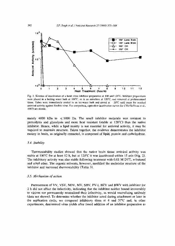
Fig. 2. Kinetics of inactivation of a lamb virus inhibitor preparation at 100 and 120°C. Inhibitor preparations were placed in a boiling water bath at 100°C, or in an autoclave at 120°C, and removed at predetermined times. Tubes were immediately cooled in an ice-water bath and stored at -20°C until assay for residual antiviral activity against Sindbis virus. For comparison, equivalent inactivation curves for CVI (Sullivan et al., 1987) are shown.
mately 4000 kDa to ~ 1000 Da. The small inhibitor molecule was resistant to proteolysis and glycolysis and more heat resistant (stable at 120°C) than the native inhibitor. Hence, while a lipid moiety is not essential for antiviral activity, it may be required to maintain structure. Taken together, the evidence demonstrates the inhibitor moiety in brain, as originally extracted, is composed of lipid, protein and carbohydrate.
3.4. Stability
Thermostabil i ty studies showed that the native brain tissue antiviral activity was stable at 100°C for at least 12 h, but at 120°C it was inactivated within 15 min (Fig. 2). The inhibitory activity was also stable following treatment with 0.01 M DTT, n-butanol and ethyl ether. The organic solvents, however, modified the molecular structure of the inhibitor and increased thermostabili ty (Table 3).
3.5. Mechanism of action
Pretreatment of VV, VSV, NDV, MV, SBV, PV-I, BZV and BWV with inhibitor for 2 h did not affect the infectivity, indicating that the inhibitor neither bound irreversibly to virions nor permanently neutralized their infectivity, as would neutralizing antibody (data not shown). To determine whether the inhibitor acted during attachment or later in the replication cycle, we compared inhibitory titers at 4 and 37°C and, in other experiments, determined virus yields after t imed addition of an inhibitor preparation to
LP. Singh et al. /Antiviral Research 27 (1995) 375-388
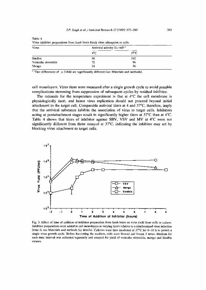
Table 4 Virus inhibitor preparations from lamb brain block virus adsorption to cells
383
Virus Antiviral activity (U /ml ) a
4°C 37°C
Sindbis 96 192 Vesicular stomatitis 72 96 Mengo 24 36
a Titer differences of >/3-fold are significantly different (see Materials and methods).
cell monolayers. Virus titers were measured after a single growth cycle to avoid possible complications stemming from suppression of subsequent cycles by residual inhibitor.
The rationale for the temperature experiment is that at 4°C the cell membrane is physiologically inert, and hence virus replication should not proceed beyond initial attachment to the target cell. Comparable antiviral titers at 4 and 37°C, therefore, imply that the antiviral substance inhibits the association of virus to target cells. Inhibitors acting at postattachment stages result in significantly higher titers at 37°C than at 4°C. Table 4 shows that titers of inhibitor against SBV, VSV and MV at 4°C were not significantly different from those assayed at 37°C, indicating the inhibitor may act by blocking virus attachment to target cells.
10 7.
E
IJ.
0,1 >-
¢o
2 >
10 6.
10 6.
10 4
10 3 -2
Mongo
Sindbis
©
[]
i I i i i i i i i i
-1 0 1 2 3 4 5 6 7 8 9
T ime of Add i t ion of Inhib i tor ( h o u r s )
Fig. 3. Effect of time of addition of inhibitor preparation from lamb brain on virus yield from cells in culture. Inhibitor preparations were added to cell monolayers at varying times relative to a synchronized virus infection (time 0, see Materials and methods for details). Cultures were then incubated at 37°C for 8 -10 h to permit a single virus growth cycle. Before harvesting the medium, cells were thawed and frozen 3 times. Medium for each time interval was collected separately and assayed for yield of vesicular stomatitis, mengo and Sindbis viruses.
384 LP. Singh et al. /Antiviral Research 27 (1995) 375-388
10 5 I I I I I I 10
¢-
.>
<
g C <
10 4
10 3
10 2
101
/
0 v i r u s ~
0 /
0 1 2 3 4 5 6 7
6
5
4
3
2
1
0
-1 8
O
¢/I
. m
Day after infection with Banzi Virus
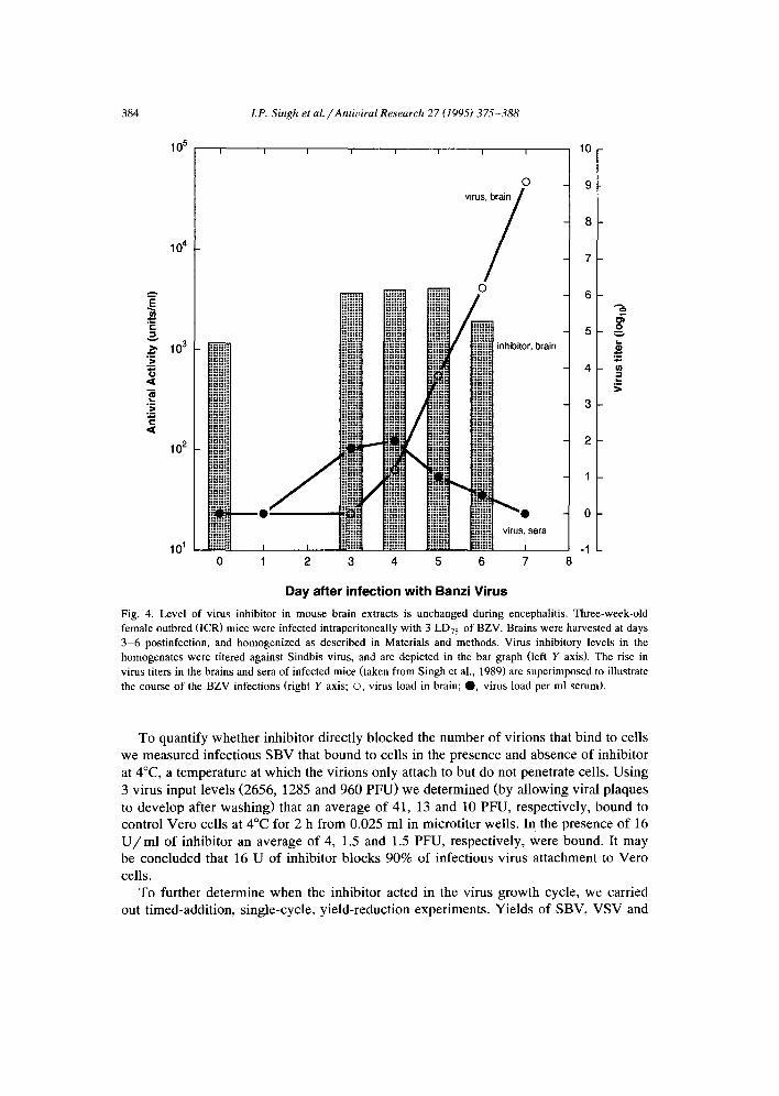
Fig. 4. Level of virus inhibitor in mouse brain extracts is unchanged during encephalitis. Three-week-old female outbred (ICR) mice were infected intraperitoneally with 3 LD75 of BZV. Brains were harvested at days 3-6 postinfection, and homogenized as described in Materials and methods. Virus inhibitory levels in the homogenates were titered against Sindbis virus, and are depicted in the bar graph (left Y axis). The rise in virus titers in the brains and sera of infected mice (taken from Singh et al., 1989) are superimposed to illustrate the course of the BZV infections (right Y axis; O, virus load in brain; O, virus load per ml serum).
To quantify whether inhibitor directly blocked the number of virions that bind to cells
we measured infectious SBV that bound to cells in the presence and absence of inhibitor
at 4°C, a temperature at which the virions only attach to but do not penetrate cells. Using
3 virus input levels (2656, 1285 and 960 PFU) we determined (by allowing viral plaques
to develop after washing) that an average of 41, 13 and 10 PFU, respectively, bound to control Vero cells at 4°C for 2 h from 0.025 ml in microtiter wells. In the presence of 16
U / m l of inhibitor an average of 4, 1.5 and 1.5 PFU, respectively, were bound. It may be concluded that 16 U of inhibitor blocks 90% of infectious virus attachment to Vero
cells. To further determine when the inhibitor acted in the virus growth cycle, we carried
out timed-addition, single-cycle, yield-reduction experiments. Yields of SBV, VSV and
LP. Singh et al. /Antiviral Research 27 (1995) 375-388 385
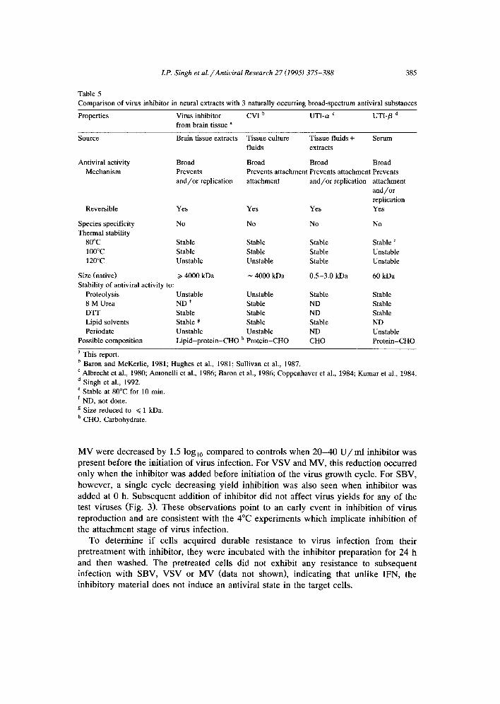
Table 5 Comparison of virus inhibitor in neural extracts with 3 naturally occurring broad-spectrum antiviral substances
Properties Virus inhibitor CVI b UTI-a c UTI-/3 d from brain tissue a
Source Brain tissue extracts Tissue culture Tissue fluids + Serum fluids extracts
Antiviral activity Broad Broad Broad Broad Mechanism Prevents Prevents attachment Prevents attachment Prevents
and/or replication attachment and/or replication attachment and/or replication
Reversible Yes Yes Yes Yes
Species specificity No No No No Thermal stability
80°C Stable Stable Stable Stable ~ 100°C Stable Stable Stable Unstable 120°C Unstable Unstable Stable Unstable
Size (native) /> 4000 kDa ~ 4000 kDa 0.5-3.0 kDa 60 kDa Stability of antiviral activity to:
Proteolysis Unstable Unstable Stable Stable 8 M Urea ND f Stable ND Stable DTF Stable Stable ND Stable Lipid solvents Stable g Stable Stable ND Periodate Unstable Unstable ND Unstable
Possible composition Lipid-protein-CHO h Protein-CHO CHO Protein-CHO
a This report. b Baron and McKerlie, 1981; Hughes et al., 1981; Sullivan et al., 1987. ¢ Albrecht et al., 1980; Antonelli et al., 1986; Baron et al., 1986; Coppenhaver et al., 1984; Kumar et al., 1984. d Singh et al., 1992. e Stable at 80°C for 10 min. f ND, not done. g Size reduced to ~< 1 kDa. h CHO, Carbohydrate.
M V were dec rea sed by 1.5 log10 c o m p a r e d to con t ro l s w h e n 2 0 - 4 0 U / m l inh ib i to r was
p re sen t be fo re the in i t i a t ion o f v i rus infec t ion . For V S V and M V , th is r educ t ion occu r r ed
on ly w h e n the i nh ib i t o r w a s added be fo re in i t i a t ion o f the v i rus g r o w t h cycle . For S B V ,
h o w e v e r , a s ing le cycle d e c r e a s i n g y ie ld i nh ib i t i on was also seen w h e n inh ib i to r was
added at 0 h. S u b s e q u e n t add i t ion o f inh ib i to r d id not af fec t v i rus y ie lds for any o f the
test v i ruses (Fig. 3). T h e s e o b s e r v a t i o n s po in t to an ear ly even t in i nh ib i t i on o f v i rus
r ep roduc t i on and are cons i s t en t w i t h the 4°C e x p e r i m e n t s w h i c h impl i ca t e i nh ib i t i on o f
the a t t a c h m e n t s tage o f v i rus infec t ion .
To d e t e r m i n e i f ce l ls acqu i r ed durab le res i s t ance to v i rus in fec t ion f rom the i r
p r e t r e a t m e n t w i th inh ib i to r , they we re i n c u b a t e d w i th the inh ib i to r p r epa ra t i on for 24 h
and then w as hed . T he p re t r ea ted cel ls d id not exh ib i t any re s i s t ance to s u b s e q u e n t
in fec t ion w i t h S B V , V S V or M V (da ta not shown) , i nd i ca t i ng that un l ike IFN, the
inh ib i to ry ma te r i a l does not i nduce an an t iv i ra l s tate in the ta rge t cells.
386 I.P. Singh et al. /Antiviral Research 27 (1995) 375-388
3.6. Effect o f virus infection on inhibitor levels in vivo
The possible regulation of antiviral activity levels in neural tissues in response to an ongoing infection was assessed using an encephalitic flavivirus infection. Inhibitor titers were determined from day 3 p.i. with BZV. This starting time point was chosen since BZV can be detected in mouse brain from day 3 p.i. (Singh et al., 1989), and thus it represents the earliest point at which a response to infection could be anticipated. No change in inhibitory titer was seen throughout this lethal infection (Fig. 4), indicating that the inhibitor, unlike IFN or antibody, is not specifically induced in response to infection.
4. Discussion
This is the first report of a broadly active antiviral substance in brain tissue. Earlier reports of the isolation and characterization of virus inhibitors in neural tissue relied on assays against single viruses only, e.g., poliovirus from human brain tissue (Low and Baron, 1960; Holland, 1961; Baron et al., 1963). Some of these earlier inhibitors of single viruses could have been similar to the material investigated here, but were not characterized sufficiently to judge.
Our interest in naturally occurring, broad-spectrum antiviral agents previously re- sulted in detection and characterization of 3 groups of inhibitors from cells in culture and from body tissues and fluids other than those of the brain (Baron and McKerlie, 1981; Hughes et al., 1981; Coppenhaver et al., 1984; Kumar et al., 1984; Baron et al., 1986; Sullivan et al., 1987; Singh et al., 1992). Table 5 summarizes the properties of these inhibitors and compares them with the presently described antiviral substance from brain tissue.
Our studies show that the antiviral activity from brain tissue resides in a large moiety of at least 4000 kDa. The properties of the brain tissue inhibitor suggest a physical aggregate of lipid, protein and carbohydrate, which is consistent with entrapment of active components in lipid micelles which could be formed during the homogenization. Purified protein (human serum albumin), glycoprotein (human serum transferrin), and lipoprotein (human low density lipoprotein) used as controls did not show activity in our assay. We note that human low density lipoprotein is similar in size to the inhibitory activity found here, suggesting that there is some specificity in the antiviral effect detected in the brain extracts. The inhibitory activity of the native aggregate seems dependent on both protein and carbohydrate structure since the antiviral activity is eliminated by treatment with pronase, periodate, or a mixture of carbohydrases. The data, however, do not distinguish between direct cleavage of the structure by enzymatic digestion and steric inhibition of the antiviral factor subsequent to alteration of the structure of the aggregate. Aggregation of protein, lipid and carbohydrate components, rather than covalent bonding, is implied by the finding that butanol-ether extraction removes a non-inhibitory lipid and releases a small molecular weight (~< 1000 Da) component with antiviral activity. The small inhibitory substance (~< 1000 Da) released
LP. Singh et al. /Antiviral Research 27 (1995) 375-388 387
by lipid extraction is resistant to proteolysis and digestion with carbohydrases, and heat stable at 120°C. This may imply that a modified or distinct antiviral component, which is initially shielded or buried in the aggregate or micelle, is released by the butanol-ether extraction. Currently, we are attempting to address these possibilities by purifying and directly determining the molecular structure of the ~< 1000 Da inhibitor.
Studies on the mechanism of action of the inhibitor in brain extracts indicate that it acts at an early stage of the virus growth cycle, perhaps at the initial association of viruses with cells. This conclusion is suggested by the following lines of evidence. First, the inhibitor does not irreversibly neutralize or inactivate the virus, since fully infective virus particles can be recovered from the mixture of inhibitor and virus. Second, the titers of viral inhibitor assayed at 4 and 37°C are virtually indistinguishable, indicating that virus replication after adsorption of virus onto target cells is not substantially affected by the inhibitor. Third, the inhibitor does not induce an antiviral state in cells preincubated with inhibitor overnight. Finally, virus yield is affected only when inhibitor preparations are added to cells before or at the same time as the exposure of the cell monolayer to virus challenge. Taken together, these data indicate that the primary mechanism of action of the inhibitor is to prevent virus attachment to target cells, which is similar to findings with other pre-existing, broadly active antiviral molecules, such as CVI, UTI-o~ and UTI-fl (Hughes et al., 1981; Kumar et al., 1984; Sullivan et al., 1987; Singh et al., 1992).
Interestingly, this virus inhibitor from brain extracts shares some similarities with CVI, which previously was known to be found in cell culture fluids and secreted into the medium (Baron and McKerlie, 1981; Hughes et al; 1981; Sullivan et al., 1987). These shared properties are the broad antiviral spectrum, high apparent molecular weight, insensitivity of antiviral activity to lipid solvents and high thermostability. The effect of lipid extraction on the size of CVI was not investigated, however (Sullivan et al., 1987).
The natural role of the antiviral activity from neural extracts during viral infection remains to be assessed. It is possible that the antiviral activity provides initial non-specific protection to the brain against viral infection before initiation of reactive host defenses such as IFN and antibodies. Some support for this hypothesis comes from our observa- tions that the inhibitor is a normal constituent of human and other vertebrate brains, the inhibitor levels remain unchanged throughout a severe viral encephalitis, and the finding that inhibitor concentrations in the brain extract diluted 1 : 6 are sufficient to reduce viral yield at least 30-fold (Fig. 3). To further support a role of the inhibitor in natural defense, we will need in vivo studies to demonstrate that deletion of this substance worsens infection or that its transfer to a body site relatively deficient in inhibitor enhances resistance to virus challenge.
Acknowledgements
The authors thank Ms. Louese M. McKerlie for her critical reading of the manuscript and Ms. Mardelle Susman for her excellent editorial assistance.
388 LP. Singh et al. /Antiviral Research 27 (1995) 375-388
References
Albrecht, T.B., Zucca, M. and Dianzani, F. (1980) Studio su un inhibitore della replicazione del virus della varicella-zoster. Est. G. Ital. Chemioter. 27-N-2, 103-105.
Albrecht, T.B., Cole, N., Speelman, D., Dianzani, F. and Baron, S. (1983) Inhibition of varicella-zoster virus replication by Neuramide. Chemioterapia 2, 225-256.
Antonelli, G., Dianzani, F., Coppenhaver, D.H., Baron, S., Calandra, P. and Folchitto, G. (1986) An influenza virus inhibitor that acts late in the replication cycle. Antimicrob. Agents Chemother. 29, 49-51.
Baron, J.L., Li, J.-L., McKerlie, M.L., Shabot, J.M. and Coppenhaver, D.H. (1986) A new subtype of a natural viral inhibitor (CVI) that is stable in the gastrointestinal tract. Microbial Pathogenesis 1, 241-247.
Baron, S. (1963) Mechanism of recovery from viral inection. In: K.M. Smith and M.A. Lauffer (Eds.), Advances in Virus Research, Vol. 10. Academic Press, New York, pp. 39-60.
Baron, S. and McKerlie, U (1981) Broadly active inhibitor of viruses spontaneously produced by many cell types in culture. Infect. Immun. 32, 449-453.
Baron, S., Friedman, R.M. and Buckler, C.E. (1963) Properties of poliovirus inhibitor from monkey brain. Proc. Soc. Exp. Med. 113, 107-110.
Bose, S., Gurari-Rotman, D., Ruegg, U.T., Corley, L. and Anflnson, C.B. (1976) Apparent dispensability of the carbohydrate moiety of human interferon for antiviral activity. J. Biol. Chem. 251, 1659-1662.
Cathala, F. and Baron, S. (1970) Interferon in rabbit brain, cerebrospinal fluid and serum following administration of polyinosinic polycytidylic acid. J. Immunol. 104, 1355-1358.
Coppenhaver, D.H., Baron, J.L., McKerlie, M.L., Sabados, J. and Baron, S. (1984) Size and stability of a naturally occurring virus inhibitor. Antimicrob. Agents Chemother. 25, 646-649.
Griffin, D.E. (1991) Therapy of viral infections of the central nervous system. A mini-review. Antiviral Res. 15, 1-10.
Grossberg, S.E., Jameson, P. and Sedmark, J.J. (1973) Interferon bioassay methods and the development of standard procedures: a critique and analysis of current observations. The Tissue Culture Monogr. No. 3, The Tissue Culture Assoc., Rockville, MD, pp. 26-34.
Holland, J.J. (1961) Receptor affinities as major determinants of enterovirus tissue tropisms in humans. Virology 15, 312-326.
Hughes, T.K., Blalock, J.E., McKerlie, M.L. and Baron, S. (1981) Cell-produced viral inhibitor: possible mechanism of action and chemical composition. Infect. lmmun. 32, 454-457.
Kumar, S., McKerlie, M.L., Albrecht, T.B., Goldman, A.S. and Baron, S. (1984) A broadly active inhibitor in human and animal organ extracts and body fluids. Proc. Soc. Exp. Biol. Med. 177, 104-111.
Low, R.L. and Baron, S. (1960) Poliovirus inhibitor from the central nervous system of the rhesus monkey. Science 132, 622-623.
Singh, I.P., Coppenhaver, D.H., Sarzotti, M., Sriyuktasuth, P., Poast, J., Levy, H.B. and Baron, S. (1989) Postinfection therapy of arbovirus infections in mice. Antimicrob. Agents Chemother. 33, 2126-2131.
Singh, I.P., Coppenhaver, D.H., Chopra, A.K. and Baron, S. (1992) Further characterization of a broad-spec- trum antiviral substance in human serum. Viral Immunol. 5, 293-303.
Singh, I.P., Coppenhaver, D.H., Chopra, A. and Baron, S. (1993) Generalized occurrence of the broadly antiviral substance UTlbeta in mammalian sera. J. Biol. Regul. Homeost. Agents 7, 7-14.
Sullivan, M.L., Niesel, D.W., Coppenhaver, D.H., Sabados, J. and Baron, S. (1987) Characterization of an antiviral agent from primary murine fibroblast cultures: murine tissue culture CVI. J. Biol. Regul. Homeost. Agents 1, 126-132.


