Embed Size (px)
Citation preview

Vertebral Column
Lectures Objectives• Describe the regions and curvatures of the vertebral
column and the number of vertebrae in each region.• Describe the basic components of a typical vertebrae and
their function.• Identify and recognise the differences between cervical,
thoracic and lumbar vertebrae.• Describe and classify the joints associated with the
vertebral column.• Describe the location and general function of ligaments. • Name the true back muscles and understand their relative
positions and actions. • Understand the relationships of neural structures and
meninges to the vertebral column, including the points of exit of spinal nerves
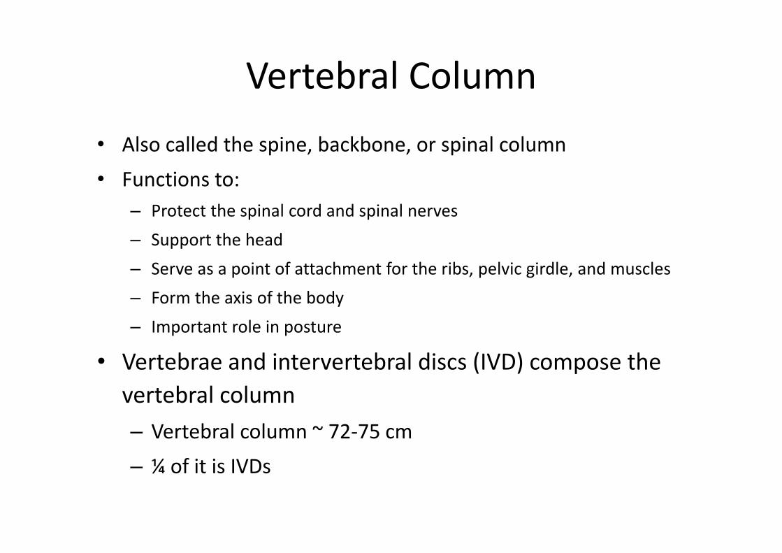
Vertebral Column• Also called the spine, backbone, or spinal column• Functions to:
– Protect the spinal cord and spinal nerves– Support the head– Serve as a point of attachment for the ribs, pelvic girdle, and muscles– Form the axis of the body– Important role in posture
• Vertebrae and intervertebral discs (IVD) compose the vertebral column– Vertebral column ~ 72‐75 cm– ¼ of it is IVDs
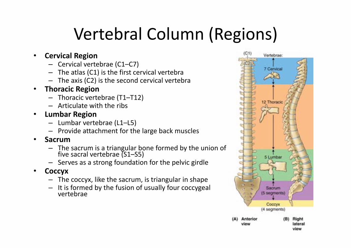
Vertebral Column (Regions)• Cervical Region
– Cervical vertebrae (C1–C7)– The atlas (C1) is the first cervical vertebra– The axis (C2) is the second cervical vertebra
• Thoracic Region– Thoracic vertebrae (T1–T12)– Articulate with the ribs
• Lumbar Region– Lumbar vertebrae (L1–L5)– Provide attachment for the large back muscles
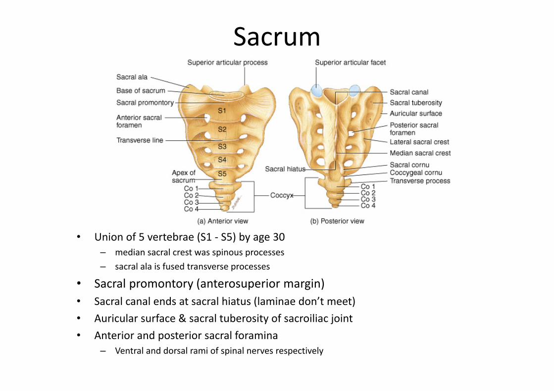
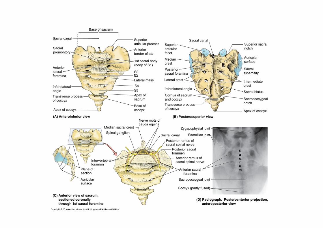
• Sacrum– The sacrum is a triangular bone formed by the union of
five sacral vertebrae (S1–S5)– Serves as a strong foundation for the pelvic girdle
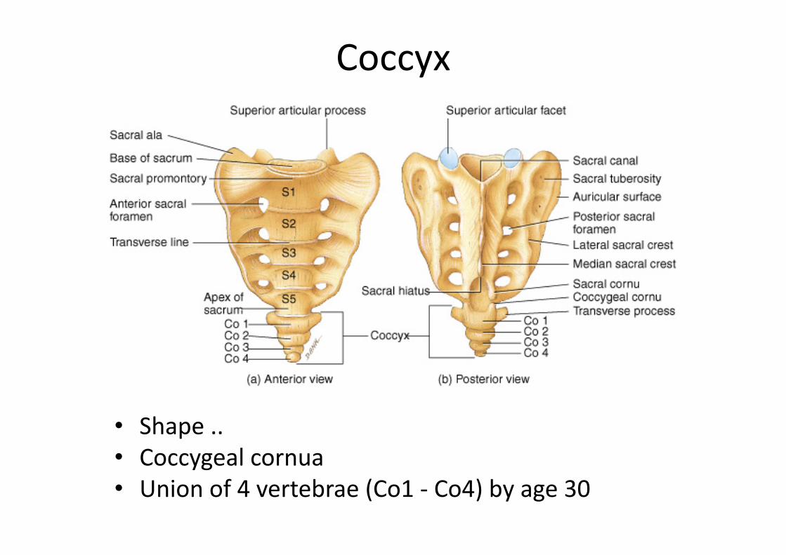
• Coccyx– The coccyx, like the sacrum, is triangular in shape– It is formed by the fusion of usually four coccygeal
vertebrae
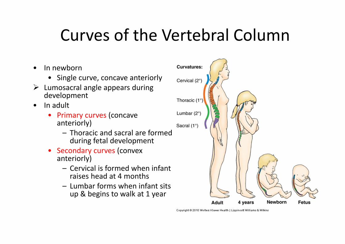
Curves of the Vertebral Column
• In newborn• Single curve, concave anteriorly
Lumosacral angle appears during development
• In adult• Primary curves (concave
anteriorly)– Thoracic and sacral are formed during fetal development
• Secondary curves (convex anteriorly)– Cervical is formed when infant raises head at 4 months
– Lumbar forms when infant sits up & begins to walk at 1 year
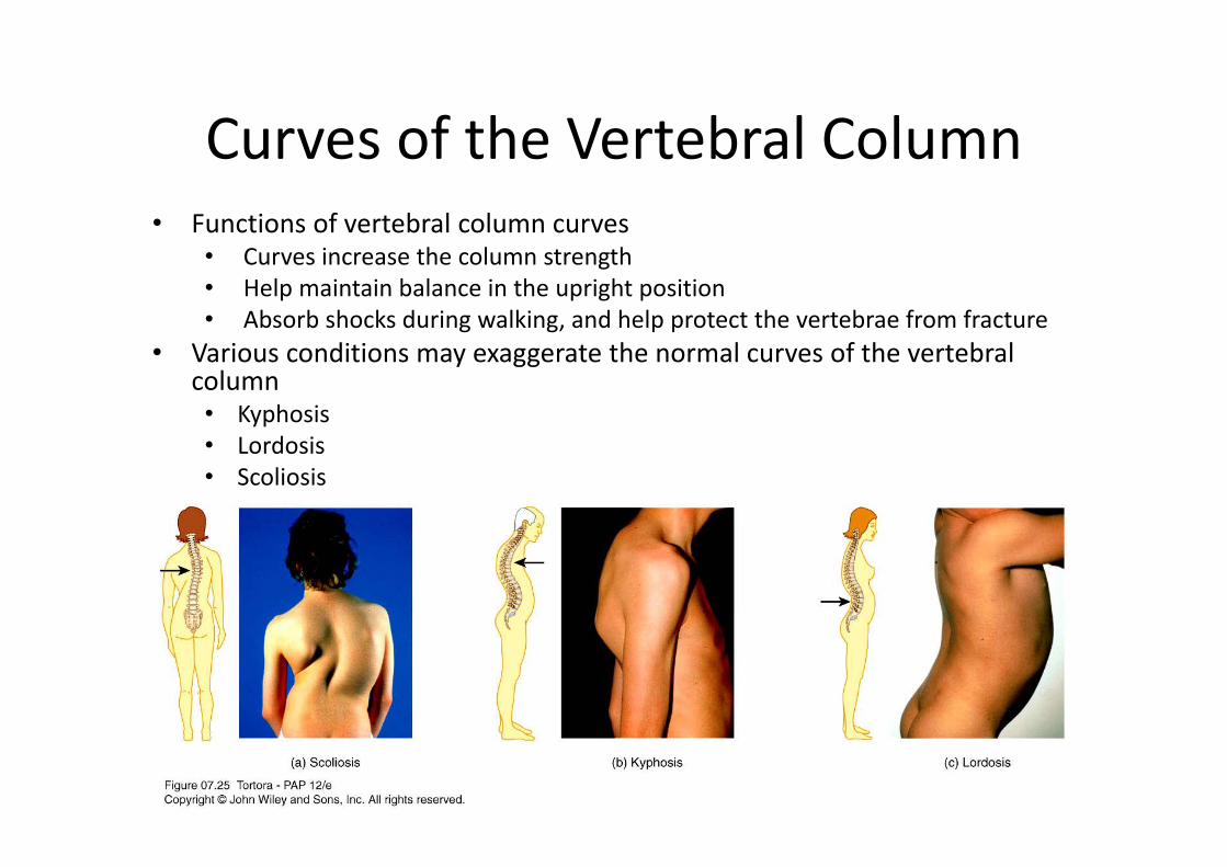
Curves of the Vertebral Column• Functions of vertebral column curves
• Curves increase the column strength• Help maintain balance in the upright position• Absorb shocks during walking, and help protect the vertebrae from fracture
• Various conditions may exaggerate the normal curves of the vertebral column• Kyphosis• Lordosis• Scoliosis
Intervertebral Discs
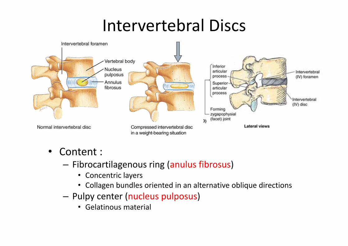
• Found between the bodies of adjacent vertebrae
• Thicker in cervical & lumbar regions (greatest movement)
• Functions to:– Form strong joints– Permit various movements of the vertebral column
– Absorb vertical shock
Intervertebral Discs
• Content :– Fibrocartilagenous ring (anulus fibrosus)
• Concentric layers• Collagen bundles oriented in an alternative oblique directions
– Pulpy center (nucleus pulposus)• Gelatinous material
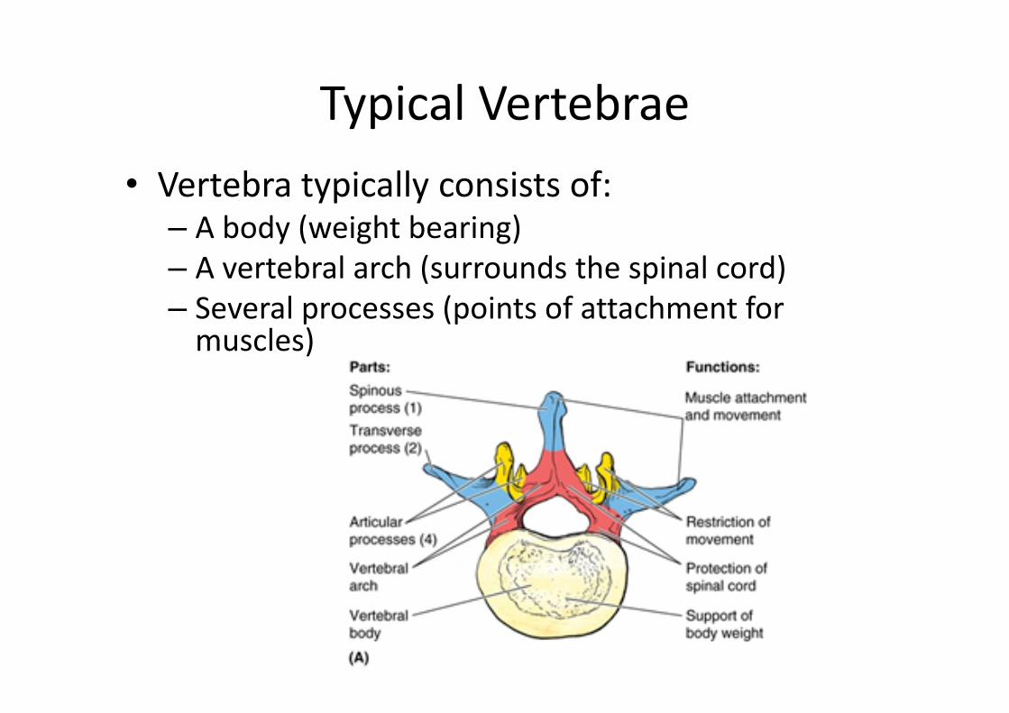
Typical Vertebrae• Vertebra typically consists of:
– A body (weight bearing)– A vertebral arch (surrounds the spinal cord)– Several processes (points of attachment for muscles)
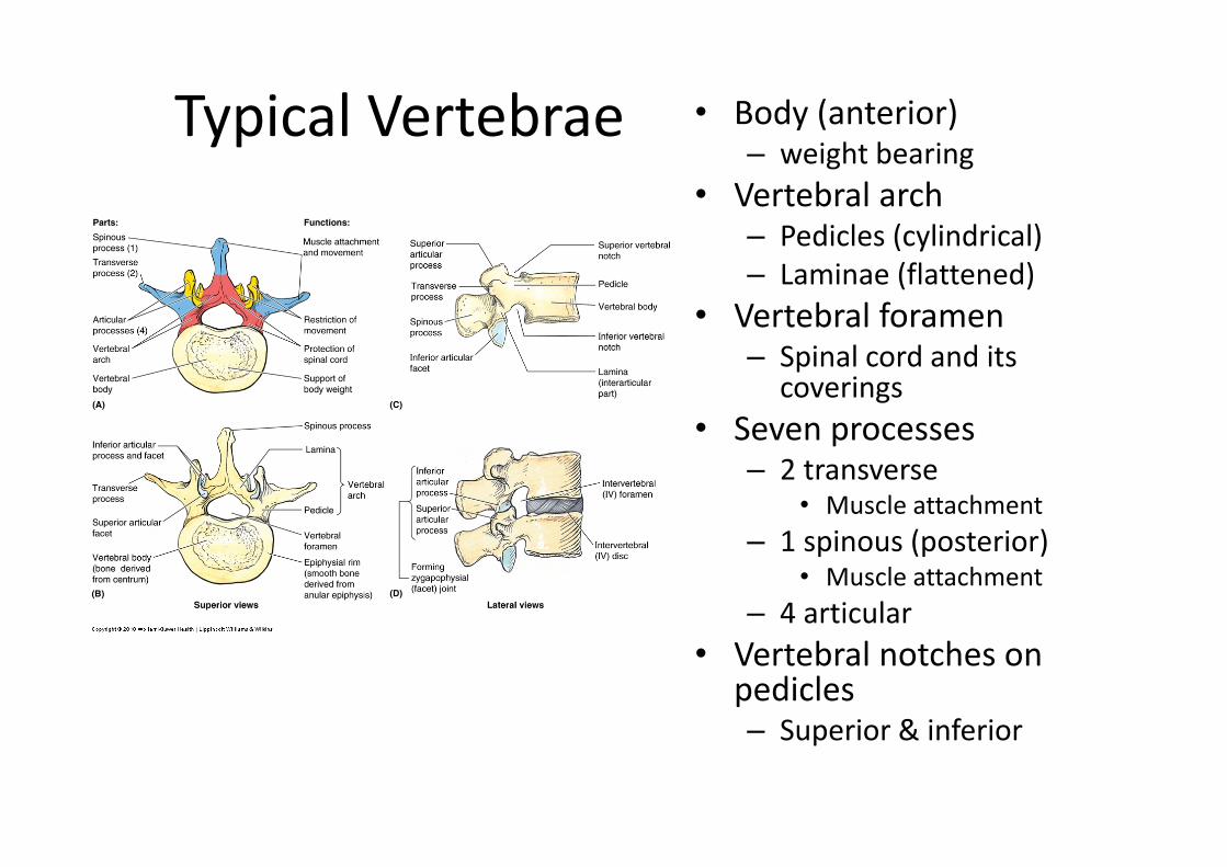
Typical Vertebrae • Body (anterior)– weight bearing
• Vertebral arch– Pedicles (cylindrical)– Laminae (flattened)
• Vertebral foramen– Spinal cord and its
coverings• Seven processes
– 2 transverse• Muscle attachment
– 1 spinous (posterior)• Muscle attachment
– 4 articular• Vertebral notches on
pedicles– Superior & inferior

Intervertebral Foramen & Spinal Canal
• Spinal canal is all vertebral foramena together• Intervertebral foramen are 2 vertebral notches together
– Transmit spinal nerves and blood vessels
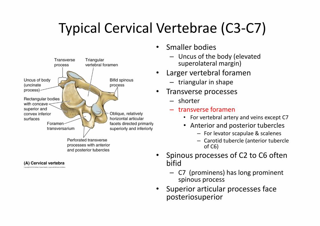
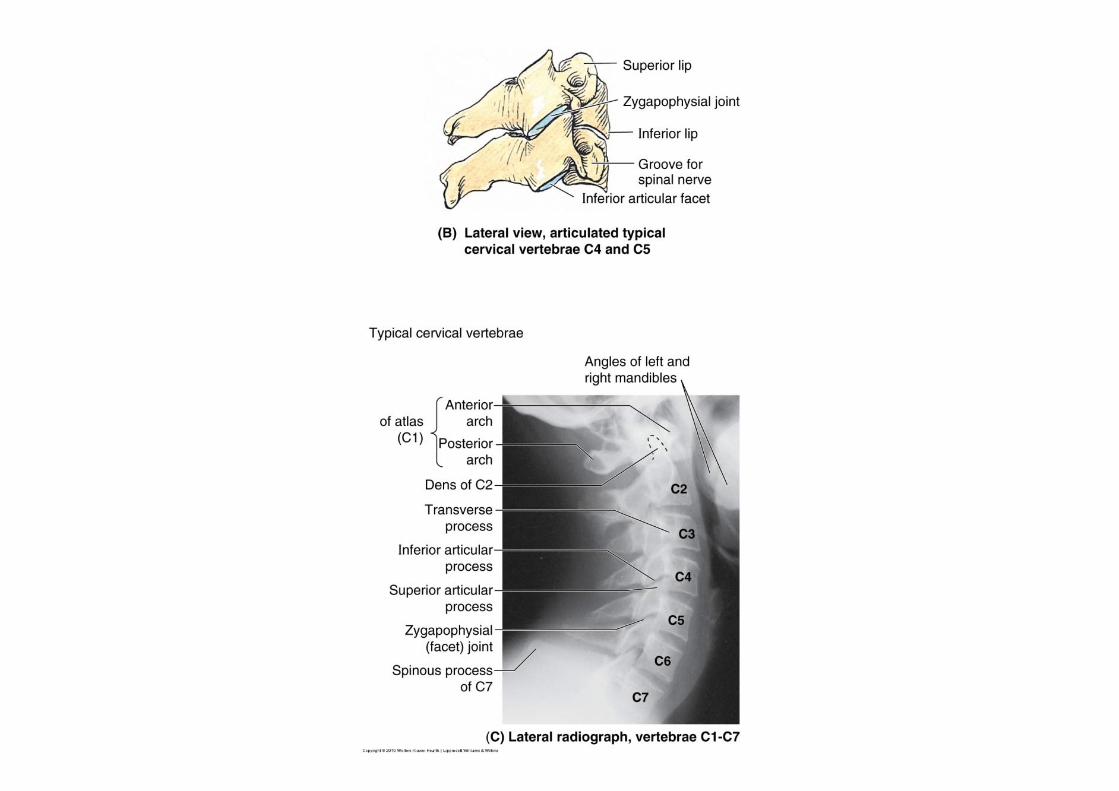
Typical Cervical Vertebrae (C3‐C7)• Smaller bodies
– Uncus of the body (elevated superolateral margin)
• Larger vertebral foramen– triangular in shape
• Transverse processes– shorter– transverse foramen
• For vertebral artery and veins except C7• Anterior and posterior tubercles
– For levator scapulae & scalenes– Carotid tubercle (anterior tubercle
of C6)• Spinous processes of C2 to C6 often
bifid– C7 (prominens) has long prominent
spinous process• Superior articular processes face
posteriosuperior
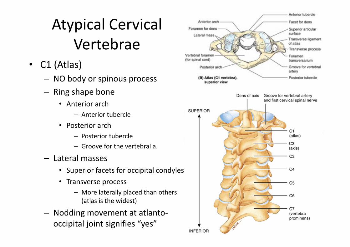
Atypical Cervical Vertebrae
• C1 (Atlas)– NO body or spinous process– Ring shape bone
• Anterior arch– Anterior tubercle
• Posterior arch– Posterior tubercle– Groove for the vertebral a.
– Lateral masses• Superior facets for occipital condyles• Transverse process
– More laterally placed than others (atlas is the widest)
– Nodding movement at atlanto‐occipital joint signifies “yes”
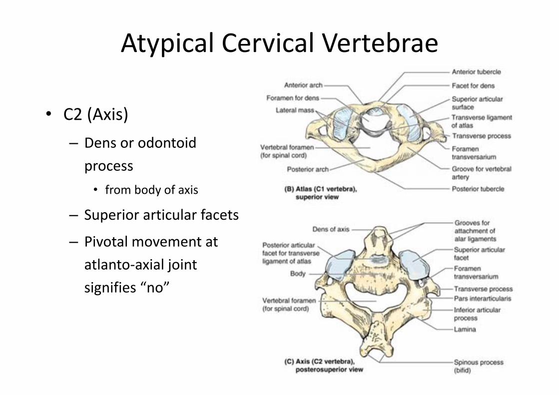
Atypical Cervical Vertebrae
• C2 (Axis) – Dens or odontoid process• from body of axis
– Superior articular facets
– Pivotal movement at atlanto‐axial joint signifies “no”
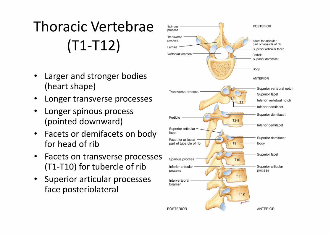
Thoracic Vertebrae(T1‐T12)
• Larger and stronger bodies (heart shape)
• Longer transverse processes• Longer spinous process
(pointed downward)• Facets or demifacets on body
for head of rib• Facets on transverse processes
(T1‐T10) for tubercle of rib• Superior articular processes
face posteriolateral
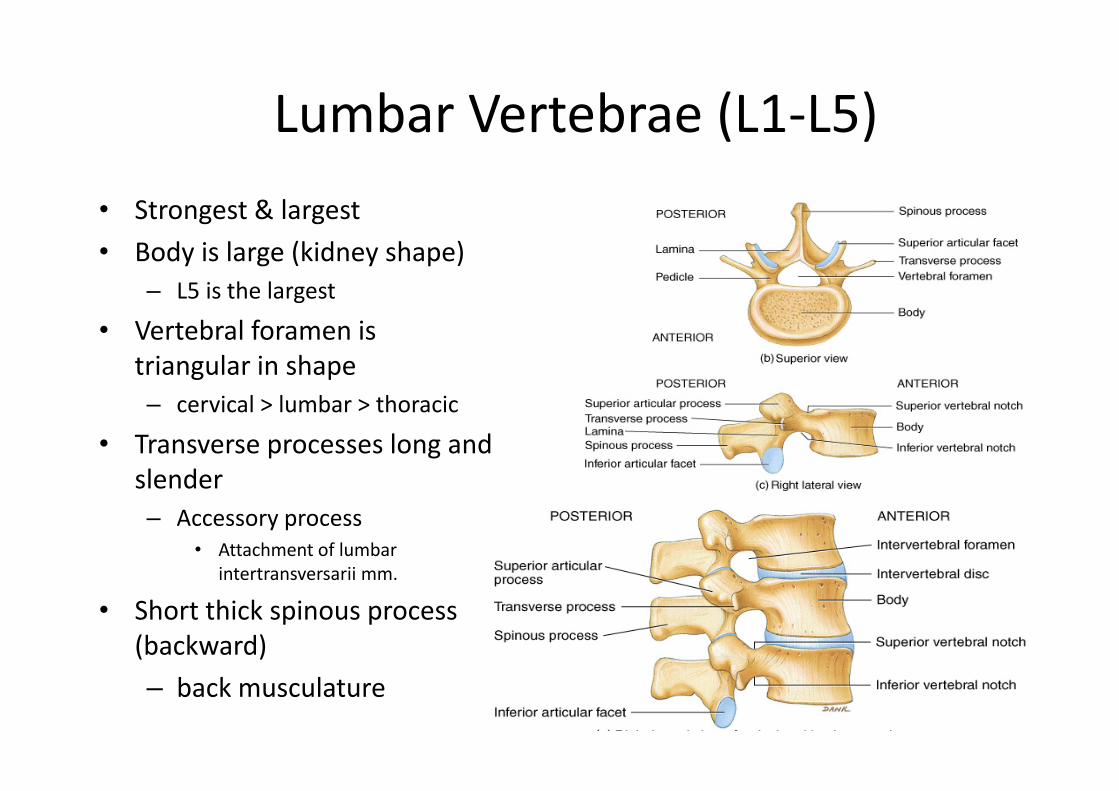
Lumbar Vertebrae (L1‐L5)• Strongest & largest• Body is large (kidney shape)
– L5 is the largest
• Vertebral foramen is triangular in shape– cervical > lumbar > thoracic
• Transverse processes long and slender– Accessory process
• Attachment of lumbar intertransversarii mm.
• Short thick spinous process (backward)– back musculature

Vertebral Column
Sacrum
• Union of 5 vertebrae (S1 ‐ S5) by age 30– median sacral crest was spinous processes– sacral ala is fused transverse processes
• Sacral promontory (anterosuperior margin)• Sacral canal ends at sacral hiatus (laminae don’t meet)• Auricular surface & sacral tuberosity of sacroiliac joint• Anterior and posterior sacral foramina
– Ventral and dorsal rami of spinal nerves respectively
Coccyx
• Shape ..• Coccygeal cornua• Union of 4 vertebrae (Co1 ‐ Co4) by age 30
Vertebral Column
Neural Content
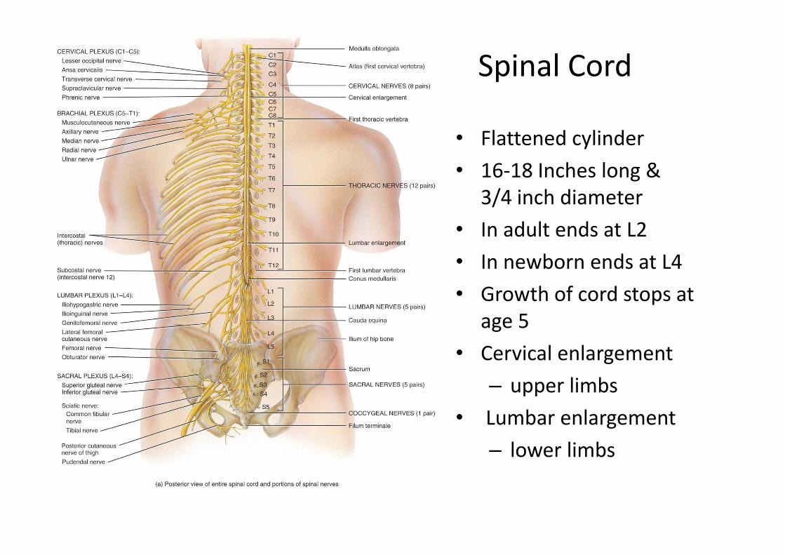
Spinal Cord
• Flattened cylinder• 16‐18 Inches long &
3/4 inch diameter• In adult ends at L2• In newborn ends at L4• Growth of cord stops at
age 5• Cervical enlargement
– upper limbs• Lumbar enlargement
– lower limbs
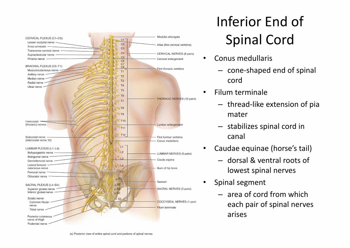
Inferior End of Spinal Cord
• Conus medullaris– cone‐shaped end of spinal
cord• Filum terminale
– thread‐like extension of piamater
– stabilizes spinal cord in canal
• Caudae equinae (horse’s tail)– dorsal & ventral roots of
lowest spinal nerves• Spinal segment
– area of cord from which each pair of spinal nerves arises
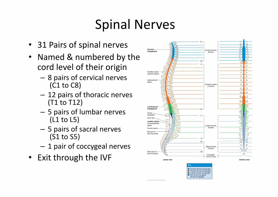
Spinal Nerves• 31 Pairs of spinal nerves• Named & numbered by the cord level of their origin– 8 pairs of cervical nerves
(C1 to C8)– 12 pairs of thoracic nerves (T1 to T12)
– 5 pairs of lumbar nerves(L1 to L5)
– 5 pairs of sacral nerves (S1 to S5)
– 1 pair of coccygeal nerves• Exit through the IVF
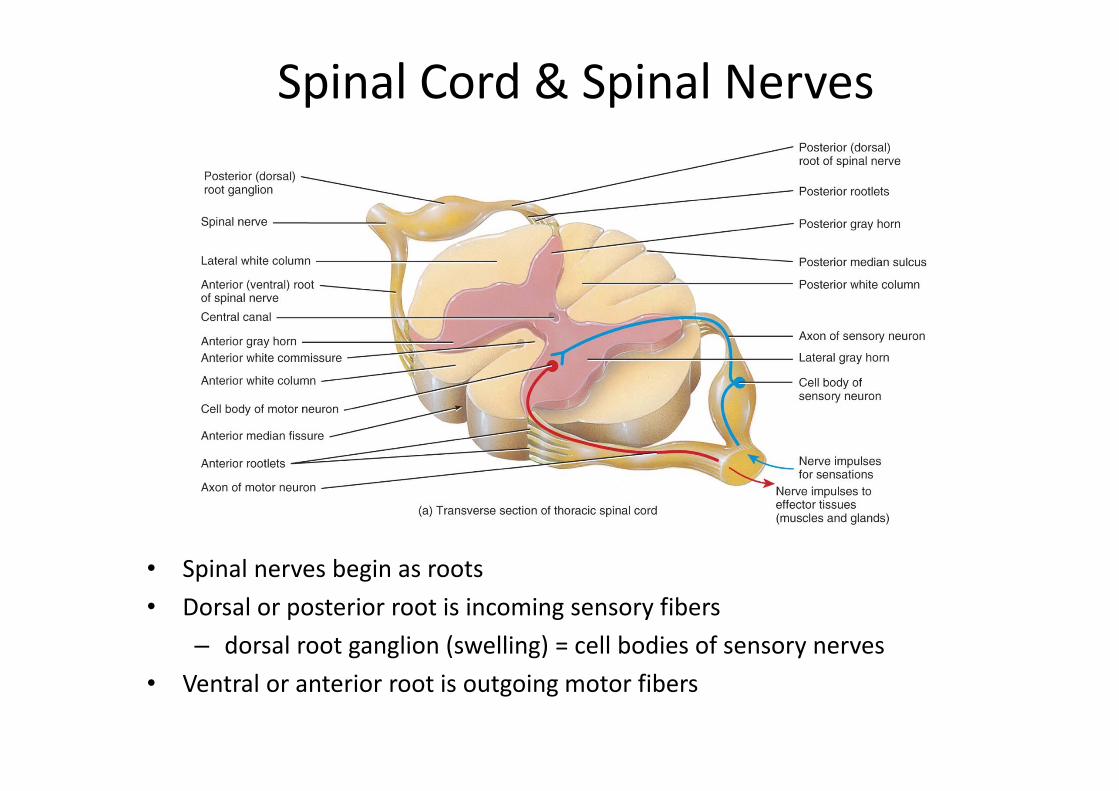
Spinal Cord & Spinal Nerves
• Spinal nerves begin as roots• Dorsal or posterior root is incoming sensory fibers
– dorsal root ganglion (swelling) = cell bodies of sensory nerves• Ventral or anterior root is outgoing motor fibers
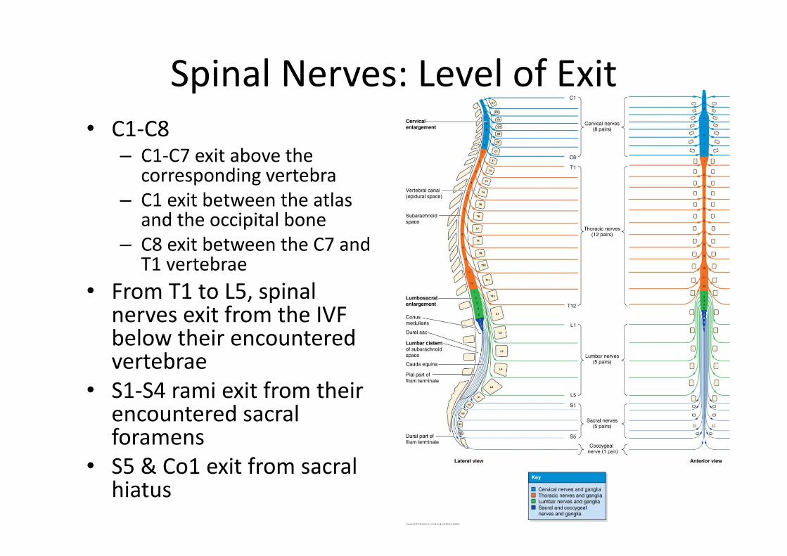
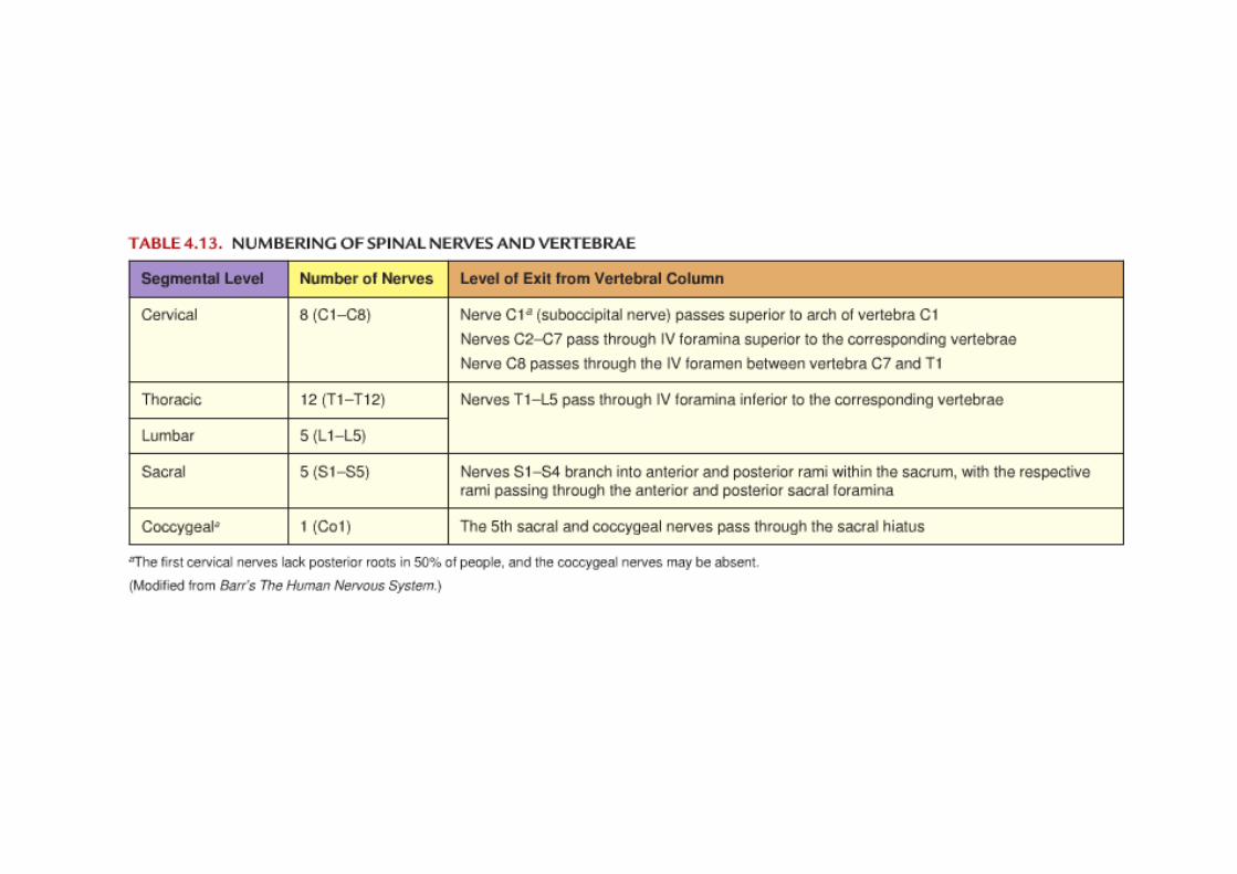
Spinal Nerves: Level of Exit• C1‐C8
– C1‐C7 exit above the corresponding vertebra
– C1 exit between the atlas and the occipital bone
– C8 exit between the C7 and T1 vertebrae
• From T1 to L5, spinal nerves exit from the IVF below their encountered vertebrae
• S1‐S4 rami exit from their encountered sacral foramens
• S5 & Co1 exit from sacral hiatus
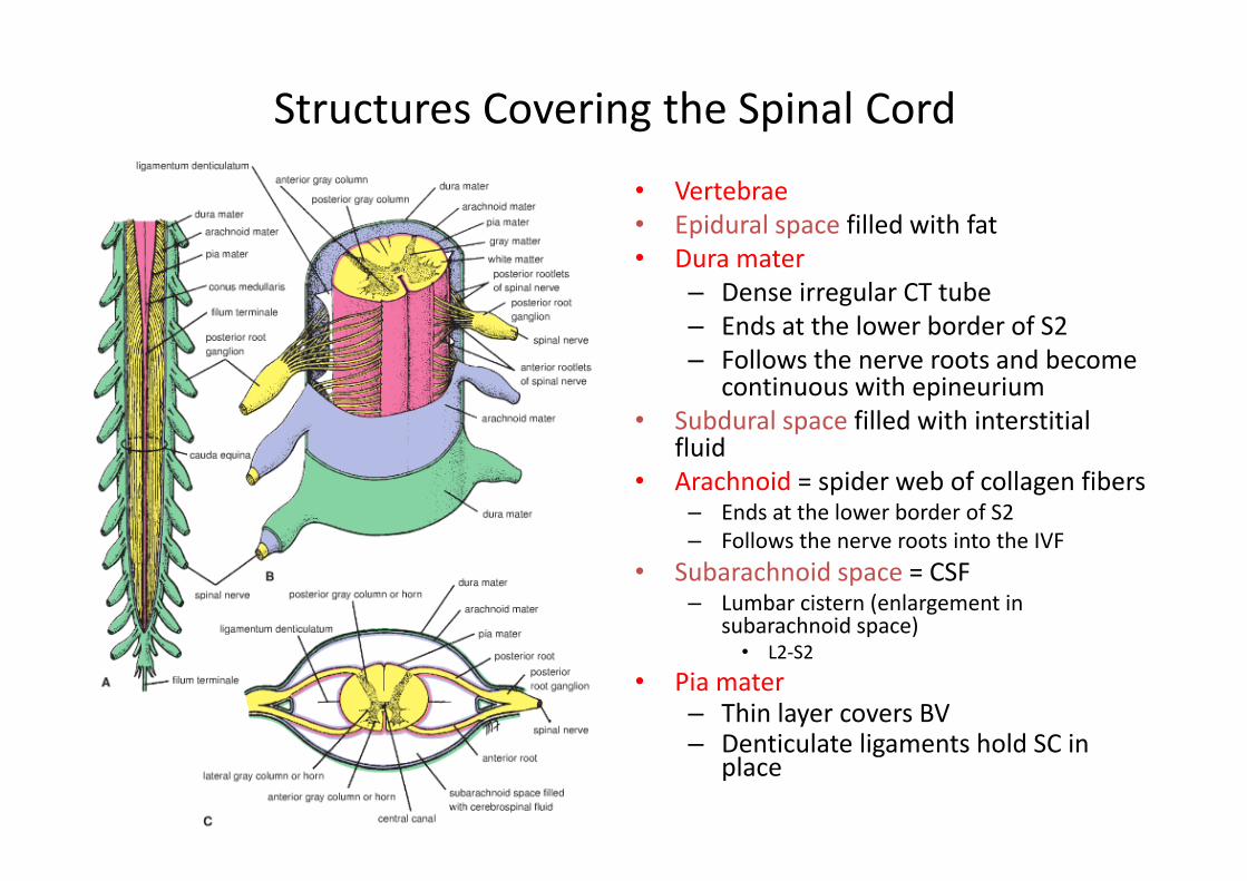
Structures Covering the Spinal Cord
• Vertebrae• Epidural space filled with fat• Dura mater
– Dense irregular CT tube– Ends at the lower border of S2– Follows the nerve roots and become
continuous with epineurium• Subdural space filled with interstitial
fluid• Arachnoid = spider web of collagen fibers
– Ends at the lower border of S2– Follows the nerve roots into the IVF
• Subarachnoid space = CSF– Lumbar cistern (enlargement in
subarachnoid space)• L2‐S2
• Pia mater– Thin layer covers BV– Denticulate ligaments hold SC in
place
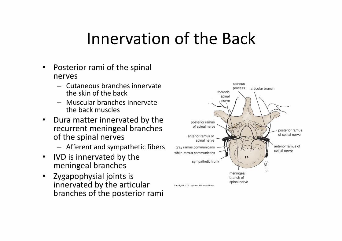
Innervation of the Back• Posterior rami of the spinal
nerves– Cutaneous branches innervate
the skin of the back – Muscular branches innervate
the back muscles• Dura matter innervated by the
recurrent meningeal branches of the spinal nerves– Afferent and sympathetic fibers
• IVD is innervated by the meningeal branches
• Zygapophysial joints is innervated by the articular branches of the posterior rami

Vertebral Column
Joints
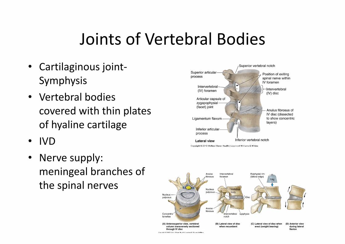
Joints of Vertebral Bodies• Cartilaginous joint‐Symphysis
• Vertebral bodies covered with thin plates of hyaline cartilage
• IVD• Nerve supply: meningeal branches of the spinal nerves
Joints of Vertebral Bodies
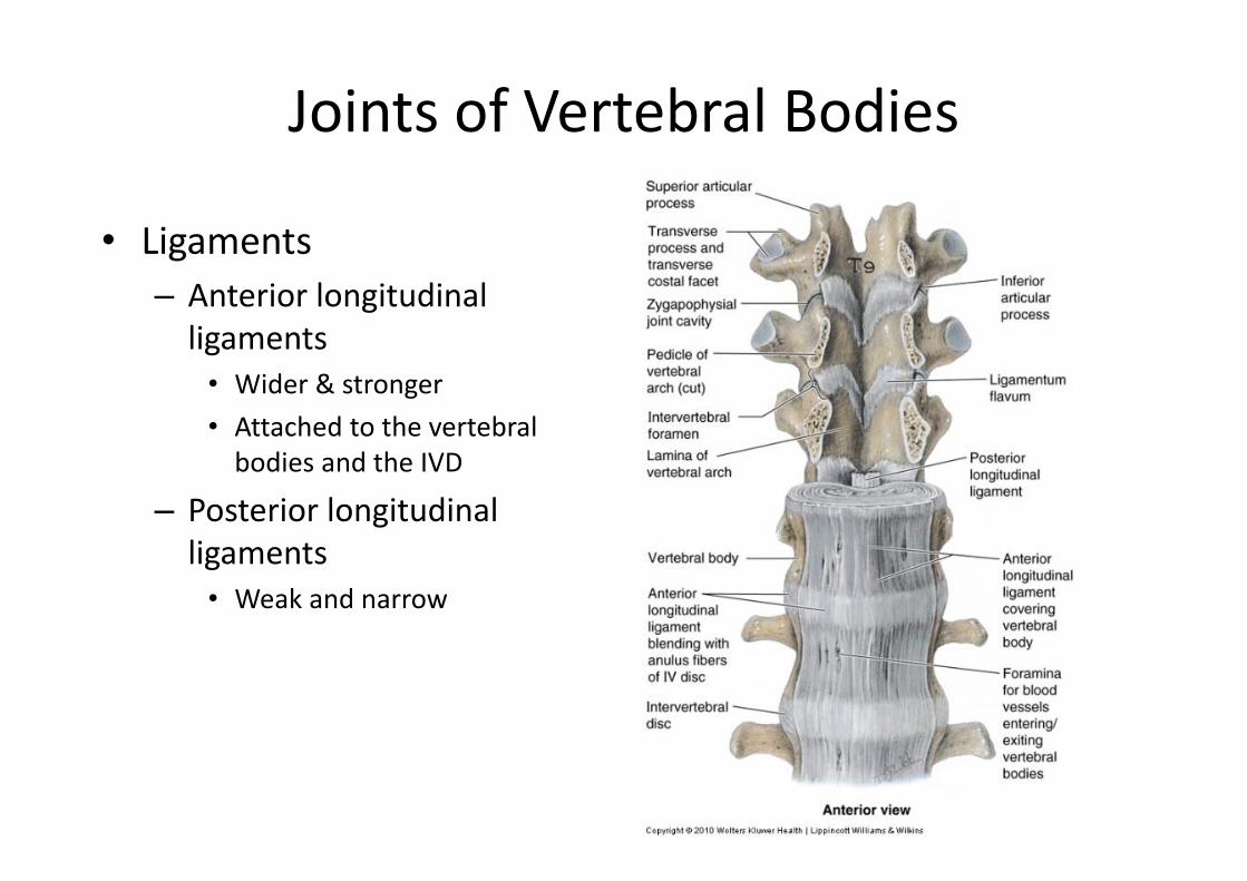
• Ligaments– Anterior longitudinal ligaments • Wider & stronger• Attached to the vertebral bodies and the IVD
– Posterior longitudinal ligaments• Weak and narrow
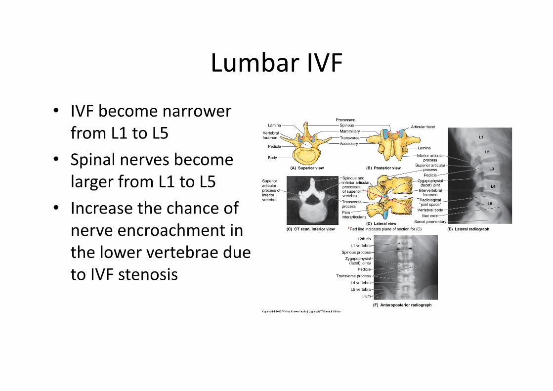
Lumbar IVF• IVF become narrower from L1 to L5
• Spinal nerves become larger from L1 to L5
• Increase the chance of nerve encroachment in the lower vertebrae due to IVF stenosis
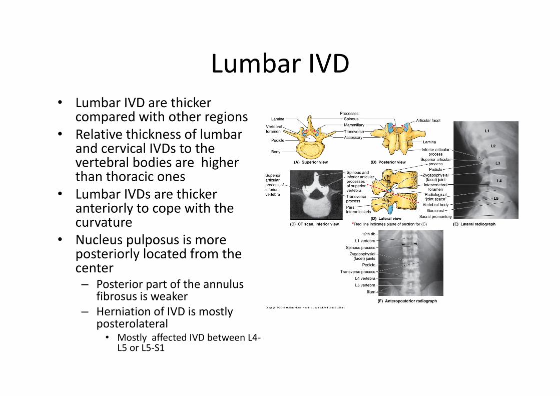
Lumbar IVD• Lumbar IVD are thicker
compared with other regions • Relative thickness of lumbar
and cervical IVDs to the vertebral bodies are higher than thoracic ones
• Lumbar IVDs are thicker anteriorly to cope with the curvature
• Nucleus pulposus is more posteriorly located from the center– Posterior part of the annulus
fibrosus is weaker– Herniation of IVD is mostly
posterolateral• Mostly affected IVD between L4‐
L5 or L5‐S1
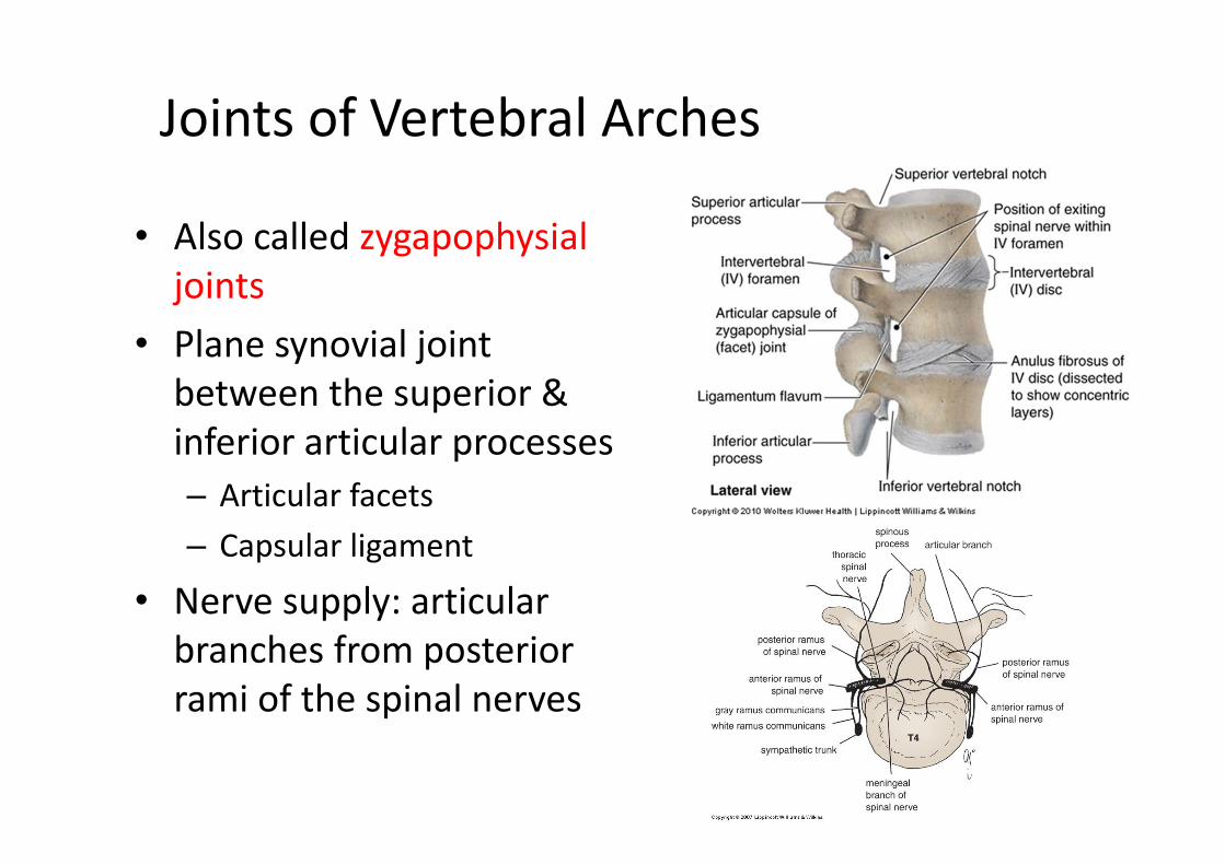
Joints of Vertebral Arches
• Also called zygapophysial joints
• Plane synovial joint between the superior & inferior articular processes– Articular facets– Capsular ligament
• Nerve supply: articular branches from posterior rami of the spinal nerves
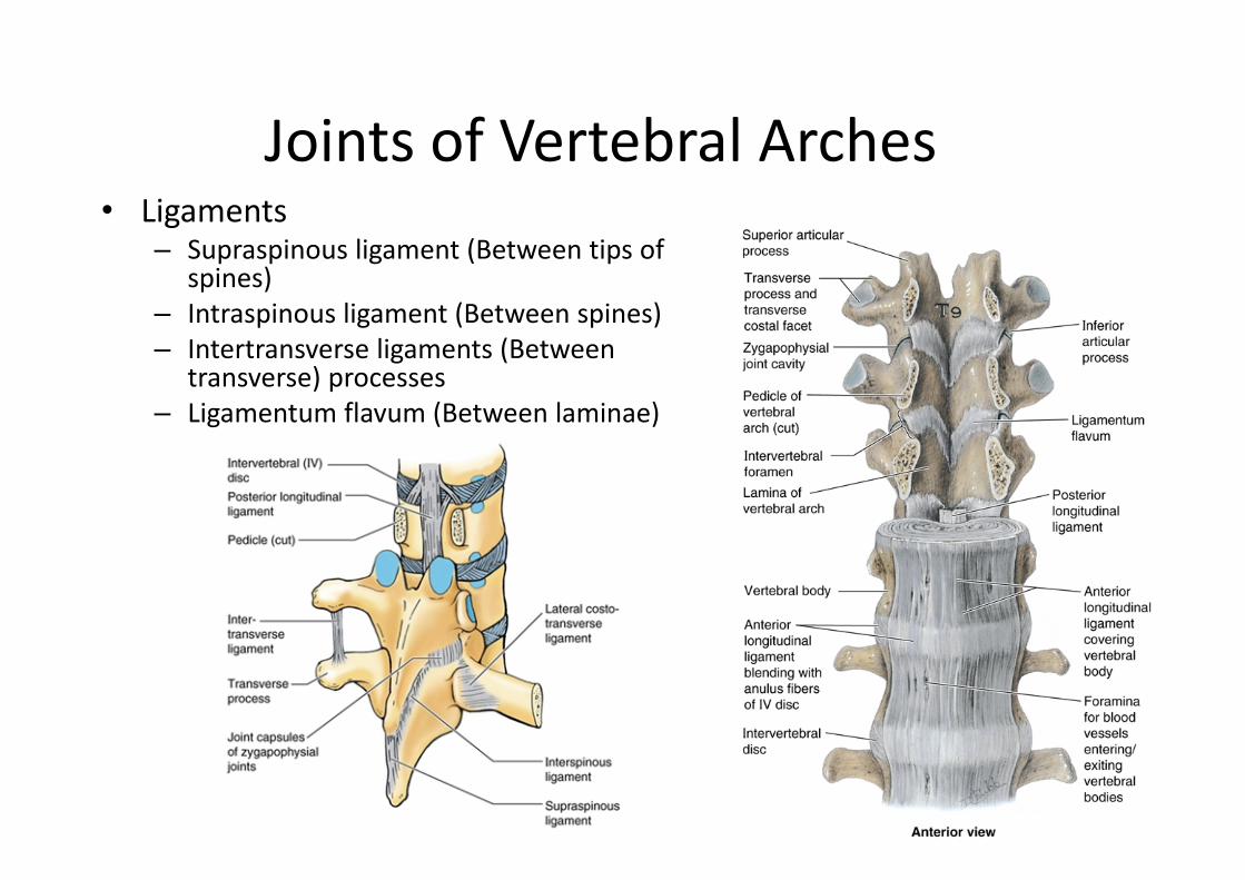
Joints of Vertebral Arches• Ligaments
– Supraspinous ligament (Between tips of spines)
– Intraspinous ligament (Between spines)– Intertransverse ligaments (Between
transverse) processes– Ligamentum flavum (Between laminae)
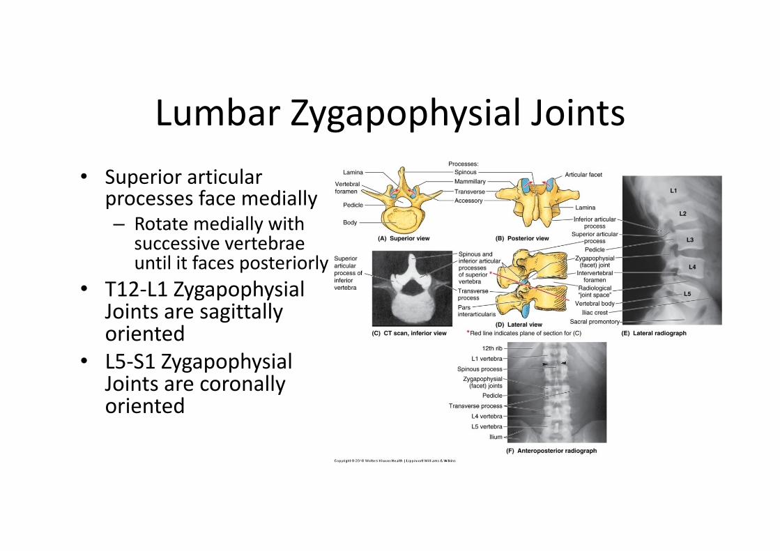
Lumbar Zygapophysial Joints• Superior articular
processes face medially– Rotate medially with
successive vertebrae until it faces posteriorly
• T12‐L1 Zygapophysial Joints are sagittallyoriented
• L5‐S1 Zygapophysial Joints are coronally oriented
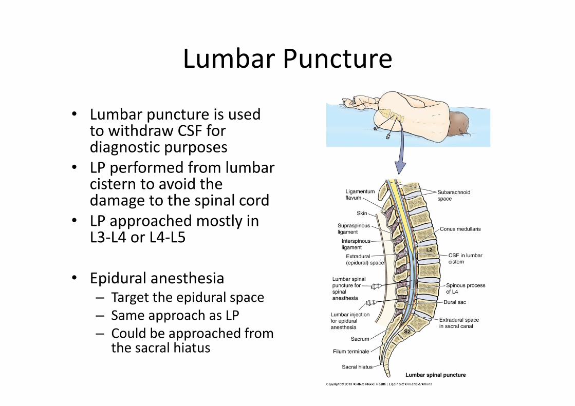
Lumbar Puncture
• Lumbar puncture is used to withdraw CSF for diagnostic purposes
• LP performed from lumbar cistern to avoid the damage to the spinal cord
• LP approached mostly in L3‐L4 or L4‐L5
• Epidural anesthesia– Target the epidural space– Same approach as LP– Could be approached from
the sacral hiatus
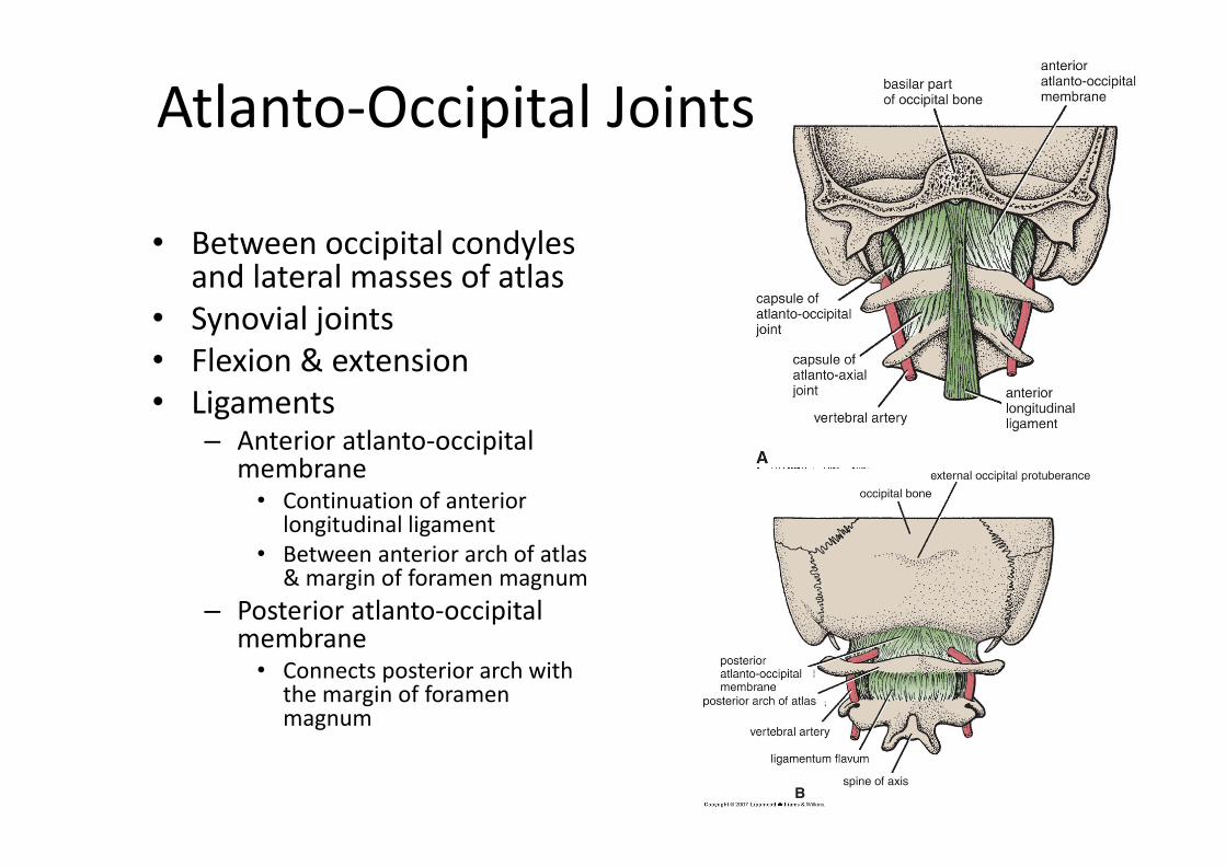
Atlanto‐Occipital Joints
• Between occipital condyles and lateral masses of atlas
• Synovial joints• Flexion & extension• Ligaments
– Anterior atlanto‐occipital membrane• Continuation of anterior
longitudinal ligament• Between anterior arch of atlas
& margin of foramen magnum– Posterior atlanto‐occipital
membrane• Connects posterior arch with
the margin of foramen magnum
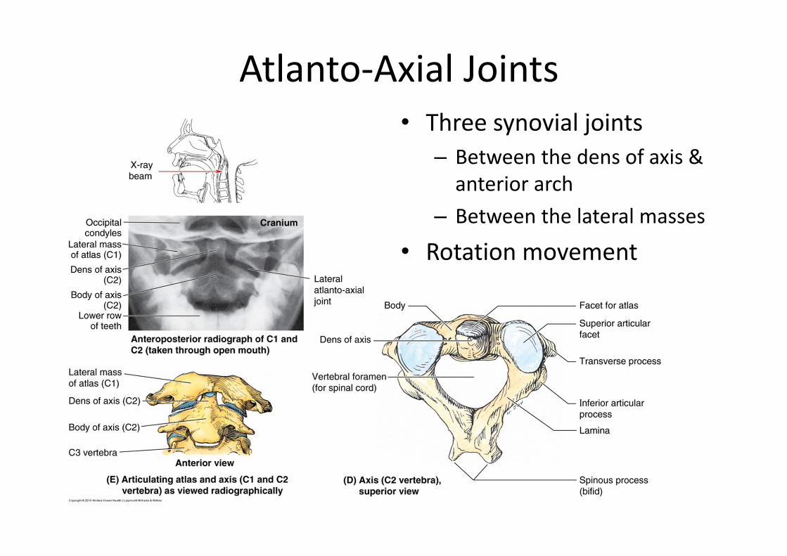
Atlanto‐Axial Joints• Three synovial joints
– Between the dens of axis & anterior arch
– Between the lateral masses
• Rotation movement
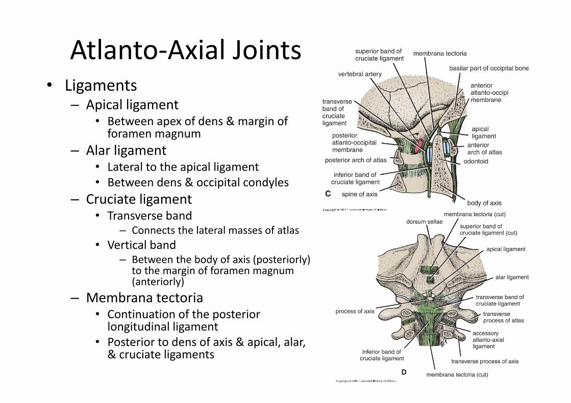
Atlanto‐Axial Joints• Ligaments
– Apical ligament• Between apex of dens & margin of foramen magnum
– Alar ligament• Lateral to the apical ligament• Between dens & occipital condyles
– Cruciate ligament• Transverse band
– Connects the lateral masses of atlas• Vertical band
– Between the body of axis (posteriorly) to the margin of foramen magnum (anteriorly)
– Membrana tectoria• Continuation of the posterior longitudinal ligament
• Posterior to dens of axis & apical, alar, & cruciate ligaments
Vertebral Column
Muscles
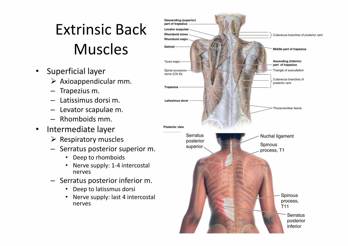
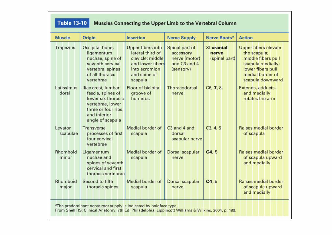
Extrinsic Back Muscles
• Superficial layer Axioappendicular mm.– Trapezius m.– Latissimus dorsi m.– Levator scapulae m.– Rhomboids mm.
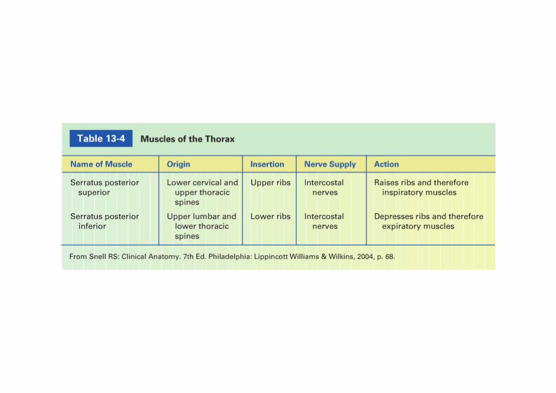
• Intermediate layer Respiratory muscles– Serratus posterior superior m.
• Deep to rhomboids• Nerve supply: 1‐4 intercostal
nerves– Serratus posterior inferior m.
• Deep to latissmus dorsi• Nerve supply: last 4 intercostal
nerves
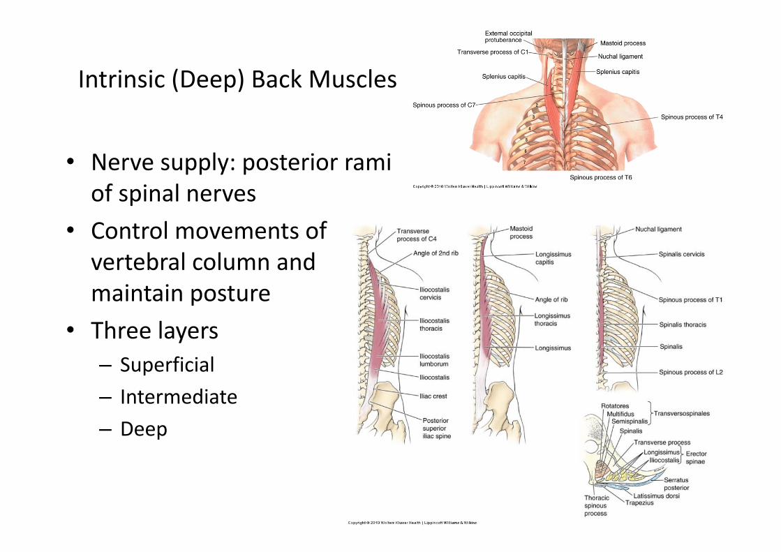
Intrinsic (Deep) Back Muscles
• Nerve supply: posterior ramiof spinal nerves
• Control movements of vertebral column and maintain posture
• Three layers– Superficial– Intermediate – Deep
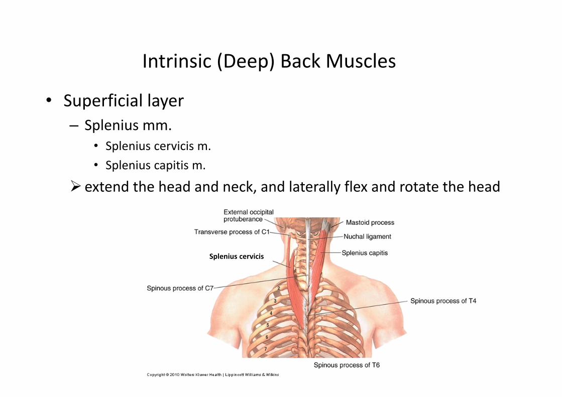
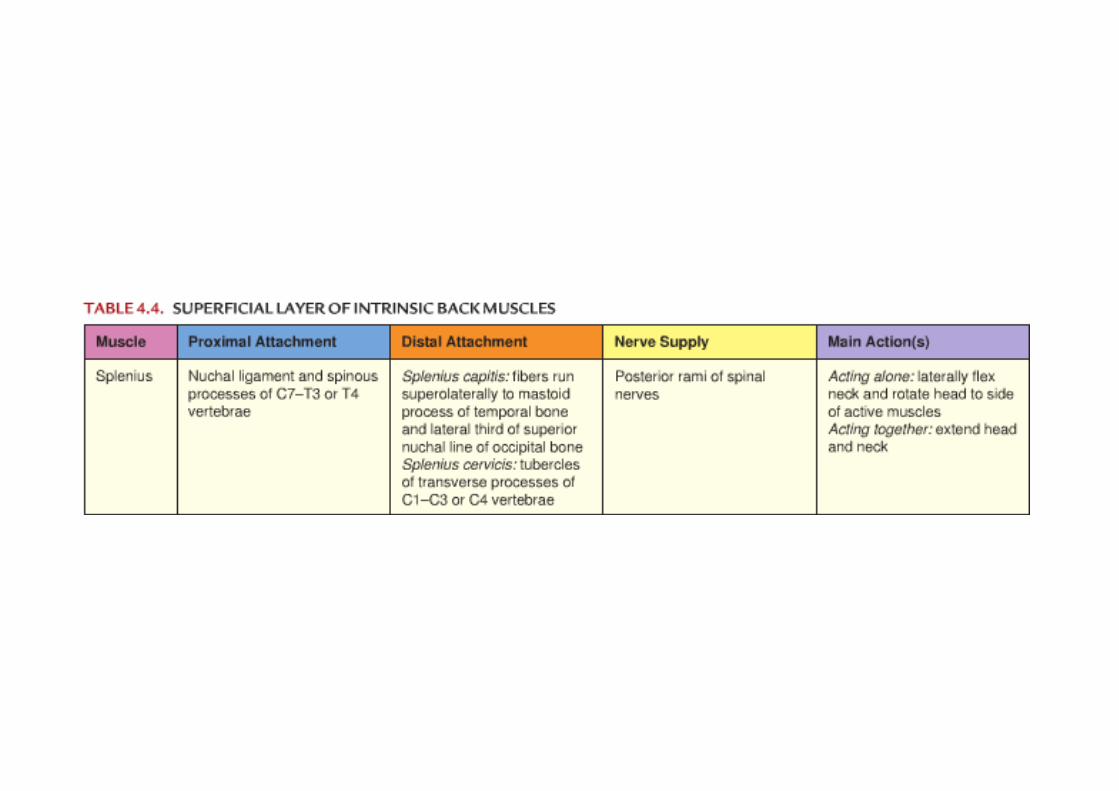
Intrinsic (Deep) Back Muscles
• Superficial layer– Splenius mm.
• Splenius cervicis m.• Splenius capitis m.
extend the head and neck, and laterally flex and rotate the head
Splenius cervicis

Intrinsic (Deep) Back Muscles
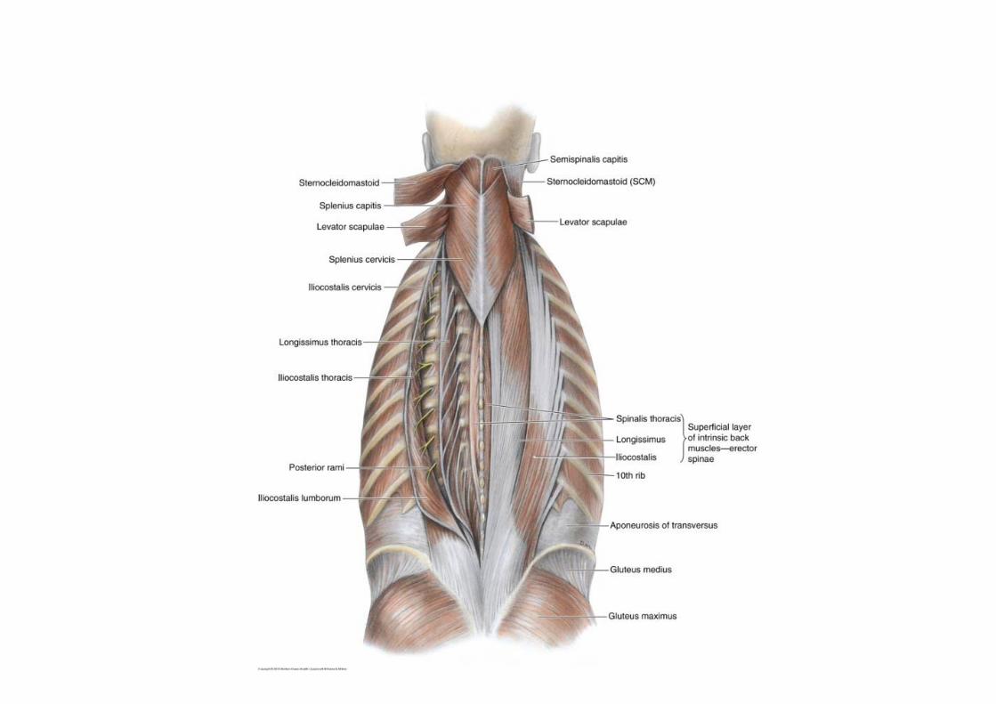
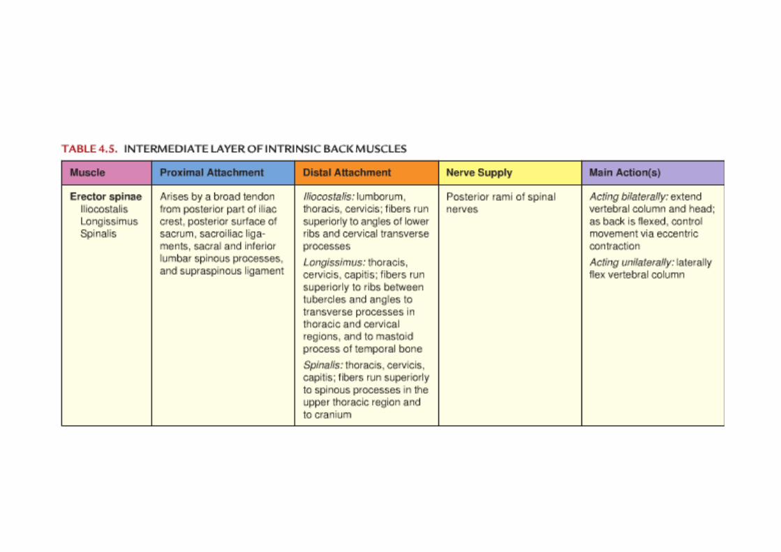
• Intermediate layer– Errector spinae mm.
• Iliocostalis (lateral column)
• Longissimus(intermediate column)
• Spinalis (medial column)
Run longitudinallyMajor extensor of the vertebral column
Intrinsic (Deep) Back Muscles
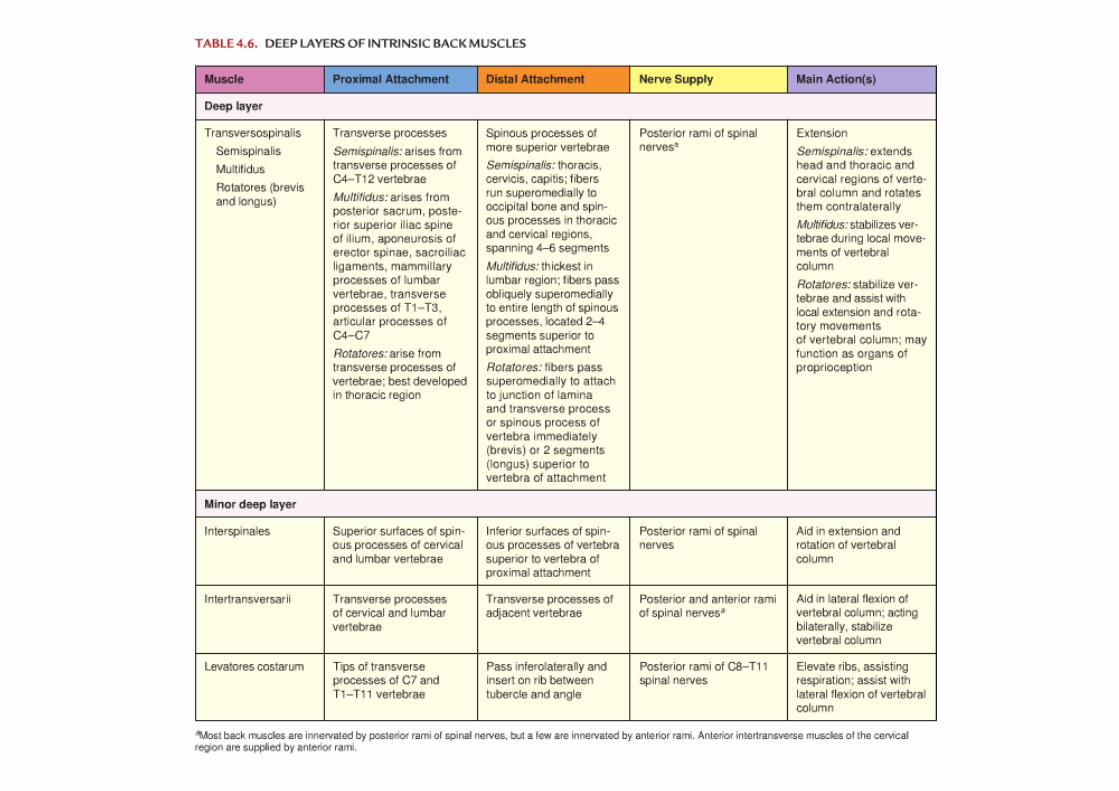
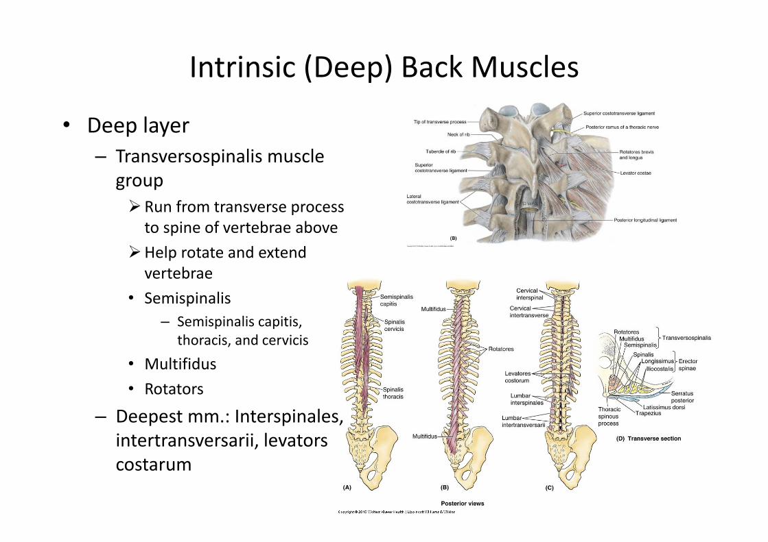
• Deep layer– Transversospinalis muscle
groupRun from transverse process to spine of vertebrae above
Help rotate and extend vertebrae
• Semispinalis– Semispinalis capitis,
thoracis, and cervicis
• Multifidus• Rotators
– Deepest mm.: Interspinales, intertransversarii, levatorscostarum