Embed Size (px)
Citation preview

Variations in rat mesenteric tissue thickness due to microvasculature
B. J. BARBER, J. OPPENHEIMER, D. C. ZAWIEJA, AND H. A. ZIMMERMANN Department of Physiology, Medical College of Wisconsin, Milwaukee, Wisconsin 53226
BARBER, B.J.,J. OPPENHEIMER, D.C. ZAWIEJA,AND H.A. ZIMMERMANN. Variations in rat mesenteric tissue thickness due to microvasculature. Am. J. Physiol. 253 (Gastrointest. Liver Physiol. 16): G549-G556,1987.-Studies of microvascular, tis- sue, and lymphatic transport using microphotometric and mi- crofluorometric techniques are potentially subject to artifact due to variations in tissue specimen thickness. Absorbance techniques utilize the Lambert-Beer law in which A = log IO/I = act, where A is absorbance, I0 is incident light intensity, I is transmitted intensity, a is an absorbance coefficient, c is con- centration of substance, and t is path length. If differences in t are known to be present, then inferences of changes in c from changes in A become suspect. In microfluorometry the amount of light gathered is proportional to the number of fluorochromes in the effective cuvette, which is determined by the microscope’s numerical aperture and the sample thickness. If variations in thickness are known to occur, the effective cuvette volume may be changing; therefore, inferences of changes in fluorochrome concentration from changes in intensity become suspect. Ex- isting data suggest that rat mesentery is 15-30 pm thick, but variation over a tissue region is unknown. Our goals are to determine thickness variation in avascular, fat-free mesenteric tissue regions; thickness variation near blood vessels; and av- erage tissue thickness. Sprague-Dawley rats were anesthetized with Inactin. Mesenteric tissue from a loop of small intestine was draped over a platform for observation; thickness was measured with an oblique microscope and a microgravimetric technique. The average variation in avascular fat-free tissue was 1.1 pm/lOO-pm distance, and average thickness was 17.4 pm. There was a significant increase in thickness over the microvasculature.
mesenteric preparation; microphotometry; tissue water con- tent; Krogh cylinder; microfluorometry; microspectropho- tometry
THE RAT mesenteric preparation (15) is widely used in microvascular studies. Variations in tissue thickness will affect techniques that are dependent on light absorption, for example, microphotometric, microfluorometric, and microspectrophotometric measurements. Thus tissue thickness in this preparation is an important parameter in the interpretation of experimental data. Mesenteric tissue thickness is also important in determining ex- change between the mesenteric tissue and superfusate in experimental preparations and in peritoneal dialysis. In theoretical analyses, leading to concepts such as the Krogh cylinder radius, a radial symmetry is assumed; this assumption would fail if the mesentery were a thin sheet in comparison to the vessels passing through it.
Witte (14) used the Jamin-Lebedeff interference tech- nique to quantitatively test for thickness variations in conjunction with studies using ultraviolet absorbance spectrophotometry. However, this technique did not give quantitative estimates of the actual thickness or its var- iation. More recently, Gahm and Witte (4) used a focus- ing-through technique to measure the thickness of rat mesentery and reported it to be between 15 and 30 pm. Fox and Wayland (2) used standard histological tech- niques to fix a tissue sample and section it for micros- copy. This single photomicrograph is the only other quantitative data available for tissue thickness in this preparation. But the histological fixing process may alter dimensions (13). Friedman (3) infused a fluorochrome until an equilibrium distribution was reached and then used the distribution of light intensity to measure rela- tive tissue water volume. Variations in volume would have suggested potential variations in tissue thickness and path length, but volume may not be precisely equated to thickness due to microheterogeneities in the intersti- tial matrix.
The primary goal of our study is to determine the variations in tissue thickness in avascular fat-free re- gions of mesenteric tissue. A second goal of the study is to estimate the thickness changes due to the microvas- culature; measurements were made with an oblique mi- croscope of thickness in the vicinity of blood vessels to determine how tissue thickness was related to vessel size. We wish to determine the largest vessel size that would be free from a thickness artifact in photometric meas- urements. The third goal of the study is to reduce the uncertainty concerning the actual thickness.
MATERIALS AND METHODS
Twenty-eight male Sprague-Dawley rats with weights ranging from 406 to 529 g (average = 460 g) were studied. The animal was anesthetized with Inactin [5-ethyl-5-( l- methylpropyl) -2-thiobarbituric acid] (50 mg/kg). Atro- pine sulfate (20 mg) was administered to reduce respi- ratory secretions. A 2-cm midline incision was made using heat cautery and a loop of small intestine was quickly exteriorized. To avoid stretching, the mesenteric tissue was loosely draped onto the animal support tray beneath the oblique microscope. A fat-free tissue region was located for measurement. After 1 min the loop was put back into the animal and another mesenteric loop was placed beneath the microscope.
Oblique microscopy. A detailed treatment of the prin- 0193-1857/87 $1.50 Copyright 0 1987 the American Physiological Society G549
G550 TISSUE THICKNESS VARIATIONS
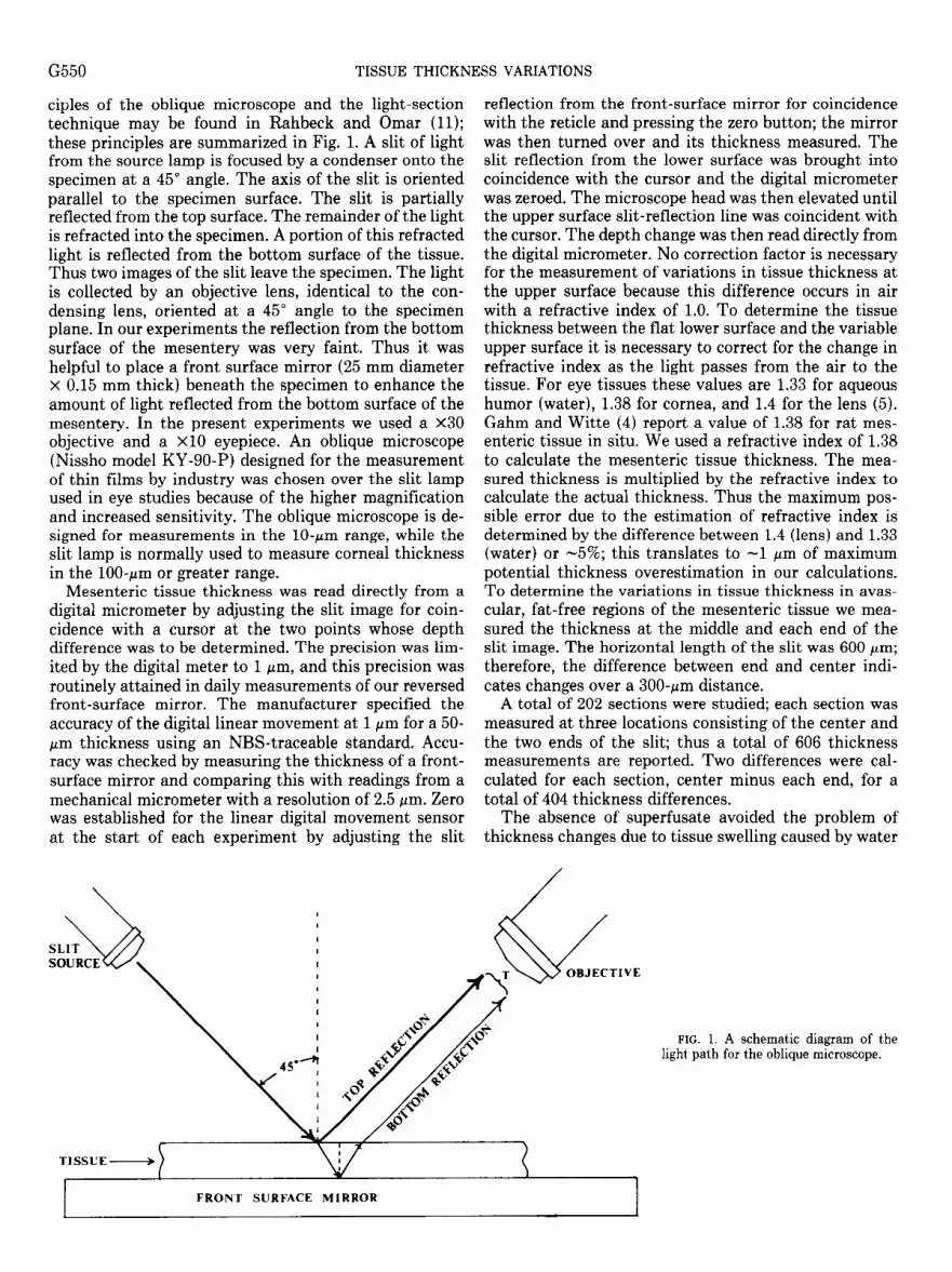
ciples of the oblique microscope and the light-section technique may be found in Rahbeck and Omar (11); these principles are summarized in Fig. 1. A slit of light from the source lamp is focused by a condenser onto the specimen at a 45" angle. The axis of the slit is oriented parallel to the specimen surface. The slit is partially reflected from the top surface. The remainder of the light is refracted into the specimen. A portion of this refracted light is reflected from the bottom surface of the tissue. Thus two images of the slit leave the specimen. The light is collected by an objective lens, identical to the con- densing lens, oriented at a 45" angle to the specimen plane. In our experiments the reflection from the bottom surface of the mesentery was very faint. Thus it was helpful to place a front surface mirror (25 mm diameter x 0.15 mm thick) beneath the specimen to enhance the amount of light reflected from the bottom surface of the mesentery. In the present experiments we used a x30 objective and a X10 eyepiece. An oblique microscope (Nissho model KY-go-P) designed for the measurement of thin films by industry was chosen over the slit lamp used in eye studies because of the higher magnification and increased sensitivity. The oblique microscope is de- signed for measurements in the lo-pm range, while the slit lamp is normally used to measure cornea1 thickness in the loo-pm or greater range.
Mesenteric tissue thickness was read directly from a digital micrometer by adjusting the slit image for coin- cidence with a cursor at the two points whose depth difference was to be determined. The precision was lim- ited by the digital meter to 1 pm, and this preci sion was routinely attained in daily measureme nts of our reversed front-surface mirror. The manufacturer specified the accuracy of the digital linear movement at 1 ,urn for a 50- pm thickness using an NBS-traceable standard. Accu- racy was checked by measuring the thickness of a front- surface mirror and comparing this with readings from a mechanical micrometer with a resolution of 2.5 pm. Zero was establish .ed for the linear digital movement sensor at the start of each experiment by adjusting the slit
reflection from the front-surface mirror for coincidence with the reticle and pressing the zero button; the mirror was then turned over and its thickness measured. The slit reflection from the lower surface was brought into coincidence with the cursor and the digital micrometer was zeroed. The microscope head was then elevated until the upper surface slit-reflection line was coincident with the cursor. The depth change was then read directly from the digital micrometer. No correction factor is necessary for the measurement of variations in tissue thickness at the upper surface because this difference occurs in air with a refractive index of 1.0. To determine the tissue thickness between the flat lower surface and the variable upper surface it is necessary to correct for the change in refractive index as the light passes from the air to the tissue. For eye tissues these values are 1.33 for aqueous humor (water), 1.38 for cornea, and 1.4 for the lens (5). Gahm and Witte (4) report a value of 1.38 for rat mes- enteric tissue in situ. We used a refractive index of 1.38 to calculate the mesenteric tissue thickness. The mea- sured thickness is multiplied by the refractive index to calculate the actual thickness. Thus the maximum pos- sible error due to the estimation of refractive index is determined by the difference between 1.4 (lens) and 1.33 (water) or -5%; this translates to -1 pm of maximum potential thickness overestimation in our calculations. To determine the variations in tissue thickness in avas- cular, fat-free regions of the mesenteric tissue we mea- sured the thickness at the middle and each end of the slit image. The horizontal length of the slit was 600 pm; therefore, the difference between end and center indi- cates changes over a 300-pm distance.
A total of 202 sections were studied; each section was measured at three locations consisting of the center and the two ends of the slit; thus a total of 606 thickness measurements are reported. Two differences were cal- culated for each section, center minus each end, for a total of 404 thickness differences.
The absence of superfusate avoided the problem of thickness changes due to tissue swelling caused by water
FIG. 1. A schematic diagram of the light pa .th for the oblique microscope.

TISSUE THICKNESS VARIATIONS G551
absorption. However, it introduced the possibility of thickness changes occurring due to tissue drying. To test for this we made measurements of thickness over a period of time and found that approximately 4 min of exposure on the stage was needed for a l-pm change in thickness to occur.
Micrograuimetric method. An independent technique was needed to verify the accuracy of the measurements with the oblique microscope; if the weight of a known tissue area could be measured then the thickness could be inferred. The standard gravimetric method was adopted to measure the wet and dry weight of mesenteric tissue samples. The rat was prepared as previously de- scribed with the mesentery exposed on the Lucite plat- form beneath a Zeiss dissecting microscope. An avascular region of mesentery was located and a trephine (Week model 9701) was used to excise a circular piece of tissue. This tissue sample was immediately transferred to an automatic electrobalance (Cahn model 25), and the weight was recorded at l-s intervals by a computer (IBM- PC)
The relationship describing mass transfer due to evap- orative water loss was first used by Dalton in 1788
dm/dt = hdA(Pws - PwA)/RT (1)
where dm/dt is the rate of loss in grams per second, hd is a mass transfer coefficient, A is the wetted surface area, PWS is the partial pressure of saturated water vapor at the surface, PWA is the partial pressure of water vapor in the ambient air, R is the gas constant, and T is the air temperature (12). This equation predicts a constant rate of water loss if one assumes that the wetted area and the vapor pressure differences remain constant. Near the end of the drying process the wetted area must diminish, and if one assumes that the area is proportional to the amount of water remaining, then the solution to Eq. 1 becomes an exponential relationship. This linear rate of loss followed by an exponential approach to zero is the result that we obtain when a small droplet of water is placed on the electrobalance and allowed to evaporate.
For small tissue samples taken with a 5mm diameter trephine (the final drying phase of large samples), the weight is best described by an exponential with an offset for the dry weight
T = D + W exp(-kt) (2)
where T is the total weight, D is the dry weight, W is the initial weight of water in the sample, k is the drying constant, and t is the time in seconds after trephining.
RESULTS
Only two lines were visible in our oblique microscope observations; the supporting mirror always appeared as a straight line, while the reflection from the upper surface of the tissue was about 1 pm thick. The upper surface reflection was a straight line in avascular, fat-free tissue regions and exhibited a distinct upward bow in the vicin- ity of blood vessels. The lines diverged strongly in the vicinity of adipose tissue. Only data from fat-free regions are reported in the present study.
The relationship between tissue thickness and tissue
thickness variation is summarized in Fig. 2. The corre- lation coefficient between thickness variation over 300 pm and- thickness was 0.33, indicating that only -11% of the difference in tissue thickness variation is related to the local tissue thickness. The slope of the regression line was 0.11 pm of variation per micrometer of tissue thickness over a 300~pm region; the intercept was 1.44 pm. The slope was significant at the 0.05 level. Therefore, thicker tissue regions showed slightly more variation.
In avascular, fat-free regions thickness averaged 17.4 t 0.4 (means t SE, n = 606) pm. The frequency distri- bution of these measurements is shown in Fig. 3. This distribution is positively skewed with a skewness coeffi- cient of 1.5 and a coefficient of kurtosis of 4.8. The skewness is statistically significant at the 0.05 level and corresponds to the obvious clustering of points near the low end of the range. For a normal distribution, the kurtosis value is zero. A positive result indicates an excess of values near the mean and far from it, as opposed to a more evenly distributed curve. The kurtosis is sig- nificant at the 0.05 level. This distribution may, in part, have been due to the occasional presence of the intestinal wall, large blood vessels, or adipose regions just beyond the field of view during the measurement. Any of these features could have produced the large thickness values indicated by the kurtosis. Values near zero are necessar- ily rare; this alone would probably be sufficient to cause a Poisson distribution.
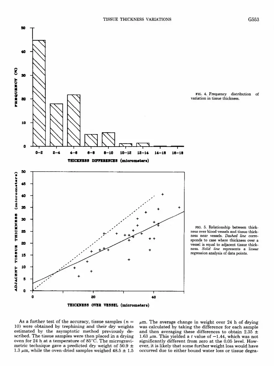
The frequency distribution for thickness variation is shown in Fig. 4. This distribution has a mean of 3.4 t 0.2 (n = 404) with a skewness of 2.25 and a kurtosis of 8.41. This value of tissue thickness variation suggests that the thickness variations in avascular, fat-free mes- enteric tissue will be on the order of 1 pm/l00 pm of distance or -1%.
For transport studies, the areas near a vessel are of considerable interest. Therefore, a series of measure- ments was made of thickness over a vessel and over avascular fat-free tissue extending six vessel diameters away from the vessel wall. These measurements were performed on each side of the vessel. The results are summarized in Fig. 5. A linear regression performed on these normally distributed data values was statistically significant at the 0.05 level. The correlation coefficient was 0.81, indicating that tissue thickness near the vessel was related to thickness over the vessel. If there was no relationship between vessel size and adjacent tissue thickness, then the correlation coefficient would be zero and the slope of the fit line would be zero. The actual slope was 0.61 (sample SD regression coefficient = 0.08, n = 31) with a t value of 7.35 and an F statistic of 54. If there had been no difference in thickness between the vessel and nearby tissue, then the slope of the regression line would have been equal to 1. However, the slope of the line is significantly different (P < 0.001) from 1; 95% (t = 2.045) confidence limits ranged from 0.45 to 0.77. The thickness anomaly persists with decreasing vessel thickness down to a value of 15 pm. Hence, changes in tissue thickness may occur down to the capillary level. Note that these statistics relate to the slope of the regression line rather than to an individual data point.

G552 TISSUE THICKNESS VARIATIONS
8 13 (3 18
II 0
0 8
3 7
0 8
28
E 18 Y t 18
i 14 3 t 12
f 10
4
0
! -4 + ++
- +
- + + +I= + -
+ + I + + + #
- ++ ++ +i++ + +
- + + +* +++I= +* + +
- +++ +I+ +*i+ ++ + + + + + + *+ i4+*+ +I+++
- + ++l+H-i++*++++ +I-+ ++ + + +
- +w++H- +t+++ ++++++ + + + ++-I+ ++w+++Ht +*+++ +
- ++=+++-I++ ++-+ +* +
. . .
. I 1 ’ I I i
0 20 40
AVBRAGB TISSUB THICXBBSS (mlcrometer~)
FIG. 2. Relationship between tissue thickness and changes in tissue thick- ness over a 300-pm distance in avascular fat-free regions of rat mesentery.
FIG. 3. Frequency distribution of tis- sue thickness in rat mesentery.
O-S s-10 10-1s m-80 eo-m a-30 30-m se40 4004a 4640
TI88uB TmCmB88 (ml~rnOtu8)
While it is unlikely, it is possible to find regions that are free of thickness artifact, for example, the single data point in Fig. 5. Furthermore, there are several points that show only a small thickness artifact.
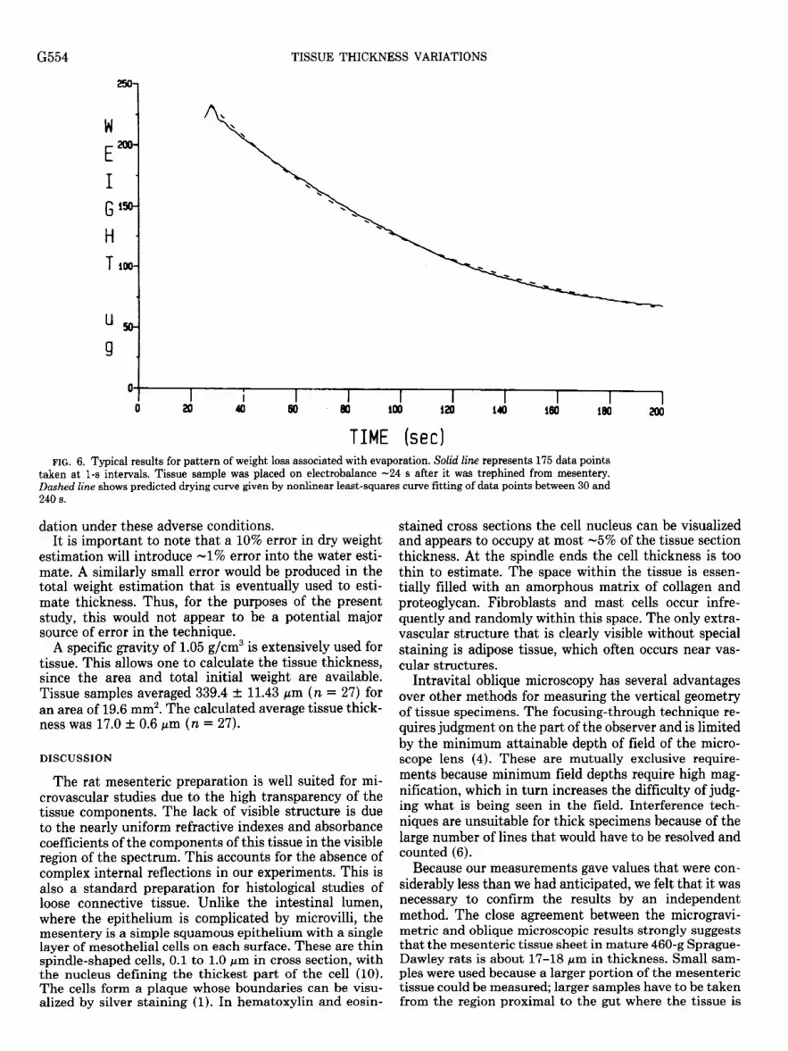
Typical results for the pattern of weight loss associated with evaporation are shown in Fig. 6. In all cases the curve-fitting correlation coefficient between observed and predicted weight exceeded 0.999. Student’s t test
values ranged from 10 to 40 on 200 readings, indicating goodness of fit at the 0.001 level, while F values ranged from 100 to 1,000, indicating significance at the 0.001 level. Goodness of fit is best evidenced by the maximum residual being ~5% of the data value for any point. The average of the residuals was ~1% and was not signifi- cantly different from zero at the 0.05 level; the standard deviation of the residuals was Cl%.
TISSUE THICKNESS VARIATIONS G553
n
2
3
fi 0
O-2 2-4 4-8 8-8 8-10 lo-12 12-M M-18 M-18
TBICRtB88 DR?BRBHCB8 (mahmmakrm)
so
48
40
35
30
25
20
15
10
5
0
FIG. 4. Frequency distribution of variation in tissue thickness.
FIG. 5. Relationship between thick- ness over blood vessels and tissue thick- ness near vessels. Dashed Line corre- sponds to case where thickness over a vessel is equal to adjacent tissue thick- ness. Solid line represents a linear regression analysis of data points.
0 20 40
TEICKNBSS CWBR VBSSBL (micrometers)
As a further test of the accuracy, tissue samples (n = 10) were obtained by trephining and their dry weights
pm. The average change in weight over 24 h of drying
estimated by the asymptotic method previously de- was calculated by taking the difference for each sample and then averaging these differences to obtain 2.35 t
scribed. The tissue samples were then placed in a drying 1.63 pm. This yielded a t value of -1.44, which was not oven for 24 h at a temperature of 85OC. The microgravi- significantly different from zero at the 0.05 level. How- metric technique gave a predicted dry weight of 50.9 t ever, it is likely that some further weight loss would have 1.3 pm, while the oven-dried samples weighed 48.5 t 1.5 occurred due to either bound water loss or tissue degra-
G554 TISSUE THICKNESS VARIATIONS
FIG. 6. Typical results for pattern of weight loss associated with evaporation. Solid line represents 175 data points taken at l-s intervals. Tissue sample was placed on electrobalance -24 s after it was trephined from mesentery. Dashed line shows predicted dryingcurve given by nonlinear least-squares curve fitting of data points between 30 and 240 s.
TIME (set)
dation under these adverse conditions. It is important to note that a 10% error in dry weight
estimation will introduce -1% error into the water esti- mate. A similarly small error would be produced in the total weight estimation that is eventually used to esti- mate thickness. Thus, for the purposes of the present study, this would not appear to be a potential major source of error in the technique.
A specific gravity of 1.05 g/cm3 is extensively used for tissue. This allows one to calculate the tissue thickness, since the area and total initial weight are available. Tissue samples averaged 339.4 t 11.43 pm (n = 27) for an area of 19.6 mm2. The calculated average tissue thick- ness was 17.0 t 0.6 pm (n = 27).
DISCUSSION
The rat mesenteric preparation is well suited for mi- crovascular studies due to the high transparency of the tissue components. The lack of visible structure is due to the nearly uniform refractive indexes and absorbance coeffic ients of the components of this tissue in the visible region of the spectrum. This accounts for the absence of complex internal reflections in our experiments. This is also a standard preparation for histological studies of loose connective tissue. Unlike the intestinal lumen, where the epithelium is complicated by microvilli, the mesentery is a simple squamous epithelium with a single layer of mesothelial cells on each surface. These are thin spindle-shaped cells, 0.1 to 1.0 pm in cross section, with the nucleus defining the thickest part of the cell (10). The cells form a plaque whose boundaries can be visu- alized by silver staining (1). In hematoxylin and eosin-
stained cross sections the cell nucleus can be visualized and appears to occupy at most -5% of the tissue section thickness. At the spindle ends the cell thickness is too thin to estimate. The space within the tissue is essen- tially filled with an amorphous matrix of collagen and proteoglycan. Fibroblasts and mast cells occur infre- quently and randomly within this space. The only extra- vascular structure that is clearly visible without special staining is adipose tissue, which often occurs near vas- cular structures.
Intravital oblique microscopy has several advantages over other methods for measuring the vertical geometry of tissue specimens. The focusing-through technique re- quires judgment on the part of the observer and is limited by the minimum attainable depth of field of the micro- scope lens (4). These are mutually exclusive require- ments because minimum field depths require high mag- nification, which in turn increases the difficulty of judg- ing what is being seen in the field. Interference tech- niques are unsuitable for thick specimens because of the large number of lines that would have to be resolved and counted (6).
Because our measurements gave values that were con- siderably less than we had anticipated, we felt that it was necessary to confirm the results by an independent method. The close agreement between the microgravi- metric and oblique microscopic results strongly suggests that the mesenteric tissue sheet in mature 460-g Sprague- Dawley rats is about 17-18 pm in thickness. Small sam- ples were used because a larger portion of the mesenteric tissue could be measured; larger samples have to be taken from the region proximal to the gut where the tissue is

TISSUE THICKNESS VARIATIONS G555
-5 pm thinner on average. Furthermore, the exponential extrapolation is more likely to overestimate tissue thick- ness compared with the linear extrapolation used with large samples. Thus this technique provides the most conservative estimate of tissue thickness when low values are in question.
It is important to note that a 10% error in dry weight estimation will introduce -1% of error into the water estimate. A similarly small error would be produced in the total weight estimation that is used to estimate thickness. Thus, for the purposes of the present study, dry weight estimation errors would not appear to be a major limitation on the accuracy of thickness estimation. The major potential source of error is almost certainly in the initial water estimate due to errors in the necessity to extrapolate a curve fit backward to the starting time for the drying process. By using small samples and ex- ponential extrapolation, we have deliberately tried to bias our results toward overestimating initial weight and therefore thickness.
Measurements using the Jamin-Lebedeff interference technique yield a qualitative result such that changes in thickness caused color changes; however, it is unclear how much variation was needed to cause a measurable color change. In any case, these measurements were restricted to avascular, fat-free mesentery, and the neg- ative results are essentially in agreement with our meas- urements and those of others (2,3). More recently Gahm and Witte (4) used a focusing-through technique to mea- sure the thickness of, mesenteric tissue. They reported that the thickness is greater than 15 and less than 30 pm. However, the focusing-through technique only gives thickness at one point, while oblique microscopy allows changes over a distance to be visualized and measured. Therefore, it is much more difficult to examine variations in thickness using the focusing-through technique. No values for variation were reported by Gahm and Witte (4). The reported range of thickness falls within the upper range of our measurements. However, Ringer so- lution was used as a superfusate, and thus may have produced edema in the mesenteric tissue due to water absorption caused by gel matrix swelling pressure.
Intravital oblique microscopy allows one to quantify the three-dimensional structure of the microvasculature in situ, thereby avoiding potential problems due to arti- facts introduced by histological fixing. The tissue thick- ness in the photomicrograph taken by Fox and Wayland (2) ranges from -17 pm in avascular regions to -40 pm in regions that were more vascularized. This is within the range of values we obtained using both light section and microgravimetric techniques; this suggests that the tissue dimensions were unaltered by histological fixing.
The dimensions of frog mesentery were measured by Mason et al. (9) using a focusing-through technique. The depth over capillaries was 25.6 t 12.7 pm (65 measure- ments in 10 mesenteries); between capillaries the depth was 12.7 t 2.9 pm (n = 72). The average capillary diameter was -20 pm. Thus thickness variation due to the microvasculature in frog mesentery is within the 95% confidence interval for rats (0.44-0.77). The value for mesenteric thickness falls just below the peak in Fig. 3;
however, the 95% confidence intervals for thickness do not overlap. This may be due to the lower body weight of the frogs.
Our data indicate that results from absorbance micro- photometric measurements in avascular, fat-free rat mesenteric tissue distant from vessels and the gut wall are likely to have at most a 2% error due to thickness variations. The results of a single scan over 100 pm of tissue should have a typical error of -1%. This is well within the generally acceptable error range for photo- metric measurements. On the other hand photometric measurements made across combined vascular and avas- cular regions are likely to have a thickness variation artifact. Figure 5 suggests that this thickness variation is fairly uniform and predictable. This permits the po- tential photometric effects to be estimated; a correction for this effect can then be made if it is deemed necessary.
The Krogh cylinder radius for oxygen is -35 pm (8). This is about twice the tissue thickness in the vicinity of the capillaries in this preparation. Thus the superfusate is likely to be the dominant factor influencing oxygen distribution in this preparation. Klitzman, Popel, and Duling (7) have shown that oxygen tension is relatively constant within 20 pm of the surface. The movement of diffusible dyes and fluorescently labeled substances is likely to be strongly affected because the path to the superfusate is short compared with paths within the tissue.
Our results show that thickness variations are likely to occur in the vicinity of the microvasculature in the rat mesenteric preparation. There is little, if any, variation in thickness over 300-pm distances in fat-free loose con- nective tissue distal to the microvasculature. Future studies are needed using instruments that combine both oblique and conventional microscopy techniques to fur- ther elucidate the vertical geometry of this microsvas- cular preparation.
The authors thank J. Salkowski for her help in preparing the illustrations for this manuscript. We are also grateful to Emile Rabito of Vickers Instruments, Malden, MA, for providing the oblique micro- scope.
This study was supported by The Whitaker Foundation and Na- tional Institutes of Health Program Project Grant HL-29587.
Received 20 October 1986; accepted in final form 14 May 1987.
REFERENCES
1.
2.
3.
BLOOM, W., AND D. W. FAWCETT. A Textbook of Histology. Phila- delphia, PA: Saunders, 1968, p. 158. FOX, J. R., AND H. WAYLAND. Interstitial diffusion of macromol- ecules in the rat mesentery. Microuczsc. Res. 18: 255-276, 1979. FRIEDMAN, J. J., AND S. WITTE. The radial protein concentration profile in the interstitial space of the rat ileal mesentery. Microuusc. Res. 31: 277-287, 1986. GAHM, T., AND S. WITTE. Measurement of the optical thickness of transparent tissue layers. J. Microsc. 141: 101-110, 1986. GUYTON, A. C. Textbook of Medical Physiology. Philadelphia, PA: Saunders, 1985, p. 703. * HEAVENS, 0. S. Optical Properties of Thin Solid Films. New York: Dover, 1965, p. 96-154. KLITZMAN, B., A. POPEL, AND B. DULING. Oxygen transport in resting and contracting hamster cremaster muscles: experimental and theoretical microvascular studies. Microuasc. Res. 25: 108-131, 1983.
8. KROGH, A. The number and distribution of capillaries in muscles

G556 TISSUE THICKNESS VARIATIONS
with calculation of the oxygen pressure head necessary for supply- ing the tissue. J. Physiol. Lord. 52: 409, 1918-1919.
9. MASON, J. C., F. E. CURRY, I. F. WHITE, AND C. C. MICHEL. The ultrastructure of frog mesenteric capillaries of known filtration coefficient. Q. J. Exp. Physiol. 64: 217-224, 1979.
10. PIETRA, G. G. Pathophysiology involving the microcirculation. In: The Physiology and Pharmacology of the Microcirculation, edited by N. A. Mortillaro. New York: Academic, 1984, vol. 2, p. 387-417.
11. RAHBECK, H., AND M. OMAR. Measurement of the thickness of transparent films with the light-profile microscope. Nature Land. 169: 1008,1952.
12. RAPP, GEORGE M. Convective mass transfer and the coefficient of evaporative water loss from human skin. In: Physiological and
Behavioral Temperature Regulation, edited by J. D. Hardy, A. P. Gagge, and J. A. J. Stolwijk. Springfield, IL: Thomas, 1970, p. 55- 80.
13. WAYLAND, H., AND P. C. JOHNSON. Future trends in microcircu- latory research. In: Microcirculation, edited by G. Kakey and B. M. Altura. Baltimore, MD: University Park, 1980, vol. 3, p. 483-519.
14. WITTE, S., AND S. ZENZES-GEPRAGS. Extravascular protein meas- urements in vivo and in situ by ultramicrospectrophotometry. Microvasc. Res. 13: 225-231, 1977.
15. ZWEIFACH, B. W. Microscopic observations of circulation in the rat mesoappendix and dog omentum. In: Methods in Medical Re- search, edited by V. R. Putte. Chicago, IL: Year Book, 1948, p. 131-139.


