Embed Size (px)
Citation preview

Journal of Inorganic Biochemistry 105 (2011) 303–312
Contents lists available at ScienceDirect
Journal of Inorganic Biochemistry
j ourna l homepage: www.e lsev ie r.com/ locate / j inorgb io
Vanadium polypyridyl compounds as potential antiparasitic and antitumoral agents:New achievements
Julio Benítez a, Lorena Becco b, Isabel Correia c, Sandra Milena Leal d, Helena Guiset e, João Costa Pessoa c,Julia Lorenzo f, Sebastian Tanco f, Patricia Escobar d, Virtudes Moreno e, Beatriz Garat b, Dinorah Gambino a,⁎a Cátedra de Química Inorgánica, Facultad de Química, UDELAR, Gral. Flores 2124, 11800 Montevideo, Uruguayb Laboratorio de Interacciones Moleculares, Facultad de Ciencias, UDELAR, Iguá 4225, 11400 Montevideo, Uruguayc Centro Química Estrutural, IST-TU-Lisbon, Av Rovisco Pais, 1049-001 Lisbon, Portugald Centro de Investigación de Enfermedades Tropicales (CINTROP), Escuela de Medicina, Departamento de Ciencias Básicas, Universidad Industrial de Santander, Bucaramanga, Colombiae Departamento de Química Inorgánica, Universitat Barcelona, Martí i Franquès 1-11, 08028 Barcelona, Spainf Institut de Biotecnologia i de Biomedecina, Universitat Autònoma de Barcelona, 08193 Bellaterra, Barcelona, Spain
⁎ Corresponding author. Tel.: +598 29249739; fax: +E-mail address: [email protected] (D. Gambino).
0162-0134/$ – see front matter © 2010 Elsevier Inc. Aldoi:10.1016/j.jinorgbio.2010.11.001
a b s t r a c t
a r t i c l e i n f oArticle history:Received 5 September 2010Received in revised form 30 October 2010Accepted 2 November 2010Available online 12 November 2010
Keywords:Vanadyl compoundsHeteroleptic complexesChagas diseaseLeishmaniasisAntitumoralPhenanthroline
In the search for new therapeutic tools against diseases produced by kinetoplastid parasites five vanadylcomplexes, [VIVO(L-2H)(phen)], including 1,10-phenanthroline (phen) and tridentate salicylaldehydesemicarbazone derivatives as ligands have been synthesized and characterized in the solid state and insolution by using different techniques. EPR suggested a distorted octahedral geometry with the tridentatesemicarbazone occupying three equatorial positions and phen coordinated in an equatorial/axial mode. Thecompounds were evaluated in vitro on epimastigotes of Trypanosoma cruzi, causative agent of Chagas disease,Leishmania panamensis and Leishmania chagasi and on tumor cells. The complexes showed higher in vitro anti-trypanosomal activities than the reference drug Nifurtimox (IC50 values in the range 1.6–3.8 μM) andincreased activities in respect to the free semicarbazone ligands. In vitro activity on promastigote andamastigote forms of Leishmania showed interesting results. The compounds [VO(L1-2H)(phen)] and [VO(L3-2H)(phen)], where L1 = 2-hydroxybenzaldehyde semicarbazone and L3 = 2-hydroxy-3-methoxybenzaldehydesemicarbazone, resulted active (IC50 2.74 and 2.75 μM, respectively, on promastigotes of L. panamensis; IC50 19.52and 20.75 μM, respectively, on intracellular amastigotes of L. panamensis) and showed low toxicity on THP-1mammalian cells (IC50 188.55 and 88.13 μM, respectively). In addition, the complexes showed cytotoxicity onhuman promyelocytic leukemia HL-60 cells with IC50 values of the same order of magnitude as cisplatin. Theinteraction of the complexes with DNA was demonstrated by different techniques, suggesting that thisbiomolecule could be a potential target either in the parasites or in tumor cells.
598 29241906.
l rights reserved.
© 2010 Elsevier Inc. All rights reserved.
1. Introduction
Among the infectious illnesses designated by the World HealthOrganization as neglected tropical diseases, American trypanosomiasis(Chagas disease) and leishmaniasis constitute important healthproblems concentrated in the poorest tropical or subtropical regionsof theplanet [1–3]. TogetherwithhumanAfrican trypanosomiasis, thesediseases constitute theneglected tropical diseaseswith the highest ratesof death [1]. The etiologic agents of both, Trypanosoma cruzi andLeishmania spp, are protozoan parasites that belong to the trypanoso-matid genus and kinetoplastid order and are mainly transmitted to themammalian host by certain insects [4–6].
American trypanosomiasis is the major parasitic disease in theAmericas, being endemic throughout Latin America, infecting 8–14
million people and causing more deaths per year in this region thanany other parasitic disease (14,000 deaths per year). In addition, thepremature disability and the effect of this disease on worker produc-tivity lead to very significant annual losses of resources and industrialproductivity. Furthermore, globalization and immigration of unknow-ingly infected people from Latin America has also led to the appearanceof several infection cases in developed countries mainly due to lack ofcontrols and screening in blood and organ banks [2]. It is interesting tonote that although exhaustively described for the first time in 1909 bythe Brazilian scientist Carlos Chagas, there are evidences demonstratingthat this disease has been present in the American continent for morethan 9000 years [4].
Leishmaniasis involves a group of diseases produced by differentprotozoa of the genus Leishmania and range from cutaneous leishman-iasis to mucocutaneous infections or fatal disseminating visceralleishmaniasis. It currently affects about 12 million people worldwidewith1.5–2millionnewcases per year, including approximately 500,000cases of the visceral form of the disease, which is nearly 100% fatal if

304 J. Benítez et al. / Journal of Inorganic Biochemistry 105 (2011) 303–312
untreated. Approximately 350 million people live at risk of infectionwith these parasites. Leishmaniases are prevalent in 88 countries inAfrica, South Asia, and Latin America. In recent years this disease hasincreased its prevalence in the south of Europe due to co-infection ofpatients affected by HIV–AIDS [7].
The chemotherapy of trypanosomatid infections mostly relies ondrugs that date back over 50 years and that are known for poor effi-cacy, high toxicity, and increasing resistance. The treatment of Chagasdisease is based on old and quite unspecific nitroheterocyclic drugs,nifurtimox and benznidazole, that have significant activity only in theacute phase of the disease and, when associated with long-termtreatments, give rise to severe side effects [2–4,8]. Only antifungal tri-azoles have demonstrated therapeutic potentiality in preclinical devel-opment to beworth the start of clinical trials. Very recently Merck & Co.Inc. decided to commence amid-stage investigational proof-of-conceptclinical study of posoconazole, as a candidate for the treatment ofchronic Chagas disease [9].
The available treatments for leishmaniases are also far from beingideal. The first-line treatment relies traditionally on the pentavalentantimonial drugs, sodium stibogluconate and meglumine antimoni-ate. These antimonials may cause severe side effects and developmentof resistance is now observed in several cases and geographicalregions, emphasizing the urgent need for new treatments. New drugsagainst both diseases are urgently needed.
The broad type of metal ions' activities in biology have stimulated,in the past decades, the development of metal-based chemother-apeutics in different fields of medicine. Even though emphasis hasbeenmainly placed on cancer treatment, leading research by Sánchez-Delgado et al. lead to some interesting potential metallopharmaceu-ticals for Chagas disease andmalaria [5,6]. Currently, the developmentof bioactivemetal complexes is a promising and attractive approach inthe search for newpotential drugs for the therapy of parasitic illnesses.Attempts towards developing trypanocidal metal-based compoundshave been described [10–12]. In particular, we have been successfullyworking on the development of potential anti-trypanosome and anti-Leishmania agents through different approaches [13–22].
Metabolic pathways of kinetoplastid parasites (Leishmania andTrypanosoma parasites) are similar to those present in tumor cellsleading to a correlation between antitrypanosomal and antitumor activ-ities. Moreover, it has been proposed that compounds that efficientlyinteract with DNA in an intercalative mode could also show antitrypa-nosomatid activity [6,23]. Having this in mind, some homoleptic andheteroleptic vanadyl complexes including DNA intercalators as ligands(dppz = dipyrido[3,2-a: 2′,3′-c]phenazine and bipy = 2,2′-bipyridine)have been previously designed by us as potential antitrypanosomalagents. Thehomoleptic vanadyl complex [VIVO(SO4)(H2O)2(dppz)]·2H2Oshowed a slightly higher in vitro activity than the reference drugNifurtimox on T. cruzi Dm28c strain epimastigotes [20]. Mixed-ligandvanadyl complexes, [VIVO(L2-2H)(L1)], including a bidentate polypyr-idyl DNA intercalator (L1) and a tridentate salycylaldehyde semicarba-zone derivative (L2) as ligands were also designed. Both complexesincluding dppz as coligand showed IC50 values in themicromolar rangeagainst the Dm28c strain (epimastigotes) of T. cruzi, being as active asNifurtimox [21]. Atomic force microscopy and gel electrophoresisexperiments pointed DNA as a potential target of these compounds.
R2
R1
N Nphen = 1,10 - phenanthroline
Fig. 1. Selected tridentate semicarbazone
Recently, further work in this research area led us to five novelheteroleptic [VIVO(L-2H)(phen)] complexes, including as ligands 1,10-phenanthroline (phen) as potential DNA intercalator and one of thefive tridentate salicylaldehyde semicarbazone derivatives (L) shownin Fig. 1. The complexes were characterized in the solid state and insolution and evaluated in vitro on T. cruzi, two Leishmania strains andhuman acute promyelocytic leukemia cells. In addition, unspecificcytotoxicity was tested on THP-1 mammalian cells. Stability of thecomplexes in solution was investigated by EPR and 51V- nuclearmagnetic resonance spectroscopies. Furthermore, to provide insightinto the probable mechanism of antiparasitic and antitumoral actions,the compounds were tested for their DNA interaction ability by usingdifferent techniques.
2. Materials and methods
2.1. Materials
All common laboratory chemicals, including [VIVO(acac)2], whereacac = acetylacetonate, and phen, were from commercial sources andwere used without further purification. Semicarbazone ligands weresynthesized from an equimolar mixture of the corresponding aldehydeand semicarbazide using a modification of a previously reportedprocedure and were characterized by C, H and N elemental analysesand FTIR [24,25].
2.2. Syntheses of the mixed-ligand vanadyl complexes [VO(L-2H)(phen)]
[VIVO(L-2H)(phen)] complexes, where L = salicylaldehyde semicar-bazone (L1), 5-bromosalicylaldehyde semicarbazone (L2), 2-hydroxy-3-methoxybenzaldehyde semicarbazone (L3), 3-ethoxysalicylaldehydesemicarbazone (L4) or 5-bromo-2-hydroxy-3-methoxybenzaldehydesemicarbazone (L5) were synthesized through a modification of apreviously reported procedure [20]: 0.375 mmol of L (67 mg L1, 97 mgL2, 78 mg L3, 84 mg L4 or 108 mg L5) and 0.375 mmol of phen (68 mg)were suspended in 15 mL of absolute ethanol previously purged withnitrogen for 10 min. [VIVO(acac)2] (0.375 mmol, 100 mg) was sus-pended in 5 mL of absolute ethanol. This suspension was added to theprevious one and nitrogenwas kept passing through the solution for ca.10 min. The suspension was refluxed under nitrogen for 24 h and thereddish-brown solid formed was filtered off from the hot mixture, andwas washed three times with 2 mL portions of EtOH:Et2O (1:1).
[VO(L1-2H)(phen)], 1. Yield: 83 mg, 52%. Anal (%) calc. forC20H15N5O3V: C, 56.6; H, 3.5; N, 16.5. Found: C, 56.5; H, 3.5; N,16.7. ESI-MS (MeOH) m/z [found (calcd)]: 425.1 (425.1) (100%)(M+H+).[VO(L2-2H)(phen)], 2. Yield: 130 mg, 69%. Anal (%) calc. forC20H14N5O3VBr: C, 47.7; H, 2.8; N, 13.9. Found: C, 48.0; H, 3.0; N,13.9. ESI-MS (MeOH) m/z [found (calcd)]: 503.0 (503.0) (30%),505.0 (505.0) (30%) (M+H+) (Br isotope pattern).[VO(L3-2H)(phen)], 3. Yield: 130 mg, 76%. Anal (%) calc. forC21H17N5O4V: C, 55.5; H, 3.7; N, 15.4. Found: C, 55.4; H, 3.8; N, 15.3.ESI-MS (MeOH)m/z [found (calcd)]: 455.2 (455.1) (100%) (M+H+).
OH
NNH
NH2
O
R1HHOCH3OCH2CH3OCH3
R2HBrHHBr
L1L2L3L4L5
ligands and phenanthroline (phen).

305J. Benítez et al. / Journal of Inorganic Biochemistry 105 (2011) 303–312
[VO(L4-2H)(phen)], 4. Yield: 129 mg, 74%. Anal (%) calc. forC22H19N5O4V: C, 56.4; H, 4.1; N, 15.0. Found: C, 56.3; H, 4.2; N, 14.9.ESI-MS (MeOH)m/z [found (calcd)]: 469.2 (469.1) (100%) (M+H+).[VO(L5-2H)(phen)], 5. Yield: 150 mg, 75%. Anal (%) calc. forC21H16N5O4VBr: C, 47.3; H, 3.0; N, 13.1. Found: C, 47.1; H, 3.4; N,13.0. ESI-MS (MeOH) m/z [found (calcd)]: 533.0 (533.0 ) (80%),535.0 (535.0) (85%) (M+H+) (Br isotope pattern).
2.3. Physicochemical characterization
C, H and N analyses were performed with a Carlo Erba ModelEA1108 elemental analyzer. Conductimetric measurements werecarried out at 25 °C in 10−3 M dimethylformamide (DMF) solutionsusing a Conductivity Meter 4310 Jenway [26].
A 500-MS Varian ion trap mass spectrometer was used to measureelectrospray ionization mass spectra (ESI-MS) of methanol solutionsof the complexes in the positivemode. Each spectrumwas obtained asa combination of several scans for each sample. FTIR spectra (4000–400 cm−1) of the complexes and the free ligands were measured asKBr pellets with a Bomen FTIR model MB102 instrument. 51V-NMRspectra of ca. 1 mM solutions of the complexes in DMSO and DMF (p.a.grade) were recorded on a Bruker Avance III 400 MHz instrumentafter dissolution, and during a 5 day period standing in aerobicconditions at room temperature. 51V chemical shifts were referencedrelative to neat VOCl3 as external standard. EPR spectra were recordedat 77 K with a Bruker ESP 300E X-band spectrometer coupled to aBruker ER041 X-band frequency meter (9.45 GHz). Complexes weredissolved at room temperature in DMSO or DMF p.a. grade, previouslydegassed by passing N2 for 10 min, using ultrasound to completelydissolve the solid. Solutions were immediately frozen in liquidnitrogen prior to recording the EPR spectrum. The spin Hamiltonianparameters were obtained by simulation of the spectra with thecomputer program of Rockenbauer and Korecz [27].
2.4. In vitro anti-T. cruzi activity
T. cruzi epimastigotes of the Dm28c strain were maintained inexponential growth at 28 °C in liver infusion tryptose (LIT) mediumcomplemented with 10% fetal calf serum. The effect on cell growthwas analyzed incubating an initial concentration of 1×10⁶ cells/mLwith various concentrations of the compounds for 5 days. Compoundswere added as stock DMSO solutions immediately after theirpreparation. The percentage of cell growth was followed measuringthe absorbance, A, of the culture at 595 nm and calculated as follows:%=(Ap−A0p)/(Ac−A0c)×100, where Ap=A595 of the culture con-taining the drug at day 5; A0p=A595 of the culture containing thedrug at day 0; Ac=A595 of the culture in the absence of any drug(control) at day 5; A0c=A595 in the absence of the drug at day 0. Theresults represent averages±SD (standard deviation). The final DMSOconcentration in the culture media never exceeded 0.4% (vol/vol) andhad no effect by itself on the proliferation of the parasites [13,20,21].Nifurtimox (Nfx) was used as the reference trypanocidal drug. Dose–response curves were recorded and the IC50 (50% inhibitory concen-tration) values were determined.
2.5. In vitro anti-Leishmania activity
Promastigotes of L. chagasi (MHOM/BR/74/PP75) and L. panamensis(MHOM/PA/71/LS94)were cultured at 28 °C in RPMI 1640 (Gibco)withHEMIN (Sigma) and Schneider's Drosophila medium supplementedwith 10% hiFCS, respectively. THP-1 cells (ATCC) were cultured in RPMI1640 medium supplemented with 10% hiFCS at 37 °C in a 5% CO2–95%air mixture. Stock solutions of the vanadyl compounds and referencedrugs were prepared in DMSO (Carlo-Erba, Rodano, Italy) at a 100xconcentration and were used immediately after their preparation.
Working solutions were prepared in culture medium before theexperiment. All of the culture media, serum and reagents used wereendotoxin free. Promastigotes of L. chagasi and L. panamensis parasiteswere treated with a three-fold dilution series of vanadyl compounds orreference drugs for 72 h at 28 °C. The inhibition of parasite growthwas microscopically determined by counting parasite numbers in ahaemocytometer. For intracellular amastigote assays, THP-1 trans-formed cells were infected with late-stage promastigotes of Leishmaniapanamensis and chagasi at a 10:1 parasite to cell ratio. After 24 h,infected cells were incubatedwith the vanadyl compounds or referencedrugs for 120 h at 37 °C in a 5% CO2–95% air mixture. Drug activity wasdetermined by the percentage of infected cells in treated and untreatedcultures in methanol-fixed and Giemsa-stained preparations. Theantiparasite activity was expressed as the concentration requiredinhibiting 50% and 90% (IC50 and IC90) of parasite growth [28,29].Results were expressed as mean±SD, and statistical significance wasdetermined by Student's t-test (pb0.05 was considered significant). Allexperiments were repeated twice in quadruplicate.
2.6. Toxicity to mammalian cells
Human acutemonocytic leukaemia cell line THP-1 cells (ATCC)werecultured in RPMI 1640 medium supplemented with 10% hiFCS at 37 °Cin a 5% CO2–95% air mixture. THP-1 cells were transformed to adherentmacrophages with phorbol myristate acetate (Sigma, St. Louis, USA) for72 h at 37 °C before the experiments. The cell toxicity was tested usingthe MTT [3-(4,5-dimethylthiazol-2-yl)-2,5-diphenyl-tetrazolium bro-mide)] reduction assay. Stock solutions of the vanadyl compounds inDMSO were prepared. Working solutions were prepared in culturemedium before the experiment. Transformed THP-1 cells wereincubated with each compound (0-2000 μM) for 72 h at 37 °C in a 5%CO2–95% air mixture. The absorbance (A) of the dissolved formazancrystals was measured using a microplate reader at a wavelength of580 nm. The percentage of cytotoxicity was calculated using thefollowing equation: 100 x(A control−A treated)/A control.
The cell toxicitywas expressed as the concentration required for 50%and 90% (CC50 and CC90) cell killing. They were calculated by sigmoidalregression analyses (MsxlfitTM, ID Business Solution, Guildford, UK).The selective index (SI)was calculated by dividingCC50 THP-1 cells/ IC50L. chagasi or L. panamensis. Results were expressed as mean±SD andstatistical significance was determined by Student's t-test (pb0.05 wasconsidered significant). All experiments were repeated twice inquadruplicate.
2.7. In vitro cytotoxicity and apoptosis assays on HL-60 cells
2.7.1. Tumor cell lines and culture conditionsThe cell line used was the human acute promyelocytic leukemia
cell line HL-60 (American Type Culture Collection (ATCC)). Cells wereroutinely maintained in RPMI-1640 medium supplemented with 10%(v/v) heat inactivated fetal bovine serum, 2 mM glutamine, 100 U/mLpenicillin, and 100 μg/mL streptomycin (Gibco BRL, InvitrogenCorporation, Netherlands) in a highly humidified atmosphere of 95%air with 5% CO2 at 37 °C.
2.7.1.1. Cytotoxicity assays. Growth inhibitory effect of the vanadiumcomplex on the leukemia HL-60 cell line was measured by the MTTassay [30]. Briefly, cells growing in the logarithmic phase were seededin 96-well plates (104 cells per well), and then were treated withvarying doses of the vanadium complex and the reference drugcisplatin at 37 °C for 24 or 72 h. For each of the variants tested, fourwells were used. Aliquots of 20 μL of MTT solution were then addedto each well. After 3 h, the color formed was quantified by a spec-trophotometric plate reader at 490 nmwavelength. The percentage ofcell viability was calculated by dividing the average absorbance of thecells treated with the complex by that of the control; IC50 values (drug

Table 1Tentative assignment of selected IR bands of the [VIVO(L-2H)(phen)] complexes 1–5.Bands for the free semicarbazone ligands are included for comparison [24,25]. Bandpositions are given in cm−1. See Fig. 1 for the structures.
Compound ν(VO) ν(C=O) ν(C=N)[a] ν(O–H) ν(N–H)
L1 – 1695 1593 3493 3155[VO(L1-2H)(phen)], 1 960 1612 1600 – –
L2 – 1698 1596 3470 3170[VO(L2-2H)(phen)], 2 963 1624 1612 – –
L3 – 1676 1586 3466 3160[VO(L3-2H)(phen)], 3 957 1635 1592 – –
L4 – 1667 1595 3433 3160[VO(L4-2H)(phen)], 4 957 1624 1603 – –
L5 – 1672 1572 3477 3191[VO(L5-2H)(phen)], 5 958 1628 1610 – –
[a] The bands assigned to ν(C=N) (azomethine) are associated with the aromatic(C=C) stretching bands [38].
306 J. Benítez et al. / Journal of Inorganic Biochemistry 105 (2011) 303–312
concentration at which 50% of the cells are viable relative to thecontrol) were obtained by GraphPad Prism software, version 4.0.
2.7.2. In vitro apoptosis assayInduction of apoptosis in vitro by each vanadium compound was
determined by a flow cytometric assay with Annexin V-FITC by usingan Annexin V-FITC Apoptosis Detection Kit (Roche) [31]. Exponen-tially growing HL-60 cells in 6-well plates (7.5×10⁵ cells/well) wereexposed to concentrations equal to the IC50 of the vanadium drug for24 h. The cells were subjected to staining with Annexin V-FITC andpropidium iodide. The amount of apoptotic cells was then analyzed byflow cytometry (BD FACSCalibur).
2.8. DNA interaction studies
2.8.1. Atomic force microscopy (AFM) studiesTo optimize the observation of the conformational changes in the
tertiary structure of pBR322 plasmid DNA, it was heated at 60 °C for30 min to obtain a majority of open circular form. 15 ng of pBR322 DNAwere incubated in an appropriate volume with the required compoundconcentration corresponding to the molar ratio base pairs (bp):compound 5:1. Each vanadyl complex was dissolved in a minimalamount ofDMSO, and (4-(2-hydroxyethyl)-1-piperazineethanesulfonicacid buffer (HEPES) pH 7.4 was then added up to the requiredconcentration. The different solutions as well as Milli-Q® water werefiltered with 0.2 μm FP030/3 filters (Schleicher & Schuell GmbH,Germany). Incubations were carried out at 37 °C for 24 h.
Samples were prepared by placing a drop of DNA solution or DNA-compound solution onto mica (Ted Pella, Inc. California, USA). Afteradsorption for 5 min at room temperature, the samples were rinsed for10 s in a jet of deionised water (18 MΩ cm−1 from a Milli-Q® waterpurification system) directed onto the surface. The samples were blowdried with compressed argon and then imaged by AFM.
The samples were imaged by a Nanoscope III Multimode AFM(Digital Instrumentals Inc., Santa Barbara, CA) operating in tappingmode in air at a scan rate of 1–3 Hz. The AFM probe was 125 mm-longmonocrystalline silicon cantilever with integrated conical shaped Sitips (Nanosensors GmbH Germany) with an average resonance fre-quency fo=330 kHz and spring constant K=50 N/m. The cantileverwas rectangular and the tip radius given by the supplier was 10 nm, acone angle of 35° and high aspect ratio. The images were obtained atroomtemperature (T=23±2 °C)and the relativehumiditywasusuallylower than 40% [21].
2.8.2. Circular dichroismA stock solution of each complex (1 mg/mL) in a TE [50 mM NaCl,
10 mM Tris–HCl, 0.1 mM EDTA]:DMSO (98:2) mixture was prepared.The use of DMSO is to facilitate the dissolution of compounds to beevaluated. The pH of the solutionwas adjusted to 7.4with 0.1 MNaOH(prepared with Milli-Q® water). A stock solution of CT DNA (calfthymus DNA) in TE was prepared (20 μg/mL) and kept at 4 °C beforeuse. The final concentration of DNAwas determined by measuring theabsorbance at 260 nm in an UV-visible (UV-Vis) spectrophotometerShimadzu UV-2101-PC. Drug–DNA complex formation was accom-plished by addition of aliquots of the compound at different concen-trations in TE buffer to the appropriate volume of the CT DNA solution(5 mL). The samples were prepared with an input molar ratio of thecomplex to nucleotide, ri=0.1, 0.3, 0.5. As a blank, a solution in TE offree native DNA was used. The reactions were run at 37 °C for 24 h inthe dark and the spectra registered in the 220–330 nm range [32].
2.8.3. Fluorescence studiesTo a 50 μM CT DNA solution in Milli-Q® water 30 μL of a 5 mM
ethidium bromide solution was added to get a 1:1 molar ratio. Themixture was incubated for 30 min at 37 °C. Increasing amounts of a1.5 mM DMSO/Milli-Q® water stock solution of the complex under
study were added to reach the following final concentrations of thecomplex: 0, 10, 20, 30, 40 and 50 μM. Fluorescence spectra (λex=520 nm) were recorded at room temperature with a HORIBA NanologiHR 320 spectrophotometer in the wavelength range 530–670 nmafter a short incubation time [33].
2.8.4. Viscosity measurementsViscosity experiments were conducted at 25 °C on an automated
AND viscometer model SV-10. Stock solutions of each complex wereprepared in DMSO/water (4:1). A 1 mM CT DNA solution was diluted1:4 with TE buffer. For each complex increasing amounts of complexstock solution were added to this DNA solution to reach complex/DNAmolar ratios in the range 0–2.0. The DMSO amount in the samplesnever exceeded 2%. After thermal equilibriumwas achieved (15 min),the viscosity of each sample was repeatedlymeasured. Mean values offive measurements performed at intervals of 1 min were used toevaluate the viscosity of each sample [33].
3. Results and discussion
Five novel mixed-ligand VIVO-complexes including 1,10-phenan-throline and one of the five tridentate salicylaldehyde semicarba-zone derivatives L (Fig. 1) in the V(IV) coordination sphere weresynthesized with high purity and reasonable yields. All of them areneutral non conducting compounds in DMF. Analytical, ESI massspectrometry and FTIR and EPR spectroscopic results are in agree-ment with the proposed formula, [VIVO(L-2H)(phen)]. ESI-MS exper-iments allowed the clear detection of the protonated complex ion,M+H+, for eachcomplex, aswell aspeaksatm/z=203.1 {[phen+Na]+}and 181.1 {[phen+H]+}. In the case of complexes containing a bro-minated ligand, two peaks were detected, reflecting the isotope distri-bution of 79Br and 81Br.
3.1. IR spectroscopic studies
As previously described for other related mixed-ligand VIVO-complexes, the simultaneous presence of the semicarbazone andphenanthroline ligands in the coordination sphere leads to quitecomplex spectra [34]. In particular, several bands corresponding toν(C C) and ν(C N) in heterocyclic compounds lie in the 1650–1550 cm−1
region [20,21,34]. Taking into account our previous experience onvibrational behavior of semicarbazone and thiosemicarbazone metalcomplexes [16,24,25,34–36], vibration bands relatedwith the semicarba-zone ligand's coordination mode were tentatively assigned and someselected vibration bands and their tentative assignments are presented inTable 1.
The non-observation of the ν(C O) bands, present in the ligands ataround 1670–1690 cm−1, indicates the enolization of the amide func-tionality upon coordination to theVIV-centre. Instead strong bands at ca.

1.61.71.81.922.12.22.32.4g value
1 week72h24h0h
1.92.02.1 g value
0h 24h72h 1 week
Fig. 2. Changes observed on the 1st derivative X-band EPR spectra of a frozen solution(77 K) of [VO(L2-2H)(phen)] (1 mM) in DMF. Inset — changes observed in the centralregion of the EPR spectra.
N
O
V
O
O
NR2
NH2
N
N
R1
Fig. 3. Proposed structure of the [VIVO(L-2H)(phen)] complexes.
307J. Benítez et al. / Journal of Inorganic Biochemistry 105 (2011) 303–312
1600–1635 cm−1 are observed which can be attributed to the asym-metric stretching vibrationof the conjugatedCH N N Cgroup, character-istic of the coordination of the enolate form of the ligands [37].
The shifts of ν(C O) and ν(C N) and the disappearance of ν(NH)and ν(OH) bands (both in the 3150–3500 cm−1 region) are in agree-ment with tridentate coordination through the carbonylic oxygen(OO C(NH2) N), the azomethyne nitrogen (Nazomethyne) and the phe-nolic oxygen (Ophenolate) and with double deprotonation of thesemicarbazone ligand [14,24,25,39]. The strong ν(VO) band around960 cm−1 could be clearly identified for all the complexes.
3.2. Characterization of the complexes in solution
The EPR spectra of the complexes dissolved in DMF (and DMSO)were measured at 77 K. Upon dissolving the complexes the solutionsshowed an orange color, which faded within few days. As expected,most complexes slowly oxidized over time. However, complex [VIVO(L2-2H)(phen)], 2, resulted quite stable and no considerable oxida-tion was observed even after 72 h. Fig. 2 depicts EPR spectra recordedover time for complex 2, demonstrating that its intensity slightlydecreases, but did not change much even 1 week after standing incontact with air.
Table 2 contains the spin Hamiltonian parameters obtained bysimulation of the experimental spectra [27]. Once a particular bind-ing mode is assumed, the values of A can be estimated (A||est) usingthe additivity relationship proposed by Würthrich [40] and Chasteen[41], with estimated accuracy of ±3×10⁻⁴ cm−1. In this work we will
Table 2Spin Hamiltonian parameters obtained by simulation of the EPR spectra with the computer
Complex Solvent g⊥ g|| A⊥ (×104 cm−
[VO(L1-2H)(phen)] DMF 1.982 1.952 54.9DMSO 1.981 1.953 55.0
[VO(L2-2H)(phen)] DMF 1.982 1.951 55.1[VO(L3-2H)(phen)] DMF 1.981 1.952 54.9
DMSO[VO(L4-2H)(phen)] DMF 1.981 1.951 54.9
DMSO
[VO(L5-2H)(phen)] DMF 1.982 1.952 54.9DMSO
a) The following A||,i values were used in the calculation of A||est (see text): Ophenolate=
(Nsmc) and O-enolate(−1)=37.6×10⁻⁴ cm−1 (OCO). The influence of axially-bound ligands won [VIVO(bipyridine)2]2+, a complex with one axially bound Nbipy, part of this influence is i
not take into account the influence of axially-bound donor groups asrecently suggested [42]. However, for some of the donor groups underconsideration their predicted contributions to the parallel hyperfinecoupling constant are not straightforward, namely the contributionsof Nsemicarbazone (=Nsmc), of OCO and of Nphenanthroline (=Nphen). In fact,the Nsmc is probably close to the values of Nimine and these may varybetween 38.1 to 43.7×10⁻⁴ cm−1 [43], while OCO may vary betweenthe contribution of a typical C O carbonyl (43.7×10⁻⁴ cm−1) [44]and O-enolate(−1) (37.6×10⁻⁴ cm−1) [43]. The contribution of Nphen
depends on the angle between the V O and N C bonds of the aromat-ic ring [45]. We expect this angle is close to zero and take Nphen=40.7×10⁻⁴ cm−1. Assuming that CO contributes as O-enolate(−1),taking the average value for Nimine (41.6×10⁻⁴ cm−1 [43]) andOphenolate (Oph) 38.9×10⁻⁴ cm−1 and Nphenantroline=40.7×10⁻⁴
cm−1, [41] we obtained the values for A||est presented in Table 2. The
contribution of the OCO is probably higher than 37.6×10⁻⁴ cm−1
as this donor atom is not a typical O-enolate(−1) donor; therefore,the A||
est presented in Table 2 probably correspond to underestimatedvalues.
The semicarbazone acts as a tridentate ligand binding with (Oph,Nsmc, OCO)equatorial and the phenanthroline binds as a bidentate ligandthrough the two N donors, one N in the equatorial position and theother is trans to the oxo oxygen donor (Fig. 3). This axial-equatorialbinding geometry has been frequently found inmixed-ligand [VIVO(L-tridentate)(L)] complexes when together with the tridentate ligand(L-tridentate) ), a hetero-aromatic similar ligand L such as bipy, phenor dppz is included in the VIVO-coordination sphere [21].
On the other hand, immediately after dissolution in DMSO someaggregation of molecules was maintained and the EPR spectra showedsome broadening of the lines and lower intensity of the spectra. Thisbehavior has been previously observed [46,47]. Moreover, some spectrashowed the presence of two different species, due to substitution of
program of Rockenbauer and Korecz [27], and assignment of equatorial binding modes.
1) A|| (×104 cm−1) A||est (×104 cm−1)a) Binding modea)
159.6 158.8 Oph OCO Nsmc Npy
159.7 158.8 Oph OCO Nsmc Npy
159.5 158.8 Oph OCO Nsmc Npy
159.4 158.8 Oph OCO Nsmc Npy
~164 160 Oph OCO Nsmc DMSO159.4 158.8 Oph OCO Nsmc Npy
159 158.8 Oph OCO Nsmc Npy
~164 160 Oph OCO Nsmc DMSO159.4 158.8 Oph OCO Nsmc Npy
159 158.8 Oph OCO Nsmc Npy
~164 160 Oph OCO Nsmc DMSO
38.9×10⁻⁴ cm−1, Nphenantroline=40.7×10⁻⁴ cm−1, Nsemicarbazone=41.6×10⁻⁴ cm−1
as not taken into account [42], but as the value of Nphen [41] was back-calculated basedmplicitly taken into account.
0
20
40
60
80
100
0 2 4 6 8 10 12
Cel
l gro
wth
(%
of
cont
rols
)
Complex concentration (µM)
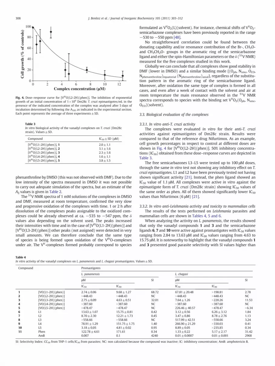
Fig. 4. Dose–response curve for [VIVO(L2-2H)(phen)]. The inhibition of exponentialgrowth of an initial concentration of 1×10⁶ Dm28c T. cruzi epimastigotes/mL in thepresence of the indicated concentration of the complex was analyzed after 5 days ofincubation determined by following the A595 as indicated in the experimental section.Each point represents the average of three experiments±SD.
Table 3In vitro biological activity of the vanadyl complexes on T. cruzi (Dm28cstrain). Values±SD.
Compound IC50±SD (μM)
[VIVO(L1-2H)(phen)], 1 2.0±1.1[VIVO(L2-2H)(phen)], 2 3.1±1.6[VIVO(L3-2H)(phen)], 3 2.3±1.6[VIVO(L4-2H)(phen)], 4 1.6±1.1[VIVO(L5-2H)(phen)], 5 3.8±1.5
308 J. Benítez et al. / Journal of Inorganic Biochemistry 105 (2011) 303–312
phenanthroline by DMSO (this was not observedwith DMF). Due to thelow intensity of the spectra measured in DMSO it was not possibleto carry out adequate simulation of the spectra, but an estimate of theA|| values is given in Table 2.
The 51V NMR spectra of 1 mM solutions of the complexes in DMSOand DMF, measured at room temperature, confirmed the very slowand progressive oxidation of the complexes with time. 1 or 2 h afterdissolution of the complexes peaks assignable to the oxidized com-plexes could be already observed at ca. −535 to −547 ppm, the δvalues also depending on the solvent used. The peaks increasedtheir intensities with time and in the case of [VIVO(L1-2H)(phen)] and[VIVO(L3-2H)(phen)] other peaks (not assigned) were detected in verysmall amounts. We can therefore conclude that the same typeof species is being formed upon oxidation of the VIVO-complexesunder air. The VV-complexes formed probably correspond to species
Table 4In vitro activity of the vanadyl complexes on L. panamensis and L. chagasi promastigotes. Va
Compound Promastigotes
L. panamensis
μM
IC50 IC90
1 [VO(L1-2H)(phen)] 2.74±0.06 9.68±1.272 [VO(L2-2H)(phen)] N448.43 N448.433 [VO(L3-2H)(phen)] 2.75±0.09 4.63±0.514 [VO(L4-2H)(phen)] N387.60 N387.605 [VO(L5-2H)(phen)] N478.47 N478.476 L1 13.63±1.07 15.75±0.817 L2 8.70±2.30 12.21±1.738 L3 N558.66 N558.669 L4 78.91±1.29 151.75±1.7510 L5 3.18±0.05 4.81±0.0211 Phen 122.78±4.61 171.61
AmB 0.007 0.1
SI: Selectivity Index: CC50 from THP-1 cells/IC50 from parasites; NC: non calculated because
formulated as VVO2(L)(solvent). For instance, chemical shifts of VVO2-semicarbazone complexes have been previously reported in the range−530 to −550 ppm [48].
No straightforward correlation could be found between thedonating capability and/or resonance contribution of the Br-, CH3O-and CH3CH2O- groups in the aromatic ring of the semicarbazoneligand and either the spin-Hamiltonian parameters or the δ (51V NMR)measured for the five complexes studied in this work.
Globally we can conclude that all complexes show good stability inDMF (lower in DMSO) and a similar binding mode {(Oph, Nsmc, OCO,Nphenanthroline)equatorial (Nphenanthroline)axial}, regardless of the substitu-tion pattern in the aromatic ring of the semicarbazone ligand.Moreover, after oxidation the same type of complex is formed in allcases, and even after a week of contact with the solvent and air atroom temperature the main resonance observed in the 51V NMRspectra corresponds to species with the binding set VVO2(Oph, Nsmc,OCO)(solvent).
3.3. Biological evaluation of the complexes
3.3.1. In vitro anti-T. cruzi activityThe complexes were evaluated in vitro for their anti-T. cruzi
activities against epimastigotes of Dm28c strain. Results werecompared to that of the reference drug Nifurtimox. As an example,cell growth percentages in respect to control at different doses areshown in Fig. 4 for [VIVO(L2-2H)(phen)]. 50% inhibitory concentra-tions (IC50) obtained from these dose–response curves are depicted inTable 3.
The free semicarbazones L3–L5 were tested up to 100 μM doses,through the same in vitro test not showing any inhibitory effect on T.cruzi epimastigotes. L1 and L2 have been previously tested not havingshown significant activity [21]. Instead, the phen ligand showed anIC50 value of 1.1 μM. All complexes were active in vitro against theepimastigote form of T. cruzi (Dm28c strain) showing IC50 values ofthe same order as phen. All of them showed significantly lower IC50values than Nifurtimox (6 μM) [21].
3.3.2. In vitro anti-Leishmania activity and toxicity to mammalian cellsThe results of the tests performed on Leishmania parasites and
mammalian cells are shown in Tables 4, 5 and 6.When analyzing the activity on L. panamensis, the results showed
that only the vanadyl compounds 1 and 3 and the semicarbazoneligands 6, 7 and 10were active against promastigotes with IC50 valuesranging from 2.84 to 13.63 μM and IC90 values ranging from 4.63 to15.75 μM. It is noteworthy to highlight that the vanadyl compounds 1and 3 presented good parasite selectivity with SI values higher than
lues±SD.
L. chagasi
SI μM SI
IC50 IC90
68.72 67.81±20.48 N198.81 2.78NC N448.43 N448.43 NC32.01 7.64±1.26 N220.26 11.53NC N387.60 N387.60 NCNC 226.46±40.57 N478.47 N6.340.42 3.12±0.56 6.26±3.12 1.840.45 3.47±0.86 8.78±2.76 1.13NC 517.99±42.51 N558.66 3.241.40 266.90±21.29 N330.03 0.410.95 8.89±0.05 N235.85 0.340.34 1.33±0.22 5.17±2.17 31.424240 0.01±0.0007 0.01±0.001 2968
the compound was inactive; IC: inhibitory concentration; AmB: amphotericin B.
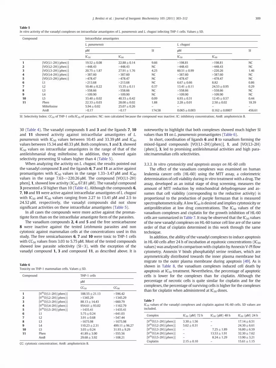
Table 5In vitro activity of the vanadyl complexes on intracellular amastigotes of L. panamensis and L. chagasi infecting THP-1 cells. Values±SD.
Compound Intracellular amastigotes
L. panamensis L. chagasi
μM SI μM SI
IC50 IC90 IC50 IC90
1 [VO(L1-2H)(phen)] 19.52±0.08 22.88±0.14 9.66 N198.81 N198.81 NC2 [VO(L2-2H)(phen)] N448.43 N448.43 NC N448.43 N448.43 NC3 [VO(L3-2H)(phen)] 20.75±1.87 27.91±1.76 4.25 60.51±0.99 N220.26 1.464 [VO(L4-2H)(phen)] N387.60 N387.60 NC N387.60 N387.60 NC5 [VO(L5-2H)(phen)] N478.47 N478.47 NC N478.47 N478.47 NC6 L1 N213.68 N213.68 NC 6.67±0.66 8.82 0.867 L2 10.46±0.22 15.35±0.11 0.37 13.41±0.11 24.53±0.95 0.298 L3 N558.66 N558.66 NC N558.66 N558.66 NC9 L4 N109.90 N109.90 NC N109.90 N109.90 NC10 L5 33.40±0.02 40.33±0.42 0.09 6.93±0.31 12.45±0.57 0.4411 Phen 22.33±0.03 28.00±0.02 1.88 2.28±0.01 2.50±0.02 18.39
Miltefosine 5.94±0.02 25.07±0.26 – – – –
AmB N0.17 N0.17 174.58 0.065±0.003 0.162±0.0007 456.61
SI: Selectivity Index: CC50 of THP-1 cells/IC50 of parasites; NC: non calculated because the compound was inactive; IC: inhibitory concentration; AmB: amphotericin B.
309J. Benítez et al. / Journal of Inorganic Biochemistry 105 (2011) 303–312
30 (Table 4). The vanadyl compounds 1 and 3 and the ligands 7, 10and 11 showed activity against intracellular amastigotes of L.panamensis with IC50 values between 10.45 and 33.39 μM and IC90
values between 15.34 and 40.33 μM. Both complexes, 1 and 3, showedIC50 values on intracellular amastigotes in the range of that of theantileishmanial drug miltefosine. In addition, they showed againselectivity presenting SI values higher than 4 (Table 5).
When analyzing the activity on L. chagasi, the results pointed outthe vanadyl compound 3 and the ligands 6, 7 and 11 as active againstpromastigotes with IC50 values in the range 1.33–3.47 μM and IC90values in the range 7.63–N220.26 μM. The compound [VO(L1-2H)phen], 1, showed low activity (IC50 67.81 μM). The vanadyl compound3 presented a SI higher than 10 (Table 4). Although the compounds 6,7, 10 and 11were active against intracellular amastigotes of L. chagasiwith IC50 and IC90 values ranging from 2.27 to 13.41 μM and 2.5 to24.52 μM, respectively, the vanadyl compounds did not showsignificant activities on these intracellular amastigotes (Table 5).
In all cases the compounds were more active against the promas-tigote form than on the intracellular amastigote form of the parasites.
The vanadium compounds 2, 4 and 5 and the free semicarbazone8 were inactive against the tested Leishmania parasites and noncytotoxic against mammalian cells at the concentrations used in thisstudy. The free semicarbazones 6, 7 and 10 were toxic to THP-1 cellswith CC50 values from 3.03 to 5.75 μM. Most of the tested compoundsshowed low parasite selectivity (SIb3), with the exception of thevanadyl compound 1, 3 and compound 11, as described above. It is
Table 6Toxicity on THP-1 mammalian cells. Values±SD.
Compound THP-1 cells
μM
CC50 CC90
1 [VIVO(L1-2H)(phen)] 188.55±21.13 N596.422 [VIVO(L2-2H)(phen)] N1345.29 N1345.293 [VIVO(L3-2H)(phen)] 88.13±14.43 N660.794 [VIVO(L4-2H)(phen)] 954.61±93.02 N1162.795 [VIVO(L5-2H)(phen)] N1435.41 N1435.416 L1 5.75±0.24 N641.037 L2 3.91±0.68 N547.448 L3 N1675.98 N1675.989 L4 110.23±2.31 499.11±96.2710 L5 3.03±0.24 31.93±9.2911 Phen 41.89±3.00 N555.56
AmB 29.68±3.92 N108.21
CC: cytotoxic concentration; AmB: amphotericin B.
noteworthy to highlight that both complexes showed much higher SIvalues than 11 on L. panamensis promastigotes (Table 6).
In short, coordination of ligands 6 and 8 to vanadium forming themixed-ligand compounds [VO(L1-2H)(phen)], 1, and [VO(L3-2H)(phen)], 3, led to promising antileishmanial activities and high para-site/mammalian cells selectivities.
3.3.3. In vitro cytotoxicity and apoptosis assays on HL-60 cellsThe effect of the vanadium complexes was examined on human
leukemia cancer cells (HL-60) using the MTT assay, a colorimetricdetermination of cell viability during in vitro treatment with a drug. Theassay, developed as an initial stage of drug screening, measures theamount of MTT reduction by mitochondrial dehydrogenase and as-sumes that cell viability (corresponding to the reductive activity) isproportional to the production of purple formazan that is measuredspectrophotometrically. A low IC50 is desired and implies cytotoxicity orantiproliferation at low drug concentrations. The IC50 values of thevanadium complexes and cisplatin for the growth inhibition of HL-60cells are summarized in Table 7. It may be observed that the IC50 valuesof the new vanadyl complexes on HL-60 tumor cell line are of the sameorder of that of cisplatin determined in this work through the sametechnique.
In addition, the ability of the vanadyl complexes to induce apoptosisin HL-60 cells after 24 h of incubation at equitoxic concentrations (IC50values)was analysed in comparisonwith cisplatin byAnnexin V-PIflowcytometry. Annexin V binds phosphatidyl serine residues, which areasymmetrically distributed towards the inner plasma membrane butmigrate to the outer plasma membrane during apoptosis [49]. As isshown in Table 8, the vanadium complexes induced cell death byapoptosis at IC50 treatment. Nevertheless, the percentage of apoptoticcells is lower for the complexes than for cisplatin. Although thepercentage of necrotic cells is quite similar for cisplatin and for thecomplexes, the percentage of surviving cells is higher for the complexesthan for cisplatin when administered at IC50 doses.
Table 7IC50 values of the vanadyl complexes and cisplatin against HL-60 cells. SD values areincluded.
Complex IC50 (μM) 72 h IC50 (μM) 48 h IC50 (μM) 24 h
[VIVO(L1-2H)(phen)] 3.30±1.56 – 17.14±6.51[VIVO(L2-2H)(phen)] 5.62±0.31 – 24.30±6.61[VIVO(L3-2H)(phen)] – 7.25±1.89 16.80±6.59[VIVO(L4-2H)(phen)] – 13.53±1.91 32.30±7.62[VIVO(L5-2H)(phen)] – 8.24±1.29 13.90±3.21Cisplatin 2.15±0.10 – 15.61±1.15

Table 8Quantification of apoptosis after 24 h exposure to concentration equal to IC50 values ofcisplatin and the vanadium complexes against HL-60 cells. IC50 values are indicated inbrackets.
Treatment(IC50 24 h, μM)
% Vitalcells
% Apoptoticcells
% Necrotic deadcells
% Damagedcells
Control 88.56 6.96 4.29 0.18Cisplatin (15.6) 27.13 61.83 10.12 0.92[VIVO(L1-2H)(phen)](17.14)
57.34 28.63 11.31 2.71
[VIVO(L2-2H)(phen)](24.30)
50.65 36.93 9.57 2.84
[VIVO(L3-2H)(phen)](16.80)
59.27 20.34 11.77 8.62
[VIVO(L4-2H)(phen)](32.30)
42.60 39.25 15.74 2.40
[VIVO(L5-2H)(phen)](13.90)
56.02 21.24 13.38 9.38
% damaged cells: 100 – % surviving cells – % apoptotic cells – % necrotic cells.
310 J. Benítez et al. / Journal of Inorganic Biochemistry 105 (2011) 303–312
3.4. DNA interaction studies
To provide insight into the probable mechanism of action, thecompounds were tested for their DNA interaction ability on plasmidDNA by AFM and on CT DNA by using DNA viscositymeasurements andcircular dichroism and fluorescence spectroscopies. In addition, theresults reported in this work will provide more data related with theinteraction of vanadium compoundswith DNA, topic that has been onlyscarcely investigated [20,21,50–52].
The results obtained by AFM for the complexes are depicted inFig. 5. In all cases, kinks, crosslinking and supercoiling were observed.
Fig. 5. AFM images showing the modifications suffered by pBR322 DNA due to interaction w(L4-2H)(phen)] and e) [VIVO(L5-2H)(phen)] for molar ratio compound: DNA base pairs 1:5
Complexes resulted less aggressive to DNA than a few previouslyreported analogous compounds which included dppz instead of phenin the vanadyl coordination sphere [21]. In addition, the effect on DNAdepended on the nature of the substituent on the semicarbazonemoiety, showing [VIVO(L1-2H)(phen)], 1, the most evident interac-tion (Fig. 5). The image corresponding to this complex showed a verysignificant increase of DNA thickness characteristic of intercalators.Probably the absense of substituents on the phenol ring improves theability of intercalation of the vanadyl complex.
All the complexes increased the viscosity of CT DNA solutions in aconcentration dependent manner. Results for [VIVO(L2-2H)(phen)]are depicted in Fig. 6. This behavior is usually shown by intercalators.
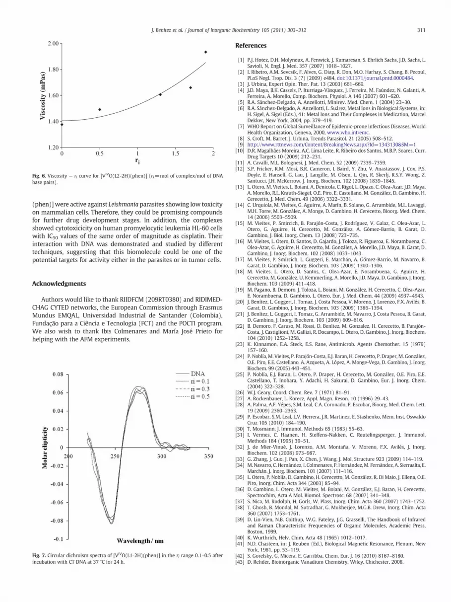
Fluorescence and circular dichroism techniques were not able todetect significant effects due to interaction of the complexes with CTDNA. Fluorescence studies showed only a slight decrease of intensity,indicative of poor displacement of intercalated ethidium bromide bythe VIVO-complexes. CD spectra showed only slight changes in DNAmolar elipticity indicating that in the conditions used interaction ofthe compounds produced only light modifications in the secondarystructure of DNA (Fig. 7).
4. Conclusions
Five novel vanadyl [VIVO(L-2H)(phen)] complexes, all includingphen in its coordination sphere as potential DNA intercalating ligand,and structurally related tridentate semicarbazone ligands have beensynthesized and characterized in the solid state and in solution. All ofthem showed higher in vitro anti-trypanosomal activities thanNifurtimox and increased activities in respect to the free semicarba-zone ligands. The compounds [VO(L1-2H)(phen)] and [VO(L3-2H)
ith a) [VIVO(L1-2H)(phen)], b) [VIVO(L2-2H)(phen)], c) [VIVO(L3-2H)(phen)], d) [VIVOand 24 h incubation at 37 °C.
1.20
1.40
1.60
1.80
2.00
0 0.5 1 1.5 2
Vis
cosi
ty (
mP
as)
ri
Fig. 6. Viscosity — ri curve for [VIVO(L2-2H)(phen)] (ri=mol of complex/mol of DNAbase pairs).
311J. Benítez et al. / Journal of Inorganic Biochemistry 105 (2011) 303–312
(phen)] were active against Leishmania parasites showing low toxicityon mammalian cells. Therefore, they could be promising compoundsfor further drug development stages. In addition, the complexesshowed cytotoxicicity on human promyelocytic leukemia HL-60 cellswith IC50 values of the same order of magnitude as cisplatin. Theirinteraction with DNA was demonstrated and studied by differenttechniques, suggesting that this biomolecule could be one of thepotential targets for activity either in the parasites or in tumor cells.
Acknowledgments
Authors would like to thank RIIDFCM (209RT0380) and RIDIMED-CHAG CYTED networks, the European Commission through ErasmusMundus EMQAL, Universidad Industrial de Santander (Colombia),Fundação para a Ciência e Tecnologia (FCT) and the POCTI program.We also wish to thank Ibis Colmenares and María José Prieto forhelping with the AFM experiments.
Fig. 7. Circular dichroism spectra of [VIVO(L1-2H)(phen)] in the ri range 0.1–0.5 afterincubation with CT DNA at 37 °C for 24 h.
References
[1] P.J. Hotez, D.H. Molyneux, A. Fenwick, J. Kumaresan, S. Ehrlich Sachs, J.D. Sachs, L.Savioli, N. Engl. J. Med. 357 (2007) 1018–1027.
[2] I. Ribeiro, A.M. Sevcsik, F. Alves, G. Diap, R. Don, M.O. Harhay, S. Chang, B. Pecoul,PLoS Negl. Trop. Dis. 3 (7) (2009) e484, doi:10.1371/journal.pntd.0000484.
[3] J. Urbina, Expert Opin. Ther. Pat. 13 (2003) 661–669.[4] J.D. Maya, B.K. Cassels, P. Iturriaga-Vásquez, J. Ferreira, M. Faúndez, N. Galanti, A.
Ferreira, A. Morello, Comp. Biochem. Physiol. A 146 (2007) 601–620.[5] R.A. Sánchez-Delgado, A. Anzellotti, Minirev. Med. Chem. 1 (2004) 23–30.[6] R.A. Sánchez-Delgado, A. Anzellotti, L. Suárez, Metal Ions in Biological Systems, in:
H. Sigel, A. Sigel (Eds.), 41: Metal Ions and Their Complexes in Medication, MarcelDekker, New York, 2004, pp. 379–419.
[7] WHO Report on Global Surveillance of Epidemic-prone Infectious Diseases, WorldHealth Organization, Geneva, 2000, www.who.int/emc.
[8] S. Croft, M. Barret, J. Urbina, Trends Parasitol. 21 (2005) 508–512.[9] http://www.rttnews.com/Content/BreakingNews.aspx?Id=1343130&SM=1
[10] D.R. Magalhães Moreira, A.C. Lima Leite, R. Ribeiro dos Santos, M.B.P. Soares, Curr.Drug Targets 10 (2009) 212–231.
[11] A. Cavalli, M.L. Bolognesi, J. Med. Chem. 52 (2009) 7339–7359.[12] S.P. Fricker, R.M. Mosi, B.R. Cameron, I. Baird, Y. Zhu, V. Anastassov, J. Cox, P.S.
Doyle, E. Hansell, G. Lau, J. Langille, M. Olsen, L. Qin, R. Skerlj, R.S.Y. Wong, Z.Santucci, J.H. McKerrow, J. Inorg. Biochem. 102 (2008) 1839–1845.
[13] L. Otero, M. Vieites, L. Boiani, A. Denicola, C. Rigol, L. Opazo, C. Olea-Azar, J.D. Maya,A. Morello, R.L. Krauth-Siegel, O.E. Piro, E. Castellano, M. González, D. Gambino, H.Cerecetto, J. Med. Chem. 49 (2006) 3322–3331.
[14] C. Urquiola, M. Vieites, G. Aguirre, A. Marín, B. Solano, G. Arrambide, M.L. Lavaggi,M.H. Torre, M. González, A. Monge, D. Gambino, H. Cerecetto, Bioorg. Med. Chem.14 (2006) 5503–5509.
[15] M. Vieites, P. Smircich, B. Parajón-Costa, J. Rodríguez, V. Galaz, C. Olea-Azar, L.Otero, G. Aguirre, H. Cerecetto, M. González, A. Gómez-Barrio, B. Garat, D.Gambino, J. Biol. Inorg. Chem. 13 (2008) 723–735.
[16] M. Vieites, L. Otero, D. Santos, D. Gajardo, J. Toloza, R. Figueroa, E. Norambuena, C.Olea-Azar, G. Aguirre, H. Cerecetto, M. González, A. Morello, J.D. Maya, B. Garat, D.Gambino, J. Inorg. Biochem. 102 (2008) 1033–1043.
[17] M. Vieites, P. Smircich, L. Guggeri, E. Marchán, A. Gómez-Barrio, M. Navarro, B.Garat, D. Gambino, J. Inorg. Biochem. 103 (2009) 1300–1306.
[18] M. Vieites, L. Otero, D. Santos, C. Olea-Azar, E. Norambuena, G. Aguirre, H.Cerecetto, M. González, U. Kemmerling, A. Morello, J.D. Maya, D. Gambino, J. Inorg.Biochem. 103 (2009) 411–418.
[19] M. Pagano, B. Demoro, J. Toloza, L. Boiani, M. González, H. Cerecetto, C. Olea-Azar,E. Norambuena, D. Gambino, L. Otero, Eur. J. Med. Chem. 44 (2009) 4937–4943.
[20] J. Benítez, L. Guggeri, I. Tomaz, J. Costa Pessoa, V. Moreno, J. Lorenzo, F.X. Avilés, B.Garat, D. Gambino, J. Inorg. Biochem. 103 (2009) 1386–1394.
[21] J. Benítez, L. Guggeri, I. Tomaz, G. Arrambide, M. Navarro, J. Costa Pessoa, B. Garat,D. Gambino, J. Inorg. Biochem. 103 (2009) 609–616.
[22] B. Demoro, F. Caruso, M. Rossi, D. Benítez, M. Gonzalez, H. Cerecetto, B. Parajón-Costa, J. Castiglioni, M. Gallizi, R. Docampo, L. Otero, D. Gambino, J. Inorg. Biochem.104 (2010) 1252–1258.
[23] K. Kinnamon, E.A. Steck, E.S. Rane, Antimicrob. Agents Chemother. 15 (1979)157–160.
[24] P. Noblía, M. Vieites, P. Parajón-Costa, E.J. Baran, H. Cerecetto, P. Draper,M. González,O.E. Piro, E.E. Castellano, A. Azqueta, A. López, A. Monge-Vega, D. Gambino, J. Inorg.Biochem. 99 (2005) 443–451.
[25] P. Noblía, E.J. Baran, L. Otero, P. Draper, H. Cerecetto, M. González, O.E. Piro, E.E.Castellano, T. Inohara, Y. Adachi, H. Sakurai, D. Gambino, Eur. J. Inorg. Chem.(2004) 322–328.
[26] W.J. Geary, Coord. Chem. Rev. 7 (1971) 81–91.[27] A. Rockenbauer, L. Korecz, Appl. Magn. Reson. 10 (1996) 29–43.[28] A. Palma, A.F. Yépes, S.M. Leal, C.A. Coronado, P. Escobar, Bioorg. Med. Chem. Lett.
19 (2009) 2360–2363.[29] P. Escobar, S.M. Leal, L.V. Herrera, J.R. Martinez, E. Stashenko, Mem. Inst. Oswaldo
Cruz 105 (2010) 184–190.[30] T. Mosmann, J. Immunol, Methods 65 (1983) 55–63.[31] I. Vermes, C. Haanen, H. Steffens-Nakken, C. Reutelingsperger, J. Immunol,
Methods 184 (1995) 39–51.[32] J. de Mier-Vinué, J. Lorenzo, A.M. Montaña, V. Moreno, F.X. Avilés, J. Inorg.
Biochem. 102 (2008) 973–987.[33] G. Zhang, J. Guo, J. Pan, X. Chen, J. Wang, J. Mol, Structure 923 (2009) 114–119.[34] M.Navarro, C. Hernández, I. Colmenares, P. Hernández,M. Fernández, A. Sierraalta, E.
Marchán, J. Inorg. Biochem. 101 (2007) 111–116.[35] L. Otero, P. Noblía, D. Gambino, H. Cerecetto, M. González, R. Di Maio, J. Ellena, O.E.
Piro, Inorg. Chim. Acta 344 (2003) 85–94.[36] D. Gambino, L. Otero, M. Vieites, M. Boiani, M. González, E.J. Baran, H. Cerecetto,
Spectrochim, Acta A Mol. Biomol. Spectrosc. 68 (2007) 341–348.[37] S. Nica, M. Rudolph, H. Gorls, W. Plass, Inorg. Chim. Acta 360 (2007) 1743–1752.[38] T. Ghosh, B. Mondal, M. Sutradhar, G. Mukherjee, M.G.B. Drew, Inorg. Chim. Acta
360 (2007) 1753–1761.[39] D. Lin-Vien, N.B. Colthup, W.G. Fateley, J.G. Grasselli, The Handbook of Infrared
and Raman Characteristic Frequencies of Organic Molecules, Academic Press,Boston, 1999.
[40] K. Wurthrich, Helv. Chim. Acta 48 (1965) 1012–1017.[41] N.D. Chasteen, in: J. Reuben (Ed.), Biological Magnetic Resonance, Plenum, New
York, 1981, pp. 53–119.[42] S. Gorelsky, G. Micera, E. Garribba, Chem. Eur. J. 16 (2010) 8167–8180.[43] D. Rehder, Bioinorganic Vanadium Chemistry, Wiley, Chichester, 2008.

312 J. Benítez et al. / Journal of Inorganic Biochemistry 105 (2011) 303–312
[44] J. Costa Pessoa, I. Cavaco, I. Correia, I. Tomaz, M.T. Duarte, P.M. Matias, J. Inorg.Biochem. 80 (2000) 35–39.
[45] G. Micera, V.L. Pecoraro, E. Garribba, Inorg. Chem. 48 (2009) 5790–5796.[46] M.R. Maurya, U. Kumar, I. Correia, P. Adão, J. Costa Pessoa, Eur. J. Inorg. Chem.
(2008) 577–587.[47] M.R. Maurya, A. Arya, A. Kumar, M.L. Kuznetsov, F. Avecilla, J. Costa Pessoa, Inorg.
Chem. 49 (2010) 6586–6600.[48] M.R. Maurya, A.A. Khan, A. Azam, S. Ranjan, N. Mondal, A. Kumar, F. Avecilla, J.
Costa Pessoa, Dalton Trans. 39 (2010) 1345–1360.
[49] M.A. Fuertes, C. Alonso, J.M. Pérez, Chem. Rev. 103 (2003) 645–662.[50] N. Butenko, A.I. Tomaz, O. Nouri, E. Escribano, V. Moreno, S. Gama, V. Ribeiro, J.P.
Telo, J. Costa Pesssoa, I. Cavaco, J. Inorg. Biochem. 103 (2009) 622–632.[51] P.K. Sasmal, A.K. Patra, M. Nethaji, A.R. Chakravarty, Inorg. Chem. 46 (2007)
11112–11121.[52] G. Verquin, G. Fontaine, M. Bria, E. Zhilinskaya, E. Abi-Aad, A. Aboukaıs, B.
Baldeyrou, C. Bailly, J. Bernier, J. Biol. Inorg. Chem. 9 (2004) 345–353.


