Embed Size (px)
Citation preview
Uttar Pradesh Rajarshi Tandon Open University
Bachelor of Science
UGBY-101
Cytology And Genetics
BLOCK-1 CYTOLOGY 03-40 UNIT-1 Cell Structure And Cellular Organelles-I 07-16 UNIT-2 Cell Structure And Cellular Organelles-II 17-28UNIT-3 Cell cycle, Mitosis And Meiosis 29-40
BLOCK-2 GENETICS-I 41-110 UNIT-4 Pre-mendelian Genetics, And Mendel’s Laws of 45-64
Inheritance UNIT-5 Linkage And Crossing Over 65-86 UNIT-6 Cytoplasmic Inheritance, Sex Linked Inheritance 87-110
And Sex Determination In Plants
BLOCK-3 GENETICS-II 111-172 115-140 141-156
UNIT-7 Pre-Chromosomal aberrations UNIT-8 Gene Mutation and induced mutation UNIT-9 Genetics in Plant improvement 157-172
UGBY-101/1

UGBY-101/2
BLOCK
1 CYTOLOGY
UNIT 1
CELL STRUCTURE AND CELLULAR ORGANELLES - I
UNIT 2
CELL STRUCTURE AND CELLULAR ORGANELLES - II
UNIT 3
CELL CYCLE, MITOSIS AND MEIOSIS
Uttar Pradesh Rajarshi Tandon Open University
Bachelor of Science
UGBY-101
07-16
17-28
29-40
UGBY-101/3
Course Design Committee Prof. Ashutosh Gupta Chairman Director, School of Science, UPRTOU, Prayagraj.
Prof. Malvika Srivastava Member Botny Department Deendayal Upadhyay University, Gorakhpur.
Prof. V.N. Pandey Member Botny Department Deendayal Upadhyay University, Gorakhpur.
Prof. Anil Kumar Diwedi Member Botny Department Deendayal Upadhyay University, Gorakhpur.
Dr. N.N Tripathi Member Acadmic Consultant, School of Science UPRTOU, Prayagraj.
Dr. Sushma Chauhan Member/Secretary Acadmic Consultant, School of Science, UPRTOU, Prayagraj.
Course Preparation Committee Dr. Dharmveer Singh Author Block-1 (Unit: 1-2) Acadmic Consultant, School of Science UPRTOU, Prayagraj. Dr. Sushma Chauhan Author Block-1 (Unit: 3) Acadmic Consultant, School of Science UPRTOU, Prayagraj. Dr Achala Srivatava Author Block-2-3 (Unit: 4-9) Assistant professor, S.S. Khanna, P.G. College, Prayagraj. Prof. G. Kumar Editor (Unit: 1-9) Professor, Department of Botany, University of Allahabad, Prayagraj Dr. Sushma Chauhan Course Coordinator Acadmic Consultant (Botany) School of Science, UPRTOU, Prayagraj.
© UPRTOU, Prayagraj. 2020 ISBN : All Rights are reserved. No part of this work may be reproduced in any form, by mimeograph or any other means, without permission in writing from the Uttar Pradesh Rajarshi Tondon Open University, Prayagraj.
Printed and Published by Dr. Arun Kumar Gupta Registrar, Uttar Pradesh Rajarshi Tandon Open University, 2020.
Printed By : Chandrakala Universal Pvt. Ltd. 42/7 Jawahar Lal Neharu Road, Prayagraj. UGBY-101/4
[DUKE-002]
COURSE INTRODUCTON This course covers the cell biology and genetics. The living body is
a mixture of groups of chemicals that processes and builds up our body structure. The body of organism is consists of cells and, the cell is basic unit of biological world. The organization of cell is called tissue, the association of tissues form organ and finally organ gives the shape of organism. The molecules which present in cell are most intriguing macromolecules and also the internal part of any living species and the part of interaction and reactions take place for the fundamental to all living being. Any organism, mainly contain only one types of cell such as either Prokaryotic cell or Eukaryotic cell. The organism having many cell in their body are called multi-cellular organism for example most of plants and animals are multi-cellular organisms.
Genetics is the study of two different aspects of nature that is heredity and variations. The process of transmission of characters from one generation to next is known as heredity. The similarity between individuals is due to heredity but, sometime individuals show dissimilarity. This dissimilarity is the result of variations.
The heredity and variations result in the formation of new species.
This course is divided into three blocks-
Block -1: Cytology
Block-2: Genetics-I
Block-3: Genetics-II
UGBY-101/5
Block -1: Cytology The study of cell and its various organelles is known as cytology.
The cell is the basic unit of all living organisms therefore; the knowledge of cell and various organelles is required to the students for understanding morphology and physiology of the plant. Keeping this in mind in this block structure and function of various cellular organelles have been describes.
This block is divided into three units-
Unit-1 - Cell structure and cellular organelles-I: this unit covers the structure and function of mitochondria, chloroplast, nucleus and ribosome
Unit-2 - Cell structure and cellular organelles-II: this unit covers the structure and function of endoplasmic reticulum, golgi complex, lysosome and chromosomes.
Unit-3 - Cell cycle, mitosis and meiosis: this unit covers the basics of cell division and cell development in brief.
UGBY-101/6
UNIT-1
CELL STRUCTURE AND CELLULAR ORGANELLES-I
Structure 1.1. Introduction
Objectives
1.2. Cell membrane
1.3. Mitochondria
Structure
Functions
1.4. Chloroplasts
Structure
Functions
1.5. Nucleus
Structure
Nuclear membrane
Nucleoplasm
Nucleolus
1.6. Ribosome
Structure
Functions
1.7. Summary
1.8. Terminal questions
1.1. Introduction
This unit covers structure and functions of cell and their organelles. We knew that the living organism is that they are complicated and highly organized. The cells of which they composed and possessing intricate internal structures containing many kinds of organelles and complex molecules. The details study of cell reveals the basic structure and living phenomena, i.e., physiological and behavioral processes of UGBY-101/7
organism. All organisms are composed of cells. Some are composed of a single cell that is called unicellular organisms while others, composed of more cells, are called multicellular organisms. Cell membrane and cell wall are the specific feature in cell, they play important role in cell growth, formation of intercellular junctions, junctions, secretion, endocytosis and cell division etc. The cells organelles have specific purpose and function. The role of organelles is highly specific in the metabolic process and plays important role in energy production, transfer and synthesis of kinds of metabolites.
Objectives To learn about basic component of Cell membrane
To understand the Mitochondria and their functions
To know the role of Chloroplasts and their structure
Structure and function of Nucleus and nucleolus
To discuss about role of Ribosome in cell
1.2. Cell membrane or Plasma membrane After invention of electron microscopy in 1950, first time cell
membrane was studied. This study reveals that all the membrane consists of double layer of lipids molecules in which proteins are embedded. These proteins account about 50% of mass of membrane. It has been found that lipids are arranged within the membrane with the polar head towards the outer sides and the hydrophobic tails towards the inner part. This ensures that the nonpolar tail of saturated hydrocarbons is protected from the aqueous environment. The cell membrane contains two types of proteins
• Lipoproteins- it contains lipids. It works as enzymes and ionsregulation.
• Glycoproteins- it contains carbohydrate, which works as receptors.
Some proteins are located in the inner surface of membrane which called as intrinsic proteins. Some are found on outer surface of membrane is called extrinsic proteins, whereas those proteins extend through the membrane called trans membrane proteins.
The lipid component of the membrane not only contains phosphoglycerides but also possess protein and carbohydrate. The ratio of protein and lipid varies considerably in different cell types. The proteins which found in cells can be classified as integral or peripheral proteins. Peripheral proteins lie on the surface of membrane while the integral proteins are partially or totally buried in the membrane. In 1972, Singer and Nicolson have proposed a model for cell membrane is called mosaic model. This model is considered as advanced or improved model for cell membranes. According to this model the quasi-fluid nature of lipid
Cytology
UGBY-101/8
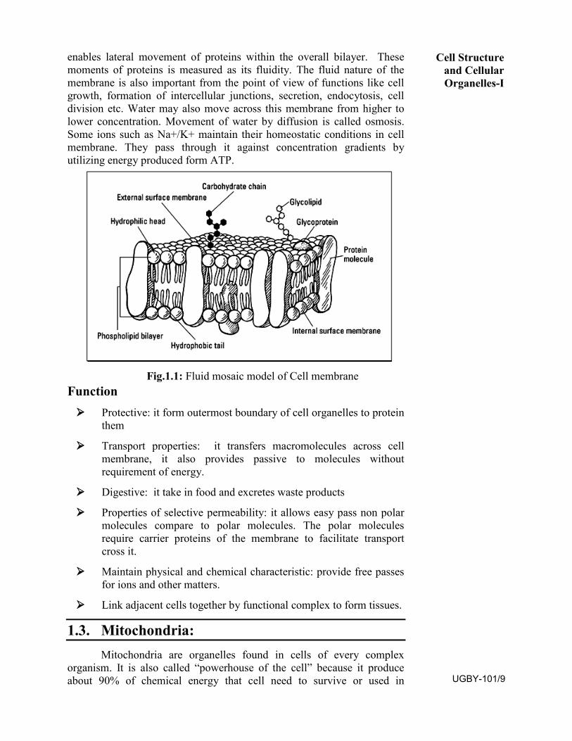
enables lateral movement of proteins within the overall bilayer. These moments of proteins is measured as its fluidity. The fluid nature of the membrane is also important from the point of view of functions like cell growth, formation of intercellular junctions, secretion, endocytosis, cell division etc. Water may also move across this membrane from higher to lower concentration. Movement of water by diffusion is called osmosis. Some ions such as Na+/K+ maintain their homeostatic conditions in cell membrane. They pass through it against concentration gradients by utilizing energy produced form ATP.
Fig.1.1: Fluid mosaic model of Cell membrane Function Protective: it form outermost boundary of cell organelles to protein
them
Transport properties: it transfers macromolecules across cellmembrane, it also provides passive to molecules withoutrequirement of energy.
Digestive: it take in food and excretes waste products
Properties of selective permeability: it allows easy pass non polarmolecules compare to polar molecules. The polar moleculesrequire carrier proteins of the membrane to facilitate transportcross it.
Maintain physical and chemical characteristic: provide free passesfor ions and other matters.
Link adjacent cells together by functional complex to form tissues.
1.3. Mitochondria: Mitochondria are organelles found in cells of every complex
organism. It is also called “powerhouse of the cell” because it produce about 90% of chemical energy that cell need to survive or used in
Cell Structure and Cellular Organelles-I
UGBY-101/9
metabolic reactions. Kolliker (1880) first observed mitochondria as cytoplamic granules in striped muscles of insect. Flemming and Altman was credited for the discovery of mitochondria, however the term mitochondria was given by C. Bendra and F. Meves in year 1904. They first observed mitochondria in plants Nymphea. Seikevitz called them power house. The mitochondria present in several numbers i.e.1000-1600 per cell. The size of mitochondria is often between 0.75 and 3 micrometers and is not visible under the microscope unless they stained. Mitochondria are split into different compartments or regions, each of which carries out distinct roles. Mitochondria also have a special role in making cells die (apoptosis). This may sound strange, but it is vital for the processes of growth and development. Sometimes cells don’t die when they should, and start to grow uncontrollably. All the mitochondria present in a cell are collelectively called chondriome. Usually plant cells have fewer mitochondria as compared to animal cell. In higher animals maximimum mitochondria are found in flight muscles of birds. Mitochondria can make its shape as ellipsoidal, oval, spherical or spiral.
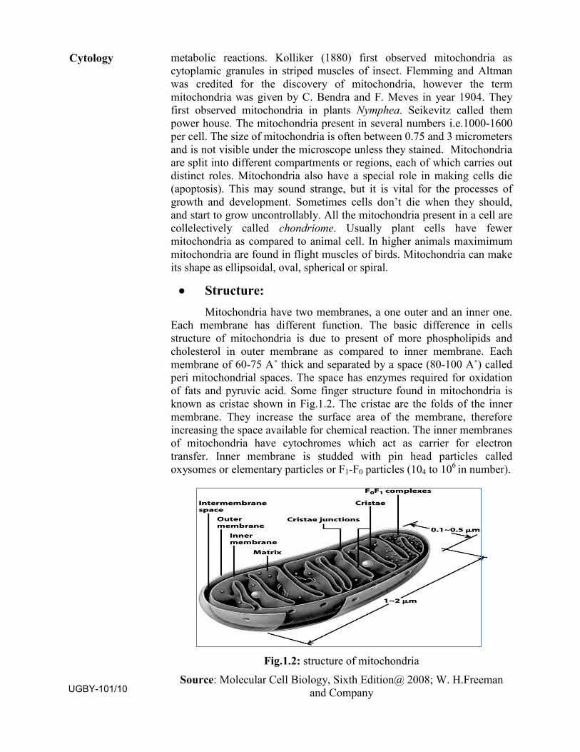
• Structure:Mitochondria have two membranes, a one outer and an inner one.
Each membrane has different function. The basic difference in cells structure of mitochondria is due to present of more phospholipids and cholesterol in outer membrane as compared to inner membrane. Each membrane of 60-75 A˚ thick and separated by a space (80-100 A˚) called peri mitochondrial spaces. The space has enzymes required for oxidation of fats and pyruvic acid. Some finger structure found in mitochondria is known as cristae shown in Fig.1.2. The cristae are the folds of the inner membrane. They increase the surface area of the membrane, therefore increasing the space available for chemical reaction. The inner membranes of mitochondria have cytochromes which act as carrier for electron transfer. Inner membrane is studded with pin head particles called oxysomes or elementary particles or F1-F0 particles (104 to 106 in number).
Fig.1.2: structure of mitochondria Source: Molecular Cell Biology, Sixth Edition@ 2008; W. H.Freeman
and Company
Cytology
UGBY-101/10
Mitochondria matrix have enzyme for kreb’s cycle. Besides these enzymes matrix have a complete protein synthesis apparatus (Ribosome 70-s, DNA, few RNA’s & few enzymes) so mitochondria is called as semiautonomous cell organelle. One or many (6 kb to 36 kb long) double stranded mainly circular naked DNA present in mitochondrial matrix. Mitrichondrial DNA can code the synthesis of 10 to 37 different types of proteins. Enzymes for replication and transcription of DNA like DNA-Polymerase and RNA polymerase are for in mitochondrial matrix. Mitochondria of mammals have 55s ribosomes. Mitochondria of oocytes called yolk nuclei.
• Functions:The mitochondria act produce energy for oxidative metabolism and
ATP production, where organic compounds are broken down to release & store metabolic energy in the form of ATP molecules. The main job of mitochondria is to perform cellular respiration. This means it takes in nutrients from the cell, breaks it down, and turns it into energy. This energy is then in turn used by the cell to carry out various functions. Mitochondria help in vitellogeneus in oocytes. Mitochondrial kinease makes the yolks viscous and insoluble longer duration storage.
1.4. Chloroplasts:
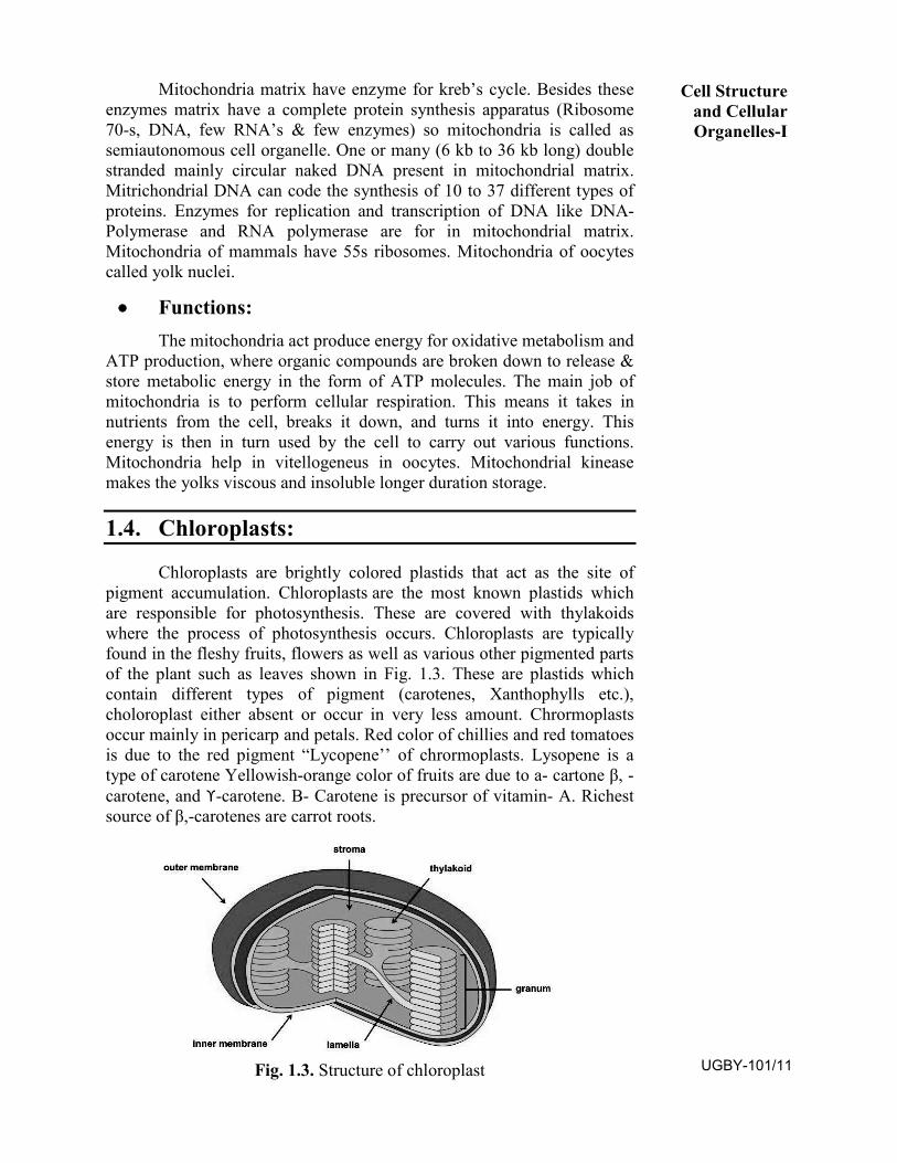
Chloroplasts are brightly colored plastids that act as the site of pigment accumulation. Chloroplasts are the most known plastids which are responsible for photosynthesis. These are covered with thylakoids where the process of photosynthesis occurs. Chloroplasts are typically found in the fleshy fruits, flowers as well as various other pigmented parts of the plant such as leaves shown in Fig. 1.3. These are plastids which contain different types of pigment (carotenes, Xanthophylls etc.), choloroplast either absent or occur in very less amount. Chrormoplasts occur mainly in pericarp and petals. Red color of chillies and red tomatoes is due to the red pigment “Lycopene’’ of chrormoplasts. Lysopene is a type of carotene Yellowish-orange color of fruits are due to a- cartone β, -carotene, and ϒ-carotene. Β- Carotene is precursor of vitamin- A. Richest source of β,-carotenes are carrot roots.
Fig. 1.3. Structure of chloroplast
Cell Structure and Cellular Organelles-I
UGBY-101/11
Function
They provide color to fruits and flowers.
They help in storage of proteins, starch and oil.
They trap solar energy to manufacture food through the process ofphotosynthesis.
They help in maintaining balance between carbon dioxide andoxygen during photosynthesis. Chloroplasts are the centres ofsynthesis and metabolism of carbohydrates.
1.5. Nucleus:
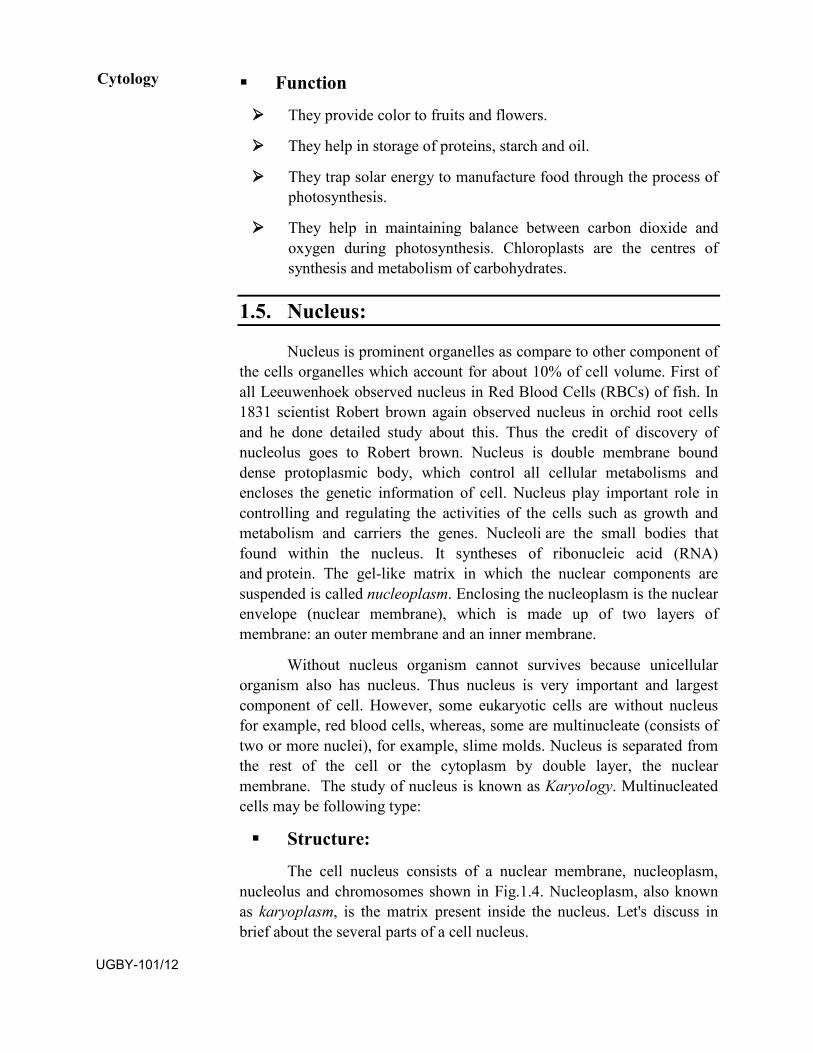
Nucleus is prominent organelles as compare to other component of the cells organelles which account for about 10% of cell volume. First of all Leeuwenhoek observed nucleus in Red Blood Cells (RBCs) of fish. In 1831 scientist Robert brown again observed nucleus in orchid root cells and he done detailed study about this. Thus the credit of discovery of nucleolus goes to Robert brown. Nucleus is double membrane bound dense protoplasmic body, which control all cellular metabolisms and encloses the genetic information of cell. Nucleus play important role in controlling and regulating the activities of the cells such as growth and metabolism and carriers the genes. Nucleoli are the small bodies that found within the nucleus. It syntheses of ribonucleic acid (RNA) and protein. The gel-like matrix in which the nuclear components are suspended is called nucleoplasm. Enclosing the nucleoplasm is the nuclear envelope (nuclear membrane), which is made up of two layers of membrane: an outer membrane and an inner membrane.
Without nucleus organism cannot survives because unicellular organism also has nucleus. Thus nucleus is very important and largest component of cell. However, some eukaryotic cells are without nucleus for example, red blood cells, whereas, some are multinucleate (consists of two or more nuclei), for example, slime molds. Nucleus is separated from the rest of the cell or the cytoplasm by double layer, the nuclear membrane. The study of nucleus is known as Karyology. Multinucleated cells may be following type:
Structure:
The cell nucleus consists of a nuclear membrane, nucleoplasm,nucleolus and chromosomes shown in Fig.1.4. Nucleoplasm, also known as karyoplasm, is the matrix present inside the nucleus. Let's discuss in brief about the several parts of a cell nucleus.
Cytology
UGBY-101/12
Fig.1.4. Structure of nucleus Source: https://alevelbiology.co.uk/notes/nucleus-structure-and-
functions/
• Nuclear membrane:Nucleus is surrounded by two unit membranes, thus nucleus is
double membranous structure that contains the nucleolus in the nucleoplasm. The space between two layers of membranes is known as perinuclear space. Outer membrane of nucleus is connected to the endoplasmic reticulum at several places and ribosome also may found on it. The nucleus communicates with the remaining of the cell or the cytoplasm through several openings called nuclear pores (300-1000A in diameter). Each nuclear pore is guarded by an actagonal discoid structure of nucleoplasm protein and structure is called as annulus (Annulus +pore=Nuclear pore complex). The inner side of inner nuclear membrane is called by nuclear lamina. This structure is formed by filaments of lamina protein.
• Nucleoplasm:Nucleoplasm is the gelatinous substance within the nuclear
envelope. It is a ground substances of nucleus which is a complex colloidal formed of a number of chemical like nucleotides , nucleosides, ATPs, proteins and enzymes of RNA and DNA polymerases, end nucleases, minerals (Ca++,Mg++) etc.
• Nucleolus:The nucleolus (plural nucleoli) is a dense, spherical-shaped
structure present inside the nucleus. Some of the eukaryotic organisms have nucleus that contains up to four nucleoli. The nucleolus plays an indirect role in protein synthesis by producing ribosomes. These ribosomes are cell organelles made up of RNA and proteins; they are transported to the cytoplasm, which are then attached to the endoplasmic reticulum.
• Chromosomes:Chromosomes are present in the form of strings of DNA and
histones (protein molecules) called chromatin. The chromatin is further classified into heterochromatin and euchromatin based on the functions.
Cell Structure and Cellular Organelles-I
UGBY-101/13
The former type is a highly condensed, transcriptionally inactive form, mostly present adjacent to the nuclear membrane. On the other hand, euchromatin is a delicate, less condensed organization of chromatin, which is found abundantly in a transcribing cell.
Functions:Peaking about the functions of a cell nucleus, it controls the
hereditary characteristics of an organism. This organelle is also responsible for the protein synthesis, cell division, growth and differentiation. Here is a list of the important functions carried out by a cell nucleus. Storage of hereditary material, the genes in the form of long and thin DNA (deoxyribonucleic acid) strands, referred to as chromatin. Storage of proteins and RNA (ribonucleic acid) in the nucleolus. Nucleus is a site for transcription in which messenger RNA (mRNA) are produced for protein synthesis. Exchange of hereditary molecules (DNA and RNA) between the nucleus and the rest of the cell. During the cell division, chromatins are arranged into chromosomes in the nucleus. Production of ribosomes (protein factories) in the nucleolus.
1.6. Summary
In this unit you have learn that- The living organism is composed of single cell or multiple cells.
The non living rigid structure is cells wall that gives shape to the cell and protect the cell from mechanical damage and infection, it also helps in cell-to-cell interaction and provides barrier to undesirable macromolecules. In this study you learned about the cell membrane that is composed of lipids and in a bilayer. Cell membrane is selectively permeable to some molecules present on either side of it. It provides passage for many molecules without any requirement of energy and this is called the passive transport. It also provides passage for few ions according to their concentration gradient. Organelles have a wide range of responsibilities that include everything from generating energy for a cell to controlling the cell's growth and reproduction. The name organelle comes from the idea that these structures are to cells what an organ is to the body.
1.7. Terminal questions
Q.1: Discusses the fluid mosaic model of cell membrane? Answer:------------------------------------------------------------------------------------------------------------------------------------------------------------------------------------------------------------------------------------------------------------------- Q.2: What do you understand by organelles; write structure and function of mitochondria in cell? Answer:-------------------------------------------------------------------------------------------------------------------------------------------------------------------------------------------------------------------------------------------------------------------
Cytology
UGBY-101/14
Q.3. What is chloroplast, discuss role in plant cell?
Answer:-------------------------------------------------------------------------------------------------------------------------------------------------------------------------------------------------------------------------------------------------------------------
Q.4: Discuss abut structure and function of nucleolus
Answer:-------------------------------------------------------------------------------------------------------------------------------------------------------------------------------------------------------------------------------------------------------------------
Q.5: Write short notes on
a) Ribosome
b) Nucleolus
c) Energy power house in cell
Further readings
1. Principles of Biochemistry: Lehninger, Nelson and Cox. StudentEdition, CBS 1439 Publishers and Distributors, Delhi.
2. Fundamentals of Biochemistry: Dr J L Jain, S. Chand andCompany
3. Cell Biology (Cytology, Biomolecules and Molecular Biology): PS Verma and V K Agarwal.
4. Textbook of Biochemistry and Human Biology: Talwar andSrivastava. Eastern Economy Edition, Prentice Hall, India.
Cell Structure and Cellular Organelles-I
UGBY-101/15

UGBY-101/16
UNIT-2
CELL STRUCTURE AND CELLULAR ORGANELLES-II
Structure 2.1 Introduction
Objectives
2.2 Endoplasmic reticulum (ER)
Modification of E.R.
Structure
Functions
2.3 Golgi complex
Structure
Functions
2.4 Lysosome
Primary Lysosome
Digestive vacuoles
Function
2.5 Chromosomes
Structure
Functions
2.6 Summary
2.7 Terminal questions
2.1. Introduction
This unit covers structure and functions of cell and their organelles. We knew that the living organism is that they are complicated and highly organized. The cells of which they composed and possessing intricate internal structures containing many kinds of organelles and complex molecules. The details study of cell reveals the basic structure and living phenomena, i.e., physiological and behavioral processes of organism. All organisms are composed of cells. Some are composed of a single cell that is called unicellular organisms while others, composed of UGBY-101/17
more cells, are called multicellular organisms. Cell membrane and cell wall are the specific feature in cell, they play important role in cell growth, formation of intercellular junctions, secretion, endocytosis and cell division etc. The cells organelles have specific purpose and function. The role of organelles is highly specific in the metabolic process and plays important role in energy production, transfer and synthesis of kinds of metabolites.
Objectives To learn about basic structure and function of Endoplasmic
reticulum (ER)
To understand the basic structure and feature of Golgi body
To study the composition and structure of Lysosome.
To know about structure and function of chromosome
2.2. Endoplasmic Reticulum (ER):
Structurally, the endoplasmic reticulum is a network of membranes found throughout the cell and connected to the nucleus. The membranes are slightly different from cell to cell and a cell’s function determines the size and structure of the endoplasmic reticulum (ER). The ER is most important organelle in eukaryotic cells. ER was first observed by Garnier (1897) and their name of proposed porter (1961) thus credit for discovery of ER goes to Porter. ER produces transmembrane proteins and lipids for its membrane and for many other cell components including lysosomes, secretory vesicles, the Golgi apparatus, the cell membrane, and plant cell vacuoles. ER contains number of components which are shown in Fig. 2.4 as discussed below;
Cisternae – These are long flattened and unbranched units arranged in stacks.
Vesicles - These are oval membrane bound structures.
Tubules – These are irregular, often branched tubes bounded by membrane. Tubules may free or associated with cisternae.
• Modification of E.R.Sarcoplasmic Reticulum: This smooth E.R. occurs in skeletal and
cardiac muscles. S.R. Stores Ca+2 and energy rich compounds required for muscle contraction.
T-Tubules: These are transversely arranged tubules in skeletal and cardiac are muscle cells. Thee transmits stimulus for contraction of muscles.
Ergastoplasm: When the ribosome’s are accumulated on the small parallel cisternae of E.R., and then called Ergastoplasm.
Cytology
UGBY-101/18
Myeloid Bodies: Myeloid bodies are the specialized smooth E.R. which found in pigmented epithelial cells of the retina. Myeloid body is light sensitive structure and may be involved in pigment migration.
Microsome: These are pices of E.R. with associated ribosomal particles (Claude 1951). These can be obtained by fragmentation and high speed centrifugation of cell. They do not exist as such in the living cell.
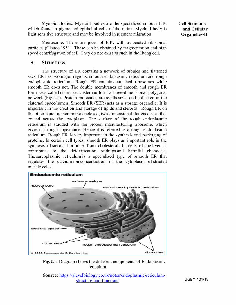
• Structure:The structure of ER contains a network of tubules and flattened
sacs. ER has two major regions: smooth endoplasmic reticulum and rough endoplasmic reticulum. Rough ER contains attached ribosomes while smooth ER does not. The double membranes of smooth and rough ER form sacs called cisternae. Cisternae form a three-dimensional polygonal network (Fig.2.1). Protein molecules are synthesized and collected in the cisternal space/lumen. Smooth ER (SER) acts as a storage organelle. It is important in the creation and storage of lipids and steroids. Rough ER on the other hand, is membrane-enclosed, two-dimensional flattened sacs that extend across the cytoplasm. The surface of the rough endoplasmic reticulum is studded with the protein manufacturing ribosome, which gives it a rough appearance. Hence it is referred as a rough endoplasmic reticulum. Rough ER is very important in the synthesis and packaging of proteins. In certain cell types, smooth ER plays an important role in the synthesis of steroid hormones from cholesterol. In cells of the liver, it contributes to the detoxification of drugs and harmful chemicals. The sarcoplasmic reticulum is a specialized type of smooth ER that regulates the calcium ion concentration in the cytoplasm of striated muscle cells.
Fig.2.1: Diagram shows the different components of Endoplasmic reticulum
Source: https://alevelbiology.co.uk/notes/endoplasmic-reticulum-structure-and-function/
Cell Structure and Cellular
Organelles-II
UGBY-101/19
• Functions:Endoplasmic reticulum is mainly responsible for the transportation
of proteins and other carbohydrates to another organelle, which includes lysosomes, Golgi apparatus, plasma membrane, etc. ER provides the increased surface area for cellular reactions such as formation of nuclear membrane during cell division. E.R. plays a vital role in the synthesis of proteins, lipids, glycogen and other steroids like cholesterol, progesterone, testosterone, etc. E.R. forms intracellular conduction system, transport of materials in cytoplasm from one place to another may occurs through the E.R. Rough ER provides site for the protein synthesis, because rough E.R. has ribosome on its surface. Endoplasmic reticulum seems to play a role in breakdown of glycogen (glycogenolysis). Smooth ER concerned with detoxification of drugs, pollutants and steroids.
2.3. Golgi complex:
Golgi complex is also called Golgi apparatus or Golgi body. It is one of organelle of eukaryotic cells which was discovered by C. Golgi (1898) in the nerve cells of owl. The Golgi complex also known as several names such as Dolton complex, Golgi complex, Baker’s body, Dictyosome (plant Golgi body) etc. The cytoplasm surrounding Golgi body has fewer or no other organelles. It is called Golgi ground substance or zone of exclusion. Golgi bodies are pleomorphic structures because component of Golgi body differ in structure & shape in different cells. The Golgi body is made up of a series of flattered, stacked pounces called cisternae. Cisternae are flat sacs that are stacked in a semicircular, bent formation. Each formation has a membrane to separate it from the cytoplasm of the cell. The Golgi apparatus has three primary compartments, known generally as “cis” (cisternae nearest the endoplasmic reticulum), “medial” (central layers of cisternae), and “trans” (cisternae farthest from the endoplasmic reticulum).The proteins and lipids received at the cis face arrive in clusters of fused vesicles.
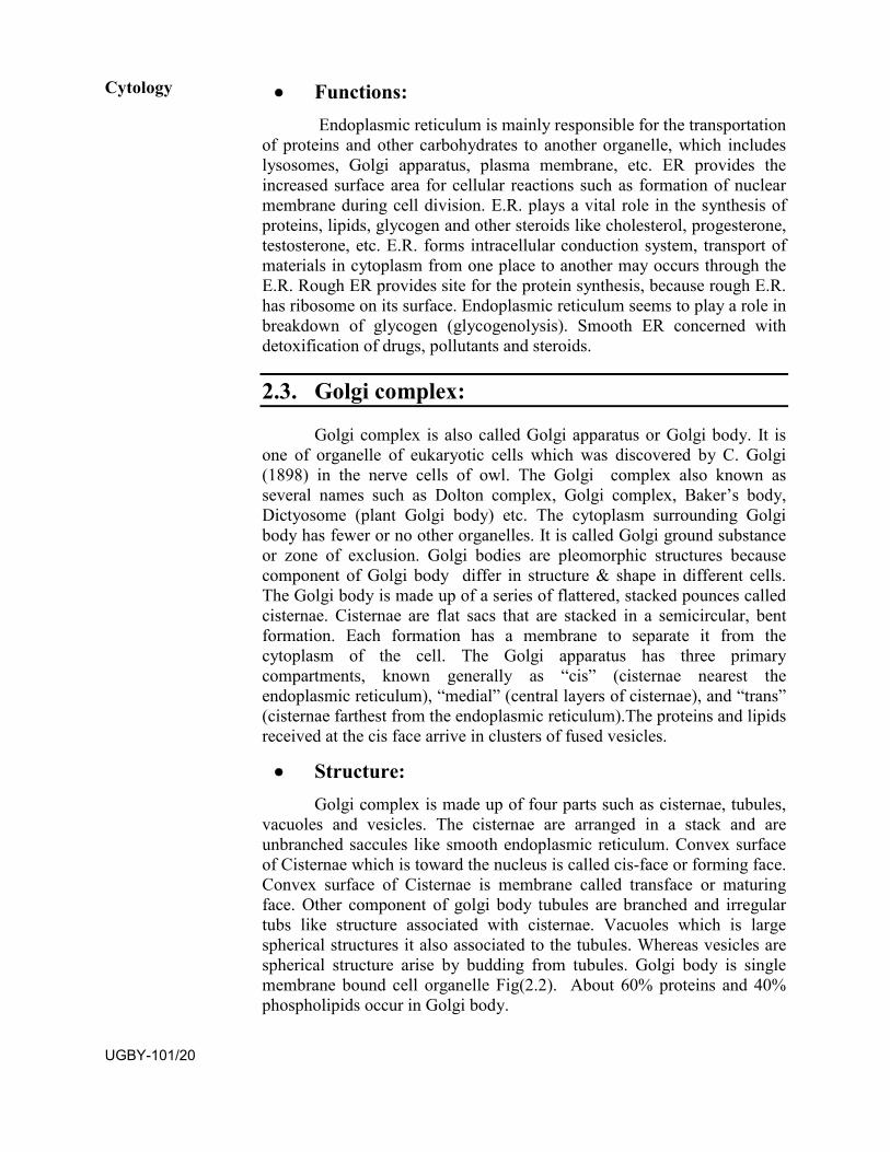
• Structure:Golgi complex is made up of four parts such as cisternae, tubules,
vacuoles and vesicles. The cisternae are arranged in a stack and are unbranched saccules like smooth endoplasmic reticulum. Convex surface of Cisternae which is toward the nucleus is called cis-face or forming face. Convex surface of Cisternae is membrane called transface or maturing face. Other component of golgi body tubules are branched and irregular tubs like structure associated with cisternae. Vacuoles which is large spherical structures it also associated to the tubules. Whereas vesicles are spherical structure arise by budding from tubules. Golgi body is single membrane bound cell organelle Fig(2.2). About 60% proteins and 40% phospholipids occur in Golgi body.
Cytology
UGBY-101/20
Fig.2.2: Golgi apparatus in section Source: https://www.aplustopper.com/structure-function-golgi-apparatus/
• Functions:The chief function of Golgi body is secretion of macromolecules
.Secretion involves three steps: Golgi body receives the materials from E.R. through its cis- face. These materials are chemically modified by Golgi body for example glycosidation of proteins and lipids takes place in Golgi body and it yields glycosidation and glycolipids. After chemical modification materials are packed in vesicles. Inadition, the golgi complex involves in secretion of zymogen granules from pancreas and lactoprotein from mammany glands. Different types of macromolecules are to be sent outside the cell move through the Golgi body. Products from the Golgi apparatus go to three main destinations: (1) inside the cell to lysosomes (2) the plasma membrane (3) outside the cell. Thus Golgi body termed as “director of macromolecular traffic in cell” or middle men of cell. Golgi apparatus also receives biochemicals in a bulk flow from the rough endoplasmic reticulum. The is only organelle in the cell that receive sorts, modifies, concentrates, packs and dispatches biochemicals for use inside and outside the cell.
2.4. Lysosome
Lysosome generally found in the cytoplasm of animal cell and exists in polymorphism. The lysosome was discovered by Christian De Duve (1955) and named as Lysosomes. The lysosomes found in all types of eukaryotic cell and responsible of digestion because it contain many enzymes capable of breaking down all types macromolecules such as protein, nucleic acids, carbohydrate and lipids. Each lysosome is surrounded by a membrane that maintained an acidic environment within the interior via a proton pump. In plant cells large central vacuole functions as lysosome. So in higher plant lysosome is less frequent but number of lysosome is high in fungi.
Cell Structure and Cellular
Organelles-II
UGBY-101/21
• Structure:Lysosome is spherical bag like structures (0.1-0.8υm) which is
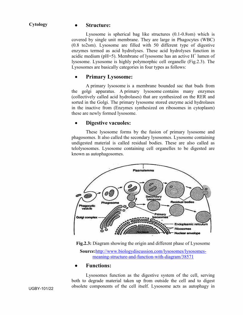
covered by single unit membrane. They are large in Phagocytes (WBC) (0.8 to2υm). Lysosome are filled with 50 different type of digestive enzymes termed as acid hydrolyses. These acid hydrolyses function in acidic medium (pH=5). Membrane of lysosome has an active H+ lumen of lysosome. Lysosome is highly polymorphic cell organelle (Fig.2.3). The Lysosomes are basically categories in four types as follows:
• Primary Lysosome:A primary lysosome is a membrane bounded sac that buds from
the golgi apparatus. A primary lysosome contains many enzymes (collectively called acid hydrolases) that are synthesized on the RER and sorted in the Golgi. The primary lysosome stored enzyme acid hydrolases in the inactive from (Enzymes synthesized on ribosomes in cytoplasm) these are newly formed lysosome.
• Digestive vacuoles:These lysosome forms by the fusion of primary lysosome and
phagosomes. It also called the secondary lysosomes. Lysosome containing undigested material is called residual bodies. These are also called as telolysosomes. Lysosome containing cell organelles to be digested are known as autophagosomes.
Fig.2.3: Diagram showing the origin and different phase of Lysosome Source:http://www.biologydiscussion.com/lysosomes/lysosomes-
meaning-structure-and-function-with-diagram/38571
• Functions:
Lysosomes function as the digestive system of the cell, servingboth to degrade material taken up from outside the cell and to digest obsolete components of the cell itself. Lysosome acts as autophagy in
Cytology
UGBY-101/22
which it digested old or dead cell organelles. Autophagy takes place during starvation of cell. Excessive secretary granules of hormone in endocrine gland may be digestion by lysosome. Sometimes all lysosome of a cell burst to dissolve the cell completely. Lysosome is helpful in digestion of egg membrane to assist fertilization and also trigger the cell division.
2.5 Chromosome The shape of chromosome changes from phase to phase in the
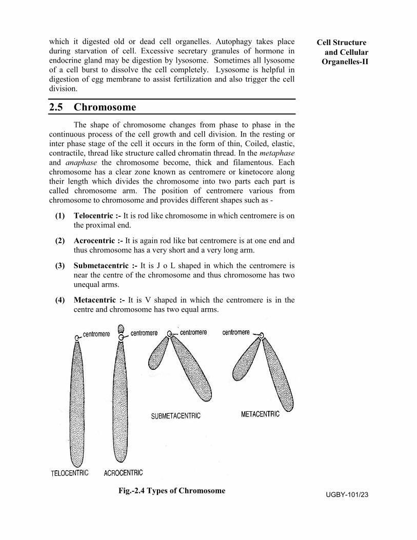
continuous process of the cell growth and cell division. In the resting or inter phase stage of the cell it occurs in the form of thin, Coiled, elastic, contractile, thread like structure called chromatin thread. In the metaphase and anaphase the chromosome become, thick and filamentous. Each chromosome has a clear zone known as centromere or kinetocore along their length which divides the chromosome into two parts each part is called chromosome arm. The position of centromere various from chromosome to chromosome and provides different shapes such as -
(1) Telocentric :- It is rod like chromosome in which centromere is on the proximal end.
(2) Acrocentric :- It is again rod like bat centromere is at one end and thus chromosome has a very short and a very long arm.
(3) Submetacentric :- It is J o L shaped in which the centromere is near the centre of the chromosome and thus chromosome has two unequal arms.
(4) Metacentric :- It is V shaped in which the centromere is in the centre and chromosome has two equal arms.
Fig.-2.4 Types of Chromosome
Cell Structure and Cellular
Organelles-II
UGBY-101/23
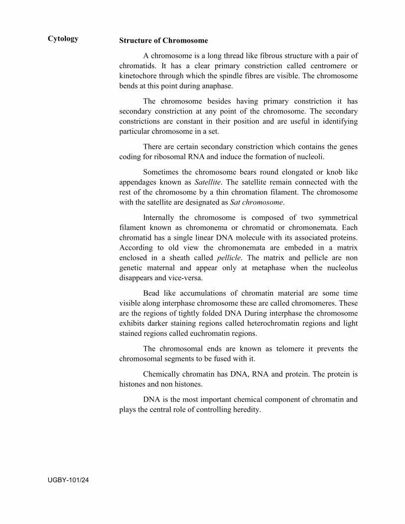
Structure of Chromosome
A chromosome is a long thread like fibrous structure with a pair of chromatids. It has a clear primary constriction called centromere or kinetochore through which the spindle fibres are visible. The chromosome bends at this point during anaphase.
The chromosome besides having primary constriction it has secondary constriction at any point of the chromosome. The secondary constrictions are constant in their position and are useful in identifying particular chromosome in a set.
There are certain secondary constriction which contains the genes coding for ribosomal RNA and induce the formation of nucleoli.
Sometimes the chromosome bears round elongated or knob like appendages known as Satellite. The satellite remain connected with the rest of the chromosome by a thin chromation filament. The chromosome with the satellite are designated as Sat chromosome.
Internally the chromosome is composed of two symmetrical filament known as chromonema or chromatid or chromonemata. Each chromatid has a single linear DNA molecule with its associated proteins. According to old view the chromonemata are embeded in a matrix enclosed in a sheath called pellicle. The matrix and pellicle are non genetic maternal and appear only at metaphase when the nucleolus disappears and vice-versa.
Bead like accumulations of chromatin material are some time visible along interphase chromosome these are called chromomeres. These are the regions of tightly folded DNA During interphase the chromosome exhibits darker staining regions called heterochromatin regions and light stained regions called euchromatin regions.
The chromosomal ends are known as telomere it prevents the chromosomal segments to be fused with it.
Chemically chromatin has DNA, RNA and protein. The protein is histones and non histones.
DNA is the most important chemical component of chromatin and plays the central role of controlling heredity.
Cytology
UGBY-101/24
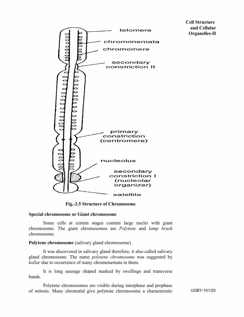
Fig.-2.5 Structure of Chromosome
Special chromosome or Giant chromosome
Some cells at certain stages contain large nuclei with giant chromosome. The giant chromosomes are Polytene and lamp brush chromosome.
Polytene chromosome (salivary gland chromosome)
It was discovered in salivary gland therefore, it also called salivary gland chromosome. The name polytene chromosome was suggested by kollar due to occurrence of many chromonemata in them.
It is long sausage shaped marked by swellings and transverse bands.
Polytene chromosomes are visible during interphase and prophase of mitosis. Many chromatid give polytene chromosome a characteristic
Cell Structure and Cellular
Organelles-II
UGBY-101/25
marphology. It has characteristic dark transverse band alternating with clear inter bands. The band has about 85% DNA and inter band about 15% DNA. In polytene chromosome maternal and paternal homologous chromosome remain associated side by side.
In polytene chromosome, chromosome puffs are the swellings of bands where DNA unfolds into open loops due to formation of m RNA. The puffing is a cyclic and reversible phenomenon. At definite time in definite tissues puffs may appear, grow and disappear.
Fig. 2.6 Polytene Chromosome
Lamp brush chromosome
It was first observed in salamander (amphibian) oocytes. The chromosome look like the brush used for cleaning the glass chimneys.
It occurs at the diplotene stage of meiotic prophase I in the primary oocytes of all vertebrates and invertebrates. It is also found in gaint nucleus of Acetabularia and even in plants.
The lamp brush chromosome are much longer than polytene chromosome. Since they occur in diplotene stage, they are in the form of bivalent in which maternal and paternal chromosomes are held together by chiasmata at those sites where crossing over has previously occurred. Each bivalent has four chromatids, two in each homologue. The axis of each homologue has a row of chromomeres from which lateral loops extend. The loops are always symmetrical and appear at constant position in the chromosome.
Each loop performs transcription of heterogenous RNA molecule i.e. precursor of mRNA molecules for various ribosomal proteins and types of histone proteins.
Each lateral loop is covered by matrix which is thicker at one end of chromtid and thinner at the other end. RNA synthesis starts at the thinner end and progress towards the thicker end.
Cytology
UGBY-101/26
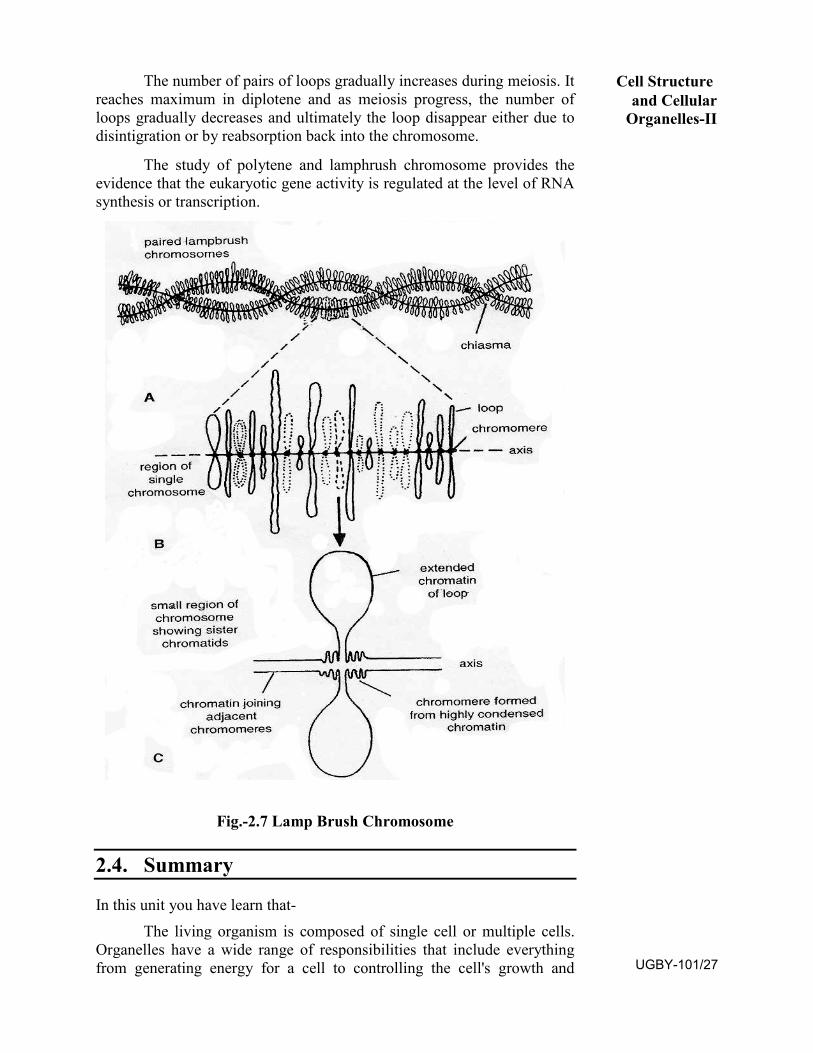
The number of pairs of loops gradually increases during meiosis. It reaches maximum in diplotene and as meiosis progress, the number of loops gradually decreases and ultimately the loop disappear either due to disintigration or by reabsorption back into the chromosome.
The study of polytene and lamphrush chromosome provides the evidence that the eukaryotic gene activity is regulated at the level of RNA synthesis or transcription.
Fig.-2.7 Lamp Brush Chromosome
2.4. Summary
In this unit you have learn that- The living organism is composed of single cell or multiple cells.
Organelles have a wide range of responsibilities that include everything from generating energy for a cell to controlling the cell's growth and
Cell Structure and Cellular
Organelles-II
UGBY-101/27
reproduction. The name organelle comes from the idea that these structures are to cells what an organ is to the body.
2.6. Terminal questions
Q.1. What is the cell organelles, write structure and functions of Endoplasmic Reticulum ?
Answer:------------------------------------------------------------------------------------------------------------------------------------------------------------------------------------------------------------------------------------------------------------------- Q.2. Write the structure and function of Golgi body? Answer:------------------------------------------------------------------------------------------------------------------------------------------------------------------------------------------------------------------------------------------------------------------- Q.3. Write the structure and function of Lysosome? Answer:------------------------------------------------------------------------------------------------------------------------------------------------------------------------------------------------------------------------------------------------------------------- Q.4. Discuss abut structure and function of chromosome. Answer:------------------------------------------------------------------------------------------------------------------------------------------------------------------------------------------------------------------------------------------------------------------- Q.5. Write short notes on
a) Lampbrush chromosomeb) Polytene Chromosome
Answer:-------------------------------------------------------------------------------------------------------------------------------------------------------------------------------------------------------------------------------------------------------------------
Suggested books
1. Cell Biology (Cytology, Biomolecules and Molecular Biology): PS Verma and V K Agarwal.
2. Principles of Biochemistry: Lehninger, Nelson and Cox. StudentEdition, CBS 1439 Publishers and Distributors, Delhi.
3. Fundamentals of Biochemistry: Dr J L Jain, S. Chand andCompany
4. Textbook of Biochemistry and Human Biology: Talwar andSrivastava. Eastern Economy Edition, Prentice Hall, India.
Cytology
UGBY-101/28
UNIT–3
CELL CYCLE, MITOSIS AND MEIOSIS
Structure 3.1 Introduction
Objectives
3.2 Cellcycle
Celldivision
3.3 Mitosis
Prophase
Metaphase
Anaphase
Telophase
Cytokinesis
Significance of mitosis
3.4 Meiosis
Meiosis I
Prophase I
Metaphase I
Anaphase I
Telophase I
Meiosis II
Prophase II
Metaphase II
Anaphase II
Telophase II
Cytokinesis
Significance of meiosis
3.5 Difference in mitosis and meiosis
3.6 Summary UGBY-101/29
3.7 Terminal Questions
3.8 Answers
3.1 Introduction
The existence of plants begin as a single cell. This cell first divides and form two cells which divide again and again continuously resulting in the formation of plant body. There are various methods by which new cells are formed by division of the preexisting cell. Thus cell maintains their continuity from one generation to another and copy the hereditary material itself most faithfully. The cell division under microscope was first discovered by German botanist Hugo Von Mohl in 1835. Cell division is the process by which a parent cell divides and give rise to two or more daughter cells. It is a means of reproduction for single cell organisms. In multicellular organisms, cell division contributes to growth, development, repair and the generation of reproductive cells (Pollen and Egg). There, are two types of cell division mitosis and meiosis. The term mitosis was coined by Walther Flemming in the early 1882. Mitosis means simply multiplication of cell number. The term ‘meiosis’ was coined by J.B. Farmer and J.E. Moore in 1905. The meiosis helps in alternation of generation. In mitosis, two daughter cells identical to the parent cells are formed where as in meiosis four daughter cells are formed. Each daughter cell has half the number of chromosome than their parent.
Objective After studying this unit you will be able to :
Know the cell cycle.
The process of mitosis occurring in plant cell.
The process of meiosis occurring in plant cell.
The difference in mitosis and meiosis.
3.2 Cell cycle
Cell cycle can be defined as the entire sequence of events happening from the end of one nuclear division to the beginning of the next.
Howard and Pelc (1953) divided cell cycle into four phases or stage-G1, S, G2 and M phase
• G1 phase
• S phase
• G2 phase
• M phase
Cytology
UGBY-101/30
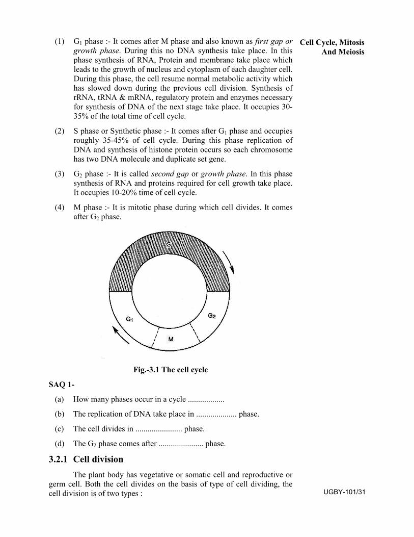
(1) G1 phase :- It comes after M phase and also known as first gap or growth phase. During this no DNA synthesis take place. In this phase synthesis of RNA, Protein and membrane take place which leads to the growth of nucleus and cytoplasm of each daughter cell. During this phase, the cell resume normal metabolic activity which has slowed down during the previous cell division. Synthesis of rRNA, tRNA & mRNA, regulatory protein and enzymes necessary for synthesis of DNA of the next stage take place. It occupies 30-35% of the total time of cell cycle.
(2) S phase or Synthetic phase :- It comes after G1 phase and occupies roughly 35-45% of cell cycle. During this phase replication of DNA and synthesis of histone protein occurs so each chromosome has two DNA molecule and duplicate set gene.
(3) G2 phase :- It is called second gap or growth phase. In this phase synthesis of RNA and proteins required for cell growth take place. It occupies 10-20% time of cell cycle.
(4) M phase :- It is mitotic phase during which cell divides. It comes after G2 phase.
Fig.-3.1 The cell cycle
SAQ 1-
(a) How many phases occur in a cycle ..................
(b) The replication of DNA take place in .................... phase.
(c) The cell divides in ....................... phase.
(d) The G2 phase comes after ...................... phase.
3.2.1 Cell division The plant body has vegetative or somatic cell and reproductive or
germ cell. Both the cell divides on the basis of type of cell dividing, the cell division is of two types :
Cell Cycle, Mitosis And Meiosis
UGBY-101/31
(i) Mitosis
(ii) Meiosis
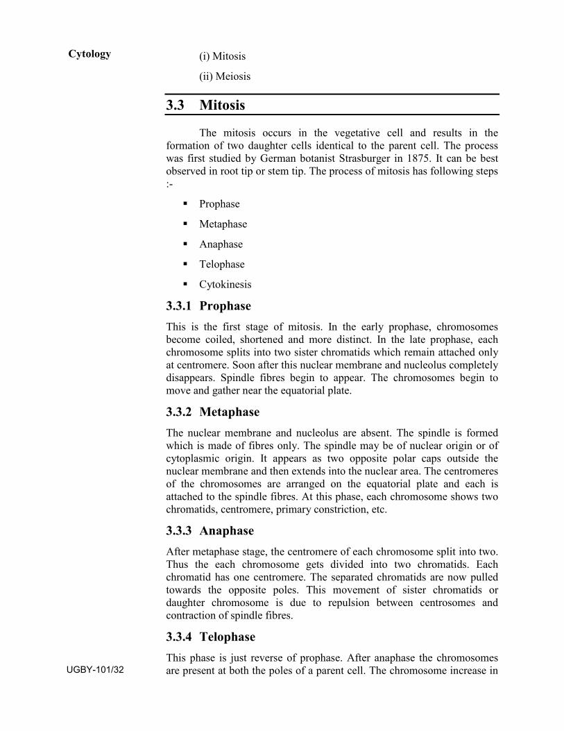
3.3 Mitosis
The mitosis occurs in the vegetative cell and results in the formation of two daughter cells identical to the parent cell. The process was first studied by German botanist Strasburger in 1875. It can be best observed in root tip or stem tip. The process of mitosis has following steps :-
Prophase
Metaphase
Anaphase
Telophase
Cytokinesis
3.3.1 Prophase This is the first stage of mitosis. In the early prophase, chromosomes become coiled, shortened and more distinct. In the late prophase, each chromosome splits into two sister chromatids which remain attached only at centromere. Soon after this nuclear membrane and nucleolus completely disappears. Spindle fibres begin to appear. The chromosomes begin to move and gather near the equatorial plate.
3.3.2 Metaphase The nuclear membrane and nucleolus are absent. The spindle is formed which is made of fibres only. The spindle may be of nuclear origin or of cytoplasmic origin. It appears as two opposite polar caps outside the nuclear membrane and then extends into the nuclear area. The centromeres of the chromosomes are arranged on the equatorial plate and each is attached to the spindle fibres. At this phase, each chromosome shows two chromatids, centromere, primary constriction, etc.
3.3.3 Anaphase After metaphase stage, the centromere of each chromosome split into two. Thus the each chromosome gets divided into two chromatids. Each chromatid has one centromere. The separated chromatids are now pulled towards the opposite poles. This movement of sister chromatids or daughter chromosome is due to repulsion between centrosomes and contraction of spindle fibres.
3.3.4 Telophase This phase is just reverse of prophase. After anaphase the chromosomes are present at both the poles of a parent cell. The chromosome increase in
Cytology
UGBY-101/32
length, becomes thread like and form chromation network. The individuality of chromosome is now lost. The nuclear membrane reappears at each pole around the chromosome to form nucleus. The nucsleolus also reappear in each nucleus. Thus at end two nuclei, one at each pole are present in the parent cell. Spindle fibres are absent.
3.3.5 Cytokinesis Cytokinesis is the division of cytoplasm. It results in the formation of two daughter cells. In the plant cell, a rigid cell plate is initiated at the centre and gradually progress towards the periphery. After this primary walls are deposited on either side of cell plate followed by thick secondary cell walls of cellulose. Thus at the end, two daughter cells each having the equal number of chromosomes as present in their parent cell are formed.
Significance of mitosis
mitosis is important because it is essential for growth and repair in the body. In mitosis the constituents of the chromosomes are equally distributed to the two daughter nuclei and thus, they become qualitatively and quantitatively similar to the mother nucleus.
Fig.3.2 Mitosis in the plant cells.
SAQ-2
(a) Mitosis occurs in .................... cell.
(b) ............ daughter cells are formed after mitosis.
(c) The process of mitosis was first studied by ....................
(d) Mitosis can be best observed in .........................
Cell Cycle, Mitosis And Meiosis
UGBY-101/33
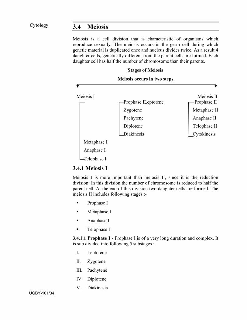
3.4 Meiosis
Meiosis is a cell division that is characteristic of organisms which reproduce sexually. The meiosis occurs in the germ cell during which genetic material is duplicated once and nucleus divides twice. As a result 4 daughter cells, genetically different from the parent cells are formed. Each daughter cell has half the number of chromosome than their parents.
Stages of Meiosis
Meiosis occurs in two steps
Meiosis I Meiosis II Prophase ILeptotene Prophase II Zygotene Metaphase II Pachytene Anaphase II Diplotene Telophase II Diakinesis Cytokinesis
Metaphase I Anaphase I
Telophase I
3.4.1 Meiosis I Meiosis I is more important than meiosis II, since it is the reduction division. In this division the number of chromosome is reduced to half the parent cell. At the end of this division two daughter cells are formed. The meiosis II includes following stages :-
Prophase I
Metaphase I
Anaphase I
Telophase I
3.4.1.1 Prophase I - Prophase I is of a very long duration and complex. It is sub divided into following 5 substages :
I. Leptotene
II. Zygotene
III. Pachytene
IV. Diplotene
V. Diakinesis
Cytology
UGBY-101/34
I. Leptotene :- This is the first stage of prophase I of meiosis I. Nuclear membrane and nucleolus are intact. Chromosomes are long thread like structure and form chromation network. On these thread like chromosomes, bead like structures called chromomeres are present all along the length of chromosomes. In certain plants like lilium, all the chromosomes finally move towards one part of the nucleus. This phenomenon is known as synizesis or boquet formation.
II. Zygotene :- This is the second stage of prophase I of meiosis I.Nuclear membrane and nucleolus are still intact. Zygotene is characterised by synapsis that is pairing of homologous chromosome. Synaptonemal complex is formed as a result of synapsis. The synapsis begins at one or more points along the length of the homologous chromosomes and at each place a pair shows two chromatids.
III. Pachytene :- This is the third stage of prophase I of meiosis I.Nuclear membrane and nucleolus are distinct. The chromosomes appear as thickened, coiled and thread like structure. Each chromosome shows its two chromatids. Pair of homologous chromosome is called bivalent. It is made up of four chromatids and hence known as tetrad. Pachytene stage is characterised by crossing over. It is the exchange of equal parts of chromatids of two different but homologous chromosomes. The length of chromosome at pachytene is found more than metaphase therefore the chromosome at this stage is used for the study of morphology.
IV. Diplotene :- This is the fourth stage of prophase I of meiosis I. Thenuclear membrane is still intact but nucleolus is disappearing. At this stage, further thickening and shortening of chromosomes take place. The homologous chromosomes start separating from one another but still remain in contact at some points called chiasmata which indicates that crossing over has been completed at these points.
V. Diakinesis :- This is the fifth and last stage of prophase I of meiosis I. Nuclear membrane and nucleolus are not seen at this stage. The chromatids start separating, beginning from the centromere towards the end in zipper like manner. The chiasmata thus open. This is known as terminalization of chiasmata.
3.4.1.2 Metaphase I At metaphase I, the spindle apparatus starts appearing and bivalents are arranged on the equatorial plate. Each chromosome of a bivalent is attached to the spindle fibres by its centromere.
3.4.1.3 Anaphase I At this stage, the spindle fibre contracts and pull the centromere along with chromosome to opposite pole. In the anaphase of mitosis the centromere divides logitudinally and two sister chromatids pass to two different poles but in case of anaphase I of meiosis I, the sister chromatids do not separate but go to the same pole. After anaphase I, the chromosome number is reduced and each pole has haploid number of chromosome.
Cell Cycle, Mitosis And Meiosis
UGBY-101/35
3.4.1.4 Telophase I This is just reverse of prophase. The nuclear membrane and nucleolus have reappeared. The cell has two nuclei one at each pole. At telophase I, meiosis I is completed which may be followed by cytokinesis giving rise to a dyad or cytokinesis may be postponed till the end of meiosis II.
3.4.2 Meiosis II Meiosis I is followed by meiosis II which is similar to mitosis. Meiosis II results in the formation of four daughter cells, each having the same number of chromosome as was present at the end of meiosis I. The meiosis two has following stages :-
Prophase II
Metaphase II
Anaphase II
Telophase II
Cytokinesis
3.4.2.1 Prophase II In the early prophase II chromosomes become short and thick. Each chromosome splits into two sister chromatids bound together by a centromere. In the late prophase II nuclear membrane and nucleolus completely disappears. Spindle fibre begins to appear.
3.4.2.2 Metaphase II The nuclear membrane and nucleolus are absent. Spindle fibres are formed and organised into a spindle. The centromers of the chromosome are arranged on the equatorial plate and each is attached to the spindle fibres. Each chromosome has two chromatids held together by a centromere.
3.4.2.3 Anaphase II At anaphase II of meiosis the centromere divides as a result the two chromatids get separated each having an individual centromere. Spindle fibres contract and each chromosome is now pulled to the opposite poles.
3.4.2.4 Telophase II It is just reverse of prophase. After anaphase the chromosomes are present at both the poles of a parent cell. The chromosome increase in length, becomes thread like and forms chromatine network. The nuclear membrane reappears at each pole around the chromosome to form nucleus. Nucleolus also reappears in each nucleus. Spindle fibres disappear completely.
Cytology
UGBY-101/36
3.4.2.5 Cytokinesis Cytokinesis occurs like the mitosis and results in the formation of four daughter cells each having the same number of chromosome as was present at the end of meiosis I.
Significance of Meiosis :- Meiosis helps in keeping chromosome number constant from generation to generation. If this reduction in chromosome number does not occur at any stage of a plant, the off- spring would have an ever-increasing number of chromosomes resulting in new peculiar and distinct types of off spring.
Fig. 3.3: Meiosis in the plant cell
Cell Cycle, Mitosis And Meiosis
UGBY-101/37
SQA-3
(a) Meiosis occurs in ...................................
(b) .......................... daughter cells are formed after meiosis.
(c) Meiosis consists of ................... successive divisions of a cell.
(d) The number of chromosome is reduced to ..................... in daughter cells formed after meiosis.
3.5 Difference in Mitosis and Meiosis
Mitosis Meiosis
It occurs in all somatic cellsof plant.
It occurs in the germ cell which forms gametes.
Whole process is completedin one step resulting in theformation of two daughtercell which have same numberof chromosome as waspresent in their parent cell.
Whole process is completed in two steps resulting in the formation of four daughter cells which have half the number of chromosome as was present in their parent cell.
Prophase I is of shortduration and does not havesub stage.
Prophase I is of long duration and has sub stages like leptotene, zygotene, pachytene, Diplotene and Diakinesis.
No pairing of chromosome(Synapsis) occurs.
Pairing of chromosome (synapsis) occurs at zygotene.
Crossing over and chiasmataformation does not occur.
Crossing over and chiasmata formation occurs at pachytene and diplotene substages during which exchange of characters take place.
At Metaphase, centromere lietowards equator and armstowards poles.
The arrangement is just reverse. The centromere lie towards poles and arms towards equator.
Centromere divides atanaphase.
Centromere divides at anaphase II
3.6 Summary
The cell cycle has 4 phases- G1 phase, S phase, G2 phase and Mphase.
Cytology
UGBY-101/38
The cell divides in M phase.
The cell division is of two types mitosis and meiosis.
Mitosis occurs in vegetative or somatic cell. It produces twodaughter cells having same number of chromosome as were intheir parent cell.
Meiosis occurs in germ cell and produces four daughter cellshaving half the number of chromosome than their parents.
Meiosis occurs in two stage-Meiosis I and meiosis II.
Meiosis I is reduction division and Meiosis II is simple mitosis.
3.7 Terminal Questions
Long Questions Q.1 With the help of labelled diagrams describe the process of mitosis
in plant cell.
Q.2 With the help of labelled diagrams describe the process of meiosis in plant cell.
Q.3 Differentiate between mitosis and meiosis
Write notes on :
Q.1 Prophase I of Meiosis.
Q.2 Cell cycle
3.8 Answers
SAQ1 (a) 4 phases (b) S phase (c) M phase (d) S phase
SAQ2 (a) Vegetative or somatic cell (b) Two
(c) Strasburger (d) Root tip or stem tip
SAQ3 (a) Germ cell (b) Four (c) Two (d) Half
Cell Cycle, Mitosis And Meiosis
UGBY-101/39

UGBY-101/40
BLOCK
2 GENETICS - I
UNIT 4
PRE-MENDELIAN GENETICS, AND MENDEL’S LAWS OF INHERITANCE
UNIT 5
LINKAGE AND CROSSING OVER
UNIT 6
CYTOPLASMIC INHERITANCE, SEX LINKED INHERITANCE AND SEX DETERMINATION IN PLANTS
Uttar Pradesh Rajarshi Tandon Open University
Bachelor of Science
UGBY-101
45-64
65-86
87-110
UGBY-101/41
Course Design Committee Prof. Ashutosh Gupta Chairman Director, School of Science UPRTOU, Prayagraj.
Prof. Malvika Srivastava Member Botny Department Deendayal Upadhyay University, Gorakhpur.
Prof. V.N. Pandey Member Botny Department Deendayal Upadhyay University, Gorakhpur.
Prof. Anil Kumar Diwedi Member Botny Department Deendayal Upadhyay University, Gorakhpur.
Dr. N.N Tripathi Member Acadmic Consultant, School of Science UPRTOU, Prayagraj.
Dr. Sushma Chauhan Member/Secretary Acadmic Consultant, School of Science UPRTOU, Prayagraj.
Course Preparation Committee Dr. Dharmveer Singh Author Block-1 (Unit: 1-2) Acadmic Consultant, School of Science UPRTOU, Prayagraj.
Dr. Sushma Chauhan Author Block-1 (Unit: 3) Acadmic Consultant, School of Science UPRTOU, Prayagraj.
Dr Achala Srivatava Author Block-2-3 (Unit: 4-9) Assistant professor S.S. Khanna, P.G. College Prayagraj.
Prof. G. Kumar Editor (Unit: 1-9) Professor Department of Botany University of Allahabad, Prayagraj
Dr. Sushma Chauhan Course Coordinator Acadmic Consultant (Botany) School of Science UPRTOU, Prayagraj.
© UPRTOU, Prayagraj. 2019 ISBN : All Rights are reserved. No part of this work may be reproduced in any form, by mimeograph or any other means, without permission in writing from the Uttar Pradesh Rajarshi Tondon Open University, Prayagraj.
UGBY-101/42
Block : II
Genetics- I The study of science that deals heredity and variation from parent
to progeny is known as Genetics. Those traits which are transmitted from one generation to next is known as hereditary via parents whereas variation among the individuals may be inherited through their parents or may be due to physical factors i.e. the environment. The genes are known as a hereditary unit which are linearly located on the chromosomes. Each individual has a fixed no. and sets of chromosomes and each chromosome carries numerous genes. The variation occurs not only among the different individuals but among the progenies of the same parents. The Block II includes Genetics I part which comprises into three units i.e. Unit 4, Unit 5 and Unit 6.
Unit 4: Introduction and definition of Genetics, Pre-Mendelian Genetics, and Mendel’s Laws of Inheritance.
Unit 5: Linkage and Crossing over
Unit 6: Cytoplasmic Inheritance, Sex linked Inheritance and Sex determination in plants
UGBY-101/43

UGBY-101/44
UNIT-4
PRE-MENDELIAN GENETICS, AND MENDEL’S LAW OF INHERITANCE
Structure 4.1 Introduction
4.2 History of Genetics: Pre-Mendelian Genetics
4.3 Life history of Mendel
4.4 Mendel’s Experiments
4.5 Laws of Heredity:
4.5.1 Law of Segregation or Law of Purity of Gametes
4.5.2 Law of Independent Assortment or Law of Free Recombination
4.6 Deviations of Mendel’s law (Other types of inheritance)
4.6.1 Incomplete dominance
4.6.2 Co-dominance
4.6.3 Lethal genes
4.7 Summary
4.8 Terminal Questions
4.9 Answers
4.1 INTRODUCTION
Genetics is the branch of biology which deals heredity and variation together during the course of evolution from one generation to another. The resemblance of children with their parents, i.e. the traits carried by offspring from their ancestors due to the inheritance is called heredity. The sexually produced offspring only 50% of characters transmitted by each parent due to this variation occurs among the individuals. The variations may genetic or inherited or may be due to physical factors. i.e. environmental. The inherited hereditary variations are dealt under genetics, and having permanent traits whereas environmental variations may be temporary and varies accordingly.
Objectives Definition of genetics UGBY-101/45
History of Genetics and Earlier Concept of Heredity
Heredity and Variation
Mendel’s Experiment
Laws of Heredity
Deviations from Mendel’s Law: Incomplete dominance, Co-dominance, Lethality
Linkage: Complete and incomplete linkage
Crossing Over: Types, Theories
Extranuclear or Cytoplasmic Inheritance
Types of Sex Determination in Plants
4.2 HISTORY OF GENETICS: PRE-MENDELIAN GENETICS
The thought of heredity reported during 400 B.C. by Hippocrates and 350 B.C. by Aristotle. According to Hippocrates the characters inherited from parents are carried through reproductive bodies which are made by all parts of the body. Aristotle has different view where children may resemble rather than their parents as their grandparents; it is derived from the nutrients required for different parts contributing the characteristics through reproductive means. Although both have different views but believed in direct inheritance of traits through sexual reproduction carried away by the parents via reproductive substances. The discovery of sexual reproduction raised the thought about the heredity is that the traits are transmitted to the offsprings from parent is either by egg or sperms or may be by both. J.Swammerdam (1969) while studying the development of insects, suggested that the development of an organism is a simple enlargement of minute performed individual, called homunculus, that could be present in the ovum or sperm. The homunculus concept was totally discarded by the scientists. Later on K.W.Wolff (1738-1794) proposed the theory of epigenesis according to which the gametes contained undifferentiated living substance forming an organised body after fertilization. This theory also suggested that many new organs and tissues, which were absent originally, may develop de novo due to mysterious vital forces.
Joseph Gottlieb Kolreuter, a German Botanist (1733-1806) while working on tobacco plant obtained some interspecific hybrids by cross pollination. The hybrids produced variable offsprings after self pollination. Thus he concluded the particulate nature of inherited traits.
Genetics-I
UGBY-101/46
During the year 1809-82 Charles Darwin proposed the theory that every part of the body very minute invisible particles called gemmules or pangenes, which are transmitted to the sex organs and assembled in the gametes through the blood streams. The gemmules of both parents brought together during fertilization and redistributed to different organs determining the different characters in children.
Knight (1799) and Goss (1824) performed hybridisation experiments on garden pea (Pisum sativum) found uniform characters in hybrids and segregation of characters in second generation, but failed to analyse and formulate their observations and results mathematically to establish the law of inheritance.
The work of various plant breeders subsequently demonstrated following three basic principles of inheritance:
1. The traits could be hidden for one or more generation and mayreappear as it is without any change.
2. The traits may remain together in one generation and maysegregate in next upcoming generations.
3. One form of particular trait may be seen more often than itsalternative form.
The elegant experiments carried out by Mendel laid the foundation of basic genetics and established the principles of heredity and laws of inheritance named as Mendelism.
SAQ.1: a. The branch of biology which deals .......... and .......... is called
genetics.
b. The homunculus concept was raised by..........
c. The theory of epigenesist proposed by ........
d. ......concluded the particulate nature inherited traits.
4.3 LIFE HISTORY OF MENDEL Gregor Johann Mendel (1822-1884), the father of Genetics, Austrian monk born on 22ndJuly 1822 in Heizendorf, a village in Sudeten region of Silosia, Austria. His father was a great lover of nature, influenced him to develop interest in living being since childhood. He received his school education in a monastery at Bruno (now Brno, Czech), later had two year university course in Philosophy at Olmitz Philosophical Institute. In 1843, he was admitted to Augustinian Monastery, Brunn, Moravia. In 1848 he has completed his theological studies and after a year got job as a teacher in a High School, Znaim. After that he joined University of Vienna for pursuing the study of Science and Mathematics. In Brunn Modern School
Pre-Mendelian Genetics, and
Mendel’s Law of Inheritance
UGBY-101/47
he joined as a teacher of Physics and Natural History in 1854 and continued for 14 years. During this he performed his popular hybridisation experiment on garden pea. In 1865 he presented his work before Brunn Society for the study of Natural Science. His paper entitled, “Versuche uber Pflenzenhybriden” (“Experiments on Plant Hybridization”) published in the Proceedings of Society in 1866. Unfortunately his remarkable monumental work remained ignored and failed to get attention to understand by the scientist and plant breeders at that time. The great natural scientist, monk, mathematician died in 1884 unknowingly.
After 34 years i.e. a long leap in 1900, three other eminent biologists of different places working independently Mendel’s experiment came to light again. Karl Correns of Germany, Hugo de Vries of Netherlands and Erich Von Tschermak of Austria got same observation and results which Mendel explained earlier during their hybridiztation experiments. These scientists rediscovered mendelian principles and gave him the recognition he deserved. Thereafter he was honoured as the father of Genetics.
4.4 MENDEL’S EXPERIMENTS
Mendel selected common garden pea (Pisum sativum) for his experiments. In his monastery garden he performed his hybridization experiments for consistently seven years. He collected seeds of 34 different varieties of pea which were grown in the garden area. The selection of pea plants for his experiments has following advantages:
1. These plants are easily available and grown easily with welldefined characters.
2. Having short life cycle i.e. annual plants, make feasible to studyseveral generations in short duration.
3. Single plant produces numerous fertile seeds, which was used togrow and study for the next generation.
4. Flowers are hermaphrodite i.e. bisexual.
5. Easy hybridization .It may be self pollinated to obtain pure lineselection, along with the cross pollination.
6. These were available in many pure line breeding varieties withobservable with alternative forms for a trait or characteristics.
7. Hybrids resulting from crossing of two different varieties wereperfectly fertile.
Though the plant selected had large number of contrasting characters but Mendel had opted and focussed only on seven pairs of contrasting characters in pea plants for his experiments. Each of the traits selected had two alternative forms which are as follows:
Genetics-I
UGBY-101/48
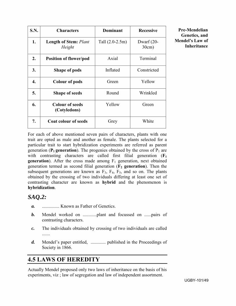
For each of above mentioned seven pairs of characters, plants with one trait are opted as male and another as female. The plants selected for a particular trait to start hybridization experiments are referred as parent generation (P1 generation). The progenies obtained by the cross of P1 are with contrasting characters are called first filial generation (F1 generation). After the cross made among F1 generation, next obtained generation termed as second filial generation (F2 generation). Then the subsequent generations are known as F3, F4, F5, and so on. The plants obtained by the crossing of two individuals differing at least one set of contrasting character are known as hybrid and the phenomenon is hybridization.
SAQ.2: a. ............... Known as Father of Genetics.
b. Mendel worked on ............plant and focussed on ......pairs ofcontrasting characters.
c. The individuals obtained by crossing of two individuals are called.......
d. Mendel’s paper entitled, ............. published in the Proceedings ofSociety in 1866.
4.5 LAWS OF HEREDITY Actually Mendel proposed only two laws of inheritance on the basis of his experiments, viz ; law of segregation and law of independent assortment.
S.N. Characters Dominant Recessive
1. Length of Stem: Plant Height
Tall (2.0-2.5m) Dwarf (20-30cm)
2. Position of flower/pod Axial Terminal
3. Shape of pods Inflated Constricted
4. Colour of pods Green Yellow
5. Shape of seeds Round Wrinkled
6. Colour of seeds (Cotyledons)
Yellow Green
7. Coat colour of seeds Grey White
Pre-Mendelian Genetics, and
Mendel’s Law of Inheritance
UGBY-101/49
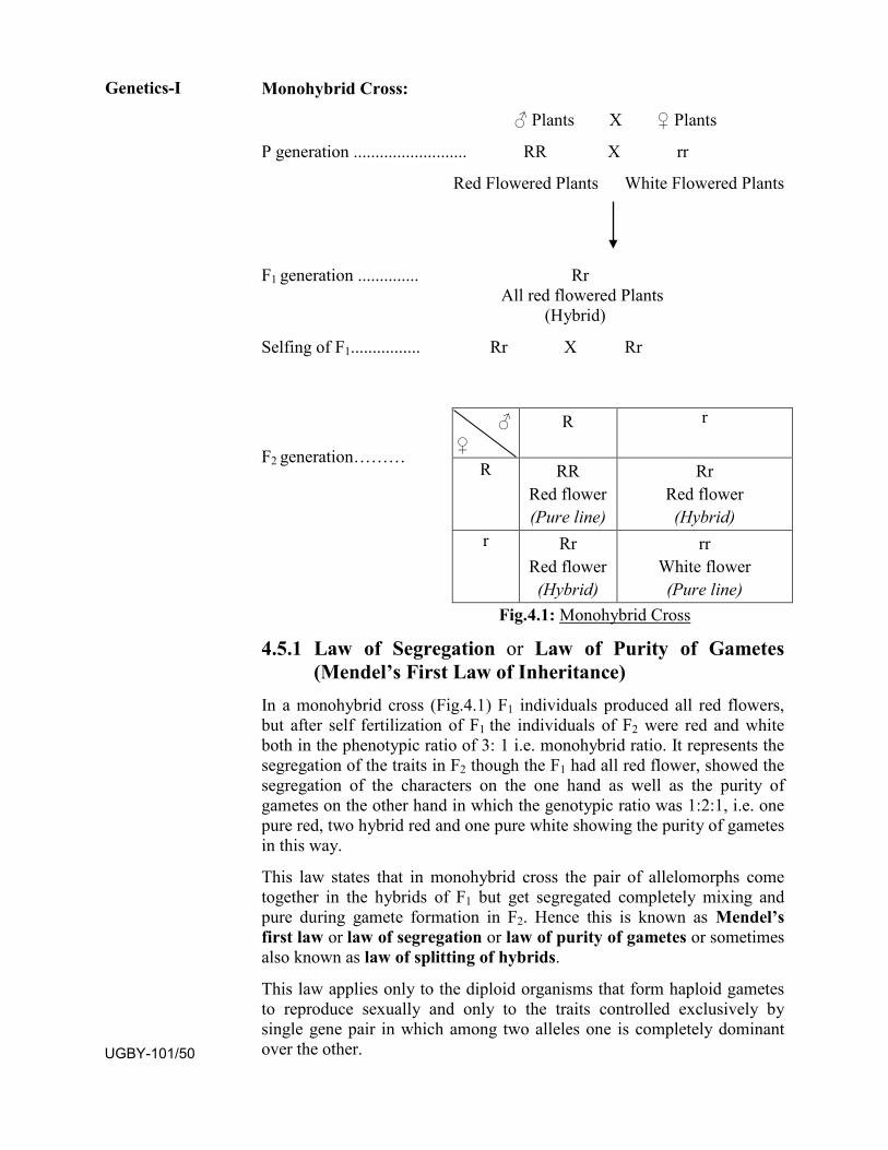
Monohybrid Cross:
♂ Plants X ♀ Plants
P generation .......................... RR X rr
Red Flowered Plants White Flowered Plants
F1 generation .............. Rr All red flowered Plants
(Hybrid)
Selfing of F1................ Rr X Rr
F2 generation………
Fig.4.1: Monohybrid Cross
4.5.1 Law of Segregation or Law of Purity of Gametes (Mendel’s First Law of Inheritance)
In a monohybrid cross (Fig.4.1) F1 individuals produced all red flowers, but after self fertilization of F1 the individuals of F2 were red and white both in the phenotypic ratio of 3: 1 i.e. monohybrid ratio. It represents the segregation of the traits in F2 though the F1 had all red flower, showed the segregation of the characters on the one hand as well as the purity of gametes on the other hand in which the genotypic ratio was 1:2:1, i.e. one pure red, two hybrid red and one pure white showing the purity of gametes in this way.
This law states that in monohybrid cross the pair of allelomorphs come together in the hybrids of F1 but get segregated completely mixing and pure during gamete formation in F2. Hence this is known as Mendel’s first law or law of segregation or law of purity of gametes or sometimes also known as law of splitting of hybrids.
This law applies only to the diploid organisms that form haploid gametes to reproduce sexually and only to the traits controlled exclusively by single gene pair in which among two alleles one is completely dominant over the other.
♂♀
R r
R RR Red flower (Pure line)
Rr Red flower (Hybrid)
r Rr Red flower (Hybrid)
rr White flower (Pure line)
Genetics-I
UGBY-101/50
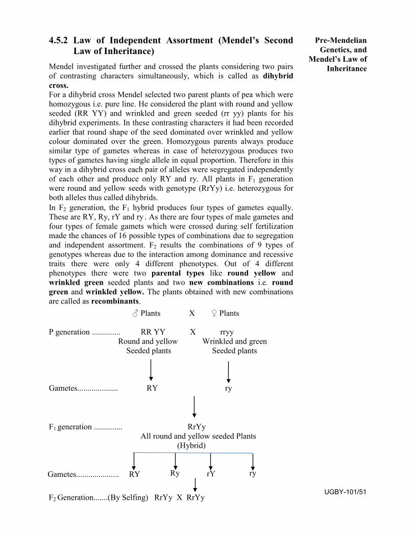
4.5.2 Law of Independent Assortment (Mendel’s Second Law of Inheritance)
Mendel investigated further and crossed the plants considering two pairs of contrasting characters simultaneously, which is called as dihybrid cross. For a dihybrid cross Mendel selected two parent plants of pea which were homozygous i.e. pure line. He considered the plant with round and yellow seeded (RR YY) and wrinkled and green seeded (rr yy) plants for his dihybrid experiments. In these contrasting characters it had been recorded earlier that round shape of the seed dominated over wrinkled and yellow colour dominated over the green. Homozygous parents always produce similar type of gametes whereas in case of heterozygous produces two types of gametes having single allele in equal proportion. Therefore in this way in a dihybrid cross each pair of alleles were segregated independently of each other and produce only RY and ry. All plants in F1 generation were round and yellow seeds with genotype (RrYy) i.e. heterozygous for both alleles thus called dihybrids. In F2 generation, the F1 hybrid produces four types of gametes equally. These are RY, Ry, rY and ry . As there are four types of male gametes and four types of female gamets which were crossed during self fertilization made the chances of 16 possible types of combinations due to segregation and independent assortment. F2 results the combinations of 9 types of genotypes whereas due to the interaction among dominance and recessive traits there were only 4 different phenotypes. Out of 4 different phenotypes there were two parental types like round yellow and wrinkled green seeded plants and two new combinations i.e. round green and wrinkled yellow. The plants obtained with new combinations are called as recombinants.
♂ Plants X ♀ Plants
P generation .............. RR YY X rryy Round and yellow Wrinkled and green
Seeded plants Seeded plants
Gametes.................... RY ry
F1 generation .............. RrYy All round and yellow seeded Plants
(Hybrid)
Gametes..................... RY Ry rY ry
Pre-Mendelian Genetics, and
Mendel’s Law of Inheritance
F2 Generation.......(By Selfing) RrYy X RrYy UGBY-101/51
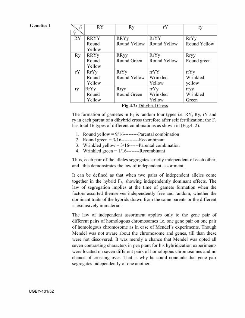
Fig.4.2: Dihybrid Cross
The formation of gametes in F1 is random four types i.e. RY, Ry, rY and ry in each parent of a dihybrid cross therefore after self fertilization; the F2 has total 16 types of different combinations as shown in (Fig.4. 2):
1. Round yellow = 9/16---------Parental combination2. Round green = 3/16-----------Recombinant3. Wrinkled yellow = 3/16------Parental combination4. Wrinkled green = 1/16--------Recombinant
Thus, each pair of the alleles segregates strictly independent of each other, and this demonstrates the law of independent assortment.
It can be defined as that when two pairs of independent alleles come together in the hybrid F1, showing independently dominant effects. The law of segregation implies at the time of gamete formation when the factors assorted themselves independently free and random, whether the dominant traits of the hybrids drawn from the same parents or the different is exclusively immaterial.
The law of independent assortment applies only to the gene pair of different pairs of homologous chromosomes i.e. one gene pair on one pair of homologous chromosome as in case of Mendel’s experiments. Though Mendel was not aware about the chromosome and genes, till than these were not discovered. It was merely a chance that Mendel was opted all seven contrasting characters in pea plant for his hybridization experiments were located on seven different pairs of homologous chromosomes and no chance of crossing over. That is why he could conclude that gene pair segregates independently of one another.
♂ ♀
RY Ry rY ry
RY RRYY Round Yellow
RRYy Round Yellow
RrYY Round Yellow
RrYy Round Yellow
Ry RRYy Round Yellow
RRyy Round Green
RrYy Round Yellow
Rryy Round green
rY RrYy Round Yellow
RrYy Round Yellow
rrYY Wrinkled Yellow
rrYy Wrinkled yellow
ry RrYy Round Yellow
Rryy Round Green
rrYy Wrinkled Yellow
rryy Wrinkled Green
Genetics-I
UGBY-101/52
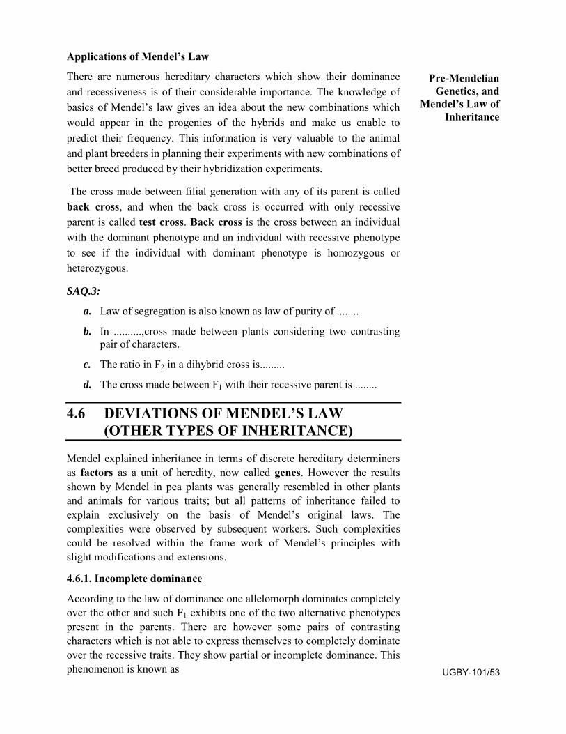
Applications of Mendel’s Law
There are numerous hereditary characters which show their dominance and recessiveness is of their considerable importance. The knowledge of basics of Mendel’s law gives an idea about the new combinations which would appear in the progenies of the hybrids and make us enable to predict their frequency. This information is very valuable to the animal and plant breeders in planning their experiments with new combinations of better breed produced by their hybridization experiments.
The cross made between filial generation with any of its parent is called back cross, and when the back cross is occurred with only recessive parent is called test cross. Back cross is the cross between an individual with the dominant phenotype and an individual with recessive phenotype to see if the individual with dominant phenotype is homozygous or heterozygous.
SAQ.3:
a. Law of segregation is also known as law of purity of ........
b. In ..........,cross made between plants considering two contrastingpair of characters.
c. The ratio in F2 in a dihybrid cross is.........
d. The cross made between F1 with their recessive parent is ........
4.6 DEVIATIONS OF MENDEL’S LAW (OTHER TYPES OF INHERITANCE)
Mendel explained inheritance in terms of discrete hereditary determiners as factors as a unit of heredity, now called genes. However the results shown by Mendel in pea plants was generally resembled in other plants and animals for various traits; but all patterns of inheritance failed to explain exclusively on the basis of Mendel’s original laws. The complexities were observed by subsequent workers. Such complexities could be resolved within the frame work of Mendel’s principles with slight modifications and extensions.
4.6.1. Incomplete dominance
According to the law of dominance one allelomorph dominates completely over the other and such F1 exhibits one of the two alternative phenotypes present in the parents. There are however some pairs of contrasting characters which is not able to express themselves to completely dominate over the recessive traits. They show partial or incomplete dominance. This phenomenon is known as
Pre-Mendelian Genetics, and
Mendel’s Law of Inheritance
UGBY-101/53
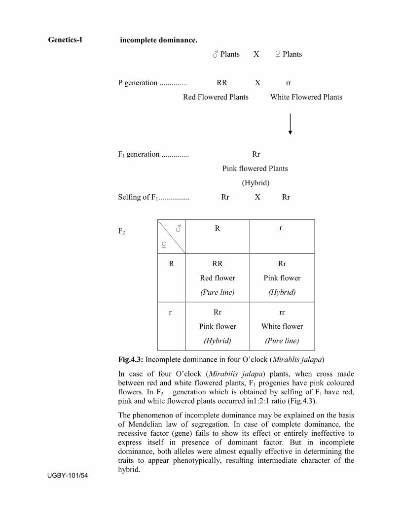
incomplete dominance.
♂ Plants X ♀ Plants
P generation .............. RR X rr
Red Flowered Plants White Flowered Plants
F1 generation .............. Rr
Pink flowered Plants
(Hybrid)
Selfing of F1................ Rr X Rr
F2
Fig.4.3: Incomplete dominance in four O’clock (Mirablis jalapa)
In case of four O’clock (Mirabilis jalapa) plants, when cross made between red and white flowered plants, F1 progenies have pink coloured flowers. In F2 generation which is obtained by selfing of F1 have red, pink and white flowered plants occurred in1:2:1 ratio (Fig.4.3).
The phenomenon of incomplete dominance may be explained on the basis of Mendelian law of segregation. In case of complete dominance, the recessive factor (gene) fails to show its effect or entirely ineffective to express itself in presence of dominant factor. But in incomplete dominance, both alleles were almost equally effective in determining the traits to appear phenotypically, resulting intermediate character of the hybrid.
♂
♀
R r
R RR
Red flower
(Pure line)
Rr
Pink flower
(Hybrid)
r Rr
Pink flower
(Hybrid)
rr
White flower
(Pure line)
Genetics-I
UGBY-101/54
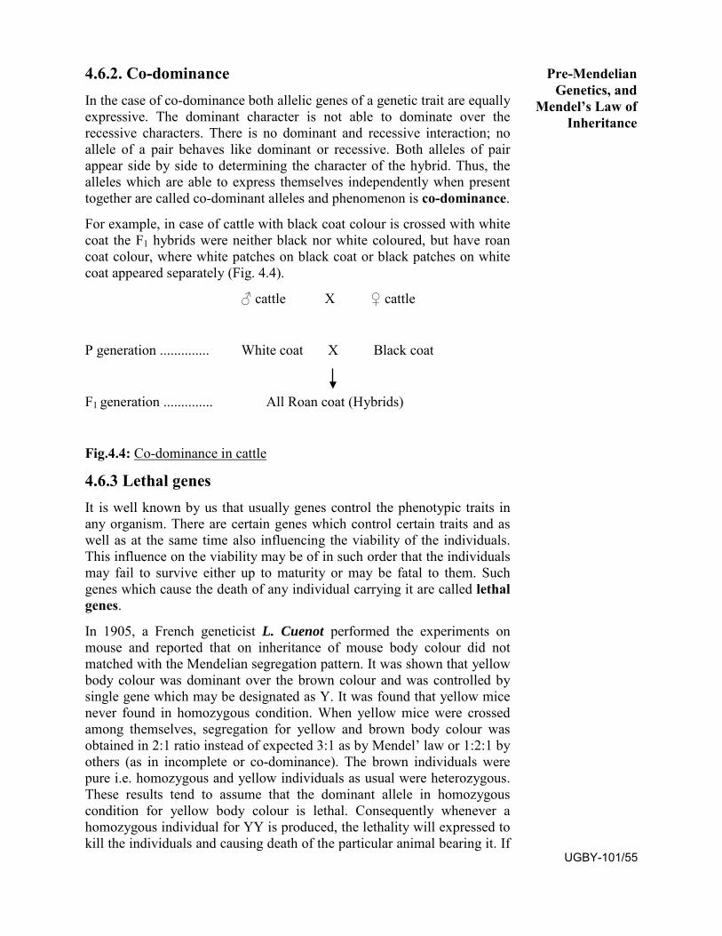
4.6.2. Co-dominance In the case of co-dominance both allelic genes of a genetic trait are equally expressive. The dominant character is not able to dominate over the recessive characters. There is no dominant and recessive interaction; no allele of a pair behaves like dominant or recessive. Both alleles of pair appear side by side to determining the character of the hybrid. Thus, the alleles which are able to express themselves independently when present together are called co-dominant alleles and phenomenon is co-dominance.
For example, in case of cattle with black coat colour is crossed with white coat the F1 hybrids were neither black nor white coloured, but have roan coat colour, where white patches on black coat or black patches on white coat appeared separately (Fig. 4.4).
♂ cattle X ♀ cattle
P generation .............. White coat X Black coat
F1 generation .............. All Roan coat (Hybrids)
Fig.4.4: Co-dominance in cattle
4.6.3 Lethal genes It is well known by us that usually genes control the phenotypic traits in any organism. There are certain genes which control certain traits and as well as at the same time also influencing the viability of the individuals. This influence on the viability may be of in such order that the individuals may fail to survive either up to maturity or may be fatal to them. Such genes which cause the death of any individual carrying it are called lethal genes.
In 1905, a French geneticist L. Cuenot performed the experiments on mouse and reported that on inheritance of mouse body colour did not matched with the Mendelian segregation pattern. It was shown that yellow body colour was dominant over the brown colour and was controlled by single gene which may be designated as Y. It was found that yellow mice never found in homozygous condition. When yellow mice were crossed among themselves, segregation for yellow and brown body colour was obtained in 2:1 ratio instead of expected 3:1 as by Mendel’ law or 1:2:1 by others (as in incomplete or co-dominance). The brown individuals were pure i.e. homozygous and yellow individuals as usual were heterozygous. These results tend to assume that the dominant allele in homozygous condition for yellow body colour is lethal. Consequently whenever a homozygous individual for YY is produced, the lethality will expressed to kill the individuals and causing death of the particular animal bearing it. If
Pre-Mendelian Genetics, and
Mendel’s Law of Inheritance
UGBY-101/55
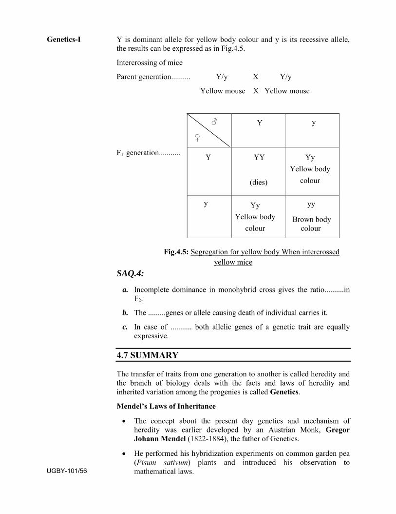
Y is dominant allele for yellow body colour and y is its recessive allele, the results can be expressed as in Fig.4.5.
Intercrossing of mice
Parent generation.......... Y/y X Y/y
Yellow mouse X Yellow mouse
F1 generation...........
y
Fig.4.5: Segregation for yellow body When intercrossed yellow mice
SAQ.4:
a. Incomplete dominance in monohybrid cross gives the ratio..........inF2.
b. The .........genes or allele causing death of individual carries it.
c. In case of ........... both allelic genes of a genetic trait are equallyexpressive.
4.7 SUMMARY
The transfer of traits from one generation to another is called heredity and the branch of biology deals with the facts and laws of heredity and inherited variation among the progenies is called Genetics.
Mendel’s Laws of Inheritance
• The concept about the present day genetics and mechanism ofheredity was earlier developed by an Austrian Monk, GregorJohann Mendel (1822-1884), the father of Genetics.
• He performed his hybridization experiments on common garden pea(Pisum sativum) plants and introduced his observation tomathematical laws.
♂
♀
Y y
Y YY
(dies)
Yy Yellow body
colour
Yy Yellow body
colour
yy
Brown body colour
Genetics-I
UGBY-101/56
• These laws are known as Mendel’s law of inheritance orMedelism. However his remarkable piece of work remainedunattended and ignored
• Later on after 34 years, in 1900 when three different eminentbiologists; Karl Correns of Germany, Hugo de Vries ofNetherlands and Erich Von Tschermak of Austria workingindependently got same results which Mendel predicted earlier.
Mendel’s Experiments • He selected Pea (Pisum sativum) plants having seven pairs of
contrasting characters, each with two alternative forms.
• The dominant character of each pair is represented by the upper caseof letter e.g. for Tall and Dwarf , T and t is used respectively todenote in which tallness showed dominance.
• The plants of different or same traits with which the hybridizationexperiments started is referred as P (Parental generation).
• The population obtained by the cross made between P generation isdenoted by F1 (First filial generation).
• The progenies of F1 obtained are F2 (Second filial generation), thesubsequent generations may be represented as F3, F4, F5 and so on.
• The cross made between two individuals of having one set ofcontrasting character is called Monohybrid Cross, if two pairs ofcharacters chosen for hybridization is known as Dihybrid Cross andprogenies are termed as hybrids.
Mendel’s Laws of Inheritance Correns established the laws of inheritance on the basis of Mendel’s hybridization experiments and results.
• Law of dominance: It states that out of the two or a pair ofcontrasting allelomorphic traits only one could be able to expressphenotypically which is dominant in F1 hybrid generation inheterozygous condition.
• Law of Segregation or Law of Purity of Gametes: This law statesthat when the pair of allelomorphs brought together in the F1 hybridswithout blending get separated during the formation of the gamets.
• Law of independent Assortment: In a dihybrid cross i.e. when twoindependent pairs of alleles come together in F1 hybrid may showtheir independent dominant effects. At the time of gamete formation,the law of segregation operates, but the factors assorted themselvesindependently at random and free. Therefore F2 have two newcombination of traits along with the two older parental combinationappears.
Pre-Mendelian Genetics, and
Mendel’s Law of Inheritance
UGBY-101/57
• Choosing pea plant for his experiment was an excellent choice.
• He had properly maintained all records of each cross and calculatedstatistically so that probability of pedigrees and progenies maypredict accurately.
• He took only one or two characters at a time to observe for hisexperiments.
• Fortunately and unknowingly all seven pairs of contrastingcharacters did not show any linkage, incomplete dominance, geneticinteraction.
Deviations of Mendel’s law • Incomplete dominance
There are many characters, may be exceptions to Mendel’s law of dominance and show incomplete dominance. The hybrids of these possess the characteristics of both parents and showing intermediate traits. This phenomenon is known as incomplete dominance. For example in case of four O’clock (Mirabilis jalapa) plants, when white flowered crossed with red flowered plants the F1 generation produced all pink flowered plants. After selfing of F1 red, pink and white flowered plants produced in 1:2:1 ratio due to incomplete dominance.
• Co-dominance
In co-dominance both allelic genes of a genetic trait are equally expressive. The dominant allele could not completely mask the effect of recessive allele; the characters of both alleles appear side by side in F1 hybrids. For example the cross made between black coat colours with white coat colour, all F1 progenies have roan coat colour i.e. white coat with black patches or vice-versa.
A, B, O blood groups in humans is another well known example of co-dominance.
• Lethal genes
Those genes which are responsible for the death or become lethal for the individuals bearing are known as lethal genes and phenomenon is lethality.
4.6 TERMINAL QUESTIONS
OBJECTIVE TYPE QUESTIONS:
Q.1. The unit of Hereditary is:
a. Chromosomes b. Genes
c. Golgibody d. Nucleus
Genetics-I
UGBY-101/58
Q.2. The father of genetics is:
a. T.H. Morgan b. Charles Darwin
c. Gregor Johann Mendel d. Hugo de Vries
Q.3. The plant selected by Mendel for his experiment was:
a. Mustard b. Sunflower
c. Wheat d. Pea
Q.4. The test cross is the cross made between:
a. F1 with either of any one parent b. F1 with recessive parent
c. F1 with dominant parent d. None of these
Q.5. The phenotypic ratio of dihybrid cross made by Mendel is:
a. 9:3:3:1 b. 9:7
c. 3:1 d. 9:4:3
Q.6. Which one of these could be normal gamete:
a. GgRr b. GRr
c. GgR d. gR
Q.7. The ratio of Mendelian recombination is due to:
a. Mutation b. Linkagec. Independent Assortment d. Crossing Over
Q.8. The observable traits of an organism are its:
a. Phenotype b. Genotype
c. Sociotype d. Biotype
Q.9. The second generation offsprings from a cross are:
a. F1 Generation b. F2 Generation
c. Hybrids d. None of these
Q.10. Allelles are:
a. Different molecular forms of gene
b. Self fertilizing Zygotes
c. Different forms of chromosome
d. Nucleus
Pre-Mendelian Genetics, and
Mendel’s Law of Inheritance
UGBY-101/59
Q.11. Which of following pairs of characters in pea plants selected by Mendel found recessive?
a. Yellow coloured pod and wrinkled seed
b. Yellow colour and wrinkled seeds
c. Dwarf plants with axial flower
d. Constricted pods with axial flower
Fill in the blanks:
1. ....... is the branch of biology that deals with the facts and laws ofheredity and inherited variations.
2. ........is the genetic constitution of an organism.
3. In case of...........both allelic genes of a genetic trait are equallyexpressive.
4. ........are the alternative forms of a gene which occur at the samelocus on homologous chromosomes.
5. ........is the cross between an individual with the dominantphenotype and an individual with recessive phenotype to see if the individual with dominant phenotype is homozygous or heterozygous.
6. When single pair of alleles used for hybridization is called ...........
7. Mendel took total ........ pairs of traits in pea plants, each have ....alternative forms.
8. F2 generation have phenotypic ratio 1:2:1 obtained in........dominance.
9. The gametes formed have only....... allele of a pair.
10. Mendel’s law has not considered the cases of........... due to thecontrasting characters chosen by him were located on different chromosomes.
Find out the following statements true/false:
1. A gene which can express itself in only homozygous condition isrecessive.
2. A phenotype is an observable character.
3. The F2 generation is conventionally produced by random union ofthe gametes produced by F1.
4. All phenotypes have same genotypes.
5. A cross between F1 hybrid with homozygous recessive parent iscalled test cross.
Genetics-I
UGBY-101/60
6. The plants are considered to be pure line breeding when all plantsof parental generation resemble each other.
7. In incomplete dominance the filial generation resembles to theirparent.
8. Alternative forms of same genes are called allele.
9. The law of independent assortment applies to the genes located onsame chromosome.
10. Mendel, the father of genetics coined the term genetics.
Give very short answer of following questions:
1. Who coined the term Genetics in which year?
2. Name of three eminent biologists who rediscover Mendelianprinciples after his death?
3. Give an example of incomplete dominance in respect of the colourof the flowers in the plants?
4. Write the percentage of pea plants that would be homozygousrecessive in F2 generation after the selfing of all heterozygous tallplants of F1 hybrids.
5. How many phenotypes do you expect in F2 generation in case ofco-dominance in monohybrid cross and in which proportion?
6. What term was used by the Mendel for the term genes?
7. Give the name of two different kinds of ratios 3:1 and 1:2:1Mendel observed in F2 generation during his experiment ofmonohybrid cross.
8. What is the standard F2 phenotypic ratio in basic Mendel’sdihybrid cross?
9. How many types of contrasting characters opted by the Mendel forhis experiments in pea plants?
10. How would you find the genotype of an organism exhibitingdominant trait?
Give short answers of following questions:
1. What are the merits of selecting pea plants by Mendel for hishybridization experiments?
2. Why the work of Mendel remained unrecognised and unattenededduring 35 years after the publication?
Pre-Mendelian Genetics, and
Mendel’s Law of Inheritance
UGBY-101/61
3. Why Mendel honoured as father of genetics, though hispredecessors failed to get similar results in various otherexperiments?
4. Differentiate the following terms in short:
a. Back cross and test cross
b. Phenotype and genotype
c. Dominance , Co-dominance and incomplete dominance
d. Genes and alleles
e. Heredity and genetics
f. Monohybrid and dihybrid cross
5. By using the example of Mendel’s monohybrid cross, find thereasoning to imply the law of segregation.
Give the Descriptive answer of following questions:
1. Throw some light on the earlier concept of heredity and pre-mendelian genetics.
2. Describe the outline of Mendel’s experiments and also mention thereasons to select pea plant for his experiments.
3. What are the Mendel’s laws of inheritance?
4. Discuss the deviations from expected Mendel’s laws and whycould Mendel not noticed in his observations and results.
5. Explain incomplete dominance and co-dominance with suitableexamples.
4.8 ANSWERS
Objective type questions:
1. (b) 2. (c) 3. (d) 4. (a)
5. (a) 6. (d) 7. (c) 8. (a)
9. (b) 10. (a) 11. (a)
Fill in the blanks:
1.Genetics 2. Genotype 3. Co-dominance 4. Allele 5. Back cross
6. Monohybrid 7. Seven, two 8. Incomplete 9. One 10. Linkage
True / False:
1.True 2.True 3.True 4.False 5.True 6.True 7.False 8.True 9.False10.False
Genetics-I
UGBY-101/62
Short type answers:
1. William Bateson, in 1906.
2. Karl Correns of Germany, Hugo de vries of Netherlands and ErichVon Tschermak of Austria.
3. Four O’clock (Mirabilis jalapa)
4. 25 percent
5. Three, 1:2:1
6. Factor
7. Phenotypic and genotypic ratios respectively.
8. 9:3:3:1
9. Seven pairs of contrasting characters
10. By back cross, i.e. Cross of F1 with both dominant and recessiveparent
SAQ.1:
a. Heredity, variation b. J. Swammardam
c. K.W. Wolff d. Joseph Gottlieb Kolreuter
SAQ.2:
a. Gregor Johann Mendel
b. Pea (Pisum sativum), seven
c. Hybrid
d. “Versuche uber Pflenzenhybriden” (“Experiments on PlantHybridization”)
SAQ.3:
a. Gametes b. Dihybrid
C. 9:3:3:1 d. Test cross
SAQ.4:
a. 1:2:1 b. Lethal
c. Co-dominance
Pre-Mendelian Genetics, and
Mendel’s Law of Inheritance
UGBY-101/63

UGBY-101/64
UNIT-5
LINKAGE AND CROSSING OVER
Structure 5.1 Introduction
• 5.1.2Chromosomal Theory of Inheritance
• 5.1.2 Introduction of linkage and crossing over
5.2 Hypothesis
• 5.2.1 Coupling and Repulsion
5.3 Concept of Linkage
• 5.3.1 Chromosomal Theory of Linkage
• 5.3.2 Arrangements of Linked Genes
• 5.3.3 Kinds of Linkage
• 5.3.4 Significance of Linkage
5.4 Crossing over
• 5.4.1 Concept of Crossing Over
5.4.1.1 Mitotic Crossing Over
5.4.1.2 Meiotic Crossing Over
• 5.4.2.1 Mechanism of Crossing Over
5.4.2.2 Theories of Mechanism of Crossing Over
5.4.2.3 Gene Conversion
• 5.4.3 Kinds of Crossing Over
• 5.4.4 Factors Influencing the Frequency of Crossing Over
• 5.4.5 Significance of crossing over
5.5 Summary
5.6 Terminal Questions
5.7 Answer
UGBY-101/65
5.1 INTRODUCTION
As you studied earlier that the characters determined by certain factors of inheritance which are stable and particulate and segregate independently at the time of gamete formation as assumed by Gregor Johann Mendel by his law of independent assortment, Mendel’s second law of inheritance. At that time detailed discovery of cell division, nucleus, chromosomes and genes make him unaware about the location of these factors inside cell on the chromosomes. Later on further investigations revealed chromosomes are the factors which assort independently at the time of cell division and formation of gametes. W. Roux (1883) perhaps first time indicated the involvement of chromosomes in the mechanism of inheritance.
5.1.1 Chromosomal theory of Inheritance
In 1903 an American graduate student, Walter S. Sutton and a German biologist Theodor Boveri i.e. soon after the rediscovery of Mendel’s law (1900), have independently observed parallelism between the behaviour of chromosomes and Mendelian factors (genes). The comparison between the behaviour of both noticed which were as follows:
1. Chromosomes occur in pairs like the alleles of Mendelian factors(now known as genes).
2. The homologous chromosomes also segregate during the meiosisat the time of gamete formation like the similar or dissimilar allelesof Mendelian factor supporting the law of segregation or law ofpurity of gametes.
3. Different chromosomes orient and separate independently duringmeiosis like that of Mendelian factors.
4. During fertilization in both cases pairing is restored i.e. thechromosomes as well as Mendelian factors.
5. The chromosomes and Mendelian factors maintain theirindividuality and identity carried away from generation togeneration.
On the basis of above studies Sutton and Boveri (1903) postulated the Chromosomal Theory of Inheritance. According to this theory, “the Mendelian factors (genes) are located on chromosomes and it is the chromosome, which segregate and assorts independently during meiosis”.
5.1.2 Introduction of linkage and crossing over The study of meiosis revealed that the independent assortment of the characters is belonging to non-homologous chromosomes. Therefore it becomes clear that if those characters which are assorted independently
Genetics-I
UGBY-101/66
must be located on separate non- homologous chromosomes. The chromosomal theory of inheritance does not clarify and support of the concept of more than one gene located on the same chromosome. This theory would expect that independent assortment will not take place in those cases where more than two genes located on same chromosome except when genes are located very far on same chromosome.
Later on after further investigations and researches shown that there numerous genes located on a single chromosome in an organism. There is fixed number and sets of chromosome in individuals. It has been found that in those examples in which two different genes located closely are inherited together and deviated the expected Mendelian ratio; such type of genes which are inherited together are closely linked known as linked genes and phenomenon is called linkage. Whereas when the genes are located distantly have probability of exchange of chromosomal segments during chiasma formation and new combinations may arise is called due to cross over segments process called crossing over.
SAQ.1: a. ........and....... postulated chromosomal theory of inheritance.
b. When the genes are located closely and inherited together iscalled.......genes.
c. The phenomenon of exchange of chromosomal segments duringchiasma formation is called .............
5.2 HYPOTHESIS
5.2.1 Coupling and Repulsion In 1905, W.Bateson performed hybridization experiment in a variety of sweet pea plants. He made a cross between blue flowered (B) with long pollen grains (L) with red flowered (r) with round pollen grains (l). He noticed that blue colour of flowers and long pollen grains were dominant characters over the red coloured flower with round pollen grains of the plants. The F1 individuals were blue flowered with long pollen grains having BbLl genotypic configuration. After the test cross made i.e. F1 crossed with recessive parent (BbLl X bbll), mendelian law of independent assortment fails to exhibit and results obtained were 7:1:1:7 instead of expected result 1:1:1:1 (Fig.5.1). This result indicated that both dominant alleles tending to remain together and similarly the recessive alleles also remained together, was explained by Bateson as gametic coupling.
Linkage and Crossing Over
UGBY-101/67
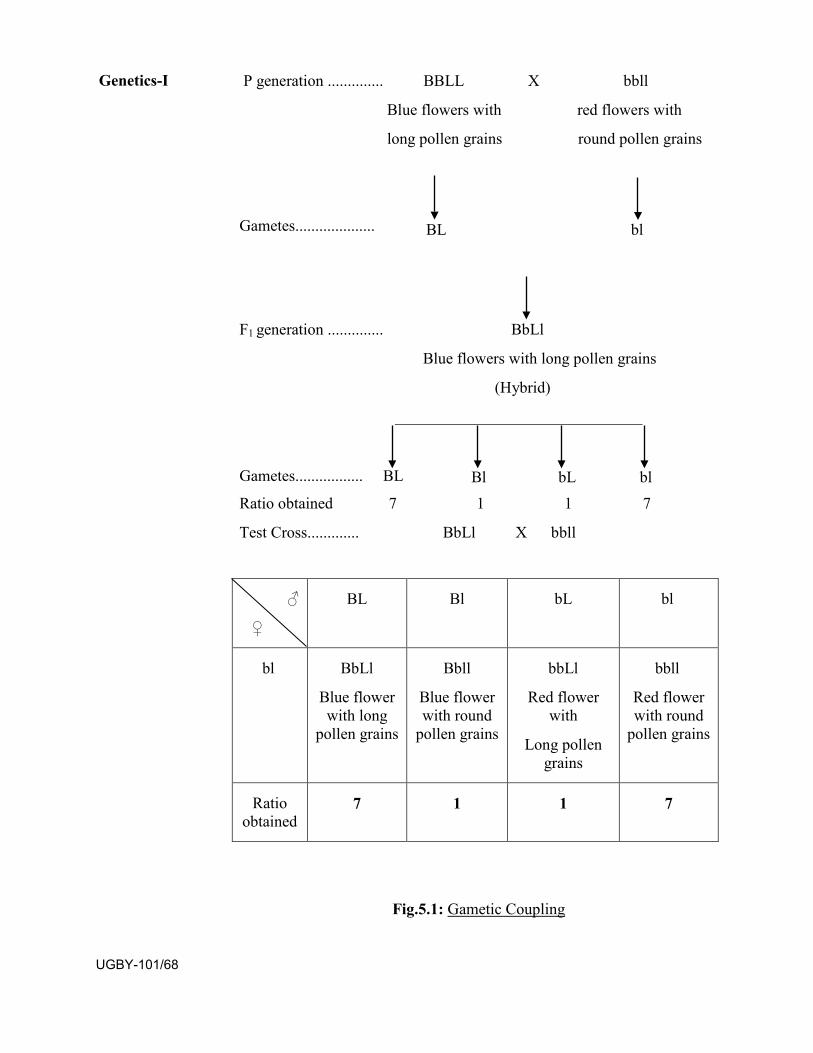
P generation .............. BBLL X bbll
Blue flowers with red flowers with
long pollen grains round pollen grains
Gametes.................... BL bl
F1 generation .............. BbLl
Blue flowers with long pollen grains
(Hybrid)
_______________________________
Gametes................. BL Bl bL bl Ratio obtained 7 1 1 7
Test Cross............. BbLl X bbll
Fig.5.1: Gametic Coupling
♂
♀
BL Bl bL bl
bl BbLl
Blue flower with long
pollen grains
Bbll
Blue flower with round
pollen grains
bbLl
Red flower with
Long pollen grains
bbll
Red flower with round
pollen grains
Ratio obtained
7 1 1 7
Genetics-I
UGBY-101/68
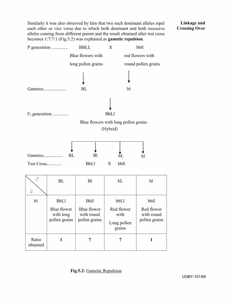
Similarly it was also observed by him that two such dominant alleles repel each other or vice versa due to which both dominant and both recessive alleles coming from different parent and the result obtained after test cross becomes 1:7:7:1 (Fig.5.2) was explained as gametic repulsion.
P generation .............. BBLL X bbll
Blue flowers with red flowers with
long pollen grains round pollen grains
Gametes.................... BL bl
F1 generation .............. BbLl
Blue flowers with long pollen grains
_______________________________
Gametes................. BL Bl bL bl Test Cross............. BbLl X bbll
Fig.5.2: Gametic Repulsion
♂
♀
BL Bl bL bl
bl BbLl
Blue flower with long
pollen grains
Bbll
Blue flower with round
pollen grains
bbLl
Red flower with
Long pollen grains
bbll
Red flower with round
pollen grains
Ratio obtained
1 7 7 1
Linkage and Crossing Over
(Hybrid)
UGBY-101/69
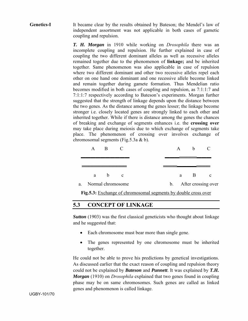
It became clear by the results obtained by Bateson; the Mendel’s law of independent assortment was not applicable in both cases of gametic coupling and repulsion.
T. H. Morgan in 1910 while working on Drosophila there was an incomplete coupling and repulsion. He further explained in case of coupling the two different dominant alleles as well as recessive alleles remained together due to the phenomenon of linkage; and be inherited together. Same phenomenon was also applicable in case of repulsion where two different dominant and other two recessive alleles repel each other on one hand one dominant and one recessive allele become linked and remain together during gamete formation. Thus Mendelian ratio becomes modified in both cases of coupling and repulsion, as 7:1:1:7 and 7:1:1:7 respectively according to Bateson’s experiments. Morgan further suggested that the strength of linkage depends upon the distance between the two genes. As the distance among the genes lesser; the linkage become stronger i.e. closely located genes are strongly linked to each other and inherited together. While if there is distance among the genes the chances of breaking and exchange of segments enhances i.e. the crossing over may take place during meiosis due to which exchange of segments take place. The phenomenon of crossing over involves exchange of chromosomal segments (Fig.5.3a & b).
A B C A b C
▬▬▬▬▬▬▬▬▬▬ ▬▬▬═════▬▬▬
═════════════ ════▬▬▬▬════
a b c a B c
a. Normal chromosome b. After crossing over
Fig.5.3: Exchange of chromosomal segments by double cross over
5.3 CONCEPT OF LINKAGE
Sutton (1903) was the first classical geneticists who thought about linkage and he suggested that:
• Each chromosome must bear more than single gene.
• The genes represented by one chromosome must be inheritedtogether.
He could not be able to prove his predictions by genetical investigations. As discussed earlier that the exact reason of coupling and repulsion theory could not be explained by Bateson and Punnett. It was explained by T.H. Morgan (1910) on Drosophila explained that two genes found in coupling phase may be on same chromosomes. Such genes are called as linked genes and phenomenon is called linkage.
Genetics-I
UGBY-101/70
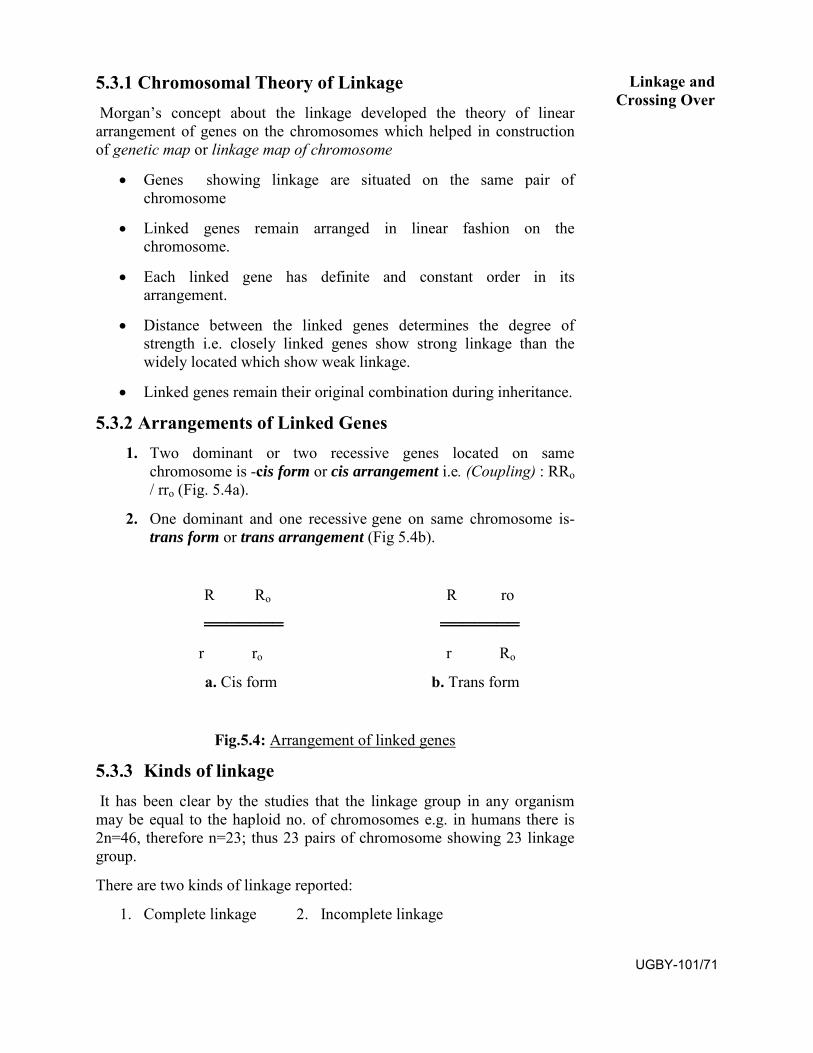
5.3.1 Chromosomal Theory of Linkage Morgan’s concept about the linkage developed the theory of linear arrangement of genes on the chromosomes which helped in construction of genetic map or linkage map of chromosome
• Genes showing linkage are situated on the same pair ofchromosome
• Linked genes remain arranged in linear fashion on thechromosome.
• Each linked gene has definite and constant order in itsarrangement.
• Distance between the linked genes determines the degree ofstrength i.e. closely linked genes show strong linkage than thewidely located which show weak linkage.
• Linked genes remain their original combination during inheritance.
5.3.2 Arrangements of Linked Genes 1. Two dominant or two recessive genes located on same
chromosome is -cis form or cis arrangement i.e. (Coupling) : RRo / rro (Fig. 5.4a).
2. One dominant and one recessive gene on same chromosome is-trans form or trans arrangement (Fig 5.4b).
R Ro R ro
═══════ ═══════
r ro r Ro
a. Cis form b. Trans form
Fig.5.4: Arrangement of linked genes
5.3.3 Kinds of linkage It has been clear by the studies that the linkage group in any organism may be equal to the haploid no. of chromosomes e.g. in humans there is 2n=46, therefore n=23; thus 23 pairs of chromosome showing 23 linkage group.
There are two kinds of linkage reported:
1. Complete linkage 2. Incomplete linkage
Linkage and Crossing Over
UGBY-101/71
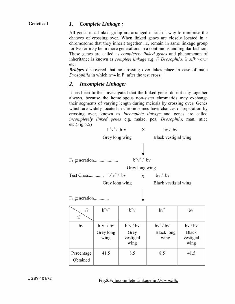
1. Complete Linkage :All genes in a linked group are arranged in such a way to minimise the chances of crossing over. When linked genes are closely located in a chromosome that they inherit together i.e. remain in same linkage group for two or may be in more generations in a continuous and regular fashion. These genes are called as completely linked genes and phenomenon of inheritance is known as complete linkage e.g. ♂ Drosophila, ♀ silk worm etc. Bridges discovered that no crossing over takes place in case of male Drosophila in which n=4 in F1 after the test cross.
2. Incomplete Linkage:It has been further investigated that the linked genes do not stay together always, because the homologous non-sister chromatids may exchange their segments of varying length during meiosis by crossing over. Genes which are widely located in chromosomes have chances of separation by crossing over, known as incomplete linkage and genes are called incompletely linked genes e.g. maize, pea, Drosophila, man, mice etc.(Fig.5.5)
b+v+ / b+v+ X bv / bv Grey long wing Black vestigial wing
F1 generation..................... b+v+ / bv Grey long wing
Test Cross............. b+v+ / bv X bv / bv Grey long wing Black vestigial wing
F2 generation.............
Fig.5.5: Incomplete Linkage in Drosophila
♂ ♀
b+v+ b+v bv+ bv
bv b+v+ / bv Grey long
wing
b+v / bv Grey
vestigial wing
bv+ / bv Black long
wing
bv / bv Black
vestigial wing
Percentage Obtained
41.5 8.5 8.5 41.5
Genetics-I
UGBY-101/72
Results of F2: Grey long wing =41.5 %
Grey vestigial wing=8.5%
Black long wing = 8.5%
Recombinant or new type = 17.0%
5.3.4 Significance of linkage It is very significant in reducing the probability of variability in the gamete unless the crossing over occurs. Linkage is a strong mode to express its identity during the course of inheritance.
SAQ.2:
a. Gametic coupling and repulsion hypothesis was raisedby......and.......
b. T.H.Morgan worked on ............ to demonstrate the phenomenon oflinkage.
c. The number of possible groups in any organism is equal to its .....number of chromosomes.
5.4 CROSSING OVER
5.4.1 Concept of Crossing Over Crossing over or recombination occurs during meiotic cell division during prophase I stage. Homologous chromosomes come close to each other called synapsis at zygotene of prophase I. The chromosome splits longitudinally into two chromatids. After that these non sister chromatids of homologous chromosomes cross over and exchange their segments at pachytene of prophase I. Then these cross over and non-cross overs chromatids get separate from each other called terminalisation. The points of contact where chromatids break and reunite after the exchange of segments are known as chiasmata. It becomes the matter of discussion that weather the formation of chaismata causes the crossing over or crossing over causing chiasmata formation. There may be two types of crossing over:
1. Mitotic Crossing Over 2. Meiotic Crossing Over
5.4.1.1 Mitotic or Somatic Crossing Over: Crossing over sometimes occurs during mitosis of somatic cells as reported by Stern in Drosophila and by Pontecorvo in Aspergillus nidulans. Here two chromosomes with unequal chromatids are being formed in 50% cases original combination is restored while in other 50%
Linkage and Crossing Over
UGBY-101/73
Black vestigial wing=41.5%
Parental type= 83.0%
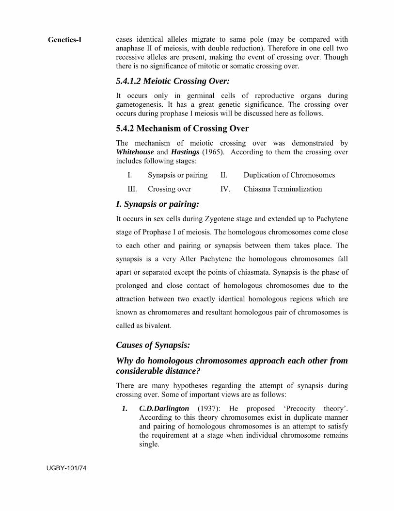
cases identical alleles migrate to same pole (may be compared with anaphase II of meiosis, with double reduction). Therefore in one cell two recessive alleles are present, making the event of crossing over. Though there is no significance of mitotic or somatic crossing over.
5.4.1.2 Meiotic Crossing Over: It occurs only in germinal cells of reproductive organs during gametogenesis. It has a great genetic significance. The crossing over occurs during prophase I meiosis will be discussed here as follows.
5.4.2 Mechanism of Crossing Over The mechanism of meiotic crossing over was demonstrated by Whitehouse and Hastings (1965). According to them the crossing over includes following stages:
I. Synapsis or pairing II. Duplication of Chromosomes
III. Crossing over IV. Chiasma Terminalization
I. Synapsis or pairing: It occurs in sex cells during Zygotene stage and extended up to Pachytene
stage of Prophase I of meiosis. The homologous chromosomes come close
to each other and pairing or synapsis between them takes place. The
synapsis is a very After Pachytene the homologous chromosomes fall
apart or separated except the points of chiasmata. Synapsis is the phase of
prolonged and close contact of homologous chromosomes due to the
attraction between two exactly identical homologous regions which are
known as chromomeres and resultant homologous pair of chromosomes is
called as bivalent.
Causes of Synapsis:
Why do homologous chromosomes approach each other from considerable distance?
There are many hypotheses regarding the attempt of synapsis during crossing over. Some of important views are as follows:
1. C.D.Darlington (1937): He proposed ‘Precocity theory’.According to this theory chromosomes exist in duplicate mannerand pairing of homologous chromosomes is an attempt to satisfythe requirement at a stage when individual chromosome remainssingle.
Genetics-I
UGBY-101/74
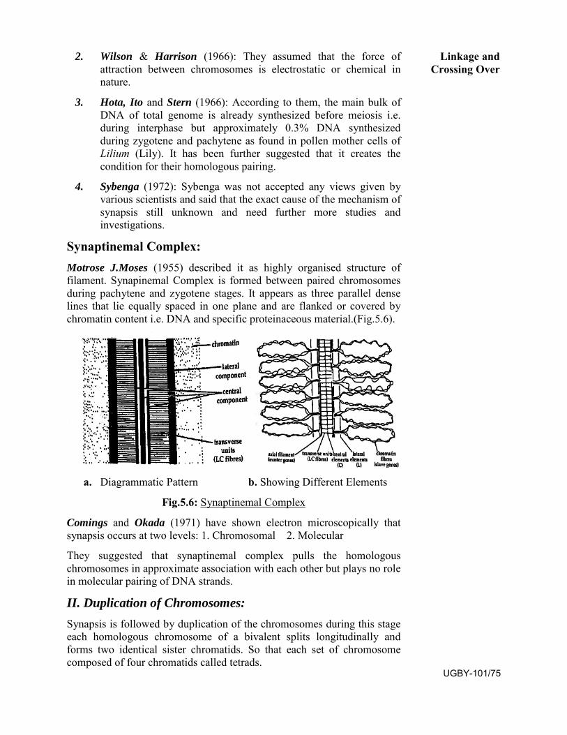
2. Wilson & Harrison (1966): They assumed that the force ofattraction between chromosomes is electrostatic or chemical innature.
3. Hota, Ito and Stern (1966): According to them, the main bulk ofDNA of total genome is already synthesized before meiosis i.e.during interphase but approximately 0.3% DNA synthesizedduring zygotene and pachytene as found in pollen mother cells ofLilium (Lily). It has been further suggested that it creates thecondition for their homologous pairing.
4. Sybenga (1972): Sybenga was not accepted any views given byvarious scientists and said that the exact cause of the mechanism ofsynapsis still unknown and need further more studies andinvestigations.
Synaptinemal Complex: Motrose J.Moses (1955) described it as highly organised structure of filament. Synapinemal Complex is formed between paired chromosomes during pachytene and zygotene stages. It appears as three parallel dense lines that lie equally spaced in one plane and are flanked or covered by chromatin content i.e. DNA and specific proteinaceous material.(Fig.5.6).
a. Diagrammatic Pattern b. Showing Different Elements
Fig.5.6: Synaptinemal Complex
Comings and Okada (1971) have shown electron microscopically that synapsis occurs at two levels: 1. Chromosomal 2. Molecular
They suggested that synaptinemal complex pulls the homologous chromosomes in approximate association with each other but plays no role in molecular pairing of DNA strands.
II. Duplication of Chromosomes:Synapsis is followed by duplication of the chromosomes during this stage each homologous chromosome of a bivalent splits longitudinally and forms two identical sister chromatids. So that each set of chromosome composed of four chromatids called tetrads.
Linkage and Crossing Over
UGBY-101/75
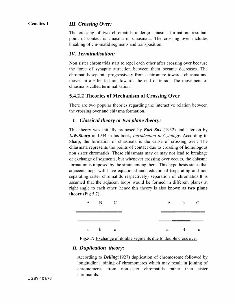
III. Crossing Over:The crossing of two chromatids undergo chiasma formation, resultant point of contact is chiasma or chiasmata. The crossing over includes breaking of chromatid segments and transposition.
IV. Terminalisation:
Non sister chromatids start to repel each other after crossing over because the force of synaptic attraction between them became decreases. The chromatids separate progressively from centromere towards chiasma and moves in a zifer fashion towards the end of tetrad. The movement of chiasma is called terminalisation.
5.4.2.2 Theories of Mechanism of Crossing Over
There are two popular theories regarding the interactive relation between the crossing over and chiasma formation.
I. Classical theory or two plane theory:
This theory was initially proposed by Karl Sax (1932) and later on by L.W.Sharp in 1934 in his book, Introduction to Cytology. According to Sharp, the formation of chiasmata is the cause of crossing over. The chiasmata represents the points of contact due to crossing of homologous non sister chromatids. These chiasmata may or may not lead to breakage or exchange of segments, but whenever crossing over occurs, the chiasma formation is imposed by the strain among them. This hypothesis states that adjacent loops will have equational and reductional (separating and non separating sister chromatids respectively) separation of chromatids.It is assumed that the adjacent loops would be formed in different planes at right angle to each other, hence this theory is also known as two plane theory (Fig 5.7).
A B C A b C
▬▬▬▬▬▬▬▬▬▬ ▬▬▬═════▬▬▬
═════════════ ════▬▬▬▬════
a b c a B c
Fig.5.7: Exchange of double segments due to double cross over
II. Duplication theory:
According to Belling(1927) duplication of chromosome followed by longitudinal joining of chromomeres which may result in joining of chromomeres from non-sister chromatids rather than sister chromatids.
Genetics-I
UGBY-101/76
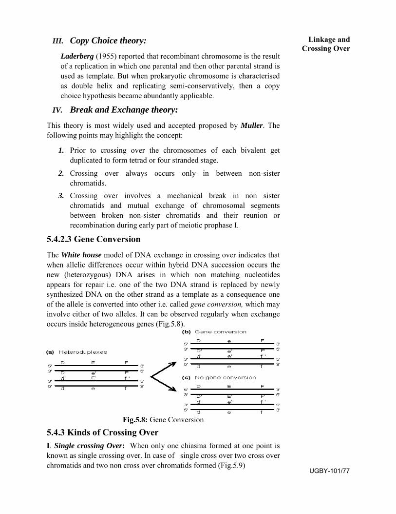
III. Copy Choice theory:
Laderberg (1955) reported that recombinant chromosome is the result of a replication in which one parental and then other parental strand is used as template. But when prokaryotic chromosome is characterised as double helix and replicating semi-conservatively, then a copy choice hypothesis became abundantly applicable.
IV. Break and Exchange theory:
This theory is most widely used and accepted proposed by Muller. The following points may highlight the concept:
1. Prior to crossing over the chromosomes of each bivalent getduplicated to form tetrad or four stranded stage.
2. Crossing over always occurs only in between non-sisterchromatids.
3. Crossing over involves a mechanical break in non sisterchromatids and mutual exchange of chromosomal segmentsbetween broken non-sister chromatids and their reunion orrecombination during early part of meiotic prophase I.
5.4.2.3 Gene Conversion
The White house model of DNA exchange in crossing over indicates that when allelic differences occur within hybrid DNA succession occurs the new (heterozygous) DNA arises in which non matching nucleotides appears for repair i.e. one of the two DNA strand is replaced by newly synthesized DNA on the other strand as a template as a consequence one of the allele is converted into other i.e. called gene conversion, which may involve either of two alleles. It can be observed regularly when exchange occurs inside heterogeneous genes (Fig.5.8).
Fig.5.8: Gene Conversion
5.4.3 Kinds of Crossing Over I. Single crossing Over: When only one chiasma formed at one point is known as single crossing over. In case of single cross over two cross over chromatids and two non cross over chromatids formed (Fig.5.9)
Linkage and Crossing Over
UGBY-101/77
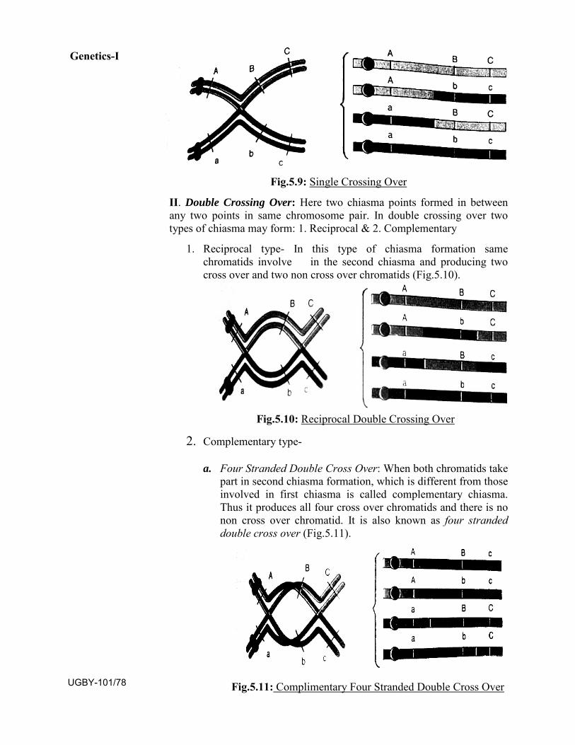
Fig.5.9: Single Crossing Over
II. Double Crossing Over: Here two chiasma points formed in betweenany two points in same chromosome pair. In double crossing over two types of chiasma may form: 1. Reciprocal & 2. Complementary
1. Reciprocal type- In this type of chiasma formation samechromatids involve in the second chiasma and producing twocross over and two non cross over chromatids (Fig.5.10).
Fig.5.10: Reciprocal Double Crossing Over
2. Complementary type-
a. Four Stranded Double Cross Over: When both chromatids takepart in second chiasma formation, which is different from those involved in first chiasma is called complementary chiasma. Thus it produces all four cross over chromatids and there is no non cross over chromatid. It is also known as four stranded double cross over (Fig.5.11).
Fig.5.11: Complimentary Four Stranded Double Cross Over
Genetics-I
UGBY-101/78
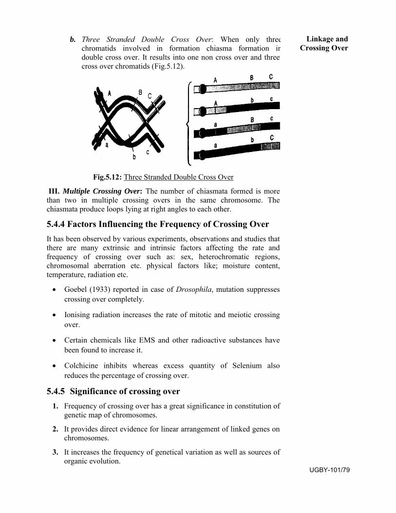
b. Three Stranded Double Cross Over: When only threechromatids involved in formation chiasma formation indouble cross over. It results into one non cross over and threecross over chromatids (Fig.5.12).
Fig.5.12: Three Stranded Double Cross Over
III. Multiple Crossing Over: The number of chiasmata formed is morethan two in multiple crossing overs in the same chromosome. The chiasmata produce loops lying at right angles to each other.
5.4.4 Factors Influencing the Frequency of Crossing Over It has been observed by various experiments, observations and studies that there are many extrinsic and intrinsic factors affecting the rate and frequency of crossing over such as: sex, heterochromatic regions, chromosomal aberration etc. physical factors like; moisture content, temperature, radiation etc.
• Goebel (1933) reported in case of Drosophila, mutation suppressescrossing over completely.
• Ionising radiation increases the rate of mitotic and meiotic crossingover.
• Certain chemicals like EMS and other radioactive substances havebeen found to increase it.
• Colchicine inhibits whereas excess quantity of Selenium alsoreduces the percentage of crossing over.
5.4.5 Significance of crossing over 1. Frequency of crossing over has a great significance in constitution of
genetic map of chromosomes.
2. It provides direct evidence for linear arrangement of linked genes onchromosomes.
3. It increases the frequency of genetical variation as well as sources oforganic evolution.
Linkage and Crossing Over
UGBY-101/79
SAQ.3: a. The mechanism of crossing over was demonstrated
by.............and........
b. ........complex formed between paired chromosomes duringpachytene and zygotene of prophase I of meiosis.
c. Break and exchange theory of crossing over is proposed by.......
5.5 SUMMARY Walters S. Sutton and Theodor Boveri in 1903 have independently observed a parallelism between the behaviour of chromosomes and Mendelian factors.
These found following similarities between chromosomes and Mendelian factors:
1. Chromosomes found in pairs like the alleles of Mendelian factor(now as genes).
2. The homologous chromosomes separate during meiosis like the pairof similar or dissimilar alleles of a Mendelian factor which separateat the time of gamete formation.
3. Different chromosomes arrange and separate independently duringmeiosis like the Mendelian factors.
4. The paired condition of both chromosomes and Mendelian factors isrestored during fertilization.
5. Both Mendelian factors and chromosomes maintain theirindividuality from generation to generation.
On the basis of above characteristics led Sutton and Boveri to postulate the “Chromosomal Theory of Inheritance”. It states that the Mendelian factors (genes) are located on chromosomes and segregates and assort independentely.
Linkage: The tendency of two or more genes of same chromosome to remain together during the course of inheritance is known as Linkage.
There are mainly two types of linkage; 1.Complete linkage 2.Incomplete linkage.
1. Complete linkage: When the genes for particular traits are locatedtoo close to each other on a chromosome, remains unbreakable orcould not be separated, i.e. remain completely linked and inheritedtogether is known as complete linkage e.g. Male Drosophila for greycolour with long wings.
2. Incomplete linkage: It occurs when the genes for different charactersare separated at the time of gamete formation and due to breakage
Genetics-I
UGBY-101/80
and exchange of chromosome during meiosis. For example in case of sweet pea blue coloured flower and long pollen grain, exhibiting as incompletely linked traits.
Linkage is affected by several factors such as distance between the linked genes, age of the organism, exposure to X-rays and increase in temperature.
Significance of linkage: Linkage has a great significance in reducing the probability of variability in gametes unless the crossing over which separates the linked genes.
Crossing Over: Crossing over is a phenomenon in which mutual exchange of segments among non-sister or sister homologous chromatids of a pair of homologous chromosomes. A cross over between linked genes may form recombinants. Crossing over involves breaking and rejoining of chromosomal segments in the synaptinemal complex. The point of contact where homologous chromatids held together and exchange of bits of chromatids is called chiasmata i.e. forming synatenemal complex.
Kinds of crossing over
I. Single crossing Over: When only one chiasma formed and resultant two cross over chromatids and two non cross over chromatids formed.
II. Double Crossing Over: Here two chiasma points formed inbetween any two points in same chromosome pair. In doublecrossing over two types of chiasma may form: 1. Reciprocal & 2.Complementary
1. Reciprocal type- In this type of chiasma formation samechromatids involve in the second chiasma and producingtwo cross over and two non cross over chromatids.
2. Complementary type-
a. Four Stranded Double Cross Over: When both chromatids take part in second chiasma formation, which is different from those involved in first chiasma is called complementary chiasma. Thus it produces all four cross over chromatids.
b. Three Stranded Double Cross Over: When only threechromatids involved in formation chiasma formation indouble cross over. It results into one non cross over andthree cross over chromatids.
III. Multiple Crossing Over : The number of chiasmata formed ismore than two in multiple crossing overs in the same chromosome.
Linkage and Crossing Over
UGBY-101/81
Significance of Crossing Over:
It provides an inexhaustible genetic variability in sexually reproducing organisms. As a result of crossing over new gene combinations are produced. Thus crossing over plays an important role in genetic variation among the generations and helps in constitution of genetic map.
5.6 TERMINAL QUESTIONS: OBJECTIVE TYPE QUESTIONS:
1. The phenomenon of linkage was explained by:
a. Punnett c. Morgan
b. Hugo de vries d. Gregor Johann Mendel
2. The term linkage implies for :
a. Two genes located on same chromosome
b. Two genes located on different chromosomes
c. Mutated genes
d. Recombinant genes
3. The recombinant frequency between genes:
a. Always constant
b. Depending upon the distance between the genes
c. Depending upon the interaction between non homologous chromosomes
d. None of these
4. The exchange of parts of non-sister chromatids of homologous chromosomes results in:
a. Genetic recombination c. Mutation b. Chromosomal elongation d. Loss of chromosome 5. The two closely linked genes on same chromosome:
a. Can be separated by crossing over occurring between them b. Cannot be separated c. Always controlling same trait d. Always be dominant
6. If the number of segregating gene is four than the number of gametes produced may be:
a. Two c. Eight
b. Four d. Sixteen
GENETICS-I
UGBY-101/82
LINKAGE AND CROSSING OVER
7. If the recombination frequency between the genes A & B is 60% and that between A and C 9% it means :
a. A and B are closed to each other
b. A and C are closed to each other
c. B and C are closed to each other
d. All genes are located on separate chromosomes
8. Crossing over takes place during:
a. Prophase of meiosis I c. Prophase of mitosis
b. Anaphase of meiosis I d. Anaphase of mitosis
9. The probability of cross over occurring between two gene loci is proportional to :
a. Activity of two loci c. Distance of loci from centromere
b. Distance between the two loci d. None of these
10. The cross over percentage signifies:
a. It enables certain gene frequencies to increase in a population
b. Neutralizes the effect of linkage
c. In gene mapping
d. Proves that genes are DNA
11. The chromosome theory was proposed by:
a. Theodor Boveri c. W.S.Sutton
b. Thomas Hunt Morgan d. Sutton and Boveri
12. How many linkage groups would be revealed by careful analysis of human genetics?
a. Twenty three c. One
b. Four d. Forty Six
13. All genes located on the same chromosome:
a. From one linkage group
b. Form different groups upon their relative distance
c. Do not form any linkage group
d. Interactive group of same phenotype UGBY-101/83
FILL IN THE BLANKS: 1. The linked genes do not follow the law of ...........
2. The chromosomal theory of heredity was first proposed by ..........
3. There are .......... linkage groups reported in Drosophila.
4. The limit of recombination due to the crossing over is ..........
5. The genes located on the same chromosomes are said to be .......... in inheritance.
6. Independent assortment recombines genes from .......... chromosomes, and crossing over recombines the genes from ............ chromosomes.
7. The more chromosomes as organism has the more genetic variability it gets from ...........
8. An example of complete linkage is observed in ..........
9. .......... and .......... postulated the Chromosomal Theory of Inheritance, though working independently.
10. According to ............... theory or ............. theory the chiasmata are the direct result of crossing over.
STATE THE FOLLOWING STATEMENTS TRUE OR FALSE:
1. The number of linkage group in Drosophila is more than in human beings.
2. Each chiasma is equivalent to 1% crossing over.
3. The limit of recombination is 50%.
4. Crossing over takes place at anaphase.
5. In Drosophila crossing over takes place only in females.
6. T.H. Morgan in 1910 performed his experiment on pea plant.
7. Precocity theory given by C.D. Darlington in context with chromosomal pairing and also to explain recombination.
8. According to classical theory or two plane theory chiasmata is the cause of crossing over.
9. Four stranded double cross over or complementary crossing over resultant all four cross over chromatids formed.
10. The phenomenon of linkage is following Mendel’s law of independent assortment.
GENETICS-I
UGBY-101/84
LINKAGE AND CROSSING OVER
SHORT ANSWER TYPE QUESTIONS:
Q.1. What is linkage?
Q.2. How many linkage groups are known in human beings?
Q.3. What is the main significance of linkage?
Q.4. What is crossing over?
Q.5. Name the phenomenon in which the homologous chromosome do not separate during Meiosis.
Q. 6. Write short notes on following:
a. Linkage groups e. Synaptinemal Complex
b. Chiasma type theory f. Copy choice theory
c. Precocity theory g. Four stranded crossing over
d. Coupling and repulsion
LONG ANSWER TYPE QUESTIONS: 1. Who proposed chromosomal theory of inheritance and what are
the main features of theory?
2. Describe linkage briefly. How is it related to the phenomenon of cross-over and to Mendel’s law of segregation?
3. What is coupling and repulsion hypothesis? How was it shown that coupling and repulsion are the manifestation of the same phenomenon?
4. How many types of linkage known to you? Illustrate your answer with suitable examples.
5. What do you know about recombination? Discuss the views regarding the mechanism of recombination.
6. Throw the light on the hypothesis regarding chiasma formation and crossing over, whether chiasmata are the cause of crossing over or the result of crossing over.
5.7 ANSWERS
Objective type Questions:
1. c 2. A 3. b 4. a
5. a 6. a 7. b 8. A
9. b 10. c 11. d 12. a 13. a UGBY-101/85
Fill in the Blanks:
1. Independent Assortment 2. Beadle and Tatum
3. Four 4. 50%
5. Linked 6. Non-homologous, homologous
7. Independent Assortment 8. Male Drosophila
9. Sutton, Boveri 10. Chiasmata type, one plane
STATE THE FOLLOWING STATEMENTS AS TRUE OR FALSE:
1. False 2. False 3. True 4. False
5. True 6. False 7. True 8. True
9. True 10. False
SAQ.1:
a. Sutton , Boveri b. Linked c. Crossing over
SAQ.2: a. Bateson, Punnet b. Drosophila c. haploid
SAQ.3:
a. Whitehouse, Hastings b. Synaptnemal c. Muller
GENETICS-I
UGBY-101/86
UNIT-6
CYTOPLASMIC INHERITANCE, SEX LINKED INHERITANCE AND SEX
DETERMINATION IN PLANTS
Structure
6.1 Cytoplasmic Inheritance
6.1.1 Introduction
6.1.2 Maternal Inheritance or Non Particulate Inheritance
6.1.3 Extranuclear Inheritance or Particulate Inheritance
6.1.4. Extranuclear Inheritance of Endosymbionts
6.2 Sex Linked Inheritance
6.2.1 Introduction
6.2.2 Sex Linked Traits
6.2.3 Experiment of Morgan on Drosophila
6.2.4 Sex Linked Inheritance in Drosophila
6.2.5 Sex Linked Inheritance in Human Beings
6.3 Sex Determination in Plants
6.3.1 Introduction
6.3.2 Chromosomal Theory of Sex Determination
6.3.3 Balance Theory of Sex Determination
6.3.4 Single Gene control of Sex
6.4 Summary
6.5 Terminal Questions
6.6 Answer
UGBY-101/87
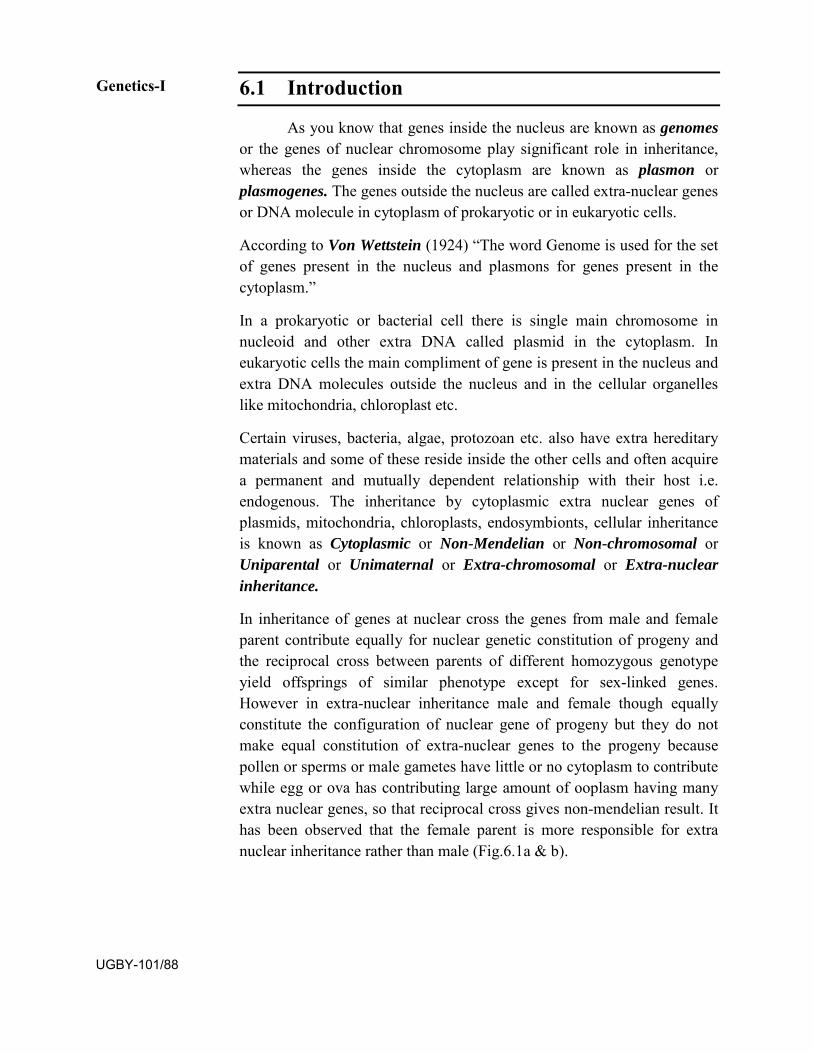
6.1 Introduction
As you know that genes inside the nucleus are known as genomes or the genes of nuclear chromosome play significant role in inheritance, whereas the genes inside the cytoplasm are known as plasmon or plasmogenes. The genes outside the nucleus are called extra-nuclear genes or DNA molecule in cytoplasm of prokaryotic or in eukaryotic cells.
According to Von Wettstein (1924) “The word Genome is used for the set of genes present in the nucleus and plasmons for genes present in the cytoplasm.”
In a prokaryotic or bacterial cell there is single main chromosome in nucleoid and other extra DNA called plasmid in the cytoplasm. In eukaryotic cells the main compliment of gene is present in the nucleus and extra DNA molecules outside the nucleus and in the cellular organelles like mitochondria, chloroplast etc.
Certain viruses, bacteria, algae, protozoan etc. also have extra hereditary materials and some of these reside inside the other cells and often acquire a permanent and mutually dependent relationship with their host i.e. endogenous. The inheritance by cytoplasmic extra nuclear genes of plasmids, mitochondria, chloroplasts, endosymbionts, cellular inheritance is known as Cytoplasmic or Non-Mendelian or Non-chromosomal or Uniparental or Unimaternal or Extra-chromosomal or Extra-nuclear inheritance.
In inheritance of genes at nuclear cross the genes from male and female parent contribute equally for nuclear genetic constitution of progeny and the reciprocal cross between parents of different homozygous genotype yield offsprings of similar phenotype except for sex-linked genes. However in extra-nuclear inheritance male and female though equally constitute the configuration of nuclear gene of progeny but they do not make equal constitution of extra-nuclear genes to the progeny because pollen or sperms or male gametes have little or no cytoplasm to contribute while egg or ova has contributing large amount of ooplasm having many extra nuclear genes, so that reciprocal cross gives non-mendelian result. It has been observed that the female parent is more responsible for extra nuclear inheritance rather than male (Fig.6.1a & b).
Genetics-I
UGBY-101/88
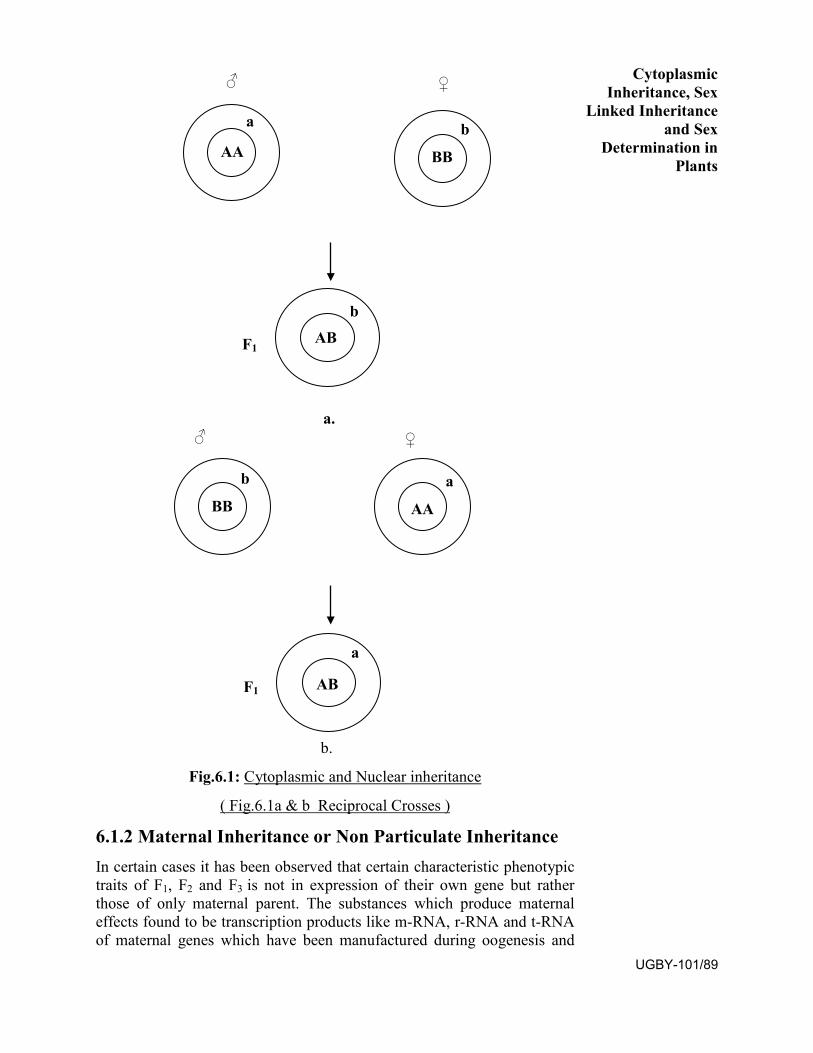
♂ ♀
a b AA BB
b
F1 AB
a. ♂ ♀
b a BB AA
a
F1 AB
b.
Fig.6.1: Cytoplasmic and Nuclear inheritance
( Fig.6.1a & b Reciprocal Crosses )
6.1.2 Maternal Inheritance or Non Particulate Inheritance In certain cases it has been observed that certain characteristic phenotypic traits of F1, F2 and F3 is not in expression of their own gene but rather those of only maternal parent. The substances which produce maternal effects found to be transcription products like m-RNA, r-RNA and t-RNA of maternal genes which have been manufactured during oogenesis and
Cytoplasmic Inheritance, Sex
Linked Inheritance and Sex
Determination in Plants
UGBY-101/89
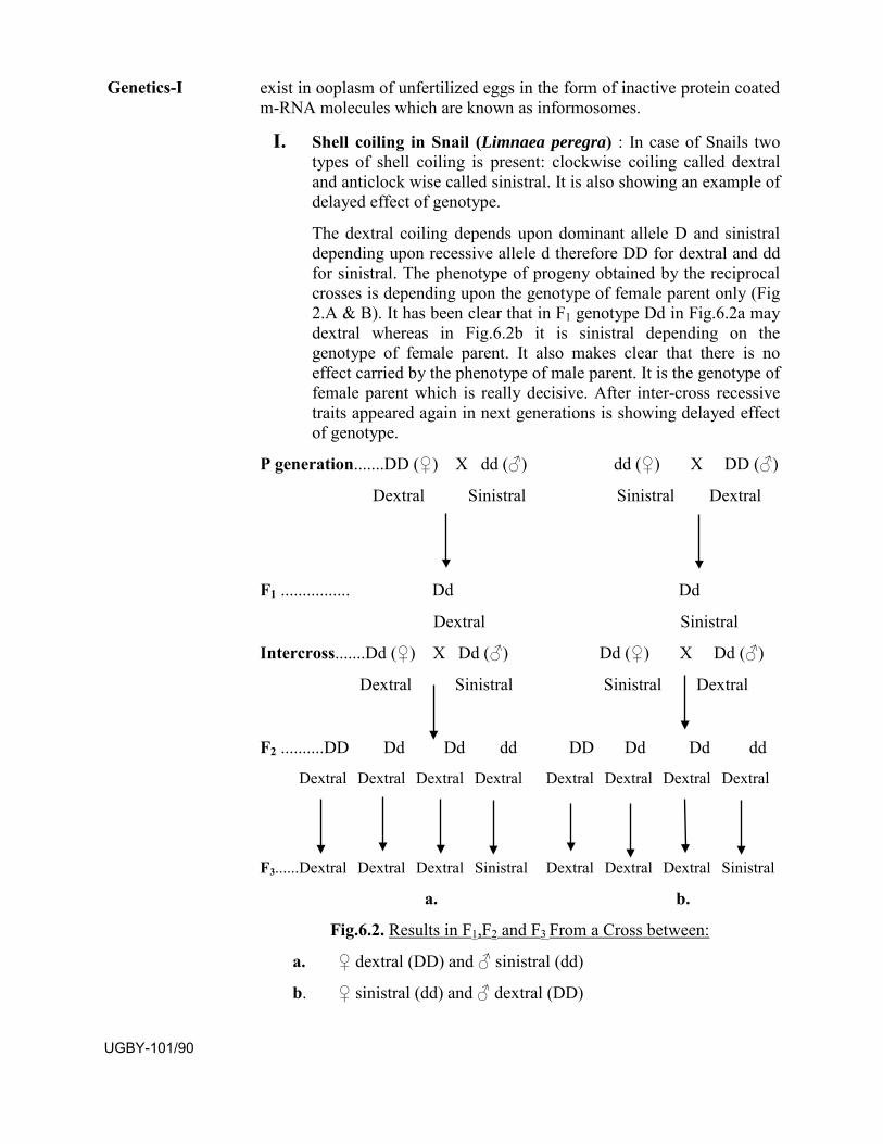
exist in ooplasm of unfertilized eggs in the form of inactive protein coated m-RNA molecules which are known as informosomes.
I. Shell coiling in Snail (Limnaea peregra) : In case of Snails two types of shell coiling is present: clockwise coiling called dextral and anticlock wise called sinistral. It is also showing an example of delayed effect of genotype.
The dextral coiling depends upon dominant allele D and sinistral depending upon recessive allele d therefore DD for dextral and dd for sinistral. The phenotype of progeny obtained by the reciprocal crosses is depending upon the genotype of female parent only (Fig 2.A & B). It has been clear that in F1 genotype Dd in Fig.6.2a maydextral whereas in Fig.6.2b it is sinistral depending on the genotype of female parent. It also makes clear that there is no effect carried by the phenotype of male parent. It is the genotype of female parent which is really decisive. After inter-cross recessive traits appeared again in next generations is showing delayed effect of genotype.
P generation.......DD (♀) X dd (♂) dd (♀) X DD (♂)
Dextral Sinistral Sinistral Dextral
F1 ................ Dd Dd
Dextral Sinistral
Intercross.......Dd (♀) X Dd (♂) Dd (♀) X Dd (♂)
Dextral Sinistral Sinistral Dextral
F2 ..........DD Dd Dd dd DD Dd Dd dd
Dextral Dextral Dextral Dextral Dextral Dextral Dextral Dextral
F3...... Dextral Dextral Dextral Sinistral Dextral Dextral Dextral Sinistral
a. b.
Fig.6.2. Results in F1,F2 and F3 From a Cross between:
a. ♀ dextral (DD) and ♂ sinistral (dd)
b. ♀ sinistral (dd) and ♂ dextral (DD)
Genetics-I
UGBY-101/90
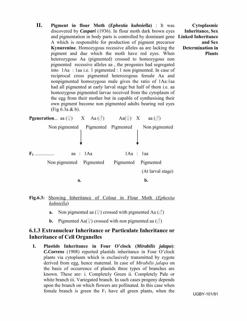
II. Pigment in flour Moth (Ephestia kuhniella) : It wasdiscovered by Caspari (1936). In flour moth dark brown eyesand pigmentation in body parts is controlled by dominant geneA which is responsible for production of pigment precursorKynurenine. Homozygous recessive alleles aa are lacking thepigment and due which the moth have red eyes. Whenheterozygous Aa (pigmented) crossed to homozygous nonpigmented recessive alleles aa , the progenies had segregatedinto 1Aa : 1aa i.e. 1 pigmented : 1 non pigmented. In case ofreciprocal cross pigmented heterozogous female Aa andnonpigmented homozygous male gives the ratio of 1Aa:1aahad all pigmented at early larval stage but half of them i.e. aahomozygous pigmented larvae received from the cytoplasm ofthe egg from their mother but in capable of synthesising theirown pigment become non pigmented adults bearing red eyes(Fig 6.3a & b).
Pgeneration... aa (♀) X Aa (♂) Aa(♀) X aa (♂)
Non pigmented Pigmented Pigmented Non pigmented
F1 ................ aa : 1Aa 1Aa : 1aa
Non pigmented Pigmented Pigmented Pigmented
(At larval stage)
a. b.
Fig.6.3: Showing Inheritance of Colour in Flour Moth (Ephestia kuhniella)
a. Non pigmented aa (♀) crossed with pigmented Aa (♂)
b. Pigmented Aa(♀) crossed with non pigmented aa (♂)
6.1.3 Extranuclear Inheritance or Particulate Inheritance or Inheritance of Cell Organelles
I. Plastids Inheritance in Four O’clock (Mirabilis jalapa): C.Correns (1908) reported plastids inheritance in Four O’clock plants via cytoplasm which is exclusively transmitted by zygote derived from egg, hence maternal. In case of Mirabilis jalapa on the basis of occurrence of plastids three types of branches are known. These are: i. Completely Green ii. Completely Pale or white branch iii. Variegated branch. In such cases progeny depends upon the branch on which flowers are pollinated. In this case when female branch is green the F1 have all green plants, when the
Cytoplasmic Inheritance, Sex
Linked Inheritance and Sex
Determination in Plants
UGBY-101/91
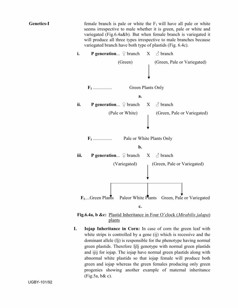
female branch is pale or white the F1 will have all pale or white seems irrespective to male whether it is green, pale or white and variegated (Fig.6.4a&b). But when female branch is variegated it will produce all three types irrespective to male branches because variegated branch have both type of plastids (Fig. 6.4c).
i. P generation... ♀ branch X ♂ branch
(Green) (Green, Pale or Variegated)
F1 ................ Green Plants Only
a.
ii. P generation... ♀ branch X ♂ branch
(Pale or White) (Green, Pale or Variegated)
F1 ................ Pale or White Plants Only
b.
iii. P generation... ♀ branch X ♂ branch
(Variegated) (Green, Pale or Variegated)
______________________________
F1....Green Plants Paleor White Plants Green, Pale or Variegated
c.
Fig.6.4a, b &c: Plastid Inheritance in Four O’clock (Mirabilis jalapa) plants
I. Iojap Inheritance in Corn: In case of corn the green leaf with white strips is controlled by a gene (ij) which is recessive and the dominant allele (Ij) is responsible for the phenotype having normal green plastids. Therefore IjIj genotype with normal green plastids and ijij for iojap. The iojap have normal green plastids along with abnormal white plastids so that iojap female will produce both green and iojap whereas the green females producing only green progenies showing another example of maternal inheritance (Fig.5a, b& c).
Genetics-I
UGBY-101/92
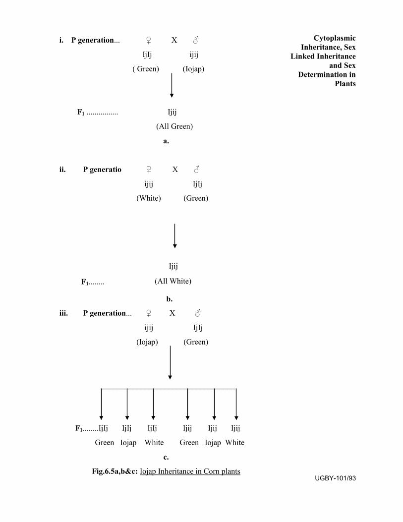
i. P generation... ♀ X ♂
IjIj ijij
( Green) (Iojap)
F1 ................ Ijij
(All Green)
a.
ii. P generatio ♀ X ♂
ijij IjIj
(White) (Green)
Ijij
F1........ (All White)
b.
iii. P generation... ♀ X ♂
ijij IjIj
(Iojap) (Green)
__________________________________
F1........IjIj IjIj IjIj Ijij Ijij Ijij
Green Iojap White Green Iojap White
c.
Fig.6.5a,b&c: Iojap Inheritance in Corn plants
Cytoplasmic Inheritance, Sex
Linked Inheritance and Sex
Determination in Plants
UGBY-101/93
III. Male Sterility in Plants: There are three type of male sterilityknown among plants: i. Cytoplasmic male sterility ii. Geneticmale sterility iii. Cytoplasmic genetic male sterility.
i. Cytoplasmic Male Sterility: The male sterility in maize is anexample of cytoplasmic inheritance. If female paren is malesterile the F1 will be always male sterile because the cytoplasmis derived from female parent i.e. egg (Fig.6.6). The malefertile plants produce both male and female fertile. Whereasmale sterile plants are used as female parent will produce onlymale sterile plants after making back crosses. Seldom malesterile produce rare pollen, when treated as male parent thenmale fertile progenies may develop.
P generation... ♀ X ♂
Male sterile Male fertile
F1.................. All Male Sterile
Fig.6.6: Cytoplasmic Male Sterility
ii. Genetic Male Sterility: Male sterility is controlled by singlegene S and is recessive to fertility i.e. F for fertile. So that allF1 will be fertile due to the dominance of F over S. In F2
generation fertile and sterile individuals segregated into 3:1 asmendelian law irrespective to their male or femalecharacteristics as in tomato (Fig.6.7).
P generation... ♀ X ♂
SS FF
Male Sterile Male Fertile
F1................... SF
All Male Fertile
Genetics-I
UGBY-101/94
Intercross....... SF X SF
_____________________________
1 SS 2 SF 1 FF
Male Sterile Male Fertile Male Fertile
Fig.6.7: Genetic or Mendelian Male Sterility
iii. Cytoplasmic Genetic Male Sterility: Male sterility is
controlled by both cytoplasm and nucleus (genes) as reported
in onion (Allium cepa). Fertlity factor F is dominant and may
appear whether it is present in cytoplasm or inside nuclear gene
as a dominant allele which masks the effect of recessive allele
S for male sterility as shown in (Fig.6.8).
Cytoplasmic Inheritance, Sex
Linked Inheritance and Sex
Determination in Plants
UGBY-101/95
♀ ♂ Progeny
S F S i. SS X SS SS
Male Sterile
S S S ii. SS X F FS
Male Fertile
S F S iii. SS X FF FS
Male Fertile
S F S iv. SS X FS FS
Male Fertile
S S S v. SS X FS SS
Male Sterile
Fig.6.8: Cytoplasmic Genetic Male Sterility in Onion (Allium cepa)
I. Mitochondrial inheritance in Yeast: Due to the loss of aerobic respiration and utilization of less efficient fermentation certain small sized slow growing colonies of yeast developed are known as ‘littles’ petite mutant which is due to the suppressed activity of mitochondria. Genes of mitochondria mutated artificially by Acridine dye or Euflavin develop vegetative petities. The little phenotype depends upon the kind of cytoplasm and mitochondria present. Though the nuclear gene found to be segregated normally. When normal colonies crossed with littles develop 1:1 normal and littles i.e. segregational petities (Fig.6.9a). In other case when normal colonies crossed with vegetative petities (artificially mutated strains), it develops the colonies in which genes of mitochondria also mutated due to artificial mutation (Fig. 6.9b). The phenotype of littles petites depends upon kind of cytoplasm and mitochondria present and nuclear genes segregate normally.
8 8
a. Aa X aa 1 Normal : 1 Little
Normal Littles
Genetics-I
UGBY-101/96
8 8
b. Aa X aa Genes of Mitochondria
also mutate
Normal Vegetative
Fig.6.9a& b: Mitochondrial Inheritance in Yeast
6.1.4 Extranuclear Inheritance of Endosymbionts:
Sometimes it has been seen that the endosymbionts which live inside the
living cells of any host may behave as the part of cell or cellular organelle
viz. Bacteria viruses etc. These intercellular parasites namely bacteria,
viruses etc. are maintaining a symbiotic relationships with the host cell.
They are self reproducing and appear like other cell inclusions. Sometime
they exhibit as an infection and may be transmitted hereditary and
continue to the generation to generation with their own. Such symbionts
are termed as Greek words e.g. Sigma (∑) particles, Kappa particles, mu
(µ) etc.
I. Kappa Particles in Paramecium: It was reported by T.N,
Sonneborn (1938) according to the studies it was found that
some strains of Paramecium aurelia namely killers (KK)
having kappa particles are producing a poisonous substance
called paramecin. It is toxic to the sensitive (kk) strains which
do not possess kappa particles. The production of kappa
particles is dependent upon a dominant allele K, so that killer
strains have KK or Kk alleles whereas the sensitive strains bear
kk alleles. Paramecin produced through the cytoplasm and
located particles called kappa particles may appear to be either
parasites or symbionts and they do not harm their host. Their
transmission depending upon the duration of conjugation as
shown below (Fig.6.10a&b).
Cytoplasmic Inheritance, Sex
Linked Inheritance and Sex
Determination in Plants
UGBY-101/97
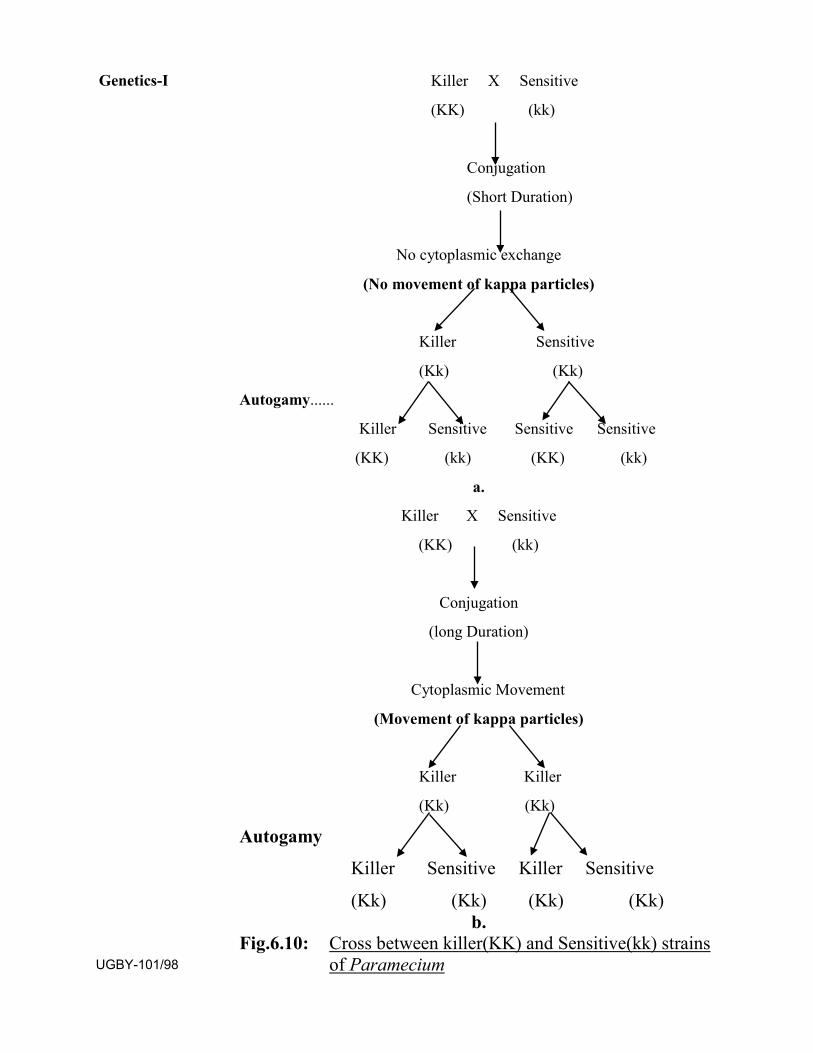
Killer X Sensitive
(KK) (kk)
Conjugation
(Short Duration)
No cytoplasmic exchange
(No movement of kappa particles)
Killer Sensitive
(Kk) (Kk)
Autogamy......
Killer Sensitive Sensitive Sensitive
(KK) (kk) (KK) (kk)
a.
Killer X Sensitive
(KK) (kk)
Conjugation
(long Duration)
Cytoplasmic Movement
(Movement of kappa particles)
Killer Killer
(Kk) (Kk)
Autogamy
Killer Sensitive Killer Sensitive
(Kk) (Kk) (Kk) (Kk) b.
Fig.6.10: Cross between killer(KK) and Sensitive(kk) strains of Paramecium
Genetics-I
UGBY-101/98
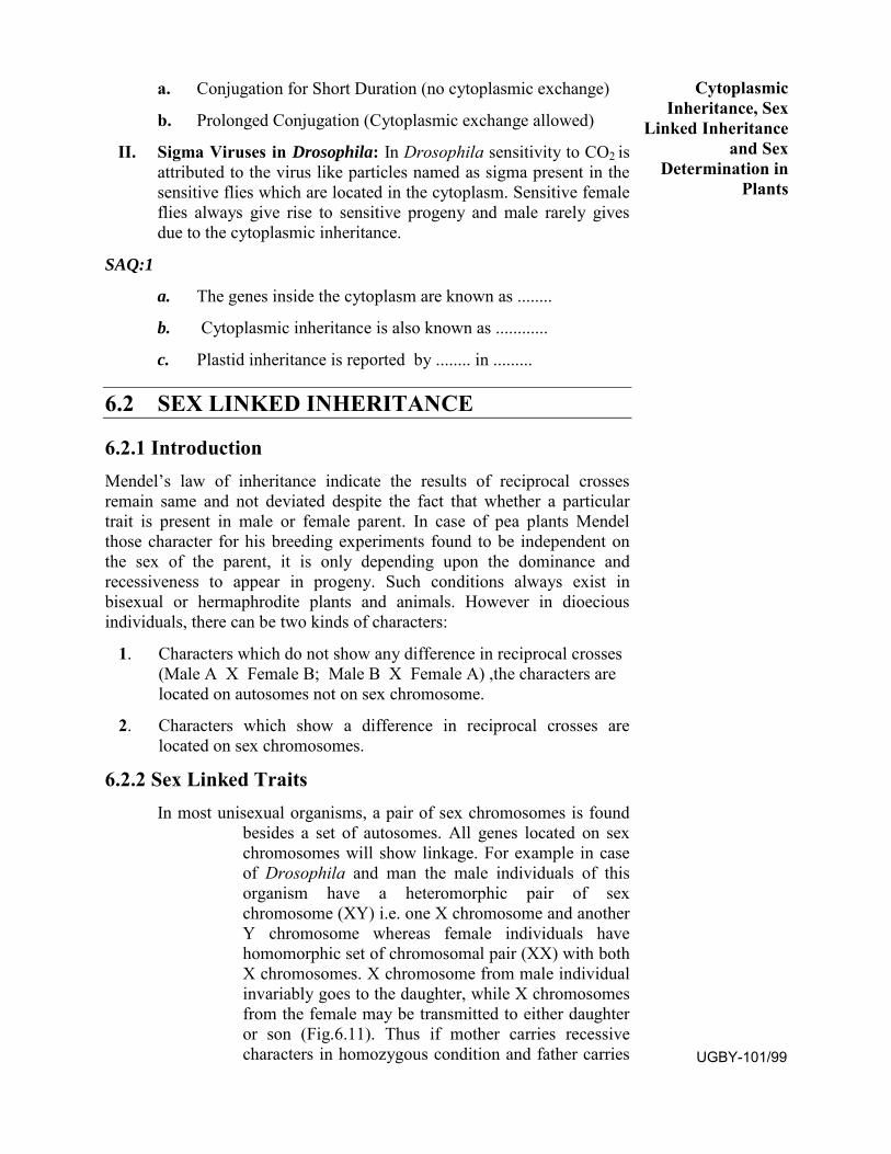
a. Conjugation for Short Duration (no cytoplasmic exchange)
b. Prolonged Conjugation (Cytoplasmic exchange allowed)
II. Sigma Viruses in Drosophila: In Drosophila sensitivity to CO2 isattributed to the virus like particles named as sigma present in thesensitive flies which are located in the cytoplasm. Sensitive femaleflies always give rise to sensitive progeny and male rarely givesdue to the cytoplasmic inheritance.
SAQ:1
a. The genes inside the cytoplasm are known as ........
b. Cytoplasmic inheritance is also known as ............
c. Plastid inheritance is reported by ........ in .........
6.2 SEX LINKED INHERITANCE
6.2.1 Introduction Mendel’s law of inheritance indicate the results of reciprocal crosses remain same and not deviated despite the fact that whether a particular trait is present in male or female parent. In case of pea plants Mendel those character for his breeding experiments found to be independent on the sex of the parent, it is only depending upon the dominance and recessiveness to appear in progeny. Such conditions always exist in bisexual or hermaphrodite plants and animals. However in dioecious individuals, there can be two kinds of characters:
1. Characters which do not show any difference in reciprocal crosses(Male A X Female B; Male B X Female A) ,the characters arelocated on autosomes not on sex chromosome.
2. Characters which show a difference in reciprocal crosses arelocated on sex chromosomes.
6.2.2 Sex Linked Traits In most unisexual organisms, a pair of sex chromosomes is found
besides a set of autosomes. All genes located on sex chromosomes will show linkage. For example in case of Drosophila and man the male individuals of this organism have a heteromorphic pair of sex chromosome (XY) i.e. one X chromosome and another Y chromosome whereas female individuals have homomorphic set of chromosomal pair (XX) with both X chromosomes. X chromosome from male individual invariably goes to the daughter, while X chromosomes from the female may be transmitted to either daughter or son (Fig.6.11). Thus if mother carries recessive characters in homozygous condition and father carries
Cytoplasmic Inheritance, Sex
Linked Inheritance and Sex
Determination in Plants
UGBY-101/99
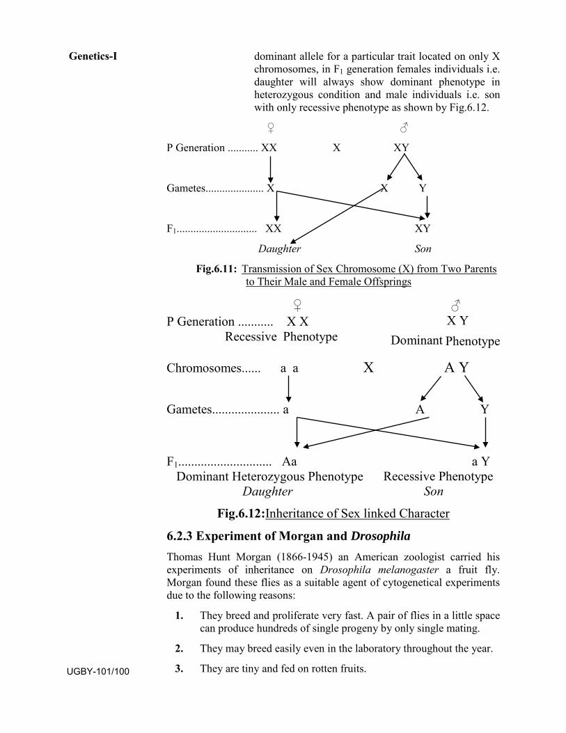
dominant allele for a particular trait located on only X chromosomes, in F1 generation females individuals i.e. daughter will always show dominant phenotype in heterozygous condition and male individuals i.e. son with only recessive phenotype as shown by Fig.6.12.
♀ ♂
P Generation ........... XX X XY
Gametes..................... X X Y
F1............................. XX XY
Daughter Son
Fig.6.11: Transmission of Sex Chromosome (X) from Two Parents to Their Male and Female Offsprings
♀ ♂ P Generation ........... X X X Y
Recessive Phenotype Dominant Phenotype
Chromosomes...... a a X A Y
Gametes..................... a A Y
F1............................. Aa a Y Recessive Phenotype Dominant Heterozygous Phenotype
Daughter Son
Fig.6.12:Inheritance of Sex linked Character
6.2.3 Experiment of Morgan and Drosophila Thomas Hunt Morgan (1866-1945) an American zoologist carried his experiments of inheritance on Drosophila melanogaster a fruit fly. Morgan found these flies as a suitable agent of cytogenetical experiments due to the following reasons:
1. They breed and proliferate very fast. A pair of flies in a little spacecan produce hundreds of single progeny by only single mating.
2. They may breed easily even in the laboratory throughout the year.
3. They are tiny and fed on rotten fruits.
Genetics-I
UGBY-101/100
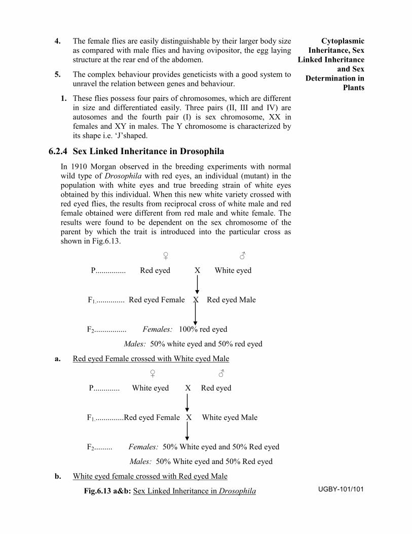
4. The female flies are easily distinguishable by their larger body sizeas compared with male flies and having ovipositor, the egg layingstructure at the rear end of the abdomen.
5. The complex behaviour provides geneticists with a good system tounravel the relation between genes and behaviour.
1. These flies possess four pairs of chromosomes, which are differentin size and differentiated easily. Three pairs (II, III and IV) areautosomes and the fourth pair (I) is sex chromosome, XX infemales and XY in males. The Y chromosome is characterized byits shape i.e. ‘J’shaped.
6.2.4 Sex Linked Inheritance in Drosophila In 1910 Morgan observed in the breeding experiments with normal wild type of Drosophila with red eyes, an individual (mutant) in the population with white eyes and true breeding strain of white eyes obtained by this individual. When this new white variety crossed with red eyed flies, the results from reciprocal cross of white male and red female obtained were different from red male and white female. The results were found to be dependent on the sex chromosome of the parent by which the trait is introduced into the particular cross as shown in Fig.6.13.
♀ ♂
P............... Red eyed X White eyed
F1............... Red eyed Female X Red eyed Male
F2................ Females: 100% red eyed
Males: 50% white eyed and 50% red eyed
a. Red eyed Female crossed with White eyed Male
♀ ♂
P............. White eyed X Red eyed
F1...............Red eyed Female X White eyed Male
F2......... Females: 50% White eyed and 50% Red eyed
Males: 50% White eyed and 50% Red eyed
b. White eyed female crossed with Red eyed Male
Fig.6.13 a&b: Sex Linked Inheritance in Drosophila
Cytoplasmic Inheritance, Sex
Linked Inheritance and Sex
Determination in Plants
UGBY-101/101
A. Red eyed Female crossed with White eyed male: Here in F1 generation both male and female flies were red eyed. When these were bred together one fourth i.e. 25% F2 offspring were white eyed indicating that red and white eye colours are due to an allelic pair of genes of which red reappears as the dominant. Besides, in F2 offspring, all females were red eyed whereas in male flies half i.e. 50% red eyed and half white eyed (Fig13a).
B. White eyed Female crossed with Red eyed male: In this case F1 found to be red eyed females and white eyed males. When these were bred together, their offspring consisted of both red eyed and white eyed flies equally i.e. 50% in both sexes (Fig.13b).
The first mutant in Drosophila was male, observed by Morgan. Since the male has only X chromosome and there is no gene on Y chromosome to mark the expression of white. The white mutant got his X chromosome from mother. Sex linked genes are always transmitted by mother to son and not by his father.
Wing length in Drosophila: Normal wings are found dominant to miniature wings. Its inheritance is also like eye colour, the genes for wing length are also sex linked.
6.2.5 Sex Linked Inheritance in Human Beings There are 23 pairs or 2n=46 chromosomes known in human beings. Out of
23 pairs 22 pairs of autosomes and one pair of sex chromosome is present
there. In case of females sex chromosomes are XX whereas male bears
XY. So that male always produces two types of gametes X and Y and
female produces only one type i.e. X. Therefore male progenies are
dependent on Y chromosome which can be transmitted only by the father
paired with X chromosome carried by the mother. Daughters have XX one
from father and another from mother. Sex linked characters in human
beings thus follow the same pattern as reported in Drosophila. Colour
blindness and haemophilia are two important sex linked traits in human
beings.
I. Colour Blindness: A particular trait which makes the person unable to differentiate red and green colour. This gene for red-green blindness is located on X chromosome. Colour blindness is recessive to normal vision and is found more often in man. Therefore father can transmit it to daughters only and not to son. On the other hand a colour blind mother is able transmit the disease to the son regardless of the colour vision of husband and all daughters have normal vision. The daughters carring the genes for colour blindness when married to a man with normal vision will produce all normal girl children, but among sons half with
Genetics-I
UGBY-101/102
normal vision and half of them with colour blindness. A colour blind daughter is produced only in cases if colour blind man marries with a carrier or homozygous colour blind woman.
II. Haemophilia: The person suffering from haemophilia lack the
factor which is responsible for clotting of blood. Such persons are very
difficult to survive till maturity because a minor injury may cause
prolonged bleeding leading to death. It is a recessive character and is
therefore, masked by the dominant allele in heterozygous condition. It is
also inherited like trait of Colour blindness. A carrier mother may survive
by having heterozygous condition when married with healthy man the
progenies may be as follows:
a. Healthy normal daughter by coupling of non carrier alleles eachone from mother and father.
b. Carrier daughter with one non carrier from father and anothercarrier allele from her mother.
c. Healthy son by pairing of non carrier alleles from mother andfather.
d. Haemophilic son received carrier allele from mother and Y
chromosome in absence of this particular allele make the
individual sufferer.
SAQ: 2
a. ............ an American zoologist has carried experiments ofinheritance on Drosophila melanogaster.
b. Drosophila flies possess ..........pairs of chromosomes.
c. ........and.......... is a sex linked inherited disease reported in humanbeings
6.3 SEX DETERMINATION IN PLANTS
6.3.1 Introduction Sex determination deals with the aspects of factor responsible to determine a particular sex in the individuals whether it would be male or female individuals in unisexual or dioecious organisms and in bisexuals or hermaphrodite or monecious individuals. Regarding sex determination various biologists and non biologists gave several mistaken hypothesis and guesses about the progenies. After a long discussion, the valid solution
Cytoplasmic Inheritance, Sex
Linked Inheritance and Sex
Determination in Plants
UGBY-101/103
became possible after the discovery of sex chromosome during early years of the 20th century.
Henking in 1991 found a specific nuclear structure through
spermatogenesis in some insects and he observed 50% of sperms received
and rest 50% not received this. He called this as ‘X-body’ but he failed to
explain its significance. Later it was found that it was in fact a
chromosome named as X-chromosome. It was also concluded that this X-
chromosome plays an important role in determination of sex. Thereafter it
was designated and differentiated as ‘sex chromosome’ and rest set of
chromosomes were labelled as ‘autosomes’.
6.3.2 Chromosomal Theory of Sex Determination According to chromosomal theory each individual posses two sets of chromosomes; autosomes and sex chromosomes. The constitution of sex chromosome defines the sex of the individual whether it is male or female, not by the autosomes. The set of autosomes are similar in both male and female. Some examples of sex determination are as follows:
I. In Drosophila and Man: In both there are two types of sex chromosomes; X and Y. The female individuals have XX and male possesses XY. Both X and Y chromosomes differ morphologically.
II. In Birds: In this case two different types of gametes are producedby females’ i.e. female heterogametes and male producing sperms with only one type of sex chromosome. In order to distinguish, female is designated as ZW being heterogametic and male is designated as ZZ being homogametic.
III. In Plants: In plants also sex is mainly controlled by Ychromosome like previous examples. Presence of Y chromosomedefines the male individuals and absence of it results in female.
IV. In Honey Bees: The male honey bee i.e. drones ariseparthenogenetically so that they have haploid no. of chromosomebearing 16 chromosomes, while female i.e. queen bee and workershave 32 chromosomes. Here the number or set of chromosomedefine the sex called Haplodiploid sex determination system.
V. In Grasshoppers: It is an example of XO type of sex determination in which male bears only one X chromosome; XO besides the autosomes, whereas females have a pair of X chromosome i.e. XX.
Genetics-I
UGBY-101/104
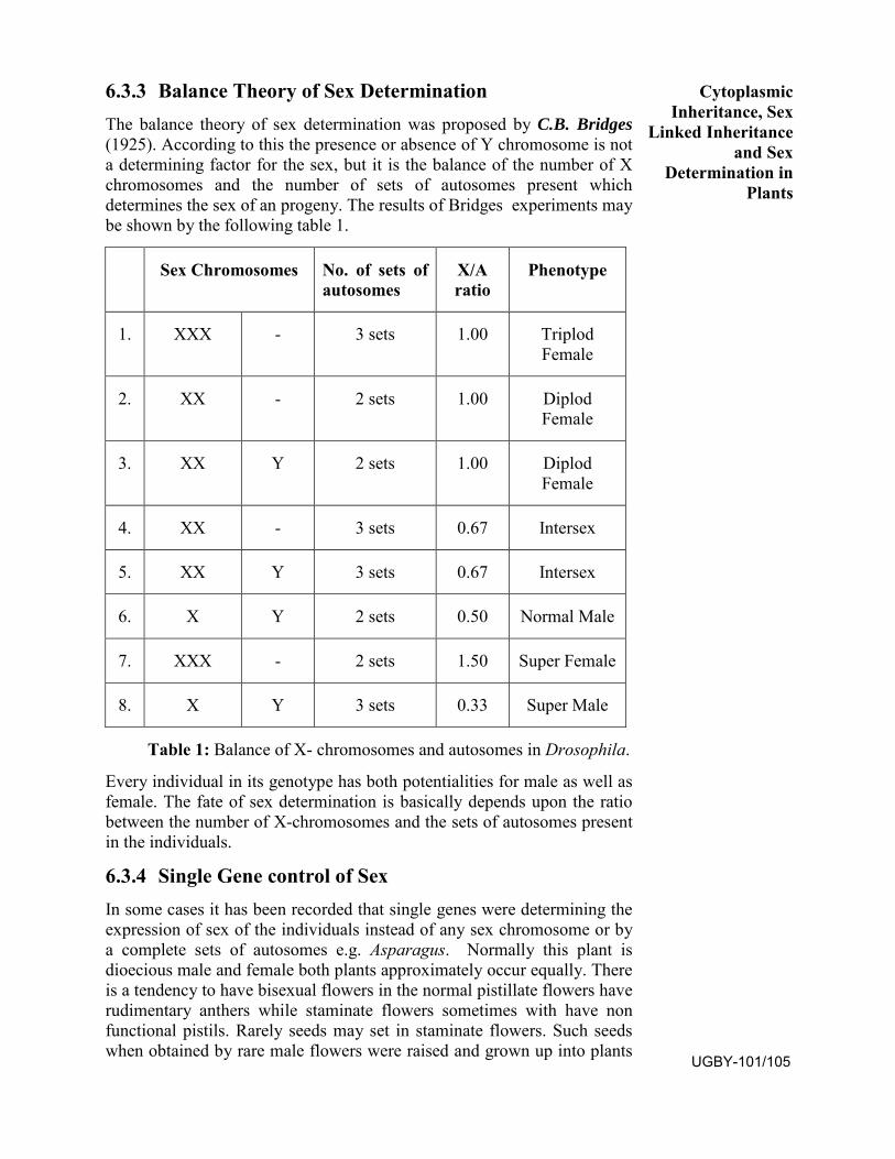
6.3.3 Balance Theory of Sex Determination The balance theory of sex determination was proposed by C.B. Bridges (1925). According to this the presence or absence of Y chromosome is not a determining factor for the sex, but it is the balance of the number of X chromosomes and the number of sets of autosomes present which determines the sex of an progeny. The results of Bridges experiments may be shown by the following table 1.
Sex Chromosomes No. of sets of autosomes
X/A ratio
Phenotype
1. XXX - 3 sets 1.00 Triplod Female
2. XX - 2 sets 1.00 Diplod Female
3. XX Y 2 sets 1.00 Diplod Female
4. XX - 3 sets 0.67 Intersex
5. XX Y 3 sets 0.67 Intersex
6. X Y 2 sets 0.50 Normal Male
7. XXX - 2 sets 1.50 Super Female
8. X Y 3 sets 0.33 Super Male
Table 1: Balance of X- chromosomes and autosomes in Drosophila.
Every individual in its genotype has both potentialities for male as well as female. The fate of sex determination is basically depends upon the ratio between the number of X-chromosomes and the sets of autosomes present in the individuals.
6.3.4 Single Gene control of Sex In some cases it has been recorded that single genes were determining the expression of sex of the individuals instead of any sex chromosome or by a complete sets of autosomes e.g. Asparagus. Normally this plant is dioecious male and female both plants approximately occur equally. There is a tendency to have bisexual flowers in the normal pistillate flowers have rudimentary anthers while staminate flowers sometimes with have non functional pistils. Rarely seeds may set in staminate flowers. Such seeds when obtained by rare male flowers were raised and grown up into plants
Cytoplasmic Inheritance, Sex
Linked Inheritance and Sex
Determination in Plants
UGBY-101/105
were found to be present in female plants only two third of them showed segregation indicating that sex is controlled by a single gene.
SAQ.3: a. The balanced theory of sex determination was proposed by
............in.....
b. In case of .......... plants sex is determined by single gene.
c. If a fruit fly possess three sets of autosomes i.e. XXX the fly willbe.....
6.4 Summary
6.4.1 Cytoplasmic Inheritance The inheritance by cytoplasm, is carried away by the mother only through
the ooplasm or egg where male gametes fertilize so that female contributes
a lot in development of embryo after fertilization. There are various
example of cytoplasmic inheritance i.e. extra nuclear genes of plasmids,
mitochondria, chloroplasts, endosymbionts, cellular inheritance and these
type o inheritance are known by several names such as Cytoplasmic or
Non-Mendelian or Non-chromosomal or Uniparental or Unimaternal or
Extra-chromosomal or Extra-nuclear inheritance.
However in extra-nuclear inheritance, male and female though equally
constitute the configuration of nuclear gene of progeny but they do not
make equal constitution of extra-nuclear genes to the progeny because
pollen or sperms or male gametes have little or no cytoplasm to contribute
while egg or ova contributes large amount of ooplasm having many extra
nuclear genes, so that reciprocal cross gives non-mendelian result. It has
been observed that the female parent is more responsible for extra nuclear
inheritance rather than male. The cytoplasmic inheritance may be
categorised as follows:
1. Maternal Inheritance or Non Particulate Inheritance
2. Extranuclear Inheritance or Particulate Inheritance
3. Extranuclear Inheritance of Endosymbionts
Genetics-I
UGBY-101/106
6.4.2 Sex Linked Inheritance
Mostly in unisexual organisms, a pair of sex chromosomes is found
besides a set of autosomes. The genes located on sex chromosomes
showing linkage and may inherited from generation to generation. This
type of inheritance is called sex linked inheritance and the traits called
sex linked traits.
Sex Linked Inheritance in Drosophila T.H.Morgan (1910) observed an individual (mutant) with white eyes in the population of Drosophila with normal wild type with red eyes during his breeding experiments. When mutant white eyed fly crossed with normal red eyed fly; the results of reciprocal crosses were different which is as follows:
1. White eyed male crossed with red eyed female: The F1progenies were red eyed in both the sexes and F1 bred together, theF2 resulting ¼ white eyed indicating that red and white eye colourswere due to an allelic pair of genes of which red reappeared as thedominant. Besides in F2 all females were red whereas half maleswere red and half white.
2. Red eyed male crossed with white eyed female: here theprogenies in F1 had red eyed males and white eyed females, whenthese were inbred together the F2 progenies consist of red eyed andwhite eyed flies equally in both sexes.
Sex Linked Inheritance in Human Beings Sex linked traits in human beings follow the same pattern as in
Drosophila. Colour blindness and haemophilia are very important diseases
in human beings and these diseases are the example of sex linked
inheritance.
6.4.3 Sex Determination in Plants Sex determination is concerned with the study of factors which are responsible for making an individual male, female or hermaphrodite.
Chromosomal Theory of Sex Determination: According to this theory two types of chromosomes i.e. autosomes and sex chromosomes are present in each individual and male and female differ only by the constitution of sex chromosome not by the autosomes.
Balance Theory of Sex Determination: This theory was proposed by C.B.Bridges (1925), the presence or absence of Y sex chromosome is not determining factor of sex, but it is the number of X chromosomes and the
Cytoplasmic Inheritance, Sex
Linked Inheritance and Sex
Determination in Plants
UGBY-101/107
number of X-chromosome and the number of sets of autosomes i.e. X: A determines the sex of an offspring.
Single Gene Sex Determination: In some cases individual single gene may be the deciding factor of sex expression.
6.5 TERMINAL QUESTIONS
OBJECTIVE TYPE QUESTIONS:
1. T. H.Morgan found white eyed Drosophila is due to mutant geneon :
a. Autosomes c. X-chromosome
b. Y-chromosome d. None of these
2. The genes for colour blindness in human beings located on :
a. X-chromosomes c. Autosomes
b. Y-chromosomes d. None of these
3. The colour blind daughter born when:
a. Colour blind mother and normal father
b. Carrier mother and Colour blind father
c. Carrier mother and normal father
d. Normal mother colour blind father
4. In plastid inheritance when female branch is variegated and andmale branch is green in four o’clock plants, the F1 will be:
a. Green only c. Pale only
b. Variegated only d. All green, pale and variegated
5. Shell coiling in snails showing the example of:
a. Mitochondrial inheritance c. Maternal inheritance
b. Particulate inheritance d. Nuclear inheritance
6. Female Drosophila possess:
a. XX chromosomes with 3 sets of autosomes
b. XY chromosomes with 3 sets of autosomes
c. XX chromosomes with 4 sets of autosomes
d. XY chromosomes with 4 sets of autosomes
Genetics-I
UGBY-101/108
7. White eyes in Drosophila (fruit fly) is determined by:
a. Dominant gene on X chromosome
b. Dominant gene on Y chromosome
c. Recessive gene on Y chromosome
d. Recessive gene on X chromosome
8. Man bears total .........no. of chromosomes with ...... sexchromosome. a. 23 pairs , XY c. 24 pairs, XX
b. 23 pairs , XX d. 24 pairs, XY
9. If a genetic disease is transmitted by phenotypically normal carrierfemaleI to only some of the male progeny, the disease may be:a. Sex linked recessive c. Autosomal dominant
b. Sex linked dominant d. Autosomal recessive
10. How many genes are received by the children from their father?
a. 25% b. 50% c. 75% d.100%
SHORT ANSWER TYPE QUESTIONS: Q.1 What is plastid inheritance?
Q.2 Write short notes on:
a. Maternal inheritance b. Male sterility c. Kappa paricles
Q.3 How do white eyed mutant Drosophila transfer the trait to the next generation?
Q.4. Write short notes on:
a. Haemophilia b. Colour Blindness
c. Experiments of Morgan
Q.5. A colour blind man has normal brother and a colour blind sister, find the genotype of the parents.
Q.6. A woman with normal vision had colour blind father and she marries with colour blind man find the probabilities of children with their genotype.
LONG ANSWER TYPE QUESTIONS: Q.1. Discuss various kinds of genetic controls of leaf variegation in plants
by using the example from four o’clock plants.
Q.2. How do you explain shell coiling in snails?
Cytoplasmic Inheritance, Sex
Linked Inheritance and Sex
Determination in Plants
UGBY-101/109
Q.3. By giving the examples discuss the evidences of chloroplast and mitochondria in cytoplasmic inheritance.
Q.4. On the basis of sex expression describe chromosomal constitution for heterogametic sex with the help of any suitable example.
Q.5. How would you differentiate the chromosomal theory and the gene balance theory of sex determination?
Q.6. What are sex linked traits and how do they inherit in progeny?
6.6 ANSWER OBJECTIVE TYPE QUESTIONS:
1. C 2. A 3. B 4. D 5. C
6. A 7. D 8. A 9. A 10. b
SAQ.1:
a. Plasmon or plasmongenes b. Extra-nuclear inheritancec. Correns, Mirabilis jalapa
SAQ.2:
a. Thomas Hunt Morgan b. Four
c. Colour blindness, haemophilia
SAQ.3
a. C.B. Bridges, Drosophila melanogaster
b. Asparagus
c. Super female
Genetics-I
UGBY-101/110
BLOCK
3 GENETICS-II
UNIT 7
PRE-CHROMOSOMAL ABERRATIONS
UNIT 8
GENE MUTATION AND INDUCED MUTATION
UNIT 9
GENETICS IN PLANT IMPROVEMENT
Uttar Pradesh Rajarshi Tandon Open University
Bachelor of Science
UGBY-101
115-140
141-156
157-172
UGBY-101/111

Course Design Committee Prof. Ashutosh Gupta Chairman Director, School of Science UPRTOU, Prayagraj.
Prof. Malvika Srivastava Member Botny Department Deendayal Upadhyay University, Gorakhpur.
Prof. V.N. Pandey Member Botny Department Deendayal Upadhyay University, Gorakhpur.
Prof. Anil Kumar Diwedi Member Botny Department Deendayal Upadhyay University, Gorakhpur.
Dr. N.N Tripathi Member Acadmic Consultant, School of Science UPRTOU, Prayagraj.
Dr. Sushma Chauhan Member/Secretary Acadmic Consultant, School of Science UPRTOU, Prayagraj.
Course Preparation Committee Dr. Dharmveer Singh Author Block-1 (Unit: 1-2) Acadmic Consultant, School of Science UPRTOU, Prayagraj.
Dr. Sushma Chauhan Author Block-1 (Unit: 3) Acadmic Consultant, School of Science UPRTOU, Prayagraj.
Dr Achala Srivatava Author Block-2-3 (Unit: 4-9) Assistant professor S.S. Khanna, P.G. College Prayagraj.
Prof. G. Kumar Editor (Unit: 1-9) Professor Department of Botany University of Allahabad, Prayagraj
Dr. Sushma Chauhan Course Coordinator Acadmic Consultant (Botany) School of Science UPRTOU, Prayagraj.
© UPRTOU, Prayagraj. 2019 ISBN : All Rights are reserved. No part of this work may be reproduced in any form, by mimeograph or any other means, without permission in writing from the Uttar Pradesh Rajarshi Tondon Open University, Prayagraj.
UGBY-101/112
Block-III
Genetics II
In Block II, you have studied about earlier concept of heredity and variations, Mendel’s experiment and laws of inheritance. As we further see the deviation of Mendel’s law after the discovery of linkage and crossing over. Further it has been also found that there are non-nuclear inheritances i.e. maternal inheritance also in which only mother may be responsible for transferring the particular traits among the progeny not by both parents.
In Block III, we will discuss about the types of chromosomal aberrations and their significance which may be either induced or due to the mutation i.e. by sudden change in Unit 7. Various types of mutations will be discussed in Unit 8. In Unit 9 the role and applications of genetics in plant improvement will be focussed for the study. The Block III includes Genetics II part which comprises of three units i.e. Unit 7, Unit 8 and Unit 9.
Unit 7: Chromosomal Aberrations: First part of the chapter reveals on various types of changes in chromosomes such as inter and intra chromosomal aberrations and their significance have also been studied. The different types of variation in basic chromosome number and their impact on the organism have been focussed in the second part of the chapter
Unit 8: Mutation-Gene Mutation, Induced Mutation
Unit 9: Genetics in Plant Improvement.
UGBY-101/113

UGBY-101/114
UNIT-7CHROMOSOMAL ABERRATIONS
Structure 7.1 Introduction
7.2 Structural Changes in Chromosomes
7.2. 1 Intra-chromosomal Aberration
• Deficiencies or Deletion• Duplications or Addition• Inversion• Shifts• Isochromosome
7.2. 2 Interchromosomal Aberration
• Homozygotic Translocation
• Heterozygotic Translocation
7.3 Numerical Changes in Chromosomes
7.3.1 Euploidy
• Monoploidy
• Diploidy
• Polyploidy
7.3.2 Aneuploidy
• Hypoploidy
• Hyperploid
7.4 Summary
7.5 Terminal Questions
7.6 Answer
7.1. INTRODUCTION Among natural population, there are some variations in the structure and the number of chromosomes and it was also found that it could be produced artificially in variety of organisms. Further studies revealed that these variations can be due to the alterations either in the structure or in number of chromosomes. Any type of changes in chromosomes which appear phenotypically due to structure is known as chromosomal aberration or chromosomal mutations. These alterations do not involve UGBY-101/115
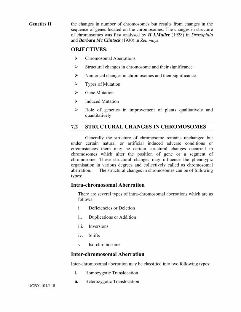
the changes in number of chromosomes but results from changes in the sequence of genes located on the chromosomes. The changes in structure of chromosomes was first analysed by H.J.Muller (1928) in Drosophila and Barbara Mc Clintock (1930) in Zea mays
OBJECTIVES: Chromosomal Aberrations
Structural changes in chromosome and their significance
Numerical changes in chromosomes and their significance
Types of Mutation
Gene Mutation
Induced Mutation
Role of genetics in improvement of plants qualitatively andquantitatively
7.2 STRUCTURAL CHANGES IN CHROMOSOMES
Generally the structure of chromosome remains unchanged but under certain natural or artificial induced adverse conditions or circumstances there may be certain structural changes occurred in chromosomes which alter the position of gene or a segment of chromosome. These structural changes may influence the phenotypic organisation in various degrees and collectively called as chromosomal aberration. The structural changes in chromosomes can be of following types:
Intra-chromosomal Aberration There are several types of intra-chromosomal aberrations which are as follows:
i. Deficiencies or Deletion
ii. Duplications or Addition
iii. Inversions
iv. Shifts
v. Iso-chromosome.
Inter-chromosomal Aberration
Inter-chromosomal aberration may be classified into two following types:
i. Homozygotic Translocation
ii. Heterozygotic Translocation
Genetics II
UGBY-101/116
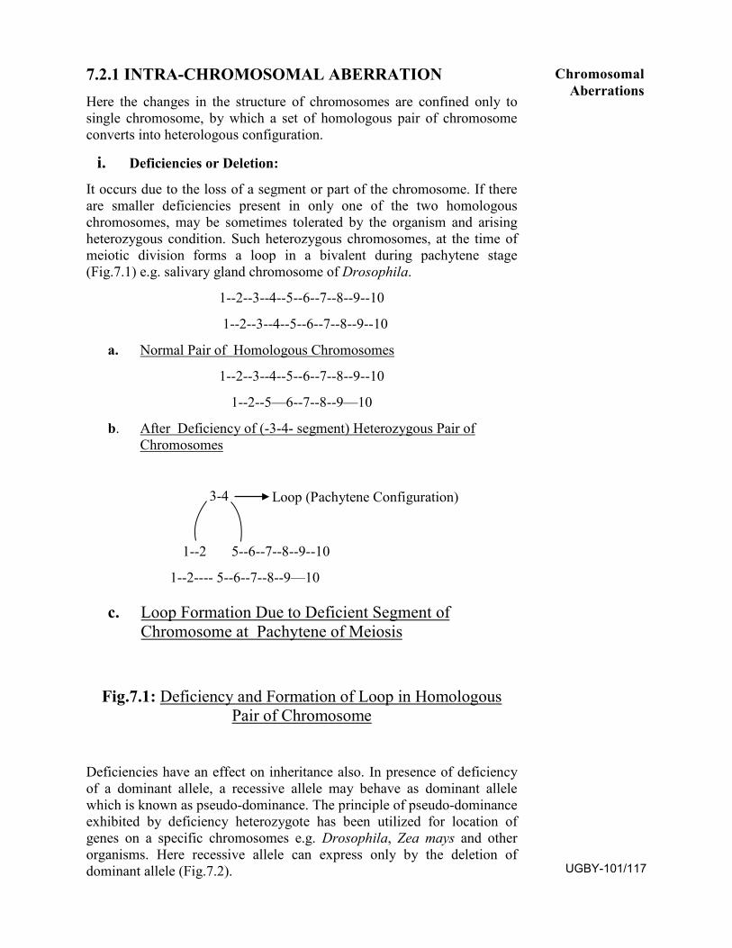
7.2.1 INTRA-CHROMOSOMAL ABERRATION
Here the changes in the structure of chromosomes are confined only to single chromosome, by which a set of homologous pair of chromosome converts into heterologous configuration.
i. Deficiencies or Deletion:
It occurs due to the loss of a segment or part of the chromosome. If there are smaller deficiencies present in only one of the two homologous chromosomes, may be sometimes tolerated by the organism and arising heterozygous condition. Such heterozygous chromosomes, at the time of meiotic division forms a loop in a bivalent during pachytene stage (Fig.7.1) e.g. salivary gland chromosome of Drosophila.
1--2--3--4--5--6--7--8--9--10
1--2--3--4--5--6--7--8--9--10
a. Normal Pair of Homologous Chromosomes
1--2--3--4--5--6--7--8--9--10
1--2--5—6--7--8--9—10
b. After Deficiency of (-3-4- segment) Heterozygous Pair ofChromosomes
3-4 Loop (Pachytene Configuration)
1--2 5--6--7--8--9--10
1--2---- 5--6--7--8--9—10
c. Loop Formation Due to Deficient Segment ofChromosome at Pachytene of Meiosis
Fig.7.1: Deficiency and Formation of Loop in Homologous Pair of Chromosome
Deficiencies have an effect on inheritance also. In presence of deficiency of a dominant allele, a recessive allele may behave as dominant allele which is known as pseudo-dominance. The principle of pseudo-dominance exhibited by deficiency heterozygote has been utilized for location of genes on a specific chromosomes e.g. Drosophila, Zea mays and other organisms. Here recessive allele can express only by the deletion of dominant allele (Fig.7.2).
Chromosomal Aberrations
UGBY-101/117
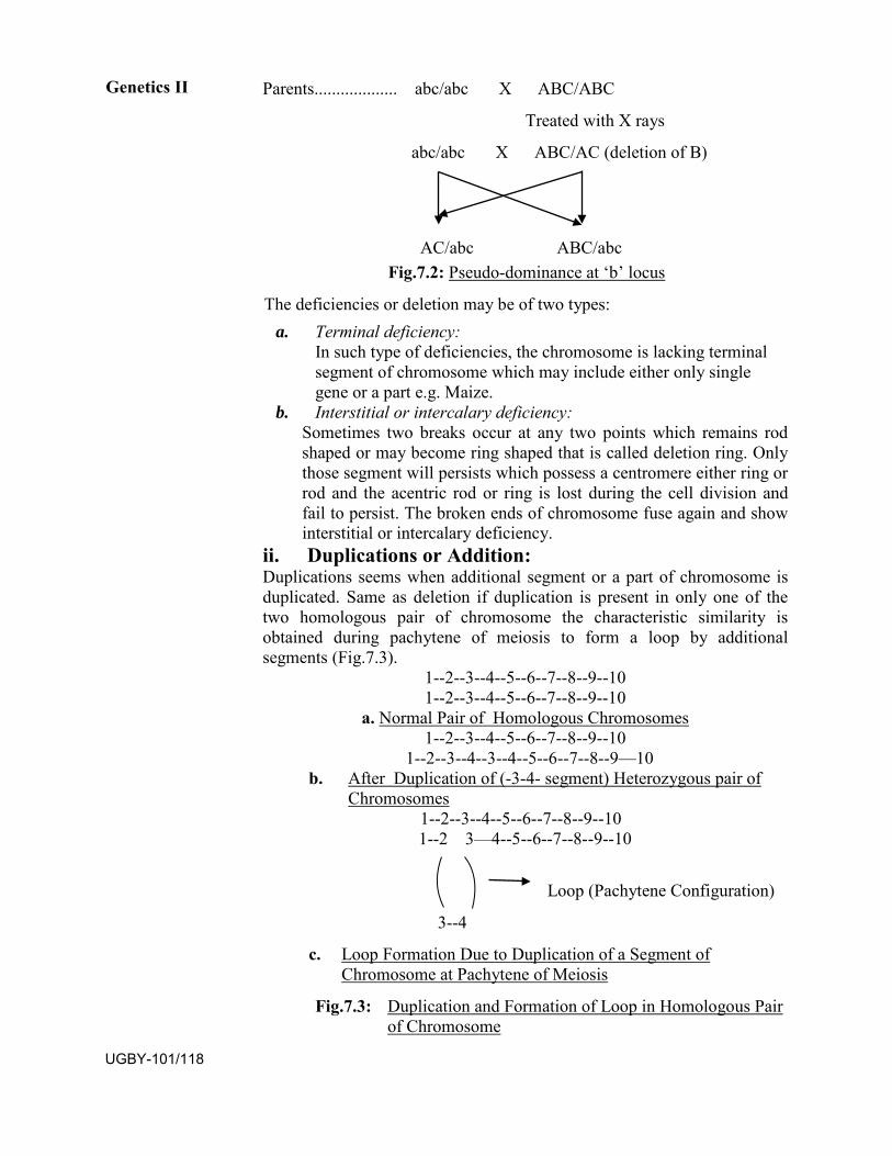
Parents................... abc/abc X ABC/ABC
Treated with X rays
abc/abc X ABC/AC (deletion of B)
AC/abc ABC/abc Fig.7.2: Pseudo-dominance at ‘b’ locus
The deficiencies or deletion may be of two types: a. Terminal deficiency:
In such type of deficiencies, the chromosome is lacking terminalsegment of chromosome which may include either only singlegene or a part e.g. Maize.
b. Interstitial or intercalary deficiency:Sometimes two breaks occur at any two points which remains rod shaped or may become ring shaped that is called deletion ring. Only those segment will persists which possess a centromere either ring or rod and the acentric rod or ring is lost during the cell division and fail to persist. The broken ends of chromosome fuse again and show interstitial or intercalary deficiency.
ii. Duplications or Addition:Duplications seems when additional segment or a part of chromosome is duplicated. Same as deletion if duplication is present in only one of the two homologous pair of chromosome the characteristic similarity is obtained during pachytene of meiosis to form a loop by additional segments (Fig.7.3).
1--2--3--4--5--6--7--8--9--10 1--2--3--4--5--6--7--8--9--10
a. Normal Pair of Homologous Chromosomes1--2--3--4--5--6--7--8--9--10
1--2--3--4--3--4--5--6--7--8--9—10 b. After Duplication of (-3-4- segment) Heterozygous pair of
Chromosomes1--2--3--4--5--6--7--8--9--10 1--2 3—4--5--6--7--8--9--10
Loop (Pachytene Configuration)
3--4
c. Loop Formation Due to Duplication of a Segment ofChromosome at Pachytene of Meiosis
Fig.7.3: Duplication and Formation of Loop in Homologous Pair of Chromosome
Genetics II
UGBY-101/118
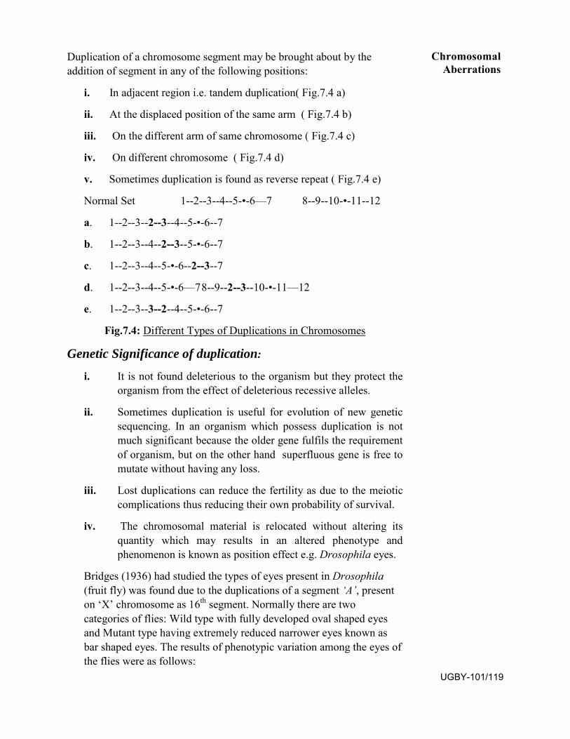
Duplication of a chromosome segment may be brought about by the addition of segment in any of the following positions:
i. In adjacent region i.e. tandem duplication( Fig.7.4 a)
ii. At the displaced position of the same arm ( Fig.7.4 b)
iii. On the different arm of same chromosome ( Fig.7.4 c)
iv. On different chromosome ( Fig.7.4 d)
v. Sometimes duplication is found as reverse repeat ( Fig.7.4 e)
Normal Set 1--2--3--4--5-•-6—7 8--9--10-•-11--12
a. 1--2--3--2--3--4--5-•-6--7
b. 1--2--3--4--2--3--5-•-6--7
c. 1--2--3--4--5-•-6--2--3--7
d. 1--2--3--4--5-•-6—7 8--9--2--3--10-•-11—12
e. 1--2--3--3--2--4--5-•-6--7
Fig.7.4: Different Types of Duplications in Chromosomes
Genetic Significance of duplication:
i. It is not found deleterious to the organism but they protect theorganism from the effect of deleterious recessive alleles.
ii. Sometimes duplication is useful for evolution of new geneticsequencing. In an organism which possess duplication is notmuch significant because the older gene fulfils the requirementof organism, but on the other hand superfluous gene is free tomutate without having any loss.
iii. Lost duplications can reduce the fertility as due to the meioticcomplications thus reducing their own probability of survival.
iv. The chromosomal material is relocated without altering itsquantity which may results in an altered phenotype andphenomenon is known as position effect e.g. Drosophila eyes.
Bridges (1936) had studied the types of eyes present in Drosophila (fruit fly) was found due to the duplications of a segment ‘A’, present on ‘X’ chromosome as 16th segment. Normally there are two categories of flies: Wild type with fully developed oval shaped eyes and Mutant type having extremely reduced narrower eyes known as bar shaped eyes. The results of phenotypic variation among the eyes of the flies were as follows:
Chromosomal Aberrations
UGBY-101/119
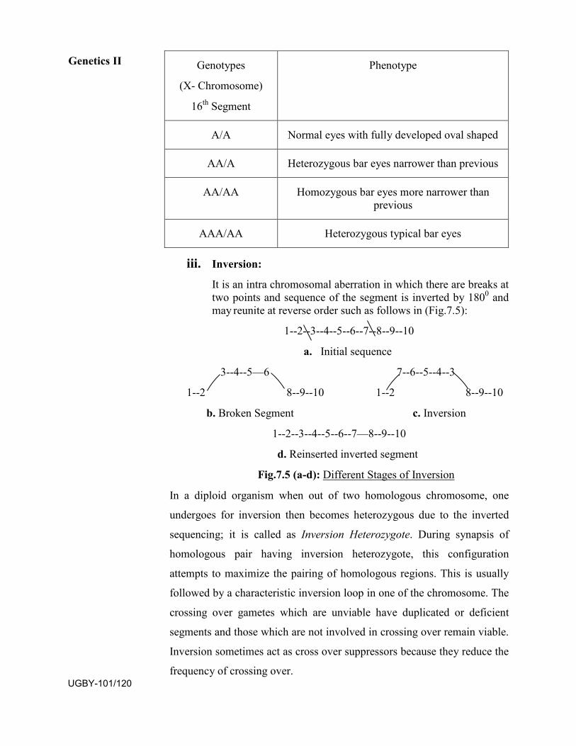
Genotypes
(X- Chromosome)
16th Segment
Phenotype
A/A Normal eyes with fully developed oval shaped
AA/A Heterozygous bar eyes narrower than previous
AA/AA Homozygous bar eyes more narrower than previous
AAA/AA Heterozygous typical bar eyes
iii. Inversion:
It is an intra chromosomal aberration in which there are breaks attwo points and sequence of the segment is inverted by 1800 andmay reunite at reverse order such as follows in (Fig.7.5):
1--2--3--4--5--6--7--8--9--10
a. Initial sequence
3--4--5—6 7--6--5--4--3
1--2 8--9--10 1--2 8--9--10
b. Broken Segment c. Inversion
1--2--3--4--5--6--7—8--9--10
d. Reinserted inverted segment
Fig.7.5 (a-d): Different Stages of Inversion
In a diploid organism when out of two homologous chromosome, one
undergoes for inversion then becomes heterozygous due to the inverted
sequencing; it is called as Inversion Heterozygote. During synapsis of
homologous pair having inversion heterozygote, this configuration
attempts to maximize the pairing of homologous regions. This is usually
followed by a characteristic inversion loop in one of the chromosome. The
crossing over gametes which are unviable have duplicated or deficient
segments and those which are not involved in crossing over remain viable.
Inversion sometimes act as cross over suppressors because they reduce the
frequency of crossing over.
Genetics II
UGBY-101/120
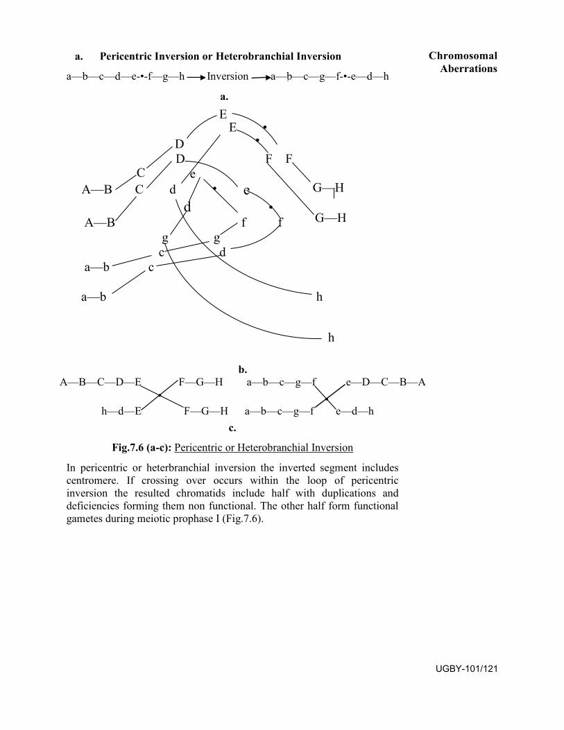
a. Pericentric Inversion or Heterobranchial Inversion
a—b—c—d—e-•-f—g—h Inversion a—b—c—g—f-•-e—d—h
a.
b. A—B—C—D—E F—G—H a—b—c—g—f e—D—C—B—A
• h—d—E F—G—H a—b—c—g—f e—d—h
c.
Fig.7.6 (a-c): Pericentric or Heterobranchial Inversion
In pericentric or heterbranchial inversion the inverted segment includes centromere. If crossing over occurs within the loop of pericentric inversion the resulted chromatids include half with duplications and deficiencies forming them non functional. The other half form functional gametes during meiotic prophase I (Fig.7.6).
Chromosomal Aberrations
UGBY-101/121
E E •
D • D F F
e C A—B C d • e G—H
d • A—B f f G—H
g g c d
a—b c
a—b h
h
•
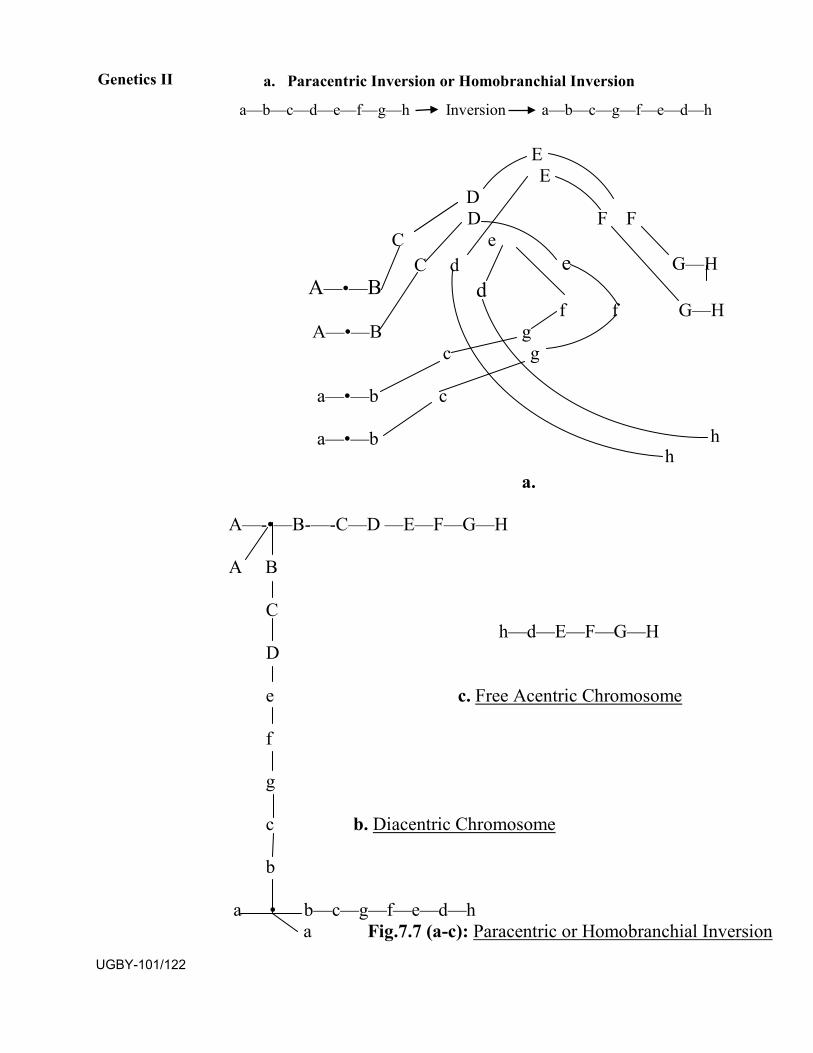
a. Paracentric Inversion or Homobranchial Inversion
a—b—c—d—e—f—g—h Inversion a—b—c—g—f—e—d—h
Genetics II
E E
D D F F
C e C d e G—H
A—•—B d f f G—H
A—•—B g c g
a—•—b c
a—•—b h h
a.
A—-•—B-—-C—D —E—F—G—H
A B
h—d—E—F—G—H C
D
c. Free Acentric Chromosome e
f
g
c b. Diacentric Chromosome
b
a • b—c—g—f—e—d—h a Fig.7.7 (a-c): Paracentric or Homobranchial Inversion
UGBY-101/122
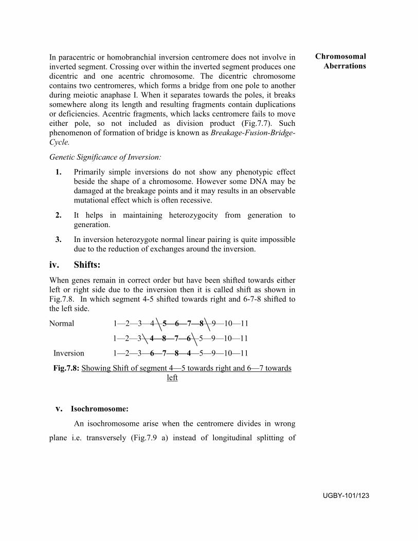
In paracentric or homobranchial inversion centromere does not involve in inverted segment. Crossing over within the inverted segment produces one dicentric and one acentric chromosome. The dicentric chromosome contains two centromeres, which forms a bridge from one pole to another during meiotic anaphase I. When it separates towards the poles, it breaks somewhere along its length and resulting fragments contain duplications or deficiencies. Acentric fragments, which lacks centromere fails to move either pole, so not included as division product (Fig.7.7). Such phenomenon of formation of bridge is known as Breakage-Fusion-Bridge-Cycle.
Genetic Significance of Inversion:
1. Primarily simple inversions do not show any phenotypic effectbeside the shape of a chromosome. However some DNA may bedamaged at the breakage points and it may results in an observablemutational effect which is often recessive.
2. It helps in maintaining heterozygocity from generation togeneration.
3. In inversion heterozygote normal linear pairing is quite impossibledue to the reduction of exchanges around the inversion.
iv. Shifts:When genes remain in correct order but have been shifted towards either left or right side due to the inversion then it is called shift as shown in Fig.7.8. In which segment 4-5 shifted towards right and 6-7-8 shifted to the left side.
Normal 1—2—3—4—5—6—7—8—9—10—11
1—2—3—4—8—7—6—5—9—10—11
Inversion 1—2—3—6—7—8—4—5—9—10—11
Fig.7.8: Showing Shift of segment 4—5 towards right and 6—7 towards left
v. Isochromosome:
An isochromosome arise when the centromere divides in wrong
plane i.e. transversely (Fig.7.9 a) instead of longitudinal splitting of
Chromosomal Aberrations
UGBY-101/123
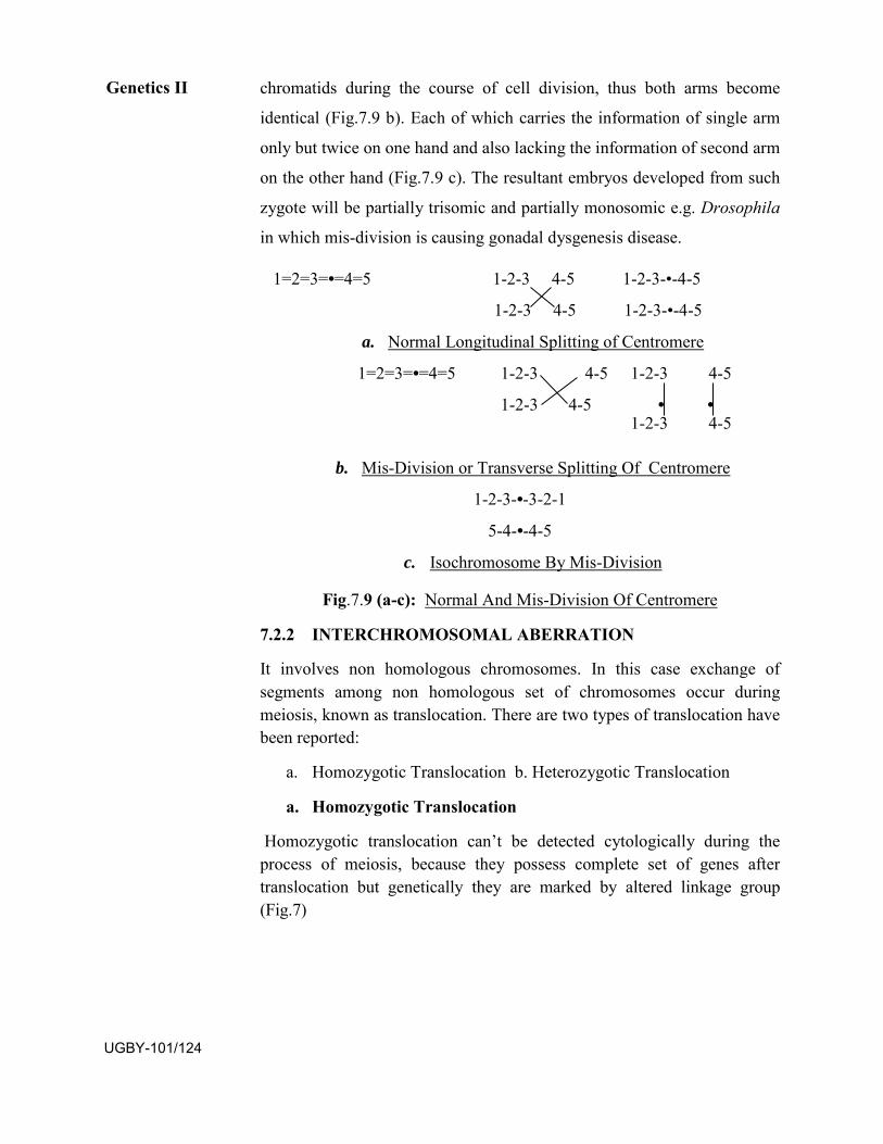
chromatids during the course of cell division, thus both arms become
identical (Fig.7.9 b). Each of which carries the information of single arm
only but twice on one hand and also lacking the information of second arm
on the other hand (Fig.7.9 c). The resultant embryos developed from such
zygote will be partially trisomic and partially monosomic e.g. Drosophila
in which mis-division is causing gonadal dysgenesis disease.
1=2=3=•=4=5 1-2-3 4-5 1-2-3-•-4-5
1-2-3 4-5 1-2-3-•-4-5
a. Normal Longitudinal Splitting of Centromere
1=2=3=•=4=5 1-2-3 4-5 1-2-3 4-5
1-2-3 4-5 • • 1-2-3 4-5
b. Mis-Division or Transverse Splitting Of Centromere
1-2-3-•-3-2-1
5-4-•-4-5
c. Isochromosome By Mis-Division
Fig.7.9 (a-c): Normal And Mis-Division Of Centromere
7.2.2 INTERCHROMOSOMAL ABERRATION
It involves non homologous chromosomes. In this case exchange of segments among non homologous set of chromosomes occur during meiosis, known as translocation. There are two types of translocation have been reported:
a. Homozygotic Translocation b. Heterozygotic Translocation
a. Homozygotic Translocation
Homozygotic translocation can’t be detected cytologically during the process of meiosis, because they possess complete set of genes after translocation but genetically they are marked by altered linkage group (Fig.7)
Genetics II
UGBY-101/124
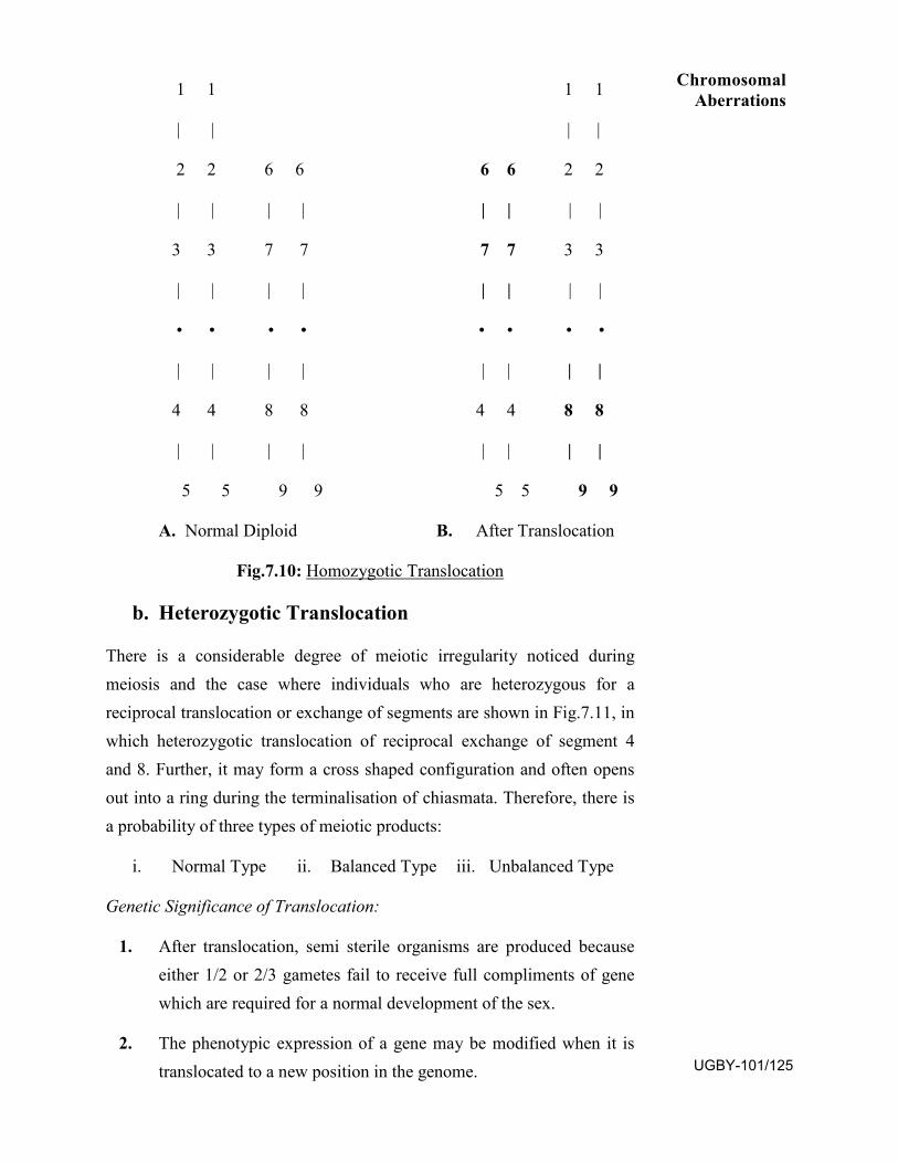
1 1 1 1
| | | |
2 2 6 6 6 6 2 2
| | | | | | | |
3 3 7 7 7 7 3 3
| | | | | | | |
• • • • • • • •
| | | | | | | |
4 4 8 8 4 4 8 8
| | | | | | | |
5 5 9 9 5 5 9 9
A. Normal Diploid B. After Translocation
Fig.7.10: Homozygotic Translocation
b. Heterozygotic Translocation
There is a considerable degree of meiotic irregularity noticed during meiosis and the case where individuals who are heterozygous for a reciprocal translocation or exchange of segments are shown in Fig.7.11, in which heterozygotic translocation of reciprocal exchange of segment 4 and 8. Further, it may form a cross shaped configuration and often opens out into a ring during the terminalisation of chiasmata. Therefore, there is a probability of three types of meiotic products:
i. Normal Type ii. Balanced Type iii. Unbalanced Type
Genetic Significance of Translocation:
1. After translocation, semi sterile organisms are produced becauseeither 1/2 or 2/3 gametes fail to receive full compliments of genewhich are required for a normal development of the sex.
2. The phenotypic expression of a gene may be modified when it istranslocated to a new position in the genome.
Chromosomal Aberrations
UGBY-101/125
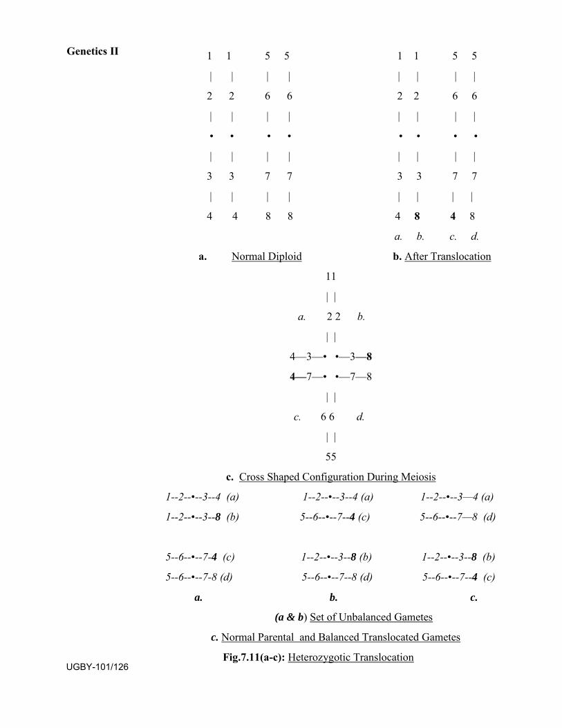
1 1 5 5 1 1 5 5
| | | | | | | |
2 2 6 6 2 2 6 6
| | | | | | | |
• • • • • • • •
| | | | | | | |
3 3 7 7 3 3 7 7
| | | | | | | |
4 4 8 8 4 8 4 8
a. b. c. d.
a. Normal Diploid b. After Translocation
11
| |
a. 2 2 b.
| |
4—3—• •—3—8
4—7—• •—7—8
| |
c. 6 6 d.
| |
55
c. Cross Shaped Configuration During Meiosis
1--2--•--3--4 (a) 1--2--•--3--4 (a) 1--2--•--3—4 (a)
1--2--•--3--8 (b) 5--6--•--7--4 (c) 5--6--•--7—8 (d)
5--6--•--7-4 (c) 1--2--•--3--8 (b) 1--2--•--3--8 (b)
5--6--•--7-8 (d) 5--6--•--7--8 (d) 5--6--•--7--4 (c)
a. b. c.
(a & b) Set of Unbalanced Gametes
c. Normal Parental and Balanced Translocated Gametes
Fig.7.11(a-c): Heterozygotic Translocation
Genetics II
UGBY-101/126
SAQ.1:
a. Any type of changes in the structure of chromosome is calledchromosomal ...............
b. If the change in the structure of chromosomes confined to onlysingle chromosome is ..........aberration.
c. In case of deficiency of dominant allele, a recessive allele behavesas dominant allele is known as .............
d. Bridges had studied the types of eyes present in Drosophila wasfound due to duplications of segment ‘A’ present on......chromosome as.......segment.
e. ..............inversion the inverted segment includes centomere.
7.3 NUMERICAL CHANGES IN CHROMOSOMES
Each organism is characterised by having a particular set of chromosome or chromosomal compliment or a set of genome. If it is represented only once, then it is called haploid number found in germ cells and if twice then it is called diploid as in somatic cells. Such set of genomes having a specific fixed number or set of chromosomes are present in any organism.
Sometimes, due to some irregularities during mitosis, meiosis and fertilization cells with variant chromosome numbers are produced, such variation in chromosome number is known as ploidy. Different types of ploidy may occur either due to duplication or loss of complete set of chromosome or by single chromosome, which are as follows:
1. Euploidy
2. Aneuploidy
7.3.1 EUPLOIDY Euploid organisms have balanced set of genomes i.e. number of chromosomes in their cells. Usually higher organisms bear diploid number i.e. 2n in their somatic cells and haploid number i.e. n in their gametic or germinal cells after meiosis or reduction division. The lower organisms have single set of chromosome and during sexual reproduction they become 2n after fusion of gametes than it undergoes reduction division to develop haploid individuals. There are following types of euploidy:
i. Monoploidy
ii. Diploidy
iii. Polyploidy
i. Monoploidy
Monoploids have single genome. Most of the microorganisms like bacteria, viruses etc. and lower plants like algae, fungi and bryophytes
Chromosomal Aberrations
UGBY-101/127
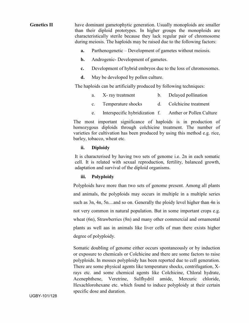
have dominant gametophytic generation. Usually monoploids are smaller than their diploid prototypes. In higher groups the monoploids are characteristically sterile because they lack regular pair of chromosome during meiosis. The haploids may be raised due to the following factors:
a. Parthenogenetic – Development of gametes without meiosis.
b. Androgenic- Development of gametes.
c. Development of hybrid embryos due to the loss of chromosomes.
d. May be developed by pollen culture.
The haploids can be artificially produced by following techniques:
a. X- ray treatment b. Delayed pollination
c. Temperature shocks d. Colchicine treatment
e. Interspecific hybridization f. Anther or Pollen Culture
The most important significance of haploids is in production of homozygous diploids through colchicine treatment. The number of varieties for cultivation has been produced by using this method e.g. rice, barley, tobacco, wheat etc.
ii. Diploidy
It is characterised by having two sets of genome i.e. 2n in each somatic cell. It is related with sexual reproduction, fertility, balanced growth, adaptation and survival of the diploid organisms.
iii. Polyploidy
Polyploids have more than two sets of genome present. Among all plants
and animals, the polyploids may occurs in multiple in a multiple series
such as 3n, 4n, 5n....and so on. Generally the ploidy level higher than 4n is
not very common in natural population. But in some important crops e.g.
wheat (6n), Strawberries (8n) and many other commercial and ornamental
plants as well aas in animals like liver cells of man there exists higher
degree of polyploidy.
Somatic doubling of genome either occurs spontaneously or by induction or exposure to chemicals or Colchicine and there are some factors to raise polyploids. In mosses polyploidy has been reported due to cell generation. There are some physical agents like temperature shocks, centrifugation, X-rays etc. and some chemical agents like Colchicine, Chloral hydrate, Acenephthene, Veretrine, Sulfhydril amide, Mercuric chloride, Hexachlorohexane etc. which found to induce polyploidy at their certain specific dose and duration.
Genetics II
UGBY-101/128
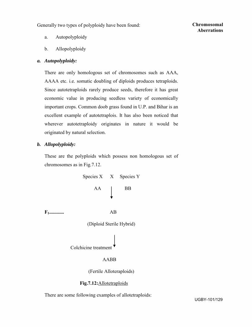
Generally two types of polyploidy have been found:
a. Autopolyploidy
b. Allopolyploidy
a. Autopolyploidy:
There are only homologous set of chromosomes such as AAA,
AAAA etc. i.e. somatic doubling of diploids produces tetraploids.
Since autotetraploids rarely produce seeds, therefore it has great
economic value in producing seedless variety of economically
important crops. Common doob grass found in U.P. and Bihar is an
excellent example of autotetraplois. It has also been noticed that
wherever autotetraploidy originates in nature it would be
originated by natural selection.
b. Allopolyploidy:
These are the polyploids which possess non homologous set of
chromosomes as in Fig.7.12.
Species X X Species Y
AA BB
F1............ AB
(Diploid Sterile Hybrid)
Colchicine treatment
AABB
(Fertile Alloteraploids)
Fig.7.12:Allotetraploids
There are some following examples of allotetraploids:
Chromosomal Aberrations
UGBY-101/129
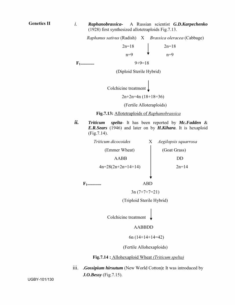
i. Raphanobrassica- A Russian scientist G.D.Karpechenko(1928) first synthesized allotetraploids Fig.7.13.
Raphanus sativus (Radish) X Brassica oleracea (Cabbage)
2n=18 2n=18
n=9 n=9
F1............ 9+9=18
(Diploid Sterile Hybrid)
Colchicine treatment
2n+2n=4n (18+18=36)
(Fertile Alloteraploids)
Fig.7.13: Allotetraploids of Raphanobrassica
ii. Triticum spelta- It has been reported by Mc.Fadden &E.R.Sears (1946) and later on by H.Kihara. It is hexaploid(Fig.7.14).
Triticum dicocoides X Aegilopsis squarrosa
(Emmer Wheat) (Goat Grass)
AABB DD
4n=28(2n+2n=14+14) 2n=14
F1............ ABD
3n (7+7+7=21)
(Triploid Sterile Hybrid)
Colchicine treatment
AABBDD
6n (14+14+14=42)
(Fertile Allohexaploids)
Fig.7.14 : Allohexaploid Wheat (Triticum spelta)
iii. .Gossipium hirsutum (New World Cotton): It was introduced byJ.O.Bessy (Fig.7.15).
Genetics II
UGBY-101/130
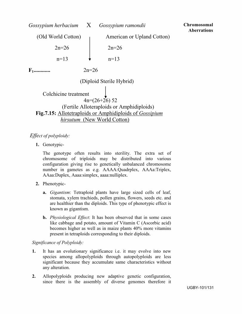
Gossypium herbacium X Gossypium ramondii
(Old World Cotton) American or Upland Cotton)
2n=26 2n=26
n=13 n=13
F1............ 2n=26
(Diploid Sterile Hybrid)
Colchicine treatment 4n=(26+26) 52
(Fertile Alloteraploids or Amphidiploids) Fig.7.15: Allotetraploids or Amphidiploids of Gossipium
hirsutum (New World Cotton)
Effect of polyploidy:
1. Genotypic-
The genotype often results into sterility. The extra set ofchromosome of triploids may be distributed into variousconfiguration giving rise to genetically unbalanced chromosomenumber in gametes as e.g. AAAA:Quadrplex, AAAa:Triplex,AAaa:Duplex, Aaaa:simplex, aaaa:nulliplex.
2. Phenotypic-
a. Gigantism: Tetraploid plants have large sized cells of leaf,stomata, xylem trachieds, pollen grains, flowers, seeds etc. and are healthier than the diploids. This type of phenotypic effect is known as gigantism.
b. Physiological Effect: It has been observed that in some caseslike cabbage and potato, amount of Vitamin C (Ascorbic acid)becomes higher as well as in maize plants 40% more vitaminspresent in tetraploids corresponding to their diploids.
Significance of Polyploidy:
1. It has an evolutionary significance i.e. it may evolve into newspecies among allopolyploids through autopolyploids are lesssignificant because they accumulate same characteristics withoutany alteration.
2. Allopolyploids producing new adaptive genetic configuration,since there is the assembly of diverse genomes therefore it
Chromosomal Aberrations
UGBY-101/131
provides adaptability to the species for wide variety of habitats which strengthen the chances of natural selection.
3. Polyploidy causes the accumulation of storage food materials likevitamins etc. and there is a quantitative and qualitativeimprovement in some economically important crops.
SAQ.2: a. Monoploids have ....... set of genomes.
b. Raphanobrassica is an example of ..............
c. A polyploid which posses homologous set of chromosomes isknown as ...........
7.3.2 ANEUPLOIDY When there is one or more chromosomes either lost or gained then it is called aneuploidy unlike euploidy where complete set of chromosome is altered. There are two types of aneuploidy.
i. Hypoploidy
i. Hperploidy
i. Hypoploidy:
When there is loss of one or more chromosomes form a homologous pair is then it is known as hypoploidy. Two types of hypoploidy have been seen. a. Monosomy b. Nullisomy
a. MonosomyIn monosomy, there is loss of one chromosome hence it becomes2n-1. Thus, this configuration produces two types of gametes i.e.50% with n and 50% have n-1 chromosomal complements. Itshows that 50% with n or haploid gametes behave normally due tofull complements but the gametes having n-1 remains nonfunctional due the lack of one chromosome in case of plants.Furthermore, animals having n-1 complement results into geneticunbalance that is manifested by the reduced fertility, since inmonosomics there is lack of one complete chromosome and it can’tbe tolerated in diploids. Whereas in polyploids there are severalsets of chromosome of same type, therefore this loss can betolerated easily. The number of possible monosomics in anyorganism is equal to their haploid number of chromosome e.g. E.R.Sears recorded in wheat that there are 21 pairs of chromosomesproducing 21 types of monosomics. Recently, monosomy isreported in other diploid plants like tobacco, maize etc.
Sometimes there is loss of two different chromosomes i.e. 2n-1-1this is called Double Monosomics. If three different chromosomesare lost i.e. 2n-1-1-1 then it is known as Triple Monosomics.
Genetics II
UGBY-101/132
b. NullisomyNullisomic are lacking a complete set of homologous chromosomei.e. 2n-2. They exhibit reduce figure of fertility and survival butpolyploidic nullisomics may survive upto maturity.
ii. Hyperploidy
Hyperploids raised by having one or more accessory or additionalextra chromosomes along with the normal set of chromosomes.There are following types of hyerploidy:
a. Trisomy
b. Tetrasomy
c. Double Trisomy
a. TrisomyIn a diploid organism when there is an extra chromosome it is calledtrisomy i.e. 2n+1. Therefore at the time of gamete formation twotypes of gametes may develop, at the time of division duringanaphase of meiosis the polarisation and separation of two differentsets on both poles i.e. one with normal set i.e. with n number andanother with n+1. There are various examples existing in trisomywith different specific phenotypic traits, some of them are listedbelow:
1. In man trisomy of 21st autosome causes Mongolism.
2. Among plants, first case of trisomy reported in Datura stramoniumby Blackslee & Belling(1924). In this case 2n+1= 24+1,pheotypically effects on the characteristic shape and spines of thefruits, seeds and capsule of the plants. Further they succeeded inachieving to produce all possible trisomics which werephenotypically distinguished from each other.
b. TetrasomyWhen one set of extra chromosome in diploids is present asquadruplicate i.e. 4 instead of 2, the resultant is called tetrasomy i.e.having 2n+2. Thus during meiosis the segregation of chromosomesare as it is that one with normal and another with extra set behaves asautotetraploids with full compliments.
c. Double TrisomyIn any diploid organism when two extra chromosomes of differentset are present is known as double trisomy i.e. 2n+1+1.
SAQ.3: a. The loss or gain of one or more chromosome along with normal
setof chromosome is called........
Chromosomal Aberrations
UGBY-101/133
b. An organism possessing 2n-1 is called ........
c. In man trisomy of 21st chromosome causes.........
7.4 SUMMARY
Any type of changes in chromosomes which appear phenotypically due to structure is known as chromosomal aberration or chromosomal mutations. The changes in structure of chromosomes was first analysed by H.J.Muller (1928) in Drosophila and Barbara Mc Clintock (1930) in Zea mays.
The changes in chromosomes may be studied into two parts: Structure of chromosomes and number of chromosomes
STRUCTURAL CHANGES IN CHROMOSOMES The structural changes occurred in chromosomes which alter the position of gene or a segment of chromosome. The structural changes in chromosomes can be of following types:
Intra-Chromosomal Aberration There are several types of intra-chromosomal aberrations which are as follows:
Deficiencies or Deletion: It occurs due to the loss of a segment or part of the chromosome. In presence of deficiency of a dominant allele, a recessive allele may behave as dominant allele known as pseudo-dominance. The deficiencies or deletion may be of three types:
a. Terminal deficiency:
b. Interstitial or intercalary deficiency
Duplications or Addition: Duplications seems when additional segment or a part of chromosome is duplicated. Duplication of a chromosome segment may be brought about by the addition of segment in any of the following positions:
i. In adjacent region i.e. tandem duplication.
ii. At the displaced position of the same arm
iii. On the different arm of same chromosome
iv. On different chromosome
v. Sometimes duplication is found as reverse repeat
Inversions: It is an intra chromosomal aberration in which there are breaks at two points and sequence of the segment inverted up to 1800 and may reunite at reverse order. The inversions can be of two types:
Genetics II
UGBY-101/134
a. Pericentric Inversion or Heterobranchial InversionIn pericentricor heterobranchial inversion the inverted segment includescentromere.
b. Paracentric Inversion or Homobranchial Inversion
In paracentric or homobranchial inversion centromere does notinvolve in inverted segment. Crossing over within the invertedsegment produces one dicentric and one acentric chromosome.
Shifts: When genes remain in correct order but have been shifted towards either left or right side due to the inversion is called shift.
Iso-chromosome: An isochromosome arise when the centromere divides in wrong plane i.e. transversely instead of longitudinal splitting of chromatids during the course of cell division, thus both arms become identical. Each of which carries the information of single arm only but twice on one hand and also lacking the information of second arm on the other hand
Inter-Chromosomal Aberration It involves non homologous chromosomes. In this case exchange of segments among non homologous set of chromosomes occur during meiosis, known as translocation. There are two types of translocation have been reported:
Homozygotic Translocation: It can’t be detected cytologically during the process of meiosis, because they possess complete set of genes after translocation but genetically they are marked by altered linkage group.
Heterozygotic Translocation: There is a considerable degree of meiotic irregularity noticed during meiosis and individuals who are heterozygous for a reciprocal translocation or exchange of segments.
There may have probability of three types of meiotic products:
a. Normal Type b. Balanced Type c. Unbalance
Numerical Changes in Chromosomes
Sometimes due to some irregularities have been noticed during mitosis, meiosis and fertilization and may produce cells with variant chromosome number, such variation in chromosome number is known as ploidy. There are different types of ploidy may occur due to either by duplication or loss of complete set of chromosome or by single chromosome which are as follows:
Euploidy Euploid organisms have balanced set of genomes i.e. number of chromosomes in their cells. There are following types of euploidy:
Chromosomal Aberrations
UGBY-101/135
Monoploidy: Monoploids have single genome. Most of the microorganisms like bacteria, viruses etc. and lower plants like algae, fungi and bryophytes have dominant gametophytic generation.
Diploidy: It is characterised by having two sets of genome i.e. 2n in each somatic cell.
Polyploidy : Polyploids have more than two sets of genome present. Among all plants and animals the polyploids may occurs in multiple in a multiple series such as 3n, 4n, 5n....and so on. Generally two types of polyploidy has been found: Autopolyploidy and Allopolyploidy
Autopolyploidy: There are only homologous set of homologous chromosomes such as AAA, AAAA etc. i.e. somatic doubling of diploids produces tetraploids. Allopolyploidy: These are the polyploids which possess non homologous set of chromosomes
Aneuploidy When there is one or more chromosomes either lost
or gained is called aneuploidy unlike euploidy where complete set of chromosome is altered. There are two types of aneuploidy.
Hypoploidy:
There is loss of one or more chromosomes are known as hypoploidy. Two types of hypoploidy have been seen.
a. Monosomy b. Nullisomy.
Monosomy : In monosomy there is loss of one chromosome hence it becomes 2n-1. Sometimes there is loss of two different chromosomes i.e. 2n-1-1 called Double Monosomics. If three different chromosomes lost i.e. 2n-1-1-1 then it is known as Triple Monosomics.
Nullisomy: Nullisomics are lacking a complete set of homologous chromosome i.e. 2n-2. They exhibit reduce figure of fertility and survival but polyploidic nullisomics may survive up to maturity.
Hyperploidy: Hyperploids raised by having one or more accessory or additional extra chromosomes along with the normal set of chromosomes. There are following types of hyerploidy: a. Trisomy b.Tetrasomy c. Double Trisomy Trisomy: In a diploid organism when there is an extra chromosome it is called trisomy i.e. 2n+1.
Tetrasomy: When one set of extra chromosome in diploids is present as quadruplicate i.e. 4 instead of 2, the resultant is called tetrasomy i.e. having 2n+2.
Double Trisomy: In any diploid organism when two extra chromosomes of different set are present is known as double trisomy i.e. 2n+1+1.
Genetics II
UGBY-101/136
7.5 TERMINAL QUESTIONS
OBJECTIVE TYPE QUESTIONS: Q.1. An inversion, when present in just one of the chromosomes, it
suppresses
a. Mutation c. Translocations
b. Duplications d. Cross over
Q.2. Which of the following chromosomal changes is causing major damage in homozygous condition?
a. Deletion c. Translocation
b. Duplication d. Inversion
Q.3. What is called the condition in which there is more than one complete set of chromosomes?
a. Aneuploidy c. Duplication
b. Polyploidy d. Addition
Q.4. Pseudo-dominance occurs when:
a. Both alleles are recessive
b. Both alleles are dominant
c. One dominant and one recessive allele
d. Only one recessive allele and dominant allele is deficient
Q.5. Monosomics are:
a. An extra set of Chromosome
b. An extra chromosome with normal set of chromosome
c. loss of one chromosome
d. loss of one set of chromosome
Q.6. Trisomics have:
a. 2n+1 c. 3n+1
b. 2n-1 d. 3n-1
Q.7. Allopolyploids are:
a. Polyploids of homologous set of chromosomes
b. Polyploids of non-homologous set of chromosomes
c. Both of these
d. None of these
Chromosomal Aberrations
UGBY-101/137
Q.8. Due to non disjunction, an extra chromosome is observed in an individual. This will be in addition to two homologous chromosomes already present is known as:
a. Monosomy c. Trisomy
b. Nullisomy d. Disomy
Q.9. If a cell of human body possess 92 chromosomes, the condition will be:
a. Haploid c. Triploid
b. Diploid d. Tetraploid
Q.10. If an organism has 2n= 42 chromosomes how many types of monosomics possible to generate?
a. 21 b. 42
c. 63 d. 84
SHORT ANSWER TYPE QUESTIONS: 1. What do you know about deletion or deficiency?
2. What is paracentric inversion?
3. Differentiate among the translocation heterozygote andtranslocation homozygote.
4. What is pseudodominance?
5. How does a dicenric bridge formed?
6. How will you distinguish cytologically between doublemonosomics and nullisomics?
7. Write short notes on trisomy and tetrasomy.
8. What is Raphanobrassica?
9. With the help of a suitable sketch demonstrate the types of gametesdeveloped in case of heterozygotic translocations.
10. What are amphidiploids, give an example?
LONG ANSWER TYPE QUESTIONS: 1. How many types of polyploids are known to you? Distinguish
autopolyploids and allopolyploids.
2. How many types of duplications are known and how are theygenetically significant?
3. What are Inversions? With the help of sketches explainhomobranchial and heterobranchial inversions.
Genetics II
UGBY-101/138
4. Define the types of Aneupoids and their impact on the organisms.
5. What are Euploids? Explain it by giving suitable examples andtheir significance.
7.6 ANSWER
OBJECTIVE TYPE: 1. d 2. a 3. b 4. d 5. c6. a 7. b 8. c 9. d 10. A
SAQ.1: a. Aberration or mutation b. Intra chromosomal
c. Pseudo dominance d. ‘X’, 16th
e. Pericentric or Heterobranchial
SAQ.2: a. Single b. Allotetraploidy c. Autopolyploid
SAQ.3: a. Aneuploidy b. Monosomics c. Mongolism
Chromosomal Aberrations
UGBY-101/139

UGBY-101/140
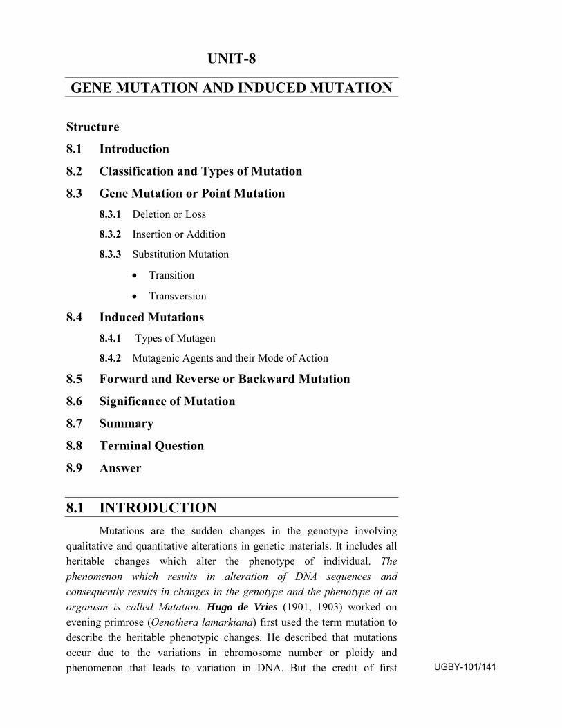
UNIT-8
GENE MUTATION AND INDUCED MUTATION
Structure
8.1 Introduction
8.2 Classification and Types of Mutation
8.3 Gene Mutation or Point Mutation 8.3.1 Deletion or Loss
8.3.2 Insertion or Addition
8.3.3 Substitution Mutation
• Transition
• Transversion
8.4 Induced Mutations 8.4.1 Types of Mutagen
8.4.2 Mutagenic Agents and their Mode of Action
8.5 Forward and Reverse or Backward Mutation
8.6 Significance of Mutation
8.7 Summary
8.8 Terminal Question
8.9 Answer
8.1 INTRODUCTION Mutations are the sudden changes in the genotype involving
qualitative and quantitative alterations in genetic materials. It includes all heritable changes which alter the phenotype of individual. The phenomenon which results in alteration of DNA sequences and consequently results in changes in the genotype and the phenotype of an organism is called Mutation. Hugo de Vries (1901, 1903) worked on evening primrose (Oenothera lamarkiana) first used the term mutation to describe the heritable phenotypic changes. He described that mutations occur due to the variations in chromosome number or ploidy and phenomenon that leads to variation in DNA. But the credit of first UGBY-101/141
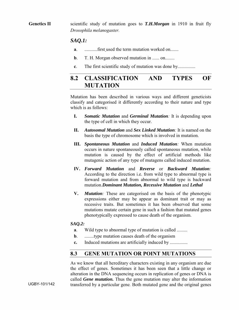
scientific study of mutation goes to T.H.Morgan in 1910 in fruit fly Drosophila melanogaster.
SAQ.1: a. ...........first used the term mutation worked on.......
b. T. H. Morgan observed mutation in ...... on........
c. The first scientific study of mutation was done by...............
8.2 CLASSIFICATION AND TYPES OF MUTATION
Mutation has been described in various ways and different geneticists classify and categorised it differently according to their nature and type which is as follows:
I. Somatic Mutation and Germinal Mutation: It is depending upon the type of cell in which they occur.
II. Autosomal Mutation and Sex Linked Mutation: It is named on thebasis the type of chromosome which is involved in mutation.
III. Spontaneous Mutation and Induced Mutation: When mutationoccurs in nature spontaneously called spontaneous mutation, whilemutation is caused by the effect of artificial methods likemutagenic action of any type of mutagens called induced mutation.
IV. Forward Mutation and Reverse or Backward Mutation:According to the direction i.e. from wild type to abnormal type isforward mutation and from abnormal to wild type is backwardmutation.Dominant Mutation, Recessive Mutation and Lethal
V. Mutation: These are categorised on the basis of the phenotypic expressions either may be appear as dominant trait or may as recessive traits. But sometimes it has been observed that some mutations mutate certain gene in such a fashion that mutated genes phenotypically expressed to cause death of the organism.
SAQ.2: a. Wild type to abnormal type of mutation is called .........b. ........type mutation causes death of the organismc. Induced mutations are artificially induced by ...............
8.3 GENE MUTATION OR POINT MUTATIONS
As we know that all hereditary characters existing in any organism are due the effect of genes. Sometimes it has been seen that a little change or alteration in the DNA sequencing occurs in replication of genes or DNA is called Gene mutation. Thus the gene mutation may alter the information transferred by a particular gene. Both mutated gene and the original genes
Genetics II
UGBY-101/142
are located at the fixed locus or point on a particular chromosome. The gene mutations include very small or limited segment of DNA, also termed as Point mutation because any change in sequencing of single base or nucleotide may raise a huge change in reading frame by the alteration of triplet genetic code. This type of mutation occurs at molecular level usually at the time of DNA replication when new DNA strands are synthesized. Hence this is also said to be Copy-error mutations which may be due to nucleotide changes in DNA and RNA. These mutations mostly include alteration in the sequences of nucleotide in the nucleic acids which forms genetic material. Mutations may be due to the either by deletion, insertion, inversion, replacement or substitution of bases. Thus the gene mutations are classified into following types:
1. Deletion or loss of bases 2. Insertion or Addition
3. Substitution
8.3.1 Deletion or Loss: There is change of sequence by the loss or deletion of single base which alter the genetic code and consequently series of amino acids may be changed (Fig.8.1). It is frequently known in T4 bacteriophage. Normal Sequence ......... AAG AGU CCC UCA CUU AAU........
Deletion of Base A ........ AAG GUC CCU CAC UUA AU_..........
Fig.8.1: Deletion or Loss of ‘A’ Base Showing Changed Triplet Gnetic Code
8.3.2 Insertion or Addition: Just as the loss of a single nucleotide changes the sequence here in case of addition or insertion of single base may alter the entire sequence of amino acids by triplets of codon (Fig.8.2). It has been reported that insertion of segment or base may be induced by certain chemicals that act like mutagen e.g. Acridine dye, Proflavin etc., which add or insert one or more nucleotide in the regular sequencing.
Normal Sequence .........AAG AGU CCC UCA CUU AAU........
Insertion of Base A ........AAG AGU CCC UUC ACU UAA U..........
Fig.8.2: Insertion or Addition of ‘U’ Base Showing Changed Triplet Gnetic Code
The deletions, insertions and inversions include those changes in base sequences which involve breakage and reunion of DNA fragments. Though it has been found that deletion or insertion of bases causes gross changes in the amino acid sequencing of proteins. Since they sometimes shift the entire reading frame of codons beginning at the site of mutation by equal addition and deletion of the base, it is known as Frame shift mutation (Fig.8.3).
Normal Sequence .........AAG CGU CCC UCA CUU AAU........
Gene Mutation and Induced
Mutation
UGBY-101/143
Mutant ....... AAG GUC CCU ACA CUU AAU.........
Fig.8.3: Frame Shift Mutation by Deletion of ‘C’ and Insertion of ‘A’ Base
8.3.3 Substitution Mutation:
The replacement of a base pair may take place during replication of DNA without any breakage of DNA. That is a Nitrogen base of triplet codon is replaced by another Nitrogen base or some derivatives of Nitrogen base. An alternative codon may designate different amino acid which may produce a protein molecule with single amino acid substitution. Such mutations have a great genetic significance. The base pair replacements or substitutions are following two types:
i. Transition ii. Transversion
i. Transition:
In transition there is a replacement of base pair occur in such a manner that purine is replaced by another purine base and pyrimidine is by other pyrimidine as follows:
A G
T C
The transitions are further divided into three categories:
a. Tautomerisation b. Deamination c. Base Analogue
a. Tautomerisation- According to Watson and Crick (1953) in DNAmolecule ‘A’ remains linked with ‘T’ by -2H- bonds and ‘G’ with‘C’ by -3H- bonds. However these bases can exist in alternativevalences in consistent states. The tautomeric shift occurs due to theshift of electrons or protons.
Fig.8.4: Tautomerisation
Due to tautomerisation Amino form of C & A is converted into Imino form i.e. –NH2 → -NH and Keto form of T & G is converted into Enol form C=O → C-O (Fig.8.4).
Genetics II
UGBY-101/144
A:T Tautomerisation G:C
T:A Reversal of Tautomers G:C A:C Normal Replication
A:C
A:T G:C A:T C:G
Mutant Mutant
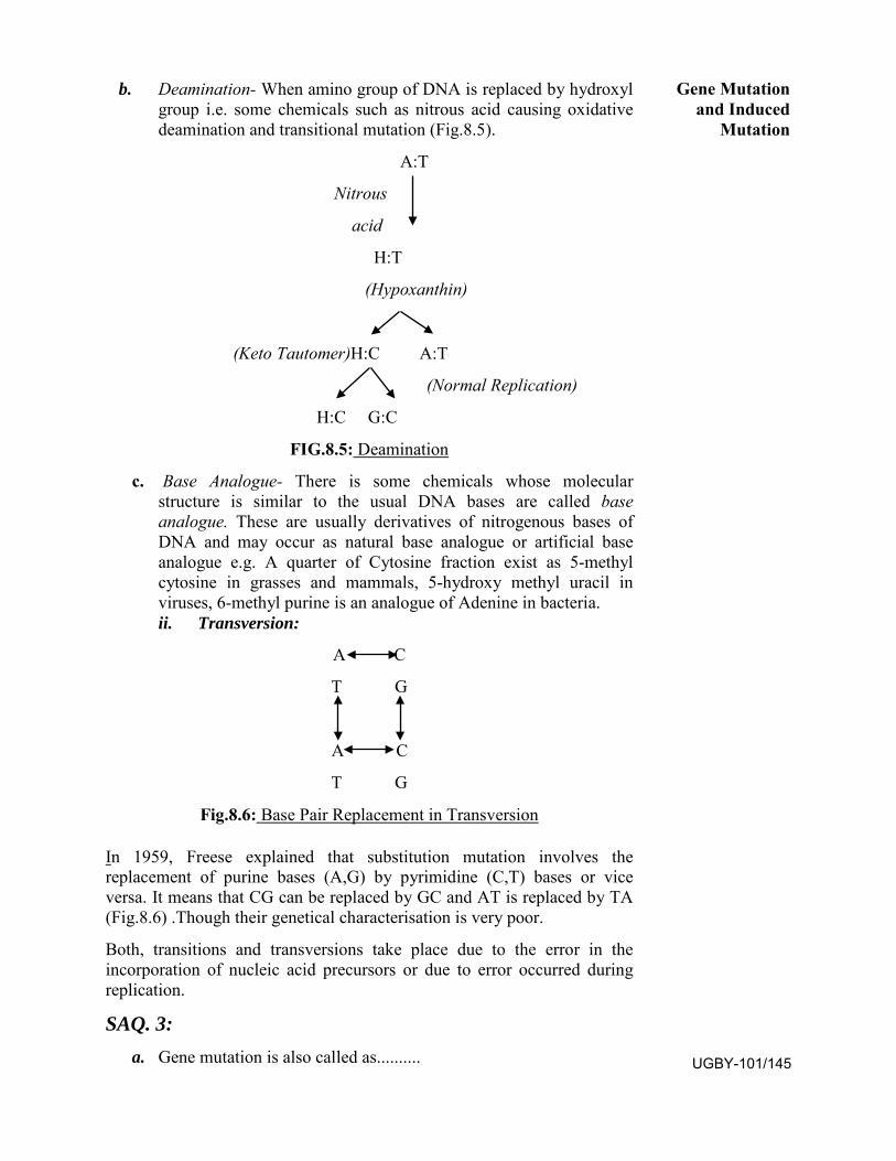
b. Deamination- When amino group of DNA is replaced by hydroxylgroup i.e. some chemicals such as nitrous acid causing oxidativedeamination and transitional mutation (Fig.8.5).
A:T
Nitrous
acid
H:T
(Hypoxanthin)
(Keto Tautomer)H:C A:T
(Normal Replication)
H:C G:C
FIG.8.5: Deamination
c. Base Analogue- There is some chemicals whose molecularstructure is similar to the usual DNA bases are called baseanalogue. These are usually derivatives of nitrogenous bases ofDNA and may occur as natural base analogue or artificial baseanalogue e.g. A quarter of Cytosine fraction exist as 5-methylcytosine in grasses and mammals, 5-hydroxy methyl uracil inviruses, 6-methyl purine is an analogue of Adenine in bacteria.ii. Transversion:
A C
T G
A C
T G
Fig.8.6: Base Pair Replacement in Transversion
In 1959, Freese explained that substitution mutation involves the replacement of purine bases (A,G) by pyrimidine (C,T) bases or vice versa. It means that CG can be replaced by GC and AT is replaced by TA (Fig.8.6) .Though their genetical characterisation is very poor.
Both, transitions and transversions take place due to the error in the incorporation of nucleic acid precursors or due to error occurred during replication.
SAQ. 3: a. Gene mutation is also called as..........
Gene Mutation and Induced
Mutation
UGBY-101/145
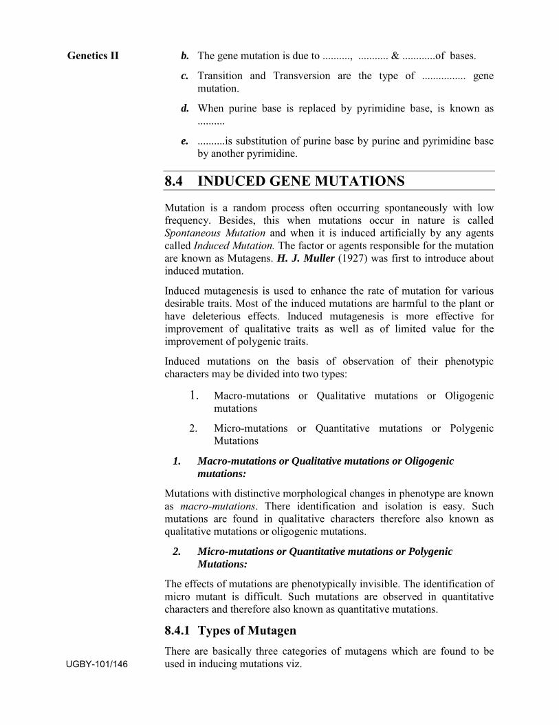
b. The gene mutation is due to .........., ........... & ............of bases.
c. Transition and Transversion are the type of ................ genemutation.
d. When purine base is replaced by pyrimidine base, is known as..........
e. ..........is substitution of purine base by purine and pyrimidine baseby another pyrimidine.
8.4 INDUCED GENE MUTATIONS
Mutation is a random process often occurring spontaneously with low frequency. Besides, this when mutations occur in nature is called Spontaneous Mutation and when it is induced artificially by any agents called Induced Mutation. The factor or agents responsible for the mutation are known as Mutagens. H. J. Muller (1927) was first to introduce about induced mutation.
Induced mutagenesis is used to enhance the rate of mutation for various desirable traits. Most of the induced mutations are harmful to the plant or have deleterious effects. Induced mutagenesis is more effective for improvement of qualitative traits as well as of limited value for the improvement of polygenic traits.
Induced mutations on the basis of observation of their phenotypic characters may be divided into two types:
1. Macro-mutations or Qualitative mutations or Oligogenicmutations
2. Micro-mutations or Quantitative mutations or PolygenicMutations
1. Macro-mutations or Qualitative mutations or Oligogenicmutations:
Mutations with distinctive morphological changes in phenotype are known as macro-mutations. There identification and isolation is easy. Such mutations are found in qualitative characters therefore also known as qualitative mutations or oligogenic mutations.
2. Micro-mutations or Quantitative mutations or PolygenicMutations:
The effects of mutations are phenotypically invisible. The identification of micro mutant is difficult. Such mutations are observed in quantitative characters and therefore also known as quantitative mutations.
8.4.1 Types of Mutagen There are basically three categories of mutagens which are found to be used in inducing mutations viz.
Genetics II
UGBY-101/146
i. Physical Mutagens ii. Chemical Mutagens iii. BiochemicalMutagens
i. Physical Mutagens:
Certain physical factors are designated as mutagen like- various types of radiations, temperature etc.
A. Radiation
Electromagnetic radiation, waves of short wave length e.g. Ultra violet rays X-rays, β-rays, γ-rays etc., charged and uncharged particles like; Electrons, Protons, Neutrons, Deuterons etc. are various sources of radiation causing mutation at specific dose treated for a particular duration.
a. Ionising radiation- α-, β- and γ- radiations of radioactivesubstances, X- rays, neutrons and protons induced mutation byionising the matter. Besides gene mutation, X-rays and other highenergy radiation also produce all types of chromosomalaberrations. χ- rays, γ- rays and other beams of some atomicparticles are ionising radiations. Ionising radiation ionises H2Omolecules with the electron ejected from water molecule (H2O)and then the molecules become unstable and may split into H+ andOH-.
b. Non-Ionising radiation- Non-ionising UV-rays also inducemutations but not to the extent as ionising radiations.
c. Natural Radiations- The natural radiations like cosmic rays, whichcome from outer space, are also responsible for spontaneousmutation in living beings.
B. Temperature
A rise in temperature may sometimes lead to disturbance in genes which cause mutation. It has been found that an increase of 3-100C in temperature may increases the rate of mutation approximately 2-3 times in different organisms. Temperature probably affects both thermal stability of DNA and rate of reaction of other substances with DNA.
Gene Mutation and Induced
Mutation
UGBY-101/147
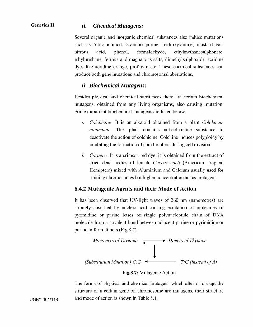
ii. Chemical Mutagens:
Several organic and inorganic chemical substances also induce mutations such as 5-bromouracil, 2-amino purine, hydroxylamine, mustard gas, nitrous acid, phenol, formaldehyde, ethylmethanesulphonate, ethylurethane, ferrous and magnanous salts, dimethylsulphoxide, acridine dyes like acridine orange, proflavin etc. These chemical substances can produce both gene mutations and chromosomal aberrations.
ii Biochemical Mutagens:
Besides physical and chemical substances there are certain biochemical mutagens, obtained from any living organisms, also causing mutation. Some important biochemical mutagens are listed below:
a. Colchicine- It is an alkaloid obtained from a plant Colchicumautumnale. This plant contains anticolchicine substance todeactivate the action of colchicine. Colchine induces polyploidy byinhibiting the formation of spindle fibers during cell division.
b. Carmine- It is a crimson red dye, it is obtained from the extract ofdried dead bodies of female Coccus cacti (American TropicalHemiptera) mixed with Aluminium and Calcium usually used forstaining chromosomes but higher concentration act as mutagen.
8.4.2 Mutagenic Agents and their Mode of Action
It has been observed that UV-light waves of 260 nm (nanometres) are strongly absorbed by nucleic acid causing excitation of molecules of pyrimidine or purine bases of single polynucleotide chain of DNA molecule from a covalent bond between adjacent purine or pyrimidine or purine to form dimers (Fig.8.7).
Monomers of Thymine Dimers of Thymine
(Substitution Mutation) C:G T:G (instead of A)
Fig.8.7: Mutagenic Action
The forms of physical and chemical mutagens which alter or disrupt the structure of a certain gene on chromosome are mutagens, their structure and mode of action is shown in Table 8.1.
Genetics II
UGBY-101/148
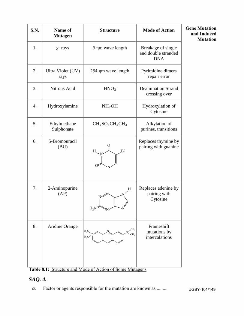
Table 8.1: Structure and Mode of Action of Some Mutagens
SAQ. 4. a. Factor or agents responsible for the mutation are known as .........
Gene Mutation and Induced
Mutation
UGBY-101/149
S.N. Name of Mutagen
Structure Mode of Action
1. χ- rays 5 ηm wave length Breakage of single and double stranded
DNA
2. Ultra Violet (UV) rays
254 ηm wave length Pyrimidine dimers repair error
3. Nitrous Acid HNO2 Deamination Strand crossing over
4. Hydroxylamine NH2OH Hydroxylation of Cytosine
5. Ethylmethane Sulphonate
CH2SO3CH2CH3 Alkylation of purines, transitions
6. 5-Bromouracil (BU)
Replaces thymine by pairing with guanine
7. 2-Aminopurine (AP)
R eplaces adenine by pairing with
Cytosine
8. Aridine Orange Frameshift mutations by intercalations
N
N
O
O
BrH
N
NH2N N
NH
NH3C
H3C N NCH3
CH3
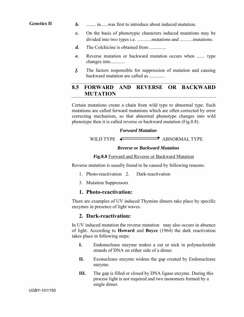
b. ........ in......was first to introduce about induced mutation.
c. On the basis of phenotypic characters induced mutations may bedivided into two types i.e. ............mutations and ...........mutations.
d. The Colchicine is obtained from ..............
e. Reverse mutation or backward mutation occurs when ....... typechanges into............
f. The factors responsible for suppression of mutation and causingbackward mutation are called as .............
8.5 FORWARD AND REVERSE OR BACKWARD MUTATION
Certain mutations create a chain from wild type to abnormal type. Such mutations are called forward mutations which are often corrected by error correcting mechanism, so that abnormal phenotype changes into wild phenotype then it is called reverse or backward mutation (Fig.8.8).
Forward Mutation
WILD TYPE ABNORMAL TYPE
Reverse or Backward Mutation
Fig.8.8 Forward and Reverse or Backward Mutation
Reverse mutation is usually found to be caused by following reasons:
1. Photo-reactivation 2. Dark-reactivation
3. Mutation Suppressors
1. Photo-reactivation:There are examples of UV induced Thymine dimers take place by specific enzymes in presence of light waves.
2. Dark-reactivation:In UV induced mutation the reverse mutation may also occurs in absence of light. According to Howard and Boyce (1964) the dark reactivation takes place in following steps:
I. Endonuclease enzyme makes a cut or nick in polynucleotide strands of DNA on either side of a dimer.
II. Exonuclease enzyme widens the gap created by Endonucleaseenzyme.
III. The gap is filled or closed by DNA ligase enzyme. During thisprocess light is not required and two monomers formed by asingle dimer.
Genetics II
UGBY-101/150
3. Mutation Suppressors:When primary mutation occurs from one site and the reverse mutation occurs from another site, which reverses the mutational affect of primarily mutated gene. This phenomenon is known as mutation suppression and the factors responsible for the suppression of mutation to cause backward or reverse mutation are called Mutation Suppressors.
8.6 SIGNIFICANCE OF MUTATION
When gene mutation or point mutations occurring during gamete formation are transmitted to all the cells of the offspring and may significant for the next generations. In case of somatic gene mutations which arise in the organism is confined to only the particular cell which is derived during mitotic division becomes mutant and they may affect only that organism and are lost along with the death of organism and could not transmitted to the next generation during inheritance.
There are extremely variable effects of gene mutations. Sometimes minor gene mutations are recessive so they remain unnoticed phenotypically and passed through it. Whereas sometimes there is a minor alteration or a change only in a single nucleotide or base may affect drastically in the genetic code to affect the phenotype of an organism. There are few examples of variability by the point mutations which are as follows:
Sickle Cell Anaemia: In human being sickle cell anaemia is an example of base substitution mutation affecting a base in one of the genes responsible for haemoglobin production. The respiratory pigment, haemoglobin, is made up of four polypeptide chains (two α and two β chains) attached to the prosthetic group haem. The polypeptide chains influence oxygen carrying capacity of the haemoglobin molecules. A change in a base sequence gives rise to triplet coding for one particular amino acid out of 146 in the β chains responsible in production of sickle cell haemoglobin. The sequencing of normal and abnormal chains differ in the substitution of Valine for Glutamic acid at one point in the abnormal polypeptide chains of sickle cell haemoglobin which causes it to be crystalline at low oxygen so that red blood cells get distorted and appear into sickle shape.
Advantages of induced mutation Induced mutagenesis is used for induction of cytoplasmic male sterility e.g. Ethidium bromide (EtBr) has been found to induce cytoplasmic male sterility in Pearl millet and barley.
• Mutation breeding is low cost fast method of developing newvarieties as compared to other methodologies.
• It is found more effective in improving disease resistant varieties.
• It is a simple, quick and best way to induce new characters invegetatively propagated crops.
Gene Mutation and Induced
Mutation
UGBY-101/151

• However they have great evolutionary consequences, because theprocess of speciation depends upon it.
Limitations of induced mutation • Mutations are mostly found to be deleterious and undesirable.
• Identification of useful micro-mutations is practically difficult tosort out.
• Since mutations are produced at a very low frequency among thelarge plant population, hence forth its screening, identification andisolation of desirable mutant is tough screened to identify andisolate desirable mutants.
• Mutation breeding has limited scope for the genetic improvementof quantitative or polygenic traits.
• The mutant types are generally unable to compete equally with
wild type individuals.
8.7 SUMMARY Hugo de Vries (1901) first used the term mutation while working on evening prime rose (Oenothera lamarckiana) , to describe sudden heritable phenotypic changes. The first scientific study was done by T.H.Morgan in 1910 in fruit fly Drosophila melanogaster.
Mutation has been described in various ways which is as follows:
• Somatic Mutation and Germinal Mutation- on the basis of types ofcells
• Autosomal Mutation and Sex Linked Mutation- on the basis oftypes of chromosomes
• Spontaneous Mutation and Induced Mutation- Either natural orinduced by mutagens
• Forward Mutation and Reverse or Backward Mutation-
Forward Mutation
WILD TYPE ABNORMAL TYPE
Reverse or Backward Mutation
• Dominant Mutation, Recessive Mutation and Lethal- on the basisof phenotypic expressions
• Gene Mutation or Point Mutations
Genetics II
UGBY-101/152
When there is a little change or alteration in the DNA sequencing occurs in replication of genes or DNA is called Gene mutation or Point mutation or Copy-error mutations.
Types of Gene Mutation: 1. Deletion or loss of bases 2. Insertion or Addition 3. Substitution
1. Deletion or Loss: There is change of sequence by the loss ordeletion of single base.
2. Insertion or Addition: Addition or insertion of single base mayalter the entire sequence of amino acids by triplets of codon.
• Frame Shift Mutation: The deletions, insertions andinversions include those changes in base sequences whichinvolve breakage and reunion of DNA fragments. At the site ofmutation occurs by equal addition and deletion of the base.
3. Substitution Mutation: The replacement of a base pair may takeplace during replication of DNA without any breakage of DNA.The base pair replacements or substitutions are following twotypes: i.Transition ii. Transversion
i. Transition: In transition there is substitution or replacementof one purine by another purine or one pyrimidine by anotherpyrimidine.The transitions are further divided into threecategories: a.Tautomerisation b. Deamination c. Base Analogue
a. Tautomerisation: The process of shifting of hydrogen atoms fromone position to another in a purine or in a pyrimidine. Product ofthis process is known as tautomer.
b. Deamination: When amino group of DNA is replaced by hydroxylgroup.
c. Base Analogue: There are some chemical compounds whosemolecular structure is similar to the usual DNA bases are calledbase analogues. It may occur as natural base analogue or artificialbase analogue
ii. Transversion: The replacement of purine bases (A, G) bypyrimidine (C, T) bases or vice versa.
Induced Gene Mutation When mutations are induced artificially by any agents called Induced Mutation.
Induced mutations on the basis of observation of their phenotypic characters may be divided into two types:
1. Macro-mutations or Qualitative mutations or Oligogenic mutations
2. Micro-mutations or Quantitative mutations or Polygenic Mutations
Gene Mutation and Induced
Mutation
UGBY-101/153
Types of Mutagen There are basically three categories of mutagens which are found to be used in inducing mutations viz.
i. Physical Mutagens: Various types of radiations, temperature etc.
ii. Chemical Mutagens: Several organic and inorganic chemicalsubstances such as 5-bromouracil, 2-amino purine, hydroxylamine,mustard gas, nitrous acid, phenol, formaldehyde,ethylmenthesulphonate, ethylurethane, ferrous and magnanoussalts, dimethylsulphoxide, acridine dyes like acridine orange,proflavin etc.
iii. Biochemical Mutagens: Colchicine, Carmine etc.
Mutation Suppressors: the factors responsible backward or reverse mutation
8.8 TERMINAL QUESTIONS
MULTIPLE CHOICE QUESTIONS: 1. The first scientific study of mutation was done by:
a. Hugo de vries c. Gregor Johann Mendel
b. T.H. Morgan d. Watson and Crick
2. The term mutation was introduced by:
a. Erich von Tschermak c. Hugo de Vries
b. Carl Correns d. T. H. Morgan
3. A point mutation may be responsible for:
a. Termination of protein synthesis
b. Addition of wrong amino acid during protein synthesis
c. Neither termination of protein synthesis nor substitution ofwrong amino acid
d. All of the above
4. Transition and Transversion are:
a. Addition point mutation c. Deletion point mutation
b. Substitution mutation d. All of these
5. Forward mutation means:
a. Abnormal to wild type c. abnormal to abnormal type
b. Wild to abnormal type d. Wild to wild type
Genetics II
UGBY-101/154
6. Mutation suppressors are responsible for:
a. Induced mutations c. Forward mutations
b. Spontaneous mutations d. Reverse mutation
7. In case of transition:
a. Adenine replaced by Guanine
b. Thymine replaced by Cytosine
c. Guanine replaced by Adenine
d. All are correct
8. Tautomerisation , Base analogue and Deamination are various typesof :
a. Transition substitution c. Deletion point mutation
b. Addition point mutation d. Transversion substitution
9. Trasversion is a type of substitution in which:
a. Purine base is replaced by Purine and Pyrimidine base isreplaced by pyrimidine base
b. Purine base is replaced by Pyrimidine and vice versa
c. Both of above statements are true
d. None of these
10. Frame shift mutation occurs due to:
a. By addition of single nucleotide in a sequence
b. By deletion of single nucleotide in a sequence
c. By both deletion and addition of nucleotides in a sequence
d. None of these
SHORT ANSWER TYPE QUESTIONS:
1. What is the spontaneous and induced mutation?
2. What is substitution point mutation?
3. How will you differentiate transition and transversion?
4. Define briefly forward and backward mutation.
5. How many types of induced mutations are known to you?
6. What are mutation suppressors?
7. Write briefly about the significance of mutation.
Gene Mutation and Induced
Mutation
UGBY-101/155
8. Write in short about the mode of action of different mutagen.
LONG ANSWER TYPE QUESTIONS:
1. How many types of mutations are categorised by differentgeneticists?
2. How many types of mutagens and their mode of action are known toyou?
3. Explain that how does a point mutation alter the sequencing ofgenetic code either by addition or deletion of single nucleotide base?
4. Which factors are responsible for causing reverse mutations?
5. Describe briefly the role of induced mutation in crop improvement.
8.9 ANSWER MULTIPLE CHOICE QUESTIONS:
1. b 2. c 3. c 4. b 5. b
6. d 7. d 8. a 9. B 10. c
SAQ.1 a. Hugo de Vries , Oenothera lamarkianab. 1910, Drosophila melanogasterc. T.H. Morgan
SAQ.2 a. Forward mutation b. Lethal c. Mutagens
SAQ. 3: a. Point mutation b. Deletion, Addition & Substitution
c. Substitution d. Transversion e. Transition
SAQ. 4.
a. Mutagensb. H. J. Muller, 1927
c. Macro or qualitative, micro or quantitative
d. Colchicum autumnale
e. Abnormal, Wild
f. Mutation suppressors
Genetics II
UGBY-101/156
UNIT-9
GENETICS IN PLANT IMPROVEMENT
Structure 9.1 Introduction
9.2 Objectives
9.3 Crop Improvement
9.3.1 Quantitative Enhancement
9.3.2 Qualitative Enhancement
9.3.3 Shortening of Life Cycle
9.3.4 Insensitivity to Physical Factors
9.3.5 Photosynthesis and photorespiration
9.3.6 Synchronized Maturity
9.3.7 Desirable Traits by Inter-generic Hybridization
9.3.8 Distant Hybridization
9.3.9 Removal of Deleterious Traits
9.3.10 Wider Adaptability
9.3.11 Other Characteristics
9.4 Crop Protection
9.4.1 Resistant Varieties
9.4.2 Herbicide Resistant Plants
9.4.3 Insect-Pest Resistant Varieties
9.4.4 Rhizobacterial Manipulation
9.4.5 Disease Resistant Varieties
9.4.6 Eradication of Toxic Compounds
9.5 Methodologies
9.6 Future Strategies
9.7 Summary
9.8 Terminal Question
9.9 Answer UGBY-101/157
9.1 INTRODUCTION
All of us are depending on plants directly or indirectly for the food, for survival and thus a constant urge of human beings is to increase the production. It provided us endless motivation for devising ever more effective and reliable means of securing or conserving it. The qualitative and quantitative crop improvement is the ultimate aim of plant breeders and of course by geneticists through various plant breeding programs which combines with genetic variability from different but taxonomically related cultivars by sexual crosses. Sometimes the desirable genes are unavailable in genetically compatible species chosen for the cross breeding or fail to induce or inherited in next generations. However now a days, with the advent of the modern technological era, the desired characters may be induced through genetic engineering via recombinant DNA technologies to make new combinations of choice. There are numerous successful experiments in which a desired gene has been identified and selected to isolate from any sources such as from bacteria, taxonomically unrelated plants or animals to incorporate into the chosen cultivar to grow as the functional part of it. If the identified and selected segment has once successfully incorporated without affecting the other traits of the crop, then the crop attains better adaptability. The genetic engineering or recombinant DNA technology though requires much attention and effort in improvement of yield and its quality.
9.2 OBJECTIVES
Genetics is a branch of biology to cover entire biological aspects and holding a central position of great significance in evolution, establishment and progression of any species. It includes heredity and variation among various life forms. During last few decades’ genetics plays significant role in studying different structural and physiological aspects. The prime object is to develop economically important superior plants. Plant breeding is also considered as specialised technology based on genetics. The objectives of plant breeding and genetics are to obtain genetically superior and more adaptive varieties of crops. This object is implicated by applying different strategies and methodologies in existing environmental circumstances. Recent years, technique for manipulating DNA along with their genetics has witnessed by a remarkable development focussed in the area of agriculture. Basically the role of genetics, plant breeding and genetic engineering in improvement of production may be bifurcated under two major categories:-
1. Crop Improvement 2. Crop Protection
9.3 CROP IMPROVEMENT
Much effort is aimed to improve productivity by manipulating photosynthesis, N2 fixation, seed storage proteins etc. Some general
Genetics II
UGBY-101/158
objectives of crop improvement are listed below in respect with the significance of genetics:
9.3.1 Quantitative Enhancement: The higher yield is the ultimate aim for the economical growth. The production of any crop may be evaluated in terms of quantity of grains, fibre, fodder, oil tuber, wood, timber etc. as depending upon the type of crop, species and its variety. Improvement of quantity is estimated for generations to generations and efforts are made for the desired traits and repeated crosses are done and the thorough study is required prior to release of any variety.
9.3.2 Qualitative Enhancement: The improvement of quality is also a prime object along with the increased yield. The quality of product enhances the value and makes it economically strengthened. The quality of economically important crops such as grains, vegetables fruits pulses etc. may be improved with the help of various mutagenic treatments. There are few examples of mutagenesis in which quality may be improved either by boosting protein synthesis or by amino acid replacement which are as follows:
a. Boosting protein synthesis:
It has been found that 70% of required protein is deficient in some of the important food crops e.g.
i. Cereal seeds have low Lysine
ii. Legumes and pulses are deficient in Sulphur containingamino acid, Methionine ,Tryptophan and / or Threonine andCysteine.
It is considered that site directed mutagenesis may make possible to alter amino acids composition of seed storage proteins e.g. by employing Agrobacterium tumefaciens in beans, boosted 𝛽-Phaseolin production.
b. Amino acid replacement:
Unwanted amino acids may be replaced by desired amino acid by site directed mutagenesis which may lead to changes in DNA structure that can help in improving nutritional value of crop plants.
9.3.3 Shortening of Life Cycle: Earliness is the most desirable aspect having several advantages. It reduces the crop management period due to shorter span of life cycle. It also multiplies the production due to early maturation and more generations to grow and with less effort and cost.
9.3.4 Insensitivity to Physical Factors: Development of varieties insensitive to light and temperature i.e. photo and thermo insensitivity helps in crossing the cultivation of crop plants.
Genetics in Plant Improvement
UGBY-101/159
Genetically grown crops have no boundary of season. Winter crops can be grown in summer and vice-versa.
9.3.5 Photosynthesis and photorespiration : Entire production of crop depends upon photosynthesis. Ribulose bisphosphate carboxylase (Rubisco) plays a central role in fixing atmospheric CO2 which combines with Ribulose 1-5 diphosphate and via Calvin Cycle fixed in the form of glucose. This enzyme can also catalyse the addition of O2 to Ribulose 1-5 diphosphate to produce Glyoxylate through photorespiration and release low energy. The aims are –
i. To enhance the affinity of Rubisco for atmospheric CO2
ii. To eliminate or reduce the competitive reactions ofphotorespiration i.e. to reduced the affinity of Rubisco for O2.
These targets may be achieved by site- directed mutagenesis which modifies the enzyme activity.
9.3.6 Synchronized Maturity: It refers one time maturity of any crop species. The character is highly preferential in crops like green gram, cowpea, cotton etc. in which several pickings needed to harvest.
9.3.7 Desirable Traits by Inter-generic Hybridization: It refers to the cross made between two different genera belonging to same families. Though these methods are rarely used in crop improvement practically due to reduced rate of successful experiments and results. However inter-generic crosses are applicable when the desired traits are not found in same genera or different species. The requirement of such type of hybridization is the transfer of some specific traits into cultivated species from allied genera. Generally, interspesific F1 hybrids are sterile and are either propagated asexually or by doubling of chromosome through colchicines treatment e.g. Raphanobrassica, Triticale etc.
It also includes certain phenotypic traits like height, branching, tillering and growth habits etc., usefulness of these traits differs among the crops like tallness, high tillering and profuse branching are preferred in fodder crops whereas dwarfness in wheat, rice, sorghum and pearl millet etc. confers the lodging resistance and better fertilizer responses among field crops.
9.3.8 Distant Hybridization: Distant hybridization is also found as effective method in crop improvement by choosing desirable characters from wild species to insert in cultivated plants. Thus the significance of distant hybridization have found to be more disease and pest resistance, better adaptability, better quality, more productive and with better mode of reproduction etc.
Genetics II
UGBY-101/160
9.3.9 Removal of Deleterious Traits:
By making inter-specific or desired crosses, the problem of frequent recurrence appearing of harmful traits may be overcome. With the help of hybridization methodology, in the heterozygous conditions dominant alleles may mask the effect of recessive alleles. In which a pair of allele segregate at the time of gamete formation and generate new recombinants in next generations.
9.3.10 Wider Adaptability: Better adaptation is observed in new varieties which is another important issue. Adaptability refers to the suitability of growth in wide range of varied environment. It helps to grow crop in stabilizing the production in a broader areas having variable weather or seasons.
9.3.11 Other Characteristics: • Manipulation of N2 fixing micro- organism:
Normally leguminous crops have root nodules showing symbiotic relationship with nitrogen fixing micro-organisms like various types of N2 fixing bacteria, which facilitate in fixing atmospheric N2. It may be done by producing enzyme nitrogenase. With the help of genetic engineering or recombinant DNA technology, Nif (Nitrogen Fixing) genes of N2 fixing bacteria are incorporated in non-nitrogen fixing organism and these organisms are made capable to fix atmospheric nitrogen.
SAQ.1:
a. The main objectives of geneticists and plant breeders are ..........and ........enhancement of crop.
b. The production may be improved by crop improvement andcrop............ both.
c. The unwanted amino acids may be replaced by desired amino acidby...........
9.4 CROP PROTECTION
The production of improved plant varieties which are resistant to herbicides, viral diseases and insect pests is known as crop protection. The measures have been developed to protect the crop plants from various extrinsic abiotic and biotic factors which are either directly or indirectly influence the quality and quantity of the yield.
9.4.1 Resistant Varieties: The crop has to be protected from various environmental factors such as biotic and abiotic components, therefore required to develop biotic and abiotic resistant varieties.
Genetics in Plant Improvement
UGBY-101/161

a. Biotic Resistant
It has been observed that crops are often destroyed by various insects, parasites, different types of pathogens causing diseases either at pre-maturity i.e. prior to flowering or at post- maturity i.e. after fertilization which causes heavy loss to the production. Therefore, genetic resistance is found as strong and cheapest methods to minimising the loss by developing resistant varieties by choosing resistant donor parents to inherit to next filial generations through their genetic pool.
b. Abiotic Resistant
The abiotic factors such as alterations in climatic factors like temperature fluctuation, drought, soil pH, heat and cold, wind, frost etc. affect the growth and development of crops. Plant breeders are consistently practicing to develop resistance by selecting better adapted parents which may enable to produce abiotic resistant varieties to grow during adverse environmental conditions.
9.4.2 Herbicide Resistant Plants: a. Atrazine resistant -
It is a herbicide or weedicide which usually kills the dicot weed. Atrazine effects photophosphorylation specially Plastoquinone (PQ) an electron carrier of the process. There are two varieties: Atrazine resistant and Atrazine susceptible. The main difference between the two is of single nucleotide. Adenine is replaced by guanine by using the techrique of genetic engineering. The plants can be made resistant to Atrazine by manipulating particular nucleotide.
b. Glyphosphate (Phosphono methyl glycine) resistant -
Herbicide, inhibits synthesis of EPSP enzyme [5 Enol pyruvil skikmate 3- phosphate synthetase ] which is involved in the synthesis of (aromatic asconcatic amino acids. This recombinant genes formed by using R plasmid as vector resistant against herbicide through genetic engineering the mutant gene isolated from bacteria and introduced into tobacco plant which develop resistance against this herbicide.
9.4.3 Insect-Pest Resistant Varieties: The genes of Bacillus thuringiensis toxic to lepidopteran larvae introduced into tobacco plant through insertion into mini T-DNA vector and transferred to Agrobacterium tumefaciens. Transformed tobacco plants were infested with larvae which were killed within 3 days after eating of leaves of such plants.
9.4.4 Rhizobacterial Manipulation: Rhizobacteria is root colonizing bacteria. It is used as bio control agent. By two genetic mechanisms, it can inhibit growth and multiplication of soil borne pathogenic bacteria.
Genetics II
UGBY-101/162
i. It secrets chemical substance in the soil which have inhibitoryeffect on the soil borne pathogenic organism.
ii. They show antagonism to soil borne pathogenic microorganism.Their nutrient requirements are similar to the requirement of soil borne pathogenic microorganism. Therefore, competition takes place between the rhizobacteria and soil borne pathogenic microorganism which again has inhibitory influence on the pathogenic microorganism.
9.4.5 Disease Resistant Varieties: a. Control of Viral Diseases :
Most of living organisms are suffering from viral diseases. In case of plants it has been observed that if coat protein is introduced into host plant then it develops capability of preventing itself from specific pathogen. In 1986, use of coat protein in tobacco plant was done and these plants develop resistance against TMV. In 1987, Tumer introduced coat protein in alpha-alpha plant due to which these plants developed resistance against alpha-alpha mosaic virus.
b. Control of Soil-borne Diseases :
There are many well known soil borne diseases which are dangerous to some economically very important crops on one hand and difficult to eradicate on the other hand e.g. Pithophthroa root rot, Fusarium wilt of Cajanus cajan, red rot of sugarcane caused by Colletotricum etc. In nature so many suppressive soils are known which contain micro-organisms which have capability of inhibiting growth and development of soil borne pathogenic micro-organisms. The genetic engineering helps in identifying their genetic constitution and genes responsible for control of pathogenic organism can be isolated and then incorporated in host plants due to which these host plant also develop capability of becoming resistant to pathogen.
9.4.6 Eradication of Toxic Compounds: It is an essential aspect to develop such crop varieties which are free from toxic compounds and responsible for causing any harm to the consumers either animals or humans. There are number of such crops e.g. Khesari dal which contains neurotoxins causing paralysis of lower limbs, some toxic acidic compounds reported in mustard etc. study is required to identify such crops and develop such varieties which are suitable for consumption without any harm to the health of consumers.
SAQ.2:
a. The root colonizing bacteria used as bio control agent is...............
b. The resistant varieties may be basically categorised into.........and........
c. The wilt disease of arhar (Cajanus cajan) caused by....... is a......borne disease.
Genetics in Plant Improvement
UGBY-101/163
9.5 METHEDOLOGIES
The process of bringing wild species of plants under cultivation to fulfil human needs is called domestication. There are different methods applied for crop improvement which may be categorised as follows:
a. Selection: This is an oldest and conventional method, where theplants with useful desirable characters are chosen as a parent to get improved better generations. The selection, may be of three types:
i. Mass Selection- It is known to sort out number of plants, flowersor seeds on the basis of phenotypic traits of superior quality fromthe field population and during harvesting to raise next generation.The selection is consistently repeated in each generation so as toachieve the improved desired traits.
ii. Pure Line Selection- When a progeny is obtained from singleinbred homozygous plant then it is pure line. Selection of pure linefor breeding is called pure line selection.
iii. Clonal Selection- The group of plants when by vegetativepropagation of single plant is known as clone. Developing varietyfrom selecting single clone is called clonal selection
b. Hybridization: The hybridization method is used to produce newcrop varieties by making cross between two or more plants having variable genetic constitution belonging to either of same or different species or genera. There are various types of hybrids grown such as:
i. Intravarietal- The cross made between the plants of same varietyand is mostly found useful in self pollinated crops.
ii. Intervarietal- Crossing is made between two plants belonging todifferent varieties of same species e.g. cereals.
iii. Interspecific- The cross is made between the plants of two differentspecies of same genus. This method is usually applicable inintransferring genes for disease, pests and drought resistance.
iv. Intergeneric- In this case, the crosses made between the plantsbelonging to different genera e.g. Raphanobrassica, Triticale etc.
v. Interogressive- When one species is completely replaced byanother by natural selection.
• Emasculation: To prevent selfing in bisexual flowers, removal ofanthers or stamens is done which is called emasculation. The emasculated flower act as female parent and a male parent of desired characteristics has to be chosen for cross pollination.
• Hybrid Vigour or Heterosis: When two inbreds of differentgenetic constitution or different genera are crossed together, produced vigorous hybrids are known as hybrid vigour or heterosis. The increased
Genetics II
UGBY-101/164
vigourosity and superiority of the hybrids are called hybrid vigour or heterosis. The term was coined by G.H. Shull (1914). Resultant hybrids are found to be more vigorous, productive, sturdier and giant then their remote parents. Though hybrid vigour is generally found sterile due to unbalanced set of chromosomes, but it is economically very beneficial method for crop improvement. It is commercially exploited in various crops like ornamentals, fruits, cereals, vegetables etc.
c. Polyploid breeding: When an individual possess more than ausual two sets of chromosome, it is known as polyploidy. The term was coined by Winkle (1916). Polyploids may be autopolyploids and allopolyploids.. There are many natural polyploids also e.g. wheat, rice, sugarcane, flax, cotton, tobacco etc. It is an important source of variation. Allopolyploids are significant hybrids produced by the cross made between either different genera or species. These hybrids may be sterile so that these are treated by colchicine to make them fertile by duplication of chromosomes. Induced polyploids have desirable quality along with better adaptation and also found disease resistant e.g. Triticale, a new cereal, hybrid of Nicotiana (N.tabacum X N. glutinosa) produced was found to be virus resistant.
d. Induced Mutations: Mutation breeding is an useful tool ingenerating variability in self pollinated crops which may be raised by inducing different types of mutagens called induced mutation for improvement of crops. In India mutational breeding is very popular. Number of economically important crops have been evolved by these methodologies e.g. varieties of wheat (Sharbati Sonora-64, Pusa Lerma) raised by Gamma rays, in mung bean resistant varieties (resistant to yellow mosaic virus and powdery mildew) has been evolved.
9.6 FUTURE STRATEGIES
Genetics and plant breeding has played a very important role in crop improvement as well as crop protection. Now-a-days, there are more dimensions of genetics to improve productivity along with its quality via somatic hybridization, genetic engineering, gene cloning etc.
The role of genetics via gene action in crop improvement involves the expression of a character. Basically, the strategies adopted under variable conditions are as follows. The gene action involves either additive or non-additive. Selection is the method to obtain pure line from the genetically variable varieties, which may be either pure line or mass selection in self pollinated crops then to raise the breeds of desired characteristics of hybrids due to additive gene actions. Whereas the non-additive gene action is implied for various other breeding methods to obtain hybrid vigour or heterosis. The hybrid vigours are often found sterile. For the improvement of population the plant breeders and geneticists have to take care about the selection of superior lines having genes of desirable traits which has to develop for future generations.
Genetics in Plant Improvement
UGBY-101/165
SAQ.3:
a. When a progeny is obtained from single inbred homozygous plantis……
b. Raphanobrassica is …………….hybrid.
c. Hybrid Vigour is also known as ………..
d. The removal of anthers or stamens from bisexual flower iscalled…….
9.7 SUMMARY
Genetics and plant breeding is used as an important tool for crop improvement and holding a central position of great significance in evolution, establishment and progression of any species. The prime object is to develop economically important superior plants by focusing in the area of agriculture basically under two major categories:- 1. CropImprovement 2. Crop Protection
CROP IMPROVEMENT
Productivity of crops can be improved manipulating their various metabolic activities genetically such as photosynthesis, N2 fixation, seed storage proteins etc. Some general objectives of crop improvement are listed below in respect with the significance of genetics:
1. Quantitative Enhancement:
The improvement of quantity is estimated from generations togenerations and effort is made by the desired and repeated crosses and their thorough study required prior to release of any variety. It may be evaluated in terms of quantity of grains, fibre, fodder, oil tuber, wood, timber etc. as depending upon the type of crop, species and its variety.
1. Qualitative Enhancement:
The quality of economically important crops such as grains,vegetables fruits pulses etc. may be improved with the help of various mutagenic treatments.
a. Boosting protein synthesis:
Site directed mutagenesis may make possible to alter aminoacids composition of seed storage proteins.
b. Amino acid replacement:
Unwanted amino acids may be replaced by desired amino acidby site directed mutagenesis that can help in improvingnutritional value of crop plants.
Genetics II
UGBY-101/166
2. Shortening of Life Cycle:
The shorter span of life cycle multiplies the production due to earlymaturation and allows more generations to grow, with less effortsand cost.
3. Insensitivity of Physical Factors:
To develop new breeds which are insensitive to light andtemperature with the help of selecting the parent. These crops haveno boundary of season.
4. Photosynthesis and Photorespiration :
Entire production of crop depends upon photosynthesis. Thesetargets may be achieved by site- directed mutagenesis whichmodifies the enzyme activity.
5. Synchronized Maturity:
The character is highly preferential in crops like green gram,cowpea, cotton etc. in which several pickings is needed to harvestrather than one time maturity.
6. Desirable Traits by Inter-generic Hybridization:
The requirement of such type of hybridization is to transfer ofsome specific traits into cultivated species from allied genera e.g.Raphanobrassica, Triticale etc.
7. Distant Hybridization:
Distant hybridization is also found as effective method in cropimprovement with more disease and pest resistance, betteradaptability, better quality, more productive and with better modeof reproduction etc. by choosing desirable characters from wildspecies to insert in cultivated plants.
8. Removal of Deleterious Traits:
This can be achieved with the help of hybridization methodology,in the heterozygous conditions dominant alleles may mask theeffect of deleterious recessive alleles.
9. Wider Adaptability:
Adaptability refers to the suitability of growth in wide range ofvaried environment to grow crop in stabilizing the production in abroader areas having variable weather or seasons.
10. Other Characteristics:
a. Manipulation of N2 fixing micro- organism:
With the help of genetic engineering or recombinant DNAtechnology Nif (nitrogen Fixing) genes of N2 fixing
Genetics in Plant Improvement
UGBY-101/167
bacteria are incorporated in non nitrogen fixing organism and plants are made capable to fix atmospheric nitrogen.
CROP PROTECTION
The measures have been developed to protect the crop plants from various extrinsic abiotic and biotic factors which either directly or indirectly influence the quality and quantity of the yield.
1. Resistant Varieties:
The genetic resistance is found as strong and cheapest methods to minimise the loss by developing resistant varieties by choosing resistant donor parents to inherit to next filial generations through their genetic pool. Following resistant varieties developed through genetics and genetic engineering
a. Biotic Resistant
b. Abiotic Resistant
c. Herbicide Resistant Plants
d. Insect-Pest Resistant Varieties
2. Rhizobacterial Manipulation:
Rhizobacteria is root colonizing bacteria. It is used as bio control agent. By two genetic mechanisms, it can inhibit growth and multiplication of soil borne pathogenic bacteria.
3. Disease Resistant Varieties:
a. Control of Viral Diseases :
In case of those plants which are suffering from viral diseases it has been observed that if coat protein is introduced into host plant then it develops capability of preventing itself from specific pathogen.
b. Control of Soil-borne Diseases :
The genetic engineering helps in identifying their genetic constitution and genes responsible for control of pathogenic organism can be isolated and then incorporated in host plants due to which these host plant also develop capability of becoming resistant to pathogen.
4. Eradication of Toxic Compounds:
The hybridisation techniques are found helpful to develop bettercrop varieties which are free from toxic compounds and responsible for causing any harm to the consumers.
Genetics II
UGBY-101/168
METHEDOLOGIES
There are different methods applied for crop improvement which may be categorised as follows:
a. Selection: This is an oldest and conventional method, where theplants with useful desirable characters are chosen as a parent to get improved better generations. The selection done may be of three types:
i. Mass Selection- This is done on the basis of phenotypic traits ofsuperior quality from the field population and during harvesting toraise next generation. The selection is consistently repeated ineach generation so as to achieve the improved desired traits.
ii. Pure Line Selection- When a progeny is obtained from singleinbred homozygous plant then it is pure line. Selection of pureline for breeding is called pure line selection.
iii. Clonal Selection- The group of plants when obtained byvegetative propagation of single plant is known as clone.Developing variety from selecting single clone is called clonalselection.
b. Hybridization: The hybridization method is used to produce newcrop varieties by making cross between two or more plants having variable genetic constitution belonging to either of same or different species or genera. There are various types of hybrids grown such as:
i. Intravarietal- The cross made between the plants of same varietyand is mostly found useful in self pollinated crops.
ii. Intervarietal- Crossing is made between two plants belonging todifferent varieties of same species e.g. cereals.
iii. Interspecific- The cross is made between the plants of two differentspecies of same genus. This method is usually applicableintransferring genes for disease, pests and drought resistance.
iv. Intergeneric- In this case the crosses made between the plantsbelonging to different genera e.g. Raphanobrassica, Triticale etc.
v. Interogressive- When one species is completely replaced byanother by natural selection.
• Emasculation: To prevent selfing in bisexual flowers by removalof anthers or stamens is called emasculation. The emasculatedflower act as female parent and a male parent of desiredcharacteristics is chosen for cross pollination.
• Hybrid Vigour or Heterosis: When two inbreds of differentgenetic constitution or different genera are crossed together,produce vigorous hybrids is known as hybrid vigour or heterosis..Though hybrid vigour is generally found sterile due to unbalanced
Genetics in Plant Improvement
UGBY-101/169
set of chromosomes but is economically very beneficial method for crop improvement. It is commercially exploited in various crops like ornamentals, fruits, cereals, vegetables etc.
c. Polyploid breeding: There are many natural polyploids also e.g.wheat, rice, sugarcane, flax, cotton, tobacco etc. It is an importantsource of variation. Allopolyploids are significant hybridsproduced by the cross made between either different genera orspecies.
d. Induced Mutations: Mutation breeding is a useful tool ingenerating variability in self pollinated crops which may be raisedby inducing different types of mutagens called induced mutationfor improvement of crops.
9.8 TERMINAL QUESTIONS
MULTIPLE CHOICE QUESTIONS: 1. The process of bringing wild species of plants under cultivation to
fulfill human demands is called:
a. Selection c. Hybridization
b. Domestication d. Cultivation
2. Most popular crop selected for continuous selection anddomestication by man is:
a. Wheat c. Maize
b. Rice d. Millet
3. Removal of anthers from bisexual flowers is called:
a. Hybridization c. Emasculation
b. Selection d. Inbreeding
4. The methods of plant breeding are:
a. Selection c. Emasculation
b. Hybridization d. All of these
5. Heterosis is also known as:
a. Heterothallism c. Heteromorphism
b. Hybrid Vigour d. Hybrid sterility
6. Genetic variability may be due to:
a. Mass Selection c. Clonal Selection
b. Mutation d. Hybridization
Genetics II
UGBY-101/170
7. Hybrid Vigour is due to:
a. Homozygosity c. Linkage
b. Autopolyploidy d. Overdominance
8. Triticale is developed by intergeneric cross between:
a. Wheat and Rice c. Rice and Rye
b. Wheat and Rye d. Rice and Maize
9. Hybrid vigour are generally found sterile due to:
a. Vigourosity
b. Unbalanced pairing of chromosomal set
c. Overdominance
d. None of these
10. The cultivation of improved varieties of economically importantcrops in fields are mainly by:
a. Selection of healthy seeds from healthy plants
b. Introducing new varieties of various crops under different
environmental conditions
c. Judicious combination of Selection, Introduction and
Hybridization of different varieties
d. Only by natural selection
MARK THE FOLLLOWING STATEMENTS AS TRUE/FALSE:
1. In introgressive hybridization, one species is completely replacedby another in nature.
2. Mass selection is the method of developing varieties by singleclone.
3. The increased superiority of hybrids over the parents is calledheterosis.
4. The main objectives of plant breeding are to develop new superiorcrop varieties than the existing.
5. When two or more plants of unlike genetical constitution arecrossed together it is known as clonal selection.
6. The process of removal of either anthers or ovules beforefertilization is called emasculation.
Genetics in Plant Improvement
UGBY-101/171
SHORT ANSWER TYPE QUESTIONS:
Q.1. What is distant hybridisation?
Q.2. How will you differentiate quantitative and qualitative traits?
Q.3. Why different types of resistant varieties considered economically better?
Q.4. How induced mutation is helpful in crop improvement?
Q.5. What do you know about emasculation?
Q.6. What is hybrid vigour?
LONG ANSWER TYPE QUESTIONS:
Q.1. Describe briefly about the objectives of genetics among crop improvement.
Q.2. How does genetics help in crop protection and its production?
Q.3. How many types of resistant varieties are known to you, give few suitable examples?
Q.4. What is selection and how many types of selection methods implicated by the plant breeders?
Q.5. Briefly describe the various methods used in crop improvement.
9.9 ANSWER
MULTIPLE CHOICE QUESTIONS:
1. b 2. c 3. c 4. d 5. b 6. b 7. d 8. b 9.c 10. c
TRUE/FALSE:
1. True 2. False 3. True 4. True 5. True 6. False
SAQ.1:
a. Quantitative, qualitative b. Protection c. Site directed mutagenesis
SAQ.2:
a. Rhizobacteria b. Abiotic , biotic c. Fusarium, soil
SAQ.3:
a. Pure line b. Intergeneric c. Heterosis d. Emasculation
Genetics II
UGBY-101/172


