Embed Size (px)
Citation preview

Send Orders of Reprints at [email protected]
Current Alzheimer Research, 2013, 10, 000-000 1
1567-2050/13 $58.00+.00 © 2013 Bentham Science Publishers
Treadmill Exercise Prevents Learning and Memory Impairment in Alzheimer’s Disease-Like Pathology
An T. Dao, Munder A. Zagaar, Amber T. Levine, SaminaSalim, Jason Eriksen and Karim A. Alkadhi
Department of Pharmacological and Pharmaceutical Sciences, University of Houston, Houston, TX 77204, USA
Abstract: Alzheimer’s disease (AD) is a neurodegenerative disorder that is characterized by progressive memory loss. In contrast, accumulating evidence suggests a neuroprotective role of regular exercise in aging associated memory impair-ment. In this study, we investigated the ability of regular exercise to prevent impairments of short-term memory (STM) and early long-term potentiation (E-LTP) in area CA1 of the hippocampus in a rat model of AD (i.c.v. infusion of 250 pmol/day A 1-42 peptides). We utilized behavioral assessment, in vivo electrophysiological recording, and immunoblotting in 4 groups of adult Wistar rats: control, treadmill exercise (Ex), -amyloid-infused (A ), and amyloid-infused/treadmill exercised (Ex/A ). Our findings indicated that A rats made significantly more errors in the radial arm water maze (RAWM) compared to all other groups and exhibited suppressed E-LTP in area CA1, which correlated with deleterious alterations in the levels of memory and E-LTP-related signaling molecules including calcineurin (PP2B), brain derived-neurotrophic factor (BDNF) and phosphorylated CaMKII (p-CaMKII). Compared to controls, Ex and Ex/A rats showed a similar behavioral performance and a normal E-LTP with no detrimental changes in the levels of PP2B, BDNF, and p-CaMKII. We conclude that treadmill exercise maybe able to prevent cognitive impairment associated with AD pathology.
Keywords: Alzheimer’s disease; brain-derived neurotrophic factor (BDNF); calcineurin (PP2B); calcium-calmodulin depend-ent protein kinase II (CaMKII); exercise, learning and memory; synaptic plasticity.
INTRODUCTION
Currently, the amyloid hypothesis holds strong support based on the fact that the prime neuropathological hallmarks of AD are extracellular deposits of amyloid plaques and in-tracellular formation of neurofibrillary tangles. Even though full-blown characteristics of AD have not yet been repro-duced, various animal models of AD have shown important symptomssimilar to the disease manifestation in humans. AD animals exhibit a marked impairment of learning and mem-ory function as shown in various behavioral tasks such as Morris water maze (MWM), radial arm maze (RAM),and radial arm water maze (RAWM) [1-5]. In addition to cogni-tive deficits, aged transgenic AD mice also displayed non-cognitive disturbances (e.g. irritability) compared to their wildtype counterparts [6]. Learning and memory impairment in AD animals correlates, at the cellular level,with an en-hanced long-term depression (LTD) and a suppressed long-term potentiation (LTP)[1, 7].
The expression of LTP and LTD requires the entry of calcium into the postsynaptic cell. A high level of intracellu-lar calcium induces LTP but onlya modest rise in calcium can induce LTD. While the expression of LTP involves the action of kinases, phophastases are responsible forthe ex-pression of LTD. Two major phases of LTP have been iden-tified; an early phase (E-) LTP, lasting up to 3 hours and the *Address correspondence to this author at the Professor of Pharmacology and Neuroscience, Department of PPS, College of Pharmacy, University of Houston, Houston, TX 77204-5037, USA; Tel: 713-743-1212; Fax: 713-743-1229; E-mail: [email protected]
more durable late phase (L-) LTP, lasting more than 3 hours and even days. At the synapses, E-LTP and L-LTP are con-sidered to be cellular analogues of short-term and long-term memory respectively. A single train of high frequency stimu-lation (HFS) induces E-LTP, a protein-independent process. The released glutamate activates post-synaptic glutamate receptors and with the post-synaptic membranesufficiently depolarized, the Mg2+ blockage of the N-methyl-D-aspartate (NMDA) receptors is removed allowing the entry of Ca2+
into the cell. Calcium ioninflux dissociates calmodulin from neurogranin and activates Ca2+/calmodulin-dependent pro-tein kinase II (CaMKII), which autophosphorylatesonce ac-tivated. CaMKII serves as a critical molecule whose activa-tion duration determines transiency or persistency of LTP [8].
Regular exercise is a non-pharmacological approach that ameliorates memory impairment secondary to brain injury, enhances cognitive function, and prevents memory decline in the aged brain [9, 10]. In addition to animal studies, epide-miological analysis in humans suggests that regular exercise can act as a preventive treatment against cognitive disorders (e.g. amnesia, dementia) with minimal cost and adverse ef-fects [11]. The neuroprotective effects of exercise are thought to be mediated by various molecular mechanisms including upregulation ofneurotrophins (e.g.BDNF) and other molecules associated with learning and memory func-tion including CaMKII andcalcineurin, which in turn en-hance brain plasticity (LTP) and improve performance in memory tasks. Exercised animals performed better in the spatial memory tasks (e.g. MWM or RAWM) compared to

2 Current Alzheimer Research, 2013, Vol. 10, No. 5 Dao et al.
sedentary animals as indicated by shorter swim paths or fewer errors to find the target platform [10, 12-14]. Simi-larly, exercise can also modify non-spatial memory as tested in the passive avoidance paradigm and object recognition tasks [9, 10, 12, 15-18]. Additionally, treadmill exercise is reported to restore memory impairment in rats treated with alcohol [19], streptozocin[20], reserpine [21], or sleep depri-vation[22].
In this study, we investigated the effect of treadmill exer-cise on learning and short-term memory, E-LTP inarea CA1and related signaling molecules levels ina rat model of AD achieved by exogenous administration of amyloid pep-tides.
MATERIALS AND METHODS
Animals
Adult male Wistar rats (Charles River Laboratories, Wilmington, MA) weighed 176-200g at the beginning of all experiments. Upon arrival, rats were housed in a Plexiglas cage (4-6 rats/cage) in a climate-controlled room (250C) on a 12hr/12hr light/dark cycle. Rats were provided with a regular rat chow diet and water ad libitum and allowed to acclimate in the new environment for a week.All experiments were conducted following the instructions from National Research Council’s Guide of The Care and Use of Laboratory Animals and with the approval of University of Houston Institutional Animal Care and Use Committee.
Exercise Protocol
Rats ran on a leveled motorized treadmill (Columbus In-struments, Columbus, OH) on weekdays between 9:00 am and 4:00 pm for 4 weeks with a customized regimen as pre-viously described by us [22]. Rats were familiarized with the treadmill environment before the exercise training com-menced. Rats ran 2 sessions (15 minutes each) at a speed of 10 m/min (week 1 and week 2) followed bya gradual in-crease in exercise duration and intensity (3 sessions for week 3 and 4 sessions for week 4, 15 m/min). A 5 minute break was given between the sessions to reduce the risk of muscle fatigue. To encourage the rats to continue running, a mild foot shock (0.5mA) that caused a tingling sensation was de-livered. The rats eventually learn to avoid this shock,which did not appear to be stressful.
Osmotic Pump Implantation
After two weeks of exercise, the rats underwent either a sham operation (control and exercise groups) or pump im-plantation (A and Ex/A groups). Rats in the A and Ex/A groups were implanted with mini-osmotic pumps (Alzet, Cupertino, CA) as previously described by us[1, 2, 23]. Amyloid peptide(A 1-42; AnaSpecInc.,San Jose, CA) solu-tion was prepared following our previous protocols [23]. Briefly, the peptide was dissolved in a solution containing 64.9% distilled water, 35% acetonitrile, and 0.1% trifluoroacetate (TFA) to prevent peptide aggregation in the pump. One day before the implantation, the 14-day pumps were assembled, filled with A 1-42 solution (designed to de-liver 250 pmol/day), and primed in isotonic (0.9%) saline at 370C overnight. Rats were anesthetized with an i.p. injection
of a cocktail mixture containing ketamine (75 mg/kg) and xylazine (2.5 mg/kg) (Webster Veterinary, Devens, MA). The implantation site was shaved and disinfected with iso-propyl alcohol. Once positioned in the stereotaxic frame, the rat’s skull was exposed by a 2.5 cm midline sagittal incision starting behind the eyes. The infusion cannula was implanted into the right cerebral lateral ventricle (AP: -0.3, L: 1.2, V: 4) according to the atlas of Paxinos and Watson (1986). The cannula was initially held by drops of cyanoacrylicglue, con-nected to the pump and steadily fixed with dental cement. The pump was placed in a subdermal pocket in the back of the rat. The wound was then closed with wound clips, and tincture of iodine, diluted chlorohexidine, and triple antibi-otic ointment were applied to prevent bacterial infec-tions.Rats were monitored until full recovery.
Radial Arm Water Maze (RAWM)
The RAWM is a hybrid of the Morris water maze and ra-dial arm maze. It is a circular black water pool thatconsists of six swimming arms with an open central area. The ex-perimentswere done in a dimly lit room with visual cues placed on the surrounding walls. Each rat was randomly as-signed a goal arm,with a hidden platformplaced near its end. The behavioral testing protocol consisted of 12 learning tri-als followed by a short-term memory test (STM),which were administered 30 minutes after the 12th learning trial as previ-ously described [22].
Electrophysiological Experiment
Seventeen days after implantation surgery, in vivo elec-trophysiological recordings from area Cornu Ammonis 1 (CA1) of the hippocampus was accomplished as described [24, 25]. Briefly, rats were urethane-anesthetized (1.2 g/kg,i.p., Sigma Aldrich, USA) and two holes werestereo-taxically drilled for placing the stimulating and recording electrodes. On the left side of the brain, a hole was drilled for placing the concentric bipolar stimulating electrode in area CA3of the left hippocampus, which was positioned at 50 angle toward the midline (AP: -3, L: 3.5, V: 2.8). Similarly, a capillary glass (1–5 M ) recording electrode, filled with 2M NaClsolution, was inserted through the hole into area CA1 of the right hippocampus (AP: -3, L: 2, V: 2). Once the maximal response was attained, a 30 minute stabilization period was allowed without further stimulation. The test stimulus was adjusted to 30% of the maximal response. A baseline was established with test stimuli (1pulse/30s) to evoke population spikes (pspike)for 20 minutes. The magni-tude of E-LTP, evoked by one train of HFS(8 pulses of 400 Hz every 10 seconds, repeated 8 times), is quantifiedby changes in the field excitatory post-synaptic potential (fEPSP) slope and pspike amplituderecorded for 1 hour fol-lowing HFS. The fEPSP slope and pspike amplitude repre-sent synaptic strength and the number of neurons reaching firing threshold respectively[24, 25].
Western Blotting
The procedures were conducted as described [1, 22, 23]. At the end of the electrophysiological experiments, the brains were immediately removed and the CA1 area of the hippocampus was dissected out and cut into the septal (ven-

Exercise Prevents AD-Induced Impairment of Memory Current Alzheimer Research, 2013, Vol. 10, No. 5 3
tral) and temporal (dorsal) portions and stored at -800 C for later processing. Unstimulated samples were prepared from control rats that did not receive high frequency stimulation (HFS) while stimulated (S) samples were prepared from rats that received a single train of HFS to evoke E-LTP of the Schaffer collaterals pathway. As the septal side of the CA1 area received most of the stimulation, its value is expressed as ratio to that of the temporal side after normalization to Glyceraldehyde 3-phosphate dehydrogenase(GAPDH), a loading control [22, 26]. The homogenates were prepared as described and proteins loaded (15 g/well) on a high throughput E-PAGE 48 system. The proteins were trans-ferred to a PVDF membrane on a dry blot system (Invitrogen Corp., Grand Island, NY). Detection of proteins was done using specific primary antibodies and subsequent conjuga-tion with secondary horse-radish peroxidase antibodies. The protein bands were visualized using chemiluminescence rea-gents (Santa Cruz Biotechnology, Santa Cruz, CA) and quantified by densitometry using AlphaEase software.
The following antibodies were used: mouse monoclonal anti-p-CaMKII (1:500);rabbit polyclonal anti- t-CaMKII (1:1000);rabbit polyclonal anti-BDNF (1:500); rabbit poly-clonal anti-PP2B (1:1000); rabbit polyclonal anti-GAPDH (1:1000); secondary anti-mouse/rabbit antibodies (1:5000). All antibodies were purchased from Santa Cruz Technology except GAPDH antibody, which was purchased from Cell Signaling Inc., Boston, MA.
Statistical Analysis
Unpaired t-test was used to compare two groups while one-way analysis of variance (ANOVA) followed by Tukey post-hoc test was used to compare all groups. All statistical analyses were done with GraphPad Prism software. P<0.05 was considered statistically significant. Data were expressed as mean + S.E.M.
RESULTS
Exercise Prevents AD-Induced Learning and Short-Term Memory Impairment
In the RAWM task, the number of errors made by ratsin each learning trial or memory test was used as a quantitative measurement of learning and memory function. During the first six learning trials, rats in all groups made similar num-ber of errors. However, during the second set oflearning tri-als, theA rats made significantly more errors than other groups (p= 0.01-0.05)(Fig. 1A). In contrast, Ex/A rats showed a learning ability similar to that of the control and exercised rats. These results suggest that amyloid infusion impairs the learning ability in rats whereas regular treadmill exercise prevents this impairment.
In the STM test, administered 30 minutes after the last learning trial,the A rats made significantly higher number of errors than all other groups (p= 0.01) (control: 0.5 ± 0.224, Ex: 0.917 ± 0.26, A : 2.545 ± 0.529, Ex/A : 0.917 ± 0.358) (Fig. 1B). Four weeks of treadmill exercise reduced the number of errors that A rats made, which was not dif-ferentfrom those of control and exercise rats. It is noteworthy that our exercise regimen in normal rats did not enhance per-formance as both the sedentary control and exercised rats
performed similarly in this behavioral paradigm. Hence, at the behavioral level,regular exercise prevented thelearning and memory deficits in this AD model.
Fig. (1). Radial arm water maze (RAWM) performance in seden-tary or exercised rats with and without A 42 infusion. The A group revealed impairment in the learning trials (A) and short-term mem-ory test (B). Rats in the A group made more errors compared to other groups during the last5 learning trials. Ex/A performed simi-larlyas the control and exercised rats. Note that exercised rats showed no significant difference from controls. (*) denotes signifi-cant difference from all other groups (p<0.05, 10-12 rats/group).
AD-Induced Suppression of E-LTP in Area CA1 is Pre-vented by Regular Exercise
Long-term potentiation (LTP) measurement in our AD rat model was used toassess changes at the syn-apses.Compared to baseline, the fEPSP slope from the A rat group was significantly lower than that of the other groups (p= 0.001-0.05) (control: 141.29% ± 8.24, A : 106.93% ± 2.99, Ex: 138.36% ± 3.5, Ex/A : 140.7% ± 7.36)but not sig-nificantly than from the baseline (Fig. 2A). This result indi-cates impaired synaptic plasticity in this AD rat model.Interestingly, one hour after HFS, the fEPSP slope of A rats was back to the baseline value. In contrast, the fEPSP slope of Ex/A rats was similar to that of control rats throughout the recording period of one hour. Thus, treadmill exercise prevented AD-induced synaptic inhibitioninA rats. Additionally, 1 hour after HFS the pspike amplitude of A

4 Current Alzheimer Research, 2013, Vol. 10, No. 5 Dao et al.
rats (92.81% ± 7.99) was markedly lower thanthose of the other groups (p= 0.001-0.05) (control: 184.94% ± 8.79, Ex: 168.79% ± 10.35, Ex/A : 132.98 ± 7.03) (Fig. 2B). To-gether, these results indicate that 4 weeks of treadmill exer-cise prevents the AD-induced E-LTP suppression.
Fig. (2). Hippocampal early phase LTP (E-LTP) measured as in-creases in the slope of the fEPSP (A) and pspike amplitude (B) inarea CA1 was evoked by HFS (applied at time zero) of the Schaf-fer collateral synapses in anesthetized rats. In rats with A 1-42 infu-sion (A ), E- LTP was significantly more impaired than other groups. Ex/A rats exhibited a similar fEPSP slope compared to that of control and exercised rats. Additionally, the pspike ampli-tude of Ex/A rats was markedly different from those of A rats. Each point is the mean ± SEM of 5-6 rats. Points between the two asterisks (*) indicate significant difference from all groups.
Levesl of p-CaMKII During E-LTP
Calcium-calmodulin dependent protein kinase II (CaM-KII) is considered to be an “inducible molecular switch” for the expression of LTP [27]. Various studies have demon-strated that CaMKII plays a critical role in the hippocampus dependent short-term memory and E-LTP[28-30]. Inhibi-tionof CaMKII results inimpaired spatial learning and mem-ory accompanied by failed LTP induction [31-33]. In con-trast, increased CaMKII activity leads to better hippocampus dependent memory performance, which correlates with an enhanced LTP [34-36].
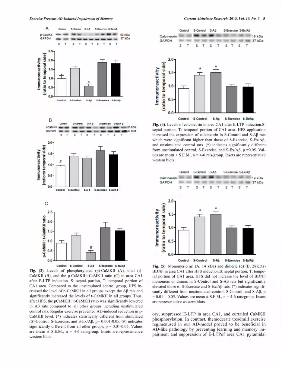
The levels of phosphorylated (p)-CaMKII in unstimu-lated control (1.0 ± 0.152) were statistically different com-pared to those ofstimulated (S-)Control, S-Ex, and S-Ex/A groups (S-Control: 1.575 ± 0.152, S-Ex: 1.889 ± 0.153, S-Ex/A : 1.825 ± 0.18, p= 0.01-0.05) but werenot significantly different from those of S-A rats (0.578 ± 0.122) (Fig. 3A). Total (t)-CaMKII levels were similar across all stimulated groups but markedly higher than those of unstimulated con-trol (unstimulated control: 0.956 ± 0.028, S-Control:1.404 ± 0.096, S-A : 1.301 ± 0.138, S-Ex: 1.648 ± 0.175, S-Ex/A : 1.453 ± 0.137, p= 0.05(Fig. 3B). This result indicates that CaMKII phosphorylation is inhibited in A rats, which is prevented by treadmill exercise. Furthermore, this conclu-sion is supported by the finding that the ratio of p-CaMKII:t-CaMKII was markedly lower in S-A rats compared to all other stimulated groups and even the unstimulated control (p=0.05-0.001, Fig. 3C). These datasuggest that although the total pool of CaMKII is unaltered,phosphorylation of this protein is negatively affected and that this effect is prevented by 4 weeks of treadmill exercise.
Upregulation of Calcineurin During E-LTP is Inhibited by Exercise
Calcineurin (PP2B) is a protein phosphatase that is im-portant for dephosphorylation of CaMKII. One hour after HFS application, the level of calcineurin in stimulated (S)-Control (1.404 ± 0.107) and S-A (1.51 ± 0.117) rats in-creased significantly compared to unstimulated control (0.913 ±0.098) (p= 0.05). In contrast, the levels of cal-cineurin after HFS in S-Ex (0.995 ± 0.08) and S-Ex/A (0.974 ±0.113) rats were similar to those inunstimulated con-trols suggesting that exercise prevents upregulation of PP2B (Fig. 4).
Levels of BDNF in Exercised Rats During E-TLP
Brain-derived neurotrophic factor (BDNF) is a neurotro-phic factor critical for neurogenesis, cognition, and synaptic plasticity [37-41]. BDNF can exist in both monomeric and homodimeric forms in cells [42]. The present protocol of high frequency stimulation of the Schaffer collateral syn-apses did not appreciably increase the levels of BDNF in S-Control (monomer level: 1.084 ± 0.103, dimer level: 1.087 ± 0.098) and S-A rats (monomer level: 1.093 ± 0.116, dimer level: 1.149 ± 0.134). However, treadmill exercise signifi-cantly increased both BDNF monomer (Fig. 5A) and dimer protein (Fig. 5B) levels in exercised animals including S-Ex (monomer level: 1.679 ± 0.179, dimer level: 1.854 ± 0.195) and S-Ex/A rats (monomer level: 1.696 ± 0.193, dimer level: 2.012 ± 0.398) compared to unstimulated control rats (monomer level: 1.005 ± 0.049, dimer level: 1.149 ± 0.134) (p= 0.05-0.01). Thus, in our rat model of AD, treadmill exer-cise markedly up-regulated both forms of BDNF, even in the presence of A (Fig. 5).
DISCUSSION
This study investigated the effect of 4 weeks of treadmill exercise on learning and memory and synaptic plasticity in a rat model of AD. Our findings consistentlyindicated a neu-roprotective effect of exercise in this ADmodel. The A rats exhibited severely impaired learning and short-term mem-
Exercise Prevents AD-Induced Impairment of Memory Current Alzheimer Research, 2013, Vol. 10, No. 5 5
Fig. (3). Levels of phosphorylated (p)-CaMKII (A), total (t)-CaMKII (B), and the p-CaMKII:t-CaMKII ratio (C) in area CA1 after E-LTP induction. S: septal portion, T: temporal portion of CA1 area. Compared to the unstimulated control group, HFS in-creased the level of p-CaMKII in all groups except the A rats and significantly increased the levels of t-CaMKII in all groups. Thus, after HFS, the pCaMKII : t-CaMKII ratio was significantly lowered in A rats compared to all other groups including unstimulated control rats. Regular exercise prevented AD-induced reduction in p-CaMKII level. (*) indicates statistically different from stimulated (S)-Control, S-Exercise, and S-Ex/A , p= 0.001-0.05. (#) indicates significantly different from all other groups, p = 0.01-0.05. Values are mean ± S.E.M., n = 4-6 rats/group. Insets are representative western blots.
Fig. (4). Levels of calcineurin in area CA1 after E-LTP induction.S: septal portion, T: temporal portion of CA1 area. HFS application increased the expression of calcineurin in S-Control and S-A rats which were significant higher than those of S-Exercise, S-Ex/A , and unstimulated control rats. (*) indicates significantly different from unstimulated control, S-Exercise, and S-Ex/A , p =0.05. Val-ues are mean ± S.E.M., n = 4-6 rats/group. Insets are representative western blots.
Fig. (5). Monomeric(m) (A, 14 kDa) and dimeric (d) (B, 28KDa) BDNF in area CA1 after HFS induction.S: septal portion, T: tempo-ral portion of CA1 area. HFS did not increase the level of BDNF monomers or dimers in S-Control and S-A rats but significantly elevated those of S-Exercise and S-Ex/A rats. (*) indicates signifi-cantly different from unstimulated control, S-Control, and S-A , p = 0.01 – 0.05. Values are mean ± S.E.M., n = 4-6 rats/group. Insets are representative western blots.
ory, suppressed E-LTP in area CA1, and curtailed CaMKII phosphorylation. In contrast, themoderate treadmill exercise regimenused in our AD-model proved to be beneficial in AD-like pathology by preventing learning and memory im-pairment and suppression of E-LTPof area CA1 pyramidal

6 Current Alzheimer Research, 2013, Vol. 10, No. 5 Dao et al.
cells, which correlated with a reduction incalcineurinincrease and exercise-induced elevation inBDNF and p-CaMKII lev-els.
Important cognitive impairment and neuropathological changes associated with AD have been recapitulated in dif-ferentanimal models ranging from the fruit fly to non-human primates. Among these, rodent transgenic and non-transgenic models have provided useful insights into the understanding of the pathogenesis as well as the therapeutic approaches for AD.In this study, we used a rat model of AD generated by i.c.v.infusion of A 1-42 peptides for 2 weeksas a model of-sporadic AD that accounts for the majority of AD cases.
It is thought that cleavage of amyloid precursor protein (APP) by a family of secretasesleads tothe production of A 1-40, A 1-42, and other peptide fragments. Even though A 1-40 is considered to be the most abundant among APP cleavage products, A 1-42 is thought to trigger the amyloi-dogenic cascade that leads to AD [43]. Under physiological conditions, extracellular amyloid proteins can be taken up by microglia or degraded by neprilysinshortly after their pro-duction [44]. Failure of adequate qAA clearance conse-quently favors amyloid accumulation and extracellular plaque deposition[45, 46]. The exact mechanism by which amyloid plaque formation leads to AD is unclear. However, it is known that amyloid aggregationlead to neurotoxicity often associated with oxidative stress, calcium dysregulation via amyloid channel formation and glutamate receptors, metabolic off-balance, and deleterious alterations in intracel-lular transduction pathways [47, 48]. In addition, A aggre-gates facilitate hyperphosphorylation of tau via modulation of kinases that phosphorylate this protein [34, 49, 50].
In agreement with previous findings, ourdata showed that in the RAWM, the performance of A rats is severely im-paired [1, 2]. During behavioral assessment, we also ob-served that the A rats spent significantly more time in the central area of the pool and appeareddisoriented. At the end of each learning trial, these rats simply sat on the platform without showing any interest in exploring the surroundings, which was not observed in rats of the other groups. In con-trast, the performance of Ex/A rats in the RAWM was simi-lar to that of the control and exercise rats.
In experimental setting, LTP is believed to bethe closest cellular correlate of learning and memory and thus used as a valuable tool for evaluating therapeutic treatmentsfor disor-ders of the central nervous system (CNS).Amyloid oligomers can inhibit LTP both in vitro and in vivo in various hippo-campal areas involved in the learning and memory processes [1, 2, 7, 51-54]. Previous findings from this lab have shown that amyloid infusion can impair synaptic plasticity both in the hippocampus and the sympathetic ganglia[1, 7, 55]. In the present study, E-LTP inarea CA1of A rats was mark-edly suppressed as shown by impairedfEPSP slope and pspikeamplitude. The mechanism of AD-induced LTP sup-pression remains unknown. However, it has been postulated that cellular ionic dysregulation may contribute to AD pa-thology by embedding amyloid oligomers into the membrane to form amyloid channels [48, 56, 57], which in turn directly or indirectly alter membrane ion channel expression and ac-tivity [58-62]. Regular exercise is known to produce a posi-tive effect on cognition and synaptic plasticity. For example,
treadmill exercise increases LTP expression measured as increases in fEPSP slope and pspike amplitude in the dentate gyrus (DG) both in vivo and in vitro [63, 64]. The ability of exercise to enhance synaptic plasticity is thought to be medi-ated by lowering LTP threshold for induction [64]. Addi-tionally, treadmill exercise can also prevent learning and memory impairment in rats that are acutely sleep-deprived, diabetic, or alcohol intoxicated [20, 22, 65].
How does muscle movement initiate these protectiveef-fects in the CNS? This is an intriguing question that has, as yet, no definitive answer, even though evidence for “cross-talk” between the skeletal muscles and CNS exists. For ex-ample, as running velocity increases, the discharge frequency of hippocampal CA1 pyramidal cells and interneurons in-creases [66]. Studies proposed that muscle can secrete nu-merous humoral factors that exert a protective effect on the brain [43]. Even though the exact molecular mechanism re-sponsible for the cross-talk between the skeletal muscles and CNS remains to be elucidated, experimental data support two possible mechanisms. First, events associated with en-ergy balance play an important role in CNS function. For example, exercise up-regulates hippocampal expression of the mitochondrial molecule, uncoupling protein 2 (UCP2), which in turn protects neuronal mitochondria from oxidative stress, enhances ATP production, and regulatesnormal cal-cium level [67]. In addition, UCP2 can also modulate BDNF signaling and its downstream mediators such as CREB and CaMKII [68]. Second,interleukin-6 (IL-6), an immunomodu-latory cytokine may be responsible for the crosstalk between the periphery and CNS. During exercise training, IL-6 in-crease is linked to reduced level of glycogen [69], which in turn can positively regulate glucose homeostasis in the brain.
It is well-documented that regular exercise can modulate the expression of several cognition-related molecules (e.g. BDNF, CaMKII) whose functions are severely affected by AD pathology. For example, during LTP induction the level of p-CaMKII is reduced due to AD-induced impairment of CaMKII phosphorylation[1, 55, 70]. In normal rats, HFS results in an increase in the levels of both phosphorylated and total CaMKII.Our data revealed thatHFS failed to appre-ciably increase the level of p-CaMKII in A rats and this change was prevented by exercise even though the total-CaMKII level is unchanged across all groups after HFS. This finding indicates that the diseasemainly targets the phos-phorylation process enabled by CaMKII, which is a critical step for LTP induction.In addition to LTP inhibition, amy-loid exposure can result in up-regulation of phosphatases that regulate CaMKII activation [53, 62, 71, 72]. Calcineurin (PP2B) is considered a gating mechanism of LTP as it inac-tivates kinases. In normal rats, HFS is expected to increase the level of PP2B, which is thought to prevent LTP satura-tion. Our data showed that LTP induction significantly ele-vated the levels of PP2B in stimulated control and A rats but did not affect those of exercised rats. Together, these findings demonstrate that exercise exerts a beneficial effect on memory and LTP possibly by restoring normal phospha-tase-kinase balance.
It is not coincidental that the level of BDNF, a potent mediator of synaptic plasticity and memory, is highly ele-vated during exercise training[73-75].In addition to its cen-

Exercise Prevents AD-Induced Impairment of Memory Current Alzheimer Research, 2013, Vol. 10, No. 5 7
tral production and release, BDNF can be produced from peripheral non-neuronal tissues and cross the blood brain barrier via a “high capacity, saturable transport system”[76, 77].In brain tissues, BDNF exists in both monomeric and dimeric forms, which exert maximal effect on neuronal sur-vival at saturated concentration as tested in dorsal root gan-glion neurons [78]. BDNF can act pre- or post-synaptically to modulate its own signaling or other pathways including CaMKIV and CREB, that are important in the learning and memory process [79-81]. In addition to its neurotrophic ef-fect, BDNF also possessesmetabotrophicproperties. BDNF up-regulates the expression of energy-related molecules in-cluding AMP-activated protein kinase (AMPK), ubiquitous mitochondrial creatinekinase (uMtCK) and UCP2 [82-84]. Thus, it is reasonable to suggest that depletion/disruption of BDNF expression/activity would significantly alter the ac-tion of these metabolic factors and eventually disrupt learn-ing and memory function. The present findings show thatthe levels of monomeric and dimeric BDNF after HFSare sig-nificantly elevated in exercised rats compared to the seden-tary group.The inability of HFS to increase BDNF level in S-Control rats suggests that, perhaps the stimulation protocol we used is not strong enough to cause a significant change in BDNF protein levels. However, HFS and exercise training seem to act in concert to increase the BDNF levels well be-yond the normal levels in both normal-exercised and A /exercised rats. This increase in BDNF may lead to im-proved performance in the RAWM and preservation of E-LTP in Ex/A rats. In contrast, disruption of BDNF or its receptor (TrkB) impairs spatial memory and suppresses LTP expression[85, 86]. The fact that these cognitive deficits can be restored by exogenous administration of BDNF suggests that BDNF plays an integral role in synaptic plasticity and memory [87-89].
In summary, our behavioral, electrophysiological and molecular findings from rat model of AD-like pathology strongly support the proposition that regular physical exer-cise may be beneficial in alleviating/preventing neurodegen-erative disorders including AD.
CONFLICT OF INTEREST
The author(s) confirm that this article content has no con-flicts of interest.
ACKNOWLEDGEMENTS
This study was funded by SGP grants from University of Houston. The authors disclose no conflict of biomedical or financial interest.
REFERENCES [1] Srivareerat M, Tran TT, Alzoubi KH, Alkadhi KA. Chronic psy-
chosocial stress exacerbates impairment of cognition and long-term potentiation in beta-amyloid rat model of Alzheimer's disease. Biol Psychiatry 65(11): 918-26 (2009).
[2] Srivareerat M, Tran TT, Salim S, Aleisa AM, Alkadhi KA. Chronic nicotine restores normal Abeta levels and prevents short-term memory and E-LTP impairment in Abeta rat model of Alzheimer's disease. Neurobiol Aging 32(5): 834-44 (2011).
[3] Xiong H, Callaghan D, Wodzinska J, Xu J, Premyslova M, Liu QY, et al. Biochemical and behavioral characterization of the dou-ble transgenic mouse model (APPswe/PS1dE9) of Alzheimer's dis-ease. Neurosci Bull 27(4): 221-32 (2011).
[4] Malm T, Koistinaho J, Kanninen K. Utilization of APPswe/PS1dE9 Transgenic Mice in Research of Alzheimer's Disease: Focus on Gene Therapy and Cell-Based Therapy Applications. Int J Alz-heimers Dis 2011: 517160 (2011).
[5] Alkadhi KA, Srivareerat M, Tran TT. Intensification of long-term memory deficit by chronic stress and prevention by nicotine in a rat model of Alzheimer's disease. Mol Cell Neurosci 45(3): 289-96 (2010).
[6] Walker JM, Fowler SW, Miller DK, Sun AY, Weisman GA, Wood WG, et al. Spatial learning and memory impairment and increased locomotion in a transgenic amyloid precursor protein mouse model of Alzheimer's disease. Behav Brain Res, 222(1): 169-75 (2011).
[7] Alkadhi KA, A.K., Srivareerat M, Tran TT. Chronic psychosocial stress exacerbates impairment of synaptic plasticity in -amyloid rat model of Alzheimer's disease: prevention by nicotine. Curr Alz-heimer Res 8(7): 718-31 (2011).
[8] Blitzer RD, Iyengar R, Landau EM, Postsynaptic signaling net-works: cellular cogwheels underlying long-term plasticity. Biol Psychiatry 57(2): 113-9 (2005).
[9] Kim SE, KI, Kim BK, Shin MS, Cho S, Kim CJ, et al. Treadmill exercise prevents aging-induced failure of memory through an in-crease in neurogenesis and suppression of apoptosis in rat hippo-campus. Exp Gerontol 45(5): 357-65 (2010).
[10] Nichol KE, Parachikova AI, Cotman CW. Three weeks of running wheel exposure improves cognitive performance in the aged Tg2576 mouse. Behav Brain Res 184(2): 124-32 (2007).
[11] Sofi F, Valecchi D, Bacci D, Abbate R, Gensini GF, Casini A, et al. Physical activity and risk of cognitive decline: a meta-analysis of prospective studies. J Intern Med 269(1): 107-17 (2011).
[12] Grace L, Hescham S, Kellaway LA, Bugarith K, Russell VA. Ef-fect of exercise on learning and memory in a rat model of devel-opmental stress. Metab Brain Dis 24(4): p. 643-57 (2009).
[13] Khabour OF, Alzoubi KH, Alomari MA, Alzubi MA. Changes in spatial memory and BDNF expression to concurrent dietary restric-tion and voluntary exercise. Hippocampus 20(5): 637-45 (2010).
[14] Luo CX, J.J., Zhou QG, Zhu XJ, Wang W, Zhang ZJ, Han X, Zhu DY. Voluntary exercise-induced neurogenesis in the postischemic dentate gyrus is associated with spatial memory recovery from stroke. J Neurosci Res 85(8): 1637-46 (2007).
[15] Radak Z, Toldy A, Szabo Z, Siamilis S, Nyakas C, Silye G, et al. The effects of training and detraining on memory, neurotrophins and oxidative stress markers in rat brain. Neurochem Int 49(4): 387-92 (2006).
[16] Albeck DS, Sano K, Prewitt GE, Dalton L. Mild forced treadmill exercise enhances spatial learning in the aged rat. Behav Brain Res 168(2): 345-8 (2006).
[17] Alaei H, Borjeian L, Azizi M, Orian S, Pourshanazari A, Hanninen O. Treadmill running reverses retention deficit induced by mor-phine. Eur J Pharmacol 536(1-2): 138-41 (2006).
[18] Hopkins ME, Bucci DJ. Interpreting the effects of exercise on fear conditioning: the influence of time of day. Behav Neurosci 124(6): 868-72 (2010).
[19] Helfer JL, Goodlett CR, Greenough WT, Klintsova AY. The ef-fects of exercise on adolescent hippocampal neurogenesis in a rat model of binge alcohol exposure during the brain growth spurt. Brain Res 1294: 1-11 (2009).
[20] Reisi P, Alaei H, Babri S, Sharifi MR, Mohaddes G. Effects of treadmill running on spatial learning and memory in streptozotocin-induced diabetic rats. Neurosci Lett 455(2): 79-83 (2009).
[21] Aguiar AS, Jr. Araújo AL, da-Cunha TR, Speck AE, Ignácio ZM, De-Mello N, et al. Physical exercise improves motor and short-term social memory deficits in reserpinized rats. Brain Res Bull 79(6): 452-7 (2009).
[22] Zagaar M, Alhaider I, Dao A, Levine A, Alkarawi A, Alzubaidy M, et al. The beneficial effects of regular exercise on cognition in REM sleep deprivation: behavioral, electrophysiological and mo-lecular evidence. Neurobiol Dis 45(3): 1153-62 (2012).
[23] Tran TT, Srivareerat M, Alkadhi KA. Chronic psychosocial stress triggers cognitive impairment in a novel at-risk model of Alz-heimer's disease. Neurobiol Dis 37(3): 756-63 (2010).
[24] Alzoubi KH, Aleisa AM, Alkadhi KA. Nicotine prevents disruption of the late phase LTP-related molecular cascade in adult-onset hy-pothyroidism. Hippocampus 17(8): 654-64 (2007).
[25] Alzoubi KH, Alkadhi KA. A critical role of CREB in the impair-ment of late-phase LTP by adult onset hypothyroidism. Exp Neurol 203(1): p. 63-71 (2007).

8 Current Alzheimer Research, 2013, Vol. 10, No. 5 Dao et al.
[26] Papatheodoropoulos C, Kostopoulos G. Dorsal-ventral differentia-tion of short-term synaptic plasticity in rat CA1 hippocampal re-gion. Neurosci Lett 286(1): 57-60 (2000).
[27] Hedou G, Mansuy IM. Inducible molecular switches for the study of long-term potentiation. Philos Trans R Soc Lond B Biol Sci 358(1432): 797-804 (2003).
[28] Fukunaga K, Miyamoto E. A working model of CaM kinase II activity in hippocampal long-term potentiation and memory. Neu-rosci Res 38(1): 3-17 (2000).
[29] Malenka RC, Kauer JA, Perkel DJ, Mauk MD, Kelly PT, Nicoll RA, et al. An essential role for postsynaptic calmodulin and protein kinase activity in long-term potentiation. Nature 340(6234): 554-7 (1989).
[30] Aleisa AM, Alzoubi KH, Gerges NZ, Alkadhi KA. Chronic psy-chosocial stress-induced impairment of hippocampal LTP: possible role of BDNF. Neurobiol Dis 22(3): 453-62 (2006).
[31] Silva AJ, Wang Y, Paylor R, Wehner JM, Stevens CF, Tonegawa S. Impaired spatial learning in alpha-calcium-calmodulin kinase II mutant mice. Science 257(5067): 206-11 (1992).
[32] Silva AJ, Wang Y, Paylor R, Wehner JM, Stevens CF, Tonegawa S. Alpha calcium/calmodulin kinase II mutant mice: deficient long-term potentiation and impaired spatial learning. Cold Spring Harb Symp Quant Biol 57: 527-39 (1992).
[33] Wang JH, Kelly PT. The balance between postsynaptic Ca(2+)-dependent protein kinase and phosphatase activities controlling synaptic strength. Learn Mem 3(2-3): 170-81 (1996).
[34] Moriguchi S, SN, Han F, Yeh JZ, Narahashi T, Fukunaga K. Gal-antamine enhancement of long-term potentiation is mediated by calcium/calmodulin-dependent protein kinase II and protein kinase C activation. Hippocampus 19(9): 845-54 (2009).
[35] Takahashi E, Niimi K, Itakura C. Enhanced CaMKII activity and spatial cognitive function in SAMP6 mice. Behav Neurosci 123(3): 527-32 (2009).
[36] Oomura Y, Hori N, Shiraishi T, Fukunaga K, Takeda H, Tsuji M, et al. Leptin facilitates learning and memory performance and en-hances hippocampal CA1 long-term potentiation and CaMK II phosphorylation in rats. Peptides 27(11): 2738-49 (2006).
[37] Rossi C, Angelucci A, Costantin L, Braschi C, Mazzantini M, Babbini F, et al. Brain-derived neurotrophic factor (BDNF) is re-quired for the enhancement of hippocampal neurogenesis following environmental enrichment. Eur J Neurosci 24(7): 1850-6 (2006).
[38] Lee J, Duan W, Mattson MP. Evidence that brain-derived neu-rotrophic factor is required for basal neurogenesis and mediates, in part, the enhancement of neurogenesis by dietary restriction in the hippocampus of adult mice. J Neurochem 82(6): 1367-75 (2002).
[39] Tyler WJ, Zhang XL, Hartman K, Winterer J, Muller W, Stanton PK, et al., BDNF increases release probability and the size of a rap-idly recycling vesicle pool within rat hippocampal excitatory syn-apses. J Physiol 574(Pt 3): 787-803 (2006).
[40] Kang H, Schuman EM. Long-lasting neurotrophin-induced en-hancement of synaptic transmission in the adult hippocampus. Sci-ence 267(5204): 1658-62 (1995).
[41] Kang HJ, Schuman EM. Neurotrophin-induced modulation of synaptic transmission in the adult hippocampus. J Physiol Paris 89(1): p. 11-22 (1995).
[42] Shen J, Maruyama IN. Brain-derived neurotrophic factor receptor TrkB exists as a preformed dimer in living cells. J Mol Signal 7(1): 2 (2012).
[43] Hardy J, The amyloid hypothesis for Alzheimer's disease: a critical reappraisal. J Neurochem, 110(4): 1129-34 (2009).
[44] Gotz J, Streffer JR, David D, Schild A, Hoerndli F, Pennanen L, et al. Transgenic animal models of Alzheimer's disease and related disorders: histopathology, behavior and therapy. Mol Psychiatry 9(7): 664-83 (2004).
[45] Wetzel R. Kinetics and thermodynamics of amyloid fibril assem-bly. Acc Chem Res 39(9): 671-9 (2006).
[46] Zerovnik, E. Amyloid-fibril formation. Proposed mechanisms and relevance to conformational disease. Eur J Biochem 269(14): 3362-71 (2002).
[47] Di Carlo M. Beta amyloid peptide: from different aggregation forms to the activation of different biochemical pathways. Eur Bio-phys J 39(6): 877-88 (2010).
[48] Capone R, J.H., Kotler SA, Connelly L, Teran Arce F, Ramachandran S, et al. All-d-Enantiomer of -Amyloid Peptide Forms Ion Channels in Lipid Bilayers. J Chem Theory Comput 8(3): 1143-1152 (2012).
[49] Blurton-Jones M, Laferla FM. Pathways by which Abeta facilitates tau pathology. Curr Alzheimer Res 3(5): 437-48 (2006).
[50] Lloret A, Badia MC, Giraldo E, Ermak G, Alonso MD, Pallardó FV, et al. Amyloid-beta toxicity and tau hyperphosphorylation are linked via RCAN1 in Alzheimer's disease. J Alzheimers Dis 27(4): 701-9 (2011).
[51] Ondrejcak T, Wang Q, Kew JN, Virley DJ, Upton N, Anwyl R, Rowan MJ. Activation of 7 nicotinic acetylcholine receptors per-sistently enhances hippocampal synaptic transmission and prevents Aß-mediated inhibition of LTP in the rat hippocampus. Eur J Pharmacol 677(1-3): 63-70 (2012).
[52] Barry AE, Klyubin I, Mc Donald JM, Mably AJ, Farrell MA, Scott M, et al. Alzheimer's disease brain-derived amyloid- -mediated in-hibition of LTP in vivo is prevented by immunotargeting cellular prion protein. J Neurosci 31(20): 7259-63 (2011).
[53] Jo J, Whitcomb D, Olsen KM, Kerrigan TL, Lo SC, Bru-Mercier G, et al. A (1-42) inhibition of LTP is mediated by a signaling pathway involving caspase-3, Akt1 and GSK-3 . Nat Neurosci 14(5): 545-7(2011).
[54] Ma T, Hoeffer CA, Wong H, Massaad CA, Zhou P, Iadecola C, et al. Amyloid beta-induced impairments in hippocampal synaptic plasticity are rescued by decreasing mitochondrial superoxide. J Neurosci 31(15): 5589-95 (2011).
[55] Alzoubi KH, Alhaider IA, Tran TT, Mosely A, Alkadhi KK. Im-paired neural transmission and synaptic plasticity in superior cervi-cal ganglia from beta-amyloid rat model of Alzheimer's disease. Curr Alzheimer Res 8(4): 377-84 (2011).
[56] Capone R, Jang H, Kotler SA, Kagan BL, Nussinov R, Lal R. Prob-ing structural features of Alzheimer's amyloid- pores in bilayers using site-specific amino acid substitutions. Biochemistry 51(3): 776-85 (2012).
[57] Connelly L, Jang H, Arce FT, Capone R, Kotler SA, Ramachandran S, et al. Atomic force microscopy and MD simula-tions reveal pore-like structures of all-D-enantiomer of Alzheimer's beta-amyloid peptide: relevance to the ion channel mechanism of AD pathology. J Phys Chem B 116(5): 1728-35 (2012).
[58] Demuro A, S.M., Parker I, Single-channel Ca(2+) imaging impli-cates A 1-42 amyloid pores in Alzheimer's disease pathology. J Cell Biol 195(3): 515-24 (2011).
[59] Kim S, Rhim H. Effects of amyloid- peptides on voltage-gated L-type Ca(V)1.2 and Ca(V)1.3 Ca(2+) channels. Mol Cells 32(3): 289-94 (2011).
[60] Mezler M, B.S., Schoemaker H, Gross G, Nimmrich V, A -amyloid oligomer directly modulates P/Q-type calcium currents in Xenopus oocytes. Br J Pharmacol 165(5): 1572-83 (2012).
[61] Dewachter I, Filipkowski RK, Priller C, Ris L, Neyton J, Croes S, et al. Deregulation of NMDA-receptor function and down-stream signaling in APP[V717I] transgenic mice. Neurobiol Aging, 30(2): 241-56 (2009).
[62] Calon F, Lim GP, Morihara T, Yang F, Ubeda O, Salem N Jr, et al. Dietary n-3 polyunsaturated fatty acid depletion activates caspases and decreases NMDA receptors in the brain of a transgenic mouse model of Alzheimer's disease. Eur J Neurosci 22(3): 617-26 (2005).
[63] O'Callaghan RM, Ohle R, Kelly AM. The effects of forced exercise on hippocampal plasticity in the rat: A comparison of LTP, spatial- and non-spatial learning. Behav Brain Res 176(2): 362-6 (2007).
[64] Farmer J, hao X, van Praag H, Wodtke K, Gage FH, Christie BR. Effects of voluntary exercise on synaptic plasticity and gene ex-pression in the dentate gyrus of adult male Sprague-Dawley rats in vivo. Neuroscience 124(1): 71-9 (2004).
[65] Sim, Y.J., Kim H, Shin MS, Chang HK, Shin MC, Ko IG, Kim KJ, et al. Effect of postnatal treadmill exercise on c-Fos expression in the hippocampus of rat pups born from the alcohol-intoxicated mothers. Brain Dev 30(2): 118-25 (2008).
[66] Czurko A, Hirase H, Csicsvari J, Buzsáki G. Sustained activation of hippocampal pyramidal cells by 'space clamping' in a running wheel. Eur J Neurosci 11(1): 344-52 (1999).
[67] Vaynman S, Ying Z, Wu A, Gomez-Pinilla F. Coupling energy metabolism with a mechanism to support brain-derived neurotro-phic factor-mediated synaptic plasticity. Neuroscience 139(4): 1221-34 (2006).
[68] Vaynman S, Ying Z, Gomez-Pinilla F. Interplay between brain-derived neurotrophic factor and signal transduction modulators in the regulation of the effects of exercise on synaptic-plasticity. Neu-roscience 122(3): 647-57 (2003).

Exercise Prevents AD-Induced Impairment of Memory Current Alzheimer Research, 2013, Vol. 10, No. 5 9
[69] Steensberg A, Febbraio MA, Osada T, Schjerling P, van Hall G, Saltin B, et al. Interleukin-6 production in contracting human skeletal muscle is influenced by pre-exercise muscle glycogen con-tent. J Physiol 537(Pt 2): 633-9 (2001).
[70] Zha, D, Watson JB, Xie CW. Amyloid beta prevents activation of calcium/calmodulin-dependent protein kinase II and AMPA recep-tor phosphorylation during hippocampal long-term potentiation. J Neurophysiol 92(5): 2853-8 (2004).
[71] Knobloch M, Farinelli M, Konietzko U, Nitsch RM, Mansuy IM. Abeta oligomer-mediated long-term potentiation impairment in-volves protein phosphatase 1-dependent mechanisms. J Neurosci 27(29): 7648-53 (2007).
[72] Chen QS, Wei WZ, Shimahara T, Xie CW. Alzheimer amyloid beta-peptide inhibits the late phase of long-term potentiation through calcineurin-dependent mechanisms in the hippocampal dentate gyrus. Neurobiol Learn Mem 77(3): 354-71 (2002).
[73] Castren E, Pitkänen M, Sirviö J, Parsadanian A, Lindholm D, Thoenen H, et al. The induction of LTP increases BDNF and NGF mRNA but decreases NT-3 mRNA in the dentate gyrus. Neurore-port 4(7): 895-8 (1993).
[74] Silhol M, Arancibia S, Maurice T, Tapia-Arancibia L. Spatial memory training modifies the expression of brain-derived neu-rotrophic factor tyrosine kinase receptors in young and aged rats. Neuroscience 146(3): 962-73 (2007).
[75] Kesslak JP, So V, Choi J, Cotman CW, Gomez-Pinilla F. Learning upregulates brain-derived neurotrophic factor messenger ribonu-cleic acid: a mechanism to facilitate encoding and circuit mainte-nance? Behav Neurosci 112(4): 1012-9 (1998).
[76] Pan W, Banks WA, Fasold MB, Bluth J, Kastin AJ. Transport of brain-derived neurotrophic factor across the blood-brain barrier. Neuropharmacology 37(12): 1553-61 (1998).
[77] Zoladz JA, Pilc A. The effect of physical activity on the brain de-rived neurotrophic factor: from animal to human studies. J Physiol Pharmacol 61(5): 533-41 (2010).
[78] Kolbeck R, Jungbluth S, Barde YA. Characterisation of neurotro-phin dimers and monomers. Eur J Biochem 225(3): 995-1003 (1994).
[79] Spencer TK, Mellado W, Filbin MT. BDNF activates CaMKIV and PKA in parallel to block MAG-mediated inhibition of neurite out-growth. Mol Cell Neurosci 38(1): 110-6 (2008).
[80] Williams CM, El Mohsen MA, Vauzour D, Rendeiro C, Butler LT, Ellis JA, et al. Blueberry-induced changes in spatial working mem-ory correlate with changes in hippocampal CREB phosphorylation and brain-derived neurotrophic factor (BDNF) levels. Free Radic Biol Med 45(3): 295-305 (2008).
[81] Cassilhas RC, Lee KS, Fernandes J, Oliveira MG, Tufik S, Meeusen R, et al. Spatial memory is improved by aerobic and re-sistance exercise through divergent molecular mechanisms. Neuro-science 202: 309-17 (2012).
[82] Gomez-Pinilla F, Vaynman S, Ying Z. Brain-derived neurotrophic factor functions as a metabotrophin to mediate the effects of exer-cise on cognition. Eur J Neurosci 28(11): 2278-87 (2008).
[83] Chaldakov G, The metabotrophic NGF and BDNF: an emerging concept. Arch Ital Biol 149(2): 257-63 (2011).
[84] Pedersen BK, Pedersen M, Krabbe KS, Bruunsgaard H, Matthews VB, Febbraio MA. Role of exercise-induced brain-derived neu-rotrophic factor production in the regulation of energy homeostasis in mammals. Exp Physiol 94(12): 1153-60 (2009).
[85] Hennigan A, Callaghan CK, Kealy J, Rouine J, Kelly AM. Deficits in LTP and recognition memory in the genetically hypertensive rat are associated with decreased expression of neurotrophic factors and their receptors in the dentate gyrus. Behav Brain Res 197(2): 371-7 (2009).
[86] Zhou J, Zhang F, Zhang Y. Corticosterone inhibits generation of long-term potentiation in rat hippocampal slice: involvement of brain-derived neurotrophic factor. Brain Res 885(2): 182-91 (2000).
[87] Kline, D.D., Ogier M, Kunze DL, Katz DM. Exogenous brain-derived neurotrophic factor rescues synaptic dysfunction in Mecp2-null mice. J Neurosci 30(15): 5303-10 (2010).
[88] Rex CS, Lauterborn JC, Lin CY, Kramár EA, Rogers GA, Gall CM, et al. Restoration of long-term potentiation in middle-aged hippocampus after induction of brain-derived neurotrophic factor. J Neurophysiol 96(2): 677-85 (2006).
[89] Shaw KN, Commins S, O'Mara SM. Deficits in spatial learning and synaptic plasticity induced by the rapid and competitive broad-spectrum cyclooxygenase inhibitor ibuprofen are reversed by in-creasing endogenous brain-derived neurotrophic factor. Eur J Neu-rosci 17(11): 2438-46 (2003).
Received: ????????????? Revised: ??????????????? Accepted: ????????????


