Embed Size (px)
Citation preview
Transcriptome of Pneumocystis carinii during FulminateInfection: Carbohydrate Metabolism and the Concept ofa Compatible ParasiteMelanie T. Cushion1,5.*, A. George Smulian1., Bradley E. Slaven1., Tom Sesterhenn1, Jonathan Arnold2, Chuck Staben3, Aleksey Porollo4, RafalAdamczak4, Jarek Meller4
1 University of Cincinnati College of Medicine, Department of Internal Medicine, Division of Infectious Diseases, Cincinnati, Ohio, United States ofAmerica, 2 University of Georgia, Department of Genetics, Athens, Georgia, United States of America, 3 University of Kentucky, Lexington, Kentucky,United States of America, 4 Division of Biomedical Informatics, Children’s Hospital Research Foundation, Cincinnati, Ohio, United States of America,5 Cincinnati Veterans Administration Medical Center, Cincinnati, Ohio, United States of America
Members of the genus Pneumocystis are fungal pathogens that cause pneumonia in a wide variety of mammals withdebilitated immune systems. Little is known about their basic biological functions, including life cycle, since no species can becultured continuously outside the mammalian lung. To better understand the pathological process, about 4500 ESTS derivedfrom sequencing of the poly(A) tail ends of P. carinii mRNAs during fulminate infection were annotated and functionallycharacterized as unassembled reads, and then clustered and reduced to a unigene set with 1042 members. Because of thepresence of sequences from other microbial genomes and the rat host, the analysis and compression to a unigene set wasnecessarily an iterative process. BLASTx analysis of the unassembled reads (UR) vs. the Uni-Prot and TREMBL databasesrevealed 56% had similarities to existing polypeptides at E values of#1026, with the remainder lacking any significanthomology. The most abundant transcripts in the UR were associated with stress responses, energy production, transcriptionand translation. Most (70%) of the UR had similarities to proteins from filamentous fungi (e.g., Aspergillus, Neurospora) andexisting P. carinii gene products. In contrast, similarities to proteins of the yeast-like fungi, Schizosaccharomyces pombe andSaccharomyces cerevisiae, predominated in the unigene set. Gene Ontology analysis using BLAST2GO revealed P. cariniidedicated most of its transcripts to cellular and physiological processes (,80%), molecular binding and catalytic activities(,70%), and were primarily derived from cell and organellar compartments (,80%). KEGG Pathway mapping showed theputative P. carinii genes represented most standard metabolic pathways and cellular processes, including the tricarboxylic acidcycle, glycolysis, amino acid biosynthesis, cell cycle and mitochondrial function. Several gene homologs associated withmating, meiosis, and sterol biosynthesis in fungi were identified. Genes encoding the major surface glycoprotein family (MSG),heat shock (HSP70), and proteases (PROT/KEX) were the most abundantly expressed of known P. carinii genes. The apparentpresence of many metabolic pathways in P. carinii, sexual reproduction within the host, and lack of an invasive infectionprocess in the immunologically intact host suggest members of the genus Pneumocystis may be adapted parasites and havea compatible relationship with their mammalian hosts. This study represents the first characterization of the expressed genesof a non-culturable fungal pathogen of mammals during the infective process.
Citation: Cushion MT, Smulian AG, Slaven BE, Sesterhenn T, Arnold J, et al (2007) Transcriptome of Pneumocystis carinii during Fulminate Infection:Carbohydrate Metabolism and the Concept of a Compatible Parasite. PLoS ONE 2(5): e423. doi:10.1371/journal.pone.0000423
INTRODUCTIONOnce thought to be protozoan parasites, members of the genus
Pneumocystis were placed in the fungal kingdom by phylogenetic
analyses of several genes [1–5]. The genus Pneumocystis was then
placed in the fungal phylum Ascomycota, subphylum Taphrino-
mycotina (O.E. Eriksson and Winka 1997), Order Pneumocysti-
dales (O.E. Erikss. 1994), Class Pneumocystidomycetes (sensu
O.E. Erikss.&Winka 1997), Family Pneumocystidaceae (O.E.
Erikss. 1994), Genus Pneumocystis (Delanoe&Delanoe 1912) [6].
The Taphrinomycotina are a paraphyletic group of organisms and
the identity of the closest extant relative to the genus Pneumocystis
is not yet clear and varies by gene sequences examined and
method of comparison. The fungi included within this group are
highly diverse and include such members as the fission yeast,
Schizosaccharomyces pombe, the plant pathogen, Taphrina deformans,
and Neolecta vitellina, the only member with a fruiting body
structure [7]. The genus, Pneumocystis, is comprised of multiple
species that inhabit specific mammalian hosts. To date, 5 species
have been formally described [8]. Pneumocystis jirovecii infects
human beings [9,10]; P. murina is found in mice [11]; P. oryctolagi
infects rabbits [12] and P. carinii [9,10]and P. wakefieldiae [13,14]
both inhabit the lungs of rats.
These non-filamentous, yeast-like fungal organisms inhabit the
lungs of mammals and can cause a lethal pneumonia when the
Academic Editor: Debbie Fox, The Research Institute for Children, United Statesof America
Received November 6, 2006; Accepted April 8, 2007; Published May 9, 2007
This is an open-access article distributed under the terms of the CreativeCommons Public Domain declaration which stipulates that, once placed in thepublic domain, this work may be freely reproduced, distributed, transmitted,modified, built upon, or otherwise used by anyone for any lawful purpose.
Funding: NIH: R01 AI44651 (M.T.C., A.G.S., J.A., C.S.) and R21 (J.M., A.G.S.); theVeterans Administration (Medical Research Equipment Grant) (A.G.S., M.T.C.). TheNIH or the VA had no role in the design or conduct etc. of the study. Theyprovided funds to execute the studies after award of grants (NIH) or equipment(VA). We also thank the UGA College of Agricultural and Environmental Sciencesfor their support.
Competing Interests: The authors have declared that no competing interestsexist.
* To whom correspondence should be addressed. E-mail: [email protected], [email protected]
. These authors contributed equally to this work.
PLoS ONE | www.plosone.org 1 May 2007 | Issue 5 | e423
host immune system becomes debilitated or compromised.
Infection due to viruses, such as the Human Immunodeficiency
Virus (HIV); malnutrition; chemotherapeutic agents; and other
underlying diseases can create an environment that permits the
growth of Pneumocystis. In persons with HIV, pneumonia caused by
Pneumocystis (PCP) had been a major cause of mortality prior to the
advent of Highly Affective Anti-Retroviral Therapy (HAART) [15].
Although treatment with HAART reduced the frequency of
infections with P. jirovecii and other opportunistic microbes in the
United States and Europe, PCP remains an important disease of the
immunocompromised. In contrast, there has been a sharp increase
in PCP in HIV-infected individuals in underdeveloped and
developing countries, such as in sub-Saharan Africa, Asia, and in
India where access to HAART is limited or unavailable [16–18].
The role of P. jirovecii as a potential co-morbidity factor in underlying
diseases processes such as chronic obstructive pulmonary disease
(COPD) is a focus of several ongoing investigations [19,20].
Limited therapy is available with which to treat PCP, since these
fungi are not susceptible to standard anti-fungal drugs like
Amphotericin B or the azole family of compounds. Exacerbating
the problem of few alternative chemotherapeutic options is the
emergence of mutations in the gene encoding dihydropteroate
synthase [21–24], the target of the sulfa component of the most
efficacious therapy used to treat PCP, trimethoprim-sulfamethox-
azole, and in the gene encoding cytochrome b, a target of
a secondary therapy, atovaquone [25]. Such mutations in other
organisms increased the resistance to these therapies and have
been linked to failure of PCP prophylaxis.
Pneumocystis maintain an extracellular existence in lung alveoli.
Microscopic studies at the light and electron microscopic levels
have lead to several proposed life cycles, reviewed [26]. Most
include an asexual mode of replication via binary fission of the
trophic form and a sexual mode resulting in formation of an ascus
(cyst) containing 8 ascospores. Mating is likely mediated by the
trophic forms, as evidenced by homologs to yeast pheromone
receptor genes present in the P. carinii genome [27,28] and the
expression of a pheromone receptor protein on the surface of some
trophic forms [28]. Besides the cyst and trophs, there are several
intermediate forms that likely represent the progression from
zygote through meiosis; the additional mitotic step to produce 8
nuclei; then separation into ascospores. The infection is thought to
be initiated by attachment of the trophic forms to the Type I
pneumocyte in the host alveoli. However, the mode of travel by
the trophic form to the alveoli is unknown, as is the actual
infectious propagule. Once in the alveolus, clusters of organisms
grow from trophic forms anchored to the Type I cells and fill the
lumen. The mode of transmission from one host to another is not
known. No environmental form or cycle has been identified. All of
the current information on the life cycle has been derived from the
study of organisms in the lungs of mammals with debilitated
immune systems.
Experimental approaches for the study of these fungi have been
limited by the lack of an in vitro culture system. Research has
relied on animal models of infection as a source of organisms for
biochemical testing, drug evaluation, and microscopic visualiza-
tion for life cycle analyses. This report describes the transcriptional
analysis of P. carinii during fulminate infection in the immunosup-
pressed rat host.
A genome sequencing project was undertaken to probe the
complexity of the Pneumocystis genome and identify genes that may
serve as potential therapeutic targets [29]. The species P. carinii
was chosen for the project because the immunosuppressed rat
provides the highest numbers of organisms that can be obtained
reliably from any animal model. Sources of the species found in
humans, P. jirovecii, are limited and often low in organism numbers.
Although similar in some phenotypic traits such as response to
therapies and expression of surface glycoprotein variants, the
genomes of each species are likely to have unique characteristics. It
is a goal of this first Pneumocystis genome project to provide
a potential genomic scaffold for assembly and comparison of the
other members of this genus.
One aim of the project was to create an expressed sequence tag
(EST) database from organisms harvested during fulminate
pneumonia to identify genes that may be associated with the
pathogenic process. Assembly and annotation of the ESTs
produced approximately 1,632 gene transcripts. The correspond-
ing cDNA clones were sequenced in the forward and reverse
directions to obtain full length gene sequences. In some cases,
additional closure was needed to complete the sequences. These
sequences were further purged of duplicated genes and sequences
from other microbes and host cDNAs to establish a unigene set of
1042 members. Analysis of ESTs and cDNAs showed over-
whelming homologies to fungi, further supporting placement of
the genus into the fungal kingdom. Functional analyses using the
Gene Ontology and KEGG processes showed that P. carinii is
likely to be capable of a wide variety of metabolic functions but
devotes large portions of its transcriptome to the expression of
Pneumocystis-specific Major Surface Glycoprotein genes (MSG) and
to energy production during infection.
MATERIALS AND METHODS
cDNA library construction and generation of an EST
databaseA cDNA library of Pneumocystis carinii karyotype form 1 organisms
was made from RNA purified using the TriZOL reagent
(Invitrogen, Carlsbad, CA) from the lungs of a single, naturally-
infected Long Evans rat with a fulminate infection, by construction
in the Uni-ZAP XR vector (Stratagene Inc., LaJolla, CA) [27].
The rat was a member of a Pneumocystis-infected rat colony
maintained at the Cincinnati Veterinary Medical Unit, Veterans
Affairs Medical Center in standard caging racks with access to
room air. Fulminate infection in selected members was induced by
chronic administration of dexamethasone (4 mg/kg/week) for 10
to 14 weeks as previously described [30]. The primary library
consisted of 56105 clones which was amplified once to a titer of
961011. The ESTs were sequenced at the University of Georgia
sequence center from the 39 polyA tails (Athens, GA) with Big Dye
termination protocols using ABI 3700 instrumentation (Applied
Biosystems, Foster City, CA) resulting in about 4500 reads. The
average length of a read was about 500 bp.
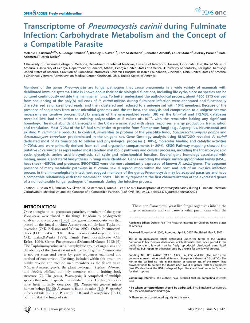
Unassembled EST sequencesThe ESTs were processed according to the scheme outlined in
Figure 1. In the initial analysis, the sequences were screened for
quality, vector and other contaminants (e.g. rat, bacteria) using the
Phred and Cross_ Match processing tools (http://www.phrap.org/)
[31–34] and BLASTn resulting in 3896 ESTs which were submitted
to GenBank with ID numbers from AW331850-335745 (Jan. 31,
2000). This initial step was performed in compliance with the
Bermuda Principles [35]. The 4500 unassembled ESTs were then
analyzed by BLASTx and BLASTn (Oct. 20, 2003) against the UNI-
PROT_TREMBL databases (http://www.pir.uniprot.org) to pro-
vide an assessment of overall contamination, gene homologies, and
relative transcript expression. This set of sequences is referred to as
the ‘‘Total ESTs’’ throughout the study. The Total EST set was
used to populate BLAST2GO and KASS categories at a BLASTx E-
value of 1026 or less required for entry.
P. carinii Transcriptome
PLoS ONE | www.plosone.org 2 May 2007 | Issue 5 | e423
Processing of the unigene set
To arrive at a unigene set, the 4500 sequences were re-processed
using Phred and Cross_Match, then trimmed for quality, using an
in-house Perl program qTrim available at pgp.cchmc.org, that
removed lesser quality flanking sequence that contributed to error
rates of 10% or greater [36] (Figure 1). In addition to standard
vector screens, several genomes of potential microbes living within
the immunosuppressed rat lung were added to the screening
library. They included the genomes of Bacillus subtilis, Pseumodmonas
aeruginosa, Pasteurella putida, P. multocida, Staphylococcus aureus,
adenovirus type 12, Haemophilus influenzae, mouse adenovirus 1,
mouse adenovirusA, and murid herpes virus. The trimmed
sequences were then subjected to homology analysis using
Figure 1. EST Analysis and Unigene Process. The raw sequence reads of the Expressed Sequence Tags were first purged of poor quality sequence(arrows to right of starting point). The resulting 3,896 reads were deposited to NHLBI GenBank and further analyzed for similarities to genes and geneproducts in the UNI-PROT_TREMBL databases using BLASTn and BLASTx and putative function with BLAST2GO and KASS. The raw reads were thenprocessed using an iterative scheme to form the unigene set starting with a primary screen to purge sequences originating from the cloning vector,rat host and bacteria, followed by a trimming of the sequence ends to reduce poor quality sequence, using an in house program (qTrim). Aftertrimming, the sequences were again purged of contaminants using BLASTn and BLASTx, then assembled using the CAP3 assembly program. Afteranother round of contaminant removal, 1,632 cDNA clones representing putative unique genes were selected for full sequencing. These sequenceswere then subjected to the same qTrim program, then assembled by CAP3. The primer design program, Primer3 was used to design primers to closegaps in those clone sequence that did not represent full sequences. After another round of assembly, the cDNA sequences were compared to oneanother for sequence identity using BLASTn, to identify any redundant gene sequences. This resulted in a set of 1042 unique sequences (both contigsand singletons) of which 994 had significant similarities (E#1026) to genes within the UNI-PROT_TREMBL and 48 did not have significant similaritiesto existing genes (E$1026). The unigene set was then analyzed for putative functions by BLAST2GO and KAAS.doi:10.1371/journal.pone.0000423.g001
P. carinii Transcriptome
PLoS ONE | www.plosone.org 3 May 2007 | Issue 5 | e423
BLASTn and BLASTx against the NCBI databases (www.ncbi.
nlm.nih.gov) (Oct. 2003). P. carinii sequences with BLASTn Expect
values (E) of less than 102100 to rat, mouse, or bacterial sequences
were removed; a BLASTx E value of less than 102100 (i.e. 102101
to complete identity of 0) to rat, mouse or bacterial proteins were
also eliminated from the set. The sequences were then assembled
using the Cap3 Assembler downloaded from http://www.cs.
iastate.edu/,xqhuang/[37] to reduce redundancy and increase
read reliability by condensing overlapping sequence and associated
quality scores. Sequences were evaluated for similarity using
BLASTx and BLASTn [38] against the NCBI non-redundant
database (http://www.ncbi.nlm.nih.gov/Ftp/); UniPROT and
UNI-PROT_TREMBL databases (http://www.pir.uniprot.org).
Data were analyzed and compiled using Microsoft Excel XP. The
UNI-PROT_TREMBL database was chosen for the EST and
cDNA sequence analyses due to the extensive degree of annotation
and output format.
After manual screening for additional contaminating sequences,
1632 cDNA clones were submitted for sequencing of the full
length inserts using the forward and reverse primers of the
pBluescript plasmid (T3, T7) to the Cincinnati Childrens Hospital
Medical Center Sequencing Core, Cincinnati, OH. The cDNA
clones represented what we believed to be non-redundant, non-
contaminant sequences. The sequences were evaluated for quality,
and trimmed as described above, then assembled with CAP3. In
800 cases, the clone was not fully sequenced and primers were
designed to close the gap using Primer3 [39]. The cDNA
sequences were screened for redundancy by BLAST analysis of
each constituent against the entire cDNA dataset. This resulted in
2 groups within the dataset; one group of contiguous sequences
and another comprised of singletons, identified as ‘‘cDNAv1_0.
fasta.screen.ContigX’’ and Plate No., Well No.uni.t/f.ab1 (e.g.
14e02.uni.t.ab1), respectively. Both groups were evaluated for
homology to other gene sequences using BLASTx and BLASTn
against the databases from NCBI (nr), Swiss Prot, and TREMBL
(May 2004). This resulted in removal of additional rat/mouse and
bacterial sequences (E#10240) and resulted in 981 sequences in
the unigene collection that had homology to proteins in the
databases, including hypothetical proteins. An additional 74 had
E-values greater than 1026 . Within the 981 sequences were 45
contigs with similarities to mammalian genes (E values$10240).
During the revision of this manuscript, the 74 ‘‘low score’’ hits and
the putative mammalian conserved genes were re-analyzed.
Twenty-six of the low score contigs were found to have similarities
to proteins in the database with E values#1026 resulting in a shift
of 26 contigs to the 981 sequences with significant identities to
gene homologs. The remaining 48 sequences with E values of
1026 or greater were retained in the unigene set as well. The 45
sequences with BLASTX E-values of$10240 to mammalian gene
homologs were also re-analyzed to assess whether these were host
in origin, or if they were indeed conserved genes in the P. carinii
genome. The sequences were analyzed for total AT content, then
re-assessed in relation to their BLASTx and BLASTn E-values.
The P. carinii genome has a high AT content (,68%) vs. the rat
genome (,50%). There was a clear demarcation between
sequences in the 45 contig set based on AT content. Thirteen
contigs were characterized as mammalian genes and eliminated
from the unigene set. The 13 eliminated sequences had an average
AT content of 52.6 while the retained sequences had an AT
content average of 68.8. The remaining 33 sequences all had
significant similarities (E values of#10-6) to fungal or other
protistan proteins and were included in the subsequent unigene
homolog analyses. Within the unigene set were 994 sequences with
significant similarities to proteins within the databases queried plus
48 sequences that did not have any significant similarities for a total
of 1042 unigenes. This set is referred to as the ‘‘Unigene set’’
throughout the study.
Analysis of functionThe 1042 unigenes and the ,4500 ESTs were submitted for Gene
Ontology (GO) annotation to the online version of the
BLAST2GO v1 program (www.Blast2GO.de) [40] (November
2006) . The program extracts the GO terms associated with
homologies identified with NCBI’s QBLAST and returns a list of
GO annotations represented as hierarchical categories of in-
creasing specificity. BLAST2GO allows the selection of a signifi-
cance level for the False Discovery Rate (FDR) which was used as
a cut-off at a 0.05% probability level. The data presented herein
represent the level 2 analysis, illustrating general functional
categories.
Placement into metabolic pathways was accomplished with the
tools supplied by the Kyoto Encyclopedia of Genes and Genomes
(KEGG) (June, 2006), located at the KEGG Automatic Annota-
tion Server (KAAS), http://www.genome.jp/kegg/kaas/. The
EST reads and cDNAs were processed using the bi-directional best
hit method (forward and reverse reads) to assign orthologs. KAAS
provides functional annotation of putative genes by BLAST
comparisons against the KEGG GENES database. The output
includes KO (KEGG Orthology) assignments and automatically
generated KEGG pathways that are populated with the KO
assignments. The sequences were submitted for analysis using all
available databases and to those databases that only included
fungal genomes. In some cases, manual annotation, literature
searches and the yeast website, http://www.yeastgenome.org were
used to supplement pathway details.
Transcript abundanceThe unassembled and trimmed ESTs were aligned with the
unigene set prior to purging of redundant genes using the BLAST
algorithm to identify those genes with the highest transcription
abundance [38]. The cutoff for identity of the aligned ESTs to
each unigene was set at E#10250.
RESULTS
Transcript abundanceMembers of the genus Pneumocystis conduct their extra cellular
life cycle in the presence of a mammalian immune system and in
an environment provided by the host lung which includes many
factors such as surfactant proteins, lipids, and extra cellular
matrices. Analysis of the 17 most abundant transcripts used by the
organisms in the context of this milieu revealed a striking pattern
(Table 1.). The majority of the transcripts were related to stress
responses (7/17). Other abundant transcripts included gene
homologs associated with aerobic respiration (2/17), transcription
and translation (6/17), and sporogenesis and mating (2/17). The
stress responses may have been induced by nutritional limitation
or other adverse factors within the lung alveoli induced by late
stage infection; by oxidative stress initiated by the host immune
system; or due to the isolation process that was used to separate the
organisms from the host tissue. Of interest was the expression of
genes involved in sporogenesis and mating (STE11, CON7). Many
fungi initiate sexual reproduction resulting in spores as a result of
stress stimuli or nutritional limitation. The co-expression of the
stress-related gene homologs and those involved in sporogenesis
may indicate that sexual replication in P. carinii may also be
induced by such factors.
P. carinii Transcriptome
PLoS ONE | www.plosone.org 4 May 2007 | Issue 5 | e423
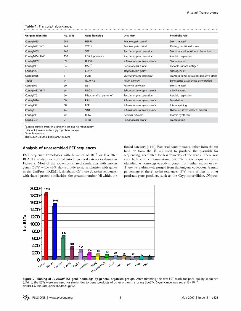
Analysis of unassembled EST sequences
EST sequence homologies with E values of 1026 or less after
BLASTx analysis were sorted into 13 general categories shown in
Figure 2. Most of the sequences shared similarities with known
genes (56%) while 44% showed little to no similarities with genes
in the UniProt_TREMBL database. Of those P. carinii sequences
with shared protein similarities, the greatest number fell within the
fungal category (44%). Bacterial contaminants, either from the rat
lung or from the E. coli used to produce the plasmids for
sequencing, accounted for less than 3% of the reads. There was
very little viral contamination, but 7% of the sequences were
identified as homologs to rodent genes, from either mouse or rat.
These were ultimately purged from the unigene collection. A small
percentage of the P. carinii sequences (5%) were similar to other
protistan gene products, such as the Cryptosporidiidae, Dictyoste-
Table 1. Transcript abundance.. . . . . . . . . . . . . . . . . . . . . . . . . . . . . . . . . . . . . . . . . . . . . . . . . . . . . . . . . . . . . . . . . . . . . . . . . . . . . . . . . . . . . . . . . . . . . . . . . . . . . . . . . . . . . . . . . . . . . . . . . . . . . . . . . . . . . . . . . . . . . . . . . .
Unigene identifier No. ESTs Gene homolog Organism Metabolic role
Contig1035 281 HSP70 Pneumocystis carinii Stress related
Contig1031/147* 148 STE11 Pneumocystis carinii Mating; nutritional stress
Contig1055 140 SFP1 Saccharomyces cerevisiae Stress related; nutritional limitation
Contig1034/946* 138 COX II precursor Saccharomyces cerevisiae Aerobic respiration
Contig1039 89 HSP90 Schizosaccharomyces pombe Stress-related
Contig498 84 MSG** Pneumocystis carinii Variable surface antigen
Contig520 84 CON7 Magnaporthe grisea Sporogenesis
Contig1056 81 PDR3 Saccharomyces cerevisiae Transcriptional activator; oxidative stress
11d08 74 Q9AVH2 Pisum sativum Senescence-associated; dehydration
Contig909 69 SIS1 Yarrowia lipolyticaI Stress related
Contig1037/287* 68 MLO3 Schizosaccharomyces pombe mRNA export
Contig176 66 Mitochondrial genome1 Saccharomyces cerevisiae Aerobic respiration
Contig1016 60 PSI1 Schizosaccharomyces pombe Translation
Contig799 38 BBP Schizosaccharomyces pombe Intron splicing
Contig8 24 SIN1 Schizosaccharomyces pombe Oxidative stress related; mitosis
Contig298 22 EF1A Candida albicans Protein synthesis
Contig 947 21 TFIID Pneumocystis carinii Transcription
*Contig purged from final unigene set due to redundancy**Variant 2 major surface glycoprotein isotype1Low homologydoi:10.1371/journal.pone.0000423.t001..
....
....
....
....
....
....
....
....
....
....
....
....
....
....
....
....
....
....
....
....
....
.
Figure 2. Binning of P. carinii EST gene homologs by general organism groups. After trimming the raw EST reads for poor quality sequence(qTrim), the ESTs were analyzed for similarities to gene products of other organisms using BLASTx. Significance was set at E#1026.doi:10.1371/journal.pone.0000423.g002
P. carinii Transcriptome
PLoS ONE | www.plosone.org 5 May 2007 | Issue 5 | e423
lium discoideum, and trypanosomes. Plant genes were also
represented with about 2% of the P. carinii ESTs showing
similarity to a putative senescence-associated protein in the pea.
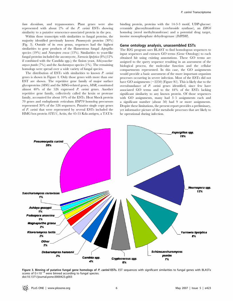
Within those transcripts with similarities to fungal proteins, the
majority identified previously known Pneumocystis proteins (30%)
(Fig. 3). Outside of its own genus, sequences had the highest
similarities to gene products of the filamentous fungal Aspergillus
species (19%) and Neurospora crassa (13%). Similarities to yeast-like
fungal proteins included the ascomycete, Yarrowia lipolytica (8%)(12%
if combined with the Candida spp.); the fission yeast, Schizosacchar-
omyces pombe (7%); and the Saccharomyces species (1%). The remaining
homologs were spread over a wide variety of fungal species.
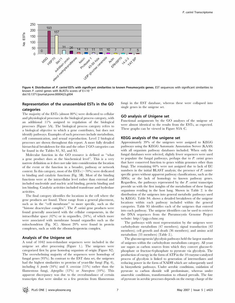
The distribution of ESTs with similarities to known P. carinii
genes is shown in Figure 4. Only those genes with more than one
EST are shown. The repetitive gene family of major surface
glycoproteins (MSG) and the MSG-related genes, MSR, constituted
almost 40% of the 526 expressed P. carinii genes. Another
repetitive gene family, collectively called the kexin or protease
family, accounted for about 10% of the ESTs. Heat Shock protein
70 genes and endoplasmic reticulum HSP70 homolog precursors
represented 30% of the 526 sequences. Putative single copy genes
of P. carinii that were represented by several ESTs included the
HMG box protein STE11, Actin, the 45-55 Kda antigen, a TATA-
binding protein, proteins with the 14-3-3 motif, UDP-glucose:
ceramide glucosyltransferase (cerebroside synthase), an ERG6
homolog (sterol methyltransferase) and a potential drug target,
inosine monophosphate dehydrogenase (IMPDH).
Gene ontology analysis, unassembled ESTsThe B2G program uses BLAST to find homologous sequences to
input sequences and extracts GO terms (Gene Ontology) to each
obtained hit using existing annotations. These GO terms are
assigned to the query sequence resulting in an assessment of the
biological process, the molecular function and the cellular
compartments represented. In this case, the GO assignments
would provide a basic assessment of the more important organism
processes occurring in severe infection. Most of the ESTs did not
have GO assignments (,2250) (Figure S1). This is likely due to the
overabundance of P. carinii genes identified, since few have
associated GO terms and to the 44% of the ESTs lacking
significant similarity to any known protein. Of those sequences
with GO assignments, many had 3–5 assignments each and
a significant number (about 50) had 9 or more assignments.
Despite these limitations, the present report provides a preliminary,
yet informative picture of the metabolic processes that are likely to
be operational during infection.
Figure 3. Binning of putative fungal gene homologs of P. carinii ESTs. EST sequences with significant similarities to fungal genes with BLASTxscores of E#1026 were binned according to fungal species.doi:10.1371/journal.pone.0000423.g003
P. carinii Transcriptome
PLoS ONE | www.plosone.org 6 May 2007 | Issue 5 | e423
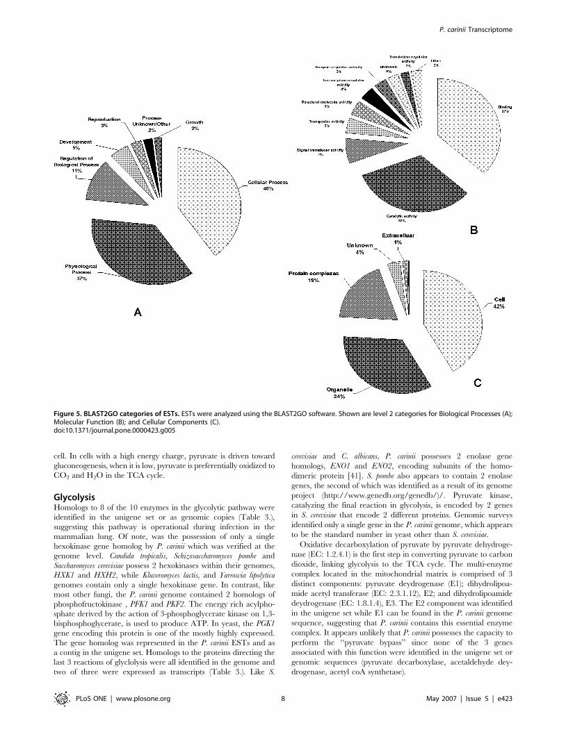
Representation of the unassembled ESTs in the GO
categoriesThe majority of the ESTs (almost 80%) were dedicated to cellular
and physiological processes in the biological process category, with
an additional 11% assigned to regulation of the biological
processes (Figure 5A). The biological process category refers to
a biological objective to which a gene contributes, but does not
identify pathways. Examples of such processes include metabolism,
cell communication, and sexual reproduction. Level 2 biological
processes are shown throughout this report. A more fully detailed
hierarchical breakdown for this and the other 2 GO categories can
be found in the Tables S1, S2, and S3.
Molecular function in the GO resource is defined as ‘‘what
a gene product does at the biochemical level’’. This is a very
narrow definition as it does not take into consideration the location
of the event or the function in a broader, pathway or network
context. In this category, most of the ESTs (,70%) were dedicated
to binding and catalytic functions (Fig. 5B). Most of the binding
functions were at the intracellular level rather than external and
included nucleotide and nucleic acid binding, protein binding and
ion binding. Catalytic activities included transferase and hydrolase
activities.
The final category identifies the locations in the cell where the
gene products are found. These range from a general placement,
such as in the ‘‘cell membrane’’ to more specific, such as the
‘‘histone deacteylase complex’’. The P. carinii gene products were
found generally associated with the cellular components, in the
intracellular space (42%) or in organelles, (34%), of which more
were associated with membrane bound organelles such as the
mitochondrion (Fig. 5C). Almost 20% were found in protein
complexes, such as with the ribonucleoprotein complex.
Analysis of the Unigene setA total of 1042 non-redundant sequences were included in the
unigene set after processing (Figure 1.). The unigenes were
categorized first by gene homolog organism of origin (Figure S2).
The overwhelming majority of the sequences were homologs of
fungal genes (93%). In contrast to the EST data set, the unigenes
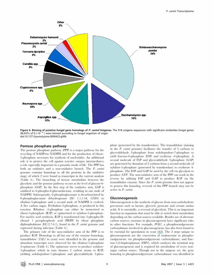
had the highest similarities to proteins of yeast-like fungi (Fig. 6),
including S. pombe (34%) and S. cerevisiae (15%) rather than the
filamentous fungi, Aspergillus (12%) or Neurospora (10%). This
apparent discrepancy was due to the overabundance of certain
transcripts that were similar to a few proteins from filamentous
fungi in the EST database, whereas these were collapsed into
single genes in the unigene set.
GO analysis of Unigene setFunctional assignments by the GO analyses of the unigene set
were almost identical to the results from the ESTs, as expected.
These graphs can be viewed in Figure S3A–C.
KEGG analysis of the unigene setApproximately 39% of the unigenes were assigned to KEGG
pathways using the KEGG Automatic Annotation Server (KAAS)
with all organism pathway databases included. When only the
fungal databases were selected, slightly fewer sequences were used
to populate the fungal pathways, perhaps due to P. carinii genes
that have conserved function to genes within genomes other than
fungi. The remaining 60% were not assigned due to lack of EC
numbers in the initial BLAST analysis; the presence of P. carinii-
specific genes without apparent pathway classifications, such as the
MSGs; or the lack of homology to known pathway genes.
Regardless, the pathways represented by the P. carinii transcripts
provide us with the first insights of the metabolism of these fungal
organisms residing in the host lung. Shown in Table 2. is the
distribution of the unigenes into general metabolic pathways used
by KEGG. Table S4. shows a detailed breakdown of the unigene
locations within each pathway included within the general
categories. Table S5 identifies each of the unigenes that entered
into each pathway. The unigene identifiers can be used to retrieve
the DNA sequences from the Pneumocystis Genome Project
website: http://pgp.cchmc.org
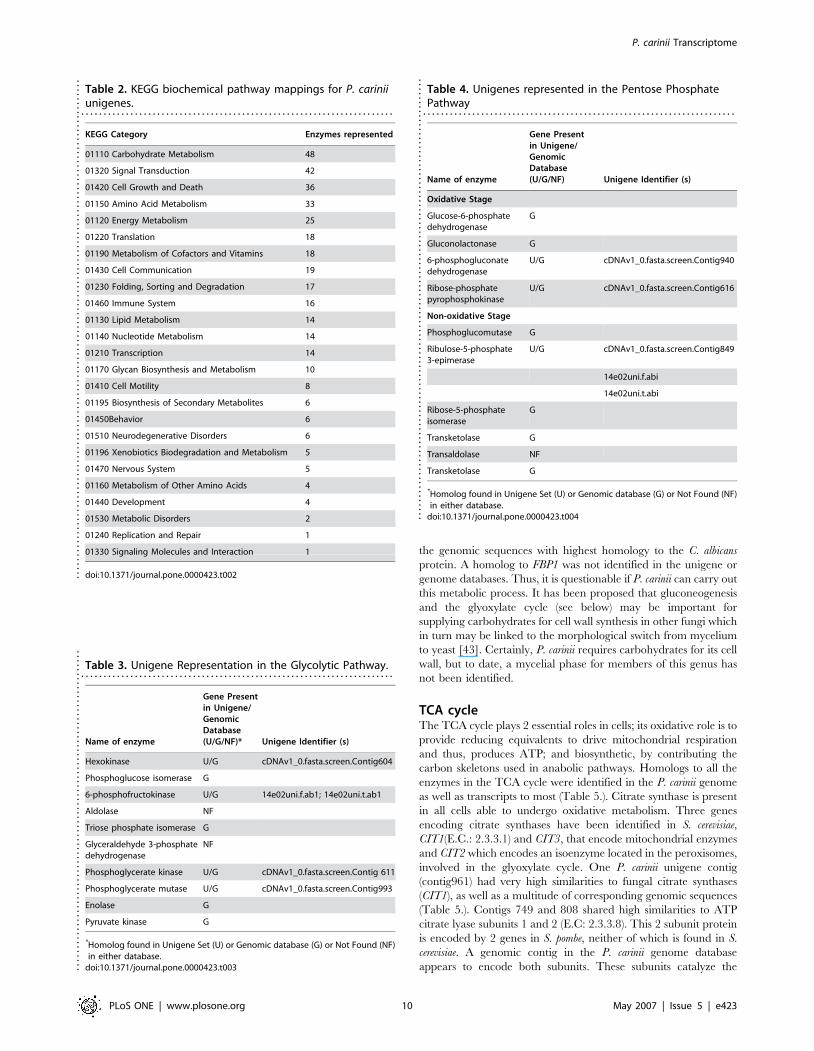
The pathways with most representation by the unigenes were
carbohydrate metabolism (47 members); signal transduction (42
members); cell growth and death (36 members); and amino acid
metabolism (33 members) (Table 2.).
The gluconeogenesis/glycolysis pathways held the largest number
of unigenes within the carbohydrate metabolism category. All yeast
use sugars as carbon sources from which they convert glucose-6-
phosphate or fructose-6-phosphate to pyruvate via glycolysis. The
production of energy in the form of ATP in the 10 enzyme-catalyzed
process of glycolysis is linked to generation of intermediates and
reducing power in the form of NADH which are subsequently used
in biosynthetic pathways. Under aerobic conditions, oxidation of
pyruvate to carbon dioxide will predominate, whereas under
anaerobic conditions, transformation to ethanol prevails. The fate
of pyruvate in aerobic processes depends on the energy charge of the
Figure 4. Distribution of P. carinii ESTs with significant similarities to known Pneumocystis genes. EST sequences with significant similarities toknown P. carinii genes with BLASTx scores of E#1026
doi:10.1371/journal.pone.0000423.g004
P. carinii Transcriptome
PLoS ONE | www.plosone.org 7 May 2007 | Issue 5 | e423
cell. In cells with a high energy charge, pyruvate is driven toward
gluconeogenesis, when it is low, pyruvate is preferentially oxidized to
CO2 and H2O in the TCA cycle.
GlycolysisHomologs to 8 of the 10 enzymes in the glycolytic pathway were
identified in the unigene set or as genomic copies (Table 3.),
suggesting this pathway is operational during infection in the
mammalian lung. Of note, was the possession of only a single
hexokinase gene homolog by P. carinii which was verified at the
genome level. Candida tropicalis, Schizosaccharomyces pombe and
Saccharomyces cerevisiae possess 2 hexokinases within their genomes,
HXK1 and HXH2, while Kluveromyces lactis, and Yarrowia lipolytica
genomes contain only a single hexokinase gene. In contrast, like
most other fungi, the P. carinii genome contained 2 homologs of
phosphofructokinase , PFK1 and PKF2. The energy rich acylpho-
sphate derived by the action of 3-phosphoglycerate kinase on 1,3-
bisphosphoglycerate, is used to produce ATP. In yeast, the PGK1
gene encoding this protein is one of the mostly highly expressed.
The gene homolog was represented in the P. carinii ESTs and as
a contig in the unigene set. Homologs to the proteins directing the
last 3 reactions of glyclolysis were all identified in the genome and
two of three were expressed as transcripts (Table 3.). Like S.
cerevisiae and C. albicans, P. carinii possesses 2 enolase gene
homologs, ENO1 and ENO2, encoding subunits of the homo-
dimeric protein [41]. S. pombe also appears to contain 2 enolase
genes, the second of which was identified as a result of its genome
project (http://www.genedb.org/genedb/)/. Pyruvate kinase,
catalyzing the final reaction in glycolysis, is encoded by 2 genes
in S. cerevisiae that encode 2 different proteins. Genomic surveys
identified only a single gene in the P. carinii genome, which appears
to be the standard number in yeast other than S. cerevisiae.
Oxidative decarboxylation of pyruvate by pyruvate dehydroge-
nase (EC: 1.2.4.1) is the first step in converting pyruvate to carbon
dioxide, linking glycolysis to the TCA cycle. The multi-enzyme
complex located in the mitochondrial matrix is comprised of 3
distinct components: pyruvate deydrogenase (E1); dihydrolipoa-
mide acetyl transferase (EC: 2.3.1.12), E2; and dihydrolipoamide
deydrogenase (EC: 1.8.1.4), E3. The E2 component was identified
in the unigene set while E1 can be found in the P. carinii genome
sequence, suggesting that P. carinii contains this essential enzyme
complex. It appears unlikely that P. carinii possesses the capacity to
perform the ‘‘pyruvate bypass’’ since none of the 3 genes
associated with this function were identified in the unigene set or
genomic sequences (pyruvate decarboxylase, acetaldehyde dey-
drogenase, acetyl coA synthetase).
Figure 5. BLAST2GO categories of ESTs. ESTs were analyzed using the BLAST2GO software. Shown are level 2 categories for Biological Processes (A);Molecular Function (B); and Cellular Components (C).doi:10.1371/journal.pone.0000423.g005
P. carinii Transcriptome
PLoS ONE | www.plosone.org 8 May 2007 | Issue 5 | e423
Pentose phosphate pathwayThe pentose phosphate pathway (PPP) is a major pathway for the
recycling of NADP+to NADPH and for the production of ribose-
5-phosphate necessary for synthesis of nucleotides. An additional
role is to protect the cell against reactive oxygen intermediates;
a role especially important in a parasitic mode of life. The PPP has
both an oxidative and a non-oxidative branch. The P. carinii
genome contains homologs to all the proteins in the oxidative
stage; of which 2 were found as transcripts in the current analysis
(Table 4.). The branching of hexose metabolism between the
glycolytic and the pentose pathway occurs at the level of glucose-6-
phosphate (G6P). In the first step of the oxidative arm, G6P is
oxidized to 6-phospho-d-gluconolactone, resulting in one mole of
NADPH. Subsequently, 6-phosphogluconate is decarboxylated by
6-phosphogluconate dehydrogenase (EC 1.1.1.44, GND1) to
ribulose-5-phosphate and a second mole of NADPH is evolved.
A five carbon sugar, D-ribulose-5-phosphate, is produced in this
reaction. Ribulose -5-phosphate can either be isomerized to
ribose-5-phosphate (R5P) or epimerized to xylulose-5-phosphate.
For nucleic acid synthesis, R5P is transformed into 5-phospho-D-
ribosyl- 1 pyrophosphoric acid (PRPP) by ribose phosphate
diphosphokinase (EC:2.7.6.1), found in the P. carinii genome and
expressed during infection (Table 4.).
The primary role of the non-oxidative arm of the PPP is to
produce R5P. Homologs to all but one of the enzyme homologs,
transaldolase (TAL1 in yeast) were identified in the genome and
abundant transcripts were observed for the ribulose-5-phosphate
3-epimerase (Table 4.) The epimerase serves to produce xylulose-
5-phosphate which in turn donates 2 carbon atoms to R5P,
yielding sedoheptulose-7-phosphate and glyceraldehyde 3-phos-
phate (generated by the transketolase). The transaldolase (missing
in the P. carinii genome) facilitates the transfer of 3 carbons to
glyceraldehyde 3-phosphate from sedoheptulose-7-phosphate to
yield fructose-6-phosphate (F6P) and erythrose -4-phosphate. A
second molecule of F6P and glyceraldehyde 3-phosphate (GAP)
are generated by donation of 2 carbons from a second molecule of
xylulose-5-phosphate (generated by transketolase) to erythrose 4-
phosphate. The F6P and GAP be used by the cell via glycolysis to
produce ATP. The non-oxidative arm of the PPP can work in the
reverse by utilizing F6P and GAP to produce R5P via the
transaldolase enzyme. Since the P. carinii genome does not appear
to possess this homolog, reversal of this PPP branch may not be
active in P. carinii.
GluconeogenesisGluconeogenesis is the synthesis of glucose from non-carbohydrate
precursors such as lactate, glycerol, pyruvate and certain amino
acids. It is essentially, a reversal of glycolysis. This is an important
function in organisms that must be able to switch their metabolism
depending on the carbon sources available. Besides use of alternate
carbon sources, enzymes in gluconeogenesis have significant roles
in other functions. For example, PCK1, a phosphoenolpyruvate
carboxykinase involved in gluconeogenesis, has also been found to
be essential for sporulation in yeast [42]. The 2 steps unique to
gluconeogenesis are the conversion of oxaloacetate to phospho-
enolpyruvate via phosphoenolpyruvate carboxykinase; and fruc-
tose-1,6-bisphosphatase (FBP1), which catalyzes the terminal step
of gluconeogenesis and is required for metabolism of every non-
sugar carbon source. Though not in the unigene set, the gene
homolog to phosphoenolpyruvate carboxykinase was identified in
Figure 6. Binning of putative fungal gene homologs of P. carinii Unigenes. The 918 unigene sequences with significant similarities fungal genes(BLASTx of E#1026 ) were binned according to fungal organism of origin.doi:10.1371/journal.pone.0000423.g006
P. carinii Transcriptome
PLoS ONE | www.plosone.org 9 May 2007 | Issue 5 | e423
the genomic sequences with highest homology to the C. albicans
protein. A homolog to FBP1 was not identified in the unigene or
genome databases. Thus, it is questionable if P. carinii can carry out
this metabolic process. It has been proposed that gluconeogenesis
and the glyoxylate cycle (see below) may be important for
supplying carbohydrates for cell wall synthesis in other fungi which
in turn may be linked to the morphological switch from mycelium
to yeast [43]. Certainly, P. carinii requires carbohydrates for its cell
wall, but to date, a mycelial phase for members of this genus has
not been identified.
TCA cycleThe TCA cycle plays 2 essential roles in cells; its oxidative role is to
provide reducing equivalents to drive mitochondrial respiration
and thus, produces ATP; and biosynthetic, by contributing the
carbon skeletons used in anabolic pathways. Homologs to all the
enzymes in the TCA cycle were identified in the P. carinii genome
as well as transcripts to most (Table 5.). Citrate synthase is present
in all cells able to undergo oxidative metabolism. Three genes
encoding citrate synthases have been identified in S. cerevisiae,
CIT1(E.C.: 2.3.3.1) and CIT3, that encode mitochondrial enzymes
and CIT2 which encodes an isoenzyme located in the peroxisomes,
involved in the glyoxylate cycle. One P. carinii unigene contig
(contig961) had very high similarities to fungal citrate synthases
(CIT1), as well as a multitude of corresponding genomic sequences
(Table 5.). Contigs 749 and 808 shared high similarities to ATP
citrate lyase subunits 1 and 2 (E.C: 2.3.3.8). This 2 subunit protein
is encoded by 2 genes in S. pombe, neither of which is found in S.
cerevisiae. A genomic contig in the P. carinii genome database
appears to encode both subunits. These subunits catalyze the
Table 2. KEGG biochemical pathway mappings for P. cariniiunigenes.
. . . . . . . . . . . . . . . . . . . . . . . . . . . . . . . . . . . . . . . . . . . . . . . . . . . . . . . . . . . . . . . . . . . . . .
KEGG Category Enzymes represented
01110 Carbohydrate Metabolism 48
01320 Signal Transduction 42
01420 Cell Growth and Death 36
01150 Amino Acid Metabolism 33
01120 Energy Metabolism 25
01220 Translation 18
01190 Metabolism of Cofactors and Vitamins 18
01430 Cell Communication 19
01230 Folding, Sorting and Degradation 17
01460 Immune System 16
01130 Lipid Metabolism 14
01140 Nucleotide Metabolism 14
01210 Transcription 14
01170 Glycan Biosynthesis and Metabolism 10
01410 Cell Motility 8
01195 Biosynthesis of Secondary Metabolites 6
01450Behavior 6
01510 Neurodegenerative Disorders 6
01196 Xenobiotics Biodegradation and Metabolism 5
01470 Nervous System 5
01160 Metabolism of Other Amino Acids 4
01440 Development 4
01530 Metabolic Disorders 2
01240 Replication and Repair 1
01330 Signaling Molecules and Interaction 1
doi:10.1371/journal.pone.0000423.t002....
....
....
....
....
....
....
....
....
....
....
....
....
....
....
....
....
....
....
....
....
....
....
....
....
....
....
....
Table 3. Unigene Representation in the Glycolytic Pathway.. . . . . . . . . . . . . . . . . . . . . . . . . . . . . . . . . . . . . . . . . . . . . . . . . . . . . . . . . . . . . . . . . . . . . .
Name of enzyme
Gene Presentin Unigene/GenomicDatabase(U/G/NF)* Unigene Identifier (s)
Hexokinase U/G cDNAv1_0.fasta.screen.Contig604
Phosphoglucose isomerase G
6-phosphofructokinase U/G 14e02uni.f.ab1; 14e02uni.t.ab1
Aldolase NF
Triose phosphate isomerase G
Glyceraldehyde 3-phosphatedehydrogenase
NF
Phosphoglycerate kinase U/G cDNAv1_0.fasta.screen.Contig 611
Phosphoglycerate mutase U/G cDNAv1_0.fasta.screen.Contig993
Enolase G
Pyruvate kinase G
*Homolog found in Unigene Set (U) or Genomic database (G) or Not Found (NF)in either database.
doi:10.1371/journal.pone.0000423.t003....
....
....
....
....
....
....
....
....
....
....
....
....
....
....
....
....
....
Table 4. Unigenes represented in the Pentose PhosphatePathway
. . . . . . . . . . . . . . . . . . . . . . . . . . . . . . . . . . . . . . . . . . . . . . . . . . . . . . . . . . . . . . . . . . . . . .
Name of enzyme
Gene Presentin Unigene/GenomicDatabase(U/G/NF) Unigene Identifier (s)
Oxidative Stage
Glucose-6-phosphatedehydrogenase
G
Gluconolactonase G
6-phosphogluconatedehydrogenase
U/G cDNAv1_0.fasta.screen.Contig940
Ribose-phosphatepyrophosphokinase
U/G cDNAv1_0.fasta.screen.Contig616
Non-oxidative Stage
Phosphoglucomutase G
Ribulose-5-phosphate3-epimerase
U/G cDNAv1_0.fasta.screen.Contig849
14e02uni.f.abi
14e02uni.t.abi
Ribose-5-phosphateisomerase
G
Transketolase G
Transaldolase NF
Transketolase G
*Homolog found in Unigene Set (U) or Genomic database (G) or Not Found (NF)in either database.
doi:10.1371/journal.pone.0000423.t004....
....
....
....
....
....
....
....
....
....
....
....
....
....
....
....
....
....
....
....
....
....
....
....
...
P. carinii Transcriptome
PLoS ONE | www.plosone.org 10 May 2007 | Issue 5 | e423
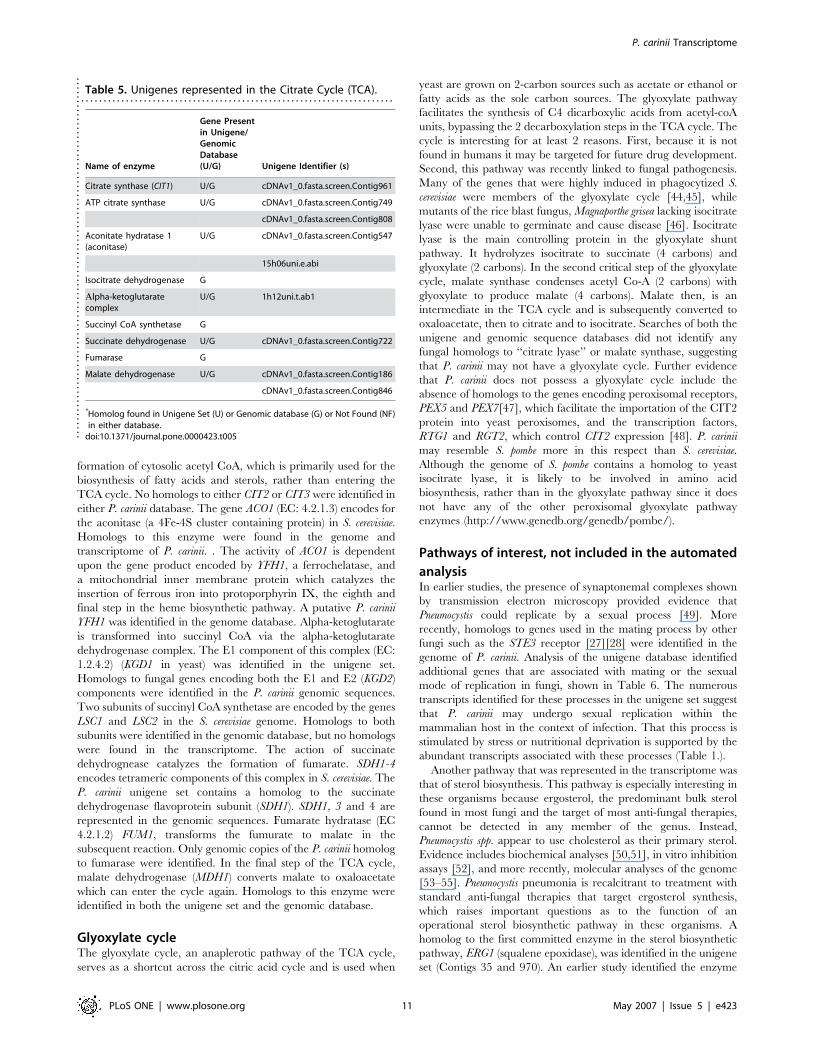
formation of cytosolic acetyl CoA, which is primarily used for the
biosynthesis of fatty acids and sterols, rather than entering the
TCA cycle. No homologs to either CIT2 or CIT3 were identified in
either P. carinii database. The gene ACO1 (EC: 4.2.1.3) encodes for
the aconitase (a 4Fe-4S cluster containing protein) in S. cerevisiae.
Homologs to this enzyme were found in the genome and
transcriptome of P. carinii. . The activity of ACO1 is dependent
upon the gene product encoded by YFH1, a ferrochelatase, and
a mitochondrial inner membrane protein which catalyzes the
insertion of ferrous iron into protoporphyrin IX, the eighth and
final step in the heme biosynthetic pathway. A putative P. carinii
YFH1 was identified in the genome database. Alpha-ketoglutarate
is transformed into succinyl CoA via the alpha-ketoglutarate
dehydrogenase complex. The E1 component of this complex (EC:
1.2.4.2) (KGD1 in yeast) was identified in the unigene set.
Homologs to fungal genes encoding both the E1 and E2 (KGD2)
components were identified in the P. carinii genomic sequences.
Two subunits of succinyl CoA synthetase are encoded by the genes
LSC1 and LSC2 in the S. cerevisiae genome. Homologs to both
subunits were identified in the genomic database, but no homologs
were found in the transcriptome. The action of succinate
dehydrognease catalyzes the formation of fumarate. SDH1-4
encodes tetrameric components of this complex in S. cerevisiae. The
P. carinii unigene set contains a homolog to the succinate
dehydrogenase flavoprotein subunit (SDH1). SDH1, 3 and 4 are
represented in the genomic sequences. Fumarate hydratase (EC
4.2.1.2) FUM1, transforms the fumurate to malate in the
subsequent reaction. Only genomic copies of the P. carinii homolog
to fumarase were identified. In the final step of the TCA cycle,
malate dehydrogenase (MDH1) converts malate to oxaloacetate
which can enter the cycle again. Homologs to this enzyme were
identified in both the unigene set and the genomic database.
Glyoxylate cycleThe glyoxylate cycle, an anaplerotic pathway of the TCA cycle,
serves as a shortcut across the citric acid cycle and is used when
yeast are grown on 2-carbon sources such as acetate or ethanol or
fatty acids as the sole carbon sources. The glyoxylate pathway
facilitates the synthesis of C4 dicarboxylic acids from acetyl-coA
units, bypassing the 2 decarboxylation steps in the TCA cycle. The
cycle is interesting for at least 2 reasons. First, because it is not
found in humans it may be targeted for future drug development.
Second, this pathway was recently linked to fungal pathogenesis.
Many of the genes that were highly induced in phagocytized S.
cerevisiae were members of the glyoxylate cycle [44,45], while
mutants of the rice blast fungus, Magnaporthe grisea lacking isocitrate
lyase were unable to germinate and cause disease [46]. Isocitrate
lyase is the main controlling protein in the glyoxylate shunt
pathway. It hydrolyzes isocitrate to succinate (4 carbons) and
glyoxylate (2 carbons). In the second critical step of the glyoxylate
cycle, malate synthase condenses acetyl Co-A (2 carbons) with
glyoxylate to produce malate (4 carbons). Malate then, is an
intermediate in the TCA cycle and is subsequently converted to
oxaloacetate, then to citrate and to isocitrate. Searches of both the
unigene and genomic sequence databases did not identify any
fungal homologs to ‘‘citrate lyase’’ or malate synthase, suggesting
that P. carinii may not have a glyoxylate cycle. Further evidence
that P. carinii does not possess a glyoxylate cycle include the
absence of homologs to the genes encoding peroxisomal receptors,
PEX5 and PEX7[47], which facilitate the importation of the CIT2
protein into yeast peroxisomes, and the transcription factors,
RTG1 and RGT2, which control CIT2 expression [48]. P. carinii
may resemble S. pombe more in this respect than S. cerevisiae.
Although the genome of S. pombe contains a homolog to yeast
isocitrate lyase, it is likely to be involved in amino acid
biosynthesis, rather than in the glyoxylate pathway since it does
not have any of the other peroxisomal glyoxylate pathway
enzymes (http://www.genedb.org/genedb/pombe/).
Pathways of interest, not included in the automated
analysisIn earlier studies, the presence of synaptonemal complexes shown
by transmission electron microscopy provided evidence that
Pneumocystis could replicate by a sexual process [49]. More
recently, homologs to genes used in the mating process by other
fungi such as the STE3 receptor [27][28] were identified in the
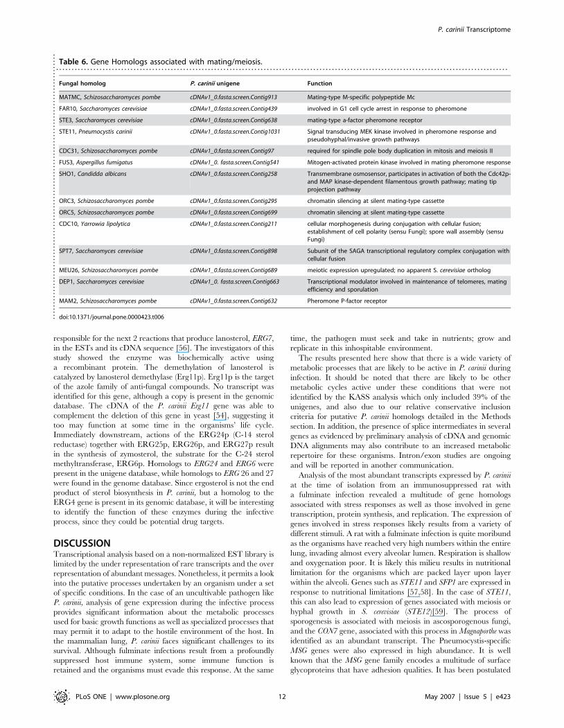
genome of P. carinii. Analysis of the unigene database identified
additional genes that are associated with mating or the sexual
mode of replication in fungi, shown in Table 6. The numerous
transcripts identified for these processes in the unigene set suggest
that P. carinii may undergo sexual replication within the
mammalian host in the context of infection. That this process is
stimulated by stress or nutritional deprivation is supported by the
abundant transcripts associated with these processes (Table 1.).
Another pathway that was represented in the transcriptome was
that of sterol biosynthesis. This pathway is especially interesting in
these organisms because ergosterol, the predominant bulk sterol
found in most fungi and the target of most anti-fungal therapies,
cannot be detected in any member of the genus. Instead,
Pneumocystis spp. appear to use cholesterol as their primary sterol.
Evidence includes biochemical analyses [50,51], in vitro inhibition
assays [52], and more recently, molecular analyses of the genome
[53–55]. Pneumocystis pneumonia is recalcitrant to treatment with
standard anti-fungal therapies that target ergosterol synthesis,
which raises important questions as to the function of an
operational sterol biosynthetic pathway in these organisms. A
homolog to the first committed enzyme in the sterol biosynthetic
pathway, ERG1 (squalene epoxidase), was identified in the unigene
set (Contigs 35 and 970). An earlier study identified the enzyme
Table 5. Unigenes represented in the Citrate Cycle (TCA).. . . . . . . . . . . . . . . . . . . . . . . . . . . . . . . . . . . . . . . . . . . . . . . . . . . . . . . . . . . . . . . . . . . . . .
Name of enzyme
Gene Presentin Unigene/GenomicDatabase(U/G) Unigene Identifier (s)
Citrate synthase (CIT1) U/G cDNAv1_0.fasta.screen.Contig961
ATP citrate synthase U/G cDNAv1_0.fasta.screen.Contig749
cDNAv1_0.fasta.screen.Contig808
Aconitate hydratase 1(aconitase)
U/G cDNAv1_0.fasta.screen.Contig547
15h06uni.e.abi
Isocitrate dehydrogenase G
Alpha-ketoglutaratecomplex
U/G 1h12uni.t.ab1
Succinyl CoA synthetase G
Succinate dehydrogenase U/G cDNAv1_0.fasta.screen.Contig722
Fumarase G
Malate dehydrogenase U/G cDNAv1_0.fasta.screen.Contig186
cDNAv1_0.fasta.screen.Contig846
*Homolog found in Unigene Set (U) or Genomic database (G) or Not Found (NF)in either database.
doi:10.1371/journal.pone.0000423.t005....
....
....
....
....
....
....
....
....
....
....
....
....
....
....
....
....
....
....
....
.
P. carinii Transcriptome
PLoS ONE | www.plosone.org 11 May 2007 | Issue 5 | e423
responsible for the next 2 reactions that produce lanosterol, ERG7,
in the ESTs and its cDNA sequence [56]. The investigators of this
study showed the enzyme was biochemically active using
a recombinant protein. The demethylation of lanosterol is
catalyzed by lanosterol demethylase (Erg11p). Erg11p is the target
of the azole family of anti-fungal compounds. No transcript was
identified for this gene, although a copy is present in the genomic
database. The cDNA of the P. carinii Erg11 gene was able to
complement the deletion of this gene in yeast [54], suggesting it
too may function at some time in the organisms’ life cycle.
Immediately downstream, actions of the ERG24p (C-14 sterol
reductase) together with ERG25p, ERG26p, and ERG27p result
in the synthesis of zymosterol, the substrate for the C-24 sterol
methyltransferase, ERG6p. Homologs to ERG24 and ERG6 were
present in the unigene database, while homologs to ERG 26 and 27
were found in the genome database. Since ergosterol is not the end
product of sterol biosynthesis in P. carinii, but a homolog to the
ERG4 gene is present in its genomic database, it will be interesting
to identify the function of these enzymes during the infective
process, since they could be potential drug targets.
DISCUSSIONTranscriptional analysis based on a non-normalized EST library is
limited by the under representation of rare transcripts and the over
representation of abundant messages. Nonetheless, it permits a look
into the putative processes undertaken by an organism under a set
of specific conditions. In the case of an uncultivable pathogen like
P. carinii, analysis of gene expression during the infective process
provides significant information about the metabolic processes
used for basic growth functions as well as specialized processes that
may permit it to adapt to the hostile environment of the host. In
the mammalian lung, P. carinii faces significant challenges to its
survival. Although fulminate infections result from a profoundly
suppressed host immune system, some immune function is
retained and the organisms must evade this response. At the same
time, the pathogen must seek and take in nutrients; grow and
replicate in this inhospitable environment.
The results presented here show that there is a wide variety of
metabolic processes that are likely to be active in P. carinii during
infection. It should be noted that there are likely to be other
metabolic cycles active under these conditions that were not
identified by the KASS analysis which only included 39% of the
unigenes, and also due to our relative conservative inclusion
criteria for putative P. carinii homologs detailed in the Methods
section. In addition, the presence of splice intermediates in several
genes as evidenced by preliminary analysis of cDNA and genomic
DNA alignments may also contribute to an increased metabolic
repertoire for these organisms. Intron/exon studies are ongoing
and will be reported in another communication.
Analysis of the most abundant transcripts expressed by P. carinii
at the time of isolation from an immunosuppressed rat with
a fulminate infection revealed a multitude of gene homologs
associated with stress responses as well as those involved in gene
transcription, protein synthesis, and replication. The expression of
genes involved in stress responses likely results from a variety of
different stimuli. A rat with a fulminate infection is quite moribund
as the organisms have reached very high numbers within the entire
lung, invading almost every alveolar lumen. Respiration is shallow
and oxygenation poor. It is likely this milieu results in nutritional
limitation for the organisms which are packed layer upon layer
within the alveoli. Genes such as STE11 and SFP1 are expressed in
response to nutritional limitations [57,58]. In the case of STE11,
this can also lead to expression of genes associated with meiosis or
hyphal growth in S. cerevisiae (STE12)[59]. The process of
sporogenesis is associated with meiosis in ascosporogenous fungi,
and the CON7 gene, associated with this process in Magnaporthe was
identified as an abundant transcript. The Pneumocystis-specific
MSG genes were also expressed in high abundance. It is well
known that the MSG gene family encodes a multitude of surface
glycoproteins that have adhesion qualities. It has been postulated
Table 6. Gene Homologs associated with mating/meiosis.. . . . . . . . . . . . . . . . . . . . . . . . . . . . . . . . . . . . . . . . . . . . . . . . . . . . . . . . . . . . . . . . . . . . . . . . . . . . . . . . . . . . . . . . . . . . . . . . . . . . . . . . . . . . . . . . . . . . . . . . . . . . . . . . . . . . . . . . . . . . . . . . . .
Fungal homolog P. carinii unigene Function
MATMC, Schizosaccharomyces pombe cDNAv1_0.fasta.screen.Contig913 Mating-type M-specific polypeptide Mc
FAR10, Saccharomyces cerevisiae cDNAv1_0.fasta.screen.Contig439 involved in G1 cell cycle arrest in response to pheromone
STE3, Saccharomyces cerevisiae cDNAv1_0.fasta.screen.Contig638 mating-type a-factor pheromone receptor
STE11, Pneumocystis carinii cDNAv1_0.fasta.screen.Contig1031 Signal transducing MEK kinase involved in pheromone response andpseudohyphal/invasive growth pathways
CDC31, Schizosaccharomyces pombe cDNAv1_0.fasta.screen.Contig97 required for spindle pole body duplication in mitosis and meiosis II
FUS3, Aspergillus fumigatus cDNAv1_0. fasta.screen.Contig541 Mitogen-activated protein kinase involved in mating pheromone response
SHO1, Candidda albicans cDNAv1_0.fasta.screen.Contig258 Transmembrane osmosensor, participates in activation of both the Cdc42p-and MAP kinase-dependent filamentous growth pathway; mating tipprojection pathway
ORC3, Schizosaccharomyces pombe cDNAv1_0.fasta.screen.Contig295 chromatin silencing at silent mating-type cassette
ORC5, Schizosaccharomyces pombe cDNAv1_0.fasta.screen.Contig699 chromatin silencing at silent mating-type cassette
CDC10, Yarrowia lipolytica cDNAv1_0.fasta.screen.Contig211 cellular morphogenesis during conjugation with cellular fusion;establishment of cell polarity (sensu Fungi); spore wall assembly (sensuFungi)
SPT7, Saccharomyces cerevisiae cDNAv1_0.fasta.screen.Contig898 Subunit of the SAGA transcriptional regulatory complex conjugation withcellular fusion
MEU26, Schizosaccharomyces pombe cDNAv1_0.fasta.screen.Contig689 meiotic expression upregulated; no apparent S. cerevisiae ortholog
DEP1, Saccharomyces cerevisiae cDNAv1_0. fasta.screen.Contig663 Transcriptional modulator involved in maintenance of telomeres, matingefficiency and sporulation
MAM2, Schizosaccharomyces pombe cDNAv1_0.fasta.screen.Contig632 Pheromone P-factor receptor
doi:10.1371/journal.pone.0000423.t006....
....
....
....
....
....
....
....
....
....
....
....
....
....
....
....
....
....
....
....
....
..
P. carinii Transcriptome
PLoS ONE | www.plosone.org 12 May 2007 | Issue 5 | e423
that a function of these adhesins may be similar to the FLO genes
of S. cerevisiae [60], which also encode surface glycoproteins that
promote cell to cell interactions, especially in a nutritionally
depleted environment. Thus, it is plausible that P. carinii undergoes
sexual replication stimulated by the nutritionally poor environ-
ment at end stage disease. A second stimulus for the stress
responses may be a consequence of oxidative stress due to the
production of reactive oxygen species by the host’s immune cells.
The PDR3 [61]and SIN1 [62]genes are associated with oxidative
stress in other fungi . The HSP90 gene of S. pombe (also called
CDC37) was specifically associated with the positive regulation of
a stress activated protein kinase (SAPK) that plays a crucial role in
cellular survival to inflammatory responses [63]. Although P. carinii
expressed superoxide dismutase (Contig346) and catalase (Con-
tig327), they were not found as abundant transcripts, and thus the
organisms could have experienced oxidative cell damage. It is
interesting to note that a homolog to transaldolase (TAL) is
apparently absent from the transcriptome and the genome of P.
carinii. This is the key enzyme in the reversible non-oxidative
branch of the PPP that is responsible for generation of NADPH. A
primary function of the PPP is to maintain glutathione in a reduced
state, which functions to provide protection of sulfhydryl groups
and cellular integrity from oxygen radicals. If the reversible nature
of the PPP is compromised by the absence of the transaldolase, the
ability to fully ward off the detrimental effects of the reactive
species may be attenuated, leading to the observed stress reaction.
The third stimulus for the stress responses could have arisen from
the lengthy purification process used to extricate the organisms
from host lung tissue [64,65]. This process involves the use of
mechanical disruption of lung tissue with a tissue homogenizer and
processing over a 3–4 hour period.
The majority of transcripts associated with metabolic cycles
were dedicated to carbohydrate metabolism, specifically glycolysis.
Glycolysis has been shown to be essential for growth in the
mammalian host by a number of fungal pathogens. Recently, it
was shown that the energy production strategies used by C. albicans
changed in response to physiologically distinct host niches [66].
Gluconeogenic and glyoxylate–associated genes were active early
in the infection when the yeast were phagocytized by host cells, but
progression of systemic disease was dependent upon glycolysis.
The authors postulated that the nutritionally poor environment of
the phagocyte reflected starvation conditions that stimulated the
alternative pathways. The emphasis on expression of glycolytic
enzymes by P. carinii and not those in alternative pathways,
suggests that although the alveolar compartment may immuno-
logically challenge the organisms, the milieu may provide
a sufficient nutritional environment, circumventing the need for
alternative carbohydrate pathways. The paucity of homologs in
the gluconeogenesis and glyoxylate pathways implies that these
organisms may not be able to utilize non-fermentable carbon
sources. This may have been an adaptation to the host
environment that occurred during the evolution of the host-
parasite relationship. It is also well known that Pneumocystis are
easily phagocytized and killed by macrophages, suggesting a lack
of survival strategy in this compartment of the immune response.
The apparent lack of a transaldolase homolog in the PPP may
have effects on the organisms’ ability to respond to oxidative
damage (discussed above), but may also limit an alternative mode
for ATP production via glycolysis by blocking the synthesis of F6P
and GAP. Similarly, non-reversibility of the PPP would also
reduce the ability to synthesize R5P from F6P and GAP obtained
from glycolysis. It is notable that the genome of Plasmodium
falciparum, another host-dependent parasite lacks a homolog to
transaldolase as well [67].
Whether P. carinii is able to undergo fermentation remains
a question. The characteristics of fermentation are at least 3 of the
following: the release of energy from a sugar or other organic
compound; no requirement for molecular oxygen; no requirement
for an electron transport system; or use of an organic compound as
the final electron acceptor. Fermentation in yeast uses the same
processes as glycolysis, except in the absence of oxygen, it is
blocked from entering the TCA cycle and thus converts the
pyruvate to acetaldehyde via pyruvate decarboxylase, and then to
ethanol via alcohol dehydrogenase, losing one carbon in the
process that evolves as carbon dioxide. The evidence at hand that
argues against fermentation by P. carinii includes the lack of
a homolog to pyruvate dehydrogenase in the unigene set or the
genomic database and its inability to survive under anaerobic
conditions. Recent studies have shown that P. carinii rapidly loses
viability in an anaerobic atmosphere and seems to require some
oxygen, although it is able to survive in an atmosphere of reduced
oxygen levels [68]. On the other hand, its genome contains 3 genes
encoding putative alcohol dehydrogenases. In S. cerevisiae, there are
five genes that encode alcohol dehydrogenases involved in ethanol
metabolism, ADH1 to ADH5. Four of these enzymes, ADH1p,
ADH3p, ADH4p, and ADH5p, reduce acetaldehyde to ethanol
during glucose fermentation, while ADH2p catalyzes the reverse
reaction of oxidizing ethanol to acetaldehyde. Homologs to ADH1,
ADH2 and ADH3 were identified in the P. carinii genomic database.
In contrast, the genome of the fission yeast Schizosaccharomyces
pombe, contains only one alcohol dehydrogenase gene, adh1(+), and
is able to ferment [69]. It is clear that use of respiration or
fermentation by yeasts is regulated by the availability of glucose and
oxygen. Some yeasts are obligate respirers like the Cryptococcus
species which are incapable of fermentation or anaerobic growth
[35]. Others, like species of Candida, Kluveromyces and Pichia can
respire anaerobically, but fermentation only occurs in pre-grown
cells and they are not able to grow anaerobically. S. pombe is capable
of aerobic fermentation, but also cannot grow under anaerobic
conditions. At the other end of the spectrum, Torulopsis are obligate
fermenters and cannot respire, but grow and ferment only under
anaerobic conditions. The most versatile of the group is S. cerevisiae,
which is considered a facultative aerobic fermenter that can ferment
under both aerobic and anaerobic conditions and is capable of
facultative growth under anaerobic conditions. Without direct
experimental data, it is difficult to place P. carinii within its proper
category, but assuming it cannot grow without a source of molecular
oxygen, it would seem to be either an obligate respirer or perhaps
a facultative aerobic fermenter, depending on the function of its
alcohol dehydrogenases.
The entire life cycle of any member of the genus Pneumocystis
has not been defined. Microscopic observations of organisms
within the alveoli have led to many proposed life cycles that focus
on development only within the lung [7]. Most agree that there is
an asexual cycle that consists of the smaller trophic forms dividing
by binary fission and a phase which results in formation of a cyst or
ascus which contains 8 spores or daughter forms. The process used
by the organisms to produce the cyst and spores has been
hypothesized to be through a sexual process, although there is not
full consensus on this point. Several genes related to the sexual
reproductive cycle were identified in the transcriptome of P. carinii,
suggesting that sexual reproduction may occur in the mammalian
lung during active infection. If this assumption is correct, it is in
striking contrast to most other fungal pathogens causing human
diseases that do not undergo sexual reproduction in their
respective hosts, such as Cryptococcus spp., Blastomyces dermatiditis,
Coccidioides spp., Histoplasma capsulatum, Penicillium marnefeii,
Candida spp. or Paracoccidioides brasiliensis. With the exception of
P. carinii Transcriptome
PLoS ONE | www.plosone.org 13 May 2007 | Issue 5 | e423
Candida spp., these and other fungal pathogens are not considered
normal flora or commensals of the immunologically intact host.
Moreover, most of these fungi have a primary environmental
niche in which sex, if it does occur, takes place. P. jirovecii have
been reported to ‘‘colonize’’ certain sectors of the human
population [19,70] and P. carinii are frequently detected in the
lungs of healthy adult rodents [30,71] and within hours after birth
in neonatal rats [72]. The serological responses to P. jirovecii
antigens early in life are also indicative of their role as normal flora
[73]. There is no known environmental habitat for Pneumocystis,
and thus it would follow that if sex does occur, it would necessarily
take place in the mammalian host. A further distinction between
Pneumocystis spp. and most other medically important fungal
pathogens is communicability of the infection. Most fungal
infections are acquired by inhalation of infectious propagules, by
deep or superficial wound trauma, or other environmental exposures
and terminate in the infected host. There is no transmission. In
contrast, Pneumocystis appears to be a highly transmissible infection
as evidenced by serological responses and extensive experimental
transmission studies. In that sense, Pneumocystis are transmitted
directly to the next host, much like Candida spp., but by an airborne
rather than contact route. Thus, members of Pneumocystis spp. are
distinct from other medically significant fungi in that they appear to
undergo sexual replication in their mammalian hosts and are able to
transmit the infection from host to host.
We posit that through the process of evolution, Pneumocystis
spp. have adapted to their specific mammalian hosts to form
a sustainable relationship. In many microbial infections, the host:
parasite relationship is defined by the virulence factors produced
by the parasite and the resistant counter defenses by the host. This
is largely due to the sophisticated immune surveillance systems and
innate defense mechanisms by the host and the lack of adaptation
of many microbial pathogens, resulting in a tug of war between
resistance and virulence. However, that relationship is just one of
a continuum of combinations among two species. The concept of
‘‘compatibility’’ is emerging as an alternative to the standard mode
of thinking where there is a constant battle of the invader vs prey.
In the fungal plant pathogen field, ‘‘compatibility’’ is defined as the
complementary relationship between a plant species and an
adapted pathogen species that underlies susceptibility and
ultimately results in disease [74]. Biotrophic fungi derive their
energy from the living cells of their plant hosts without loss of host
cell viability. Obligate biotrophs complete their entire life cycle
within the plant host, including the sexual cycle, and are incapable
of ex vivo growth or limited in vitro cultivation, like Pneumocystis.
It is generally accepted that members of the genus Pneumocystis in
the immunologically intact mammalian host, cause little or no
clinical disease. It is only when the host tips the balance toward
these organisms by losing immune capability to control the
organisms that disease ensues. This would infer there may
be‘‘compatibility’’ between the mammalian host and its resident
Pneumocystis species. Seen in this light, Pneumocystis are quite
accommodating parasite partners. There is no invasive entry into
the host; rather it is likely through a passive inhalation of spores
wafting in the air. Once inside the lung alveoli of the immune
intact host, there is no massive infection, rapid reproduction, or
invasive process. Although these early steps are poorly understood,
it appears likely that a cryptic, low level infection ensues which the
host immune system recognizes by humoral and cellular means.
Indeed, the presence of only a single copy of the rDNA locus has
been hypothesized to permit slow growth of the organisms [60],
which also could serve to maintain the balance of host and
pathogen. As a counter defense to immune recognition, all
members of Pneumocystis spp. surveyed to date contain a multi-
gene family capable of encoding perhaps hundreds of isotypes of
their major surface antigens, the Major Surface Glycoproteins
(MSG) [75]. Switching of the surface antigen coat provides
a survival strategy similar to that used by other microbial
pathogens, such as the African trypanosomes. The length of time
that the host: Pneumocystis tete a tete may last is not presently
known, but it appears that there may be a constant re-infection of
the hosts by Pneumocystis without serious harm to the immune
intact host. Molecular evidence presented in the present report
suggests that P. carinii is capable of a wide variety of metabolic
processes, which would also accommodate its stay in the host.
Without the need to pirate host metabolic products or processes by
Pneumocystis, the inter-relationship between the 2 species would
not result in death of the host or expulsion of the parasite, leading
to a sustainable situation. And like the biotrophic plant fungi, the
entire life cycle appears to take place in the host and they are also
uncultivable outside the host.
The pathology created by Pneumocystis during active infection
in the immunocompromised host is due to the mechanical
obstruction of the air-gas interchange of the host, leading to
decreased oxygenation and to the inflammatory response by the
host, largely controlled by the CD8 cell infiltration [76]. The
Pneumocystis organisms have not been reported to secrete
virulence factors and likely do not facilitate the infection by these
means. The last piece of evidence regarding the adaptation of the
members of the Pneumocystis spp. is their host specificity. Most
mammalian species harbor at least one species of Pneumocystis
which is genetically and phenotypically distinct from those
members in other mammalian hosts. This would infer an
adaptation that occurred over millions of years, resulting in an
almost commensal relationship with each host. This adaptation is
in stark contrast to most of the fungal pathogens discussed above
that do not depend on the host for survival.
Many of the genes expressed by P. carinii were organism-specific.
The MSG, MSR, and the family of proteases that are thought to be
necessary for processing of the surface antigens were highly
expressed. The MSGp are known to bind extra cellular matrix
proteins like vitronectin and fibronectin [77–79]; aid in adherence to
the Type I pneumocyte in the alveoli [80]; and also aid in adherence
of the organisms to one another [60]. Thus it is likely that these
glycoproteins perform many functions that are necessary for the
infective process and may contribute to the adaptation process.
P. carinii is not an opportunistic pathogen in the fungal pathogen
sense of the word. The mammalian host appears to be necessary
for its survival and life cycle. In what may be its natural host, the
immunologically intact mammal, it exerts little to no pathogenic
effects. Loss of immune competence permits the organisms to
grow, resulting in pathogenesis. The apparent presence of a large
number of metabolic pathways as detected by the present analysis,
the slow growth of the organisms, its host specificity and the lack of
pathogenesis imply that these organisms have adapted to form
a compatible relationship within their natural habitat, the immune
intact mammalian host.
SUPPORTING INFORMATION
Figure S1 Annotation distribution of ESTs. The number of
ESTs that were assigned to GO categories.
Found at: doi:10.1371/journal.pone.0000423.s001 (0.23 MB TIF)
Figure S2 Binning of P. carinii unigene homologs by general
organism groups. The unigenes were analyzed for similarities to
gene products of other organisms using BLASTx. Significance was
set at E#10-6.
Found at: doi:10.1371/journal.pone.0000423.s002 (0.08 MB TIF)
P. carinii Transcriptome
PLoS ONE | www.plosone.org 14 May 2007 | Issue 5 | e423
Figure S3 Unigenes were analyzed using the BLAST2GO
software. Shown are level 2 categories for Biological Processes
Found at: doi:10.1371/journal.pone.0000423.s003 (0.41 MB TIF)
Table S1 EST BLAST2GO Biological Processes-All Levels. All
hierarchical levels of BLAST2GO Biological Processes for the
ESTs.
Found at: doi:10.1371/journal.pone.0000423.s004 (0.30 MB
XLS)
Table S2 EST BLAST2GO Molecular Function-All levels.
Hierarchical distribution of the Molecular Function of the ESTs
using BLAST2GO.
Found at: doi:10.1371/journal.pone.0000423.s005 (0.15 MB
XLS)
Table S3 EST BLAST2GO Cellular Compartmentalization
Assignments-All levels. Hierarchical distribution of the cellular
compartmentalization assignments of the ESTs by BLAST2GO.
Found at: doi:10.1371/journal.pone.0000423.s006 (0.08 MB
XLS)
Table S4 Unigene Population of KEGG Pathways
Found at: doi:10.1371/journal.pone.0000423.s007 (0.10 MB
DOC)
Table S5 Orthology List for Unigenes in KEGG pathways.
Found at: doi:10.1371/journal.pone.0000423.s008 (0.24 MB
DOC)
ACKNOWLEDGMENTS
Author Contributions
Conceived and designed the experiments: MC AS. Performed the
experiments: MC AS TS JA. Analyzed the data: CS MC AS BS AP RA
JM. Contributed reagents/materials/analysis tools: MC AS BS JA AP JM.
Wrote the paper: MC AS.
REFERENCES1. Stringer SL, Stringer JR, Blase MA, Walzer PD, Cushion MT (1989) Pneumocystis
carinii: sequence from ribosomal RNA implies a close relationship with fungi.
Exp Parasitol 68: 450–461.
2. Edman JC, Kovacs JA, Masur H, Santi DV, Elwood HJ, Sogin ML (1988)
Ribosomal RNA sequence shows Pneumocystis carinii to be a member of the fungi.
Nature 334: 519–522.
3. Li J, Edlind T (1994) Phylogeny of Pneumocystis carinii based on beta-tubulin
sequence. J Eukaryot Microbiol 41: 97S.
4. Liu YJ, Hall BD (2004) Body plan evolution of ascomycetes, as inferred from anRNA polymerase II phylogeny. Proc Natl Acad Sci U S A 101: 4507–4512.
5. Pixley FJ, Wakefield AE, Banerji S, Hopkin JM (1991) Mitochondrial gene
sequences show fungal homology for Pneumocystis carinii. Mol Microbiol 5:1347–1351.
6. Smulian AG, Cushion MT (2006) Pathogenesis of Pneumocystis. In: Heitman J,
Filler SG, Edwards Jr JE, Mitchell AP, eds. Molecular Principles of FungalPathogenesis. Washington, D.C.: ASM Press. pp 347–362.
7. Cushion MT (2006) Pneumocystis pneumonia. In: Merz WG, Hay RJ, eds.
Topley&Wilson’s Medical Mycology. Washington DC: Edward Arnold (Publish-ers) Ltd. pp 763–806.
8. Redhead S, Cushion MT, Frenkel JK, Stringer JR (2005) Pneumocystis and
Trypanosoma cruzi nomenclature and typifications. J Eukaryot Microbiol (In press).
9. Frenkel JK (1976) Pneumocystis jiroveci n. sp. from man: morphology, physiology,
and immunology in relation to pathology. Natl Cancer Inst Monogr 43: 13–30.
10. Frenkel JK (1999) Pneumocystis pneumonia, an immunodeficiency-dependent
disease (IDD): a critical historical overview. J Eukaryot Microbiol 46: 89S–92S.
11. Keely SP, Fischer JM, Cushion MT, Stringer JR (2004) Phylogeneticidentification of Pneumocystis murina sp. nov., a new species in laboratory mice.
Microbiology 150: 1153–1165.
12. Dei-Cas E, Chabe M, Moukhlis R, Durand-Joly I, Aliouat eM, et al. (2006)Pneumocystis oryctolagi sp. nov., an uncultured fungus causing pneumonia in rabbits
at weaning: review of current knowledge, and description of a new taxon on
genotypic, phylogenetic and phenotypic bases. FEMS Microbiol Rev 30:853–871.
13. Cushion MT, Keely SP, Stringer JR (2004) Molecular and phenotypic
description of Pneumocystis wakefieldiae sp. nov., a new species in rats. Mycologia96: 429–438.
14. Cushion MT, Keely SP, Stringer JR (2005) Validation of the name Pneumocystis
wakefieldiae. Mycologia 97: 268.
15. Kovacs JA, Gill VJ, Meshnick S, Masur H (2001) New insights into transmission,
diagnosis, and drug treatment of Pneumocystis carinii pneumonia. JAMA 286:
2450–2460.
16. Fisk DT, Meshnick S, Kazanjian PH (2003) Pneumocystis carinii pneumonia in
patients in the developing world who have acquired immunodeficiency
syndrome. Clin Infect Dis 36: 70–78.
17. Zar HJ, Alvarez-Martinez MJ, Harrison A, Meshnick SR (2004) Prevalence of
dihydropteroate synthase mutants in HIV-infected South African children with
Pneumocystis jiroveci pneumonia. Clin Infect Dis 39: 1047–1051.
18. Sharma SK, Kadhiravan T, Banga A, Goyal T, Bhatia I, et al. (2004) Spectrum
of clinical disease in a series of 135 hospitalised HIV-infected patients from north
India. BMC Infect Dis 4: 52.
19. Morris A, Sciurba FC, Lebedeva IP, Githaiga A, Elliott WM, et al. (2004)
Association of chronic obstructive pulmonary disease severity and Pneumocystis
colonization. Am J Respir Crit Care Med 170: 408–413.
20. Morris A, Lundgren JD, Masur H, Walzer PD, Hanson DL, et al. (2004)
Current epidemiology of Pneumocystis pneumonia. Emerg Infect Dis 10:
1713–1720.
21. Calderon E, de la HC, Medrano FJ, Lopez-Suarez A, Montes-Cano MA, et al.
(2004) Pneumocystis jiroveci isolates with dihydropteroate synthase mutations inpatients with chronic bronchitis. Eur J Clin Microbiol Infect Dis 23: 545–549.
22. Kazanjian PH, Fisk D, Armstrong W, Shulin Q, Liwei H, et al. (2004) Increase
in prevalence of Pneumocystis carinii mutations in patients with AIDS and P. carinii
pneumonia, in the United States and China. J Infect Dis 189: 1684–1687.
23. Huang L, Crothers K, Atzori C, Benfield T, Miller R, et al. (2004)
Dihydropteroate synthase gene mutations in Pneumocystis and sulfa resistance.Emerg Infect Dis 10: 1721–1728.
24. Crothers K, Beard CB, Turner J, Groner G, Fox M, et al. (2005) Severity and
outcome of HIV-associated Pneumocystis pneumonia containing Pneumocystis
jirovecii dihydropteroate synthase gene mutations. AIDS 19: 801–805.
25. Kazanjian P, Armstrong W, Hossler PA, Lee CH, Huang L, et al. (2001)
Pneumocystis carinii cytochrome b mutations are associated with atovaquoneexposure in patients with AIDS. J Infect Dis 183: 819–822.
26. Cushion MT, Walzer PD, Smulian AG, Linke MJ, Ruffolo JJ, et al. (1997)
Terminology for the life cycle of Pneumocystis carinii. Infect Immun 65: 4365.
27. Smulian AG, Sesterhenn T, Tanaka R, Cushion MT (2001) The ste3
pheromone receptor gene of Pneumocystis carinii is surrounded by a cluster of
signal transduction genes. Genetics 157: 991–1002.
28. Vohra PK, Park JG, Sanyal B, Thomas CF Jr (2004) Expression analysis of
PCSTE3, a putative pheromone receptor from the lung pathogenic fungus
Pneumocystis carinii. Biochem Biophys Res Commun 319: 193–199.
29. Cushion MT, Arnold J (1997) Proposal for a Pneumocystis genome project.
J Eukaryot Microbiol 44: 7S.
30. Cushion MT, Kaselis M, Stringer SL, Stringer JR (1993) Genetic stability anddiversity of Pneumocystis carinii infecting rat colonies. Infect Immun 61:
4801–4813.
31. Ewing B, Green P (1998) Base-calling of automated sequencer traces usingphred. II. Error probabilities. Genome Res 8: 186–194.
32. Ewing B, Hillier L, Wendl MC, Green P (1998) Base-calling of automated
sequencer traces using phred. I. Accuracy assessment. Genome Res 8: 175–185.
33. Gordon D, Abajian C, Green P (1998) Consed: a graphical tool for sequencefinishing. Genome Res 8: 195–202.
34. Gordon D, Desmarais C, Green P (2001) Automated finishing with autofinish.
Genome Res 11: 614–625.
35. Feldmann H (2005) A short compendium on basic features and novel aspects.
http://biochemie.web.med.uni-muenchen.de/Yeast_Biol/.
36. Batzoglou S, Jaffe DB, Stanley K, Butler J, et al. (2002) ARACHNE: a whole-genome shotgun assembler. Genome Res 12: 177–189.
37. Huang X, Madan A (1999) CAP3: A DNA sequence assembly program.
Genome Res 9: 868–877.
38. Altschul SF, Gish W, Miller W, Myers EW, Lipman DJ (1990) Basic localalignment search tool. J Mol Biol 215: 403–410.
39. Rozen S, Skaletsky H (2000) Primer3 on the WWW for general users and for
biologist programmers. Methods Mol Biol 132: 365–386.
40. Conesa A, Gotz S, Garcia-Gomez JM, Terol J, Talon M, et al. (2005) Blast2GO:
a universal tool for annotation, visualization and analysis in functional genomics
research. Bioinformatics 21: 3674–3676.
41. Flores C-L, Rodriguez C, Petit T, Gancedo C (2006) Carbohydrate and energy-
yielding metabolism in non-conventional yeasts. FEMS Microbiol Rev 24:
507–529.
42. Enyenihi AH, Saunders WS (2003) Large-scale functional genomic analysis of
sporulation and meiosis in Saccharomyces cerevisiae. Genetics 163: 47–54.
43. Eschrich D, Kotter P, Entian KD (2002) Gluconeogenesis in Candida albicans.
FEMS Yeast Res 2: 315–325.
P. carinii Transcriptome
PLoS ONE | www.plosone.org 15 May 2007 | Issue 5 | e423
44. Lorenz MC, Fink GR (2001) The glyoxylate cycle is required for fungal
virulence. Nature 412: 83–86.45. Lorenz MC, Fink GR (2002) Life and death in a macrophage: role of the
glyoxylate cycle in virulence. Eukaryot Cell 1: 657–662.
46. Solomon PS, Lee RC, Wilson TJ, Oliver RP (2004) Pathogenicity of Stagonospora
nodorum requires malate synthase. Mol Microbiol 53: 1065–1073.
47. Lee JG, Cho SP, Lee HS, Lee CH, Bae KS, et al. (2000) Identification ofa cryptic N-terminal signal in Saccharomyces cerevisiae peroxisomal citrate synthase
that functions in both peroxisomal and mitochondrial targeting. J Biochem
(Tokyo) 128: 1059–1072.48. Liao X, Butow RA (1993) RTG1 and RTG2: two yeast genes required for
a novel path of communication from mitochondria to the nucleus. Cell 72:61–71.
49. Matsumoto Y, Yoshida Y (1984) Sporogony in Pneumocystis carinii: synaptonemalcomplexes and meiotic nuclear divisions observed in precysts. J Protozool 31:
420–428.
50. Kaneshiro ES, Ellis JE, Jayasimhulu K, Beach DH (1994) Evidence for thepresence of ‘‘metabolic sterols’’ in Pneumocystis: identification and initial
characterization of Pneumocystis carinii sterols. J Eukaryot Microbiol 41: 78–85.51. Furlong ST, Samia JA, Rose RM, Fishman JA (1994) Phytosterols are present in
Pneumocystis carinii. Antimicrob Agents Chemother 38: 2534–2540.
52. Kaneshiro ES, Collins MS, Cushion MT (2000) Inhibitors of sterol biosynthesisand amphotericin B reduce the viability of Pneumocystis carinii f. sp. carinii.
Antimicrob Agents Chemother 44: 1630–1638.53. Kaneshiro ES, Rosenfeld JA, Basselin-Eiweida M, Stringer JR, Keely SP, et al.
(2002) The Pneumocystis carinii drug target S-adenosyl-L-methionine:sterol C-24methyl transferase has a unique substrate preference. Mol Microbiol 44:
989–999.
54. Morales IJ, Vohra PK, Puri V, Kottom TJ, Limper AH, et al. (2003)Characterization of a lanosterol 14 alpha-demethylase from Pneumocystis carinii.
Am J Respir Cell Mol Biol 29: 232–238.55. Cushion MT (2004) Comparative genomics of Pneumocystis carinii with other
protists: implications for life style. J Eukaryot Microbiol 51: 30–37.
56. Milla P, Viola F, Oliaro BS, Rocco F, Cattel L, et al. (2002) Subcellularlocalization of oxidosqualene cyclases from Arabidopsis thaliana, Trypanosoma cruzi,
and Pneumocystis carinii expressed in yeast. Lipids 37: 1171–1176.57. Shiozaki K, Russell P (1996) Conjugation, meiosis, and the osmotic stress
response are regulated by Spc1 kinase through Atf1 transcription factor in fissionyeast. Genes Dev 10: 2276–2288.
58. Teixeira MC, Fernandes AR, Mira NP, Becker JD, Sa-Correia I (2006) Early
transcriptional response of Saccharomyces cerevisiae to stress imposed by theherbicide 2,4-dichlorophenoxyacetic acid. FEMS Yeast Res 6: 230–248.
59. Park YU, Hur H, Ka M, Kim J (2006) Identification of translational regulationtarget genes during filamentous growth in Saccharomyces cerevisiae: regulatory role
of Caf20 and Dhh1. Eukaryot Cell.
60. Cushion MT (2004) Pneumocystis: unraveling the cloak of obscurity. TrendsMicrobiol 12: 243–249.
61. Larochelle M, Drouin S, Robert F, Turcotte B (2006) Oxidative stress-activatedzinc cluster protein Stb5 has dual activator/repressor functions required for
pentose phosphate pathway regulation and NADPH production. Mol Cell Biol26: 6690–6701.
62. Toone WM, Kuge S, Samuels M, Morgan BA, Toda T, et al. (1998) Regulation
of the fission yeast transcription factor Pap1 by oxidative stress: requirement for
the nuclear export factor Crm1 (Exportin) and the stress-activated MAP kinase
Sty1/Spc1. Genes Dev 12: 1453–1463.63. Tatebe H, Shiozaki K (2003) Identification of Cdc37 as a novel regulator of the
stress-responsive mitogen-activated protein kinase. Mol Cell Biol 23: 5132–5142.
64. Cushion MT, Walzer PD (1984) Growth and serial passage of Pneumocystis carinii
in the A549 cell line. Infect Immun 44: 245–251.
65. Langreth SG, Kim GM, Cushion MT (1994) Evaluation of procedures used toisolate Pneumocystis carinii from infected rat lungs. J Eukaryot Microbiol 41:
95S–96S.
66. Barelle CJ, Priest CL, Maccallum DM, Gow NA, Odds FC, et al. (2006) Niche-specific regulation of central metabolic pathways in a fungal pathogen. Cell
Microbiol 8: 961–971.67. Bozdech Z, Ginsburg H (2005) Data mining of the transcriptome of Plasmodium
falciparum: the pentose phosphate pathway and ancillary processes. Malar J 4:17.
68. Joffrion T, Collins MS, Cushion MT (2006) Microaerophillic conditions increase
the viability of and affect drug responses to P. carinii in vitro. Journal of EukaryoticMicrobiology In press.
69. Sakurai M, Tohda H, Kumagai H, Giga-Hama Y (2004) A distinct type ofalcohol dehydrogenase, adh4+, complements ethanol fermentation in an adh1-
deficient strain of Schizosaccharomyces pombe. FEMS Yeast Res 4: 649–654.
70. Vidal S, De La HC, Martin J, Montes-Cano MA, Rodriguez E, et al. (2006)Pneumocystis jirovecii colonisation in patients with interstitial lung disease. Clin
Microbiol Infect 12: 231–235.71. Icenhour CR, Rebholz SL, Collins MS, Cushion MT (2001) Widespread
occurrence of Pneumocystis carinii in commercial rat colonies detected usingtargeted PCR and oral swabs. J Clin Microbiol 39: 3437–3441.
72. Icenhour CR, Rebholz SL, Collins MS, Cushion MT (2002) Early acquisition of
Pneumocystis carinii in neonatal rats as evidenced by PCR and oral swabs.Eukaryot Cell 1: 414–419.
73. Pifer LL, Hughes WT, Stagno S, Woods D (1978) Pneumocystis carinii infection:evidence for high prevalence in normal and immunosuppressed children.
Pediatrics 61: 35–41.
74. O’Connell RJ, Panstruga R (2006) Tete a tete inside a plant cell: establishingcompatibility between plants and biotrophic fungi and oomycetes. New
Phytologist 171: 699–718.75. Keely SP, Cushion MT, Stringer JR (2003) Diversity at the locus associated with
transcription of a variable surface antigen of Pneumocystis carinii as an index ofpopulation structure and dynamics in infected rats. Infect Immun 71: 47–60.
76. Beck JM, Warnock ML, Curtis JL, Sniezek MJ, Arraj-Peffer SM, et al. (1991)
Inflammatory responses to Pneumocystis carinii in mice selectively depleted ofhelper T lymphocytes. Am J Respir Cell Mol Biol 5: 186–197.
77. Limper AH, Standing JE, Hoffman OA, Castro M, Neese LW (1993)Vitronectin binds to Pneumocystis carinii and mediates organism attachment to
cultured lung epithelial cells. Infect Immun 61: 4302–4309.
78. Wisniowski P, Martin WJ (1995) Interaction of vitronectin with Pneumocystis
carinii: evidence for binding via the heparin binding domain. J Lab Clin Med
125: 38–45.79. Pottratz ST, Paulsrud JR, Smith JS, Martin WJ (1994) Evidence for Pneumocystis
carinii binding to a cell-free substrate: role of the adhesive protein fibronectin.J Lab Clin Med 123: 273–281.
80. Pottratz ST, Weir AL (1995) Attachment of Pneumocystis carinii to primary
cultures of rat alveolar epithelial cells. Exp Cell Res 221: 357–362.
P. carinii Transcriptome
PLoS ONE | www.plosone.org 16 May 2007 | Issue 5 | e423


