Embed Size (px)
Citation preview

European Journal of Pharmacology 588 (2008) 33–40
Contents lists available at ScienceDirect
European Journal of Pharmacology
j ourna l homepage: www.e lsev ie r.com/ locate /e jphar
Transcriptional down-regulation of human α2A-adrenoceptors by IFNγ and TNFα inintestinal cells
Cécile Cayla a,b, Stéphane Schaak a,d, Pierre-Antoine Crassous a,d, Bénédicte Buffin-Meyer c,d,Christine Delage c,d, Hervé Paris a,d, Jean-Michel Senard a,d, Colette Denis a,d,⁎a INSERM, U858/I2MR, Department of Renal and Cardiac Remodelling, team #8, 1 avenue Jean Poulhès, BP 84225, 31432 Toulouse Cedex 4, Franceb Anaesthesiology Department, Charite, Universitätsmedizin Berlin, Campus Benjamin Franklin, Hindenburgdamm 30, D-12200 Berlin, Germanyc INSERM, U858/I2MR, Department of Renal and Cardiac remodelling, team #5, 1 avenue Jean Poulhès, BP 84225, 31432 Toulouse Cedex 4, Franced Université Paul Sabatier, 31400 Toulouse, France
⁎ Corresponding author. INSERM, U858/I2MR, CHU R31432 Toulouse Cedex 4, France. Tel.: +33 561 32 30 90;
E-mail address: [email protected] (C. Denis).
0014-2999/$ – see front matter © 2008 Elsevier B.V. Aldoi:10.1016/j.ejphar.2008.04.006
A B S T R A C T
A R T I C L E I N F OArticle history:
α2A-adrenoceptors are expr Received 3 October 2007Received in revised form 11 March 2008Accepted 1 April 2008Available online 8 April 2008Keywords:α2-adrenoceptorIntestinal epitheliumDown-regulationInterferon-γTumor necrosis factor-α
essed on intestinal cells and they participate in the control of epithelial functionssuch as solute and water transport or cell proliferation. In pathological conditions, pro-inflammatorycytokines secreted by lymphocytes are responsible for modification of intestinal cell characteristics includingphenotype switch and changes in the expression of pumps and ion channels. Using the HT29 cell line as amodel, the present work examined the effect of two inflammatory cytokines, interferon-γ (IFNγ) and tumornecrosis factor-α (TNFα), on the expression of the human α2A-adrenoceptor. Exposure of cells to either IFNγor TNFα resulted in a concentration- and time-dependent diminution of [3H]RX821002 binding sites, which ispreceded by a large decrease in the amount of α2A-adrenoceptor mRNA. The cytokines did not affect thereceptor mRNA half-life, but inhibited the activity of a luciferase construct containing the promoter region ofα2A-adrenoceptor gene, indicating that a decrease in the transcription rate is primarily responsible for thediminution of receptor expression. Exposure of cells to either IFNγ or TNFα caused increased production ofreactive oxygen species and transient phosphorylation of extracellular signal-regulated kinase (Erk1/2). Theeffect of cytokines wasmimicked by H2O2 but was unaffected by the addition of anti-oxidants. The blockade ofErk1/2 activation by PD98059 blunted the effect of TNFα but not of IFNγ. In conclusion, the present findingsdemonstrate that IFNγ and TNFα diminish the α2A-adrenoceptor expression in HT29 cells by decreasing thetranscription rate without modifying the stability of mRNA. The transcription inhibition is however triggeredvia different signalling pathways. The results suggest that cytokine-mediated down-regulation of α2A-adrenoceptor could contribute to the pathogenesis of inflammatory bowel disease.
© 2008 Elsevier B.V. All rights reserved.
1. Introduction
α2-adrenoceptors are G-protein coupled receptors which arewidely distributed throughout the body and which are involved in alarge panel of physiological processes such as regulation of bloodpressure, anti-lipolysis or inhibition of insulin secretion (Brede et al.,2004; Ruffolo et al., 1993). Pharmacological and molecular studiescarried out on different species, including human, have shown thatα2-adrenoceptors of the A subtype (α2A-adrenoceptors) are expressed onintestinal epithelial cells, especially in the crypt compartment (Valetet al., 1993). According to in vivo studies, these receptors are res-ponsible for major effects of catecholamines on intestinal functionsincluding stimulation of Na+ and H2O absorption, inhibition of Cl− andHCO3
− secretion (Chang et al., 1983; Liu and Coupar, 1997) and
angueil — Bat. L3, BP 84225,fax: +33 562 17 25 54.
l rights reserved.
promotion of epithelial cell proliferation (Tutton and Barkla, 1987).On the basis of in vitro experiments on human colon cancerous celllines, α2-adrenoceptors were also proposed to increase the activity ofthe peptide transporter pepT1 (Berlioz et al., 2000) and to acceleratecell proliferation and migration (Schaak et al., 2000; Buffin-Meyeret al., 2007).
The activity of G-protein coupled receptors is under the control of aset of timely-operated phenomena including receptor desensitization,receptor internalization and receptor down-regulation (von Zastrow,2001, 2003). Desensitization classically proceeds via receptor phos-phorylation, followed by its interaction with arrestins, which uncou-ples receptors from G proteins and results in signal disruption. Down-regulation usually involves the endocytic trafficking; it consists inreceptor internalization, intracellular sorting and degradation. Thus, incontrast to desensitization, down-regulation is associated with adecreased density of cell surface receptors. Another way to modulateG-protein coupled receptor density is achieved by change in genetranscription, alteration of mRNA stability or modification of transla-tion rate. The above-mentioned changes can be induced not only by
34 C. Cayla et al. / European Journal of Pharmacology 588 (2008) 33–40
specific agonists of the considered receptor (homologous regulation)but also by agonists of other receptors (heterologous regulation). Ho-mologous and heterologous regulation of α2A-adrenoceptor has beenstudied in different cell systems, including the human colon cancerouscell-line HT29. In these cells, exposure to norepinephrine caused bothdesensitization and down-regulation (Jones et al., 1990). Furthermore,the expression of α2A-adrenoceptor increased upon exposure tovasoactive intestinal peptide or cAMP (Sakaue and Hoffman, 1991)whereas it decreased after treatment with insulin, epidermal growthfactor (Devedjian et al.,1991) or short chain fatty acids (Devedjian et al.,1996).
There is now clear evidence that interferon-γ (IFNγ) and tumornecrosis factor-α (TNFα), released by cells from the immune system,play a key role in the pathogenesis of inflammatory bowel diseases(Papadakis and Targan, 2000). Experiments carried out on differentanimal models and on cultured epithelia have shown that these twopro-inflammatory cytokines exert their deleterious effects by differentmeans including disruption of tight junctions, induction of enterocyteapoptosis, switch of epithelial cell phenotype to antigen-presentingcells, and changes in the expression of several genes and their trans-cription factors. Other studies but in non-intestinal cells have alsodemonstrated that cytokines can also change the expression or thefunction of G-protein coupled receptors. Examples of receptors whichare affected upon exposure to IFNγ or TNFα include adenosine A2A andA2B receptors (Khoa et al., 2001; Nguyen et al., 2003; Trincavelli et al.,2002), bradykinin B1 and B2 receptors (Phagoo et al.,1997; Schanstra etal.,1999) aswell asβ1-,β2- andβ3-adrenoceptors (Hadri et al.,1997). Toour knowledge, no study has examined the effects of cytokines on α2-adrenoceptor expression. Nevertheless someevidences argue in favourof a possible regulation by cytokines in gut. In experimental colitisinduced by intrarectal administration of 2,4-dinitrobenzenesulphonicacid to rats, Blandizzi et al. (2003) observed an increased α2A-adreno-ceptor expression in both ileal and colonic muscular layers, withoutconcomitant change in mucosal tissues. Furthermore, an increaseddensity of α2-adrenergic binding sites was detected in cell membranepreparations obtained from jejunal muscular tissues of guinea-pigswith small bowel inflammation (Martinolle et al., 1993).
The aim of the present work was to investigate the effects of IFNγand TNFα, on the expression of α2A-adrenoceptor in cells from intes-tinal epithelium origin. Experiments were performed in HT29 cells.Previous studies of this human colon adenocarcinoma cell line havedemonstrated that it expresses α2-adrenoceptors (Bouscarel et al.,1985) as well as receptors for the two cytokines (Crotty et al., 1992;Panja et al., 1998). We demonstrate that exposure of HT29 cells to IFNγor TNFα induces a concentration- and time-dependent decrease ofα2-adrenoceptor density. This effect is correlated with a decreasedamount of receptor mRNA due to an attenuation of transcriptionrate. The cellular mechanisms responsible of these effects were alsoinvestigated.
2. Materials and methods
2.1. Drugs and reagents
α[32P]-UTP (800 Ci/mmol) was from ICN (Costa Mesa, CA). Themouse anti phosphorylated-Erk1/2 monoclonal antibody, rabbit anti-Erk2 polyclonal antibody and horseradish peroxidase-conjugated goatanti-mouse IgG were from Santa Cruz Biotechnology (Santa Cruz, CA).The horseradish peroxidase-conjugated donkey anti-rabbit IgG,nitrocellulose membranes, ECL Western blotting system and [3H]RX821002 ([3H]-2-(2,3-dihydro-2-methoxy-1,4-benzodioxin-2-yl)-4,5-dihydro-1H-imidazole, 59 Ci/mmol) were from Amersham Bios-ciences (Little Chalfont, UK). PD98059 (2′-amino-3′-methoxyflavone)was obtained from Calbiochem (La Jolla, CA). Fetal calf serum waspurchased from Gibco-BRL (Cergy Pontoise, France). Phentolaminewas donated by Ciba-Geigy (Basel, Switzerland). Human recombinant
tumor necrosis factor-α (TNFα) and interferon-γ (IFNγ), actinomycinD, o-nitrophenyl β-D-galactopyranoside, hydrogen peroxide (H2O2),N-acetyl cysteine, pyrrolidine dithiocarbamate, phenyl-N-tert-butylni-trone, lazaroid U83836E and NG-nitro-L-arginine methyl ester werefrom Sigma (St Louis, MO). T3 RNA polymerase, TFX-50 transfectionreagent, and luciferase assay reagents came from Promega (Madison,WI).
2.2. Cell culture
The HT29 cell line was routinely cultured in Dulbecco's modifiedEagle's medium, containing 25 mM glucose, 100 μg/ml streptomycin,100 U/ml penicillin and supplemented with 5% heat inactivated fetalcalf serum. Unless otherwise indicated, all experiments were carriedout on post-confluent cells deprived of serum for 24 h.
2.3. Receptor quantification
Frozen cells were harvested in 50 mM Tris–HCl buffer (pH 7.5)containing 5 mM EDTA and centrifuged at 27,000 g for 10 min at 4 °C.The pellet was taken up in TM buffer (50 mM Tris–HCl, 0.5 mMMgCl2,pH 7.5) and centrifuged again. The final pellet was suspended in theappropriate volume of TM buffer and immediately used for bindingexperiments. Briefly, membranes were incubated at 25 °C in a 400 μlfinal volume of TM buffer containing [3H]RX821002. After a 45 minperiod of incubation, membrane bound radioactivity was separatedfrom free by rapid filtration through a Whatman GF/C filter. Retainedradioactivity was counted by liquid scintillation spectrometry andspecific binding was calculated as the difference between total andnon-specific binding determined in the presence of 10−5 M phen-tolamine. Saturation experiments were performed in the presenceof [3H]RX821002 concentration ranging from 0.25 to 8.5 nM.Values of Bmax and KD were calculated by computer-assisted ana-lysis of the data using GraphPad Prism (GraphPad Software, SanDiego, CA).
2.4. RNA extraction and RNase protection assay
Cellular RNA was extracted using the guanidinium isothiocyanate/phenol-chloroform method (Chomczynski and Sacchi, 1987) and theriboprobes for quantification of α2A-adrenoceptor and β-actin mRNAswere generated as previously described (Cayla et al., 1999; Devedjianet al., 1991). Briefly, the plasmids pBlueScript KS+ (Statagene, La Jolla,CA), containing either a 352-base fragment corresponding to nucleo-tides 1041–1392 of theα2A-adrenoceptor gene or a 236-base fragmentcorresponding to nucleotides 415–650 of the β-actin cDNA (exon 3),were linearized with the appropriated restriction enzyme and the[32P]-labelled antisense RNAs were synthesized in using T3 RNApolymerase. RNase protection assays were performed as describedpreviously (Schaak et al., 1997). Lyophilized RNA were taken up in30 μl of hybridization buffer (80% deionized formamide, 0.4 M NaCl,1 mM EDTA, 40 mM Pipes, pH 6.7) containing an excess of [32P]-labelled riboprobe. The samples were heated to 95 °C for 5 min andthen placed at 55 °C for 14 h. Non-hybridized probe was eliminated bythe addition of 0.3 ml of RNase A (40 μg/ml) and RNase T1 (2 μg/ml) in300 mMNaCl, 5 mM EDTA,10mM Tris–HCl (pH 7.5). After 2 h at 37 °C,digestion was stopped by addition of 5 μl of proteinase K (10 mg/ml)and the samples were further incubated for 15 min at 37 °C. CarriertRNA (10 μg) and 0.3 ml of solution D (4 M guanidiniumisothiocyanate, 25 mM sodium citrate, pH 7.0, 0.1 M 2-mercaptoetha-nol and 0.5% sarkosyl) were added to each tube and protected hybridswere precipitated with isopropyl alcohol. Pellets were washed with70% ethanol, air-dried, taken up in sample buffer (97% deionizedformamide, 0.1% SDS, 10 mM Tris–HCl, pH 7.0) and run on a 5%polyacrylamide gel containing 7 M urea. Gels were fixed, dried andexposed for 48 h at −80 °C to X-ray film for autoradiography.

35C. Cayla et al. / European Journal of Pharmacology 588 (2008) 33–40
2.5. DNA constructs, cell transfection and reporter gene assays
The plasmid pGL3-α2A-promoter, which contains the promoterregion of α2A-adrenoceptor gene (nucleotides −2076/+4 relative totranslation start) in fusionwith the luciferase gene, was constructed asfollow. The HPalpha2GEN clone (American Type Culture Collection,clone number 59302), which contains a genomic 5.5 kb BamHI/BamHIfragment encompassing the α2A-adrenoceptor gene (Kobilka et al.,1987), was digested with either BamHI and NheI or NheI and NcoI. Thefragments of interest (respectively 1.9 and 0.2 kb) were purified andligated into the BglII and NcoI sites of pGL3-Basic vector (Promega,MadisonWI). HT29 cells were seeded in 6 well plates at the density of106 cells per well. The following day, they were rinsed with PBS andtransfected with a mixture containing pGL3-α2A-promoter (2 μg),pCMV-LacZ (0.5 μg) and TFX-50 reagent (7.5 μl) in 400 μl of serum-freeculture medium. After 4 h at 37 °C, 2.5 ml of complete medium wereadded to each well. One day post-transfection, cells were placed infreshmediumand treated or notwith IFNγ or TNFα for 24h. Theywerethen rinsed with PBS, harvested and luciferase and β-galactosidaseactivities were measured using luciferase assay reagent and o-nitrophenyl β-D-galactopyranoside as substrates.
2.6. Measurement of H2O2 production
H2O2 productionwasmeasured by chemiluminescence assay in thepresence of luminol (10 μM) and horseradish peroxidase (0.1 U/ml), aspreviously described (Pizzinat et al., 1999). Chemiluminescence wasmonitored during 60 min at 37 °C, using a luminometer (Bio-Orbit1251, Turku, Finland) and the area under the curve (total chemilumi-nescence emission)was calculated by the Bio-OrbitMultiUse program.
2.7. Immunodetection of Erk1/2
Cells were rapidly rinsedwith ice-cold PBS and harvested in 1ml ofTris–HCl buffer (pH 7.4) containing 10 mM, 1% Triton-X100, 1% Na-deoxycholate, 0.1% SDS, 150 mM NaCl, 2 mM Na-orthovanadate, 1 mMPMSF and 0.5 mM aprotinin. Soluble proteins were extracted by cen-trifugation (15,000 g, 15 min at 4 °C), separated by sodium dodecylsulfate-polyacrylamide gel electrophoresis and blotted onto a nitro-cellulose membrane. Phosphorylated forms of Erk1/2 were detectedusing an anti phosphorylated-Erk1/2 monoclonal antibody (1/2000)and revealed by chemiluminescence using the corresponding horse-radish peroxidase-conjugated secondary antibody (1/10,000 goat anti-mouse IgG). In all experiments, membranes were stripped out of IgG,reprobed with anti-Erk2 polyclonal antibody (1/2000) and revealedusing 1/5000 donkey anti-rabbit IgG to assess equal protein loading.
2.8. Statistical analysis
Data are expressed as mean±S.E.M. Statistical differences betweenmeans were tested using the repeated measures ANOVA and checkedfor significance using Tukey's multiple comparison test. Values ofPb0.05 were considered as statistically significant.
3. Results
3.1. IFNγ and TNFα decrease α2-adrenoceptor density in HT29 cells
The effect of IFNγ and TNFα on α2-adrenoceptor density was firstexamined by measuring [3H]RX821002 binding to crude membranesprepared from HT29 cells treated for 48 h with different concentra-tions of each cytokine. As shown in Fig. 1A, exposure to either IFNγ orTNFα decreased [3H]RX821002 binding in a concentration-dependentmanner, indicating that both cytokines caused a diminution of α2-adrenoceptor expression in these cells. This effect was significant at aconcentration as low as 0.1 U/ml for IFNγ and 0.1 ng/ml for TNFα.
Maximal effect was reached with 100 U/ml IFNγ and 20 ng/ml TNFαand resulted in a 65±2% and 49±4% reduction of binding site number,respectively. The decrease in α2-adrenoceptors is not associated withan alteration of receptor affinity for the radioligand as no change in thedissociation constant was observed (KD=0.85 to 1.2 nM, not shown).Results in Fig. 1B indicated that the effects of the two cytokines werealso time-dependent, but with slightly different kinetics. Indeed,whereas the effect of IFNγwas rather rapid with a significant decreasein receptor density after 6 h of treatment only, the effect of TNFαbecame evident for periods of exposure longer than 12 h.
3.2. IFNγ and TNFα affect α2A-adrenoceptor mRNA levels
The decrease in α2-adrenoceptor density could be the result of anacceleration of its degradation or be the reflection of a decrease of itssynthesis due to decreased mRNA amount. The possibility that IFNγand TNFα reduce α2-adrenoceptor mRNA was therefore explored byRNase protection assay, using an antisense riboprobe derived from thegene encoding human α2A-adrenoceptor. As depicted in Fig. 2,exposure of HT29 cells to either IFNγ (100 U/ml) or TNFα (20 ng/ml)reduced markedly the amount of α2A-adrenoceptor mRNA. This effectappeared somewhat specific because the amount of β-actin mRNAtranscripts was not significantly changed by treatments. In agreementwith what previously observed for receptor density, IFNγ and TNFαexhibited a slight difference in their kinetics of action (Fig. 2B). Theeffect of IFNγ was very rapid, the maximum decrease of α2-adrenoceptor mRNA being observed within 4 h of exposure to thecytokine. In comparison, TNFα acted more gradually and necessitatedprolonged exposure (12 h) to reach maximum effect. As shown in Fig.2C the effects of both cytokines on α2-adrenoceptor mRNA were alsoconcentration-dependent.
3.3. Mechanism of α2-adrenoceptor mRNA decline induced by IFNγ andTNFα
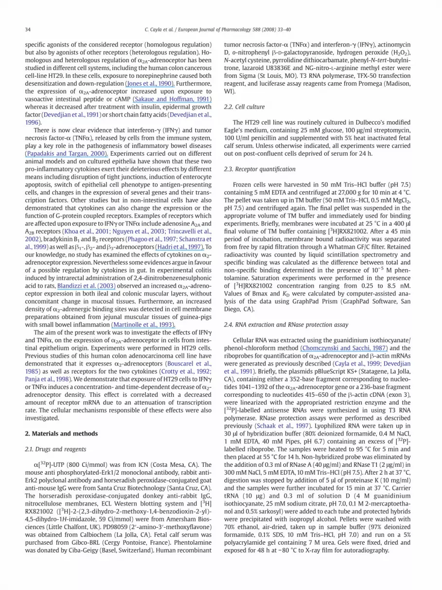
Because an accelerated rate of mRNA degradation may account forthe decrease of α2-adrenoceptor mRNA, the effect of cytokines onmRNA stabilitywas tested. For this purpose, HT29 cellswere incubatedwith IFNγ or TNFα in the presence of actinomycin D (5 μg/ml) in orderto abolish gene transcription; the disappearance of α2A-adrenoceptormRNA was then monitored over 6 h. Analysis of the data depicted inFig. 3A, indicated that the half-life ofα2A-adrenoceptormRNA in IFNγ-or TNFα-treated cells (165±24 and 189±15 min, respectively) was notsignificantly different from that in control cells (180±22 min), thussuggesting that a lower rate of gene transcription may be responsiblefor the effects of both cytokines. To test this hypothesis, HT29 cellswere transiently transfectedwith a plasmid containing luciferase geneunder the control of the 5′ non-coding region of theα2A-adrenoceptorgene. As shown in Fig. 3B, a 24 h-period of treatment with 100 U/mlIFNγ or 20 ng/ml TNFα respectively resulted in a 38% and 36% decreasein luciferase activity. Taken together, these results strongly suggest thatreceptor down-regulation promoted by IFNγ and TNFα is due to adecreased in the transcription rate of α2A-adrenoceptor gene.
3.4. Signalling pathway mediating cytokines effect
Finally, the signalling pathways whereby IFNγ and TNFα decreasethe α2A-adrenoceptor expression were investigated. Since reactiveoxygen species were recognized to act as intracellular mediators ofIFNγ and TNFα action, their implication was tested. As demonstratedby the use of a chemiluminescencemethod allowing tomeasure intra-cellular content of hydrogen peroxide (H2O2) in intact cells (Fig. 4A), a24 h-period of treatment with 100 U/ml IFNγ or 20 ng/ml TNFαsignificantly increased H2O2 levels. Moreover, a 24 h-exposure of HT29cells to exogenous H2O2, resulted in a concentration-dependent de-crease in α2-adrenoceptor density (Fig. 4B). This effect was correlated

Fig. 1. IFNγ and TNFα decrease α2-adrenoceptor density. (A) Concentration-dependent effect of IFNγ and TNFα: Serum-deprived HT29 cells were incubated for 48 h with increasingconcentrations of IFNγ (left panel) or TNFα (right panel). (B) Kinetics of IFNγ and TNFα action: Serum-deprived HT29 cells were incubated with 100 U/ml IFNγ (black symbol) or20 ng/ml TNFα (white symbol) for different period of times. In (A) and (B), cell membraneswere prepared andα2-adrenoceptor density was determined by binding studies using [3H]RX821002. The number of [3H]RX821002 binding sites (Bmax) was expressed as percentage of that in untreated cells. Results aremeans±S.E.M. from three independent experiments.
36 C. Cayla et al. / European Journal of Pharmacology 588 (2008) 33–40
to dramatic decrease ofα2A-adrenoceptormRNAandwas not due to anincreased cellularmortality (not shown). However, inconsistent with arole of reactive oxygen species, cell pre-treatment with 20 mM of N-acetyl cysteine did not impair the incidence of IFNγ or TNFα on α2-adrenoceptor expression (Table 1). Similar resultswere obtained in thepresence of other anti-oxidants such as 100 μM pyrrolidine dithio-carbamate, 500 μM phenyl-N-tert-butylnitrone or 20 μM lazaroidU83836E. Therefore, although reactive oxygen species decrease theexpression of the α2A-adrenoceptor in HT29 cells, they are probablynot involved in the effect of IFNγ or TNFα. The implication of nitricoxide (NO) was also investigated, because this messenger has beenassociated with the initiation and maintenance of inflammation inhuman inflammatory bowel disease (Kolios et al., 2004). Similarly towhat foundwithH2O2, exposure of HT29 cells to theNO donor, sodiumnitroprussiate (100 μM), resulted in a 35% decrease of α2A-adreno-ceptor density. However, the addition of the NO synthase inhibitor(NG-nitro-L-arginine methyl ester, 1 mM) did not reverse the cytokineeffect (Table 1). It is known that cytokines can activate extracellularsignal-regulated kinases (Erk1/2). Therefore the possibility that thispathwaymediates IFNγ or TNFα effects was tested. As depicted in Fig.5A, IFNγ and TNFα induce a rapid and transient increase of Erk1/2phosphorylation, which peaked 15 min after the beginning of thetreatment. The implication of Erk1/2 in the effect of cytokines on α2A-
adrenoceptor expression was then examined by using PD98059, aspecific inhibitor of mitogen-activated protein kinase kinases (MEK)which abolishes Erk1/2 activation. Results presented in Fig. 5B indi-cated that a 30 min preincubation with PD98059 blunted the effect ofTNFα but did not affect that of IFNγ. Therefore, the effect of TNFα onthe expression of α2-adrenoceptor appears to be mediated by Erk1/2activation, while the effect of IFNγ is triggered by another mechanism.
4. Discussion
In intestinal mucosa, epithelial cells are in close contact with cellsfrom the lymphoid and myeloid lineage, which are able to producecytokines. Numerous studies carried out on patients or animal modelshave shown that the production of pro-inflammatory cytokines, suchas IFNγ and TNFα, is highly increased in inflammatory bowel disease(Fuss, 2003; Papadakis and Targan, 2000; Wittig and Zeitz, 2003). Thepresent work demonstrates that IFNγ and TNFα effectively reduce theexpression of the α2A-adrenoceptor in HT29 cells. The effect of bothcytokines is concentration- and time-dependent, and is detected bothat mRNA and protein levels. The concentration of IFNγ promotinghalf-maximal effect is 2.2 U/ml (equivalent to 0.22 ng/ml) a value thatis in the range of the circulating concentrations of this cytokine ininflammatory situations. Indeed, the seric level of IFNγ was found to

Fig. 2. IFNγ and TNFα decreaseα2A-adrenoceptor mRNA levels. Serum-deprived HT29 cells were treated with either IFNγ or TNFα. Cellular RNAs were extracted and the amounts ofα2A-adrenoceptor mRNA and β-actin mRNA (taken as internal standard) were measured by RNase protection assay using specific riboprobes. (A) Autoradiogram of a typical RNaseprotection assay: In this specific experiment HT29 cells were exposed for 12 h to 100 U/ml IFNγ, 20 ng/ml TNFα or vehicle (control). The amounts of α2A-adrenoceptor mRNA (upperpanel) and β-actin mRNA (lower panel) were determined using corresponding riboprobes. (B) Time-course of the change in α2A-adrenoceptor mRNA: HT29 cells were exposed fordifferent periods of time to 100 U/ml IFNγ (black symbol) or 20 ng/ml TNFα (white symbol). At each experimental point, the amounts of α2A-adrenoceptor and β-actin mRNAs weredetermined by RNase protection assay and densitometric analysis of the autoradiograms. The amounts of α2-adrenoceptor mRNAwere normalized by β-actin and then expressed asthe percent of that in untreated cells. Results are means±S.E.M. from three independent experiments. (C) The effects of cytokines on α2A-adrenoceptor mRNA amount areconcentration-dependent: HT29 cells were incubated for 6 h with increasing concentrations of IFNγ (left panel) or TNFα (right panel) and the amounts of α2A-adrenoceptor mRNAwere determined as in panel A.
37C. Cayla et al. / European Journal of Pharmacology 588 (2008) 33–40
be 0.63 ng/ml in children with food allergy (Hofman, 1995) and toreach concentration as high as 20 μg/ml in rats treated with trinitro-benzenesulfonic acid (Dasgupta et al., 2001). Half-maximal effect ofTNFαwas observed at 1 ng/ml. Such a concentration is also relevant inthe colon, as TNFα concentration in stool from normal children isaround 60 pg/g stool but attains 1 ng/g in Crohn's disease (Braeggeret al., 1992). The modulation of intestinal α2-adrenoceptor expressionwas already studied in experimental models of inflammatory boweldisease such as guinea-pigs treated with trinitrobenzenesulfonic acid(Martinolle et al., 1993) and rats treated with 2,4-dinitrobenzenesul-phonic acid (Blandizzi et al., 2003). A significant increase in [3H]-rauwolscine binding sites was found in smooth-muscle membranepreparations from guinea-pig jejunum (Martinolle et al., 1993).Similarly, an increase in the α2-adrenoceptor mRNA measured byRT-PCR was found in the muscular layer from ileum and colon of 2,4-dinitrobenzenesulphonic acid-treated rats. By contrast, no changewasobserved when mucosal compartment was considered (Blandizziet al., 2003). Beside the fact that the purity of mucosa scraped frominflamed gut is questionable; the apparent discrepancy between thislater result and ours may result from the fact that the present workinvestigated the effects of two cytokines, IFNγ and TNFα, on asimplified in vitro model, while previous studies were conducted inmore complex animal models in which levels of other cytokines, suchas interleukins 1, 5, 6, 8 and 12, are also affected (Papadakis andTargan, 2000) and in which sympathetic innervation of the inflamedmucosa is partially lost (Straub et al., 2006).
In our study, the decrease in receptor number after IFNγ or TNFαtreatment is preceded and correlatedwith reducedmRNA steady-statelevels and the delay between mRNA reduction and protein decrease isconsistentwith aα2A-adrenoceptor half-life of 26 h inHT29 cells (Pariset al., 1987). The combined use of actinomycin D and of a luciferaseconstruct containing the promoter region of α2A-adrenoceptor geneprovided some insight into the mechanisms whereby the two cyto-
kines decrease the steady-state level of α2A-adrenoceptor mRNA.Indeed, the determination of the receptormRNAhalf-life indicates thatthe decrease in receptor mRNA is not due to an accelerated rate of itsdegradation, but results from an attenuated rate of gene transcription.A change in the expression of various G-protein coupled receptors hasbeen reported in several models of inflammatory diseases. For ex-ample, a marked decrease in angiotensin AT1 receptor expression wasobserved in all organs of septic rats (Bucher et al., 2001). Similarly, α1-adrenoceptors are down-regulated in several organs (including heart,aorta and lung) after injection of lipopolysaccharide to rats (Bucheret al., 2003). TNFα and IFNγ were also shown to decrease the amountof vasopressin V1A receptor mRNA and vasopressin binding in rathepatocytes (Bucher et al., 2002). An increase in the number and anaugmentation of the functionality of adenosine A2A and A2B receptorswere noticed in the human monocytic cell line THP-1 treated withTNFα, while a down-regulation of adenosineA2A receptorwas found inthe same cells treated with IFNγ (Khoa et al., 2001). Recently, Khoa etal. (2006) showed that treatment of THP-1 cells with TNFα decreasesG-protein coupled receptor kinase 2 expression and inhibits adenosineA2A receptor desensitization, indicating that pro-inflammatory cyto-kines may modulate cellular signalling and modify density of G-protein coupled receptors by multiple mechanisms. The use of high-density microarrays on biopsies taken from the sigmoid colon mucosahas recently allowed comparative examination of the transcriptome inpatients suffering Crohn's disease or ulcerative colitis (Costello et al.,2005). No significant change in the level of α2A-adrenoceptortranscript was reported in this study, but a decrease in norepinephrinetransporter expression was observed in patients with Crohn's disease.According to other studies in mice with dextran sodium sulphate-induced colitis as well as in patients with Crohn's disease (Straub et al.,2005, 2006), the decrease of the norepinephrine transporter is likelythe consequence of a loss of sympathetic nerve fibres in the mucosaand submucosa. One may therefore speculate that a diminution of
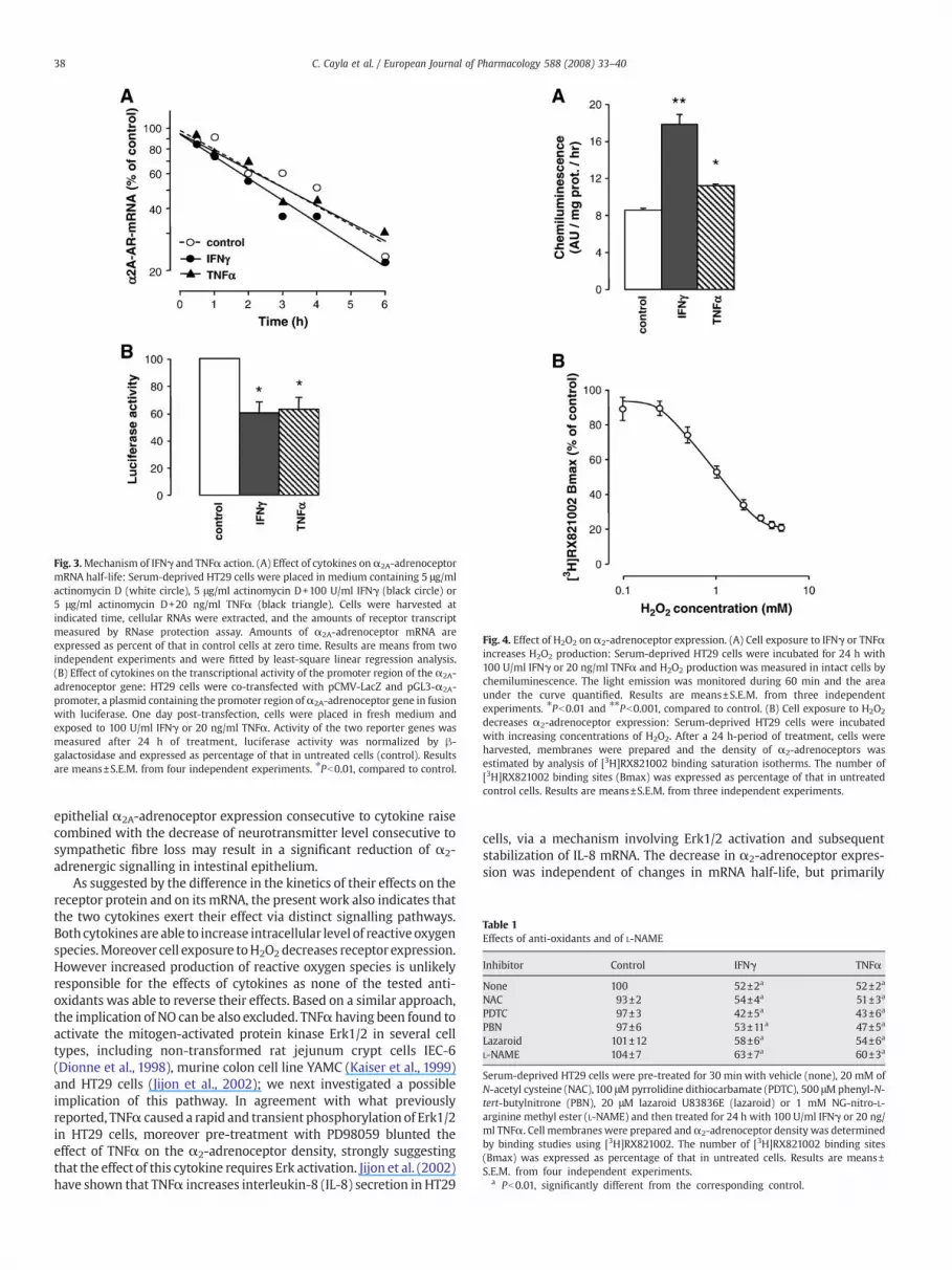
Fig. 3.Mechanism of IFNγ and TNFα action. (A) Effect of cytokines onα2A-adrenoceptormRNA half-life: Serum-deprived HT29 cells were placed in medium containing 5 μg/mlactinomycin D (white circle), 5 μg/ml actinomycin D+100 U/ml IFNγ (black circle) or5 μg/ml actinomycin D+20 ng/ml TNFα (black triangle). Cells were harvested atindicated time, cellular RNAs were extracted, and the amounts of receptor transcriptmeasured by RNase protection assay. Amounts of α2A-adrenoceptor mRNA areexpressed as percent of that in control cells at zero time. Results are means from twoindependent experiments and were fitted by least-square linear regression analysis.(B) Effect of cytokines on the transcriptional activity of the promoter region of the α2A-adrenoceptor gene: HT29 cells were co-transfected with pCMV-LacZ and pGL3-α2A-promoter, a plasmid containing the promoter region ofα2A-adrenoceptor gene in fusionwith luciferase. One day post-transfection, cells were placed in fresh medium andexposed to 100 U/ml IFNγ or 20 ng/ml TNFα. Activity of the two reporter genes wasmeasured after 24 h of treatment, luciferase activity was normalized by β-galactosidase and expressed as percentage of that in untreated cells (control). Resultsare means±S.E.M. from four independent experiments. ⁎Pb0.01, compared to control.
Fig. 4. Effect of H2O2 on α2-adrenoceptor expression. (A) Cell exposure to IFNγ or TNFαincreases H2O2 production: Serum-deprived HT29 cells were incubated for 24 h with100 U/ml IFNγ or 20 ng/ml TNFα and H2O2 production was measured in intact cells bychemiluminescence. The light emission was monitored during 60 min and the areaunder the curve quantified. Results are means±S.E.M. from three independentexperiments. ⁎Pb0.01 and ⁎⁎Pb0.001, compared to control. (B) Cell exposure to H2O2
decreases α2-adrenoceptor expression: Serum-deprived HT29 cells were incubatedwith increasing concentrations of H2O2. After a 24 h-period of treatment, cells wereharvested, membranes were prepared and the density of α2-adrenoceptors wasestimated by analysis of [3H]RX821002 binding saturation isotherms. The number of[3H]RX821002 binding sites (Bmax) was expressed as percentage of that in untreatedcontrol cells. Results are means±S.E.M. from three independent experiments.
Table 1Effects of anti-oxidants and of L-NAME
Inhibitor Control IFNγ TNFα
None 100 52±2a 52±2a
NAC 93±2 54±4a 51±3a
PDTC 97±3 42±5a 43±6a
PBN 97±6 53±11a 47±5a
Lazaroid 101±12 58±6a 54±6a
L-NAME 104±7 63±7a 60±3a
Serum-deprived HT29 cells were pre-treated for 30 min with vehicle (none), 20 mM ofN-acetyl cysteine (NAC), 100 μM pyrrolidine dithiocarbamate (PDTC), 500 μMphenyl-N-tert-butylnitrone (PBN), 20 μM lazaroid U83836E (lazaroid) or 1 mM NG-nitro-L-arginine methyl ester (L-NAME) and then treated for 24 h with 100 U/ml IFNγ or 20 ng/ml TNFα. Cell membranes were prepared andα2-adrenoceptor density was determinedby binding studies using [3H]RX821002. The number of [3H]RX821002 binding sites(Bmax) was expressed as percentage of that in untreated cells. Results are means±S.E.M. from four independent experiments.
a Pb0.01, significantly different from the corresponding control.
38 C. Cayla et al. / European Journal of Pharmacology 588 (2008) 33–40
epithelial α2A-adrenoceptor expression consecutive to cytokine raisecombined with the decrease of neurotransmitter level consecutive tosympathetic fibre loss may result in a significant reduction of α2-adrenergic signalling in intestinal epithelium.
As suggested by the difference in the kinetics of their effects on thereceptor protein and on its mRNA, the present work also indicates thatthe two cytokines exert their effect via distinct signalling pathways.Both cytokines are able to increase intracellular level of reactive oxygenspecies.Moreover cell exposure toH2O2 decreases receptor expression.However increased production of reactive oxygen species is unlikelyresponsible for the effects of cytokines as none of the tested anti-oxidants was able to reverse their effects. Based on a similar approach,the implication of NO can be also excluded. TNFα having been found toactivate the mitogen-activated protein kinase Erk1/2 in several celltypes, including non-transformed rat jejunum crypt cells IEC-6(Dionne et al., 1998), murine colon cell line YAMC (Kaiser et al., 1999)and HT29 cells (Jijon et al., 2002); we next investigated a possibleimplication of this pathway. In agreement with what previouslyreported, TNFα caused a rapid and transient phosphorylation of Erk1/2in HT29 cells, moreover pre-treatment with PD98059 blunted theeffect of TNFα on the α2-adrenoceptor density, strongly suggestingthat the effect of this cytokine requires Erk activation. Jijon et al. (2002)have shown that TNFα increases interleukin-8 (IL-8) secretion in HT29
cells, via a mechanism involving Erk1/2 activation and subsequentstabilization of IL-8 mRNA. The decrease in α2-adrenoceptor expres-sion was independent of changes in mRNA half-life, but primarily

Fig. 5. Role of Erk onα2-adrenoceptor down-regulation by cytokines. (A) IFNγ and TNFα induce Erk phosphorylation: Serum-deprived HT29 cells were incubated for different periodsof time with 100 U/ml IFNγ (left panels) or 20 ng/ml TNFα (right panels). Soluble proteins were extracted, separated by gel electrophoresis and blotted onto a nitrocellulosemembrane. Phosphorylated forms of Erk1/2 were detected using an anti phosphorylated-Erk1/2 monoclonal antibody (upper panels). Blots were reprobed with an anti-Erk2antibody to assess for equal protein loading (lower panels). (B) Effect of TNFα, but not IFNγ, is dependent on Erk activation: Serum-deprived HT29 cells were pre-treated for 30 minwith vehicle (right panel) or 50 μMPD98059 (right panel) and then exposed for 24 h to 100 U/ml IFNγ or 20 ng/ml TNFα. Cell membraneswere prepared andα2-adrenoceptor densitywas determined by binding studies using [3H]RX821002. The number of [3H]RX821002 binding sites (Bmax) was expressed in fmol per mg of protein. Results are means±S.E.M. fromfour independent experiments. ⁎Pb0.01, compared to corresponding control.
39C. Cayla et al. / European Journal of Pharmacology 588 (2008) 33–40
relied on a decrease of the transcription rate. Similar to TNFα, someeffects of IFNγ require Erk1/2 activation. In CaCo2 cells, IFNγ reducesthe activity of Na+,K+-ATPase and of type 1 Na+/H+ exchanger via amechanism involving Erk1/2 phosphorylation (Magro et al., 2004;Magro et al., 2005). There is also phosphorylation of Erk1/2 aftertreatment of HT29 cells with IFNγ, however the effect of IFNγ on α2-adrenoceptor level was insensitive to the presence of MEK inhibitor. Itis nowclearly established that the effects of IFNγ on gene transcriptionare triggered not only via the classical JAK/STAT pathway but also viaSTAT-independent mechanisms including PI3K/Akt and p38 MAPKpathways (Gil et al., 2001; Platanias, 2005), there is moreover accumu-lating evidence that coordinated activation of more than one pathwayis required for generation of a givenbiological effect. Clarification of themechanisms accounting for the effect of IFNγ onα2-adrenoceptor willcertainly require the combination of different approaches.
Diarrhoea is a common clinical feature of immune-mediated boweldysfunction, and numerous in vivo or in vitro experiments indicate thatIFNγ and TNFα play a key role in the changes of mucosa permeability.Effects of cytokines result from alteration of intestinal epitheliumintegrity as well as from modification of the expression of pumps andion channels. For example, IFNγ markedly reduced the abundance ofNa+,K+-ATPase in the colon of trinitrobenzenesulfonic acid-treated rats(Magro et al., 2005). Inmice treatedwith an anti-CD3 antibody in orderto induce diarrhoea, mucosal Na+,K+-ATPase activity was decreasedand this effectwas blocked byadministration of an anti-TNFα antibody(Musch et al., 2002). TNFα inhibited water and Cl− absorption and itdown-regulated Na+-K+-2Cl− transporter and Na+,K+-ATPase in per-fused rat distal colon (Markossian and Kreydiyyeh, 2005). In humandistal colon mounted in an Ussing chamber, the addition of TNFα tothe serosal compartment increased the 36Cl− serosal-to-mucosal flux,
decreased the 36Cl−mucosal-to-serosal flux, and increased the 86Rb netefflux, which reflects the K+ secretion (Schmitz et al., 1996). The activ-ation of α2-adrenoceptors is known to stimulate Na+ and water ab-sorption and to inhibit Cl− and HCO3
− secretion (Chang et al., 1983; Liuand Coupar, 1997). Moreover, clinical trials have demonstrated thepotential interest of α2-adrenoceptor agonists for the treatment ofpatients suffering cancer therapy-related diarrhoea or short bowelsyndrome (Ippoliti, 1998; Schworer et al., 1995). It is thus possible thatthe decrease of epithelial α2-adrenoceptor may contribute to harmfulcytokine effects observed in inflammatory intestinal diseases. More-over, numerous molecules inhibiting cytokine action have beendeveloped and tested in clinical trials. An antibody against IFNγ(fontolizumab) did not demonstrate efficacy when administered at asingle dose in patients with Crohn's disease, but was more efficientwhen a second dosewas administered later (Nakamura et al., 2006). Bycontrast, anti-TNFα agents such as infliximab, CDP571 or CDP870,which are mouse/human chimeric antibodies, gave very promisingresults in the treatment of Crohn's disease, although adverse effects arealso described (Nakamura et al., 2006; Sandborn and Faubion, 2004). Itis possible that maintenance of α2-adrenoceptor expression, whichmay occur following the treatment with anti-TNFα antibodies, wouldrepresent a part of beneficial effects of these molecules.
References
Berlioz, F., Maoret, J.J., Paris, H., Laburthe, M., Farinotti, R., Roze, C., 2000. α2-Adrenergicreceptors stimulate oligopeptide transport in a human intestinal cell line. J.Pharmacol. Exp. Ther. 294, 466–472.
Blandizzi, C., Fornai, M., Colucci, R., Baschiera, F., Barbara, G., De Giorgio, R., De Ponti, F.,Breschi, M.C., Del Tacca, M., 2003. Altered prejunctional modulation of intestinalcholinergic and noradrenergic pathways by α2-adrenoceptors in the presence ofexperimental colitis. Br. J. Pharmacol. 139, 309–320.
40 C. Cayla et al. / European Journal of Pharmacology 588 (2008) 33–40
Bouscarel, B., Cortinovis, C., Carpene, C., Murat, J.C., Paris, H., 1985. α2-Adrenoceptors inthe HT 29 human colon adenocarcinoma cell line: characterization with [3H]clonidine; effects on cyclic AMP accumulation. Eur. J. Pharmacol. 107, 223–231.
Braegger, C.P., Nicholls, S., Murch, S.H., Stephens, S., MacDonald, T.T., 1992. Tumournecrosis factor α in stool as a marker of intestinal inflammation. Lancet 339, 89–91.
Brede, M., Philipp, M., Knaus, A., Muthig, V., Hein, L., 2004. α2-Adrenergic receptorsubtypes — novel functions uncovered in gene-targeted mouse models. Biol. Cell96, 343–348.
Bucher, M., Hobbhahn, J., Taeger, K., Kurtz, A., 2002. Cytokine-mediated downregulationof vasopressin V1A receptors during acute endotoxemia in rats. Am. J. Physiol. 282,R979–R984.
Bucher, M., Ittner, K.P., Hobbhahn, J., Taeger, K., Kurtz, A., 2001. Downregulation ofangiotensin II type 1 receptors during sepsis. Hypertension 38, 177–182.
Bucher, M., Kees, F., Taeger, K., Kurtz, A., 2003. Cytokines down-regulate α1-adrenergicreceptor expression during endotoxemia. Crit. Care Med. 31, 566–571.
Buffin-Meyer, B., Crassous, P.A., Delage, C., Denis, C., Schaak, S., Paris, H., 2007. EGFreceptor transactivation and PI3-kinase mediate stimulation of ERK by α2A-adre-noreceptor in intestinal epithelial cells: a role in wound healing. Eur. J. Pharmacol.574, 85–93.
Cayla, C., Schaak, S., Roquelaine, C., Gales, C., Quinchon, F., Paris, H., 1999. Homologousregulation of theα2C-adrenoceptor subtype in human hepatocarcinoma, HepG2. Br.J. Pharmacol. 126, 69–78.
Chang, E.B., Field, M., Miller, R.J., 1983. Enterocyte a2-adrenergic receptors: yohimbineand p-aminoclonidine binding relative to ion transport. Am. J. Physiol. 244,G76–G82.
Chomczynski, P., Sacchi, N., 1987. Single-step method of RNA isolation by acid gua-nidinium thiocyanate-phenol-chloroform extraction. Anal. Biochem. 162, 156–159.
Costello, C.M., Mah, N., Hasler, R., Rosenstiel, P., Waetzig, G.H., Hahn, A., Lu, T., Gurbuz, Y.,Nikolaus, S., Albrecht, M., Hampe, J., Lucius, R., Kloppel, G., Eickhoff, H., Lehrach, H.,Lengauer, T., Schreiber, S., 2005. Dissection of the inflammatory bowel diseasetranscriptome using genome-wide cDNA microarrays. PLoS Med. 2, e199.
Crotty, B., Rosenberg, W.M., Aronson, J.K., Jewell, D.P., 1992. Inhibition of binding ofinterferon γ to its receptor by salicylates used in inflammatory bowel disease. Gut33, 1353–1357.
Dasgupta, A., Ramaswamy, K., Giraldo, J., Taniguchi, M., Amenta, P.S., Das, K.M., 2001.Colon epithelial cellular protein induces oral tolerance in the experimental modelof colitis by trinitrobenzene sulfonic acid. J. Lab. Clin. Med. 138, 257–269.
Devedjian, J.C., Fargues, M., Denis-Pouxviel, C., Daviaud, D., Prats, H., Paris, H., 1991.Regulation of the α2A-adrenergic receptor in the HT29 cell line. Effects of insulinand growth factors. J. Biol. Chem. 266, 14359–14366.
Devedjian, J.C., Schaak, S., Gamet, L., Denis-Pouxviel, C., Paris, H., 1996. Regulation ofα2A-adrenergic receptor expression in the human colon carcinoma cell line HT29:SCFA-induced enterocytic differentiation results in an inhibition of α2C10 genetranscription. Proc. Assoc. Am. Physicians 108, 334–344.
Dionne, S., D'Agata, I.D., Ruemmele, F.M., Levy, E., St-Louis, J., Srivastava, A.K., Levesque,D., Seidman, E.G., 1998. Tyrosine kinase and MAPK inhibition of TNFα- and EGF-stimulated IEC-6 cell growth. Biochem. Biophys. Res. Commun. 242, 146–150.
Fuss, I.J., 2003. Cytokine network in inflammatory bowel disease. Curr. Drug TargetsInflamm. Allergy 2, 101–112.
Gil, M.P., Bohn, E., O'Guin, A.K., Ramana, C.V., Levine, B., Stark, G.R., Virgin, H.W.,Schreiber, R.D., 2001. Biologic consequences of Stat1-independent IFN signaling.Proc. Natl. Acad. Sci. U. S. A. 98, 6680–6685.
Hadri, K.E., Courtalon, A., Gauthereau, X., Chambaut-Guerin, A.M., Pairault, J., Feve, B.,1997. Differential regulation by tumor necrosis factor-alpha of β1-, β2-, and β3-adrenoreceptor gene expression in 3T3-F442A adipocytes. J. Biol. Chem. 272,24514–24521.
Hofman, T., 1995. IL-4 and IFN-γ level in blood serum of childrenwith food allergy. Rocz.Akad. Med. Bialymst. 40, 462–467.
Ippoliti, C., 1998. Antidiarrheal agents for the management of treatment-relateddiarrhea in cancer patients. Am. J. Health. Syst. Pharm. 55, 1573–1580.
Jijon, H.B., Panenka, W.J., Madsen, K.L., Parsons, H.G., 2002. MAP kinases contribute toIL-8 secretion by intestinal epithelial cells via a posttranscriptional mechanism. Am.J. Physiol. 283, C31–C41.
Jones, S.B., Leone, S.L., Bylund, D.B., 1990. Desensitization of the α2-adrenergic receptorin HT29 and opossum kidney cell lines. J. Pharmacol. Exp. Ther. 254, 294–300.
Kaiser, G.C., Yan, F., Polk, D.B., 1999. Conversion of TNF-α from antiproliferative toproliferative ligand in mouse intestinal epithelial cells by regulating mitogen-activated protein kinase. Exp. Cell Res. 249, 349–358.
Khoa, N.D., Montesinos, M.C., Reiss, A.B., Delano, D., Awadallah, N., Cronstein, B.N., 2001.Inflammatory cytokines regulate function and expression of adenosine A2A
receptors in human monocytic THP-1 cells. J. Immunol. 167, 4026–4032.Khoa, N.D., Postow, M., Danielsson, J., Cronstein, B.N., 2006. Tumor necrosis factor-α
prevents desensitization of Gαs-coupled receptors by regulating GRK2 associationwith the plasma membrane. Mol. Pharmacol. 69, 1311–1319.
Kobilka, B.K., Matsui, H., Kobilka, T.S., Yang-Feng, T.L., Francke, U., Caron, M.G., Lefkowitz,R.J., Regan, J.W., 1987. Cloning, sequencing, and expression of the gene coding forthe human platelet α2-adrenergic receptor. Science 238, 650–656.
Kolios, G., Valatas, V., Ward, S.G., 2004. Nitric oxide in inflammatory bowel disease: auniversal messenger in an unsolved puzzle. Immunology 113, 427–437.
Liu, L., Coupar, I.M., 1997. Role of α2-adrenoceptors in the regulation of intestinal watertransport. Br. J. Pharmacol. 120, 892–898.
Magro, F., Fraga, S., Ribeiro, T., Soares-da-Silva, P., 2004. Intestinal Na+-K+-ATPaseactivity and molecular events downstream of interferon-gamma receptor stimula-tion. Br. J. Pharmacol. 142, 1281–1292.
Magro, F., Fraga, S., Soares-da-Silva, P., 2005. Signaling of short- and long-term re-gulation of intestinal epithelial type 1 Na+/H+ exchanger by interferon-γ. Br. J.Pharmacol. 145, 93–103.
Markossian, S., Kreydiyyeh, S.I., 2005. TNF-α down-regulates the Na+-K+ ATPase and theNa+-K+-2Cl− cotransporter in the rat colon via PGE2. Cytokine 30, 319–327.
Martinolle, J.P., More, J., Dubech, N., Garcia-Villar, R., 1993. Inverse regulation of α- andβ-adrenoceptors during trinitrobenzenesulfonic acid (TNB)-induced inflammationin guinea-pig small intestine. Life Sci. 52, 1499–1508.
Musch, M.W., Clarke, L.L., Mamah, D., Gawenis, L.R., Zhang, Z., Ellsworth, W., Shalowitz,D., Mittal, N., Efthimiou, P., Alnadjim, Z., Hurst, S.D., Chang, E.B., Barrett, T.A., 2002. Tcell activation causes diarrhea by increasing intestinal permeability and inhibitingepithelial Na+/K+-ATPase. J. Clin. Invest. 110, 1739–1747.
Nakamura, K., Honda, K., Mizutani, T., Akiho, H., Harada, N., 2006. Novel strategies forthe treatment of inflammatory bowel disease: selective inhibition of cytokines andadhesion molecules. World J. Gastroenterol. 12, 4628–4635.
Nguyen, D.K., Montesinos, M.C., Williams, A.J., Kelly, M., Cronstein, B.N., 2003. Th1cytokines regulate adenosine receptors and their downstream signaling elementsin human microvascular endothelial cells. J. Immunol. 171, 3991–3998.
Panja, A., Goldberg, S., Eckmann, L., Krishen, P., Mayer, L., 1998. The regulation andfunctional consequence of proinflammatory cytokine binding on human intestinalepithelial cells. J. Immunol. 161, 3675–3684.
Papadakis, K.A., Targan, S.R., 2000. Role of cytokines in the pathogenesis ofinflammatory bowel disease. Annu. Rev. Med. 51, 289–298.
Paris, H., Taouis, M., Galitzky, J., 1987. In vitro study of α2-adrenoceptor turnover andmetabolism using the adenocarcinoma cell line HT29. Mol. Pharmacol. 32,646–654.
Phagoo, S.B., Yaqoob, M., McIntyre, P., Jones, C., Burgess, G.M., 1997. Cytokines increaseB1 bradykinin receptor mRNA and protein levels in human lung fibroblasts.Biochem. Soc. Trans. 25, 43S.
Pizzinat, N., Copin, N., Vindis, C., Parini, A., Cambon, C., 1999. Reactive oxygen speciesproduction by monoamine oxidases in intact cells. Naunyn. Schmiedeberg's Arch.Pharmacol. 359, 428–431.
Platanias, L.C., 2005. Mechanisms of type-I- and type-II-interferon-mediated signalling.Nat. Rev. Immunol. 5, 375–386.
Ruffolo Jr., R.R., Nichols, A.J., Stadel, J.M., Hieble, J.P., 1993. Pharmacologic and thera-peutic applications of a2-adrenoceptor subtypes. Annu. Rev. Pharmacol. Toxicol. 33,243–279.
Sakaue, M., Hoffman, B.B., 1991. cAMP regulates transcription of the α2A-adrenergicreceptor gene in HT-29 cells. J. Biol. Chem. 266, 5743–5749.
Sandborn, W.J., Faubion, W.A., 2004. Biologics in inflammatory bowel disease: howmuch progress have we made? Gut 53, 1366–1373.
Schaak, S., Cayla, C., Blaise, R., Quinchon, F., Paris, H., 1997. HepG2 and SK-N-MC: twohuman models to study α2-adrenergic receptors of the a2C subtype. J. Pharmacol.Exp. Ther. 281, 983–991.
Schaak, S., Cussac, D., Cayla, C., Devedjian, J.C., Guyot, R., Paris, H., Denis, C., 2000. α2-Adrenoceptors regulate proliferation of human intestinal epithelial cells. Gut 47,242–250.
Schanstra, J.P., Marin-Castano, M.E., Alric, C., Pesquero, J.B., Claire, M., Girolami, J.P.,Bascands, J.L., 1999. Homologous and heterologous induction of the human brady-kinin B1-receptor and B1-receptor localisation along the rat nephron. Immuno-pharmacology 45, 29–34.
Schmitz, H., Fromm, M., Bode, H., Scholz, P., Riecken, E.O., Schulzke, J.D., 1996. Tumornecrosis factor-α induces Cl− and K+ secretion in human distal colon driven byprostaglandin E2. Am. J. Physiol. 271, G669–G674.
Schworer, H., Munke, H., Stockmann, F., Ramadori, G., 1995. Treatment of diarrhea incarcinoid syndrome with ondansetron, tropisetron, and clonidine. Am. J. Gastro-enterol. 90, 645–648.
Straub, R.H., Stebner, K., Harle, P., Kees, F., Falk, W., Scholmerich, J., 2005. Key role of thesympathetic microenvironment for the interplay of tumour necrosis factor andinterleukin 6 in normal but not in inflamedmouse colonmucosa. Gut 54,1098–1106.
Straub, R.H., Wiest, R., Strauch, U.G., Harle, P., Scholmerich, J., 2006. The role of thesympathetic nervous system in intestinal inflammation. Gut 55, 1640–1649.
Trincavelli, M.L., Costa, B., Tuscano, D., Lucacchini, A., Martini, C., 2002. Up-regulation ofA2A adenosine receptors by proinflammatory cytokines in rat PC12 cells. Biochem.Pharmacol. 64, 625–631.
Tutton, P.J., Barkla, D.H., 1987. Biogenic amines as regulators of the proliferative activityof normal and neoplastic intestinal epithelial cells (review). Anticancer Res. 7, 1–12.
Valet, P., Senard, J.M., Devedjian, J.C., Planat, V., Salomon, R., Voisin, T., Drean, G.,Couvineau, A., Daviaud, D., Denis, C., Laburthe, M., Paris, H., 1993. Characterizationand distribution of a2-adrenergic receptors in the human intestinal mucosa. J. Clin.Invest. 91, 2049–2057.
von Zastrow, M., 2001. Role of endocytosis in signalling and regulation of G-protein-coupled receptors. Biochem. Soc. Trans. 29, 500–504.
von Zastrow, M., 2003. Mechanisms regulating membrane trafficking of G protein-coupled receptors in the endocytic pathway. Life Sci. 74, 217–224.
Wittig, B.M., Zeitz, M., 2003. The gut as an organ of immunology. Int. J. Colorectal Dis. 18,181–187.


![Imaging beta-adrenoceptors in the human brain with (S)-1'-[18F]fluorocarazolol](https://img.dokumen.tips/doc/110x75/63377b99d102fae1b6076339/imaging-beta-adrenoceptors-in-the-human-brain-with-s-1-18ffluorocarazolol.jpg)

