Embed Size (px)
Citation preview

23 TIN AND TIN COMPOUNDS
3. HEALTH EFFECTS
3.1 INTRODUCTION
The primary purpose of this chapter is to provide public health officials, physicians, toxicologists, and
other interested individuals and groups with an overall perspective on the toxicology of ton and tin
compounds. It contains descriptions and evaluations of toxicological studies and epidemiological
investigations and provides conclusions, where possible, on the relevance of toxicity and toxicokinetic
data to public health.
A glossary and list of acronyms, abbreviations, and symbols can be found at the end of this profile.
Because there is such a large number of inorganic tin and organotin compounds, only the most widely
studied compounds and those that present the greatest potential for human exposure have been selected
for the discussion of health effects. In addition to primary studies, review articles and government reports
are occasionally provided in order to assist the reader in understanding more fully the toxicology of the
tin compounds.
3.2 DISCUSSION OF HEALTH EFFECTS BY ROUTE OF EXPOSURE
To help public health professionals and others address the needs of persons living or working near
hazardous waste sites, the information in this section is organized first by route of exposure (inhalation,
oral, and dermal) and then by health effect (death, systemic, immunological, neurological, reproductive,
developmental, genotoxic, and carcinogenic effects). These data are discussed in terms of three exposure
periods: acute (14 days or less), intermediate (15–364 days), and chronic (365 days or more).
Levels of significant exposure for each route and duration are presented in tables and illustrated in
figures. The points in the figures showing no-observed-adverse-effect levels (NOAELs) or lowest-
observed-adverse-effect levels (LOAELs) reflect the actual doses (levels of exposure) used in the studies.
LOAELs have been classified into "less serious" or "serious" effects. "Serious" effects are those that
evoke failure in a biological system and can lead to morbidity or mortality (e.g., acute respiratory distress
or death). "Less serious" effects are those that are not expected to cause significant dysfunction or death,
or those whose significance to the organism is not entirely clear. ATSDR acknowledges that a
24 TIN AND TIN COMPOUNDS
3. HEALTH EFFECTS
considerable amount of judgment may be required in establishing whether an end point should be
classified as a NOAEL, "less serious" LOAEL, or "serious" LOAEL, and that in some cases, there will be
insufficient data to decide whether the effect is indicative of significant dysfunction. However, the
Agency has established guidelines and policies that are used to classify these end points. ATSDR
believes that there is sufficient merit in this approach to warrant an attempt at distinguishing between
"less serious" and "serious" effects. The distinction between "less serious" effects and "serious" effects is
considered to be important because it helps the users of the profiles to identify levels of exposure at which
major health effects start to appear. LOAELs or NOAELs should also help in determining whether or not
the effects vary with dose and/or duration, and place into perspective the possible significance of these
effects to human health.
The significance of the exposure levels shown in the Levels of Significant Exposure (LSE) tables and
figures may differ depending on the user's perspective. Public health officials and others concerned with
appropriate actions to take at hazardous waste sites may want information on levels of exposure
associated with more subtle effects in humans or animals (LOAELs) or exposure levels below which no
adverse effects (NOAELs) have been observed. Estimates of levels posing minimal risk to humans
(Minimal Risk Levels or MRLs) may be of interest to health professionals and citizens alike.
Levels of exposure associated with carcinogenic effects (Cancer Effect Levels, CELs) of tin compounds
are indicated in Table 3-5 and Figure 3-5.
A User's Guide has been provided at the end of this profile (see Appendix B). This guide should aid in
the interpretation of the tables and figures for Levels of Significant Exposure and the MRLs.
3.2.1 Inhalation Exposure
Little information has been published regarding the effects of inhaled inorganic tin or organotin
compounds on human health. Reports of human occupational exposures often involve multiple chemicals
and lack details on actual exposure concentrations and conditions. Some reports of humans must also be
regarded as anecdotal. The older animal literature (from the 1950s) includes inhalation studies that are
lacking in description of methods and in reporting of experimental findings. However, it is still possible
to characterize some aspects of tin toxicity due to inhalation of inorganic tin and organotin compounds.
Exposure levels of the inhaled organotin compounds are expressed as milligrams per cubic meter (mg/m3)
of the specific tin compound unless otherwise noted. Doses are not expressed as doses of tin due to the
25 TIN AND TIN COMPOUNDS
3. HEALTH EFFECTS
covalent bond between the tin and the organic moiety. There are no data for specific inorganic tin
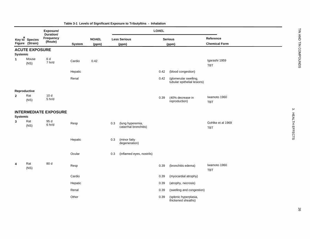
compounds. Calculations of parts per million (ppm) values are included where appropriate. Table 3-1
and Figure 3-1 summarize available quantitative information on health effects that have been observed in
animals after inhalation exposure to tributyltins. Exposure levels are expressed as ppm in Table 3-1 and
Figure 3-1. A table and figure are not presented for inorganic tin compounds due to limitations of the
available studies.
3.2.1.1 Death
Inorganic Tin Compounds. No studies were located regarding lethality in humans or animals after
inhalation exposure to inorganic tin compounds.
Organotin Compounds. Deaths have been reported in humans following exposure to organotins. One of
six workers died 12 days following exposure to a mixture of half dimethyltin and half trimethyltin
chloride vapor that occurred during the cleaning of a caldron at a chemical plant. Maximum exposure
was a total of 1.5 hours over a 3-day working period (Rey et al. 1984). No estimates of exposure levels
were given. The symptoms preceding death included excretion of high levels of tin in the urine,
respiratory depression, and coma. More uncertain is the report of a female worker who died following a
drenching with triphenyltin chloride, diphenyltin dichloride, and other unidentified compounds. No
estimates of exposure levels were given. Death was apparently caused by renal failure 12 days after
exposure (NIOSH 1976). No other studies were located regarding lethality in humans after inhalation
exposure to organotin compounds.
A 4-hour LC50 of 77 mg/m3 for tributyltin oxide (as total particles) was described by Schweinfurth and
Gunzel (1987) in a summary of acute studies; the LC50 for particles with a diameter of <10 µm was
65 mg/m3. The summary also indicates that a concentration of 20 mg/m3 of an aerosol of tributyltin oxide
was lethal to guinea pigs within 1 hour of exposure. Lethality in mice was observed following single or
repeated daily exposures to a butyltin mixture (81.2% tributyltin bromide and 3.7% dibutyltin dibromide)
together with other unidentified compounds (15.1%) (Igarashi 1959). The concentration was 5.65 mg
tin/m3 (1.16 ppm) as the butyltin mixture for different durations of exposure. The tributyltin bromide
concentration was 1.1 ppm and that for dibutyltin bromide was 0.06 ppm. For a 2-day, 8-hour/day
exposure, approximately 80–90% of the exposed mice died. Despite the observation of other signs of
toxicity (see Section 3.2.1.2) the exposure of the mice to multiple compounds confound the interpretation
of the data.
TIN A
ND
TIN C
OM
PO
UN
DS
26
3. HE
ALTH
EFFE
CTS
Table 3-1 Levels of Significant Exposure to Tributyltins - Inhalation
Exposure/ LOAEL Duration/
a Frequency Reference Key to Species NOAEL Less Serious Serious (Route) Figure (Strain) System (ppm) (ppm) (ppm) Chemical Form
ACUTE EXPOSURE Systemic 1 Mouse 6 d Cardio 0.42 Igarashi 1959 7 hr/d (NS) TBT
Hepatic 0.42 (blood congestion)
Renal 0.42 (glomerular swelling, tubular epithelial lesions)
Reproductive 2 Rat 10 d 0.39 (40% decrease in Iwamoto 1960 5 hr/d (NS) reproduction) TBT
INTERMEDIATE EXPOSURE Systemic 3 Rat 95 d Resp 0.3 (lung hyperemia, Gohlke et al 1969 6 hr/d (NS) catarrhal bronchitis) TBT
Hepatic 0.3 (minor fatty degeneration)
Ocular 0.3 (inflamed eyes, nostrils)
4 Rat 80 d Resp 0.39 (bronchitis edema) Iwamoto 1960 (NS) TBT
Cardio 0.39 (myocardial atrophy)
Hepatic 0.39 (atrophy, necrosis)
Renal 0.39 (swelling and congestion)
Other 0.39 (splenic hyperplasia, thickened sheaths)

Table 3-1 Levels of Significant Exposure to Tributyltins - Inhalation (continued)
Exposure/ LOAEL
TIN
aKey to Figure
Species (Strain)
Duration/ Frequency
(Route) System
NOAEL (ppm)
AN
D TIN
CO
MP
OU
ND
S
Reference Chemical Form
Less Serious (ppm)
Serious (ppm)
Reproductive 5 Rat
(NS) 80 d 0.39 Iwamoto 1960
TBT
a The number corresponds to entries in Figure 3-1.
Cardio = cardiovascular; d = day(s); Derm = dermal; hr = hour(s); LC50 = lethal concentration, 50% kill; LOAEL = lowest-observed-adverse-effect level; NOAEL =
27
3. HE
ALTH
EFFE
CTS
no-observed-adverse-effect level; Resp = respiratory
Cardiovascular
Hepati
Renal
Reproductive
c
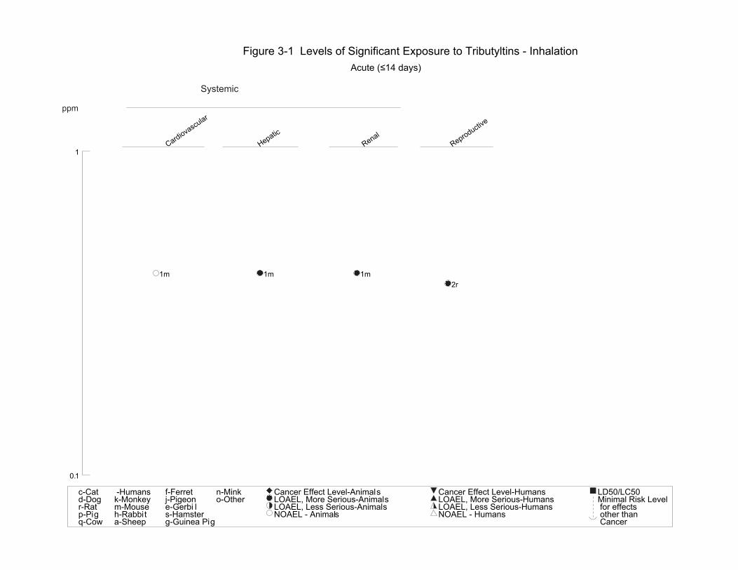
Figure 3-1 Levels of Significant Exposure to Tributyltins - Inhalation Acute (≤14 days)
Systemic
ppm
TIN AND TIN COMPOUNDS
3. HEALTH EFFECTS
28
1
1m 1m 1m 2r
0.1
c-Cat d-Dogr-Rat p-Pigq-Cow
-Humans k-Monkeym-Mouse h-Rabbit a-Sheep
f-Ferret j-Pigeone-Gerbi l s-Hamster g-Guinea Pig
n-Mink o-Other
Cancer Effect Level-Animals LOAEL, More Serious-Animals LOAEL, Less Serious-Animals NOAEL - Animals
Cancer Effect Level-Humans LOAEL, More Serious-HumansLOAEL, Less Serious-HumansNOAEL - Humans
LD50/LC50Minimal Risk Level for effects other than Cancer
Respiratory
Cardiovascular
Hepati
Renal
Ocular
Other
Reproductive
c
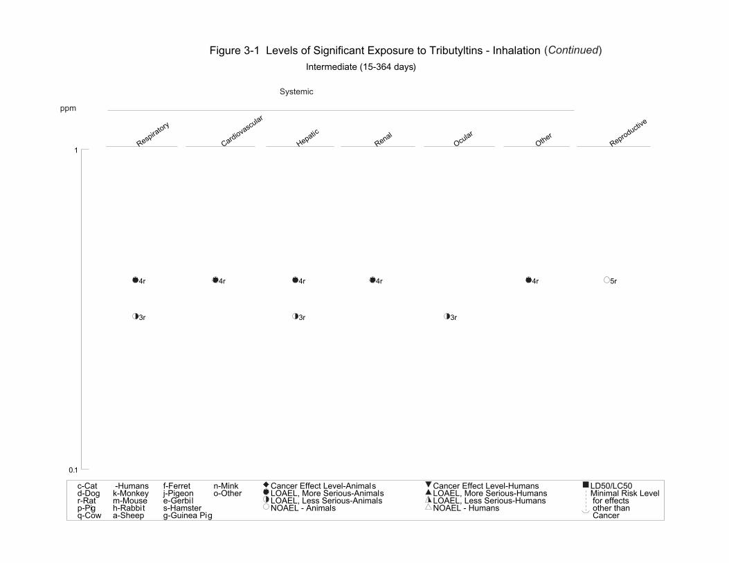
Figure 3-1 Levels of Significant Exposure to Tributyltins - Inhalation (Continued) Intermediate (15-364 days)
Systemic
ppm
TIN AND TIN COMPOUNDS
3. HEALTH EFFECTS
29
1
4r 4r 4r 4r 4r 5r
3r 3r 3r
0.1
c-Cat d-Dogr-Rat p-Pigq-Cow
-Humans k-Monkeym-Mouse h-Rabbit a-Sheep
f-Ferret j-Pigeone-Gerbil s-Hamster g-Guinea Pig
n-Mink o-Other
Cancer Effect Level-Animals LOAEL, More Serious-Animals LOAEL, Less Serious-Animals NOAEL - Animals
Cancer Effect Level-Humans LOAEL, More Serious-HumansLOAEL, Less Serious-HumansNOAEL - Humans
LD50/LC50Minimal Risk Level for effects other than Cancer
30 TIN AND TIN COMPOUNDS
3. HEALTH EFFECTS
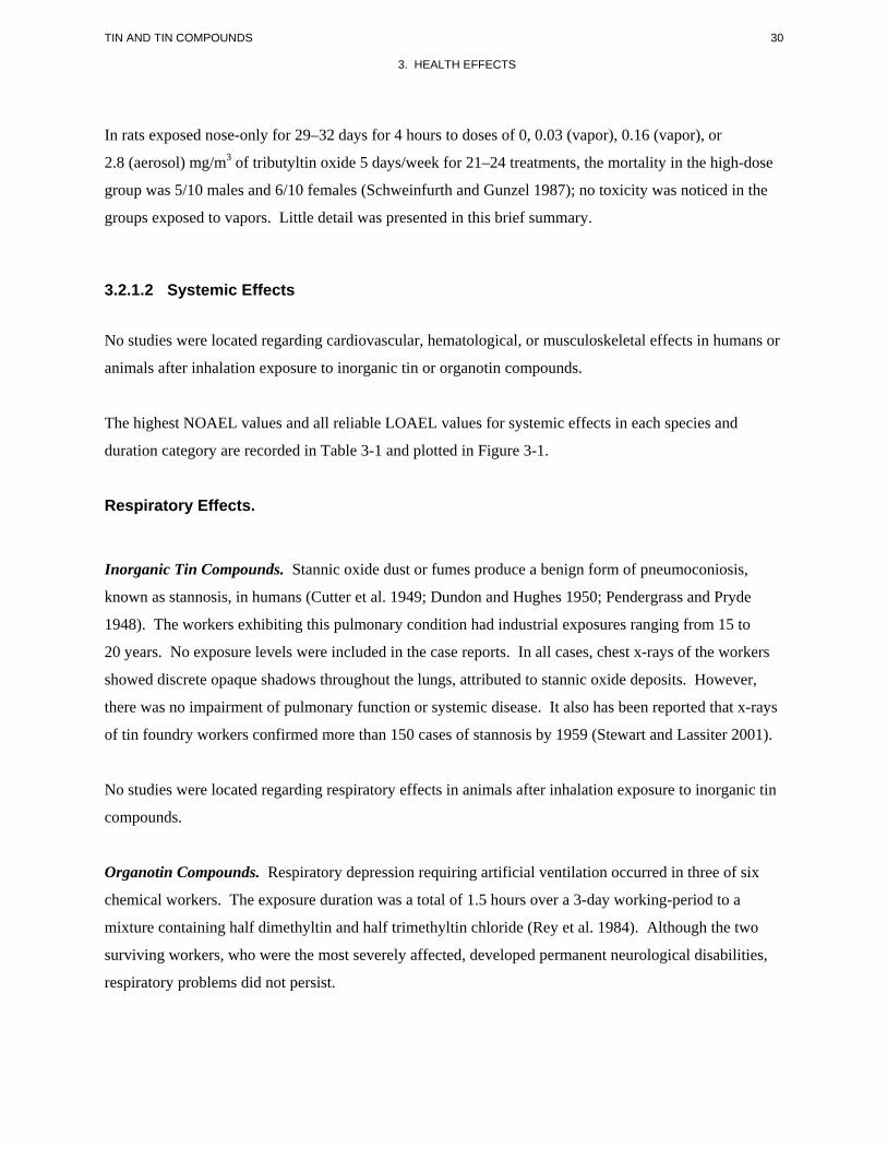
In rats exposed nose-only for 29–32 days for 4 hours to doses of 0, 0.03 (vapor), 0.16 (vapor), or
2.8 (aerosol) mg/m3 of tributyltin oxide 5 days/week for 21–24 treatments, the mortality in the high-dose
group was 5/10 males and 6/10 females (Schweinfurth and Gunzel 1987); no toxicity was noticed in the
groups exposed to vapors. Little detail was presented in this brief summary.
3.2.1.2 Systemic Effects
No studies were located regarding cardiovascular, hematological, or musculoskeletal effects in humans or
animals after inhalation exposure to inorganic tin or organotin compounds.
The highest NOAEL values and all reliable LOAEL values for systemic effects in each species and
duration category are recorded in Table 3-1 and plotted in Figure 3-1.
Respiratory Effects.
Inorganic Tin Compounds. Stannic oxide dust or fumes produce a benign form of pneumoconiosis,
known as stannosis, in humans (Cutter et al. 1949; Dundon and Hughes 1950; Pendergrass and Pryde
1948). The workers exhibiting this pulmonary condition had industrial exposures ranging from 15 to
20 years. No exposure levels were included in the case reports. In all cases, chest x-rays of the workers
showed discrete opaque shadows throughout the lungs, attributed to stannic oxide deposits. However,
there was no impairment of pulmonary function or systemic disease. It also has been reported that x-rays
of tin foundry workers confirmed more than 150 cases of stannosis by 1959 (Stewart and Lassiter 2001).
No studies were located regarding respiratory effects in animals after inhalation exposure to inorganic tin
compounds.
Organotin Compounds. Respiratory depression requiring artificial ventilation occurred in three of six
chemical workers. The exposure duration was a total of 1.5 hours over a 3-day working-period to a
mixture containing half dimethyltin and half trimethyltin chloride (Rey et al. 1984). Although the two
surviving workers, who were the most severely affected, developed permanent neurological disabilities,
respiratory problems did not persist.
31 TIN AND TIN COMPOUNDS
3. HEALTH EFFECTS
Tributyltin oxide has been implicated in producing irritation of the upper respiratory tract and chest
irritation, tightness, and pain in workers using a rubber material containing tributyltin oxide. Exposure
conditions were not described. No changes were observed in pulmonary function tests (NIOSH 1976).
Wax and Dockstader (1995) reported that all members of a family of five (two adults and three children)
complained of sore throat, burning nose, and wheezing 24 hours after a room in their home had been
painted with a paint containing tributyltin oxide for mildew control. Cough and difficulty in breathing,
characterized by inspiratory discomfort, were observed in a man a few hours after inhaling an unspecified
amount of powdered trimethyltin chloride (Saary and House 2002). Shortness of breath and chest
discomfort was still present 20 days after the exposure.
Inflammatory changes consisting of hyperemia and bronchitis were observed in the respiratory system of
rabbits exposed to 4–6 mg/m3 (0.30–0.45 ppm) tributyltin chloride for 95 days (Gohlke et al. 1969).
Histopathology, consisting of severe bronchitis and vascular and alveolar edema, was seen in rats exposed
to 2 mg tin/m3 (0.41 ppm) as a mixture of tributyltin bromide (0.39 ppm), dibutyltin dibromide
(0.02 ppm), and hydrocarbon impurities for 80 days (Iwamoto 1960). Since these were terminal
histopathological evaluations only, it is not known whether the changes were reversible or would have
produced functional impairment in the animals if exposure had continued.
Information summarized by Schweinfurth and Gunzel (1987) indicate that a single 4-hour exposure of
rats to aerosols of tributyltin oxide produced signs of irritation such as nasal discharge, lung edema and
congestion.
Gastrointestinal Effects.
Inorganic Tin Compounds. No studies were located regarding gastrointestinal effects in humans or in
animals after inhalation exposure to inorganic tin compounds.
Organotin Compounds. Very limited information is available in humans. Wax and Dockstader (1995)
reported that nausea and vomiting occurred among all the members of a family of five who were exposed
at home to tributyltin oxide contained in paint for mildew control. Saary and House (2002) reported that
a man who inhaled powdered trimethyltin chloride complained of substernal and epigastric burning with
flatulence a few hours after exposure. The abdominal pain still persisted 2 months after exposure.
32 TIN AND TIN COMPOUNDS
3. HEALTH EFFECTS
Hematological Effects.
Inorganic Tin Compounds. No studies were located regarding hepatic effects in humans or in animals
after inhalation exposure to inorganic tin compounds.
Organotin Compounds. Data concerning hepatic effects of organotins in humans and animals are
limited.
Autopsy of a chemical worker who died following exposure to a combination of methyltin salts (see
Section 3.2.1.1) revealed massive fatty degeneration of liver cells and necrosis (Rey et al. 1984).
Fatty degeneration was observed at necropsy in animals killed after a 95-day exposure period to 4–
6 mg/m3 (0.30–0.45 ppm) tributyltin chloride (Gohlke et al. 1969). Histopathology, consisting of atrophy
and slight necrosis of the liver, was seen in rats exposed to 2 mg tin/m3 (0.41 ppm) as a mixture of
tributyltin bromide (0.39 ppm), dibutyltin dibromide (0.02 ppm), and hydrocarbon impurities for up to
80 days as part of a study of reproductive function (Iwamoto 1960). Atrophy of the liver cells increased
with exposure duration in the females. Some recovery was apparent if exposure to tin was stopped prior
to sacrifice. The longer the duration of exposure, the less complete the recovery.
Renal Effects.
Inorganic Tin Compounds. No studies were located regarding renal effects in humans and animals after
inhalation exposure to inorganic tin compounds.
Organotin Compounds. Data concerning renal effects of organotins in humans and animals are limited.
Autopsy of the one chemical worker who died following exposure to the combination of the methyltin
salts (see Section 3.2.1.1) revealed shock kidneys (i.e., proximal tubule degeneration), which represents
serious tubule damage (Rey et al. 1984). The other five exposed men had high tin concentrations in the
urine with the highest levels occurring in the most severely affected.
Inhalation exposure of mice to a concentration of 5.65 mg tin/m3 (1.16 ppm) as a mixture of tributyltin
bromide (1.1 ppm), dibutyltin dibromide (0.06 ppm), and hydrocarbon impurities for 7 hours/day over
6 days produced pathological changes in the kidney (Igarashi 1959). Necropsy of animals revealed slight
33 TIN AND TIN COMPOUNDS
3. HEALTH EFFECTS
degenerative changes in the glomeruli, convoluted tubules, and collecting tubules as well as extra
medullary hematopoiesis. More extensive kidney pathology was observed in rats exposed to 2 mg tin/m3
(0.41 ppm) as a mixture of tributyltin bromide (0.39 ppm) and dibutyltin dibromide (0.02 ppm) for
2 hours/day for 80 days. Kidney damage consisted of extensive congestion and swelling of the renal
tubular epithelium (Iwamoto 1960).
Dermal Effects.
Inorganic Tin Compounds. Contact with inorganic tin salts produces mild irritation of the skin and
mucous membranes (WHO 1980). However, no specific studies were located regarding dermal effects in
humans and animals after inhalation exposure to inorganic tin compounds.
Organotin Compounds. No studies were located regarding dermal effects in humans after inhalation
exposure to organotin compounds. Occupational exposure produces such effects as discussed in
Section 3.2.3.1.
Dermal effects were observed during inhalation studies in mice that were exposed to a butyltin mixture
(30 parts tributyltin bromide to 1 part dibutyltin dibromide) and consisted of reddening of the skin and
dilatation of the blood vessels of the nose, feet, and tail (Igarashi 1959). These effects may have been
caused by direct contact with the chemical.
Ocular Effects.
Inorganic Tin Compounds. No information was located regarding ocular effects in humans following
exposure to inorganic tin compounds.
Organotin Compounds. Inflamed eyes and nasal mucous membranes were observed in the last month of
a 95-day inhalation study of tributyltin chloride in female rats (Gohlke et al. 1969). The animals were
exposed to concentrations of 4–6 mg/m3 (0.30–0.45 ppm) for 6 hours/day, 5 days/week.
34 TIN AND TIN COMPOUNDS
3. HEALTH EFFECTS
3.2.1.3 Immunological and Lymphoreticular Effects
No studies were located regarding immunological effects in humans or animals after inhalation exposure
to inorganic tin or organotin compounds. However, some lymph node atrophy was observed in rats
exposed to a butyltin mixture for 14 days (Iwamoto 1960).
3.2.1.4 Neurological Effects
Inorganic Tin Compounds. No studies were located regarding neurological effects in humans or in
animals after inhalation exposure to inorganic tin compounds.
Organotin Compounds. A study by Rey et al. (1984) provides some information on neurobehavioral
changes in humans after exposure to organotin compounds (dimethyltin dichloride and trimethyltin
chloride). The study describes the cases of six chemical workers exposed to methyltins primarily by
inhalation who experienced headache, tinnitus, deafness, impaired memory, disorientation,
aggressiveness, psychotic and other severe neuropsychiatric behavior, syncope, and loss of consciousness
as symptoms of exposure; one subject died. The two surviving workers with the highest urinary tin levels
exhibited fixed neurological effects which were not resolved more than 6 years after exposure. The
remaining three survivors returned to work, but had memory loss, which persisted for 6 months. Similar
cases have been reported by other investigators. Fortemps et al. (1978) reported that two chemists who
had been intermittently exposed to vapors of dimethyltin dichloride and trimethyltin chloride for about
3 months abruptly developed a status of mental confusion with generalized epileptic seizures. Before the
acute episode, the subjects had complained of headaches, pain in various organs, and psychological
disturbances such as memory defects, vigilance loss, insomnia, anorexia, and disorientation. Both
patients recovered completely following removal from exposure. Ross et al. (1981) examined 22 male
workers 1 month following exposure to trimethyltin spillage (presumable inhalation and dermal exposure
occurred) and compared the frequency of neurological symptoms between those who suffered high
exposure with those with lower exposure. Those highly exposed showed a significantly higher incidence
of nonspecific symptoms such as forgetfulness, fatigue and weakness, loss of motivation, and specific
symptoms such as bouts of depression and attacks of rage and temper compared to those with lower
exposure. Some symptoms persisted for at least 3 years following the accident. Yanofsky et al. (1991)
and Feldman et al. (1993) described the case of a 23-year-old male who was accidentally exposed to
vapors of a trimethyltin compound and 72 hours later exhibited delirium, spatial disorientation,
perseveration, inappropriate affect, and memory loss. Urine and serum assays for tin showed
35 TIN AND TIN COMPOUNDS
3. HEALTH EFFECTS
considerably elevated concentrations of trimethyltin when tested 3 weeks following the accident. Five
months after the accident, the man experienced complex partial seizures that required him to take
anticonvulsant medication for 7 years. Four years after exposure, tests revealed persistent memory
defects, cognitive dysfunction, and dysphoria. Saary and House (2002) described the case of a man who
worked in a chemistry laboratory and inhaled an undetermined amount of powdered trimethyltin chloride
on a single occasion. Within 3 hours of exposure he felt agitated and he later developed a headache,
dizziness, and twitching of the right eye and cheek. Two months after exposure, he continued
experiencing twitching of his eyelids and arms and complained of suffering short-term memory problems
and difficulty retaining new information.
No relevant studies were located regarding neurological effects in animals after inhalation exposure to
organotin compounds. It was reported that no histopathological changes were observed in the brains of
mice following a 6-day inhalation exposure to 2.12 mg tin/m3 (0.44 ppm) as a mixture of tributyltin
bromide (0.42 ppm), dibutyltin dibromide (0.02 ppm), and hydrocarbon impurities (Igarashi 1959).
3.2.1.5 Reproductive Effects
Inorganic Tin Compounds. No studies were located regarding reproductive effects in humans or animals
after inhalation exposure to inorganic tin compounds.
Organotin Compounds. No studies were located regarding reproductive effects in humans after
inhalation exposure to organotin compounds.
A study in rats was conducted to assess reproductive effects of a mixture of tributyltin bromide (81.2%)
with other compounds such as dibutyltin dibromide (Iwamoto 1960). The rats were exposed to 2 mg
tin/m3 (0.41 ppm) for acute- and intermediate-duration exposures (equivalent to 0.39 ppm tributyltin
bromide and 0.02 ppm dibutyltin dibromide). Pregnancy rates were markedly reduced after 4 weeks to
3 months of exposure, but returned to near control rates when exposure was discontinued. Histo
pathological evaluations were made in separate studies of different exposure durations (14–80 days)
followed by recovery periods. No changes were seen in males, but atrophy of the glandular uterus was
observed as early as 14 days of exposure in females. All effects were reversed during the recovery
period. Although a mixture of butyltin compounds was used and the results were not clearly reported,
this study suggests that some impairment of female reproductive functions may occur after inhalation of
these compounds.
36 TIN AND TIN COMPOUNDS
3. HEALTH EFFECTS
3.2.1.6 Developmental Effects
No studies were located regarding developmental effects in humans or animals after inhalation exposure
to inorganic tin or organotin compounds.
3.2.1.7 Cancer
No studies were located regarding cancer effects in humans and animals after inhalation exposure to
inorganic tin or organotin compounds.
3.2.2 Oral Exposure
In contrast to the limited information on the inhalation toxicity of tin compounds (Section 3.2.1), there are
considerable more data regarding the effects of oral exposure to organotin compounds, particularly in
animal studies. Although there is less information concerning health effects produced by oral exposure to
inorganic tin compounds, the data from animal studies allow some characterization of health effects of
these compounds. Dosages are expressed as milligrams of tin per kilogram of body weight per day (mg
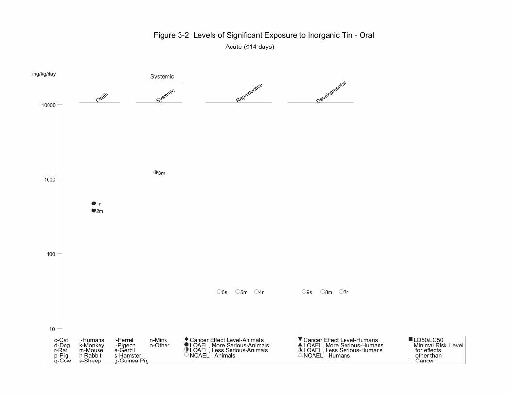
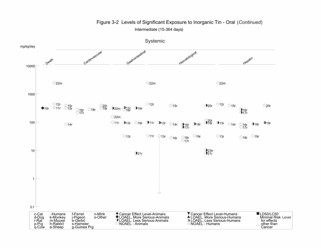
tin/kg/day) as the specific inorganic tin compound fed or administered orally. Table 3-2 and Figure 3-2
summarize available quantitative information on health effects that have been observed in animals after
oral exposure to inorganic tin compounds. Similar information for organotin compounds is given in
Tables 3-3 through 3-8 and Figures 3-3 through 3-8. In order to be consistent with most studies in the
literature, dosages are expressed as mg/kg/day of the specific organotin compound rather than as a tin
equivalent.
3.2.2.1 Death
Inorganic Tin Compounds. No studies were located regarding lethality in humans after oral ingestion of
inorganic tin compounds.
In animals, the lowest oral dose that produced deaths in rats following a single gavage administration was
473 mg/kg body weight stannous chloride (NTP 1982). However, all rats survived doses up to
945 mg/kg/day when the compound was fed in the diet for 14 days (NTP 1982). For mice, the lowest oral
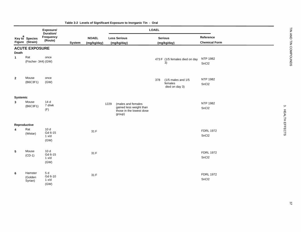
Table 3-2 Levels of Significant Exposure to Inorganic Tin
Exposure/
TIN
- Oral
LOAEL
aKey to Figure
ACUTE EXPOSURE
Species (Strain)
Duration/ Frequency
(Route) System
NOAEL (mg/kg/day)
Less Serious (mg/kg/day)
Serious (mg/kg/day)
AN
D TIN
CO
MP
OU
ND
S
Reference Chemical Form
Death 1 Rat once
(Fischer- 344) (GW) 473 F (1/5 females died on day
3) NTP 1982 SnCl2
2 Mouse once (B6C3F1) (GW)
378 (1/5 males and 1/5 females
NTP 1982 SnCl2
died on day 3)
Systemic 3 Mouse 14 d
7 d/wk (B6C3F1) (F)
1229 (males and females gained less weight than those in the lowest dose
3. H
NTP 1982 SnCl2
group)
EA
LTH
Reproductive
EFF
4 Rat 10 d Gd 6-15 (Wistar) 1 x/d
31 F
EC
TS
FDRL 1972 SnCl2
(GW)
5 Mouse 10 d Gd 6-15 (CD-1) 1 x/d
31 F FDRL 1972 SnCl2
(GW)
6 Hamster 5 d Gd 6-10 (Golden 1 x/d Syrian)
31 F FDRL 1972 SnCl2
(GW)
37
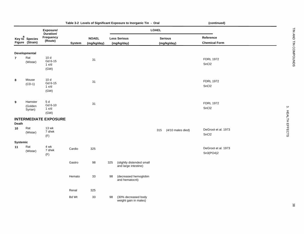
Table 3-2 Levels of Significant Exposure to Inorganic Tin - Oral (continued)
aKey to Figure
Species (Strain)
Exposure/ Duration/
Frequency (Route)
System NOAEL
(mg/kg/day)
LOAEL
TIN A
ND
TIN C
OM
PO
UN
DS
Reference Chemical Form
Less Serious (mg/kg/day)
Serious (mg/kg/day)
Developmental 7 Rat 10 d
Gd 6-15 (Wistar) 1 x/d (GW)
8 Mouse 10 d Gd 6-15 (CD-1) 1 x/d (GW)
9 Hamster 5 d Gd 6-10 (Golden 1 x/d Syrian) (GW)
INTERMEDIATE EXPOSURE Death 10 Rat 13 wk
7 d/wk (Wistar) (F)
Systemic 11 Rat 4 wk
7 d/wk (Wistar) (F)
Cardio
Gastro
Hemato
Renal
Bd Wt
31
31
31
325
98
33
325
33
325
98
98
(slightly distended small and large intestine)
(decreased hemoglobin and hematocrit)
(30% decreased body weight gain in males)
315 (4/10 males died)
38
3. HE
ALTH
EFFE
CTS
FDRL 1972 SnCl2
FDRL 1972 SnCl2
FDRL 1972 SnCl2
DeGroot et al. 1973 SnCl2
DeGroot et al. 1973 Sn3(PO4)2
Table 3-2 Levels of Significant Exposure to Inorganic Tin - Oral (continued)
aKey to Species Figure (Strain)
Exposure/ Duration/
Frequency(Route)
System NOAEL
(mg/kg/day)
LOAEL
TIN A
ND
TIN C
OM
PO
UN
DS
Reference Chemical Form
Less Serious (mg/kg/day)
Serious (mg/kg/day)
12
13
Rat (Wistar)
Rat (Wistar)
13 wk 7 d/wk (F)
13 wk ad libitum (F)
Cardio
Hemato
Hepatic
Renal
Bd Wt
Cardio
Gastro
Hemato
Hepatic
Renal
Endocr
Bd Wt
Other
440
440
440
440
440
315
32 b
32
32
315
315
95
32
95
95
95
95
(abdominal distension)
(reduced hemoglobin concentration)
(bile duct epithelium proliferation)
(14% reduced food consumption on week 2)
315 (weight loss)
39
3. HE
ALTH
EFFE
CTS
DeGroot et al. 1973 SnO
DeGroot et al. 1973 SnCl2
TIN A
ND
TIN C
OM
PO
UN
DS
40
3. HE
ALTH
EFFE
CTS
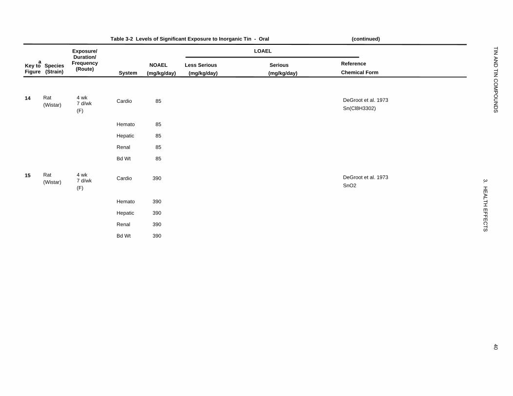
Table 3-2 Levels of Significant Exposure to Inorganic Tin - Oral (continued)
Exposure/ LOAEL Duration/
a Frequency Reference Key to Species NOAEL Less Serious Serious (Route) Figure (Strain) System (mg/kg/day) (mg/kg/day) (mg/kg/day) Chemical Form
14 Rat 4 wk Cardio 85 DeGroot et al. 1973 7 d/wk (Wistar) Sn(Cl8H3302) (F)
Hemato 85
Hepatic 85
Renal 85
Bd Wt 85
15 Rat 4 wk Cardio 390 DeGroot et al. 1973 7 d/wk (Wistar) SnO2 (F)
Hemato 390
Hepatic 390
Renal 390
Bd Wt 390
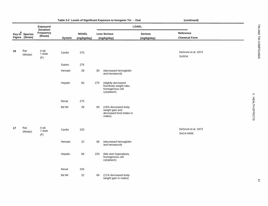
Table 3-2 Levels of Significant Exposure to Inorganic Tin - Oral (continued)
aKey to Figure
Species (Strain)
Exposure/ Duration/
Frequency (Route)
System NOAEL
(mg/kg/day)
LOAEL
TIN A
ND
TIN C
OM
PO
UN
DS
Reference Chemical Form
Less Serious (mg/kg/day)
Serious (mg/kg/day)
16
17
Rat (Wistar)
Rat (Wistar)
4 wk 7 d/wk (F)
4 wk 7 d/wk (F)
Cardio
Gastro
Hemato
Hepatic
Renal
Bd Wt
Cardio
Hemato
Hepatic
Renal
Bd Wt
275
275
28
83
275
28
220
22
66
220
22
83
275
83
66
220
66
(decreased hemoglobin and hematocrit)
(slightly decreased liver/body weight ratio, homogenous cell cytoplasm)
(16% decreased body weight gain and decreased food intake in males)
(decreased hemoglobin and hematocrit)
(bile duct hyperplasia, homogenous cell cytoplasm)
(11% decreased body weight gain in males) 41
3. HE
ALTH
EFFE
CTS
DeGroot et al. 1973 SnSO4
DeGroot et al. 1973 SnC4-H406
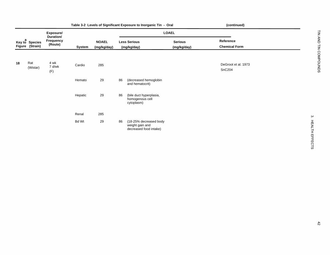
Table 3-2 Levels of Significant Exposure to Inorganic Tin - Oral (continued)
Exposure/ LOAEL
TIN
aKey to Figure
Species (Strain)
Duration/ Frequency
(Route) System
NOAEL (mg/kg/day)
AN
D TIN
CO
MP
OU
ND
S
Reference Chemical Form
Less Serious (mg/kg/day)
Serious (mg/kg/day)
18 Rat (Wistar)
4 wk 7 d/wk (F)
Cardio 285 DeGroot et al. 1973 SnC204
Hemato 29 86 (decreased hemoglobin and hematocrit)
Hepatic 29 86 (bile duct hyperplasia, homogenous cell cytoplasm)
Renal 285
3
Bd Wt 29 86 (18-25% decreased body weight gain and decreased food intake)
42
. HE
ALTH
EFFE
CTS
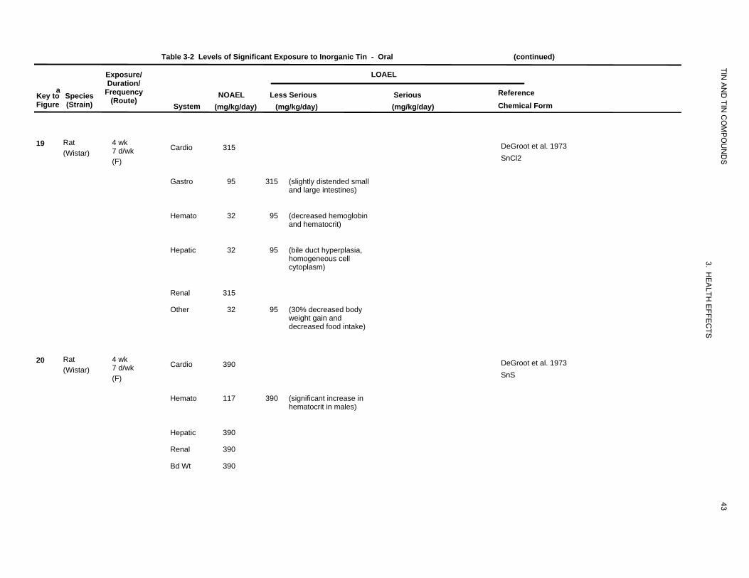
Table 3-2 Levels of Significant Exposure to Inorganic Tin - Oral (continued)
aKey to Figure
Species (Strain)
Exposure/ Duration/
Frequency (Route)
System NOAEL
(mg/kg/day)
LOAEL
TIN A
ND
TIN C
OM
PO
UN
DS
Reference Chemical Form
Less Serious (mg/kg/day)
Serious (mg/kg/day)
19
20
Rat (Wistar)
Rat (Wistar)
4 wk 7 d/wk (F)
4 wk 7 d/wk (F)
Cardio
Gastro
Hemato
Hepatic
Renal
Other
Cardio
Hemato
Hepatic
Renal
Bd Wt
315
95
32
32
315
32
390
117
390
390
390
315
95
95
95
390
(slightly distended small and large intestines)
(decreased hemoglobin and hematocrit)
(bile duct hyperplasia, homogeneous cell cytoplasm)
(30% decreased body weight gain and decreased food intake)
(significant increase in hematocrit in males)
43
3. HE
ALTH
EFFE
CTS
DeGroot et al. 1973 SnCl2
DeGroot et al. 1973 SnS
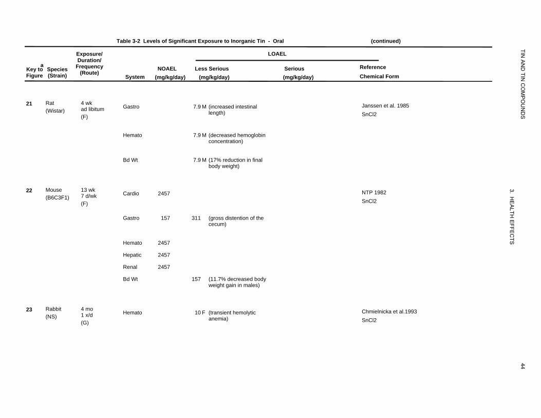
Table 3-2 Levels of Significant Exposure to Inorganic Tin - Oral (continued)
Exposure/ LOAEL
TIN
aKey to Figure
Species (Strain)
Duration/ Frequency
(Route) System
NOAEL (mg/kg/day)
AN
D TIN
CO
MP
OU
ND
S
Reference Chemical Form
Less Serious (mg/kg/day)
Serious (mg/kg/day)
21 Rat (Wistar)
4 wk ad libitum (F)
Gastro 7.9 M (increased intestinal length)
Janssen et al. 1985 SnCl2
Hemato 7.9 M (decreased hemoglobin concentration)
Bd Wt 7.9 M (17% reduction in final body weight)
22 Mouse (B6C3F1)
13 wk 7 d/wk (F)
Cardio 2457
3. HE
ALTH
EFFE
CTS
NTP 1982 SnCl2
Gastro 157 311 (gross distention of the cecum)
Hemato 2457
Hepatic 2457
Renal 2457
Bd Wt 157 (11.7% decreased body weight gain in males)
23 Rabbit (NS)
4 mo 1 x/d (G)
Hemato 10 F (transient hemolytic anemia)
44
Chmielnicka et al.1993 SnCl2
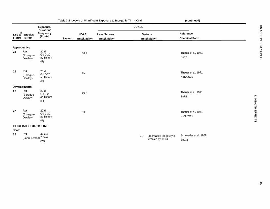
Table 3-2 Levels of Significant Exposure to Inorganic Tin - Oral (continued)
Exposure/ LOAEL
TIN
aKey to Figure
Species (Strain)
Duration/ Frequency
(Route) System
NOAEL (mg/kg/day)
AN
D TIN
CO
MP
OU
ND
S
Reference Chemical Form
Less Serious (mg/kg/day)
Serious (mg/kg/day)
Reproductive 24 Rat 20 d
Gd 0-20 (Sprague-ad libitum Dawley)
56 F Theuer et al. 1971 SnF2
(F)
25 Rat 20 d Gd 0-20 (Sprague-ad libitum Dawley)
45 Theuer et al. 1971 NaSn2Cl5
(F)
Developmental 26 Rat 20 d
Gd 0-20 (Sprague-ad libitum Dawley)
56 F
3.
Theuer et al. 1971 SnF2
(F)
27 Rat 20 d Gd 0-20 (Sprague-ad libitum Dawley)
45
HE
ALTH
EFFE
C
Theuer et al. 1971 NaSn2Cl5
(F)
TS
CHRONIC EXPOSURE Death 28 Rat 42 mo
7 d/wk (Long- Evans) (W)
0.7 (decreased longevity in females by 11%)
45
Schroeder et al. 1968 SnCl2

Table 3-2 Levels of Significant Exposure to Inorganic Tin - Oral (continued)
Exposure/ LOAEL
TIN
aKey toFigure
Species (Strain)
Duration/ Frequency
(Route)System
NOAEL (mg/kg/day)
AN
D TIN
CO
MP
OU
ND
S
Reference Chemical Form
Less Serious(mg/kg/day)
Serious (mg/kg/day)
Systemic 29 Rat
(Fischer- 344) 105 wk 7 d/wk (F)
Cardio 63 NTP 1982 SnCl2
Gastro 63
Hepatic 63
Renal 63
Bd Wt 63
30 Rat (Long- Evans)
42 mo 7 d/wk (W)
Hepatic 0.7 (fatty degeneration)
3. HE
Schroeder et al. 1968 SnCl2
Renal 0.7 (tubular degeneration, vacuolization)
ALTH
EFFE
CTSBd Wt 0.7 (11-16% decreased body
weight, compared to controls)
31 Mouse (B6C3F1)
105 wk 7 d/wk (F)
Cardio 164 NTP 1982 SnCl2
Gastro 164
Hepatic 164
Bd Wt 164
46
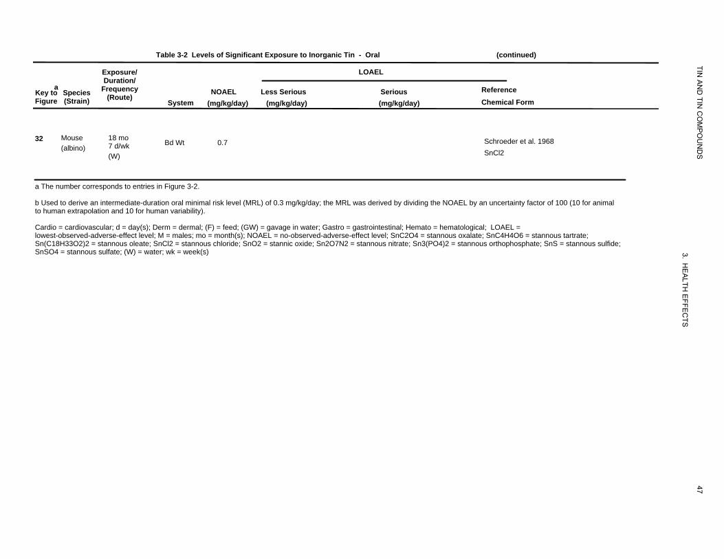
Table 3-2 Levels of Significant Exposure to Inorganic Tin - Oral (continued)
Exposure/ LOAEL
TIN
aKey to Figure
Species (Strain)
Duration/ Frequency
(Route) System
NOAEL (mg/kg/day)
AN
D TIN
CO
MP
OU
ND
S
Reference Chemical Form
Less Serious (mg/kg/day)
Serious (mg/kg/day)
32 Mouse (albino)
18 mo 7 d/wk (W)
Bd Wt 0.7 Schroeder et al. 1968 SnCl2
a The number corresponds to entries in Figure 3-2.
b Used to derive an intermediate-duration oral minimal risk level (MRL) of 0.3 mg/kg/day; the MRL was derived by dividing the NOAEL by an uncertainty factor of 100 (10 for animal to human extrapolation and 10 for human variability).
Cardio = cardiovascular; d = day(s); Derm = dermal; (F) = feed; (GW) = gavage in water; Gastro = gastrointestinal; Hemato = hematological; LOAEL = lowest-observed-adverse-effect level; M = males; mo = month(s); NOAEL = no-observed-adverse-effect level; SnC2O4 = stannous oxalate; SnC4H4O6 = stannous tartrate; Sn(C18H33O2)2 = stannous oleate; SnCl2 = stannous chloride; SnO2 = stannic oxide; Sn2O7N2 = stannous nitrate; Sn3(PO4)2 = stannous orthophosphate; SnS = stannous sulfide;
47
3. HE
ALTH
EFFE
CTS
SnSO4 = stannous sulfate; (W) = water; wk = week(s)
Death
System
Reproductive
Developm
10000
ic ental
Figure 3-2 Levels of Significant Exposure to Inorganic Tin - Oral Acute (≤14 days)
mg/kg/day
TIN AND TIN COMPOUNDS
3. HEALTH EFFECTS
48
Systemic
3m 1000
1r 2m
100
6s 5m 4r 9s 8m 7r
10
c-Cat d-Dogr-Rat p-Pigq-Cow
-Humans k-Monkeym-Mouse h-Rabbit a-Sheep
f-Ferret j-Pigeone-Gerbil s-Hamster g-Guinea Pig
n-Mink o-Other
Cancer Effect Level-Animals LOAEL, More Serious-Animals LOAEL, Less Serious-Animals NOAEL - Animals
Cancer Effect Level-Humans LOAEL, More Serious-HumansLOAEL, Less Serious-HumansNOAEL - Humans
LD50/LC50Minimal Risk for effects other than Cancer
Level
Death
Cardiovascular
Gastrointestinal
Hem
Hepatiato
logical
c
Figure 3-2 Levels of Significant Exposure to Inorganic Tin - Oral (Continued) Intermediate (15-364 days)
Systemic mg/kg/day
TIN AND TIN COMPOUNDS
3. HEALTH EFFECTS
49
10000
22m 22m 22m
1000
12r 12r 12r15r 20r 15r 20r 15r 20r11r 11r10r 13r 19r 19r22m18r16r 16r 16r
17r 17r 22m
20r100 11r 11r 13r13r 19r 19r 13r 19r18r 18r14r 14r 14r16r 16r 17r 17r
11r 13r13r 19r 13r 19r18r 18r16r 17r
10 23h 21r 21r
1
0.1
c-Cat d-Dogr-Rat p-Pigq-Cow
-Humans k-Monkeym-Mouse h-Rabbit a-Sheep
f-Ferret j-Pigeone-Gerbil s-Hamster g-Guinea Pig
n-Mink o-Other
Cancer Effect Level-Animals LOAEL, More Serious-Animals LOAEL, Less Serious-Animals NOAEL - Animals
Cancer Effect Level-Humans LOAEL, More Serious-HumansLOAEL, Less Serious-HumansNOAEL - Humans
LD50/LC50Minimal Risk for effects other than Cancer
Level
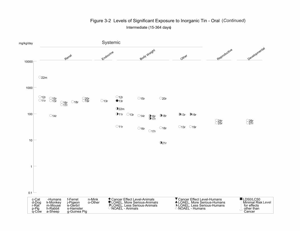
Renal
Endocrine
Body
Other
Reproductive
Developmental
Weight
Figure 3-2 Levels of Significant Exposure to Inorganic Tin - Oral (Continued) Intermediate (15-364 days)
mg/kg/day Systemic
TIN AND TIN COMPOUNDS
3. HEALTH EFFECTS
50
10000
22m
1000
12r 12r15r 20r 15r 20r11r 13r 19r 13r 13r18r16r
17r 22m
100 11r 13r 13r 19r18r14r 14r 16r 17r 24r 26r
25r 27r 11r 13r 19r18r16r
17r
10 21r
1
0.1
c-Cat d-Dogr-Rat p-Pigq-Cow
-Humans k-Monkeym-Mouse h-Rabbit a-Sheep
f-Ferret j-Pigeone-Gerbil s-Hamster g-Guinea Pig
n-Mink o-Other
Cancer Effect Level-Animals LOAEL, More Serious-Animals LOAEL, Less Serious-Animals NOAEL - Animals
Cancer Effect Level-Humans LOAEL, More Serious-HumansLOAEL, Less Serious-HumansNOAEL - Humans
LD50/LC50Minimal Risk for effects other than Cancer
Level
Death
Cardiovascular
Gastrointestinal
Hepati
Renal
Body
c Weight
Figure 3-2 Levels of Significant Exposure to Inorganic Tin - Oral (Continued) Chronic (≥365 days)
Systemicmg/kg/day
TIN AND TIN COMPOUNDS
3. HEALTH EFFECTS
51
1000
31m
100
29r
10
1
28r
0.1
31m 31m
29r 29r
30r
c-Cat d-Dogr-Rat p-Pigq-Cow
-Humans k-Monkeym-Mouse h-Rabbit a-Sheep
f-Ferret j-Pigeone-Gerbil s-Hamster g-Guinea Pig
n-Mink o-Other
Cancer Effect Level-Animals LOAEL, More Serious-Animals LOAEL, Less Serious-Animals NOAEL - Animals
Cancer Effect Level-Humans LOAEL, More Serious-HumansLOAEL, Less Serious-HumansNOAEL - Humans
LD50/LC50Minimal Risk for effects other than Cancer
Level
31m
29r 29r
30r 32m 30r
Exposure/
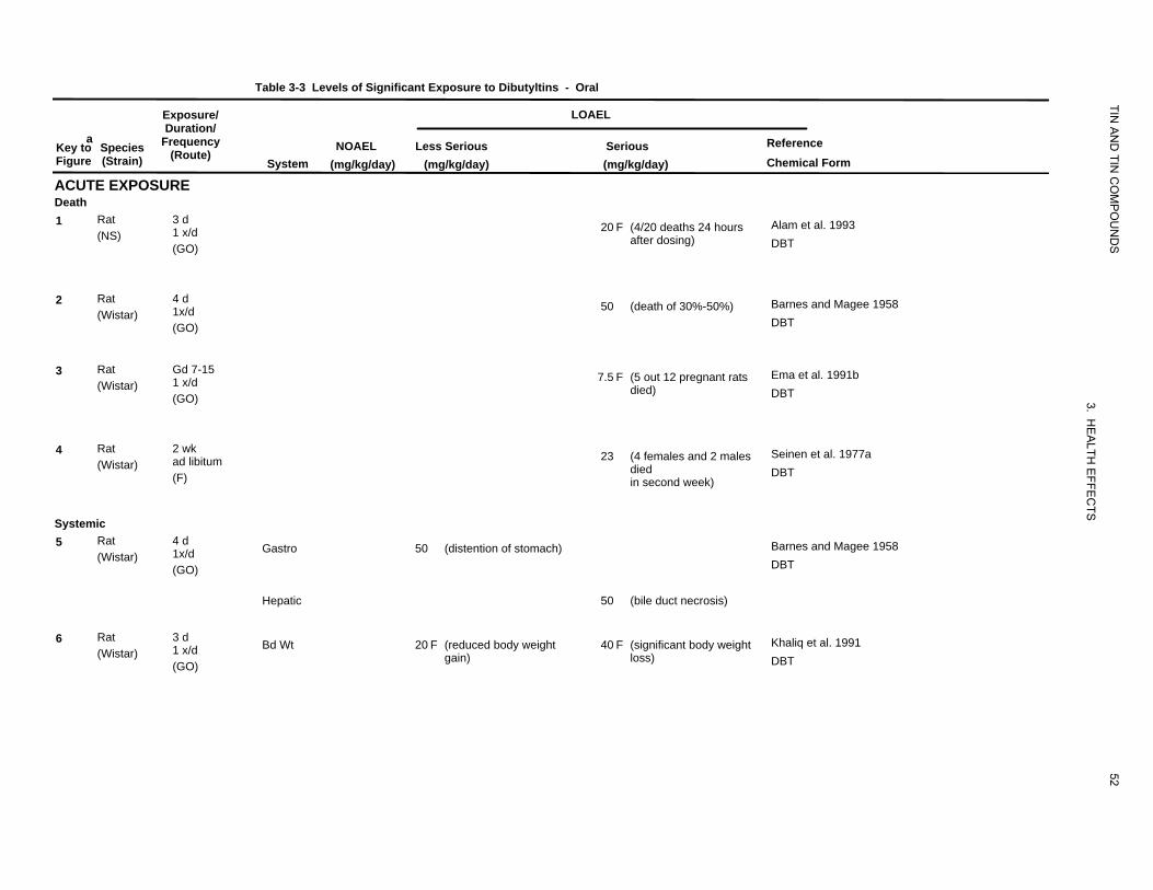
Table 3-3 Levels of Significant Exposure to Dibutyltins - Oral
LOAEL
TIN
aKey to SpeciesFigure (Strain)
ACUTE EXPO
Duration/ Frequency
(Route)
SURE System
NOAEL (mg/kg/day)
Less Serious(mg/kg/day)
Serious (mg/kg/day)
AN
D TIN
CO
MP
OU
ND
S
Reference Chemical Form
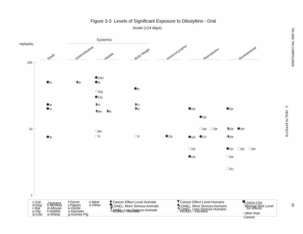
Death 1 Rat 3 d
1 x/d(NS)(GO)
20 F (4/20 deaths 24 hours after dosing)
Alam et al. 1993 DBT
2 Rat 4 d 1x/d(Wistar)(GO)
50 (death of 30%-50%) Barnes and Magee 1958 DBT
3 Rat Gd 7-15 1 x/d(Wistar)(GO)
7.5 F (5 out 12 pregnant rats died)
3. H
Ema et al. 1991b DBT
4 Rat 2 wk ad libitum(Wistar) (F)
23 (4 females and 2 males died in second week)
EA
LTH E
FFEC
Seinen et al. 1977a DBT
Systemic
TS
5 Rat 4 d 1x/d(Wistar)(GO)
Gastro 50 (distention of stomach) Barnes and Magee 1958 DBT
Hepatic 50 (bile duct necrosis)
6 Rat 3 d 1 x/d(Wistar)(GO)
Bd Wt 20 F (reduced body weight gain)
40 F (significant body weight loss)
52
Khaliq et al. 1991 DBT
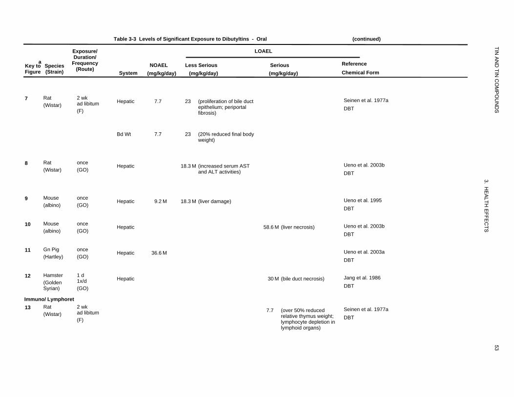
Table 3-3 Levels of Significant Exposure to Dibutyltins - Oral (continued)
Exposure/ LOAEL
TIN
aKey to Figure
Species (Strain)
Duration/ Frequency
(Route) System
NOAEL (mg/kg/day)
AN
D TIN
CO
MP
OU
ND
S
Reference Chemical Form
Less Serious (mg/kg/day)
Serious (mg/kg/day)
7 Rat (Wistar)
2 wk ad libitum (F)
Hepatic 7.7 23 (proliferation of bile duct epithelium; periportal fibrosis)
Seinen et al. 1977a DBT
Bd Wt 7.7 23 (20% reduced final body
weight)
8 Rat (Wistar)
once (GO)
Hepatic 18.3 M (increased serum AST and ALT activities)
3.
Ueno et al. 2003b DBT
9 Mouse (albino)
once (GO)
Hepatic 9.2 M 18.3 M (liver damage)
HE
ALTH
EFFE
CTS
Ueno et al. 1995 DBT
10 Mouse (albino)
once (GO)
Hepatic 58.6 M (liver necrosis) Ueno et al. 2003b DBT
11 Gn Pig (Hartley)
once (GO)
Hepatic 36.6 M Ueno et al. 2003a DBT
12 Hamster (Golden Syrian)
1 d 1x/d (GO)
Hepatic 30 M (bile duct necrosis) Jang et al. 1986 DBT
Immuno/ Lymphoret 13 Rat
(Wistar) 2 wk ad libitum (F)
7.7 (over 50% reduced relative thymus weight; lymphocyte depletion in
Seinen et al. 1977a DBT
lymphoid organs)
53
Table 3-3 Levels of Significant Exposure to Dibutyltins - Oral (continued)
Exposure/ LOAEL
TIN
aKey to Species Figure (Strain)
Duration/ Frequency
(Route) System
NOAEL (mg/kg/day)
AN
D TIN
CO
MP
OU
ND
S
Reference Chemical Form
Less Serious (mg/kg/day)
Serious (mg/kg/day)
Reproductive 14 Rat
(Wistar) Gd 4-7 1 x/d (GO)
3.8 F (significant increase in postimplantation loss)
Ema and Harazono 2000 DBT
15 Rat (Wistar)
Gd 7-15 1 x/d (GO)
5 F 7.5 F (increased resorptions, dead fetuses, and postimplantation loss)
Ema et al. 1991b DBT
16 Rat (Wistar)
Gd 7-9 1 x/d (GO)
20 F (increased resorptions, dead fetuses, and postimplantation loss)
3. HE
ALTH
EFFE
CTS
Ema et al. 1992 DBT
17 Rat (Wistar)
Gd 0-3 1 x/d (GO)
7.6 F (reduced fertility rate; increased pre-implantation loss)
Ema et al. 2003 DBT
18 Rat (Wistar)
Gd 6-15 1 x/d (GO)
10 F Farr et al. 2001 DBT
19 Rat (Wistar)
Gd 7-17 1 x/d (GO)
10 F 15 F (increased incidence of dead or resorbed fetuses)
54
Noda et al. 1992b DBT
Table 3-3 Levels of Significant Exposure to Dibutyltins - Oral (continued)
Exposure/ LOAEL
TIN
aKey to Species Figure (Strain)
Duration/ Frequency
(Route) System
NOAEL (mg/kg/day)
AN
D TIN
CO
MP
OU
ND
S
Reference Chemical Form
Less Serious (mg/kg/day)
Serious (mg/kg/day)
Developmental 20 Rat
(Wistar) Gd 4-7 1 x/d (GO)
3.8 7.6 (significantly reduced fetal body weight)
Ema and Harazono 2000 DBT
21 Rat (Wistar)
Gd 7-15 1 x/d (GO)
2.5 5 (increased incidence of external and skeletal malformations)
Ema et al. 1991b DBT
22 Rat (Wistar)
Gd 7-9 1 x/d (GO)
20 (increased incidence of malformations)
3. HE
ALTH
Ema et al. 1992 DBT
23 Rat (Wistar)
Gd 6-15 1 x/d (GO)
5 10 (slight increase in malformations)
EFFE
CTS
Farr et al. 2001 DBT
24 Rat (Wistar)
Gd 7-17 1 x/d (GO)
5 10 (increased external and skeletal malformations)
55
Noda et al. 1992b DBT
Table 3-3 Levels of Significant Exposure to Dibutyltins - Oral (continued)
Exposure/ LOAEL
TIN
aKey to Figure
Species (Strain)
Duration/ Frequency
(Route) System
NOAEL (mg/kg/day)
AN
D TIN
CO
MP
OU
ND
S
Reference Chemical Form
Less Serious (mg/kg/day)
Serious (mg/kg/day)
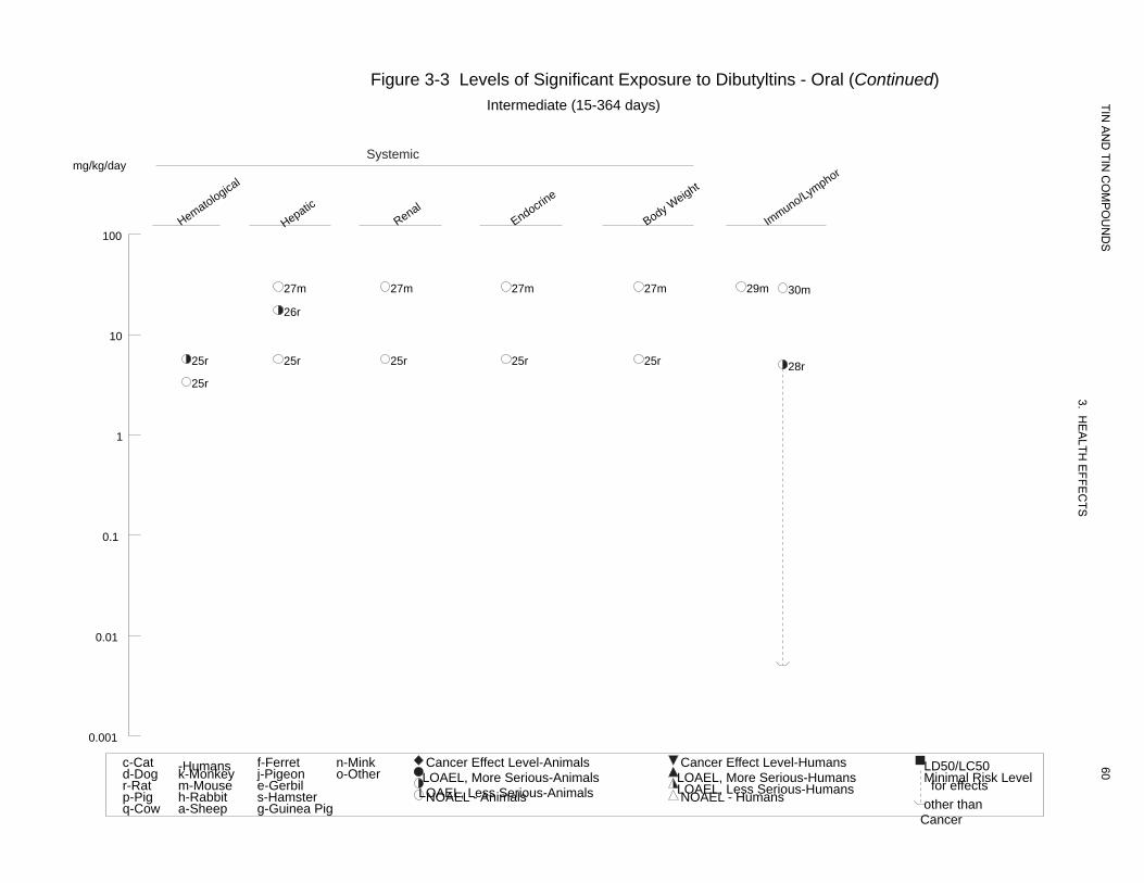
INTERMEDIATE EXPOSURE Systemic 25 Rat 90 d
ad libitum (Fischer- 344) (F)
Hemato 3.4 M 5.7 F (8% reduced hemoglobin concentration)
Gaunt et al. 1968 DBT
Hepatic 5.7 F
Renal 5.7 F
Endocr 5.7 F
Bd Wt 5.7 F
26 Rat 15 d 1 x/d (albino) (GO)
Hepatic 17.5 M (increased heme oxygenase activity, decreased activity of
3. HE
A
Mushtaq et al 1981 DBT
microsomal enzymes)
LTH E
FFEC
TS27 Mouse 4 wk ad libitum (Swiss-
Webster) (F)
Hepatic 30 M Seinen et al. 1977a DBT
Renal 30 M
Endocr 30 M
Bd Wt 30 M
Immuno/ Lymphoret 28 Rat 4-6 wk
ad libitum (Wistar) (F)
b5 M (depressed humoral
response against SRBC)
Seinen et al. 1977b DBT
29 Mouse 4 wk ad libitum (Swiss-
Webster) (F)
30 M
56
Seinen et al. 1977a DBT
Table 3-3 Levels of Significant Exposure to Dibutyltins - Oral (continued)
aKey to Figure
Species (Strain)
Exposure/ Duration/
Frequency (Route)
System NOAEL
(mg/kg/day)
LOAEL
TIN A
ND
TIN C
OM
PO
UN
DS
Reference Chemical Form
Less Serious (mg/kg/day)
Serious (mg/kg/day)
30 Mouse 4 wk ad libitum (Swiss-
Webster) (F)
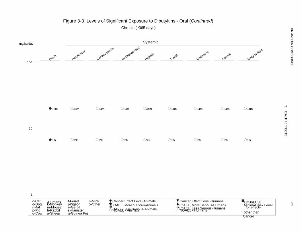
CHRONIC EXPOSURE Death 31 Rat 78 wk
ad libitum (Fischer- 344) (F)
32 Mouse 78 wk ad libitum (B6C3F1) (F)
Systemic 33 Rat 78 wk
ad libitum (Fischer- 344) (F)
34 Mouse 78 wk ad libitum (B6C3F1) (F)
Resp
Cardio
Gastro
Hepatic
Renal
Endocr
Dermal
Bd Wt
Resp
Cardio
Gastro
Hepatic
29 M
6.65
6.65
6.65
6.65
6.65
6.65
6.65
6.65
19.76
19.76
19.76
19.76
6.65 M (52% survival at termination compared to 85% in controls)
19.76 F (86% survival compared with 95% in controls)
57
3. HE
ALTH
EFFE
CTS
Seinen et al. 1977b DBT
NCI 1978a DBT
NCI 1978a DBT
NCI 1978a DBT
NCI 1978a DBT
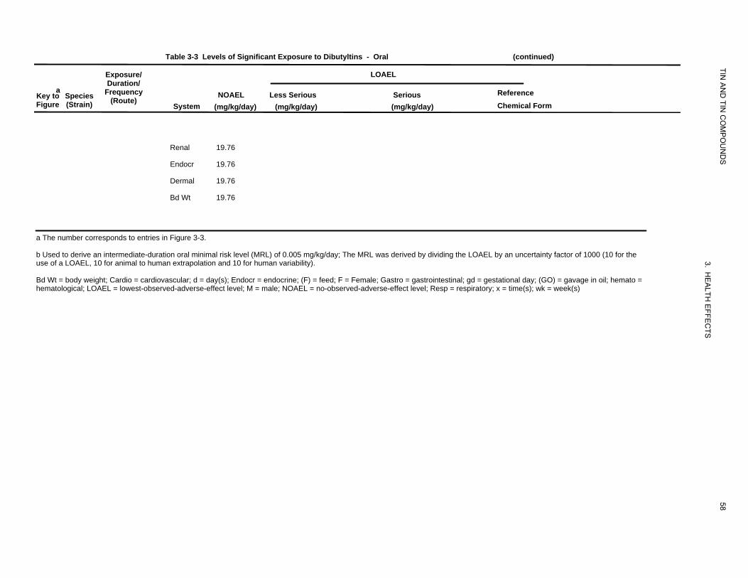
Table 3-3 Levels of Significant Exposure to Dibutyltins - Oral (continued)
Exposure/ LOAEL
TIN
aKey to Species Figure (Strain)
Duration/Frequency
(Route)
System NOAEL
(mg/kg/day)
AN
D TIN
CO
MP
OU
ND
S
Reference Chemical Form
Less Serious (mg/kg/day)
Serious (mg/kg/day)
Renal 19.76
Endocr 19.76
Dermal 19.76
Bd Wt 19.76
a The number corresponds to entries in Figure 3-3.
b Used to derive an intermediate-duration oral minimal risk level (MRL) of 0.005 mg/kg/day; The MRL was derived by dividing the LOAEL by an uncertainty factor of 1000 (10 for the
3. use of a LOAEL, 10 for animal to human extrapolation and 10 for human variability).
HE
Bd Wt = body weight; Cardio = cardiovascular; d = day(s); Endocr = endocrine; (F) = feed; F = Female; Gastro = gastrointestinal; gd = gestational day; (GO) = gavage in oil; hemato =
58
ALTH
EFFE
CTS
hematological; LOAEL = lowest-observed-adverse-effect level; M = male; NOAEL = no-observed-adverse-effect level; Resp = respiratory; x = time(s); wk = week(s)
Death Gastro
intestinal
Hepatic
Body WImmuno/L
Reproductive
Developmental
eight ymphor
Figure 3-3 Levels of Significant Exposure to Dibutyltins - Oral Acute (≤14 days)
Systemic mg/kg/day
TIN A
ND
TIN C
OM
PO
UN
DS
3. HE
ALTH
EFFE
CTS
59
100
10m 2r 5r 5r
6r 11g
12s
4r 7r 7r 1r 6r 16r 22r
9m 8r
19r
10 18r 19r 23r 24r 9m
3r 7r 7r 13r 17r 20r15r
15r 21r 23r 24r
14r 20r
21r
1
c-Cat -Humans f-Ferret n-Mink Cancer Effect Level-Animals Cancer Effect Level-Humans LD50/LC50d-Dog k-Monkey j-Pigeon o-Other LOAEL, More Serious-Animals LOAEL, More Serious-Humans Minimal Risk Levelr-Rat m-Mouse e-Gerbil LOAEL, Less Serious-Animals LOAEL, Less Serious-Humans for effectsp-Pig h-Rabbit s-Hamster NOAEL - Animals NOAEL - Humans other thanq-Cow a-Sheep g-Guinea Pig
Cancer
Hematological
Hepatic
Renal Endocrin
e
Body Weight
Figure 3-3 Levels of Significant Exposure to Dibutyltins - Oral (Continued) Intermediate (15-364 days)
Systemicmg/kg/day
TIN A
ND
TIN C
OM
PO
UN
DS
3. HE
ALTH
EFFE
CTS
60
Immuno/Lymphor
100
27m 27m 27m 27m 29m 30m
26r
10
25r 25r 25r 25r 25r 28r 25r
1
0.1
0.01
0.001
c-Cat -Humans f-Ferret n-Mink Cancer Effect Level-Animals Cancer Effect Level-Humans LD50/LC50d-Dog k-Monkey j-Pigeon o-Other LOAEL, More Serious-Animals LOAEL, More Serious-Humans Minimal Risk Levelr-Rat m-Mouse e-Gerbil LOAEL, Less Serious-Animals LOAEL, Less Serious-Humans for effectsp-Pig h-Rabbit s-Hamster NOAEL - Animals NOAEL - Humans other thanq-Cow a-Sheep g-Guinea Pig
Cancer
Death Respiratory
Cardiovascular
Gastrointestin
al
Hepatic
Renal Endocrin
e
Dermal
Body Weight
Figure 3-3 Levels of Significant Exposure to Dibutyltins - Oral (Continued) Chronic (≥365 days)
Systemicmg/kg/day
TIN A
ND
TIN C
OM
PO
UN
DS
3. HE
ALTH
EFFE
CTS
61
100
32m 34m 34m 34m 34m 34m 34m 34m 34m
10
31r 33r 33r 33r 33r 33r 33r 33r 33r
1
c-Cat -Humans f-Ferret n-Mink Cancer Effect Level-Animals Cancer Effect Level-Humans LD50/LC50d-Dog k-Monkey j-Pigeon o-Other LOAEL, More Serious-Animals LOAEL, More Serious-Humans Minimal Risk Levelr-Rat m-Mouse e-Gerbil LOAEL, Less Serious-Animals LOAEL, Less Serious-Humans for effectsp-Pig h-Rabbit s-Hamster NOAEL - Animals NOAEL - Humans other thanq-Cow a-Sheep g-Guinea Pig
Cancer

Table 3-4 Levels of Significant Exposure to Dioctyltins
Exposure/
TIN
- Oral
LOAEL
aKey to Figure
ACUTE EXPOSURE
Species (Strain)
Duration/ Frequency
(Route) System
NOAEL (mg/kg/day)
Less Serious (mg/kg/day)
Serious (mg/kg/day)
AN
D TIN
CO
MP
OU
ND
S
Reference Chemical Form
Systemic 1 Rat 2 wk
ad libitum (Wistar) (F)
Hepatic 23 Seinen et al. 1977a DOT
Renal 23
Endocr 23
Bd Wt 7.7 F 23 F (12% reduced final body weight)
Immuno/ Lymphoret 2 Rat 2 wk
ad libitum (Wistar) (F)
7.7 (over 35% reduction in relative thymus weight; lymphocyte depletion in
3. HE
Seinen et al. 1977a DOT
lymphoid organs)
ALTH
EFFE
CTS
INTERMEDIATE EXPOSURE Death 3 Gn Pig 5-7 wk
ad libitum (Hartley) (F)
7 F (10 of 16 deaths on weeks 4-5)
62
Seinen et al. 1977b DOT
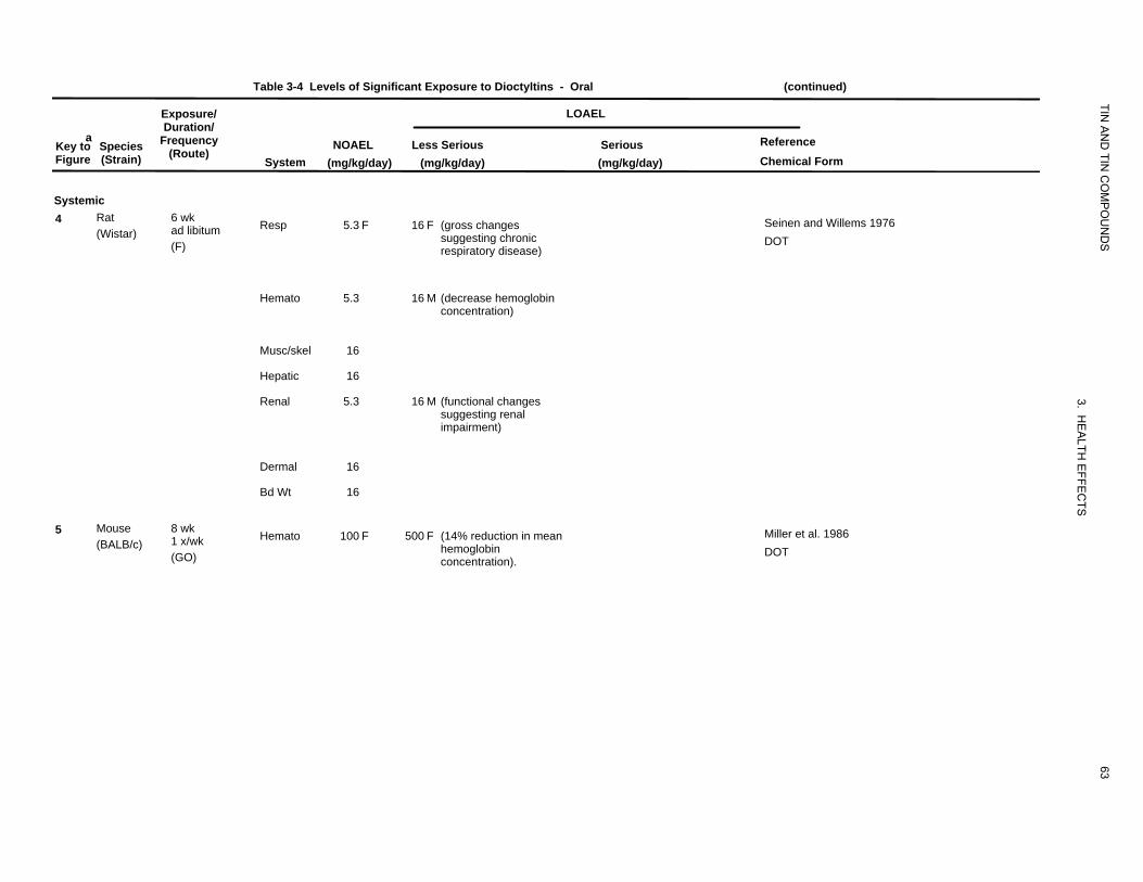
Table 3-4 Levels of Significant Exposure to Dioctyltins - Oral (continued)
Exposure/ LOAEL
TIN
aKey to Species Figure (Strain)
Duration/ Frequency
(Route)System
NOAEL (mg/kg/day)
AN
D TIN
CO
MP
OU
ND
S
ReferenceChemical Form
Less Serious(mg/kg/day)
Serious (mg/kg/day)
Systemic 4 Rat
(Wistar) 6 wk ad libitum(F)
Resp 5.3 F 16 F (gross changes suggesting chronic respiratory disease)
Seinen and Willems 1976 DOT
Hemato 5.3 16 M (decrease hemoglobin
concentration)
Musc/skel 16
Hepatic 16
Renal 5.3 16 M (functional changes
3. suggesting renal
impairment)
HE
ALTH
Dermal 16
EF
Bd Wt 16
FEC
TS
5 Mouse(BALB/c)
8 wk 1 x/wk(GO)
Hemato 100 F 500 F (14% reduction in mean hemoglobinconcentration).
63
Miller et al. 1986DOT
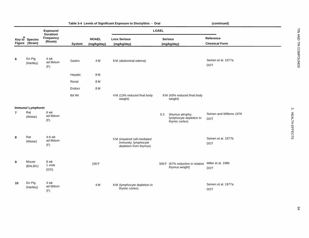
Table 3-4 Levels of Significant Exposure to Dioctyltins - Oral (continued)
Exposure/ LOAEL
TIN
aKey to Species Figure (Strain)
Duration/ Frequency
(Route)System
NOAEL (mg/kg/day)
AN
D TIN
CO
MP
OU
ND
S
Reference Chemical Form
Less Serious (mg/kg/day)
Serious (mg/kg/day)
6 Gn Pig (Hartley)
4 wk ad libitum (F)
Gastro 4 M 8 M (abdominal edema) Seinen et al. 1977a DOT
Hepatic 8 M
Renal 8 M
Endocr 8 M
Bd Wt 4 M (13% reduced final body 8 M (43% reduced final body weight) weight)
Immuno/ Lymphoret
3.
7 Rat (Wistar)
6 wk ad libitum(F)
5.3 (thymus atrophy; lymphocyte depletion in thymic cortex)
HE
ALTH
EFFE
CTS
Seinen and Willems 1976 DOT
8 Rat(Wistar)
4-6 wk ad libitum(F)
5 M (impaired cell-mediated immunity; lymphocyte depletion from thymus)
Seinen et al. 1977b DOT
9 Mouse(BALB/c)
8 wk 1 x/wk(GO)
100 F 500 F (67% reduction in relative thymus weight)
Miller et al. 1986 DOT
10 Gn Pig(Hartley)
4 wk ad libitum(F)
4 M 8 M (lymphocyte depletion in thymic cortex)
64
Seinen et al. 1977a DOT
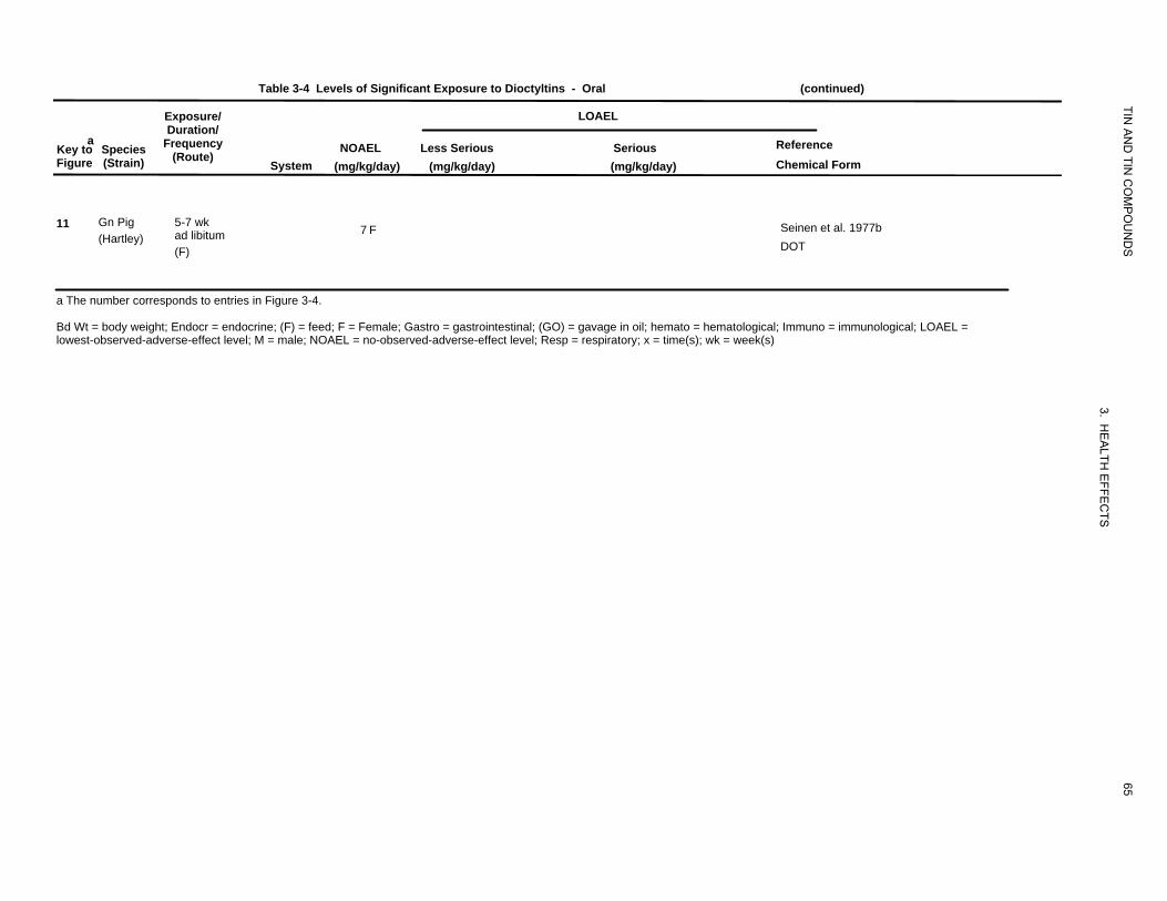
Table 3-4 Levels of Significant Exposure to Dioctyltins - Oral (continued)
Exposure/ LOAEL
TIN
aKey to Figure
Species (Strain)
Duration/ Frequency
(Route) System
NOAEL (mg/kg/day)
AN
D TIN
CO
MP
OU
ND
S
Reference Chemical Form
Less Serious (mg/kg/day)
Serious (mg/kg/day)
11 Gn Pig (Hartley)
5-7 wk ad libitum (F)
7 F Seinen et al. 1977b DOT
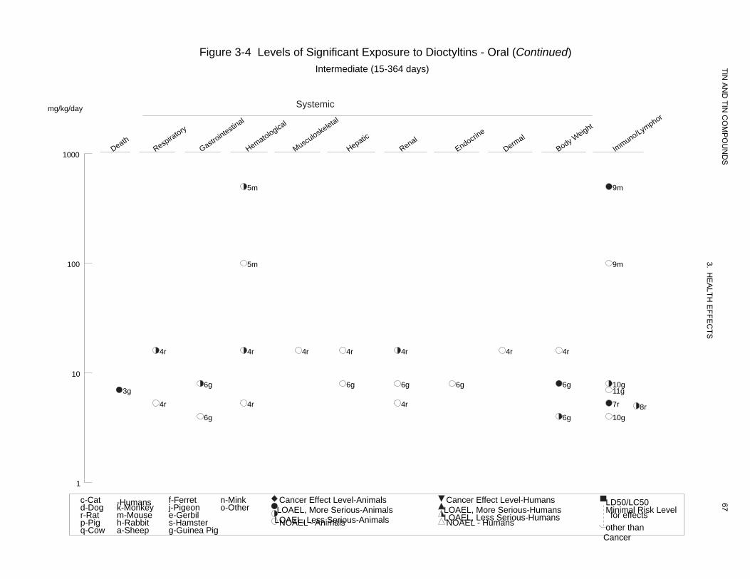
a The number corresponds to entries in Figure 3-4.
Bd Wt = body weight; Endocr = endocrine; (F) = feed; F = Female; Gastro = gastrointestinal; (GO) = gavage in oil; hemato = hematological; Immuno = immunological; LOAEL =
65
3. HE
ALTH
EFFE
CTS
lowest-observed-adverse-effect level; M = male; NOAEL = no-observed-adverse-effect level; Resp = respiratory; x = time(s); wk = week(s)
Hepatic
Renal
Endocrine
Body WImmuno/L
100
eight ymphor
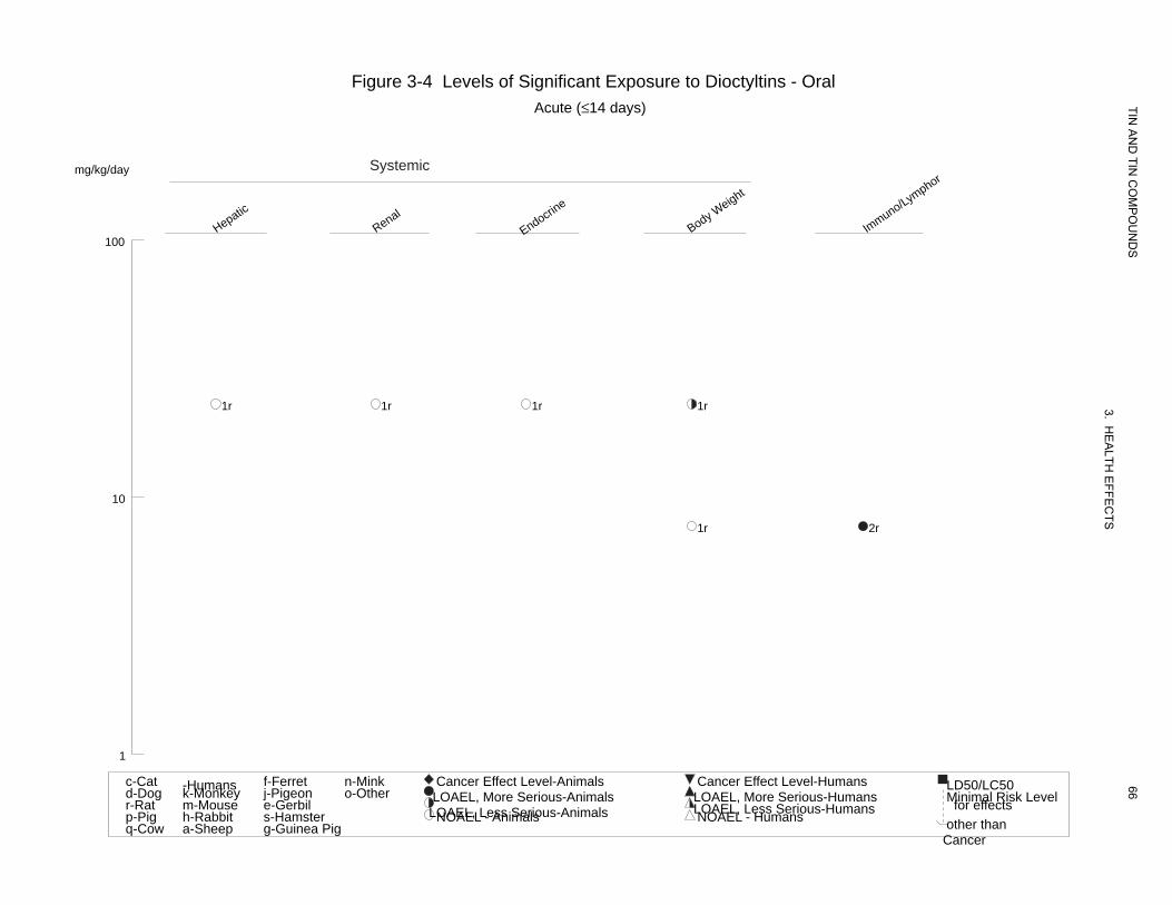
Figure 3-4 Levels of Significant Exposure to Dioctyltins - Oral Acute (≤14 days)
Systemicmg/kg/day
TIN A
ND
TIN C
OM
PO
UN
DS
3. HE
ALTH
EFFE
CTS
66
1r 1r 1r 1r
10
1r 2r
1
c-Cat -Humans f-Ferret n-Mink Cancer Effect Level-Animals Cancer Effect Level-Humans LD50/LC50d-Dog k-Monkey j-Pigeon o-Other LOAEL, More Serious-Animals LOAEL, More Serious-Humans Minimal Risk Levelr-Rat m-Mouse e-Gerbil LOAEL, Less Serious-Animals LOAEL, Less Serious-Humans for effectsp-Pig h-Rabbit s-Hamster NOAEL - Animals NOAEL - Humans other thanq-Cow a-Sheep g-Guinea Pig
Cancer
Death Respiratory
Gastrointestin
al
Hematological
Musculoske
letal
Hepatic
Renal Endocrin
e
Dermal
Body WImmuno/Leight ym
phor
Figure 3-4 Levels of Significant Exposure to Dioctyltins - Oral (Continued) Intermediate (15-364 days)
Systemicmg/kg/day
TIN A
ND
TIN C
OM
PO
UN
DS
3. HE
ALTH
EFFE
CTS
67
1000
5m 9m
100 5m 9m
4r 4r 4r 4r 4r 4r 4r
10 6g 6g 6g 6g 6g 10g
3g 11g
4r 4r 4r 7r 8r 6g 6g 10g
1
c-Cat -Humans f-Ferret n-Mink Cancer Effect Level-Animals Cancer Effect Level-Humans LD50/LC50d-Dog k-Monkey j-Pigeon o-Other LOAEL, More Serious-Animals LOAEL, More Serious-Humans Minimal Risk Levelr-Rat m-Mouse e-Gerbil LOAEL, Less Serious-Animals LOAEL, Less Serious-Humans for effectsp-Pig h-Rabbit s-Hamster NOAEL - Animals NOAEL - Humans other thanq-Cow a-Sheep g-Guinea Pig
Cancer
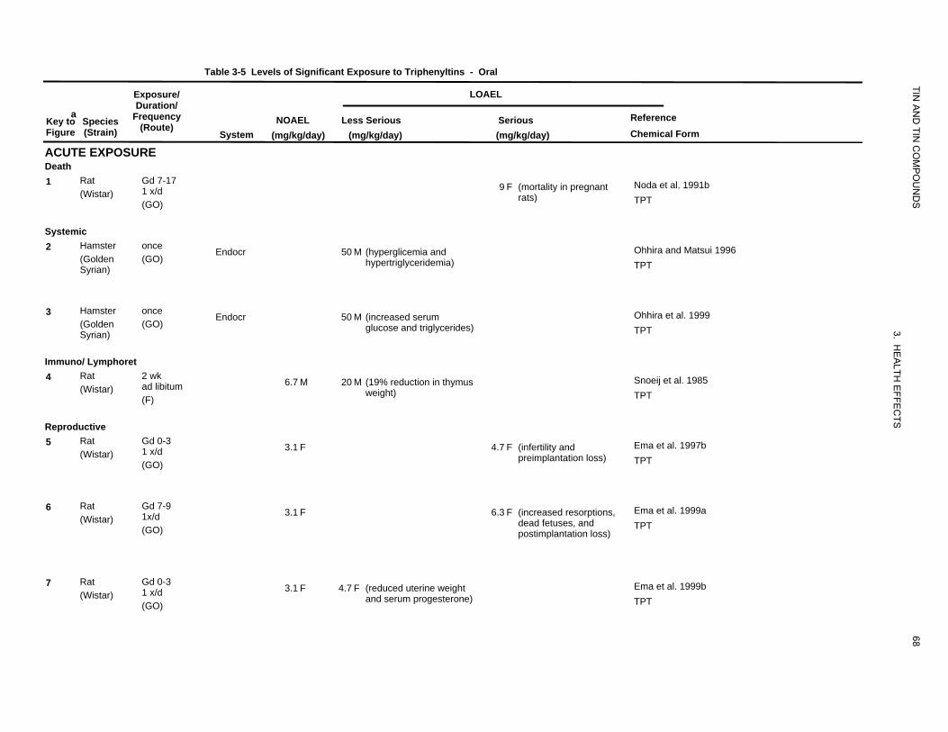
Table 3-5 Levels of Significant Exposure to Triphenyltins
Exposure/
TIN
- Oral
LOAEL
aKey to Figure
ACUTE EXPOSURE
Species (Strain)
Duration/ Frequency
(Route) System
NOAEL (mg/kg/day)
Less Serious (mg/kg/day)
Serious (mg/kg/day)
AN
D TIN
CO
MP
OU
ND
S
Reference Chemical Form
Death 1 Rat Gd 7-17
1 x/d (Wistar) (GO)
9 F (mortality in pregnant rats)
Noda et al. 1991b TPT
Systemic 2 Hamster once
(Golden (GO) Syrian)
Endocr 50 M (hyperglicemia and hypertriglyceridemia)
Ohhira and Matsui 1996 TPT
3 Hamster once (Golden (GO) Syrian)
Endocr 50 M (increased serum glucose and triglycerides)
3. H
Ohhira et al. 1999 TPT
Immuno/ Lymphoret
EA
LTH E
FFEC
TS
4 Rat 2 wk ad libitum (Wistar) (F)
6.7 M 20 M (19% reduction in thymus weight)
Snoeij et al. 1985 TPT
Reproductive 5 Rat Gd 0-3
1 x/d (Wistar) (GO)
3.1 F 4.7 F (infertility and preimplantation loss)
Ema et al. 1997b TPT
6 Rat Gd 7-9 1x/d (Wistar) (GO)
3.1 F 6.3 F (increased resorptions, dead fetuses, and postimplantation loss)
Ema et al. 1999a TPT
7 Rat Gd 0-3 1 x/d (Wistar) (GO)
3.1 F 4.7 F (reduced uterine weight and serum progesterone)
68
Ema et al. 1999b TPT
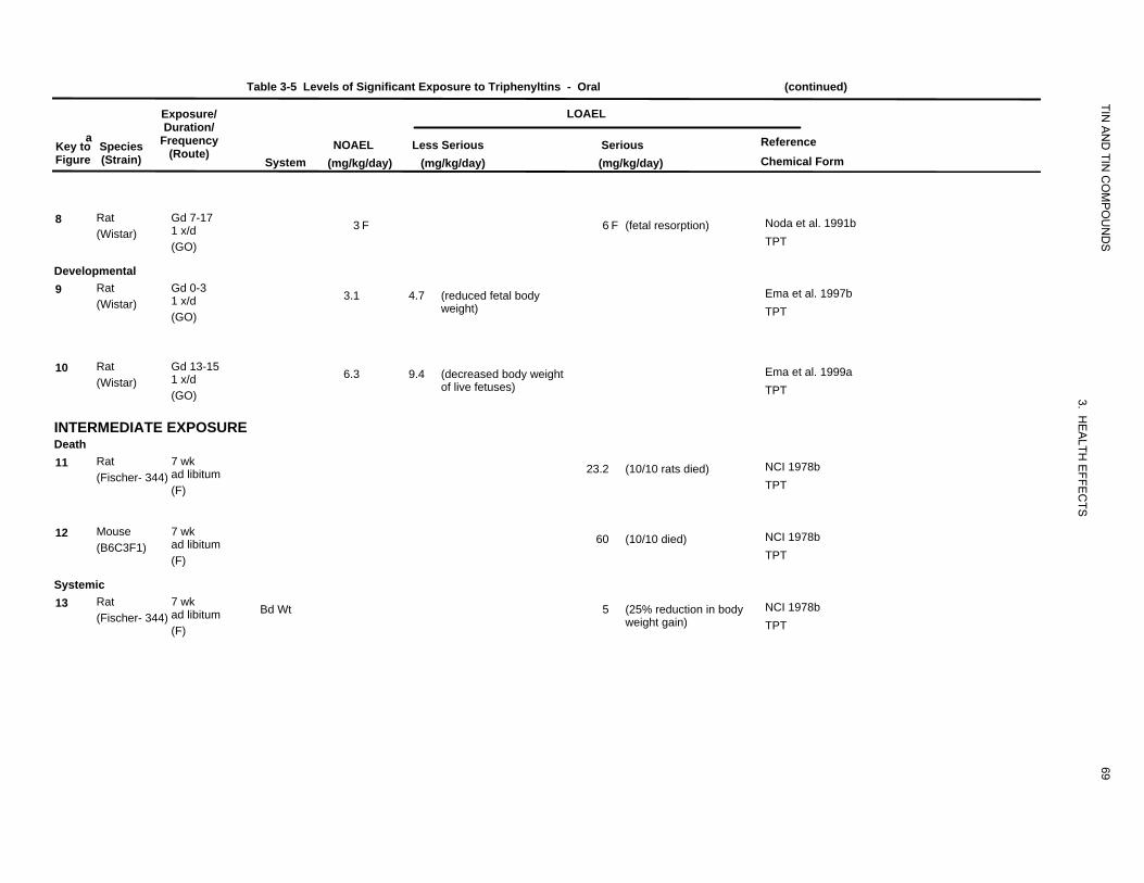
Table 3-5 Levels of Significant Exposure to Triphenyltins - Oral (continued)
Exposure/ LOAEL
TIN
aKey to Figure
Species (Strain)
Duration/ Frequency
(Route) System
NOAEL (mg/kg/day)
AN
D TIN
CO
MP
OU
ND
S
Reference Chemical Form
Less Serious (mg/kg/day)
Serious (mg/kg/day)
8 Rat Gd 7-17 1 x/d (Wistar) (GO)
3 F 6 F (fetal resorption) Noda et al. 1991b TPT
Developmental 9 Rat Gd 0-3
1 x/d (Wistar) (GO)
3.1 4.7 (reduced fetal body weight)
Ema et al. 1997b TPT
10 Rat Gd 13-15 1 x/d (Wistar) (GO)
6.3 9.4 (decreased body weight of live fetuses)
3.
Ema et al. 1999a TPT
INTERMEDIATE EXPOSURE
HE
A
Death 11 Rat 7 wk
ad libitum (Fischer- 344) (F)
23.2 (10/10 rats died)
LTH E
FFEC
TS
NCI 1978b TPT
12 Mouse 7 wk ad libitum (B6C3F1) (F)
60 (10/10 died) NCI 1978b TPT
Systemic 13 Rat 7 wk
ad libitum (Fischer- 344) (F)
Bd Wt 5 (25% reduction in body weight gain)
69
NCI 1978b TPT
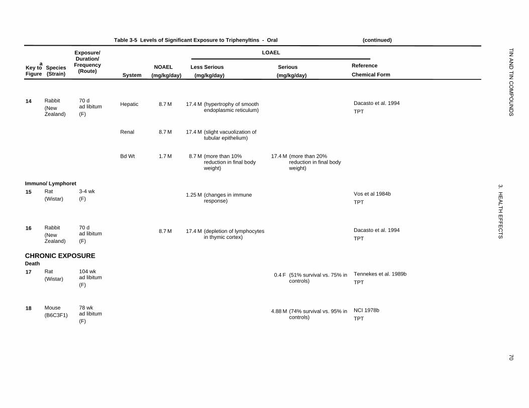
Table 3-5 Levels of Significant Exposure to Triphenyltins - Oral (continued)
Exposure/ LOAEL
TIN
aKey to Figure
Species (Strain)
Duration/ Frequency
(Route) System
NOAEL (mg/kg/day)
AN
D TIN
CO
MP
OU
ND
S
Reference Chemical Form
Less Serious (mg/kg/day)
Serious (mg/kg/day)
14 Rabbit 70 d ad libitum (New
Zealand) (F)
Hepatic 8.7 M 17.4 M (hypertrophy of smooth endoplasmic reticulum)
Dacasto et al. 1994 TPT
Renal 8.7 M 17.4 M (slight vacuolization of tubular epithelium)
Bd Wt 1.7 M 8.7 M (more than 10% 17.4 M (more than 20% reduction in final body reduction in final body weight) weight)
Immuno/ Lymphoret
3
15 Rat 3-4 wk (Wistar) (F)
1.25 M (changes in immune response)
. HE
ALTH
EFFE
CTS
Vos et al 1984b TPT
16 Rabbit 70 d ad libitum (New
Zealand) (F)
8.7 M 17.4 M (depletion of lymphocytes in thymic cortex)
Dacasto et al. 1994 TPT
CHRONIC EXPOSURE Death 17 Rat 104 wk
ad libitum (Wistar) (F)
0.4 F (51% survival vs. 75% in controls)
Tennekes et al. 1989b TPT
18 Mouse 78 wk ad libitum (B6C3F1) (F)
4.88 M (74% survival vs. 95% in controls)
70
NCI 1978b TPT
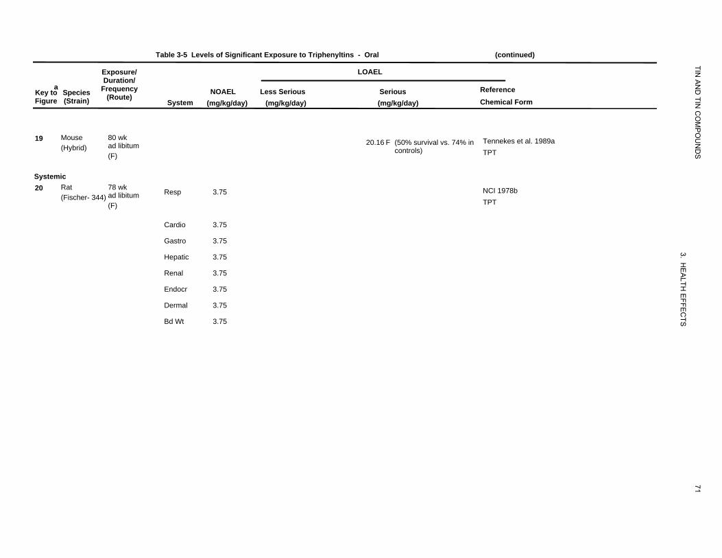
Table 3-5 Levels of Significant Exposure to Triphenyltins - Oral (continued)
Exposure/ LOAEL
TIN
aKey to Figure
Species (Strain)
Duration/ Frequency
(Route) System
NOAEL (mg/kg/day)
AN
D TIN
CO
MP
OU
ND
S
Reference Chemical Form
Less Serious (mg/kg/day)
Serious (mg/kg/day)
19 Mouse (Hybrid)
80 wk ad libitum (F)
20.16 F (50% survival vs. 74% in controls)
Tennekes et al. 1989a TPT
Systemic 20 Rat
(Fischer- 344) 78 wk ad libitum (F)
Resp 3.75 NCI 1978b TPT
Cardio 3.75
Gastro 3.75
Hepatic 3.75
3.
Renal 3.75
Endocr 3.75
HE
ALTH
EFFE
CTS
Dermal 3.75
Bd Wt 3.75
71
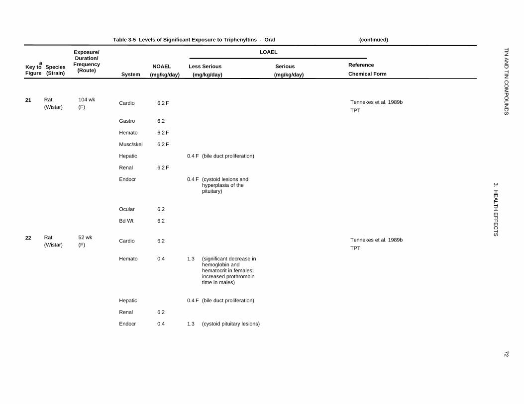
Table 3-5 Levels of Significant Exposure to Triphenyltins - Oral (continued)
aKey to Figure
Species (Strain)
Exposure/ Duration/
Frequency (Route)
System NOAEL
(mg/kg/day)
LOAEL
TIN A
ND
TIN C
OM
PO
UN
DS
ReferenceChemical Form
Less Serious(mg/kg/day)
Serious (mg/kg/day)
21
22
Rat (Wistar)
Rat (Wistar)
104 wk (F)
52 wk (F)
Cardio
Gastro
Hemato
Musc/skel
Hepatic
Renal
Endocr
Ocular
Bd Wt
Cardio
Hemato
Hepatic
Renal
Endocr
6.2 F
6.2
6.2 F
6.2 F
6.2 F
6.2
6.2
6.2
0.4
6.2
0.4
0.4 F (bile duct proliferation)
0.4 F (cystoid lesions andhyperplasia of thepituitary)
1.3 (significant decrease in hemoglobin and
hematocrit in females;
increased prothrombin
time in males)
0.4 F (bile duct proliferation)
1.3 (cystoid pituitary lesions)
72
3. HE
ALTH
EFFE
CTS
Tennekes et al. 1989b TPT
Tennekes et al. 1989b TPT
TIN A
ND
TIN C
OM
PO
UN
DS
73
3. HE
ALTH
EFFE
CTS
Table 3-5 Levels of Significant Exposure to Triphenyltins - Oral (continued)
Exposure/ LOAEL Duration/
a Frequency Reference Key to Species NOAEL Less Serious Serious (Route) Figure (Strain) System (mg/kg/day) (mg/kg/day) (mg/kg/day) Chemical Form
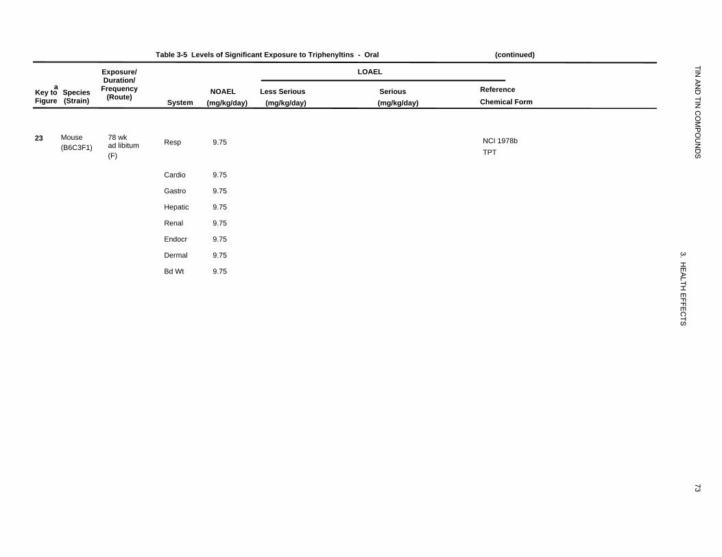
23 Mouse 78 wk Resp 9.75 NCI 1978b ad libitum (B6C3F1) TPT (F)
Cardio 9.75
Gastro 9.75
Hepatic 9.75
Renal 9.75
Endocr 9.75
Dermal 9.75
Bd Wt 9.75
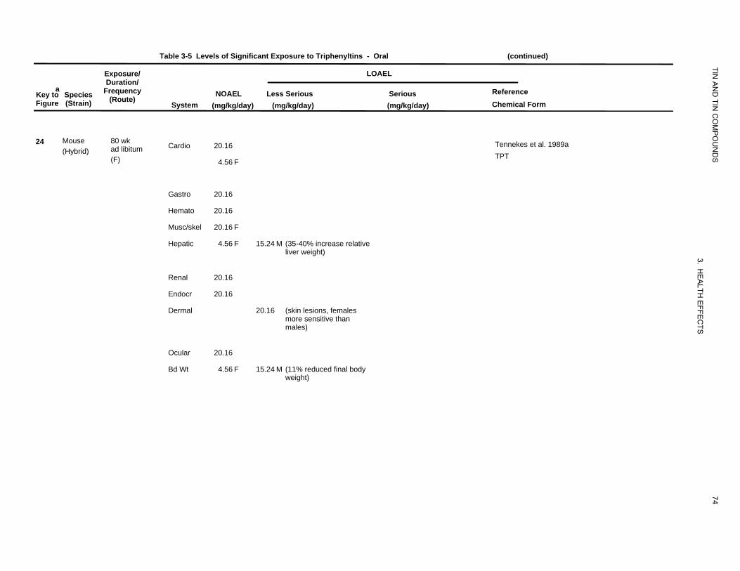
Table 3-5 Levels of Significant Exposure to Triphenyltins - Oral (continued)
Exposure/ LOAEL
TIN
aKey to Figure
Species (Strain)
Duration/ Frequency
(Route) System
NOAEL (mg/kg/day)
AN
D TIN
CO
MP
OU
ND
S
Reference Chemical Form
Less Serious(mg/kg/day)
Serious (mg/kg/day)
24 Mouse (Hybrid)
80 wk ad libitum (F)
Cardio 20.16
4.56 F
Tennekes et al. 1989a TPT
Gastro 20.16
Hemato 20.16
Musc/skel 20.16 F
Hepatic 4.56 F 15.24 M (35-40% increase relative
liver weight)
3.
Renal 20.16
HE
ALTHEndocr 20.16
Dermal 20.16 (skin lesions, females
EFF
more sensitive than
EC
males)
TS
Ocular 20.16
Bd Wt 4.56 F 15.24 M (11% reduced final body weight)
74
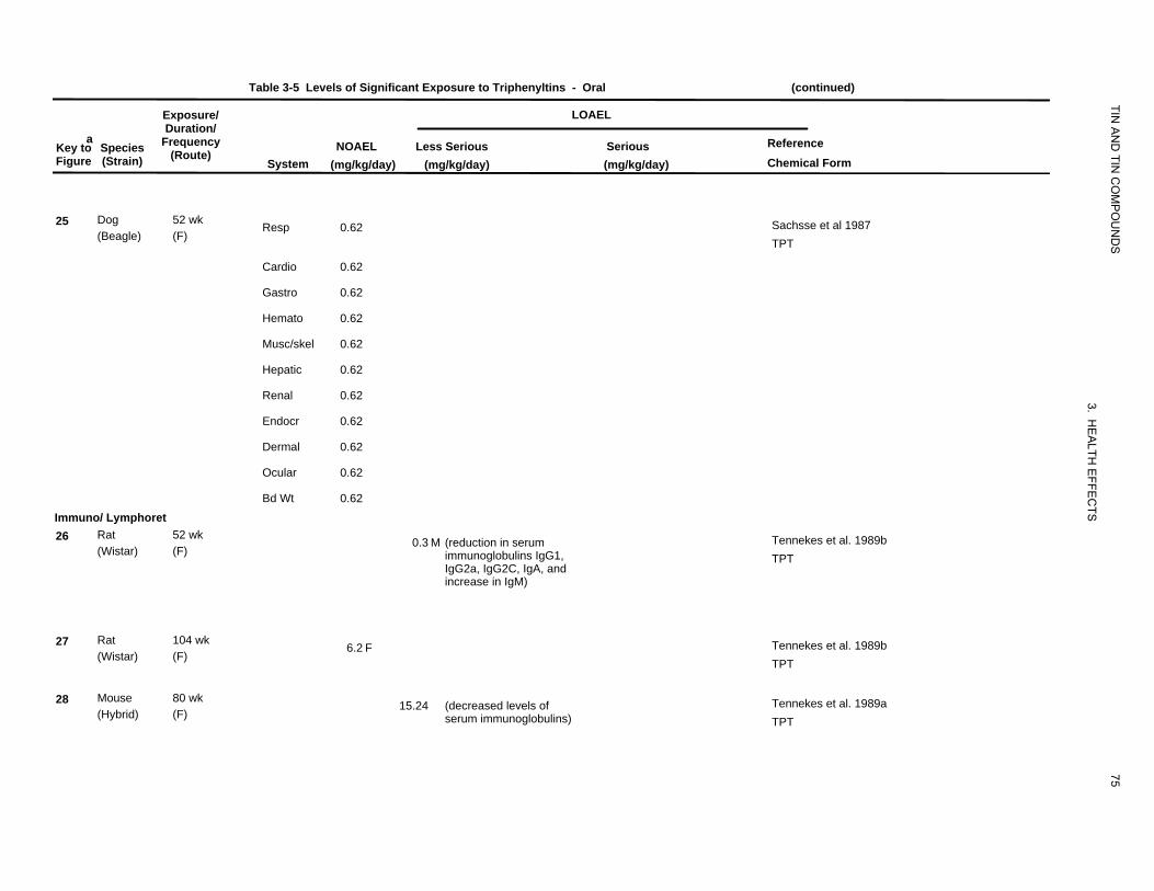
Table 3-5 Levels of Significant Exposure to Triphenyltins - Oral (continued)
aKey to Species Figure (Strain)
Exposure/ Duration/
Frequency (Route)
System NOAEL
(mg/kg/day)
LOAEL
TIN A
ND
TIN C
OM
PO
UN
DS
Reference Chemical Form
Less Serious (mg/kg/day)
Serious (mg/kg/day)
25 Dog (Beagle)
Immuno/ Lymphoret 26 Rat
(Wistar)
27 Rat (Wistar)
28 Mouse (Hybrid)
52 wk (F)
52 wk (F)
104 wk (F)
80 wk (F)
Resp
Cardio
Gastro
Hemato
Musc/skel
Hepatic
Renal
Endocr
Dermal
Ocular
Bd Wt
0.62
0.62
0.62
0.62
0.62
0.62
0.62
0.62
0.62
0.62
0.62
6.2 F
0.3 M (reduction in serum immunoglobulins IgG1, IgG2a, IgG2C, IgA, and increase in IgM)
15.24 (decreased levels of serum immunoglobulins)
75
3. HE
ALTH
EFFE
CTS
Sachsse et al 1987 TPT
Tennekes et al. 1989b TPT
Tennekes et al. 1989b TPT
Tennekes et al. 1989a TPT
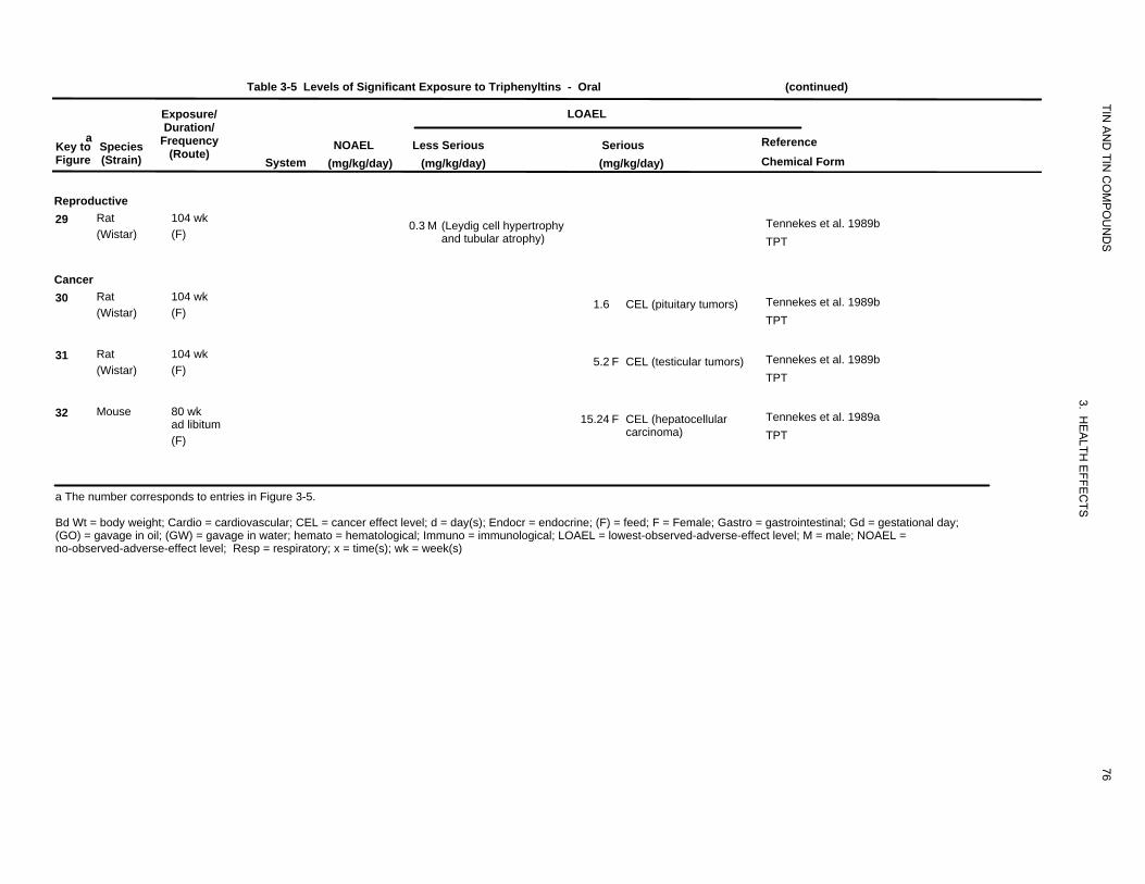
Table 3-5 Levels of Significant Exposure to Triphenyltins - Oral (continued)
Exposure/ LOAEL
TIN
aKey to Figure
Species (Strain)
Duration/ Frequency
(Route) System
NOAEL (mg/kg/day)
AN
D TIN
CO
MP
OU
ND
S
Reference Chemical Form
Less Serious (mg/kg/day)
Serious (mg/kg/day)
Reproductive 29 Rat
(Wistar) 104 wk (F)
0.3 M (Leydig cell hypertrophy and tubular atrophy)
Tennekes et al. 1989b TPT
Cancer 30 Rat
(Wistar) 104 wk (F)
1.6 CEL (pituitary tumors) Tennekes et al. 1989b TPT
31 Rat (Wistar)
104 wk (F)
5.2 F CEL (testicular tumors) Tennekes et al. 1989b TPT
32 Mouse 80 wk ad libitum (F)
3. HE
ALTH
EFF
15.24 F CEL (hepatocellular Tennekes et al. 1989a carcinoma) TPT
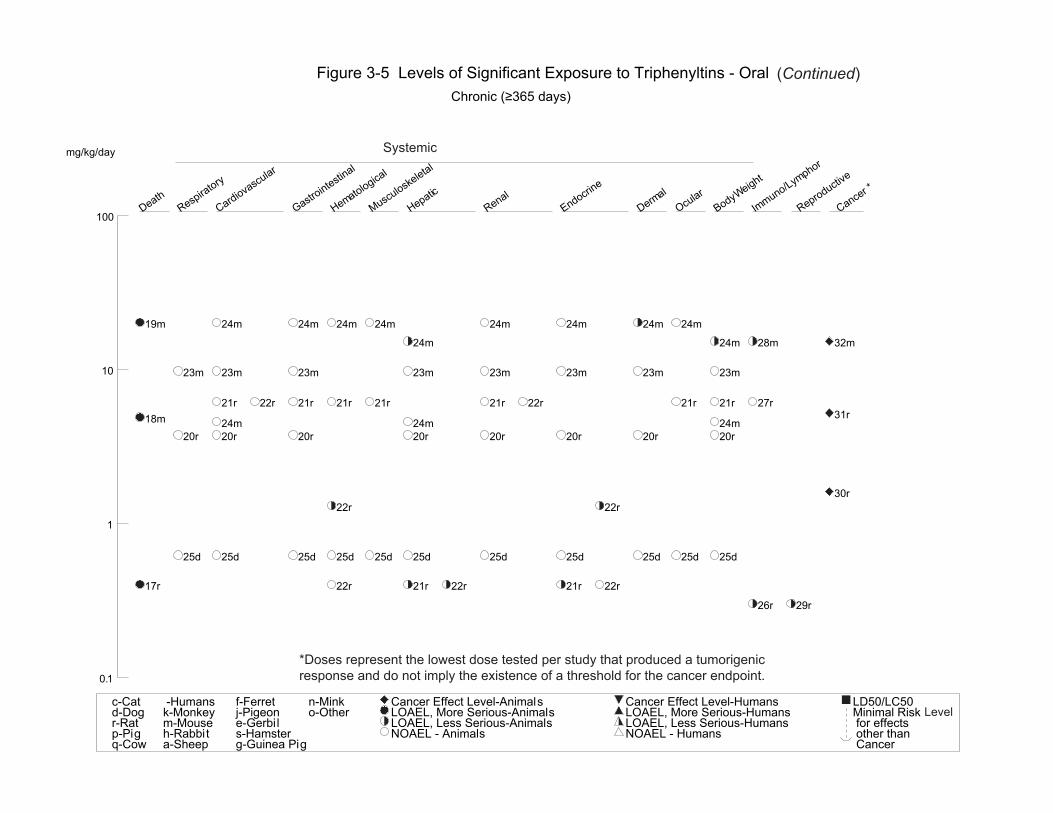
a The number corresponds to entries in Figure 3-5.
EC
TS
Bd Wt = body weight; Cardio = cardiovascular; CEL = cancer effect level; d = day(s); Endocr = endocrine; (F) = feed; F = Female; Gastro = gastrointestinal; Gd = gestational day; (GO) = gavage in oil; (GW) = gavage in water; hemato = hematological; Immuno = immunological; LOAEL = lowest-observed-adverse-effect level; M = male; NOAEL =
76
no-observed-adverse-effect level; Resp = respiratory; x = time(s); wk = week(s)
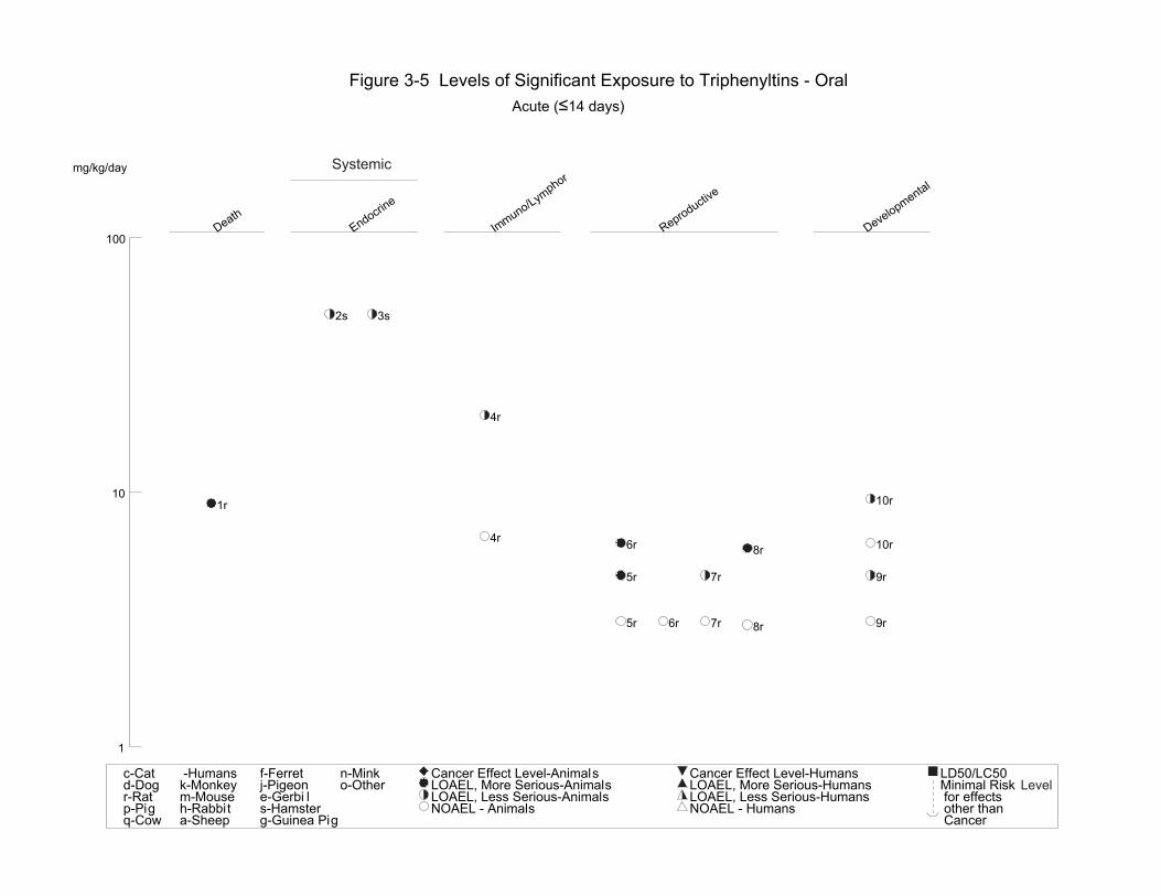
Death
Endocrine
Im Reproductive
Developm
100 muno/Lymphor
ental
Figure 3-5 Levels of Significant Exposure to Triphenyltins - Oral Acute (≤14 days)
TIN AND TIN COMPOUNDS
3. HEALTH EFFECTS
77
mg/kg/day Systemic
2s 3s
4r
10 10r1r
4r 6r 10r8r
5r 7r 9r
5r 6r 7r 9r8r
1
c-Cat d-Dogr-Rat p-Pigq-Cow
-Humans k-Monkeym-Mouse h-Rabbit a-Sheep
f-Ferret j-Pigeone-Gerbi l s-Hamster g-Guinea Pig
n-Mink o-Other
Cancer Effect Level-Animals LOAEL, More Serious-Animals LOAEL, Less Serious-Animals NOAEL - Animals
Cancer Effect Level-Humans LOAEL, More Serious-HumansLOAEL, Less Serious-HumansNOAEL - Humans
LD50/LC50Minimal Risk for effects other than Cancer
Level
Death
Hepati
Renal
Body
Imc We
ight
muno/Lymphor
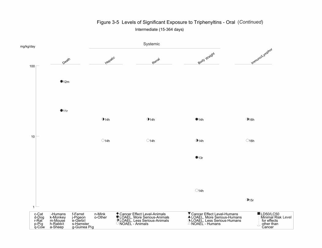
Figure 3-5 Levels of Significant Exposure to Triphenyltins - Oral (Continued) Intermediate (15-364 days)
Systemicmg/kg/day
TIN AND TIN COMPOUNDS
3. HEALTH EFFECTS
78
100
12m
11r
14h 14h 14h 16h
10 14h 14h 14h 16h
13r
14h
15r
1
c-Cat d-Dogr-Rat p-Pigq-Cow
-Humans k-Monkeym-Mouse h-Rabbit a-Sheep
f-Ferret j-Pigeone-Gerbil s-Hamster g-Guinea Pig
n-Mink o-Other
Cancer Effect Level-Animals LOAEL, More Serious-Animals LOAEL, Less Serious-Animals NOAEL - Animals
Cancer Effect Level-Humans LOAEL, More Serious-HumansLOAEL, Less Serious-HumansNOAEL - Humans
LD50/LC50Minimal Risk for effects other than Cancer
Level
Death Respiratory
Cardiovascular
Gastrointestinal
Hem
Musculoskeletal
Hepati
Renal
Endocrine
Derm
Ocular
Body
Im Reproductive
Cancer * ato
logical
c al Weight
muno/Lymphor
Figure 3-5 Levels of Significant Exposure to Triphenyltins - Oral (Continued) Chronic (≥365 days)
Systemicmg/kg/day
TIN AND TIN COMPOUNDS
3. HEALTH EFFECTS
79
100
19m 24m 24m 24m 24m 24m 24m 24m 24m
24m 24m 28m 32m
10 23m 23m 23m 23m 23m 23m 23m 23m
21r 22r 21r 21r 21r 21r 22r 21r 21r 27r 31r18m 24m 24m 24m
20r 20r 20r 20r 20r 20r 20r 20r
30r 22r 22r
1
25d 25d 25d 25d 25d 25d 25d 25d 25d 25d 25d
17r 22r 21r 22r 21r 22r
26r 29r
*Doses represent the lowest dose tested per study that produced a tumorigenic response and do not imply the existence of a threshold for the cancer endpoint.0.1
c-Cat d-Dogr-Rat p-Pigq-Cow
-Humans k-Monkeym-Mouse h-Rabbit a-Sheep
f-Ferret j-Pigeone-Gerbil s-Hamster g-Guinea Pig
n-Mink o-Other
Cancer Effect Level-Animals LOAEL, More Serious-Animals LOAEL, Less Serious-Animals NOAEL - Animals
Cancer Effect Level-Humans LOAEL, More Serious-HumansLOAEL, Less Serious-HumansNOAEL - Humans
LD50/LC50Minimal Risk for effects other than Cancer
Level
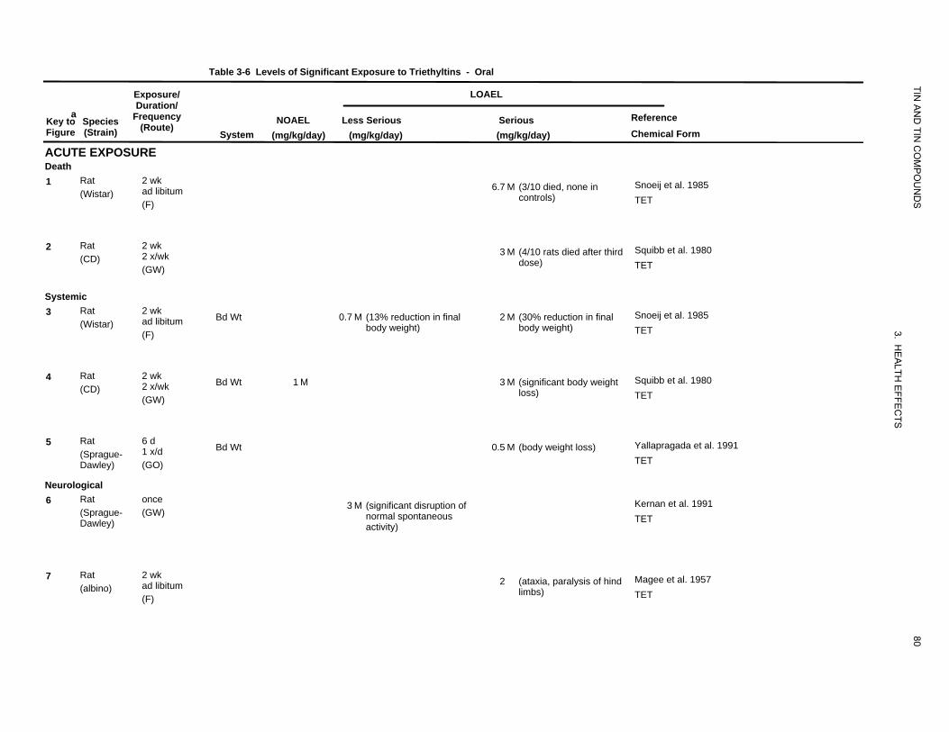
Table 3-6 Levels of Significant Exposure to Triethyltins
Exposure/
TIN
- Oral
LOAEL
aKey to Figure
ACUTE EXPOSURE
Species (Strain)
Duration/ Frequency
(Route) System
NOAEL (mg/kg/day)
Less Serious (mg/kg/day)
Serious (mg/kg/day)
AN
D TIN
CO
MP
OU
ND
S
Reference Chemical Form
Death 1 Rat 2 wk
ad libitum (Wistar) (F)
6.7 M (3/10 died, none in controls)
Snoeij et al. 1985 TET
2 Rat 2 wk 2 x/wk (CD) (GW)
3 M (4/10 rats died after third dose)
Squibb et al. 1980 TET
Systemic 3 Rat 2 wk
ad libitum (Wistar) (F)
Bd Wt 0.7 M (13% reduction in final body weight)
2 M (30% reduction in final body weight)
3. HE
Snoeij et al. 1985 TET
4 Rat 2 wk 2 x/wk (CD) (GW)
Bd Wt 1 M 3 M (significant body weight loss)
ALTH
EFFE
CT
Squibb et al. 1980 TET
5 Rat 6 d 1 x/d (Sprague-
Dawley) (GO)
Bd Wt 0.5 M (body weight loss)
S
Yallapragada et al. 1991 TET
Neurological 6 Rat once
(Sprague- (GW) Dawley)
3 M (significant disruption of normal spontaneous activity)
Kernan et al. 1991 TET
7 Rat 2 wk ad libitum (albino) (F)
2 (ataxia, paralysis of hind limbs)
80
Magee et al. 1957 TET
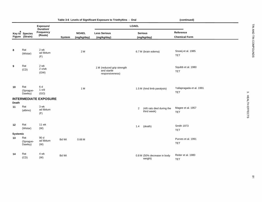
Table 3-6 Levels of Significant Exposure to Triethyltins - Oral (continued)
Exposure/ LOAEL
TIN
aKey to Figure
Species (Strain)
Duration/ Frequency
(Route) System
NOAEL (mg/kg/day)
AN
D TIN
CO
MP
OU
ND
S
Reference Chemical Form
Less Serious (mg/kg/day)
Serious (mg/kg/day)
8 Rat 2 wk ad libitum (Wistar) (F)
2 M 6.7 M (brain edema) Snoeij et al. 1985 TET
9 Rat 2 wk 2 x/wk (CD) (GW)
1 M (reduced grip strength and startle responsiveness)
Squibb et al. 1980 TET
10 Rat 6 d 1 x/d (Sprague-
Dawley) (GO)
1 M 1.5 M (hind limb paralysis)
3.
Yallapragada et al. 1991 TET
INTERMEDIATE EXPOSURE
HE
ALTHDeath
11 Rat 3 wk ad libitum (albino) (F)
2 (4/6 rats died during the third week)
EFFE
CTS
Magee et al. 1957 TET
12 Rat 11 wk (Wistar) (W)
1.4 (death) Smith 1973 TET
Systemic 13 Rat 90 d
ad libitum (Sprague-Dawley) (W)
Bd Wt 0.66 M Purves et al. 1991 TET
14 Rat 4 wk (CD) (W)
Bd Wt 0.8 M (50% decrease in body weight)
81
Reiter et al. 1980 TET
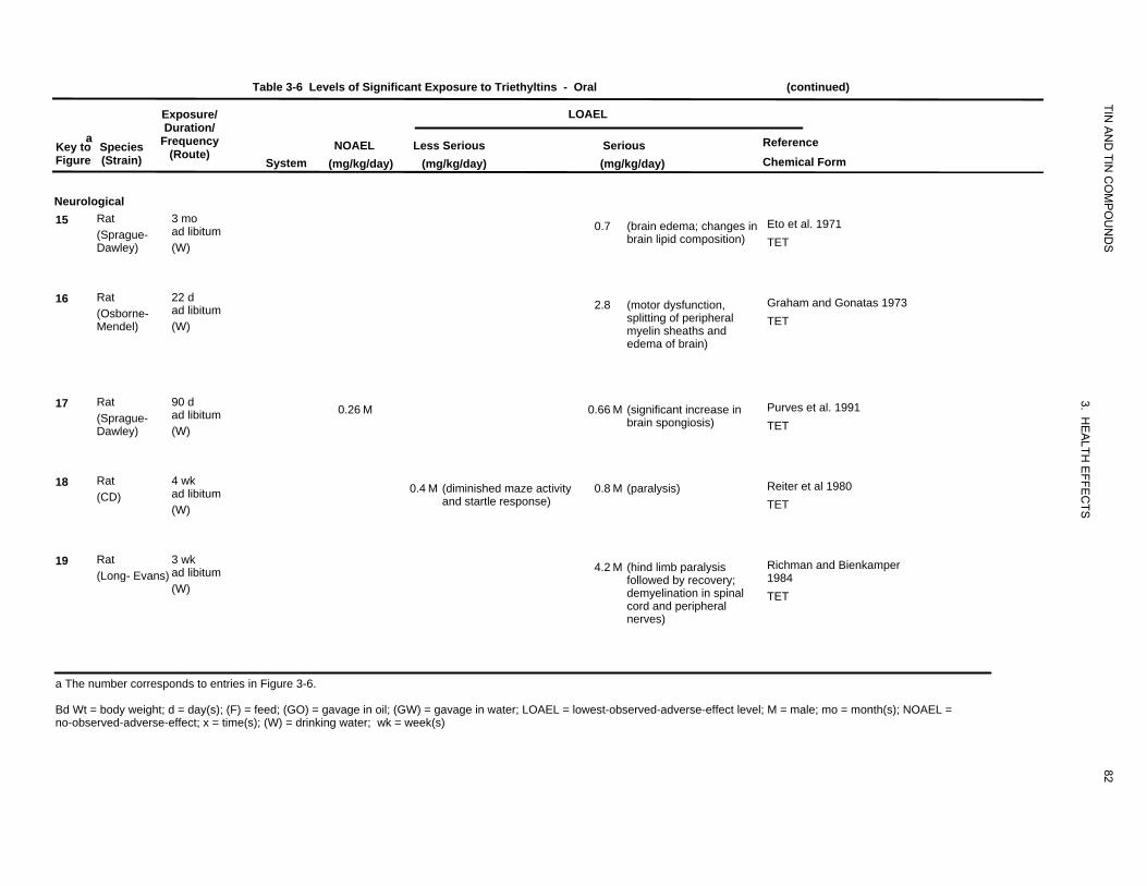
Table 3-6 Levels of Significant Exposure to Triethyltins - Oral (continued)
Exposure/ LOAEL
TIN
aKey to Figure
Species (Strain)
Duration/ Frequency
(Route) System
NOAEL (mg/kg/day)
AN
D TIN
CO
MP
OU
ND
S
Reference Chemical Form
Less Serious (mg/kg/day)
Serious (mg/kg/day)
Neurological 15 Rat
(Sprague-Dawley)
3 mo ad libitum (W)
0.7 (brain edema; changes in Eto et al. 1971 brain lipid composition) TET
16 Rat (Osborne-Mendel)
22 d ad libitum (W)
2.8 (motor dysfunction, Graham and Gonatas 1973 splitting of peripheral TET myelin sheaths and edema of brain)
17 Rat (Sprague-Dawley)
90 d ad libitum (W)
0.26 M
3. HE
ALTH
EFFE
CTS
0.66 M (significant increase in Purves et al. 1991 brain spongiosis) TET
18 Rat (CD)
4 wk ad libitum (W)
0.4 M (diminished maze activity 0.8 M (paralysis) Reiter et al 1980 and startle response) TET
19 Rat 3 wk ad libitum (Long- Evans) (W)
4.2 M (hind limb paralysis Richman and Bienkamper followed by recovery; 1984 demyelination in spinal TET cord and peripheral nerves)
a The number corresponds to entries in Figure 3-6.
Bd Wt = body weight; d = day(s); (F) = feed; (GO) = gavage in oil; (GW) = gavage in water; LOAEL = lowest-observed-adverse-effect level; M = male; mo = month(s); NOAEL =
82
no-observed-adverse-effect; x = time(s); (W) = drinking water; wk = week(s)
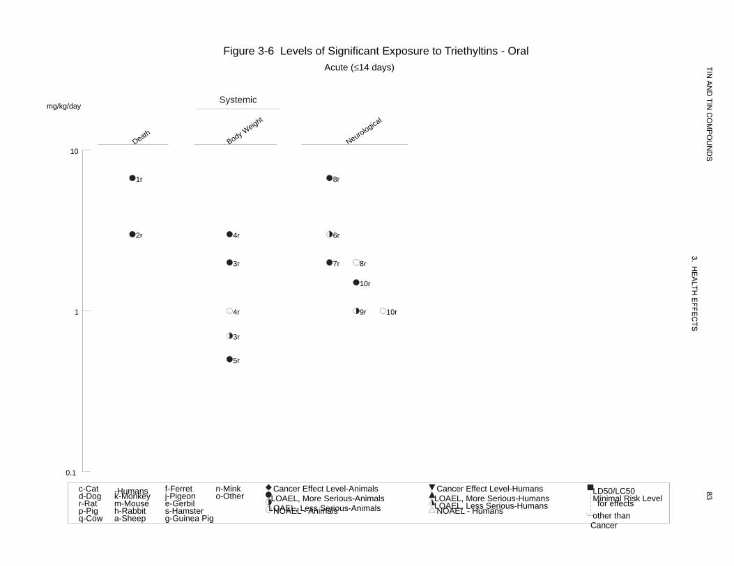
Death Body W
Neurological
eight
Figure 3-6 Levels of Significant Exposure to Triethyltins - Oral Acute (≤14 days)
Systemicmg/kg/day
10
1r 8r
2r 4r 6r
3r 7r 8r
10r
1 4r 9r 10r
3r
5r
0.1
TIN A
ND
TIN C
OM
PO
UN
DS
3. HE
ALTH
EFFE
CTS
83c-Cat -Humans f-Ferret n-Mink Cancer Effect Level-Animals Cancer Effect Level-Humans LD50/LC50d-Dog k-Monkey j-Pigeon o-Other LOAEL, More Serious-Animals LOAEL, More Serious-Humans Minimal Risk Levelr-Rat m-Mouse e-Gerbil LOAEL, Less Serious-Animals LOAEL, Less Serious-Humans for effectsp-Pig h-Rabbit s-Hamster NOAEL - Animals NOAEL - Humans other thanq-Cow a-Sheep g-Guinea Pig
Cancer
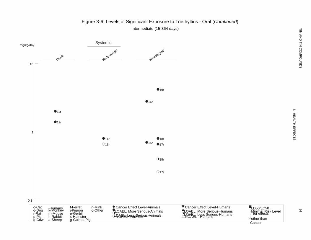
Death Body W
Neurological
eight
Figure 3-6 Levels of Significant Exposure to Triethyltins - Oral (Comtinued) Intermediate (15-364 days)
Systemicmg/kg/day
10
19r
16r
11r
12r
1
14r 18r 15r13r 17r
18r
17r
0.1
TIN A
ND
TIN C
OM
PO
UN
DS
3. HE
ALTH
EFFE
CTS
84c-Cat -Humans f-Ferret n-Mink Cancer Effect Level-Animals Cancer Effect Level-Humans LD50/LC50d-Dog k-Monkey j-Pigeon o-Other LOAEL, More Serious-Animals LOAEL, More Serious-Humans Minimal Risk Levelr-Rat m-Mouse e-Gerbil LOAEL, Less Serious-Animals LOAEL, Less Serious-Humans for effectsp-Pig h-Rabbit s-Hamster NOAEL - Animals NOAEL - Humans other thanq-Cow a-Sheep g-Guinea Pig
Cancer

TIN A
ND
TIN C
OM
PO
UN
DS
85
3. HE
ALTH
EFFE
CTS
Table 3-7 Levels of Significant Exposure to Trimethyltins - Oral
Exposure/ LOAEL Duration/
a Frequency Reference Key to Species NOAEL Less Serious Serious (Route) Figure (Strain) System (mg/kg/day) (mg/kg/day) (mg/kg/day) Chemical Form
ACUTE EXPOSURE Death 1 Rat once 7 M (4 out 10 rats died) Alessandri et al. 1994
(albino) (GO) TMT
2 Rat once 5 (fatal seizures after 4 Bouldin et al. 1981 (Long- Evans) (G) doses) TMT
3 Rat once 12.6 M (LD50) Brown et al. 1979 (Wistar) (GO) TMT
4 Rat 2 wk 2 M (2/10 deaths, none in Snoeij et al. 1985 ad libitum (Wistar) controls) TMT (F)
5 Hamster once 4 F (death within 4 days of Brown et al. 1984 (NS) (GO) dosing) TMT
6 Primate once 3.75 M (4/11 died within 3 days Brown et al. 1984 (NS) (GO) of dosing) TMT
7 Gerbil once 3 F (death within 2-7 days of Brown et al. 1984 (NS) (GO) dosing) TMT
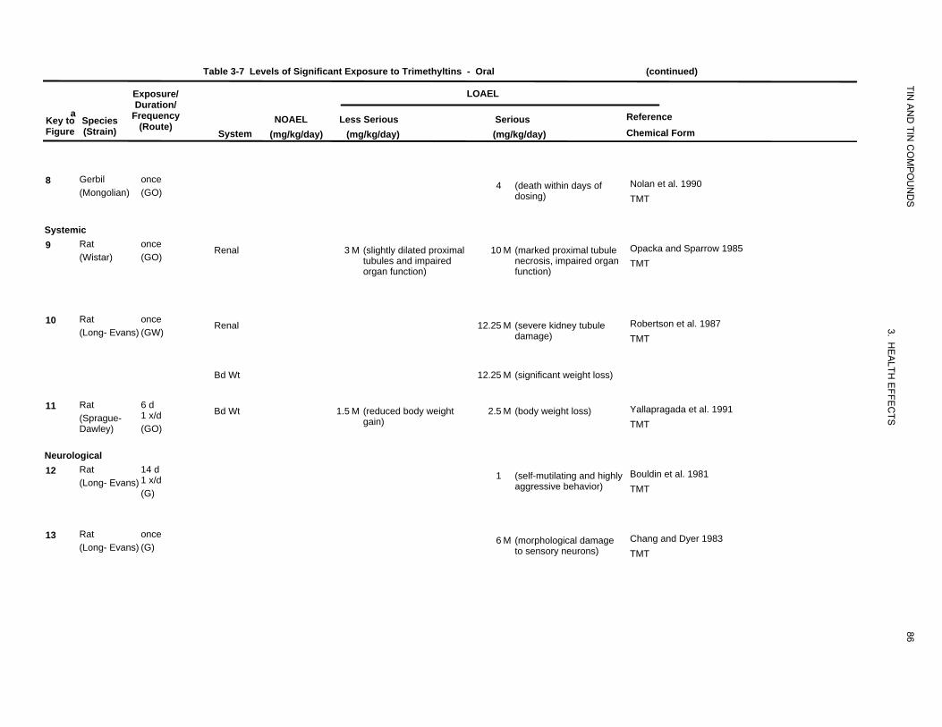
Table 3-7 Levels of Significant Exposure to Trimethyltins - Oral (continued)
Exposure/ LOAEL
TIN
aKey to Figure
Species (Strain)
Duration/ Frequency
(Route) System
NOAEL (mg/kg/day)
AN
D TIN
CO
MP
OU
ND
S
Reference Chemical Form
Less Serious (mg/kg/day)
Serious (mg/kg/day)
8 Gerbil (Mongolian)
once (GO)
4 (death within days of dosing)
Nolan et al. 1990 TMT
Systemic 9 Rat
(Wistar) once (GO)
Renal 3 M (slightly dilated proximal tubules and impaired
10 M (marked proximal tubule necrosis, impaired organ
Opacka and Sparrow 1985 TMT
organ function) function)
10 Rat (Long- Evans)
once (GW)
Renal 12.25 M (severe kidney tubule damage)
3. HE
Robertson et al. 1987 TMT
Bd Wt 12.25 M (significant weight loss)
11 Rat (Sprague-Dawley)
6 d 1 x/d (GO)
Bd Wt 1.5 M (reduced body weight gain)
2.5 M (body weight loss)
ALTH
EFFE
CTS
Yallapragada et al. 1991 TMT
Neurological 12 Rat
(Long- Evans) 14 d 1 x/d (G)
1 (self-mutilating and highly aggressive behavior)
Bouldin et al. 1981 TMT
13 Rat (Long- Evans)
once (G)
6 M (morphological damage to sensory neurons)
86
Chang and Dyer 1983 TMT
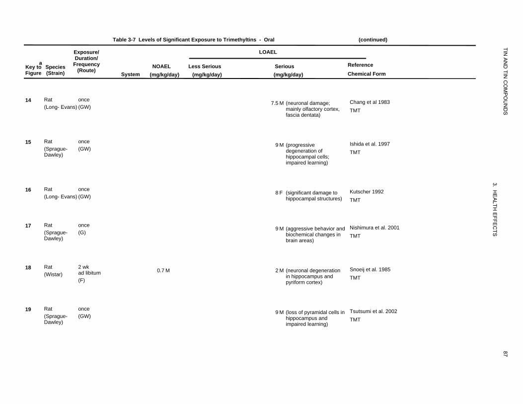
Table 3-7 Levels of Significant Exposure to Trimethyltins - Oral (continued)
Exposure/ LOAEL
TIN
aKey to Figure
Species (Strain)
Duration/ Frequency
(Route) System
NOAEL (mg/kg/day)
AN
D TIN
CO
MP
OU
ND
S
Reference Chemical Form
Less Serious (mg/kg/day)
Serious (mg/kg/day)
14 Rat (Long- Evans)
once (GW)
7.5 M (neuronal damage; mainly olfactory cortex, fascia dentata)
Chang et al 1983 TMT
15 Rat (Sprague-Dawley)
once (GW)
9 M (progressive degeneration of hippocampal cells; impaired learning)
Ishida et al. 1997 TMT
16 Rat (Long- Evans)
once (GW)
8 F (significant damage to hippocampal structures)
3. HE
ALTH
Kutscher 1992 TMT
17 Rat (Sprague-Dawley)
once (G)
9 M (aggressive behavior and biochemical changes in brain areas)
EFFE
CTS
Nishimura et al. 2001 TMT
18 Rat (Wistar)
2 wk ad libitum (F)
0.7 M 2 M (neuronal degeneration in hippocampus and pyriform cortex)
Snoeij et al. 1985 TMT
19 Rat (Sprague-Dawley)
once (GW)
9 M (loss of pyramidal cells in hippocampus and impaired learning)
87
Tsutsumi et al. 2002 TMT
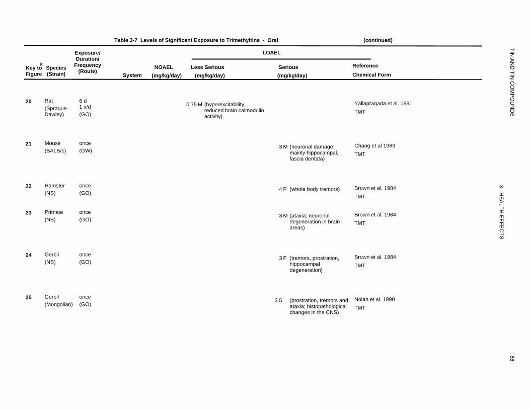
Table 3-7 Levels of Significant Exposure to Trimethyltins - Oral (continued)
Exposure/ LOAEL
TIN
aKey to Species Figure (Strain)
Duration/ Frequency
(Route) System
NOAEL (mg/kg/day)
AN
D TIN
CO
MP
OU
ND
S
Reference Chemical Form
Less Serious (mg/kg/day)
Serious (mg/kg/day)
20 Rat (Sprague-Dawley)
6 d 1 x/d (GO)
0.75 M (hyperexcitability; reduced brain calmodulin activity)
Yallapragada et al. 1991 TMT
21 Mouse (BALB/c)
once (GW)
3 M (neuronal damage; mainly hippocampal, fascia dentata)
Chang et al 1983 TMT
22 Hamster (NS)
once (GO)
4 F (whole body tremors)
3. HE
ALTH
Brown et al. 1984 TMT
23 Primate (NS)
once (GO)
3 M (ataxia; neuronal degeneration in brain areas)
EFFE
CTS
Brown et al. 1984 TMT
24 Gerbil (NS)
once (GO)
3 F (tremors, prostration, hippocampal degeneration)
Brown et al. 1984 TMT
25 Gerbil (Mongolian)
once (GO)
3.5 (prostration, tremors and ataxia; histopathological changes in the CNS)
88
Nolan et al. 1990 TMT
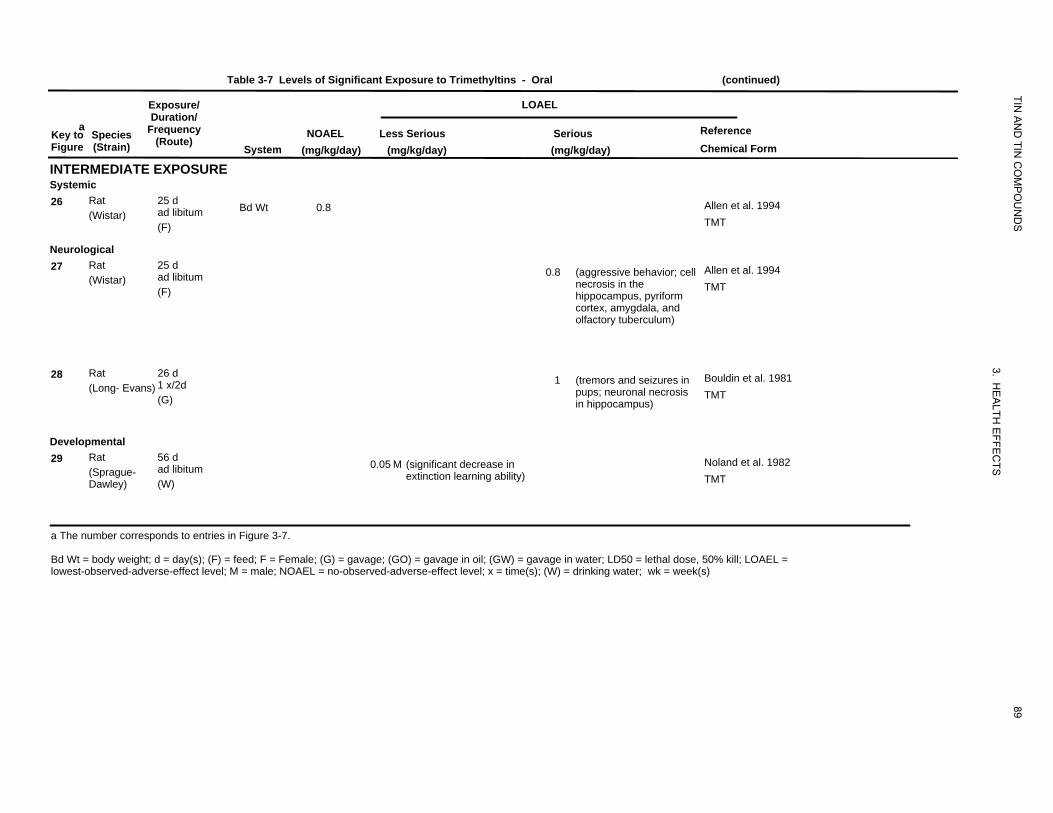
Table 3-7 Levels of Significant Exposure to Trimethyltins - Oral (continued)
Exposure/ LOAEL
TIN
aKey to Figure
Species (Strain)
Duration/ Frequency
(Route) System
NOAEL (mg/kg/day)
AN
D TIN
CO
MP
OU
ND
S
Reference Chemical Form
Less Serious (mg/kg/day)
Serious (mg/kg/day)
INTERMEDIATE EXPOSURE Systemic 26 Rat 25 d Bd Wt ad libitum (Wistar)
(F)
0.8 Allen et al. 1994 TMT
Neurological 27 Rat 25 d
ad libitum (Wistar) (F)
0.8 (aggressive behavior; cell Allen et al. 1994 necrosis in the TMT hippocampus, pyriform cortex, amygdala, and olfactory tuberculum)
28 Rat 26 d 1 x/2d (Long- Evans) (G)
3. HE
ALTH
EFFE
CTS
1 (tremors and seizures in Bouldin et al. 1981 pups; neuronal necrosis TMT in hippocampus)
Developmental 29 Rat 56 d
ad libitum (Sprague-Dawley) (W)
0.05 M (significant decrease in Noland et al. 1982 extinction learning ability) TMT
a The number corresponds to entries in Figure 3-7.
Bd Wt = body weight; d = day(s); (F) = feed; F = Female; (G) = gavage; (GO) = gavage in oil; (GW) = gavage in water; LD50 = lethal dose, 50% kill; LOAEL =
89
lowest-observed-adverse-effect level; M = male; NOAEL = no-observed-adverse-effect level; x = time(s); (W) = drinking water; wk = week(s)
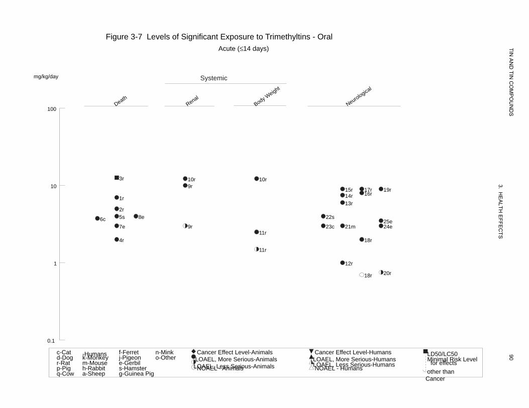
Death Renal
Body WNeurologica
l eight
Figure 3-7 Levels of Significant Exposure to Trimethyltins - Oral Acute (≤14 days) TIN
AN
D TIN
CO
MP
OU
ND
S
3. HE
ALTH
EFFE
CTS
90
mg/kg/day Systemic
100
3r 10r 10r 10 9r 15r 17r 19r
16r14r1r 13r
2r 5s 8e 22s6c 25e 7e 9r 23c 21m 24e
11r 4r 18r
11r
1 12r
20r18r
0.1
c-Cat -Humans f-Ferret n-Mink Cancer Effect Level-Animals Cancer Effect Level-Humans LD50/LC50d-Dog k-Monkey j-Pigeon o-Other LOAEL, More Serious-Animals LOAEL, More Serious-Humans Minimal Risk Levelr-Rat m-Mouse e-Gerbil LOAEL, Less Serious-Animals LOAEL, Less Serious-Humans for effectsp-Pig h-Rabbit s-Hamster NOAEL - Animals NOAEL - Humans other thanq-Cow a-Sheep g-Guinea Pig
Cancer
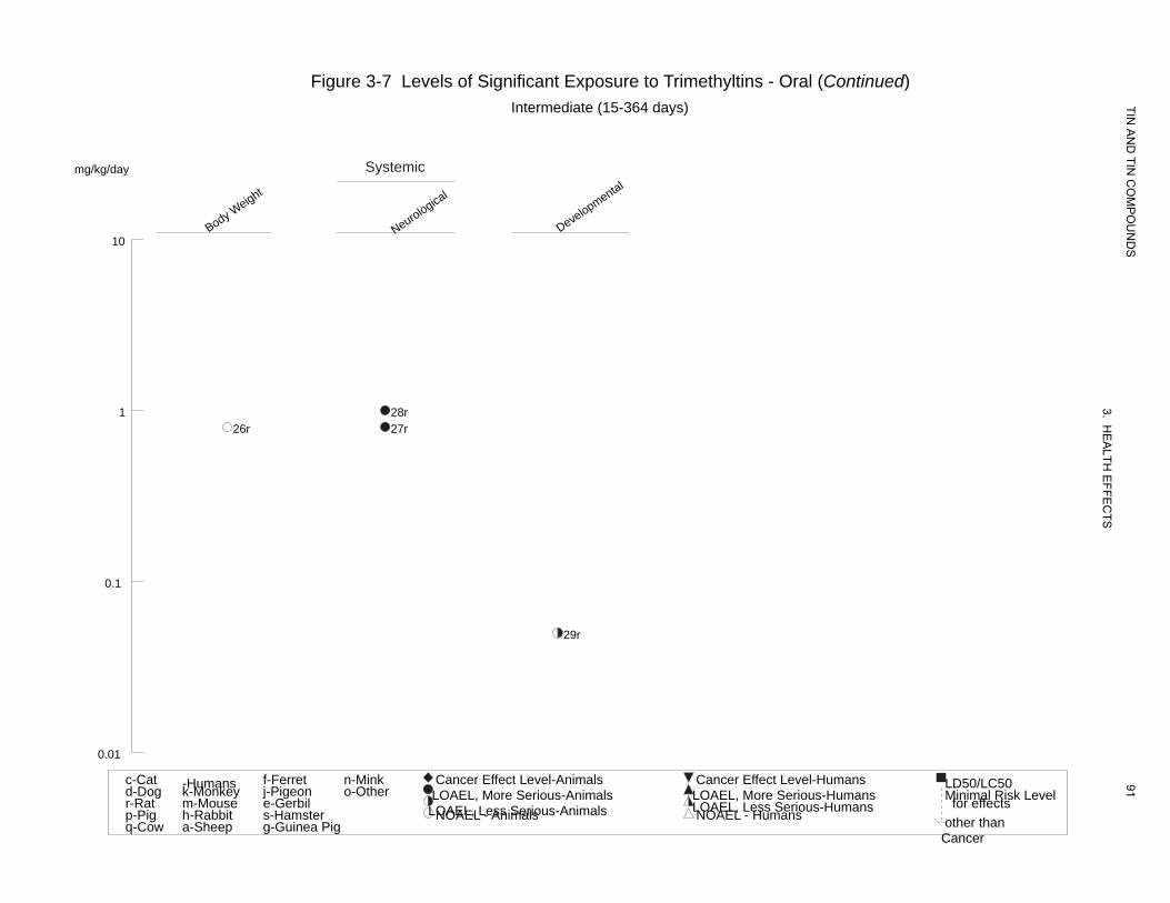
Body WNeurologica
l
Developmental
eight
Figure 3-7 Levels of Significant Exposure to Trimethyltins - Oral (Continued) Intermediate (15-364 days)
mg/kg/day Systemic
TIN A
ND
TIN C
OM
PO
UN
DS
3. HE
ALTH
EFFE
CTS
91
10
1 28r 26r 27r
0.1
29r
0.01
c-Cat -Humans f-Ferret n-Mink Cancer Effect Level-Animals Cancer Effect Level-Humans LD50/LC50d-Dog k-Monkey j-Pigeon o-Other LOAEL, More Serious-Animals LOAEL, More Serious-Humans Minimal Risk Levelr-Rat m-Mouse e-Gerbil LOAEL, Less Serious-Animals LOAEL, Less Serious-Humans for effectsp-Pig h-Rabbit s-Hamster NOAEL - Animals NOAEL - Humans other thanq-Cow a-Sheep g-Guinea Pig
Cancer
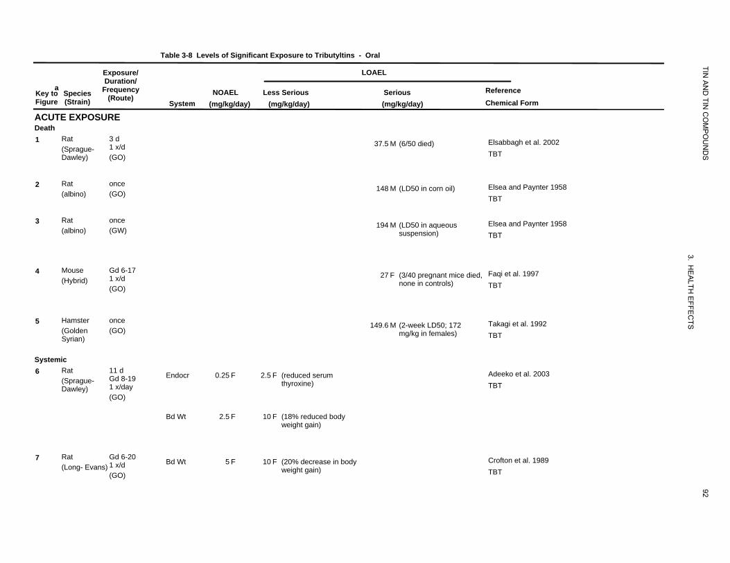
Table 3-8 Levels of Significant Exposure to Tributyltins
Exposure/
TIN
- Oral
LOAEL
aKey to Figure
ACUTE EXPOSURE
Species (Strain)
Duration/ Frequency
(Route) System
NOAEL (mg/kg/day)
Less Serious (mg/kg/day)
Serious (mg/kg/day)
AN
D TIN
CO
MP
OU
ND
S
Reference Chemical Form
Death 1 Rat 3 d
1 x/d (Sprague-Dawley) (GO)
37.5 M (6/50 died) Elsabbagh et al. 2002 TBT
2 Rat once (albino) (GO)
148 M (LD50 in corn oil) Elsea and Paynter 1958 TBT
3 Rat once (albino) (GW)
194 M (LD50 in aqueous suspension)
Elsea and Paynter 1958 TBT
4 Mouse Gd 6-17 1 x/d (Hybrid) (GO)
27 F (3/40 pregnant mice died, none in controls)
3. HE
ALTH
EF
Faqi et al. 1997 TBT
5 Hamster once (Golden (GO) Syrian)
149.6 M (2-week LD50; 172 mg/kg in females)
FEC
TSTakagi et al. 1992 TBT
Systemic 6 Rat 11 d
Gd 8-19 (Sprague-1 x/day Dawley)
Endocr 0.25 F 2.5 F (reduced serum thyroxine)
Adeeko et al. 2003 TBT
(GO)
Bd Wt 2.5 F 10 F (18% reduced body weight gain)
7 Rat Gd 6-20 1 x/d (Long- Evans) (GO)
Bd Wt 5 F 10 F (20% decrease in body weight gain)
92
Crofton et al. 1989 TBT
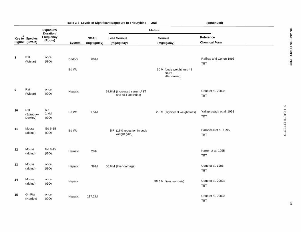
Table 3-8 Levels of Significant Exposure to Tributyltins - Oral (continued)
Exposure/ LOAEL
TIN
aKey to Figure
Species (Strain)
Duration/ Frequency
(Route) System
NOAEL (mg/kg/day)
AN
D TIN
CO
MP
OU
ND
S
Reference Chemical Form
Less Serious (mg/kg/day)
Serious (mg/kg/day)
8 Rat (Wistar)
once (GO)
Endocr 60 M Raffray and Cohen 1993 TBT
Bd Wt 30 M (body weight loss 48 hours after dosing)
9 Rat (Wistar)
once (GO)
Hepatic 58.6 M (increased serum AST and ALT activities)
Ueno et al. 2003b TBT
10 Rat (Sprague-Dawley)
6 d 1 x/d (GO)
Bd Wt 1.5 M 2.5 M (significant weight loss)
3. HE
ALTH
E
Yallapragada et al. 1991 TBT
11 Mouse (albino)
Gd 6-15 (GO)
Bd Wt 5 F (18% reduction in body weight gain)
FFEC
TS
Baroncelli et al. 1995 TBT
12 Mouse (albino)
Gd 6-15 (GO)
Hemato 20 F Karrer et al. 1995 TBT
13 Mouse (albino)
once (GO)
Hepatic 39 M 58.6 M (liver damage) Ueno et al. 1995 TBT
14 Mouse (albino)
once (GO)
Hepatic 58.6 M (liver necrosis) Ueno et al. 2003b TBT
15 Gn Pig (Hartley)
once (GO)
Hepatic 117.2 M
93
Ueno et al. 2003a TBT
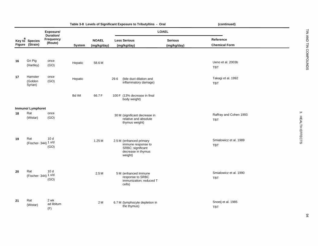
Table 3-8 Levels of Significant Exposure to Tributyltins - Oral (continued)
Exposure/ LOAEL
TIN
aKey to Figure
Species (Strain)
Duration/ Frequency
(Route) System
NOAEL (mg/kg/day)
AN
D TIN
CO
MP
OU
ND
S
Reference Chemical Form
Less Serious (mg/kg/day)
Serious (mg/kg/day)
16 Gn Pig (Hartley)
once (GO)
Hepatic 58.6 M Ueno et al. 2003b TBT
17 Hamster (Golden Syrian)
once (GO)
Hepatic 29.6 (bile duct dilation and inflammatory damage)
Takagi et al. 1992 TBT
Bd Wt 66.7 F 100 F (13% decrease in final body weight)
Immuno/ Lymphoret 18 Rat
(Wistar) once (GO)
30 M (significant decrease in relative and absolute thymus weight)
3. HE
ALTH
Raffray and Cohen 1993 TBT
19 Rat (Fischer- 344)
10 d 1 x/d (GO)
1.25 M 2.5 M (enhanced primary immune response to SRBC; significant decrease in thymus weight)
EFFE
CTSSmialowicz et al. 1989
TBT
20 Rat (Fischer- 344)
10 d 1 x/d (GO)
2.5 M 5 M (enhanced immune response to SRBC immunization; reduced T cells)
Smialowicz et al. 1990 TBT
21 Rat (Wistar)
2 wk ad libitum (F)
2 M 6.7 M (lymphocyte depletion in the thymus)
94
Snoeij et al. 1985 TBT
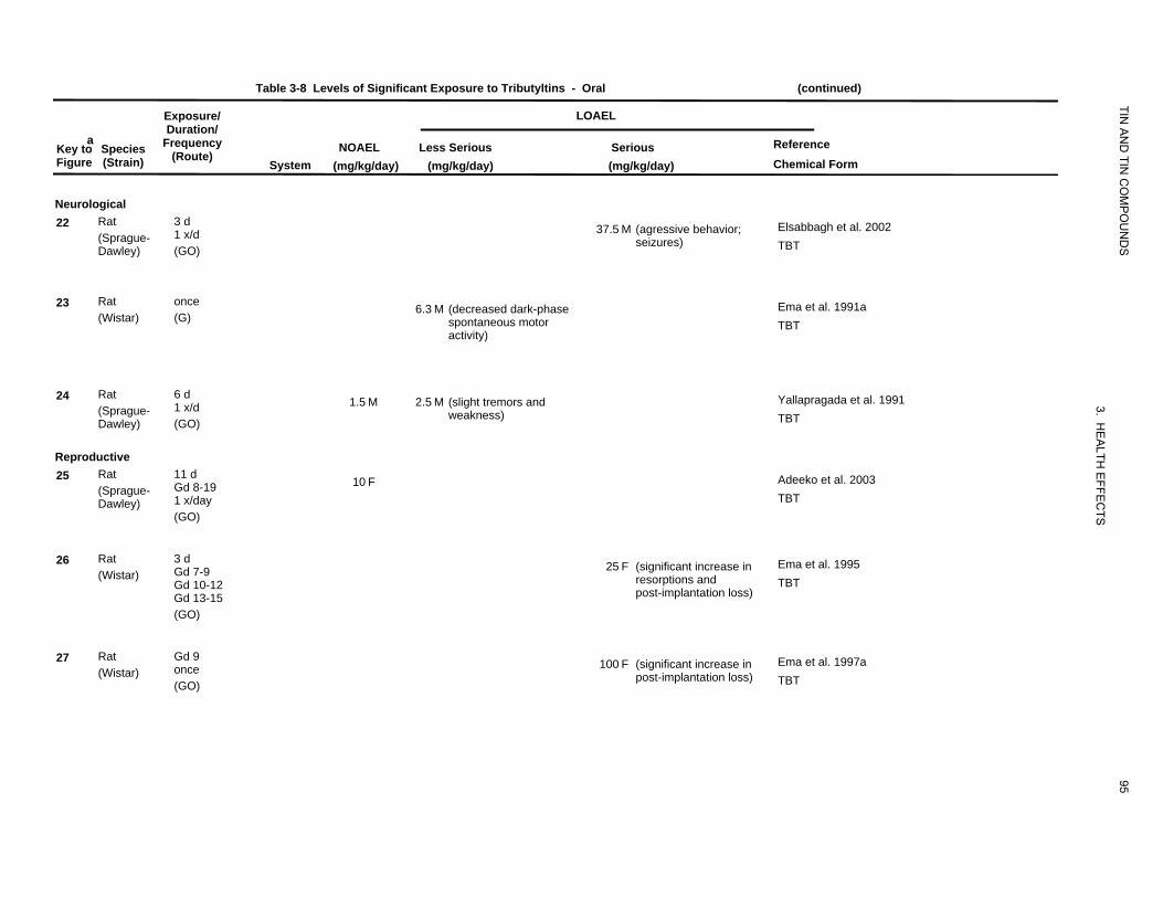
Table 3-8 Levels of Significant Exposure to Tributyltins - Oral (continued)
Exposure/ LOAEL
TIN
aKey to Species Figure (Strain)
Duration/ Frequency
(Route) System
NOAEL (mg/kg/day)
AN
D TIN
CO
MP
OU
ND
S
Reference Chemical Form
Less Serious (mg/kg/day)
Serious (mg/kg/day)
Neurological 22 Rat
(Sprague-Dawley)
3 d 1 x/d (GO)
37.5 M (agressive behavior; seizures)
Elsabbagh et al. 2002 TBT
23 Rat (Wistar)
once (G)
6.3 M (decreased dark-phase spontaneous motor
Ema et al. 1991a TBT
activity)
24 Rat (Sprague-Dawley)
6 d 1 x/d (GO)
1.5 M 2.5 M (slight tremors and weakness)
3. HE
Yallapragada et al. 1991 TBT
Reproductive 25 Rat
(Sprague-Dawley)
11 d Gd 8-19 1 x/day (GO)
10 F
ALTH
EFFE
CTS
Adeeko et al. 2003 TBT
26 Rat (Wistar)
3 d Gd 7-9 Gd 10-12 Gd 13-15 (GO)
25 F (significant increase in resorptions and post-implantation loss)
Ema et al. 1995 TBT
27 Rat (Wistar)
Gd 9 once (GO)
100 F (significant increase in post-implantation loss)
95
Ema et al. 1997a TBT
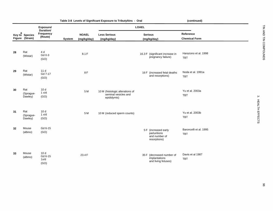
Table 3-8 Levels of Significant Exposure to Tributyltins - Oral (continued)
Exposure/ LOAEL
TIN
aKey to Species Figure (Strain)
Duration/ Frequency
(Route) System
NOAEL (mg/kg/day)
AN
D TIN
CO
MP
OU
ND
S
Reference Chemical Form
Less Serious (mg/kg/day)
Serious (mg/kg/day)
28 Rat (Wistar)
4 d Gd 0-3 (GO)
8.1 F 16.3 F (significant increase in pregnancy failure)
Harazono et al. 1998 TBT
29 Rat (Wistar)
11 d Gd 7-17 (GO)
8 F 16 F (increased fetal deaths and resorptions)
Noda et al. 1991a TBT
30 Rat (Sprague-Dawley)
10 d 1 x/d (GO)
5 M 10 M (histologic alterations of seminal vesicles and epididymis)
3. HE
A
Yu et al. 2003a TBT
31 Rat (Sprague-Dawley)
10 d 1 x/d (GO)
5 M 10 M (reduced sperm counts)
LTH E
FFEC
TS
Yu et al. 2003b TBT
32 Mouse (albino)
Gd 6-15 (GO)
5 F (increased early parturitions and number of resorptions)
Baroncelli et al. 1995 TBT
33 Mouse (albino)
10 d Gd 6-15 1x/d (GO)
23.4 F 35 F (decreased number of implantations and living fetuses)
96
Davis et al 1987 TBT
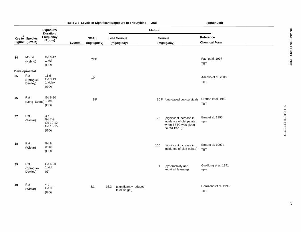
Table 3-8 Levels of Significant Exposure to Tributyltins - Oral (continued)
Exposure/ LOAEL
TIN
aKey toFigure
Species
(Strain)
Duration/Frequency
(Route)
System NOAEL
(mg/kg/day)
AN
D TIN
CO
MP
OU
ND
S
Reference Chemical Form
Less Serious (mg/kg/day)
Serious (mg/kg/day)
34 Mouse (Hybrid)
Gd 6-17 1 x/d (GO)
27 F Faqi et al. 1997 TBT
Developmental 35 Rat
(Sprague-Dawley)
11 d Gd 8-19 1 x/day (GO)
10 Adeeko et al. 2003 TBT
36 Rat (Long- Evans)
Gd 6-20 1 x/d (GO)
5 F 10 F (decreased pup survival)
3. H
Crofton et al. 1989 TBT
37 Rat (Wistar)
3 d Gd 7-9 Gd 10-12 Gd 13-15 (GO)
25 (significant increase in incidence of clef palate when TBTC was given on Gd 13-15)
EA
LTH E
FFEC
TS
Ema et al. 1995 TBT
38 Rat (Wistar)
Gd 9 once (GO)
100 (significant increase in incidence of cleft palate)
Ema et al. 1997a TBT
39 Rat (Sprague-Dawley)
Gd 6-20 1 x/d (G)
1 (hyperactivity and impaired learning)
Gardlung et al. 1991 TBT
40 Rat (Wistar)
4 d Gd 0-3 (GO)
8.1 16.3 (significantly reduced fetal weight)
97
Harazono et al. 1998 TBT
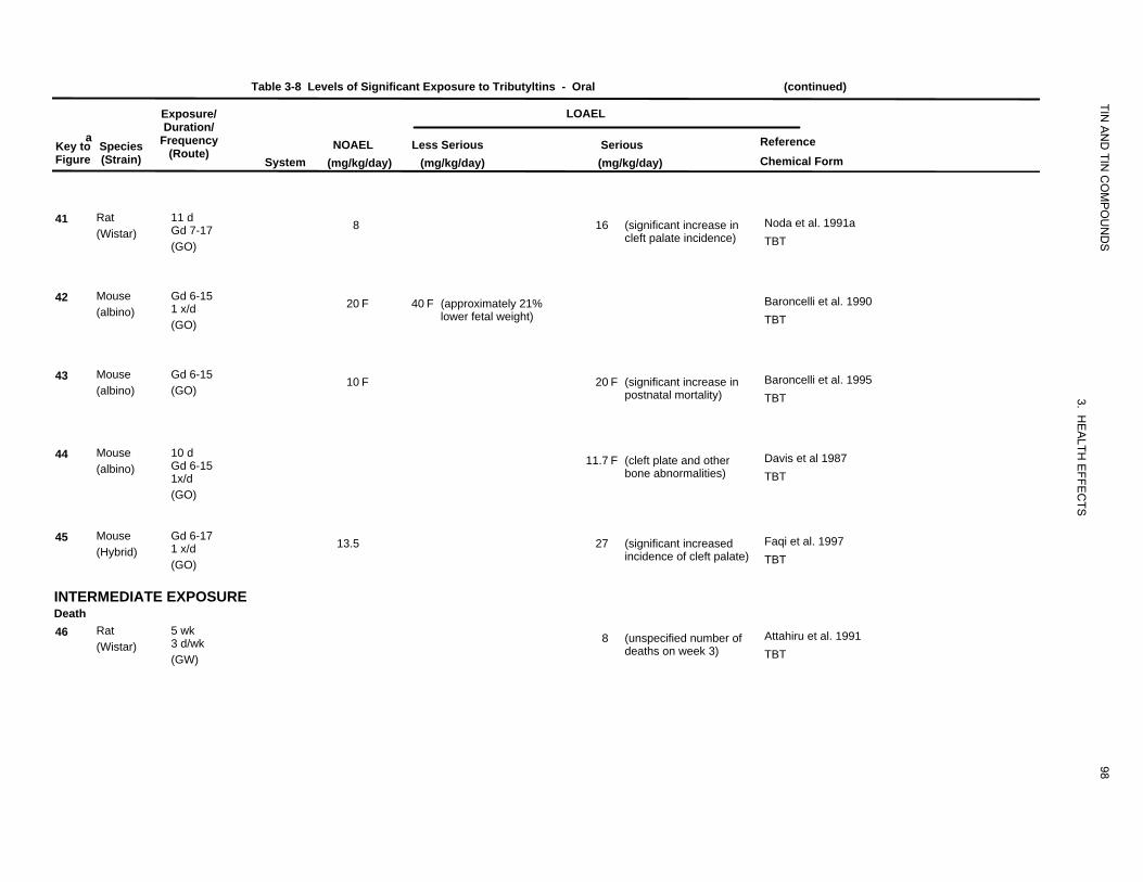
Table 3-8 Levels of Significant Exposure to Tributyltins - Oral (continued)
Exposure/ LOAEL
TIN
aKey to Figure
Species (Strain)
Duration/ Frequency
(Route) System
NOAEL (mg/kg/day)
AN
D TIN
CO
MP
OU
ND
S
Reference Chemical Form
Less Serious (mg/kg/day)
Serious (mg/kg/day)
41 Rat 11 d Gd 7-17 (Wistar) (GO)
8 16 (significant increase in cleft palate incidence)
Noda et al. 1991a TBT
42 Mouse Gd 6-15 1 x/d (albino) (GO)
20 F 40 F (approximately 21% lower fetal weight)
Baroncelli et al. 1990 TBT
43 Mouse Gd 6-15 (albino) (GO)
10 F 20 F (significant increase in postnatal mortality)
3. H
Baroncelli et al. 1995 TBT
44 Mouse 10 d Gd 6-15 (albino) 1x/d
11.7 F (cleft plate and other bone abnormalities)
EA
LTH E
FFEC
TS
Davis et al 1987 TBT
(GO)
45 Mouse Gd 6-17 1 x/d (Hybrid) (GO)
13.5 27 (significant increased incidence of cleft palate)
Faqi et al. 1997 TBT
INTERMEDIATE EXPOSURE Death 46 Rat 5 wk
3 d/wk (Wistar) (GW)
8 (unspecified number of deaths on week 3)
98
Attahiru et al. 1991 TBT
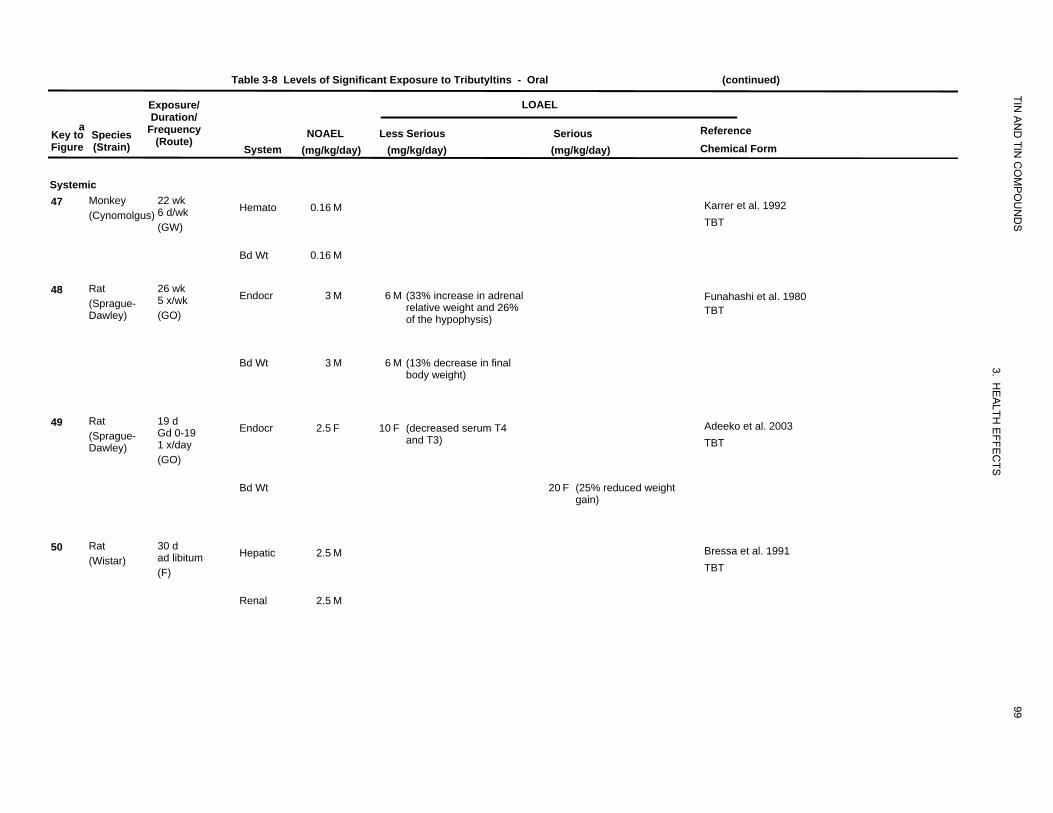
Table 3-8 Levels of Significant Exposure to Tributyltins - Oral (continued)
Exposure/ LOAEL
TIN
aKey to Figure
Species (Strain)
Duration/ Frequency
(Route) System
NOAEL (mg/kg/day)
AN
D TIN
CO
MP
OU
ND
S
Reference Chemical Form
Less Serious (mg/kg/day)
Serious (mg/kg/day)
Systemic 47 Monkey
(Cynomolgus) 22 wk 6 d/wk (GW)
Hemato 0.16 M Karrer et al. 1992 TBT
Bd Wt 0.16 M
48 Rat (Sprague-Dawley)
26 wk 5 x/wk (GO)
Endocr 3 M 6 M (33% increase in adrenal relative weight and 26% of the hypophysis)
Funahashi et al. 1980 TBT
Bd Wt 3 M 6 M (13% decrease in final body weight)
3. HE
49 Rat (Sprague-Dawley)
19 d Gd 0-19 1 x/day (GO)
Endocr 2.5 F 10 F (decreased serum T4 and T3)
ALTH
EFFE
CTS
Adeeko et al. 2003 TBT
Bd Wt 20 F (25% reduced weight gain)
50 Rat (Wistar)
30 d ad libitum (F)
Hepatic 2.5 M Bressa et al. 1991 TBT
Renal 2.5 M
99
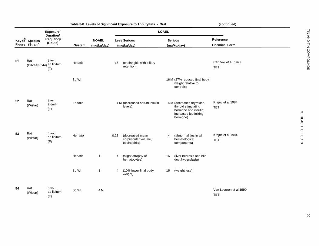
Table 3-8 Levels of Significant Exposure to Tributyltins - Oral (continued)
Exposure/ LOAEL
TIN
aKey to Figure
Species (Strain)
Duration/Frequency
(Route)
System NOAEL
(mg/kg/day)
AN
D TIN
CO
MP
OU
ND
S
Reference Chemical Form
Less Serious(mg/kg/day)
Serious (mg/kg/day)
51 Rat(Fischer- 344)
6 wk ad libitum (F)
Hepatic 16 (cholangitis with biliaryretention)
Carthew et al. 1992 TBT
Bd Wt 16 M (27% reduced final body weight relative to controls)
52 Rat(Wistar)
6 wk 7 d/wk (F)
Endocr 1 M (decreased serum insulin levels)
4 M (decreased thyroxine, thyroid stimulating hormone and insulin; increased leutinizing hormone)
3. HE
ALTH
EFFE
CTS
Krajnc et al 1984 TBT
53 Rat(Wistar)
4 wk ad libitum (F)
Hemato 0.25 (decreased mean corpuscular volume, eosinophils)
4 (abnormalities in all hematological components)
Krajnc et al 1984 TBT
Hepatic 1 4 (slight atrophy of 16 (liver necrosis and bile hematocytes) duct hyperplasia)
Bd Wt 1 4 (10% lower final body weight)
16 (weight loss)
54 Rat(Wistar)
6 wk ad libitum (F)
Bd Wt 4 M
100
Van Loveren et al 1990 TBT
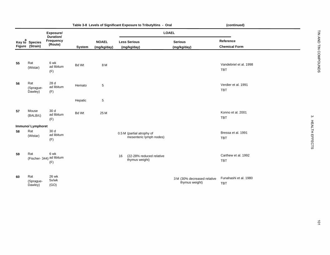
Table 3-8 Levels of Significant Exposure to Tributyltins - Oral (continued)
Exposure/ LOAEL
TIN
aKey to Figure
Species (Strain)
Duration/ Frequency
(Route) System
NOAEL (mg/kg/day)
AN
D TIN
CO
MP
OU
ND
S
Reference Chemical Form
Less Serious (mg/kg/day)
Serious (mg/kg/day)
55 Rat (Wistar)
6 wk ad libitum (F)
Bd Wt 8 M Vandebriel et al. 1998 TBT
56 Rat (Sprague-Dawley)
28 d ad libitum (F)
Hemato 5 Verdier et al. 1991 TBT
Hepatic 5
57 Mouse (BALB/c)
30 d ad libitum (F)
Bd Wt 25 M
3. H
Konno et al. 2001 TBT
Immuno/ Lymphoret 58 Rat
(Wistar) 30 d ad libitum (F)
0.5 M (partial atrophy of mesenteric lymph nodes)
EA
LTH E
FFEC
T
Bressa et al. 1991 TBT
59 Rat (Fischer- 344)
6 wk ad libitum (F)
16 (22-28% reduced relative thymus weight)
S
Carthew et al. 1992 TBT
60 Rat (Sprague-Dawley)
26 wk 5x/wk (GO)
3 M (30% decreased relative thymus weight)
101
Funahashi et al. 1980 TBT
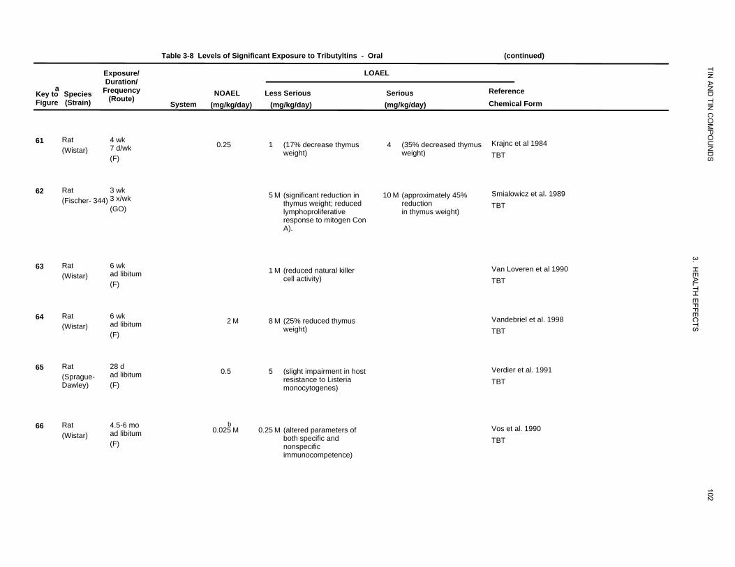
Table 3-8 Levels of Significant Exposure to Tributyltins - Oral (continued)
Exposure/ LOAEL
TIN
aKey to Figure
Species (Strain)
Duration/ Frequency
(Route) System
NOAEL (mg/kg/day)
AN
D TIN
CO
MP
OU
ND
S
Reference Chemical Form
Less Serious (mg/kg/day)
Serious (mg/kg/day)
61 Rat (Wistar)
4 wk 7 d/wk (F)
0.25 1 (17% decrease thymus weight)
4 (35% decreased thymus weight)
Krajnc et al 1984 TBT
62 Rat (Fischer- 344)
3 wk 3 x/wk (GO)
5 M (significant reduction in thymus weight; reduced lymphoproliferative response to mitogen Con A).
10 M (approximately 45% reduction in thymus weight)
Smialowicz et al. 1989 TBT
63 Rat (Wistar)
6 wk ad libitum (F)
1 M (reduced natural killer cell activity)
3. HE
ALTH
Van Loveren et al 1990 TBT
64 Rat (Wistar)
6 wk ad libitum (F)
2 M 8 M (25% reduced thymus weight)
EFFE
CTS
Vandebriel et al. 1998 TBT
65 Rat (Sprague-Dawley)
28 d ad libitum (F)
0.5 5 (slight impairment in host resistance to Listeria monocytogenes)
Verdier et al. 1991 TBT
66 Rat (Wistar)
4.5-6 mo ad libitum (F)
b0.025 M
0.25 M (altered parameters of both specific and nonspecific immunocompetence)
102
Vos et al. 1990 TBT
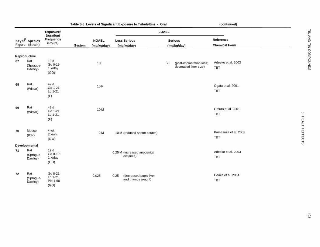
Table 3-8 Levels of Significant Exposure to Tributyltins - Oral (continued)
Exposure/ LOAEL
TIN
aKey to Species Figure (Strain)
Duration/ Frequency
(Route) System
NOAEL (mg/kg/day)
AN
D TIN
CO
MP
OU
ND
S
Reference Chemical Form
Less Serious (mg/kg/day)
Serious (mg/kg/day)
Reproductive 67 Rat
(Sprague-Dawley)
19 d Gd 0-19 1 x/day (GO)
10 20 (post-implantation loss; decreased litter size)
Adeeko et al. 2003 TBT
68 Rat (Wistar)
42 d Gd 1-21 Ld 1-21 (F)
10 F Ogata et al. 2001 TBT
69 Rat (Wistar)
42 d Gd 1-21 Ld 1-21 (F)
10 M
3. HE
ALTH
EFFE
CTS
Omura et al. 2001 TBT
70 Mouse (ICR)
4 wk 2 x/wk (GW)
2 M 10 M (reduced sperm counts) Kamasaka et al. 2002 TBT
Developmental 71 Rat
(Sprague-Dawley)
19 d Gd 0-19 1 x/day (GO)
0.25 M (increased anogenital distance)
Adeeko et al. 2003 TBT
72 Rat (Sprague-Dawley)
Gd 8-21 Ld 1-21 Pld 1-60 (GO)
0.025 0.25 (decreased pup's liver and thymus weight)
103
Cooke et al. 2004 TBT
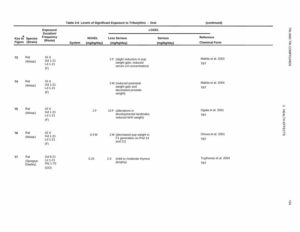
Table 3-8 Levels of Significant Exposure to Tributyltins - Oral (continued)
Exposure/ LOAEL
TIN
aKey to Species Figure (Strain)
Duration/ Frequency
(Route) System
NOAEL (mg/kg/day)
AN
D TIN
CO
MP
OU
ND
S
Reference Chemical Form
Less Serious (mg/kg/day)
Serious (mg/kg/day)
73 Rat (Wistar)
42 d Gd 1-21 Ld 1-21 (F)
2 F (slight reduction in pup weight gain; reduced serum LH concentration)
Makita et al. 2003 TBT
74 Rat (Wistar)
42 d Gd 1-21 Ld 1-21 (F)
2 M (reduced postnatal weight gain and decreased prostate weight)
Makita et al. 2004 TBT
75 Rat (Wistar)
42 d Gd 1-21 Ld 1-21 (F)
2 F 10 F (alterations in developmental landmaks; reduced birth weight)
3. HE
ALTH
EF
Ogata et al. 2001 TBT
76 Rat (Wistar)
42 d Gd 1-21 Ld 1-21 (F)
0.4 M 2 M (decreased pup weight in F1 generation on Pnd 14 and 21)
FEC
TSOmura et al. 2001 TBT
77 Rat (Sprague-Dawley)
Gd 8-21 Ld 1-21 Pld 1-70 (GO)
0.25 2.5 (mild to moderate thymus atrophy)
104
Tryphonas et al. 2004 TBT
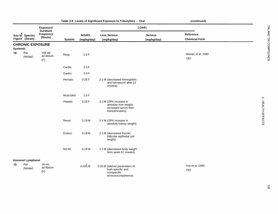
Table 3-8 Levels of Significant Exposure to Tributyltins - Oral (continued)
aKey to Species Figure (Strain)
Exposure/ Duration/
Frequency(Route)
System NOAEL
(mg/kg/day)
LOAEL
TIN A
ND
TIN C
OM
PO
UN
DS
Reference Chemical Form
Less Serious (mg/kg/day)
Serious (mg/kg/day)
CHRONIC EXPOSURE Systemic 78 Rat 106 wk
ad libitum(Wistar) (F)
Immuno/ Lymphoret 79 Rat 18 mo
ad libitum(Wistar) (F)
Resp
Cardio
Gastro
Hemato
Musc/skel
Hepatic
Renal
Endocr
Bd Wt
2.5 F
2.5 F
2.5 F
0.25 F
2.5 F
0.25 F
0.19 M
0.19 M
0.19 M
c0.025 M
2.1 M (decreased hemoglobin and hematocrit after 12 months)
2.1 M (29% increase in absolute liver weight; increased serum liver transaminases)
2.1 M (29% increase in absolute kidney weight)
2.1 M (decreased thyroid follicular epithelial cell height)
2.1 M (decreased body weight from week 67 onward)
0.25 M (altered parameters of both specific and nonspecific immunocompetence)
105
3. HE
ALTH
EFFE
CTS
Wester et al. 1990 TBT
Vos et al. 1990 TBT
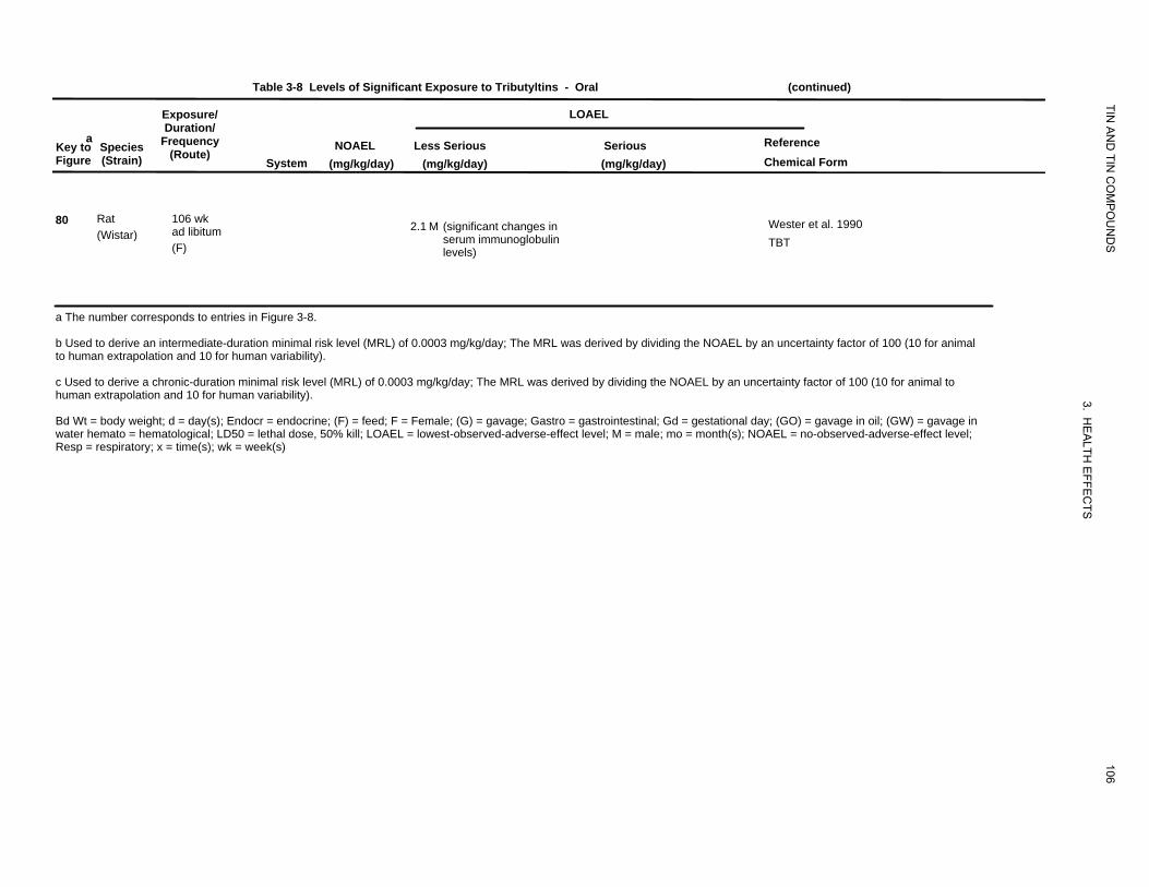
Table 3-8 Levels of Significant Exposure to Tributyltins - Oral (continued)
Exposure/ LOAEL
TIN
aKey to Figure
Species (Strain)
Duration/ Frequency
(Route) System
NOAEL (mg/kg/day)
AN
D TIN
CO
MP
OU
ND
S
Reference Chemical Form
Less Serious (mg/kg/day)
Serious (mg/kg/day)
80 Rat (Wistar)
106 wk ad libitum (F)
2.1 M (significant changes in serum immunoglobulin levels)
Wester et al. 1990 TBT
a The number corresponds to entries in Figure 3-8.
b Used to derive an intermediate-duration minimal risk level (MRL) of 0.0003 mg/kg/day; The MRL was derived by dividing the NOAEL by an uncertainty factor of 100 (10 for animal to human extrapolation and 10 for human variability).
c Used to derive a chronic-duration minimal risk level (MRL) of 0.0003 mg/kg/day; The MRL was derived by dividing the NOAEL by an uncertainty factor of 100 (10 for animal to
3human extrapolation and 10 for human variability).
. HBd Wt = body weight; d = day(s); Endocr = endocrine; (F) = feed; F = Female; (G) = gavage; Gastro = gastrointestinal; Gd = gestational day; (GO) = gavage in oil; (GW) = gavage in EAwater hemato = hematological; LD50 = lethal dose, 50% kill; LOAEL = lowest-observed-adverse-effect level; M = male; mo = month(s); NOAEL = no-observed-adverse-effect level;
106
LTH E
FFEC
TS
Resp = respiratory; x = time(s); wk = week(s)

Death Hematologica
l
Hepatic
Endocrine
Body WImmuno/L
Neurological
Reproductive
Developmental
1000
eight ymphor
Figure 3-8 Levels of Significant Exposure to Tributyltins - Oral Acute (≤14 days) TIN
AN
D TIN
CO
MP
OU
ND
S
3. HE
ALTH
EFFE
CTS
107
mg/kg/day Systemic
3r 5s 2r
15g100 17s 27r 38r
17s8r16g 13m 14m 9r
42m13m1r 22r 33m8r 18r17s4m 34m 45m26r 37r33m
12m 42m 43m 28r 40r29r
45m44m
10 6r 7r 25r 30r 31r 43m 35r 36r 28r 40r29r
21r 23r 11m 7r 20r 32m 30r 31r 36r
6r 6r 10r 19r 20r 24r 21r
10r 24r 19r
1 39r
6r
0.1
c-Cat -Humans f-Ferret n-Mink Cancer Effect Level-Animals Cancer Effect Level-Humans LD50/LC50d-Dog k-Monkey j-Pigeon o-Other LOAEL, More Serious-Animals LOAEL, More Serious-Humans Minimal Risk Levelr-Rat m-Mouse e-Gerbil LOAEL, Less Serious-Animals LOAEL, Less Serious-Humans for effectsp-Pig h-Rabbit s-Hamster NOAEL - Animals NOAEL - Humans other thanq-Cow a-Sheep g-Guinea Pig
Cancer
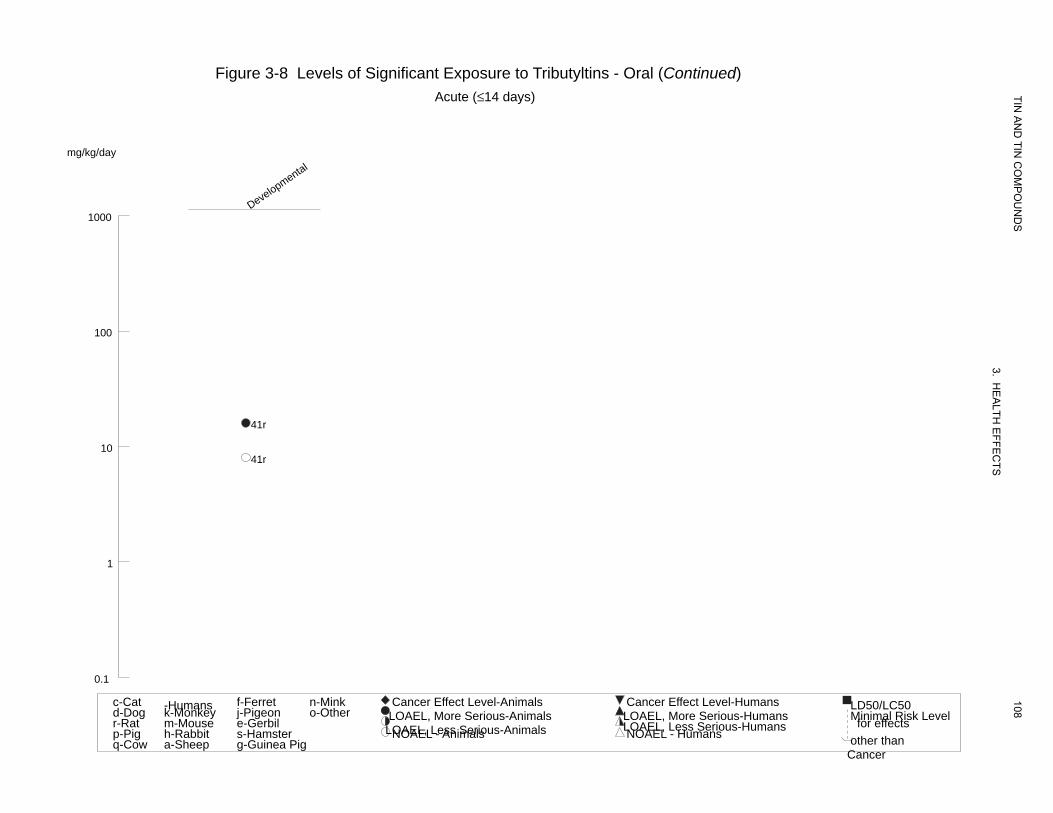
Developmental
Figure 3-8 Levels of Significant Exposure to Tributyltins - Oral (Continued) Acute (≤14 days)
mg/kg/day
1000
100
41r
10 41r
1
0.1
TIN A
ND
TIN C
OM
PO
UN
DS
3. HE
ALTH
EFFE
CTS
108c-Cat -Humans f-Ferret n-Mink Cancer Effect Level-Animals Cancer Effect Level-Humans LD50/LC50d-Dog k-Monkey j-Pigeon o-Other LOAEL, More Serious-Animals LOAEL, More Serious-Humans Minimal Risk Levelr-Rat m-Mouse e-Gerbil LOAEL, Less Serious-Animals LOAEL, Less Serious-Humans for effectsp-Pig h-Rabbit s-Hamster NOAEL - Animals NOAEL - Humans other thanq-Cow a-Sheep g-Guinea Pig
Cancer
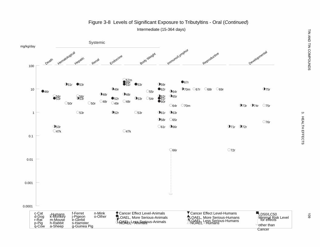
Death Hematologica
l
Hepatic
Renal Endocrin
e
Body WImmuno/L
Reproductive
Developmental
eight ymphor
Figure 3-8 Levels of Significant Exposure to Tributyltins - Oral (Continued) Intermediate (15-364 days) TIN
AN
D TIN
CO
MP
OU
ND
S
3. HE
ALTH
EFFE
CTS
109
Systemicmg/kg/day
100
57m49r 67r
51r 53r 51r 53r 59r 10 49r 62r 70m 67r 68r 69r 75r
46r 55r 64r 48r 48r56r 56r 62r 65r
53r 53r 52r 53r 54r 61r 48r 48r 60r50r 50r 49r
64r 70m 73r 74r 75r
1 53r 52r 53r 61r 63r
58r 65r 76r
53r 61r 66r 71r 72r 47k 47k
0.1
66r 72r
0.01
0.001
0.0001
c-Cat -Humans f-Ferret n-Mink Cancer Effect Level-Animals Cancer Effect Level-Humans LD50/LC50d-Dog k-Monkey j-Pigeon o-Other LOAEL, More Serious-Animals LOAEL, More Serious-Humans Minimal Risk Levelr-Rat m-Mouse e-Gerbil LOAEL, Less Serious-Animals LOAEL, Less Serious-Humans for effectsp-Pig h-Rabbit s-Hamster NOAEL - Animals NOAEL - Humans other thanq-Cow a-Sheep g-Guinea Pig
Cancer
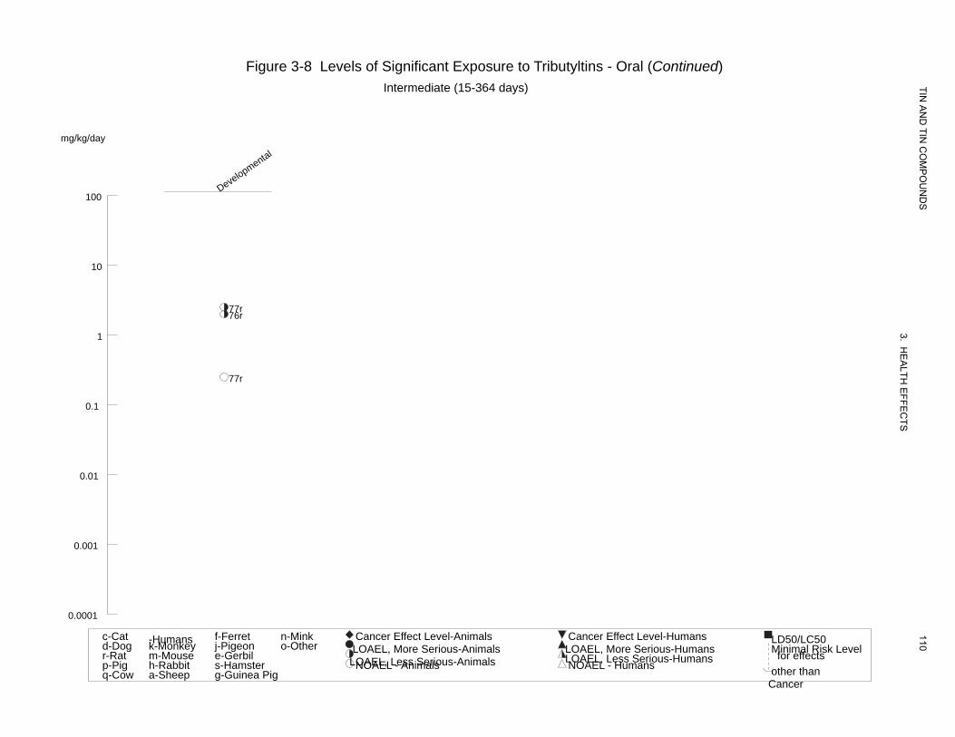
Developmental
Figure 3-8 Levels of Significant Exposure to Tributyltins - Oral (Continued) Intermediate (15-364 days)
mg/kg/day
100
10
77r76r
1
77r
0.1
0.01
0.001
0.0001
TIN A
ND
TIN C
OM
PO
UN
DS
3. HE
ALTH
EFFE
CTS
110c-Cat -Humans f-Ferret n-Mink Cancer Effect Level-Animals Cancer Effect Level-Humans LD50/LC50d-Dog k-Monkey j-Pigeon o-Other LOAEL, More Serious-Animals LOAEL, More Serious-Humans Minimal Risk Levelr-Rat m-Mouse e-Gerbil LOAEL, Less Serious-Animals LOAEL, Less Serious-Humans for effectsp-Pig h-Rabbit s-Hamster NOAEL - Animals NOAEL - Humans other thanq-Cow a-Sheep g-Guinea Pig
Cancer
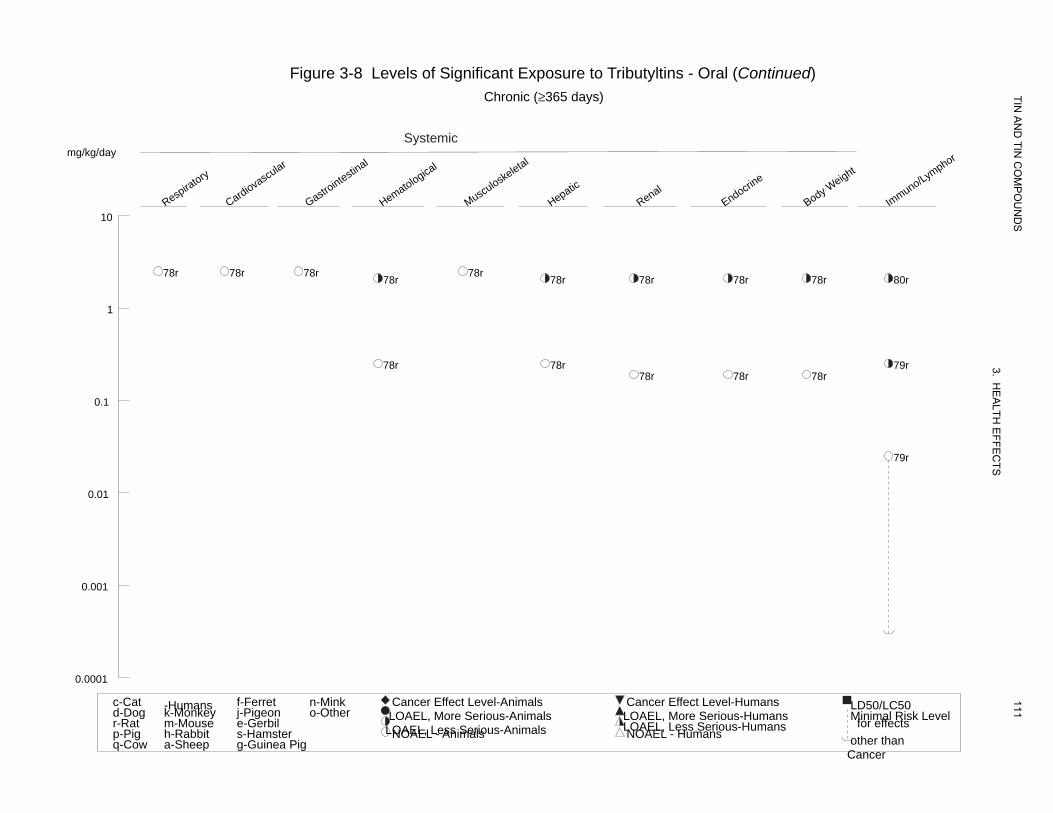
Respiratory
Cardiovascular
Gastrointestin
al
Hematological
Musculoske
letal
Hepatic
Renal Endocrin
e
Body WImmuno/Leight ym
phor
Figure 3-8 Levels of Significant Exposure to Tributyltins - Oral (Continued) Chronic (≥365 days)
Systemic mg/kg/day
TIN A
ND
TIN C
OM
PO
UN
DS
3. HE
ALTH
EFFE
CTS
111
10
78r 78r 78r 78r78r 78r 78r 78r 78r 80r
1
78r 78r 79r 78r 78r 78r
0.1
79r
0.01
0.001
0.0001
c-Cat -Humans f-Ferret n-Mink Cancer Effect Level-Animals Cancer Effect Level-Humans LD50/LC50d-Dog k-Monkey j-Pigeon o-Other LOAEL, More Serious-Animals LOAEL, More Serious-Humans Minimal Risk Levelr-Rat m-Mouse e-Gerbil LOAEL, Less Serious-Animals LOAEL, Less Serious-Humans for effectsp-Pig h-Rabbit s-Hamster NOAEL - Animals NOAEL - Humans other thanq-Cow a-Sheep g-Guinea Pig
Cancer
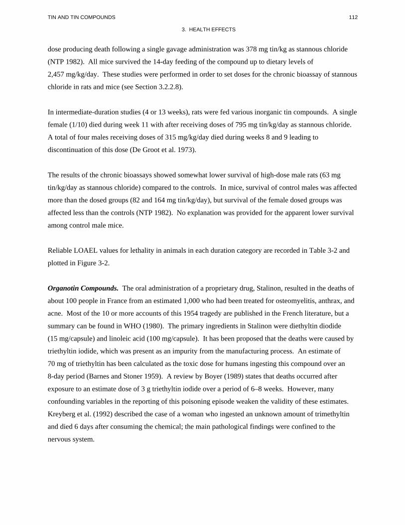
112 TIN AND TIN COMPOUNDS
3. HEALTH EFFECTS
dose producing death following a single gavage administration was 378 mg tin/kg as stannous chloride
(NTP 1982). All mice survived the 14-day feeding of the compound up to dietary levels of
2,457 mg/kg/day. These studies were performed in order to set doses for the chronic bioassay of stannous
chloride in rats and mice (see Section 3.2.2.8).
In intermediate-duration studies (4 or 13 weeks), rats were fed various inorganic tin compounds. A single
female (1/10) died during week 11 with after receiving doses of 795 mg tin/kg/day as stannous chloride.
A total of four males receiving doses of 315 mg/kg/day died during weeks 8 and 9 leading to
discontinuation of this dose (De Groot et al. 1973).
The results of the chronic bioassays showed somewhat lower survival of high-dose male rats (63 mg
tin/kg/day as stannous chloride) compared to the controls. In mice, survival of control males was affected
more than the dosed groups (82 and 164 mg tin/kg/day), but survival of the female dosed groups was
affected less than the controls (NTP 1982). No explanation was provided for the apparent lower survival
among control male mice.
Reliable LOAEL values for lethality in animals in each duration category are recorded in Table 3-2 and
plotted in Figure 3-2.
Organotin Compounds. The oral administration of a proprietary drug, Stalinon, resulted in the deaths of
about 100 people in France from an estimated 1,000 who had been treated for osteomyelitis, anthrax, and
acne. Most of the 10 or more accounts of this 1954 tragedy are published in the French literature, but a
summary can be found in WHO (1980). The primary ingredients in Stalinon were diethyltin diodide
(15 mg/capsule) and linoleic acid (100 mg/capsule). It has been proposed that the deaths were caused by
triethyltin iodide, which was present as an impurity from the manufacturing process. An estimate of
70 mg of triethyltin has been calculated as the toxic dose for humans ingesting this compound over an
8-day period (Barnes and Stoner 1959). A review by Boyer (1989) states that deaths occurred after
exposure to an estimate dose of 3 g triethyltin iodide over a period of 6–8 weeks. However, many
confounding variables in the reporting of this poisoning episode weaken the validity of these estimates.
Kreyberg et al. (1992) described the case of a woman who ingested an unknown amount of trimethyltin
and died 6 days after consuming the chemical; the main pathological findings were confined to the
nervous system.
113 TIN AND TIN COMPOUNDS
3. HEALTH EFFECTS
Lethal doses for monoorganotins ranging from 1,500 to more than 6,000 mg/kg have been reported for
rodents suggesting that these compounds have relatively low toxicity (Pelikan and Cerny 1970).
Acute-duration studies with dibutyltins have described lethal doses in rats between 20 and 50 mg/kg
administered by gavage (Alam et al. 1993; Barnes and Magee 1958). In a 2-week dietary study, a dose of
approximately 23 mg/kg/day was lethal to 6 out of 20 rats during the second week of the study (Seinen et
al. 1977a). In a developmental study, daily gavage doses of 7.5 mg/kg/day administered on gestation
days (Gds) 7–15 killed 5 out of 12 rats with a mean time of 8 days (Ema et al. 1991b). Long-term
treatment (78-week study) with dibutyltin diacetate significantly decreased survival in rats and mice at the
termination of the study (NCI 1978a). A dose of 7 mg dioctyltin dichloride/kg/day in the food was lethal
to 10 out of 16 guinea pigs after 4–5 weeks of treatment (Seinen et al. 1977b).
In male albino rats, the oral LD50 for tributyltin oxide was 148 mg/kg when the chemical was
administered by gavage in corn oil and 194 mg/kg when administered as an aqueous suspension (Elsea
and Paynter 1958). Three consecutive daily doses of 37.5 mg of tributyltin oxide/kg killed 6 out of
50 rats (Elsabbagh et al. 2002). In pregnant mice, a dose of 27 mg/kg of tributyltin oxide administered
during gestation was lethal to 3 out of 40 mice; no deaths occurred in controls (Faqi et al. 1997). Takagi
et al. (1992) calculated a 2-week LD50 of approximately 150 mg/kg for tributyltin chloride in male
hamsters and 172 mg/kg in females.
Numerous studies provide information on the lethal effects of trimethyltins. In general, lethal doses,
mostly in acute-duration studies, are below 10 mg/kg. For example, Brown et al. (1979) calculated an
oral LD50 of 12.6 mg/kg in rats following a single gavage dose; most deaths occurred 2–5 days after
dosing. Other studies have reported lethal single doses in rats of 5 mg/kg (Bouldin et al. 1981) and
7 mg/kg (Alessandri et al. 1994). In a 2-week dietary study, doses of 2 mg/kg/day were lethal to 2 out of
10 rats and doses of ≥6.7 mg/kg/day killed 10/10 rats in a few days (Snoeij et al. 1985). Three female
hamsters that received a single dose of 4 or 5 mg/kg showed whole body tremors and were almost
moribund when killed 4 days after dosing (Brown et al. 1984). In the same study, a single dose of
3.75 mg/kg of trimethyltin chloride killed 4 out of 11 marmoset monkeys within 3 days of dosing (Brown
et al. 1984). Single doses of 3–4 mg/kg were lethal to gerbils within a few days of treatment (Brown et al.
1984; Nolan et al. 1990).
Triethyltins are also highly toxic. In a 2-week study, doses of 6.7 mg/kg/day in the diet were lethal to
3 out of 10 rats, but no lethality occurred with doses of 2 mg/kg/day (Snoeij et al. 1985). In an additional
114 TIN AND TIN COMPOUNDS
3. HEALTH EFFECTS
2-week study, rats were gavaged with triethyltin bromide in water twice/week and a dose of 3 mg/kg
killed 4 out of 10 rats after the third dose (Squibb et al. 1980). Four out of six rats died during the third
week on a diet that provided 2 mg/kg/day of triethyltin hydroxide (Magee et al. 1957) and in an 11-week
drinking water study with triethyltin sulfate in rats at dose levels of 1.4 mg/kg/day; deaths occurred after
4 weeks (Smith 1973).
An extensive listing of LD50 values for organotin compounds in several animal species can be found in
Smith (1978) and WHO (1980).
Reliable LOAEL values for lethality, and LD50 values in each species and duration category are recorded
in Tables 3-3 through 3-8 and plotted in Figures 3-3 through 3-8.
3.2.2.2 Systemic Effects
No studies were located regarding musculoskeletal, or ocular effects in humans or animals after oral
exposure to inorganic tin or organotin compounds.
The highest NOAEL values and all reliable LOAEL values for systemic effects in each species and
duration category are recorded in Table 3-2 and plotted in Figure 3-2 for inorganic tin compounds.
Similar information is given in Tables 3-3 through 3-8 and Figures 3-3 through 3-8 for organotin
compounds.
Respiratory Effects.
Inorganic Tin Compounds. No studies were located regarding respiratory effects in humans or in
animals after oral exposure to inorganic tin compounds.
Organotin Compounds. No histopathological alterations were observed in the lungs, bronchi, and
trachea from rats and mice fed diets that provided up to 6.7 and 19.8 mg/kg/day of dibutyltin diacetate,
respectively, for 78 weeks (NCI 1978a) or up to 3.8 and 9.8 mg/kg/day, respectively, of triphenyltin
hydroxide for 78 weeks (NCI 1978b). Beagle dogs dosed with up to 0.62 mg triphenyltin
hydroxide/kg/day for up to 52 weeks did not show any gross or microscopic alterations in the respiratory
tract (Sachsse et al. 1987). Similar results were reported for rats dosed with up to 2.5 mg/kg/day of
tributyltin oxide for 106 weeks (Wester et al. 1990). In a 6-week dietary study with dioctyltin dichloride
115 TIN AND TIN COMPOUNDS
3. HEALTH EFFECTS
in rats, Seinen and Willems (1976) reported that rats dosed with approximately 16 mg/kg/day had grayish
areas in the lungs at termination, suggesting chronic respiratory disease, and that three rats that died early
in the study showed severe pneumonic alterations. No further relevant information was located.
Cardiovascular Effects.
Inorganic Tin Compounds. No studies were located regarding cardiovascular effects in humans after
oral exposure to inorganic tin compounds.
In a feeding study in rats, at dietary levels ranging from <10 to 315 mg/kg/day as stannous chloride for
13 weeks, relative heart weights of males were higher than those of controls (De Groot et al. 1973). This
effect was not dose-dependent and there were no associated histopathological findings. By itself, the
significance of the observation is not clear. In a 4-week exposure to the same doses, there were no
changes in heart weights (De Groot et al. 1973).
Organotin Compounds. No studies were located regarding cardiovascular effects in humans after oral
exposure to organotin compounds.
No gross or microscopic alterations were observed in the heart of rats dosed with up to 5.7 mg dibutyltin
dichloride/kg/day for 90 days (Gaunt et al. 1968). In a chronic-duration study, no histopathological
alterations were observed in the hearts of rats and mice fed diets that provided up to 6.7 and
19.8 mg/kg/day of dibutyltin diacetate, respectively, for 78 weeks (NCI 1978a). Similar findings
regarding the heart were reported in chronic-duration studies with triphenyltin hydroxide in rats and mice
dosed with up to 6.2 and 20 mg/kg/day, respectively, for 78–106 weeks (NCI 1978b; Tennekes et al.
1989a, 1989b). Also, no cardiovascular effects were reported in dogs administered up to 0.62 mg
triphenyltin hydroxide/kg/day for up to 52 weeks (Sachsse et al. 1987).
Dietary treatment of rats with up to 16 mg dioctyltin dichloride/kg/day for 6 weeks (Seinen and Willems
1976) or up to 2.5 mg tributyltin oxide/kg/day for 106 weeks (Wester et al. 1990) did not induce gross or
microscopic alterations in the heart.
116 TIN AND TIN COMPOUNDS
3. HEALTH EFFECTS
Gastrointestinal Effects.
Inorganic Tin Compounds. There are several accounts of people who experienced gastrointestinal
effects such as diarrhea, gastrointestinal pain, nausea, or gastroenteritis after ingestion of various foods
stored in tin cans (WHO 1980, 2003). Doses ranged from 250 to 1,000 mg tin/kg body weight. Recent
studies by Boogaard et al. (2003) showed that tin levels up to approximately 270 ppm in canned food
caused no adverse effects in healthy humans.
Data from studies in animals show that inorganic tin compounds can cause adverse gastrointestinal
effects. Slightly distended small and large intestines were observed at necropsy of rats fed for 4 weeks
diets containing 315–325 mg tin/kg/day as either stannous chloride or stannous orthophosphate.
However, there were no histopathological changes (De Groot et al. 1973). In a 13-week study by the
same investigators, doses of ≥95 mg Sn/kg/day as stannous chloride caused abdominal distension in rats
during the first 2 weeks of the study, doses of 32 mg/kg/day caused no significant effects. Rats dosed
with 315 mg/kg/day, which had to be terminated prematurely, showed distended intestines and slight
ascites.
In another study, effects on the morphology and on absolute and relative weights of the gastrointestinal
tract were evaluated after feeding rats dietary levels of 7.9 and 15.9 mg tin/kg/day stannous chloride for
4 weeks. Feed restriction was also studied in an attempt to distinguish between tin effects and the effects
of decreased food intake and poor growth (Janssen et al. 1985). Increased relative weights of the
stomach, cecum, and colon were observed at the lowest tin dose, but were apparently caused by
diminished food intake since these changes were present in the pair fed controls as well as in the tin
exposed animals. On the other hand, increases in the weight and length of the small intestines were
observed to be independent of food consumption and thus a consequence of the exposure to stannous
chloride. There was also an increase in the villus length, a decrease in the number of villi per unit
surface, an increase in villus cell turnover, and changes in villi morphology in the intestines of the treated
rats. Although similar changes of the intestinal villi were reported in another study (Dreef-van der
Meulen et al. 1974), there are not enough data at this time to verify the intestinal changes as adverse.
Mice fed 311–2,457 mg tin/kg/day as stannous chloride for 13 weeks showed gross distention of the
cecum and reddened gastric mucosa at necropsy but no compound-related histopathological changes
(NTP 1982). Similar findings were observed in rats fed 120–236 mg tin/kg/day (NTP 1982). However,
117 TIN AND TIN COMPOUNDS
3. HEALTH EFFECTS
no such changes were observed in rats fed 32 or 63 mg tin/kg/day or mice fed 82 or 164 mg tin/kg/day as
stannous chloride during a 105-week study (NTP 1982).
Organotin Compounds. Limited information is available regarding gastrointestinal effects in humans
after oral exposure to organotin compounds. Nausea and vomiting were reported in more than 70% of the
individuals intoxicated presumably with triethyltin in a massive accidental poisoning episode in France in
1954 (WHO 1980). Abdominal pain, diarrhea, nausea, and vomiting have been reported in cases of oral
intoxication with triphenyltin (Lin and Hsueh 1993; Lin et al. 1998; Wu et al. 1990).
Stomach distention was observed in rats 24 hours after a single dose of 50 mg dibutyltin dichloride/kg
(Barnes and Magee 1958). The duodenum was also examined in many rats, but no changes were evident.
Chronic studies with dibutyltin diacetate in rats and mice did not report any significant alterations in the
gastrointestinal tract from rats or mice dosed with up to 6.7 and 19.8 mg/kg/day of the test material,
respectively, for 78 weeks (NCI 1978a).
Histopathological evaluation of the gastrointestinal tract (at six different levels) from rats dosed with up
to approximately 16 mg dioctyltin dichloride/kg/day did not reveal any significant alterations (Seinen and
Willems 1976).
Single doses of 500 mg/kg of various tributyltin salts (chloride, acetate, benzoate, oleate) produced
hemorrhages in the digestive tract of mice (Pelikan and Cerny 1968). Similar gross changes in the
gastrointestinal tract were seen in another study in mice treated with much higher doses (4,000 mg/kg) of
monobutyltin trichloride and other monobutyltin salts (Pelikan and Cerny 1970). No gastrointestinal
alterations were observed in rats treated with up to 2.5 mg tributyltin oxide/kg/day for 106 weeks (Wester
et al. 1990).
No treatment-related alterations in the gastrointestinal tract were reported in rats, mice, and dogs in
chronic-duration studies with triphenyltin hydroxide (NCI 1978b; Sachsse et al. 1987; Tennekes et al.
1989a, 1989b). Rats were dosed with up to 6.2 mg/kg/day and mice were dosed with up to 20 mg/kg/day;
the dogs were dosed with up to 0.62 mg/kg/day.
118 TIN AND TIN COMPOUNDS
3. HEALTH EFFECTS
Hematological Effects.
Inorganic Tin Compounds. No studies were located regarding hematological effects in humans after oral
exposure to inorganic tin compounds.
Data from 4-week feeding studies in rats showed some hematological changes (De Groot et al. 1973). A
significant increase was observed in the hematocrit of male, but not in female, rats fed a dietary level of
395 mg tin/kg/day as stannous sulfide. Both sexes of rats fed tin at dietary levels ranging from 68 to
325 mg tin/kg/day as the chloride, orthophosphate, sulfate, oxalate, and tartrate showed anemia. The
signs of anemia were decreased hematocrit, total erythrocytes, and hemoglobin levels. Lower mean
corpuscular volume and hemoglobin concentrations were seen at the highest doses (225–325 mg
tin/kg/day). In 13-week studies, stannic oxide produced no hematological changes in rats (De Groot et al.
1973). However, dietary levels of ≥7.9 mg tin/kg/ as stannous chloride produced decreased
hematological values in rats with 4-week exposures (Janssen et al. 1985). It is possible that the mineral
content of the diet had an effect on the results of these studies since the no effect levels (22–440 mg
Sn/kg/day) for hematological effects in studies with diets adequate in copper and iron (De Groot et al.
1973; Dreef-van der Meulen et al. 1974) exceeded the LOAEL (7.9 mg/kg/day) from the work by Janssen
et al. (1985) with diets that contained only one fifth as much iron and copper. Iron and copper are key
nutrients in hematopoiesis; deficiencies in these elements are associated with microcytic anemias
characterized by low hemoglobin and hematocrit values. It is suggested that the poor iron and copper
nutrition in the Janssen et al. (1985) work was a predisposing factor, which amplified the adverse effects
of tin on hematological parameters. This hypothesis is supported by studies in which the dietary
concentrations of copper, tin, and iron were varied (De Groot 1973). High levels of copper and iron (well
above dietary requirements) added to semipurified diets containing up to 75 mg/kg/day tin almost
completely prevented hematological changes. Transient hemolytic anemia also was reported in rabbits
treated daily by gavage with 10 mg tin/kg (as stannous chloride), the only dose tested, for 4 months
(Chmielnicka et al. 1993). However, no information was provided in that study regarding the trace
mineral composition of the diet. The NOAEL of 32 mg/kg/day for tin, as stannous chloride, in the
13-week study by De Groot et al. (1973) was used to derive an intermediate-duration oral MRL for
inorganic tin.
Organotin Compounds. No studies were located regarding hematological effects in humans after oral
exposure to organotin compounds.
119 TIN AND TIN COMPOUNDS
3. HEALTH EFFECTS
Treatment of Fischer-344 rats with 5–6 mg dibutyltin dichloride/kg/day in the diet for up to 13 weeks
produced a slight, but significant decrease in hemoglobin concentration in females after 6 weeks and in
males after 13 weeks (Gaunt et al. 1968). This decrease was not associated with reductions of other
erythrocyte parameters or with a reticulocytosis. Lower doses of approximately 3 mg/kg/day caused no
significant effect. Differential leukocyte counts were not altered by treatment with dibutyltin dichloride.
Decreased hemoglobin and hematocrit values, lowered mean corpuscular volume and hemoglobin mass,
and decreased leucocytes were observed in rats fed a diet that provided approximately 4 and
16 mg/kg/day tributyltin oxide for 4 weeks (Krajnc et al. 1984). Erythrocytes were reduced, and
spherocytes and Howell-Jolly body-containing erythrocytes were increased in the 16 mg/kg/day group
only. However, a study in Sprague-Dawley rats found no significant alterations in a complete set of
hematological parameters following treatment with approximately 5 mg tributyltin oxide/kg/day for
28 days (Verdier et al. 1991). The reason for the discrepancy between these two studies is unknown. In a
2-year bioassay in Wistar rats, hematological determinations were made in weeks 13 and 53, and at
termination (Wester et al. 1990). Significant hematological effects restricted to the high-dose males
(dosed with approximately 2.1 mg/kg/day) were seen only at 12 months, and consisted of decreased
hemoglobin, hematocrit, and mean corpuscular hemoglobin levels, and mean corpuscular volume (also at
3 months. In females, there was an indication of increased young erythrocytes at 3 and 12 months, but the
doses were not indicated. Leucocytes were decreased in high-dose males (24 months) and females
(12 and 24 months). In a 22-week gavage study in Cynomolgus monkeys, treatment with doses of
tributyltin oxide of 0.16 mg/kg/day (only dose level tested) decreased total leukocytes in weeks 8–10 at
weeks 16–20 (Kerrer et al. 1992). The biological significance of this finding is unknown.
In a 6-week dietary study with dioctyltin dichloride in male and female Wistar rats, hematological
investigations conducted on blood collected at termination included hemoglobin concentration and total
and differential leukocyte counts (Seinen and Willems 1976). Doses of approximately 16 mg/kg/day
significantly decreased hemoglobin concentration in males, but there was no effect on total or differential
leukocyte counts; the NOAEL for hemoglobin concentration was approximately 5.3 mg/kg/day. A study
in female Balb/c mice treated by gavage with 500 mg dioctyltin dichloride/kg once per week for 8 weeks
reported a 14% reduction in hemoglobin concentration at termination but no significant alterations in red
or white blood cell counts (Miller et al. 1986). A dose of 100 mg/kg had no significant effect on
hemoglobin concentration.
120 TIN AND TIN COMPOUNDS
3. HEALTH EFFECTS
Triphenyltin hydroxide at a dose of 1.3 mg/kg/day caused a transient decrease in hemoglobin and
hematocrit values at 26 and 52 weeks in female rats, but not in the males (Tennekes et al. 1989b). These
changes were not apparent at 78 and 104 weeks (Tennekes et al. 1989b), nor were they seen in dogs given
the same compound at doses of approximately 0.62 mg/kg/day for up to 42 weeks (Sachsse et al. 1987).
Musculoskeletal Effects.
Inorganic Tin Compounds. No studies were located regarding musculoskeletal effects in humans or
animals following oral exposure to inorganic tin compounds.
Organotin Compounds. No studies were located regarding musculoskeletal effects in humans following
oral exposure to organotin compounds. Treatment of rats with up to 16 mg dioctyltin dichloride/kg/day
for 6 weeks did not induce histopathological alterations in skeletal muscle (Seinen and Willems 1976).
No treatment-related alterations in skeletal muscles were observed in a 104-week study in rats dosed with
up to 6.2 mg triphenyltin hydroxide/kg/day or in mice dosed with up to 9.8 mg triphenyltin
hydroxide/kg/day (Tennekes et al. 1989a, 1989b). Beagle dogs dosed with up to 0.62 mg triphenyltin
hydroxide/kg/day for up to 52 weeks showed no gross or microscopic alterations in skeletal muscle or in
the sternum bone (Sachsse et al. 1987). Similar findings were reported in a 106-week study with
tributyltin oxide in rats dosed with to 2.5 mg/kg/day of the chemical (Wester et al. 1990).
Hepatic Effects.
Inorganic Tin Compounds. No studies were located regarding hepatic effects in humans after oral
exposure to inorganic tin compounds.
Hepatic effects have been observed following intermediate and chronic oral exposure of rats. Data from a
4-week feeding study in Wistar rats showed some histopathological changes (De Groot et al. 1973). Both
sexes fed tin as the chloride, orthophosphate, sulfate, oxalate, and tartrate had histopathological changes
in the liver. The cytoplasm exhibited a clear homogeneous appearance, which suggested a disappearance
of the cellular organelles and impaired cell function at the highest dietary level of 226–325 mg/kg/day and
to a lesser extent at a level of 68–98 mg tin/kg/day (the doses varied with the tin compound used). A
slight but definite oval cell type hyperplasia of the bile ducts was also apparent. Changes in organ
weights were inconsistent. The authors suggested that the changes in liver cell morphology were due, in
121 TIN AND TIN COMPOUNDS
3. HEALTH EFFECTS
part, to the reduced food intake and resultant impaired weight gain. These changes were apparent in the
animals with the poorest weight gains.
In a 13-week study in Wistar rats, histopathological changes were observed in the livers of both sexes at a
dietary level of 315 mg tin/kg/day as stannous chloride, but in only a few rats at 95 mg tin/kg/day
(De Groot et al. 1973). The changes were a homogeneous appearance of the cell cytoplasm and mild
proliferation of the bile duct epithelium. Organ weights were not affected. In another 13-week study,
similar changes were seen in the livers of rats fed a diet that was gradually increased to a final level of
252 mg tin/kg/day as stannous chloride (Dreef-van der Meulen et al. 1974).
No hepatic effects were reported in Fischer-344 rats and B6C3F1/N mice fed stannous chloride for either
14 days or 13 weeks (NTP 1982). The highest dietary levels were 236 mg tin/kg/day as stannous chloride
for rats and 2,457 mg tin/kg/day for mice. Considering the extremely high doses used, it is surprising that
hepatic changes were not observed.
Limited hepatic changes were seen following chronic oral exposure of rats and mice to stannous chloride.
In a drinking water study at 0.7 mg tin/kg/day as stannous chloride for life, 80 rats were evaluated for
hepatic and other health effects (Schroeder et al. 1968). There was a significant increase in fatty
degeneration of the liver in the tin-exposed rats. Thirty-eight percent of the control rats had liver lesions,
whereas 68% of the tin-exposed rats had liver lesions. Degeneration and necrosis, as well as fatty
changes moderate to severe, were found in 55% of the control rats with lesions and in 65% of the tin-
exposed rats with lesions. Although similar hepatic effects were reported in the 105-week chronic
bioassay of stannous chloride in rats and mice, the findings were not dose-related and comparable in
treated and control animals (NTP 1982).
Organotin Compounds. Liver impairment, as judged by increased serum levels of transaminases, was
described in two cases of acute oral intoxication with triphenyltin (Lin et al. 1998; Wu et al. 1990).
Hepatitis also was reported in three subjects who ingested between 20 and 50 grams of a preparation
containing 45% triphenyltin acetate (Lin and Hsueh 1993). No further relevant information was located.
Hepatic and bile duct effects were observed following acute and intermediate oral exposures of animals to
organotin compounds. A single dose of dibutyltin dichloride of 50 mg/kg produced inflammation of the
common bile duct of Wistar rats (Barnes and Magee 1958). Severe hepatic injury occurred in rats
following three consecutive doses of 50 mg dibutyltin dichloride/kg/day; this treatment was lethal to
122 TIN AND TIN COMPOUNDS
3. HEALTH EFFECTS
some rats in 6–10 days. The main features of the bile-duct injury included thickening, inflammation, and
dilatation of the proximal duct. Histologically, the epithelium of the wall was replaced by granulomatous
tissue. In cases in which the bile duct was perforated, severe peritonitis and fatty necrosis were seen.
Multiple yellow infarcts developed in lobes of the liver, followed by inflammation of the portal blood
vessels. In some cases, there was complete necrosis of the bile ducts. In rats examined 6–12 months after
receiving the three doses of dibutyltin, the bile duct was shorter and thicker than normal and there was
wall fibrosis in the adjacent pancreas, and in the portal tracts of the liver. Seinen et al. (1977a) noticed
proliferation of the bile duct epithelium and periportal fibrosis in Wistar rats fed a diet that provided
approximately 23 mg/kg/day of dibutyltin dichloride for 2 weeks; doses of 7.7 mg/kg/day caused no
significant effect. Bile duct necrosis also was seen in Syrian hamsters treated with a single dose of
30 mg/kg (Jang et al. 1986). Bile duct necrosis also occurred in mice, but not in rabbits or guinea pigs
(20–50 mg/kg dose ranges) (Barnes and Magee 1958). No adverse hepatic effects (histopathology and
serum transaminases) were reported in Fischer-344 rats dosed with up to 5.7 mg dibutyltin
dichloride/kg/day for 90 days (Gaunt et al. 1968). In variance with the findings of bile duct necrosis in
mice reported by Barnes and Magee (1958), Seinen et al. (1977a) did not observe histological changes in
the liver from Swiss mice dosed with dibutyltin dichloride in doses of up to 30 mg/kg/day for 4 weeks;
however, it is unclear whether the bile duct was examined. No microscopic alterations were reported in
the liver from Fischer-344 rats or B6C3F1 mice treated with dibutyltin diacetate in doses of up to 6.7 and
20 mg/kg/day, respectively, for 78 weeks (NCI 1978a).
A significant increase in serum levels of ornithine carbamyl transferase (used as index of hepatotoxicity)
was observed in albino mice gavaged once with 58 mg tributyltin chloride/kg (Ueno et al. 1994). The
increase was first apparent 24 hours after dosing. Parallel experiments with dibutyltin dichloride and
monobutyltin trichloride showed that the hepatotoxicity potency followed the order: dibutyltin >
tributyltin > monobutyltin (Ueno et al. 1994). Monobutyltin was not hepatotoxic. Further studies by the
same group of investigators showed that the liver toxicity of tributyltin chloride could be prevented by
pretreatment of the mice with the cytochrome P-450 inhibitor SKF-525 (Ueno et al. 1995, 1997).
Conversely, pretreatment with the P-450 inducer phenobarbital (PB) increased the toxicity of tributyltin
chloride. These results suggest that the liver toxicity of tributyltin is due to a metabolite, most likely
dibutyltin. Comparative studies with tributyltin and dibutyltin in mice and guinea pigs showed the mice
to be much more sensitive to the hepatotoxicity of tri- and dibutyltin dichloride than guinea pigs (Ueno et
al. 2003a), and this was correlated with differential inhibition of mitochondrial respiration in the two
species. Additional experiments suggested that the difference in susceptibility between mice and guinea
pigs might be due to the high affinity of butyltins, particularly dibutyltin, for hepatic mitochondria in
123 TIN AND TIN COMPOUNDS
3. HEALTH EFFECTS
mice containing higher levels of sulfhydril groups relative to guinea pigs. In a three species comparison,
the susceptibilities followed the order: mice > rats > guinea pigs (Ueno et al. 2003b). Kinetic studies
showed that the differences in susceptibility appeared to be partly due to differences in metabolism of
tributyltin and in the distribution of dibutyltin within cell organelles. In hepatocytes of mice treated with
dibutyltin chloride, 41% of the dibutyltin was found in organelles compared to 27% in rats and 9% in
guinea pigs (Ueno et al. 2003b).
A single dose of 29.6 mg tributyltin chloride/kg (lowest dose tested) induced bile duct changes in Syrian
hamsters consisting of adhesion in the liver, pancreas, and duodenum, and severe inflammation (Takagi et
al. 1992). In a separate experiment, following a single dose of 44.4 mg/kg of tributyltin, the maximum
concentrations of tributyltin and dibutyltin appeared in the liver 1 day after treatment and rapidly
decreased thereafter. The concentration of dibutyltin was 10 times higher than that of tributyltin 1 day
after dosing (Takagi et al. 1992). By day 14, neither compound could be detected in the liver, and
aspartate aminotransferase (AST), alanine aminotransferase (ALT), gamma-guanosine triphosphate
(GTP), and alkaline phosphatase activities were not significantly different than control. These results
suggest that dibutyltin has an important role in the hepatotoxicity of tributyltin. In a 4-week feeding
study in Wistar rats, doses of approximately 16 mg tributyltin oxide/kg/day induced hepatic changes
consisting of liver necrosis and bile duct hyperplasia (Krajnc et al. 1984). Slight atrophy of the
hepatocytes was seen at 4 mg/kg/day and no significant alterations were seen at 1 mg/kg/day. Consistent
with these observations, no microscopic changes were observed in the livers of Sprague-Dawley rats
treated with ≤5 mg tributyltin oxide/kg/day for 28 days (Verdier et al. 1991). Cholangitis with biliary
retention was reported in Fischer-344 rats dosed with tributyltin oxide at 16 mg/kg/day for 6 weeks
(Carthew et al. 1992). In a 2-year bioassay with tributyltin oxide in Wistar rats, liver effects were
restricted to high-dose rats (2.1 mg/kg/day) and consisted of slight bile duct changes (hyperplasia, cellular
hypertrophy, minimal mononuclear cell infiltration) observed at 12 months, increased serum AST and
ALT activities at 24 months, and an approximate 30% increase in absolute liver weight at termination; no
histopathologic alterations were seen at 24 months (Wester et al. 1990). The NOAEL was
0.25 mg/kg/day.
No hepatotoxicity was seen in dogs exposed through the diet to up to 0.62 mg triphenyltin
hydroxide/kg/day for up to 52 weeks (Sachsse et al. 1987). Hypertrophy of the smooth endoplasmic
reticulum was reported in New Zealand rabbits exposed to approximately 17.4 mg triphenyltin
acetate/kg/day via the diet for 70 days (Dacasto et al. 1994a). A dose-related trend towards portal
sclerosis and bile duct proliferation was observed in Wistar rats given doses of 0.3–6.2 mg/kg/day
124 TIN AND TIN COMPOUNDS
3. HEALTH EFFECTS
triphenyltin hydroxide for 52 and 104 weeks; there was no corresponding increase in liver weight
(Tennekes et al. 1989b). The dose-related trend was stronger in females (p<0.0005) than in males
(p<0.005). However, no liver pathology was reported in Fischer-344 rats dose with the same compound
in doses of up to 3.8 mg/kg/day for 78 weeks (NCI 1978b). In NMRI mice, this same compound was
associated with a 35–40% increase in relative liver weight and nodular hyperplasia at doses of
15.2 mg/kg/day for males and 20.2 mg/kg/day for females but not at lower doses (Tennekes et al. 1989a).
No significant liver alterations were reported in B6C3F1 mice in the 78-week bioassay with triphenyltin
hydroxide (NCI 1978b).
Studies with dioctyltin dichloride showed no significant histopathologic alterations in the livers from rats
treated in the diet with doses of approximately 23 mg/kg/day for 2 weeks (Seinen et al. 1977a) or
16 mg/kg/day for 6 weeks (Seinen and Willems 1976), or in guinea pigs treated with 8 mg/kg/day for
4 weeks (Seinen et al. 1977a). No significant changes in liver weight were reported in mice gavaged with
up to 500 mg/kg/day once per week for 8 weeks, but no other liver end points were evaluated (Miller et
al. 1986).
Renal Effects.
Inorganic Tin Compounds. No studies were located regarding renal effects in humans after oral
exposure to inorganic tin compounds.
Histopathological changes in the kidneys were reported in Wistar rats that received dietary levels up to
approximately 315 mg tin/kg/day as stannous chloride for 13 weeks (De Groot et al. 1973). The changes
included large protein-like droplets in renal tubular epithelial cells. This appears to be a common finding
in the strain of rats used in this study and did not appear to be related to tin exposure. The authors also
mentioned the absence of calcareous deposits in the high-dose level female rats. This appears to be an
unusual finding since these deposits are commonly seen with the species of rats used in the study.
However, the toxicological significance of these kidney findings is not clear.
In another 13-week study, Wistar rats that were fed the compound up to a maximum level of 252 mg
tin/kg/day as stannous chloride showed increased relative kidney weights (Dreef-van der Meulen et al.
1974). The protein-like droplets and calcareous deposits, which are common in the rat strains used, were
present in the controls but were absent in the tin-fed animals. The absence of calcareous deposits in the
females confirms the observations of De Groot et al. (1973), but the relevance of these finding to
125 TIN AND TIN COMPOUNDS
3. HEALTH EFFECTS
compound toxicity is unclear. The organ weight change itself, in the absence of histopathological or other
effects, is usually not considered a toxic effect.
Renal changes have been evaluated following chronic oral exposure of rats and mice to stannous chloride
and the studies were described under Hepatic Effects. Vacuolar changes in the proximal convoluted
tubules of the kidney were significantly increased in rats administered stannous chloride, compared with
controls (Schroeder et al. 1968). However, in 14-day, 13-week, and 105-week studies of stannous
chloride in rats and mice, no treatment-related nonneoplastic renal changes were reported (NTP 1982).
Organotin Compounds. Acute nephropathy was reported in three subjects who ingested between 20 and
50 grams of a preparation containing 45% triphenyltin acetate (Lin and Hsueh 1993). No further
information was located regarding renal effects in humans after oral exposure to organotin compounds.
Treatment of rats with up to 5.7 mg dibutyltin dichloride/kg/day for 90 days (Gaunt et al. 1968) or mice
with up to 30 mg dibutyltin dichloride/kg/day (Seinen et al. 1977a) for 4 weeks did not induce any
significant gross or microscopic alterations in the kidneys. Also, no significant renal effects were
reported in rats or mice dosed with up to 6.7 or 19.8 mg dibutyltin diacetate/kg/day, respectively, for
78 weeks (NCI 1978a).
Rats dosed with up to approximately 23 mg dioctyltin dichloride/kg/day for 2 weeks showed no
significant histopathological alterations in the kidneys at termination (Seinen et al. 1977a). However,
treatment with approximately 16 mg/kg/day for 6 weeks produced signs of slight impairment of renal
function (decreased specific gravity of the urine, increased BUN), but no histopathologic alterations were
noticed (Seinen and Willems 1976). No significant alterations were observed in the kidneys from guinea
pigs treated with up to 8 mg dioctyltin dichloride/kg/day for 4 weeks (Seinen et al. 1977a).
Administration of a single dose of 4,000 mg/kg of tributyltin laureate to mice caused gross renal changes
observed at necropsy 24 hours later (Pelikan and Cerny 1970). The kidneys were light red and slightly
enlarged, and histopathological findings included steatosis of the renal cortical tubular epithelium and
hyperemia of the renal medulla. In an intermediate-duration study, treatment of rats with doses of
2.5 mg/kg/day of tributyltin chloride in the diet for 30 days did not cause any gross kidney alterations
(Bressa et al. 1991). A significant increase (29–33%) in absolute kidney weight was observed in male
and female rats dosed with approximately 2 mg tributyltin oxide/kg/day for 2 years (Wester et al. 1990).
Increased urine production, seen after 3, 12, and 24 months of treatment, suggested a decreased renal
126 TIN AND TIN COMPOUNDS
3. HEALTH EFFECTS
concentration capacity, but microscopic examination of the kidneys did not reveal any significant
treatment-related alterations.
The renal effects of trimethyltin chloride were examined in male Wistar rats (Opacka and Sparrow 1985).
Gavage administration of single doses (3, 6, or 10 mg/kg) of the tin compound significantly increased
urine production over an observation period of 3 days; this effect was dose-related. Water consumption
was significantly increased in the high-dose group beginning the first 24 hours after dosing.
Histopathological examinations of the kidneys showed changes ranging from slight vacuolization of the
proximal tubular cells with loss of brush borders at 3 mg/kg to extensive vacuolar degeneration with
tubular dilation and evidence of regeneration in the 10 mg/kg dose-group. Severe nephrotoxicity was also
reported in Long-Evans rats treated once with a dose of approximately 12 mg trimethyltin chloride/kg
(Robertson et al. 1987). This dose was lethal to 16 out of 43 rats. Examination of the kidneys from
surviving animals showed hyaline droplet inclusions, attenuated brush border, basolateral vacuolization,
and eosinophilic granular casts in the proximal tubule cells. These lesions could be detected as early as
2 days after dosing and were partially reversed during the 14-day observation period following treatment.
Maximum severity was observed 7–11 days after treatment.
Triphenyltin hydroxide did not induce morphological or functional alterations in the kidney from rats,
mice, or dogs given doses of 0.6–20 mg/kg/day for up to 104 weeks (NCI 1978b; Sachsse et al. 1987;
Tennekes et al. 1989a, 1989b).
Endocrine Effects.
Inorganic tin compounds. No studies were located regarding endocrine effects in humans following
exposure to inorganic tin compounds.
In a study in rats, there were no treatment-related alterations in the gross or microscopic appearance of the
thyroid following dietary administration of up to 315 mg tin/kg/day as stannous chloride for 13 weeks
(De Groot et al. 1973). No further relevant information was located.
Organotin compounds. No information was located regarding endocrine effects in humans following
oral exposure to organotin compounds.
127 TIN AND TIN COMPOUNDS
3. HEALTH EFFECTS
Treatment of rats with up 5.7 mg dibutyltin dichloride/kg/day for 13 weeks did not induce any significant
alterations in absolute or relative weight of the pituitary, thyroid, or adrenals or gross or microscopic
appearance of these organs (Gaunt et al. 1968). Similar findings were reported for the adrenals of mice
treated with up to 30 mg dibutyltin dichloride/kg/day for 4 weeks (Seinen et al. 1977a). Chronic-duration
studies with dibutyltin diacetate also found no significant histopathological alterations in endocrine
glands from rats and mice treated with doses of up to 6.7 and 19.8 mg/kg/day, respectively, for 78 weeks
(NCI 1978a).
Dietary exposure of rats to up to 23 mg dioctyltin dichloride/kg/day for 2 weeks had no significant effect
on the weight or morphological appearance of the adrenals (Seinen et al. 1977a). Similar lack of effects
was reported in the adrenals, thyroid, and pituitary glands from rats exposed to doses of up to
16 mg/kg/day via the diet for 6 weeks (Seinen and Willems 1976). In contrast, guinea pigs exposed for
4 weeks to 8 mg/kg/day showed a 50% increase in relative adrenal weight, suggesting that an increase of
glucocorticoids may have been indirectly responsible for the thymus atrophy observed in this study
(Seinen et al. 1977a). No significant effects were seen at 4 mg/kg/day.
Administration of a single gavage dose of 60 mg tributyltin oxide/kg to Wistar rats had no significant
effect on the weight of the adrenals (Raffray and Cohen 1993). Treatment of male Fischer-344 rats with a
single dose of 100 mg/kg of tributyltin oxide increased serum cortisol levels and induced adrenal
hypertrophy (Funahashi et al. 1980). It also caused changes consistent with activation of both secretion
and synthesis of ACTH, and to subsequent adrenal hypertrophy. Serum levels of thyroxine (T4) and
thyrotrophin (TSH) were markedly reduced, but the intensity of TSH cells staining was increased,
suggesting that tributyltin oxide inhibited TSH release, which caused thyroid hypofunction. Since the
decrease in circulating T4 was much more pronounced than that of TSH, Funahashi et al. (1980)
suggested that tributyltin oxide has a direct effect on the thyroid. Most acute effects appeared to be
reversible within 14 days. Treatment of rats with ≥6 mg/kg/day for 26 weeks increased adrenals and
hypophysis weight and caused signs of thyroid hypofunction (Funahashi et al. 1980). In a study by
Krajnc et al. (1984), Wistar rats were fed diets that provided approximately 0, 1, or 4 mg tributyltin
oxide/kg/day for 6 weeks. Treatment with 4 mg/kg/day significantly decreased serum levels of T4 and
TSH, whereas luteinizing hormone (LH) was significantly increased. Both exposure levels decreased
insulin levels in serum; however, the results of a glucose tolerance test were unremarkable, suggesting
that the decrease in serum insulin may have been due to a marked decrease in feed intake. No significant
changes were measured in concentrations of follicle-stimulating hormone (FSH) and corticosterone.
Release of TSH after administration of thyrotropin-releasing hormone (TRH) was slightly reduced at
128 TIN AND TIN COMPOUNDS
3. HEALTH EFFECTS
4 mg/kg/day, but release of both LH and FSH were enhanced. Light microscopy revealed flattening of
the epithelial lining of the thyroid follicles at 4 mg/kg/day, but not at lower doses. In the pituitary,
treatment with 4 mg/kg/day tributyltin oxide reduced the number of TSH-immunoreactive cells and the
staining intensity, increased the number of cells staining for LH, and had no significant effect in the
staining for GH-, FSH-, or ACTH-producing cells. Krajnc et al. (1984) concluded that their results
suggested that treatment with tributyltin oxide for 6 weeks did not stimulate the pituitary-adrenal axis. In
a 2-year dietary study with tributyltin oxide in Wistar rats, no significant alterations were observed at
12 and 24 months in serum levels of TSH, LH, FSH, insulin, T4, or free thyroxine (FT4) with a dose level
of up to 2.1 mg/kg/day (Wester et al. 1990). However, decreased thyroid follicular epithelial cell height
was observed at 12 and 24 months. Treatment of pregnant rats with ≥10 mg tributyltin chloride/kg/day on
Gds 0–19 significantly reduced serum T4 and T3, and treatment with ≥2.5 mg/kg/day on Gds 8–19
significantly reduced only T4 (measurements were conducted on Gd 20) (Adeeko et al. 2003).
No significant gross or microscopic alterations were seen in the adrenal, thyroid, and parathyroid glands
of dogs dosed with up to 0.62 mg triphenyltin hydroxide/kg/day in the diet for up to 52 weeks (Sachsse et
al. 1987). Triphenyltin hydroxide caused dose-related cystoid changes in the pars intermedia of the
pituitary gland from male and female Wistar rats administered this compound for 52 or 104 weeks at
doses of 0.3–6.2 mg/kg/day (Tennekes et al. 1989b). At the highest dose, up to 40% of the males and
80% of the females were affected at 52 weeks. At the end of 104 weeks, 72.3% of the high dose males
and 55.6% of the females exhibited the cystoid changes. The lower incidence in females at 104 weeks
was related to a high early mortality from fatal pituitary adenomas (see Section 3.2.2.7). However, no
significant histopathological alterations were observed in endocrine organs from male or female
Fischer-344 rats treated with up to 3.8 mg/kg/day of the test material in the diet for 78 weeks (NCI
1978b). Also, chronic treatment of mice with up to 20 mg triphenyltin oxide/kg/day did not result in
histopathological alterations in endocrine glands (NCI 1978b; Tennekes et al. 1989a).
Studies conducted by Ohhira and coworkers showed that administration of a single dose of 50 mg of
triphenyltin chloride/kg produced transient hyperglycemia and hypertriglyceridemia in hamsters but not
in rats (Ohhira and Matsui 1996). Kinetic studies showed that hamsters accumulated significantly more
triphenyltin in the pancreas than did rats, and peak levels of triphenyltin in the pancreas correlated well
with peak levels of glucose in plasma. Additional studies by these investigators showed that pretreatment
of hamsters with the cytochrome P-450 inducer phenobarbital (PB) suppressed the diabetogenic effects of
triphenyltin compared to PB-untreated hamsters (Ohhira et al. 1999). Pretreatment with the CYP1A and
2A inducers β-naphthoflavone and 3-methylcholanthrene, respectively, was not as effective as
129 TIN AND TIN COMPOUNDS
3. HEALTH EFFECTS
pretreatment with PB in preventing the organotin-induced hyperglycemia and hypertriglyceridemia. On
the other hand, pretreatment with the P-450 inhibitor, SKF-525A, increased the diabetogenic effects of
triphenyltin (Ohhira et al. 2000). Overall, these findings suggested that the hyperglycemic toxicity of
triphenyltin is due primarily to accumulation of the parent compound, triphenyltin, in the pancreas.
Dermal Effects.
Inorganic Tin Compounds. No studies were located regarding dermal effects in humans or animals after
oral exposure to inorganic tin compounds.
Organotin Compounds. No studies were located regarding dermal effects in humans after oral exposure
to organic tin compounds.
Administration of dibutyltin diacetate to rats (6.7 mg/kg/day) and mice (19.8 mg/kg/day) for 78 weeks did
not cause any significant alteration in the skin (NCI 1978a). Similar findings were reported in rats dosed
with up to 16 mg dioctyltin dichloride/kg/day for 6 weeks (Seinen and Willems 1976).
No skin alterations were observed in rats dosed with up to 3.8 mg dibutyltin diacetate/kg/day for
78 weeks or in mice dosed with up to 9.8 mg/kg/day for the same duration (NCI 1978b). In female mice,
a dose of 20.2 mg/kg/day triphenyltin hydroxide administered for 80 weeks was associated with dermal
sores and burn-like lesions, and was sometimes accompanied by hair loss (Tennekes et al. 1989a). These
lesions were present primarily in the cervical area of the back, but were also identified on the head, ears,
forelimb, and abdomen. Males were affected to a much lesser extent than the females. No skin lesions
were associated with the chronic administration of triphenyltin hydroxide to rats or dogs (Sachsse et al.
1987; Tennekes et al. 1989b).
Ocular Effects.
Inorganic Tin Compounds. No studies were located regarding ocular effects in humans or animals after
oral exposure to inorganic tin compounds.
Organotin Compounds. No studies were located regarding ocular effects in humans after oral exposure
to organic tin compounds. The only information available in animals is that no ophthalmologic
alterations were observed in rats treated with up to 6.2 mg triphenyltin hydroxide/kg/day or in mice dosed
130 TIN AND TIN COMPOUNDS
3. HEALTH EFFECTS
with up to 20.2 mg/kg/day of the same compound for 80–104 weeks and examined at 6, 12, and
18 months (Tennekes et al. 1989a, 1989b). Dogs dosed with up to 0.62 mg triphenyltin hydroxide/kg/day
for up to 2 weeks also exhibited no gross or histologic alterations in the eyes (Sachsse et al. 1987).
Body Weight Effects.
Inorganic Tin Compounds. Reductions in body weight, food intake, and water consumption were
observed in oral studies of inorganic tin compounds. Decreases in body weights and reduced food intake
were recorded in studies in which stannous chloride and other inorganic tin (≥7.9 mg tin/kg/day)
compounds were administered to rats for acute and intermediate durations (De Groot et al. 1973; Janssen
et al. 1985). This was usually accompanied by reduced food consumption. However, these parameters
were comparable between control and treated rats fed stannous chloride during chronic studies (NTP
1982; Schroeder et al. 1968). The findings appear to suggest direct action of some inorganic tin
compounds on growth and food intake after acute- and intermediate-duration dosing but not during
chronic dosing. When assessing effects of inorganic tin on growth, it is important to monitor the status of
some essential minerals such as zinc, since reduced growth is a common symptom of zinc deficiency and
excess dietary tin reduces zinc absorption (Greger and Johnson 1981; Johnson and Greger 1982).
Organotin Compounds. Reduced body weight gain and even body weight loss have been reported in
numerous studies with organotins following various exposure durations. In some cases, but not all,
information on food and water consumption was also provided. Rats treated once daily for 3 days with
40 mg dibutyltin laureate/kg/day lost weight, and a dose level of 20 mg/kg/day significantly reduced body
weight gain (Khaliq et al. 1991). In a 2-week dietary study in rats, a dose level of 23 mg/kg/day of
dibutyltin dichloride reduced final body weight by 20%, a lower dose of 7.7 mg/kg/day was without
significant effect (Seinen et al. 1977a). No significant effect on body weight was reported in a 90-day
dietary study in rats dosed with up to 57 mg/kg/day of dibutyltin dichloride (Gaunt et al. 1968). Mice
treated for 4 weeks with up to 30 mg/kg/day of dibutyltin dichloride also showed no treatment-related
effects on body weight (Seinen et al. 1977a). Chronic-duration studies with dibutyltin diacetate did not
report significant differences in body weight between treated and control groups of rats and mice treated
with up to 6.7 and 19.8 mg/kg/day, respectively, for 78 weeks (NCI 1978a).
Doses of 23 mg/kg/day of dioctyltin dichloride for 2 weeks reduced final body weight in rats by
approximately 12% relative to controls; the NOAEL was 7.7 mg/kg/day (Seinen et al. 1977a). A 7–9%
reduction in final weight was seen in a 6-week dietary study in rats that received doses of up to
131 TIN AND TIN COMPOUNDS
3. HEALTH EFFECTS
16 mg/kg/day of dioctyltin dichloride (Seinen and Willems 1976). Since food consumption was
practically unaffected, the authors suggested that the treatment slightly lowered food efficiency. Guinea
pigs seemed more susceptible to treatment with dioctyltin dichloride since a 4-week treatment with
8 mg/kg/day caused a 43% reduction in final body weight and half that dose reduced it by 13% (Seinen et
al. 1977a).
Single-dose studies with tributyltin oxide and chloride reported reduced weight gain and weight loss that
became noticeable 48 hours following doses of 30–50 mg/kg (Ema et al. 1991a; Raffray and Cohen
1993). Significant weight loss was also reported in rats following 6 days of treatment with 2.5 mg/kg/day
of tributyltin bromide (Yallapragada et al. 1991). A single dose of 100 mg/kg of tributyltin chloride
produced a 13% reduction in body weight in hamsters 2 weeks after dosing (Takagi et al. 1992).
Intermediate-duration studies with tributyltins have reported alterations in body weight in the range of
2.5–16 mg/kg/day (Bressa et al. 1991; Funahashi et al. 1980; Krajnc et al. 1984). In a 106-week study,
body weights of rats were unaffected up to week 67, at which time, body weights of high-dose males
(2.1 mg/kg/day) began to decrease (Wester et al. 1990); no quantitative data were provided.
Acute studies with triethyltin reported weight loss with doses ≥0.5 mg/kg/day (Yallapragada et al. 1991),
and significant weight loss was reported in an intermediate-duration study with doses of 0.8 mg/kg/day in
drinking water (Reiter et al. 1980). The lowest dose of trimethyltin that caused weight loss in rats in an
acute study was 2.5 mg/kg/day (Yallapragada et al. 1991).
Intermediate-duration studies with triphenyltins reported a 25% reduction in body weight gain in rats
following 7 weeks on a diet that provided 5 mg/kg/day of triphenyltin hydroxide (NCI 1978b). Rabbits
also experienced a significant reduction in final weight gain following 70 days of treatment with
approximately 17 mg triphenyltin acetate/kg (Dacasto et al. 1994a). Triphenyltin hydroxide was also
associated with reduced body weight in male NMRI mice following 80 weeks on a diet that provided
15.2 mg/kg/day of the chemical (Tennekes et al. 1989a). No significant alterations in body weight gain
were reported in dogs dosed with up to 0.62 mg triphenyltin hydroxide/kg/day for up to 52 weeks
(Sachsse et al. 1987).
3.2.2.3 Immunological and Lymphoreticular Effects
Inorganic Tin Compounds. No studies were located regarding immunological effects in humans after
oral exposure to inorganic tin compounds.
132 TIN AND TIN COMPOUNDS
3. HEALTH EFFECTS
The only information available regarding effects in animals is that from a study by De Groot et al. (1973),
which observed no significant histological alterations in the thymus and spleen from rats fed a diet that
provided up to 440 mg Sn/kg/day as stannous oxide or up to 315 mg Sn/kg/day as stannous chloride for
13 weeks.
Organotin Compounds. No studies were located regarding immunological effects in humans after oral
exposure to organotin compounds.
Numerous studies have shown that the lymphoreticular system, specifically the thymus, is the main target
for some organotin compounds. For example, in Wistar rats fed diets that provided approximately 7.7 mg
of dibutyltin dichloride/kg/day (the lowest dose tested) for 2 weeks, there was approximately a 50%
reduction in relative thymus weight accompanied by lesser reductions in the relative weight of the spleen
and popliteal lymph nodes (Seinen et al. 1977a). All treated rats showed marked lymphocyte depletion in
the thymus, particularly the thymic cortex, but no signs of cell destruction could be seen. Rats dosed with
23 mg/kg/day showed almost complete depletion of lymphocytes. In addition to the thymus, lymphocyte
depletion was evident in thymus-dependent areas of the spleen and popliteal nodes. Similar results were
obtained with dioctyltin dichloride (Seinen and Williams 1977a). A 4-week dosing with dioctyltin
dichloride followed by an 8-week period on a control diet showed that the effects on the thymus were
completely reversed within 2 weeks after treatment ceased. Similar experiments conducted with
diethyltin dichloride and dipropyltin dichloride showed similar but less pronounced effects. In contrast,
dimethyltin dichloride, didodecyltin dibromide, dioctadecyltin dibromide, monooctyltin trichloride,
trioctyltin chloride, and tetraoctyltin did not induce atrophy of the lymphoid organs (Seinen et al. 1977a).
Functional changes occurring in conjunction with the loss thymus weight and cellularity included a
depression in the humoral response to immunization with sheep red blood cells (SRBC) in rats dosed with
approximately 5 mg of dibutyltin dichloride/kg/day for 4–6 weeks and a significant delay in an allograft
response at 15 mg/kg/day (Seinen et al. 1977b). Rats treated similarly with a 5 mg/kg/day dioctyltin
dichloride exhibited a depressed delayed-type hypersensitivity (DTH) to tuberculin, a cell-mediated
immunity parameter. Seinen et al. (1977b) also showed that the immune effects were more pronounced in
rats exposed in the developmental phase of the lymphoid system. The immune effects of these organotin
compounds were not induced by stress-related release of glucocorticoids, since adrenalectomy did not
prevent the reduction in thymus weight (Seinen and Willems 1976). In addition, relative adrenal weight
was unaffected in these studies and there were no histological signs of hyperactivity in the adrenal cortex
133 TIN AND TIN COMPOUNDS
3. HEALTH EFFECTS
(Seinen and Willems 1976). The findings of Seinen et al. (1977b) with dibutyltin dichloride in rats were
used as basis for derivation of an intermediate-duration oral MRL for dibutyltin dichloride.
In contrast to rats, the immune functions of Swiss mice were unaffected by exposure to up to
approximately 30 mg dibutyltin dichloride/kg/day for 4 weeks (Seinen et al. 1977a), and neither were the
immune functions of guinea pigs treated with approximately 7 mg dioctyltin dichloride/kg/day for 5–
7 weeks (Seinen et al. 1977b). However, exposure of Hartley guinea pigs to higher doses of dioctyltin
dichloride for 4 weeks caused a reduction of the size of the thymus and its relative weight by about 37%
compared with controls, and a marked depletion of lymphocytes in the thymic cortex (Seinen et al.
1977a). Treatment of Balb/c mice with a much higher dose of 500 mg dioctyltin dichloride/kg by gavage
once per week for 8 weeks caused a reduction in relative thymus weight of approximately 67% relative to
controls, and no significant changes occurred at 100 mg/kg (Miller et al. 1986).
Snoeij et al. (1985) studied the effects of a series of triorganotins in Wistar rats fed the compounds in the
diet for 2 weeks. At doses of approximately 20 mg/kg/day, tripropyltin chloride, tributyltin chloride, and
triphenyltin chloride induced a reduction in relative thymus weight of 47, 61, and 19%, respectively,
relative to controls, and caused reduction of cellularity in the thymus. These effects were completely
reversed within 2 weeks. Trihexyltin chloride was less effective, whereas trioctyltin chloride was
ineffective. Trimethyltin chloride and triethyltin chloride were primarily neurotoxic (see Section 3.2.2.4).
In a 70-day dietary study in New Zealand rabbits, a dose of approximately 17.4 mg triphenyltin
acetate/kg/day caused blurring of the demarcation between cortex and medulla of the thymus and
depletion of lymphocytes in the cortex (Dacasto et al. 1994a). Also, lymph nodes showed decreased
cellularity in the thymic-dependent areas.
A chronic-duration dietary study with dibutyltin dichloride did not report histopathological alterations in
lymphoid tissues of Fischer-344 rats and B6C3F1 mice following treatment with doses of up to 6.7 and
19.8 mg/kg/day, respectively, for 78 weeks (NCI 1978a, 1978b). Long-term studies with triphenyltin
hydroxide reported a reduction in serum immunoglobulins in Wistar rats following treatment with a dose
of 0.3 mg/kg/day and higher for 52 weeks (Tennekes et al. 1989b), and in NMRI mice following
administration of 15.2 mg/kg/day for 80 weeks (Tennekes et al. 1989a). No histopathological effects
were observed in lymphoid tissues from Fischer-344 rats or B6C3F1 mice administered up to 3.8 and
9.8 mg of triphenyltin hydroxide/kg/day, respectively, for 78 weeks (NCI 1978a, 1978b), or in dogs dosed
with up to 0.62 mg/kg/day for 52 weeks (Sachsse et al. 1987). No tests of immunocompetence were
conducted in any of these long-term studies.
134 TIN AND TIN COMPOUNDS
3. HEALTH EFFECTS
Several additional acute- and intermediate-duration studies with tributyltin hydroxide (and also oxide)
have reported decreased weight in lymphoid organs (Bressa et al. 1991; Carthew et al. 1992; Funahashi et
al. 1980; Krajnc et al. 1984; Raffray and Cohen 1993; Smialowicz et al. 1989, 1990; Vandebriel et al.
1998; Vos et al. 1990). Other immune parameters such as the primary immune response to SRBC and
lymphoproliferative responses to stimulation with mitogens were affected by exposure to tributyltin oxide
(Smialowicz et al. 1989, 1990). Furthermore, comparative 3-week studies in adult and preweanling
Fischer-344 rats showed that younger animals were more sensitive to the immunosuppressive effects of
tributyltin oxide than mature rats (Smialowicz et al. 1989). A 4.5–6-month dietary study in male Wistar
rats showed that doses of 0.25 mg tributyltin oxide/kg/day, or higher, altered both parameters of specific
resistance and nonspecific resistance (Vos et al. 1990). Neither the IgM nor the IgG response to
ovalbumin and T. spiralis were altered after 5.5 months, but the IgE responses to T. spiralis was
suppressed in a dose-related manner (significant at ≥0.25 mg/kg/day). The DTH reactions to ovalbumin
and tuberculin were not significantly altered after 6 months of dosing. There also was an increased
number of larvae of T. spiralis in muscle after infection at ≥0.25 mg/kg/day after 5.5 months of exposure
to the test compound. There was no significant effect on the response of spleen cells to T- and
B-mitogens after 4.5 months of treatment. The cell surface marker analysis of mesenteric lymph node
cells showed a reduction in the relative count of T-lymphocytes and an increase in the percentage of
B-lymphocytes at ≥0.25 mg/kg/day after 6 months. The in vivo clearance of L. monocytogenes was
impaired at 2.5 mg/kg/day after 5 months of treatment. Treatment with tributyltin oxide for 4.5 months
had no significant effect on natural killer cell activity of spleen and peritoneal cells. No significant effects
were seen at 0.025 mg/kg/day and this dose, the study NOAEL, was used to derive an intermediate-
duration oral MRL for tributyltin oxide. The same tests conducted after groups of rats had been on the
experimental diets for 15–16.5 months yielded similar results and a LOAEL was defined at
0.25 mg/kg/day for depression of IgE titers and increased T. spiralis larvae in muscle after 16.5 of dosing;
the NOAEL was 0.025 mg/kg/day and was used to derive a chronic-duration oral MRL for tributyltin
oxide.
In another 2-year study of tributyltin oxide in Wistar rats, doses of 2.1–2.5 mg/kg/day significantly
increased serum immunoglobin A (IgA) after 12 and 24 months in males and females, decreased IgG in
females after 3 and 13 months, and increased IgM after 3, 12, and 24 months (Wester et al. 1990). There
were no histopathological changes in the thymus or lymph nodes, but the spleen showed decreased
hemosiderin content after 12 months of exposure in males and females. No significant effects were seen
with doses of approximately 0.2 mg/kg/day.
135 TIN AND TIN COMPOUNDS
3. HEALTH EFFECTS
The highest NOAEL values and all reliable LOAEL values for immunological effects in each species and
duration category are recorded in Tables 3-3 through 3-8 and plotted in Figures 3-3 through 3-8 for
organotin compounds.
3.2.2.4 Neurological Effects
Inorganic Tin Compounds. No studies were located regarding neurological effects in humans after oral
exposure to inorganic tin compounds.
In the studies of systemic and other effects of inorganic tin compounds in animals (Sections 3.2.2.1 and
3.2.2.2), clinical signs of neurotoxicity or behavioral changes were not noted. However, central nervous
system effects in animals consisting of ataxia, muscular weakness, and depression have apparently been
associated with oral exposure to the inorganic compounds (WHO 1980). Histopathological examinations
of rats fed levels of 315 mg tin/kg/day as stannous chloride for 8–9 weeks revealed a spongy state of the
white matter of the brain (De Groot et al. 1973). However, the treatment of these animals was terminated
at 9 weeks because of the number of rats that were dead or moribund. It is, accordingly, difficult to
determine if the tissue changes observed were due to a direct effect of tin on the brain or were secondary
to the poor health of the animals. There were no other neurological changes reported and the meaning of
the finding is not clear.
Organotin Compounds. Death and intoxication resulting from the Stalinon incidents are described in
Section 3.2.2.1. Stalinon contained diethyltin diodide and an undetermined amount of triethyltin iodide.
It has been proposed that the effects were caused by triethyltin iodide, which was present as an impurity
from the manufacturing process (WHO 1980). Symptoms in the affected persons appeared suddenly,
about 4 days following ingestion of the drug, and included vertigo, intense headache, photophobia, altered
consciousness, visual impairment, and convulsions. Sensory disturbances, hypoflexia, and loss of
sphincter control were common observations. Deaths occurred after 4–10 days as the result of deep
coma, or more frequently, acute intracranial hypertension. Autopsies revealed diffuse edema in central
nervous system white matter (Foncin and Gruner 1979). Kreyberg et al. (1992) described the
neuropathological effects associated with a fatal case of trimethyltin intoxication. A few hours after the
intoxication, the patient experienced tinnitus, lightheadedness, aggression, and episodes of
unresponsiveness. The patient died of multiorgan failure six days after consumption of the chemical.
Postmortem examination revealed generalized chromatolysis of the neurons in the brain, spinal cord, and

136 TIN AND TIN COMPOUNDS
3. HEALTH EFFECTS
spinal ganglia. There was recent neuronal necrosis in the fascia dentata of the hippocampus and spinal
ganglia, and also in the pyramidal cell layer of the hippocampus, cerebral cortex, basal ganglia, and
Purkinje cell layer of the cerebellum. Kreyberg et al. (1992) noted that some of these changes could have
been caused by an anoxic episode shortly before death. Ultrastructurally, there was marked accumulation
of lysosomal dense bodies and disorganization of the granular endoplasmic reticulum in the neurons.
Acute intoxication with an unknown amount of triphenyltin produced severe ataxia, dysmetria,
nystagmus, and blurring of vision in a 23-year-old male (Wu et al. 1990). Twelve days later, the patient
developed disturbance of consciousness and confusion that lasted for 2 months. Electrophysiological
tests revealed a delayed sensorimotor polyneuropathy due to axonal degeneration and demyelination. Lin
et al. (1998) described an additional case of triphenyltin intoxication in a 19-year-old female who
presented with spontaneous involuntary movement of the hands, facial twitching, diplopia, drowsiness,
giddiness, vertigo, bidirectional nystagmus, impairment of calculations ability, and disorientation to
people, time, and places. No seizures occurred, but 12 days after the poisoning episode the
electroencephalogram (EEG) showed mild cortical dysfunction. Follow-up of the patient showed
complete recovery within a year.
The effects associated with oral exposure of animals to triorganotins, particularly trimethyltin and
triethyltin, have been described in a number of studies conducted mostly in rats. End points that have
been monitored include neurochemistry, neurophysiology, and behavior. While the main target of both
trimethyltins and triethyltin is the nervous system, exposure to trimethyltin is characterized by neuronal
necrosis, particularly in the hippocampus, whereas triethyltin treatment causes primarily intramyelinic
edema. Rats dosed in the food with approximately 2 mg triethyltin oxide/kg/day (only dose level tested)
for 2 weeks had ataxia and paralysis of the hind limbs (Magee et al. 1957). Necropsy revealed swelling in
the brain and spinal cord with compression of structures. Microscopic examination revealed interstitial
edema of the white matter in all sections of the central nervous system; neurons of the brain and spinal
cord seemed not to be affected. Rats that survived the treatment for 2 weeks followed by doses of
1 mg/kg/day for 6 weeks and then 4 months on a normal diet did not show evidence of the characteristic
edema or obvious loss of myelinated fibers. Similar results were reported by in Osborne-Mendel rats
dosed with approximately 2.8 mg triethyltin sulfate/kg/day in the drinking water for 22 days (Graham and
Gonatas 1973). Signs of motor dysfunction were evident between the 10th and 17th day of intoxication.
There was also greater involvement of the anterior than the posterior nerve roots, both of which showed
more intramyelinic vacuole formation and splitting than did the sciatic nerve. Older rats appeared more
susceptible than younger rats. Exposure of Sprague-Dawley rats to approximately 0.7–1.4 mg triethyltin
137 TIN AND TIN COMPOUNDS
3. HEALTH EFFECTS
sulfate/kg/day in the water for 3 months caused mild brain edema as early as day 10 (Eto et al. 1971).
After 30 days, there was a noticeable decrease in the amount of stainable myelin. In the treated rats, the
yield of myelin per brain was reduced by half, but the isolated myelin appeared morphologically normal.
Analysis of whole brains showed decreased proteolipid protein and total lipid, particularly galactolipids.
Eto et al. (1971) hypothesized that treatment with triethyltin causes nonspecific chemical abnormalities in
the myelin sheath undergoing secondary degeneration. Some Wistar rats treated with approximately
1.4 mg triethyltin sulfate/kg/day in the water developed weakness and paralysis after 4 weeks of treatment
and some died (Smith 1973). Necropsy showed edema of the brain and spinal cord.
In a 2-week dietary study with several trialkyltin compounds in male Wistar rats, Snoeij et al. (1985)
observed that triethyltin chloride and trimethyltin chloride were neurotoxic (cerebral edema and neuronal
necrosis, respectively), whereas tripropyltin chloride, tributyltin chloride, and triphenyltin were mainly
immunotoxic (see Section 3.2.2.4), trihexyltin chloride was slightly immunotoxic, and trioctyltin chloride
was not toxic at the doses tested.
In addition to examination of the morphological effects of triethyltin, behavioral testing has also been
conducted. In male CD rats, no toxic signs were seen during treatment with 1 mg triethyltin
bromide/kg/day twice per week (1, 2, or 3 mg/kg/day) for 2 weeks (Squibb et al. 1980). However, grip
strength (hindlimb and forelimb) was significantly reduced during the second week of treatment at
2 mg/kg even during a week free of treatment. Four weeks after treatment ceased, limb strength had
returned to normal (1 and 2 mg/kg). Startle responsiveness was significantly reduced at 1 and 2 mg/kg,
beginning the first week of treatment, but recovered in the posttreatment period. Two weeks after start of
treatment, all treated groups showed edema of the white matter in the central nervous system but none
was seen in the sciatic nerve. There appeared to be no neuronal damage. Partial recovery of the lesions
was seen 4 weeks after treatment ceased. In another drinking water study, repeated doses of triethyltin
bromide (0.4–0.8 mg/kg/day) produced performance decrements in a series of behavioral toxicity tests in
rats (Reiter et al. 1980). The effects were rapid in onset, but were reversible 1 month after exposure was
discontinued. Such findings correlate well with effects on the myelin sheath (i.e., demyelination).
The neurotoxicity of trimethyltin has been examined in numerous acute-duration studies and in a smaller
number of intermediate-duration studies. Bouldin et al. (1981) conducted a detailed analysis of the
morphological effects of trimethyltin hydroxide in adult and neonatal Long-Evans rats. Both groups were
dosed with 1 mg/kg, the adults once a day for 14 days, and the neonates once every other day for 26 days.
Adult rats became self-mutilating and highly aggressive after 10–12 days, whereas the neonates exhibited
138 TIN AND TIN COMPOUNDS
3. HEALTH EFFECTS
spontaneous tremors and seizures, and were reactive to noise but were not aggressive. The major finding
in both groups was neuronal necrosis in the neocortex, pyriform cortex, hippocampal formation, basal
ganglia, brain stem, spinal cord, and dorsal root ganglia. The neurons of the hippocampal formation and
pyriform cortex were most vulnerable to the effects of trimethyltin. Bouldin et al. (1981) also observed
that acute high doses affected preferentially neurons of the fascia dentata, whereas longer-term low doses
affected the neurons of Ammon’s horn. Ultrastructurally, the changes were characterized by cytoplasmic
accumulations of dense-core vesicles and tubules, autophagic vacuoles, and polymorphic dense bodies
both in acute and chronic intoxications in both mature and immature rats. Light- or electron-microscopy
provided no evidence of neuronal necrosis in the hippocampal formation or pyriform cortex of neonatal or
adult rats exposed to dimethyltin, diethyltin, tripropyltin, tributyltin, tricyclohexyltin, or triphenyltin
(Bouldin et al. 1981).
Chang et al. (1983) conducted a comparative study in two strains of rats (Long-Evans and Sprague-
Dawley) and mice (Balb/c and C57BL/6). All groups were dosed once, mice with 3 mg/kg and rats with
7.5 mg/kg trimethyltin chloride. Mice showed signs of intoxication earlier than rats and more prominent
hippocampal lesions than rats. Long-Evans rats showed signs of intoxication earlier than Sprague-
Dawley rats (3 days vs. 5 days). Furthermore, while mice showed most lesions in the hippocampal fascia
dentata, rats showed more prominent neuronal damage in the olfactory cortex and hippocampal Ammon’s
horn. Trimethyltin also has been shown to induce neuronal damage in sensory neurons of the central and
peripheral nervous system (Chang and Dyer 1983). These investigators found that a single gavage dose
of 6 mg/kg of trimethyltin chloride produced extensive damage in the retina, inner ear, pyriform cortex,
olfactory tubercle, and dorsal root ganglia of rats. Inner ear damage was already evident 72 hours after
dosing and extensive destruction was apparent 15–30 days after treatment. Small neurons in the olfactory
cortex (pyriform cortex and olfactory tubercle) also degenerated rapidly after treatment with trimethyltin.
Fifteen days after exposure, there was extensive destruction of the pyriform cortex and olfactory cortex.
No necrotic changes were seen in the dorsal root ganglia, but electron microscopy showed accumulation
of lysosomes and formation of myeloid bodies both in the cell bodies and axons. Hypertrophy and
hyperplasia of the neuronal mitochondria were seen 30 days after treatment; these changes were thought
to represent a compensatory response.
The neurological effects of trimethyltin also have been studied in other species. Brown et al. (1984)
conducted studies with trimethyltin chloride in hamsters, marmosets, and gerbils. Hamsters receiving a
single dose of 4 or 5 mg/kg showed whole-body tremors and were almost moribund when sacrificed at
4 days. These animals showed neuronal necrosis and chromatolysis primarily in the hippocampus but
139 TIN AND TIN COMPOUNDS
3. HEALTH EFFECTS
also in the pyriform cortex, amygdala, neocortex, and various brain stem nuclei. Motor neurons of the
cervical spinal cord were also involved. The brains of hamsters treated with 1 mg/kg once per week for
5 weeks were normal and those of animals treated similarly for 7 weeks showed neuronal degeneration
confined to the hippocampus. The marmoset monkeys were gavaged with single doses (3–4.5 mg/kg) or
two doses (3 plus 3 mg/kg or 3 plus 1.5 mg/kg) of trimethyltin chloride. Signs of poisoning at 24 hours
included fine tremor, diarrhea, and salivation. Two days later, these signs increased to whole body
tremor, ataxia, agitation, aggression, and loss of appetite. A dose of 3.75 mg/kg caused prostration at 2–
3 days with continuous body tremors and myoclonic jerks of the head and body. No convulsions were
seen. One monkey at 3 mg/kg was moribund on day 4, four at 3.75 mg/kg on days 2–3, and one at
4.5 mg/kg on day 1. Two monkeys given 3 mg/kg survived to days 35 and 45. The monkeys that died
early showed neuronal necrosis and chromatolysis primarily in the hippocampus but also in the pyriform
cortex, amygdala, neocortex, various brain stem nuclei, and retina. Some signs of histopathological
alterations were still present in the two monkeys that survived 35 and 45 days. No lesions were seen in
the lumbar spinal ganglia or sciatic nerve. Gerbils were gavaged with single doses between 3 and
12 mg/kg of trimethyltin chloride. All dose levels caused lethality. Clinical signs included whole body
tremors, prostration, and convulsions. Histopathologic examinations showed neuronal necrosis and
chromatolysis primarily in the hippocampus but also in the pyriform cortex, amygdala, neocortex, and
various brain stem nuclei. In two animals given 3 mg/kg that survived to 7 and 18 days, the pyramidal
cells in the hippocampus were normal.
Less information is available for other organotins. For example, Wistar rats treated once with 6.3 mg
tributyltin chloride/kg (the lowest dose level tested) showed no overt signs of toxicity (Ema et al. 1991a).
Diurnal activity was higher than in controls on days 1–4 in the groups receiving the highest dose
(50 mg/kg). Spontaneous motor activity during the dark phase was significantly decreased, but returned
to normal 4 days after dosing. Also, the acquisition of conditioned avoidance responses was significantly
impaired at ≥25 mg/kg. An additional acute study reported that a daily dose of 2.5 mg tributyltin
bromide/kg for 6 days induced slight tremors and weakness in Sprague-Dawley rats; doses of 1.5 mg/kg
caused no adverse effects (Yallapragada et al. 1991). Administration of 37.5 or 75 mg tributyltin
oxide/kg/day for 3 days to rats induced significant reductions in serotonin, dopamine, and noradrenaline
in whole brain preparations (Elsabbagh et al. 2002). In general, the reductions were dose-related.
ATPase activities also were significantly reduced. Histopathological examination of the brains showed
hyperemic meningeal and cerebral blood vessels. There were focal hemorrhages in vacuolated
myelinated fibers and some neurones showed chromatolysis and others necrosis. The purkinje cells
showed degenerative changes. In general, the severity of the effects was dose-related. In a 2-year
140 TIN AND TIN COMPOUNDS
3. HEALTH EFFECTS
bioassay with tributyltin oxide, no histopathologic alterations were observed in the brain and spinal cord
from Wistar rats administered dietary doses of up to 2.5 mg/kg/day (Wester et al. 1990).
Rats treated acutely with 20 mg dibutyltin laureate/kg/day for 3 days showed decreased motor activity
and learning, but that dose also caused lethality (Alam et al. 1993). In 78-weeks dietary studies with
dibutyltin chloride, there was no evidence of adverse gross or microscopic alterations in the brains of
Fischer-344 rats and B6C3F1 mice dosed with up 6.7 and 19.8 mg/kg/day, respectively (NCI 1978a). No
neurological effects have been observed in chronic-duration studies with triphenyltin hydroxide in rats
and mice (NCI 1978b), and dogs (Sachsse et al. 1987).
The highest NOAEL values and all reliable LOAEL values for neurological effects in each species and
duration category are recorded in Tables 3-3 through 3-8 and plotted in Figures 3-3 through 3-8 for
organotin compounds.
3.2.2.5 Reproductive Effects
Inorganic Tin Compounds. No studies were located regarding reproductive effects in humans after oral
exposure to inorganic tin compounds.
Limited information was found on effects in animals. No reproductive effects (number of corpora lutea
and of implantation and resorption sites) were reported in rats, mice, and hamsters administered up to
31 mg tin/kg/day (as stannous chloride) during gestation (Gds 6–15 for rats and mice, Gds 6–10 for
hamsters) (FDA 1972). Exposure of rats during Gds 0–20 to up to approximately 45 mg tin/kg/day (as
sodium pentachlorostannite) or 56 mg tin/kg/day (as tin fluoride) in the diet had no significant effect on
the number of resorptions or placental weight (Theuer et al. 1971). In a 13-week study in rats, dietary
levels ranging from 1.5 to 9.2 mg tin/kg/day as stannous chloride caused testicular degeneration
(De Groot et al. 1973). Histopathological degeneration was seen in a few animals that were treated for
9 weeks with 315 mg/kg/day and then sacrificed because of their moribund physiological state. The
biological significance of the findings is unclear.
Organotin Compounds. No studies were located regarding reproductive effects in humans after oral
exposure to organotin compounds.
141 TIN AND TIN COMPOUNDS
3. HEALTH EFFECTS
The reproductive effects of some organotin compounds have been studied mostly in rats, although some
information in mice is also available. In most studies in rats, the pregnant dams were dosed at various
times during pregnancy and sacrifices were conducted on Gd 20. Treatment of pregnant Wistar rats with
doses of ≥7.5 mg dibutyltin dichloride/kg/day on Gds 7–15 significantly increased the number of
resorptions and dead fetuses per litter and the percentage of postimplantation loss (Ema et al. 1991b).
These dose levels also caused rats mortality. Doses of 5 mg/kg/day produced no significant maternal or
reproductive effects. In a similar study, doses of 15 mg dibutyltin diacetate/kg/day administered on Gds
7–17 significantly increased the incidence of dead or resorbed fetuses, but a lower dose of 10 mg/kg/day
was without significant reproductive effects (Noda et al. 1992). Maternal thymus weight was reduced by
54% with a dose of 5 mg/kg/day and body weight was significantly reduced at 15 mg/kg/day, suggesting
that the adverse reproductive effects observed at 15 mg/kg/day may have been secondary to maternal
toxicity and that thymic involution, while a sensitive index of maternal toxicity, may be unrelated to the
manifestation of reproductive effects. In a more recent study with dibutyltin dichloride administered on
Gds 6–15, the highest dose tested, 10 mg/kg/day, was maternally toxic (reduced weight gain and food
consumption), but did not significantly affect any reproductive parameter, (i.e., total implantations, mean
implantations/litter, total early resorptions, mean early resorptions/litter, total late resorptions, and mean
late resorptions/litter) (Farr et al. 2001). Further studies of Ema and coworkers showed that adminis
tration of dibutyltin dichloride on Gds 7–9 induced more resorptions and postimplantation losses than
when given on Gds 10–12 or 13–15 (Ema et al. 1992). Furthermore, within that 3-day period, Gd 8 was
the day of highest susceptibility. Treating rats with ≥3.8 mg dibutyltin chloride/kg/day on Gds 4–
7 significantly increased the percentage of postimplantation losses/litter and doses of ≥7.6 mg/kg/day on
Gds 0–3 increased the number and percentage of preimplantation losses (Ema and Harazono 2000). In a
subsequent study, Ema et al. (2003) reported that subcutaneous administration of progesterone partially
prevented the preimplantation losses induced by dibutyltin and hypothesized that a decline in
progesterone is a primary mechanism for the implantation failure induced by dibutyltin. Results from
further studies by the same group of investigators suggested that the early embryonic loss induced by
dibutyltin is due to inhibition of uterine decidualization, which is caused by inhibition of the development
of uterine sensitivity due to decreased serum progesterone levels (Harazono and Ema 2003).
In long-term studies, female Fischer-344 rats fed diets that provided approximately 3.33 and
6.65 mg/kg/day dibutyltin diacetate showed inflammation and hyperplasia of the uterus (NCI 1978a).
The frequency with which these changes were observed was greater in the low-dose group than in the
high-dose group. However, the tissues from 17 of the 50 high-dose group animals were lost before
142 TIN AND TIN COMPOUNDS
3. HEALTH EFFECTS
microscopic examination; therefore, these findings must be regarded as inconclusive. No significant
alterations in reproductive organs from B6C3F1 mice were seen in a 78-week bioassay (NCI 1978a).
Studies with tributyltins have provided results similar to those with dibutyltins. Increased fetal deaths and
resorptions were seen in rats dosed with 16 mg tributyltin acetate/kg/day on Gds 7–17 (Noda et al.
1991a); this dose levels also caused maternal toxicity (reduced food consumption and body weight gain
and 28% reduction in thymus weight). A significant increase in resorptions and in the incidence of
postimplantation loss was seen in rats dosed with 25 mg tributyltin chloride/kg/day (the lowest dose
tested) on Gds 7–9 relative to controls and to treatments on Gds 10–12 or 13–15 (Ema et al. 1995b). In a
subsequent study from the same group, Gd 9 was identified as the most susceptible for postimplantation
loss to occur compared to Gds 7, 8, 10–15 (Ema et al. 1997a). In another study, a significant increase in
pregnancy failure occurred when dosing with 16.3 mg/kg/day on Gds 0–3, whereas a much higher dose,
65.1 mg/kg/day, was needed to cause pregnancy failure if treatment was done on Gds 4–7 (Harazono et
al. 1998). A more recent study reported decreased fertility, increased postimplantation loss, and
decreased litter size in rats treated with 20 mg tributyltin chloride/kg/day on Gds 0–19; no such effects
were seen at 10 mg/kg/day (Adeeko et al. 2003).
In four studies of similar design in mice (treatment for at least 10 days during gestation) (Baroncelli et al.
1990, 1995; Davis et al. 1987; Faqi et al. 1997), the LOAEL for tributyltins was 5 mg/kg/day for
increased early parturitions and resorptions (Baroncelli et al. 1995). A study that evaluated a different
type of reproductive parameter showed that treatment of male ICR mice with 10 mg tributyltin oxide/kg
2 times/week for 4 weeks significantly reduced sperm counts to about 70% of controls (Kumasaka et al.
2002). In addition, light microscopy of the testis revealed disorganized seminiferous tubules with
vacuolization of Sertoli cells and some loss of germ cells. No effects were seen on spermatogonia,
spermatocytes, spermatids, Leydig cells, and the basement membrane of the seminiferous tubules; the
study NOAEL was 2 mg/kg/day.
Daily administration of tributyltin chloride (5–20 mg/kg/day) to 35-day-old male rats did not significantly
alter the weights of the testes, epididymis, or prostate, but doses of 10 and 20 mg/kg/day significantly
decreased seminal vesicle weight in a dose-related manner (Yu et al. 2003a). Doses of 10 and
20 mg/kg/day did not produce morphological alterations in the testes or prostate, but did so in seminal
vesicles and epididymis. In rats that underwent the same treatment but were examined 5 weeks after the
last dose, the 20 mg/kg/day dose of tributyltin chloride significantly reduced sperm counts recovered from
the testes relative to controls (Yu et al. 2003b). Epididymal sperm counts also were significantly reduced
143 TIN AND TIN COMPOUNDS
3. HEALTH EFFECTS
at 10 and 20 mg/kg/day. In general, sperm motility was not significantly altered by treatment with
tributyltin.
In 2-generation reproductive studies with tributyltin chloride in male and female Wistar rats, the highest
dose tested, 10 mg/kg/day, had no significant effect on the fertility index of females (females with
delivery/females copulated) of either the parental generation (P) or the F1 generation (Ogata et al. 2001)
or on the copulation index or the fertility index of F1 males (Omura et al. 2001). In a 2-year bioassay with
tributyltin oxide in Wistar rats, no histopathological alterations were observed in the ovaries, uterus,
testis, or prostate (Wester et al. 1990).
Studies with triphenyltins in which pregnant rats were dosed during most of the pregnancy (Gd 5–17)
reported significant increases in resorptions at dose levels of 13 mg/kg/day (only dose level tested)
(Chernoff et al. 1990) and 6 mg/kg/day (Noda et al. 1991b); a NOAEL of 3 mg/kg/day was identified in
the latter study. Dosing rats with 4.7 mg/kg/day on Gds 0–3 induced pregnancy failure, preimplantation
loss and a decrease in the number of implantations per female (Ema et al. 1997b). However, pregnancy
failure occurred only at ≥12.5 mg/kg/day and increased implantations losses only at 25 mg/kg/day when
the rats were treated on Gds 4–6. It was suggested that preimplantation losses are caused by changes in
the development of uterine receptivity induced by triphenyltin (Ema et al. 1999b). Similar to findings
with other alkyltins, the most vulnerable dosing period for resorptions and postimplantation losses to
occur was Gds 7–9 relative to Gds 10–12 or 13–15 (Ema et al. 1999a). Transient reduced fertility was
reported in male rats treated with approximately 5 mg triphenyltin hydroxide/kg/day for up to 64 days
(Gaines and Kimbrough 1968). However, because changes in food consumption closely followed the
gradual change in fertility and the recovery, it appeared that the decrease in food intake was responsible
for the reduced fertility.
Male rats fed triphenyltin hydroxide at doses of ≥5.2 mg/kg/day for 2 years displayed a dose-related
increase in Leydig cell hyperplasia (p<0.0005) and tubular atrophy (p=0.004) of the testes (Tennekes
et al. 1989b), which was not seen in either rats dosed with up to 9.8 mg/kg/day or mice dosed with up to
3.75 mg/kg/day for 78 weeks (NCI 1978b). Administration of up to 0.62 mg triphenyltin
hydroxide/kg/day in the diet for up to 52 weeks to male and female dogs did not produce any significant
gross or microscopic changes in the reproductive organs (Sachsse et al. 1987).
The effects of diphenyltin also have been studied. Administration of ≥16.5 mg diphenyltin
dichloride/kg/day on Gds 0–3 to rats significantly increased the incidence of pre-implantation losses and

144 TIN AND TIN COMPOUNDS
3. HEALTH EFFECTS
24.8 mg/kg/day also decreased the pregnancy rate (Ema et al. 1999c). Results from a subsequent study
showed that the early pregnancy failure was due to suppressed uterine decidualization and reduced serum
progesterone levels (Ema and Miyawaki 2002).
No significant alterations in reproductive parameters were observed in rats treated with up to 400 mg
monobutyltin trichloride/kg/day on Gds 7–17 (Noda et al. 1992b).
The highest NOAEL values and all reliable LOAEL values for reproductive effects in each species and
duration are recorded in Tables 3-3 through 3-8 and plotted in Figures 3-3 through 3-8 for organotin
compounds.
3.2.2.6 Developmental Effects
Inorganic Tin Compounds. No studies were located regarding developmental effects in humans after
oral exposure to inorganic tin compounds.
Limited information is available from studies in animals. Treatment of rats, mice, and hamsters with up
to 31 mg tin/kg/day by gavage in water during gestation (Gds 6–15 for mice and rats, Gds 6–10 for
hamsters) has no significant effect on fetal weight, the number of live of dead fetuses, and the incidence
of external and internal malformations (FDA 1972). Administration of up to approximately 56 mg
tin/kg/day (as tin fluoride) or 45 mg tin/kg/day (as sodium pentachlorostannite) to rats on Gds 0–20 had
no significant effect on average fetal weight or the number of live fetuses per litter (Theuer et al. 1971).
Organotin Compounds. No studies were located regarding developmental effects in humans after oral
exposure to organotin compounds.
Several organotins have been evaluated for potential developmental effects in animals. A dose of 5 mg
dibutyltin dichloride/kg/day administered by gavage to pregnant Wistar rats on Gds 7–15 significantly
increased the incidence of external and skeletal malformations but not of internal malformations (Ema et
al. 1991b). Cleft jaw and ankyloglossia were the most frequent malformations. A lower dose of
2.5 mg/kg/day did not cause any significant effect. Since adjusted maternal weight and food consumption
during pregnancy were not affected at 5 mg/kg/day, it would appear that the developmental effects
occurred in the absence of maternal toxicity. In a similar study in Wistar rats dosed on Gds 7–17, Noda et
al. (1992) observed increased external and skeletal malformations at 10 mg/kg/day, but not at
145 TIN AND TIN COMPOUNDS
3. HEALTH EFFECTS
5 mg/kg/day. Neither maternal weight not food consumption were significantly altered at 10 mg/kg/day,
but maternal thymus weight was significantly reduced at ≥5 mg/kg/day suggesting that for dibutyltin, a
known immunotoxicant (see Section 3.2.2.3), maternal changes in thymus weight may be a better
predictor of embryotoxicity and teratogenicity than changes in body weight. In a third study of similar
design, Farr et al. (2001) observed a slight increase in malformations at 10 mg/kg/day, a dose level that
also reduced maternal weight gain and food consumption, and decreased thymus weight; 5 mg/kg/day
was the maternal and developmental NOAEL.
Studies have been conducted to determine the period of highest susceptibility during gestation. For
example, Ema et al. (1992) dosed rats with dibutyltin dichloride (20 mg/kg/day) at various times during
gestation, after Gd 6, and noticed that the highest incidence of malformations occurred when dosing on
Gd 8. No teratogenicity was evident when the rats were treated on Gds 10–12 or 13–15. Fetal weights
were most severely decreased when dosing on Gds 7–9. In a more recent study, Ema and Harazono
(2000) reported that doses of up to 15.2 mg dibutyltin dichloride/kg/day administered on either Gds 0–
3 or 4–7 caused no external malformations.
Doses of 16 mg tributyltin acetate/kg/day administered to pregnant Wistar rats on Gds 7–17 significantly
increased the incidence of external malformations, particularly cleft palate and also reduced maternal
weight gain and food consumption, and thymus weight by about 28% (Noda et al. 1991a). The
developmental NOAEL was 8 mg/kg/day, but even a lower dose, 4 mg/kg/day, reduced maternal thymus
weight. A dose of 25 mg tributyltin chloride/kg/day administered on Gds 13–15 caused more external
malformations in rats (particularly cleft palate) than when given on Gds 10–12 (Ema et al. 1995b).
Single-day treatments from Gd 7 onward showed that the most vulnerable periods for increased external
malformations were Gds 11, 12, 13, and 14; a smaller increase also occurred when dosing on Gd 8 (Ema
et al. 1997a). As with dibutyltin, doses of up to 16.3 mg tributyltin chloride administered on Gds 0–3 or
4–7 did not cause malformations (Harazono et al. 1998). Doses of tributyltin chloride up to 10 mg/kg/day
administered on Gds 8–19 did not significantly affect fetal weight, anogenital distance (male and female
pups), or sex ratio, and caused no external malformations (Adeeko et al. 2003). However,
0.25 mg/kg/day and higher doses given on Gds 0–19 significantly increased anogenital distance in male
pups and ≥10 mg/kg/day increased the percentage of unfused ossification centers in the sternebrae
(Adeeko et al. 2003).
In a 2-generation reproductive toxicity study in female Wistar rats designed to examine the reproductive
effects of tributyltin chloride (0.4, 2, 10 mg/kg/day), exposure to the highest dose (10 mg/kg/day)
146 TIN AND TIN COMPOUNDS
3. HEALTH EFFECTS
significantly decreased the percentage of live pups and the birth weight of female pups (Ogata et al.
2001). Gestational body weight was significantly reduced in the high-dose parental (P) and F1
generations. There were no gross malformations. The day of eye opening was significantly delayed in
the high-dose F2 pups. Body weights of F1 and F2 high-dose pups were significantly lower than controls
for both pre- and postweaning. Anogenital distance was significantly increased in F1 and F2 females on
postnatal days (Pnds) 1 and 4 with the high-dose and on Pnd 1 in mid-dose F1. The day of vaginal
opening was significantly delayed (6 days) in the high-dose F1 and F2 groups. Analysis of the estrous
cycles between Pnds 71 and 92 showed no alterations in F1, but the number of cycles was significantly
decreased in the high-dose F2. Also, the percentage of normal cycles was decreased in the high-dose F1
and F2 rats. The NOAEL was 2 mg/kg/day.
A study of similar design was conducted to evaluate the development of reproductive parameters in male
Wistar rats (Omura et al. 2001). The doses tested were 0.4, 2, and 10 mg/kg/day. Body weight was
significantly reduced in the high-dose F1 pups on Pnds 1, 4, 14, and 21 and in the mid-dose F1 pups on
Pnds 14 and 21. Body weight was also reduced in the high-dose F2 pups on Pnds 1, 4, 14, and 21.
Anogenital distance and day of testes descent (measured on Pnds 1 and 4) was not significantly altered in
F1 or F2 males. The day of eye opening was significantly delayed in the mid- and high-dose F1 males and
in the high-dose F2 pups. Postnatal body weight gain, but not food consumption, was significantly
depressed in the high-dose F1 and F2 pups. Effects on the weight of the sex organs included: decreased
absolute testis weight in all F1 groups (dose-related); decrease absolute epididymis weight in the low- and
high-dose F1 groups; decrease absolute testis and epididymis weight in the high-dose F2 groups and in
relative prostate weight in the mid- and high-dose F2 groups. The only sperm parameters that were
significantly altered were sperm count in the high-dose F2 rats and spermatid count in high-dose F1 rats
and the mid- and high-dose F2 rats. Histological examination of the testes revealed minimal alterations in
the high-dose F1 groups, but more frequent and severe effects in F2 groups, which were considered
abnormal and consisted of vacuolization of the seminiferous epithelium, spermatid retention, and delayed
spermiation. Serum testosterone was increased and estradiol was decreased in the high-dose F1; serum
estradiol was decreased, and luteinizing hormone (LH) was increased in the high-dose F2. Based on
decreased pups weight on Pnds 14 and 21, the authors established the developmental LOAEL at
2 mg/kg/day and the NOAEL at 0.4 mg/kg/day. The changes in sex organ weight were not considered
biologically significant.
Tributyltin chloride has also been shown to cause neurodevelopmental effects in rats. Treatment of
pregnant Sprague-Dawley rats with 1 mg/kg/day (the lowest dose tested) on Gds 6–20 caused

147 TIN AND TIN COMPOUNDS
3. HEALTH EFFECTS
hyperactivity in the offspring when tested on Pnds 60–70 and impaired learning in a radial arm maze test
on Pnds 65–68 (Gardlung et al. 1991). No obvious maternal toxicity was noticed and there were effects
on the physical development of the offspring. No significant effects were seen on open-field activity
(Pnds 120–125) or on performance on a swim-maze test (Pnds 66–70). Also, in rats sacrificed on Pnds
60–70, no significant alterations were found in the levels of noradrenaline in the frontal and occipital
cortex, hippocampal formation, and cerebellum; levels of serotonin and metabolites in the frontal cortex,
striatum, olfactory tubules, hippocampus, mesencephalon, and cerebellum; and levels of dopamine and its
metabolites in the striatum, olfactory tubules, and mesencephalon. Trihexyltin chloride, which was also
tested in the study, was much less effective than tributyltin.
Cooke et al. (2004) and Tryphonas et al. (2004) evaluated systemic and immunological parameters in rats
that were exposed to tributyltin chloride in utero (Gds 8–21), through the mother’s milk, and directly as
young adults until the age of 90 days. The doses tested were 0.025, 0.25, and 2.5 mg/kg/day. Neither
body weights nor food consumption was affected in the dams. No effects were observed on litter size,
pup's weight at birth, sex ratio, or survival until weaning. Growth of the treated pups after weaning was
slightly reduced (<10%) relative to controls and analysis of food consumption and weight gain showed
that male pups converted feed into weight gain less effectively than females. No effects were seen on the
weights of pup's brain, kidney or adrenals, but there was a decrease in absolute and relative liver weight
in 60-day-old females at 0.025 and 2.5 mg/kg/day, a decrease in absolute and relative liver weight in
90-day-old males at 2.5 mg/kg/day, decrease in absolute spleen weight in 30-day-old males at
2.5 mg/kg/day and in relative spleen weight in 60-day-old females at 2.5 mg/kg/day, a decrease in relative
thymus weight in 60-day-old females at 0.25 and 2.5 mg/kg/day and in absolute thymus weight in 30-day
old males at 2.5 mg/kg/day. No consistent treatment-related gross or microscopic lesions were observed
in dams and pups. Clinical chemistry changes of potential biological importance included a decrease in
serum amylase in 90-day-old males at 0.25 and 2.5 mg/kg/day and decreased T4 also in 90-day-old males
at 2.5 mg/kg/day. Based on the changes in pup's organ weights and in clinical chemistry parameters, the
0.25 mg/kg/day dose is a LOAEL and 0.025 mg/kg/day a NOAEL. The reduced weight gain of the pups
is not considered adverse because the difference with controls was less than 10%.
In the study of immunological parameters (Tryphonas et al. 2004), the only significant change in serum
immunoglobulin levels that appeared dose-related was an increase in IgG at 0.25 and 2.5 mg/kg/day in
90-day old males. Flow cytometric analysis of splenocytes showed a significant increase mean percent
and absolute NK cell numbers in high-dose 30-day-old males and females, a decrease in the percentage,
but not in absolute numbers of CD4+8+ T cells in 60-day old females, and an increase in the percentage
148 TIN AND TIN COMPOUNDS
3. HEALTH EFFECTS
of NK cells in 90-day-old males. The anti-SRBC IgM response was not affected by exposure to
tributyltin. No significant alterations were observed in the lymphoproliferative activity of splenocytes in
response to mitogen stimulation. The delayed-type hypersensitivity response (DTH) was not affected in
60-day-old females, but 90-day-old males showed a significant trend toward a decrease in DTH response
with increasing dosis of tributyltin. The assays for L. monocytogenes infectivity and NK cell activity did
not give dose-related responses. Cytokine levels in serum were not affected. Gross examination of
lymphoid tissues was unremarkable. The most consistent histological finding was mild to moderate
cortical atrophy of the thymus, characterized by decreased numbers of cortical lymphocytes at
2.5 mg/kg/day at all ages.
In mice, doses of ≥11.7 mg tributyltin oxide/kg/day on Gds 6–15 induced cleft palate and other bone
abnormalities and also decreased weight gain in the pregnant mice (Davis et al. 1987). Similar findings
were reported by Faqi et al. (1997) following dosing the mice on Gds 6–17 with 27 mg/kg/day, a dose
level that also caused maternal toxicity. The developmental and maternal NOAEL was 13.5 mg/kg/day.
Doses of up to 20 mg tributyltin oxide/kg/day did not increase the incidence of malformations in pups
from dams treated on Gds 6–15, but significant early pup mortality occurred with this dose level
(Baroncelli et al. 1995). That same dose level did not significantly alter hematological parameters in the
dams, neonates, or pups on Pnds 7, 14, and 21 (Karrer et al. 1995). Absolute and relative thymus weight
was reduced in pups on Pnd 7 and increased on Pnd 21 relative to controls; spleen weight was not
affected by treatment.
Experiments conducted with triphenyltin chloride in Wistar rats showed that doses of up to
12.5 mg/kg/day administered on either Gds 7–9, 10–12, or 13–15 did not significantly increase the
incidence of malformations, but fetal weight was decreased at 9.4 mg/kg/day when dosed on Gds 13–
15 (Ema et al. 1999a). No significant effect was seen on the incidence of malformations in doses up to
25 mg/kg/day administered on Gds 4–6 or up to 6.3 mg/kg/day on Gds 0–3; fetal weight was decreased
with doses of 4.7 mg/kg/day administered on Gds 0–3 (Ema et al. 1997b). An unpublished study reported
that fetal weights were slightly depressed (11%) in the offspring of New Zealand white rabbits that were
administered 0.9 mg triphenyltin hydroxide/kg/day by gavage on Gds 6–18 (Rodwell 1987). Delayed
ossification of the hyoid bone was also present, but there were no teratogenic effects. Food consumption
and maternal weight gain were also significantly reduced at this dose level. The maternal and
developmental NOAEL was 0.3 mg/kg/day. No other study in rabbits was identified that could have been
used to corroborate or refute these findings.
149 TIN AND TIN COMPOUNDS
3. HEALTH EFFECTS
In a study with diphenyltin dichloride, treatment of rats with 16.5 mg/kg/day and higher doses on Gds 0–3
significantly decreased the body weight of the live fetuses, but no significant effect was seen with doses
of 8.3 mg/kg/day (Ema et al.1999c). Treatment with up to 33 mg/kg/day did not alter the sex ratio or
induce external malformations.
Monobutyltin trichloride did not induce maternal or developmental effects in rats administered the
compound on Gds 7–17 in doses of up to 400 mg/kg/day (Noda et al. 1992).
Trimethyltin chloride (0.05, 0.16, 0.34 mg/kg/day) altered extinction learning ability in 11-day-old rat
pups from rats treated for a period that included 14 days premating, gestation, and lactation (Noland et al.
1982). This specific effect (altered extinction learning ability) was dose-related, but no dose-response
was evident for other behavioral tests. Tests with monomethyltin trichloride (3.7, 12.5, and
37 mg/kg/day) were inconclusive.
All reliable NOAEL and LOAEL values for developmental effects in each species and duration category
are recorded in Tables 3-3 through 3-8 are plotted in Figures 3-3 through 3-8 for organotin compounds.
3.2.2.7 Cancer
Inorganic Tin Compounds. No studies were located regarding cancer effects in humans after oral
exposure to inorganic tin compounds.
A carcinogenesis bioassay for stannous chloride was conducted in male and female Fischer-344 rats and
B6C3F1/N mice (NTP 1982). Diets containing 32 or 63 mg tin/kg/day as stannous chloride were fed to
rats and 82 or 164 mg tin/kg/day to mice for 105 weeks. Aspects of the toxicity of stannous chloride
observed during prechronic studies completed prior to the bioassay have been presented in
Sections 3.2.2.1 and 3.2.2.2. Tumors occurred at increased incidences in the dosed groups in the
bioassay. These included C-cell adenomas of the thyroid in low-dose male rats, lung adenomas in the
high-dose male rats, and hepatocellular adenomas and carcinomas and histiocytic lymphomas in both
low- and high-dose female mice. However, the authors concluded that the incidences of the tumors
relative to the histological control rat and mouse data were similar and were not clearly related to
administration of stannous chloride. The possibility that the C-cell tumors in the thyroid may have been
related to stannous chloride feeding was not ruled out since the incidence in the low-dose group, but not
the high-dose group, was significant by comparison to the controls and to historical controls. Despite the
150 TIN AND TIN COMPOUNDS
3. HEALTH EFFECTS
reservation, the conclusion from the NTP (1982) data was that stannous chloride was not carcinogenic for
male or female rats or mice.
An earlier chronic oral study that evaluated the carcinogenic potential of sodium chlorostannate must be
regarded as flawed for several reasons. The rats were fed on irregular dose schedules and most of the
animals developed pneumonia (Roe et al. 1965). After 1 year, three malignant tumors were identified in
30 rats. Long-term chronic studies of stannous chloride in rats and mice were conducted using a single
low-dose exposure and limited pathology studies (Schroeder and Balassa 1967; Schroeder et al. 1968).
The authors concluded that stannous chloride was not carcinogenic.
Organotin Compounds. No studies were located regarding cancer effects in humans after oral exposure
to organotin compounds.
A carcinogenesis bioassay for dibutyltin diacetate was conducted in male and female Fischer-344 rats and
B6C3F1 mice (NCI 1978a). Rats were fed diets that provided approximately 0, 3.33, or 6.65 mg
dibutyltin diacetate/kg/day for 78 weeks followed by a period of no compound administration for
26 weeks. Mice also were fed diets that provided approximately 0, 9.9, or 19.8 mg/kg/day for 78 weeks
followed by a period of no compound administration for 14 weeks. There were no significant increased
tumor incidence in treated groups of rats and mice compared to their respective controls. However,
accidental loss of tissues from the uterus from 17 of the 50 high-dose female rats precluded a complete
evaluation of neoplasms in this organ. Apparently, there were no historical control data available at the
time for evaluation of background versus experimental findings. The general conclusion was that
dibutyltin diacetate was not carcinogenic for male rats and male or female mice under the experimental
conditions of the study. The loss of the tissues prevented reaching a conclusion with regard to the
relationship between dibutyltin diacelate and the occurrence of uterine neoplasms in female rats.
Another organotin compound, triphenyltin hydroxide, was tested in a bioassay using male and female
Fischer-344 rats and B6C3F1 mice (NCI 1978b). The regimen included dietary feeding for 78 weeks
followed by a 26-week observation period. Dosage levels were approximately 0, 1.88, and
3.75 mg/kg/day as triphenyltin hydroxide for rats and 0, 4.88, and 9.75 mg/kg/day for mice. Survival was
affected in male mice, but no other effects were observed in the mice or the rats. The incidence of tumors
seen in treated animals was comparable to controls. Historical control data were apparently not available
at the time for evaluation of background versus experimental findings. The general conclusion was that
151 TIN AND TIN COMPOUNDS
3. HEALTH EFFECTS
triphenyltin hydroxide was not carcinogenic for male and female rats and mice under the experimental
conditions of the study.
In contrast to these results, longer-term studies on the carcinogenicity of triphenyltin hydroxide in Wistar
rats and NMRI mice, using higher maximum doses, produced tumors in both species (Tennekes et al.
1989a, 1989b). In rats administered doses of 0.3–6.2 mg/kg/day triphenyltin hydroxide in the diet, there
was a dose-related increase in pituitary adenomas in the exposed females at 104 weeks. Although the
incidence of this lesion was high in the control animals (64.4%), it was even greater in the exposed
animals, especially at the two highest dose levels (76.8 and 93.1%, respectively). There was also a dose-
related decrease in survival for the females that was related to tumor incidence. Only 23% of the females
receiving the highest dose were alive at the termination of the study as opposed to 80% of the males.
The number of males with testicular Leydig cell tumors was increased in animals exposed to
5.2 mg/kg/day triphenyltin hydroxide for 104 weeks (16.7 as opposed to 1.7% in the controls).
Tumors were also present in mice given diets containing 0.9–20.2 mg/kg/day triphenyltin hydroxide.
After sacrifice at 80 weeks, examination of the tissue revealed an increased incidence of hepatocellular
adenomas in both sexes. These tumors were consistent with the nodular hyperplasia seen in the livers of
the treated animals. As was the case with the rat study, the females appeared to be more sensitive to tin
treatment than the males. There was a decrease in survival for the females at the highest dose. Only 50%
of the females receiving this dose were alive at the termination of the study as opposed to 70% of the
males in the same dose group and 74% of the female control animals. The difference between the high-
dose treated females and control females was statistically significant.
A 2-year bioassay was conducted with tributyltin oxide in male and female Wistar rats (Wester et al.
1990). The rats were fed a diet that provided 0, 0.019, 0.19, or 2.1 mg/kg/day of tributyltin oxide for
males and 0, 0.025, 0.25, or 2.5 mg/kg/day for females. In high-dose males, survival at termination was
40 vs. 60% in controls; in females, it was 54 vs. 74% in controls. There was a significant increase in total
pituitary tumors in males and females from the low- and high-dose groups, but not in the mid-dose
groups. Also, the total pheochromocytomas (adrenal gland) were significantly increased in high-dose
males and females. In addition, the number of parathyroid adenomas was significantly increased in high-
dose males. Wester et al. (1990) stated that the increase incidence of some tumors may have been due to
hormonal or immunological changes. They further noted that because there is a high spontaneous
incidence of these tumors in this strain of rat, the variable incidence in the treated groups, and the absence
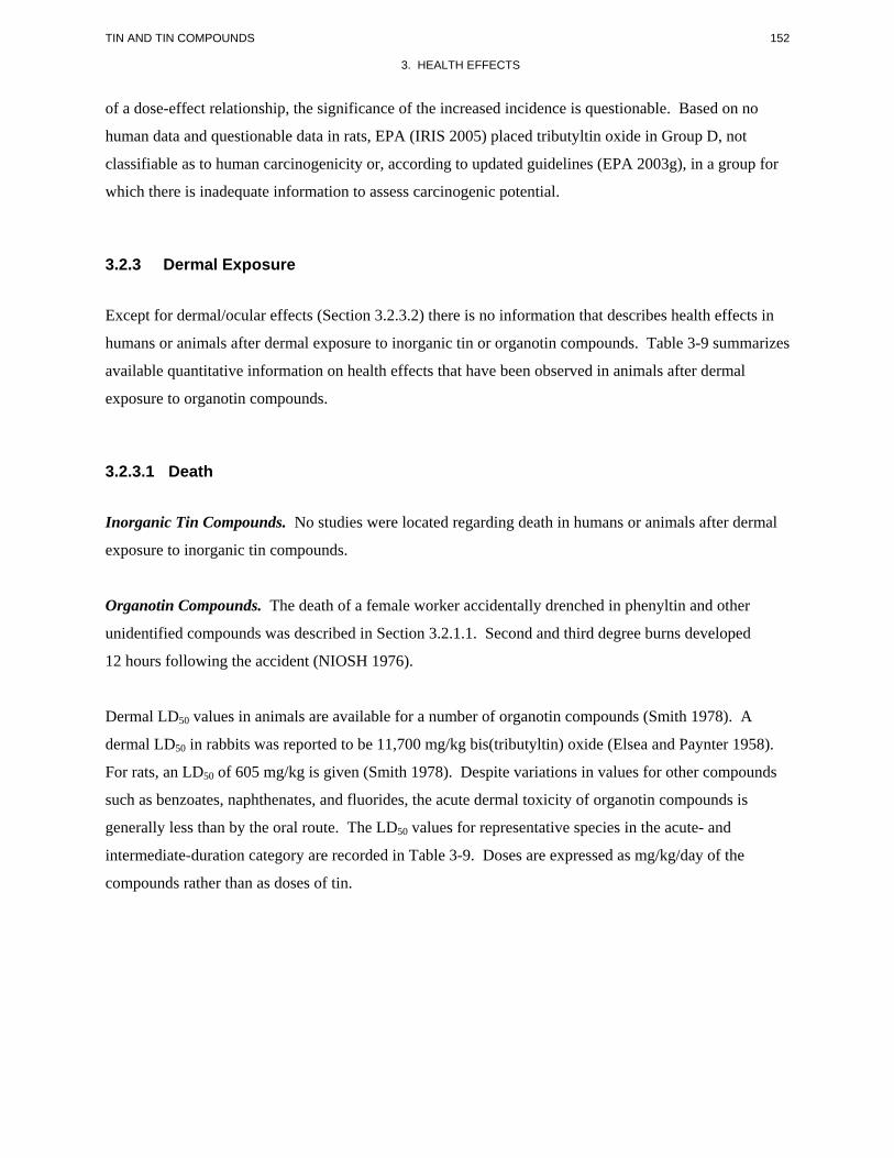
152 TIN AND TIN COMPOUNDS
3. HEALTH EFFECTS
of a dose-effect relationship, the significance of the increased incidence is questionable. Based on no
human data and questionable data in rats, EPA (IRIS 2005) placed tributyltin oxide in Group D, not
classifiable as to human carcinogenicity or, according to updated guidelines (EPA 2003g), in a group for
which there is inadequate information to assess carcinogenic potential.
3.2.3 Dermal Exposure
Except for dermal/ocular effects (Section 3.2.3.2) there is no information that describes health effects in
humans or animals after dermal exposure to inorganic tin or organotin compounds. Table 3-9 summarizes
available quantitative information on health effects that have been observed in animals after dermal
exposure to organotin compounds.
3.2.3.1 Death
Inorganic Tin Compounds. No studies were located regarding death in humans or animals after dermal
exposure to inorganic tin compounds.
Organotin Compounds. The death of a female worker accidentally drenched in phenyltin and other
unidentified compounds was described in Section 3.2.1.1. Second and third degree burns developed
12 hours following the accident (NIOSH 1976).
Dermal LD50 values in animals are available for a number of organotin compounds (Smith 1978). A
dermal LD50 in rabbits was reported to be 11,700 mg/kg bis(tributyltin) oxide (Elsea and Paynter 1958).
For rats, an LD50 of 605 mg/kg is given (Smith 1978). Despite variations in values for other compounds
such as benzoates, naphthenates, and fluorides, the acute dermal toxicity of organotin compounds is
generally less than by the oral route. The LD50 values for representative species in the acute- and
intermediate-duration category are recorded in Table 3-9. Doses are expressed as mg/kg/day of the
compounds rather than as doses of tin.
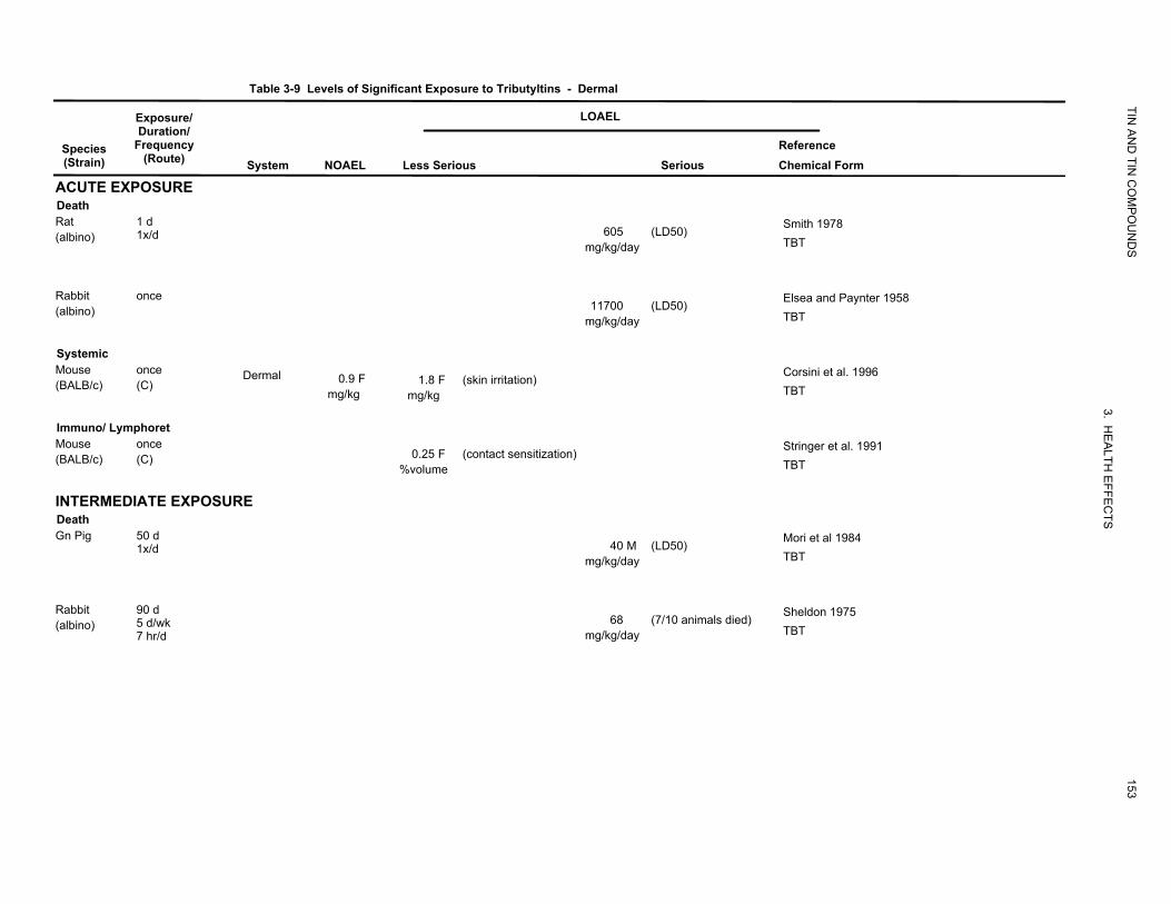
Exposure/
Table 3-9 Levels of Significant Exposure to Tributyltins - Dermal
LOAEL
TIN
Species (Strain)
ACUTE EXPOSURE
Duration/ Frequency
(Route) System NOAEL Less Serious Serious
AN
D TIN
CO
MP
OU
ND
S
Reference Chemical Form
Death Rat 1 d (albino) 1x/d 605
mg/kg/day (LD50)
Smith 1978 TBT
Rabbit once (albino) 11700
mg/kg/day (LD50)
Elsea and Paynter 1958 TBT
Systemic Mouse once Dermal (BALB/c) (C) 0.9 F
mg/kg 1.8 F
mg/kg (skin irritation)
Corsini et al. 1996 TBT
Immuno/ Lymphoret
3. H
Mouse once (BALB/c) (C) 0.25 F
%volume (contact sensitization)
EA
LTH
Stringer et al. 1991 TBT
INTERMEDIATE EXPOSURE
EFFE
C
Death
TS
Gn Pig 50 d 1x/d 40 M
mg/kg/day (LD50)
Mori et al 1984 TBT
Rabbit 90 d (albino) 5 d/wk
7 hr/d 68
mg/kg/day (7/10 animals died)
153
Sheldon 1975 TBT
Table 3-9 Levels of Significant Exposure to Tributyltins - Dermal (continued)
Exposure/ LOAEL
TIN
Species (Strain)
Duration/ Frequency
(Route) System NOAEL
AN
D TIN
CO
MP
OU
ND
S
Reference Chemical Form Less Serious Serious
Systemic Gn Pig (Hartley)
50 d 1 x/d Renal 10 M
mg/kg/day
Mori et al 1984 (tubule degeneration)
TBT
Bd Wt 10 M (decreased body weight 40 M (severe decrease in body mg/kg/day mg/kg/day weight)
Rabbit
Bd Wt = bod
90 d 5 d/wk 7 hr/d
y weight; (C) = cap
Dermal
sule; d = day(s);
14 mg/kg/day
F = Female; (G) = gavage; Gn pig = Guinea pig; hr = hour(s); L
3. HE
ALTH
Sheldon 1975 TBT
D50 = lethal dose, 50% kill; LOAEL = lowest-observed-adverse-effect
154
EFFE
CTS
level; M = male; NOAEL = no-observed-adverse-effect level; x = time(s); wk = week(s)
155 TIN AND TIN COMPOUNDS
3. HEALTH EFFECTS
3.2.3.2 Systemic Effects
Inorganic Tin Compounds. No studies were located regarding respiratory, cardiovascular,
gastrointestinal, hematological, musculoskeletal, hepatic, or renal effects in humans or animals after
dermal exposure to inorganic tin compounds.
Organotin Compounds. No studies were located regarding respiratory, cardiovascular, gastrointestinal,
hematological, musculoskeletal, or renal effects in humans after dermal exposure to organotin
compounds.
No studies were located regarding respiratory, cardiovascular, gastrointestinal, hematological,
musculoskeletal, or hepatic effects in animals after dermal exposure to organotin compounds.
The highest NOAEL values and reliable LOAELs are recorded in Table 3-9.
Hepatic Effects. Signs of hepatic injury, as judged by increased serum AST and ALT activities, were
reported in a case of acute dermal exposure to triphenyltin acetate (Colosio et al. 1991). The patient, a
36-year-old man, spilled powder of a 19% formulation of triphenyltin acetate on exposed skin on his
arms. The acute rise in transaminase activities was followed by a gradual decrease for the next 18 days.
Twelve days after poisoning, echotomography showed a generalized enlargement of the liver. Three days
later, examination of a liver needle biopsy showed slight and nonspecific inflammatory abnormalities.
Slight hepatomegaly persisted when the patient was discharged 21 days after poisoning.
Renal Effects. Doses of 10 or 40 mg tributyltin oxide/kg/day were applied to the shaved skin of male
guinea pigs for 50 days (Mori et al. 1984). Swelling, degeneration, and destruction of tubular epithelium
were observed, but there were no changes in the glomerulus. There was also an increased excretion of
sodium, chloride, phosphate, glucose, and amino acids in the urine. In serum, the concentrations of
phosphate and certain amino acids were low reflecting the excessive loss in the urine. According to the
authors, these findings were consistent with a secondary Fanconi syndrome. These renal tubular changes
are similar to those seen with inorganic tin compounds after oral exposure (see Section 3.2.2.2) and
suggest that the compound was absorbed systemically.
156 TIN AND TIN COMPOUNDS
3. HEALTH EFFECTS
Dermal Effects.
Inorganic Tin Compounds. No studies were located regarding dermal effects in humans after dermal
exposure to inorganic tin compounds.
Stannous fluoride (0.25 and 0.5%) and stannous chloride (1 and 2%) produced leukocyte pustules in
rabbit skin along the area adjacent to an abdominal epidermal scratch (Stone and Willis 1968).
Infiltration of the tissue with polymorphonuclear and mononuclear leukocytes was present in the absence
of pustules at a stannous chloride concentration of 0.5% and a stannous fluoride concentration of 0.1%.
Organotin Compounds. It is known that organotins are skin irritants in humans (Sheldon 1975). Direct
skin contact with triphenyltin fluoride produced an irritant contact folliculitis in a male worker (Andersen
and Petri 1982). Patch tests were performed in human subjects, as well as in guinea pigs and rabbits, but
the dermatitis could not be reproduced. An irritant contact dermatitis was also seen in workers using a
paint containing tributyltin oxide (Goh 1985). Sensitization was not observed in any of the referenced
studies or in a separate study of tributyltin oxide-based paints (Gammeltoft 1978). Lyle (1958) described
the following time-course of events in five volunteers who had undiluted tributyltin chloride painted on
the skin of the back of the hand. Reddening and swelling of the mouths of the hair follicles appeared after
2–3 hours; this was followed by progressively intense follicular inflammation. The pruritus was confined
to the tested area and persisted for 2 or 3 days. Pustules appeared on the second day and remained small
until they dried up on the third or fourth day. On the fifth day, resolution was well advanced and, after a
week, all that remained was faint punctate erythema with a little perifollicular scaling.
Tributyltin oxide is a severe irritant to the skin in rabbits (Sheldon 1975). By contrast, tributyltin fluoride
and triphenyltin fluoride produced only minimal skin irritation (Sheldon 1975). Other acute studies have
likewise demonstrated the skin irritating potential of tributyltin oxide and triphenyltin acetate in rats and
mice (Corsini et al. 1996a; Klimmer 1969; Pelikan and Cerny 1968).
Dermal exposure of rats to doses of 80 mg/kg of dipropyltin dichloride, diisopropyltin dichloride, or
diethyltin dichloride for 5 consecutive days produced necrosis, edema, and inflammation of the skin
(Barnes and Stoner 1958). The same dose of dimethyltin dichloride produced dermal necrosis with black
scar formation; dibutyltin dichloride produced little superficial damage to the skin and some edema of
subcutaneous tissues. Dihexyltin dichloride and dioctyltin dichloride did not produce skin lesions
(Barnes and Stoner 1958). In a 90-day repeated-dose dermal study, rabbits developed skin irritation at
157 TIN AND TIN COMPOUNDS
3. HEALTH EFFECTS
each of three levels tested (14, 27, and 68 mg/kg/day tributyltin fluoride) (Sheldon 1975). Deaths
occurred in 7 of 10 rabbits at 68 mg/kg, but surviving animals eventually returned to normal a few days
after exposure was terminated. The authors stated that a dose of 14 mg/kg (65 applications) was a
NOAEL despite local irritation at the application sites. In view of the exaggerated daily contact with the
rabbit skin, this value seems reasonable since such high levels of daily exposure would not be the case in
humans. However, a detailed report of this study was not available for review.
A study in guinea pigs did not find tributyltin oxide to be a contact sensitizer (Schweinfurth and Gunzel
1987).
Ocular Effects.
Inorganic Tin Compounds. No studies were located regarding ocular effects of inorganic tin in humans
or animals following dermal exposure.
Organotin Compounds. Lyle (1958) described the case of a worker who was not wearing protective
goggles and splashed an unspecified butyltin compound on the face and both eyes were affected.
Lachrimation and intense suffusion of the conjunctiva appeared within minutes and, despite immediate
lavage, persisted for 4 days. Tributyltin oxide, tributyltin fluoride and triphenyltin fluoride are extreme
irritants to rabbits’ eyes (Sheldon 1975).
3.2.3.3 Immunological and Lymphoreticular Effects
Inorganic Tin Compounds. No studies were located regarding immunological and lymphoreticular
effects in humans or animals following dermal exposure to inorganic tin.
Organotin Compounds. Colosio et al. (1991) reported that a man who spilled a powder pesticide
formulation containing 19% triphenyltin acetate over exposed skin on his arm developed severe genital
edema and urticarial eruptions on his trunk. In addition, on day 11 after the accident his serum IgE was
elevated. Although patch tests conducted with the entire formulation and with each single component of
the formulation gave negative results, the investigators attributed the findings to poisoning with
triphenyltin. Tributyltin oxide induced contact sensitization in mice applied the test material for 3 days
and challenged with it 3 days later (Stringer et al. 1991). The lowest concentration tested, 0.25% by
volume, triggered a positive response.
158 TIN AND TIN COMPOUNDS
3. HEALTH EFFECTS
3.2.3.4 Neurological Effects
Inorganic Tin Compounds. No studies were located regarding neurological effects in humans or animals
following dermal exposure to inorganic tin.
Organic Tin Compounds. The only relevant information is that from the case of a man who spilled a
powder pesticide formulation containing 19% triphenyltin acetate on his exposed arms, and 10 days after
the accident, his EEG showed alterations consisting of generalized paroxysmal abnormalities and
bradyrhythmia (Colosio et al. 1991). Four months after the accident, the EEG showed slight anomalies
during hyperpnea.
No studies were located regarding the following effects in humans or animals after dermal exposure to
inorganic tin or organotin compounds:
3.2.3.5 Reproductive Effects
3.2.3.6 Developmental Effects
3.2.3.7 Cancer
Inorganic Tin Compounds. No studies were located regarding cancer effects in humans or animals after
dermal exposure to inorganic tin compounds.
Organotin Compounds. No studies were located regarding cancer effects in humans after dermal
exposure to organotin compounds.
In a limited evaluation of carcinogenicity, tributyltin fluoride was applied to the shaved backs of male
white mice 3 times/week for a period of 6 months. Treated mice received 15 mg of 5 or 10% of the
compound in propylene glycol. Hyperplastic skin changes were observed in the 5% group, but not in the
10% group (Sheldon 1975). Carcinogenic effects were not observed in this study, which was only of
intermediate duration. No other studies were located regarding cancer effects in animals after dermal
exposure to organotin compounds.
159 TIN AND TIN COMPOUNDS
3. HEALTH EFFECTS
3.2.4 Other Routes of Exposure
This section provides brief examples of effects of tin compounds that have been studied primarily by
exposing the animals by a route other than inhalation, oral, or dermal.
A considerable number of studies have evaluated the developmental effects of both trimethyltin and
triethyltin following perinatal exposure by intraperitoneal injection of animals, and some studies have
demonstrated that some alterations persist until adulthood. For example, a single intraperitoneal dose of
trimethyltin hydroxide (4–6 mg/kg) to rat pups on Pnd 5 reduced growth and impaired performance on
rope descent when tested on Pnd 20 and 21 (Ruppert et al. 1983). Motor activity in a figure-eight maze
was increased at 57 days of age and at 120 days of age. The response to acoustic startle was decreased
during preweaning and as adults. At termination (Pnd 120), whole brain weight and weight of olfactory
bulbs decreased at 4, 5, and 6 mg/kg, whereas the hippocampus weight was decreased at 5 and 6 mg/kg.
Similar results were obtained following a single intraperitoneal injection of triethyltin bromide (3 or
6 mg/kg) also on Pnd 5 (Reiter et al. 1981). Barone et al. (1995) showed that some behavioral alterations
that can be detected on Pnd 23 after a single injection of triethyltin on Pnd 5, which were no longer
apparent 3 or 12 months postdosing, became apparent again in 24-month-old rats, suggesting an
unmasking effects by the natural aging process.
Chang (1984a, 1984b) did not observe lesions in the hippocampal formation of rats injected
intraperitoneally with 6 mg/kg trimethyltin chloride between Pnd 1 and 4, but increasing damage to
Ammon’s horn was seen when dosing occurred between the ages of Pnd 5 and 15. This was followed by
an apparently reduced sensitivity after Pnd 20. Since the pathological patterns were well-correlated with
the development and functional maturity of the hippocampal neurons, Chang (1984a, 1984b, 1990)
postulated that the production of lesions, particularly those in subfield CA3, require functionally mature
and intact granule cells and their fibers, the mossy fibers. It has also been shown that the day of exposure
greatly influences the magnitude of cognitive deficits and neuropathology associated with exposure to
triethyltin (Freeman et al. 1994).
Trimethyltin and triethyltin have induced ototoxicity in rodents. A single intraperitoneal injection of 4–
6 mg trimethyltin/kg produced a frequency-dependent loss of auditory sensitivity in rats that was severe
in the high frequency range (Eastman et al. 1987; Ruppert et al. 1984). Subsequent studies showed that
the alterations were long-lasting and consisted of a high-frequency hearing loss characterized by elevated
thresholds in the auditory startle response test detected 11 weeks postdosing (Crofton et al. 1990).
Thresholds for the brainstem auditory evoked response were also elevated in treated rats 9 weeks
160 TIN AND TIN COMPOUNDS
3. HEALTH EFFECTS
postdosing. Microscopic examination of the cochlea from base to apex showed dead outer hair cells
preferentially in regions associated with high-frequency hearing, in a dose-related manner. A study in
guinea pigs treated intraperitoneally with a single dose of 2 mg of trimethyltin chloride/kg showed high-
frequency impairment, which improved throughout a 6-week period of testing (Fechter and Carlisle
1990). As seen in the rat, hair cell loss occurred in a portion of the cochlea responsible for encoding high-
frequency sound. There also was a marked increase in the diameter of the vessels of the stria vascularis
(an area containing one of the primary vascular networks in the cochlea) along with signs of atrophy in
the stria vascularis. However, since the increases in vessel diameter were not confined to the basal
portion of the cochlea, and were greater in the middle and apical regions than in the base, it seemed that
the strial pathology was not directly related to hair cell loss or functional impairment. In a different study,
both trimethyltin and triethyltin were shown to severely disrupt (increase) the compound action potential
(CAP) threshold in guinea pigs within 30–60 minutes of dosing, but had no significant effect on the
cochlear microphonic (CM) potential (Clerici et al. 1991). The CAP is generated by the release of
neurotransmitters from the inner hair cells and the subsequent depolarization of spiral ganglion cells,
whereas CM reflects electromechanical function of the outer hair cells. In a further study, trimethyltin
was shown to reduce CAP sensitivity and CM amplitude (Fechter et al. 1992). The effect was relatively
broad across test frequencies 6 hours after dosing and gradually became restricted to higher frequencies.
The effect of trimethyltin appears to be a direct effect on the cochlea, as disruption of sound-evoked
cochlear action potentials can be observed after direct application of trimethyltin to the round window of
guinea pigs (Liu and Fechter 1995). The results of these and other studies were thought to be consistent
with the hypothesis that trimethyltin disrupts function at the synapse between the inner hair cell and the
Type I spiral ganglion cell, possibly by damaging the hair cells or ganglia from uncontrolled production
of reactive oxygen species (ROS) (Clerici 1996; Fechter and Liu 1994).
A series of publications from Merkord and coworkers have described the effects of dibutyltin dichloride
on the pancreas from rats following intravenous injection of the chemical. Earlier studies described an
acute interstitial pancreatitis in rats developing 24 hours after a single dose followed by a more severe
pancreatitis with mononuclear cell infiltrates 4–6 days later (Merkord and Hennighausen 1989). In a
more recent study, the time-course of the pancreatic alterations was followed for up to 28 days with
interim sacrifices at various intervals after a single dose of 6 mg dibutyltin dichloride/kg (Merkord et al.
1997). The findings suggested an initial cytotoxic effect on the biliopancreatic duct epithelium leading to
epithelial necrosis with obstruction of the duct. This was followed by hematogenic effects directly
injuring pancreatic cells followed by interstitial edema and inflammation. A tendency to a chronic course
occurred when the obstruction of the duct and cholestasis persisted. Extending the observation period
161 TIN AND TIN COMPOUNDS
3. HEALTH EFFECTS
showed that an active inflammatory process persisted for up to 60 days after dosing (Sparmann et al.
1997). A study of repeated administration of a slightly lower dose of dibutyltin dichloride (4 mg/kg) at
intervals of 3 weeks, reported the development of acute pancreatitis and biliopancreatic lesions after
6 weeks and pancreatic fibrosis and liver lesions after 9–12 weeks (Merkord et al. 2001). In rats followed
for up to 1 year after a single injection of 6 mg dibutyltin dichloride/kg, the permanent obstruction of
biliopancreatic secretion and chronic cholestasis led to the formation of deposits inside the dilated duct,
occasionally with bacterial infiltration and growth. Considerable amounts of tin were detected inside the
bacterially infected deposits (Jonas et al. 2002).
3.3 GENOTOXICITY
In vitro studies with inorganic tin have provided mixed results (Table 3-10). DNA damage was noted in
Chinese hamster ovary cells incubated with stannous chloride in the absence of metabolic activation, but
the results for stannic chloride were negative (McLean et al. 1983). Cytogenetic studies also gave
positive responses with stannous chloride for chromosomal aberrations and sister chromatid exchanges in
Chinese hamster ovary cells with or without metabolic activation (Gulati et al. 1989). Ganguly et al.
(1992) incubated peripheral lymphocytes from 27 healthy male volunteers with stannic chloride and
observed a significant increase in the frequency of chromosomal aberrations. In K562 cells (a cell line
derived from a chronic myelogenic human leukemia), stannous chloride reduced viability and induced
DNA damage, as determined by the comet assay (Dantas et al. 2002). The investigators (Dantas et al.
2002) proposed that genetic damage is produced by ROS generated by the reduction of hydrogen
peroxide by stannous ions. Earlier research from this group had demonstrated that ROS scavengers and
metal-ion chelators could prevent, at least partially, the inactivation of Escherichia coli cultures treated
with stannous chloride (Dantas et al. 1996). Stannous chloride has also been reported to rapidly convert
hydroperoxy thymidine to mutagenic hydroxymethyl deoxyuridine species in vitro, suggesting a redox
component in the genotoxic potential of stannous chloride in vivo (Tofigh and Frenkel 1989).
Table 3-11 presents data on the genotoxicity of organotin compounds in in vitro assays. Hamasaki et al.
(1993) tested 14 different organotin compounds in two strains of Salmonella typhimurium, TA98 and
TA100, without metabolic activation. All but dibutyltin dichloride gave negative results in TA98. In
TA100, the monobutyltins, dibutyltins, and tributyltin compounds gave positive results. Results from
assays in mammalian cells for a number of trialkyl organotins, with and without metabolic activation,
showed mostly negative results (Davis et al. 1987; Sasaki et al. 1993). However, other studies have
reported elevated incidences of chromosomal aberrations, sister chromatid exchanges, and micronuclei in
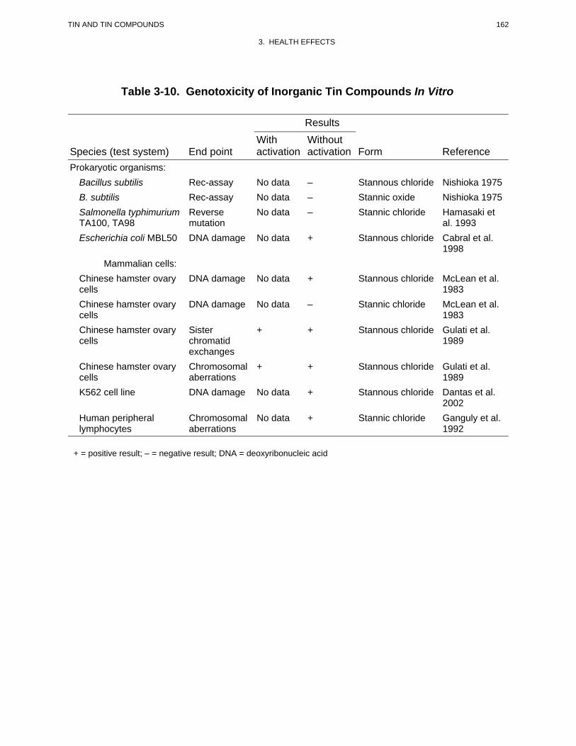
162 TIN AND TIN COMPOUNDS
3. HEALTH EFFECTS
Table 3-10. Genotoxicity of Inorganic Tin Compounds In Vitro
Results
Species (test system) End point With activation
Without activation Form Reference
Prokaryotic organisms: Bacillus subtilis Rec-assay No data – Stannous chloride Nishioka 1975 B. subtilis Rec-assay No data – Stannic oxide Nishioka 1975 Salmonella typhimurium TA100, TA98
Reverse mutation
No data – Stannic chloride Hamasaki et al. 1993
Escherichia coli MBL50 DNA damage No data + Stannous chloride Cabral et al. 1998
Mammalian cells: Chinese hamster ovary cells
DNA damage No data + Stannous chloride McLean et al. 1983
Chinese hamster ovary cells
DNA damage No data – Stannic chloride McLean et al. 1983
Chinese hamster ovary cells
Sister chromatid exchanges
+ + Stannous chloride Gulati et al. 1989
Chinese hamster ovary cells
Chromosomal aberrations
+ + Stannous chloride Gulati et al. 1989
K562 cell line DNA damage No data + Stannous chloride Dantas et al. 2002
Human peripheral lymphocytes
Chromosomal aberrations
No data + Stannic chloride Ganguly et al. 1992
+ = positive result; – = negative result; DNA = deoxyribonucleic acid
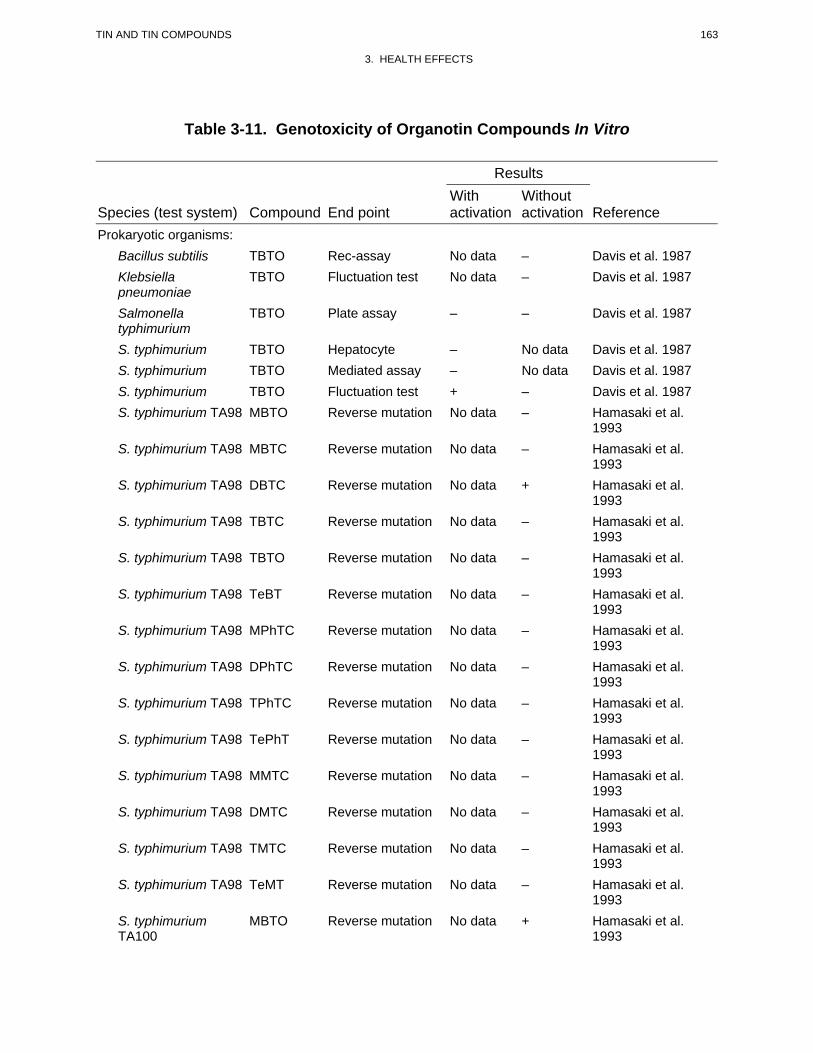
163 TIN AND TIN COMPOUNDS
3. HEALTH EFFECTS
Table 3-11. Genotoxicity of Organotin Compounds In Vitro
Results
Species (test system) Compound End point With activation
Without activation Reference
Prokaryotic organisms: Bacillus subtilis TBTO Rec-assay No data – Davis et al. 1987 Klebsiella pneumoniae
TBTO Fluctuation test No data – Davis et al. 1987
Salmonella typhimurium
TBTO Plate assay – – Davis et al. 1987
S. typhimurium TBTO Hepatocyte – No data Davis et al. 1987 S. typhimurium TBTO Mediated assay – No data Davis et al. 1987 S. typhimurium TBTO Fluctuation test + – Davis et al. 1987 S. typhimurium TA98 MBTO Reverse mutation No data – Hamasaki et al.
1993 S. typhimurium TA98 MBTC Reverse mutation No data – Hamasaki et al.
1993 S. typhimurium TA98 DBTC Reverse mutation No data + Hamasaki et al.
1993 S. typhimurium TA98 TBTC Reverse mutation No data – Hamasaki et al.
1993 S. typhimurium TA98 TBTO Reverse mutation No data – Hamasaki et al.
1993 S. typhimurium TA98 TeBT Reverse mutation No data – Hamasaki et al.
1993 S. typhimurium TA98 MPhTC Reverse mutation No data – Hamasaki et al.
1993 S. typhimurium TA98 DPhTC Reverse mutation No data – Hamasaki et al.
1993 S. typhimurium TA98 TPhTC Reverse mutation No data – Hamasaki et al.
1993 S. typhimurium TA98 TePhT Reverse mutation No data – Hamasaki et al.
1993 S. typhimurium TA98 MMTC Reverse mutation No data – Hamasaki et al.
1993 S. typhimurium TA98 DMTC Reverse mutation No data – Hamasaki et al.
1993 S. typhimurium TA98 TMTC Reverse mutation No data – Hamasaki et al.
1993 S. typhimurium TA98 TeMT Reverse mutation No data – Hamasaki et al.
1993 S. typhimurium TA100
MBTO Reverse mutation No data + Hamasaki et al. 1993

164 TIN AND TIN COMPOUNDS
3. HEALTH EFFECTS
Table 3-11. Genotoxicity of Organotin Compounds In Vitro
Results With Without
Species (test system) Compound End point activation activation Reference S. typhimurium MBTC Reverse mutation No data + Hamasaki et al. TA100 1993 S. typhimurium DBTC Reverse mutation No data + Hamasaki et al. TA100 1993 S. typhimurium TBTC Reverse mutation No data + Hamasaki et al. TA100 1993 S. typhimurium TBTO Reverse mutation No data + Hamasaki et al. TA100 1993 S. typhimurium TeBT Reverse mutation No data – Hamasaki et al. TA100 1993 S. typhimurium MPhTC Reverse mutation No data – Hamasaki et al. TA100 1993 S. typhimurium DPhTC Reverse mutation No data – Hamasaki et al. TA100 1993 S. typhimurium TPhTC Reverse mutation No data – Hamasaki et al. TA100 1993 S. typhimurium TePhT Reverse mutation No data – Hamasaki et al. TA100 1993 S. typhimurium MMTC Reverse mutation No data – Hamasaki et al. TA100 1993 S. typhimurium DMTC Reverse mutation No data + Hamasaki et al. TA100 1993 S. typhimurium TMTC Reverse mutation No data – Hamasaki et al. TA100 1993 S. typhimurium TeMT Reverse mutation No data – Hamasaki et al. TA100 1993
Eukaryotic organisms: Saccharomyces TBTO Forward mutation – – Davis et al. 1987 pombe Saccharomyces TBTO Mitotic gene – – Davis et al. 1987 cerevesiae conversion
Mammalian cells: Chinese hamster TBTO 8-Azaguanine and – – Davis et al. 1987 cells ovarian resistance Chinese hamster TBTO 6-Thioguanine – – Davis et al. 1987 cells resistance Mouse lymphoma TBTO 6-Thioguanine and No data – Davis et al. 1987 cells Buer resistance Chinese hamster TBTO Sister chromatid – – Davis et al. 1987 cells exchange
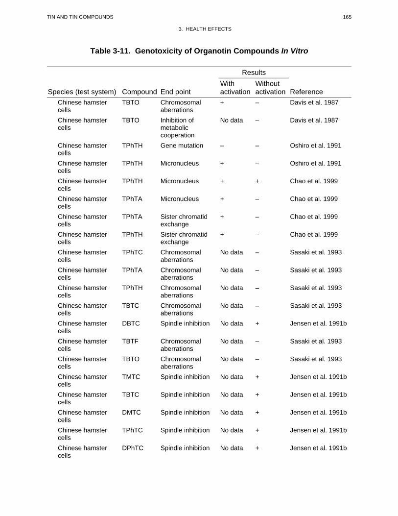
165 TIN AND TIN COMPOUNDS
3. HEALTH EFFECTS
Table 3-11. Genotoxicity of Organotin Compounds In Vitro
Results With Without
Species (test system) Compound End point activation activation Reference Chinese hamster TBTO Chromosomal + – Davis et al. 1987 cells aberrations Chinese hamster TBTO Inhibition of No data – Davis et al. 1987 cells metabolic
cooperation Chinese hamster TPhTH Gene mutation – – Oshiro et al. 1991 cells Chinese hamster TPhTH Micronucleus + – Oshiro et al. 1991 cells Chinese hamster TPhTH Micronucleus + + Chao et al. 1999 cells Chinese hamster TPhTA Micronucleus + – Chao et al. 1999 cells Chinese hamster TPhTA Sister chromatid + – Chao et al. 1999 cells exchange Chinese hamster TPhTH Sister chromatid + – Chao et al. 1999 cells exchange Chinese hamster TPhTC Chromosomal No data – Sasaki et al. 1993 cells aberrations Chinese hamster TPhTA Chromosomal No data – Sasaki et al. 1993 cells aberrations Chinese hamster TPhTH Chromosomal No data – Sasaki et al. 1993 cells aberrations Chinese hamster TBTC Chromosomal No data – Sasaki et al. 1993 cells aberrations Chinese hamster DBTC Spindle inhibition No data + Jensen et al. 1991b cells Chinese hamster TBTF Chromosomal No data – Sasaki et al. 1993 cells aberrations Chinese hamster TBTO Chromosomal No data – Sasaki et al. 1993 cells aberrations Chinese hamster TMTC Spindle inhibition No data + Jensen et al. 1991b cells Chinese hamster TBTC Spindle inhibition No data + Jensen et al. 1991b cells Chinese hamster DMTC Spindle inhibition No data + Jensen et al. 1991b cells Chinese hamster TPhTC Spindle inhibition No data + Jensen et al. 1991b cells Chinese hamster DPhTC Spindle inhibition No data + Jensen et al. 1991b cells
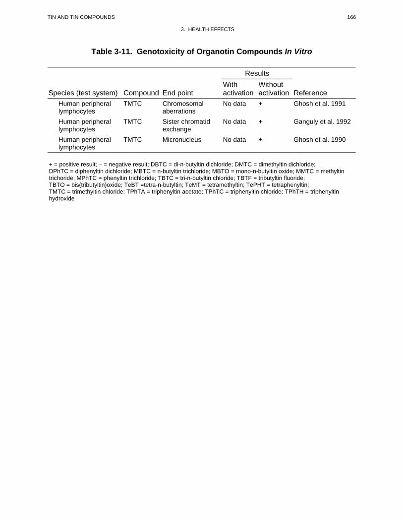
166 TIN AND TIN COMPOUNDS
3. HEALTH EFFECTS
Table 3-11. Genotoxicity of Organotin Compounds In Vitro
Results With Without
Species (test system) Compound End point activation activation Reference Human peripheral TMTC Chromosomal No data + Ghosh et al. 1991 lymphocytes aberrations Human peripheral TMTC Sister chromatid No data + Ganguly et al. 1992 lymphocytes exchange Human peripheral TMTC Micronucleus No data + Ghosh et al. 1990 lymphocytes
+ = positive result; – = negative result; DBTC = di-n-butyltin dichloride; DMTC = dimethyltin dichloride; DPhTC = diphenyltin dichloride; MBTC = n-butyltin trichloride; MBTO = mono-n-butyltin oxide; MMTC = methyltin trichoride; MPhTC = phenyltin trichloride; TBTC = tri-n-butyltin chloride; TBTF = tributyltin fluoride; TBTO = bis(tributyltin)oxide; TeBT =tetra-n-butyltin; TeMT = tetramethyltin; TePHT = tetraphenyltin; TMTC = trimethyltin chloride; TPhTA = triphenyltin acetate; TPhTC = triphenyltin chloride; TPhTH = triphenyltin hydroxide
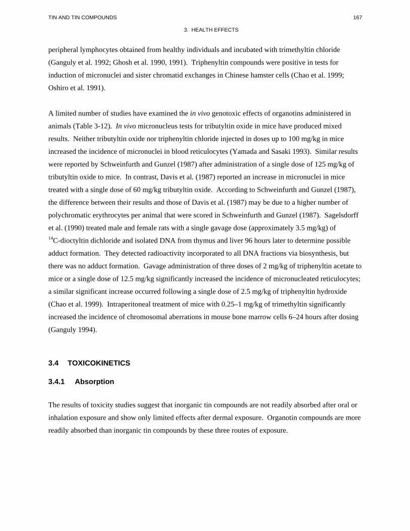
167 TIN AND TIN COMPOUNDS
3. HEALTH EFFECTS
peripheral lymphocytes obtained from healthy individuals and incubated with trimethyltin chloride
(Ganguly et al. 1992; Ghosh et al. 1990, 1991). Triphenyltin compounds were positive in tests for
induction of micronuclei and sister chromatid exchanges in Chinese hamster cells (Chao et al. 1999;
Oshiro et al. 1991).
A limited number of studies have examined the in vivo genotoxic effects of organotins administered in
animals (Table 3-12). In vivo micronucleus tests for tributyltin oxide in mice have produced mixed
results. Neither tributyltin oxide nor triphenyltin chloride injected in doses up to 100 mg/kg in mice
increased the incidence of micronuclei in blood reticulocytes (Yamada and Sasaki 1993). Similar results
were reported by Schweinfurth and Gunzel (1987) after administration of a single dose of 125 mg/kg of
tributyltin oxide to mice. In contrast, Davis et al. (1987) reported an increase in micronuclei in mice
treated with a single dose of 60 mg/kg tributyltin oxide. According to Schweinfurth and Gunzel (1987),
the difference between their results and those of Davis et al. (1987) may be due to a higher number of
polychromatic erythrocytes per animal that were scored in Schweinfurth and Gunzel (1987). Sagelsdorff
et al. (1990) treated male and female rats with a single gavage dose (approximately 3.5 mg/kg) of 14C-dioctyltin dichloride and isolated DNA from thymus and liver 96 hours later to determine possible
adduct formation. They detected radioactivity incorporated to all DNA fractions via biosynthesis, but
there was no adduct formation. Gavage administration of three doses of 2 mg/kg of triphenyltin acetate to
mice or a single dose of 12.5 mg/kg significantly increased the incidence of micronucleated reticulocytes;
a similar significant increase occurred following a single dose of 2.5 mg/kg of triphenyltin hydroxide
(Chao et al. 1999). Intraperitoneal treatment of mice with 0.25–1 mg/kg of trimethyltin significantly
increased the incidence of chromosomal aberrations in mouse bone marrow cells 6–24 hours after dosing
(Ganguly 1994).
3.4 TOXICOKINETICS
3.4.1 Absorption
The results of toxicity studies suggest that inorganic tin compounds are not readily absorbed after oral or
inhalation exposure and show only limited effects after dermal exposure. Organotin compounds are more
readily absorbed than inorganic tin compounds by these three routes of exposure.
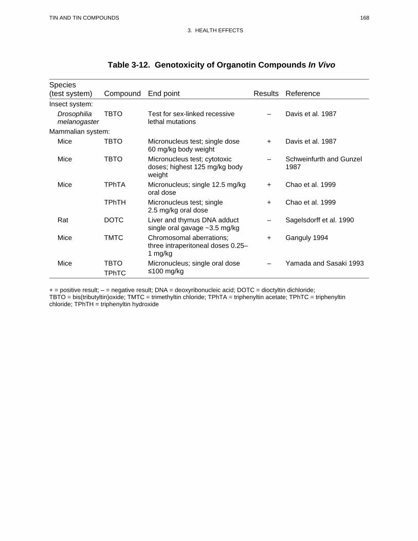
168 TIN AND TIN COMPOUNDS
3. HEALTH EFFECTS
Table 3-12. Genotoxicity of Organotin Compounds In Vivo
Species (test system) Compound End point Results Reference Insect system:
Drosophilia TBTO melanogaster
Mammalian system: Mice TBTO
Mice TBTO
Mice TPhTA
TPhTH
Rat DOTC
Mice TMTC
Mice TBTO TPhTC
Test for sex-linked recessive lethal mutations
Micronucleus test; single dose 60 mg/kg body weight Micronucleus test; cytotoxic doses; highest 125 mg/kg body weight Micronucleus; single 12.5 mg/kg oral dose Micronucleus test; single 2.5 mg/kg oral dose Liver and thymus DNA adduct single oral gavage ~3.5 mg/kg Chromosomal aberrations; three intraperitoneal doses 0.25– 1 mg/kg Micronucleus; single oral dose ≤100 mg/kg
–
+
–
+
+
–
+
–
Davis et al. 1987
Davis et al. 1987
Schweinfurth and Gunzel 1987
Chao et al. 1999
Chao et al. 1999
Sagelsdorff et al. 1990
Ganguly 1994
Yamada and Sasaki 1993
+ = positive result; – = negative result; DNA = deoxyribonucleic acid; DOTC = dioctyltin dichloride; TBTO = bis(tributyltin)oxide; TMTC = trimethyltin chloride; TPhTA = triphenyltin acetate; TPhTC = triphenyltin chloride; TPhTH = triphenyltin hydroxide
169 TIN AND TIN COMPOUNDS
3. HEALTH EFFECTS
3.4.1.1 Inhalation Exposure
No quantitative studies were located regarding absorption in humans or animals after inhalation exposure
to inorganic tin or organotin compounds. However, limited data summarized in Section 3.2.1 suggest that
absorption of organotins by the inhalation route is possible, as occurred for example in cases for subjects
who exhibited serious neurological effects after accidental exposure to vapors of a trimethyltin (Feldman
et al. 1993; Fortemps et al. 1978; Rey et al. 1984; Ross et al. 1981; Saary and House 2002; Yanofsky et
al. 1991). Dermal exposure may have also occurred in these cases.
3.4.1.2 Oral Exposure
Inorganic Tin Compounds. Johnson and Greger (1982) conducted a balance study in eight healthy adult
males, who were placed on diets containing either 0.1 mg Sn/day (basal diet) or 50 mg Sn/day
(supplemented with stannous chloride). Subjects were placed on the diets for 20 days and intakes and
excretion were measured daily in two 6-day periods (following a 6-day adjustment to the diets). Average
fecal excretion was 55% of the daily intake in the basal group and 97% in the supplemented group,
suggesting net absorption of 45 and 3%, respectively. Average urinary excretion was 26% of the daily
intake in the basal group and 2.4% in the supplemented group. Estimates of absorption in individuals
ranged from -4 to 71% of the daily intake in the basal group and from -7 to 9% in the supplemented
group. These observations suggest that gastrointestinal absorption of tin, in humans, decreases with
increasing dose. An alternative explanation for the differences in absorption of tin in the basal and
supplemented diets is that tin naturally incorporated into food may be more readily absorbed than tin
added as stannous chloride to food. Consistent with the former explanation are observations from
Calloway and McMullen (1966). In this study, nine healthy adults were placed on diets for 24 days
consisting of either fresh food (10 mg Sn/day), canned food that had been stored for 20 months at 1 °C
(26 mg Sn/day), or canned foods that had been stored for 20 months at 37 °C (163 mg Sn/day). Tin was
not detected in the urine in this study, and the amount excreted in the feces was the same as the amount
ingested. Thus, net absorption of tin could not be detected at these higher levels of intake, in contrast to
the observations made at lower intakes (0.1 mg Sn/day; Johnson and Greger 1982).
Studies conducted in animals suggest that fractional absorption of ingested inorganic Sn[II] is higher, by a
factor of approximately 4, than Sn[IV]; however, the associated anion appears to have little or no effect
on the absorption fraction. Gastrointestinal absorption was 2.85 and 0.64% of the administered dose, in
170 TIN AND TIN COMPOUNDS
3. HEALTH EFFECTS
rats, after a single oral dose of 113Sn[II]citrate or 113Sn[IV]citrate (20 mg Sn/kg), respectively (Hiles
1974). Fractional absorption of tin after single oral doses (20 mg Sn/kg) of stannous pyrophosphate
(113Sn[II]2P2O7), stannous fluoride (113Sn[II]F2), or stannic fluoride (113Sn[IV]F4) appeared to be similar to
that of 113Sn[II]citrate and 113Sn[IV]citrate (i.e., <5%), based on comparisons of tissue and excreta levels
(Hiles 1974). Furchner and Drake (1976) concluded, from comparisons of tissue retention kinetics after
oral gavage and intravenous injection of stannous chloride (113SnCl2), that gastrointestinal absorption of
Sn[II] was similar (less than 5%) in dogs, mice, rats, and monkeys.
Organotin Compounds. No quantitative estimates of absorption of organotin compounds in humans
were located. The detection of butyltin compounds in blood and in postmortem human liver samples
indicates that butyltin compounds are absorbed in humans (Kannan et al. 1999; Nielsen and Strand 2002).
Also, numerous deaths occurred in a poisoning episode with presumably accidental ingestion of
triethyltin in France in 1954 (WHO 1980) indicating that absorption occurred. In addition, Kreyberg et
al. (1992) described the case of a woman who ingested an unknown amount of trimethyltin and died
6 days after consuming the chemical.
Methyltin Compounds. Quantitative estimates of absorption of methyltin compounds after ingestion
were not located. Tin levels in brain, kidney, and liver were similar in neonatal rats that received oral
doses of 1 mg/kg trimethyltin hydroxide (0.66 mg Sn/kg) or triethyltin sulfate (0.44 mg Sn/kg),
suggesting that both compounds may be absorbed similarly (Mushak et al. 1982).
Ethyltin Compounds. Rats administered a single oral dose of approximately 25 mg/kg ethyltin
trichloride (12 mg Sn/kg) excreted 92% (range 89–95%) of the dose in feces and 1.2% (range 1–2%) in
urine, suggesting that at least 8% of the dose had been absorbed (Bridges et al. 1967). This is a minimum
estimate of absorption, since absorbed ethyltin compounds are secreted in bile and excreted in feces (see
Section 3.4.4.4).
Butyltin Compounds. Mice administered a single oral dose of approximately 180 µmol/kg, respectively,
of mono-, di-, or tributyltin (23 mg Sn/kg) excreted, approximately 2, 20, or 35% of the dose in urine
within 96 hours following dosing (Ueno et al. 1994). These values are minimum estimates of the
absorption of the ingested dose because they do not account for absorbed tin excreted by other routes
(e.g., bile-fecal pathway; see Section 3.4.4.4). However, these results indicate that the fraction of an
ingested dose of butyltin compounds excreted in urine increases with increasing number of butyl
171 TIN AND TIN COMPOUNDS
3. HEALTH EFFECTS
moieties, suggesting that more-highly butylated tin compounds may be absorbed to a greater extent
(Kimmel et al. 1977).
Phenyltin Compounds. Quantitative estimates of absorption of phenyltin compounds after ingestion
were not located. Urinary excretion of tin compounds (as total tin) over a 96-hour period following a
single oral dose of tri-, di-, or monophenyltin (15.5 mg Sn/kg) was <1% of the administered dose of tin
(Ohhira and Matsui 1993a). This is a minimum estimate of the absorbed fraction as it does not account
for excretion by other routes or retention of tin.
3.4.1.3 Dermal Exposure
Inorganic Tin Compounds. No studies were located regarding absorption in humans or animals after
dermal exposure to inorganic tin compounds.
Organotin Compounds. No studies were located regarding absorption in humans after dermal exposure
to organotin compounds.
Quantitative estimates of dermal absorption of organotin compounds in animals were not located.
Organotin compounds, including trimethyltin, triethyltin, tributyltin, and triphenyltin, have produced
systemic toxicity in animals after dermal exposure, indicating that dermal exposures can result in
systemic absorption of tin (Mori et al. 1984; Stoner 1966).
3.4.2 Distribution
The human body has been estimated to contain less than 17 mg of tin, with approximately 6 mg in soft
tissues and the remaining fraction associated with skeletal tissues (ICRP 1981a). In a survey of tin
concentrations in postmortem human tissues collected from several hundred subjects, the highest
concentrations occurred in the kidney, liver, lung, and bone (Kehoe et al. 1940; Schroeder et al. 1964; see
Table 3-13). Tin was not detected in brain tissue (Kehoe et al. 1940). In kidney and liver, the highest
concentrations (kidney 57–60 mg/kg, liver 48–61 mg/kg) were observed at ages 1–10 years;
concentrations were 20–40 mg/kg thereafter; tin was not detected in kidney or liver at birth (Schroeder et
al. 1964). In the lungs, tin appeared to increase with age, with the highest levels (53–64 mg tin/kg) at
ages 51–84 (Schroeder et al. 1964). Although, these data indicate trends in tin accumulation in human
tissues, wide variations in tissue concentrations were observed, most likely reflecting variation in
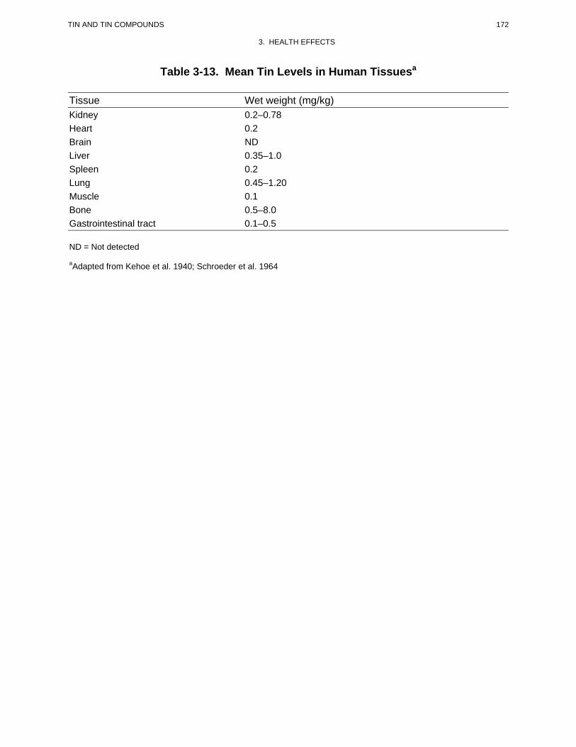
172 TIN AND TIN COMPOUNDS
3. HEALTH EFFECTS
Table 3-13. Mean Tin Levels in Human Tissuesa
Tissue Wet weight (mg/kg) Kidney 0.2–0.78 Heart 0.2 Brain ND Liver 0.35–1.0 Spleen 0.2 Lung 0.45–1.20 Muscle 0.1 Bone 0.5–8.0 Gastrointestinal tract 0.1–0.5
ND = Not detected
aAdapted from Kehoe et al. 1940; Schroeder et al. 1964
173 TIN AND TIN COMPOUNDS
3. HEALTH EFFECTS
exposures and, possibly the health/exposure history of the tissue donors (Tipton and Cook 1963; Tipton et
al. 1963). Additional information regarding tin and organotin levels in human tissues and fluids is
presented in Table 6-5.
When fresh human whole blood was incubated with triethyl[113Sn]tin chloride, the red blood cell:plasma
tin ratio was 1.9 (Rose and Aldridge 1968). This ratio was substantially different from the ratio observed
in rat blood (19), and similar to that in other rodent species (range, 1–5). Interspecies differences have
been attributed to variable binding of tin (or triethyltin) to hemoglobin (Rose 1969) and may also be
applicable to trimethyltin, which also shows a pronounced accumulation in rat red blood cells (Brown et
al. 1984; see Section 3.4.2.2 for further discussion).
3.4.2.1 Inhalation Exposure
No studies were located regarding distribution in humans or animals after inhalation exposure to
inorganic tin or organotin compounds.
3.4.2.2 Oral Exposure
Inorganic Tin Compounds. No studies were located regarding distribution in humans after oral exposure
to inorganic tin compounds; however, data are available on tissue levels of tin in general populations
(Schroeder et al. 1964; see Section 3.4.2).
Consistent with observations made of the tissue distribution of tin in humans, bone, kidney, and liver are
major sites of deposition of tin in rats and mice, after oral administration of inorganic tin compounds
(Hiles 1974; NTP 1982; Schroeder et al. 1968; Yamaguchi et al. 1980). Levels of tin in bone, kidney, and
liver were 0.02–1% of the administered dose of 113Sn in rats that received a single oral dose of 20 mg
Sn/kg/day as stannous pyrophosphate (113Sn[II]2P2O7), stannous fluoride (113Sn[II]F2), stannic fluoride
(113Sn[IV]F4), 113Sn[II]citrate, or 113Sn[IV]citrate (Hiles 1974). Levels in blood were 0.01% (or less) of
the administered dose. In rats that received 20 mg Sn/kg/day of stannous fluoride (113Sn[II]F2) or stannic
fluoride (113Sn[IV]F4), for a period of 28 days, levels of tin in kidneys and liver were approximately the
same as after a single oral dose (Hiles 1974); however, levels in bone were higher after multiple dosing,
suggesting slower elimination kinetics of tin from bone, relative to kidney and liver (see Section 3.4.4.4).
174 TIN AND TIN COMPOUNDS
3. HEALTH EFFECTS
Accumulation of tin in bone has also been observed in rats and mice exposed to stannous chloride
(Sn[II]Cl2) (NTP 1982; Yamaguchi et al. 1980). Tissue concentration ratios were approximately 43 for
bone:kidney and 32 for bone:liver after 90 days of oral (gavage) doses of stannous chloride; the
bone:tissue ratios increased with increasing doses of 0.3, 1, or 3 mg/kg/day (Yamaguchi et al. 1980).
Chronic exposures of rats, at much higher doses (60–70 mg/kg/day, 1,000 ppm in diet), resulted in tissue
concentration ratios of approximately 0.5 for bone:kidney and 55 for bone:liver; increasing the dose by a
factor of approximately 2, resulted in a proportional increase bone:kidney and bone:liver ratios (NTP
1982). Chronic exposures of mice (230–280 mg/kg/day, 1,000 ppm in diet), resulted in tissue
concentration ratios of approximately 30 for bone:kidney and 60 for bone:liver (NTP 1982). Bone:kidney
and bone:liver ratios, in rats and mice, were similar in female and males (NTP 1982). These studies
indicate a dose-dependence and possible species differences (i.e., mice compared to rats) in the tissue
distribution of tin after exposures to stannous chloride. However, it should be noted that the dosages
administered in the NTP (1982) study resulted in gastrointestinal tract toxicity, which may have affected
absorption of administered tin.
Schroeder et al. (1968) chronically exposed rats to 5 ppm stannous chloride in drinking water and found
relatively high concentrations of tin in spleen. Tissue concentration ratios (spleen:tissue) were:
kidney, 11; liver, 5; heart, 2; and lung, 3. Tin concentrations in brain were approximately twice that of
blood in rats exposed to stannous chloride (Sn[II]Cl2, 100, 250, or 500 mg/L, 63, 156, or 313 mg Sn/L)
for up to 18 weeks, and appeared to increase with increasing duration of exposure, suggesting the
possibility of accumulation of tin in brain with prolonged exposure to stannous chloride (Savolainen and
Valkomen 1986).
Animal studies in which absorbed tin was measured in tissues following parenteral injection of
Sn[II]chloride (113SnCl2), confirmed the above observations; i.e., that bone, kidney, and liver are major
sites of deposition of absorbed Sn[II] (see Section 3.4.4.4).
Tin was not detected in the uterine horns or combined fetuses and placentas in rats following daily
ingestion of 20 mg Sn/kg/day as 113SnF2, or 113SnF4 beginning on the day of conception (Hiles 1974).
However, on Gd 21, fetuses of dams administered 20 mg Sn/kg/day as SnF2 (approximately 100 mg Sn
cumulative dose) contained approximately 0.2 µg Sn/g, or approximately 0.2% of the cumulative
administered dose (detection limit, 0.1 µg/g). This suggests the possibility that, in the rat, tin
administered orally as stannous chloride may be transferred to the fetus.
175 TIN AND TIN COMPOUNDS
3. HEALTH EFFECTS
No studies on transfer of tin to breast milk following oral exposure (or exposure by other routes) to
inorganic tin compounds were located.
Organotin Compounds. No studies were located regarding distribution in humans after oral exposure to
organotin compounds.
Methyltin Compounds. Studies conducted in animals indicate that ingested methyltin compounds
distribute to soft tissues, with the highest levels usually observed in liver. Species differences in the
tissue distribution of trimethyltin have been observed. In marmosets, 1–13 days following a single oral
dose of 3–4.5 mg/kg trimethyltin chloride (1.8–2.4 mg Sn/kg), brain:blood concentration ratios ranged
from 6 to 10; whereas, in rats, 5 days following an oral dose of 10 mg/kg (6 mg Sn/kg), blood:brain ratios
were approximately 38 (Brown et al. 1979, 1984). Mushak et al. (1982) also observed relatively high
blood:tissue ratios of tin in neonatal rats that received oral doses of trimethyltin hydroxide (0.66 mg
Sn/kg/day, Pnds 2–29): brain, 42; kidney, 22; and liver, 8. Following multiple oral doses of 4 mg/kg
(2.4 mg Sn/kg) for 7 days, blood:brain ratios in the rat ranged from 30 to 48 (Brown et al. 1979). The
relatively high blood levels of tin in rats, compared to other species, have been attributed to a more
pronounced accumulation of trimethyltin in red blood cells. When samples of blood from rats were
incubated with trimethyltin, the blood:plasma concentration ratio was approximately 67, compared to
approximately 1 in the marmoset, gerbil, and hamster (Brown et al. 1984). The mechanism for the
difference has not been elucidated.
Ethyltin Compounds. In animals, ingested ethyltin, along with five dealkylation products, distribute to
soft tissues, including brain, kidney and liver. In rats, following 5 oral doses of 10 mg/kg/day triethyltin
(5.8 mg Sn/kg/day), tissue:blood concentration ratios of triethyltin were approximately: brain, 8; kidney,
4; and liver, 0.5 (Iwai et al. 1982b). Following the same dose of tetraethyltin (5.1 mg Sn/kg/day), both
tetra- and triethyltin were detected in tissues, reflecting dealkylation of tetraethyltin (see Section 3.4.3).
The brain:blood ratio of tetraethyltin was <1 whereas the brain:blood ratio of triethyltin was >8. In
neonatal rats that received oral doses of triethyltin sulfate (0.44 mg Sn/kg/day, Pnds 2–29), tissue:blood
tin concentration ratios were: 0.7, brain; kidney, 0.8; liver, 3. 4 (Mushak et al. 1982). The higher
liver:blood ratio of total tin, compared to that of triethyltin, following ingestion of triethyltin, also may
reflect the dealkylation of trimethyltin in the liver (see Section 3.4.3).
Butyltin Compounds. Similar to ethyltin compounds, ingested butyltin compounds and their dealkylation
products distribute to soft tissues, including brain, kidney, and liver. In rats, following five oral doses of
176 TIN AND TIN COMPOUNDS
3. HEALTH EFFECTS
10 mg/kg tributyltin (5.8 mg Sn/kg), tissue:blood concentration ratios of tributyltin were approximately:
brain, <1; kidney, 2–4; and liver 1–2 (Iwai et al. 1982b). Following the same dose of tetrabutyltin,
tissue:blood concentration ratios of tetrabutyltin were approximately: brain, <1; kidney, 10–12; and
liver, 20. In rats given a single oral dose of 40 mg/kg tributyltin fluoride (15 mg Sn/kg), transient
elevations in tributyltin, dibutyltin, monobutyltin, and inorganic tin were observed in brain and liver over
the 8-day period following the dose, indicating that dealkylation had occurred (Iwai et al. 1981, see
Section 3.4.3). In neonatal rats that received oral doses of tributyltin acetate (1.0 mg Sn/kg/day, Pnds 2–
29), tissue:blood tin concentration ratios(possibly limited by the detection limit for blood tin) were:
brain, 2.6; kidney, 19; and liver, 24 (Mushak et al. 1982).
In rats exposed to tributyltin oxide in the diet (0.25, 1, 4, or 16 mg/kg/day; 0.1, 0.4, 1.6, or 6.4 mg
Sn/kg/day) for 4 weeks, total tin in kidney, liver, and brain increased with increasing dosage, and were
similar in females and males (Krajnc et al. 1984). Levels in the brain and adipose tissue were 10–20% of
the kidney and liver levels.
Twenty-four hours after administration of a single dose of 22 mg dibutyltin diacetate/kg to pregnant rats
on Gd 8, dibutyltin and monobutyltin were detected in maternal blood and liver, and in the embryos,
indicating placental transfer (Noda et al. 1994). In the embryos, the concentration of dibutyltin was 6–
7 times higher than that of monobutyltin. Nakamura et al. (1993) had also detected dibutyltin in fetuses
on Gd 18 after administration of the chemical to the pregnant rats on Gd 7–17.
Phenyltin Compounds. Studies conducted in animals indicate that ingested phenyltin compounds and
dearylated metabolites (see Section 3.4.3) distribute to soft tissues, including brain, kidney, liver, and
pancreas. In hamsters and rats, following a single oral dose of 50 mg triphenyltin chloride (15 mg Sn/kg),
triphenyltin and dearylated metabolites, including inorganic tin, were detected in brain, blood, kidney,
liver and pancreas (Ohhira and Matsui 1996). The highest concentrations of triphenyltin and metabolites
were found in liver and kidney. In rats, tissue:blood ratios for triphenyltin, 48 hours after the dose, were
approximately: brain, 10; kidney, 21; liver, 17; and pancreas, 4. Similar ratios were observed in hamsters:
brain, 9; kidney, 11; liver, 21; and pancreas, 11. In neonatal rats that received oral doses of triphenyltin
acetate (0.87 mg Sn/kg/day, Pnds 2–29), tissue:blood tin concentration ratios (based on the detection limit
for blood) were: brain, 3; kidney, 6; and liver, 14 (Mushak et al. 1982).
177 TIN AND TIN COMPOUNDS
3. HEALTH EFFECTS
In rats, following a single oral dose of 55.4 mg/kg tetraphenyltin (15 mg Sn/kg), tetraphenyltin, and the
dearylated metabolites (tri-, di-, and monophenyltin) were detected in kidney and liver (Ohhira and
Matsui 2003).
3.4.2.3 Dermal Exposure
No studies were located regarding distribution in humans or animals after dermal exposure to inorganic
tin or organotin compounds.
3.4.2.4 Other Routes of Exposure
Inorganic Tin Compounds. Animal studies, in which absorbed tin was measured in tissues following
parenteral injection of Sn[II]chloride (113SnCl2), confirm observations from oral exposure studies
(Section 3.4.2.3) that bone, kidney, and liver are major sites of deposition of absorbed Sn[II] (Furchner
and Drake 1976; Hiles 1974). In rats that received an intraperitoneal injection of stannous chloride
(113Sn[II]Cl2, 0.006 µg/kg), tissue levels (percent of body burden) 1 day after dosing were: bone, 50%,
kidney, 3.5%; liver, 6%; and skeletal muscle, 20%; thus, muscle also appears to a major site of deposition
of Sn[II] (Furchner and Drake 1976).
Organotin Compounds. Animal studies, in which organotin compounds were administered parenterally
confirm observations made following oral exposures; organotin compounds distribute to soft tissues,
including brain, kidney, and liver.
Methyltin Compounds. In rats administered a single intraperitoneal dose of 6 mg/kg trimethyltin (4.3 mg
Sn/kg), tin distributed to brain, heart, kidney, and liver, with levels in brain (ng/g protein) that were 15–
50% of that in other tissues (Cook et al. 1984a). Tin distribution was uniform across the brain regions,
cerebellum, medulla-pons, hypothalamus, hippocampus, and striatum (Cook et al. 1984a). Tin in brain,
kidney, and liver were lower after a dose of 6 mg/kg trimethyltin (4.3 mg Sn/kg) than after a dose of
6 mg/kg triethyltin (2.5 mg Sn/kg).
Ethyltin Compounds. In rats, 5 days following a single intravenous dose of 10 mg/kg triethyl[113Sn]tin
chloride (5 mg Sn/kg), tissue:blood tin ratios were: liver, 1.3; kidney, 0.6; brain, 0.2; skeletal muscle,
0.15; and spinal cord, 0.1; levels were similar in brain stem cerebellum, cerebrum, and cortex (Rose and
Aldridge 1968). These observations are consistent with those of Cook et al. (1984a): following a single
178 TIN AND TIN COMPOUNDS
3. HEALTH EFFECTS
intraperitoneal dose of 6 mg/kg triethyltin (3.5 mg Sn/kg), similar levels of tin were observed in heart,
kidney, and liver, and levels in brain were 15–25% of that of kidney and liver. Increasing the intravenous
dose of triethyltin from approximately 0.6 to 5 mg/kg (0.3–2.5 mg Sn/kg) in rats resulted in proportional
increases in levels of tin in blood, brain, kidney, and liver, with no evidence of a limitation in capacity for
deposition in these tissues (Rose and Aldridge 1968).
Species differences in deposition of tin in red blood cells have been observed following parenterally-
administered triethyltin to animals (Rose and Aldridge 1968). In rats, 4–5 hours following a single
intravenous dose of 10 mg/kg triethyl[113Sn]tin chloride (5 mg Sn/kg), substantially higher levels of tin in
blood (relative to other tissues) were observed, compared to the hamster and guinea pig, although the
distribution to other tissues was similar among rodent species. When samples of whole blood from rats
were incubated with triethyl[113Sn]tin chloride, the red blood cell:plasma tin concentration ratio was 19;
ratios observed in blood from other rodent species were considerably lower (1–5) and, in human blood,
the ratio was 1.9, suggesting that the mechanism for the species differences in red blood cell:plasma ratios
involved uptake and/or retention of triethyltin (or tin derived from triethyltin) in red blood cells (Rose and
Aldridge 1968). Rat hemoglobin bound more 113Sn when incubated with triethyl[113Sn]tin than
hemoglobins isolated from other rodents, or from humans (Rose and Aldridge 1968).
3.4.3 Metabolism
Inorganic Tin Compounds. No studies were located in humans or animals on metabolism of inorganic
tin after inhalation, oral, or dermal exposure.
Organotin Compounds. No studies were located in humans on metabolism after inhalation, oral, or
dermal exposure to organotin compounds. Microsomes prepared from human liver dealkylate tributyltin
to form di- and monobutyltin metabolites, suggesting that similar pathways may be active in humans, in
vivo (Ohhira et al. 2003). This would be consistent with the detection of dibutyltin and monobutyltin in
postmortem human liver samples (Nielsen and Strand 2002) and with the more substantial evidence for
dealkylation of alkyltins, including butyltins, in various nonhuman species (see below).
Ethyltin Compounds. Studies conducted in rats indicate that tetra-, tri- and diethyltin undergo
dealkylation to ethyltin compounds (Bridges et al. 1967; Cremer 1958). Dealkylation and hydroxylation
of the ethyl moieties are catalyzed by microsomal monooxygenase(s) of liver, and possibly other tissues
(Kimmel et al. 1977).
179 TIN AND TIN COMPOUNDS
3. HEALTH EFFECTS
Butyltin Compounds. Studies conducted in rats indicate that tributyltin undergoes dealkylation to di- and
monobutyltin compounds (Iwai et al. 1981, 1982; Matsuda et al. 1993; Ueno et al. 1994). The butyl
moieties are also oxidized at carbon 3 to yield 3-hydroxybutyl and 3-oxobutyl metabolites; and at carbon
4, to yield the 4-hydroxybutyl and 3-carboxy metabolites (Matsuda et al. 1993). The simple dealkylation
products were the principal metabolites detected in blood and brain following a 2 mg/kg oral dose of
tributyltin chloride, whereas in kidney and liver, hydroxy-, carboxy-, and oxo-metabolites were the
dominant metabolites (Matsuda et al. 1993). Dealkylation and hydroxylation are catalyzed by
microsomal monooxygenase(s) (Kimmel et al. 1977; Ohhira et al. 2003). The alkyl products of
dealkylation are conjugated with glutathione and further metabolized to mercapturic acid derivatives
(Suzuki et al. 1999b).
Phenyltin Compounds. Studies conducted in hamsters and rats indicate that tetra-, tri-, di-, and
monophenyltin compounds are dearylated. The dearylated metabolites, including inorganic tin, can be
found in kidney and liver after an oral exposure to phenyltin compounds (Ohhira and Matsui 1993a,
1993b, 2003; Ohirra et al. 1996). Dearylation of phenyltin compounds is catalyzed by microsomal
monooxygenase(s) in liver, and possibly in other tissues (Ohhira et al. 2003). A recent study of CYP
isoforms in rat hepatocytes showed that CYP2B1 had a small metabolic capacity for triphenyltin, but the
principal CYP for triphenyltin metabolism in rats was CYP2C6 (Ohhira et al. 2004). Support for this
finding was provided by experiments in which anti-rat CYP2C6 antibodies and cimetidine, a selective
CYP2C6 inhibitor, inhibited triphenyltin dearylation activity in the hepatic microsomes of rats (Ohhira et
al. 2004).
3.4.4 Elimination and Excretion
3.4.4.1 Inhalation Exposure
Inorganic Tin Compounds. No studies were located regarding excretion in humans or animals after
inhalation exposure to inorganic tin.
Organotin Compounds. No studies were located regarding excretion in humans or animals after
inhalation exposure to inorganic tin, although tin was detected in the urine from a fatal inhalation case
described by Rey et al. (1984).
180 TIN AND TIN COMPOUNDS
3. HEALTH EFFECTS
3.4.4.2 Oral Exposure
Inorganic Tin Compounds. Feces and urine are major routes of excretion of ingested tin in humans
(Calloway and McMullen 1966; Johnson and Greger 1982, see Section 3.1.4.2). In eight healthy adult
males who were placed on diets (for 20 days) containing 0.1 mg Sn/day (basal diet) or 50 mg Sn/day
(supplemented with stannous chloride), average fecal excretion was 55% of the daily intake in the basal
group and 97% in the supplemented group (Johnson and Greger 1982). Average urinary excretion was
26% of the daily intake in the basal group and 2.4% in the supplemented group, suggesting that urine was
a major route of excretion of absorbed tin. These and other observations (Calloway and Mullen 1966)
also suggest that gastrointestinal absorption of tin in humans may decrease with increasing dose, possibly
reflecting a tight homeostatic control of tin absorption (see Section 3.4.1.2).
In dogs, mice, rats, and Rhesus monkeys, tin ingested as Sn[II] or Sn[IV] compounds is excreted
primarily in feces; however, urine and bile appear to be major routes of excretion of absorbed Sn[II] (see
Section 3.4.4.4). In rats, 48 hours after dosing (20 mg Sn/kg/day), 95% of the administered 113Sn (as Sn
[II], Sn [IV] citrate, Sn[II]2P2O7, Sn[II]F2, or Sn[IV]F4) was recovered in feces, while <1% was detected
in urine (Hiles 1974). Similar results were obtained in dogs, mice, rats, and Rhesus monkeys (Furchner
and Drake 1976).
Whole body and tissue retention kinetics have been measured in mice, monkeys, rats, and dogs after an
oral gavage dose of stannous chloride (Sn[II]Cl2) (Furchner and Drake 1976). Whole-body elimination
kinetics of absorbed Sn[II] were similar in all four species and could be described with 1- or
2-compartment models in which 96–100% of the initial body burden is eliminated with a half-time of
0.2–0.4 days. In Rhesus monkeys (dose, 0.0004 µg/kg), approximately 96% of the body burden was
eliminated with a half-time of 0.3 days (reflecting mainly excretion of unabsorbed tin in feces), and 4%
was eliminated with a half-time of 3 days.
Organotin Compounds. No studies were located regarding excretion in humans after oral exposure to
organotin compounds.
Methyltin Compounds. In rats, after a single oral dose of 3 mg/kg (1.8 mg Sn/kg; Brown et al. 1984),
blood concentrations of trimethyltin decreased by one-half in approximately 3 days and, in brain, in
approximately 2 (or less) days. Information identifying the relative contributions of various excretory
routes for elimination of methyltin compounds was not located.
181 TIN AND TIN COMPOUNDS
3. HEALTH EFFECTS
Ethyltin Compounds. Studies in rats indicate that urine and feces are the major routes of excretion of
ethyltin following oral exposures to ethyltin compounds (Bridges et al. 1967). Over a 3-day period
following a single oral dose of approximately 25 mg/kg ethyltin trichloride (12 mg Sn/kg), rats excreted
92% (range 89–95%) of the dose in feces and 1.2% (range 1–2%) in urine (Bridges et al. 1967).
Following absorption, ethyltin is excreted predominantly in urine, whereas absorbed diethyltin and
triethyltin are excreted to a greater extent in feces (See Section 3.4.4.4).
Butyltin Compounds. In mice that received a single oral dose of 1.2 mg/kg tri[14C]butyltin acetate,
approximately 16% of the 14C dose was excreted in urine in 5 days, 53% was excreted in feces, and 22%
was exhaled as [14C]CO2 (Kimmel et al. 1977). Following a similar dose di[14C]butyltin diacetate the
excretion pattern (% of dose) was: urine, 10%; feces, 66%; and carbon dioxide, 7% (Kimmel et al. 1977).
In mice, the amount of tin excreted in urine following an oral dose of different butyltins also increased
with the number of butyl groups. Five days following a single oral dose of 180 µmol/kg of tri-, di-, or
monobutyltin, urinary excretion of tin (percent of dose) was approximately: tributyltin, 5%; dibutyltin,
3%; and butyltin, 0.3% (Ueno et al. 1994). These differences may reflect real differences in urinary
excretion of absorbed tin compounds, or greater absorption of the tin compounds having a larger number
of butyl groups.
Phenyltin Compounds. Information or the rates or relative contributions of various excretory routes for
elimination of phenyltin compounds was not located.
3.4.4.3 Dermal Exposure
No studies were located regarding excretion in humans or animals after dermal exposure to inorganic tin
or organotin compounds.
3.4.4.4 Other Routes of Exposure
Inorganic Tin Compounds. Studies in which inorganic tin compounds have been parenterally injected
into animals have shown that absorbed inorganic tin is excreted in urine and bile. Forty-eight hours after
an intravenous injection to rats of 2 mg Sn/kg, as 113Sn [II] citrate, 35% of the administered radioactivity
was excreted in urine and approximately 12% was (2 mg Sn/kg, as 113Sn [II] citrate) was excreted in the
feces. In bile-duct cannulated rats, 23% was excreted in the urine, 11% in bile, and 2% in the feces.
These observations indicate that urine and bile appear to be major routes of excretion of absorbed Sn[II].
182 TIN AND TIN COMPOUNDS
3. HEALTH EFFECTS
The biliary contribution to excretion of Sn[IV] was less than that of Sn[II], and after intravenous injection
of 2 mg Sn/kg, as 113Sn [IV] citrate, 40% of the administered radioactivity was excreted in urine and 3%
in feces. Following the same intravenous dose administered to rats that had bile duct cannulas, 25% of
the administered dose was excreted in urine and 0.5% in bile (Hiles 1974). Urine-feces excretion ratios
after intravenous injection of stannous chloride were approximately 10 in dogs, 3 in mice and rats, and
5 in Rhesus monkeys (Furchner and Drake 1976).
Rates of elimination of absorbed inorganic tin have been measured in mice, monkeys, rats, and dogs after
intravenous or intraperitoneal injection of stannous chloride (Sn[II]Cl2) (Furchner and Drake 1976).
Whole-body elimination kinetics of absorbed Sn[II] were similar in all four species and could be
described with similar 4-compartment models. In Rhesus monkeys (dose, 0.0004 µg/kg), approximately
39% of the body burden was eliminated with a half-time of 0.6 days, 11% was eliminated with a half-time
of 5 days, 8% eliminated with a half-time of 24 days, and 42% was eliminated with a half-time of
88 days. The pseudo-first order elimination half-times (all components combined) were approximately
7 days in monkeys and 1–2 days in dogs, mice, and rats; the difference reflects the larger contribution of
the slow compartment in the monkey (42%) compared to the other three species (20–30%).
Measurements of the elimination kinetics of absorbed Sn[II], from individual tissues in rats (dose,
0.006 µg/kg), suggested that bone was a major contributor to the slowest compartment, comprising
approximately 50% of the body burden 1 day after dosing, and approximately 70–75% of the body burden
from days 6–113 after dosing. Elimination rates were similar in all soft tissues measured (blood, brain,
kidney, liver, spleen). These observations are consistent with the observations of accumulation of tin in
bone with repeated exposures to Sn[II] compounds (Hiles 1974; NTP 1982; Yamaguchi et al. 1980).
Methyltin Compounds. The elimination kinetics of tin from tissues, following parenteral injection of
trimethyltin has been investigated in the rat. Cook et al. (1984a) administered single intraperitoneal doses
of 6 mg/kg trimethyltin bromide (4.4 mg Sn/kg) to rats and measured tissue tin over 22 days following the
dose. The pseudo-first order elimination half-times for tin were: blood, 10 days; brain, 10 days; heart,
11 days; kidney, 12 days; and liver, 15 days. These rates were slower than those estimated for tin
following a dose of triethyltin (Cook et al. 1984a). Ekuta et al. (1998) derived empirical, single-
compartment models of the kinetics of 14C in blood of four inbred mouse strains following single
intraperitoneal injections of tri[14C]methyltin. Elimination half-times were 28.5 hours (AKR/J),
31.3 hours (BalbcByJ), 30.0 hours (C57Bi6J), and 57.4 hours (DBA/2J).
183 TIN AND TIN COMPOUNDS
3. HEALTH EFFECTS
Ethyltin Compounds. Following absorption, ethyltin is excreted predominantly in urine, whereas
absorbed diethyltin is excreted to a much greater extent in feces. In rats, during the 72-hour period
following an intraperitoneal dose of 12.7 mg/kg ethyltin trichloride (6 mg Sn/kg), 73% of the dose was
excreted in urine and none in feces; approximately 4% of the dose was secreted into bile (Bridges et al.
1967). Biliary secretion appears to be quantitatively more important in the excretion of absorbed
diethyltin, compared to ethyltin. During the 72 hours following an intraperitoneal dose of 10 mg/kg
[14C]diethyltin dichloride (5 mg Sn/kg), approximately 64% of the administered dose of tin was excreted
in feces and 31% in urine (fecal:urine ratio, 2.2); 32% of the administered 14C was excreted in feces and
19% in urine (fecal:urine ratio, 1.8). In rats in which the bile duct had been cannulated, 56% of an
intraperitoneal dose of [14C]diethyltin was secreted into bile; essentially all of the 14C secreted into bile
was identified as diethyltin. Biliary secretion of triethyltin has also been observed in hamsters and guinea
pigs, following intraperitoneal injection of 10 mg/kg triethyl[113Sn]tin chloride (5 mg Sn/kg) (Rose and
Aldridge 1968).
In rats, absorbed tetraethyltin appears to be excreted primarily as the trialkyltin metabolite. During the
3 days following a subcutaneous dose of 10 mg/kg triethyltin in rats, approximately 0.20% of the dose
was excreted in urine and 0.08% in feces (urine:feces ratio, 2.5; Iwai et al. 1982b). During the 3 days
following a subcutaneous dose of 10 mg/kg tetraethyltin in rats, approximately 0.13% of the dose was
excreted in urine and 0.07% in feces (urine:feces ratio, 1.9); however, no tetraethyltin was detected in
excreta.
The elimination kinetics of tin from tissues, following parenteral injection of triethyltin has been
investigated in the rat. Cook et al. (1984a) administered single intraperitoneal doses of 6 mg/kg triethyltin
as the bromide salt (3.5 mg Sn/kg) to rats and measured tissue tin levels over 22 days following the dose.
The pseudo-first-order elimination half-times for tin were: blood, 2.5 days; brain, 4.6 days; heart,
3.4 days; kidney, 5.6 days; and liver, 6.1 days. These estimates are consistent with rates of decline in tin
levels measured in blood, brain, kidney, and liver 1–5 days following a single intravenous dose of
10 mg/kg triethyl[113Sn]tin chloride (5 mg Sn/kg) in rats (Rose and Aldridge 1968).
Butyltin Compounds. In rats, absorbed tetrabutyltin appears to be excreted as the trialkyltin metabolite.
In rats, during the 3 days following a subcutaneous dose of 10 mg/kg tributyltin, approximately 0.18% of
the dose was excreted in urine and 0.04% in feces (urine:feces ratio, 4.5; Iwai et al. 1982b). Following
the same subcutaneous dose of tetrabutyltin, approximately 0.12% of the dose was excreted in urine and
0.16% in feces (urine:feces ratio, 0.8); however, no tetrabutyltin was detected in excreta.
184 TIN AND TIN COMPOUNDS
3. HEALTH EFFECTS
3.4.5 Physiologically Based Pharmacokinetic (PBPK)/Pharmacodynamic (PD) Models
Physiologically based pharmacokinetic (PBPK) models use mathematical descriptions of the uptake and
disposition of chemical substances to quantitatively describe the relationships among critical biological
processes (Krishnan et al. 1994). PBPK models are also called biologically based tissue dosimetry
models. PBPK models are increasingly used in risk assessments, primarily to predict the concentration of
potentially toxic moieties of a chemical that will be delivered to any given target tissue following various
combinations of route, dose level, and test species (Clewell and Andersen 1985). Physiologically based
pharmacodynamic (PBPD) models use mathematical descriptions of the dose-response function to
quantitatively describe the relationship between target tissue dose and toxic end points.
PBPK/PD models refine our understanding of complex quantitative dose behaviors by helping to
delineate and characterize the relationships between: (1) the external/exposure concentration and target
tissue dose of the toxic moiety, and (2) the target tissue dose and observed responses (Andersen et al.
1987; Andersen and Krishnan 1994). These models are biologically and mechanistically based and can
be used to extrapolate the pharmacokinetic behavior of chemical substances from high to low dose, from
route to route, between species, and between subpopulations within a species. The biological basis of
PBPK models results in more meaningful extrapolations than those generated with the more conventional
use of uncertainty factors.
The PBPK model for a chemical substance is developed in four interconnected steps: (1) model
representation, (2) model parametrization, (3) model simulation, and (4) model validation (Krishnan and
Andersen 1994). In the early 1990s, validated PBPK models were developed for a number of
toxicologically important chemical substances, both volatile and nonvolatile (Krishnan and Andersen
1994; Leung 1993). PBPK models for a particular substance require estimates of the chemical substance-
specific physicochemical parameters, and species-specific physiological and biological parameters. The
numerical estimates of these model parameters are incorporated within a set of differential and algebraic
equations that describe the pharmacokinetic processes. Solving these differential and algebraic equations
provides the predictions of tissue dose. Computers then provide process simulations based on these
solutions.
The structure and mathematical expressions used in PBPK models significantly simplify the true
complexities of biological systems. If the uptake and disposition of the chemical substance(s) are
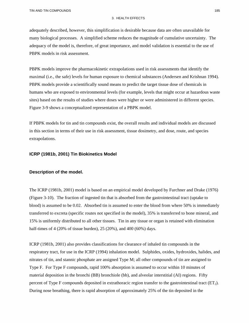
185 TIN AND TIN COMPOUNDS
3. HEALTH EFFECTS
adequately described, however, this simplification is desirable because data are often unavailable for
many biological processes. A simplified scheme reduces the magnitude of cumulative uncertainty. The
adequacy of the model is, therefore, of great importance, and model validation is essential to the use of
PBPK models in risk assessment.
PBPK models improve the pharmacokinetic extrapolations used in risk assessments that identify the
maximal (i.e., the safe) levels for human exposure to chemical substances (Andersen and Krishnan 1994).
PBPK models provide a scientifically sound means to predict the target tissue dose of chemicals in
humans who are exposed to environmental levels (for example, levels that might occur at hazardous waste
sites) based on the results of studies where doses were higher or were administered in different species.
Figure 3-9 shows a conceptualized representation of a PBPK model.
If PBPK models for tin and tin compounds exist, the overall results and individual models are discussed
in this section in terms of their use in risk assessment, tissue dosimetry, and dose, route, and species
extrapolations.
ICRP (1981b, 2001) Tin Biokinetics Model
Description of the model.
The ICRP (1981b, 2001) model is based on an empirical model developed by Furchner and Drake (1976)
(Figure 3-10). The fraction of ingested tin that is absorbed from the gastrointestinal tract (uptake to
blood) is assumed to be 0.02. Absorbed tin is assumed to enter the blood from where 50% is immediately
transferred to excreta (specific routes not specified in the model), 35% is transferred to bone mineral, and
15% is uniformly distributed to all other tissues. Tin in any tissue or organ is retained with elimination
half-times of 4 (20% of tissue burden), 25 (20%), and 400 (60%) days.
ICRP (1981b, 2001) also provides classifications for clearance of inhaled tin compounds in the
respiratory tract, for use in the ICRP (1994) inhalation model. Sulphides, oxides, hydroxides, halides, and
nitrates of tin, and stannic phosphate are assigned Type M; all other compounds of tin are assigned to
Type F. For Type F compounds, rapid 100% absorption is assumed to occur within 10 minutes of
material deposition in the bronchi (BB) bronchiole (bb), and alveolar interstitial (AI) regions. Fifty
percent of Type F compounds deposited in extrathoracic region transfer to the gastrointestinal tract (ET2).
During nose breathing, there is rapid absorption of approximately 25% of the tin deposited in the
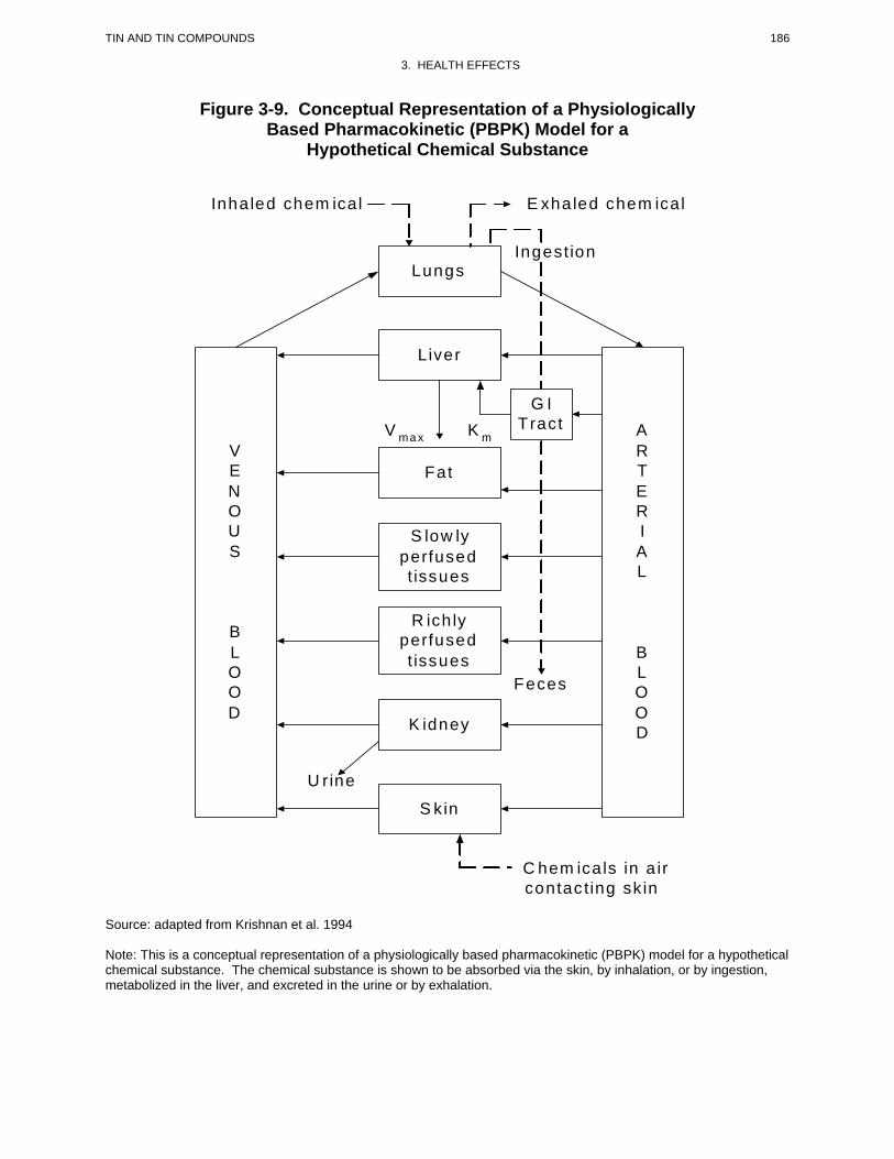
Inha led chem ica l E xhaled chem ica l
Lungs
Liver
Fat
S low ly perfused t issues
R ich ly perfused t issues
K idney
S kin
V E N O U S
B L O O D
A R T E R I A L
B L O O D
V ma x K m
Ingest ion
G I T ract
Fe ces
U r ine
C hem ica ls in a ir contac ting sk in
186 TIN AND TIN COMPOUNDS
3. HEALTH EFFECTS
Figure 3-9. Conceptual Representation of a Physiologically Based Pharmacokinetic (PBPK) Model for a
Hypothetical Chemical Substance
Source: adapted from Krishnan et al. 1994
Note: This is a conceptual representation of a physiologically based pharmacokinetic (PBPK) model for a hypothetical chemical substance. The chemical substance is shown to be absorbed via the skin, by inhalation, or by ingestion, metabolized in the liver, and excreted in the urine or by exhalation.
Blood Other Tissues Bone Mineral 35% 15%
20%
*Elimination half -life
Gastrointestinal Tract
20%
Excreta
4 days*
20%
25 days*
20%
400 days*
20%
4 days*
20%
25 days*
20%
25 days*
50%
TIN AND TIN COMPOUNDS 187
3. HEALTH EFFECTS
Figure 3-10. ICRP (1981b, 2001) Tin Biokinetic Model
188 TIN AND TIN COMPOUNDS
3. HEALTH EFFECTS
extrathoracic region, and 50% absorption during mouth breathing. For Type M compounds,
approximately 70% of the tin deposited in AI eventually is transferred to blood and there is rapid
absorption of about 10% of the tin deposited in BB and bb, and 5% of tin deposited in ET2. During nose
breathing, approximately 2.5% of the deposit in ET is rapidly absorbed and 5% is rapidly absorbed during
mouth breathing.
Validation of the model.
The extent to which the ICRP model has been validated is not described in ICRP (1981b).
Risk assessment.
The model has been used to establish radiation dose equivalents (Sv/Bq) of ingested and inhaled
radioactive tin isotopes for ages 1 day to 50 years (ICRP 2001).
Target tissues.
The model is designed to calculate intake limits for radioactive tin, based on radiation dose to all major
organs, including the bone surfaces, bone marrow, and liver, to which the highest doses would be
expected.
Species extrapolation.
The model is designed for applications to human dosimetry and cannot be applied to other species without
modification.
Interroute extrapolation.
The ingestion model (ICRP 1981b, 2001), together with the respiratory tract model (ICRP 1994) are
designed to simulate oral and inhalation exposures to tin and cannot be applied to other routes of exposure
without modification.
189 TIN AND TIN COMPOUNDS
3. HEALTH EFFECTS
3.5 MECHANISMS OF ACTION
3.5.1 Pharmacokinetic Mechanisms
Absorption. The mechanism(s) of absorption of inorganic and organotin compounds have not been
elucidated. In rats, following five oral doses of 10 mg/kg/day of tetra- or triethyltin, or tetra- or
tributyltin, the alkyltin in the gastrointestinal tract tissue was primarily associated with the duodenum and
jejunum indicating that these may be sites of absorption (Iwai et al. 1982b). A recent study with the
Caco-2 human intestinal cell-line suggested that, in general, butyltins have a low in vivo permeability
(Azenha et al. 2004). The study also showed that the permeability pattern correlated with the in vivo
toxicity (trialkyltin > dialkyltin > monoalkyltin). However, the accumulation pattern (dialkyltin >
trialkyltin > monoalkyltin) was different than that of permeability, presumably due to the strong affinity
of dibutyltin for dithiol groups. Finally, the permeability of monobutyltin and dibutyltin, but not that of
tributyltin, was found to be dependent of the paracellular route status.
Distribution. Inorganic tin deposits in bone mineral; however, the mechanisms for the uptake and
retention in bone have not been elucidated.
Species differences have been demonstrated in the distribution of methyltin and ethyltin compounds
within whole blood. Rats show higher red blood cell:plasma concentration ratios than other species,
including humans and nonhuman primates (Brown et al. 1984; Rose and Aldridge 1968). Uptake and
retention of methyltin and ethyltin in red blood cells has been attributed to binding to hemoglobin;
however, the mechanism for the difference has not been elucidated. When hemoglobin from rat red blood
cells is incubated with triethyl[113Sn]tin, radioactivity was bound to hemoglobin with an apparent affinity
constant of approximately 3.5x10⁵ M-1 (Rose 1969). The pH-dependence of binding is consistent with
involvement of histidine residues in hemoglobin (Rose 1969).
The subcellular distribution of tin, following parenterally-administered trimethyltin or triethyltin, has
been examined in rats and guinea pigs (Cook et al. 1984a; Rose and Aldridge 1968). Fractionation of
brain tissue from animals that received a single intraperitoneal dose of 6 mg/kg trimethyltin (4.3 mg
Sn/kg), revealed that tin was associated with the crude mitochondrial and microsomal fractions and,
within the crude mitochondrial fraction, tin was associated with myelin, synaptosomes, and mitochondria
(Cook et al. 1984b). The concentration in the mitochondrial fraction increased over time, reaching a
maximum of 5 days after the dose. Subcellular concentrations of tin were 4–20 times lower after the
6 mg/kg dose of trimethyltin (4.3 mg Sn/kg), compared to the 6 mg/kg dose of triethyltin (2.5 mg Sn/kg).
190 TIN AND TIN COMPOUNDS
3. HEALTH EFFECTS
The subcellular distribution of tin in kidney and liver was similar in rats and guinea pigs following a
parenteral dose of triethyltin; however, differences were observed between the distribution in rat brain
compared to kidney and liver (Rose and Aldridge 1968). In rats, 2 hours following a single intravenous
dose of 10 mg/kg triethyl[113Sn]tin chloride (5 mg Sn/kg), approximately 60% of the 113Sn was associated
with the 40,000 x g supernatant (cytosolic) fraction and approximately 13% was associated with the
heavy mitochondrial fraction. In brain, approximately 32% was associated with the supernatant fraction
and 40% with the heavy mitochondrial fraction. Cook et al. (1984b) also found tin associated with the
crude mitochondrial (13,000 x g) fraction of rat brain after a single intraperitoneal dose of 6 mg/kg
triethyltin bromide (2.5 mg Sn/kg). Subfractionation of the crude mitochondrial fraction on a Ficoll
gradient revealed tin associated with the myelin, synaptosomes, and mitochondria.
In a comparative study with rats, mice, and guinea pigs, the susceptibility to develop liver damage
followed the order: mice > rats > guinea pigs (Ueno et al. 2003b). This appeared to be partly due to
differences in metabolism of tributyltin and in the distribution of dibutyltin within cell organelles. In
hepatocytes of mice treated with dibutyltin chloride, 41% of the dibutyltin was found in organelles
compared to 27% in rats and 9% in guinea pigs (Ueno et al. 2003b).
Metabolism. No information on speciation of inorganic tin after absorption was located; therefore, the
extent to which Sn[II] and Sn[IV] are interconverted is unknown. The major metabolic pathways for
alkyltin compounds include dealkylation and hydroxylation and further oxidation of the alkyl moieties
(see Section 3.4.3). These reactions have been found to occur in the microsomal fraction of liver
(including human liver), are dependent on reduced nicotinamide adenosine dinucleotide phosphate
(NADPH), and are inhibited by carbon monoxide, suggesting involvement of cytochrome P-450 (Kimmel
et al. 1977; Ohhira et al. 2003). For triphenyltin, CYP2C6 constitutes the principal CYP for dearylation
in hepatic microsomes of rats (Ohhira et al. 2004).
Several studies have examined the involvement of metabolism in the toxicity of some organotin
compounds. Cases have been described in which metabolism can either increase or decrease the toxicity
of these compounds. For example, studies in hamsters showed that pretreatment of the animals with the
cytochrome P-450 inducer, phenobarbital (PB), suppressed the diabetogenic effects of triphenyltin
compared to PB-untreated hamsters (Ohhira et al. 1999). Pretreatment with the CYP1A and 2A inducers
β-naphthoflavone and 3-methylcholanthrene, was not as effective as PB in preventing the organotin
induced hyperglycemia and hypertriglyceridemia. On the other hand, pretreatment with the
191 TIN AND TIN COMPOUNDS
3. HEALTH EFFECTS
P-450 inhibitor, SKF-525A, increased the diabetogenic effects of triphenyltin (Ohhira et al. 2000).
Overall, these findings suggested that the hyperglycemic toxicity of triphenyltin is due primarily to
accumulation of the parent compound, triphenyltin, in the pancreas, and not to a metabolite. Studies by
Ueno et al. (1995, 1997) showed that the liver toxicity of tributyltin chloride could be prevented by
treatment of the mice with the cytochrome P-450 inhibitor SKF-525A and that pretreatment with the
P-450 inducer, PB increased the toxicity of tributyltin. These results suggested that the liver toxicity of
tributyltin is due to a metabolite, most likely dibutyltin.
Excretion. Bile, feces, and urine are major routes of elimination of absorbed inorganic and organotin
compounds (see Section 3.4.4.4). Information on the mechanisms of biliary secretion and urinary
excretion of tin compounds was not located.
3.5.2 Mechanisms of Toxicity
Studies in laboratory animals have shown that exposure to tin and tin compounds can produce a wide
array of effects, but it is unknown whether exposure of humans to levels of tin compounds found in the
environment will cause similar effects. General mechanisms of neurotoxicity and immunotoxicity of
organotin compounds are briefly discussed below, as effects on these two systems may cause the most
concern following potential exposures of humans to these substances. Although exposure to toxic
amounts of neurotoxic organotins is somewhat unlikely, studies with trimethyltin and triethyltin in
animals have shown a very steep dose-response curve for these substances and similar severe effects have
been observed following acute high exposure of humans. Exposure to immunotoxic organotins, such as
tributyltin, is much more likely because of substances’ use and, environmental prevalence, based on
monitoring data.
Neurotoxicity. Trimethyltin has been shown to produce degenerative lesions in the hippocampus and
associated structures of the limbic system (e.g., dentate gyrus) in nonhuman primates and in several
rodent species (see Section 3.2.2.4; Koczyk 1996). The lesions have been characterized as neuronal cell
apoptosis and are accompanied with astrocyte swelling and reactive gliosis (Aschner and Aschner 1992;
Fiederowicz et al. 2001; Haga et al. 2002; Koczyk and Oderfeld-Nowak 2000; McCann et al. 1996;
Monnet-Tschudi et al. 1995a, 1995b). The glial response may be secondary to the primary neuronal
lesion or a direct effect of trimethyltin on glial cell activation. Glial cell activation has been observed in
primary cultures of rat cortical astrocytes (Mizuhashi et al. 2000a, 2000b; Röhl et al. 2001) and in primary
cultures of neuronal/glial cells (including hippocampal cells) at exposures that did not produce changes in
192 TIN AND TIN COMPOUNDS
3. HEALTH EFFECTS
neuronal cells (Figiel and Fiedorowicz 2002; Monet-Tschudi et al. 1995a, 1995b), suggesting direct
effects of trimethyltin on microglia. Glial cell activation could contribute to neuronal cell degeneration
by local release of pro-inflammatory cytokines, tumor necrosis factor-α, and/or interleukins (Bruccoleri et
al. 1998; Harry et al. 2002; Maier et al. 1995; McPherson et al. 2003). Trimethyltin has also been shown
to induce apoptosis (and necrosis at higher exposure concentrations) in primary cell cultures of rat
neuronal cells and in other cell models, suggesting possible direct effects on neuronal cells (Gunasekar et
al. 2001; Jenkins and Barone 2004; Thompson et al. 1996; Viviani et al. 1998). Specific gene products,
stannin and calcitonin gene-related peptide, may render neurons more vulnerable to trimethyltin-induced
apoptosis (Bulloch et al. 1999; Thompson et al. 1996; Toggas et al. 1992). Astrocyte swelling may be
related to perturbation of the regulation of transmembrane potassium (or other solute) gradients (Aschner
et al. 1992; Brand et al. 1997).
Numerous functional disturbances, at the cellular level, have been observed in association with the
trimethyltin neuropathology. These effects may be contributing mechanisms to the primary lesion or may
represent secondary phenomena associated with neuronal cell loss. Trimethyltin has been shown to
stimulate the neuronal release of and/or to decrease neuronal cell uptake of neurotransmitters in brain
tissue, including aspartate, GABA, glutamate, norepinephrine, and serotonin (Aschner et al. 1992; Costa
1985; Dawson et al. 1995; Doctor et al. 1982; Earley et al. 1992; Gass\ et al. 2000; Naalsund and
Fonnum 1986; Patterson et al. 1996). Such effects could give rise to imbalances in neuronal inhibition
and excitation; however, their contributions as either primary or secondary mechanisms of trimethyltin
induced neuronal degeneration and/or neurological impairment have not been established.
In mice, exposure to trimethyltin decreases the expression of neural cell adhesion molecule (NCAM) and
depresses NCAM levels in the hippocampus (Dey et al. 1994, 1997). NCAM functions by establishing
intercellular contact between neurons (e.g., synaptogenesis) and in cell migration during the development
of the nervous system. In mature mice, NCAM continues to be expressed in the hippocampus, where it
appears to function in the acquisition and consolidation of memory. Thus, altered NCAM expression may
contribute to trimethyltin-induced impairments in learning and memory.
At doses of trimethyltin that produce loss of hippocampal neurons, expression of several neuropeptides
(e.g., dynorphin, enkephalin, neuropeptide Y, somatostatin) and neuropeptide receptors (e.g., neuro
peptide Y) are altered in affected areas of the brain (Ishikura et al. 2001, 2002; Sadamatsu et al. 1998;
Tsunashima et al. 1998). Altered expression of enkephalin and dynorphin may be contributing
mechanisms to trimethyltin-induced brain seizures (Ishikura et al. 2001). Trimethyltin also has been
193 TIN AND TIN COMPOUNDS
3. HEALTH EFFECTS
shown to alter various factors in the lymbic system associated with the pathophysiology of Alzheimer’s
disease (Nilsberth et al. 2002).
Triethyltin induces an edema that is largely restricted to brain white matter (intramyelinic edema) in
animal models without a prominent gliosis, in contrast to the reactive gliosis observed in trimethyltin
toxicity (see Section 3.2.2.4). The mechanism for the intramyelinic edema observed in triethyltin
neurotoxicity has not been established. Altered expression of myelin basic protein is an early event in the
intralamellar vacuolization that precedes the development of intramyelinic edema (Veronesi et al. 1991a,
1991b). Increased expression of various pro-inflammatory cytokines in affected areas also appears to
coincide with vacuolization; these include tumor necrosis factor-", interleukin-1 $, and monocyte
chemoattractant protein 1-" (Mehta et al. 1998). Results from a study with cultured oligodendrocytes, the
myelin-forming cells of the central nervous system, suggested that triethyltin causes the onset of
programmed cell death in oligodendrocytes, as indicated by DNA fragmentation (Stahnke and Richter-
Landsberg 2004). Programmed cell death was accompanied by induction of a heat shock protein, HSP32,
an indicator of oxidative stress, and ERK1,2, a signal-regulated kinases known to be activated under
conditions similar to those that induce HSP32 transcription.
Immunotoxicity. Thymic atrophy produced by certain organotins, such as triphenyltin, tributyltin,
dibutyltin, and dioctyltin compounds, involves a decrease in the number of cortical thymocytes, resulting
in reduced thymus weight (see Section 3.2.2.3, Seinen and Willems 1976; Seinen et al. 1977a, 1977b).
With prolonged exposure, T-cell-mediated immune responses are suppressed (Seinen et al. 1977b). Loss
of thymocytes appears to involve suppression of proliferation of immature thymocytes and, at higher
dosages, apoptosis of mature thymocytes (Bollo et al. 1996; Raffay and Cohen 1993). These appear to be
direct effects on the thymus as both cytotoxicity and apoptosis have been observed in thymocyte cell
cultures exposed to di- or tributyltin (Gennari et al. 1997, 2000, 2002a; Raffay et al. 1993; Umebayashi et
al. 2004) and triphenyltin (Dacasto et al. 2001; Stridh et al. 1999b). Cytotoxicity of butyltin compounds
in thymocyte cultures involves suppression of DNA and protein synthesis (Gennari et al. 2002a; Raffay et
al. 1993), and also induction of the expression of genes involved in apoptosis, such as nur77, a
transcription factor member of the steroid/thyroid hormone receptor superfamily (Gennari et al. 2002b).
An early and, possibly, the initiating event of apoptosis is a rise in cytosolic ionized calcium (Ca2+)
concentration, caused both by intracellular calcium stores as well as by disruption of calcium transport at
the cell membrane (Chow et al. 1992; Corsini et al. 1997; Gennari et al. 2000; Oyama et al. 1991, 1994,
2003). Disruption of the regulation of intracellular calcium levels and, possibly, direct effects on energy
metabolism of mitochondria either contributes to or gives rise to the uncontrolled production of reactive
194 TIN AND TIN COMPOUNDS
3. HEALTH EFFECTS
oxygen species, release of cytochrome c to the cytosol, and the proteolytic and nucleolytic cascade of
apoptosis (Gennari et al. 2000; Okada et al. 2000). Modification of the cytoskeleton through
Ca2+-independent disruption of F-actin may also contribute to DNA fragmentation (Chow and Orrenius
1994.
Alkyltin compounds, in particular butyltin compounds, suppress T-cell-mediated immune responses,
including antibody formation against foreign antigens, delayed hypersensitivity reactions, and allograft
rejection (see Section 3.2.2.3). These effects appear to result from suppression of proliferation of
immature thymocytes (CD4-CD8+) which would, otherwise, differentiate into mature T-cells possessing
the complete antigen-recognizing T-cell receptor complex, resulting in lower numbers of circulating
functional T-cells (Pieters et al. 1994b, 1994c). Suppression of lymphoproliferative responses to T- and
B-cell mitogens also has been demonstrated for triphenyltin (Dacasto et al. 1994b, 2001a, 2001b).
Direct effects on lymphocyte function may also contribute certain aspects of immune suppression
observed in animals exposed to butyltin compounds, including decreased natural killer cell activity. In
vitro, butyltin compounds suppress cytotoxic activity of human natural killer cells that function in the
immune response to tumors and virally-infected cells (Whalen et al. 1999, 2000, 2002a, 2003).
Mechanisms for suppression of killer cells appear to involve loss of cell surface receptors important for
binding to target cells (Odman-Ghazi et al. 2003; Whalen et al. 2002b), possibly secondary to a loss of
regulation of intracellular cAMP (Whalen and Loganathan 2001), and disruption of the transcription of
genes for the cytotoxic proteins granzyme B and perforin (Thomas et al. 2004), which are proteins
contained in granules released by NK cells.
3.5.3 Animal-to-Human Extrapolations
Although information is available to support development of models of toxicokinetics for various tin
compounds in rodents and nonhuman primates (e.g., Rhesus monkey, marmoset), evaluation of such
models for applications to predicting the toxicokinetics of tin in humans would be highly uncertain
because of the near complete lack of observations in humans (see Section 3.12.2). In addition, studies
conducted in animals suggest differences between nonhuman primates and rats in certain important
features of the toxicokinetics of tin compounds. For example, the elimination kinetics of absorbed
inorganic tin occurs more slowly in Rhesus monkeys than in rodents; this difference appears to be the
result of a larger fraction of the tin body burden associated with the slowest kinetic compartment
(presumably bone) in monkeys. Organotin compounds, in particular methyltin and ethyltin, accumulate
195 TIN AND TIN COMPOUNDS
3. HEALTH EFFECTS
in red blood cells to a much greater extent in rats than in other species, including nonhuman primates
(Brown et al. 1984; Rose and Aldridge 1968). Also, studies of several rodent species showed different
sensitivities to liver toxicity induced by tri- and dibutyltin which were related to the subcellular
distribution of dibutyltin in hepatocytes (Ueno et al. 2003a, 2003b). The susceptibilities followed the
order: mice > rats > guinea pigs. Another case in which extrapolation from rats or mice to humans may
not be appropriate is that represented by the biliary duct necrosis produced by some organotins. Bile duct
necrosis following administration of dibutyltin occurred in rats, mice, and hamsters, species which unlike
man, have common bile duct systems, but did not occur in rabbits, guinea pigs, hens, and cats, which
have separate bile duct and pancreatic duct systems (Boyer 1989; Kimbrough 1976). Thus, some
toxicities of organotins in some animal species are not directly extrapolatable to humans.
3.6 TOXICITIES MEDIATED THROUGH THE NEUROENDOCRINE AXIS
Recently, attention has focused on the potential hazardous effects of certain chemicals on the endocrine
system because of the ability of these chemicals to mimic or block endogenous hormones. Chemicals
with this type of activity are most commonly referred to as endocrine disruptors. However, appropriate
terminology to describe such effects remains controversial. The terminology endocrine disruptors,
initially used by Colborn and Clement (1992), was also used in 1996 when Congress mandated the EPA
to develop a screening program for “...certain substances [which] may have an effect produced by a
naturally occurring estrogen, or other such endocrine effect[s]...”. To meet this mandate, EPA convened a
panel called the Endocrine Disruptors Screening and Testing Advisory Committee (EDSTAC), and in
1998, the EDSTAC completed its deliberations and made recommendations to EPA concerning endocrine
disruptors. In 1999, the National Academy of Sciences released a report that referred to these same types
of chemicals as hormonally active agents. The terminology endocrine modulators has also been used to
convey the fact that effects caused by such chemicals may not necessarily be adverse. Many scientists
agree that chemicals with the ability to disrupt or modulate the endocrine system are a potential threat to
the health of humans, aquatic animals, and wildlife. However, others think that endocrine-active
chemicals do not pose a significant health risk, particularly in view of the fact that hormone mimics exist
in the natural environment. Examples of natural hormone mimics are the isoflavinoid phytoestrogens
(Adlercreutz 1995; Livingston 1978; Mayr et al. 1992). These chemicals are derived from plants and are
similar in structure and action to endogenous estrogen. Although the public health significance and
descriptive terminology of substances capable of affecting the endocrine system remains controversial,
scientists agree that these chemicals may affect the synthesis, secretion, transport, binding, action, or
elimination of natural hormones in the body responsible for maintaining homeostasis, reproduction,
196 TIN AND TIN COMPOUNDS
3. HEALTH EFFECTS
development, and/or behavior (EPA 1997). Stated differently, such compounds may cause toxicities that
are mediated through the neuroendocrine axis. As a result, these chemicals may play a role in altering,
for example, metabolic, sexual, immune, and neurobehavioral function. Such chemicals are also thought
to be involved in inducing breast, testicular, and prostate cancers, as well as endometriosis (Berger 1994;
Giwercman et al. 1993; Hoel et al. 1992).
In recent years, concern has been raised that many pesticides and industrial chemicals are endocrine-
active compounds capable of having widespread effects on humans and wildlife (Crisp et al. 1998; Daston
et al. 1997; Safe et al. 1997). Particular attention has been paid to the possibility of these compounds
mimicking or antagonizing the action of estrogen and exhibiting antiandrogenic properties. Estrogen
influences the growth, differentiation, and functioning of many target tissues, including female and male
reproductive systems, such as mammary gland, uterus, vagina, ovary, testes, epididymis, and prostate.
Another way that endocrine-active compounds can affect development is by acting on thyroid hormones.
Thyroid hormones are essential for the normal development of the nervous system, lung, skeletal muscle,
and possible other organs. The fetus is dependent on maternal thyroid hormones at least until the fetal
thyroid begins to produce T4 and triiodothyronine (T3), which occurs in humans at approximately 16–
2 weeks of gestation.
Thus far, there is no evidence that tin and tin compounds are endocrine disruptors in humans at the levels
found in the environment.
No studies were located regarding endocrine disruption in humans following exposure to tin or tin
compounds or regarding effects in animals following exposure to inorganic tin.
Inorganic Tin Compounds. The only relevant information located regarding inorganic tin is that
stannous chloride induced the growth of MCF-7 breast cancer cells in vitro, decreased the steady-state
amount of estrogen receptor protein and mRNA, induced the two estrogen-regulated genes, progesterone
receptor and pS2, and activated the estrogen receptor in transient transfection experiments (Martin et al.
2003). Tin exhibited 1/3 to 1/4 the estrogenic potency of estradiol when measured in MCF-7 cells
transiently transfected with the luciferase reporter construct.
Organotin Compounds. Intermediate- and chronic-oral studies with dibutyltin in rats and mice did not
show alterations in the weight or in microscopic appearance of endocrine glands (Gaunt et al. 1968; NCI
1978a; Seinen et al. 1977a). Dibutyltin dichloride, tributyltin chloride, and triphenyltin chloride induced
197 TIN AND TIN COMPOUNDS
3. HEALTH EFFECTS
pre- and postimplantation loss and resorptions in rats when administered during pregnancy (Ema et al.
1991b, 1995b, 1997b; Noda et al. 1991a, 1992). In all cases, the highest incidence of effects was
observed when the chemicals were administered on Gds 7–9 (Ema et al. 1992, 1997a, 1999a). It was
suggested that implantation loss that occurs after dosing the pregnant animals early during gestation is
caused by an organotin-induced suppression of the uterine decidual cell response and decrease in
progesterone levels (Ema and Miyawaki 2002; Ema et al. 1999b; Harazono and Ema 2000, 2003).
Male ICR mice treated with 10 mg tributyltin oxide/kg 2 times/week for 4 weeks showed significantly
reduced sperm counts (Kauasaka et al. 2002). In addition, light microscopy of the testis revealed
disorganized seminiferous tubules with vacuolization of Sertoli cells and some loss of germ cells. No
effects were seen on spermatogonia, spermatocytes, spermatids, Leydig cells, or the basement membrane
of the seminiferous tubules; the study NOAEL was 2 mg/kg/day. A 2-year study with tributyltin oxide
did not report histopathological alterations in endocrine glands from male and female rats treated with up
to 2.1 mg/kg/day, except for a decrease in thyroid follicular epithelial cell height observed at 12 and
24 months (Wester et al. 1990). However, no significant alterations were observed at 12 and 24 months
in serum levels of TSH, LH, FSH, insulin, T4, or FT4. Chronic-duration studies with dibutyltin diacetate
found no significant histopathological alterations in endocrine glands from rats and mice treated with
dietary doses of up to 6.7 and 19.8 mg/kg/day, respectively, for 78 weeks (NCI 1978a). However, a long-
term study with triphenyltin hydroxide reported an increase in Leydig cell hyperplasia and tubular atrophy
of the testes in rats dosed with 5.2 mg/kg/day for 2 years (Tennekes et al. 1989b). This was not seen in
rats dosed with up to 9.8 mg/kg/day or in mice dosed with up to 3.8 mg/kg/day for 78 weeks (NCI
1978b).
In male Fischer-344 rats treated with a single dose of 100 mg/kg of tributyltin oxide, there was an
increase in serum cortisol and adrenal hypertrophy (Funahashi et al. 1980). Tributyltin oxide also
significantly reduced serum T4 and TSH, but at the same time increased the stainability of TSH cells,
suggesting that tributyltin oxide inhibited TSH release, which caused thyroid hypofunction. Since the
decrease in circulating T4 was much more pronounced than that of TSH, Funahashi et al. (1980)
suggested that tributyltin oxide also has a direct effect on the thyroid. Most acute effects appeared to be
reversible within 14 days. Treatment of rats with ≥6 mg/kg/day for 26 weeks increased adrenal and
hypophysis weight and also caused signs of thyroid hypofunction (Funahashi et al. 1980). In an
additional intermediate-duration study with tributyltin oxide, treatment of Wistar rats with doses of
approximately 4 mg/kg/day significantly decreased serum levels of T4 and TSH, and increased LH
(Krajnc et al. 1984). No significant changes were measured in the concentrations of follicle-stimulating
198 TIN AND TIN COMPOUNDS
3. HEALTH EFFECTS
hormone (FSH) and corticosterone. Release of TSH after administration of thyrotropin-releasing
hormone (TRH) was slightly reduced at 4 mg/kg/day, but releases of both LH and FSH were enhanced.
Light microscopy revealed flattening of the epithelial lining of the thyroid follicles at 4 mg/kg/day, but
not at lower doses. In the pituitary, treatment with 4 mg/kg/day tributyltin oxide reduced the number of
TSH-immunoreactive cells and the staining intensity, increased the number of cells staining for LH, and
had no significant effect in the staining for GH-, FSH-, or ACTH-producing cells. Krajnc et al. (1984)
concluded that their results suggested that treatment with tributyltin oxide for 6 weeks did not stimulate
the pituitary-adrenal axis.
In a 2-generation reproductive toxicity study in female Wistar rats, there was suggestive evidence that
tributyltin chloride may alter developmental landmarks controlled by sex hormones. Doses of
10 mg/kg/day significantly delayed the day of eye opening in F2 pups (Ogata et al. 2001). Anogenital
distance was significantly increased in F1 and F2 females on Pnds 1 and 4 and on Pnd 1 in F1 pups at
2 mg/kg/day. The day of vaginal opening was significantly delayed (6 days) with 10 mg/kg/day in F1 and
F2 groups. Analysis of the estrous cycles between Pnds 71 and 92 showed no alterations in F1 rats, but the
number of cycles was significantly decreased in F2 rats. Also, the percentage of normal cycles was
decreased F1 and F2 rats dosed with 10 mg/kg/day.
A study of similar design with tributyltin chloride was conducted to evaluate the development of
reproductive parameters in male Wistar rats (Omura et al. 2001). The doses tested were 0.4, 2, and
10 mg/kg/day. Anogenital distance (measured on Pnds 1 and 4) was not significantly altered in F1 or F2
males and neither was the day of testes descent. The day of eye opening was significantly delayed in
mid- and high-dose F1 rats and in high-dose F2 rats. Effects on the weight of the sex organs included:
decreased absolute testis weight in all F1 groups (dose-related); decrease absolute epididymis weight in
low- and high-dose F1 groups; decrease absolute testis and epididymis weight in high-dose F2 groups and
in relative prostate weight in mid- and high-dose F2 groups. The only sperm parameters that were
significantly altered were sperm count in high-dose F2 rats and spermatid count in mid- and high-dose F2
rats and high-dose F1 rats. Histological examination of the testes revealed minimal alterations in high-
dose F1 rats, but were more frequent and severe in F2 rats and were considered abnormal in this group.
These effects consisted of vacuolization of the seminiferous epithelium, spermatid retention, and delayed
spermiation. Serum testosterone was increased and estradiol was decreased in high-dose F1 rats; serum
estradiol was decreased and LH was increased in high-dose F2 rats.
199 TIN AND TIN COMPOUNDS
3. HEALTH EFFECTS
The information available is insufficient to ascertain whether organotins cause endocrine disruption in
laboratory mammals, but the studies of Ogata et al. (2001) and Omura et al. (2001) suggest that tributyltin
may have such a property. In addition, assays in vitro support this hypothesis. A brief summary of some
recent in vitro studies is presented below.
A commonly used test of potential endocrine-disruption activity is the assay of aromatase activity.
Aromatase cytochrome P-450 is an enzyme that catalyzes the conversion of androgens to estrogens.
Because estrogens are involved in processes including development of female secondary characteristics,
regulation of bone density, menstruation cycle, and spermatogenesis, interference with their metabolism
can have widespread consequences. Using human term placenta as source of enzymes, Heidrich et al.
(2001) evaluated the aromatase activity of a series of organotins. The results showed that tributyltin
chloride was a partial competitive inhibitor of aromatase activity. Dibutyltin dichloride was a less potent
inhibitor, whereas tetrabutyltin and monobutyltin trichloride had no significant effect. In contrast,
tributyltin had only moderate inhibitory activity toward 3β-HSD type I activity, an enzyme that converts
dehydroepiandrosterone to androstenedione. None of the other butyltins tested inhibited 3β-HSD type I
activity. Cooke (2002) found that tributyltin chloride and dibutyltin dichloride inhibited aromatase
activity (a commercial preparation), but not monobutyltin or mono-, di-, or trioctyltins.
Tributyltin chloride inhibited human 5α-reductase type1 and 5α-reductase type 2 (Doering et al. 2002),
enzymes that mediate the activation of androgens, suggesting that this organotin could potentially disturb
normal male reproductive physiology. 5α-Reductase type 2 also was inhibited by dibutyltin dichloride
and neither enzyme was affected by monobutyltin trichloride or by tetrabutyltin, which suggested that at
least two butyl groups bound to tin are required for the interaction with these enzymes. Triphenyltin
chloride also was found to be a significant inhibitor of human sex steroid hormone metabolism by
interacting with critical cysteine residues of the enzymes (Lo et al. 2003). McVey and Cooke (2003)
examined the effects of organotins on the activity of 3β-hydroxysteroid dehydrogenase (3β-HSD),
17-hydroxylase (17-OHase), and 17β-hydroxysteroid dehydrogenase (17β-HSD), enzymes that catalyze
steps in the synthesis of steroids. In microsomes from rat testes, tributyltin chloride inhibited 17-OHase
and 3β-HSD, whereas monooctyltin trichloride inhibited only 3β-HSD. 17β-HSD activity was unaffected
by mono-, di-, or tributyltin, or mono-, di-, or trioctyltin. Triphenyltin chloride, tributyltin chloride, and
dibutyltin dichloride suppressed testosterone production in Leydig cells in vitro from neonatal pig testes
by a yet unknown mechanism (Nakajima et al. 2003).
200 TIN AND TIN COMPOUNDS
3. HEALTH EFFECTS
3.7 CHILDREN’S SUSCEPTIBILITY
This section discusses potential health effects from exposures during the period from conception to
maturity at 18 years of age in humans, when all biological systems will have fully developed. Potential
effects on offspring resulting from exposures of parental germ cells are considered, as well as any indirect
effects on the fetus and neonate resulting from maternal exposure during gestation and lactation.
Relevant animal and in vitro models are also discussed.
Children are not small adults. They differ from adults in their exposures and may differ in their
susceptibility to hazardous chemicals. Children’s unique physiology and behavior can influence the
extent of their exposure. Exposures of children are discussed in Section 6.6, Exposures of Children.
Children sometimes differ from adults in their susceptibility to hazardous chemicals, but whether there is
a difference depends on the chemical (Guzelian et al. 1992; NRC 1993). Children may be more or less
susceptible than adults to health effects, and the relationship may change with developmental age
(Guzelian et al. 1992; NRC 1993). Vulnerability often depends on developmental stage. There are
critical periods of structural and functional development during both prenatal and postnatal life and a
particular structure or function will be most sensitive to disruption during its critical period(s). Damage
may not be evident until a later stage of development. There are often differences in pharmacokinetics
and metabolism between children and adults. For example, absorption may be different in neonates
because of the immaturity of their gastrointestinal tract and their larger skin surface area in proportion to
body weight (Morselli et al. 1980; NRC 1993); the gastrointestinal absorption of lead is greatest in infants
and young children (Ziegler et al. 1978). Distribution of xenobiotics may be different; for example,
infants have a larger proportion of their bodies as extracellular water and their brains and livers are
proportionately larger (Altman and Dittmer 1974; Fomon 1966; Fomon et al. 1982; Owen and Brozek
1966; Widdowson and Dickerson 1964). The infant also has an immature blood-brain barrier (Adinolfi
1985; Johanson 1980) and probably an immature blood-testis barrier (Setchell and Waites 1975). Many
xenobiotic metabolizing enzymes have distinctive developmental patterns. At various stages of growth
and development, levels of particular enzymes may be higher or lower than those of adults, and
sometimes unique enzymes may exist at particular developmental stages (Komori et al. 1990; Leeder and
Kearns 1997; NRC 1993; Vieira et al. 1996). Whether differences in xenobiotic metabolism make the
child more or less susceptible also depends on whether the relevant enzymes are involved in activation of
the parent compound to its toxic form or in detoxification. There may also be differences in excretion,
particularly in newborns who all have a low glomerular filtration rate and have not developed efficient
tubular secretion and resorption capacities (Altman and Dittmer 1974; NRC 1993; West et al. 1948).
201 TIN AND TIN COMPOUNDS
3. HEALTH EFFECTS
Children and adults may differ in their capacity to repair damage from chemical insults. Children also
have a longer remaining lifetime in which to express damage from chemicals; this potential is particularly
relevant to cancer.
Certain characteristics of the developing human may increase exposure or susceptibility, whereas others
may decrease susceptibility to the same chemical. For example, although infants breathe more air per
kilogram of body weight than adults breathe, this difference might be somewhat counterbalanced by their
alveoli being less developed, which results in a disproportionately smaller surface area for alveolar
absorption (NRC 1993).
No studies were located that specifically addressed exposure to inorganic tin in children. Data in adults
regarding exposure to inorganic tin are derived from occupational exposure settings (a nonrelevant
exposure scenario for children) and from ingesting food items contaminated with tin. This has produced
nausea, vomiting, and diarrhea (WHO 1980, 2003) and it is expected that children would experience the
same types of effects if exposed to high amounts of inorganic tin in the same manner. In a small number
of studies available, exposure of rodents to inorganic tin during gestation did not result in embryotoxicity
or teratogenicity (FDA 1972; Theuer et al. 1971).
No specific information was found regarding exposure of children to organotins. The only information
that involves exposure to children is that from a report by Wax and Dockstader (1995) indicating that all
members of a family of five, including three children, complained of nausea, vomiting, headache, sore
throat, burning nose, and wheezing 24 hours after a room in their home had been painted with a paint
containing tributyltin oxide for mildew control.
Studies in animals indicate that tributyltins, dibutyltins, and dioctyltins are mainly immunotoxic, whereas
trimethyltins and triethyltins are neurotoxic. No studies of immunotoxicity in humans exposed to
organotins have been conducted. A study in rats observed that the immunological effects produced by
dibutyltin dichloride were more pronounced in rats exposed in the developmental phase of the lymphoid
system (Seinen et al. 1977b). The neurotoxic effects of trimethyltin, triethyltin, and triphenyltin observed
in experimental animals have been observed in adult humans accidentally exposed to these substances
(Colosio et al. 1991; Feldman et al. 1993; Fortemps et al. 1978; Lin et al. 1998; Ross et al. 1981; Wu et
al. 1990; Yanofsky et al. 1991) and it is reasonable to assume that similar types of effects would occur in
children acutely exposed to high amounts of these substances. Studies with trimethyltin in rats showed
that the development of lesions in the developing hippocampus is age-dependent and the most vulnerable
202 TIN AND TIN COMPOUNDS
3. HEALTH EFFECTS
age period is between Pnds 9 and 15 (Chang 1984a, 1984b). Organotins are also known to be skin and
eye irritants in adult humans (Goh 1985; Lyle 1958; Sheldon 1975) and similar effects would be expected
in exposed children.
There are no developmental studies of organotins in humans. However, studies in animals have shown
that triphenyltin, dibutyltin, and tributyltin administered to rodents during pregnancy induce adverse
developmental effects and that the severity of the effects depends of the specific day(s) of gestation when
treatment occurs (Baroncelli et al. 1995; Ema et al. 1991a, 1991b, 1992, 1995b, 1997b; Faqi et al. 1997;
Farr et al. 2001; Harazono et al. 1998; Noda et al. 1991a, 1991b). The most commonly seen
malformations are facial malformations, particularly cleft plate. These studies also showed that
organotins can induce adverse reproductive effects such as pre- and postimplantation losses, resorptions,
and fetal deaths. One key issue not yet resolved is whether these effects occur secondary to maternal
toxicity or can occur in the absence of maternal toxicity.
Perinatal administration of organotins can cause neurodevelopmental (neurochemical and behavioral)
effects in animals, which vary with the age at treatment and can persist until adulthood. This has been
studied mostly with triethyltin and trimethyltin with the chemicals administered parenterally (Barone
1993; Barone et al. 1995; Freeman et al. 1994; Miller and O’Callaghan 1984; O’Callaghan and Miller
1988a; Reiter et al. 1981; Stanton 1991; Stanton et al. 1991).
There is no information regarding the pharmacokinetics of tin and tin compounds in children. Studies in
animals have shown that both inorganic tin (Theuer et al. 1971) and organotin compounds (Nakamura et
al. 1993; Noda et al. 1994) can cross the placenta and reach the developing organism. There are no data
on tin and tin compounds in human breast milk and no animal studies that have conclusively
demonstrated transfer of tin and tin compounds to the offspring via maternal milk. Recently, Cooke et al.
(2004) found negligible transfer of tributyltin and dibutyltin from rats dosed during lactation with up to
2.5 mg tributyltin/kg/day to the pups via the milk.
In two multi-generation studies in rats, exposure to tributyltin chloride induced slight alterations in
developmental landmarks in male and female animals, suggesting the possibility that this substance
possesses endocrine modulatory properties in mammals (Ogata et al. 2001; Omura et al. 2001). However,
no comprehensive testing has been done with tributyltin or other organotins in laboratory mammals.
Tests in vitro indicate that organotins can affect the activities of enzymes involved in the synthesis of
203 TIN AND TIN COMPOUNDS
3. HEALTH EFFECTS
male and female sex hormones, which could affect the balance of androgens and estrogens in the body of
developing mammals (Cooke 2002; Doering et al. 2002; Heidrich et al. 2001; McVey and Cooke 2003).
3.8 BIOMARKERS OF EXPOSURE AND EFFECT
Biomarkers are broadly defined as indicators signaling events in biologic systems or samples. They have
been classified as markers of exposure, markers of effect, and markers of susceptibility (NAS/NRC
1989).
Due to a nascent understanding of the use and interpretation of biomarkers, implementation of biomarkers
as tools of exposure in the general population is very limited. A biomarker of exposure is a xenobiotic
substance or its metabolite(s) or the product of an interaction between a xenobiotic agent and some target
molecule(s) or cell(s) that is measured within a compartment of an organism (NAS/NRC 1989). The
preferred biomarkers of exposure are generally the substance itself or substance-specific metabolites in
readily obtainable body fluid(s) or excreta. However, several factors can confound the use and
interpretation of biomarkers of exposure. The body burden of a substance may be the result of exposures
from more than one source. The substance being measured may be a metabolite of another xenobiotic
substance (e.g., high urinary levels of phenol can result from exposure to several different aromatic
compounds). Depending on the properties of the substance (e.g., biologic half-life) and environmental
conditions (e.g., duration and route of exposure), the substance and all of its metabolites may have left the
body by the time samples can be taken. It may be difficult to identify individuals exposed to hazardous
substances that are commonly found in body tissues and fluids (e.g., essential mineral nutrients such as
copper, zinc, and selenium). Biomarkers of exposure to tin and tin compounds are discussed in
Section 3.8.1.
Biomarkers of effect are defined as any measurable biochemical, physiologic, or other alteration within an
organism that, depending on magnitude, can be recognized as an established or potential health
impairment or disease (NAS/NRC 1989). This definition encompasses biochemical or cellular signals of
tissue dysfunction (e.g., increased liver enzyme activity or pathologic changes in female genital epithelial
cells), as well as physiologic signs of dysfunction such as increased blood pressure or decreased lung
capacity. Note that these markers are not often substance specific. They also may not be directly
adverse, but can indicate potential health impairment (e.g., DNA adducts). Biomarkers of effects caused
by tin and tin compounds are discussed in Section 3.8.2.
204 TIN AND TIN COMPOUNDS
3. HEALTH EFFECTS
A biomarker of susceptibility is an indicator of an inherent or acquired limitation of an organism's ability
to respond to the challenge of exposure to a specific xenobiotic substance. It can be an intrinsic genetic or
other characteristic or a preexisting disease that results in an increase in absorbed dose, a decrease in the
biologically effective dose, or a target tissue response. If biomarkers of susceptibility exist, they are
discussed in Section 3.10 “Populations that are Unusually Susceptible.”
3.8.1 Biomarkers Used to Identify or Quantify Exposure to Tin and Tin Compounds
Absorbed inorganic tin distributes to soft tissues and bone (see Sections 3.4.2.2 and 3.4.2.4). Elimination
of inorganic tin from blood and other soft tissues is relatively rapid (half-time, 1–3 days in monkeys);
therefore, acute exposure may be detectable as an elevation in blood tin levels only for a few days (see
Section 3.4.4.4). Blood measurements may be suitable for detecting exposures of intermediate or chronic
duration. Models that would support quantitative estimates of exposure, based on blood tin levels, have
not been developed. Inorganic tin is retained in bone for much longer periods (half-time, 2–3 months in
monkeys); however, noninvasive methods for measuring elevations in bone tin levels are not available.
Absorbed inorganic tin is excreted primarily in urine (see Section 3.4.4.4); therefore, measurements of
urinary tin may allow detection of long-term exposures to tin; however, models for translating this
information into quantitative estimates of exposure have not been developed.
Detection of exposures to specific organotin compounds requires measurements of the specific compound
(and metabolites). Organic tin compounds (alkyltin compounds) appear to be eliminated relatively
rapidly from blood and other soft tissues (half-times, 2–15 days in rats); therefore, detection of exposure
from measurements of alkyltin compounds in blood would require measurements made within a few days
of an acute exposure (see Section 3.4.4.4). Models for estimating exposure levels from blood
measurements have not been developed. Since absorbed alkyltin compounds are excreted in urine,
urinary measurements may provide a means for detecting exposures. Methods for determining tin and tin
compounds in biological materials are discussed in Section 7.1.
3.8.2 Biomarkers Used to Characterize Effects Caused by Tin and Tin Compounds
There are no specific biomarkers of effects for inorganic tin compounds. Certain organotin compounds
do produce more specific effects than inorganic tin compounds. For example, studies in animals have
shown that trimethyltin and triethyltin are primarily neurotoxic and affect specific areas or morphological
substrates in the central nervous system (Aschner and Aschner 1992; Chang 1990). The structural
205 TIN AND TIN COMPOUNDS
3. HEALTH EFFECTS
alterations observed in the brain from intoxicated animals (hippocampal lesions, intramyelinic edema)
have also been observed in humans acutely exposed to high amounts of trimethyltin and triethyltin
(Feldman et al. 1993; Foncin and Gruner 1979; Kreyberg et al. 1992; Yanofsky et al. 1991). Certain
behavioral alterations seen in intoxicated animals, such as memory deficits and aggressive behavior, also
have been observed in humans acutely exposed to high amounts of trimethyltin. While the neurological
effects induced by these substances cannot be considered specific biomarkers of exposure for this group
of chemicals, their manifestation can direct trained professionals to investigate potential exposure.
Studies with other organotins, such as tributyltin, dibutyltin, and dioctyltin, in animals have reported
effects on the bile duct and liver, immune system (thymus and lymphoid organs), kidneys, and blood, but
these effects are not specific for organotin compounds.
3.9 INTERACTIONS WITH OTHER CHEMICALS
Tin is an element that affects the metabolism of various essential minerals such as zinc, copper, and iron
by mechanisms not totally elucidated, but that may involve effects on absorption and retention. For
example, Yamaguchi et al. (1979) reported that the increase in serum calcium concentration that occurred
in rats following administration of a single oral dose of calcium chloride was significantly inhibited by
previous administration of tin as stannous chloride (30 mg/kg every 12 hours for 3 days). Since calcium
in the duodenal mucosa was reduced and the activity of alkaline phosphatase was also reduced in the
mucosa, Yamaguchi et al. (1979) suggested that tin inhibited the duodenal active transport of calcium. In
a study in volunteers, consumption of a diet that provided approximately 5 times more tin (0.65 mg
Sn/kg/day) than a control diet (0.14 mg Sn/kg/day) for 20 days had no significant effect on fecal losses,
urinary losses, apparent retention, and serum levels of calcium (Johnson and Greger 1982).
Administration of tin (as stannous chloride) in the diet (200 ppm for 21 days) to rats resulted in reduced
concentration of zinc in the tibias, reduced retention of zinc in the kidneys, and increased amounts of zinc
in the feces (Greger and Johnson 1981). In addition, the copper content of the kidneys and liver of the
animals fed the diet with added tin was significantly lower than in rats fed a control diet. Also in the
Greger and Johnson (1981) study, tin had no effect on the retention of iron in the kidneys or in fecal
losses of iron, but the concentration of iron in the livers from treated rats was significantly higher than in
livers from control rats. The effect on zinc appeared to be, at least in part, due to reduced absorption of
zinc since food intake was not depressed in the treated rats. In contrast, the effect of tin on copper status
seemed to be caused by a different mechanism based on the fact that the tin diet did not increase fecal
excretion of copper. Greger and Johnson (1981) suggested that the increase in liver iron could indicate an
206 TIN AND TIN COMPOUNDS
3. HEALTH EFFECTS
improvement in the iron nutritional status of the rats or impairment in the rat’s abilities to mobilize iron
from the liver. They further hypothesized that the effect of tin on iron metabolism may have been a
reflection of the effect of tin on copper metabolism because ceruloplasmin, a copper metalloenzyme, is
one enzyme involved in mobilization of iron from the liver. In a subsequent study, the same group of
investigators showed that feeding rats diets containing >500 ppm tin reduced plasma copper levels to 13%
of those in control rats and also depressed copper levels in kidneys and liver (Johnson and Greger 1985).
Similar results regarding the effects of tin on copper and zinc metabolism were reported by Rader et al.
(1990). The results of these studies are consistent with the findings of De Groot (1973), who observed
that supplementation of a high-tin diet with copper and iron could reduce signs of anemia in rats, but
could not correct the reduced growth, and reduced growth is a common symptom of zinc deficiency
(Rader et al. 1990). Tin was also found to interact with zinc metabolism in humans; individuals fed diets
with excess tin lost significantly more zinc in their feces and less zinc in the urine than those fed a control
diet (Johnson et al. 1982).
In a study in rats, Noda et al. (1994) examined the effect of pretreatment of pregnant animals with carbon
tetrachloride on the teratogenic activity of dibutyltin dichloride. Pregnant rats were treated
subcutaneously with carbon tetrachloride on Gds 6 and 7 and orally with various dose levels of dibutyltin
dichloride on Gd 8; sacrifices were conducted on Gd 20. Pretreatment with carbon tetrachloride
significantly increased the incidence of external and skeletal malformations caused by the organotin
alone. Moreover, pretreatment with carbon tetrachloride increased the concentration of dibutyltin in
embryos, maternal liver, and blood. Carbon tetrachloride caused maternal hepatotoxicity (increased
serum transaminases) and decreased the activity of hepatic microsomal drug-metabolizing enzymes,
suggesting that dibutyltin itself (and not a metabolite) was teratogenic.
Makita et al. (2003, 2004) studied the effects of the simultaneous administration of tributyltin chloride
and p,p’-DDE on developmental end points in rats. Rats were treated orally with tributyltin
chloride/kg/day alone (2 mg/kg/day) or tributyltin chloride plus p,p’-DDE (10 mg/kg/day) during
gestation and lactation. Developmental parameters examined in the pups at various times up to 6 weeks
of age included gender and gross malformations, body and sex organ weights, anogenital distance, eye
opening, nipple retention (in males), vaginal opening, vaginal opening, preputial separation, and serum
testosterone, luteinizing hormone (LH), and follicle-stimulating hormone (FSH). Tributyltin significantly
depressed growth rate of the pups from Pnd 7 to 28, but simultaneous administration of p,p’-DDE
prevented this effect. Sex organs’ weights were not affected by tributyltin, except for prostate weight
which was decreased, but this decrease also was prevented by administration of p,p’-DDE. Ovary weight
207 TIN AND TIN COMPOUNDS
3. HEALTH EFFECTS
was increased in the combination group relative to the tributyltin alone group. Serum testosterone was
not affected in any group and serum LH was reduced in the tributyltin group, but not in the combination
group. Tributyltin did not affect anogenital distance, nipple retention, or vaginal opening, but delayed eye
opening (not observed in the combination group). The mechanism of interaction between tributyltin and
p,p’-DDE is unknown.
3.10 POPULATIONS THAT ARE UNUSUALLY SUSCEPTIBLE
A susceptible population will exhibit a different or enhanced response to tin and tin compounds than will
most persons exposed to the same level of tin and tin compounds in the environment. Reasons may
include genetic makeup, age, health and nutritional status, and exposure to other toxic substances (e.g.,
cigarette smoke). These parameters result in reduced detoxification or excretion of tin and tin
compounds, or compromised function of organs affected by tin and tin compounds. Populations who are
at greater risk due to their unusually high exposure to tin and tin compounds are discussed in Section 6.7,
Populations with Potentially High Exposures.
There are no specific populations that have been identified as being unusually susceptible to inorganic tin
compounds with respect to health effects. However, studies in animals and humans indicate that
inorganic tin affects the metabolism of various essential trace elements. For example, levels of dietary tin
much higher than those in normal diets reduce zinc absorption, which reduces growth, and reduces
plasma copper levels, and may lead to anemia. Therefore, children or adults who consume diets already
poor in these minerals may be at higher risk of developing signs of lack of zinc or copper if their dietary
tin is excessive (as in a canned-food-based diet). However, it is important to note that >90% of tin-lined
cans used for food today are lacquered.
No population has been identified that is unusually susceptible to the effects of exposure to organotin
compounds.
3.11 METHODS FOR REDUCING TOXIC EFFECTS
This section will describe clinical practice and research concerning methods for reducing toxic effects of
exposure to tin and tin compounds. However, because some of the treatments discussed may be
experimental and unproven, this section should not be used as a guide for treatment of exposures to tin
and tin compounds. When specific exposures have occurred, poison control centers and medical
208 TIN AND TIN COMPOUNDS
3. HEALTH EFFECTS
toxicologists should be consulted for medical advice. No texts were found that provided specific
information about treatment following exposures to tin and tin compounds.
Human exposure to tin may occur by inhalation, ingestion, or dermal contact (see Chapter 6).
Gastrointestinal effects have been observed following ingestion of inorganic tin compounds and
inhalation, ingestion or dermal exposure to some organotin compounds may cause neurological effects
(see Section 3.2). Inorganic tin salts and organotins are reported to be skin and eye irritants (WHO 1980).
The information below has been extracted from HSDB (2003).
3.11.1 Reducing Peak Absorption Following Exposure
Usually, it is unnecessary to induce emesis in cases of ingestion of inorganic tin compounds, and induced
emesis may be dangerous in patients who have ingested caustic tin compounds such as stannic chloride.
Emesis is contraindicated following ingestion of trimethyltin. Immediate dilution with 4–8 ounces of
milk or water is recommended after oral exposure, as well as administration of activated charcoal slurry
(240 mL water/30 g charcoal). Following inhalation exposure, the patient should be moved to fresh air.
In cases of dermal exposure, contaminated clothing should be removed and the exposed area should be
washed thoroughly with soap and water. Irrigation with copious amounts of tepid water (or preferably a
physiologically-balanced eye wash solution) for at least 15 minutes is recommended in cases of eye
exposure. Gastric lavage can be considered after ingestion of a potentially life-threatening amount of a
tin compound if it can be performed soon after ingestion (generally within 1 hour). Caution should be
used to protect the airway by placement in Trendelburg and left lateral decubitus position or by
endotracheal intubation.
3.11.2 Reducing Body Burden
No specific information was located to reduce the body burden of tin and compounds following exposure.
3.11.3 Interfering with the Mechanism of Action for Toxic Effects
Information summarized in HSDB (2003) describes standard measures to support vital functions
following exposure. There are specific measures to interfere with the mechanism of toxicity of tin and
compounds. Because the toxicity of dialkyltins is related to reactions with biological dithiol groups, BAL
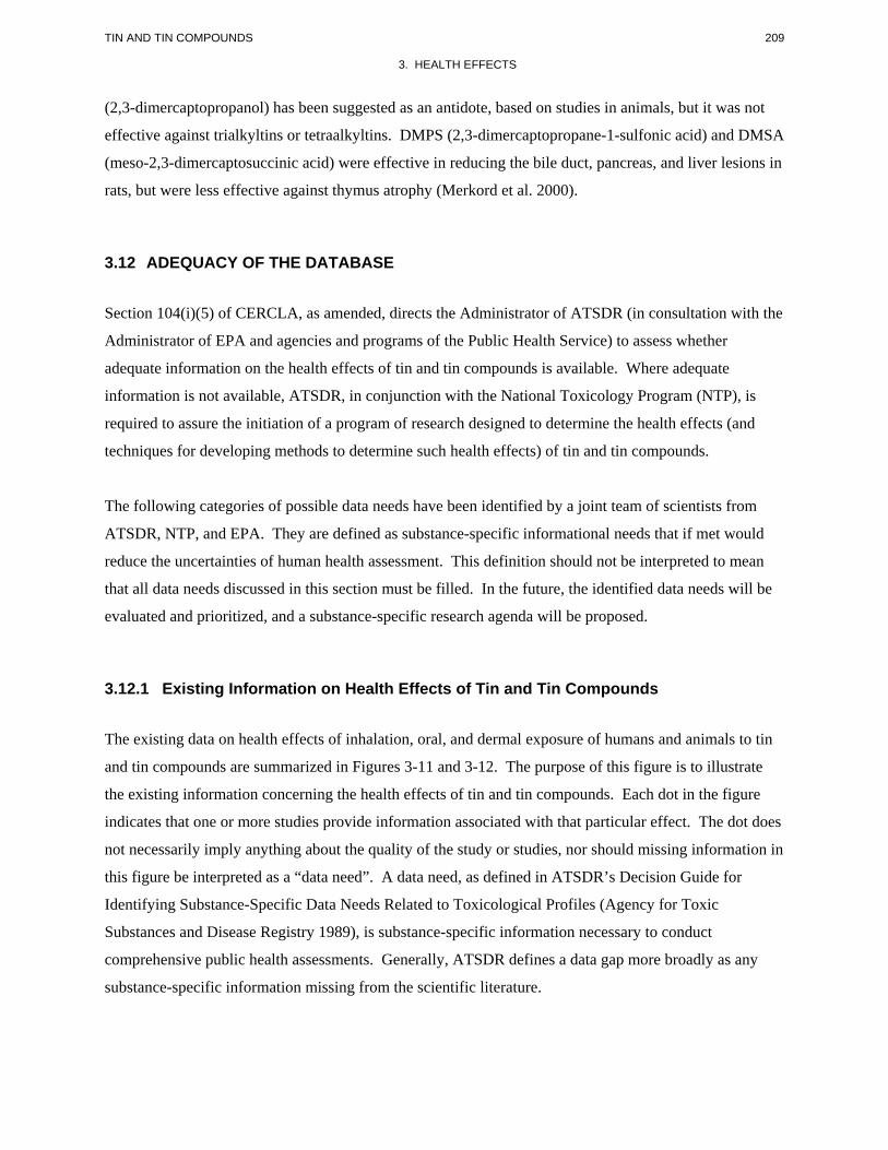
209 TIN AND TIN COMPOUNDS
3. HEALTH EFFECTS
(2,3-dimercaptopropanol) has been suggested as an antidote, based on studies in animals, but it was not
effective against trialkyltins or tetraalkyltins. DMPS (2,3-dimercaptopropane-1-sulfonic acid) and DMSA
(meso-2,3-dimercaptosuccinic acid) were effective in reducing the bile duct, pancreas, and liver lesions in
rats, but were less effective against thymus atrophy (Merkord et al. 2000).
3.12 ADEQUACY OF THE DATABASE
Section 104(i)(5) of CERCLA, as amended, directs the Administrator of ATSDR (in consultation with the
Administrator of EPA and agencies and programs of the Public Health Service) to assess whether
adequate information on the health effects of tin and tin compounds is available. Where adequate
information is not available, ATSDR, in conjunction with the National Toxicology Program (NTP), is
required to assure the initiation of a program of research designed to determine the health effects (and
techniques for developing methods to determine such health effects) of tin and tin compounds.
The following categories of possible data needs have been identified by a joint team of scientists from
ATSDR, NTP, and EPA. They are defined as substance-specific informational needs that if met would
reduce the uncertainties of human health assessment. This definition should not be interpreted to mean
that all data needs discussed in this section must be filled. In the future, the identified data needs will be
evaluated and prioritized, and a substance-specific research agenda will be proposed.
3.12.1 Existing Information on Health Effects of Tin and Tin Compounds
The existing data on health effects of inhalation, oral, and dermal exposure of humans and animals to tin
and tin compounds are summarized in Figures 3-11 and 3-12. The purpose of this figure is to illustrate
the existing information concerning the health effects of tin and tin compounds. Each dot in the figure
indicates that one or more studies provide information associated with that particular effect. The dot does
not necessarily imply anything about the quality of the study or studies, nor should missing information in
this figure be interpreted as a “data need”. A data need, as defined in ATSDR’s Decision Guide for
Identifying Substance-Specific Data Needs Related to Toxicological Profiles (Agency for Toxic
Substances and Disease Registry 1989), is substance-specific information necessary to conduct
comprehensive public health assessments. Generally, ATSDR defines a data gap more broadly as any
substance-specific information missing from the scientific literature.
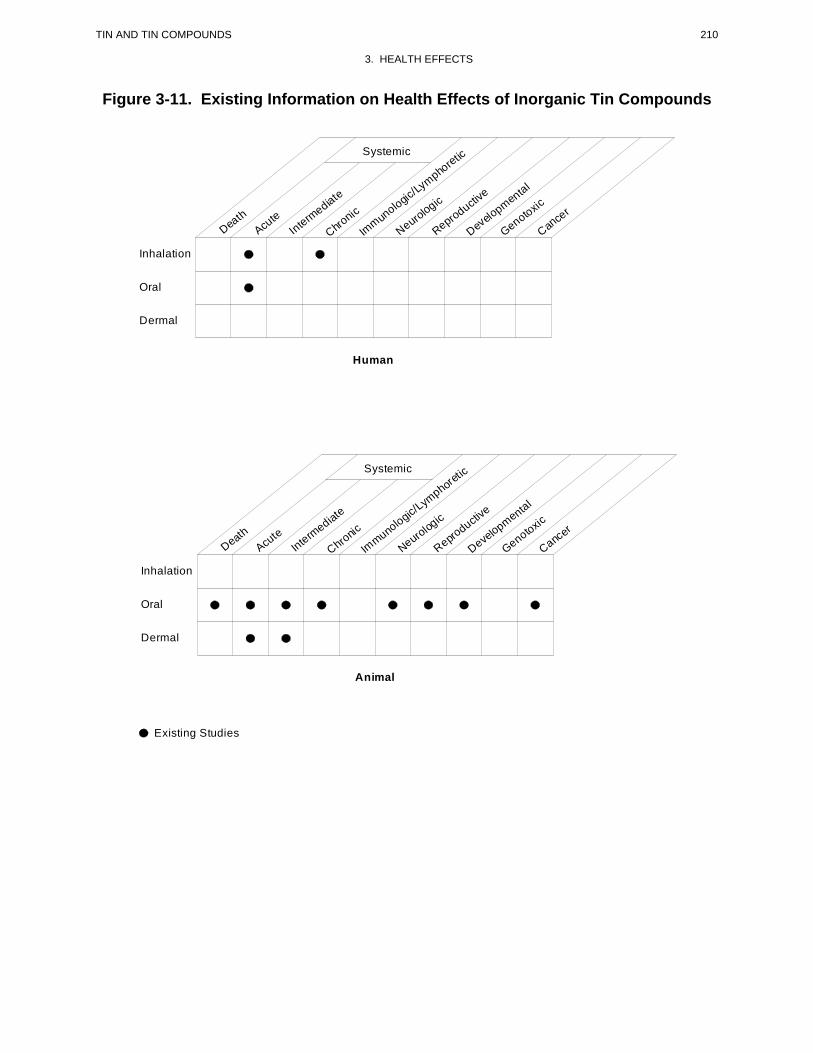
210 TIN AND TIN COMPOUNDS
3. HEALTH EFFECTS
Figure 3-11. Existing Information on Health Effects of Inorganic Tin Compounds
Death
Acute
Intermediate
Chronic
Immun
ologic/Ly
mphoretic
Neurologic
Reproductive
Develo
pmental
Genotoxic
Cance
r
Systemic
Human
Inhalation
Oral
Dermal
DeathAcu
te Interm
ediate
Chronic
Immuno
logic/Lym
phoretic
Neurologic
Reproductiv
e
Developmental
Genotoxic
Cancer
Systemic
Animal
Inhalation
Oral
Dermal
Existing Studies
211 TIN AND TIN COMPOUNDS
3. HEALTH EFFECTS
Figure 3-12. Existing Information on Health Effects of Organotin Compounds
DeathAcute
Int
ermediate
Chronic
Immuno
logic/Lymphore
tic
Neurologic
Reproductive
Developmental
Genotoxic
Cancer
Systemic
Human
Inhalation
Oral
Dermal
DeathAcute
Interm
ediate
Chronic
Immuno
logic/Lym
phoret
ic
Neurologic
Reproductive
Developmental
Genotoxic
Cancer
Systemic
Animal
Inhalation
Oral
Dermal
Existing Studies
212 TIN AND TIN COMPOUNDS
3. HEALTH EFFECTS
Figure 3-11 provides the information for inorganic tin compounds. There are case reports that describe
acute and chronic effects of inhaled inorganic tin compounds on humans. There are also reports of
humans that developed health effects after oral exposure to food and drink from tin cans. No other
studies were located regarding health effects in humans after inhalation, oral, or dermal routes of
exposure. The most relevant route of exposure to inorganic tin for humans is the oral route.
The health effects of inorganic tin compounds have been chiefly studied in animals after oral exposure, as
shown in Figure 3-11. The figure also shows that no inhalation studies and only a few dermal studies
were located regarding health effects from inorganic tin compounds.
Figure 3-12 provides health effects information on humans and animals after exposure to organotin
compounds. The database for these compounds as a class is much more complete than for the inorganic
tin compounds. There are case reports that describe deaths and other effects associated with inhalation,
oral, and dermal routes of exposure. In addition to acute-duration inhalation studies and acute and
intermediate dermal studies, there are reports of neurobehavioral effects in humans after inhalation, oral,
and dermal exposures. The main route of exposure to organotins for humans is the oral route.
The extent of the database on health effects in animals resulting from exposure to organotin compounds is
shown in Figure 3-12. Except for genotoxic studies, there are oral studies that describe all the other
toxicological end points considered in this profile. By contrast, the information is more limited for the
inhalation and dermal routes of exposure.
3.12.2 Identification of Data Needs
Acute-Duration Exposure. There are no data in humans following acute inhalation exposure to
inorganic tin and the data in animals are limited mostly to death (Igarashi 1959; Schweinfurth and Gunzel
1987). Therefore, an acute-duration inhalation MRL was not derived for inorganic tin. For organic tin
compounds, limited acute inhalation data exist for death in humans (Rey et al. 1984) and in animals
(Igarashi 1959; Schweinfurth and Gunzel 1987), systemic effects in humans (Rey et al. 1984; Saary and
House 2002; Wax and Dockstader 1995) and in animals (Igarashi 1959), and neurological effects in
humans (Feldman et al. 1993; Rey et al. 1984; Ross et al. 1981; Saary and House 2002; Yanofsky et al.
1991). The information provided in these studies is inadequate for derivation of acute-duration inhalation
MRLs for inorganic tin or organotins largely because of lack of quantitative data. Oral exposure is the
main route of exposure to inorganic tin for humans. The available acute oral data for inorganic tin
213 TIN AND TIN COMPOUNDS
3. HEALTH EFFECTS
provide information on lethality in animals (NTP 1982), on reproductive/developmental effects in rodents
(FDA 1972), and on systemic effects in humans (Boogaard et al. 2003; WHO 1980, 2003). In the case of
organotins, data exist for lethality in humans (Kreyberg et al. 1992; WHO 1980) and in animals (WHO
1980), systemic effects in humans (Lin and Hsueh 1993; Lin et al. 1998; WHO 1980; Wu et al. 1990) and
in animals (Barnes and Magee 1958; Pelikan and Cerny 1970; Raffray and Cohen 1993; Funahashi et al.
1980; Seinen et al. 1977a; Takagi et al. 1992; Ueno et al. 1994, 1995, 1997, 2003a, 2003b), immuno
logical effects in animals (Seinen et al. 1977a; Smialowicz et al. 1989, 1990; Snoeij et al. 1985), neuro
logical effects in humans (Kreyberg et al. 1992; Lin et al. 1998; WHO 1980; Wu et al. 1990) and in
animals (Baroncelli et al. 1990, 1995; Brown et al. 1984; Chang and Dyer 1983; Chang et al. 1983; Davis
et al. 1987; Ema et al. 1991a; Magee et al. 1957; Squibb et al. 1980), and reproductive and developmental
effects in animals (Ema and Harazono 2000; Ema et al. 1991b, 1992, 1997b, 1999b, 1999c, 2003; Farr et
al. 2001; Harazono and Ema 2003; Noda et al. 1991a, 1992b). Again, the data for inorganic tin were
insufficient for derivation of an acute oral MRL. The data for organotins were either insufficient or
inadequate in that no NOAELs were available and most LOAELs were serious LOAELs, thus precluding
derivation of acute oral MRLs. Acute dermal data were limited to a lethal human case (NIOSH 1976) and
several reports in animals (Smith 1978) exposed to organotins, information on hepatic effects in humans
(Colosio et al. 1991), and dermal and ocular effects in humans and animals (Barnes and Stoner 1958; Goh
1985; Klimmer 1969; Lyle 1958; Sheldon 1975). Excessive acute exposure to inorganic tin is unlikely to
occur unless there is consumption of unusually high amounts of canned foods in a short period of time.
Further acute studies with inorganic tin are unlikely to provide new key information. The decision to
expand the database for acute exposure to some organotin compounds should be based on the results of a
case-by-case evaluation of the likelihood of potential exposure to high concentrations of these substances
for people living near waste sites. It is unlikely that the general population will be acutely exposed to
high amounts of organotins.
Intermediate-Duration Exposure. There are currently no data concerning the effects of inorganic
tin or organotin compounds on humans for this exposure duration for the inhalation, oral, or dermal routes
of exposure. No data were available regarding effects in animals after intermediate-duration exposure to
inorganic tin by the inhalation route. Studies of inorganic tin in rodents, primarily rats and mice, treated
orally demonstrated effects in the gastrointestinal tract, the blood, the kidney, the liver, and bile ducts
(Chmielnicka et al. 1993; De Groot et al. 1973; Dreef-van der Meulen et al. 1974; Janssen et al. 1985;
NTP 1982; Schroeder et al. 1968). Very limited information was located regarding reproductive and
developmental effects of inorganic tin compounds in animals (Theuer et al. 1971). The study by
De Groot et al. (1973) with stannous chloride was used to derive an intermediate-duration oral MRL for

214 TIN AND TIN COMPOUNDS
3. HEALTH EFFECTS
inorganic tin. Few studies were located that tested organotin compounds by the inhalation route in
animals. These studies provided some information on respiratory, hepatic, renal, and reproductive effects
but lacked enough detail to be considered for MRL derivation (Gohlke et al. 1969; Igarashi 1959;
Iwamoto 1960). Intermediate-duration oral exposure studies with organotins provide information on
lethality in various species (Magee et al. 1957; NCI 1978b; Seinen et al. 1977b) and on a variety of end
points (hematological, body weight, endocrine, hepatic, renal, immunological, neurological, reproductive,
and developmental in rodents (Adeeko et al. 2003; Barnes and Magee 1958; Bouldin et al. 1981; Bressa et
al. 1991; Carthew et al. 1992; Cooke et al. 2004; Dacasto et al. 1994a; Funahashi et al. 1990; Gaunt et al.
1968; Graham and Gonatas 1973; Jang et al. 1986; Krajnc et al. 1984; Noland et al. 1982; Ogata et al.
2001; Omura et al. 2001; Purves et al. 1991; Seinen and Willems 1976; Seinen et al. 1977a, 1977b; Smith
1973; Snoeij et al. 1985; Tryphonas et al. 2004; Verdier et al. 1991; Vos et al. 1990). Most of these
studies tested dibutyltin, dioctyltin, tributyltin, triethyltin, trimethyltin, and/or triphenyltin. Adequate
information was available for derivation of an intermediate-duration oral MRL for dibutyltin dichloride
based on altered humoral immune responses in rats (Seinen et al. 1977b) and for tributyltin oxide, also
based on immunotoxicity (Vos et al. 1990). Information regarding effects following intermediate-
duration dermal exposure was restricted to a study by Sheldon (1975), who described skin alterations in
rabbits during a 90-day exposure period. Oral exposure is the main route of exposure to inorganic tin for
humans; therefore, inhalation and dermal exposure studies seem unnecessary. Also, further oral studies
are unlikely to provide new information. The effects of some organotins (i.e., trimethyltin, triethyltin,
tributyltin, dibutyltin) are well characterized. Still, the information available for trimethyltin and
triethyltin was inadequate for MRL derivation, largely because of the steepness of the dose-response
curve for these compounds. The decision to conduct additional intermediate-duration studies designed to
define NOAELs should rest on results of monitoring studies for these compounds in the environment and
on the identification of populations potentially exposed to them.
Chronic-Duration Exposure and Cancer. Data on chronic exposure to inorganic tin were limited
to cases of occupational exposures in which the main effects were respiratory effects (Cutter et al. 1949;
Dundon and Hughes 1950; Pendergrass and Pryde 1948; Stewart and Lassiter 2001). Inhalation is
assumed to have been the main route of exposure in these cases. Affected individuals showed a benign
form of pneumoconiosis, or stannosis. Information regarding health effects in animals following chronic-
duration exposure to inorganic tin is restricted to a 2-year oral bioassay in rats and mice (NTP 1982), a
42-month oral study in rats (Schroeder et al. 1968), and an 18-month oral study in mice (Schroeder and
Balassa 1967), all with stannous chloride. While there were no compound-related nonneoplastic effects
in the NTP (1982) study in rats and mice, Schroeder et al. (1968) described hepatic and renal effects as
215 TIN AND TIN COMPOUNDS
3. HEALTH EFFECTS
well as decreased longevity in rats exposed to doses approximately 100 times lower than those tested by
NTP (1982). There is no apparent explanation for this discrepancy except that the NTP (1982) study was
a dietary study, while Schroeder et al. (1968) administered the compound in the drinking water. Studies
comparing the bioavailability of tin in solid food vs. water may provide useful information. No chronic
oral MRL was derived for inorganic tin because the lowest dose from Schroeder et al. (1968) was a
serious LOAEL. The need to conduct inhalation and dermal chronic-duration studies with inorganic tin is
less clear since the main route of exposure for humans to inorganic tin is the oral route. No data were
located regarding health effects in humans following chronic exposure to organotin compounds. Long-
term bioassays have been conducted for dibutyltin diacetate in rats and mice (NCI 1978a), triphenyltin
hydroxide in rats and mice (NCI 1978b; Tennekes et al. 1989a, 1989b), and tributyltin oxide in rats
(Wester et al. 1990). In addition, an 18-month study of the immunotoxicity of tributyltin oxide in rats is
available and was used as basis for deriving a chronic-duration oral MRL for tributyltin oxide (Vos et al.
1990). No chronic oral MRL was derived for dibutyltin because a relative low dose in the NCI (1978a)
study caused significant early mortality in rats. For the same reason, no chronic oral MRL was derived
for triphenyltin (Tennekes et al. 1989b). Research to produce chronic oral data for other organotins that
may be present in hazardous waste sites and represent a potential source of exposure for those living in
the vicinity may be warranted. However, a comprehensive evaluation of 90-day studies should be
conducted first. Environmental monitoring information suggests that the inhalation and dermal routes of
exposure to organotins are much less relevant to humans than the oral route and, therefore, may be given
lower priority.
There is no information regarding cancer in humans exposed to inorganic tin or organic tin compounds.
An oral bioassay for stannous chloride in rats and mice provided no evidence of carcinogenicity at the
levels tested (NTP 1982). Similar negative results were found for dibutyltin diacetate in rats and mice,
although technical problems did not allow for a complete evaluation of uterine tumors in female rats (NCI
1978a). A bioassay for triphenyltin hydroxide in rats and mice also gave negative results (NCI 1978b),
but studies with higher doses did find triphenyltin hydroxide to induce pituitary and testicular tumors in
rats (Tennekes et al. 1989b) and hepatocellular carcinomas in mice (Tennekes et al. 1989a). A bioassay
with tributyltin oxide in rats yielded questionable results (Wester et al. 1990), which led the EPA (IRIS
2005) to assign this chemical to a group for which “there is inadequate information to assess carcinogenic
potential.” Since the observed tumors were considered to have high incidence in the strain of rat used
(Wistar), it may be necessary to repeat the study in a different strian of rat. An oral bioassay seems more
relevant than inhalation or dermal studies since these two routes of exposure are less relevant for humans.
Further studies concerning the fates of the organic and tin moieties from these compounds and the

216 TIN AND TIN COMPOUNDS
3. HEALTH EFFECTS
contribution of each moiety to mechanisms of carcinogenicity are needed in order to evaluate the role of
tin in the tumorigenic response.
Genotoxicity. There are no human data regarding the genotoxic potential of inorganic tin or organotin
compounds after inhalation, oral, or dermal exposures. The limited in vitro data for inorganic tin consist
mostly of studies with stannous chloride and stannic chloride. The results in prokaryotic organisms have
been mostly negative (Hamasaki et al. 1993; Nishioka 1975), but the opposite has been observed in tests
conducted with mammalian cells (Dantas et al. 2002; Ganguly et al. 1992; Gulati et al. 1989). Further
studies are unlikely to add new key information. Tests of many organotin compounds in S. typhimurium
gave predominantly negative results (Hamasaki et al. 1993) and tests conducted in mammalian cells in
vitro also gave predominantly negative results (Chao et al. 1999; Davis et al. 1987; Oshiro et al. 1991;
Sasaki et al. 1993). Studies of organotin compounds in vivo, mostly in mice, have given mixed results
(Chao et al. 1999; Davis et al. 1987; Ganguly 1994; Yamada and Sasaki 1993). Further genotoxicity
studies with organotin compounds do not seem warranted at this time.
Reproductive Toxicity. No studies were located regarding reproductive effects of inorganic tin in
humans following inhalation, oral, or dermal exposure or in animals following inhalation or dermal
exposure. The only information available regarding oral exposure of animals to inorganic tin is that from
Theuer et al. (1971), FDA (1972), and De Groot et al. (1973). FDA (1972) reported no reproductive
effects (number of corpora lutea and of implantation and resorption sites) in rats, mice, and hamsters
administered up to 0.31 mg tin/kg/day (as stannous chloride) during gestation (Gds 6–15 for rats and
mice, Gds 6–10 for hamsters) (FDA 1972). Theuer et al. (1971) reported that administration of tin, as tin
fluoride or sodium pentachlorostannite, to rats in the diet during gestation had no significant effect on the
number of resorptions or placental weight. De Groot et al. (1973) observed moderate testicular
degeneration in rats dosed for 9 weeks with approximately 315 mg tin/kg/day. Most rats in this group
were moribund and had to be sacrificed; therefore, the biological significance of the testicular finding is
unclear. The limited data available suggest that the reproductive system is not a target for inorganic tin
toxicity at the levels commonly found in the environment. No data were available regarding reproductive
effects in humans following exposure to organotin compounds by any route or in animals after dermal
exposure. Reduced fertility was reported in female rats following inhalation exposure to a mixture of
tributyltin bromide and dibutyltin dibromide for periods ranging from a few weeks to a few months
(Iwamoto 1960). Numerous studies in rodents have examined the effects of organotins (mostly
dibutyltin, tributyltin, and triphenyltin) on reproductive parameters such as pregnancy rates, pre- and
postimplantation loss, and fetal deaths following dosing during pregnancy (Adeeko et al. 2003; Ema and
217 TIN AND TIN COMPOUNDS
3. HEALTH EFFECTS
Harazono 2000; Ema et al. 1991b, 1992, 1999c; Faqi et al. 1997; Harazono et al. 1998; Noda et al. 1991a,
1992b) and found that the highest incidence of resorptions and postimplantation losses occurred when the
chemicals were administered on Gds 7–9 (Ema et al. 1992, 1997a, 1999a). Most long-term studies have
not reported histopathological alterations in reproductive organs from rats or mice (NCI 1978a, 1978b;
Wester et al. 1990) with the exception of Tennekes et al. (1989b), who reported an increase in Leydig cell
hyperplasia and tubular atrophy of the testes in rats treated with triphenyltin. The mechanism by which
some organotins affect reproduction is not known, but there is evidence that suppression of uterine
decidualization may be a cause of preimplantation losses (Ema and Miyawaki 2002; Ema et al. 1999b;
Harazono and Ema 2003). It would be helpful to elucidate whether effects such as pre- and
postimplantation loss occur secondary to maternal toxicity or can happen independent of maternal
toxicity. Since direct effects on reproductive organs do not seem to have an important role (except for the
findings of Tennekes et al. 1989b), further research should focus on the effects of organotins on the
endocrine control of reproductive functions in adult animals and on the hormonally-controlled
development of reproductive organs in animals exposed in utero and early in life. Numerous studies in
vitro have shown that organotins can affect the activities of enzymes involved in the synthesis of steroid
hormones, with potentially widespread consequences (i.e., Cooke 2002; Doering et al. 2002; Heidrich et
al. 2001; McVey and Cooke 2003). Additional tests to evaluate the potential endocrine disrupting ability
of organotins in mammals should be conducted. Pilot studies in primates would be valuable to reduce the
uncertainty of extrapolating observations in animals to human health.
Developmental Toxicity. No studies were located regarding developmental effects of inorganic tin
in humans following inhalation, oral, or dermal exposure, or in animals following inhalation or dermal
exposure. Limited oral data on inorganic tin showed that treatment of rats, mice, and hamsters with up to
31 mg tin/kg/day by gavage in water during gestation (Gds 6–15 for mice and rats, Gds 6–10 for
hamsters) has no significant effect on fetal weight, the number of live of dead fetuses, or the incidence of
external and internal malformations (FDA 1972). Also, tin, in the form of tin fluoride or sodium
pentachlorostannite, administered to rats during pregnancy had no significant effect on average fetal
weight or the number of live fetuses per litter (Theuer et al. 1971). This study also showed that tin from
inorganic compounds can cross the placenta and reach the fetus. There are no studies of developmental
effects in humans following inhalation, oral, or dermal exposure to organotins. Several studies in
animals, mostly rats and mice, have shown that oral exposure to some organotin compounds (mostly
tributyltin, dibutyltin, and triphenyltin) during pregnancy induces external and skeletal malformations, the
most common of which were cleft jaw and ankyloglossia (Ema and Harazono 2000; Ema et al. 1991b,
1992; Faqi et al. 1997; Harazono et al. 1998; Noda et al. 1991a, 1992b) and found that the highest
218 TIN AND TIN COMPOUNDS
3. HEALTH EFFECTS
incidence of malformations occurred when the chemicals were administered on Gds 7–9 (Ema et al.
1992). Limited data in rats indicate that dibutyltin can cross the placenta and reach the embryo
(Nakamura et al. 1993; Noda et al. 1994). A study in rats that included maternal exposure to tributyltin
chloride during lactation showed that little, if any, tributyltin or dibutyltin is transferred to the suckling
pups via the maternal milk (Cooke et al. 2004). A companion paper reported subtle alterations in
immunological parameters in pups from rats that were exposed during pregnancy during lactation, and
then the pups were exposed directly until 90 days of age (Tryphonas et al. 2004). As with reproductive
effects, the role of maternal toxicity in the manifestation of adverse developmental effects is not totally
clear. The mechanism responsible for the teratogenic activity of organotins is not known and studies
should continue to investigate the events at the molecular level that may be affected. The use of in vitro
systems (i.e., cultured rat embryos) may be preferable to studies in the whole animal, as the experimental
conditions in the former are easier to manipulate than in the latter. Two studies in rats exposed to
tributyltin chloride suggested that perinatal exposure can affect some developmental landmarks (Ogata et
al. 2001; Omura et al. 2001). Whether this is a result of endocrine disrupting ability of these compounds
or from other mechanisms is important to know. Triethyltins and trimethyltins are neurotoxic to humans
and animals and have been extensively used as tools to investigate the relationship between localized
lesions within the central nervous system and behavioral alterations. Continued research in this area is
important to determine susceptible developmental periods during which alterations of neuronal structures
will cause long-lasting effects or accelerate specific aspects of the normal aging processes.
Immunotoxicity. There currently is no information available in humans or animals suggesting that
the immune system is a major target of inorganic tin toxicity, or that the immune system is a target of
organic tin toxicity in humans. However, there is considerable information on the immunotoxic effects of
some organotins administered to animals orally or by injection, and from in vitro tests systems. Studies
have compared various organotins for several animal species (Seinen et al. 1977a, 1977b; Snoeij et al.
1985). The immunotoxic effect is characterized by reduced thymus weight and size and lymphocyte
depletion (Krajnc et al. 1984; Seinen and Willems 1976; Seinen et al. 1977a, 1977b; Smialowicz et al.
1989, 1990; Snoeij et al. 1985). While dialkyltins appear to interfere directly with the proliferation of
lymphocytes, tributyltin oxide has a direct action on lymphocytes in the thymus (Boyer 1989). The
results of a study that reported alterations in the humoral immune response in rats exposed orally to
dibutyltin dichloride were used as basis for derivation of an intermediate-duration oral MRL for dibutyltin
dichloride (Seinen et al. 1977b). Long-term studies with tributyltin oxide in rats showed alterations in
parameters of specific and nonspecific resistance (Vos et al. 1990). The results from this study were used
as the basis for derivation of an intermediate- and a chronic-duration oral MRL for tributyltin oxide. It is
219 TIN AND TIN COMPOUNDS
3. HEALTH EFFECTS
reasonable to assume that similar effects would occur in animals exposed to sufficiently high amounts of
organotins by the inhalation and dermal routes. Future research should continue to focus on determining
the basis for interspecies differences including pharmacokinetic differences, and differences at the cellular
and molecular levels. It would be valuable to determine whether primates exhibit responses similar to
rodents and if so, whether subtle alterations in immune parameters alter resistance to challenges with
pathogens. A recent study evaluated immunocompetence in rats exposed during gestation and as
juveniles and found subtle alterations of unknown toxicological significance (Tryphonas et al. 2004).
Replication of these findings would be valuable.
Neurotoxicity. There are no studies in humans regarding neurotoxic effects after inhalation, oral, or
dermal exposure to inorganic tin compounds. Limited animal data suggest that oral exposures to high
concentrations of inorganic tins may induce effects on the central nervous system (WHO 1980), but the
nervous system is not a sensitive target for inorganic tin toxicity.
Neurotoxic effects have been reported in humans after inhalation (Feldman et al. 1993; Rey et al. 1984;
Ross et al. 1981; Saary and House 2002; Yanofsky et al. 1991), oral (Foncin and Gruner 1979; Kreyberg
et al. 1992; Lin et al. 1998; Wu et al. 1990), and dermal (Colosio et al. 1991) exposure to organotins and
in animals after oral exposure (i.e., Bouldin et al. 1981; Brown et al. 1984; Eto et al. 1971; Graham and
Gonatas 1973; Magee et al. 1957; Snoeij et al. 1985; Squibb et al. 1980) to these compounds. Among the
organotins, trimethyltin and triethyltin have been the most widely studied in acute- and intermediate-
duration oral studies. These organotins are highly toxic and their effects are well characterized and are
expected to occur across routes of exposure. Trimethyltin induces neuronal necrosis, particularly in the
hippocampal region, whereas triethyltin produces intramyelinic edema (Chang 1990). Case studies of
humans acutely exposed (accidentally or intentionally) to high amounts of trimethyltin or triethyltin and
studies in animals reported morphological changes in the central nervous system as well as behavioral
changes that may persist for a long time after the poisoning episode. One typical manifestation of
trimethyltin intoxication in both humans and animals is aggressive behavior. Both trimethyltin and
triethyltin have become important research tools for the study of brain function, in particular, to examine
the association between damage to specific brain structures, such as the hippocampus or brain cell groups
and behavioral alterations. Additional studies using nerve cells in vitro can provide more information on
possible mechanisms of action of these organotins at the cellular and molecular levels. Also, studies on
the effects of tin compounds on glial function, both during neurodevelopment and adulthood, would be
useful. Studies of the potential effects of long-term exposure to low levels of trimethyltin or triethyltin, as
it may occur near a waste site that contains these substances, may provide valuable information.
220 TIN AND TIN COMPOUNDS
3. HEALTH EFFECTS
Epidemiological and Human Dosimetry Studies. The general population is exposed to
inorganic tin compounds through consumption of contaminated food, in industrial settings, and
potentially at hazardous waste sites through contact with contaminated air, water, and soil. Organotin
compounds are also used in agricultural and other uses with potential exposure of people by different
routes, although the main route of exposure is also the oral route. Only limited case reports of human
exposure and no retrospective or prospective epidemiological studies are available. Occupational studies
of inorganic tin exposure provide information mostly on respiratory effects in workers exposed
chronically (Cutter et al. 1949; Dundon and Hughes 1950). In contrast, the neurotoxic effects of some
organotins are well documented in workers and members of the general population exposed by all routes
(Colosio et al. 1991; Feldman et al. 1993; Kreyberg et al. 1992; Lin et al. 1998; Rey et al. 1984; Ross et
al. 1981; WHO 1980; Wu et al. 1990; Yanofsky et al. 1991). These cases have generally involved
accidental or intentional exposure to high amounts of organotins. Should populations with past or
ongoing exposure to organotins be identified, emphasis should be placed on the evaluation of organs and
systems that have appeared to be particularly sensitive in animal studies. For example, evaluation of
immunocompetence should have high priority in people exposed to tributyltin, dibutyltin, or dioctyltin,
whereas neurobehavioral tests should be conducted in those known to have been exposed or are exposed
to trimethyltin or triethyltin.
Biomarkers of Exposure and Effect.
Exposure. The development of models that would support quantitative estimates of exposure to tin and
tin compounds based on blood or urine levels of tin, or a specific organotin or metabolite, would be
valuable.
Effect. There are no biomarkers of effect specific for tin or tin compounds. Research to identify reliable
biomarkers for exposure to tin and tin compounds in humans would be useful in order to evaluate the
prevalence and magnitude of exposure in an at-risk population.
Absorption, Distribution, Metabolism, and Excretion. The toxicokinetics of tin compounds
have not been adequately characterized to support the development of predictive PBPK models in humans
(see Section 3.4). Existing models are based entirely on observations in animals (ICRP 2001). No
quantitative estimates of absorption of inhaled inorganic or organotin compounds are available, for either
humans or animals. Two balance studies of dietary exposures to inorganic tin provide estimates of
221 TIN AND TIN COMPOUNDS
3. HEALTH EFFECTS
gastrointestinal absorption in humans that suggest considerable interindividual variability and, possibly,
dose dependence (Calloway and McMullen 1966; Johnson and Greger 1982). Estimates of gastro
intestinal absorption of organotin compounds in humans are not available. Studies in animals suggest that
redox state (i.e., Sn[II] vs. Sn[IV]) (Hiles 1974) and number of alkyl moieties affect gastrointestinal
absorption of tin compounds (Bridges et al. 1967; Kimmel et al. 1977; Mushak et al. 1982; Ohhira and
Matsui 1993a; Ueno et al. 1994) thus, studies in humans that address these potential variables would be
particularly useful. Information on the distribution of absorbed tin compounds in humans derives from
analyses of human cadaver tissues (Kehoe et al. 1940; Schroeder et al. 1964). These studies, together
with studies conducted in animals, suggest that the major sites of deposition of tin in humans appear to be
similar to those in animals exposed to inorganic tin compounds; however, they provide no information on
the distribution of specific inorganic or organotin compounds in humans. Quantitative estimates of the
elimination rates of absorbed tin in humans are not available. Studies in animals indicate that elimination
rates vary with chemical form and across species, for given tin compounds (Bridges et al. 1967; Brown
1984; Furchner and Drake 1976; Kimmel et al. 1977; Ueno et al. 1994).
Available information on the toxicokinetics in animals, while providing abundant information on the
distribution and elimination kinetics of tin, do not provide adequate information for extrapolation of doses
from one route of exposure to another (e.g., oral-to-dermal, oral-to-inhalation), for which health effects
studies are lacking. The major information deficit, in this regard, is insufficient characterization of the
extents and rates of absorption of tin from major potential routes of exposure (i.e., no information is
available for the inhalation and dermal routes) and insufficient characterization of the effects of dose on
gastrointestinal absorption.
Comparative Toxicokinetics. As noted in Section 3.5.2, information on the toxicokinetics of tin
compounds in humans is sufficiently limited to preclude comparisons with other species (see Section 3.4).
Studies in animals are also insufficient for comparisons of the absorption of tin compounds across
species, from inhalation, oral, or dermal routes (see Section 3.4.1). Several studies have compared rates
of elimination of tin, administered in various inorganic forms or as organotin compounds, in various
animal species (Furchner and Drake 1976; Hiles 1974; NTP 1982). These studies demonstrate species
differences in elimination kinetics that may be germane to extrapolations to humans. For example, the
elimination kinetics of absorbed inorganic tin occurs more slowly in Rhesus monkeys than in rodents; this
difference appears to be the result of a larger fraction of the tin body burden associated with the slowest
kinetic compartment (presumably bone) in monkeys (Furchner and Drake 1976). Organotin compounds,
in particular methyltin and ethyltin, accumulate in red blood cells to a much greater extent in rats than in
222 TIN AND TIN COMPOUNDS
3. HEALTH EFFECTS
other species, including nonhuman primates (Brown 1984; Rose 1969; Rose and Aldridge 1968). Species
differences to the hepatotoxic effects of some organotins have been described. Comparative studies with
tributyltin and dibutyltin in rats, mice and guinea pigs showed the susceptibilities for liver toxicity
followed the order: mice > rats > guinea pigs (Ueno et al. 2003b). Kinetic studies showed that the
differences in susceptibility appeared to be partly due to differences in metabolism of tributyltin and in
the distribution of dibutyltin within cell organelles. These observations suggest that a more complete
characterization of inter-species variability in the toxicokinetics of tin would be useful for extrapolating
doses across species, in particular, from rats to other species, including humans.
Information is available to support the development of models of toxicokinetics of various tin compounds
in rodents and nonhuman primates (e.g., Rhesus monkey, marmoset); however, the current lack of
observations on the toxicokinetics of tin compounds in humans makes evaluation of such models for
applications to predicting the toxicokinetics of tin in humans highly uncertain. Useful types of
observations in humans to support toxicokinetic model development would include: (1) quantifying
extent and rates of absorption of tin compounds in humans from the inhalation, oral, and dermal
pathways; (2) quantifying the relative contribution of various excretory routes to elimination of tin
compounds in humans, including bile, urine, feces, and milk; (3) time course for the concentrations of tin
in blood and blood plasma (or other tissues) following a single dose or during repeated exposures;
(4) observing of blood:tissue and/or plasma:tissue concentration ratios for tin; and (5) identifying
pathways and rates of metabolism of tin compounds.
Methods for Reducing Toxic Effects. Recommended methods for the mitigation of effects of
acute exposure to tin compounds include standard treatments and measures to support vital functions
(HSDB 2003). No information was located concerning mitigation of effects of lower-level or longer-term
exposure to tin. This, in part, may reflect the fact that no population has been identified as having been
subjected or currently undergoing exposure to excessive amounts of tin and compounds. Attempts to
propose studies of specific methods to reduce possible adverse effects do not appear warranted at this
time.
Children’s Susceptibility. Data needs relating to both prenatal and childhood exposures, and
developmental effects expressed either prenatally or during childhood, are discussed in detail in the
Developmental Toxicity subsection above.
223 TIN AND TIN COMPOUNDS
3. HEALTH EFFECTS
There are no studies that specifically addressed exposure to inorganic tin in children. Workers exposed to
tin in the air experienced respiratory effects (Cutter et al. 1949; Dundon and Hughes 1950; Pendergrass
and Pryde 1948) and ingestion of excessive inorganic tin caused gastrointestinal effects (WHO 1980,
2003). It is reasonable to assume that children exposed in similar manners will experience similar effects.
Dermal contact with some organotins such as tributyltin can cause skin irritation and exposure by any
route to trimethyltin or triethyltin can cause serious neurological effects. There is no reason to believe
that children exposed to these chemicals would exhibit a different response. There is no information on
whether the developmental process is altered in humans exposed to tin and compounds. Limited evidence
with tributyltin chloride in rats suggests that this substance may alter developmental events controlled by
hormones (Ogata et al. 2001; Omura et al. 2001), but further studies are necessary on this issue. The
possibility that organotin compounds may have endocrine-disrupting ability in mammals has not been
systematically studied.
There are no data to evaluate whether pharmacokinetics of tin and tin compounds in children are different
from adults. There is limited information indicating that inorganic tin can cross the placenta (Theuer et
al. 1971), and also that dibutyltin can do so and reach the fetus (Nakamura et al. 1993; Noda et al. 1994).
Administration of dibutyltin and other organotins, such as tributyltin and triphenyltin, to pregnant rodents
has caused malformations in the fetuses (Ema et al. 1991a, 1991b, 1992, 1995b, 1997b; Faqi et al. 1997;
Farr et al. 2001; Harazono et al. 1998; Noda et al. 1991a, 1991b), indirectly suggesting that organotins (or
metabolites) other than dibutyltin also can cross the placenta. There are no studies on whether tin
compounds can be transferred from mother to offspring through maternal milk. Cross-fostering studies
can provide important information regarding the role of in utero vs. lactation exposure to tin compounds
in normal development. There is evidence that acute perinatal exposure to some organotins results in
altered behavioral responses in rodents tested as adults (i.e., Barone et al. 1995; Reiter et al. 1981;
Ruppert et al. 1983). Research efforts should continue to focus on the possible underlying mechanisms
that are responsible for such long-lasting postexposure toxicities. There are no data to permit an
evaluation of whether metabolism of tin and compounds is different in children than in adults.
Research into the development of sensitive and specific biomarkers of exposures and effects for tin
compounds would be valuable for both adults and children. There are no data on the interactions of tin
with other chemicals in children; however, studies in humans and in animals have shown that dietary tin
can influence the metabolism of zinc (Greger and Johnson 1981; Johnson and Greger 1982; Rader et al.
1990), which is essential for normal growth. There are no pediatric-specific methods to reduce peak
absorption for tin and compounds, to reduce body burdens, or to interfere with the mechanisms of action.

224 TIN AND TIN COMPOUNDS
3. HEALTH EFFECTS
Based on the information available, it is reasonable to assume that the supportive methods recommended
for maintaining vital functions in adults will also be applicable to children.
Child health data needs relating to exposure are discussed in Section 6.8.1, Identification of Data Needs:
Exposures of Children.
3.12.3 Ongoing Studies
The following ongoing studies concerning health effects associated with tin and tin compounds were
identified in the Federal Research in Progress database (FEDRIP 2004).
Dr. W.D. Atchison, from Michigan State University, East Lansing, Michigan, plans to examine the
process by which environmental chemicals, such as some organotins, can destroy distinct populations of
neurons in the brain, especially during development. Specifically, Dr. Atchison will study the effect of
trimethyltin on the generation of mechanical tension in developing hippocampal pyramidal neurons in
primary culture, or transformed neuronal cells. This research is sponsored by the National Institute of
Environmental Health Sciences.
Dr. M.L. Billingsley, from Penn State University, Hershey, Pennsylvania, plans to use molecular biologic
approaches to address specific mechanisms that may explain the selective actions of organotin toxicants.
The first aim will be to investigate the normal function of stannin (a protein isolated from organotin
sensitive tissues) and to use targeted gene disruptions to determine the consequences of loss of stannin on
the elaboration of organotin toxicity. The second aim will use in vivo antisense disruption of stannin
expression to determine whether this protein is needed for the elaboration of organotin toxicity. This
research is sponsored by the National Institute of Environmental Health Sciences.
Dr. A.Z. Mason, from California State University, Long Beach, California, proposes to determine the sub
lethal model of toxicity of tributyltin (TBT) and assess whether it and other toxicological analogues could
constitute an environmental hazard to humans. A series of in vivo and in vitro experiments using the
TBT-sensitive mollusk, Nucella emarginata, and human prostate cancer and hepatoma cell lines have
been designed to specifically test each of the identified mechanisms of action and determine if TBT acts
via perturbing aromatase activity, androgen conjugation, and elimination or by potentiation gonadotropin
neuropeptide release. This research is sponsored by the National Institute of General Medical Sciences.
225 TIN AND TIN COMPOUNDS
3. HEALTH EFFECTS
Dr. K.R. Pennypacker, from the University of South Florida, Tampa, Florida, proposes that brain injury
leads to activation of NF-kB (nuclear factor kB) in neurons surviving injury and that this activation
induces the transcription of growth factors that have a decisive role in promoting cell survival. NF-kB
expression and activity in the rat hippocampus in response to injury caused by excitotoxicity (kainite),
ischemia (middlecerebral arterial occlusion), and neurotoxicity (trimethyltin) will be examined to
determine whether activation of NF-kB is a common event in injury to the brain. This research is
sponsored by the National Institute of Neurological Disorders and Stroke.
Dr. C.R. Rice, of Mississippi State University, Mississippi State, Mississippi, plans to evaluate the
immunotoxicity of mixtures of halogenated hydrocarbons (a co-planar PCB) and organotin (TBT) using
channel catfish and mice as comparative vertebrate models, using the guidelines of the National
Toxicology Program. Early studies will be devoted to establishing dose-dependent indices of toxicity that
will be used to monitor the relative immunotoxicity of a co-planar PCB and TBT. Subsequent research
will be devoted to evaluating the effects of the co-planar PCB and TBT, alone and in combination, on
innate and antigen-specific immune parameters. This research is sponsored by the Department of
Agriculture.


