Embed Size (px)
Citation preview


110 JOURNAL OF VERTEBRATE PALEONTOLOGY, VOL. 30, NO. 1, 2010
FIGURE 1. Late Cretaceous and Paleocenestrata of the Mahajanga Basin, northwest-ern Madagascar. The Berivotra and Masi-akakoho study areas are indicated by rectan-gular outlines.
Gigantophis from the Paleogene of Egypt (Andrews, 1901, 1906).Examination of both Hoffstetter’s (1961a:fig. 1) and Lavocat’s(1955:fig. 2) maps (see also Krause et al., 2007:fig. 6B) suggeststhat Perrier de la Bathie’s and Lavocat’s specimens of snakevertebrae were all recovered from the Maevarano Formation,even though the rock unit had not yet been formally delimitedand named (see Rogers et al., 2000).
The Mahajanga Basin Project, conducted jointly by StonyBrook University and the University of Antananarivo, was ini-tiated in 1993, 60 years after Piveteau’s (1933) description of thefirst fossil snake specimen from Madagascar. The reconnaissanceexpedition and eight field campaigns since (1995, 1996, 1998,1999, 2001, 2003, 2005, 2007) have focused primarily on the col-lection of fossil vertebrates and associated contextual data fromthe Maevarano Formation in the Berivotra Study Area, whichlies some 35 km southeast of Mahajanga, but recent reconnais-sance (2003, 2005, 2007) has established two additional study ar-eas, the Masiakakoho and Lac Kinkony study areas, west of theBetsiboka River and southwest of Mahajanga (Fig. 1). The Mae-varano Formation, which crops out in all three study areas, wasnamed and described by Rogers et al. (2000). It has been ascer-tained to be of Maastrichtian age and to have been deposited ina highly seasonal, semi-arid climate (Rogers et al., 2000, 2007;Rogers and Krause, 2007). The majority of the contained fossilswere entombed in massive debris flows (Rogers, 2005) as sedi-ments were washed from the crystalline highlands that run downthe north-south axis of the island northwestward toward theMozambique Channel. The vertebrate fauna of the Maevarano
Formation includes ray-finned fishes, frogs, turtles, snakes, non-ophidian squamates, crocodyliforms, birds, non-avian dinosaurs,and mammals (most recently reviewed in Krause et al., 2006).The snake specimens described in this report were recoveredfrom the Berivotra and Masiakakoho study areas; none have yetbeen found in the Lac Kinkony Study Area. Snakes are repre-sented by over 125 specimens, most of them isolated vertebraeand vertebral and rib fragments. One specimen, the holotype ofa new genus and species of madtsoiid, consists of associated ele-ments: a sizable braincase fragment, a partial atlas, several com-plete vertebrae from the mid-trunk and posterior trunk regions,and many vertebral and rib fragments.
METHODS
The specimens described in this report were collected by fieldcrew members of the Mahajanga Basin Project through nine fieldseasons from 1993 to 2007 via surface collecting, quarrying, andboth dry- and wet-screening methods. All specimens were recov-ered from the Maevarano Formation in the Berivotra and Masi-akakoho study areas, Mahajanga Basin, northwestern Madagas-car, and prepared in the Stony Brook University Fossil Prepara-tion Laboratory.
Comparisons were made with skeletal material in the col-lections of the AMNH (including direct comparison with theholotype of Madtsoia bai), CMNH, and MVZ, as well as those ofthe authors. Other fossils were compared through descriptionsand figures in the literature. Vertebral anatomical terminology

LADUKE ET AL.—LATE CRETACEOUS SNAKES FROM MADAGASCAR 111
follows LaDuke (1991), except as modified by Head (2005).However, we continue to follow LaDuke in referring to vertebralregions as divisions of the column (e.g., anterior, mid-, andposterior trunk; cloacal; postcloacal). It must be emphasized,however, that because intracolumnar variation is continuous, avertebra from, for instance, a posterior position in the anteriortrunk region will be difficult to differentiate from one in ananterior position in the mid-trunk region.
The partial basicranium of Menarana nosymena, gen. et sp.nov., (UA 9684-3) was scanned at the High-Resolution X-ray CT(HRXCT) Facility at The University of Texas at Austin and thedataset was rendered in three dimensions using VGStudio MAX1.2 (Volume Graphics, Heidelberg, Germany). An interactive,Web-deliverable version of the HRXCT data set, as well asanimations of 3-D reconstructions and technical informationconcerning the scans and image processing, can be viewed athttp://www.digimorph.org/specimens/Menarana nosymena; theoriginal full-resolution HRXCT data are available from theauthors.
Measurements and Anatomical Abbreviations—All measure-ments were made with hand-held calipers (Helios) or, in the caseof small specimens, with an ocular micrometer in a dissecting mi-croscope. The following measurements (in order of presentationin the tables) were made where possible, following abbreviationsof LaDuke (1991): CL = centrum length; NAW = neural archwidth; PRW = width across the prezygapophyses; POW = widthacross the postzygapophyses; PR-PO = length from the ante-rior edge of one prezygapophyseal facet to the posterior edgeof the ipsilateral postzygapophyseal facet; COW = width of thecotyle measured from the outside of the cotylar rim; CNW =condyle width; NSH = vertical height of the neural spine mea-sured from the top of the zygosphene to the highest extremityof the spine; and ZSW = zygosphene width. In addition; ATV =anterior trunk vertebra; MTV = mid-trunk vertebra; PTV = pos-terior trunk vertebra; CV = cloacal vertebra; and PCV = post-cloacal vertebra.
Institutional Abbreviations—AMNH, American Mu-seum of Natural History, New York; CMNH, CarnegieMuseum of Natural History, Pittsburgh; FMNH, TheField Museum, Chicago; MVZ, Museum of VertebrateZoology, University of California at Berkeley; MNHN,Museum national d’Histoire naturelle, Paris; QM, Queens-land Museum, Brisbane; SAMP, South Australian Museum(Adelaide) Palaeontology; UA, Universite d’Antananarivo,Antananarivo, Madagascar.
Taxonomic Abbreviations—In relevant places throughout thetext, Madtsoia is abbreviated to Ma. and Menarana is abbreviatedto Me. to save space but also to facilitate differentiation betweenspecies of the two genera.
SYSTEMATIC PALEONTOLOGY
SQUAMATA Oppel, 1811SERPENTES Linnaeus, 1758
MADTSOIIDAE (Hoffstetter, 1961a) McDowell, 1987MADTSOIA Simpson, 1933
Type Species—Madtsoia bai Simpson, 1933.Referred Species—Madtsoia madagascariensis Hoffstetter,
1961a and Ma. camposi Rage, 1998.Revised Diagnosis—Distinguished from Alamitophis, Heren-
sugea, Menarana (below), Nanowana, and Patagoniophis by largesize and relatively short, broad mid-trunk vertebrae (CL approx-imately half of PRW). Vertebrae further differ from those ofMenarana in having taller neural spines and less depressed neu-ral arches, from those of Gigantophis and Rionegrophis in hav-ing less distinct hemal keels, and from those of Wonambi and
Yurlunggur in having a single parazygantral foramen on eachside. Ribs differ from those of Wonambi and Yurlunggur, whichhave multiple small foramina in dorsal groove, in having a singlelarge foramen in that position (but a much smaller accessory fora-men can be present). They differ from those of Menarana in hav-ing a less strongly recessed dorsal facet, in not having the tubercostae drawn out into a crest, and in possessing fewer foraminaon anteroventral and posterior surfaces.
Comparisons and Discussion
At the time Madtsoia was named, diagnosed, and describedby Simpson in 1933, and then even 28 years later when itwas reassessed by Hoffstetter (1961a), only one other genus(Gigantophis) of the clade now identified as Madtsoiidae wasknown. Since 1961, seven additional genera (Alamitophis, Heren-sugea, Nanowana, Patagoniophis, Rionegrophis, Wonambi, andYurlunggur) have been named and assigned to the Madtsoiidaeand at least two others (Najash [see Apesteguıa and Zaher, 2006]and Helagras [see Head and Holroyd, 2008]) are questionably al-lied. Yet, no formal rediagnosis of Madtsoia has been publishedsince that time. As such, and because of the removal of “Madt-soia” laurasiae from the genus (see below) and the considerableaddition to knowledge of Ma. madagascariensis based on the newspecimens described here, reassessment of vertebral and rib fea-tures relative to those of other madtsoiid genera and revision ofthe diagnosis of Madtsoia are in order, especially because it servesas the type genus of the Madtsoiidae.
The genus Madtsoia, as here defined, consists of three largespecies: Ma. bai, Ma. camposi, and Ma. madagascariensis, withmaximum centrum lengths (CL) of 18–25 mm, and maximumwidths across the prezygapophyses (PRW) of 35–65 mm. Threemadtsoiid genera (Gigantophis, Wonambi, and Yurlunggur) in-clude species of comparable size. Species of Menarana (definedbelow) appear to have maximum sizes about one-half to two-thirds those of the large genera (CL = 11–13 mm; PRW =20–22 mm). Several madtsoiid genera (Alamitophis, Herensugea,Nanowana, and Patagoniophis) have maximum sizes that aremuch smaller (CL < 8 mm, PRW < 10 mm). Thus, the madt-soiid genera segregate into three distinct size classes. Membersof each size class can be distinguished further by differences invertebral shape: the smaller madtsoiids tend to have relativelyelongate vertebrae (length nearly as great as width); Menaranahas vertebrae that are depressed overall with extremely low neu-ral spines; and the larger genera have vertebrae that are broaderthan they are long, and that are never depressed to the degreeseen in Menarana.
Vertebrae of the larger madtsoiid genera can be distinguishedfrom one another on the basis of more detailed comparisons.Madtsoia madagascariensis and Wonambi naracoortensis werebriefly compared by Smith (1976:43), who stated that, “There isa striking resemblance between Wonambi vertebrae and thoseof Madstoia [sic] bai. . .and M. madagascariensis.” However, shedid not make detailed comparisons of the two species that wouldallow differentiation, stating that, “the relationship of Wonambito Madstoia [sic] or any other boid will remain obscure until theskull [of Madtsoia] is known.” Nevertheless, most paleontologistswho work extensively with snakes use vertebrae to differenti-ate genera and even species. Comparison of vertebrae of Ma.madagascariensis and W. naracoortensis does indeed reveal astrong resemblance in shape. However, the large Australianmadtsoiids (Wonambi and Yurlunggur) have a series of smallparazygantral foramina, whereas Madtsoia (indeed, most madt-soiids) usually have a single, large foramen recessed in a dis-tinct fossa. Posterior trunk vertebrae of Ma. bai bear pairedposterior tubercles on an otherwise broad, low hemal keel thatwere referred to as ‘paired hypapophyses’ (Simpson, 1933:3, 8);similar structures are seen in Ma. madagascariensis (Hoffstetter

112 JOURNAL OF VERTEBRATE PALEONTOLOGY, VOL. 30, NO. 1, 2010
1961a) and species of Yurlunggur (Scanlon, 1992, 1995), but notMa. camposi, which has a more typical rhombic termination ofthe hemal keel (Rage, 1998). Posterior bifurcation of the keelalso occurs in a different form (mostly narrower keels) in Won-ambi (Smith, 1976) and other, smaller Australian taxa (Scanlon,1997, 2005b). Gigantophis vertebrae have distinctively shapedneural arches (Andrews, 1906:pl. XXVI, figs. 1–3). The laminaeare thickened and strongly arched in posterior view (slightly an-gled in Madtsoia, Wonambi, and Yurlunggur). Anteriorly, the zy-gosphene also appears hypertrophied, being much broader thanthe opening of the neural canal. The hypapophysis is low andblunt. Andrews makes no mention of paired tubercles or hypa-pophyses, and his illustrations do not appear to show any.
The vertebrae of Najash rionegrina are similar to those ofmadtsoiids in possessing parazygantral foramina, a shallow in-terzygapophyseal constriction, and large, broad synapophysesthat exceed the prezygapophyseal facets laterally, and in lack-ing accessory processes of the prezygapophyses (Apesteguıa andZaher, 2006). Najash is distinct from madtsoiids, but similar tovarious fossil “anilioids,” in that it lacks a posterior neural archnotch and has hemal keels that are ‘shallow and thin.’ This mo-saic of vertebral characters makes Najash vertebrae identifiable,but provides little support for assignment to a higher level taxon.
MADTSOIA MADAGASCARIENSIS Hoffstetter, 1961a(Figs. 2–4; Table 1)
Holotype Specimen—MNHN MAJ 5, posterior trunk vertebra(Hoffstetter, 1961a:fig. 2A).
Type Locality—‘“Gite du Guide,’ North of Berivotra, Mada-gascar” (Rage, 1984:30).
Referred Specimens—Anterior trunk vertebrae: FMNH PR2545–FMNH PR 2549, FMNH PR 2558, FMNH PR 2569, FMNHPR 2702, UA 9688–UA 9693, UA 9703, UA 9728. Mid-trunkvertebrae: FMNH PR 2550–FMNH PR 2553, MNHN MAJ9 (Hoffstetter, 1961a:fig. 3E), MNHN MAJ 10 (Hoffstetter,1961a:fig. 3F), UA 9695, UA 9697, UA 9698, UA 9745. Poste-rior trunk vertebrae: FMNH PR 2554, FMNH PR 2555, MNHNMAJ 7 (Hoffstetter, 1961a:fig. 2C), UA 9700. Cloacal vertebra:FMNH PR 2556. Postcloacal vertebrae: FMNH PR 2557, UA9701. Fragmentary vertebrae not assigned to region: FMNH PR2559–FMNH PR 2568, FMNH PR 2570, FMNH PR 2584, FMNHPR 2585, MNHN MAJ 6 (Hoffstetter, 1961a:fig. 2B), MNHNMAJ 8 (Hoffstetter, 1961a:fig. 3D), UA 9694, UA 9696, UA 9699,UA 9702, UA 9704–UA 9712, UA 9715–UA 9718, UA 9721, UA9726, UA 9727, UA 9729–UA 9731, UA 9735, UA 9736, UA9738–UA 9744, UA 9747, UA 9765, UA 9766, UA 9768, UA9772. Nearly complete ribs: UA 9746, UA 9763, UA 9764, UA9775. Proximal rib fragments: FMNH PR 2571, FMNH PR 2582,FMNH PR 2583, UA 9714.
Localities—The first-known specimen of Madtsoia madagas-cariensis, described by Piveteau (1933), was listed as com-ing from the region of Marovoay, southeast of Mahajanga(= Majunga). The specimens described by Hoffstetter (1961a:fig.1) were recovered from three areas listed as: (1) north ofBerivotra (the holotype, MNHN MAJ 5), (2) south of Beriv-otra (MNHN MAJ 8–MNHN MAJ 10), and (3) north of theMahajanga-Ambalabe road, between km 20 and 25 (MNHNMAJ 6, MNHN MAJ 7). The Mahajanga Basin Project, initiatedin 1993, has discovered specimens of Ma. madagascariensis in twomajor areas (Fig. 1): (1) Berivotra Study Area localities MAD93-01, 93-09, 93-14, 93-16, 93-17, 93-18, 93-25, 93-28, 93-30, 93-33,93-34, 93-35, 93-36, 93-38, 93-73, 93-81, 95-14, 96-01, 96-04, 96-32,98-08, 98-31, 99-15, 99-39, 01-03, 03-03, 03-04, 03-05, 03-09, 05-64;and (2) Masiakakoho Study Area locality MAD03-23 (Fig. 1).
Age and Distribution—Known only from the Upper Cre-taceous (Maastrichtian) Maevarano Formation, Berivotra and
Masiakakoho study areas, Mahajanga Basin, northwesternMadagascar.
Revised Diagnosis—Neural spines differ from those of Madt-soia bai and Ma. camposi in being taller and more posteriorlycanted. Zygosphenes relatively narrower than in Ma. camposi.Zygapophyses broad and rectangular, similar to those of Ma. baibut broader than those of Ma. camposi. Synapophyses project lat-erally beyond prezygapophyseal facets, as in Ma. camposi, butnot as in Ma. bai, in which synapophyses project far beyond zy-gapophyses. Ribs differ from those of Ma. bai in lacking a stronganterodorsal process and from those of Ma. camposi in not hav-ing the ventral articular facet projecting strongly anteriorly.
Description
An isolated vertebra of this species was described brieflyby Piveteau (1933), but no name was applied at that time.Hoffstetter (1961a) described six additional specimens (five ver-tebrae and one zygosphene) and, in addition to naming thespecies Madtsoia madagascariensis, listed three differences be-tween it and Ma. bai, the only other species of Madtsoia thenrecognized: (1) the neural spine is taller and its distal portionis inclined posteriorly; (2) in the posterior trunk vertebrae, thehemal keel is more clearly delimited by more marked lateral de-pressions; and (3) the condyle is more circular in outline and lessdepressed dorsoventrally. He also described diagnostic charac-teristics of his new subfamily Madtsoiinae. However, Hoffstet-ter (1961a) provided only a cursory description of the vertebralmorphology of Ma. madagascariensis, and the only regions ofthe vertebral column known to him were the mid- and poste-rior trunk regions. Based on the specimens recovered as part ofthe Mahajanga Basin Project, detailed descriptions of vertebraefrom the anterior trunk, mid-trunk, posterior trunk, cloacal, andpostcloacal regions, as well as parts of seven ribs are providedhere.
Table 1 provides measurements for the specimens of Madtsoiamadagascariensis collected by Mahajanga Basin Project teamsand allows comparisons of the proportions of vertebrae from dif-ferent regions of the column. Such comparisons reveal, for ex-ample, that the largest complete vertebrae were not the largestspecimens in the assemblage, as some fragments (e.g., isolatedzygosphenes) were larger than those present on any of the morecomplete vertebrae. Furthermore, the zygosphene described andillustrated by Hoffstetter (1961a:fig. 3D, MNHN MAJ 8) is listedas being 22 mm wide and is therefore larger than the largest of thezygosphenes (FMNH PR 2564; ca. 19.4 mm wide) in the samplescollected as part of the Mahajanga Basin Project.
Anterior Trunk Vertebrae—At least 16 specimens repre-sent this vertebral region, previously undescribed in Madt-soia madagascariensis. Several specimens are very well pre-served and essentially complete. One of these (FMNH PR 2546;Fig. 2A), from the anterior portion of the anterior trunkregion of a large individual, bears a strong hypapophysis.Another specimen (FMNH PR 2548), representing a moreposterior segment of the anterior trunk region, has a much re-duced hypapophysis and three specimens that are particularlycomplete and well preserved (FMNH PR 2545, FMNH PR 2547,FMNH PR 2549; Fig. 2B) are from the far posterior portion ofthis region, resembling mid-trunk vertebrae except for the pres-ence of slightly developed hypapophyses, just anterior to the ven-tral lip of the condyle. The following description is based primar-ily on these five specimens.
The centra of anterior trunk vertebrae are narrower than thoseof mid-trunk vertebrae and the subcentral fossae are less pro-nounced, especially anteriorly in the region (e.g., FMNH PR2546). Subcentral foramina are on the sloping portion of thekeel in FMNH PR 2546 or in shallow subcentral fossae in moreposterior vertebrae. The hypapophysis is robust, elongate, and

LADUKE ET AL.—LATE CRETACEOUS SNAKES FROM MADAGASCAR 113
FIGURE 2. Trunk vertebrae of Madtsoia madagascariensis from the Late Cretaceous of Madagascar in l, lateral; a, anterior; p, posterior; d, dorsal;and v, ventral views. A, vertebra from anterior part of anterior trunk region with well-developed hypapophysis, FMNH PR 2546; B, vertebra fromposterior part of anterior trunk region, FMNH PR 2549; C, mid-trunk vertebra, FMNH PR 2551; D, vertebra from anterior part of posterior trunkregion, FMNH PR 2554; E, vertebra from middle part of posterior trunk region, FMNH PR 2555.

114 JOURNAL OF VERTEBRATE PALEONTOLOGY, VOL. 30, NO. 1, 2010
FIGURE 3. Cloacal and postcloacal vertebrae of Madtsoia madagascariensis from the Late Cretaceous of Madagascar in l, lateral; a, anterior; p,posterior; d, dorsal; and v, ventral views. A, cloacal vertebra, FMNH PR 2556; B, postcloacal vertebra, FMNH PR 2557. Articular facets on ventralaspect of postcloacal vertebra for chevron bone enlarged at bottom right.
FIGURE 4. Ribs and rib fragments of Madtsoia madagascariensis from the Late Cretaceous of Madagascar. A, anterior; and B, posterior views ofUA 9764, nearly complete left rib exhibiting a pathological lesion (indicated by arrows). C, anterior; D, posterior; and I, stereophotographic proximalviews of UA 9746, proximal half of right rib (reversed to facilitate comparison). E, anterior; F, posterior; and J, stereophotographic proximal views ofFMNH PR 2571, proximal fragment of left rib. G, anterior; and H, posterior views of UA 9763, nearly complete left rib.

LADUKE ET AL.—LATE CRETACEOUS SNAKES FROM MADAGASCAR 115
laterally compressed in FMNH PR 2546, much shorter in FMNHPR 2548, and reduced to a nubbin in FMNH PR 2545, FMNHPR 2547, and FMNH PR 2549. The elongated hypapophysis onFMNH PR 2546, which is paddle-like in lateral view, exhibits aswelling at approximately mid-length of the hypapophysis. Thisirregular, asymmetrical swelling resembles a bone callus, and mayindicate a healed break in the bone. The tip of the hypapoph-ysis is bent slightly toward the left beyond the callus. However,we note that in some madtsoiids, such as Yurlunggur camfielden-sis and Riversleigh Yurlunggur spp., bilateral expansions of thehypapophyses are present that may represent serial homologs of‘paired hypapophyses’ (Simpson, 1933:3, 8) in the mid- and pos-terior trunk regions and presumably served as sites for muscleattachment. The subcentral ridges are not as distinct as thosepresent on mid-trunk vertebrae, especially anteriorly in the an-terior trunk region.
The cotyle and condyle are depressed, especially anteri-orly in the region, with a slightly recessed ventral cotylarlip, but they are not emarginated ventrally; they differ fromthe mid-trunk vertebrae in these respects. The neural canalis strongly trifoliate in shape and highly depressed, approx-imately twice as broad ventrally as high. Paracotylar fossaeare present and usually contain one large foramen each, butthe number can vary from zero (e.g., FMNH PR 2546—rightside, FMNH PR 2548—left side) to two (e.g., UA 9727—bothsides).
The zygosphene, although thick, is not massive, but gently con-vex to flat dorsally. Its lateral margins are not elevated as theyare in the mid-trunk region. Its anterior margin is incised by abroad, shallow notch. Zygosphenes from more posterior verte-brae of the anterior trunk approach the massiveness of those ofmid-trunk vertebrae. The zygantrum is very large, and similar tothose of the mid-trunk vertebrae with two notable exceptions.First, fine subvertical ridges that descend from the laminae in themid-trunk vertebrae are absent in the more anterior vertebrae ofthe anterior trunk region (e.g., FMNH PR 2546) and only faintlydiscernible in the more posterior vertebrae of the region. Second,the roof of the zygantral cavity is distinctly peaked in the vertebrafrom a far anterior position (FMNH PR 2546), though flattenedin the posterior portion of the anterior trunk and all mid-trunkvertebrae.
In FMNH PR 2546, the neural spine is very tall and robust.Its height is slightly greater than twice the height of the laminaeabove the centrum. Anteriorly, the spine is laterally compressed,but posteriorly it is thickened, creating a triangular section. Thespine is canted, extending posteriorly well beyond the level ofthe postzygapophyses. Postzygosphenal fossae are present, butdo not contain foramina. More posterior vertebrae in the ante-rior trunk series have shorter and anteroposteriorly longer neuralspines and can contain up to three small foramina in the postzy-gosphenal fossae, though the foramina are not all necessarily re-stricted to the bottom of these fossae (FMNH PR 2545).
The zygapophyseal facets are small, approximately as broadas long. The zygapophyses are not markedly divergent from thecentrum, producing a relatively narrow aspect for the vertebra.The synapophyses are large, and well preserved in FMNH PR2546, in which the diapophyseal portion is separated from theparapophysis by a distinct constriction as a result of a posteriorindentation. This indentation becomes more prominent in moreposterior vertebrae in the series and is particularly prominent inFMNH PR 2549. The diapophyseal facet is bulbous whereas theparapophysis is relatively flat. The latter extends ventrally wellbelow the lower lip of the cotyle in FMNH PR 2546, the most an-terior vertebra in the series, but this disparity is less extreme oreven absent (FMNH PR 2545) in more posterior vertebrae of theanterior trunk region.
Mid-trunk Vertebrae—Several complete and nearly completemid-trunk vertebrae are represented in the Mahajanga Basin
Project collection, significantly augmenting the sample availableto Hoffstetter (1961a). The following description is derived pri-marily from a typical larger specimen, FMNH PR 2551 (Fig. 2C).
The centrum is roughly triangular in ventral view, muchbroader anteriorly than long. A transversely convex anterior por-tion is flanked by lateral depressions that contain distinct, pairedforamina. The hemal keel is poorly defined anteriorly, but nar-rows abruptly in its posterior third into a much better defined keelthat bears a pair of small, blunt processes posteriorly. The lateralmargins of the centrum form prominent subcentral ridges that ex-tend posteromedially from the posteroventral border of the para-pophysis, almost to the condyle. The postzygapophyses are trans-versely broad and elliptical, almost subrectangular in outline.
In anterior view, the cotyle is almost round (very slightly widerthan high) and deep, but its ventral lip is recessed posteriorlyand emarginated ventrolaterally. The neural canal is relativelysmall, slightly broader than tall, and roughly triangular; inden-tations formed by internal ridges along the floor and each of thelateral walls produce a trifoliate outline. Well-developed para-cotylar fossae typically contain one or two foramina. Two speci-mens (FMNH PR 2550 and FMNH PR 2551) have two paracoty-lar foramina on each side, one larger than the other. Another(FMNH PR 2552) has paired foramina on the left, but a singleforamen on the right. Other specimens in which the paracotylarfossae are visible (FMNH PR 2553, UA 9695) have a single fora-men on each side. The neural arch laminae rise sharply from frontto back and from lateral to medial. The zygosphene is massiveand wedge-shaped in anterior view and its facets are angled atapproximately 30◦ from the midline axis. The dorsolateral mar-gins of the zygosphene project upward, due to the large facets,creating a dorsal concavity on each side of the anterior margin ofthe neural spine.
In posterior view, the condyle is almost round in outline butslightly flattened ventrally; it is directed strongly posterodorsally.The zygantrum is spacious and deep, with substantial zygantralfacets that project posteriorly, slightly beyond the margins of thelaminae. A deep, broad, V-shaped notch in the posterior marginof the neural arch laminae exposes the zygantrum from above.The anterior face of the zygantral cavity is smooth. A thin, deli-cate ridge descends ventromedially from the neural arch laminaapproximately 30% of the distance to the ventral edge of the cav-ity on either side of the midline. Directly below these ridges, deepventral fossae penetrate anteroventrally from the vicinity of theventral edge of the zygantral facet. The ventrolateral edges ofthese fossae contain the endozygantral foramina. Parazygantralforamina (one on each side) are also present in well-marked fos-sae on the posterior face of the neural arch, between the zygantraland postzygapophyseal facets.
In lateral view, the neural spine is prominent, projecting highabove the laminae. It is laterally compressed, and elongate, ex-tending from the base of the zygosphene to the posterior edgeof the neural arch. The neural spine has a posteriorly curved an-terior margin and an overhanging posterior end that gives it a‘swept-back’ appearance. It overhangs the deeply incised neu-ral arch notch to a considerable degree. At the base of the neu-ral spine, on either side, a pronounced fossa is excavated intothe lamina of the neural arch posterior to the zygosphene. Oneto three small parazygosphenal foramina (Head, 2005) may befound at or near the base of these fossae. Although these fossaeand foramina appear to be present in at least one other madtsoiid(Alamitophis argentinus; Albino, 2000:fig. 2C), they are describedspecifically here for the first time in Madtsoia madagascariensis.Posteriorly, the neural spine is buttressed by the neural archlaminae, which rise to meet the spine at about three-fourths ofits height and about two-thirds of the distance back from theanterior tip. The dorsal edge of the neural spine is laterallycompressed. The synapophyses are reniform in shape and mas-sive, their articular surfaces largely eroded, leaving a roughened

116 JOURNAL OF VERTEBRATE PALEONTOLOGY, VOL. 30, NO. 1, 2010
TABLE 1. Measurements of vertebral specimens of Madtsoia madagascariensis. See text for list of abbreviations. ? = vertebral fragment notassigned to region.
Specimen Position CL NAW PRW POW ZSW COW CNW NSH PR-PO
FMNH PR 2545 ATV 12.9 20.6 27.6 28.1 11.6 12.2 11.3 12.0 16.2FMNH PR 2546 ATV 17.1 19.2 24 25.5 12.6 11.8 10.8 23.0 19.6FMNH PR 2547 ATV 17.0 23.8 31.9 31.4 14.3 11.8 10.9 19.3 21.1FMNH PR 2548 ATV 19.8 25 31.8 32.6 15.7 12.8 11.8 — 22.5FMNH PR 2549 ATV 18 26 35.4 35.7 16.1 13.2 12.1 19.7 20.5FMNH PR 2558 ATV — — — — — — 10.6∗ — —FMNH PR 2702 ATV 16.4 21.7 — 30.0∗ — 12.8 11.3 19.4 19.6UA 9688 ATV — — — — — — 9.8∗ — —UA 9689 ATV 15.7∗ — — — 14.7∗ 14.3∗ 12.2∗ — 18.4∗UA 9690 ATV — — — — — — 10.4 — —UA 9691 ATV 13.8∗ — — — — 11.7 10.4 — —UA 9692 ATV 16.1∗ — — — — — 11.1∗ — —UA 9693 ATV — — — — — — 9.7 — —UA 9703 ATV — — — — — — 12.0 — —UA 9728 ATV — — — — — — 11.7∗ — —FMNH PR 2550 MTV 17.8∗ 27.2 — 36.0 — 15.5∗ 14.0 — 23.3FMNH PR 2551 MTV 17.7 28.1 39.5 39.2 14.4 14.9 14.1 16.2 22.5FMNH PR 2552 MTV 16.8∗ 25.9 38.7 — — 14.3 13.6 14.6 22.7FMNH PR 2553 MTV 18.6 29.2 41.2 41.6 15.3 15.4 14.6∗ 17.7 22.6UA 9695 MTV — 28.1 — — 15.5∗ — — — —UA 9697 MTV — — — 33.2 — 12.1 — — —UA 9698 MTV — — — — 12.4 — — — 22.0UA 9745 MTV 16.4∗ — 35.8 — 13.4∗ 14.9 13.3 — 21.1FMNH PR 2554 PTV 15.6∗ 24.4 34.2 33.7 12.8 14.2 12.2 10.4 20.1FMNH PR 2555 PTV 14.3 19.6 — 26.7 — 10.5 9.7∗ 10.1 18.2UA 9700 PTV — — — — — — 14.0 — 23.8FMNH PR 2556 CV 7.7∗ 13.5 19.4∗ 18.8∗ 8.1∗ 6.0∗ 5.2∗ — 11.8∗FMNH PR 2557 PCV — 9.9 — — 5.2∗ 4.7 — — —FMNH PR 2560 ? 11.0 15.2 — — — 8.6∗ 7.7∗ — —FMNH PR 2561a ? — — — — 17.1 — — — —FMNH PR 2561b ? — — — — 15.1 — — — —FMNH PR 2561c ? — — — — 15.9 — — — —FMNH PR 2562 ? — — — — — — 17.8 — —FMNH PR 2564 ? — — — — 19.4∗ — — — —FMNH PR 2565 ? — — — — — — 15.7∗ — —FMNH PR 2566 ? — — — — — — 18.2 — —FMNH PR 2567 ? — — — 27.6∗ 11.7∗ — — — —FMNH PR 2584 ? — — — — 16.3 — — — —FMNH PR 2585 ? — — — — 10.8 — — — —UA 9694 ? — 19.4 28.2 — — 10.8 — — 17.4UA 9696 ? 11.6∗ 18.7 — — 10.0∗ 11.0∗ 10.3∗ 9.9∗ —UA 9705 ? — — — — — — 12.2 — —UA 9706 ? — — — — — — 12.3 — —UA 9707 ? — — — — 11.3 — — 13.2 —UA 9709 ? — — — — — — 12.1 — —UA 9710 ? — — — — — — 15.7∗ — —UA 9712 ? — — — — — — 14.9∗ — —UA 9715 ? 13.1∗ — — — 9.5 — 7.0∗ — —UA 9717 ? 11.4∗ — — — — — 8.0∗ — —UA 9718 ? 14.6∗ — — — — — 10.1 — —UA 9726 ? 15.8∗ — — — — — 14.4∗ — —UA 9727 ? — — — — 12.0∗ — — 12.7 —UA 9730 ? — — — — — 15.6∗ — — —UA 9731 ? — — — — — 12.1 — — —UA 9735 ? 16.1 — — — — — 10.9 — —UA 9736 ? — — — — 19.3 — — 16.5 —UA 9738 ? 20.7 — — — — — 15.3 — —UA 9739 ? — — — — — — 9.9 — —UA 9740 ? — — — — — — — — 14.5UA 9741 ? 15.4∗ — — — — — 10.0 — —UA 9743 ? — — — — — — 9.7 — —
∗Estimated because of slight breakage or erosion.
surface. Indentation of the posterior border of these structuresdemonstrates that they were each at least partially constrictedinto a dorsal diapophysis and ventral parapophysis. The para-pophysis does not extend below the ventral lip of the cotyle. Asingle foramen pierces the lateral face of the pedicle.
In dorsal view, the prezygapophyses have large, subrectangularfacets whose main axes are oriented laterally. No trace of acces-sory processes is present. Pre- and postzygapophyses, which con-
tribute greatly to the overall width of the vertebra, are connectedby a broad, thick, interzygapophyseal ridge. The zygosphene isbroad, but not unusually so, and its anterior margin is shallowlyconcave (nearly flat).
Posterior Trunk Vertebrae—In addition to the holotype ver-tebra (MNHN MAJ 5) and MNHN MAJ 7, three vertebrae re-covered by Mahajanga Basin Project field crews can be allocatedto this region on the basis of their broad and flattened hemal

LADUKE ET AL.—LATE CRETACEOUS SNAKES FROM MADAGASCAR 117
keels, more widely spaced posterior hemal keel tubercles, andthe presence of deeper subcentral fossae and paracotylar notches.One of these specimens represents the anterior part of the se-ries (FMNH PR 2554; Fig. 2D), another in the middle of the se-ries (FMNH PR 2555; Fig. 2E), and another, fragmentary spec-imen (UA 9700), a relatively posterior vertebra in the series.These intra-regional differences are revealed primarily by theincreasing depth and distinctness of the subcentral fossae, therelated separation of the parapophyses from the cotyle (para-cotylar notches), and the increasing breadth of the hemal keel.In general, these vertebrae have narrower zygosphenes, smallerneural canals, and slightly more depressed cotyles and condylesthan mid-trunk vertebrae. Where intact, the neural spines arelower, and slightly expanded dorsally with rugose distal sculptur-ing, which is particularly marked in FMNH PR 2554.
Cloacal Vertebra—FMNH PR 2556 (Fig. 3A) is assigned tothe cloacal region. This is a worn specimen whose extremitiesare rounded and eroded. The proportions of the vertebra, includ-ing its anteroposteriorly shortened aspect and small cotyle andcondyle, the presence of strongly arched neural laminae, and re-duced zygapophyseal facets would be most unusual for any verte-bra other than one from the cloacal region (see LaDuke, 1991). Itis assigned to Madtsoia madagascariensis on the basis of its largesize and a general correspondence in shape of various morpho-logical attributes (e.g., massive zygosphene that is slightly con-cave anteriorly, high neural arch) to other material assigned tothe species.
The specimen is short anteroposteriorly, giving it a broad as-pect when viewed from above or below. The zygapophyses arenot very divergent from the centrum. The prezygapophyses areparticularly short mediolaterally relative to those on the trunkvertebrae. They also lie in a nearly horizontal plane, and are thusmuch less inclined than in more anterior regions. The centrum isreduced in size, with a strongly projecting, but ventrally roundedhemal keel occupying most of its ventral face. A true hypapoph-ysis is absent. The condyle is eroded, but its base suggests a smalloverall size, which also can be inferred from the cotyle. Eventhough the edges of the cotyle are either broken or heavily worn,it is clear that the size of the cotyle relative to the neural canal ismuch less than in the trunk vertebrae. The zygosphene is massiveand its facets are not as vertically oriented as in more anteriorvertebrae. The neural spine, broken off near the base, is posi-tioned posteriorly and is triangular in section. Parazygosphenalfossae are present and there is a foramen in the bottom of at leastthe right fossa. The paradiapophyseal region is too badly wornto distinguish what type of processes may have been present,though based on other features typical of cloacal vertebrae, it isassumed that they supported lymphapophyses. The neural archlaminae are strongly convex dorsally and thickened in posteriorview, and the parazygantral foramina (one on each side) are verylarge.
Postcloacal Vertebrae—Two postcloacal vertebrae are as-signed to Madtsoia madagascariensis. One of these (FMNH PR2557; Fig. 3B), although exhibiting some damage to its extremi-ties, is clearly from the anterior portion of the postcloacal region.It is distinctive in possessing a transversely narrowed, dorsoven-trally thickened zygosphene, and the broken base of a neuralspine that would have been moderately tall, based on its sectionand the angle of ascent of its sides from the base. Beside the neu-ral spine are distinct left and right parazygosphenal fossae, eachpierced by a large foramen. The neural arch is vaulted and theintact right postzygapophysis has a large parazygantral foramen(one is also seen in section on the left side). All of these featuresof FMNH PR 2557, coupled with its size, support assignment toMa. madagascariensis. Assignment to the postcloacal region isbased on the general proportions of the vertebra, and the pres-ence of transverse processes, broken laterally near the base, thatare the remnants of postcloacal pleurapophyses.
In addition to the above features, this specimen is particularlynoteworthy in having two distinct, rounded articular surfaces onthe ventral face of the centrum. The raised edges of the articularsurfaces (‘pedicels’ of Scanlon and Lee, 2000; Lee and Scanlon,2002; Scanlon, 2005a) are produced into a distinct, smooth, circu-lar rim, whereas the centers are rough and pitted, resembling syn-chondroses. Based on comparisons with non-ophidian squamates(i.e.,’lizards’), and with other madtsoiids known to exhibit similarfeatures (e.g., Wonambi naracoortensis, Alamitophis tingamarra;Scanlon and Lee, 2000; Scanlon, 1993, 2005b), we interpret thesewell-defined structures as representing articular surfaces for anindependent chevron bone. However, in contrast to the far pos-terior position of the articular pedicels in W. naracoortensis andA. tingamarra, these structures in Madtsoia madagascariensis ap-pear to lie slightly nearer to the middle of the centrum than toits posterior edge (though this is difficult to determine with exactprecision because the condyle is eroded away; Fig. 3B).
A second postcloacal vertebra (UA 9701) is missing bothpostzygapophyses and has a worn condyle and other extremi-ties, but the bases of its pleurapophyses are present. It is assignedto Madtsoia madagascariensis on the basis of its relatively high,laterally compressed neural spine, vaulted neural arch, and rela-tively large size. The proportions of this vertebra suggest that itis from near the posterior extremity of the postcloacal region.
Ribs—Four nearly complete ribs (missing less than half theirshafts) and four proximal rib fragments are assigned to Madtsoiamadagascariensis, primarily on the basis of their large size; six ofthese preserve the entire head, whereas two (FMNH PR 2583,UA 9775) have the ventral articular facet broken away.
Typical of large madtsoiids (e.g., Wonambi naracoortensis;see Scanlon and Lee, 2000:fig. 2h), the rib heads have astrong, low, blunt tuber costae, a large, concave, dorsal (di-apophyseal) articular facet that is slightly recessed from therelatively flat ventral (parapophyseal) articular facet, and amodest, obtusely pointed anteroventral process. Although thedorsal facet is concave on all specimens, the ventral facetranges from slightly convex (FMNH PR 2571, UA 9714,UA 9746, UA 9764) to slightly concave (FMNH PR 2582,UA 9763). The dorsal and ventral rib facets are separatedfrom one another by a low, rounded, oblique (oriented fromposterodorsal to anteroventral) ridge and, anteriorly, by a gen-tle notch that gives the proximal view a slightly ‘waisted’ outline.This waisting is particularly noticeable on FMNH PR 2582 andUA 9714, in which the posterior border is also slightly indented.
A prominent dorsal tubercle, with accessory tubercles that de-scend onto the anterior surface, just distal to the tuber costae, ispresent on FMNH PR 2571 and UA 9746, but is less distinct onUA 9714, UA 9763, UA 9764, and UA 9775 (this area is at leastpartially broken away on FMNH PR 2582 and FMNH PR 2583).In addition, UA 9714 has a distinctive crest accentuating its ven-tral surface in a posterior position, distal to the location of theanteroventral process. This crest rises to form a tubercle near itsproximal end, then decreases in height as it runs distally to thebroken surface of the neck. This posteroventral crest is absent orpoorly developed in FMNH PR 2571, FMNH PR 2582, UA 9746,UA 9763, UA 9764, and UA 9775. In those specimens preservingthe dorsal region distal to the head, a prominent foramen, in ashallow depression posterior to the tuber costae, pierces the dor-sal surface of the rib; in FMNH PR 2582, UA 9746, and UA 9763,a smaller, accessory foramen lies immediately proximal to thisprominent foramen (in FMNH PR 2583 only the smaller fora-men is partially preserved, the remainder of the rib being brokenaway). Two (UA 9763, UA 9764), three (FMNH PR 2571, UA9714, UA 9747), or even four (FMNH PR 2582) foramina, of vari-able size and position, are present on the shallowly concave loweranterior face, distal to the ventral facet. Finally, on those spec-imens preserving this region well enough for observation, one(FMNH PR 2582, UA 9714), two (FMNH PR 2571, UA 9746,

118 JOURNAL OF VERTEBRATE PALEONTOLOGY, VOL. 30, NO. 1, 2010
UA 9775), or three (UA 9763) foramina are present on the pos-terior surface, ventral to the midline, in the area near where thehead narrows to form the neck. Differences among the eight spec-imens are likely attributable to differential preservation, individ-ual variation, variability along the length of the vertebral column,and/or even age/size of the individual at death. Size ranges from9.9 mm (UA 9763) to 16.6 mm (UA 9764) along the longest axisof the rib head (posterodorsal to anteroventral).
The most complete specimen, UA 9764, is of additional inter-est because it presents an apparent pathological lesion, likely ahealed fracture. This specimen has an abrupt swelling just beyondthe apparent midpoint of the shaft (i.e., the straightest part of theshaft, distal to the angle, and proximal to a slightly more curveddistal region). The swelling has the appearance of a bony callus,but its posterior face is rough and pitted, as though incompletelyhealed.
Comparisons
Vertebrae—In light of the vast expansion of the known sam-ple of vertebrae of Madtsoia madagascariensis, it is relevant tounderscore that this species is clearly a madtsoiid in its largesize and the following vertebral features: (1) presence of parazy-gantral foramina in fossae lateral to each zygantral facet; (2) pres-ence of paracotylar foramina; (3) wide diapophyses; (4) absenceof prezygapophyseal processes; (5) hypapophyses limited to an-terior trunk region; and (6) hemal keels moderately to well de-veloped on mid- and posterior trunk vertebrae. In addition, asin several other madtsoiids (but not other snakes), the mid- andposterior trunk vertebrae bear short, laterally paired projectionson the posterior extremity of the hemal keel.
Hoffstetter (1961a) pointed out that the vertebrae of Madt-soia bai differ from those of Ma. madagascariensis in the shapeof their neural spines. Those of Ma. bai are more or lessvertical in orientation, whereas those of Ma. madagascarien-sis lean posteriorly. Hoffstetter also stated that, in posteriortrunk vertebrae of Ma. madagascariensis, the hemal keel isbetter defined and the cotyle and condyle are more rounded.Comparisons of serially homologous portions of the presentmaterial with the holotype of Ma. bai (which includes only mid-and posterior trunk vertebrae) reveal a host of differences inshape. These include (condition of Ma. bai in parentheses): (1)Madtsoia madagascariensis vertebrae have a high, anteropos-teriorly short aspect, with rounded condyles and cotyles andhigh neural canals (depressed, broad aspect with relatively de-pressed condyles, cotyles, and neural canals); (2) the zygospheneis massive and wedge-shaped (broad, but not massive, gentlyconvex dorsally); (3) the synapophyses are large and extend lat-erally, slightly beyond the lateral margin of the prezygapophy-ses (synapophyses with similar-sized articular surface areas, butmuch larger because they project far beyond the margin of theprezygapophyses by approximately half the width of the prezy-gapophyses); (4) posterior margins of the neural arch laminaeascend to approximately three-fourths the height of the neuralspine and end about two-thirds of its length back from its ante-rior edge (the posterior margins of the laminae ascend all the wayto the dorsal margin of the neural spine, joining it approximatelymidway between anterior and posterior edges, giving the extrem-ity of the neural spine a diamond shape from above); and (5)the postzygosphenal fossae are deeply incised and contain smallforamina (shallow, no foramina observed).
Vertebrae of Madtsoia madagascariensis differ from thoseof Ma. camposi Rage 1998 in having a higher, anteroposteri-orly shorter neural spine, and in lacking a strong, dorsoven-trally oriented ridge on the anterior face of the prezygapophy-seal buttress (Rage, 1998). Rage also indicated that Ma.camposi has a relatively broader and less wedge-like zygosphenethan Ma. madagascariensis. Finally, Ma. camposi appears to have
much less broadened zygapophyseal facets (Rage, 1998:fig. 2).Both Ma. madagascariensis and Ma. bai have distinctly rectangu-lar facets, much broader (mediolaterally) than long (anteropos-teriorly), whereas those of Ma. camposi (holotype similar in sizeto FMNH PR 2551) are roughly square.
A few specimens of snake vertebrae from the Senonian ofNiger were mentioned by de Broin et al. (1974) and were il-lustrated by Rage (1981:fig. 2), who assigned them to Madt-soia aff. madagascariensis. Comparisons of Rage’s illustrationsto the available material of Ma. madagascariensis reveal that, al-though there are some general similarities, the Niger specimenshave a more depressed neural arch, with a broader, lower neu-ral canal; the centrum is more depressed with a broad, morestrongly emarginated cotyle; and the hemal keel appears to bebetter defined and ends in a distinctly angular posterior margin(in Ma. madagascariensis, the posterior margin is more roundedin shape and bears two distinct tubercles). Differences betweenthe specimens from Niger and those of Ma. madagascariensis ap-pear to be at least at the level of species and we therefore rec-ommend that the former be referred to as ?Madtsoia sp. until ad-ditional, more diagnostic material can be found, described, andcompared.
Scanlon and Lee (2000:fig. 2f, g) demonstrated that postcloa-cal vertebrae of Wonambi naracoortensis possess ‘true’ chevronbones, which are not present in any modern snakes (Hoffstet-ter and Gasc, 1969). Chevron bones are present in non-ophidianlepidosaurs, but are represented in modern and most fossilsnakes by the hemapophyses of postcloacal vertebrae, whichare fused to the centrum and nearly always paired, but notfused distally. Articular surfaces that suggest the presence ofchevron bones in FMNH PR 2557, an anterior postcloacal ver-tebra of Madtsoia madagascariensis, as well as in Alamitophistingamarra (Scanlon, 1993:fig. 2B; 2005b:fig. 6D), support theidea that these structures may be characteristic of Madtsoiidae.If this is true, and if madtsoiids lie outside of ‘crown groupsnakes’ (Alethinophidia + Scolecophidia; Serpentes sensu Leeand Caldwell, 1998), then the presence of paired postcloacalhemapophyses may represent a synapomorphy of Alethinophidia(Lee and Scanlon, 2002; Scanlon, 2005a; but see Rieppel et al.,2002), given that all scolecophidians lack both chevron bonesand hemapophyses (List, 1966; Hoffstetter and Gasc, 1969). Thepresence of chevron bones in the pachyophiid Eupodophis de-scouensi (Rage and Escuillie, 2000) may offer further corrobo-ration of this hypothesis, as pachyophiids, like madtsoiids, areoften recovered in phylogenetic analyses as basal snakes, ly-ing outside of Scolecophidia + Alethinophidia (e.g., Lee et al.,1999; Scanlon and Lee, 2000; Lee and Scanlon, 2002). How-ever, other analyses have placed these snakes as basal macros-tomatans, nested deeply within Alethinophidia (e.g., Tcher-nov et al., 2000; Rieppel et al., 2002; Apesteguıa and Zaher,2006). Moreover, the structure, position, and relations of thechevron bones in Eupodophis are rather different from thoseseen in Madtsoia, Wonambi, and Alamitophis, suggesting thatthese structures in Eupodophis might be autapomorphic ratherthan plesiomorphic (Rieppel and Head, 2004). Thus, the evolu-tion of chevron bones and hemapophyses within snakes remainsincompletely understood given the evidence that is currentlyavailable.
Ribs—The ribs of Madtsoia madagascariensis share the later-ally recessed dorsal articular facet with Ma. bai and Ma. camposi(Simpson, 1933; Rage, 1998). Madtsoia madagascariensis differsfrom Ma. bai in that it lacks a strong anterodorsal process. Madt-soia camposi is distinctive in that the ventral articular facet isthrust anteriorly relative to its position in other madtsoiids (in-deed most snakes). Madtsoia camposi apparently shares with Ma.madagascariensis the presence of a ventral crest just distal to therib head and slightly posterior in position. This crest is preservedin only a few specimens of each species, and appears to be most

LADUKE ET AL.—LATE CRETACEOUS SNAKES FROM MADAGASCAR 119
pronounced in smaller individuals. Although the distribution offoramina has not been reported for Ma. camposi, Ma. bai pos-sesses a large dorsal foramen that is comparable to that of Ma.madagascariensis.
The ribs of Madtsoia madagascariensis resemble those of Won-ambi and Yurlunggur in general proportions (Scanlon, 1992).However, a large dorsal foramen in Ma. madagascariensis is con-tained within a fossa, whereas variable numbers of smaller foram-ina are found in a dorsal groove in Wonambi and Yurlunggur(Scanlon, 1992). Although Nanowana, Alamitophis, and Patag-oniophis are much smaller than Madtsoia, Nanowana and Alami-tophis share the general proportions of the rib head of Ma. mada-gascariensis (Scanlon, 1993). Patagoniophis is more similar toMa. bai in possessing an expanded anterodorsal process. Madt-soia madagascariensis ribs differ in a number of features fromthose of Menarana nosymena, described below. Most prominentamong these differences is the less strongly recessed dorsal facet.Also, the tuber costae is not drawn out into a crest as it is inMe. nosymena, thus there is a dorsal fossa containing a foramen,rather than a sulcus or groove as in Me. nosymena. Finally, ribsof Menarana possess fewer foramina on their anteroventral andposterior surfaces.
MENARANA, gen. nov.
Type Species—Menarana nosymena, sp. nov.Referred Species—Madtsoia laurasiae Rage, 1996.Diagnosis (modified in part from diagnosis of Madtsoia
laurasiae by Rage, 1996a)—Vertebrae differ from those of otherlarge madtsoiids in having lower to obsolete neural spines andmore depressed neural arches, particularly in the posterior trunkseries, and, with the possible exception of Gigantophis, in hav-ing anteroposteriorly expanded prezygapophyseal facets. Fur-ther differs from Madtsoia in possessing relatively narrow zy-gosphenes, diapophyses that do not extend laterally beyondprezygapophyseal facets, hemal keel in posterior trunk undercutlaterally by subcentral grooves, keel approaching or exceedingwidth of condyle and cotyle, and the latter both subtriangular(flattened ventrally and narrowing dorsally). Most comparable toPatagoniophis in possessing low neural spine, depressed and shal-lowly emarginated neural arch, and hemal keel, defined laterallyby grooves, not bifurcated posteriorly but with elongate lateralridges on its posterior half; distinguished from Patagoniophis bymuch larger size and proportional differences, such as less elon-gate centrum. Ribs (based only on Me. nosymena) differ fromthose of all other madtsoiids in having a strongly recessed dor-sal articular facet, leaving a medial pillar that supports the tubercostae posteriorly, and a dorsal crest that encloses a longitudinalsulcus containing a single, prominent foramen.
Etymology—From menarana (Malagasy, meaning ‘snake’).Pronounced may-na-RAH-na.
MENARANA NOSYMENA, gen. et sp. nov.(Figs. 5–9; Table 2)
Holotype Specimen—UA 9684, partial skeleton consistingof a large number of articulated or associated complete,nearly complete, and fragmentary vertebrae (including a par-tial atlas), several fragmentary ribs, and a sizable fragment ofthe braincase, all presumed to have been derived from thesame individual because they share comparable morphologyand similar preservational characteristics, represent the same-sized snake, and were collected from the same small area(∼2 m2) at Locality MAD93-14. For descriptive purposes and fortabulation of measurements in Table 2, suffixes were added tothe specimen number for several individual elements. As such, inthe description below, UA 9684-1 is a mid-trunk vertebra, UA
9684-2 is a posterior trunk vertebra, UA 9684-3 is the basicranialfragment, UA 9684-4 is the atlas, and UA 9684-5 is the proximalfragment of a right rib.
Diagnosis—Vertebrae differ from those of Menarana laurasiaein lacking ridge extending dorsomedially from posterodorsal partof diapophysis (interrupting interzygapophyseal ridge and ex-tending to near anterior limit of neural spine), and in possess-ing shallower neural arch notch into which posterior portionof thicker neural spine projects, mediolaterally narrower zy-gapophyseal facets, and extremely broad and flat hemal keel onmid- and posterior trunk vertebrae (expanding to width of cotyleanteriorly and with margins drawn out into elongate lateral ridgesin posterior half), in which both anterior and posterior ends areundercut laterally by subcentral grooves.
Etymology—From nosy (Malagasy, meaning ‘island’) andmena (Malagasy, meaning ‘red’), in reference to the commonlyused nickname for Madagascar, the Red Island. Pronouncedknow-see-MAY-na.
Type Locality—MAD93-14, Berivotra Study Area, MahajangaBasin, northwestern Madagascar.
Referred Specimens—Anterior trunk vertebrae: UA 9687(two associated specimens, designated UA 9687-1 and UA 9687-2for descriptive purposes). Mid-trunk vertebrae: FMNH PR 2543,FMNH PR 2544, FMNH PR 2703, UA 9686 (juvenile). Posteriortrunk vertebra: FMNH PR 2542. Fragmentary vertebrae not as-signed to region: UA 9685, UA 9713, UA 9733.
Localities—Berivotra Study Area localities MAD93-14, 93-16,93-35, 99-31, 05-14; Masiakakoho Study Area localities MAD05-59, 07-37 (Fig. 1).
Age and Distribution—Known only from the Upper Cre-taceous (Maastrichtian) Maevarano Formation, Berivotra andMasiakakoho study areas, Mahajanga Basin, northwesternMadagascar.
DescriptionBraincase Fragment—A single cranial fragment was found in
association with the vertebrae and ribs of UA 9684. For descrip-tive purposes, it is designated UA 9684-3. It is considered to com-prise most of the basioccipital and adjacent parts of the pairedprootics and opisthotic-exoccipital complexes, as well as the me-dian parabasisphenoid, fused together so that few traces of su-tures are retained (Fig. 5). Such fusion of braincase elements, al-though apparently restricted among extant snakes to small fosso-rial forms (e.g., Scolecophidia, Uropeltidae; List, 1966; Rieppelet al., 2009; Rieppel and Zaher, 2002; Cundall and Irish, 2008), isknown in a large adult (but not in several smaller specimens) ofYurlunggur sp. (Scanlon 2006), and is thus consistent with refer-ral of UA9684-3 to Madtsoiidae.
Remnants of sutural margins can be identified on the ventral(external) surface, but more distinctly on the dorsal (en-docranial) surface. Postmortem cracks are also present in thisspecimen. In some instances it is difficult to differentiate betweensutures and cracks, and the sutures are not perfectly symmetricalbilaterally; it is assumed here that some of the cracks werepropagated along lines of weakness resulting from sutures orsutural remnants. The dorsal surface reveals an ‘H-shaped’sutural pattern; the transverse suture across the midline appearsto be the contact between basioccipital and parabasisphenoid,and it meets longitudinal sutures (approximately symmetrical,but indistinct posteriorly on the right side, where fusion may bemore complete) interpreted as the junctions between the lateralmargins of the basioccipital and parabasisphenoid and the medialmargins of the prootics. Ventrally, there are deep transversefissures approaching the midline between ridges representingthe posterior margin of the parabasisphenoid and anterolateralcrests of the basioccipital (the prootics are presumably exposed

120 JOURNAL OF VERTEBRATE PALEONTOLOGY, VOL. 30, NO. 1, 2010
FIGURE 5. Braincase fragment, UA 9684-3 (part of holotype), of Menarana nosymena, gen. et sp. nov., from the Late Cretaceous of Madagascar.Stereophotographs (left and center) and interpretive drawings (right) of A, dorsal; B, ventral; C, left lateral; and D, right lateral views. Abbreviations:bbs, basisphenoid-basioccipital suture; ci, crista interfenestralis; ct, crista tuberalis; ds, dorsum sellae; eap, exoccipital ascending process; lr, lagenarrecess; pbs, prootic-basisphenoid suture; pip, inferior process of prootic; rst, recessus scalae tympani; sot, spheno-occipital (basal) tubercle; vc, Vid-ian canal; VII, facial canal; VII h, foramen for hyomandibular branch of facial nerve; VII p, foramen for palatine branch of facial nerve; and XII,hypoglossal canal.
ventrally in the lateral part of these fissures, but recessedrelative to the other bones), and a thin and interrupted sutureacross the midline where the sagittal crests of basioccipital andparabasisphenoid meet. No trace of sutures has been detectedwhere the opisthotic-exoccipitals meet either the basioccipital or
prootics, but the approximate locations of these boundaries canbe inferred by comparison with Yurlunggur, Wonambi, and othersquamates.
Posteriorly, the occipital condyle is broken off at an obliquefracture through its neck; this is nearly round in posterior

LADUKE ET AL.—LATE CRETACEOUS SNAKES FROM MADAGASCAR 121
FIGURE 6. Braincase fragment, UA 9684-3 (part of holotype), ofMenarana nosymena, gen. et sp. nov., from the Late Cretaceous of Mada-gascar in dorsolateral view (image obtained from HRXCT dataset). Ab-breviations: bbs, basisphenoid-basioccipital suture; ci, crista interfenes-tralis; ds, dorsum sellae; eap, exoccipital ascending process; lc, lagenarcrest; lr, lagenar recess; rst, recessus scalae tympani; VII, facial canal; andXII, hypoglossal canal.
view (slightly flattened dorsally) and no trace of exoccipital-basioccipital sutures is visible on the broken face, so it is un-clear whether the exoccipitals met broadly on the dorsal surfaceof the condyle and neck. Robust ventral tubercles form a ‘collar’on the neck, separated by a distinct median notch containing aprominent foramen and continuous laterally with the crista tu-beralis of the exoccipital (nearly complete on the left side, dam-aged on the right), the combined crest being strongly concaveventrally. The specimen is broken horizontally just dorsal to thecondylar neck, so that only a small ventral segment of the marginof the foramen magnum is preserved.
Nearly symmetrical, roughly triangular areas of breakageare seen in dorsal view immediately anterior to the condy-lar neck on either side of the foramen magnum. Betweenthem is the anteriorly widening posterior part of the brain-case floor, where sutures between exoccipitals and basioccip-ital would be expected (here regarded as fully fused, as inYurlunggur sp. QMF45111; Scanlon, 2006). Six small foraminaare present in this area, three on each side of the midline [cf.two and four foramina in Wonambi naracoortensis and Yurlung-
gur sp., respectively]. The preserved part of the braincase floorforms an elongate, bowl-shaped depression surrounded by bro-ken surfaces, canals, and recesses of the ear region on eitherside.
The triangular broken areas (sections through ascendingarches of exoccipitals) are bounded anterolaterally by canalsthat extend in a horizontal plane from the endocranial surface toemerge posterolaterally in a concavity dorsal to the cristatuberalis; these are interpreted as foramina for branches ofthe hypoglossal nerve (XII). (The only other identification tobe considered, that of jugular foramina, is suggested by theirrelatively large size, but ruled out by their ventral position.)Anteriorly adjacent to these openings are a second pair ofcanals (fully exposed by breakage on the left side, but stillpartly roofed by bone on the right) that are impressed moredeeply (ventrally) into the bone and open more widely in amore lateral position, as a dorsal trough extending (on theleft side) to the most lateral part of the crista tuberalis. Thesecanals are identified as the recessus scalae tympani (and thus asbeing bordered by the basioccipital, exoccipital, and opisthotic,where the latter two elements remain separated by the metoticfissure), and are discussed further below where comparisonsare made with other taxa. The recess is overhung anteriorly bythe narrow broken end of a bridge-like structure, expandinganteriorly into smoothly concave dorsolateral and dorsome-dial surfaces separated by a dorsal ridge; this is the cristainterfenestralis (anteroventral, opisthotic part of theopisthotic-exoccipital), bearing the ventral part of the crestdefining the fenestra ovalis (occupied in life by the stapedialfootplate) and thus separating the (lateral) juxtastapedial re-cess from the cavum vestibuli. In the floor of the latter is thedeep, rounded lagenar recess, and these recesses, each mostlysurrounded by vertical crests, are among the most conspicuousfeatures of the specimen in dorsal view. The crista interfenestralisis less conspicuous on the right side as its lateral part is brokenaway. On the left, it apparently extended to the lateral surfaceof the braincase as part of the spheno-occipital (basal) tuber,but no distinct traces of sutures are visible between the cristainterfenestralis, crista tuberalis, and prootic (in either dorsal orlateral view). Medial to the crista interfenestralis, the anteriorwall of the recessus scalae tympani is smoothly continuous withthe medially convex wall of the tympanic bulla (sensu Oelrich,1956). Each lagenar recess is partly encircled by a dorsally opengroove narrowest posteromedially, then widening anteriorly andultimately curving back laterally around the medial edge of therecess, and sharply defined from it by an overhanging ridge ofbone, the lagenar crest. Anterior to each recess is a transverseparapet of broken bone bordered anteriorly by the floor of atransverse canal, apparently quite unconnected to the cavumvestibuli, and identified here as the canal for the facial nerve(VII), distal to its divergence from the vestibulocochlear nerve(VIII) intracranially. The facial nerve canal is partly preserved
FIGURE 7. Atlas, UA 9684-4 (part of holo-type), of Menarana nosymena, gen. et sp.nov., from the Late Cretaceous of Madagas-car. Stereophotographs of A, anterior; and B,posterior views.

122 JOURNAL OF VERTEBRATE PALEONTOLOGY, VOL. 30, NO. 1, 2010
FIGURE 8. Trunk vertebrae of Menarana nosymena, gen. et sp. nov., from the Late Cretaceous of Madagascar in l, lateral; a, anterior; p, posterior;d, dorsal; and v, ventral views. A, anterior trunk vertebra, UA 9687-1 (lateral view reversed); B, mid-trunk vertebra, UA 9684-1 (part of holotype); C,posterior trunk vertebra, UA 9684-2 (part of holotype).
for its full width on the left, but somewhat worn, and connectsthe cranial cavity to the external braincase wall. On the right,the medial part of the canal is broken away but the lateral partis more extensively preserved, including a ventral expansiondeep within the bone, which is inferred to be where the palatineand hyomandibular branches of the nerve diverge toward theirseparate external foramina. The floor of the braincase slopes
upward toward the anterior margin of the fragment, representingthe posterior slope of the dorsum sellae. HRXCT reveals that alongitudinal groove in the braincase floor, crossing the transversesutural remnant to the left of the midline, contains a singleforamen (transverse slice number 168) that joins a transversecanal within the bone (mainly slice numbers 138–148); however,there is no trace of paired foramina or canals for the abducens
TABLE 2. Measurements of vertebral specimens of Menarana nosymena, gen. et sp. nov. See text for list of abbreviations. ? = vertebralfragment not assigned to region.
Specimen Position CL NAW PRW POW ZSW COW CNW NSH PR-PO
UA 9687-1 ATV 8.1 7.5 11.0 — 4.5 4.3 4.1 — 9.8UA 9687-2 ATV 8.5∗ — — — — 4.4∗ — — —FMNH PR 2543 MTV 8.7∗ 12.2 17.6 — 6.7 7.1 6.6 3.1 11.7UA 9686 MTV 7.4∗ 7.4 11.1 — — 4.7 4.0∗ 2.4 8.8UA 9684-1 MTV 11.5 14.8∗ — 18.9 6.9 8.3 7.6 3.2 15.1UA 9684-7 MTV 10.4 — — — — — 5.1 — —UA 9694-8 MTV 12.6 14.5 21.2 — 7.4 8.4 7.9 3.3 16.0∗UA 9694-9 MTV — 14.8∗ 20.9 — 7.2∗ 7.6 — 3.5 15.8UA 9694-10 MTV 11.4 12.0 17.4 — 5.8 7.6 6.9 3.1 13.7UA 9694-11 MTV 11.7 — 20.7 — — 8.6 7.6 — —UA 9694-12 MTV 10.4 — — — — — 5.2 — —UA 9694-13 MTV 10.4 — — — — — 5.0 — —FMNH PR 2544 MTV 7.4 11.8 — 17.2 6.4∗ — 5.2 3.5 —FMNH PR 2703 MTV 8.2 9.8 14.2 — 5.5 6.2 5.3 2.5 9.8UA 9684-2 PTV 11.3 12.4 17.8 — — 8.0 7.2 3.5 14.1UA 9684-5 PTV — 11.5 17.6 — — 7.0 — 3.3 —FMNH PR 2542 PTV 11.4 — — — — — 7.7 — —UA 9684-6 ? — — — — 7.0 — — — —UA 9685 ? — — — — 8.0 — — 3.8 —UA 9733 ? 9.0 — — — — — 6.4 — —
∗Estimated because of slight breakage or erosion.

LADUKE ET AL.—LATE CRETACEOUS SNAKES FROM MADAGASCAR 123
FIGURE 9. Proximal fragment of right rib, UA 9684-5 (part of holo-type), of Menarana nosymena, gen. et sp. nov., from the Late Cretaceousof Madagascar in A, anterior; B, posterior, and C, stereophotographicproximal views.
(VI) nerves, so the crista sellaris must have been somewhatanterior to the broken edge.
In ventral view the specimen is marked by a distinct but smoothsagittal crest, and several more rugose and sculptured transversecrests. The sagittal crest is narrow anteriorly (the boundary ofconcave ventrolateral areas on the anterior one-third of the frag-ment) and disappears posteriorly just anterior to the paired ven-tral tubercles on the condylar neck, but is deepest where it formsa smooth-surfaced, kite-shaped expansion in the middle of theventral surface; this lies directly between the deep ventrolateraltroughs and is crossed by a narrow, sinuous groove that appearsto be a remnant of the suture between the parabasisphenoid andbasioccipital (as revealed by HRXCT scans, this groove is presentonly externally, supporting its identification as a fused suture).Extending laterally and somewhat anteriorly from this central‘boss’ are somewhat sculptured crests formed by the posteriormargin of the basisphenoid. More strongly sculptured, thicker,and more sinuous crests also extend posterolaterally from theboss, representing the anterolateral margins of the basioccipi-tal, which are continuous laterally with the basal tubera (com-plete on left, broken off on right). Posterior to these crests,there is no clear distinction between the basioccipital and ex-occipitals, and the paired tubera on the condylar neck are con-tinuous laterally with the crista tuberalis, together extending al-most directly laterally to meet the other crests at the basal tu-ber. The prootics are recessed in ventral view between the crestsof the basisphenoid and basioccipital, forming deep transversetroughs as noted above, and these form deep, overhung de-pressions, pierced by several foramina, at their medial extremi-ties where the three bones are interpreted to have met on eachside.
A direct lateral view of the element is difficult to interpret, but3-D HRXCT renderings at several angles, from ventrolateral to
dorsolateral, assist with identification of internal as well as exter-nal structures. The fragment is more complete posteriorly on theleft side, but anteriorly on the right. The ear region is seen bestin left dorsolateral view (Fig. 6), with the deep trough of the re-cessus scalae tympani diverging from the hypoglossal canal, over-hung by the crista interfenestralis, and extending to near the lat-eral edge of the basal tuber. As far as preserved, there is no signthat the apertura lateralis (occipital recess) was subdivided by adorsolateral contact between the crista tuberalis and crista inter-fenestralis (as it is in Wonambi, Yurlunggur, and most modernsnakes). However, as in these snakes, and unlike the condition ex-hibited by Dinilysia and Najash, the fenestra ovalis (as marked bythe crest on the crista interfenestralis) was deeply recessed fromthe lateral skull wall.
Fusion of the elements contributing to the basal tuberaappears to be practically complete, so that the margins ofthe crista tuberalis, crista interfenestralis, basioccipital, andprootic are not discernible laterally; however, the dorsally bro-ken crest forming the anterolateral part of the tuber andbounding the juxtastapedial recess (on both sides) can beidentified as part of the crista prootica (forming the anteriorpart of the crista circumfenestralis). At the dorsolateral mar-gin on each side, a notch represents the foramen for the hy-omandibular branch of the facial nerve (as described in dorsalview above), and extending anteriorly from directly below it isa laterally open trough (partly preserved on left, more completeon right) identified as the parabasal (Vidian) canal. The canal isopen posteriorly but defined by distinct dorsal and ventral mar-gins that become deeper anteriorly, tending to close laterally (aspreserved on right), but both margins are broken; although nosuture is preserved, the dorsal and ventral margins of the canalrepresent parts of the prootic and basisphenoid. Under the over-hanging prootic crest on each side (clearly visible ventrolater-ally) is a foramen presumably for the palatine branch of the fa-cial nerve, considerably smaller than the hyomandibular foramenposterodorsal to it. The right side preserves a considerable part ofthe lower anterior process of the prootic, but its anterior and dor-sal surfaces are broken and no part of the trigeminal foramen ispreserved.
A dorsolateral view of the specimen (Fig. 6) allows observa-tion of the medial aspect of the inner braincase wall, includingthe partly preserved hypoglossal foramen on each side (whichwould have been entirely within the exoccipital), medial aper-ture of the recessus scalae tympani (still roofed by bone on rightside; at the boundary of the exoccipital and opisthotic with—inmost squamates—the basioccipital, all three elements being fusedhere), and internal foramen of the facial nerve (partly preservedon left). No part of the acoustic foramen is preserved on eitherside, as the thin wall of the tympanic bulla is broken at too low alevel.
The lagenar recess is normally connected to the recessusscalae tympani by the perilymphatic foramen, passing below theposteromedial part of the crista interfenestralis. It was initiallyunclear whether such passages were obscured by matrix withinthe recesses, but further preparation and HRXCT scanningshows that this is not the case. The broken posterodorsal marginof the lagenar crest is interrupted on each side (transverseslices 530–540), just medial to the crista interfenestralis, by asemicircular notch that is interpreted as the lower part of theperilymphatic foramen, in a similar position to that of Wonambi(illustrated but not named in Scanlon 2005a:fig. 11). Thereis no indication in micro-CT slices that there was a contactbetween the crista interfenestralis and crista tuberalis dividingthe occipital recess into two lateral openings (i.e., the ‘fenestrapseudorotunda’ appears to be absent).
Atlas—A fragment associated with the skeleton of UA 9684,designated UA 9684-4 (Fig. 7), represents the intercentrum ofthe atlas and associated lower portions of the two neural arch

124 JOURNAL OF VERTEBRATE PALEONTOLOGY, VOL. 30, NO. 1, 2010
halves. The size of this element also serves to confirm the associ-ation of the basicranial fragment described above with the trunkvertebrae of Menarana nosymena.
The entire structure is a very short, biconcave disc from whichthe upper parts of the two halves of the neural arch have beenbroken away. What is interpreted to be the anterior cotyle isevenly concave, narrower, and deeper than what is interpreted tobe the posterior cotyle (these relative shapes are consistent withthose in comparative specimens of extant snakes). The wall be-tween the two cotyles is complete ventrally but incomplete dor-sally, and is marked by a small, ventrally projecting, V-shapednotch. Two symmetrically positioned grooves on the anterioredge appear to mark the anterior portion of the now fused su-ture between the intercentrum and the neural arch halves. Poste-riorly, the intercentrum bears a large articular surface to receivethe dens/odontoid process of the axis. The hemal keel is thick andI-shaped, with the top horizontal part of the I situated posteriorlyand longer than the bottom horizontal part of the I, which is sit-uated anteriorly. In living snakes, the atlas remains tripartite (orbipartite, as in some uropeltids) throughout ontogeny, withoutfusion of sutures (Hoffstetter and Gasc, 1969).
Anterior Trunk Vertebrae—The only two definitive anteriortrunk vertebrae, UA 9687-1 and UA 9687-2, are damaged. UA9687-1 (Fig. 8A) is the most complete specimen and forms theprimary basis for the following description; it is missing the neu-ral spine and hypapophysis, and the posterior portion of the neu-ral arch is damaged, though the right postzygapophysis is intact.UA 9687-2 is comprised of the centrum as well as the left prezy-gapophysis.
In ventral view, the centrum is short and broad, with strongsubcentral ridges that converge toward the posterior end. Thehypapophysis is broken off near its base, which is triangular inshape, with the sharp apex being directed anteriorly. The postzy-gapophyseal facet, preserved only on the right side, is rounded.
In anterior view, the cotyle is round and the neural canal trian-gular. The zygosphene is wedge-shaped with gently convex dor-sal and slightly concave anterior borders. The zygapophyses donot extend very far lateral to the neural arch, and have nearlyhorizontal facets. Two paracotylar foramina are present on theright (one much larger than the other), and three small foraminaare present on the left. The prezygapophyseal buttress is massive.There is no indication of any type of accessory processes.
From above, the zygosphene is narrow and the prezygapophy-ses are rounded. The posterior part of the neural arch is brokenaway, except for the right postzygapophysis. The interzygapophy-seal ridge is well developed.
In lateral view, there is a single lateral foramen on each side.The synapophyses are heavily eroded, but their outline showsthat they were large and rounded. The neural spine is missing.
In posterior view, the condyle is rounded and the neural canalis triangular and vaulted. There is a large parazygantral fora-men above the intact right postzygapophysis, beside the zygantralfacet. In general, the vertebra is taller, relative to its width, thanthose in the mid-trunk and posterior trunk regions.
Mid-trunk Vertebrae—A single mid-trunk vertebra, UA 9684-1 (part of holotype), was selected to serve as the primary spec-imen on which to base this description of mid-trunk vertebralmorphology (Fig. 8B).
In ventral view, the vertebra is broad with a wide, flattenedhemal keel that occupies most of the ventral surface. The lateraledge of this keel region is pierced by a single subcentral foramenon each side. These foramina are closer to the anterior than to theposterior end. Lateral to the keel region, there are raised shelvesthat represent the lateral portion of the ventral face of the cen-trum. These raised areas are confluent with paracotylar notchesand correspond to weakly developed subcentral paralymphaticfossae, indicating a position in the posterior portion of the mid-trunk region.
The anterior face is depressed, with a broad, low neural canal(though some dorsoventral crushing exaggerates the lowness)and a cotyle that is wider than high. The cotyle is also recessedand relatively emarginate below. The paracotylar fossae are dis-tinct and each is flanked by a more or less vertically orientedkeel that protrudes slightly forward from the prezygapophysealbuttress. On the anterior face of the prezygapophyseal buttress,just below the facet and lateral to the keel, there is a minutetubercle that points anteriorly. This tubercle differs in size, po-sition, and orientation from typical prezyapophyseal accessoryprocesses seen in most alethinophidian snakes. There is no obvi-ous sign of a foramen in the paracotylar fossa on the right sideand the fossa is damaged on the left. The zygosphene is nar-row (just slightly wider than the neural canal) and wedge-shapedwith a concave dorsal surface along its most anterior border.Its facets are oriented approximately 20◦ from the vertical. Theprezygapophyses project laterally to a modest extent. Their facetsare oriented at a low angle (approximately 20◦) to the horizontalplane.
In posterior view, the condyle is subspherical (wider than high)and directed posterodorsally. The zygantrum is deep but notwide. Its anterior face is convex and bears endozygantral foram-ina (visible on at least the left side) within laterally positionedfossae. The neural arch pedicles are low, bringing the postzy-gapophyses into close proximity with the condyle. The posterioredge of the neural arch has a narrow, shallow notch that is occu-pied largely by the posterior edge of the neural spine. The poste-rior surface of the postzygapophysis bears a single foramen at thebottom of a shallow fossa.
In lateral view, the neural spine is barely raised above the levelof the posterior margin of the neural arch laminae. The anteriorend of the spine drops abruptly to the base of the zygosphene.Breakage in the interzygapophyseal ridge area of this specimenmay obscure some detail. The subcentral ridges are strong andsharply angular anteriorly, just behind the synapophyses, but theycurve medially and merge with the body of the centrum beforereaching the condyle. The synapophyses of UA 9684-1 are erodedand their features cannot be determined.
From above, the vertebra appears broad and flattened. Theneural spine, rugose along its dorsal aspect, is narrowly pointedanteriorly, but broadens into a lozenge-shaped structure, widestwhere the neural arch joins it near its posterior end. The poste-rior tip of the neural spine is blunt and wide and occupies mostof the small posterior neural arch notch. Beside the base of theneural spine are distinct parazygosphenal fossae. These are de-limited laterally by low ridges that proceed posteriorly from thelateral margins of the zygosphene. No parazygosphenal foraminawere detected within or near the parazygosphenal fossae. The in-terzygapophyseal ridges are broad and thick. The zygosphene isnarrow from above and concave anteriorly. The prezygapophysesare reniform in shape from above and project anterolaterally.
Most of the vertebrae identified as from the mid-trunk regionof this species are similar to UA 9684-1. These include several ad-ditional complete and fragmentary specimens from UA 9684, aswell as FMNH PR 2703 and FMNH PR 2543 (both nearly com-plete and well-preserved specimens), UA 9686 (a nearly com-plete immature vertebra), and FMNH PR 2544 (parts of twovertebrae, one a centrum and the other a neural arch). Thewidth of the hemal keel and its distinctness from the ventralface of the centrum vary such that some specimens bear broad-ened, more flattened keels, similar to those in vertebrae identi-fied as being from the posterior trunk region. These specimensare instead assigned to the mid-trunk region because their sub-central paralymphatic fossae and paracotylar notches are notas strongly developed as those of posterior trunk specimens. Insome fragmentary vertebral centra of UA 9684, the synapophy-ses are well preserved. They include a rounded diapophysealfacet that projects laterally and slightly posteriorly, which is

LADUKE ET AL.—LATE CRETACEOUS SNAKES FROM MADAGASCAR 125
distinct from a ventral parapophyseal facet that is laterally flat-tened and projects slightly below the adjacent edge of the cen-trum (but not lower than the hemal keel). Although parazy-gosphenal foramina were not found in the parazygosphenal fos-sae of any specimens of Menarana nosymena, several vertebraepossess foramina on the base of the neural spine just abovethe fossa. These are interpreted as homologous to the parazy-gosphenal foramina identified in Madtsoia madagascariensis, butoccupying a slightly different position. The foramina in the para-cotylar fossae can be present on both sides (FMNH PR 2543,FMNH PR 2544), present on only one side (FMNH PR 2703),or absent on both sides (UA 9684-1, UA 9686). Similarly, dis-tinct endozygantral foramina can be observed deep within thelateral fossae of the zygantra on some specimens, but not onothers.
One vertebra, UA 9686, is assigned to this species on the ba-sis of similar overall morphology with differences as would beexpected in an immature specimen. Specific characters that sup-port this assignment include the broad, depressed neural arch,broad hemal keel, low neural spine, and interzygapophysealridges that are broad and sharp. Features that support identifi-cation as a juvenile include the fact that the cotyle and condyleare largely composed of rough, unfinished endochondral bonesurfaces. Also, the edge of the cotyle is particularly thin and isdamaged in several places. Unusual features of this specimen in-clude a zygosphene that is very narrow, less than the width of thecotyle. The neural spine is slightly taller than in other observedspecimens, suggesting an anterior position within the mid-trunkseries. The latter two features could also be due to its young age.
Posterior Trunk Vertebrae—Vertebrae from this region, rep-resented by several specimens of UA 9684, but also FMNH PR2542, are not noticeably more depressed, but are slightly nar-rower in aspect than the mid-trunk vertebrae. In addition, thezygapophyses do not extend as far from the neural arch and thehemal keels are broader and particularly flattened ventrally, andare more strongly differentiated from the centrum by deeply in-cised subcentral paralymphatic fossae. In the best-preserved pos-terior trunk vertebra of UA 9684, designated UA 9684-2 (Fig.8C), these fossae are incised so deeply as to undercut the lateraledge of the hemal keel, producing a distinctive lateral lip, espe-cially on the left side. The paracotylar notches are deeper on thisspecimen as well. The neural spine, however, is not lower than istypical in other vertebrae examined; indeed, it is almost identicalin size and shape as on the mid-trunk vertebra described above(UA 9684-1).
Ribs—Roughly 100 rib fragments, including approximately 20rib heads (most of them poorly preserved), were recovered in as-sociation with the partial skeleton of UA 9684 (one of these isdesignated as UA 9684-5 and illustrated in Fig. 9). The heads ofthese ribs have distinct dorsal (diapophyseal) and ventral (para-pophyseal) facets of approximately equal size. Thus, the articularsurfaces are about twice as high as they are wide. They also havewell-developed tubera costae that are oriented approximatelyparallel to the long axes of the articular surfaces. These are pro-duced into strong ridges that run distally along the posterodorsalborder of the shaft for a considerable distance until they finallymerge onto the rib shafts. The dorsal surface of each rib just distalto the dorsal articular facet is marked by a strong groove or sulcusthat contains a prominent foramen. The ridge of the tuber costaeforms the posterior border of this sulcus. The anterior aspect ofthe diapophyseal articular facet is strongly recessed distally fromthe level of the parapophyseal facet. However, the tuber costae issupported by a strong column of bone that extends dorsally fromthe more proximal parapophyseal facet. The diapophyseal articu-lar surface extends onto the anterior surface of this column, pro-ducing a concave shape anteriorly, while maintaining a dorsoven-trally convex aspect. The ventral articular facet is relatively flatand featureless, but is bounded by both anteroventral and pos-
teroventral processes, the former more robust than the latter. Theanterior face of the rib head has a ventrally situated fossa distal tothe ventral facet and below the cylindrical extension of the shaft.This contains one or two foramina. The posteroventral processmay be extended as a crest along the posteroventral border ofthe head and neck. A large foramen is present on the posteriorsurface at the point where the head narrows to form the neck ofthe rib on all specimens where this region is preserved.
Comparisons
Braincase—Relevant taxa for comparison include not onlyWonambi and Yurlunggur (madtsoiids in which the basioccip-ital and adjacent elements are known—Barrie, 1990; Scanlonand Lee, 2000; Rieppel et al., 2002; Scanlon 2003, 2005a, 2006),but also Najash (a braincase referred to N. rionegrina lackingthe basioccipital but retaining adjacent bones—Apesteguıa andZaher, 2006), Dinilysia (a close outgroup to madtsoiids, thebraincase of which is known by several specimens and closely re-sembles that of Najash as well as Australian forms—Caldwell andAlbino, 2002; Apesteguıa and Zaher, 2006; Scanlon, 2006; Cald-well and Calvo, 2008), basal modern snakes, and varanoid andmosasauroid ‘lizards’.
As noted already, the fusion of the basioccipital with all ad-jacent elements (parabasisphenoid, exoccipital-opisthotics, andprootics) is highly unusual, but a similar condition is known inone large specimen of Yurlunggur sp. (Scanlon 2006). This canbe interpreted as a shared derived character of Madtsoiidae andcan be predicted to occur in other members of the group, if onlyin late stages of ontogeny (near maximum adult size). Late on-togenetic fusions occur in some colubroid snakes, but this seemsalways to involve superficial overgrowth of sutures by discrete ex-ostoses. Fractured surfaces that cross suture lines in the Yurlung-gur specimen and in HRXCT slices in Menarana reveal that inboth, the fusion of bones (indicated by uniform bone texture)is complete internally before the external and intracranial su-ture lines begin to disappear. On the other hand, whereas thereis no positive sign of fusion of braincase elements in the com-parably large Wonambi (Scanlon, 2005a), we note that only thebasioccipital-parabasisphenoid suture was not disarticulated ei-ther before burial or during preparation, and might be co-ossifiedin SAMP30178. This suture remains unfused in the Yurlunggurspecimen, but the order of fusions might vary between taxa.
Yurlunggur, Wonambi, and Menarana all exhibit mid-ventralkeels on the basioccipital and parabasisphenoid, a common con-dition in Macrostomata (possibly convergently), whereas thereare only paired crests, and the bones are concave across the mid-line in Dinilysia and the skull referred to Najash, as in ‘lizards.’In both Wonambi and (most distinctly) Menarana, but not inYurlunggur, the crests expand into a kite-shaped boss at thesuture.
Menarana resembles Dinilysia in having the basal tubera ex-tending far posteriorly, whereas in Wonambi they project mainlylaterally, and in Yurlunggur they are oriented similarly to thoseof Menarana but are relatively shorter. Menarana also has an ex-tremely thick ‘collar’ on the condylar process of the basioccipi-tal, formed by large paired tubercles flanking a midline groovecontaining a foramen posterior to the sagittal keel, and continu-ous laterally with the almost transverse crista tuberalis. A simi-lar ‘collar’ is present in various extant booid taxa (e.g., Python,Antaresia, Boa, and Eunectes), all of which bear distinct tu-bercles similar to those of Me. nosymena. This feature seemsto be more frequently expressed in large individuals, hence itmay be more an allometric function of size than a characterindicating phylogenetic affinity. Dissection of a large Epicratescenchria (unnumbered specimen in collection of first author)revealed that this ‘collar’ provides an attachment site for theatlanto-occipital ligament. In Yurlunggur there are also distinct

126 JOURNAL OF VERTEBRATE PALEONTOLOGY, VOL. 30, NO. 1, 2010
but flatter tubercles posterior to the keel, and a midline fora-men, but the ‘collar’ extends along the margins of the ba-sioccipital toward the furthest anterolateral part of the cristatuberalis. Wonambi has weakly developed paired tubercles anda small foramen at the posterior end of the keel, and thin crestsextending along the sides of the keel and diverging anterolat-erally to intersect the thick anterolateral crests on the basioc-cipital. Dinilysia is most similar to Menarana in the orientationof these structures but crests are less prominent ventrally. Sim-ilar features and variation occur within macrostomatan groups,and the very strong development of these crests in Menaranaimplies relatively high loads at the craniovertebral joint, consis-tent with head-first burrowing behavior (see “Paleobiology andPaleoecology”).
The more posterior transverse ridges near the condylar processin the basicranial fragment of Menarana are absent in Wonambi,but are well developed in several examined specimens of Python,Antaresia, Boa, and Eunectes, all of which also bear distinct tu-bercles similar to those of Me. nosymena. In most alethinophid-ian snakes, a single basioccipital crest provides insertion pointsfor the M. rectus capitis anterior, pars ventralis (medially), andthe medial head of the M. rectus capitis anterior, pars dorsalis(lateral extremity) (Pregill, 1977).
Vertebrae—The morphology of these specimens agrees closelywith that detailed by Rage (1996a, 1999) in his diagnosis of Madt-soia laurasiae, a species known only from vertebral specimens.The following diagnostic characters are shared between thesetwo taxa: (1) neural spine very low, barely exceeding height ofposterior border of neural arch; (2) neural arch depressed com-pared to that of other large madtsoiids; (3) zygosphene not asthick as in other large madtsoiids; and (4) diapophyses do notextend laterally beyond prezygapophyses. These similarities be-tween Menarana nosymena and Me. laurasiae suggest a strongtaxonomic affinity and that the two species may tentatively beregarded as sister taxa. However, vertebral morphology is noto-riously labile among snakes and the possibility of a convergentadaptive response under similar selective regimes cannot be ex-cluded. Nonetheless, in the absence of contradictory evidence,Madtsoia laurasiae is here transferred to the genus Menarana.
Despite their close similarities, vertebrae of Menarana nosy-mena differ from those of Me. laurasiae (see Rage, 1996a:fig. 1;1999:fig. 10) in a number of features: (1) Me. nosymena lacks theraised process that extends posterodorsally from the diapophy-ses, described as diagnostic of Me. laurasiae (Rage, 1996a, 1999);(2) the zygapophyses are not as broad as those in Me. laurasiae;(3) the posterior neural arch notch is very shallow in Me. nosy-mena and largely filled by the posterior end of the neural spine,whereas the notch is deeply incised in the holotype of Me.laurasiae; and (4) the neural spines are very thick in Me. nosy-mena, especially posteriorly, and they project into the posteriorneural arch notch, whereas in Me. laurasiae the spine appearsthinner and has no posterior projection. Some of the Malagasyspecimens have well-defined, narrow hemal keels, as described byRage (1996a, 1999) for Me. laurasiae, but most have low, broad,flat hemal keels. In this respect, they agree with Rage’s (1999)description of posterior trunk vertebrae. The posterior trunk ver-tebrae are marked by strongly developed subcentral fossae andparacotylar notches, which reinforces their assignment to this re-gion of the column.
Finally, the following features of the Malagasy specimensare not mentioned by Rage (1996a, 1999) in his descriptionof Menarana laurasiae and are not determinable from his il-lustrations: (1) parazygosphenal fossae are present, though notas deeply incised as those of Madtsoia madagascariensis, de-scribed above; and (2) the prezygapophyses have a minute,but distinct tubercle on the anterior face, near the lateral ex-tremity, just below the facet, and facing directly anteriorly.The latter character appears to be a barely formed ‘acces-
sory process,’ but its homology to that of other alethinophid-ians is doubtful. It does not agree in position with the acces-sory processes of boas and pythons, caenophidians, or other ex-amined macrostomatans, in which the process is placed at theanterolateral corner of the prezygapophyses and is more later-ally than anteriorly oriented. The absence of accessory processesis otherwise characteristic of the Madtsoiidae.
Ribs—The ribs of Menarana nosymena differ from those ofMadtsoia madagascariensis in several features. The diapophysealarticular facet is much more strongly recessed distally than that ofMa. madagascariensis, producing a more strongly concave articu-lar surface that is obliquely oriented in a cranial direction, and ap-pears less prominent overall. The tuber costae of Me. nosymena isrelatively larger than that of Ma. madagascariensis, and its size isaccentuated by the recession of the diapophyseal surface from theposterior column that links it to the parapophyseal facet. The dor-sal surface of the rib head of Me. nosymena lacks the pronouncedtubercle seen in specimens of Ma. madagascariensis, and has inits place a strong crest. This crest encloses a distinct longitudi-nal sulcus that contains the dorsal rib foramen in Me. nosymena.The dorsal foramen is not found within a sulcus in Ma. madagas-cariensis. The anteroventral and posteroventral crests are not asdistinct as those of Ma. madagascariensis (as seen in UA 9714),and the ventral anterior fossa usually includes only one foramen,occasionally two, whereas in Ma. madagascariensis, the knownspecimens exhibit three such foramina.
Other authors have identified several differences among theribs of madtsoiid snakes. These include the prominence of thedorsal (diapophyseal) articular facet, its position relative to theventral (parapophyseal) facet, and whether it bears an anterodor-sal process; the size and orientation of the tuber costae; and thepresence of a dorsal groove and the number of foramina that itcontains. The ribs of Madtsoia bai differ significantly from thoseof either Malagasy species in that they possess a marked an-terodorsal process and the tuber costae is strongly tilted poste-riorly. Madtsoia bai does possess a dorsal groove.
NIGEROPHIIDAE Rage, 1975KELYOPHIS, gen. nov.
Type Species—Kelyophis hechti, gen. et. sp. nov.Diagnosis—As for the type and only species.Etymology—From kely (Malagasy), meaning ‘small,’ and
ophis (Greek), meaning ‘serpent’; in reference to the small sizeof this snake. Pronounced kay-lee-O-phis.
KELYOPHIS HECHTI, gen. et sp. nov.(Fig. 10; Table 3)
Holotype Specimen—UA 9682, a single, nearly complete mid-trunk vertebra from a juvenile individual.
Diagnosis—Nigerophiid with synapophyses positioned ven-trally, but less so than in Nigerophis and Indophis. Centrumdownswept posteriorly, as in former two genera, but also dis-tinctly narrowed posteriorly, unlike Nigerophis and Indophis.Postzygapophyses elevated above level of condyle, but not asstrongly as in Nigerophis and Indophis. Neural spines lowerthan those of Indophis, but of comparable height to those ofNigerophis. Further differs from other nigerophiid genera inhaving more robust vertebrae with stronger interzygapophysealridges, subcentral ridges, and prezygapophyseal buttresses.
Etymology—Named for the late Dr. Max K. Hecht, graduateadvisor of the senior author, for his contributions to knowledgeof reptilian evolution and for suggesting this study to the seniorauthor.
Type Locality—MAD93-01, Berivotra Study Area, MahajangaBasin, northwestern Madagascar.
Referred Specimens—Anterior trunk vertebrae: FMNH PR2539 (juvenile), FMNH PR 2540, UA 9683. Mid-trunk vertebra:

LADUKE ET AL.—LATE CRETACEOUS SNAKES FROM MADAGASCAR 127
FIGURE 10. Trunk vertebrae of Kelyophis hechti, gen. et sp. nov., from the Late Cretaceous of Madagascar in l, lateral; a, anterior; p, posterior; d,dorsal; and v, ventral views. A, anterior trunk vertebra, FMNH PR 2539 (lateral view reversed); B, mid-trunk vertebra, UA 9682 (holotype).
FMNH PR 2541 (juvenile). Fragmentary vertebra not assigned toregion: UA 9725.
Localities—Berivotra Study Area localities MAD93-01, 93-35,and 93-37; Masiakakoho Study Area localities MAD03-15 and05-59 (Fig. 1).
Age and Distribution—Known only from the Upper Cre-taceous (Maastrichtian) Maevarano Formation, Berivotra andMasiakakoho study areas, Mahajanga Basin, northwesternMadagascar.
Description
Six vertebral specimens are assigned to this new taxon, three ofwhich (FMNH PR 2539 and FMNH PR 2540, both from the an-terior trunk region, and UA 9682, from the mid-trunk region) arerelatively complete and well preserved; they serve as the primarybasis for the following description. The other three specimens,UA 9683 (partial centrum from anterior trunk series), FMNHPR 2541 (fragmentary and poorly preserved mid-trunk centrumand partial neural arch), and UA 9725 (centrum not assigned tovertebral region) contribute little, other than the fact that UA9683 is the largest known specimen of Kelyophis hechti (Table3). UA 9682, as well as FMNH PR 2539 and FMNH PR 2541,are identified as representing juvenile individuals based on their
small size as well as by preservation characteristics employed byLaDuke (1991). Specifically, the perichondral walls of the ver-tebra are thin, but opaque. This is particularly notable near theedges of damaged surfaces. Furthermore, a remnant of the earlyendochondral ossification center of the centrum is clearly visible(much lighter color) in the center of the cotyle.
Anterior Trunk Vertebrae—Two specimens clearly belong tothe anterior trunk series and form the basis for the follow-ing description. FMNH PR 2540 represents an adult individual,whereas FMNH PR 2539 (Fig. 10A) is much smaller and repre-sents a juvenile (Table 3).
In ventral view, the centrum is elongate, narrow, and hasstrong subcentral ridges. The hemal keel is broken posteriorlyon FMNH PR 2540, but clearly did not contact the condyle; thisis less clearly the case in FMNH PR 2539, which, although morecomplete, exhibits some effects of postmortem erosion. Anteri-orly, the hemal keel gives rise to a broad, flattened, triangularplate that merges into the lower lip of the cotyle. This flattenedplatform bears a slight mid-ventral ridge. The eroded parapophy-ses project ventrally to (FMNH PR 2539) or beyond (FMNH PR2540) the ventral border of the centrum.
In anterior view, the cotyle is circular in outline. Deep para-cotylar fossae possess a single foramen on each side. Thesynapophyses are eroded, but are located in a relatively ventral
TABLE 3. Measurements of vertebral specimens of Kelyophis hechti, gen. et sp. nov. See text for list of abbreviations.
Specimen Position CL NAW PRW POW ZSW COW CNW NSH PR-PO
FMNH PR 2540 ATV 5.7 4.1 6.7 — — 2.8 2.4 — 6.6UA 9683 ATV 7.6 — — — — — 3.4 — —FMNH PR 2539 MTV 3.6 2.2 3.7 3.3 1.6 1.7 — — 3.8FMNH PR 2541 MTV 3.4∗ — — — — — 1.9∗ — —UA 9682 MTV 3.0 2.1 3.5 3.5 1.5 1.5 1.3 0.3 3.7UA 9725 ? 7.0∗ — — — — — 3.2∗ — —
∗Estimated because of slight breakage or erosion.

128 JOURNAL OF VERTEBRATE PALEONTOLOGY, VOL. 30, NO. 1, 2010
position. The prezygapophyseal buttress above the diapophysis isstrong, but does not produce an anterolateral keel, as it does inNigerophis. The prezygapophyseal facets are somewhat elevated,and tilted dorsally at their lateral ends. Their distal tips are bro-ken away on both specimens. The neural canal is roughly trian-gular in shape, broader ventrally than dorsally. The zygosphene(broken off on the left side of FMNH PR 2540 but complete inFMNH PR 2539) is moderately thin and convex dorsally; as forvertebrae in the mid-trunk region, it is roughly the same width asthe cotyle.
In posterior view, the condyle is small and round. The posteriorend of the neural arch is largely broken away in FMNH PR 2540,including the zygantrum, but enough of the left postzygapophysisis present to see a parazygantral foramen in a shallow fossa. Theneural arch is complete and well preserved in FMNH PR 2539and the parazygantral foramina are clearly present.
In dorsal view, the neural arch is relatively long and narrow.The laminae meet at a slightly peaked mid-sagittal ridge (lesspronounced in FMNH PR 2539 than in FMNH PR 2540). Theposterior portion of the neural arch is broken away in FMNH PR2540, but the relatively complete neural arch of FMNH PR 2539demonstrates that the neural spine was, at most, poorly devel-oped and strongly restricted to the posteriormost end of the neu-ral arch. The prezygapophyseal facets are teardrop-shaped anddirected anterolaterally. The zygosphene is narrow and, as bestseen in FMNH PR 2539, its anterior margin is shallowly concave.The relatively shallow interzygapophyseal constriction is narrow-est anterior to mid-vertebra.
In lateral view, the neural arch laminae and pedicles meet ata sharp angle, producing a strong interzygapophyseal ridge. Thebody of the centrum is downswept posteriorly, whereas the neu-ral arch is slightly upswept. There is a distinct lateral foramenon the pedicle, just above the subcentral ridge on each side ofFMNH PR 2540. One or more smaller, much less distinct foram-ina lie between this primary foramen and the interzygapophysealridge. Two or more lateral foramina also occur on the laminaeof FMNH PR 2539. The parapophysis apparently had the sameposterior connection to the subcentral ridges seen in the holo-type. The prezygapophyseal buttress and synapophysis are morerobustly developed in FMNH PR 2540 than in the juvenile spec-imens. The hemal keel of FMNH PR 2540 is broken off poste-riorly and that of FMNH PR 2539 is eroded but, in both cases,the remnants suggest that it was produced into a low hypapoph-ysis, in which case these specimens are derived from the posteriorportion of the anterior trunk series.
Mid-trunk Vertebrae—UA 9682, the holotype, is a nearly com-plete vertebra from the posterior portion of the mid-trunk re-gion (Fig. 10B). It is small, with a centrum length (lower lip ofcotyle to extremity of condyle) of 3.0 mm and a width acrossthe postzygapophyses of 3.5 mm. We regard this specimen torepresent a juvenile individual but proportions of various partsof the vertebra suggest that it was approaching an adult shape.For example, the neural canal, although relatively large, is nar-row. Furthermore, the zygosphene is relatively narrow, the zy-gapophyses are relatively large, and the centrum is elongate andnarrow in ventral view, as in adult snakes. The specimen is identi-fied as a mid-trunk vertebra because it lacks a hypapophysis andits subcentral paralymphatic notches and fossae are only weaklydeveloped.
The centrum of the vertebra is elongate and narrow in ven-tral view, widening moderately at the synapophyses. The lat-eral border of the centrum is smoothly rounded posteriorly, butgives rise to a moderately developed subcentral ridge anteri-orly. This ridge is confluent with a posterior extension of theparapophysis. The hemal keel is distinct, and strongly project-ing ventrally from the centrum, especially at its posterior ex-tremity. Although there is a chip of bone missing from the mid-dle of the hemal keel, the posterior end is intact and indicates
the absence of a hypapophysis. The hemal keel widens anteri-orly to produce a triangular platform that forms the ventral lipof the cotyle. The ventral surface of the centrum is slightly in-dented adjacent to the margin of the hemal keel, and there is asingle, small foramen on each side, at the deepest part of the in-dentation. A slight notch occurs between the parapophyses andthe lower lip of the cotyle. Following the positioning criteria ofLaDuke (1991), this vertebra is from the posterior portion ofthe mid-trunk region. The postzygapophyseal facets are slightlyelongate ovals (slightly truncated on the right by breakage) thatare directed obliquely posterolaterally (more posteriorly thanlaterally).
In anterior view, the cotyle is depressed with only faint evi-dence of ventrolateral emargination of the lip. Dorsally, the edgeof the lip is broken away. Distinct paracotylar fossae on eachside contain a single, large foramen. The neural canal is high,narrow, arched dorsally, and surmounted by a thin, narrow zy-gosphene. The anterior border of the zygosphene is concave indorsal view, with anteriorly directed lateral extensions producedby the zygosphenal facets. The zygosphene is subequal in widthto the cotyle. The synapophyses are divided into parapophysealand diapophyseal regions by a weak posterior indentation (onlythe left synapophysis has an intact articular surface).
In dorsal view, the prezygapophyseal facets are elon-gate, oval surfaces that are directed obliquely anterolaterally(more anteriorly than laterally). They bear no trace of ac-cessory processes, and the diapophyses extend further later-ally than the prezygapophyses. The interzygapophyseal ridgeis distinct, and moderately developed. The relatively shallowinterzygapophyseal constriction is narrowest anterior to mid-vertebra. The neural arch has a smoothly rounded dorsolateralsurface. The neural spine, though eroded dorsally, is a very low,short, posteriorly restricted tubercle. The base of the neural spineis roughly triangular in section, the apex being directed anteri-orly. The posterior neural arch notch is very shallow.
In posterior view, the neural arch is depressed, but dorsallypositioned, leaving a larger-than-typical gap between the postzy-gapophyses and the condyle. The zygantral facets extend slightlybeyond the posterior margin of the neural arch laminae. Lateralto the zygantral facets are shallow fossae with distinct parazy-gantral foramina. The condyle is relatively small and depressed.A significant portion of its surface (mostly on the left side) is bro-ken away, but the overall shape is clear.
In lateral view, the neural spine is restricted to the posteriorpart of the neural arch. Although the top of the spine is slightlyeroded, it does not appear to have been more than a low tubercle.The diapophysis is larger than the parapophysis and bulbouslyconvex. The parapophysis lacks a parapophyseal process, but hasa distinct posterior extension that is connected to the anterior ex-tremity of the subcentral ridge. The interzygapophyseal ridge isdistinct, but only moderately developed. There is a minute fora-men in the middle of the pedicle of the neural arch.
Comparisons
In addition to its small size, Kelyophis is assigned to theNigerophiidae on the basis of its elongate vertebral centrum, tu-bercular shape of the neural spine and its restriction to the pos-terior portion of the neural arch, reduced notch of the neuralarch, absence of true accessory processes on the prezygapophy-ses, ventral deflection of the posterior portion of the centrum,elevation of the zygapophyseal facets above the centrum, andthe somewhat ventrally positioned and ventrolaterally directedsynapophyses (Rage and Werner, 1999). Parazygantral and para-cotylar foramina are absent in most nigerophiids (Nessovophis,Nigerophis, Nubianophis, and Woutersophis) but are present inKelyophis and some specimens of Indophis sahnii. As for manybasal snakes, members (including questionable members) of the
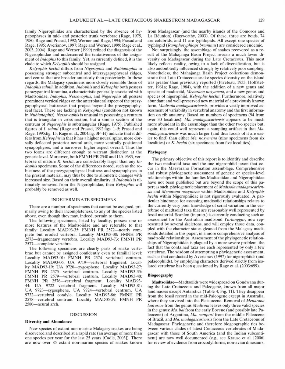
LADUKE ET AL.—LATE CRETACEOUS SNAKES FROM MADAGASCAR 129
family Nigerophiidae are characterized by the absence of hy-papophyses in mid- and posterior trunk vertebrae (Rage, 1975,1980; Rage and Prasad, 1992; Werner and Rage, 1994; Prasad andRage, 1995; Averianov, 1997; Rage and Werner, 1999; Rage et al.,2003, 2004). Rage and Werner (1999) refined the diagnosis of theNigerophiidae and underscored the tentativeness of the assign-ment of Indophis to this family. Yet, as currently defined, it is theclade to which Kelyophis should be assigned.
Kelyophis hechti differs from Nigerophis and Nubianophis inpossessing stronger subcentral and interzygapophyseal ridges,and centra that are broader anteriorly than posteriorly. In theseregards, the Malagasy specimens more closely resemble those ofIndophis sahnii. In addition, Indophis and Kelyophis both possessparazygantral foramina, a characteristic generally associated withMadtsoiidae. Indophis, Nessovophis, and Nigerophis all possessprominent vertical ridges on the anterolateral aspect of the prezy-gapophyseal buttresses that project beyond the prezygapophy-seal facet. These are lacking in Kelyophis (condition not knownin Nubianophis). Nessovophis is unusual in possessing a centrumthat is triangular in cross section, but a similar section of thecentrum of Nigerophis is subtriangular (Rage, 1975). Publishedfigures of I. sahnii (Rage and Prasad, 1992:figs. 1–5; Prasad andRage, 1995:fig. 15; Rage et al., 2004:fig. 3F–H) indicate that it dif-fers from Kelyophis in that it has a higher neural spine, more dor-sally deflected posterior neural arch, more ventrally positionedsynapophyses, and a narrower, higher aspect overall. Thus thetwo forms are different enough to warrant distinction at thegeneric level. Moreover, both FMNH PR 2540 and UA 9683, ver-tebrae of mature K. hechti, are considerably larger than any In-dophis specimens. Some of the differences noted, such as the ro-bustness of the prezygapophyseal buttress and synapophyses inthe present material, may thus be due to allometric changes withincreased size. Based on their overall similarity, if Indophis is ul-timately removed from the Nigerophiidae, then Kelyophis willprobably be removed as well.
INDETERMINATE SPECIMENS
There are a number of specimens that cannot be assigned, pri-marily owing to their incompleteness, to any of the species listedabove, even though they may, indeed, pertain to them.
The following specimens, listed by locality, possess one ormore features of the Madtsoiidae and are referable to thatfamily: Locality MAD93-35: FMNH PR 2572—nearly com-plete but eroded vertebra. Locality MAD93-38: FMNH PR2573—fragmentary vertebra. Locality MAD93-73: FMNH PR2577—complete vertebra.
The following specimens are clearly parts of snake verte-brae but cannot be assigned confidently even to familial level:Locality MAD93-01: FMNH PR 2574—vertebral centrum.Locality MAD93-06: UA 9719—vertebral fragment. Local-ity MAD93-19: UA 9720—zygosphene. Locality MAD93-27:FMNH PR 2575—vertebral centrum. Locality MAD93-35:FMNH PR 2579—vertebral centrum. Locality MAD93-40:FMNH PR 2576—vertebral fragment. Locality MAD93-44: UA 9722—vertebral fragment. Locality MAD93-81:UA 9723—zygosphene, UA 9724—vertebral centrum, UA9732—vertebral condyle. Locality MAD93-86: FMNH PR2578—vertebral centrum. Locality MAD05-59: FMNH PR2580—neural arch.
DISCUSSION
Diversity and Abundance
New species of extant non-marine Malagasy snakes are beingdiscovered and described at a rapid rate (an average of more thanone species per year for the last 25 years [Cadle, 2003]). Thereare now over 85 extant non-marine species of snakes known
from Madagascar (and the nearby islands of the Comoros andLa Reunion) (Raxworthy, 2003). Of these, three are boids, 74are colubrids, and 11 are typhlopids. All except one species oftyphlopid (Ramphotyphlops braminus) are considered endemic.
Not surprisingly, the assemblage of snakes recovered as a re-sult of the Mahajanga Basin Project reveals a much lower di-versity on Madagascar during the Late Cretaceous. This mostlikely reflects reality, owing to a lack of diversification, but isalso undoubtedly influenced strongly by relatively poor sampling.Nonetheless, the Mahajanga Basin Project collections demon-strate that Late Cretaceous snake species diversity on the islandwas greater than previously reported (Piveteau, 1933; Hoffstet-ter, 1961a; Rage, 1984), with the addition of a new genus andspecies of madtsoiid, Menarana nosymena, and a new genus andspecies of nigerophiid, Kelyophis hechti. Furthermore, relativelyabundant and well-preserved new material of a previously knownform, Madtsoia madagascariensis, provides a vastly improved as-sessment of variability in vertebral anatomy and the first informa-tion on rib anatomy. Based on numbers of specimens (94 fromover 30 localities), Ma. madagascariensis appears to be muchmore abundant in the assemblage than the other two species but,again, this could well represent a sampling artifact in that Ma.madagascariensis was much larger (and thus fossils of it are eas-ier to find) than either Me. nosymena (nine specimens from sixlocalities) or K. hechti (six specimens from five localities).
Phylogeny
The primary objective of this report is to identify and describethe two madtsoiid taxa and the one nigerophiid taxon that oc-cur in the Maevarano Formation assemblage. Comprehensiveand robust phylogenetic assessment of generic or species-levelrelationships within the families Madtsoiidae and Nigerophiidaehave not been published but are beyond the scope of this pa-per; as such, phylogenetic placement of Madtsoia madagascarien-sis and Menarana nosymena within Madtsoiidae and Kelyophishechti within Nigerophiidae is not rigorously evaluated. A par-ticular hindrance for assessing madtsoiid relationships relates tothe currently very poor knowledge of serial variation in the ver-tebrae of madtsoiid taxa that are reasonably well represented byfossil material. Scanlon (in prep.) is currently conducting such anassessment for the Australian madtsoiid Yurlunggur, now rep-resented by several skeletons, and will employ those data, cou-pled with the character states gleaned from the Malagasy madt-soiids detailed in this paper, in a more comprehensive analysis ofmadtsoiid relationships. Assessment of the phylogenetic relation-ships of Nigerophiidae is plagued by a more severe problem: thefact that the contained taxa are each represented by only a fewvertebrae. The wisdom of attempting a phylogenetic assessment,such as that conducted by Averianov (1997) for nigerophiids (andpalaeophiids), by employing characters derived strictly from iso-lated vertebrae has been questioned by Rage et al. (2003:699).
Biogeography
Madtsoiidae—Madtsoiids were widespread on Gondwana dur-ing the Late Cretaceous and Paleogene, known from all majorlandmasses except Antarctica (Table 4; Fig. 11). They disappearfrom the fossil record in the mid-Paleogene except in Australia,where they survived into the Pleistocene. Removal of Menaranalaurasiae from the genus Madtsoia leaves only three valid speciesin the genus: Ma. bai from the early Eocene (and possibly late Pa-leocene) of Argentina, Ma. camposi from the middle Paleoceneof Brazil, and Ma. madagascariensis from the Late Cretaceous ofMadagascar. Phylogenetic and therefore biogeographic ties be-tween various clades of latest Cretaceous vertebrates of Mada-gascar with those of South America (and the Indian subconti-nent) are now well documented (e.g., see Krause et al. [2006]for review of evidence from crocodyliforms, non-avian dinosaurs,

130 JOURNAL OF VERTEBRATE PALEONTOLOGY, VOL. 30, NO. 1, 2010
FIGURE 11. Temporal and geographic distribution of madtsoiid (localities indicated by solid symbols, numbers) and nigerophiid (open symbols,letters) snakes. See Tables 4 and 5 for more detailed information.
and mammals; and Evans et al. [2008] for evidence from frogs).It is tempting to suggest that the genus Madtsoia provides yetanother line of independent evidence of such a special connec-tion (i.e., to the exclusion of others). However, the fact that thereare many other snake fossils assigned to Madtsoia (or ?Madtsoia)or to the Madtsoiidae (or ?Madtsoiidae) from the Late Creta-ceous of Argentina, Niger, Sudan, Romania, France, and India,as well as from the Paleogene of Argentina, Bolivia, Morocco,India, and Australia (see Table 4; Fig. 11), reveals a potentiallymuch broader distribution and more complicated pattern. Thisunderscores the need for a comprehensive taxonomic review andphylogenetic assessment of the Madtsoiidae before any firm bio-geographic conclusions can be drawn.
Nonetheless, the disjunct distribution of the two known speciesof Menarana, Me. laurasiae from the Campanian of Spain (Rage,1996a, 1999) and Me. nosymena from the Maastrichtian of Mada-gascar, invites consideration. Specimens of madtsoiids, or ques-tionable madtsoiids, have also been reported from the Cam-panian of France (Sige et al., 1997) and the Maastrichtianof Romania (Folie and Codrea, 2005). Other typically Gond-wanan vertebrate taxa are known from these and other Campa-nian/Maastrichtian localities in southern Europe as well (Pereda-Superbiola, 2009), but there are no pre-Campanian Late Creta-ceous records of madtsoiids from Europe. Gheerbrant and Rage(2006:236) opined that the “European madtsoiids were immi-grants from Africa that reached Europe probably during the LateCretaceous” (see also Rage, 1995, 1996a, 1999; Rage et al., 2008).When during the Late Cretaceous is, however, a critical questionand Pereda-Superbiola (2009) notes that nothing excludes a dis-persal event during the Early Cretaceous. In this regard, the com-position of the snake fauna from the Late Cretaceous of Africais obviously relevant. There are no Campanian/Maastrichtianrecords of madtsoiids from Africa at all. Late Cretaceous Africanmadtsoiids are known only from the early Senonian of Niger
(de Broin et al., 1974; Rage, 1981; Madtsoia aff. madagascarien-sis—re-identified in this paper as ?Madtsoia sp.) and the Ceno-manian of Sudan (Rage and Werner, 1999; Madtsoiidae indet.).These two pre-Campanian records, therefore, are too indetermi-nate to shed light on the issue.
In light of this poor fossil record, there are several possibilitiesto explain the presence of Menarana (and other shared faunalelements) in the Campanian/Maastrichtian of Madagascar andsouthern Europe, including the following.
(1) A pan-Gondwanan (including southern Europe) or nearlypan-Gondwanan (for the issue at hand, Australia ismore or less peripheral to the argument) Early Creta-ceous distribution of which the Campanian/Maastrichtianrecords of Menarana in Madagascar and southern Eu-rope are relictual. During the Late Cretaceous, Europewas a complex archipelago (Dercourt et al., 2000) and,in a recent assessment of the biogeographic affinities ofLate Cretaceous European tetrapods, Pereda-Superbiola(2009:65) concluded that “isolation from other landmassesmay have facilitated survival of relict taxa in Europe untilCampanian-Maastrichitan times.” This scenario, if it pertainsto Menarana, requires that Menarana evolved before SouthAmerica separated from Africa at or near the Early/LateCretaceous boundary. This can be confirmed, in part,through the discovery of Menarana in pre-Late Cretaceoushorizons on any Gondwanan landmass. The absence of suchrecords, especially from the relatively well-sampled SouthAmerican record, is currently the only evidence against thisscenario.
(2) Africa (only) as an intermediate landmass, with connectionsto Madagascar via a Late Cretaceous land bridge or viasweepstakes dispersal across the marine barrier formed bythe Mozambique Channel, with a minimal distance of 430

LADUKE ET AL.—LATE CRETACEOUS SNAKES FROM MADAGASCAR 131
TABLE 4. Temporal and geographic distribution of madtsoiid and questionably madtsoiid snakes (arranged alphabetically by genus and species,and then by more uncertain family-level attributions). Abbreviations: E = Early; M = Middle; L = Late.
Taxon Locality Age Reference
1. Alamitophis argentinus Los Alamitos, Argentina Campanian/Maastrichtian Albino, 19862. Alamitophis argentinus La Colonia, Argentina Campanian/Maastrichtian Albino, 20003. Alamitophis argentinus Salinas de Trapalco, Argentina Campanian/Maastrichtian Albino, 1994; Albino, 20074. Alamitophis argentinus El Palomar, Argentina Campanian/Maastrichtian Albino, 19945. Alamitophis argentinus Salitral de Santa Rosa, Argentina Campanian/Maastrichtian Martinelli and Forasiepi, 2004;
Albino, 20076. Alamitophis elongatus Salinas de Trapalco, Argentina Campanian/Maastrichtian Albino, 1994; Albino, 20077. Alamitophis elongatus Los Alamitos, Argentina Campanian/Maastrichtian Albino, 19948. Alamitophis tingamarra Tingamarra, Australia E. Eocene Scanlon, 2005b9. Gigantophis garstini Dor et Talha, Libya L. Eocene Hoffstetter, 1961b10. Gigantophis garstini Fayum, Egypt L. Eocene∗ Andrews, 1901, 190611. Herensugea caristiorum Lano, Spain L. Campanian Rage, 1996a, 199912. Madtsoia bai Canadon Vaca, Argentina E. Eocene Simpson, 193313. Madtsoia cf. M. bai Gaiman, Argentina L. Paleocene Hoffstetter, 1959; Simpson, 193514. Madtsoia camposi Itaborai, Brazil M. Paleocene Rage, 199815. Madtsoia laurasiae (= Menarana
laurasiae in this paper)Lano, Spain L. Campanian Rage, 1996a, 1999
16. Madtsoia madagascariensis Mahajanga Basin, Madagascar Maastrichtian Hoffstetter, 1961a; LaDuke et al.,this paper
17. Madtsoia aff. M. madagascariensis (=?Madtsoia sp. in this paper)
In Beceten, Niger E. Senonian de Broin et al., 1974; Rage, 1981
18. ?Madtsoia sp. Canadon Hondo, Argentina L. Paleocene Albino, 199319. ?Madtsoia sp. Canadon Vaca, Argentina E. Eocene Albino, 199320. ?Madtsoia sp. Gran Barranca, Argentina E. Eocene Albino, 199321. Menarana nosymena Mahajanga Basin, Madagascar Maastrichtian LaDuke et al., this paper22. Najash rionegrina (?Madtsoiidae) La Buitrera, Argentina E. Cenomanian Apesteguıa and Zaher, 200623. Nanowana godthelpi Riversleigh, Australia E. Miocene Scanlon, 199724. Nanowana schrenki Riversleigh, Australia E. Miocene Scanlon, 199725. Patagoniophis australiensis Tingamarra, Australia E. Eocene Scanlon, 2005b26. Patagoniophis parvus Los Alamitos, Argentina Campanian/Maastrichtian Albino, 198627. Patagoniophis parvus Salitral de Santa Rosa, Argentina Campanian/Maastrichtian Martinelli and Forasiepi, 2004;
Albino, 200728. Rionegrophis madtsoioides Los Alamitos, Argentina Campanian/Maastrichtian Albino, 1986, 200729. Wonambi barriei Riversleigh, Australia L. Oligocene/E. Miocene Scanlon and Lee, 200030. Wonambi naracoortensis Naracoorte, Australia Pleistocene Smith, 197631. Wonambi naracoortensis Mammoth Cave, Australia Pleistocene Scanlon, 199532. Wonambi naracoortensis Koala Cave, Australia Pleistocene Scanlon, 199533. Wonambi naracoortensis Wellington Caves, Australia Pleistocene Scanlon, 199534. Wonambi naracoortensis Corra-Lynn Cave, Australia E. Pliocene? Scanlon, 1995; 2005a35. Wonambi naracoortensis Tight Entrance Cave, Australia Pleistocene Scanlon, 2005a36. Yurlunggur camfieldensis Bullock Creek, Australia M. Miocene Scanlon, 199237. Yurlunggur sp. Kanunka, Australia Pliocene Scanlon, 1995; 200438. Yurlunggur sp. Tarkarooloo, Australia L. Oligocene Scanlon, 200439. Yurlunggur sp. Lake Ngapakaldi, Australia E. or M. Miocene Scanlon, 200440. Yurlunggur sp. Chinchilla, Australia E. or M. Pliocene Mackness and Scanlon, 1999;
Scanlon, 200441. Yurlunggur sp. Gogolo-Garnpung, Willandra Lakes,
AustraliaL. Pleistocene Scanlon, 2004
42. Yurlunggur sp. Kangaroo Well, Australia L. Oligocene Megirian et al., 200443. Yurlunggur sp. Wyandotte, Australia L. Pleistocene Scanlon, 1995; 200444. Yurlunggur sp. or spp. Riversleigh, Australia L. Oligocene/E. Miocene Scanlon, 200645. Madtsoiidae indet. Salinas de Trapalco, Argentina Campanian/Maastrichtian Gomez and Baez, 200646. Madtsoiidae indet. Wadi Abu Hashim, Sudan Cenomanian Rage and Werner, 199947. Madtsoiidae indet. Adrar Mgorn, Morocco L. Paleocene Gheerbrant et al., 199348. Madtsoiidae indet. Yacimiento Las Flores, Argentina M. Paleocene Albino, 199349. Madtsoiidae indet. Hateg Basin, Romania Maastrichtian Folie and Codrea, 200550. Madtsoiidae indet. Kelapur, Maharashtra, India Maastrichtian Rage et al., 200451. Madtsoiidae indet. Salitral de Santa Rosa, Argentina Campanian/Maastrichtian Martinelli and Forasiepi, 2004;
Albino, 200752. ?Madtsoiidae Takli, India Maastrichtian Gayet et al., 198553. ?Madtsoiidae Champ-Garimond, France Campanian Sige et al., 199754. ?Madtsoiidae Los Alamitos, Argentina Campanian/Maastrichtian Albino, 200755. ?Madtsoiidae La Colonia, Argentina Campanian/Maastrichtian Albino, 200056. ?Madtsoiidae Ranquil-Co, Argentina Campanian/Maastrichtian Gonzalez Riga, 1999; Albino, 200757. ?Madtsoiidae indet. Salitral de Santa Rosa, Argentina Campanian/Maastrichtian Martinelli and Forasiepi, 2004;
Albino, 200758. ?Madtsoiidae indet. Vastan Lignite Mine, Gujarat, India E. Eocene Rage et al., 200859. ?Madtsoiidae indet cf. Madtsoia sp. Tingamarra, Australia E. Eocene Scanlon, 2005b60. ?Madtsoiidae or Boidae Tiupampa, Bolivia E. Paleocene Rage, 199161. ?Madtsoiidae or Boidae La Colonia, Argentina Campanian/Maastrichtian Albino, 200062. ?Madtsoiidae or Boidae Panandhro Mine, Kutch, India E. Eocene Rage et al., 200363. ?Madtsoiidae or Boidae Asifabad, India Maastrichtian Rage et al., 200464. ?Madtsoiidae or Boidae Pisdura, India Maastrichtian Rage et al., 2004
∗Revised age from Seiffert (2006).
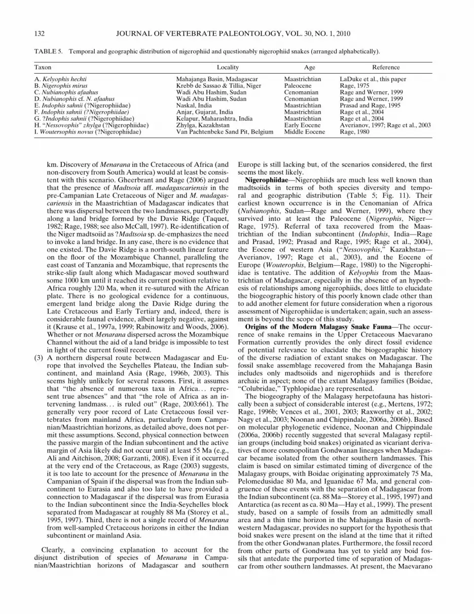
132 JOURNAL OF VERTEBRATE PALEONTOLOGY, VOL. 30, NO. 1, 2010
TABLE 5. Temporal and geographic distribution of nigerophiid and questionably nigerophiid snakes (arranged alphabetically).
Taxon Locality Age Reference
A. Kelyophis hechti Mahajanga Basin, Madagascar Maastrichtian LaDuke et al., this paperB. Nigerophis mirus Krebb de Sassao & Tillia, Niger Paleocene Rage, 1975C. Nubianophis afaahus Wadi Abu Hashim, Sudan Cenomanian Rage and Werner, 1999D. Nubianophis cf. N. afaahus Wadi Abu Hashim, Sudan Cenomanian Rage and Werner, 1999E. Indophis sahnii (?Nigerophiidae) Naskal, India Maastrichtian Prasad and Rage, 1995F. Indophis sahnii (?Nigerophiidae) Anjar, Gujarat, India Maastrichtian Rage et al., 2004G. ?Indophis sahnii (?Nigerophiidae) Kelapur, Maharashtra, India Maastrichtian Rage et al., 2004H. “Nessovophis” zhylga (?Nigerophiidae) Zhylga, Kazakhstan Early Eocene Averianov, 1997; Rage et al., 2003I. Woutersophis novus (?Nigerophiidae) Van Pachtenbeke Sand Pit, Belgium Middle Eocene Rage, 1980
km. Discovery of Menarana in the Cretaceous of Africa (andnon-discovery from South America) would at least be consis-tent with this scenario. Gheerbrant and Rage (2006) arguedthat the presence of Madtsoia aff. madagascariensis in thepre-Campanian Late Cretaceous of Niger and M. madagas-cariensis in the Maastrichtian of Madagascar indicates thatthere was dispersal between the two landmasses, purportedlyalong a land bridge formed by the Davie Ridge (Taquet,1982; Rage, 1988; see also McCall, 1997). Re-identification ofthe Niger madtsoiid as ?Madtsoia sp. de-emphasizes the needto invoke a land bridge. In any case, there is no evidence thatone existed. The Davie Ridge is a north-south linear featureon the floor of the Mozambique Channel, paralleling theeast coast of Tanzania and Mozambique, that represents thestrike-slip fault along which Madagascar moved southwardsome 1000 km until it reached its current position relative toAfrica roughly 120 Ma, when it re-sutured with the Africanplate. There is no geological evidence for a continuous,emergent land bridge along the Davie Ridge during theLate Cretaceous and Early Tertiary and, indeed, there isconsiderable faunal evidence, albeit largely negative, againstit (Krause et al., 1997a, 1999; Rabinowitz and Woods, 2006).Whether or not Menarana dispersed across the MozambiqueChannel without the aid of a land bridge is impossible to testin light of the current fossil record.
(3) A northern dispersal route between Madagascar and Eu-rope that involved the Seychelles Plateau, the Indian sub-continent, and mainland Asia (Rage, 1996b, 2003). Thisseems highly unlikely for several reasons. First, it assumesthat “the absence of numerous taxa in Africa. . . repre-sent true absences” and that “the role of Africa as an in-tervening landmass. . . is ruled out” (Rage, 2003:661). Thegenerally very poor record of Late Cretaceous fossil ver-tebrates from mainland Africa, particularly from Campa-nian/Maastrichtian horizons, as detailed above, does not per-mit these assumptions. Second, physical connection betweenthe passive margin of the Indian subcontinent and the activemargin of Asia likely did not occur until at least 55 Ma (e.g.,Ali and Aitchison, 2008; Garzanti, 2008). Even if it occurredat the very end of the Cretaceous, as Rage (2003) suggests,it is too late to account for the presence of Menarana in theCampanian of Spain if the dispersal was from the Indian sub-continent to Eurasia and also too late to have provided aconnection to Madagascar if the dispersal was from Eurasiato the Indian subcontinent since the India-Seychelles blockseparated from Madagascar at roughly 88 Ma (Storey et al.,1995, 1997). Third, there is not a single record of Menaranafrom well-sampled Cretaceous horizons in either the Indiansubcontinent or mainland Asia.
Clearly, a convincing explanation to account for thedisjunct distribution of species of Menarana in Campa-nian/Maastrichtian horizons of Madagascar and southern
Europe is still lacking but, of the scenarios considered, the firstseems the most likely.
Nigerophiidae—Nigerophiids are much less well known thanmadtsoiids in terms of both species diversity and tempo-ral and geographic distribution (Table 5; Fig. 11). Theirearliest known occurrence is in the Cenomanian of Africa(Nubianophis, Sudan—Rage and Werner, 1999), where theysurvived into at least the Paleocene (Nigerophis, Niger—Rage, 1975). Referral of taxa recovered from the Maas-trichtian of the Indian subcontinent (Indophis, India—Rageand Prasad, 1992; Prasad and Rage, 1995; Rage et al., 2004),the Eocene of western Asia (“Nessovophis,” Kazakhstan—Averianov, 1997; Rage et al., 2003), and the Eocene ofEurope (Wouterophis, Belgium—Rage, 1980) to the Nigerophi-idae is tentative. The addition of Kelyophis from the Maas-trichtian of Madagascar, especially in the absence of an hypoth-esis of relationships among nigerophiids, does little to elucidatethe biogeographic history of this poorly known clade other thanto add another element for future consideration when a rigorousassessment of Nigerophiidae is undertaken; again, such an assess-ment is beyond the scope of this study.
Origins of the Modern Malagasy Snake Fauna—The occur-rence of snake remains in the Upper Cretaceous MaevaranoFormation currently provides the only direct fossil evidenceof potential relevance to elucidate the biogeographic historyof the diverse radiation of extant snakes on Madagascar. Thefossil snake assemblage recovered from the Mahajanga Basinincludes only madtsoiids and nigerophiids and is thereforearchaic in aspect; none of the extant Malagasy families (Boidae,“Colubridae,” Typhlopidae) are represented.
The biogeography of the Malagasy herpetofauna has histori-cally been a subject of considerable interest (e.g., Mertens, 1972;Rage, 1996b; Vences et al., 2001, 2003; Raxworthy et al., 2002;Nagy et al., 2003; Noonan and Chippindale, 2006a, 2006b). Basedon molecular phylogenetic evidence, Noonan and Chippindale(2006a, 2006b) recently suggested that several Malagasy reptil-ian groups (including boid snakes) originated as vicariant deriva-tives of more cosmopolitan Gondwanan lineages when Madagas-car became isolated from the other southern landmasses. Thisclaim is based on similar estimated timing of divergence of theMalagasy groups, with Boidae originating approximately 75 Ma,Pelomedusidae 80 Ma, and Iguanidae 67 Ma, and general con-gruence of these events with the separation of Madagascar fromthe Indian subcontinent (ca. 88 Ma—Storey et al., 1995, 1997) andAntarctica (as recent as ca. 80 Ma—Hay et al., 1999). The presentstudy, based on a sample of fossils from an admittedly smallarea and a thin time horizon in the Mahajanga Basin of north-western Madagascar, provides no support for the hypothesis thatboid snakes were present on the island at the time that it riftedfrom the other Gondwanan plates. Furthermore, the fossil recordfrom other parts of Gondwana has yet to yield any boid fos-sils that antedate the purported time of separation of Madagas-car from other southern landmasses. At present, the Maevarano

LADUKE ET AL.—LATE CRETACEOUS SNAKES FROM MADAGASCAR 133
Formation snake fauna presents a pattern that is congruent withthat shown by many other Malagasy taxa in the fauna (withthe possible exception of the podocnemidid Erymnochelys—seeGaffney and Forster, 2003): the species present in the latest Cre-taceous deposits of the Mahajanga Basin belong to archaic Meso-zoic lineages that were eliminated and replaced by basal stocksof the modern fauna sometime after the close of the Cretaceous(Krause et al., 2006; Yoder and Nowak, 2006).
Paleobiology and Paleoecology
Our ability to infer aspects of the paleobiology and paleoe-cology of the Maevarano Formation snake assemblage is lim-ited for three principal reasons. First, distributional data do notprovide meaningful insight into differences among the three de-scribed species, given that two of them have been found at sev-eral localities (Madtsoia madagascariensis and Menarana nosy-mena at MAD93-16, Ma. madagascariensis and Kelyophis hechtiat MAD93-01, and Me. nosymena and K. hechti at MAD05-59)and all three co-occur at MAD93-35. Thus, the single conclu-sion that can be drawn from these data is that all three specieshad the ability to survive in the highly seasonal, semi-arid cli-mate deduced from sedimentological and taphonomic analysesof the Maevarano Formation (Rogers et al., 2000, 2007; Rogersand Krause, 2007).
Second, the phylogenetic placement of these taxa remainsuncertain. Nigerophiids are believed by some to be closelyrelated to acrochordids (Rage, 1984, 1987; McDowell, 1987),but they have not been included in any large-scale cladisticanalyses of snake interrelationships, presumably because theirmorphology remains known only very incompletely. In con-trast, madtsoiids have been included in several phylogeneticanalyses, but the results of these studies have varied signifi-cantly; some have recovered them as basal snakes, lying out-side of the clade containing Scolecophidia + Alethinophidia(Scanlon and Lee, 2000; Lee and Scanlon, 2002; Scanlon, 2006),whereas others have concluded that they are more derivedalethinophidians, nested within Macrostomata (Rieppel et al.,2002). Collectively, these points of uncertainty effectively pre-clude rigorous historical analysis of the natural histories of thesethree species.
Finally, as is the case for most fossil snakes, especially terres-trial ones, the Maevarano taxa are known predominantly fromvertebral specimens. Although some studies have attempted tocorrelate aspects of snake vertebral morphology with habitat, lo-comotor mode, diet, or method of prey capture (e.g., Johnson,1955; Hoffstetter and Gasc, 1969; Ruben, 1977; Lillywhite et al.,2000), few detailed analyses have been undertaken in this regard.Moreover, Moon (1999) recently demonstrated that considerablecaution must be exercised when attempting to make functionalinferences about snakes based solely on vertebral morphology.Consequently, much of what follows should be regarded as some-what speculative until such time as more rigorous analyses of thefunctional morphology of the vertebrae of extant snakes can becompleted.
Madtsoia madagascariensis—Although the detailed de-scriptions above serve to greatly expand our knowledge of theanatomy of Madtsoia madagascariensis, they provide frustrat-ingly few clues about its paleobiology, given our rudimentarycurrent understanding of the functional morphology of snakevertebrae. Indeed, in many respects, the vertebrae of Ma.madagascariensis strongly resemble those of many terrestrialgeneralists among extant snakes, including in particular a varietyof boids and pythonids (e.g., Gasc, 1974), which collectivelyexhibit a wide range of life history strategies. Many such snakesexhibit strong arboreal or semi-aquatic tendencies, and thedietary diversity they exhibit is extraordinarily high, includinga wide array of mammals, birds, crocodilians, and lepidosaurs
(e.g., Shine, 1991; Greene, 1997). However, whereas discreteanatomical characters may not (yet) provide significant insightinto the paleobiology of Ma. madagascariensis, one more generalfeature of its vertebrae—size—may be more informative in thisregard, as it serves as a useful proxy for estimating the overallbody size of this species.
Body size is an extremely important factor in determining anumber of life history traits in snakes, including feeding, locomo-tion, and habitat usage (e.g., Greene, 1997). However, estimatingsnake body size solely from measurements of individual verte-brae presents a number of potential difficulties, not the least ofwhich is that vertebral number varies widely, ranging from justover 100 to more than 550 (e.g., Rochebrune, 1881; Alexanderand Gans, 1966; Polly et al., 2001; upper end of range approxi-mated indirectly based on scale counts provided by Gow [1977]and Hahn and Wallach [1998]). Nevertheless, a recent study byMcCartney et al. (2008) demonstrated that nearly all standardmorphometric measurements of snake vertebrae are highly cor-related with measurements of total length. Furthermore, on thebasis of this somewhat unexpected finding, these authors pro-posed a method for estimating the size of fossil snakes from iso-lated vertebrae and provided equations for doing so based on re-gressions of vertebral size against total length, calculated acrossa phylogenetically and morphologically diverse sample of extantsnakes. Using this method, along with morphometric data fromone of the largest and best preserved mid-trunk vertebrae ofMadtsoia madagascariensis (FMNH PR 2553; Table 1), we es-timate the total length of this species to be approximately 5.1m. Moreover, the width of such mid-trunk vertebrae and thelength and curvature of the most completely preserved ribs (e.g.,UA 9764) suggest a mid-body diameter of approximately 15 cm.Taken together, these two estimates are suggestive of a relativelyheavy-bodied snake, with an overall body mass of at least 50 kg.However, large adults of Ma. madagascariensis may have reachedsignificantly greater proportions than this; the largest vertebralspecimen known from this species (MNHN MAJ 8; see Hoff-stetter, 1961a:fig. 3D) is an isolated zygosphene that is over 50%wider than that of FMNH PR 2553, suggesting that this speciesmay have at least occasionally reached lengths of nearly 8 m.
By analogy with modern snakes, the estimated proportionsof Madtsoia madagascariensis indicate a relatively slow-movingsnake that would likely have relied predominantly on a recti-linear mechanism of locomotion (e.g., Mosauer, 1932; Bogert,1947; Gray, 1968; Gans, 1974). Consistent with this hypothesis isthe complete absence of accessory prezygapophyseal processes,which tend to be greatly enlarged anterolaterally in taxa thatrely primarily on a lateral undulatory mechanism of locomotion,such as most colubroids (excluding viperids) and scolecophidi-ans (Johnson, 1955). Moreover, the overall shape of the verte-brae in Ma. madagascariensis differs greatly from that seen inrapidly moving locomotor specialists, typically characterized byrelatively narrow, elongate vertebrae (Johnson, 1955).
The large size of Madtsoia madagascariensis also suggestsit was a sit-and-wait ambush predator rather than an activeforager. Unfortunately, with no knowledge of its cranial mor-phology, it is difficult to estimate a size range for prey thatit may have exploited, and thus the type of prey that it mayhave consumed. Nevertheless, it can be safely assumed thatthis species faced the same fundamental physiological chal-lenge that all snakes do in feeding: it had to provide ad-equate nourishment for a very long body while retaining arelatively small head, a most serious challenge indeed for a gape-limited predator that must swallow prey whole, and a problemwith a limited number of solutions (Gans, 1961; Greene, 1997).One such solution is to eat very large numbers of relativelysmall prey (microphagy). However, this strategy has evolved onlyrarely within Serpentes; it is restricted primarily to blindsnakes(Scolecophidia), which feed almost exclusively on ant brood and

134 JOURNAL OF VERTEBRATE PALEONTOLOGY, VOL. 30, NO. 1, 2010
termites (e.g., Shine and Webb, 1990; Webb and Shine, 1993;Webb et al., 2000; Kley, 2003a, 2003b, 2003c). It is significant tonote that few microphagous snakes even approach 1 m in length,and most are in fact significantly smaller than this. The vast ma-jority of all other modern snakes—nearly 90% of approximately3000 recognized species, including all those that approach or ex-ceed 5 m in length—are macrophagous, feeding infrequently onrelatively large prey. Therefore, it is reasonable to assume thatMa. madagascariensis was also macrophagous. Furthermore, andagain by analogy with modern giant snakes (e.g., Shine et al.,1998), it is also likely that this species would have exhibited anontogenetic shift in diet, such that relatively small vertebrateswould have been deleted from the diets of large adult individ-uals (a very common phenomenon among extant snakes and onefully predicted by optimal foraging theory; Arnold, 1993). Thus,although juvenile Ma. madagascariensis may have fed on a rel-atively wide array of small vertebrates, adults probably preyedon a narrower range of larger taxa. Possible adult prey amongthe known fauna of the Maevarano Formation (see Krause etal., 2006, for a recent review) would have included medium-sizedcrocodyliforms (e.g., adult Simosuchus clarki, subadult Maha-jungasuchus insignis) as well as small theropod dinosaurs (e.g.,adult Masiakasaurus knopfleri, subadult Majungasaurus crenatis-simus). Such large and potentially injurious prey would have al-most certainly necessitated a highly efficient mechanism of preysubjugation. Given the lack of any evidence suggesting the pres-ence of a venom delivery system in madtsoiids (e.g., Scanlon,2005a, 2006), and the relative inefficiency of some mechanismsof prey subjugation, such as simple biting and/or pinioning usedby many modern snakes when feeding on relatively harmlessprey (e.g., Loop and Bailey, 1972; Willard, 1977; Greenwald,1978; de Queiroz, 1984), we consider it most likely that Ma.madagascariensis was a constrictor. However, we emphasize thatthis inference is based entirely on a process of elimination fromthe collective constellation of prey subjugation mechanisms ex-hibited by living snakes (for a recent review, see Cundall andGreene, 2000); constriction appears to be first and foremost abehavioral innovation, and osteological correlates of this behav-ior have yet to be identified conclusively (Greene and Burghardt,1978).
Menarana nosymena—Although known much less completelythan Madtsoia madagascariensis, Menarana nosymena differsfrom the former species in exhibiting a number of clearly adap-tive morphological traits that collectively offer considerable in-sight into its paleobiology.
Perhaps the most remarkable aspect of the morphologyof Menarana nosymena is the extensive degree to whichindividual elements of the braincase (viz., parabasisphenoid, ba-sioccipital, exoccipitals, prootics) have become fused together.Among the more than 25,000 recognized species of extanttetrapods, such a pattern of extensive basicranial fusion is seenonly among the most highly specialized limb-reduced, head-first burrowers, such as in the ‘os basale’ of caecilians (e.g.,Wiedersheim, 1879; Taylor, 1969; Wake and Hanken, 1982), the‘otic-occipital complex’ of amphisbaenians (e.g., Zangerl, 1944;Maisano et al., 2006; Montero and Gans, 2008), and the ‘otico-occipital complex’ of derived uropeltid snakes (Rieppel andZaher, 2002; Baumeister, 1908; Cundall and Irish, 2008). The re-peated convergent evolution of this morphological phenomenonin these three highly fossorial clades, together with its generalabsence among other tetrapods, suggests strongly that either Me.nosymena was itself a very powerful head-first burrower, or that itevolved from ancestors that were. Discovery of additional cranialmaterial, in particular elements of the snout, would likely helpto refine this interpretation. However, it is interesting to note inthis context that adult Yurlunggur also exhibit fusion among sev-eral elements of the posterior braincase (though fewer than inMenarana), yet clearly retain a rather unspecialized snout that
would appear to be very poorly suited for head-first burrowing(Scanlon, 2006). Moreover, the very large size of this taxon alone(estimated to be approximately 5 m; Scanlon, 2006) would appearto be enough to make burrowing through compact soils impossi-ble (see below). Thus, the presence of these basicranial fusions inlarge adult Yurlunggur likely represents the retention of an an-cestral trait, rather than direct evidence of adaptive modificationswithin this particular taxon.
The morphology of the trunk vertebrae of Menarana nosymenais also consistent with the hypothesis of a burrowing lifestyleor ancestry. Most telling in this respect are the very low neuralspines and the rather depressed overall appearance of the ver-tebrae, features that are nearly ubiquitous among extant snakesthat exhibit strong fossorial tendencies (e.g., scolecophidians,Anilius, Cylindrophis, uropeltids, Xenopeltis, Loxocemus) (Hoff-stetter and Gasc, 1969; Gasc, 1974). Somewhat more difficultto interpret is the morphology of the atlas, in which the neu-ral arches are fused completely with the intercentrum. How-ever, in light of other available evidence, it is tempting to con-sider this also an adaptation for burrowing, as this might serveto maintain the integrity of the anterior atlantal cotyle (and thusthe craniovertebral joint as a whole) under the extreme loadingregimes that would be expected during tunnel construction, asit would prevent the three constituent components of the cotylefrom being forced apart. Indeed, modifications of the atlas-axiscomplex are relatively common among head-first limbless bur-rowers (Gans, 1958; Williams, 1959; Taylor, 1977; Wake, 1980).However, among the most specialized head-first burrowing squa-mates, most notably amphisbaenians and uropeltid snakes, mod-ifications to the atlas most commonly involve reduction ratherthan fortification. Specifically, there is a tendency toward reduc-tion or loss of the atlantal intercentrum, as well as reduction insize of the articular facets on the pedicles of the atlantal neuralarches, which together greatly reduce the overall contribution ofthe atlas to the craniovertebral joint (Zangerl, 1945; Williams,1959). The result of these modifications is ultimately very simi-lar to that of atlantal fusion in Menarana: the occipital condyleis received predominantly by a single (rather than tripartite)element—the odontoid process in amphisbaenians, and the axialcotyle in uropeltids (members of the latter clade lack an odontoidprocess).
Despite the numerous morphological features discussed abovethat Menarana nosymena shares with many extant forms of limb-reduced, head-first burrowing amphibians and reptiles, it must beemphasized that few such taxa exceed even 1 m in total length,and the vast majority are significantly smaller than this. Perhapseven more significant, most are less than 1 cm in diameter, andnearly all are less than 2.5 cm in diameter. The latter is particu-larly important because it has been suggested that the force re-quired to push an object through a given substrate increases withthe cross-sectional area (and thus the square of the diameter)of that object (Gans, 1960). That is, it would be predicted thatas the diameter of a burrowing snake’s head increases, the forcethat would be required to effectively burrow through compactsoil would increase very rapidly, according to a quadratic func-tion rather than a linear one. This implies, for example, that ifa snake having a head diameter of 1 cm required 25 N of forceto burrow through a given substrate, one with a head diameterof 3 cm would have to generate a much greater 225 N of push-ing force to burrow through the same medium. Given this rela-tionship, and the inherent physiological and mechanical limita-tions common to all vertebrate skeletal muscles with respect totheir capacity to generate force, it remains somewhat question-able whether a head-first burrowing mechanism could have beeneffective, or even possible, in a limbless animal of the size of Me.nosymena, which we estimate to have been approximately 2.4 min total length (using the equations of McCartney et al., 2008) andin excess of 7 cm in mid-body diameter. Nevertheless, available

LADUKE ET AL.—LATE CRETACEOUS SNAKES FROM MADAGASCAR 135
anatomical evidence points strongly toward this species having atleast a burrowing ancestry, if not a burrowing lifestyle itself, asinferred for some other madtsoiids (e.g., Herensugea caristiorum;Rage, 1999).
The jaw apparatus of Menarana nosymena, like that of Madt-soia madagascariensis, remains completely unknown, making in-ferences about its diet and feeding difficult at best. However,once again body size provides the most important clues in thisrespect, as it significantly narrows the list of potential prey. Byanalogy with modern alethinophidian snakes, the estimated pro-portions of Me. nosymena suggest a maximum prey size for thisspecies of well under 5 kg, and possibly as small as 1–2 kg. Thus,we can eliminate from the list of potential prey all of the largerknown fauna of the Maevarano Formation, including adults ofall non-avian dinosaurs and most crocodyliforms (the one knownexception being the very small Araripesuchus tsangatsangana;Turner, 2006). More likely forms of potential prey include muchsmaller ground-dwelling or fossorial vertebrates, possibly includ-ing other snakes, non-ophidian squamates (‘lizards’) or smallmammals.
Kelyophis hechti—Kelyophis hechti, the Malagasynigerophiid, was probably less than 1 m long and thus muchsmaller than the madtsoiids, Madtsoia madagascariensis andMenarana nosymena. It appears to have been more generalizedthan other nigerophiids, such as Nigerophis, Nubianophis,and Indophis, which have been interpreted to have been veryhighly specialized aquatic snakes on the basis of their ventrallypositioned synapophyses, their ‘peculiar’ prezygapophysealbuttresses, and the high, narrow shape of their mid-trunk,posterior trunk, and postcloacal vertebrae (Rage and Prasad,1992; Prasad and Rage, 1995; Rage and Werner, 1999). Basedon its somewhat shorter vertebrae with less ventrally shiftedsynapophyses, Kelyophis was apparently less well adapted to anaquatic lifestyle. However, given the extremely limited nature ofthe available material representing this species, few additionalinferences can be made about its paleobiology.
ACKNOWLEDGMENTS
We gratefully acknowledge field teams of the Maha-janga Basin Project for the collection of specimens de-scribed in this report; A. Rasoamiaramanana of the Univer-site d’Antananarivo, B. Andriamihaja and his staff of theMadagascar Institute pour la Conservation des EnvironnementsTropicaux, and the villagers of Berivotra for logistical supportin the field; J. Groenke and V. Heisey for preparation of fos-sils; M. Colbert for scanning the partial basicranium of the holo-type of Menarana nosymena; J. Maisano for processing of µCTimages; R. Bonett, M. Norell, and D. Wake for access to com-parative material; S. Burch, R. Jacobs, J. Sertich, and W. Simp-son for assistance with curation; M. Stewart for photography; L.Betti-Nash for drawing or arranging the figures; J.-C. Rage andM. Godinot for curatorial information concerning MNHN speci-mens of Madtsoia madagascariensis; and R. Jacobs for assistancein compiling distributional data. We also thank J. McCartney,J. Pruetz, and J. Sertich for reviewing early drafts, or parts ofdrafts, of the manuscript, and the anonymous JVP reviewers fortheir helpful comments on the submitted draft. This research wasfunded by grants from the National Science Foundation (DEB-9224396, EAR-9418816, EAR-9706302, DEB-9904045, EAR-0106477, EAR-0116517, EAR-0446488) and the National Geo-graphic Society (1999, 2001, 2004) to DWK.
LITERATURE CITED
Albino, A. M. 1986. Nuevos Boidae Madtsoiinae en el Cretacico tardiode Patagonia (Formacion Los Alamitos, Rio Negro, Argentina); pp.15–21 in J. F. Bonaparte (ed.), Simposio Evolucion de los Vertebra-
dos Mesozoicos. Actas IV Congreso Argentino de Paleontologıa yBioestratigrfıa, Mendoza.
Albino, A. M. 1993. Snakes from the Paleocene and Eocene of Patagonia,Argentina: paleoecology and coevolution with mammals. HistoricalBiology 7:51–69.
Albino, A. M. 1994. Una nueva serpiente (Reptilia) en el Cretacico supe-rior de Patagonia, Argentina. Pesquisas 21:58–63.
Albino, A. M. 2000. New record of snakes from the Cretaceous of Patag-onia (Argentina). Geodiversitas 22:247–253.
Albino, A. M. 2007. Lepidosauria; pp. 87–115 in Z. Gasparini, L. Salgadoand R. A. Coria (eds.), Patagonian Mesozoic Reptiles. Indiana Uni-versity Press, Bloomington & Indianapolis, Indiana.
Alexander, A. A., and C. Gans. 1966. The pattern of dermal-vertebralcorrelation in snakes and amphisbaenians. Zoologische Mededelin-gen 41:171–190.
Ali, J. R., and J. C. Aitchison. 2008. Gondwana to Asia: plate tectonics,paleogeography and the biological connectivity of the Indian sub-continent from the Middle Jurassic through latest Eocene (166–35Ma). Earth-Science Reviews 88:145–166.
Andrews, C. W. 1901. Preliminary note on some recently discoveredextinct vertebrates from Egypt (Part II). Geological Magazine8:434–444.
Andrews, C. W. 1906. A descriptive catalogue of the Tertiary Verte-brata of the Fayum, Egypt. British Museum, London. Ophidia, pp.306–312.
Apesteguıa, S., and H. Zaher. 2006. A Cretaceous terrestrial snake withrobust hindlimbs and a sacrum. Nature 440:1037–1040.
Arnold, S. J. 1993. Foraging theory and prey-size–predator-size relationsin snakes; pp. 87–115 in R. A. Seigel and J. T. Collins (eds.), Snakes:Ecology and Behavior. McGraw-Hill, New York, New York.
Averianov, A. O. 1997. Paleogene sea snakes from the eastern part ofTethys. Russian Journal of Herpetology 4:128–142.
Barrie, D. J. 1990. Skull elements and additional remains of thePleistocene boid snake Wonambi naracoortensis. Memoirs of theQueensland Museum 28:139–151.
Baumeister, L. 1908. Beitrage zur Anatomie und Physiologie derRhinophiden. Integument, Drusen der Mundhohle, Augen undSkeletsystem. Zoologische Jahrbucher, Abteilung fur Anatomieund Ontogenie der Tiere 26;423–526.
Bogert, C. M. 1947. Rectilinear locomotion in snakes. Copeia1947:253–254.
Cadle, J. E. 2003. Colubridae, snakes; pp. 997–1004 in S. M. Goodmanand J. P. Benstead (eds.), The Natural History of Madagascar. Uni-versity of Chicago Press, Chicago, Illinois.
Caldwell, M. W., and A. M. Albino. 2002. Exceptionally preserved skele-tons of the Cretaceous snake Dinilysia patagonica Woodward, 1901.Journal of Vertebrate Paleontology 22:861–866.
Caldwell, M. W., and J. Calvo. 2008. Details of a new skull and articulatedcervical column of Dinilysia patagonica Woodward, 1901. Journal ofVertebrate Paleontology 28:349–362.
Cundall, D., and H. W. Greene. 2000. Feeding in snakes; pp. 293–333 in K.Schwenk (ed.), Feeding: Form, Function, and Evolution in TetrapodVertebrates. Academic Press, San Diego, California.
Cundall, D., and F. Irish. 2008. The snake skull; pp. 349–692 in C. Gans,A. S Gaunt, and K. Adler (eds.), Biology of the Reptilia, Volume 20,Morphology H (The Skull of Lepidosauria). Society for the Study ofAmphibians and Reptiles, Ithaca, New York.
de Broin, F., E. Buffetaut, J.C. Koeniguer, J.-C. Rage, P. Taquet, C.Vergnaud-Grazzini, and S. Wenz. 1974. La faune de Vertebres con-tinentaux du gisement d’In Beceten (Senonien du Niger). Comptesrendus de l’Academie des Sciences, Paris 279:469–472.
de Queiroz, A. 1984. Effects of prey type on the prey-handling behaviorof the bullsnake, Pituophis melanoleucus. Journal of Herpetology18:333–336.
Dercourt, J., M. Gaetani, B. Vrielynick, E. Barrier, B. Biju-Duval, M.-F. Brunet, J.-P. Cadet, S. Crasquin, and Sandulescu, M. (eds.).2000. Atlas Peri-Tethys. Palaeogeographical maps. CCGM/CGMW,Paris, 24 maps, 269 pp.
Estes, R., T. H. Frazzetta, and E. E. Williams. 1970. Studies on the fossilsnake Dinilysia patagonica Woodward. Part 1. Cranial morphology.Bulletin of the Comparative Zoology, Harvard University140:25–74.
Evans, S. E., M. E. H. Jones, and D. W. Krause. 2008. A giant frog withSouth American affinities from the Late Cretaceous of Madagascar.Proceedings of the National Academy of Sciences of the UnitedStates of America 105:2951–2956.

136 JOURNAL OF VERTEBRATE PALEONTOLOGY, VOL. 30, NO. 1, 2010
Folie, A., and V. Codrea. 2005. New lissamphibians and squamates fromthe Maastrichtian of Hateg Basin, Romania. Acta PalaeontologicaPolonica 50:57–71.
Gaffney, E. S., and C. A. Forster. 2003. Side-necked turtle lower jaws(Podocnemididae, Bothremydidae) from the Late Cretaceous Mae-varano Formation of Madagascar. American Museum Novitates3397:1–13.
Gans, C. 1958. Modifications of the head joint in acrodont amphis-baenids and their functional implication [abstract]. AnatomicalRecord 132:441.
Gans, C. 1960. Studies on amphisbaenids (Amphisbaenia, Reptilia). 1.A taxonomic revision of the Trogonophinae, and a functional in-terpretation of the amphisbaenid adaptive pattern. Bulletin of theAmerican Museum of Natural History 119:129–204.
Gans, C. 1961. The feeding mechanism of snakes and its possible evolu-tion. American Zoologist 1:217–227.
Gans, C. 1974. Biomechanics: An Approach to Vertebrate Biology. Uni-versity of Michigan Press, Ann Arbor, Michigan, 261 pp.
Garzanti, E. 2008. Comment on “When and where did India andAsia collide?” by Jonathan C. Aitchison, Jason R. Ali, andAileen M. Davis. Journal of Geophysical Research, 113:B04411,doi:10.1029/2007JB005276.
Gasc, J.-P. 1974. L’interpretation fonctionelle de l’appareil musculo-squelettique de l’axe vertebral chez les serpents (Reptilia).Memoires du Museum National d’Histoire Naturelle, Paris, Serie A,Zoologie 83:1–182.
Gayet, M., J.-C. Rage, and R. S. Rana. 1985. Nouvelles ichthyofauneet herpetofaune de Gitti Khadan, le plus ancient gisement connudu Deccan (Cretace/Paleocene) a microvertebres. Implicationspaleogeographiques; pp. 55–65 in E. Buffetaut, J.-J. Jaeger, and J.-C. Rage (eds.), Paleogeographie de l’Inde, du Tibet et du Sud-EstAsiatique. Memoire de la Societe geologique de France 147.
Gheerbrant, E., and J.-C. Rage. 2006. Paleobiogeography of Africa: Howdistinct from Gondwana and Laurasia? Palaeogeography, Palaeocli-matology, Palaeoecology 241:224–246.
Gheerbrant, E., H. Cappetta, M. Feist, J.-J. Jaeger, J. Sudre, M.Vianey-Liaud, and B. Sige. 1993. La succession des faunes devertebres d’age paleocene superieur et eocene inferieur dans lebasin d’Ouarzazate, Maroc. Contexte geologique, portee bios-tratigraphique et paleogeographique. Newsletters in Stratigraphy28:33–58.
Gomez, R. O., and A. M. Baez. 2006. A new madtsoiid snake (Squa-mata, Ophidia) from the Upper Cretaceous of Patagonia. XXIIJornadas Argentinas de Paleontologıa de Vertebrados, San Juan,Argentina (2006):21.
Gonzalez Riga, B. J. 1999. Hallazgo de vertebrados fosiles en la For-macion Loncoche, Cretacico Superior de la provincia de Mendoza,Argentina. Ameghiniana 36:401–410.
Gow, G. F. 1977. A new species of Python from Arnhem Land. AustralianZoologists 19:133–139.
Gray, J. 1968. Animal Locomotion. W.W. Norton & Company, NewYork, New York, 479 pp.
Greene, H. W. 1997. Snakes: The Evolution of Mystery in Nature. Uni-versity of California Press, Berkeley, California, 351 pp.
Greene, H. W., and G. M. Burghardt. 1978. Behavior and phylogeny: con-striction in ancient and modern snakes. Science 200:74–76.
Greenwald, O. E. 1978. Kinematics and time relations of prey capture bygopher snakes. Copeia 1978:263–268.
Hahn, D. E., and V. Wallach. 1998. Comments on the systematics of OldWorld Leptotyphlops (Serpentes: Leptotyphlopidae), with descrip-tion of a new species. Hamadryad 23;50–62.
Hay, W. W., R. M. DeConto, C. N. Wold, K. M. Wilson, S. Voigt, M.Schulz, A. R. Wold, W. C. Dullo, A. B. Ronov, A. N. Balukhovsky,and E. Soding. 1999. Alternative global Cretaceous paleogeography;pp. 1–47 in E. Barrera and C. C. Johnson (eds.), Evolution of theCretaceous Ocean-Climate System. Geological Society of America,Special Paper 33.
Head, J. J. 2005. Snakes of the Siwalik Group (Miocene of Pakistan): sys-tematics and relationship to environmental change. PalaeontologiaElectronica 8.1.18A:1–33.
Head, J., and P. Holroyd. 2008. Assembly and biogeography of NorthAmerican Paleogene snake faunas based on an expanded fossilrecord. Journal of Vertebrate Paleontology 28(3, Supplement):90A.
Hoffstetter, R. 1959. Un serpent terrestre dans le Cretace inferieur duSahara. Bulletin de la Societe geologique de France 7:897–902.
Hoffstetter, R. 1961a. Nouveaux restes d’un serpent boıde (Madtsoiamadagascariensis nov. sp.) dans le Cretace superieur de Mada-gascar. Bulletin du Museum national d’Histoire naturelle, Paris33:152–160.
Hoffstetter, R. 1961b. Nouvelles recoltes de serpents fossiles dansl’Eocene superieur du desert Libyque. Bulletin du Museum nationald’Histoire naturelle, Paris 33:326–331.
Hoffstetter, R., and J.-P. Gasc. 1969. Vertebrae and ribs of modern rep-tiles; pp. 201–310, in C. Gans, A. d’A. Bellairs, and T. S. Parsons(eds.), Biology of the Reptilia, Volume 1, Morphology A. AcademicPress, London.
Johnson, R. G. 1955. The adaptive and phylogenetic significance of ver-tebral form in snakes. Evolution 9:367–388.
Kley, N. J. 2003a. Early blindsnakes (Anomalepididae); pp. 369–372 inM. Hutchins, J. B. Murphy, and N. Schlager (eds.), Grzimek’s Ani-mal Life Encyclopedia, 2nd edition. Gale Group, Farmington Hills,Michigan.
Kley, N. J. 2003b. Slender blindsnakes (Leptotyphlopidae); pp. 373–377in M. Hutchins, J. B. Murphy, and N. Schlager (eds.), Grzimek’s An-imal Life Encyclopedia, 2nd edition. Gale Group, Farmington Hills,Michigan.
Kley, N. J. 2003c. Blindsnakes (Typhlopidae); pp. 379–385 in M.Hutchins, J. B. Murphy, and N. Schlager (eds.), Grzimek’s Ani-mal Life Encyclopedia, 2nd edition. Gale Group, Farmington Hills,Michigan.
Krause, D. W., S. E. Evans, and K.-Q. Gao. 2003. First definitive evi-dence of Mesozoic lizards from Madagascar. Journal of VertebratePaleontology 23:842–856.
Krause, D. W., J. H. Hartman, and N. A. Wells. 1997. Late Cretaceousvertebrates from Madagascar: Implications for biotic change in deeptime; pp. 3–43 in S. D. Goodman and B. D. Patterson (eds.), NaturalChange and Human Impact in Madagascar. Smithsonian InstitutionPress, Washington, D.C.
Krause, D. W., S. D. Sampson, M. T. Carrano, and P. M. O’Connor. 2007.Overview of the history of discovery, taxonomy, phylogeny, and bio-geography of Majungasaurus crenatissimus (Theropoda: Abelisauri-dae) from the Late Cretaceous of Madagascar. Society of VertebratePaleontology Memoir 8:1–20.
Krause, D. W., P. M. O’Connor, K. Curry Rogers, S. D. Sampson, G. A.Buckley, and R. R. Rogers. 2006. Late Cretaceous terrestrial verte-brates from Madagascar: implications for Latin American biogeog-raphy; pp. 178–208 in Latin American Biogeography—Causes andEffects. 51st Annual Systematics Symposium, Missouri BotanicalGarden, Latin American Biogeography—Causes and Effects. An-nals of the Missouri Botanical Garden 93.
Krause, D. W., R. R. Rogers, C. A. Forster, J. H. Hartman, G. A. Buckley,and S. D. Sampson. 1999. The Late Cretaceous vertebrate fauna ofMadagascar: implications for Gondwanan paleobiogeography. GSAToday 9:1–7.
LaDuke, T. C. 1991. The fossil snakes of Pit 91, Rancho La Brea, Cal-ifornia. Contributions in Science, Natural History Museum of LosAngeles County 424:1–28.
Lavocat, R. 1955. Etude des gisements de Dinosauriens de la region deMajunga (Madagascar). Travaux du Bureau Geologique 69:1–19.
Lee, M. S. Y., and M. W. Caldwell. 1998. Anatomy and relationshipsof Pachyrhachis problematicus, a primitive snake with hindlimbs.Philosophical Transactions of the Royal Society of London, SeriesB353:1521–1552.
Lee, M. S. Y., and J. D. Scanlon. 2002. Snake phylogeny based on os-teology, soft anatomy and behaviour. Biological Reviews 77:333–402.
Lee, M. S. Y., M. W. Caldwell, and J. D. Scanlon. 1999. A second prim-itive marine snake: Pachyophis woodwardi from the Cretaceous ofBosnia-Herzegovina. Journal of Zoology, London 248;509–520.
Lillywhite, H. B., J. R. LaFrentz, Y. C. Lin, and M. C. Tu. 2000. The can-tilever ability of snakes. Journal of Herpetology 34:523–528.
Linnaeus, C. 1758. Systema naturæ per regna tria naturæ, secundumclasses, ordines, genera, species, cum characteribus, differentiis, syn-onymis, locis. Tomus I. Editio decima, reformata. Laurentii Salvii,Holmiæ, 824 pp.
List, J. C. 1966. Comparative osteology of the snake families Typhlopidaeand Leptotyphlopidae. Illinois Biological Monographs 36:1–112.
Loop, M. S., and L. G. Bailey. 1972. The effect of relative prey size on theingestion behavior of rodent-eating snakes. Phychonomic Science28:167–169.

LADUKE ET AL.—LATE CRETACEOUS SNAKES FROM MADAGASCAR 137
Mackness, B. S., and J. D. Scanlon. 1999. The first Pliocene record of themadtsoiid snake genus Yurlunggur Scanlon, 1992 from Queensland.Memoirs of the Queensland Museum 43:783–785.
Maisano, J. A., M. Kearney, and T. Rowe. 2006. Cranial anatomy of thespade-headed amphisbaenian Diplometopon zarudnyi (Squamata,Amphisbaenia) based on high-resolution X-ray computed tomog-raphy. Journal of Morphology 267:70–102.
Martinelli, A. G., and A. M. Forasiepi. 2004. Late Cretaceous vertebratesfrom Bajo de Santa Rosa (Allen Formation), Rıo Negro province,Argentina, with the description of a new sauropod dinosaur (Ti-tanosauridae). Revista del Museo Argentino de Ciencias Naturales6:257–305.
McCall, R. A. 1997. Implications of recent geological investigations of theMozambique Channel for the mammalian colonization of Madagas-car. Proceedings of the Royal Society of London B 264:663–665.
McCartney, J. A., N. J. Kley, and M. A. O’Leary. 2008. Body size of thegiant Eocene snake Palaeophis colossaeus (Serpentes: Palaeophi-idae) estimated from recently collected material from Mali. Journalof Vertebrate Paleontology 28(3, Supplement):114A.
McDowell, S. B. 1987. Systematics; pp. 3–50 in R. A. Siegel, J. T. Collins,and S. S. Novak (eds.), Snakes: Ecology and Evolutionary History.McGraw-Hill Publications Company, New York.
Megirian, D., P. Murray, L. Schwartz, and C. von der Borch. 2004. LateOligocene Kangaroo Well Local Fauna from the Ulta Limestone(new name), and climate of the Miocene oscillation across CentralAustralia. Australian Journal of Earth Sciences 51:701–741.
Mertens, R. 1972. Madagaskars Herpetofauna und die Kontinentaldrift.Studien uber die Reptilienfauna Madagaskars VI. Zool. Med.46:91–98.
Montero, R., and C. Gans. 2008. An atlas of amphisbaenian skullanatomy; pp. 621–738 in C. Gans, A. S. Gaunt, and K. Adler(eds.), Biology of the Reptilia, Volume 21, Morphology I (TheSkull and Appendicular Locomotor Apparatus of Lepidosauria).Society for the Study of Amphibians and Reptiles, Ithaca,New York.
Moon, B. R. 1999. Testing an inference of function from structure: snakevertebrae do the twist. Journal of Morphology 241:217–225.
Mosauer, W. 1932. On the locomotion of snakes. Science 76:583–585.Nagy, Z. T., U. Joger, M. Wink, F. Glaw, and M. Vences. 2003. Multi-
ple colonization of Madagascar and Socotra by colubrid snakes: ev-idence from nuclear and mitochondrial gene phylogenies. Proceed-ings of the Royal Society of London B 270:2613–2621.
Noonan, B. P., and P. T. Chippindale. 2006a. Dispersal and vicariance:The complex evolutionary history of boid snakes. Molecular Phylo-genetics and Evolution 40:347–358.
Noonan, B. P., and P. T. Chippindale. 2006b. Vicariant origin of Malagasyreptiles supports Late Cretaceous Antarctic landbridge. AmericanNaturalist 168:730–741.
Oelrich, T. M. 1956. The anatomy of the head of Ctenosaura pectinata(Iguanidae). Miscellaneous Publications, Museum of Zoology, Uni-versity of Michigan, 94:1–122.
Oppel, M. 1811. Die Ordnungen, Familien, und Gatungen der Rep-tilien, als Prodrom einer Naturgeschichte derselben. Joseph Lin-dauer, Munchen, 87 pp.
Pereda-Superbiola, X. 2009. Biogeographical affinities of Late Creta-ceous continental tetrapods of Europe: a review. Bulletin de laSociete geologique de France 180:57–71.
Piveteau, J. 1933. Un ophidien du Cretace superieur de Madagascar. Bul-letin de la Societe geologique de France (5) 3:597–602.
Polly, P. D., J. J. Head, and M. J. Cohn. 2001. Testing modularityand dissociation: the evolution of regional proportions in snakes;pp. 305–335 in Zelditch, M. L. (ed.), Beyond Heterochrony: TheEvolution of Development. John Wiley & Sons, New York, NewYork.
Prasad, G. V. R., and J.-C. Rage. 1995. Amphibians and squamates fromthe Maastrichtian of Naskal, India. Cretaceous Research 16:95–107.
Pregill, G. K. 1977. Axial myology of the racer Coluber constrictor withemphasis on the neck region. Transactions of the San Diego Societyof Natural History 18:185–206.
Rabinowitz, P. D., and S. Woods. 2006. The Africa-Madagascar connec-tion and mammalian migrations. Journal of African Earth Sciences44:270–276.
Rage, J.-C. 1975. Un serpent du Paleocene du Niger. Etude preliminairesur l’origine des Caenophidiens (Reptilia, Serpentes). ComptesRendus de l’Academie des Sciences, Paris (D) 281:515–518.
Rage, J.-C. 1980. Un serpent marin nouveau de l’Eocene de Belgique. Leprobleme des serpents marine du Paleogene. Comptes Rendus del’Academie des Seances, Paris (D) 291:469–471.
Rage, J.-C. 1981. Les continents peri-atlantiques au Cretacesuperieur: migrations des faunes continentales et problemespaleogeographiques. Cretaceous Research 2:65–84.
Rage, J.-C. 1984. Serpentes. Handbuch der Palaoherpetologie, part 11.Gustav Fischer Verlag, Stuttgart, 80 pp.
Rage, J.-C. 1987. Fossil history; pp. 51–76 in R. A. Seigel, J. T. Collinsand S. S. Novak (eds.), Snakes: Ecology and Evolutionary Biology,McGraw-Hill, New York, New York.
Rage, J.-C. 1988. Gondwana, Tethys, and terrestrial vertebrates duringthe Mesozoic and Cenozoic; pp. 255–273 in M. G. Audley-Charlesand A. Hallam (eds.), Gondwana and Tethys. Geological SocietySpecial Publication 37.
Rage, J.-C. 1991. Squamate reptiles from the early Paleocene of the Tiu-pampa area (Santa Lucia Formation), Bolivia; pp. 503–508 in R.Suarez-Soruco (ed.), Fosiles y Facies de Bolivia—Volume 1 Verte-brados. Revista Tecnica de YPFB 12.
Rage, J.-C. 1995. La Tethys et les dispersions transtethysiennes par voieterrestre. Biogeographica 71:109–126.
Rage, J.-C. 1996a. Les Madtsoiidae (Reptilia, Serpentes) du Cretacesuperieur d’Europe: temoins gondwaniens d’une dispersiontranstethysienne. Comptes Rendus de l’Academie des SciencesParis (2) 322:603–608.
Rage, J.-C. 1996b. Le peuplement animal de Madagascar: une com-posante venue de Laurasie est-elle envisageable?; pp. 27–35 in W.R. Lourenco (ed.), Biogeographie de Madagascar. ORSTOM, Paris.
Rage, J.-C. 1998. Fossil snakes from the Palaeocene of Sao Jose de Itabo-rai, Brazil. Part 1. Madtsoiidae, Aniliidae. Palaeovertebrata, Mont-pellier 27:109–144.
Rage, J.-C. 1999. Squamates (Reptilia) from the Upper Cretaceous ofLano (Basque Country, Spain). Estudios del Museo Ciencias Nat-urales. de Alava 14(1):121–133.
Rage, J.-C. 2003. Relationships of the Malagasy fauna during the LateCretaceous: Northern or Southern routes? Acta PalaeontologicaPolonica 48:661–662.
Rage, J.-C., and F. Escuillie. 2000. Un nouveau serpent bipede duCenomanien (Cretace). Implications phyletiques. Comptes Ren-dus de l’Academie des Sciences, Paris, Sciences de la Terre et desplanetes 330:513–520.
Rage, J.-C., and G. V. R. Prasad. 1992. New snakes from the Late Creta-ceous (Maastrichtian) of Naskal, India. Neues Jahrbuch fur Geolo-gie und Palaontologie Abhandlungen 187:83–97.
Rage, J.-C., and C. Werner. 1999. Mid-Cretaceous (Cenomanian) Snakesfrom Wadi Abu Hashim, Sudan: the earliest snake assemblage.Palaeontologia Africana 35:85–110.
Rage, J.-C., G. V. R. Prasad, and S. Bajpai. 2004. Additional snakesfrom uppermost Cretaceous (Maastrichtian) of India. CretaceousResearch 25:425–434.
Rage, J.-C., S. Bajpai, J. G. M. Thewissen, and B. N. Tiwari. 2003. EarlyEocene snakes from Kutch, Western India, with a review of thePalaeophiidae. Geodiversitas 25:695–716.
Rage, J.-C., A. Folie, R. S. Rana, H. Singh, K. D. Rose, and T. Smith.2008. A diverse snake fauna from the early Eocene of Vastan LigniteMine, Gujarat, India. Acta Palaeontologica Polonica 53:391–403.
Raxworthy, C. J. 2003. Introduction to the reptiles; pp. 934–949 in S. M.Goodman and J. P. Benstead (eds.), The Natural History of Mada-gascar. University of Chicago Press, Chicago, Illinois.
Raxworthy, C. J., M. R. J. Forstner and R. A. Nussbaum. 2002.Chameleon radiation by oceanic dispersal. Nature 415:784–787.
Rieppel, O., and J. J. Head. 2004. New specimens of the fossil snake genusEupodophis Rage & Escuillie, from Cenomanian (Late Cretaceous)of Lebanon. Memorie della Societa Italiana di Scienze Naturali e delMuseo Civico di Storia Naturale di Milano 32(2):1–26.
Rieppel, O., and H. Zaher. 2002. The skull of the Uropeltinae (Reptilia,Serpentes), with special reference to the otico-occipital region. Bul-letin of the Natural History Museum (Zoology) 68:123–130.
Rieppel, O., N. J. Kley, and J. A. Maisano. 2009. Morphology of theskull of the white-nosed blindsnake, Liotyphlops albirostris (Scole-cophidia: Anomalepididae), as revealed by high-resolution X-raycomputed tomography. Journal of Morphology 270:536–557.
Rieppel, O., A. G. Kluge, and H. Zaher. 2002. Testing the phyloge-netic relationships of the Pleistocene snake Wonambi naracoortensisSmith. Journal of Vertebrate Paleontology 22:812–829.

138 JOURNAL OF VERTEBRATE PALEONTOLOGY, VOL. 30, NO. 1, 2010
de Rochebrune, A.-T. 1881. Memoire sur les Vertebres des Ophidiens.Journal de L’Anatomie et de la Physiologie 17:185–229.
Rogers, R. R. 2005. Fine-grained debris flows and extraordinary ver-tebrate burials in the Late Cretaceous of Madagascar. Geology33:297–300.
Rogers, R. R., and D. W. Krause. 2007. Tracking an ancient killer. Scien-tific American 296:42–51.
Rogers, R. R., J. H. Hartman, and D. W. Krause. 2000. Stratigraphic anal-ysis of Upper Cretaceous Rocks in the Mahajanga Basin, northwest-ern Madagascar: implications for ancient and modern faunas. Jour-nal of Geology 108:275–301.
Rogers, R. R., D. W. Krause, K. Curry Rogers, A. H. Rasoamiaramanana,and L. Rahantarisoa. 2007. Paleoenvironment and paleoecology ofMajungasaurus crenatissimus (Theropoda: Abelisauridae) from theLate Cretaceous of Madagascar. Society of Vertebrate PaleontologyMemoir 8:21–31.
Ruben, J. A. 1977. Some correlates of cranial and cervical morphologywith predatory modes in snakes. Journal of Morphology 152:89–100.
Scanlon, J. D. 1992. A new large madtsoiid snake from the Miocene of theNorthern Territory. The Beagle, Records of the Northern TerritoryMuseum of Arts and Sciences 9:49–60.
Scanlon, J. D. 1993. Madtsoiid snakes from the Eocene Tingamarrafauna of eastern Queensland. Kaupia: Darmstadter Beitrage zurNaturgeschichte 3:3–8.
Scanlon, J. D. 1995. First records from Wellington Caves, New SouthWales, of the extinct madtsoiid snake Wonambi naracoortensisSmith, 1976. Proceedings of the Linnean Society of New SouthWales 115:233–238.
Scanlon, J. D. 1997. Nanowana gen. nov., small madtsoiid snakesfrom the Miocene of Riversleigh; sympatric species withdivergently specialized dentition. Memoirs of the QueenslandMuseum 41:393–412.
Scanlon, J. D. 2003. The basicranial morphology of madtsoiid snakes(Squamata, Ophidia) and the earliest Alethinophidia (Serpentes).Journal of Vertebrate Paleontology 23:971–976.
Scanlon, J. D. 2004. First known axis vertebra of a madtsoiid snake(Yurlunggur camfieldensis) and remarks on the neck of snakes. TheBeagle: Records of the Museums and Art Galleries of the NorthernTerritory 20:207–215.
Scanlon, J. D. 2005a. Cranial morphology of the Plio-Pleistocene gi-ant madtsoiid snake Wonambi naracoortensis. Acta PalaeontologicaPolonica 50:139–180.
Scanlon, J. D. 2005b. Australia’s oldest known snakes: Patagonio-phis, Alamitophis, and cf. Madtsoia (Squamata: Madtsoiidae) fromthe Eocene of Queensland. Memoirs of the Queensland Museum51:215–235.
Scanlon, J. D. 2006. Skull of the large non-macrostomatan snake Yurlung-gur from the Australian Oligo-Miocene. Nature 439:839–842.
Scanlon, J. D., and M. S. Y. Lee. 2000. The Pleistocene serpent Wonambiand the early evolution of snakes. Nature 403:416–420.
Seiffert, E. 2006. Revised age estimates for the later Paleogene mammalfaunas of Egypt and Oman. Proceedings of the National Academyof Science 103:5000–5005.
Shine R. 1991. Australian Snakes: A Natural History. Reed Books, Bal-gowlah, New South Wales, 223 pp.
Shine, R., and J. K. Webb. 1990. Natural history of Australian typhlopidsnakes. Journal of Herpetology 24:357–363.
Shine R., P. S. Harlow, J. S. Keogh, and Boeadi. 1998. The influence ofsex and body size on food habits of a giant tropical snake, Pythonreticulatus. Functional Ecology 12:248–258.
Sige, B., A. D. Buscalioni, S. Dugaud, M. Gayet, B. Orth, J.-C. Rage, andJ. L. Sanz. 1997. Etat des donnees sur le gisement Cretace superieurcontinental de Champ-Garimond (Gard, Sud de la France). Munch-ner Geowissenschaftliche Abhandlungen A 34:111–130.
Simpson, G. G. 1933. A new fossil snake from the Notostylops beds ofPatagonia. Bulletin of the American Museum of Natural History67:1–22.
Simpson, G. G. 1935. Early and middle Tertiary geology of the Gaimanregion, Chubut, Argentina. American Museum Novitates 775:1–29.
Smith, M. J. 1976. Small fossil vertebrates from Victoria Cave, Nara-coorte, South Australia. IV. Reptiles. Transactions of the Royal So-ciety of South Australia 100:39–51.
Storey, M., J. J. Mahoney, and A. D. Saunders. 1997. Cretaceous basaltsin Madagascar and the transition between plume and continentallithosphere mantle sources. Geophysical Monograph 100:95–122.
Storey, M., J. J. Mahoney, A. D. Saunders, R. A. Duncan, S. P. Kelley,and M. F. Coffin. 1995. Timing of hot spot-related volcanism andthe breakup of Madagascar and India. Science 267:852–855.
Taquet, P. 1982. Une connexion continentale entre Afrique et Madagas-car au Cretace superieur: donnees geologiques et paleontologiques;pp. 385–391 in E. Buffetaut, P. Janvier, J. C. Rage, and P.Tassy (eds)., Phylogenie et Paleobiogeographie. Livre jubilaire enl’honneur de R. Hoffstetter. Geobios, Memoire Special 6.
Taylor, E. H. 1969. Skulls of Gymnophiona and their significance inthe taxonomy of the group. University of Kansas Science Bulletin48:585–687.
Taylor, E. H. 1977. Comparative anatomy of caecilian anterior vertebrae.University of Kansas Science Bulletin 51:219–231.
Tchernov E., O. Rieppel, H. Zaher, M. J. Polcyn, and L. L. Jacobs. 2000.A fossil snake with limbs. Science 287:2010–2012.
Turner, A. H. 2006. Osteology and phylogeny of a new species ofAraripesuchus (Crocodyliformes: Mesoeucrocodylia) from the LateCretaceous of Madagascar. Historical Biology 18:255–369.
Vences, M., F. Glaw, J. Kosuch, W. Bohme, and M. Veith. 2001. Phy-logeny of South American and Malagasy boine snakes: molecularevidence for the validity of Sanz inia and Acrantophis and biogeo-graphic implications. Copeia 2001:1151–1154.
Vences, M., D. R. Vieites, F. Glaw, H. Brinkmann, J. Kosuch, M. Veith,and A. Meyer. 2003. Multiple overseas dispersal in amphibians. Pro-ceedings of the Royal Society of London, B 270:2435–2442.
Wake, M. H. 1980. Morphometrics of the skeleton of Dermophis mexi-canus (Amphibia: Gymnophiona). Part I. The vertebrae, with com-parisons to other species. Journal of Morphology 165:117–130.
Wake, M. H., and J. Hanken. 1982. Development of the skull of Der-mophis mexicanus (Amphibia: Gymnophiona), with comments onskull kinesis and amphibian relationships. Journal of Morphology173;203–223.
Webb, J. K., and R. Shine. 1993. Dietary habits of Australian blindsnakes(Typhlopidae). Copeia 1993:762–770.
Webb, J. K., R. Shine, W. R. Branch, and P. S. Harlow. 2000. Life-historystrategies in basal snakes: reproduction and dietary habits of theAfrican thread snake Leptotyphlops scutifrons (Serpentes: Lepto-typhlopidae). Journal of Zoology, London 250:321–327.
Werner, C., and J-C. Rage. 1994. Mid-Cretaceous snakes from Sudan.A preliminary report on an unexpectedly diverse snake fauna.Comptes Rendus de l’Academie des Sciences, Paris (2) 319:247–252.
Wiedersheim R. 1879. Die Anatomie der Gymnophionen. Gustav Fis-cher, Jena, Germany, 101 pp.
Willard, D. E. 1977. Constricting methods of snakes. Copeia1977:379–382.
Williams, E. E. 1959. The occipito-vertebral joint in the burrowing snakesof the family Uropeltidae. Breviora 106:1–10.
Yoder, A. D., and M. D. Nowak, 2006. Has vicariance or dispersal beenthe predominant biogeographic force in Madagascar? Only timewill tell. Annual Reviews of Ecology, Evolution and Systematics37:405–431.
Zangerl, R. 1944. Contributions to the osteology of the skull of the Am-phisbaenidae. American Midland Naturalist 31:417–454.
Zangerl, R. 1945. Contributions to the osteology of the post-cranialskeleton of the Amphisbaenidae. American Midland Naturalist33:764–780.
Submitted January 5, 2009; accepted April 15, 2009.


