Embed Size (px)
Citation preview
TNFa Levels and Macrophages Expression Reflect anInflammatory Potential of Trigeminal Ganglia in a MouseModel of Familial Hemiplegic MigraineAlessia Franceschini1¤, Sandra Vilotti1, Michel D. Ferrari2, Arn M. J. M. van den Maagdenberg2,3,
Andrea Nistri1, Elsa Fabbretti1,4*
1 Neuroscience Department, International School for Advanced Studies, Trieste, Italy, 2 Department of Neurology, Leiden University Medical Centre, Leiden, The
Netherlands, 3 Department of Human Genetics, Leiden Genetics University Medical Centre, Leiden, The Netherlands, 4 Center for Biomedical Sciences and Engineering,
University of Nova Gorica, Nova Gorica, Slovenia
Abstract
Latent changes in trigeminal ganglion structure and function resembling inflammatory conditions may predispose to acuteattacks of migraine pain. Here, we investigated whether, in trigeminal sensory ganglia, cytokines such as TNFa mightcontribute to a local inflammatory phenotype of a transgenic knock-in (KI) mouse model of familial hemiplegic migrainetype-1 (FHM-1). To this end, macrophage occurrence and cytokine expression in trigeminal ganglia were comparedbetween wild type (WT) and R192Q mutant CaV2.1 Ca2+ channel (R192Q KI) mice, a genetic model of FHM-1. Cellular andmolecular characterization was performed using a combination of confocal immunohistochemistry and cytokine assays.With respect to WT, R192Q KI trigeminal ganglia were enriched in activated macrophages as suggested by their morphologyand immunoreactivity to the markers Iba1, CD11b, and ED1. R192Q KI trigeminal ganglia constitutively expressed highermRNA levels of IL1b, IL6, IL10 and TNFa cytokines and the MCP-1 chemokine. Consistent with the report that TNFa is amajor factor to sensitize trigeminal ganglia, we observed that, following an inflammatory reaction evoked by LPS injection,TNFa expression and macrophage occurrence were significantly higher in R192Q KI ganglia with respect to WT ganglia. Ourdata suggest that, in KI trigeminal ganglia, the complex cellular and molecular environment could support a new tissuephenotype compatible with a neuroinflammatory profile. We propose that, in FHM patients, this condition might contributeto trigeminal pain pathophysiology through release of soluble mediators, including TNFa, that may modulate the crosstalkbetween sensory neurons and resident glia, underlying the process of neuronal sensitisation.
Citation: Franceschini A, Vilotti S, Ferrari MD, van den Maagdenberg AMJM, Nistri A, et al. (2013) TNFa Levels and Macrophages Expression Reflect anInflammatory Potential of Trigeminal Ganglia in a Mouse Model of Familial Hemiplegic Migraine. PLoS ONE 8(1): e52394. doi:10.1371/journal.pone.0052394
Editor: Stefan Bereswill, Charite-University Medicine Berlin, Germany
Received June 13, 2012; Accepted November 13, 2012; Published January 11, 2013
Copyright: � 2013 Franceschini et al. This is an open-access article distributed under the terms of the Creative Commons Attribution License, which permitsunrestricted use, distribution, and reproduction in any medium, provided the original author and source are credited.
Funding: The financial support of Telethon - Italy (Grant no. GGP10082 to AN) is gratefully acknowledged. This work was also supported by the Italian Institute ofTechnology and Cariplo Foundation Grant no. 2011-0505 (to AN), and by the Research Agency of the Republic of Slovenia grant J3-2376-1540 (to EF), and by agrant from the Centre for Medical Systems Biology within the framework of the Netherlands Genomics Initiative/Netherlands Organization for Scientific Research(to AMJMVDM and MDF). The funders had no role in study design, data collection and analysis, decision to publish, or preparation of the manuscript.
Competing Interests: The authors have declared that no competing interests exist.
* E-mail: [email protected]
¤ Current address: Department of Experimental and Diagnostic Medicine, University of Ferrara, Ferrara, Italy
Introduction
Familial Hemiplegic Migraine type 1 (FHM-1) is a rare
monogenic subtype of migraine caused by missense mutations in
the CACNA1A gene, which encodes the a1 subunit of CaV2.1 (P/
Q-type) Ca2+ channels [1,2]. A transgenic knock-in (KI) FHM
mouse model generated by introducing the human pathogenic
FHM-1 mutation R192Q into the endogenous Cacna1a gene [3],
can be used to better understand the underlying pathophysiology
[4,5]. The R192Q KI mice show increased neuronal Ca2+ influx
and enhanced glutamate release [6], which can explain the
increased susceptibility to cortical spreading depression [3,6], the
underlying mechanism of the migraine aura [7] and perhaps the
headache mechanisms [8,9]. It is noteworthy that, in R192Q KI
mice, hyperactivation of trigeminal ganglion ATP-sensitive noci-
ceptors [10] may represent an important process for sensitization
of sensory neurons that trigger headache. As it has been suggested
that FHM and common migraine may share some pathogenetic
mechanisms [11,12,13], the study of FHM mechanisms may, thus,
provide unique insights into the pathophysiology of migraine [11]
and familial hemiplegic migraine will continue to be one of the
main models to study molecular genetics of migraine [13].
The etiology of migraine pain remains poorly understood
because the exact cause of headache onset, and its predisposing
factors, remains a matter for investigation. One theory proposes
that several migraine mediators including CGRP and ATP [14,15]
released by sensory neurons and satellite cells concur to trigger
hyperactivity of the peripheral afferents of trigeminal sensory
neurons innervating the dura mater [16]. Another possibility is
that, during a migraine attack, activated macrophages and other
non-neuronal cells might induce a meningeal ‘‘sterile inflamma-
tion’’ [17,18], a phenomenon that can contribute to strong
headache when associated with local production of inflammatory
substances [9,19]. Furthermore, it has been reported that
inflammation in the broad trigeminal nerve territory is often
PLOS ONE | www.plosone.org 1 January 2013 | Volume 8 | Issue 1 | e52394

observed during migraine attacks [20] so that acute administration
of corticosteroids has been tested to block pain, albeit with mixed
results [21]. It is, however, unclear how common these mecha-
nisms are to the various migraine subtypes.
Since the functional cross talk between non-neuronal cells and
sensory neurons seems an important phenomenon in the
pathophysiology of chronic pain [22], we investigated whether
inflammatory-like alterations are present in the trigeminal ganglia
of R192Q KI and WT mice, as sensory neuron somata integrate
(via ionic conductances) afferent nociceptive inputs and send
frequency-coded signals to trigeminal brainstem nuclei [23,24].
We, therefore, examined, in trigeminal ganglia of R192Q KI
and WT mice, the presence, morphology and distribution of
macrophages. Furthermore, we investigated trigeminal ganglion
ability to express cytokines typical of inflammatory responses
under basal conditions and following a strong inflammatory
stimulus evoked by the endotoxin lipopolysaccaride (LPS; [25]).
While LPS is not a model tool to induce migraine pain, it can
induce expression and release of TNFa, which is known to have a
role in trigeminal ganglia sensitisation [26,27,28]. Our data
suggest that R192Q KI trigeminal ganglia expressed a cellular and
molecular phenotype that responded more readily to inflamma-
tory stimulation. These alterations outline potential mechanisms
that might facilitate trigeminal ganglion excitability through the
complex crosstalk between neurons and glia that occurs during
pain generation [22,29].
Results
Comparison of macrophages in trigeminal ganglia fromWT and R192Q KI mice
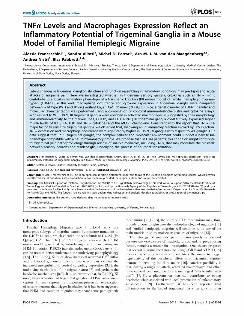
Fig. 1 A shows the typical subdivision of trigeminal ganglion
neurons (immunostained for b-tubulin III) in three clusters that
correspond to the V1, V2 and V3 regions [30]. Inspection (at
higher magnification) of the three subdivisions (as exemplified in
Fig. 1 B) indicated sparse occurrence of Iba1-positive cells that
were more abundant in R192Q KI sections throughout (Fig. 1 C).
In peripheral tissues, Iba1-positive cells are usually identified as
macrophages [31,32]. In line with the notion that CaV2.1 (P/Q-
type) calcium channels are exclusively expressed by neurons [12],
we confirmed that no CaV2.1 mRNA signal was detected in
cultured peritoneal macrophages (n = 5 mice). In trigeminal
ganglia, Iba1-immunoreactive cells were distinct from satellite
glial cells, as demonstrated by the lack of co-localization of Iba1
signal with the glutamine synthetase signal (GS; Fig. 1 D), a
canonical satellite cell marker [33]. Across a broad ganglion area,
Iba1-positive cells were not only more frequent in KI ganglia
(Fig. 1 E), but they were also preferentially localized in the
proximity of neuronal cell bodies rather than fibers (Fig. 1 F).
Iba1 expression is typically upregulated in activated macro-
phages/microglia that exhibit distinct morphology with ameboid
shape and short processes resulting in a larger cell volume
[34,35,36]. 3D reconstruction of confocal images of WT and
R192Q KI trigeminal ganglion Iba1-positive cells showed that KI
macrophages displayed increased cell volume (with amoeboid
morphology and shorter processes) compared with WT cells (Fig. 2
A, B). Interestingly, larger macrophages were detected near KI
ganglion neurons (Fig. 2 C).
These data suggest that in R192Q KI ganglia Iba1 macro-
phages showed an activation state with their preferential location
close to neuronal somata.
Biomarker characteristics of Iba1 cells in WT or R192Q KItrigeminal ganglia
Fig. 3 A shows examples of confocal microscopy images from
WT or R192Q KI ganglion sections that were co-immunostained
with antibodies against Iba1 and CD11b, an adhesion molecule
marker for active macrophages and microglia [37,38,39].
Expression of CD11b was quantified in Iba1-positive cells only.
In WT ganglia, 20% of Iba1-positive cells (without topographical
difference within the three areas) expressed CD11b (Fig. 3 B). On
the other hand, in KI tissue, the percentage of Iba1 and CD11b
co-expressing cells was significantly higher (up to 45% of Iba1-
positive cells; Fig. 3 B).
Iba1-negative satellite cells (see Fig. 1 D) were also stained with
the CD11b antibody in R192Q KI sections (Fig. 3 A, right panel).
Nevertheless, in KI ganglia, co-localization of CD11b and Iba1 in
macrophages was clearly confirmed in fiber regions devoid of
satellite cells (Figure S1).
Similar experiments were performed to evaluate the presence of
the macrophage antigen ED1 (CD68), a glycoprotein highly
expressed by monocytes and tissue macrophages and associated
with larger phagocytic ability [40] (Fig. 3 C, D). While, in WT
tissue, ED1/Iba1 double-positive cells were fewer than 15%, this
value was significantly higher in R192Q KI ganglia, in particular
in the V3 region (Fig. 3 D). ED1 was also detected in KI satellite
glial cells (Fig. 3 C, right).
Expression of the F4/80 antigen is restricted to most resident
mature macrophages and quiescent microglia [41,42,43], and has
recently been linked to the induction of immunological tolerance
[44]. In WT and R192Q KI ganglia, the F4/80 signal was rarely
observed in Iba1-positive cells and not significantly different
between WT and R192Q KI ganglia (Fig. 3 E, F).
These data indicate different subpopulations of Iba1-positive
cells of R192Q KI ganglia.
Cytokine expression in trigeminal ganglia from WT andR192Q KI mice
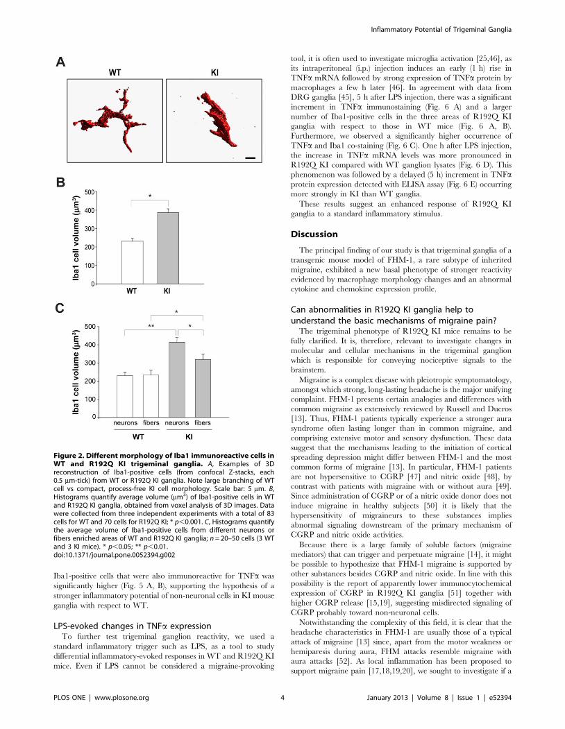
A neuroinflammatory role of higher cytokine and chemokine
expression is thought to be associated with chronic pain models
[29]. We measured the protein and RNA cytokines content of
whole WT and R192Q KI ganglion extracts (Fig. 4 A, B). ELISA
experiments demonstrated no significant difference in protein
levels of IL1b, IL6 and TNFa between WT or R192Q KI lysates.
The anti-inflammatory cytokine IL10, however, was significantly
lower in R192Q KI than WT ganglia (Fig. 4 A). Although these
observations, at the protein level, do not suggest an on-going
inflammatory state in R192Q KI ganglia, it is interesting that
cytokines mRNA data were, however, significantly higher in
R192Q KI than WT samples for all four genes (Fig. 4B).
High levels of monocyte chemoattractant protein-1 (MCP-1;
also known as chemokine receptor type 2, CCR2) are associated
with meningeal neuroinflammation in migraine [45]. As shown in
Fig. 4 C, elevated mRNA levels of this marker were found in KI
rather than WT ganglia; furthermore, stronger expression of the
MCP-1 protein by KI lysates was observed (Fig. 4 D), consistent
with the more numerous DAPI-positive elements (4330660 for KI
vs 3740670 for WT; p,0.001; n = 4), and suggestive of possible
cell recruitment to trigeminal ganglia of R192Q KI mice.
Since TNFa powerfully sensitizes trigeminal ganglia [27,28], we
studied the potential difference in TNFa expression at the single
cell level between WT and R192Q KI ganglia. Fig. 5 A
exemplifies confocal microscopy images from WT or R192Q KI
ganglia co-stained with antibodies against Iba1 and TNFa in
WT and KI ganglia. In R192Q KI ganglia, the number of
Inflammatory Potential of Trigeminal Ganglia
PLOS ONE | www.plosone.org 2 January 2013 | Volume 8 | Issue 1 | e52394
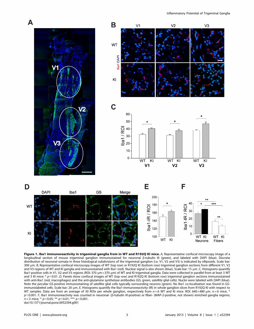
Figure 1. Iba1 immunoreactivity in trigeminal ganglia from in WT and R192Q KI mice. A, Representative confocal microscopy image of alongitudinal section of mouse trigeminal ganglion immunostained for neuronal b-tubulin III (green), and labeled with DAPI (blue). Discretedistribution of neuronal somata in three histological subdivisions of the trigeminal ganglion (i.e. V1, V2 and V3) is indicated by ellipsoids. Scale bar:300 mm. B, Representative confocal microscopy images of WT (top row) or R192Q KI (bottom row) trigeminal ganglion sections from different V1, V2and V3 regions of WT and KI ganglia and immunostained with Iba1 (red). Nuclear signal is also shown (blue). Scale bar: 15 mm. C, Histograms quantifyIba1-positive cells in V1, V2 and V3 regions (ROI: 370 mm6370 mm) of WT and KI trigeminal ganglia. Data were collected in parallel from at least 3 WTand 3 KI mice; * p,0.01. D, Panels show confocal images of WT (top row) and R192Q KI (bottom row) trigeminal ganglion sections immunostainedwith anti-Iba1 (red, macrophages) and the anti-glutamine synthetase antibodies (GS, green, satellite glial cells). Nuclei were labeled with DAPI (blue).Note the peculiar GS-positive immunostaining of satellite glial cells typically surrounding neurons (green). No Iba1 co-localisation was found in GS-immunolabeled cells. Scale bar: 20 mm. E, Histograms quantify the Iba1-immunoreactivity (IR) in whole ganglion slices from R192Q KI with respect toWT samples. Data are from an average of 30 ROIs per whole ganglion, respectively from n = 6 WT and KI mice. ROI: 6406480 mm. n = 6 mice, *p,0.001. F, Iba1 immunoreactivity was counted in neuronal- (b-tubulin III-positive) or fiber- (MAP-2-positive, not shown) enriched ganglia regions;n = 3 mice; * p,0.05; ** p,0.01; *** p,0.001.doi:10.1371/journal.pone.0052394.g001
Inflammatory Potential of Trigeminal Ganglia
PLOS ONE | www.plosone.org 3 January 2013 | Volume 8 | Issue 1 | e52394
Iba1-positive cells that were also immunoreactive for TNFa was
significantly higher (Fig. 5 A, B), supporting the hypothesis of a
stronger inflammatory potential of non-neuronal cells in KI mouse
ganglia with respect to WT.
LPS-evoked changes in TNFa expressionTo further test trigeminal ganglion reactivity, we used a
standard inflammatory trigger such as LPS, as a tool to study
differential inflammatory-evoked responses in WT and R192Q KI
mice. Even if LPS cannot be considered a migraine-provoking
tool, it is often used to investigate microglia activation [25,46], as
its intraperitoneal (i.p.) injection induces an early (1 h) rise in
TNFa mRNA followed by strong expression of TNFa protein by
macrophages a few h later [46]. In agreement with data from
DRG ganglia [45], 5 h after LPS injection, there was a significant
increment in TNFa immunostaining (Fig. 6 A) and a larger
number of Iba1-positive cells in the three areas of R192Q KI
ganglia with respect to those in WT mice (Fig. 6 A, B).
Furthermore, we observed a significantly higher occurrence of
TNFa and Iba1 co-staining (Fig. 6 C). One h after LPS injection,
the increase in TNFa mRNA levels was more pronounced in
R192Q KI compared with WT ganglion lysates (Fig. 6 D). This
phenomenon was followed by a delayed (5 h) increment in TNFaprotein expression detected with ELISA assay (Fig. 6 E) occurring
more strongly in KI than WT ganglia.
These results suggest an enhanced response of R192Q KI
ganglia to a standard inflammatory stimulus.
Discussion
The principal finding of our study is that trigeminal ganglia of a
transgenic mouse model of FHM-1, a rare subtype of inherited
migraine, exhibited a new basal phenotype of stronger reactivity
evidenced by macrophage morphology changes and an abnormal
cytokine and chemokine expression profile.
Can abnormalities in R192Q KI ganglia help tounderstand the basic mechanisms of migraine pain?
The trigeminal phenotype of R192Q KI mice remains to be
fully clarified. It is, therefore, relevant to investigate changes in
molecular and cellular mechanisms in the trigeminal ganglion
which is responsible for conveying nociceptive signals to the
brainstem.
Migraine is a complex disease with pleiotropic symptomatology,
amongst which strong, long-lasting headache is the major unifying
complaint. FHM-1 presents certain analogies and differences with
common migraine as extensively reviewed by Russell and Ducros
[13]. Thus, FHM-1 patients typically experience a stronger aura
syndrome often lasting longer than in common migraine, and
comprising extensive motor and sensory dysfunction. These data
suggest that the mechanisms leading to the initiation of cortical
spreading depression might differ between FHM-1 and the most
common forms of migraine [13]. In particular, FHM-1 patients
are not hypersensitive to CGRP [47] and nitric oxide [48], by
contrast with patients with migraine with or without aura [49].
Since administration of CGRP or of a nitric oxide donor does not
induce migraine in healthy subjects [50] it is likely that the
hypersensitivity of migraineurs to these substances implies
abnormal signaling downstream of the primary mechanism of
CGRP and nitric oxide activities.
Because there is a large family of soluble factors (migraine
mediators) that can trigger and perpetuate migraine [14], it might
be possible to hypothesize that FHM-1 migraine is supported by
other substances besides CGRP and nitric oxide. In line with this
possibility is the report of apparently lower immunocytochemical
expression of CGRP in R192Q KI ganglia [51] together with
higher CGRP release [15,19], suggesting misdirected signaling of
CGRP probably toward non-neuronal cells.
Notwithstanding the complexity of this field, it is clear that the
headache characteristics in FHM-1 are usually those of a typical
attack of migraine [13] since, apart from the motor weakness or
hemiparesis during aura, FHM attacks resemble migraine with
aura attacks [52]. As local inflammation has been proposed to
support migraine pain [17,18,19,20], we sought to investigate if a
Figure 2. Different morphology of Iba1 immunoreactive cells inWT and R192Q KI trigeminal ganglia. A, Examples of 3Dreconstruction of Iba1-positive cells (from confocal Z-stacks, each0.5 mm-tick) from WT or R192Q KI ganglia. Note large branching of WTcell vs compact, process-free KI cell morphology. Scale bar: 5 mm. B,Histograms quantify average volume (mm3) of Iba1-positive cells in WTand R192Q KI ganglia, obtained from voxel analysis of 3D images. Datawere collected from three independent experiments with a total of 83cells for WT and 70 cells for R192Q KI; * p,0.001. C, Histograms quantifythe average volume of Iba1-positive cells from different neurons orfibers enriched areas of WT and R192Q KI ganglia; n = 20–50 cells (3 WTand 3 KI mice). * p,0.05; ** p,0.01.doi:10.1371/journal.pone.0052394.g002
Inflammatory Potential of Trigeminal Ganglia
PLOS ONE | www.plosone.org 4 January 2013 | Volume 8 | Issue 1 | e52394

special phenotype of trigeminal ganglia (responsible for conveying
nociceptive signals to the brainstem) consistent with propensity to
neuroinflammation might be observed.
Experimental neuroinflammation within trigeminalganglia
The molecular and cellular phenotype characteristics of R192Q
KI mice could perhaps reside in a different basal condition with
distinct contributions by various cells to the process of neuronal
sensitization. Studies of meningeal tissue from migraine animal
models and migraineurs show molecular and histological changes
typical of neuroinflammation [8,53,54,55]. In particular, experi-
mental studies of rat dura mater have indicated that a strong
chemical trigger like, for example glyceryl trinitrate or LPS, can
induce expression of several cytokines such as IL1b, IL6 and the
nitric oxide synthetic enzyme iNOS [56]. Release of soluble factors
including inflammatory cytokines is proposed to activate small
afferent fibers to produce headache and to further stimulate
release of neuropeptides from neuronal afferents [57]. It is,
however, unclear whether trigeminal ganglia per se are also
affected by these neuroinflammatory processes, and how much
their phenotype may be generalized to other migraine types. This
possibility is not unlikely because antigen-presenting immune cells
have been characterized in human trigeminal ganglia [58] and
certain chronic pain models are associated with neuroinflamma-
tory changes in dorsal root ganglia [59]. Our data support that, at
the level of trigeminal ganglia, a FHM-1 mouse model with a
R192Q missense mutation in the a1 subunit of voltage-gated
Figure 3. Characterization of Iba1-positive cells in trigeminal ganglia from WT and R192Q KI mice. A, C, E, Representative confocalimages of WT or R192Q KI trigeminal ganglion sections (from V3 region) immunostained for Iba1 (red) and CD11b (A), ED1 (C), or F4/80 (E) in green.Nuclei were labeled with DAPI (blue). Scale bar: 40 mm. Insets represent larger magnification of immunoreactive Iba1 cells. Scale bar: 7 mm. B, D, F,Histograms quantify the percentage of occurrence of Iba1 signal with CD11b (B), ED1 (D) or F4/80 (F) in different WT and KI ganglion regions (V1, V2or V3). Expression of different markers was quantified in Iba1 positive cells only. n = 3 WT and 3 KI mice; * p,0.05; ** p,0.01. Data are expressed asmean 6 S.D.doi:10.1371/journal.pone.0052394.g003
Inflammatory Potential of Trigeminal Ganglia
PLOS ONE | www.plosone.org 5 January 2013 | Volume 8 | Issue 1 | e52394
CaV2.1 calcium channels elicits cellular and molecular changes
consistent with this hypothesis. Of course, since these channels are
typical of neurons [12], and macrophages did not express CaV2.1
channels, any observed molecular and/or cellular alteration within
ganglion cell populations implies cross talk between neurons and
non-neuronal cells.
Macrophages in trigeminal ganglia of R192Q KI miceshow activation state
Macrophages play an important role in tissue pathophysiological
responses like chronic pain [29]. In trigeminal ganglia, Iba1-positive
macrophages were a small cell population that, nonetheless, in
R192Q KI ganglia, showed significant signs of activation (ameboid
shape due to their larger volume [60,61]) that resembled the one
observed in sensory ganglia of injured rodents [62]. Future studies
will be necessary to find out if macrophage changes can also be
observed in dorsal root ganglia of R192Q KI mice.
Our study found no difference in macrophage intra-ganglion
sub-distribution in relation to the three main branches of the
trigeminal nerve. Consistent with these findings, it has been
reported that, despite the few meningeal afferents to trigeminal
ganglia observed with retrograde labeling [15], a chemical
stimulation related to a specific topographic neuronal area causes
alteration also in the other areas of the trigeminal ganglion [30].
Furthermore, functional studies have indicated widespread
involvement of different ganglion regions, since CaV2.1 expression
is not restricted to the fine meningeal afferents of trigeminal
ganglia only [15]. The relevance of our finding is not contradicting
the notion that headache is the principal trigeminal pain in
migraine. Nonetheless, an extensive clinical study carried out on
1,413 patients has indicated that, during an attack of migraine, the
majority of them suffer from allodynia affecting the entire
trigeminal territory [63], a finding confirmed to be independent
from the presence of aura [64].
To characterise Iba1-positive macrophages in R192Q KI
trigeminal ganglia, we found that a significant fraction of them
co-expressed CD11b and ED1, but not F4/80. Interestingly,
satellite cells surrounding neuronal somata of R192Q KI ganglia
also showed large expression of CD11b and ED1 markers. Satellite
glial cells in sensory ganglia tightly envelop the neuronal cell body
to form discrete anatomical units [33], and may express an
immune-related function within human sensory ganglia [58]. We
propose that, in R192Q KI mice, active Iba1-positive cells that are
located close to the neuronal units in the ganglia, with likely
Figure 4. Expression of inflammatory mediators in WT and R192Q KI trigeminal ganglia under basal conditions. A, Histogramsquantify IL1b, IL6, IL10 and TNFa cytokine protein levels from WT or R192Q KI ganglia; n = 4 WT and 4 KI mice; data were normalized on total proteincontent, and represented as fraction of WT. * p,0.001. B, Real-time RT-PCR experiments quantify IL1b, IL6, IL10 and TNFa mRNA levels in WT andR192Q KI trigeminal ganglia. PCR data were normalized with respect to corresponding GAPDH and b-Tubulin housekeeping gene expression andexpressed as fraction of WT; n = 4 WT and 4 KI mice; * p,0.05. C, Real-time RT-PCR experiments quantify MCP-1 mRNA levels in WT and R192Q KItrigeminal ganglia (expressed as in B); n = 4 WT and 4 KI mice; * p,0.05. D, Representative western blot experiment of WT or R192Q KI trigeminalganglia extracts immuno-probed with anti-MCP-1 antibodies. Actin levels were used as loading control. Histograms quantify the differences. n = 3; *p,0.05.doi:10.1371/journal.pone.0052394.g004
Inflammatory Potential of Trigeminal Ganglia
PLOS ONE | www.plosone.org 6 January 2013 | Volume 8 | Issue 1 | e52394

release of soluble factors, could mediate de novo expression of
CD11b and ED1 in satellite glial cells, even if their functional role
remains to be investigated.
Cytokine profile of R192Q KI gangliaIn neuroinflammatory processes, immune cell activity is
typically associated with higher expression of chemokines, like
MCP-1 [65,66], and cytokines that are believed to be key
contributors to chronic pain [22,67]. In R192Q KI ganglia, new
antigen expression and macrophage activation were associated to
larger cytokine mRNA levels, without significant changes in
corresponding proteins. Still, the anti-inflammatory cytokine IL10
was lower in KI ganglia, a result which would be in line with their
propensity to a neuroinflammatory reaction [68]. An alternative
possibility to be explored is that the inflammatory nociceptive
activation in the ganglia could arise from hyperactivity of the
trigeminal vascular system leading to increased cytokine expres-
sion at the meninges.
During inflammatory conditions, activated macrophages release
TNFa that modulates immune responses and stimulates crosstalk
between neurons and glia to facilitate pain [22,27,68]. Studies of
rat dorsal root ganglia have demonstrated that TNFa is primarily
synthesized by ED1-expressing macrophages [46]. It is known that
pro-inflammatory cytokines are rapidly released to stimulate the
arrival and activation of immune cells generating an inflammatory
response [69]. Our study indicates that R192Q KI trigeminal
ganglia had higher mRNA and protein levels of the macrophage-
related chemokine MCP-1/CCL2 involved in macrophage
recruitment/mobility and activation [70,71] and also found raised
in the plasma of migraine patients [72,73].
Electrophysiological studies will be necessary to investigate how
inflammatory mediators might change the operation of ligand-
and voltage-activated channels of trigeminal sensory neurons, and,
consequently, shape the firing properties of these cells. For
example, after injury, TNFa enhances the sodium current of rat
DRG neurons, lowers the spike threshold and promotes high
frequency firing [74]. Future investigations will need to analyze if
this cytokine may differentially affect spike firing by WT and
R192Q KI trigeminal neurons.
Figure 5. TNFa expression in WT and KI ganglia. A, Representative confocal microscopy images of WT (top row) or R192Q KI (bottom row)trigeminal ganglion sections immunostained for Iba1 (red) or TNFa(green) in basal condition. Pseudocolor images showing areas of high (yellow) andlow (blue) Iba1-TNFa expressing cell co-localization. Color scale was also included. Note TNFa immunostaining detected as spots alongperimembrane regions. The larger magnification insets show immunostaining of Iba1-TNFa signal (yellow) in KI rather than WT. Scale bar: 30 mm, forlarge images; Scale bar: 10 mm for larger magnification insets. B, Histograms quantify the percentage of TNFa immunoreactivity over the total of Iba1expressing cells in different V1, V2 or V3 trigeminal regions (ROI: 3706370 mm). n = 4 WT and 4 R192Q KI mice; * p,0.05. Data are expressed as mean6 S.D.doi:10.1371/journal.pone.0052394.g005
Inflammatory Potential of Trigeminal Ganglia
PLOS ONE | www.plosone.org 7 January 2013 | Volume 8 | Issue 1 | e52394

Inflammatory stimulus strongly enhanced TNFaproduction
LPS per se is not considered to be an agent capable of inducing
an acute attack of migraine. We employed LPS to explore whether
the basal inflammatory profile of KI ganglia could be expressed
(by LPS administration) into a biochemical substrate compatible
with a strong inflammatory reaction and production of soluble
factors (e.g. TNFa) potentially promoting the release of migraine
mediators. Thus, using this approach, the basal pro-inflammatory
potential of R192Q KI ganglia was readily converted into
enhanced inflammatory reactivity together with elevated TNFaexpression.
The origin of the neuroinflammatory reaction strongly detected
in R192Q KI ganglia remains uncertain as it could be initiated in
the ganglion itself perhaps because of the altered milieu caused by
low-threshold spreading depression occurring in these mice [2,3].
The innate immune system is increasingly viewed to play an
important role in mediating chronic pain [75], especially through
the Toll-like receptor 4 (TLR4) that was found also involved in
several sterile inflammation processes and activated by endoge-
nous ligands [76]. TLR4 is the primary target of bacterial LPS
[77,78] and induces the expression of proinflammatory cytokines
and chemokines [79].
In addition, an inflammatory nociceptive activation developing
at the meninges might have driven the increase in cytokine
Figure 6. LPS evoked acute TNFa expression in R192Q KI ganglia. A, Representative confocal microscopy images of WT (top row) or KI(bottom row) trigeminal ganglion sections immunostained for Iba1 (red) or TNFa(green) after saline (left) or LPS injection (i.p., 5 h; right). LPS evokesTNFa expression in WT and KI after injection. Scale bar: 20 mm. B, C, Histograms quantify the occurrence of Iba1 signal (B) and Iba1-TNFa co-localisation (C) in different V1, V2 or V3 trigeminal ganglion regions from WT or KI mice, after saline or LPS injection (i.p., 5 h). ROI: 3706370 mm. n = 3WT and 3 R192Q KI mice; * p,0.05; ** p,0.01; *** p,0.001. D, Histograms quantify changes in TNFa mRNA fold increase in WT or R192Q KI gangliafollowing LPS-injection (i.p., 1 h). Data are expressed as fold increase with respect to saline-injected mice samples; n = 3 WT and 3 KI mice; * p,0.05. E,TNFa protein levels in whole ganglia (pg cytokine/mg protein content) from LPS-injected (i.p. 5 h) WT or R192Q KI mice, expressed as fold increasewith respect to saline-injected mice. n = 3 WT and 3 KI mice; * p,0.05.doi:10.1371/journal.pone.0052394.g006
Inflammatory Potential of Trigeminal Ganglia
PLOS ONE | www.plosone.org 8 January 2013 | Volume 8 | Issue 1 | e52394

expression in the ganglion. Since the basal inflammatory reactivity
of R192Q KI trigeminal ganglia has also been observed in
primary culture [80], this finding is compatible with the view that
the KI ganglion had a constitutive neuroinflammatory profile.
LPS activates monocytes and macrophages to produce proin-
flammatory cytokines such as TNFa [81]. Inflammatory cytokines
such as TNFa contribute to peripheral sensitization of nociceptor
neurons [82], and in particular, in migraine patients, raised
concentrations of TNFa have been reported in the jugular blood
of patients 2 h after the onset of an attack [57]. We found that, at
the ganglion level, a strong rise in TNFa production by R192Q KI
ganglia after LPS together with substantial increment in the
number of Iba1-positive cells co-expressing TNFa. These obser-
vations are consistent with hyper-reactivity of R192Q KI ganglia
to inflammatory stimulation to which various non-neuronal cell
types could contribute with their own synthesis and release of
TNFa. In fact, studies of mRNA and Western blotting could not
identify the source of cytokines: nonetheless, single cell immuno-
histochemistry indicated that TNFa was abundantly expressed by
macrophages.
Conclusions
Future experiments will be necessary to identify the molecular
mechanisms linking the gain-of-function of mutated CaV2.1
channels in R192Q sensory neurons to the inflammatory profile
of the ganglion tissue, and how raised TNFa production and
release are converted into over-activity of nociceptors of trigeminal
sensory neurons as experimentally observed in the R192Q KI
mouse [10]. It is, however, feasible to hypothesize that TNFa is a
significant player in triggering trigeminal pain as it can release a
host of algogenic substances like BDNF [26] and CGRP [28]. In
the FHM-1 mouse model, the genetic mutation not only confers a
sensitized pain receptor phenotype to a subclass of sensory neurons
[10,15], but it can also modify the trigeminal ganglion microen-
vironment that may predispose to chronic pathological conditions
specific for a subset of migraine patients, as reported in the current
study. Previous investigations have demonstrated that R192Q KI
mice are very susceptible to strong cortical spreading depression,
believed to be the equivalent of human aura [2,3,7]. An interesting
possibility is that former bouts of cortical spreading depression
with associated release of soluble factors into the extracellular
space might have primed macrophages to synthesize and liberate
proinflammatory mediators. Whether this process actually occurs
in vivo during an aura remains a matter of conjecture.
Nonetheless, our data add the role of inflammatory cells to the
growing body of evidence showing how macrophages/microglia
(via purinergic receptors) play a key role in pain signaling through
neuron/glia interaction [83].
Methods
Animal proceduresCaV2.1 R192Q KI and WT littermates (P30) were used.
Animals were maintained in accordance with the guidelines of the
Italian Animal Welfare Act and their use has been approved by
the Local Ethical Committee. Our experimental protocols, have
been approved by SISSA ethics committee board and by National
Ministry of Health (reference # 13184), as they are in accordance
with the European Union guidelines. Genotyping was performed
by PCR as previously reported [3]. To evoke acute inflammation,
WT or R192Q KI mice (P30) were injected intraperitoneally (i.p.)
with a single dose of saline (sham) or LPS (5 mg/kg, from E. coli
0111:B4; Sigma, Milan, Italy) 1 or 5 h prior to sacrificing the
animals [46]. Ganglion tissue samples were collected and
processed in parallel for WT and R192Q KI mice.
ImmunohistochemistryFor immunohystochemistry, WT or R192Q KI mice were
deeply anesthetized with i.p. urethane (0.3 ml of 1 g/ml; Sigma)
and perfused transcardially with PBS followed by 4% parafor-
maldehyde. Trigeminal ganglia were removed, postfixed for 1 h at
room temperature and cryoprotected overnight in 30% sucrose at
4uC. Each immunohystochemistry experiment was performed on
an average of 5 cryostat-cut serial longitudinal slices (14 mm-thick)
sampled every ,70 mm, and thus covering the entire ganglion.
Samples were incubated in a blocking solution containing 5%
bovine serum albumin, 1% fetal bovine serum and 0.1% Triton
X-100 in phosphate saline buffer for 2 h at room temperature, and
immunostained with primary (for 16 h at 4uC) and secondary
antibodies (2 h at room temperature). The following antibodies
were used: anti-b-Tubulin III (1:1000; Sigma); anti-Iba1 (1:300;
Wako, Richmond, VA, USA; [31,32]); anti-TNFa antibody
(1:100, eBioscience, [46]); anti-biotin-F4/80 (1:50; eBioscience,
S.Diego, CA, USA; specificity of anti-F4/80 immunoreactivity is
shown in Figure S2 [41,42,84]), anti-CD11b (1:50; eBioscience;
[85]); anti-ED-1 (1:50; AbD Serotec, Oxford, UK; [86]); anti-
glutamine synthetase (1:150; Millipore, Milan, Italy; [33]),
AlexaFluor488- or 594-conjugated antibodies (1:500; Invitrogen,
Milan, Italy). For F4/80 immunoreactivity, streptavidin-Alexa-
Fluor 647 antibodies (1:100, Invitrogen) were used. Nuclei were
counterstained with DAPI (Sigma). Specificity of anti-Iba1
antibody was validated with Western immunoblotting (Figure
S2) of extracts from WT and R192Q KI trigeminal lysates and
purified macrophage population extracted from mouse peritone-
um used as positive control [87]. Images from whole ganglion
sections were visualized with Leica confocal microscope (Leica
TCS SP2, Wetzlar, Germany) or a Zeiss Axioskop fluorescence
microscope (Zurich, Switzerland). Cellular imaging and analysis of
3D reconstruction (Z-stack; 0.5 mm steps) of high magnification
confocal images (Leica, Wetzlar, Germany) and co-expression
analysis of multiple antigens were obtained with Volocity 5.5
software (Perkin Elmer, Waltham, MA, USA) and ImageJ Voxel
counter (voxel, in mm3). Cell counting was carried out with
MetaMorph software (Molecular Devices, Downingtown, PA,
USA). The average total number of ganglion cells stained with
DAPI was larger in R192Q KI than WT ganglia (1300620 vs
1120620, n = 4 mice). In view of the cell heterogeneity, in each
experiment we compared equivalent regions of interest (ROIs)
from WT and R192Q KI samples, processed and examined in
parallel. Full details of ROIs are indicated in each Figure legend.
Morphological and histological subdivision of neuronal-enriched
V1, V2 and V3 areas of trigeminal ganglia is consistent with
imaging experiments following retrograde labeling of trigeminal
neurons [30,37].
Western blotFor western blotting, cells were lysed as previously detailed [10]
in ODG buffer (2% n-octyl-beta-D-glucopyranoside, contaning
1% Nonidet P-40, 10 mM Tris pH 7.5, 150 mM NaCl plus
protease inhibitors mixture; Complete, Roche Applied Science)
and immunoblotted with rabbit anti-Iba1 antibodies (1:1000,
Wako), anti-F4-80 (1:1000, eBioscience), anti-MCP-1 antibodies
(1:1000; Santa Cruz, CA, USA) or anti-actin antibodies (1:3000,
Sigma). Signals were detected with the enhanced chemilumines-
cence light system ECL (Amersham Biosciences, Piscataway, NJ,
USA) and recorded by the digital imaging system Alliance 4.7
Inflammatory Potential of Trigeminal Ganglia
PLOS ONE | www.plosone.org 9 January 2013 | Volume 8 | Issue 1 | e52394
(UVITEC, Cambridge, UK). Quantification of the optical density
of the bands was performed with ImageJ software plug-in.
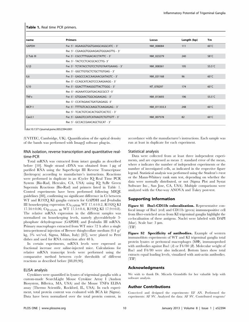
RNA isolation, reverse transcription and quantitative real-time-PCR
Total mRNA was extracted from intact ganglia as described
before [10]. Single strand cDNA was obtained from 1 mg of
purified RNA using the SuperScript III Reverse Transcriptase
(Invitrogen) according to manufacture’s instructions. Reactions
were performed in duplicate in an iCycler IQ Real Time PCR
System (Bio-Rad, Hercules, CA, USA) using IQ SyBr Green
Supermix Reactions (Bio-Rad) and primers listed in Table 1.
Control experiments have been performed following MIQE
guidelines [88], confirming no significant difference in Ct between
WT and R192Q KI ganglia extracts for GAPDH and b-tubulin
III housekeeping expression (CtGAPDH WT 17.460.2, R192Q KI
17.3460.06; Ctb-tubulin III WT 17.560.4, R192Q KI 17.960.8).
The relative mRNA expression in the different samples was
normalized on housekeeping levels, namely glyceraldehyde 3-
phosphate dehydrogenase (GAPDH) and b-tubulin III mRNA.
Primary macrophages extracted from WT mice 72 h after a single
intra-peritoneal injection of Brewer thioglycollate medium (0.4 g/
kg, 3% wt/vol, Sigma, Milan, Italy) [87], were plated to Petri
dishes and used for RNA extraction after 48 h.
In certain experiments, mRNA levels were expressed as
fractional increase over saline-injected mice. Calculations for
relative mRNA transcript levels were performed using the
comparative method between cycle thresholds of different
reactions as described before [88,89,90].
ELISA analysisCytokines were quantified in lysates of trigeminal ganglia with a
custom-made SearchLight Mouse Cytokine Array I (Aushon
Biosystem, Billerica, MA, USA) and the Mouse TNFa ELISA
assay (Thermo Scientific, Rockford, IL, USA). In each experi-
ment, total protein content was evaluated with BCA kit (Sigma).
Data have been normalized over the total protein content, in
accordance with the manufacturer’s instructions. Each sample was
run at least in duplicate for each experiment.
Statistical analysisData were collected from at least three independent experi-
ments, and are expressed as mean 6 standard error of the mean,
where n indicates the number of independent experiments or the
number of investigated cells, as indicated in the respective figure
legend. Statistical analysis was performed using the Student’s t-test
or the Mann-Whitney rank sum test, depending on whether the
data were normally distributed, or not (Sigma Plot and Systat
Software Inc., San Jose, CA, USA). Multiple comparisons were
analyzed with the One-way ANOVA and Tukey post-test.
Supporting Information
Figure S1 Iba1-CD11b colocalisation. Representative con-
focal image of Iba1 (red) and CD11b (green) immunopositive cell
from fiber-enriched areas from KI trigeminal ganglia highlight the
co-localisation of these antigens. Nuclei were labeled with DAPI
(blue). Scale bar: 3 mm.
(TIF)
Figure S2 Specificity of antibodies. Example of western
immunoblots experiments of WT and KI trigeminal ganglia total
protein lysates or peritoneal macrophages (MW), immunoprobed
with antibodies against Iba1 (A) or F4/80 (B). Molecular weight of
Iba1 and F4/80 were also indicated. Bottom lanes show total
extracts equal loading levels, visualized with anti-actin antibodies.
(TIF)
Acknowledgments
We wish to thank Dr. Micaela Grandolfo for her valuable help with
software analysis.
Author Contributions
Conceived and designed the experiments: EF AN. Performed the
experiments: AF SV. Analyzed the data: AF SV. Contributed reagents/
Table 1. Real time PCR primers.
name Primers Locus Length (bp) Tm
GAPDH Fw: 59- AGAAGGTGGTGAAGCAGGCATC - 39 NM_008084 111 60uC
Rw: 59- CGAAGGTGGAAGAGTGGGAGTTG - 39
b Tub III Fw: 59- CGCCTTTGGACACCTATTC - 39 NM_023279 240 58uC
Rw: 59- TACTCCTCACGCACCTTG- 39
IL1b Fw: 59- TCTATACCTGTCCTGTGTAATGAAAG - 39 NM_008361 195 55.5uC
Rw: 59- GGCTTGTGCTCTGCTTGTGAG - 39
IL6 Fw: 59- GAGCCCACCAAGAACGATAGTC - 39 NM_031168 96 60uC
Rw: 59- CCAGCATCAGTCCCAAGAAGG - 39
IL10 Fw: 59- GGACTTTAAGGGTTACTTGGG - 39 NT_078297 174 60uC
Rw: 59- AGAAATCGATGACAGCGCCT -39
TNFa Fw: 59- GTGGAACTGGCAGAAGAG - 39 NM_013693 196 55.5uC
Rw: 59- CCATAGAACTGATGAGAGG -39
MCP-1 Fw: 59- TTTTGTCACCAAGCTCAAGAGAG - 39 NM_011333.3 248 60uC
Rw: 59- TCACTGTCACACTGGTCACTCC - 39
Cav2.1 Fw: 59- GAAGTCCATCATAAGTCTGTTGTT - 39 NM_007578 82 60uC
Rw: 59- GCCACCGAACAGCTGCAT - 39
doi:10.1371/journal.pone.0052394.t001
Inflammatory Potential of Trigeminal Ganglia
PLOS ONE | www.plosone.org 10 January 2013 | Volume 8 | Issue 1 | e52394

materials/analysis tools: MDF AMJMVDM AN EF. Wrote the paper:
AMJMVDM EF AN AF SV.
References
1. Ophoff RA, Terwindt GM, Vergouwe MN, van Eijk R, Oefner PJ, et al. (1996)
Familial Hemiplegic Migraine and Episodic Ataxia Type-2 Are Caused by
Mutations in the Ca2+ Channel Gene CACNL1A4. Cell 87:543–552.
2. van den Maagdenberg AMJM, Haan J, Terwindt GM, Ferrari MD (2007)
Migraine: gene mutations and functional consequences. Curr Opin Neurol
20:299–305.
3. van den Maagdenberg AM, Pietrobon D, Pizzorusso T, Kaja S, Broos LA., et al.
(2004) A Cacna1a Knockin Migraine Mouse Model with Increased Suscepti-
bility to Cortical Spreading Depression. Neuron 41:701–710.
4. Tfelt-Hansen PC, Koehler PJ (2011) One hundred years of migraine research:
major clinical and scientific observations from 1910 to 2010. Headache 51, 5:
752–778.
5. Jurkat-Rott K, Lerche H, Weber Y, Lehmann-Horn F (2010) Hereditary
channelopathies in neurology. Adv Exp Med Biol 686: 305–334.
6. Tottene A, Conti R, Fabbro A, Vecchia D, Shapovalova M, et al. (2009)
Enhanced excitatory transmission at cortical synapses as the basis for facilitated
spreading depression in Ca(v)2.1 knockin migraine mice. Neuron 12:762–773.
7. Lauritzen M (1994) Pathophysiology of the migraine aura. The spreading
depression theory. Brain 117:199–210.
8. Bolay H, Reuter U, Dunn AK, Huang Z, Boas DA, et al. (2002) Intrinsic brain
activity triggers trigeminal meningeal afferents in a migraine model. Nat Med 8:
136–142.
9. Vecchia D, Pietrobon D (2012) Migraine: a disorder of brain excitatory-
inhibitory balance? Trends Neurosci 35(8):507–520.
10. Nair A, Simonetti M, Birsa N, Ferrari MD, van den Maagdenberg AM, et al.
(2010) Familial hemiplegic migraine CaV2.1 channel mutation R192Q
enhances ATP-gated P2X3 receptor activity of mouse sensory ganglion neurons
mediating trigeminal pain. Mol Pain 6: 48.
11. Pietrobon D (2007) Familial Hemiplegic Migraine. Neurotherap 4: 274–284.
12. Rajakulendran S, Kaski D, Hanna MG (2012) Neuronal P/Q-type calcium
channel dysfunction in inherited disorders of the CNS. Nat Rev Neurol 8: 86–
96.
13. Russell MB, Ducros A (2011) Sporadic and familial hemiplegic migraine:
pathophysiological mechanisms, clinical characteristics, diagnosis, and manage-
ment. Lancet Neurology 10: 457–470.
14. Giniatullin R, Nistri A, Fabbretti E (2008) Molecular mechanisms of
sensitization of pain-transducing P2X3 receptors by the migraine mediators
CGRP and NGF. Mol Neurobiol 37:83–90.
15. Fioretti B, Catacuzzeno L, Sforna L, Gerke-Duncan MB, van den Maagdenberg
AM, et al. (2011) Trigeminal ganglion neuron subtype-specific alterations of
CaV2.1 calcium current and excitability in a Cacna1a mouse model of migraine.
J Physiol 589:5879–5895.
16. Goadsby PJ (2005) Migraine pathophysiology. Headache 45:S14–24.
17. Moskowitz MA (1993) Neurogenic inflammation in the pathophysiology and
treatment of migraine. Neurology (Suppl 3): s16–20.
18. Waeber C, Moskowitz MA (2005) Migraine as an inflammatory disorder.
Neurology (Suppl 2):s9–15.
19. Ceruti S, Villa G, Fumagalli M, Colombo L, Magni G, et al. (2011) Calcitonin
Gene-Related Peptide-Mediated Enhancement of Purinergic Neuron/Glia
Communication by the Algogenic Factor Bradykinin in Mouse Trigeminal
Ganglia from Wild-Type and R192Q Cav2.1 Knock-In Mice: Implications for
Basic Mechanisms of Migraine Pain. J Neurosci 31:3638–3649.
20. Friedman MH (2004) Local inflammation as a mediator of migraine and
tension-type headache. Headache 44(8):767–771.
21. Rowe BH, Colman I, Edmonds ML, Blitz S, Walker A, et al. (2008)
Randomized controlled trial of intravenous dexamethasone to prevent relapse
in acute migraine headache. Headache 48(3):333–340.
22. McMahon SB, Malcangio M (2009) Current challenges in glia-pain biology.
Neuron 64:46–54.
23. Puil E, Gimbarzevsky B, Spigelman I (1988) Primary involvement of K+conductance in membrane resonance of trigeminal root ganglion neurons.
J Neurophysiol 59:77–89.
24. Puil E, Spigelman I (1988) Electrophysiological responses of trigeminal root
ganglion neurons in vitro. Neuroscience 24:635–646.
25. Qin L, Wu X, Block ML, Liu Y, Breese GR, et al. (2007) Systemic LPS causes
chronic neuroinflammation and progressive neurodegeneration. Glia 55: 453–
462.
26. Balkowiec-Iskra E, Vermehren-Schmaedick A, Balkowiec A (2011) Tumor
necrosis factor- a increases brain-derived neurotrophic factor expression in
trigeminal ganglion neurons in an activity-dependent manner. Neuroscience
180:322–333.
27. Zhang X-C, Kainz V, Burstein R, Levy D (2011) Tumor necrosis factor-ainduces sensitization of meningeal nociceptors mediated via local COX and p38
MAP kinase actions. Pain 152:140–149.
28. Bowen EJ, Schmidt TW, Firm CS, Russo A, Durham PL (2006) Tumor necrosis
factor-a stimulation of calcitonin gene-related peptide expression and secretion
from rat trigeminal ganglion neurons. J Neurochem 96:65–77.
29. Milligan ED, Watkins LR (2009) Pathological and protective roles of glia in
chronic pain. Nat Rev Neurosci 10:23–36.
30. Thalakoti S, Patil VV, Damodaram S, Vause CV, Langford LE, et al. (2007)
Neuron–Glia Signaling in Trigeminal Ganglion: Implications for Migraine
Pathology. Headache 47:1008–1025.
31. Sasaki Y, Ohsawa K, Kanazawa H, Kohsaka S, Imai Y (2001) Iba1 Is an Actin-
Cross-Linking Protein in Macrophages/Microglia. Biochem Biophys Res
Commun 286:292–297.
32. Ito D, Imai Y, Ohsawa K, Nakajima K, Fukuuchi Y, et al. (1998) Microglia-
specific localisation of a novel calcium binding protein, Iba1. Brain Res Mol
Brain Res 57:1–9.
33. Hanani M (2010) Satellite glial cells in sympathetic and parasympathetic ganglia:
In search of function. Brain Res Revs 64:304–327.
34. Santos AM, Calvente R, Tassi M, Carrasco MC, Martın-Oliva D, et al. (2008)
Embryonic and postnatal development of microglial cells in the mouse retina.
J Comp Neurol 2008; 506(2): 224–239.
35. David S, Kroner A (2011) Repertoire of microglial and macrophage responses
after spinal cord injury. Nat Rev Neurosci 12:388–399.
36. Lynch MA (2009) The Multifaceted Profile of Activated Microglia. Mol
Neurobiol 40:139–156.
37. Glenn JA, Sonceau JB, Wynder HJ, Thomas WE (1993) Histochemical evidence
for microglia-like macrophages in the rat trigeminal ganglion. J Anat 183:475–
481.
38. Ji RC (2012) Macrophages are important mediators of either tumor- or
inflammation-induced lymphangiogenesis. Cell Mol Life Sci 69(6):897–914.
39. Aita M, Byers MR, Chavkin C, Xu M (2010) Trigeminal injury causes kappa
opioid-dependent allodynic, glial and immune cell responses in mice. Mol Pain
6:8.
40. Holness C, Simmons D (1993) Molecular cloning of CD68, a human
macrophage marker related to lysosomal glycoproteins. Blood 81:1607–1613.
41. Lin H-H, Faunce DE, Stacey M, Terajewicz A, Nakamura T, et al. (2005) The
macrophage F4/80 receptor is required for the induction of antigen-specific
efferent regulatory T cells in peripheral tolerance. J Exp Med 201:1615–1625.
42. Martinez-Pomares L, Platt N, McKnight AJ, da Silva RP, Gordon S (1996)
Macrophage membrane molecules: markers of tissue differentiation and
heterogeneity. Immunobiology 195:407–416.
43. Carson MJ, Reilly CR, Sutcliffe JG, Lo D (1998) Mature microglia resemble
immature antigen-presenting cells. Glia 22:72–85.
44. van den Berg TK, Kraal G (2005) A function for the macrophage F4/80
molecule in tolerance induction. Trends Immunol 26:506–509.
45. Yang G, Meng Y, Li W, Yong Y, Fan Z, et al. (2011) Neuronal MCP-1 Mediates
Microglia Recruitment and Neurodegeneration Induced by the Mild Impair-
ment of Oxidative Metabolism. Brain Pathol 21:279–297.
46. Li Y, Ji A, Weihe E, Schafer MK-H (2004) Cell-Specific Expression and
Lipopolysaccharide-Induced Regulation of Tumor Necrosis Factor a (TNF a)
and TNF Receptors in Rat Dorsal Root Ganglion. J Neurosci 24:9623–9631.
47. Hansen JM, Thomsen LL, Olesen J, Ashina M (2008a) Calcitonin gene-related
peptide does not cause the familial hemiplegic migraine phenotype. Neurology
71:841–877.
48. Hansen JM, Thomsen LL, Olesen J, Ashina M (2008b) Familial hemiplegic
migraine type 1 shows no hypersensitivity to nitric oxide. Cephalalgia 28:496–
505.
49. Olesen J, Ashina M (2011) Emerging migraine treatments and drug targets.
Trends Pharmacol Sci 32:352–359.
50. Tvedskov JF, Iversen HK, Olesen J, Tfelt-Hansen P (2010) Nitroglycerin
provocation in normal subjects is not a useful human migraine model?
Cephalalgia 30:928–932.
51. Mathew R, Andreou AP, Chami L, Bergerot A, van den Maagdenberg AM, et
al. (2011) Immunohistochemical characterization of calcitonin gene-related
peptide in the trigeminal system of the familial hemiplegic migraine 1 knock-in
mouse. Cephalalgia 31(13):1368–1380.
52. Pietrobon D (2010) Insights into migraine mechanisms and CaV2.1 calcium
channel function from mouse models of familial hemiplegic migraine. J Physiol
588:1871–1878.
53. Silberstein SD (2004) Migraine pathophysiology and its clinical implications.
Cephalalgia (Suppl 2):2–7.
54. Bolay H, Durham P (2010) Pharmacology. Handb Clin Neurol 97:47–71.
55. Reuter U, Bolay H, Jansen-Olesen I, Chiarugi A, Sanchez del Rio M, et al.
(2001) Delayed inflammation in rat meninges: implications for migraine
pathophysiology. Brain 124:2490–2502.
56. Reuter U, Chiarugi A, Bolay H, Moskowitz MA (2002) Nuclear factor-kappaB
as a molecular target for migraine therapy. Ann Neurol 51:507–516.
57. Sarchielli P, Alberti A, Baldi A, Coppola F, Rossi C, et al. (2006)
Proinflammatory cytokines, adhesion molecules, and lymphocyte integrin
expression in the internal jugular blood of migraine patients without aura
assessed ictally. Headache 46:200–207.
Inflammatory Potential of Trigeminal Ganglia
PLOS ONE | www.plosone.org 11 January 2013 | Volume 8 | Issue 1 | e52394

58. van Velzen M, Laman JD, KleinJan A, Poot A, Osterhaus ADME, et al. (2009)
Neuron-Interacting Satellite Glial Cells in Human Trigeminal Ganglia Have an
APC Phenotype. J Immunol 183:2456–2461.
59. Vega-Avelaira D, Geranton SM, Fitzgerald M (2009) Differential regulation of
immune responses and macrophage/neuron interactions in the dorsal root
ganglion in young and adult rats following nerve injury. Mol Pain 5:70.
60. Austin PJ, Moalem-Taylor G (2010) The neuro-immune balance in neuropathic
pain: Involvement of inflammatory immune cells, immune-like glial cells and
cytokines. Journal of Neuroimmunology 229, 1: 26–50.
61. Kettenmann H, Hanisch UK, Noda M, Verkhratsky A (2011) Physiology of
microglia. Physiol Rev 91:461–553.
62. Villa G, Ceruti S, Zanardelli M, Magni G, Jasmin L, et al. (2010)
Temporomandibular joint inflammation activates glial and immune cells in
both the trigeminal ganglia and in the spinal trigeminal nucleus. Mol Pain 6:89.
63. Tietjen GE, Brandes JL, Peterlin BL, Eloff A, Dafer RM, et al. (2009) Allodynia
in migraine: association with comorbid pain conditions. Headache 49:1333–
1344.
64. Kalita J, Yadav RK, Misra UK (2009) A comparison of migraine patients with
and without allodynic symptoms. Clin J Pain 25:696–698.
65. Thompson WL, Karpus WJ, Van Eldik LJ (2008) MCP-1-deficient mice show
reduced neuroinflammatory responses and increased peripheral inflammatory
responses to peripheral endotoxin insult. J Neuroinflammation 5:35.
66. Conductier G, Blondeau N, Guyon A, Nahon JL, Rovere C (2010) The role of
monocyte chemoattractant protein MCP1/CCL2 in neuroinflammatory
diseases. J Neuroimmunol 224:93–100.
67. Marchand F, Perretti M, McMahon SB (2005) Role of the Immune system in
chronic pain. Nat Rev Neurosci 6:521–532.
68. Uceyler N, Schafers M, Sommer C (2009) Mode of action of cytokines on
nociceptive neurons. Exp Brain Res 196:67–78.
69. Fujihara M, Muroi M, Tanamoto K, Suzuki T, Azuma H, et al. (2003)
Molecular mechanisms of macrophage activation and deactivation by
lipopolysaccharide: roles of the receptor complex. Pharmacol Ther 100:171–
194.
70. Verri J, Cunha TM, Parada CA, Poole S, Cunha FQ, et al. (2006)
Hypernociceptive role of cytokines and chemokines: Targets for analgesic drug
development? Pharmacol Ther 112:116–138.
71. Sun JH, Yang B, Donnelly DF, Ma C, LaMotte RH (2006) MCP-1 Enhances
Excitability of Nociceptive Neurons in Chronically Compressed Dorsal Root
Ganglia. J Neurophysiol 96:2189–2199
72. Bø SH, Davidsen EM, Gulbrandsen P, Dietrichs E, Bovim G, et al. (2009)
Cerebrospinal fluid cytokine levels in migraine, tension-type headache and
cervicogenic headache. Cephalalgia 29:365–372.
73. Sarchielli P, Alberti A, Vaianella L, Pierguidi L, Floridi A, et al. (2004)
Chemokine levels in the jugular venous blood of migraine without aura patients
during attacks. Headache 44:961–968.
74. Chen X, Pang RP, Shen KF, Zimmermann M, Xin WJ, et al. (2011) TNF-aenhances the currents of voltage gated sodium channels in uninjured dorsal rootganglion neurons following motor nerve injury. Exp Neurol 227: 279–286.
75. Kim D, Kim MA, Cho IH, Kim MS, Lee S, et al. (2007) A critical role of Toll-
like receptor 2 in nerve injury-induced spinal cord glial cell activation and painhypersensitivity. J Biol Chem 282(20):14975–14983.
76. Tsan MF, Gao B (2004) Endogenous ligands of Toll-like receptors. J Leukoc Biol76(3):514–519.
77. Sorge RE, LaCroix-Fralish ML, Tuttle AH, Sotocinal SG, Austin JS, et al.
(2011) Spinal cord Toll-like receptor 4 mediates inflammatory and neuropathichypersensitivity in male but not female mice. J Neurosci 31(43):15450–15454.
78. Beutler B, Poltorak A (2001) The sole gateway to endotoxin response: how LPSwas identified as TLR4, and its role in innate immunity. Drug Metab Dispos
29:474–478.79. Lehnardt S, Massillon L, Follett P, Jensen FE, Ratan R, et al. (2003) Activation
of innate immunity in the CNS triggers neurodegeneration through a Toll-like
receptor 4-dependent pathway. Proc Natl Acad Sci U S A 100:8514–8519.80. Franceschini A, Hullugundi SK, van den Maagdenberg AM, Nistri A, Fabbretti
E (2012) Effects of LPS on P2X3 receptors of trigeminal sensory neurons andmacrophages from mice expressing the R192Q Cacna1a gene mutation of
familial hemiplegic migraine-1. Purinergic Signal. [Epub ahead of print]
81. Leung L, Cahill CM (2010) TNF-alpha and neuropathic pain–a review.J Neuroinflam 7:27.
82. Jin X, Gereau RW (2006) Acute p38-Mediated Modulation of Tetrodotoxin-Resistant Sodium Channels in Mouse Sensory Neurons by Tumor Necrosis
Factor- a. J Neurosci 26:246–255.83. Tsuda M, Tozaki-Saitoh H, Inoue K (2012) Purinergic system, microglia and
neuropathic pain. Curr Opin Pharmacol 12: 74–79.
84. Heinsbroek SEM, Gordon S (2009) The Role of Macrophages in InflammatoryBowel Diseases. Expert Rev Mol Med 11:e14.
85. Gordon R, Hogan CE, Neal ML, Anantharam V, Kanthasamy AG, et al. (2011)A simple magnetic separation method for high-yield isolation of pure primary
microglia. J Neurosci Methods 194: 287–296.
86. Salegio EA, Pollard AN, Smith M, Zhou XF (2011) Macrophage presence isessential for the regeneration of ascending afferent fibres following a
conditioning sciatic nerve lesion in adult rats. BMC Neurosci 20;12:11.87. Ray A, Dittel BN (2010) Isolation of Mouse Peritoneal Cavity Cells. J Vis Exp
35. doi: 10.3791/1488.88. Bustin SA, Benes V, Garson JA, Hellemans J, Huggett J, et al. (2009) The MIQE
Guidelines: Minimum Information for Publication of Quantitative Real-Time
PCR Experiments. Clin Chem 55:611–622.89. Simonetti M, Fabbro A, D’Arco M, Zweyer M, Nistri A, et al. (2006)
Comparison of P2X and TRPV1 receptors in ganglia or primary culture oftrigeminal neurons and their modulation by NGF or serotonin. Mol Pain 2:11.
90. Yuan J, Reed A, Chen F, Stewart CN (2006) Statistical analysis of real-time PCR
data. BMC Bioinformatics 7:85.
Inflammatory Potential of Trigeminal Ganglia
PLOS ONE | www.plosone.org 12 January 2013 | Volume 8 | Issue 1 | e52394


