Embed Size (px)
Citation preview
J. Photochem. Photobiol. B: BioZ., 22 (1994) 105-117 105
Time-resolved fluorescence spectroscopy and intracellular imaging of disulphonated aluminium phthalocyanine
M. Ambroz Department of Chemical Physics, Charles University, Prague (Czech Republic)
A.J. MacRobert+ and J. Morgan The Rayne Institute, University College London Medical School, 5 Universi@ St., London WCIE 6JJ (UK)
G. Rumbles, M.S.C. Foley and D. Phillips Department of Chemistry, Imperial College of Science, Technology and Medicine, Exhibition Rd., London SW7 2AY (UK)
(Received June 15, 1993; accepted October 11, 1993)
Abstract
Spectroscopic studies were carried out on the photosensitizer disulphonated aluminium phthalocyanine (AI&PC) which has prospective applications in photodynamic therapy. The fluorescence lifetimes of AI&PC were measured in a range of model systems and cultured leukaemic cells using laser excitation and time-correlated, single- photon-counting detection. In an investigation of non-covalent protein binding, we studied AlS,Pc in the presence of human serum albumin (HSA) in 0.1 M phosphate-buffered saline at pH 7.4. On addition of excess concentrations of HSA, small red shifts in the fluorescence and absorbance spectra were observed, together with an increase in fluorescence polarization anisotropy, consistent with binding of the phthalocyanine. Fluorescence decays could be resolved into two lifetimes for bound AlS,Pc with a dominant component of 5.5 ns and a minor component of 1 ns. Fluorescence imaging and time-resolved microfluorometry were carried out on intracellular AI&PC using leukaemic K562 cells. Microscopic imaging with a charge-coupled device (CCD) camera revealed that AI&PC fluorescence predominated in a discrete perinuclear region which was then probed selectively by a focused laser spot for fluorescence lifetime measurements. Bi-exponential decays with lifetime components of 6.1 and 2.2 ns were observed. On irradiation at 633 nm, the fluorescence intensity increased initially and subsequently declined due to photodegradation.
Key words: Phthalocyanine; Fluorescence lifetime; Fluorescence imaging; Photodynamic therapy
1. Introduction
Photodynamic therapy (PDT) is a promising treatment for certain cancers and is based on the systematic administration of a photosensitizer fol- lowed by laser irradiation of the sensitized tumour. Preparations based on haematoporphyrin deriv- ative (HpD) are being used at present as clinical photosensitizers, but it is now widely recognized that these compounds suffer from several disad- vantages. Consequently, there has been a vigorous search for new photosensitizers, and among the most actively investigated are phthalocyanine dyes [l], in particular the water-soluble aluminium sul- phonated phthaloeyanine (AlSPc). The principal
+Author to whom correspondence should be addressed.
advantage over porphyrin sensitizers is the much stronger absorption of AlSPc (E== lo5 M-l cm-‘) at longer wavelengths near 700 nm. Furthermore, AlSPc is a good fluorophore with a peak emission near 680 nm which has enabled in vitro and in viva detection [2, 31. Promising results have been obtained with experimental animal tumours [4, 51 but, until recently, the majority of these studies employed AlSPc in the form of a mixture com- prising mono-, di-, tri- and tetrasulphonated com- ponents. This compound can now be prepared with a defined degree of sulphonation and a con- siderable amount of work [6, 71 has been devoted to comparative studies of these components, AlS,Pc (n= 1, 2, 3, and 4). The degree of sul- phonation affects the lipid solubility and aggre- gation properties, both of which are known to
loll-1344/94/$07.00 0 1994 Elsevier Sequoia. All rights reserved SSDI loll-1344(93)06955-3
106 M. Ambroz et al. I Fluorescence spectroscom and intracellular imaging of A&PC
influence cellular uptake and phototoxicity, with the lesser sulphonated, more lipophilic components exhibiting higher in vitro photoactivities [6]. The disulphonated aluminium phthalocyanine com- ponent (designated hereafter as S2) exhibits the optimum combination of in vivo pharmacokinetics and phototoxicity and appears to be the most suitable for clinical PDT [6-S], whereas the mon- osulphonated component (the most active com- ponent in vitro) undergoes relatively high uptake in vivo by the reticuloendothelial system, partic- ularly the liver, resulting in poor tumour sensi- tization.
The fluorescence from intrinsic fluorophores of biomolecules (e.g. the aromatic amino acid residues in proteins) or from fluorophores added to bio- logical samples has been widely used to probe the structure, environment and dynamics of a variety of systems of biological importance. In particular, the fluorescence spectrum, intensity, polarization and lifetime of Auorophores may all be influenced by environmental factors. Thus such studies can provide information on the binding of photosen- sitizing fluorophores to biological substrates and localization within membranes, which are believed to be among the primary targets of PDT owing to lipid peroxidation. The application of fluor- escence microscopy and, more recently, scanning confocal fluorescence microscopy has enabled the direct observation of fluorescence from isolated cells and intracellular components. With a few exceptions, microfluorometric studies have been concerned with monitoring only the fluorescence intensity and spectra of fluorophores in cellular systems. However, the steady state fluorescence intensity is a function of both the concentration and fluorescence quantum yield, which is depen- dent on the fluorescence lifetime and may vary from site to site owing to quenching and aggregation processes. Therefore, in order to quantify micro- scopic distributions of a given fluorophore, it is necessary to measure the fluorescence lifetimes at the microscopic level. A sensitive technique for fluorescence microscopic lifetime measurements has now been developed using time-correlated single-photon counting with laser excitation [9] and several groups have employed this method in studies of intracellular porphyrins. Streak camera studies have also been reported although this technique is generally less sensitive. Research in this field has been reviewed recently [lo].
This work is concerned with fluorescence spec- troscopic studies of disulphonated aluminium phthalocyanine, the aim being to determine how the fluorescence properties of the sensitizer are
influenced by microenvironmental factors. Such studies are of practical relevance since fluorescence detection of sensitizers in vitro and in vivo is a highly sensitive means of quantifying cellular up- take and localization as demonstrated recently for S2 [7, 11, 121. Two complementary experimental approaches have been adopted here as part of a comprehensive study of the photoproperties of sulphonated phthalocyanines: (i) measurements in model systems; and (ii) direct microscopic studies on individual cells containing the sensitizer. In an investigation of binding to serum proteins, we have made steady state and time-resolved studies of S2 fluorescence in the presence of an excess of human serum albumin (HSA), and similarly with micelles, which act as rudimentary models of membrane lipid environments. For the work on cells, we have combined fluorescence imaging of intracellular S2 with spatially resolved measurements of fluores- cence decays.
2. Experimental details
2.1. Materials Aluminium sulphonated phthalocyanine of
mixed sulphonation was prepared by oleum sul- phonation of chloroaluminium phthalocyanine. The mixture was then separated into the AlS,Pc components using reverse-phase medium-pressure liquid chromatography, with purity assessed using reverse-phase high-performance liquid chroma- tography (HPLC) [13]. Similar procedures have been used elsewhere and the preparation of the S2 component as a well-defined peak in HPLC chromatograms (the most hydrophobic) appears to be reproducible between different laboratories [14]. This S2 component is commonly supposed to contain sulphonates with an adjacent config- uration [ 13,141. Al&Pc and AlS,Pc were prepared as described previously [13]. HSA, essentially fatty acid free, was obtained from Sigma; Triton X100, Nonidet P40 detergent and cetyl ammonium bro- mide (CTAB) were obtained from BDH; air-equil- ibrated aqueous solutions were prepared using distilled water at pH 7.4 in 0.1 M phosphate- buffered saline (PBS) (Sigma).
For the microscopy experiments, cells from the W62 line suspended in RPMl-1640 with 2% foetal calf serum (FCS) at a concentration of 2X lo6 ml-’ were incubated with AlSPc at 10 PM for 60 min at 37 “C in the dark. The cells were then washed in RPMl-1640 with 2% FCS, centrifuged, resuspended in PBS and then placed onto a glass slide for microscopic examination at ambient tem-
M. Ambroz et al. / Fluorescence spectroscopy and intracellular imaging of AI&PC 107
perature (25 “C). Further details may be found elsewhere [ 151.
2.2. Methods Absorbance spectra were measured using a Per-
kin-Elmer Lambda 15 spectrometer and fluor- escence spectra with a Perkin-Elmer LS-5B spec- trofluorometer controlled by a PC which could generate emission spectra corrected for variations in wavelength response and polarization anisot- ropy. From the parallel (I,,) and perpendicular (II) anisotropy components, the steady state an- isotropy of each sample was evaluated using the standard equation, r= (I,, - GZ,)l(Z,, + 2GZ,), where G is the polarization correction factor de- rived from the ratio Za/ZI using horizontally po- larized excitation.
(DF650, Omega Optical Inc.) mounted on an Olympus IMT-2 microscope and directed through a 40x objective (NA 0.95) to produce a laser spot of 1 pm radius on a pre-selected cell. Fluor- escence was collected by the same objective and transmitted through the dichroic mirror to an R928 photomultiplier for time-correlated single-photon- counting analysis via barrier filters (Schott RG665) and an iris to eliminate scattered light; both s and p polarizations were equally transmitted by the dichroic mirror over the detection wavelength band (660-700 nm). In order to minimize AlSPc pho- todegradation, the power of the excitation beam was attenuated to less than 1 kW, which never- theless yielded fluorescence counting rates up to lo4 s-’ due to the high detection efficiency of the microscope objective.
2.2.1. Fluorescence decay measurements For fluorescence decay lifetime studies in model
systems, phthalocyanine concentrations of 0.5 PM in 0.1 M PBS (pH 7.4) were employed using laser excitation at 610 nm. Lifetimes were measured using time-correlated single-photon counting, and emission was detectedvia a long-pass Schott RG645 filter with a Hamamatsu red-sensitive R928 pho- tomultiplier tube, with a convoluted instrument response time of approximately 350 ps. The laser apparatus consisted of a frequency-doubled, mode- locked Nd:YAG laser (Coherent, Antares 76-S) which synchronously pumped a cavity-dumped dye laser (Coherent 701-3CD, rhodamine 6G dye) supplying output pulses of less than 10 ps at a repetition rate of 3.8 MHz. Time-resolved fluor- escence decays Z(t) and polarization anisotropy decays r(t) were analysed as a sum of exponentials using eqns. (1) and (2), where for the ith com- ponent, T,,, is the rotational correlation time and r, is the fraction of the total anisotropy
Z(t) = pi exp( - t/Ti) (1)
r(t) = poi exp( - t/r,,,,) (2)
2.2.2. Fluorescence imaging
Data were recorded to a minimum of 20 000 counts in the channel of maximum intensity for the model system studies and analysed by a non-linear, least- squares iterative reconvolution program using the reduced x2 coefficients, plots of weighted residuals and autocorrelation function for assessment of the data. Global analysis was also employed in certain cases [16].
An inverted microscope (Olympus IMT-2) with epifluorescence and phase contrast attachments, was used with excitation light provided by an 8 mW helium-neon laser emitting at 632.8 nm. Fluor- escence was imaged using a cooled, slow-scan, charge-coupled device (CCD) camera (Wright In- struments, model 1, resolution 600 X 400 pixels, 14 bit). A liquid light guide and a condenser were used to direct the laser output (via a 10 nm bandpass filter centred at 633 nm to remove extraneous light) into the dichroic mirror housing for standard epifluorescence excitation [6, 71. The phthalocy- anine fluorescence was detected in the range 660-700 nm covering the main fluorescence band of these sensitizers. The advantages of using a cooled, slow-scan, CCD camera over video imaging systems include much higher sensitivity, direct digital image integration and a high dynamic range of over 104. The high sensitivity allows low-power excitation and short integration times minimizing sensitizer bleaching that may distort the fluores- cence image. An IBM PC with a high-resolution colour monitor controlled the camera operation and was used for digital image processing, display and storage. Autofluorescence from control cells amounts to only l-2 counts on an image scale of lo3 employed in this work. Fluorescence was dig- itally quantified by either line or box superim- position on areas of interest.
3. Results
3.1. Model systems For the fluorescence decay microscopic studies 3.1.1. Human serum albumin (HSA)
on individual K562 cells, the dye laser beam (wave- Binding of porphyrins to this protein has been length, 610 nm) was reflected off a dichroic mirror widely studied owing to its importance in the
108 M. Ambroz et al. I Fluorescence spectroscopy and intracellular imaging of AISzPc
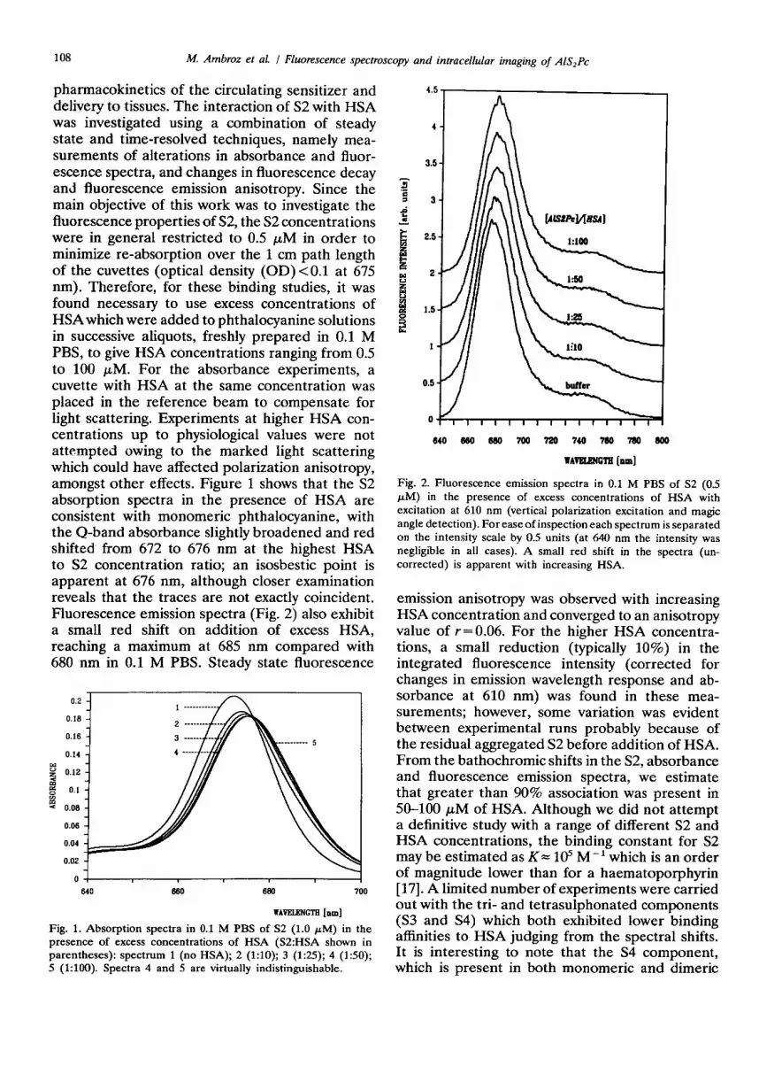
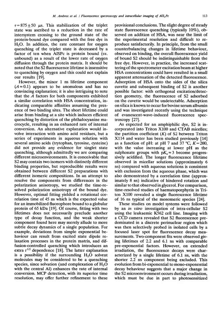
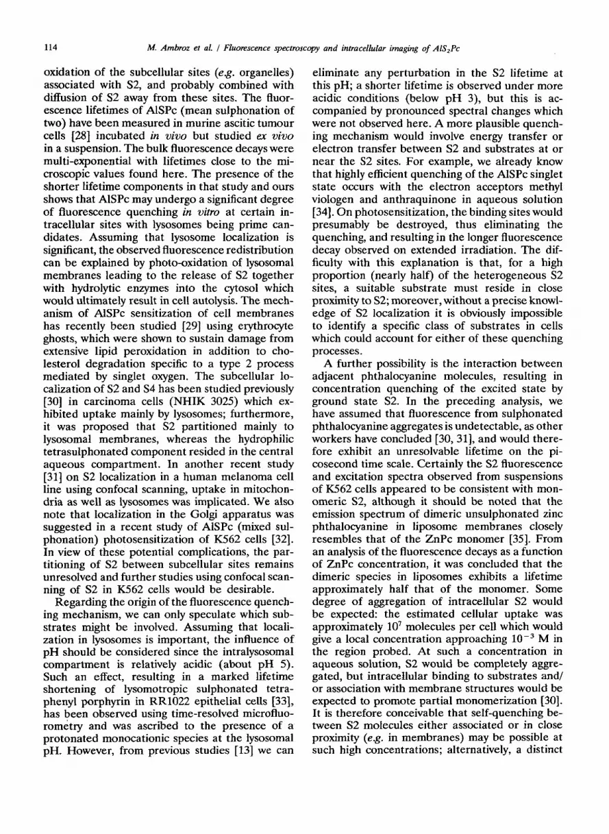
pharmacokinetics of the circulating sensitizer and delivery to tissues. The interaction of S2 with HSA was investigated using a combination of steady state and time-resolved techniques, namely mea- surements of alterations in absorbance and fluor- escence spectra, and changes in fluorescence decay and fluorescence emission anisotropy. Since the main objective of this work was to investigate the fluorescence properties of S2, the S2 concentrations were in general restricted to 0.5 FM in order to minimize re-absorption over the 1 cm path length of the cuvettes (optical density (OD) < 0.1 at 675 nm). Therefore, for these binding studies, it was found necessary to use excess concentrations of HSA which were added to phthalocyanine solutions in successive aliquots, freshly prepared in 0.1 M PBS, to give HSA concentrations ranging from 0.5 to 100 FM. For the absorbance experiments, a cuvette with HSA at the same concentration was placed in the reference beam to compensate for light scattering. Experiments at higher HSA con- centrations up to physiological values were not attempted owing to the marked light scattering which could have affected polarization anisotropy, amongst other effects. Figure 1 shows that the S2 absorption spectra in the presence of HSA are consistent with monomeric phthalocyanine, with the Q-band absorbance slightly broadened and red shifted from 672 to 676 nm at the highest HSA to S2 concentration ratio; an isosbestic point is apparent at 676 nm, although closer examination reveals that the traces are not exactly coincident. Fluorescence emission spectra (Fig. 2) also exhibit a small red shift on addition of excess HSA, reaching a maximum at 685 nm compared with 680 nm in 0.1 M PBS. Steady state fluorescence
0.2
0.16
0.16
0.14
E 2 0.12
B s
0.1
5 0.06
0.06
0.04
640 660 660 700
HAVELENCTE [nm]
Fig. 1. Absorption spectra in 0.1 M PBS of S2 (1.0 PM) in the presence of excess concentrations of HSA (S2:HSA shown in parentheses): spectrum 1 (no HSA); 2 (1:lO); 3 (195); 4 (150); 5 (1:lOO). Spectra 4 and 5 are virtually indistinguishable.
640 666 666 766 766 746 766 7#) 666
WAvEmNGTn [lull]
Fig. 2. Fluorescence emission spectra in 0.1 M PBS of S2 (0.5 FM) in the presence of excess concentrations of HSA with excitation at 610 nm (vertical polarization excitation and magic angle detection). For ease of inspection each spectrum is separated on the intensity scale by 0.5 units (at 640 nm the intensity was negligible in all cases). A small red shift in the spectra (un- corrected) is apparent with increasing HSA.
emission anisotropy was observed with increasing HSA concentration and converged to an anisotropy value of r=0.06. For the higher HSA concentra- tions, a small reduction (typically 10%) in the integrated fluorescence intensity (corrected for changes in emission wavelength response and ab- sorbance at 610 nm) was found in these mea- surements; however, some variation was evident between experimental runs probably because of the residual aggregated S2 before addition of HSA. From the bathochromic shifts in the S2, absorbance and fluorescence emission spectra, we estimate that greater than 90% association was present in 50-100 PM of HSA. Although we did not attempt a definitive study with a range of different S2 and HSA concentrations, the binding constant for S2 may be estimated as K= 105 M-’ which is an order of magnitude lower than for a haematoporphyrin [17]. A limited number of experiments were carried out with the tri- and tetrasulphonated components (S3 and S4) which both exhibited lower binding affinities to HSA judging from the spectral shifts. It is interesting to note that the S4 component, which is present in both monomeric and dimeric
M. Ambroz et al. / Fluorescence spectroscopy and intracellular imaging of AISzPc 109
forms of 0.1 M PBS [13), did not monomerize on addition of excess HSA, in contrast with solutions containing aggregated S2. Systematic studies will be reported elsewhere [18].
From time-resolved measurements, S2 in PBS alone exhibited a mono-exponential fluorescence decay with a lifetime of 5.Oi-0.1 ns, but at high HSA to S2 concentration ratios (50 and 100) bi- exponential decays were observed with lifetimes of TV = 5.5 t_ 0.2 ns and T* = 1.0 kO.2 ns, with nor- malized pre-exponential A factors converging to A1 = 0.92 + 0.1 and A2 = 0.08 + 0.01 (x’ coefficients of 1.05-0.95). At intermediate HSA to S2 ratios (1,2.5,5,10 and 25), we observed multi-exponential decays which could be fitted satisfactorily using the TV and 72 values together with the free dye lifetime of 5 ns. It should be noted that these decays were measured without a monochromator under conditions which were independent of po- larization anisotropy (G factor of unity); a con- tribution from scattered light can also be elimi- nated. Of course, even at the higher HSA concentrations the dye is never completely bound, but it is difficult to resolve the relatively small contribution from the free dye especially when the lifetime does not differ significantly. At high HSA to S2 ratios the time dependence of the polarization anisotropy [19] was studied, and we derived a rotational correlation time of T_ = 45 k 5 ns; the value for free dye in PBS was T_,= 0.3 ns corresponding to out-of-plane rotation (in-plane would exceed the resolution of our system).
3.1.2. Micelles The incorporation of S2 was examined in two
micellar solutions: CTAB (lo-’ M) and Triton Xl00 (1%) in 0.1 M PBS. A 20 ~1 aliquot of S2 stock solution was added to the micellar solutions to give an S2 concentration of 0.5 PM. In both cases a small spectral red shift was observed: the peak absorbance in Triton Xl00 is at 675 nm compared with 672 nm in aqueous solution. The addition of Triton Xl00 to a more concentrated S2 aqueous solution (10 PM), in which the S2 was partially aggregated, resulted in monomeri- zation. The fluorescence lifetimes were found to be: T= 5.9 f 0.1 ns and T_ = 8.0 f 0.7 ns for Triton X100; ~-6.0 +O.l ns for CI’AB. For comparison, in &CerOl T=% ns and 7cor= 5.7 f0.5 ns.
3.2. Microspectrojhorometry of K562 cells 3.2.1. Steady state spectroscopy and imaging studies Spectroscopic studies were carried out on cul-
tured leukaemic cells, the X562 human erythro-
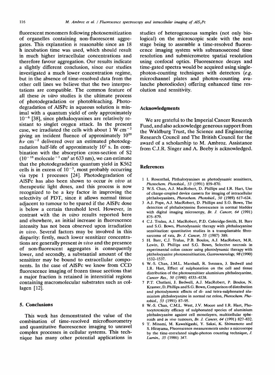
leukaemic progenitor line. This particular cell line was selected for investigation because it has been shown to be highly susceptible to AlSPc photo- sensitized destruction 115, 201 which is hoped may provide a means of selective elimination of leu- kaemic cells from infected bone marrow (pho- topurging). In this work, cells were incubated under the same conditions as used in the photosensi- tization studies, i.e. 10 PM of S2 for 1 h. Using the CCD imaging system to examine the intra- cellular S2 fluorescence distribution, it is clearly evident from Fig. 3(a) that the dye is taken up inside the cell (diameter, approximately 20 pm) at extranuclear sites. The conspicuous feature is a highly fluorescent region adjacent to the nucleus contributing over 50% of the integrated intensity, which proved ideal for spatially selective probing by a focused laser spot in subsequent time-resolved studies. Owing to the high sensitivity of the CCD, which afforded a short exposure time of 1 s, no perturbation in the S2 distribution due to pho- todegradation was apparent. A few cells were examined using a 100 x objective which suggested a granular structure in the fluorescence distri- bution. Further studies using cytospins demon- strated good correlation between the fluorescence distribution and histochemical staining for acid phosphatase, implying that the S2 was associated with lysosomes. In comparative studies with S4, a much weaker fluorescence intensity was observed, whereas S3 exhibited an intensity distribution which appeared to be intermediate between the S2 and S4 components.
On prolonged irradiation at 633 nm (40x ob- jective), it was apparent that the S2 fluorescence distribution became perturbed, and a series of images were taken during irradiation of the same cell as shown in Fig. 3(b) (recorded (with a 1 s exposure) after 30 s irradiation) and 3(c) (after a further 60 s). The power incident on the cells at 633 nm was estimated to be 2 mW over 0.2 mm’, giving an irradiance of about 1 W cm-‘. Comparing Figs. 3(a) and 3(b), it is clearly seen that the perinuclear feature intensified and ex- panded: quantitative analysis showed an increase in overall intensity of this feature by about 50% between the two images. This intensity increase was reproducible in all cells examined after 20-40 s irradiation and, in exceptional cases, a factor of two was found (but a 50% increase was the typical value). In Fig. 3(c) the fluorescence has spread throughout the cell and appears to have penetrated into the nucleus. Figure 3(d) shows a three-di- mensional intensity plot of Fig. 3(b). These ob- servations, which were found in the many cells
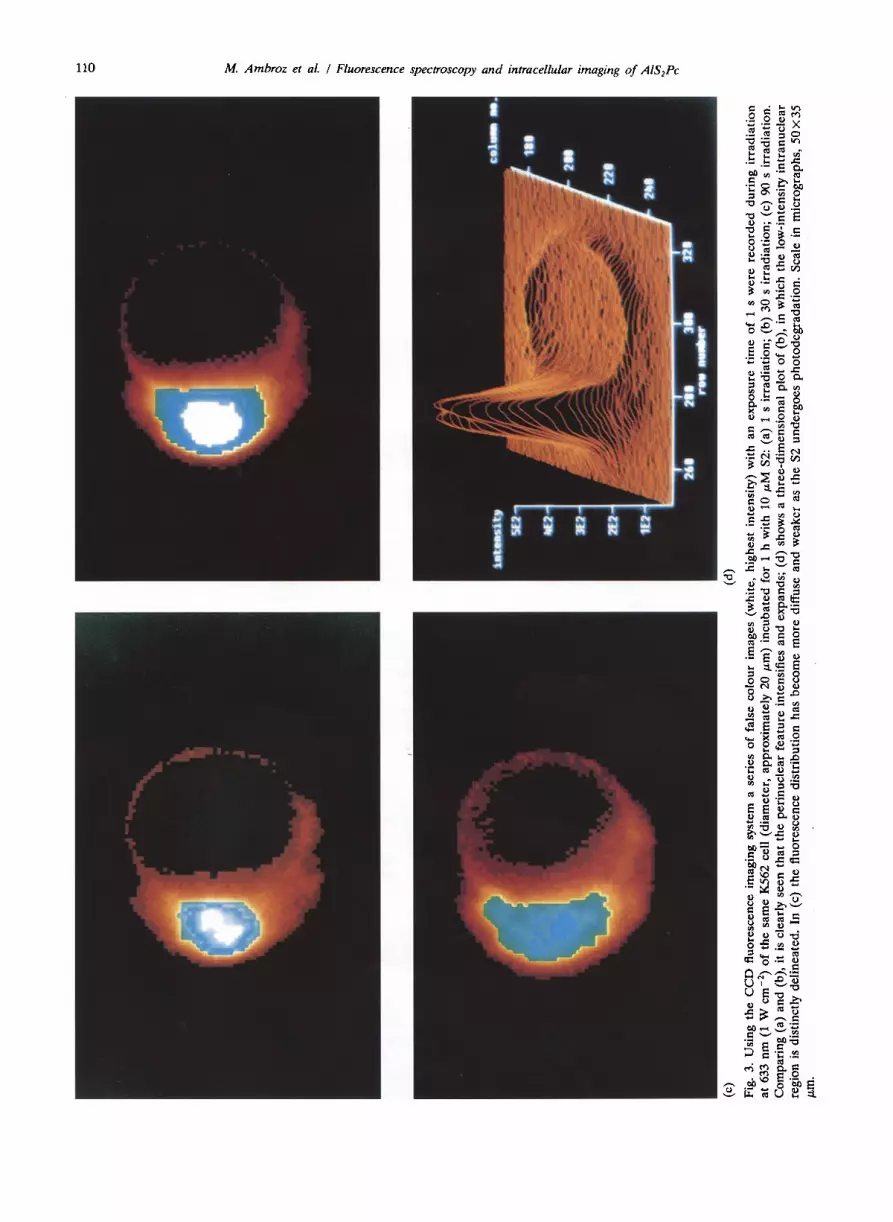
110 M. Ambroz et al. / Fluorescence spectroscopy and intracellular imaging of AIS,Pc
M. Ambnz et al. I Fluorescence spectroscopy and intracellular imaging of A&PC 111
examined from different batches and which were apparently irreversible, would be consistent with the photosensitized destruction of organelles as- sociated with S2 such as mitochondria and lyso- somes. The integrated cellular fluorescence in- tensity in Fig. 3(c), and for yet longer irradiation times, declined progressively signifying that pho- todegradation of S2 was taking place in parallel with its redistribution.
The fluorescence emission spectrum of cell sus- pensions excited at 610 nm closely resembled that observed with HSA with a peak intensity at 683 nm. The addition of Nonidet P40 detergent (1%) which lysed with cells releasing the dye in mon- omeric form, resulted in up to a twofold increase in fluorescence emission, and this effect was ob- served over a range of S2 incubation concentrations from 2 to 20 PM using 2 ml suspensions containing approximately lo6 cells. With a calibrated plot of S2 fluorescence intensity vs. concentration in the detergent solution, we were able to estimate the mean cellular S2 uptake to be approximately 10’ molecules per cell.
3.2.2. Time-resolved studies For time-resolved microfluorometry, the focused
laser beam was aligned initially by adjusting the position of the cell under phase contrast relative to the laser spot. The CCD camera, which was attached to another port on the inverted micro- scope orthogonal to the photomultiplier tube, was then used to assess the fluorescence distribution and thus guide the positioning of the laser spot over the selected intracellular region taking care to minimize irradiation of the cell. In addition to its inherent spatial resolution, this microscopic technique is more reliable than bulk measurements on a cell suspension, which are susceptible to interference from sensitizer leakage from the re- suspended cells and polarization anisotropy effects. The irradiation power of the laser spot was ap- proximately 10 W cm-’ at 610 nm and experiments were confined to cells incubated with 10 PM of S2 for 1 h in a series of studies conducted over several days. In most of this work, S2 fluorescence decays were recorded with the laser spot (2 pm in diameter) incident on the highly fluorescent cellular region (typically 4 km in diameter) as shown in Fig. 3(a). Counting rates up to 5000 s-l were detected and decays were accumulated to a maximum of 10 000 counts in the peak channel in the first instance. It was found that, under these conditions, the S2 fluorescence at this site exhibited bi-exponential decays with lifetime components near 2 and 6 ns. Modelling of the data from 15
cells including global analysis confirmed values of 2.2 _h 0.4 and 6.lkO.2 ns, with similar amplitudes for each component; good fits were obtained with the normalized A factor of the shorter component in the range 0.3-O-5 (x” coefficients of 1.2-1.4), although in some cases [4] tri-exponential fits gave a slightly better agreement with the inclusion of a minor 1 ns component. The data quality was limited by the relatively short accumulation times; these were required because, on prolonged irra- diation, the bi-exponential dependence evolved essentially to a mono-exponential decay with just the longer component of 6.1 ns remaining, i.e. counting to 20 Ooo in the peak channel reduced the A factor of the shorter component to less than 0.1. Thus the time-resolved data may be char- acterized by two regimes of fluorescence decay behaviour wit-h case 1 (initial excitation) giving a bi-exponential dependence and case 2 (excitation after irradiation) giving a mono-exponential de- pendence. The relative fluorescence yields for case 1 compared with case 2 may be estimated from the respective A factors (taking equal values for case 1) and lifetimes by the equation: Y= cAi7i; for case 1, Y, = (0.5 X2) + (0.5 x6) =4 and Y, = (1 X 6) = 6, giving a 50% increase from case 1 to 2. However, in general, the counting rate from the laser spot showed little change, presum- ably due to diffusion and photodegradation, al- though in a few cases it did increase by 10% when the laser intensity was sufficiently attenuated. This is an important point, since it has often been assumed in previous studies that a near-constant counting rate implies no perturbation to the fluo- rophore lifetime dynamics. Cells examined by the CCD system after laser spot probing exhibited fluorescence distributions similar to those shown in Figs. 3(b) and 3(c) demonstrating that, during the course of the lifetime measurements, S2 under- went intracellular redistribution. Lifetime mea- surements were attempted at other subcellular sites but with varying success due to signal lim- itations: probing at the cell periphery outside the highly fluorescent region yielded a lifetime of about 6 ns.
4. Discussion
In previous papers, we have investigated the excited singlet and triplet state properties of the AlS,Pc components using steady state and time- resolved techniques [13,21-231. Comparative stud- ies were carried out in methanol and phosphate- buffered saline which demonstrated that pertur-
112 M. Ambroz et al. / Fluorescence spectroscopy and intracellular imaging of AlS,Pc
bations to the phthalocyanine photoproperties, induced by the addition of varying numbers of sulphonate groups, are negligible [13]. However, differences in the fluorescence quantum yields and lifetimes of S2 in these solvents were found: in aqueous solution r=5.1 *O.l ns, @=0.4+0.04; in methanol r=6.1&-0.1 ns, @=0.56f0.05. It was concluded that this effect was due to an increase in the quantum yield of internal conversion to 0.4 in aqueous solution, consequently limiting the trip- let state yield to 0.2. In this work, the interaction of S2 with HSA has been studied at pH 7.4 in 0.1 M PBS. Both the steady state and time-resolved spectroscopic studies provide convincing evidence of binding, although the affinity of S2 for HSA is weaker than found for haematoporphyrin. Bath- ochromic spectral modifications have also been observed for several porphyrins on binding to HSA [24] and have been attributed to the lower overall dielectric constant of the protein microenviron- ment. HSA is predominantly an a-helical globin protein comprising three major domains, each of which contains two subdomains. This morphology offers a variety of binding sites, with two particular subdomains separated by approximately 8 nm iden- tified as being suitable conformations for small molecules [25]. From absorbance and fluorescence spectroscopic studies [24], the binding of hae- matoporphyrin to HSA is characterized by one high-affinity site, which is adjacent to the sole tryptophan residue (204-Trp) on the basis of fluor- escence energy transfer from this residue to hae- matoporphyrin, together with several other un- specified lower affinity sites. We attempted the same fluorescence energy transfer experiment using S2 but the results were unconvincing. Nevertheless, the presence of one dominant decay component clearly favours the presence of a specific binding site for S2. Our results may be compared with another study [26] of HSA binding with AlSPc containing a mixture of components giving an average sulphonation of about three. Although these results are not strictly comparable with those presented here, because the mixture components have different binding affinities, similar spectral changes were reported in the presence of HSA; time-resolved studies were confined to the triplet state and they concluded that the bound phthal- ocyanines were accessible to quenching species residing in the solvent phase. Binding of S2 to HSA to form a non-covalent complex is an im- portant factor influencing the biodistribution of the sensitizer, since it is thought that HSA delivery favours interstitial localization, and S2 is certainly known to partition to these regions, especially
submucosal sites [7, 121. Binding of S2 with lipo- proteins also occurs at similar concentrations [21] but, given the large excess of HSA in serum, binding to albumin should predominate in uivo.
In the time-resolved fluorescence studies at the highest HSA concentrations, S2 fluorescence de- cays could be fitted reproducibly within the time resolution of the system using two lifetimes: one major component of 5.5 ns and one minor com- ponent of 1 ns. These results imply two binding sites for S2. Time-resolved fluorescence studies of haematoporphyrin complexed with HSA at pH 7.4 have also shown the presence of two lifetime components with decay times near 9 and 17 ns [24]. It has been established that protein structural fluctuations occur in the nanosecond time scale; thus single lifetimes resolved from simple expo- nential fitting may represent mean values for the various conformations in the dye/protein microen- vironment [19]. As shown in Table 1 and the accompanying references, the 5.5 ns value of the major S2 lifetime component is intermediate be- tween the lifetimes measured in HZ0 and other solvents including micelles for which higher values around 6 ns are consistently found. As mentioned above, it has been shown recently that the fluor- escence quantum yield and lifetime in HZ0 are reduced owing to an enhancement in the rate of internal conversion. We therefore propose that this effect is inhibited partially when the phthal- ocyanine is protein bound, yielding the interme- diate lifetime value observed here in the presence of HSA, with the assumption that the intrinsic singlet state lifetime is not perturbed significantly on binding. A similar shielding effect has been observed in the triplet state kinetics of AlSPc studied using a tunable nanosecond flash photolysis system [21]. In argon-saturated PBS, S2 exhibits a triplet-triplet absorption band centred at 480 nm which decays as a single exponential with a lifetime of 600 +_40 ks. However, in the presence of excess HSA, the lifetime of the triplet state undergoes a substantial increase: in 100 PM HSA,
TABLE 1. Fluorescence lifetime measurements of 0.5 PM S2 in a range of solvent/model systems
Solvent 7F (ns) (A factor)
Reference
MeOH
0.1 M PBS (pH 7.4)
Triton XlOO/PBS
50 FM HSA/PBS
6.1* 0.1
5.0+0.1
5.9kO.l
5.5 f 0.2 (0.92), 1.0f 0.2 (0.08)
[13, 221
[131
This work
This work
hf. Ambroz et al. / Fluorescence spectroscopy and intracellular imaging of AlS,Pc 113
T= 875 &50 ps. This stabilization of the triplet state was ascribed to a reduction in the rate of intersystem crossing to the ground state of the protein-bound dye compared with the free dye in H20. In addition, the rate constant for oxygen quenching of the triplet state is decreased by a factor of ten when AlSPc is protein bound (VS. unbound) as a result of the lower rate of oxygen diffusion through the protein matrix. It should be noted that the S2 fluorescence lifetime is insensitive to quenching by oxygen and this could not explain our results [19].
However, the minor 1 ns lifetime component (A = 0.1) appears to be anomalous and has no convincing explanation; it is also intriguing to note that the A factors for both components exhibited a similar correlation with HSA concentration, in- dicating comparable affinities assuming the pres- ence of two binding sites. The short lifetime could arise from binding at a site which induces efficient quenching by distortion of the phthalocyanine ma- crocycle, resulting in an enhanced rate of internal conversion. An alternative explanation would in- volve interaction with amino acid residues, but a series of experiments in aqueous solution with several amino acids (trytophan, tyrosine, cysteine) did not provide any evidence for singlet state quenching, although admittedly we are comparing different microenvironments. It is conceivable that S2 may contain two isomers with distinctly different binding properties, but reproducible results were obtained between different S2 preparations with different isomeric compositions. In an attempt to resolve the components from differences in the polarization anisotropy, we studied the time-re- solved polarization anisotropy of the bound dye. However, optimal fitting yielded a rotational cor- relation time of 45 ns which is the expected value for an immobilized fluorophore bound to a globular protein of 65 kDa [19]. Of course, fitting with two lifetimes does not necessarily preclude another type of decay function, and the weak shorter component found here may merely allude to more subtle decay dynamics of a single population. For example, deviations from simple exponential be- haviour can result from excited state dipole re- laxation processes in the protein matrix, and dif- fusion-controlled quenching which introduces an extra t’” dependence [18]. The latter explanation is a possibility if the surrounding H,O solvent molecules may be considered to be a quenching species, since solvation (and complexation of HZ0 with the central Al) enhances the rate of internal conversion. MCP detection, with its superior time resolution, may offer further refinement to these
provisional conclusions. The slight degree of steady state fluorescence quenching (typically lo%), ob- served on addition of HSA, was near the limit of the experimental resolution and difficult to re- produce satisfactorily. In principle, from the small counterbalancing changes in lifetime behaviour, observed on binding, the overall fluorescence yield of bound S2 should be indistinguishable from the free dye. However, in practice, the increased scat- tering of the spectrometer excitation beam at higher HSA concentrations could have resulted in a small apparent attenuation of the detected fluorescence. Adsorption of HSA onto the sides of the silica cuvette and subsequent binding of S2 is another possible factor: with orthogonal excitation/detec- tion geometry, the fluorescence of S2 adsorbed on the cuvette would be undetectable. Adsorption on silica is known to occur for bovine serum albumin and was investigated recently using the technique of evanescent-wave-induced fluorescence spec- troscopy [27].
As expected for an amphiphilic dye, S2 is in- corporated into Triton Xl00 and CTAB micelles; the partition coefficient (K) of S2 between Triton X114 and water has been studied previously [14] as a function of pH: at pH 7 and 37 “C, K=200, with the value increasing at lower pH as the sulphonate groups with pK, = 3 become progres- sively acidified. The longer fluorescence lifetime observed in micellar solutions (approximately 6 ns) compared with aqueous solutions is consistent with exclusion from the aqueous phase, which was also demonstrated by a correlation time (approx- imately 8 ns) indicative of hindered rotation and similar to that observed in glycerol. For comparison, time-resolved studies of haematoporphyrin in Tri- ton Xl00 revealed a single fluorescence lifetime of 16 ns typical of the monomeric species [24].
These studies on model systems were followed by an in vitro investigation of intra-cellular S2 using the leukaemic K562 cell line. Imaging with a CCD camera revealed that S2 fluorescence pre- dominated in a discrete perinuclear region which was then selectively probed in isolated cells by a focused laser spot for fluorescence decay mea- surements. Two-component fits were observed giv- ing lifetimes of 2.2 and 6.1 ns with comparable pre-exponential factors. However, on extended irradiation, the fluorescence decays were char- acterized by a single lifetime of 6.1 ns, with the shorter 2.2 ns component being excluded. This evolution from bi-exponential to mono-exponential decay behaviour suggests that a major change in the S2 microenvironment occurs during irradiation, which must be due in part to photosensitized
114 M. Ambroz et al. ! Fluorescence spectroscopy and intracellular imaging of A&PC
oxidation of the subcellular sites (e.g. organelles) associated with S2, and probably combined with diffusion of S2 away from these sites. The fluor- escence lifetimes of AlSPc (mean sulphonation of two) have been measured in murine ascitic tumour cells [28] incubated in vivo but studied ex vivo in a suspension. The bulk fluorescence decays were multi-exponential with lifetimes close to the mi- croscopic values found here. The presence of the shorter lifetime components in that study and ours shows that AlSPc may undergo a significant degree of fluorescence quenching in vitro at certain in- tracellular sites with lysosomes being prime can- didates. Assuming that lysosome localization is significant, the observed fluorescence redistribution can be explained by photo-oxidation of lysosomal membranes leading to the release of S2 together with hydrolytic enzymes into the cytosol which would ultimately result in cell autolysis. The mech- anism of AlSPc sensitization of cell membranes has recently been studied [29] using erythrocyte ghosts, which were shown to sustain damage from extensive lipid peroxidation in addition to cho- lesterol degradation specific to a type 2 process mediated by singlet oxygen. The subcellular lo- calization of S2 and S4 has been studied previously [30] in carcinoma cells (NHIK 3025) which ex- hibited uptake mainly by lysosomes; furthermore, it was proposed that S2 partitioned mainly to lysosomal membranes, whereas the hydrophilic tetrasulphonated component resided in the central aqueous compartment. In another recent study [31] on S2 localization in a human melanoma cell line using confocal scanning, uptake in mitochon- dria as well as lysosomes was implicated. We also note that localization in the Golgi apparatus was suggested in a recent study of AlSPc (mixed sul- phonation) photosensitization of K562 cells [32]. In view of these potential complications, the par- titioning of S2 between subcellular sites remains unresolved and further studies using confocal scan- ning of S2 in K562 cells would be desirable.
Regarding the origin of the fluorescence quench- ing mechanism, we can only speculate which sub- strates might be involved. Assuming that locali- zation in lysosomes is important, the influence of pH should be considered since the intralysosomal compartment is relatively acidic (about pH 5). Such an effect, resulting in a marked lifetime shortening of lysomotropic sulphonated tetra- phenyl porphyrin in RR1022 epithelial cells [33], has been observed using time-resolved microfluo- rometry and was ascribed to the presence of a protonated monocationic species at the lysosomal pH. However, from previous studies [13] we can
eliminate any perturbation in the S2 lifetime at this pH; a shorter lifetime is observed under more acidic conditions (below pH 3), but this is ac- companied by pronounced spectral changes which were not observed here. A more plausible quench- ing mechanism would involve energy transfer or electron transfer between S2 and substrates at or near the S2 sites. For example, we already know that highly efficient quenching of the AlSPc singlet state occurs with the electron acceptors methyl viologen and anthraquinone in aqueous solution [34]. On photosensitization, the binding sites would presumably be destroyed, thus eliminating the quenching, and resulting in the longer fluorescence decay observed on extended irradiation. The dif- ficulty with this explanation is that, for a high proportion (nearly half) of the heterogeneous S2 sites, a suitable substrate must reside in close proximity to S2; moreover, without a precise knowl- edge of S2 localization it is obviously impossible to identify a specific class of substrates in cells which could account for either of these quenching processes.
A further possibility is the interaction between adjacent phthalocyanine molecules, resulting in concentration quenching of the excited state by ground state S2. In the preceding analysis, we have assumed that fluorescence from sulphonated phthalocyanine aggregates is undetectable, as other workers have concluded [30, 311, and would there- fore exhibit an unresolvable lifetime on the pi- cosecond time scale. Certainly the S2 Auorescence and excitation spectra observed from suspensions of K562 cells appeared to be consistent with mon- omeric S2, although it should be noted that the emission spectrum of dimeric unsulphonated zinc phthalocyanine in liposome membranes closely resembles that of the ZnPc monomer [35]. From an analysis of the fluorescence decays as a function of ZnPc concentration, it was concluded that the dimeric species in liposomes exhibits a lifetime approximately half that of the monomer. Some degree of aggregation of intracellular S2 would be expected: the estimated cellular uptake was approximately 10’ molecules per cell which would give a local concentration approaching lop3 M in the region probed. At such a concentration in aqueous solution, S2 would be completely aggre- gated, but intracellular binding to substrates and/ or association with membrane structures would be expected to promote partial monomerization [30]. It is therefore conceivable that self-quenching be- tween S2 molecules either associated or in close proximity (e.g. in membranes) may be possible at such high concentrations; alternatively, a distinct

M. Ambroz et al. / Fluorescence spectroscopy and intracellular imaging of AlS2Pc 115
type of intracellular fluorescent aggregate could be invoked. It would be desirable to replicate such behaviour in model systems, such as liposomal membranes, with high dye to lipid ratios in order to induce S2 aggregation within the membrane. Studies of the photophysics of S2 in reversed micelles [36] may also aid the interpretation of the non-exponential fluorescence decay dynamics of S2 in heterogeneous environments.
The attractive feature of a concentration quench- ing mechanism is that it could explain the transition to the longer mono-exponential lifetime behaviour when S2 undergoes dilution following photosen- sitized redistribution. In order to verify conclusively the contribution of such a mechanism, experiments at lower concentrations would have been desirable; however, the limited fluorescence intensities avail- able with the present detection system precluded a more complete investigation. The increase in S2 fluorescence intensity after the addition of de- tergent to unirradiated cellular suspensions is also consistent with the presence of fluorescence quenching in whole cells. The lack of any distinct concentration dependence using S2 concentrations in the range 2-20 PM (although only a factor of four difference in cellular uptake was observed) would appear to argue against a self-quenching mechanism, but it is possible that localization in the relevant sites becomes saturated even at the lowest concentration, thus limiting correlation with concentration. The small Stokes shift of the fluor- escence emission from the S2 Q-band may favour an energy transfer mechanism, and fluorescence depolarization studies (not attempted here) could, in principle, provide evidence of such a mechanism. It is interesting to note that, from transient ab- sorption studies [21, 221, we have established that ground state quenching limits the S2 triplet state lifetime with a rate constant of approximately 2x10s M-’ s-l in water.
Following extended irradiation, the evolution of the lifetime to just one longer component near 6 ns may be interpreted in terms of the photosen- sitized elimination of the nascent fluorescence quenching process; equivalently, we can infer that (before photodegradation becomes significant) the overall S2 fluorescence efficiency is increased dur- ing irradiation. The transition between these two lifetime regimes appears to be consistent with fluorescence imaging studies on whole cells in which photosensitized alterations in fluorescence intensity and distribution were observed using ex- tended irradiation at 633 nm, although these changes occurred over a longer time scale than with the focused laser spot at 610 nm which had
a much higher irradiance (factor of ten); moreover, the S2 absorbance at 610 nm is twice that at 633 nm. It is also pertinent to note that irradiation at 633 nm for over 90 s (see Fig. 3(c)) at 1 W cm-‘, giving an energy dose of 90 J cmp2, is equivalent to approximately 10 J cmp2 at 675 nm, which is comparable with that employed in previous in vitro studies [15] of AlSPc photosensitization of W62 cells: irradiation with approximately 30 J cmw2 (using 675 nm) was lethal to more than 95% of the cells under the same incubation con- ditions as used here. Although a precise comparison between the different sets of microscopic data is somewhat conjectural, the increases in microscopic steady state fluorescence, as shown in Figs. 3(a) and 3(b), are similar to the approximate 50% increase in the fluorescence yield from case 1 to 2 in the lifetime studies. We therefore propose that the transition between the two lifetime regimes occurs concomitantly with the intensification and redistribution of intracellular S2 as a result of the photosensitized rupture of organelles. This view is reinforced by the observation that, after the acquisition of case 2 data, the intracellular dis- tribution closely parallelled that of Fig. 3(b). These results may have phototherapeutic implications since, if significant fluorescence quenching to the ground state is present, the triplet and singlet oxygen yields will be correspondingly reduced and a lower photosensitization efficiency will be ob- tained. Moreover, differential fluorescence quench- ing at various sites would affect the correlation of sensitizer localization with fluorescence imaging of cells and frozen sections. Further studies should investigate the degree of quenching in other cell lines and tissue explants.
Similar photosensitization effects have been re- ported with AlSPc in other cell lines using quan- titative microscopic imaging. The behaviour of a mixture of S3 and S4 was studied [37] in cells at comparable concentrations as in this study, but after a much longer incubation time of 24 h for two cell lines: RR1022 rat epithelial cells and 3T3 murine fibroblasts. Following irradiation at 675 nm with 3.8 W cm-2, a fluorescence increase (factor of two) was observed for RR1022 cells, but not for the fibroblast line (this disparity may be related to different subcellular localization), and photo- bleaching was subsequently observed in each line with prolonged irradiation. In another study, com- parative experiments were performed on a human melanoma cell line with S2 and S4 [31]; a large initial increase in cellular fluorescence was ob- served for both S2 and S4 components, but this effect was attributed to disaggregation to form
116 M. Ambroz et al. I Fluorescence spectroscopy and intracellular imaging of AlS2Pc
fluorescent monomers following photosensitization of organelles containing non-fluorescent aggre- gates. This explanation is reasonable since an 18 h incubation time was used, which should result in much higher intracellular concentrations and therefore favour aggregation. Our results indicate a slightly different conclusion, since our studies investigated a much lower concentration regime, but in the absence of time-resolved data from the other cell lines we believe that the two interpre- tations are compatible. The common feature of all these in vitro studies is the ultimate process of photodegradation or photobleaching. Photo- degradation of AlSPc in aqueous solution is min- imal with a quantum yield of only approximately 10e6 [38], since phthalocyanines are relatively re- sistant to singlet oxygen attack. In the present case, we irradiated the cells with about 1 W cm-* giving an incident fluence of approximately 10” hv cm-* delivered over an estimated photodeg- radation half-life of approximately lo* s. In com- bination with the absorption cross-section of S2 (lo- I6 molecule - ’ cm* at 633 nm), we can estimate that the photodegradation quantum yield in K562 cells is in excess of 10e5, most probably occurring via type 1 processes [24]. Photodegradation of AlSPc has also been shown to occur in vivo at therapeutic light doses, and this process is now recognized to be a key factor in improving the selectivity of PDT, since it allows normal tissue adjacent to tumour to be spared if the AlSPc dose is below a certain threshold level. However, in contrast with the in vitro results reported here and elsewhere, an initial increase in fluorescence intensity has not been observed upon irradiation in vivo. Several factors may be involved in this disparity: firstly, much lower sensitizer concentra- tions are generally present in vivo and the presence of non-fluorescent aggregates is consequently lower, and secondly, a substantial amount of the sensitizer may be bound to extracellular compo- nents. In the case of AlSPc we know from CCD fluorescence imaging of frozen tissue sections that a major fraction is retained in interstitial regions containing macromolecular substrates such as col- lagen [ 121.
5. Conclusions
This work has demonstrated the value of the combination of time-resolved microfluorometry and quantitative fluorescence imaging to unravel complex processes in cellular systems. This tech- nique has many other potential applications in
studies of heterogeneous samples (not only bio- logical) on the microscopic scale with the next stage being to assemble a time-resolved fluores- cence imaging system with subnanosecond time resolution and submicrometre spatial resolution using confocal optics. Fluorescence decays and time-gated spectra would be acquired using single- photon-counting techniques with detectors (e.g. microchannel plates and photon-counting ava- lanche photodiodes) offering enhanced time res- olution and sensitivity.
Acknowledgments
We are grateful to the Imperial Cancer Research Fund, and also acknowledge generous support from the Waldburg Trust, the Science and Engineering Research Council and The British Council for the award of a scholarship to M. Ambroz. Assistance from C.J.R. Singer and A. Beeby is acknowledged.
References
I. Rosenthal, Phthalocyanines as photodynamic sensitisers, Photo&em. Photobiol., 53 (1991) 859-870.
W.S. Chan, A.J. MacRobert, D. Phillips and I.R. Hart, Use of charge-coupled device camera for imaging of intracellular phthalocyanines, Photochem. Photobiol., 50 (1989) 617-624. A.J. Pope, A.J. MacRobert, D. Phillips and S.G. Bown, The detection of phthalocyanine fluorescence in normal bladder with digital imaging microscopy, Br. J. Cancer, 64 (1991) 875-879. C.J. Tralau, A.J. MacRobert, P.D. Coleridge-Smith, H. Barr and S.G. Bown, Photodynamic therapy with phthalocyanine sensitisation: quantitative studies in a transplantable fibro- sarcoma of rats, Br. J. Cancer, 55 (1987) 389. H. Barr, C.J. Tralau, P.B. Boulos, A.J. MacRobert, M.R. Lewin, D. Phillips and S.G. Bown, Selective necrosis in experimental colon cancer using photodynamic therapy with phthalocyanine photosensitisation, Gastroenterology, 98 (1990) 1532-1537. W.-S. Char-r, J.M.L. Marshall, R. Svensen, J. Bedwell and I.R. Hart, Effect of sulphonation on the cell and tissue distribution of the photosensitiser aluminium phthalocyanine, Cancer Res., 50 (1990) 45334538. P.T. Chatlani, J. Bedwell, A.J. MacRobert, P. Boulos, N. Krasner, D. Phillips andS.G. Bown, Comparison of distribution and photodynamic effects of di- and tetra-sulphonated alu- minium phthalocyanine in normal rat colon, Photochem. Pho-
tobiol., 53 (1991) 87-95. W.-S. Chan, C.M.L. West, J.V. Moore and I.R. Hart, Pho- tocytotoxicity efficacy of sulphonated species of aluminium phthalocyanine against cell monolayers, multicellular sphe- roids and in vivo tumours, Br. 1. Cancer, 64 (1991) 827-832. T. Minami, M. Kawahigashi, Y. Sakai, K. Shimamoto and S. Hirayama, Fluorescence measurements under a microscope by the time-correlated single-photon counting technique, J. Lumin., 35 (1986) 347.
hi. Ambroz et al. I Fluorescence spectroscopy and intracellular imaging of AISzPc 117
10
11
12
13
14
15
16
17
18 19
20
21
22
23
S. Hirayama, Time-resolved fluorescence microscopy, in J.F. Rabek (ed.), Progress in Photochemistry and Photophysics, Vol. 6, CRC Press, 1992, pp. l-42. P.J. Nuutinen, J. Bedwell, A.J. MacRobert, P.T. Chatlani, D. Phillips and S.G. Bown, Distribution and photodynamic effects of disulphonated aluminium phthalocyanine in the pancreas and adjacent tissues in the Syrian golden hamster, Br. J. Cancer, 64 (1992) 1108-1115. P.T. Chatlani, J. Bedwell, A.J. MacRobert and S.G. Bown, Distribution and photodynamic effects of di- and tetra-sul- phonated aluminium phthalocyanine in normal and neoplastic rat colon, in P. Spinelli, M. Dal Fante and R. Marchesini (eds.), Photodynamic Therapy and Biomedical Lasers, Excerpta Medica Series No. 1011, Elsevier, 1992, pp. 593-644. M. Ambroz, A. Beeby, A.J. MacRobert, M.S.C. Simpson, R. Svensen and D. Phillips, Preparative, analytical and fluor- escence spectroscopic studies of sulphonated aluminium phthalocyanine photosensitisers, J. Photochem. Photobiol. B:
Biol., 9 (1991) 87-95.
K. Berg, J.C. Bommer and J. Moan, Evaluation of sulphonated aluminium phthalocyanines for use in photochemotherapy. Cellular uptake studies, Cancer Lett., 44 (1989) 7-15. C.R.J. Singer, D.C. Linch, D.C. Brown, E.R. Huehns and A.H. Goldstone, Differential phthalocyanine photosensitis- ation of acute myeloblastic leukaemic progenitor cells: a potential purging technique for autologous bone marrow transplantation, Br. J. Haematol., 68 (1988) 417422.
G. Rumbles, A.J. Brown and D. Phillips, Time-resolved evanescent wave induced fluorescence spectroscopy, Part 1 - Deviations in the fluorescence lifetime of tetrasulphonated aluminium phthalocyanine at a fused silica/methanol interface, J. Chem. Sot., Faraday Trans. 2, 87 (1991) 825-830.
R. Margalit, S. Cohen and M. Rotenburg, Porphyrin binding and aggregation, in D. Kessel (ed.), Photodynamic Therapy
of Neoplastic Disease, Vol. 2, CRC Press, 1990, pp. 209-221. M.S.C. Foley and M. Ambroz, unpublished data, 1993. J.R. Lakowicz, Topics in Fluorescence Spectroscopy, Vols. 2 and 3, Plenum, 1992. C.R.J. Singer, S.G. Bown, DC. Linch, E.R. Huehens and A.H. Goldstone, Phthalocyanine photosensitisation for in vitro
elimination of residual acute non-lymphoblastic leukaemia: preliminary evaluation, Photochem. Photobiol., 46 (1987)
745-749.
M.S.C. Simpson, A. Beeby, SM. Bishop, A.J. MacRobert, A.J. Parker and D. Phillips, Time-resolved spectroscopic studies of phthalocyanine triplet states; International Con- ference on Time-resolved Studies in Biochemistry, Proc. Sot. Photo-opt. Instrum. Eng., 1640 (1991) 520-529. A. Beeby, A.W. Parker, M.S.C. Simpson and D. Phillips, The effect of solvent deuteration on the photophysics of sul- phonated aluminium phthalocyanine, J. Photochem. Photobiol.
B: Biol., 16 (1992) 73-81.
A. Beeby, A.W. Parker, M.S.C. Simpson and D. Phillips, Deuteration effects on the photophysical properties of mol- ecules, J. Photochem. Photobiol. B: Biol., 17 (1993) 203-207.
24
25
26
27
28
29
30
31
32
33
34
35
36
37
38
E. Reddi and G. Jori, Steady-state and time-resolved spec- troscopic studies of photodynamic sensitisers: porphyrins and phthalocyanines, Rev. Chem. Intermed., 10 (1988) 241-268. D.C. Carter, X.-M. He, S.H. Munson, P.D. Twigs, K.M. Gernet, B. Broom and T.Y. Miller, Three-dimensional struc- ture of human serum albumin, Science, 244 (1989) 1195-1198. J. Davila and A. Harriman, Photoreactions of macrocyclic dyes bound to human serum albumin, Photochem. Photobiol., 51 (1990) 9-19. B. Crystall, G. Rumbles, T.A. Smith and D. Phillips, Time- resolved evanescent wave induced fluorescence measurements of surface adsorbed bovine serum albumin, J. Colloid Interface Sci., 155 (1993) 247-250. R. Cubeddu, R. Ramponi and P. Taroni, Time-gated fluor- escence spectroscopy of porphyrin derivatives and aluminium phthalocyanine incorporated in vivo in a murine ascitic tumour model, J. Photochem. Photobiol. B: Biol., I1 (1991) 319-328. G.J. Bachowski, E. Ben-Hur and A.W. Girotti, Phthalocyanine- sensitised lipid peroxidation in cell membranes: use of cho- lesterol and azide as probes of primary photochemistry, J. Photochem. Photobiol. B: Biol., 9 (1991) 307-321. J. Moan, K. Berg, J.C. Bommer and A. Western, Action spectra of phthalocyanines with respect to photosensitisation of cells, Photochem. Photobiol., 56 (1992) 171-175. Q. Peng, G.W. Farrants, K. Madslien, J.C. Bommer, J. Moan, H.E. Danielson and J.M. Nesland, Sub-cellular localisation, redistribution, and photobleaching of sulphonated aluminium phthalocyanine in a human melanoma cell line, Int. J. Cancer, 49 (1991) 290-295. C. Tempete, C. Giabbotti and G.H. Werner, Identical pho- tosensitising activities of a sulphonated aluminium phthal- ocyanine on human erythroleukaemic cell lines susceptible or resistant to the cytotoxic activity of dioxorubicin, J. Pho- tochem. Photobiol. B: Biol., 14 (1992) 201-205. J.M. Wessels, W. Strauss, H.K. Seidlitz, A. Ruck and H. Sneckenburger, Intracellular localization of meso-tetraphenyl tetrasulphonate probed by time-resolved and microscopic fluorescence microscopy, J. Photochem. Photobiol. B: Biol., 12 (1992) 275-284. J.R. Datwent, I. McCubbin and D. Phillips, Excited singlet and triplet state electron-transfer reactions of aluminium sulphonated phthalocyanine, J. Chem. Sot., Faraday Trans. 2, 78 (1982) 347-357. G. Valduga, E. Reddi, G. Jori, R. Cubeddu, P. Taroni and G. Valentini, Steady-state and time-resolved spectroscopic studies on zinc phthalocyanine in liposomes, J. Photochem. Photobiol. B: Biol., 16 (1992) 331-340. S. Dhami, M.S.C. Simpson, J.J. C&a, SM. Bishop and D. Phillips, Photophysics of sulphonated aluminium phthalo- cyanines in reversed micelles, SPIE Proc. on Photodynamic Therapy of Cancer, 1993, in press. A. Ruck, C. Hildebrandt, T. Kollner, H. Sneckenburger and R. Steiner, Competition between photobleaching and fluor- escence increase of photosensitising porphyrins and tetra- sulphonated chloroaluminium phthalocyanine, J. Photochem. Photobiol. B: Biol., 5 (1990) 311-319. I. McCubbin and D. Phillips, The photophysics and pho- tostability of zinc and aluminium sulphonated naphthalocy- anines, J. Photochem., 34 (1986) 187-195.


