Embed Size (px)
Citation preview
Cardiol Young 2004; 14: 512–519© Cambridge University Press
ISSN 1047-9511
AS MORE INFANTS WITH COMPLEX CONGENITAL
heart disease are successfully treated withpalliative and reparative interventions, the
population of children requiring permanent pacingis rapidly increasing.1 Major advances in technologyand techniques for implantation have taken placeover the past four decades.2–6 Although endocardialpacemakers are almost universally implanted in adultswith structurally normal hearts, the optimal approachis less clearly defined in children.6,7 Epicardial pacing is often preferred in neonates, infants, andchildren because of their small size, complex congen-ital defects, intracardiac shunts, limited vascular access,and potential for growth potential.8–10 Yet, in comparison to endocardial pacing, epicardial systems
are reported to have shorter longevity, in part due tohigher thresholds, exit block, and lead fractures.7,10
Moreover, children requiring life-long pacing facethe prospect of multiple reinterventions.7,9 The purpose of our study, therefore, was to assess thelong-term safety with epicardial pacing by reviewingour experience in a large cohort of children.
Methods
Population studiedWe included all children with permanent epicardialpacemakers implanted between June 1, 1971 andMarch 1, 2001 at Hôpital Sainte-Justine, Montreal,Canada, excluding those patients over 18 years, acommon North American definition of adulthood.All medical charts, operative records, and outpatientvisits to the pacemaker clinic were retrospectivelyreviewed. Data were collected on age, gender, underlying cardiac disease, surgical procedures
Original Article
Thirty years of experience with epicardial pacing in children
Nicolas Noiseux,1 Paul Khairy,2 Anne Fournier,3 Suzanne J. Vobecky1
Departments of 1Cardiovascular Surgery and the 2Electrophysiology Service, Children’s Hospital Boston, Harvard Medical School and 3Pediatric Cardiology, Hôpital Sainte-Justine, Montreal, Canada
Abstract Due to underlying cardiovascular anatomy and size, epicardial pacing may be the preferred methodof pacing in small children. To assess long-term safety, we reviewed all epicardial pacemakers implanted in chil-dren between 1971 and 2001. We found that 122 patients, with a median age of 5.4 years, had a total of 181pacemakers and 260 electrodes implanted over a total follow-up of 789 patient-years. Of the total, 12 patientsdied after the first implantation, with one death attributable to dysfunction of the pacemaker. Reinterventionwas required in 75 patients after 5.0 � 3.2 years, due to depletion of the battery in 45 patients (60%), fractureor dysfunction of electrodes in 27 patients (36%), and infection in 3 patients (4%). In univariate analyses, riskfactors for reintervention were an approach via a median sternotomy, with a relative risk of 2.3 (p � 0.0087),and an indication for pacing other than atrioventricular block, with a relative risk of 1.7 (p � 0.0314). In mul-tivariate analyses, the approach via the median sternotomy independently predicted the need for reintervention,with a relative risk of 2.1, and 95% confidence intervals from 1.1 to 4.1 (p � 0.0256). The longevity of the second pacemaker and/or its electrode, assessed in 26 patients, was 3.7 � 2.6 years, not shorter than the firstimplantation (p � 0.4037). We conclude that epicardial pacing is a reliable means of achieving permanent pacingin children, with low morbidity and mortality. A substantial proportion, nonetheless, requires reinterventionwithin five years, warranting meticulous follow-up.
Keywords: Epicardial pacing; children; long-term safety
Correspondence to: Dr Nicolas Noiseux MD, FRCS(C), MSc, Department ofCardiac Surgery, Hôtel-Dieu du CHUM, 3840 St-Urbain, Montreal, Québec,Canada H2W 1T8. Tel: �1 514 890 8131; Fax: �1 514 412 7231
Accepted for publication 19 April 2004
1405-08.qxd 9/9/04 11:56 AM Page 512
Vol. 14, No. 5 Noiseux et al: Epicardial pacing in young children 513
including implantation techniques, indication forpacing, generator and lead types, location, mode ofpacing, acute sensing and pacing thresholds and leadimpedance, complications, timing and nature of sub-sequent interventions, and all major outcomesincluding death. Perioperative mortality was definedas death occurring within 30 days of surgery. Thestudy was approved by the institutional review board.
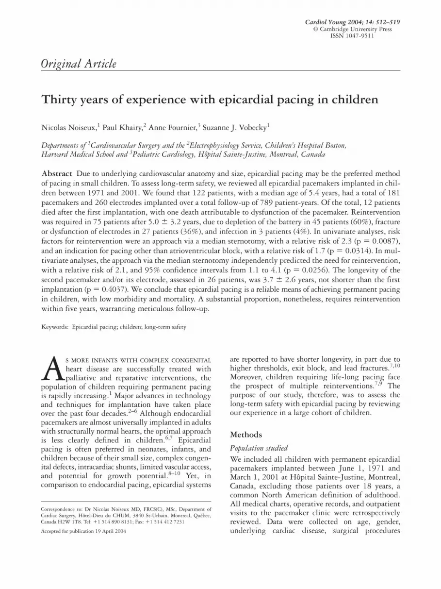
Surgical techniqueEpicardial leads, either unipolar or bipolar, wereimplanted by standard surgical techniques withaccess via an anterolateral left thoracotomy, midlinesternotomy (Fig. 1a), or subxiphoid approach,depending on underlying anatomy, prior surgicalprocedures, and whether implantation was part of acombined surgery. The ventricular lead was posi-tioned on the anterior left ventricular or diaphrag-matic right ventricular surface. The atrial lead wasplaced on either atrium, depending on the particularcardiac anatomy and surgical approach. Antibioticswere administered to all patients postoperatively for roughly 48 h. Electrodes were sutured to the epi-cardium, predominantly with Prolene (Ethicon Inc.,Somerville, NJ, USA) with care to prevent dislodg-ment. Leads were subsequently tunneled along therib margin to the generator implant site. Through a
second incision, the pacemaker generator and sur-plus pacing wire were placed in the retroperitonealspace, most commonly in the left flank or posteriorto rectus muscle (Fig. 1b).
Analysis of lead functionAcute analyses were performed using a Medtronicpacing system analyzer (most recently PSA 5311;Medtronic, Minneapolis, MN, USA). Measurementsincluded lead impedance at 0.5 ms/V, sensed P and Rwaves, and voltage pacing thresholds at a pulsewidth of 0.5 ms. Within the first month, at 3months, and 6-month intervals thereafter, pacingand sensing thresholds, lead impedance, and genera-tor voltage were routinely assessed with a Medtronicsystem analyzer (recently 5300; Medtronic).
Statistical analysisContinuous variables are expressed as mean � stan-dard deviation. Univariate comparisons were madeusing an analysis of variance, Student’s t-test, theKruskal-Wallis rank test, or Fisher’s exact test whereappropriate. Curves for freedom from reinterventionwere plotted using the Kaplan-Meier method, andanalyzed by the log-rank test. Censoring occurred inthe event of placement of endocardial leads, or loss to
(a) (b)
Figure 1.Implantation of a permanent epicardial pacemaker. A permanent epicardial pacemaker was implanted for congenital sinus nodal dysfunction ina 3-day-old child weighing 2.7 kg. In panel (a), the leads are positioned on left ventricular epicardial surfaces (atrial leads not yet implanted)via a median sternotomy (Note: the head is at top of the picture). The postoperative chest radiogram, panel (b), shows the position of the two epicardial leads with the generator positioned in the left flank.
1405-08.qxd 9/9/04 11:56 AM Page 513
514 Cardiology in the Young October 2004
follow-up. Cox proportional hazards models wereused to evaluate unadjusted univariate and adjustedmultivariate predictors of reintervention. Estimatesof relative risk, along with 95% confidence intervals,were obtained from Cox models. Two-sided p valuesof �0.05 were considered to indicate statistical sig-nificance. All analyses were performed using SASsoftware Version 8 (SAS Institute, Cary, NC, USA).
Results
Characteristics of patientsDuring this 30-year period, we implanted 181 gen-erators in 122 patients, using 260 epicardial leads,with a total of 242 surgical interventions. The medianage at initial implantation was 5.4 years, with arange from 1 day to 18 years, and 57% were male.The mean duration of follow-up was 6.4 � 4.7 years,and ranged from 1 day to 21.2 years, totaling 789patient-years. Censoring due to loss to follow-upoccurred in 8 patients. During the perioperativeperiod, 5 patients died (3.8%), with 4 of the 5 deathsoccurring in children with recent or combined cardiacsurgery for complex congenital cardiac disease. Therewere an additional 8 deaths during follow-up, 7 ofwhich were prior to any surgical reintervention, andwith one attributable to dysfunction of the pacemaker.
Of the 122 patients, 101 (83%) had structurallyabnormal hearts, 91 (75%) of whom had surgery forcongenital heart disease, as summarized in Table 1.The most common surgical procedures were theMustard or Senning procedures for patients withconcordant atrioventricular but discordant ventriculo-arterial alignments, Fontan or bi-directional Glennfor those with functionally univentricular physiology,and repair of atrioventricular and ventricular septaldefects. The median time from surgery to implanta-tion of the pacemaker was 37 days, with a range from0 days to 16.1 years.
Indications for epicardial pacingPrior to 1986, epicardial pacing was the exclusivemode of permanent pacing available at our institu-tion. Of the 122 patients, 42 received their pace-makers before transvenous systems were available.The remaining 80 patients with epicardial systemsrepresent a small proportion of all pacemakersimplanted since 1986. Of these patients, 47 wereunder 5 years of age, 37 of whom were no more than2 years of age. In these young children with struc-turally normal hearts, epicardial pacemakers werelargely justified on the basis of small bodily habitus.The remaining 33 patients had epicardial systems
Table 1. Associated congenital cardiac defects.
Atrioventricular blockBradycardia, sinus
Cardiac defects N node dysfunction Congenital Postop Arrhythmia Other
Not operatedNormal heart 21 3 17 1Atrial septal defect 2 2Transposition with discordant 3 3atrioventricular and ventriculo-arterial alignments
Complex cardiomyopathy 5 1 4
Total 31 4 26 1
OperatedTransposition with concordant 29 12 11 4 2atrioventricular but discordantventriculo-arterial alignments
Tetralogy of Fallot 5 4 1Fontan/Glenn 14 6 4 4Atrioventricular septal defect, 13 2 10 1with common or separated valvar orifices
Ventricular septal defect 14 1 13Atrial septal defect 1 1Ebstein’s malformation 2 1 1Aortic disease 4 4Miscellaneous 9 1 1 7
Total 91 22 5 50 9 5
Total overall 122 26 31 50 10 5
1405-08.qxd 9/9/04 11:56 AM Page 514
Vol. 14, No. 5 Noiseux et al: Epicardial pacing in young children 515
for various reasons. In 10 patients, construction ofthe Fontan circulation had removed transvenous accessfor ventricular pacing, 4 patients had prosthetic tricuspid valves, while the remaining patients hadintracardiac bi-directional or right-to-left shunting,with or without prior palliative shunts.
In accordance with the guidelines of the AmericanCollege of Cardiology and the American HeartAssociation,11 indications for permanent pacingincluded postoperative atrioventricular block in 50patients (41%), congenitally complete heart block in31 patients (26%), sinus nodal dysfunction in 26patients (21%), to permit adequate therapy for tachy-arrhythmias in 10 patients (8%), and others in 5patients (4%). Implantation for congenital atrioven-tricular block occurred at a median age of 3.6 years,with a range from 1 day to 17.8 years, compared to2.8 years, and a range from 0.1 to 17.1 years, inpatients with postoperative atrioventricular block(p � 0.1905). In 50 patients, pacemakers for post-operative atrioventricular block were implanted at amedian of 21 days after the initial surgery. Of theseimplants, 31 pacemakers (62%) were inserted priorto discharge from hospital, within one month fromthe cardiac surgery.
First pacemaker implantationThe varied techniques for implanting epicardialpacemakers reflect the complexity of our populationof patients, and mounting surgical experience. Leadswere implanted by a left anterolateral thoracotomyin 97 patients, including 5 redo thoracotomies, amedian sternotomy in 22 patients, including 2 redo
sternotomies, and other approaches in 3 patientsthat included access from a subxiphoid incision.
Over the 30-year course, various models ofMedtronic pacemaker pulse generators and leadswere used (Table 2). Overall, epicardial atrial leadswere implanted in 99 patients, and ventricular leads were implanted in 119 patients. Combinedsurgeries for congenital cardiac defects with implan-tation of epicardial pacemakers were performed in14 patients, mainly via midline sternotomies (93%)at a median age of 4.8 years, with a range from 3 daysto 18.7 years. Epicardial leads were implanted exclu-sively on the left ventricular surface in 31 patients,and on left atrial and ventricular surfaces in 72 pat-ients. Pacemaker generators were positioned in theleft flank, using either the retroperitoneal or pre-retroperitoneal spaces, in 115 patients (94%), in thefascia of the rectus abdominis muscle in 6 patients(5%), and in the right flank in one patient (1%). The pacing mode was atrioventricular [DDD(R)] in89 patients (73%), ventricular without atrial synchro-nization [VVI(R)] in 28 patients (23%), and exclusiveatrial sensing and pacing [AAI(R)] in 5 patients (4%).
Acute thresholds for atrial pacing and P-wavesensing, both assessed in 89 patients, were 1.4 � 1.0volts at 0.5 ms, and 4.1 � 3.5 mV, respectively.Ventricular pacing, assessed in 103 patients, andthresholds for R-wave sensing, assessed in 91, were1.2 � 0.9 volts at 0.5 ms, and 12.9 � 5.4 mV. Atrialand ventricular lead impedances, assessed in 89 and91 patients, were 510 � 256 �, and 623 � 289 �.When compared to a mid-line sternotomy, the leftlateral thoracotomy approach was associated with alower threshold for ventricular pacing, at 1.1 � 0.8 V
Table 2. Medtronic generator and lead models at first implantation.
Generator models N Chambers Leads models N Fixation Polarity Steroids
5862 1 Single AtrialMirel 5988 1 Single 4951 91 Fishhook Unipolar NoSpectrax 5941/42/43/ 11 Single 4968 Capsure 6 Sutured Bipolar Yes45/51/54/84 6913 1 Screw in Unipolar No
Spectrax 8423 1 Single 10366 1 Sutured Bipolar YesThera 8940 SR 1 Single Xyrel 5973/94 7 Single Total 99Clarity 860 1 DualElite 7074/75 8 Dual VentricularElite II 7084/85/86 16 Dual 4951 3 Fishhook Unipolar NoKappa DR 403/733 14 Dual 4968 Capsure 7 Sutured Bipolar YesMinuet 7107 2 Dual 5071 25 Screw in Unipolar NoSymbios 7005 25 Dual 5815 2 Myocardial Unipolar NoSynergyst II 7071 5 Dual 6917 AT 77 Screw in Unipolar NoThera 7940/44/50/60/64 17 Dual 10366 1 Sutured Bipolar YesVersatrax II 7000A 9 Dual Unknown 4Unknown 3
Total 122 Total 119
1405-08.qxd 9/9/04 11:56 AM Page 515
516 Cardiology in the Young October 2004
compared to 1.6 � 1.1 V (p � 0.0271), impedanceat 591 � 241 � versus 757 � 409 � (p � 0.0252),and a lower impedance at the atrial lead, at 468 � 184 � versus 662 � 371 � (p � 0.0018).
Replacement of pacemakersFollowing the initial 122 implantations, 75 patients(61%) underwent a total of 118 pacemaker-relatedreinterventions. Of the 75 patients, 7 had proceduresin adult institutions. Of the 68 with reinterventionsperformed in our institution, 17 were transitioned toendocardial transvenous systems, while epicardialpacing was pursued in 51 patients. Of the latterpatients, 30 required a new generator after 5.1 � 2.4years, with a range from 0.2 to 13.2 years, 15required a new generator and epicardial lead(s) after3.9 � 2.5 years, with a range from 0.3 to 9.7 years,and 6 required replacement only of epicardial leadsafter 0.3 � 0.2 years, with a range from 0.02 to 0.7years. A third intervention was required in 26patients after 3.7 � 2.6 years, with a range from0.03 to 9.0 years, a fourth in 4 patients, and a fifth in4 patients. The decision to pursue with epicardialpacing was individualized and at the discretion ofthe treating cardiologist and surgeon. Nevertheless,transvenous pacing was precluded in 30 of 51patients (59%) due to unavailability of this approachprior to 1986 for 14 patients, the particular congen-ital anatomy for 13 patients, very small body habitusfor 2 patients aged �2 years, and venous thrombosisin 1 patient.
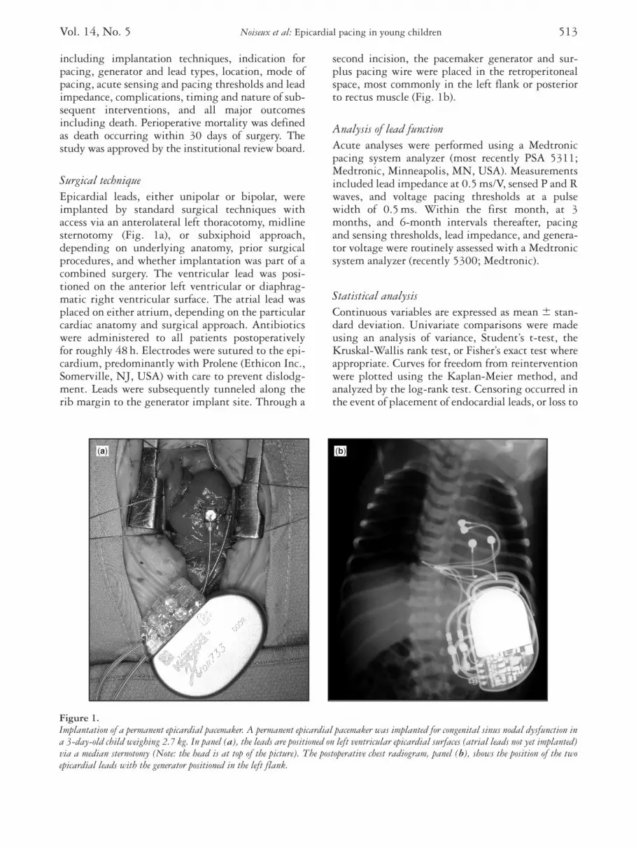
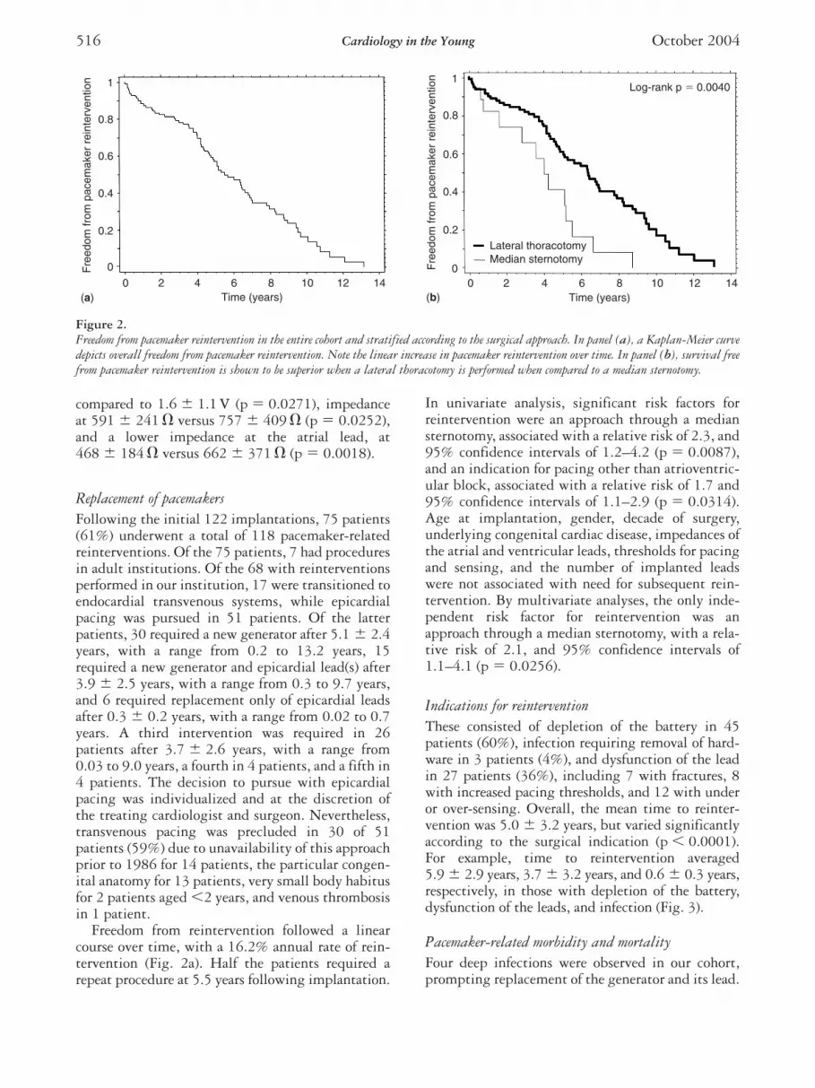
Freedom from reintervention followed a linearcourse over time, with a 16.2% annual rate of rein-tervention (Fig. 2a). Half the patients required arepeat procedure at 5.5 years following implantation.
In univariate analysis, significant risk factors forreintervention were an approach through a mediansternotomy, associated with a relative risk of 2.3, and95% confidence intervals of 1.2–4.2 (p � 0.0087),and an indication for pacing other than atrioventric-ular block, associated with a relative risk of 1.7 and95% confidence intervals of 1.1–2.9 (p � 0.0314).Age at implantation, gender, decade of surgery,underlying congenital cardiac disease, impedances ofthe atrial and ventricular leads, thresholds for pacingand sensing, and the number of implanted leadswere not associated with need for subsequent rein-tervention. By multivariate analyses, the only inde-pendent risk factor for reintervention was anapproach through a median sternotomy, with a rela-tive risk of 2.1, and 95% confidence intervals of1.1–4.1 (p � 0.0256).
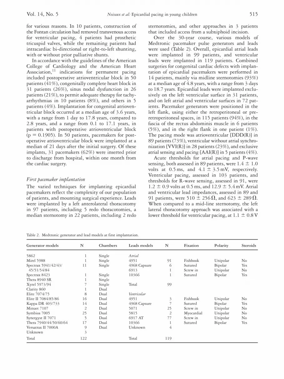
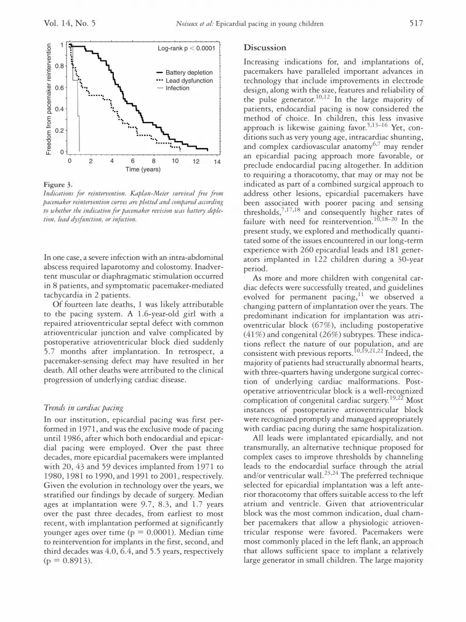
Indications for reinterventionThese consisted of depletion of the battery in 45patients (60%), infection requiring removal of hard-ware in 3 patients (4%), and dysfunction of the leadin 27 patients (36%), including 7 with fractures, 8with increased pacing thresholds, and 12 with underor over-sensing. Overall, the mean time to reinter-vention was 5.0 � 3.2 years, but varied significantlyaccording to the surgical indication (p � 0.0001).For example, time to reintervention averaged5.9 � 2.9 years, 3.7 � 3.2 years, and 0.6 � 0.3 years,respectively, in those with depletion of the battery,dysfunction of the leads, and infection (Fig. 3).
Pacemaker-related morbidity and mortalityFour deep infections were observed in our cohort,prompting replacement of the generator and its lead.
1
0.8
0.6
0.4
0.2
0
0 2 4 6Time (years)
Fre
edom
from
pac
emak
er r
eint
erve
ntio
n
8 10 12 14(a)
Log-rank p � 0.0040
Time (years)
Lateral thoracotomyMedian sternotomy
Fre
edom
from
pac
emak
er r
eint
erve
ntio
n
0 42 6 8 10 12 140
0.2
0.4
0.6
0.8
1
(b)
Figure 2.Freedom from pacemaker reintervention in the entire cohort and stratified according to the surgical approach. In panel (a), a Kaplan-Meier curvedepicts overall freedom from pacemaker reintervention. Note the linear increase in pacemaker reintervention over time. In panel (b), survival freefrom pacemaker reintervention is shown to be superior when a lateral thoracotomy is performed when compared to a median sternotomy.
1405-08.qxd 9/9/04 11:56 AM Page 516
Vol. 14, No. 5 Noiseux et al: Epicardial pacing in young children 517
In one case, a severe infection with an intra-abdominalabscess required laparotomy and colostomy. Inadver-tent muscular or diaphragmatic stimulation occurredin 8 patients, and symptomatic pacemaker-mediatedtachycardia in 2 patients.
Of fourteen late deaths, 1 was likely attributableto the pacing system. A 1.6-year-old girl with arepaired atrioventricular septal defect with commonatrioventricular junction and valve complicated bypostoperative atrioventricular block died suddenly5.7 months after implantation. In retrospect, a pacemaker-sensing defect may have resulted in herdeath. All other deaths were attributed to the clinicalprogression of underlying cardiac disease.
Trends in cardiac pacingIn our institution, epicardial pacing was first per-formed in 1971, and was the exclusive mode of pacinguntil 1986, after which both endocardial and epicar-dial pacing were employed. Over the past threedecades, more epicardial pacemakers were implantedwith 20, 43 and 59 devices implanted from 1971 to1980, 1981 to 1990, and 1991 to 2001, respectively.Given the evolution in technology over the years, westratified our findings by decade of surgery. Medianages at implantation were 9.7, 8.3, and 1.7 yearsover the past three decades, from earliest to mostrecent, with implantation performed at significantlyyounger ages over time (p � 0.0001). Median timeto reintervention for implants in the first, second, andthird decades was 4.0, 6.4, and 5.5 years, respectively(p � 0.8913).
Discussion
Increasing indications for, and implantations of,pacemakers have paralleled important advances intechnology that include improvements in electrodedesign, along with the size, features and reliability ofthe pulse generator.10,12 In the large majority ofpatients, endocardial pacing is now considered themethod of choice. In children, this less invasiveapproach is likewise gaining favor.5,13–16 Yet, con-ditions such as very young age, intracardiac shunting,and complex cardiovascular anatomy6,7 may renderan epicardial pacing approach more favorable, orpreclude endocardial pacing altogether. In additionto requiring a thoracotomy, that may or may not beindicated as part of a combined surgical approach toaddress other lesions, epicardial pacemakers havebeen associated with poorer pacing and sensingthresholds,7,17,18 and consequently higher rates offailure with need for reintervention.10,18–20 In thepresent study, we explored and methodically quanti-tated some of the issues encountered in our long-termexperience with 260 epicardial leads and 181 gener-ators implanted in 122 children during a 30-yearperiod.
As more and more children with congenital car-diac defects were successfully treated, and guidelinesevolved for permanent pacing,11 we observed achanging pattern of implantation over the years. Thepredominant indication for implantation was atri-oventricular block (67%), including postoperative(41%) and congenital (26%) subtypes. These indica-tions reflect the nature of our population, and areconsistent with previous reports.10,19,21,22 Indeed, themajority of patients had structurally abnormal hearts,with three-quarters having undergone surgical correc-tion of underlying cardiac malformations. Post-operative atrioventricular block is a well-recognizedcomplication of congenital cardiac surgery.19,22 Mostinstances of postoperative atrioventricular blockwere recognized promptly and managed appropriatelywith cardiac pacing during the same hospitalization.
All leads were implantated epicardially, and nottransmurally, an alternative technique proposed forcomplex cases to improve thresholds by channelingleads to the endocardial surface through the atrialand/or ventricular wall.23,24 The preferred techniqueselected for epicardial implantation was a left ante-rior thoracotomy that offers suitable access to the leftatrium and ventricle. Given that atrioventricularblock was the most common indication, dual cham-ber pacemakers that allow a physiologic atrioven-tricular response were favored. Pacemakers weremost commonly placed in the left flank, an approachthat allows sufficient space to implant a relativelylarge generator in small children. The large majority
1
0.8
0.6
0.4
0.2
00 2 4 6 8 10 12 14
Battery depletion
Log-rank p � 0.0001
Lead dysfunctionInfection
Time (years)
Fre
edom
from
pac
emak
er r
eint
erve
ntio
n
Figure 3.Indications for reintervention. Kaplan-Meier survival free frompacemaker reintervention curves are plotted and compared accordingto whether the indication for pacemaker revision was battery deple-tion, lead dysfunction, or infection.
1405-08.qxd 9/9/04 11:56 AM Page 517
518 Cardiology in the Young October 2004
of patients had acceptable thresholds for pacing andsensing at implantation for both atrial and ventricu-lar leads. Interestingly, an approach through amedian sternotomy, when compared to left lateralthoracotomy, was associated with a greater thantwofold increased risk for reintervention, even aftercontrolling for various baseline factors and underlyingcongenital cardiac disease. Moreover, median ster-notomies were associated with significantly higherventricular thresholds, and atrial and ventricularlead impedances. These findings may reflect theobservation that median sternotomies are associatedwith a greater extent of damage to the epicardial wall,resulting in relatively more fibrosis, dense scarring,adhesions, and inflammation.9,22,25
More than three-fifths of our patients required atleast one reintervention. The time to reintervention of5.0 � 3.2 years compares favorably to other seriesassessing epicardial pacemakers,7,19 as well as endocar-dial systems inserted in similar populations of chil-dren.9,15 While depletion of the battery occurred afteran average of 5.9 years after implantation, improvedlongevity is expected to ensue from technologicadvances, including the use of steroid-eluted epicar-dial leads that result in lower pacing thresholds. Notunexpectedly, dysfunction or infection of the leadsprompted earlier reintervention than depletion of thebattery. The incidence of fractured leads in adultswith epicardial pacing has been estimated to be 2%per patient-year.26 Most such fractures occur at sitessubjected to increased stress, such as adjacent to thepulse generator or under the costal margin.1 Giventhat fractures are more common with epicardial pac-ing, some investigators have suggested that its occur-rence should prompt transitioning to an endocardialpacing system unless otherwise contraindicated.13
When reintervention was necessary, the generalpolicy adopted by our institution was to test theleads with continued use if their function was notimpaired, followed by replacement of the generator.If leads required replacement, alternative approacheswere considered, including re-implantation of epi-cardial leads, or replacement with a transvenouspacemaker system if not contraindicated. Therationale in support of maintaining epicardial leadswas the preservation of venous access for later use inpatients requiring life-long devices. Despite thenumerous advantages of endocardial pacing systems,concerns remain over long-term vascular and valvarintegrity with multiple transvenous leads.10,19,27,28
Venous thrombosis resulting from a disproportionbetween the size of the vessel and the lead has beenreported to occur in up to almost half the transve-nous pacemakers implanted in children.1,19
Retrospective studies have numerous potentiallimitations. In order to avoid potential selection
bias, and allow accurate estimates of rate and risk,every child with an epicardial pacemaker implantedin our institution was included in our cohort. Themain variables concerning exposure and outcome,such as surgical approach and reintervention, wereobjective measures with low probability for misclas-sification. Observation bias in assessing outcome waslimited by routine at least biannual visits and mini-mizing losses to follow-up. Once patients reachedadulthood, their care was, however, transitioned toother institutions. No comparable group with endo-cardial pacing group was available. Various modelsof both leads and generators were implanted over thespan of 30 years, with all devices manufactured bythe same company. An insufficient number ofsteroid-eluting leads were implanted to assess theirimpact on thresholds and the longevity of the gener-ator. Although the population studied was hetero-geneous in nature, with diverse underlying cardiacmalformations and sizes of patients, every attemptwas made to control for baseline imbalances in theanalysis when indicated for comparisons.
We conclude that the majority of children withpermanent pacemakers will require pacing systemsfor the rest of their lives. Our experience in a largecohort of children with long-term follow-up sug-gests that epicardial pacemakers are safe and reliable.In selected patients, including the very young inwhom preservation of venous access is of concern,epicardial pacing should be considered a reasonablealternative. A substantial proportion of patientswith epicardial pacemakers do, however, requirereintervention within five years. Median sternotomyis a risk factor for such. Meticulous follow-up isessential to ensure adequate margins of safety forthreshold, to anticipate need for changes in the gen-erator, to screen for pacemaker malfunction, and tooptimize programmable settings.
Acknowledgements
This study was supported in part by research fellow-ship scholarships from the Canadian Institutes ofHealth Research (NN and PK).
References1. Sliz Jr NB, Johns JA. Cardiac pacing in infants and children.
Cardiol Rev 2000; 8: 223–239.2. Hamilton R, Gow R, Bahoric B, Griffiths J, Freedom R,
Williams W. Steroid-eluting epicardial leads in pediatrics:improved epicardial thresholds in the first year. Pacing ClinElectrophysiol 1991; 14: 2066–2072.
3. Karpawich PP, Hakimi M, Arciniegas E, Cavitt DL. Improvedchronic epicardial pacing in children: steroid contribution toporous platinized electrodes. Pacing Clin Electrophysiol 1992;15: 1151–1157.
1405-08.qxd 9/9/04 11:56 AM Page 518
Vol. 14, No. 5 Noiseux et al: Epicardial pacing in young children 519
4. Johns JA, Fish FA, Burger JD, Hammon Jr JW. Steroid-elutingepicardial pacing leads in pediatric patients: encouraging earlyresults. J Am Coll Cardiol 1992; 20: 395–401.
5. Cutler NG, Karpawich PP, Cavitt D, Hakimi M, Walters HL.Steroid-eluting epicardial pacing electrodes: six year experience ofpacing thresholds in a growing pediatric population. Pacing ClinElectrophysiol 1997; 20: 2943–2948.
6. Maginot KR, Mathewson JW, Bichell DP, Perry JC. Applicationsof pacing strategies in neonates and infants. Prog Pediatr Cardiol2000; 11: 65–75.
7. Rao V, Williams WG, Hamilton RH, Williams MG, GoldmanBS, Gow RM. Trends in pediatric cardiac pacing. Can J Cardiol1995; 11: 993–999.
8. Kugler JD, Danford DA. Pacemakers in children: an update. AmHeart J 1989; 117: 665–679.
9. Beaufort-Krol GC, Mulder H, Nagelkerke D, Waterbolk TW,Bink-Boelkens MT. Comparison of longevity, pacing, and sensingcharacteristics of steroid-eluting epicardial versus conventionalendocardial pacing leads in children. J Thorac Cardiovasc Surg1999; 117: 523–528.
10. Cohen MI, Bush DM, Vetter VL, et al. Permanent epicardial pac-ing in pediatric patients: seventeen years of experience and 1200outpatient visits. Circulation 2001; 103: 2585–2590.
11. Gregoratos G, Cheitlin MD, Conill A, et al. ACC/AHA guidelinesfor implantation of cardiac pacemakers and antiarrhythmia devices:a report of the American College of Cardiology/American HeartAssociation Task Force on Practice Guidelines (Committee onPacemaker Implantation). J Am Coll Cardiol 1998; 31: 1175–1209.
12. Jimenez M, Fournier A, Hery E, et al. Cardiac pacemakers in children. 15 years’ experience. Arch Mal Coeur Vaiss 1988; 81:665–670.
13. Villain E, Martelli H, Bonnet D, Iserin L, Butera G, Kachaner J.Characteristics and results of epicardial pacing in neonates andinfants. Pacing Clin Electrophysiol 2000; 23: 2052–2056.
14. Mond HG, Stokes KB. The steroid-eluting electrode: a 10-yearexperience. Pacing Clin Electrophysiol 1996; 19: 1016–1020.
15. Esperer HD, Singer H, Riede FT, Blum U, Mahmoud FO,Weniger J. Permanent epicardial and transvenous single- anddual-chamber cardiac pacing in children. Thorac Cardiovasc Surg1993; 41: 21–27.
16. Hoorntje T, Kammeraad J, Bennink G, Sreeram N. Transvenouspermanent pacemaker implantation in neonates. Int J Cardiol2000; 75: 103–104.
17. DeLeon SY, Ilbawi MN, Backer CL, et al. Exit block in pediatriccardiac pacing. Comparison of the suture-type and fishhook epi-cardial electrodes. J Thorac Cardiovasc Surg 1990; 99: 905–910.
18. Serwer GA, Mericle JM, Armstrong BE. Epicardial ventricularpacemaker electrode longevity in children. Am J Cardiol 1988;61: 104–106.
19. Sachweh JS, Vazquez-Jimenez JF, Schondube FA, et al. Twentyyears experience with pediatric pacing: epicardial and transvenousstimulation. Eur J Cardiothorac Surg 2000; 17: 455–461.
20. Hamilton RM, Chiu C, Gow RM, Williams WG. A comparisonof two stab-on unipolar epicardial pacing leads in children.Pacing Clin Electrophysiol 1997; 20: 631–636.
21. Cohen MI, Vetter VL, Wernovsky G, et al. Epicardial pacemakerimplantation and follow-up in patients with a single ventricleafter the Fontan operation. J Thorac Cardiovasc Surg 2001; 121:804–811.
22. Bonatti V, Agnetti A, Squarcia U. Early and late postoperativecomplete heart block in pediatric patients submitted to open-heart surgery for congenital heart disease. Pediatr Med Chir 1998;20: 181–186.
23. McCallister Jr BD, Vlietstra RE, Westbrook BM, Hayes DL. A transmural approach for endocardial ventricular pacing. Am JCardiol 1990; 65: 263–264.
24. Johnsrude CL, Backer CL, Deal BJ, Strasburger JF, Mavroudis C.Transmural atrial pacing in patients with postoperative congeni-tal heart disease. J Cardiovasc Electrophysiol 1999; 10: 351–357.
25. Ramesh V, Gaynor JW, Shah MJ, et al. Comparison of left andright atrial epicardial pacing in patients with congenital heartdisease. Ann Thorac Surg 1999; 68: 2314–2319.
26. Hayes DL, Vlietstra RE. Pacemaker malfunction. Ann InternMed 1993; 119: 828–835.
27. Figa FH, McCrindle BW, Bigras JL, Hamilton RM, Gow RM.Risk factors for venous obstruction in children with transvenouspacing leads. Pacing Clin Electrophysiol 1997; 20: 1902–1909.
28. Gillette PC, Zeigler V, Bradham GB, Kinsella P. Pediatric trans-venous pacing: a concern for venous thrombosis? Pacing ClinElectrophysiol 1988; 11: 1935–1939.
1405-08.qxd 9/9/04 11:56 AM Page 519


