Embed Size (px)
Citation preview
UNIVERSITÉ DE STRASBOURG
École doctorale des sciences de la vie et de la santé
ED 414
Institut de Génétique et de Biologie Moléculaire et Cellulaire
CNRS UMR 7104 – INSERM U964
THÈSE présentée par :
Adrien ROUSSEAU
soutenue le : 06 septembre 2013
pour obtenir le grade de : Docteur de l’université de Strasbourg
Discipline/ Spécialité : Aspects moléculaires et cellulaires de la Biologie
Tumor necrosis factor Receptor-Associated Factor 4 (TRAF4) est une nouvelle protéine interagissant
avec les phosphoinositides, impliquée dans la polarité et la migration cellulaire
THÈSE dirigée par :
Fabien ALPY IGBMC, Illkirch Catherine TOMASETTO IGBMC, Illkirch
RAPPORTEURS : Dr Catherine REGNIER Novartis Institutes for Biomedical Research, Oncology Research, Basel Dr Myriam POLETTE INSERM U903, Reims
AUTRES MEMBRES DU JURY : Pr Maxime LEHMANN UMR 7213 CNRS-LBP, Illkirch Pr Jean-Paul BORG Centre de Recherche en Cancérologie de Marseille, Marseille
Je tiens avant tout à remercier le Docteur Catherine Régnier, le Docteur Myriam
Polette, le Professeur Maxime Lehmann et le Professeur Jean-Paul Borg d’avoir accepté de
juger ce travail.
Je remercie très sincèrement Marie-Christine et Catherine pour m’avoir accueilli
dans leur laboratoire pendant ces cinq dernières années. Un cadre exceptionnel et très
formateur que je ne pourrai que recommander. De nature à foncer tête baissée, vous m’avez
appris à prendre du recul sur mes projets pour au final mieux avancer ! Je vous remercie
pour vos conseils avisés et très constructeurs. Merci Catherine pour tes corrections qui ont
dues te donner du fil à retorde !!! Nos discussions sur les perspectives de carrière et le monde
de la science ont été très instructives et m’aideront pour la suite.
Un très grand merci à Fabien. Nous sommes partis de loin avec TRAF4, une protéine
insaisissable qui fait tout et rien à la fois, pour au final voir grandir un projet qui m’a
passionné tout au long de la thèse. Tu as su me donner une grande liberté pendant ma thèse
tout en me recadrant quand il le fallait. Tu es très méthodique et tu m’as appris un large
panel de technique qui me servira beaucoup pour la suite. Tu as de nombreuses qualités qui
font de toi un encadrant d’exception : gentillesse, réflexion, patience, rigueur et
considération. Tu ne m’as jamais pris de haut et toutes nos discussions étaient très
constructives et je t’en remercie.
Je remercie l’ensemble des membres passés et actuels de l’équipe de Biologie
Moléculaire et Cellulaire des cancers du sein qui m’ont permis de travailler dans un
environnement convivial. Je te remercierai après David car ça prendra plus de temps !!
Merci à Sébastien qui m’a encadré pendant mon M1, une personne sincère et très impliquée.
Merci à Elisabeth, Emilie, Jinxiang, Nassim et François pour les longues discussions sur le
monde de la science. Merci à Isabelle et Corinne pour leur aide précieuse et leurs conseils
avisés. Merci aux deux dernières, Stéphanie et Léa, pour qui je n’en doute pas, je suis un
modèle !!!! Vous avez apporté de la convivialité et de la bonne humeur au sein de l’équipe, ne
changez pas.
Merci à nos collaborateurs qui ont vraiment fait du bon travail : Alastair, Pierre,
Yves, Didier et Benoit. Ça m’a vraiment fait plaisir de travailler avec vous. Merci à Fabrice
pour ses conseils précieux et sa disponibilité. Merci Shankar pour avoir partagé tes
connaissances et ta bonne humeur avec moi. Merci à tous les services communs de
l’IGBMC : Culture cellulaire, Imagerie et Spectrométrie de masse.
Un grand merci à toute la clique du RU !!! Thomas, Jérôme, Anne-So, Thibaut,
Stéphanie, Léa (oui encore elles…), Sara et David (ça sera bientôt ton tour David,
patience !!). Une vraie bande d’amis et des fous rires à chaque repas ! Vous allez beaucoup
me manquer. Au pire je mangerai avec vous en Skype !!
Thomas et Jérôme, dès le M2 le courant est passé ! On s’est beaucoup entraidé
pendant les périodes difficiles (Bourses, Thèses …) et vous êtes devenus mes meilleurs amis
(oui David toi aussi mais j’ai dit après…). On gardera contact dans tous les cas et plus tard
on pourra monter notre équipe ensemble. Bon Jérôme, je te pardonne pour tous les temps
d’attentes que j’ai enduré par ta faute !!! Mais sérieux, fais un effort !!! En tous cas, toutes
nos séances de sport où on a refait le monde étaient vraiment sympas. On ferra nos séances
de sport en Skype promis ! Thomas, je te dois 90% de mes fous rires, tu es vraiment quelqu’un
d’extra et, qui plus est, de très bon conseil. Et comme convenu, si ça ne marche pas en
science on se la jouera à la Breaking Bad !!
David, que dire… les deux compères purifiant encore et toujours plus ! On s’est tout
de suite bien entendu et on a développé une vraie amitié qui m’a même permis d’améliorer
mon côté Portugais !! (Superbock, choriza, pêche et même maçonnerie !! lol). On se
ressemble sur beaucoup de points et j’ai toujours apprécié nos virées et activités extra-thèses
quelques fois un peu dégantées faut le reconnaître. Je suis vraiment content de la tournure
que prennent les choses pour toi et félicitation pour ton boulot ! On gardera contact alors
habitues-toi à l’avion (Si si les hommes sont fait pour voler !).
Un grand MERCI à ma famille qui m’a toujours soutenu dans l’accomplissement de
ma thèse. Merci « Mam » pour croire en moi et toujours me pousser vers l’avant, tu es ma
première motivation. Tu t’es toujours battue pour nous et ça m’a rendu plus fort. Ne change
pas tu es la meilleure. Merci à ma petite sœur (du moins en taille) et à mon frérot par alliance
qui m’ont toujours soutenu et qui m’ont permis de me changer les idées quand j’en avais
besoin. Merci à la knacki (mon filleul) qui m’a appris que chaque chose à sa place ;) . On dit
toujours qu’on choisit ses amis mais pas sa famille, moi si j’avais à choisir, pour sûre, je vous
choisirai.
1
SOMMAIRE
LISTE DES ABREVIATIONS ............................................................................................................................... 4
AVANT-PROPOS .................................................................................................................................................... 9
RAPPELS BIBLIOGRAPHIQUES ...................................................................................................................... 11
CHAPITRE 1 : LA FAMILLE TRAF DANS L’EVOLUTION ..... ................................................................... 12
1.1. LA PHYLOGENIE DES TRAFS ............................................................................................................ 12
1.2. LA FONCTION DES PROTEINES TRAFS DANS L’EVOLUTION .. .............................................. 12
1.3. ORGANISATION STRUCTURALE DES TRAFS DANS L’EVOLUTION ..................................... 13
1.3.1. Le domaine TRAF ........................................................................................................................... 14
1.3.2. Le domaine RING ............................................................................................................................ 14
1.3.3. La région centrale des TRAFs ........................................................................................................ 16
1.4. LES PROTEINES TRAFS CHEZ LES MAMMIFERES .................................................................... 17
1.4.1. Profil d’expression des TRAFs ....................................................................................................... 17
1.4.2. La déficience des TRAFs chez la souris ......................................................................................... 18
1.4.3. Le rôle des TRAFs dans la signalisation des récepteurs aux TNF .............................................. 19
1.4.3.1. Les TNFRs à domaine de mort ................................................................................................ 20
1.4.3.1.1. La voie extrinsèque de type I ............................................................................................ 20
1.4.3.1.2. La voie extrinsèque de type II ........................................................................................... 21
1.4.3.1.3. TRADD et le recrutement de TRAF2 ................................................................................ 21
1.4.3.2. Les TNFRs à motif TIM ........................................................................................................... 21
1.4.3.2.1. Activation de NF-κB médiée par les TRAFs ..................................................................... 22
1.4.3.2.1.1. La voie NF-κB canonique ......................................................................................... 22
1.4.3.2.1.2. La voie NF-κB non-canonique .................................................................................. 23
1.4.3.2.2. Activation de JNK médié par les TRAFs ........................................................................... 23
1.4.3.3. Les TNFRs dépourvus de domaine intracellulaire ................................................................. 24
1.4.4. Le rôle des TRAFs dans la signalisation des récepteurs Toll/IL1Rs ........................................... 24
1.4.5. Le rôle des TRAFs dans la voie de signalisation du TGF-β ......................................................... 25
1.4.5.1. La voie TGF-β canonique ......................................................................................................... 26
1.4.5.2. La voie TGF-β non-canonique dépendante du complexe TAK1/TRAF6 ............................. 26
1.4.5.3. Implication de la voie TGF-β dans les cancers ....................................................................... 27
1.5. TRAF4, UN MEMBRE ATYPIQUE DE LA FAMILLE TRAF ...... .................................................... 28
1.5.1. TRAF4, at the Crossroad between Morphogenesis and Cancer ................................................. 28
1.5.2. Les données récentes sur TRAF4 ................................................................................................... 46
1.5.2.1. TRAF4, un régulateur négatif de l’immunité ......................................................................... 46
1.5.2.2. TRAF4, une protéine impliquée dans la myélinisation des axones ....................................... 47
1.5.2.3. TRAF4, une protéine impliquée dans la polarité cellulaire ................................................... 47
1.5.2.4. TRAF4, une protéine pro-tumorale ......................................................................................... 48
CHAPITRE 2 : LA POLARITE APICO-BASALE DES CELLULES EPITHELIALES .............................. 49
2.1. INTRODUCTION .................................................................................................................................... 49
2
2.2. LES DESMOSOMES ............................................................................................................................... 49
2.3. LES JONCTIONS ADHERENTES ........................................................................................................ 50
2.3.1. Le complexe nectine-afadine........................................................................................................... 50
2.3.2. Le complexe cadhérine/caténine ..................................................................................................... 50
2.4. LES JONCTIONS SERREES ................................................................................................................. 51
2.4.1. Structure des jonctions serrées ....................................................................................................... 52
2.4.1.1. Les protéines transmembranaires ........................................................................................... 52
2.4.1.1.1. L’occludine ....................................................................................................................... 52
2.4.1.1.2. Les claudines .................................................................................................................... 53
2.4.1.1.3. Les protéines JAMs ........................................................................................................... 54
2.4.1.2. Les protéines des plaques cytoplasmiques .............................................................................. 54
2.4.1.2.1. La famille ZO .................................................................................................................... 55
2.4.1.2.2. La famille MAGI ............................................................................................................... 56
2.4.2. Asymétrie protéique médiée par les JSs ........................................................................................ 57
2.4.2.1. Le complexe Crumbs ................................................................................................................ 57
2.4.2.1.1. Crumbs .............................................................................................................................. 57
2.4.2.1.2. PALS1 ............................................................................................................................... 58
2.4.2.1.3. PATJ ................................................................................................................................. 58
2.4.2.2. Le complexe PAR ...................................................................................................................... 58
2.4.2.2.1. PAR6 ................................................................................................................................. 58
2.4.2.2.2. PAR3 ................................................................................................................................. 59
2.4.2.2.3. aPKC................................................................................................................................. 59
2.4.2.3. Le complexe Scribble ................................................................................................................ 60
2.4.2.3.1. Scribble ............................................................................................................................. 60
2.4.2.3.2. DLG .................................................................................................................................. 60
2.4.2.3.3. LGL ................................................................................................................................... 60
2.4.3. Asymétrie lipidique médiée par les JSs ......................................................................................... 61
2.4.3.1. Structure et fonction des phosphoinositides ........................................................................... 61
2.4.3.2. Le rôle des PIPs dans la polarité cellulaire ............................................................................. 62
2.5. LES JONCTIONS COMMUNICANTES .............................................................................................. 63
2.6. PATHOLOGIES LIEES AUX COMPLEXES JONCTIONNELS ...... ................................................ 63
2.6.1. Anomalie des JAs ............................................................................................................................. 64
2.6.2. Anomalie des JSs ............................................................................................................................. 64
RESULTATS ET DISCUSSIONS ........................................................................................................................ 66
CHAPITRE 1 : LE ROLE DE TRAF4 DANS LES JONCTIONS SERREES ................................................. 67
1.1. INTRODUCTION .................................................................................................................................... 67
1.2. ARTICLE : TRAF4 is a novel phosphoinositides binding protein modulating tight junctions and favoring cell migration ............................................................................................................................... 67
1.3. ADDENDA DE L’ARTICLE................................................................................................................... 93
1.3.1. Les domaines TRAFs mutants forment toujours du trimère ...................................................... 93
3
1.3.2. Le domaine TRAF de TRAF4 est localisé dans des régions riches en PI(4,5)P2 et PI(3,4,5)P3 dans les cellules non polarisées ................................................................................................. 94
1.3.3. Effet pro-migratoire de TRAF4 dans des cellules mammaires non cancéreuses : les cellules MCF10A ......................................................................................................................................................... 95
1.3.3.1. Mise au point d’une méthode standardisée de quantification de la migration cellulaire .................................................................................................................................................... 95
1.3.3.2. TRAF4 a un effet pro-migratoire sur les cellules MCF10A .................................................. 96
1.4. LE DOMAINE TRAF EST UN NOUVEAU DOMAINE DE LIAISON A UX LIPIDES .................. 97
1.4.1. La liaison aux PIPs est un mécanisme conservé au sein de la famille TRAF ............................. 97
1.4.2. Les lysines 313 et 345 sont conservées chez les TRAFs ................................................................ 99
1.4.3. Analyse du modèle d’interaction entre le domaine TRAF et les PIPs ...................................... 100
1.5. CONCLUSION ....................................................................................................................................... 101
CHAPITRE 2 : LE ROLE DE TRAF4 DANS LA POLARITE PLAN AIRE ................................................. 103
2.1. INTRODUCTION .................................................................................................................................. 103
2.1.1. Le mécanisme de convergence-extension..................................................................................... 103
2.1.2. L’oreille interne ............................................................................................................................. 104
2.2. ETUDE DE LA PCP CHEZ LES SOURIS DEFICIENTES POUR TRAF4 .................................... 105
2.2.1. Etude des phénotypes liés à un défaut de convergence-extension ............................................. 105
2.2.2. Etude de la PCP au sein de la cochlée .......................................................................................... 106
2.3. ETUDE DE LA LOCALISATION SUBCELLULAIRE DE TRAF4 ET DES PROTEINES SMURFS ........................................................................................................................................................... 107
2.4. ETUDE FONCTIONNELLE DU COMPLEXE TRAF4/SMURFS ...... ............................................ 108
2.5. ETUDE DU TYPE DE POLYUBIQUITINYLATION ............. .......................................................... 110
2.6. CONCLUSION ....................................................................................................................................... 111
DISCUSSIONS ET PERSPECTIVES ................................................................................................................ 113
1. IMPLICATION DE TRAF4 DANS LA VOIE DU TGF- Β ................................................................ 114
1.1. Annexe-2 : TRAF4 promotes TGF-β receptor signaling and drives breast cancer metastasis114
1.2. TRAF4, activateur ou répresseur de SMURF2 ?........................................................................ 115
1.3. Implication potentielle de la liaison aux PIPs dans l’activation de la voie TGF-β ................... 117
1.4. Les rôles de TRAF4 dans les JSs et la migration sont-ils dépendants des TGF-βs ? ............... 118
1.4.1. Rôle de TRAF4 dans la transition épithélio-mésenchymateuse .......................................... 118
1.4.2. Le rôle de TRAF4 dans la migration ..................................................................................... 119
2. ETUDE DE LA FONCTION E3 UBIQUITINE LIGASE DE TRAF4. ............................................. 120
3. IDENTIFICATION DE SUBSTRAT DU DOMAINE RING DE TRAF4 ........................................ 121
3.1. La technique TUBE ....................................................................................................................... 121
3.2. Analyse des protéines ubiquitinylées par spectrométrie de masse ............................................ 122
3.3. Identification des cibles de l’activité E3 ubiquitine ligase de TRAF4 ....................................... 122
4. TRAF4 DANS LA CARCINOGENESE. .............................................................................................. 123
REFERENCES BIBLIOGRAPHIQUES ........................................................................................................... 125
ANNEXE-1 ............................................................................................................................................................ 145
ANNEXE-2 ............................................................................................................................................................ 159
4
LISTEDESABREVIATIONS
6His : Six Histidines
ADN : Acide Désoxyribonucléique
Act1 : NF-κB Activator 1
ALK : Activin Receptor-like Kinase
AP-1 : Activator Protein-1
Apaf-1 : Apoptotic Protease Activating Factor-1
APC : Adenomatous Polyposis Coli
aPKC : atypical Protein Kinase C
aPKCBD : aPKC binding domain
ARN : Acide Ribonucléique
ASK1 : Apoptosis Signal-related Kinase 1
ATP : Adénosine Triphosphate
BAFFR : B cell-activating factor receptor
BCMA : B cell maturation
BMP : Bone Morphegenetic Protein
CAD : Caspase Activated DNase
CBP : Calmodulin Binding Peptide
CCE : Cellule Ciliée Externe
CCI : Cellule Ciliée Interne
CCN : Cellules des Crêtes Neurales
CD40 : Cluster of Differentiation 40
CD40L : CD40 Ligand
CDK4 : Cell Division Kinase 4
Ce : Caenorhabditis elegans
CEM : Cellules Epithéliales Mammaires
Ci : Ciona intestinalis
cIAPs : cellular Inhibitor of Apoptosis
co-SMAD : common-mediator SMAD
CR1 : Conserved Region 1
CRB : Crumbs
CRIB : Cdc42/Rac Interaction Binding
D : Drosophila melanogaster
DAG : Diacylglycérol
5
DcR1 : Decoy Receptor 1
DD : Death Domain
DED : Death Effector Domain
DIL : Dilute Domain
DISC : Death-Inducing Signaling Complex
Dlg : Disc large tumor suppressor
DLS : Dynamic Light Scattering
DR3 : Death Receptor 3
Dr : Danio rerio
DUB : Deubiquitinating enzyme
Dvl : Dishevelled
E1 : Ubiquitin-activating enzyme
E2 : Ubiquitin-conjugating enzyme
E3 : Ubiquitin-ligating enzyme
EC : Ectodomain
EEA1 : Early Endosome Antigen 1
ERK : Extracellular signal Regulated Kinase
FADD : Fas-Associated protein with Death Domain
FAPP1/2 : Four-Phosphate-Adaptor Protein 1/2
FERM : band 4.1-Ezrin-Radixin-Moesin
FHA : Forkhead-Associated
FYVE : Fab-1, YOTB, Vac-1, and EEA1
GCK : Germinal Center Kinase
GCKR : GCK Related
GDP : Guanosine diphosphate
GFP : Green Fluorescent Protein
GITR : Glucocorticoid-Induced TNFR family Related gene
GMP : Guanosine monophosphate
GST : Glutathion-S-Transferse
GTP : Guanosine triphosphate
GUK : Guanylate Kinase
HAUSP : Herpesvirus-Associated Ubiquitin-Specific Protease
HECT : Homologous to E6-AP Carboxyl Terminus
HEK293T : Human Embryonic Kidney 293T
HPLC : High-Performance Liquid Chromatography
6
Hs : Homo sapiens
HVEM : Herpesvirus Entry Mediator
Hy : Hydractinia echinata
IAA : Iodoacetamide
IFN : Interféron
IκB : Inhibitor of κB
IKK : IκB Kinase
IL-1 : Interleukine-1
IP3 : Inositol Triphosphate
IRAK : IL-1 Receptor-Associated Kinase
IRF3 : Interferon Regulatory transcription Factor 3
I-SMAD : Inhibitory SMAD
JA: Jonction Adhérente
JAK/STAT : Janus Kinase/Signal Transducer and Activator of Transcription
JAM : Junctional Adhesion Molecule
JC: Jonction communicante
JNK: c-Jun N-terminal Kinase
JS: Jonction Serrées
LAP : LRR and PDZ
LAPSD : LAP Specific Domain
LB : Lymphocytes B
LEF/TCF : Lymphoid Enhancing Factor/T-Cell Factor
LGL : Lethal Giant Larvae
LMP-1 : Latent Membrane Protein-1
LPS : Lipopolysaccharide
LRR : Leucine Rich Repeats
LT : Lymphocytes T
LT-βR : Lymphotoxine-β Receptor
MAGI-1 : Membrane-Associated Guanylate kinase with Inverted domain structure 1
MAGUKs : Membrane-Associated Guanylate kinase
MAPK : mitogen-activated protein kinase
MATH : Meprin and TRAF-C homology
MDCKs : Madin-Darby Canine Kidney
MEF : Mouse Embryonic Fibroblast
MEKK1 : MAPK kinase kinase 1
7
MIF : Müllerian Inhibitory Factor
MS : Mass Spectrometry
MUPP1 : Multi-PDZ domain Protéin 1
MYD88 : Myeloid Differentiation primary response protein 88
NEM : N-ethylmaleimide
NF-κB : Nuclear Factor-Kappa B
NGFR : Nerve Growth Factor Receptor
NIK : NF-κB-Inducing Kinase
NLS : Séquences de Localisation Nucléaire
NOD2 : Nucleotide-binding Oligomerization Domain containing 2
Nogo-A : Neurite outgrowth inhibitor-A
OPG : Osteoprotegrin
p38 MAPK: p38 Mitogen Activated Protein Kinase
PALS1 : Protein-Associated with Lin Seven 1
PAR : Partitioning defective
PATJ : PALS1-Associated Tight Junction protein
PB1 : Phox/Bem 1
PCP : Planar Cell Polarity
PDZ : PSD95, Dlg1 et ZO-1
PH : Pleckstrin Homology
PLC : Phospholipase C
PI3K : Phosphatidyl-Inositol-3-Kinase
PIP : Phosphoinositides
PKC : Protéine Kinase C
PRR : Pattern Recognition Receptor
PSD95 : Post Synaptic Density protein 95
PTEN : Phosphatase and TENsin homolog
RANK : Receptor Activator of Nuclear factor κB
RING : Really Interesting New Gene
RIP : Receptor-Interacting Protein
R-SMAD : Receptor-regulated SMAD
SCFβTrCP : Skp, Cullin, F-box containing complex
SH3 : Src Homology-3
SMURFs : Smad ubiquitin regulatory factor
SRC3 : Steroid Receptor Coactivator 3
8
TAB1 : TAK1-Binding Protein-1
TACI : Transmembrane Activator and ca2+ modulator and Cyclophilin ligand Interactor
TAK1 : TGF-β-Activating Kinase-1
TAP : Tandem Affinity Purification
TBK1 : TANK-Binding Kinase 1
TEM : Transition Epithélio-Mésenchymateuse
TGF-β : Transforming Growth Factor-β
TIM : TRAF-Interacting Motif
TIR : Toll/Interleukin 1 Receptor homology domain
TLR : Toll-Like Receptor
TNF: Tumor Necrosis Factor
TNFR: Tumor Necrosis Factor Receptor
TGN : Trans-GOLGI Network
Toll/IL-1R: Toll/Interleukine-1 receptor
TRADD : TNF Receptor-Associated Death Domain
TRAF : TNF Receptor-Associated Factor
TRIF : TIR domain-containing adaptor protein Inducing INFβ
TUBE : Tandem Ubiquitin-Binding Entities
UBA : Ubiquitin-Associated
Vangl2 : Van Gogh-like protein-2
WIPI-1 : WD-repeat protein Interacting with PhosphoInosides-1
Wnt : Wingless
Xl : Xenopus laevis
Xt : Xenopus tropicalis
ZO-1 : Zonula Occludens-1
ZONAB : ZO-1-associated Nucleic Acid-Binding
9
AVANT-PROPOS
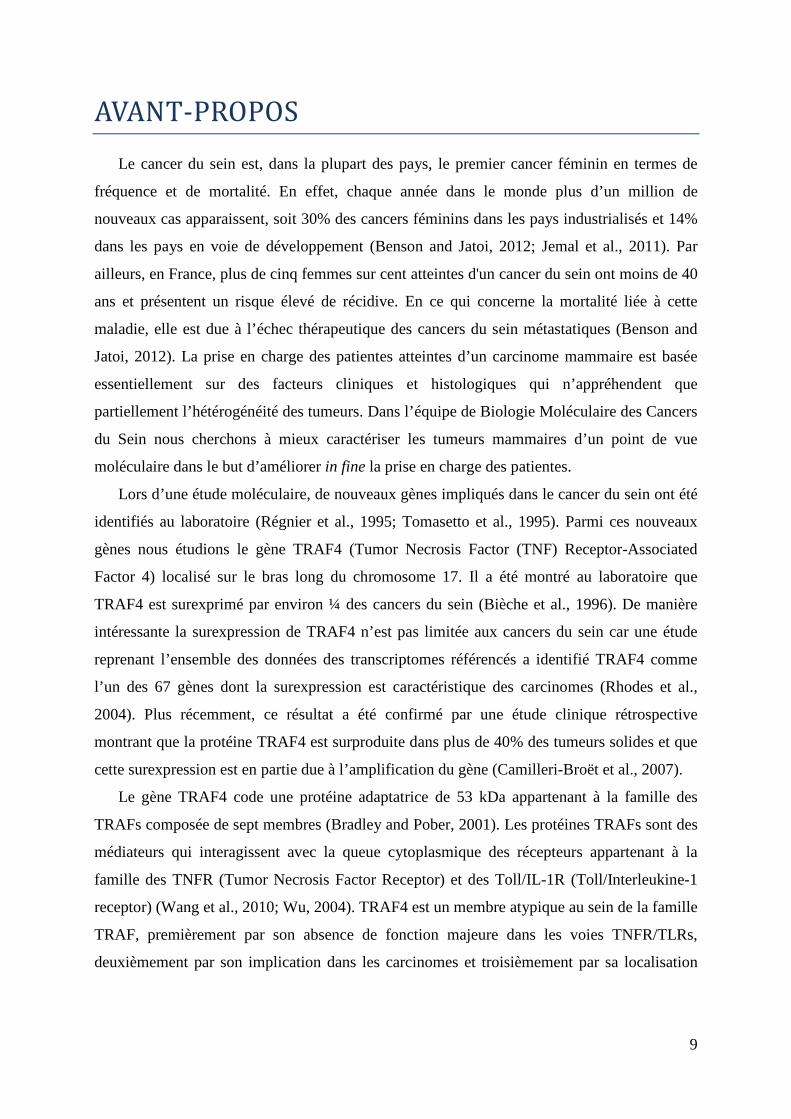
Le cancer du sein est, dans la plupart des pays, le premier cancer féminin en termes de
fréquence et de mortalité. En effet, chaque année dans le monde plus d’un million de
nouveaux cas apparaissent, soit 30% des cancers féminins dans les pays industrialisés et 14%
dans les pays en voie de développement (Benson and Jatoi, 2012; Jemal et al., 2011). Par
ailleurs, en France, plus de cinq femmes sur cent atteintes d'un cancer du sein ont moins de 40
ans et présentent un risque élevé de récidive. En ce qui concerne la mortalité liée à cette
maladie, elle est due à l’échec thérapeutique des cancers du sein métastatiques (Benson and
Jatoi, 2012). La prise en charge des patientes atteintes d’un carcinome mammaire est basée
essentiellement sur des facteurs cliniques et histologiques qui n’appréhendent que
partiellement l’hétérogénéité des tumeurs. Dans l’équipe de Biologie Moléculaire des Cancers
du Sein nous cherchons à mieux caractériser les tumeurs mammaires d’un point de vue
moléculaire dans le but d’améliorer in fine la prise en charge des patientes.
Lors d’une étude moléculaire, de nouveaux gènes impliqués dans le cancer du sein ont été
identifiés au laboratoire (Régnier et al., 1995; Tomasetto et al., 1995). Parmi ces nouveaux
gènes nous étudions le gène TRAF4 (Tumor Necrosis Factor (TNF) Receptor-Associated
Factor 4) localisé sur le bras long du chromosome 17. Il a été montré au laboratoire que
TRAF4 est surexprimé par environ ¼ des cancers du sein (Bièche et al., 1996). De manière
intéressante la surexpression de TRAF4 n’est pas limitée aux cancers du sein car une étude
reprenant l’ensemble des données des transcriptomes référencés a identifié TRAF4 comme
l’un des 67 gènes dont la surexpression est caractéristique des carcinomes (Rhodes et al.,
2004). Plus récemment, ce résultat a été confirmé par une étude clinique rétrospective
montrant que la protéine TRAF4 est surproduite dans plus de 40% des tumeurs solides et que
cette surexpression est en partie due à l’amplification du gène (Camilleri-Broët et al., 2007).
Le gène TRAF4 code une protéine adaptatrice de 53 kDa appartenant à la famille des
TRAFs composée de sept membres (Bradley and Pober, 2001). Les protéines TRAFs sont des
médiateurs qui interagissent avec la queue cytoplasmique des récepteurs appartenant à la
famille des TNFR (Tumor Necrosis Factor Receptor) et des Toll/IL-1R (Toll/Interleukine-1
receptor) (Wang et al., 2010; Wu, 2004). TRAF4 est un membre atypique au sein de la famille
TRAF, premièrement par son absence de fonction majeure dans les voies TNFR/TLRs,
deuxièmement par son implication dans les carcinomes et troisièmement par sa localisation
10
subcellulaire aux jonctions serrées (JS) au sein des cellules épithéliales polarisées (Kédinger
et al., 2008; Rousseau et al., 2011).
La localisation de TRAF4 aux JS a été fondatrice dans l’orientation de mon projet de
recherche. En effet, le rôle précis de TRAF4 dans les cancers n’est pas connu, en revanche, de
nombreuses études ont montré l’implication des jonctions serrées dans les cancers. Les JS, en
plus de jouer un rôle vital dans la cohésion et le maintien de l’intégrité de l’épithélium,
contrôlent la prolifération, la différenciation et la polarité cellulaire, trois processus altérés
dans les cancers (Forster, 2008; Furuse, 2010). Nous avons émis l’hypothèse selon laquelle, la
dérégulation de l’expression de TRAF4 dans les cancers pouvait altérer le fonctionnement
normal des jonctions serrées et contribuer à la carcinogénèse. Cette hypothèse a été le point de
départ de différents axes de recherche qui ont été développés pendant ma thèse. Dans ce
manuscrit, j’ai choisi de présenter en introduction une synthèse de la littérature couvrant la
superfamille des protéines TRAFs, TRAF4 et la polarité apico-basale. Dans la partie résultats,
le premier chapitre concerne le rôle de TRAF4 dans les jonctions serrées des cellules
épithéliales mammaires. Il inclut un article actuellement en révision et des données
additionnelles sur ce thème. Ce chapitre représente la part la plus importante de mon travail
de thèse. Dans le second chapitre, j’ai exploré le rôle de TRAF4 dans la polarité planaire.
Dans ce chapitre, après avoir présenté brièvement ce thème, j’ai détaillé les données obtenues
in vivo en exploitant le modèle de souris déficientes pour TRAF4 et in vitro. Enfin j’ai
terminé mon manuscrit par une partie discussion et perspectives dans laquelle j’ai intégré les
résultats d’un article en collaboration qui a été accepté très récemment.

11
RAPPELS
BIBLIOGRAPHIQUES
HyTRAF1
DTRAF1
DrT
RA
F4a
XlT
RA
F4b
XlT
RA
F4a
XtT
RA
F4
HsT
RA
F4
DrTR
AF4b
CiTRAF4
CeTRAFDTRAF2
DrTRAF6a
HsTRAF6
XlTRAF6a
XlTRAF6b
XtTRAF6
CiTRAF6
DrT
RA
F7
HsT
RAF7
XlTRAF7
XtTRAF7
DrT
RAF1
HsT
RA
F1
DrT
RA
F2
aH
sT
RA
F2
XlT
RA
F2
DrT
RA
F2b
DTR
AF3
DrTRAF3
HsTRAF3
XtTRAF3
DrT
RAF5
HsTRAF5
Hydractinia echinata
Ciona intestinalis
Caenorhabditis elegans
Drosophila melanogaster
Danio rerio
Xenopus laevis
Xenopus tropicalis
Homo sapiens
:
:
:
:
:
:
:
:
Hy
Ci
Ce
D
Dr
Xl
Xt
Hs
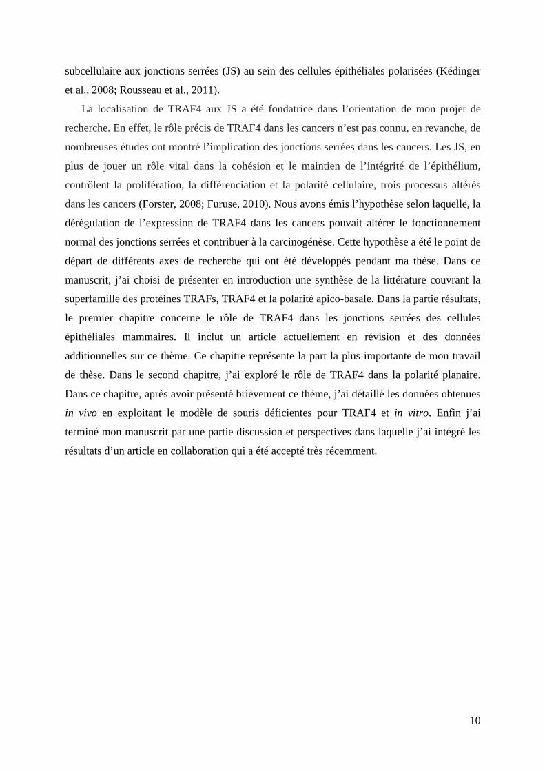
Figure 1 : Analyse phylogénique des protéines TRAFsLes séquences primaires des protéines TRAFs de différentes espèces ont été alignées en utilisant le programme Clustal W. Les données de cet alignement ont servi à la réalisation d’un arbre phylogénétique par le programme iTOL (interactive Tree Of Life). Cette analyse phylogénétique montre que les plus anciens membres TRAFs sont des orthologues de TRAF4 et TRAF6.
Verté
brés
Chordés
Antro
pode
Antropode
Antropode
Vertébrés
Vertébrés
Vertéb
rés
Vertébrés
Chordés
Cnidaire
Nématode
12
CHAPITRE1:LAFAMILLETRAFDANSL’EVOLUTION
1.1. LA PHYLOGENIE DES TRAFS
Les protéines TRAFs ont été initialement identifiées pour leurs capacités à lier les
récepteurs de la famille des TNFR (Tumor Necrosis Factor Receptor) (Chung et al., 2002;
Rothe et al., 1994). Cette famille est caractérisée par la présence d’un domaine TRAF en
carboxyterminal fortement conservé au cours de l’évolution (Zapata et al., 2007). En effet, des
protéines TRAFs sont déjà retrouvées chez les eucaryotes inférieurs tels que les protozoaires
(Dictyostelium discoideum). Elle est néanmoins absente chez les procaryotes et les
archaebactéries suggérant que le domaine TRAF est apparu de manière précoce au cours de
l’évolution des eucaryotes (Zapata et al., 2007).
Les membres les plus anciens et potentiellement fondateurs de cette famille semblent être
TRAF4 et TRAF6. Pour illustrer cette donnée, j’ai réalisé une analyse phylogénétique basée
sur la similarité des protéines TRAFs complètes. Cette analyse montre que les protéines
TRAF4 et TRAF6 sont retrouvés dans plusieurs embranchements du règne animal (cnidaires,
nématodes, arthropodes, échinodermes et chordés pour TRAF4, arthropodes et chordés pour
TRAF6) (Figure 1). En effet, un orthologue de TRAF4 est retrouvé chez Hydractinia echinata
(HyTRAF1), une Hydre appartenant à l’embranchement des cnidaires (coraux et méduses).
D’autres orthologues de TRAF4 sont retrouvés chez le nématode (CeTRAF), la drosophile
(DTRAF1), les tuniciers (CiTRAF4) et les vertébrés (XlTRAF4a, XlTRAF4b, XtTRAF4 et
hsTRAF4) (Figure 1). Les orthologues de TRAF6 sont, quant à eux, retrouvés chez la
drosophile (DTRAF2), les tuniciers (CiTRAF6) et les vertébrés (XlTRAF6a, XlTRAF6b,
XtTRAF6 et HsTRAF6) (Figure 1). Les autres membres de la famille TRAF ne sont présents
que chez les vertébrés à l’exception de TRAF3 qui a un orthologue chez la mouche
(DTRAF3) (Figure 1). Ces données sont en accord avec une analyse phylogénétique
comparant uniquement les domaines TRAFs des protéines de cette famille (Zapata et al.,
2007).
Dans leur ensemble, ces données suggèrent que TRAF4 et TRAF6 seraient les précurseurs
de la famille TRAF et que les autres membres seraient apparus plus tardivement.
1.2. LA FONCTION DES PROTEINES TRAFS DANS L’EVOLUTION
L’organisme le plus simple possédant un gène TRAF pour lequel des informations
fonctionnelles ont été rapportées est l’Hydre (un animal pluricellulaire marin) (Figure 1). Le
12 heptades
12 heptadesC3HC4
12 heptadesC3HC4
11 heptadesC3HC3D
3 heptadesC3HC3D
9 heptadesC3HC3D
3 heptadesC3HC3D
TRAF1
TRAF2
TRAF3
TRAF4
TRAF5
TRAF6
TRAF7
Les protéines TRAFs humaines
9 heptadesC3HC3D
15 heptades
3 heptades
DTRAF1
DTRAF2
DTRAF3
Les protéines TRAFs de drosophile
3 heptadesC3HC3D
3 heptadesC3HC3D
1 heptadeC3HC3D
9 heptadesC3HC3D
hyTRAF1
ceTRAF
ciTRAF4
ciTRAF6
Les membres TRAFs ancestraux
Motifs WD40 répétés
Domaine TRAF-C
Domaine TRAF-N
Domaine RING
Motif C2HC
Motif C2H2
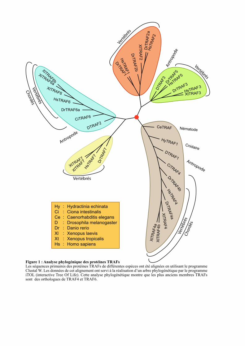
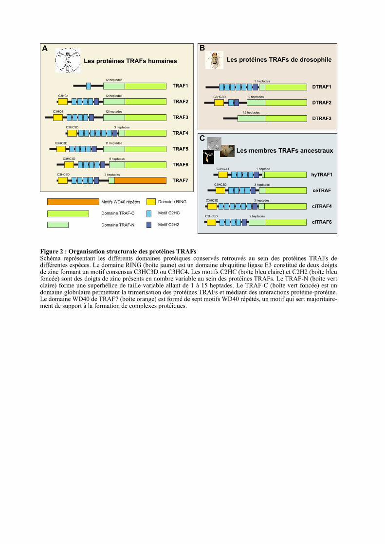
Figure 2 : Organisation structurale des protéines TRAFsSchéma représentant les différents domaines protéiques conservés retrouvés au sein des protéines TRAFs de différentes espèces. Le domaine RING (boîte jaune) est un domaine ubiquitine ligase E3 constitué de deux doigts de zinc formant un motif consensus C3HC3D ou C3HC4. Les motifs C2HC (boîte bleu claire) et C2H2 (boîte bleu foncée) sont des doigts de zinc présents en nombre variable au sein des protéines TRAFs. Le TRAF-N (boîte vert claire) forme une superhélice de taille variable allant de 1 à 15 heptades. Le TRAF-C (boîte vert foncée) est un domaine globulaire permettant la trimerisation des protéines TRAFs et médiant des interactions protéine-protéine. Le domaine WD40 de TRAF7 (boîte orange) est formé de sept motifs WD40 répétés, un motif qui sert majoritaire-ment de support à la formation de complexes protéiques.
A B
C
13
gène HyTRAF1 est présent sous deux isoformes : long (HyTRAF1) qui code une protéine
contenant un domaine RING, cinq doigts de zinc et un domaine TRAF carboxyterminal ; et
court (HyTRAF1a) qui a une délétion de 35 acides aminés conduisant à la perte d’un doigt de
zinc. HyTRAF1 est exprimé à tous les stades de développement excepté l’embryogenèse
précoce tandis que HyTRAF1a est exprimé exclusivement lors des étapes de morphogenèse
précoce (Mali and Frank, 2004). La morphogenèse précoce de cet animal est caractérisée par
une intense mort cellulaire. Les protéines TRAFs ayant des propriétés pro-apoptotiques, il a
été proposé que HyTRAF1a régule la morphogenèse en agissant comme un médiateur de
l’apoptose. D’autres études réalisées chez la mouche confirment l’implication des TRAFs
dans le développement et l’apoptose. En effet, la délétion de l’orthologue de TRAF4 chez la
drosophile, DTRAF1, est létale au stade embryonnaire et les mutants ne peuvent pas atteindre
le stade pupal. De surcroit, les mutants hétérozygotes ont un défaut de fermeture du thorax
(Cha et al., 2003). DTRAF1 est un régulateur de la voie JNK (c-Jun N-terminal Kinase) et
l’expression ectopique de DTRAF1 dans le disque imaginal de l’œil engendre une sur-
activation de la voie pro-apoptotique JNK conduisant à une altération de la morphogenèse de
l’œil (Cha et al., 2003; Liu et al., 1999). Des études réalisées chez le poisson zèbre, le xénope
et la souris ont également validé l’implication de TRAF4 au cours du développement (Kalkan
et al., 2009; Kedinger et al., 2005; Régnier et al., 2002).
TRAF6 apparait chez les insectes avec la protéine DTRAF2 (Figure 1). Elle est impliqué
dans l’immunité innée médiée par le facteur de transcription NF-κB (Nuclear Factor-Kappa
B) (Cha et al., 2003; Shen et al., 2001). En effet, il a été montré que DTRAF2 est requis pour
l’expression de la diptéricine et de la drosomycine, deux peptides antimicrobiens secrétés
suite à une infection fongique ou bactérienne chez la drosophile. L’implication de TRAF6
dans l’immunité et la réponse inflammatoire a également été démontrée chez les mammifères
(Inoue et al., 2007; Landström, 2010).
Ces données suggèrent une conservation fonctionnelle des protéines TRAFs dans
l’évolution, avec deux versants ; l’un lié aux processus développementaux et l’autre lié à la
réponse immunitaire et inflammatoire.
1.3. ORGANISATION STRUCTURALE DES TRAFS DANS L’EVOLUTIO N
Les protéines TRAFs partageant une organisation structurale très proche et ce, depuis
HyTRAF1. En effet, toutes ces protéines possèdent un domaine TRAF en carboxyterminal à
l’exception de TRAF7 où ce domaine est remplacé par sept motifs WD40 répétés (Figure 2).
La majorité des protéines TRAFs possède deux domaines additionnels : un domaine RING
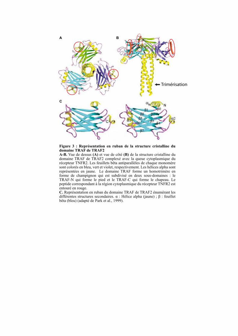
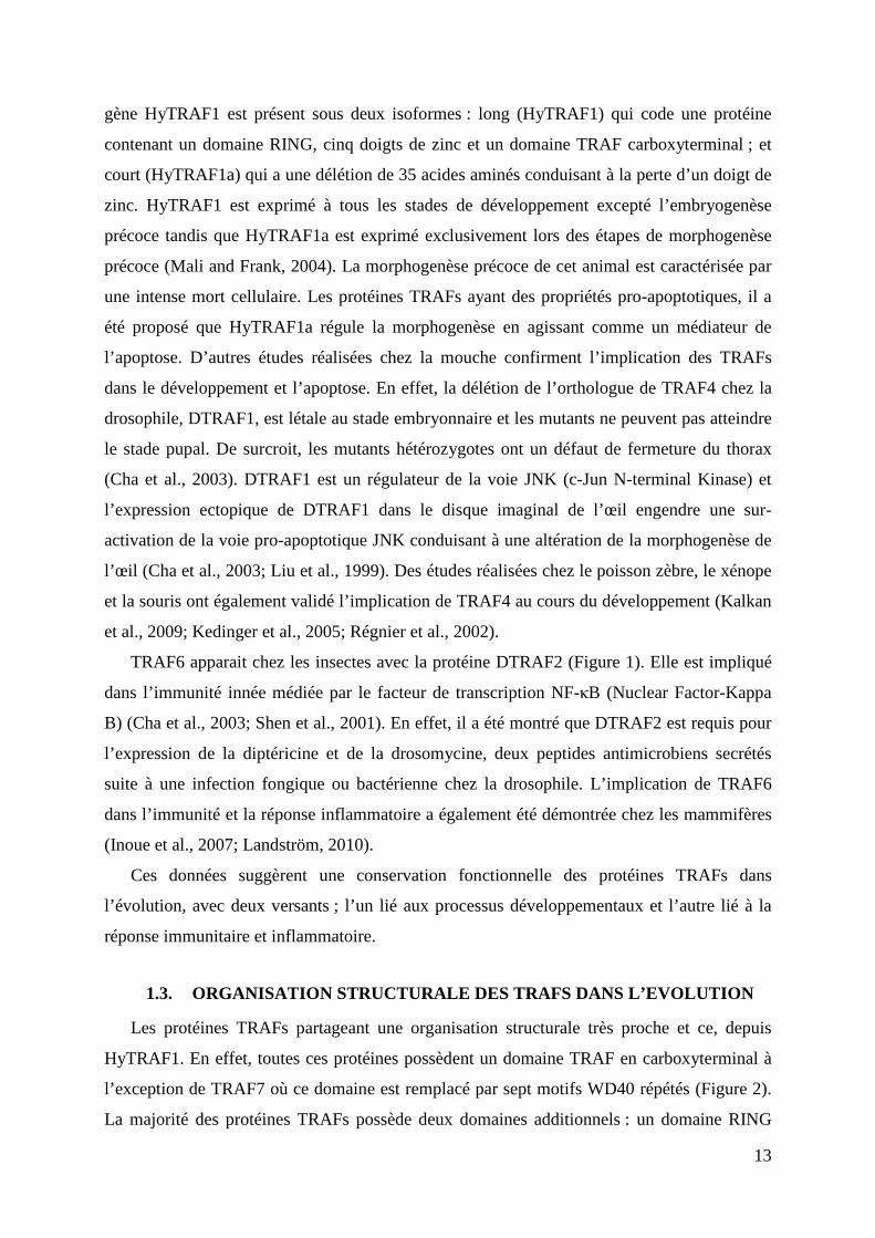
Figure 3 : Représentation en ruban de la structure cristalline du domaine TRAF de TRAF2 A-B. Vue de dessus (A) et vue de côté (B) de la structure cristalline du domaine TRAF de TRAF2 complexé avec la queue cytoplasmique du récepteur TNFR2. Les feuillets bêta antiparallèles de chaque monomère sont colorés en bleu, vert et violet, respectivement. Les hélices alpha sont représentées en jaune. Le domaine TRAF forme un homotrimère en forme de champignon qui est subdivisé en deux sous-domaines : le TRAF-N qui forme le pied et le TRAF-C qui forme le chapeau. Le peptide correspondant à la région cytoplasmique du récepteur TNFR2 est entouré en rouge. C. Représentation en ruban du domaine TRAF de TRAF2 énumérant les différentes structures secondaires. α : Hélice alpha (jaune) ; β : feuillet bêta (bleu) (adapté de Park et al., 1999).
A B
C
Trimérisation
14
(Really Interesting New Gene) en N-terminal formé de deux doigts de zinc et dans la région
centrale la présence de 1, 2, 5 ou 7 doigts de zinc (Figure 2) (Inoue et al., 2000; Regnier et al.,
1995). Pour les homologues identifiés dans les organismes les plus distants des mammifères
du point de vue de l’évolution (Figure 2 C), on retrouve le domaine TRAF, le domaine RING
et cinq à sept motifs doigt de zinc.
1.3.1. Le domaine TRAF
Une étude structurale de la protéine TRAF2 a montré que le domaine TRAF se trimèrise
pour former un complexe ayant la forme d’un champignon avec une région TRAF-N formant
le pied et une région TRAF-C forment le chapeau (Figure 3). Le domaine TRAF-N forme une
superhélice de longueur variable permettant de stabiliser le trimère. La longueur du domaine
TRAF-N semble être importante pour la spécificité de formation des hétérotrimères. En effet,
seul des hétérotrimères de TRAF1-TRAF2, TRAF2-TRAF3, TRAF2-TRAF5 et TRAF3-
TRAF5 ont été décrits et ces quatre membres sont les seuls à posséder des superhélices de
taille supérieure à 10 heptades (Figure 2) (Arch et al., 1998; He et al., 2004; Pullen et al.,
1998; Takeuchi et al., 1996). Le domaine TRAF-N de TRAF4 est très court (3 heptades), ce
qui pourrait expliquer son incapacité à former des hétérotrimères avec les autres protéines
TRAFs. Le domaine TRAF-C est, quant à lui, formé de 7 à 8 feuillets bêta antiparallèles, un
repliement qui n’est pas restreint aux protéines TRAFs (Figure 3). En effet, ce domaine est
aussi appelé MATH (meprin and TRAF-C homology) par son homologie de séquence avec
les métalloprotéases matricielles de la famille des méprines (Zapata et al., 2007). Le domaine
TRAF-C, en plus de permettre la trimérisation des TRAFs, sert de site de liaison aux
récepteurs membranaires activés par leur ligand. Une fois recruté à la membrane, le domaine
TRAF sert de plateforme de signalisation en recrutant d’autres partenaires protéiques (Inoue
et al., 2000; Wu, 2004).
1.3.2. Le domaine RING
Le domaine RING est caractérisé par un enchaînement de cystéines et d’histidines
conservées complexant 2 atomes de zinc et formant un motif consensus C3HC4 (Deshaies and
Joazeiro, 2009). Cependant, la dernière cystéine peut-être remplacée par un acide aspartique
formant un RING non-conventionnel C3HC3D (Régnier et al., 1995). Ce motif non-
conventionnel est très représenté au sein des protéines TRAFs. En effet, parmi les 33 TRAFs
analysés en figure 1 : 22 ont un motif C3HC3D, 8 ont un motif C3HC4 et 3 en sont
dépourvus. De manière intéressante, la majorité des TRAFs ancestraux a un domaine non-

Substrat
NH2
Ub
Substrat
UbUb
Ub
SubstratUb
Ub
Substrat
Ub Ub Ub
Poly-ubiquitinylation K48
Poly-ubiquitinylation K63
Mono-ubiquitinylation
Localisation subcellulaire
Signalisation
Dégradation lysosomale
Dégradation via le
protéasome 26S
Localisation subcellulaire
Signalisation
HE
CT
E3
RIN
G E
3
A
B
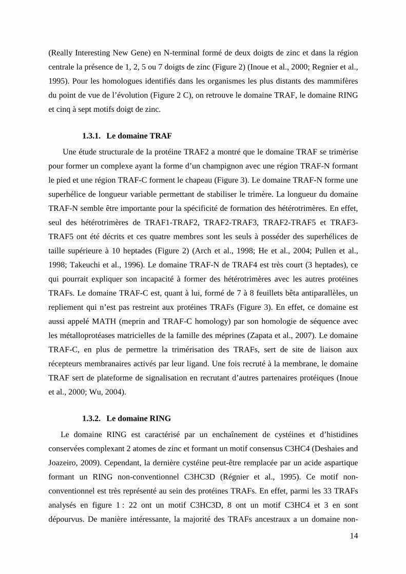
Figure 4 : Le mécanisme d’ubiquitinylation des protéinesA. Après liaison d’une molécule d’ubiquitine activée sur l’enzyme E1 (a), elle est transférée sur une enzyme E2 par transthiolation (b). Le complexe E2-Ubiquitine interagit ensuite avec une enzyme ubiquitine ligase E3 qui transfert l’ubiquitine sur le substrat (c). Le complexe E2-E3 peut ensuite ajouter des molécules d’ubiquitine additionnelles, on parle alors de poly-ubiquitinylation (d). La poly-ubiquitinylation de type K48 est connue pour induire la dégradation du substrat via le protéa-some 26S (adapté de Deshaies and Joazeiro, 2009). B. Les ubiquitines ligases E3 de type HECT et RING ont un mode d’action différent. Les domaines HECTs ont une cystéine conservée qui sert de site de liaison à l’ubiquitine provenant de l’enzyme E2. L’ubiquitine liée covalemment au domaine HECT est ensuite transférée sur le substrat. A l’inverse, le domaine RING transfère directement l’ubiquitine de l’enzyme E2 sur une lysine du substrat (adapté de Deshaies and Joazeiro, 2009). C. Représentation des principaux types d’ubiquitinylation. La mono-ubiquitinylation des protéines peut aussi bien avoir un rôle activateur en modifiant la localisation subcellulaire ou la structure du substrat, qu’un rôle inhibiteur en entraînant la dégradation via la voie lysosomale. Tandis que la poly-ubiquitinylation de type K48 est connue pour induire la dégradation du substrat via le protéa-some 26S, la poly-ubiquitinylation de type K63 a un rôle activateur sur le substrat.
d e
C
15
conventionnel tandis que les motifs C3HC4 sont apparus plus tardivement avec les protéines
TRAF2 et 3 (Figure 2).
Le domaine RING est un domaine ubiquitine ligase E3 impliqué dans l’ubiquitinylation
des protéines (Chasapis and Spyroulias, 2009). L’ubiquitinylation est une modification post-
traductionnelle qui consiste en la fixation covalente d’une ou de plusieurs molécules
d’ubiquitine sur la lysine d’un substrat. L’ubiquitine est une petite protéine de 76 acides
aminés qui est fortement conservée au cours de l’évolution. En effet, de l’homme à la levure
l’ubiquitine a 96 % d'identité. Chez les mammifères, l’ubiquitine est codée par quatre gènes
distincts (Kimura and Tanaka, 2010). Les gènes UBA52 et RPS27A codent pour un monomère
d’ubiquitine fusionné aux protéines ribosomales L40 et S27a, respectivement. Après
traduction, ces protéines de fusion sont séparées par un clivage enzymatique libérant les
protéines ribosomales du monomère d’ubiquitine. Les gènes UBB et UBC codent, quant à
eux, des précurseurs protéiques correspondant à une répétition en tandem de 4 et de 9
molécules d’ubiquitines, respectivement. Un clivage post-traductionnel permet ensuite de
produire des monomères d’ubiquitine (Kimura and Tanaka, 2010).
L’ubiquitinylation est un processus multi-étape qui fait intervenir trois complexes
enzymatiques nommés E1 (ubiquitin-activating enzyme), E2 (ubiquitin-conjugating enzyme)
et E3 (ubiquitin-ligating enzyme). Après que l’ubiquitine soit activée de façon ATP-
dépendante par l’enzyme E1, elle est transférée dans le site actif de l’enzyme E2 par
transthiolation (Figure 4A). Les enzymes ligases de type E3 confèrent la spécificité
d’ubiquitination en reconnaissant spécifiquement le substrat puis en transférant l’ubiquitine de
l’enzyme E2 sur ce dernier (Figure 4A). Il existe deux classe d’enzymes E3, la première étant
caractérisée par la présence d’un domaine RING et la deuxième par un domaine HECT
(Homologous to E6-AP Carboxyl Terminus). Ces deux classes diffèrent par leur mécanisme
de transfert de l’ubiquitine. En effet, tandis que le domaine RING permet un transfert direct
de l’ubiquitine de l’enzyme E2 sur le substrat, le domaine HECT lie l’ubiquitine de façon
covalente avant de la transférer sur le substrat (Figure 4B). Indépendamment du type
d’enzyme ligase E3, le complexe E2-E3 peut transférer une ou plusieurs molécules
d’ubiquitine sur le substrat, on parle alors respectivement de mono- ou poly-ubiquitinylation,
(Figure 4A) (Deshaies and Joazeiro, 2009). En ce qui concerne la poly-ubiquitinylation, il y a
sept lysines accessibles à la surface de l'ubiquitine et potentiellement disponibles pour la
formation de chaînes de poly-ubiquitines (Figure 4C). Ces différents types d’ubiquitinylation
auront différents effets sur le substrat comme induire sa dégradation via le protéasome 26S ou
moduler sa fonction, sa structure et/ou sa localisation subcellulaire (Figure 4C). Par exemple,
16
l’assemblage d’une chaîne d’au moins quatre ubiquitines liées par leur lysine 48 (K48) sur
une protéine est une marque conduisant à sa dégradation par le protéasome 26S. A l’inverse,
la polyubiquitinylation avec une chaîne d’ubiquitines liées par leur lysine 63 (K63) ne conduit
généralement pas à la dégradation du substrat mais modifie sa fonction et/ou sa localisation
subcellulaire (Figure 4C) (Deshaies and Joazeiro, 2009; Iwai and Tokunaga, 2009). La
majorité des protéines TRAFs est connue pour réaliser de la polyubiquitinylation de type K63,
ce qui en fait des facteurs clés impliqués dans la transduction des signaux intracellulaires
(Pineda et al., 2007).
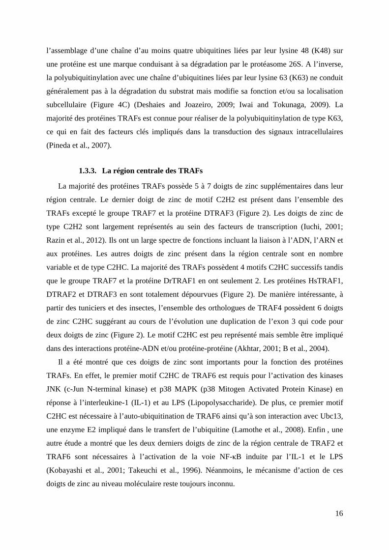
1.3.3. La région centrale des TRAFs
La majorité des protéines TRAFs possède 5 à 7 doigts de zinc supplémentaires dans leur
région centrale. Le dernier doigt de zinc de motif C2H2 est présent dans l’ensemble des
TRAFs excepté le groupe TRAF7 et la protéine DTRAF3 (Figure 2). Les doigts de zinc de
type C2H2 sont largement représentés au sein des facteurs de transcription (Iuchi, 2001;
Razin et al., 2012). Ils ont un large spectre de fonctions incluant la liaison à l’ADN, l’ARN et
aux protéines. Les autres doigts de zinc présent dans la région centrale sont en nombre
variable et de type C2HC. La majorité des TRAFs possèdent 4 motifs C2HC successifs tandis
que le groupe TRAF7 et la protéine DrTRAF1 en ont seulement 2. Les protéines HsTRAF1,
DTRAF2 et DTRAF3 en sont totalement dépourvues (Figure 2). De manière intéressante, à
partir des tuniciers et des insectes, l’ensemble des orthologues de TRAF4 possèdent 6 doigts
de zinc C2HC suggérant au cours de l’évolution une duplication de l’exon 3 qui code pour
deux doigts de zinc (Figure 2). Le motif C2HC est peu représenté mais semble être impliqué
dans des interactions protéine-ADN et/ou protéine-protéine (Akhtar, 2001; B et al., 2004).
Il a été montré que ces doigts de zinc sont importants pour la fonction des protéines
TRAFs. En effet, le premier motif C2HC de TRAF6 est requis pour l’activation des kinases
JNK (c-Jun N-terminal kinase) et p38 MAPK (p38 Mitogen Activated Protein Kinase) en
réponse à l’interleukine-1 (IL-1) et au LPS (Lipopolysaccharide). De plus, ce premier motif
C2HC est nécessaire à l’auto-ubiquitination de TRAF6 ainsi qu’à son interaction avec Ubc13,
une enzyme E2 impliqué dans le transfert de l’ubiquitine (Lamothe et al., 2008). Enfin , une
autre étude a montré que les deux derniers doigts de zinc de la région centrale de TRAF2 et
TRAF6 sont nécessaires à l’activation de la voie NF-κB induite par l’IL-1 et le LPS
(Kobayashi et al., 2001; Takeuchi et al., 1996). Néanmoins, le mécanisme d’action de ces
doigts de zinc au niveau moléculaire reste toujours inconnu.
17
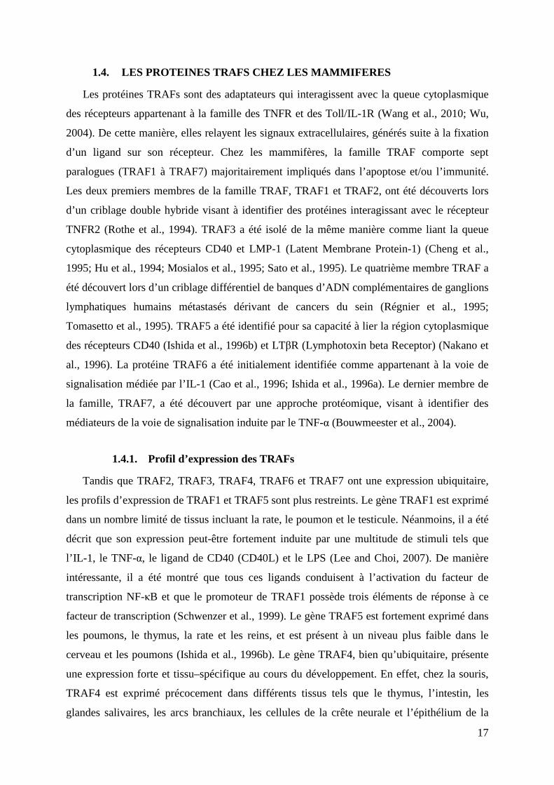
1.4. LES PROTEINES TRAFS CHEZ LES MAMMIFERES
Les protéines TRAFs sont des adaptateurs qui interagissent avec la queue cytoplasmique
des récepteurs appartenant à la famille des TNFR et des Toll/IL-1R (Wang et al., 2010; Wu,
2004). De cette manière, elles relayent les signaux extracellulaires, générés suite à la fixation
d’un ligand sur son récepteur. Chez les mammifères, la famille TRAF comporte sept
paralogues (TRAF1 à TRAF7) majoritairement impliqués dans l’apoptose et/ou l’immunité.
Les deux premiers membres de la famille TRAF, TRAF1 et TRAF2, ont été découverts lors
d’un criblage double hybride visant à identifier des protéines interagissant avec le récepteur
TNFR2 (Rothe et al., 1994). TRAF3 a été isolé de la même manière comme liant la queue
cytoplasmique des récepteurs CD40 et LMP-1 (Latent Membrane Protein-1) (Cheng et al.,
1995; Hu et al., 1994; Mosialos et al., 1995; Sato et al., 1995). Le quatrième membre TRAF a
été découvert lors d’un criblage différentiel de banques d’ADN complémentaires de ganglions
lymphatiques humains métastasés dérivant de cancers du sein (Régnier et al., 1995;
Tomasetto et al., 1995). TRAF5 a été identifié pour sa capacité à lier la région cytoplasmique
des récepteurs CD40 (Ishida et al., 1996b) et LTβR (Lymphotoxin beta Receptor) (Nakano et
al., 1996). La protéine TRAF6 a été initialement identifiée comme appartenant à la voie de
signalisation médiée par l’IL-1 (Cao et al., 1996; Ishida et al., 1996a). Le dernier membre de
la famille, TRAF7, a été découvert par une approche protéomique, visant à identifier des
médiateurs de la voie de signalisation induite par le TNF-α (Bouwmeester et al., 2004).
1.4.1. Profil d’expression des TRAFs
Tandis que TRAF2, TRAF3, TRAF4, TRAF6 et TRAF7 ont une expression ubiquitaire,
les profils d’expression de TRAF1 et TRAF5 sont plus restreints. Le gène TRAF1 est exprimé
dans un nombre limité de tissus incluant la rate, le poumon et le testicule. Néanmoins, il a été
décrit que son expression peut-être fortement induite par une multitude de stimuli tels que
l’IL-1, le TNF-α, le ligand de CD40 (CD40L) et le LPS (Lee and Choi, 2007). De manière
intéressante, il a été montré que tous ces ligands conduisent à l’activation du facteur de
transcription NF-κB et que le promoteur de TRAF1 possède trois éléments de réponse à ce
facteur de transcription (Schwenzer et al., 1999). Le gène TRAF5 est fortement exprimé dans
les poumons, le thymus, la rate et les reins, et est présent à un niveau plus faible dans le
cerveau et les poumons (Ishida et al., 1996b). Le gène TRAF4, bien qu’ubiquitaire, présente
une expression forte et tissu–spécifique au cours du développement. En effet, chez la souris,
TRAF4 est exprimé précocement dans différents tissus tels que le thymus, l’intestin, les
glandes salivaires, les arcs branchiaux, les cellules de la crête neurale et l’épithélium de la
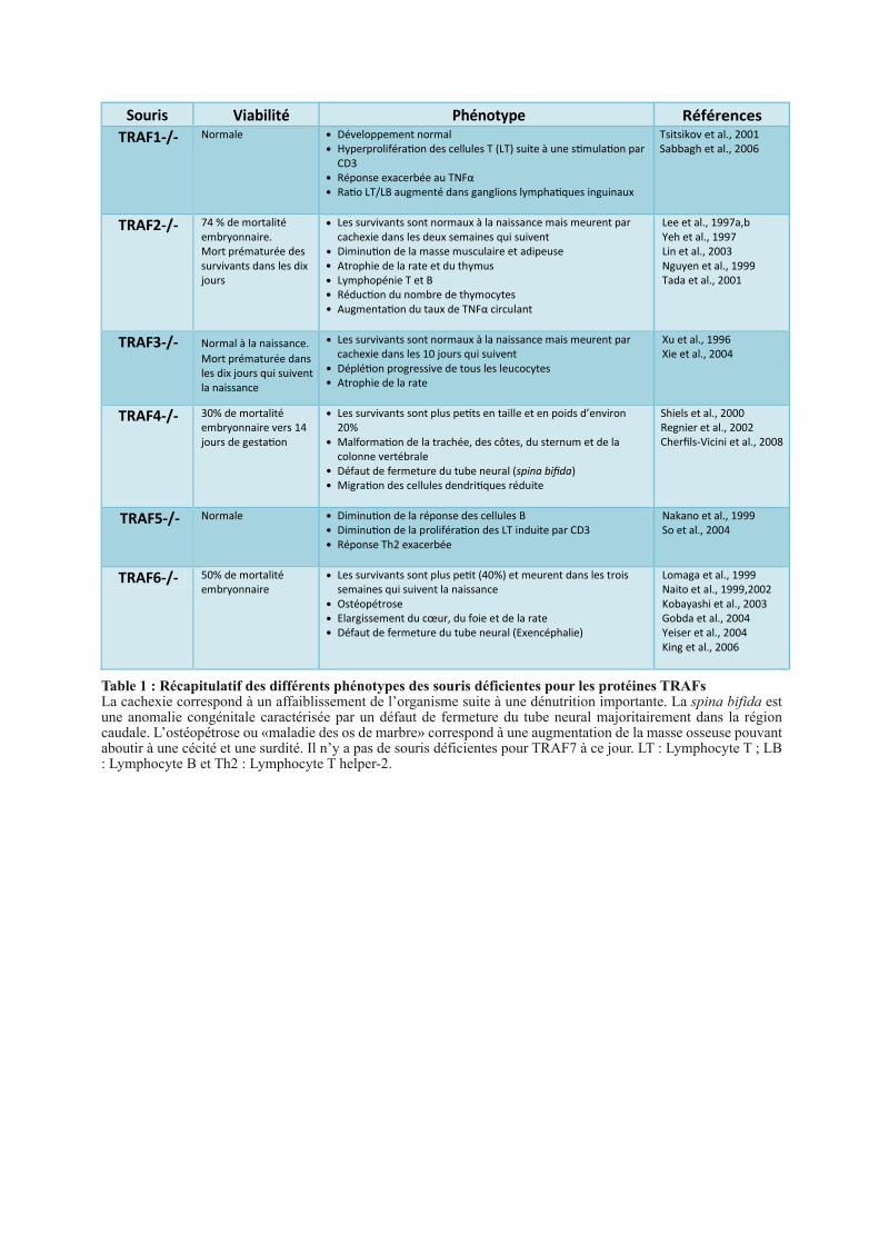
Viabilité
Phénotype
Références
TRAF1-/- Développement normal
Hyperproliféra!on des cellules T (LT) suite à une s!mula!on par
CD3
Réponse exacerbée au TNFα
Ra!o LT/LB augmenté dans ganglions lympha!ques inguinaux
Tsitsikov et al., 2001
Sabbagh et al., 2006
TRAF2-/- Les survivants sont normaux à la naissance mais meurent par
cachexie dans les deux semaines qui suivent
Diminu!on de la masse musculaire et adipeuse
Atrophie de la rate et du thymus
Lymphopénie T et B
Réduc!on du nombre de thymocytes
Augmenta!on du taux de TNFα circulant
Lee et al., 1997a,b
Yeh et al., 1997
Lin et al., 2003
Nguyen et al., 1999
Tada et al., 2001
TRAF3-/-
Les survivants sont normaux à la naissance mais meurent par
cachexie dans les 10 jours qui suivent
Déplé!on progressive de tous les leucocytes
Atrophie de la rate
Xu et al., 1996
Xie et al., 2004
TRAF4-/- Les survivants sont plus pe!ts en taille et en poids d’environ
20%
Malforma!on de la trachée, des côtes, du sternum et de la
colonne vertébrale
Défaut de fermeture du tube neural (spina bifida)
Migra!on des cellules dendri!ques réduite
Shiels et al., 2000
Regnier et al., 2002
TRAF5-/- Diminu!on de la réponse des cellules B
Diminu!on de la proliféra!on des LT induite par CD3
Réponse Th2 exacerbée
Nakano et al., 1999
So et al., 2004
TRAF6-/- Les survivants sont plus pe!t (40%) et meurent dans les trois
semaines qui suivent la naissance
Ostéopétrose
Elargissement du cœur, du foie et de la rate
Défaut de fermeture du tube neural (Exencéphalie)
Lomaga et al., 1999
Naito et al., 1999,2002
Kobayashi et al., 2003
Gobda et al., 2004
Yeiser et al., 2004
King et al., 2006
Table 1 : Récapitulatif des différents phénotypes des souris déficientes pour les protéines TRAFsLa cachexie correspond à un affaiblissement de l’organisme suite à une dénutrition importante. La spina bifida est une anomalie congénitale caractérisée par un défaut de fermeture du tube neural majoritairement dans la région caudale. L’ostéopétrose ou «maladie des os de marbre» correspond à une augmentation de la masse osseuse pouvant aboutir à une cécité et une surdité. Il n’y a pas de souris déficientes pour TRAF7 à ce jour. LT : Lymphocyte T ; LB : Lymphocyte B et Th2 : Lymphocyte T helper-2.
Mort prématurée dans
les dix jours qui suivent
la naissance
Normal à la naissance.
Cherfils-Vicini et al., 2008
30% de mortalité
embryonnaire vers 14
jours de gesta!on
Normale
50% de mortalité
embryonnaire
Normale
74 % de mortalité
embryonnaire.
Mort prématurée des
survivants dans les dix
jours
Souris
18
trachée. Une expression ubiquitaire plus faible est ensuite retrouvée chez l’adulte (Masson et
al., 1998).
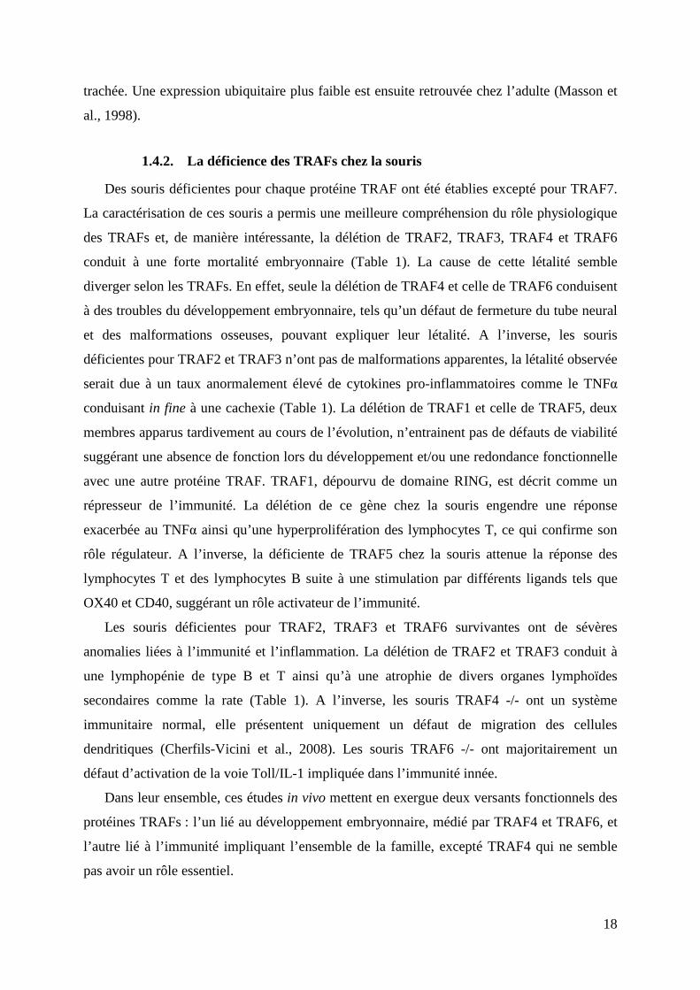
1.4.2. La déficience des TRAFs chez la souris
Des souris déficientes pour chaque protéine TRAF ont été établies excepté pour TRAF7.
La caractérisation de ces souris a permis une meilleure compréhension du rôle physiologique
des TRAFs et, de manière intéressante, la délétion de TRAF2, TRAF3, TRAF4 et TRAF6
conduit à une forte mortalité embryonnaire (Table 1). La cause de cette létalité semble
diverger selon les TRAFs. En effet, seule la délétion de TRAF4 et celle de TRAF6 conduisent
à des troubles du développement embryonnaire, tels qu’un défaut de fermeture du tube neural
et des malformations osseuses, pouvant expliquer leur létalité. A l’inverse, les souris
déficientes pour TRAF2 et TRAF3 n’ont pas de malformations apparentes, la létalité observée
serait due à un taux anormalement élevé de cytokines pro-inflammatoires comme le TNFα
conduisant in fine à une cachexie (Table 1). La délétion de TRAF1 et celle de TRAF5, deux
membres apparus tardivement au cours de l’évolution, n’entrainent pas de défauts de viabilité
suggérant une absence de fonction lors du développement et/ou une redondance fonctionnelle
avec une autre protéine TRAF. TRAF1, dépourvu de domaine RING, est décrit comme un
répresseur de l’immunité. La délétion de ce gène chez la souris engendre une réponse
exacerbée au TNFα ainsi qu’une hyperprolifération des lymphocytes T, ce qui confirme son
rôle régulateur. A l’inverse, la déficiente de TRAF5 chez la souris attenue la réponse des
lymphocytes T et des lymphocytes B suite à une stimulation par différents ligands tels que
OX40 et CD40, suggérant un rôle activateur de l’immunité.
Les souris déficientes pour TRAF2, TRAF3 et TRAF6 survivantes ont de sévères
anomalies liées à l’immunité et l’inflammation. La délétion de TRAF2 et TRAF3 conduit à
une lymphopénie de type B et T ainsi qu’à une atrophie de divers organes lymphoïdes
secondaires comme la rate (Table 1). A l’inverse, les souris TRAF4 -/- ont un système
immunitaire normal, elle présentent uniquement un défaut de migration des cellules
dendritiques (Cherfils-Vicini et al., 2008). Les souris TRAF6 -/- ont majoritairement un
défaut d’activation de la voie Toll/IL-1 impliquée dans l’immunité innée.
Dans leur ensemble, ces études in vivo mettent en exergue deux versants fonctionnels des
protéines TRAFs : l’un lié au développement embryonnaire, médié par TRAF4 et TRAF6, et
l’autre lié à l’immunité impliquant l’ensemble de la famille, excepté TRAF4 qui ne semble
pas avoir un rôle essentiel.
TNFRs à domaine de mortTNFRs dépourvus de domaine
intracellularie
TNFRs à motif TIM
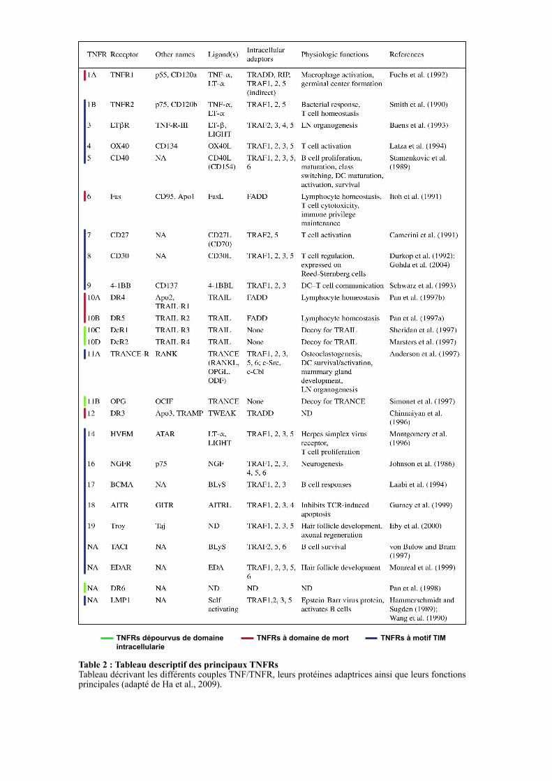
Table 2 : Tableau descriptif des principaux TNFRs Tableau décrivant les différents couples TNF/TNFR, leurs protéines adaptrices ainsi que leurs fonctions principales (adapté de Ha et al., 2009).
19
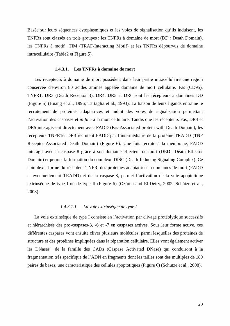
1.4.3. Le rôle des TRAFs dans la signalisation des récepteurs aux TNF
La nécrose tumorale induite par des extraits bactériens a été décrite il y a plus d’un siècle.
En 1962 le docteur O’Malley et ses collègues ont démontré que cet effet était indirect
(O’Malley et al., 1988). Ils montrèrent que le sérum provenant de souris traitées au LPS
(Lipopolysaccharide), un composant de la paroi des bactéries à Gram négatif, pouvait
engendrer une nécrose hémorragique des tumeurs issues de souris non exposées. Ainsi,
O’Malley et ses collègues furent les premiers à proposer le terme de facteur de nécrose
tumorale (TNF). Une décennie plus tard, l’analyse du sérum de souris infectées par différents
pathogènes a identifié le TNF comme étant un facteur sérique circulant sécrété par les
macrophages et les lymphocytes B (Carswell et al., 1975; Williamson et al., 1983). C’est en
1984 que l’ ADNc codant le TNF a été isolé et séquencé pour la première fois (Pennica et al.,
1984).
Parallèlement, la lymphotoxine, une cytokine sécrétée par les cellules lymphocytaires, a
été identifiée pour son activité cytotoxique sur les cellules tumorales (Kolb and Granger,
1968). Le TNF et la lymphotoxine sont très homologues, en conséquence elles furent
renommées respectivement TNFα et TNFβ. Ces deux cytokines sont les fondateurs de la
superfamille du TNF qui compte à ce jour plus de 20 membres (Table 2) (Croft et al., 2013).
Les protéines de la superfamille du TNF sont des ligands transmembranaires qui peuvent être
actifs sous cette forme, ou suite à un clivage protéolytique sous la forme de cytokines
solubles. Très souvent, un ligand peut lier plusieurs récepteurs différents et ainsi activer des
voies de signalisations distinctes ; en corollaire, différents ligands partagent souvent le même
récepteur (Table 2 et Figure 5) (Dempsey et al., 2003; Locksley et al., 2001). A ce jour, une
trentaine de récepteurs TNFRs ont été identifiés. Bien que leur profil d’expression varie
considérablement d’un récepteur à l’autre, ils sont largement exprimés au sein de cellules
immunitaires. Les récepteurs de la superfamille des TNFRs sont majoritairement présents à la
surface des cellules mais peuvent également être libérés dans le compartiment extracellulaire
via un clivage par des métalloprotéases matricielles. La libération de leur domaine
extracellulaire va être importante pour la régulation de l’activation des voies de signalisation
induites par les TNFRs (Aderka, 1996). Il a été décrit que les TNFRs forment des structures
trimériques qui, après avoir lié un trimère de ligands, vont subir des modifications
conformationnelles permettant la transduction du signal (Ha et al., 2009).
D’un point de vue fonctionnel, la superfamille des TNFRs régule un grand nombre de
processus développementaux et joue un rôle clé dans de nombreux mécanismes biologiques
incluant l’apoptose, la différenciation, la survie et la prolifération cellulaire (Grewal, 2009).
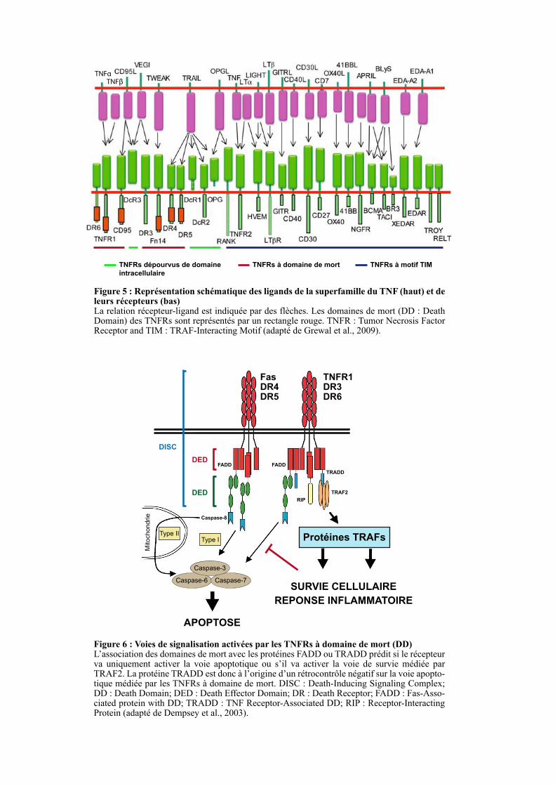
Figure 5 : Représentation schématique des ligands de la superfamille du TNF (haut) et de leurs récepteurs (bas) La relation récepteur-ligand est indiquée par des flèches. Les domaines de mort (DD : Death Domain) des TNFRs sont représentés par un rectangle rouge. TNFR : Tumor Necrosis Factor Receptor and TIM : TRAF-Interacting Motif (adapté de Grewal et al., 2009).
TNFRs à domaine de mortTNFRs dépourvus de domaine
intracellulaire
TNFRs à motif TIM
TNFβTNFα
Figure 6 : Voies de signalisation activées par les TNFRs à domaine de mort (DD) L’association des domaines de mort avec les protéines FADD ou TRADD prédit si le récepteur va uniquement activer la voie apoptotique ou s’il va activer la voie de survie médiée par TRAF2. La protéine TRADD est donc à l’origine d’un rétrocontrôle négatif sur la voie apopto-tique médiée par les TNFRs à domaine de mort. DISC : Death-Inducing Signaling Complex; DD : Death Domain; DED : Death Effector Domain; DR : Death Receptor; FADD : Fas-Asso-ciated protein with DD; TRADD : TNF Receptor-Associated DD; RIP : Receptor-Interacting Protein (adapté de Dempsey et al., 2003).
Fas DR4DR5
TNFR1DR3DR6
DED
APOPTOSE
SURVIE CELLULAIRE
REPONSE INFLAMMATOIRE
Protéines TRAFs
Caspase-6 Caspase-7
Caspase-3
Type IType II
Mitochondrie Caspase-8
FADD FADD
TRADD
RIP
TRAF2
DED
DISC
20
Basée sur leurs séquences cytoplasmiques et les voies de signalisation qu’ils induisent, les
TNFRs sont classés en trois groupes : les TNFRs à domaine de mort (DD : Death Domain),
les TNFRs à motif TIM (TRAF-Interacting Motif) et les TNFRs dépourvus de domaine
intracellulaire (Table2 et Figure 5).
1.4.3.1. Les TNFRs à domaine de mort
Les récepteurs à domaine de mort possèdent dans leur partie intracellulaire une région
conservée d'environ 80 acides aminés appelée domaine de mort cellulaire. Fas (CD95),
TNFR1, DR3 (Death Receptor 3), DR4, DR5 et DR6 sont les récepteurs à domaines DD
(Figure 5) (Huang et al., 1996; Tartaglia et al., 1993). La liaison de leurs ligands entraine le
recrutement de protéines adaptatrices et induit des voies de signalisation permettant
l’activation des caspases et in fine à la mort cellulaire. Tandis que les récepteurs Fas, DR4 et
DR5 interagissent directement avec FADD (Fas-Associated protein with Death Domain), les
récepteurs TNFR1et DR3 recrutent FADD par l’intermédiaire de la protéine TRADD (TNF
Receptor-Associated Death Domain) (Figure 6). Une fois recruté à la membrane, FADD
interagit avec la caspase 8 grâce à son domaine effecteur de mort (DED : Death Effector
Domain) et permet la formation du complexe DISC (Death-Inducing Signaling Complex). Ce
complexe, formé du récepteur TNFR, des protéines adaptatrices à domaines de mort (FADD
et éventuellement TRADD) et de la caspase-8, permet l’activation de la voie apoptotique
extrinsèque de type I ou de type II (Figure 6) (Ozören and El-Deiry, 2002; Schütze et al.,
2008).
1.4.3.1.1. La voie extrinsèque de type I
La voie extrinsèque de type I consiste en l’activation par clivage protéolytique successifs
et hiérarchisés des pro-caspases-3, -6 et -7 en caspases actives. Sous leur forme active, ces
différentes caspases vont ensuite cliver plusieurs molécules, parmi lesquelles des protéines de
structure et des protéines impliquées dans la réparation cellulaire. Elles vont également activer
les DNases de la famille des CADs (Caspase Activated DNase) qui conduiront à la
fragmentation très spécifique de l’ADN en fragments dont les tailles sont des multiples de 180
paires de bases, une caractéristique des cellules apoptotiques (Figure 6) (Schütze et al., 2008).
21
1.4.3.1.2. La voie extrinsèque de type II
La caspase-8, dans certains types cellulaires comme les hépatocytes, peut induire le
clivage de la protéine Bid en facteur pro-apoptotique tBid. La protéine tBid, en ciblant la
membrane externe des mitochondries, induit l’oligomérisation de Bak qui forme des pores
libérant le cytochrome C. Le cytochrome C conduit à la formation de l’apoptosome (7
protéines Apoptotic Protease Activating Factor-1 (Apaf-1 ) et 2 caspases-9) qui, en activant la
caspase-3, va induire le même cycle apoptotique que celui de la voie extrinsèque de type I
(Figure 6) (Schütze et al., 2008).
1.4.3.1.3. TRADD et le recrutement de TRAF2
TRADD, une fois recruté par les récepteurs TNFR1, DR3 et DR6, interagit avec TRAF2
et RIP (Receptor-Interacting Protein) afin d’activer les voies de signalisation NF-κB et JNK
qui vont protéger la cellule de l’apoptose et initier une réponse inflammatoire (Figure 6).
Ainsi, après fixation du ligand sur son récepteur, un complexe se forme entre les TNFRs,
TRADD, RIP et TRAF2 conduisant dans un premier temps à la survie cellulaire et à la
réaction inflammatoire. Par la suite, ce complexe se dissocierait et permettrait le recrutement
du complexe DISC pour induire l’apoptose (Micheau and Tschopp, 2003).
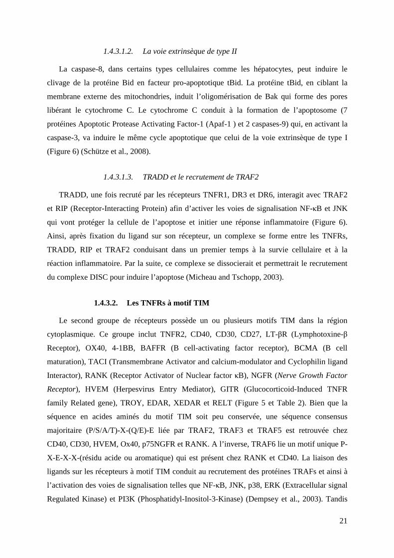
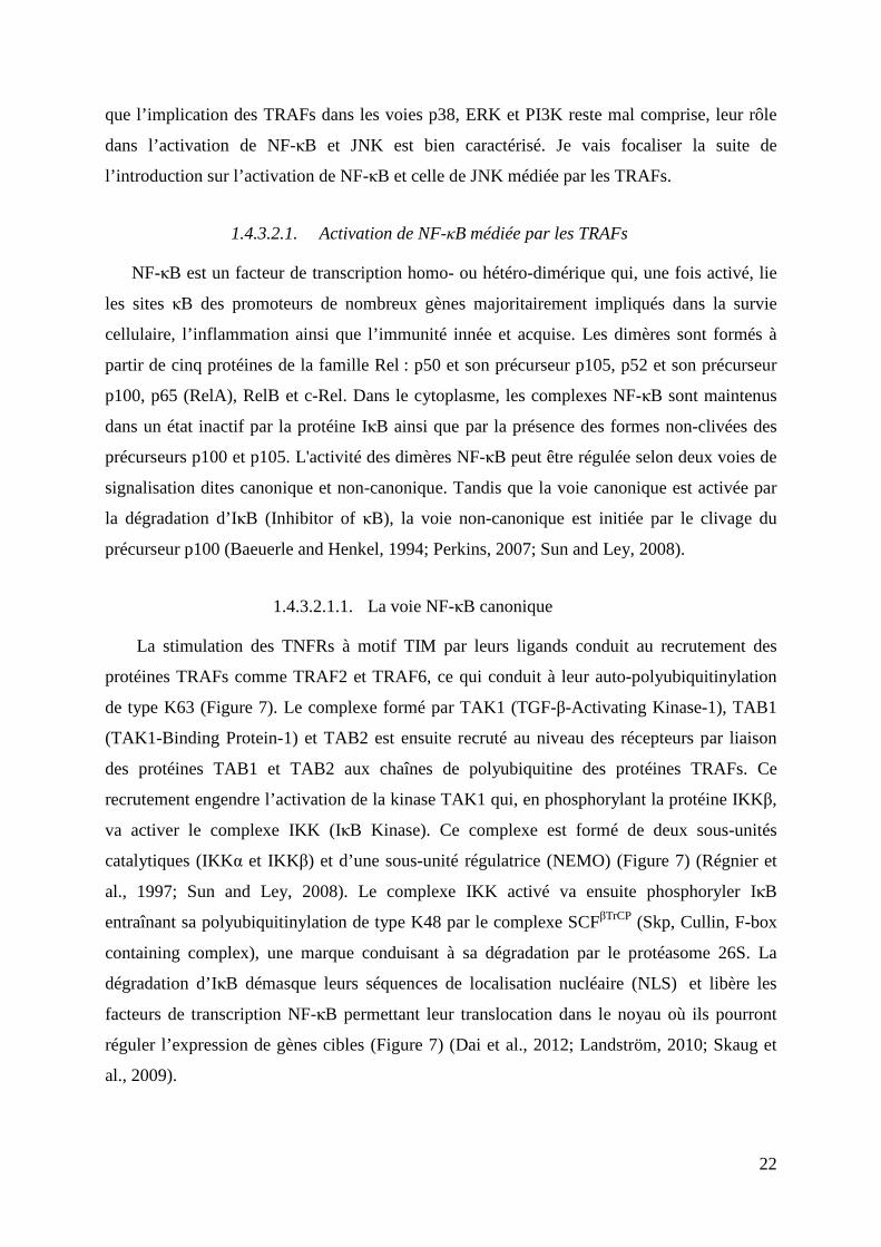
1.4.3.2. Les TNFRs à motif TIM
Le second groupe de récepteurs possède un ou plusieurs motifs TIM dans la région
cytoplasmique. Ce groupe inclut TNFR2, CD40, CD30, CD27, LT-βR (Lymphotoxine-β
Receptor), OX40, 4-1BB, BAFFR (B cell-activating factor receptor), BCMA (B cell
maturation), TACI (Transmembrane Activator and calcium-modulator and Cyclophilin ligand
Interactor), RANK (Receptor Activator of Nuclear factor κB), NGFR (Nerve Growth Factor
Receptor), HVEM (Herpesvirus Entry Mediator), GITR (Glucocorticoid-Induced TNFR
family Related gene), TROY, EDAR, XEDAR et RELT (Figure 5 et Table 2). Bien que la
séquence en acides aminés du motif TIM soit peu conservée, une séquence consensus
majoritaire (P/S/A/T)-X-(Q/E)-E liée par TRAF2, TRAF3 et TRAF5 est retrouvée chez
CD40, CD30, HVEM, Ox40, p75NGFR et RANK. A l’inverse, TRAF6 lie un motif unique P-
X-E-X-X-(résidu acide ou aromatique) qui est présent chez RANK et CD40. La liaison des
ligands sur les récepteurs à motif TIM conduit au recrutement des protéines TRAFs et ainsi à
l’activation des voies de signalisation telles que NF-κB, JNK, p38, ERK (Extracellular signal
Regulated Kinase) et PI3K (Phosphatidyl-Inositol-3-Kinase) (Dempsey et al., 2003). Tandis
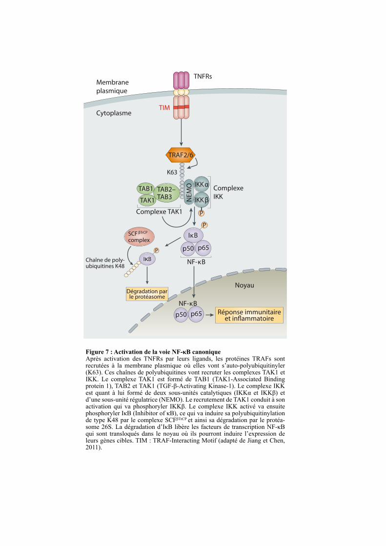
Figure 7 : Activation de la voie NF-κB canoniqueAprès activation des TNFRs par leurs ligands, les protéines TRAFs sont recrutées à la membrane plasmique où elles vont s’auto-polyubiquitinyler (K63). Ces chaînes de polyubiquitines vont recruter les complexes TAK1 et IKK. Le complexe TAK1 est formé de TAB1 (TAK1-Associated Binding protein 1), TAB2 et TAK1 (TGF-β-Activating Kinase-1). Le complexe IKK est quant à lui formé de deux sous-unités catalytiques (IKKα et IKKβ) et d’une sous-unité régulatrice (NEMO). Le recrutement de TAK1 conduit à son activation qui va phosphoryler IKKβ. Le complexe IKK activé va ensuite phosphoryler IκB (Inhibitor of κB), ce qui va induire sa polyubiquitinylation de type K48 par le complexe SCFβTrCP et ainsi sa dégradation par le protéa-some 26S. La dégradation d’IκB libère les facteurs de transcription NF-κB qui sont transloqués dans le noyau où ils pourront induire l’expression de leurs gènes cibles. TIM : TRAF-Interacting Motif (adapté de Jiang et Chen, 2011).
TNFRs
EndosomeMembrane
plasmique
Cytoplasme
TRAM TRIF
TRAF2/6
TRAF6
TBK1RIP1
PELI1
TAK1 complex
TRAF3
Réponse immunitaireet in"ammatoire
Noyau
NF-κB
IRF3Type IIFNs
P
NF-κB
Complexe TAK1
TAB2–TAB3
TAB1
TAK1
TAB2–TAB3
TAB1
TAK1
TRIF TRIF
IκB
p50 p65
p50 p65
IKKαComplexe
IKKIKKβN
EM
O
P
TIM
K63
P
SCF βTrCP
complex
Chaîne de poly-ubiquitines K48
IκB
Dégradation parle protéasome
22
que l’implication des TRAFs dans les voies p38, ERK et PI3K reste mal comprise, leur rôle
dans l’activation de NF-κB et JNK est bien caractérisé. Je vais focaliser la suite de
l’introduction sur l’activation de NF-κB et celle de JNK médiée par les TRAFs.
1.4.3.2.1. Activation de NF-κB médiée par les TRAFs
NF-κB est un facteur de transcription homo- ou hétéro-dimérique qui, une fois activé, lie
les sites κB des promoteurs de nombreux gènes majoritairement impliqués dans la survie
cellulaire, l’inflammation ainsi que l’immunité innée et acquise. Les dimères sont formés à
partir de cinq protéines de la famille Rel : p50 et son précurseur p105, p52 et son précurseur
p100, p65 (RelA), RelB et c-Rel. Dans le cytoplasme, les complexes NF-κB sont maintenus
dans un état inactif par la protéine IκB ainsi que par la présence des formes non-clivées des
précurseurs p100 et p105. L'activité des dimères NF-κB peut être régulée selon deux voies de
signalisation dites canonique et non-canonique. Tandis que la voie canonique est activée par
la dégradation d’IκB (Inhibitor of κB), la voie non-canonique est initiée par le clivage du
précurseur p100 (Baeuerle and Henkel, 1994; Perkins, 2007; Sun and Ley, 2008).
1.4.3.2.1.1. La voie NF-κB canonique
La stimulation des TNFRs à motif TIM par leurs ligands conduit au recrutement des
protéines TRAFs comme TRAF2 et TRAF6, ce qui conduit à leur auto-polyubiquitinylation
de type K63 (Figure 7). Le complexe formé par TAK1 (TGF-β-Activating Kinase-1), TAB1
(TAK1-Binding Protein-1) et TAB2 est ensuite recruté au niveau des récepteurs par liaison
des protéines TAB1 et TAB2 aux chaînes de polyubiquitine des protéines TRAFs. Ce
recrutement engendre l’activation de la kinase TAK1 qui, en phosphorylant la protéine IKKβ,
va activer le complexe IKK (IκB Kinase). Ce complexe est formé de deux sous-unités
catalytiques (IKKα et IKKβ) et d’une sous-unité régulatrice (NEMO) (Figure 7) (Régnier et
al., 1997; Sun and Ley, 2008). Le complexe IKK activé va ensuite phosphoryler IκB
entraînant sa polyubiquitinylation de type K48 par le complexe SCFβTrCP (Skp, Cullin, F-box
containing complex), une marque conduisant à sa dégradation par le protéasome 26S. La
dégradation d’IκB démasque leurs séquences de localisation nucléaire (NLS) et libère les
facteurs de transcription NF-κB permettant leur translocation dans le noyau où ils pourront
réguler l’expression de gènes cibles (Figure 7) (Dai et al., 2012; Landström, 2010; Skaug et
al., 2009).
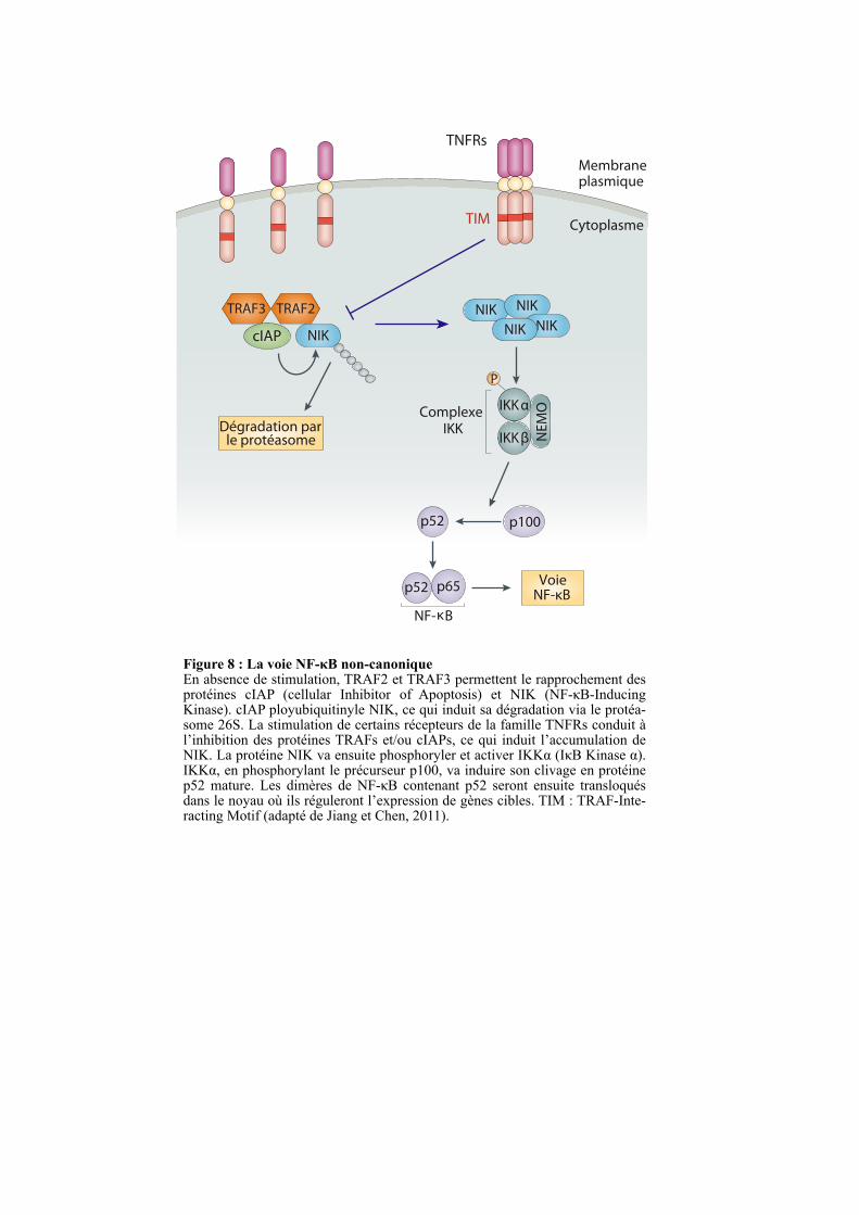
Figure 8 : La voie NF-κB non-canoniqueEn absence de stimulation, TRAF2 et TRAF3 permettent le rapprochement des protéines cIAP (cellular Inhibitor of Apoptosis) et NIK (NF-κB-Inducing Kinase). cIAP ployubiquitinyle NIK, ce qui induit sa dégradation via le protéa-some 26S. La stimulation de certains récepteurs de la famille TNFRs conduit à l’inhibition des protéines TRAFs et/ou cIAPs, ce qui induit l’accumulation de NIK. La protéine NIK va ensuite phosphoryler et activer IKKα (IκB Kinase α). IKKα, en phosphorylant le précurseur p100, va induire son clivage en protéine p52 mature. Les dimères de NF-κB contenant p52 seront ensuite transloqués dans le noyau où ils réguleront l’expression de gènes cibles. TIM : TRAF-Inte-racting Motif (adapté de Jiang et Chen, 2011).
Membraneplasmique
Cytoplasme
IKKαComplexe IKK
IKKβ NE
MO
VoieNF-κB
NF-κB
TRAF2TRAF3
TNFRs
TIM
NIK NIK
NIKNIKNIKcIAP
Dégradation parle protéasome
p100
p52 p65
p52
P
23
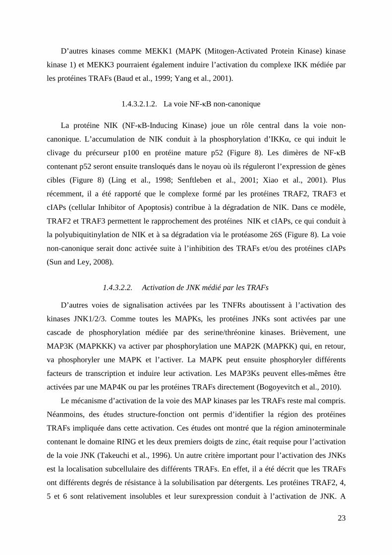
D’autres kinases comme MEKK1 (MAPK (Mitogen-Activated Protein Kinase) kinase
kinase 1) et MEKK3 pourraient également induire l’activation du complexe IKK médiée par
les protéines TRAFs (Baud et al., 1999; Yang et al., 2001).
1.4.3.2.1.2. La voie NF-κB non-canonique
La protéine NIK (NF-κB-Inducing Kinase) joue un rôle central dans la voie non-
canonique. L’accumulation de NIK conduit à la phosphorylation d’IKKα, ce qui induit le
clivage du précurseur p100 en protéine mature p52 (Figure 8). Les dimères de NF-κB
contenant p52 seront ensuite transloqués dans le noyau où ils réguleront l’expression de gènes
cibles (Figure 8) (Ling et al., 1998; Senftleben et al., 2001; Xiao et al., 2001). Plus
récemment, il a été rapporté que le complexe formé par les protéines TRAF2, TRAF3 et
cIAPs (cellular Inhibitor of Apoptosis) contribue à la dégradation de NIK. Dans ce modèle,
TRAF2 et TRAF3 permettent le rapprochement des protéines NIK et cIAPs, ce qui conduit à
la polyubiquitinylation de NIK et à sa dégradation via le protéasome 26S (Figure 8). La voie
non-canonique serait donc activée suite à l’inhibition des TRAFs et/ou des protéines cIAPs
(Sun and Ley, 2008).
1.4.3.2.2. Activation de JNK médié par les TRAFs
D’autres voies de signalisation activées par les TNFRs aboutissent à l’activation des
kinases JNK1/2/3. Comme toutes les MAPKs, les protéines JNKs sont activées par une
cascade de phosphorylation médiée par des serine/thréonine kinases. Brièvement, une
MAP3K (MAPKKK) va activer par phosphorylation une MAP2K (MAPKK) qui, en retour,
va phosphoryler une MAPK et l’activer. La MAPK peut ensuite phosphoryler différents
facteurs de transcription et induire leur activation. Les MAP3Ks peuvent elles-mêmes être
activées par une MAP4K ou par les protéines TRAFs directement (Bogoyevitch et al., 2010).
Le mécanisme d’activation de la voie des MAP kinases par les TRAFs reste mal compris.
Néanmoins, des études structure-fonction ont permis d’identifier la région des protéines
TRAFs impliquée dans cette activation. Ces études ont montré que la région aminoterminale
contenant le domaine RING et les deux premiers doigts de zinc, était requise pour l’activation
de la voie JNK (Takeuchi et al., 1996). Un autre critère important pour l’activation des JNKs
est la localisation subcellulaire des différents TRAFs. En effet, il a été décrit que les TRAFs
ont différents degrés de résistance à la solubilisation par détergents. Les protéines TRAF2, 4,
5 et 6 sont relativement insolubles et leur surexpression conduit à l’activation de JNK. A
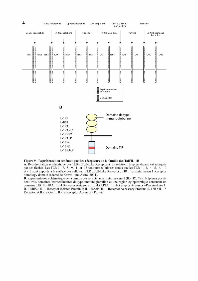
TLR1 TLR2 TLR2 TLR6 TLR3 TLR4 TLR5 TLR8TLR7 TLR9 TLR11
Répétitions riches
en leucine
Domaine TIR
Di-acyl lipopeptide
Tri-acyl lipopeptide
ARN double brins
Lipopolysaccharide
Flagelline
ARN simple brin
ARN simple brin
Ilot d’ADN CpG
non méthylé
Pro"lline
Pro"lline
ARN ribosomique
bactérien
TLR12 TLR13
IL-1R I
IL-IR II
IL-1RA
IL-1RAPL1
IL-1RRP2
IL-1RAcP
IL-18Rα
IL-18Rβ
IL-18RAcP
Domaine de type
immunoglobuline
Domaine TIR
Figure 9 : Représentation schématique des récepteurs de la famille des Toll/IL-1R A. Représentation schématique des TLRs (Toll-Like Receptors). La relation récepteur-ligand est indiquée par des flèches. Les TLR-3, -7, -8, -9, -11 et -13 sont intracellulaires tandis que les TLR-1, -2, -4, -5, -6, -10 et -12 sont exposés à la surface des cellules. TLR : Toll-Like Receptor ; TIR : Toll/Interleukin 1 Receptor homology domain (adapté de Kawai1 and Akira, 2004).B. Représentation schématique de la famille des récepteurs à l’interleukine-1 (IL-1R). Ces récépteurs possé-dent trois domaines extracellulaires de type immunoglobuline et une région cytoplasmique contenant un domaine TIR. IL-1RA : IL-1 Receptor Antagonist; IL-1RAPL1 : IL-1-Receptor Accessory-Protein-Like 1; IL-1RRP2 : IL-1-Receptor-Related Protein 2; IL-1RAcP : IL-1-Receptor Accessory Protein; IL-18R : IL-18 Receptor et IL-18RAcP : IL-18-Receptor Accessory Protein.
A
B
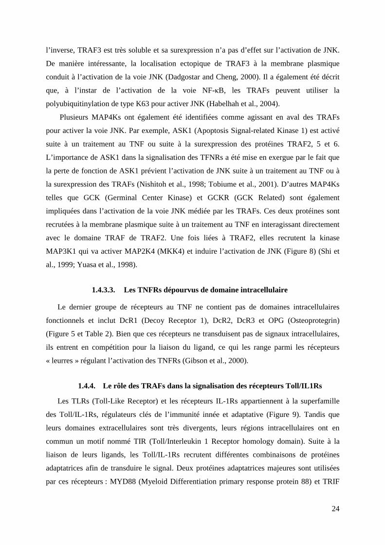
24
l’inverse, TRAF3 est très soluble et sa surexpression n’a pas d’effet sur l’activation de JNK.
De manière intéressante, la localisation ectopique de TRAF3 à la membrane plasmique
conduit à l’activation de la voie JNK (Dadgostar and Cheng, 2000). Il a également été décrit
que, à l’instar de l’activation de la voie NF-κB, les TRAFs peuvent utiliser la
polyubiquitinylation de type K63 pour activer JNK (Habelhah et al., 2004).
Plusieurs MAP4Ks ont également été identifiées comme agissant en aval des TRAFs
pour activer la voie JNK. Par exemple, ASK1 (Apoptosis Signal-related Kinase 1) est activé
suite à un traitement au TNF ou suite à la surexpression des protéines TRAF2, 5 et 6.
L’importance de ASK1 dans la signalisation des TFNRs a été mise en exergue par le fait que
la perte de fonction de ASK1 prévient l’activation de JNK suite à un traitement au TNF ou à
la surexpression des TRAFs (Nishitoh et al., 1998; Tobiume et al., 2001). D’autres MAP4Ks
telles que GCK (Germinal Center Kinase) et GCKR (GCK Related) sont également
impliquées dans l’activation de la voie JNK médiée par les TRAFs. Ces deux protéines sont
recrutées à la membrane plasmique suite à un traitement au TNF en interagissant directement
avec le domaine TRAF de TRAF2. Une fois liées à TRAF2, elles recrutent la kinase
MAP3K1 qui va activer MAP2K4 (MKK4) et induire l’activation de JNK (Figure 8) (Shi et
al., 1999; Yuasa et al., 1998).
1.4.3.3. Les TNFRs dépourvus de domaine intracellulaire
Le dernier groupe de récepteurs au TNF ne contient pas de domaines intracellulaires
fonctionnels et inclut DcR1 (Decoy Receptor 1), DcR2, DcR3 et OPG (Osteoprotegrin)
(Figure 5 et Table 2). Bien que ces récepteurs ne transduisent pas de signaux intracellulaires,
ils entrent en compétition pour la liaison du ligand, ce qui les range parmi les récepteurs
« leurres » régulant l’activation des TNFRs (Gibson et al., 2000).
1.4.4. Le rôle des TRAFs dans la signalisation des récepteurs Toll/IL1Rs
Les TLRs (Toll-Like Receptor) et les récepteurs IL-1Rs appartiennent à la superfamille
des Toll/IL-1Rs, régulateurs clés de l’immunité innée et adaptative (Figure 9). Tandis que
leurs domaines extracellulaires sont très divergents, leurs régions intracellulaires ont en
commun un motif nommé TIR (Toll/Interleukin 1 Receptor homology domain). Suite à la
liaison de leurs ligands, les Toll/IL-1Rs recrutent différentes combinaisons de protéines
adaptatrices afin de transduire le signal. Deux protéines adaptatrices majeures sont utilisées
par ces récepteurs : MYD88 (Myeloid Differentiation primary response protein 88) et TRIF
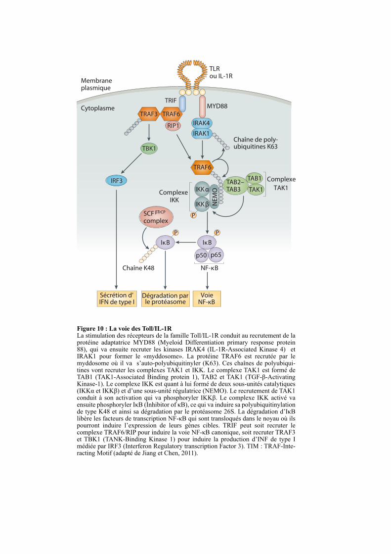
TLRou IL-1R
MYD88
Chaîne de poly-ubiquitines K63
Membraneplasmique
Cytoplasme
IRAK4
IRAK1
TRAF6
TAB2–TAB3
TAB1
TAK1IKKαComplexe IKK
TAK1
IKKβ
P
IκB IκB
SCF βTrCP
complexN
EM
O
VoieNF-κB
Dégradation parle protéasome
P
P
NF-κB
p50 p65
Chaîne K48
Complexe
Figure 10 : La voie des Toll/IL-1RLa stimulation des récepteurs de la famille Toll/IL-1R conduit au recrutement de la protéine adaptatrice MYD88 (Myeloid Differentiation primary response protein 88), qui va ensuite recruter les kinases IRAK4 (IL-1R-Associated Kinase 4) et IRAK1 pour former le «myddosome». La protéine TRAF6 est recrutée par le myddosome où il va s’auto-polyubiquitinyler (K63). Ces chaînes de polyubiqui-tines vont recruter les complexes TAK1 et IKK. Le complexe TAK1 est formé de TAB1 (TAK1-Associated Binding protein 1), TAB2 et TAK1 (TGF-β-Activating Kinase-1). Le complexe IKK est quant à lui formé de deux sous-unités catalytiques (IKKα et IKKβ) et d’une sous-unité régulatrice (NEMO). Le recrutement de TAK1 conduit à son activation qui va phosphoryler IKKβ. Le complexe IKK activé va ensuite phosphoryler IκB (Inhibitor of κB), ce qui va induire sa polyubiquitinylation de type K48 et ainsi sa dégradation par le protéasome 26S. La dégradation d’IκB libère les facteurs de transcription NF-κB qui sont transloqués dans le noyau où ils pourront induire l’expression de leurs gènes cibles. TRIF peut soit recruter le complexe TRAF6/RIP pour induire la voie NF-κB canonique, soit recruter TRAF3 et TBK1 (TANK-Binding Kinase 1) pour induire la production d’INF de type I médiée par IRF3 (Interferon Regulatory transcription Factor 3). TIM : TRAF-Inte-racting Motif (adapté de Jiang et Chen, 2011).
TBK1
TRAF6TRAF3
TRIF
IRF3
Sécrétion d’IFN de type I
RIP1
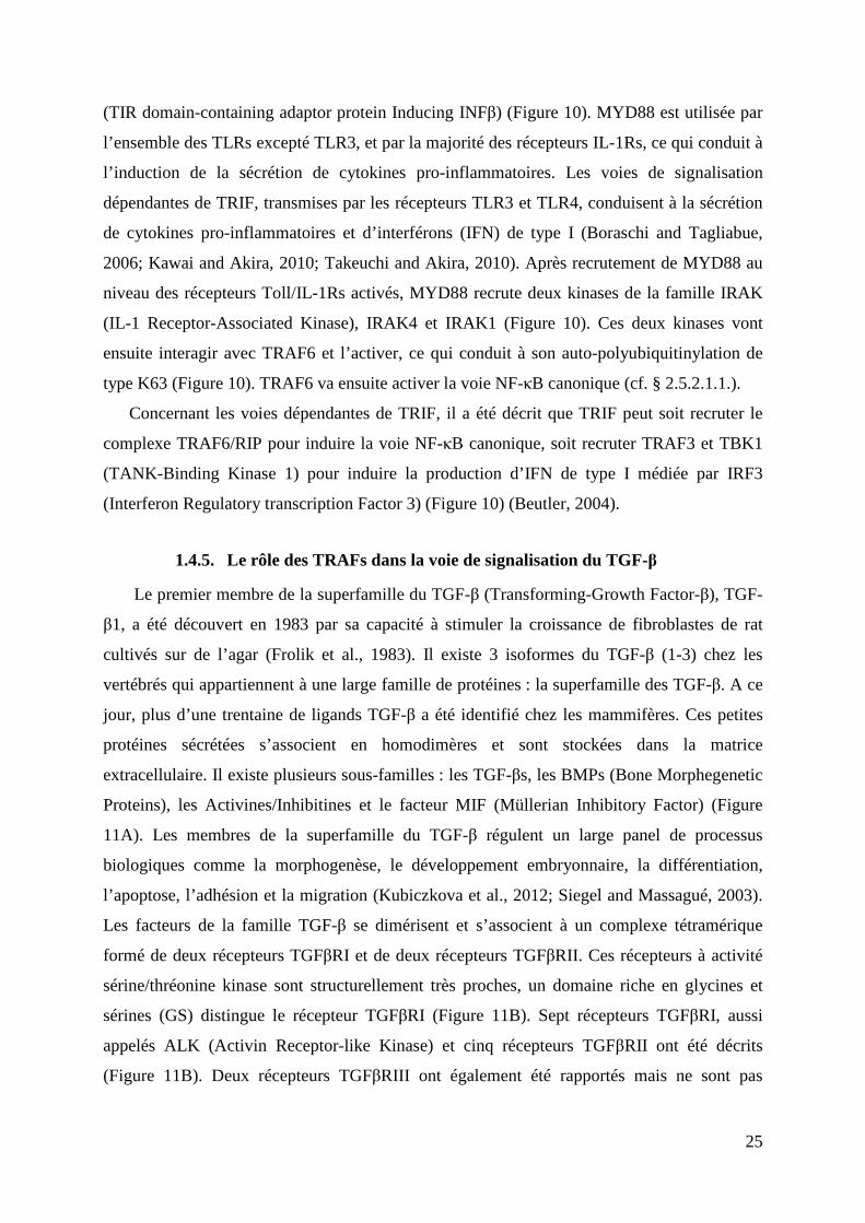
25
(TIR domain-containing adaptor protein Inducing INFβ) (Figure 10). MYD88 est utilisée par
l’ensemble des TLRs excepté TLR3, et par la majorité des récepteurs IL-1Rs, ce qui conduit à
l’induction de la sécrétion de cytokines pro-inflammatoires. Les voies de signalisation
dépendantes de TRIF, transmises par les récepteurs TLR3 et TLR4, conduisent à la sécrétion
de cytokines pro-inflammatoires et d’interférons (IFN) de type I (Boraschi and Tagliabue,
2006; Kawai and Akira, 2010; Takeuchi and Akira, 2010). Après recrutement de MYD88 au
niveau des récepteurs Toll/IL-1Rs activés, MYD88 recrute deux kinases de la famille IRAK
(IL-1 Receptor-Associated Kinase), IRAK4 et IRAK1 (Figure 10). Ces deux kinases vont
ensuite interagir avec TRAF6 et l’activer, ce qui conduit à son auto-polyubiquitinylation de
type K63 (Figure 10). TRAF6 va ensuite activer la voie NF-κB canonique (cf. § 2.5.2.1.1.).
Concernant les voies dépendantes de TRIF, il a été décrit que TRIF peut soit recruter le
complexe TRAF6/RIP pour induire la voie NF-κB canonique, soit recruter TRAF3 et TBK1
(TANK-Binding Kinase 1) pour induire la production d’IFN de type I médiée par IRF3
(Interferon Regulatory transcription Factor 3) (Figure 10) (Beutler, 2004).
1.4.5. Le rôle des TRAFs dans la voie de signalisation du TGF-β
Le premier membre de la superfamille du TGF-β (Transforming-Growth Factor-β), TGF-
β1, a été découvert en 1983 par sa capacité à stimuler la croissance de fibroblastes de rat
cultivés sur de l’agar (Frolik et al., 1983). Il existe 3 isoformes du TGF-β (1-3) chez les
vertébrés qui appartiennent à une large famille de protéines : la superfamille des TGF-β. A ce
jour, plus d’une trentaine de ligands TGF-β a été identifié chez les mammifères. Ces petites
protéines sécrétées s’associent en homodimères et sont stockées dans la matrice
extracellulaire. Il existe plusieurs sous-familles : les TGF-βs, les BMPs (Bone Morphegenetic
Proteins), les Activines/Inhibitines et le facteur MIF (Müllerian Inhibitory Factor) (Figure
11A). Les membres de la superfamille du TGF-β régulent un large panel de processus
biologiques comme la morphogenèse, le développement embryonnaire, la différentiation,
l’apoptose, l’adhésion et la migration (Kubiczkova et al., 2012; Siegel and Massagué, 2003).
Les facteurs de la famille TGF-β se dimérisent et s’associent à un complexe tétramérique
formé de deux récepteurs TGFβRI et de deux récepteurs TGFβRII. Ces récepteurs à activité
sérine/thréonine kinase sont structurellement très proches, un domaine riche en glycines et
sérines (GS) distingue le récepteur TGFβRI (Figure 11B). Sept récepteurs TGFβRI, aussi
appelés ALK (Activin Receptor-like Kinase) et cinq récepteurs TGFβRII ont été décrits
(Figure 11B). Deux récepteurs TGFβRIII ont également été rapportés mais ne sont pas
TGF-β1, TGF-β2,
TGF-β3
Les TGF-βs
BMP2,
BMP3b
BMP4,
BMP5,
BMP6,
BMP7,
BMP8a,
BMP15,
BMP16,
GDF1,
GDF3,
GDF8,
GDF9,
GDF15
BMP8b,
BMP9,
BMP10,
BMP11,
BMP12,
BMP13
BMP14,
MIF
Activine A, Activine B,
Activine AB, Activine C,
Activine E, Inhibitine A,
Inhibitine B, Inhibitine C
Superfamille
du TGF-β
Figure 11 : Les ligands de la superfamille du TGF-β et leurs récepteursA. La superfamille du TGF-β est subdivisée en quatre groupes : les Activines/Inhibitines, les TGF-βs, les BMPs (Bone Morphogenetic Proteins) qui incluent les facteurs GDFs (Growth and Differentiation Factors) et le facteur MIF (Müllerian Inhibitory Factor). Une représentation en ruban des structures cristallines du BMP2, BMP14, TGF-β1 et TGF-β2, montre une organisation structurale conservée au sein de cette superfamille (adapté de Hinck, 2012). B. Représentation en ruban des structures cristallines des récepteurs TβRI et TβRII qui montre une forte conservation structurale. TβRI et TβRII appartiennent respectivement aux groupes des récepteurs TGFβRI et TGFβRII. La différence majeure entre les récepteurs TGFβRI et TGFβRII consiste en la présence d’un domaine GS (domaine riche en glycines et serines) additionnel dans la région cytoplasmique des récepteurs TGFβRI. Il y a cinq récepteurs TGF-β de type II et sept récepteurs TGF-β de type I. Les récep-teurs aux TGF-β1, -β2 et -β3 sont TβRI et TβRII. TBMPR : Bone Morphogenetic Protein Receptor; ActR : Activin Receptor : TβR : TGF-β Receptor; ALK : Activin Receptor-like Kinase; AMHR : Anti-Mülle-rian Hormone Receptor (adapté de Hinck, 2012).
Les Activines/Inhibitines
Les BMPs
A
ALK-1
ALK-2 (ActRI)
ALK-3
ALK-4
ALK-5 (TβRI)
ALK-6
ALK-7
TGFβRI
BMPRII
ActRII
ActRIIB
TβRII
AMHR
TGFβRII
TβRII TβRI
TGFβRII TGFβRI
B
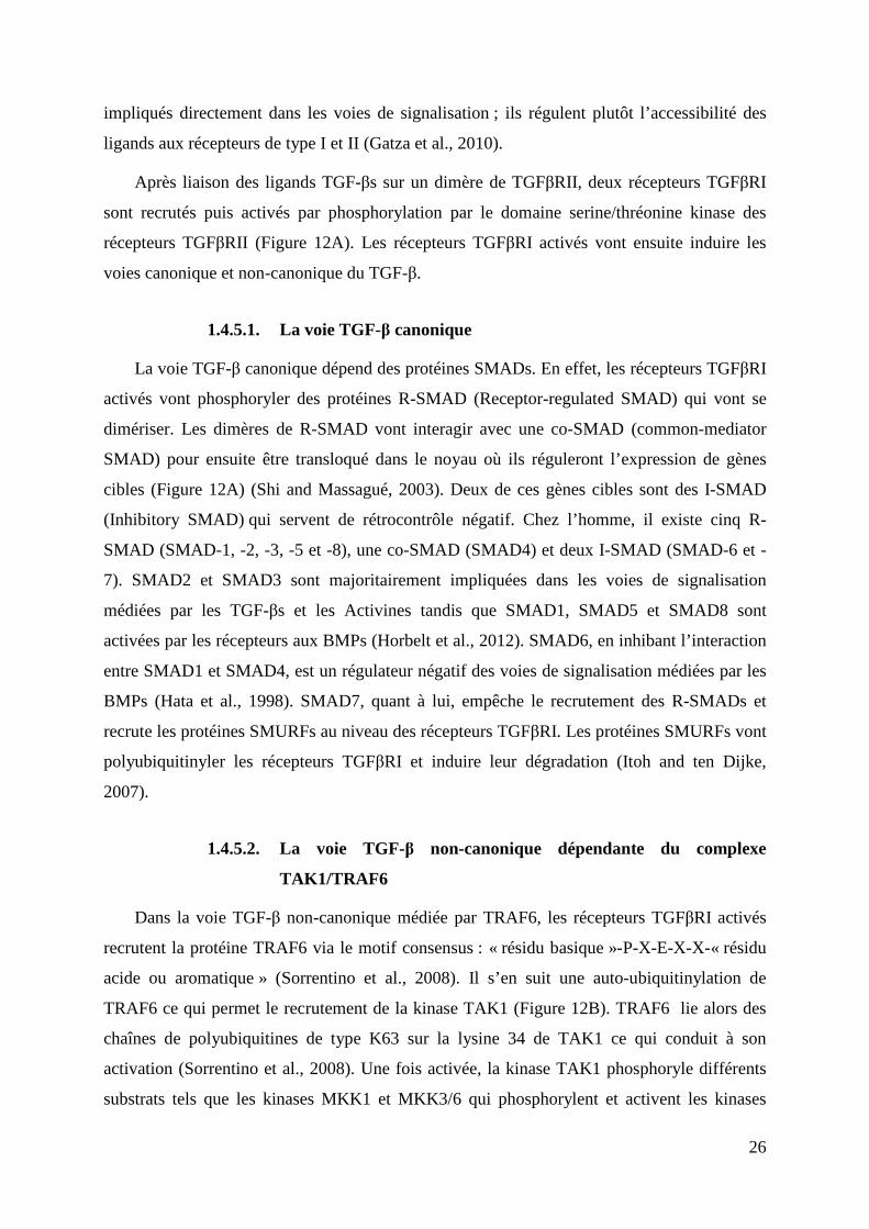
26
impliqués directement dans les voies de signalisation ; ils régulent plutôt l’accessibilité des
ligands aux récepteurs de type I et II (Gatza et al., 2010).
Après liaison des ligands TGF-βs sur un dimère de TGFβRII, deux récepteurs TGFβRI
sont recrutés puis activés par phosphorylation par le domaine serine/thréonine kinase des
récepteurs TGFβRII (Figure 12A). Les récepteurs TGFβRI activés vont ensuite induire les
voies canonique et non-canonique du TGF-β.
1.4.5.1. La voie TGF-β canonique
La voie TGF-β canonique dépend des protéines SMADs. En effet, les récepteurs TGFβRI
activés vont phosphoryler des protéines R-SMAD (Receptor-regulated SMAD) qui vont se
dimériser. Les dimères de R-SMAD vont interagir avec une co-SMAD (common-mediator
SMAD) pour ensuite être transloqué dans le noyau où ils réguleront l’expression de gènes
cibles (Figure 12A) (Shi and Massagué, 2003). Deux de ces gènes cibles sont des I-SMAD
(Inhibitory SMAD) qui servent de rétrocontrôle négatif. Chez l’homme, il existe cinq R-
SMAD (SMAD-1, -2, -3, -5 et -8), une co-SMAD (SMAD4) et deux I-SMAD (SMAD-6 et -
7). SMAD2 et SMAD3 sont majoritairement impliquées dans les voies de signalisation
médiées par les TGF-βs et les Activines tandis que SMAD1, SMAD5 et SMAD8 sont
activées par les récepteurs aux BMPs (Horbelt et al., 2012). SMAD6, en inhibant l’interaction
entre SMAD1 et SMAD4, est un régulateur négatif des voies de signalisation médiées par les
BMPs (Hata et al., 1998). SMAD7, quant à lui, empêche le recrutement des R-SMADs et
recrute les protéines SMURFs au niveau des récepteurs TGFβRI. Les protéines SMURFs vont
polyubiquitinyler les récepteurs TGFβRI et induire leur dégradation (Itoh and ten Dijke,
2007).
1.4.5.2. La voie TGF-β non-canonique dépendante du complexe
TAK1/TRAF6
Dans la voie TGF-β non-canonique médiée par TRAF6, les récepteurs TGFβRI activés
recrutent la protéine TRAF6 via le motif consensus : « résidu basique »-P-X-E-X-X-« résidu
acide ou aromatique » (Sorrentino et al., 2008). Il s’en suit une auto-ubiquitinylation de
TRAF6 ce qui permet le recrutement de la kinase TAK1 (Figure 12B). TRAF6 lie alors des
chaînes de polyubiquitines de type K63 sur la lysine 34 de TAK1 ce qui conduit à son
activation (Sorrentino et al., 2008). Une fois activée, la kinase TAK1 phosphoryle différents
substrats tels que les kinases MKK1 et MKK3/6 qui phosphorylent et activent les kinases
Expression des
gènes cibles
Ligand
Récepteurs
Membrane plasmique
Noyau
Figure 12 : La voie de signalisation canonique et non-canonique du TGF-β A. Les TGF-βs lient leurs récepteurs de manière séquentielle afin de former un complexe de signalisation. Les récepteurs de type II de ce complexe vont phosphoryler et activer les récepteurs de type I qui vont, en retour, phosphoryler les protéines R-SMADs. Les R-SMADs phosphorylées forment un dimère qui va recruter une co-SMAD (SMAD4). Le trimère de SMADs est transloqué dans le noyau où il va réguler l’expression de ses gènes cibles. SMAD6 et SMAD7 sont des I-SMAD (Inhibitory SMAD) qui vont inhiber les protéines R-SMAD et permettre le recrutement des protéines SMURFs au niveau des récep-teurs au TGF-β. Les protéines SMURFs, en plus d’inhiber la phosphorylation des R-SMADs, vont polyubiquitinyler les récepteurs de type I, une marque conduisant à leur dégradation par le protéasome 26S. SMURF : SMAD Ubiquitin Regulatory Factor (adapté de Derynck et Zhang, 2003). B. Les récepteurs de type I activés suite à la liaison du TGF-β vont recruter la protéine TRAF6 et induire son auto-polyubiquitinylation. La kinase TAK1 va interagir avec TRAF6 via sa chaîne de polyubiquitine ce qui va conduire à sa polyubiquitinylation de type K63 et à son activation. Une fois activée, la kinase TAK1 phosphoryle les kinases MKK4 et MKK3/6 qui vont respectivement activer les protéines JNK et p38. Ces deux kinases vont activer des facteurs de transcription important pour l’activation de la voie TGF-β (adapté de Zhang, 2009).
Cytoplasme
TGF-β
Apoptose TEM
Noyau
A
B
27
JNK et p38-MAPK (Figure 12B) (Atfi et al., 1997; Zhang et al., 2003). Ces deux dernières
activent différents facteurs de transcription tel que le facteur AP-1 (Activator Protein-1)
(Karin, 1995). Bien que la voie non-canonique médiée par le complexe TAK1/TRAF6 ne
dépende pas des protéines SMADs, elle va agir en synergie avec la voie canonique pour
induire les différents processus biologiques médiés par le TGF-β (Hocevar et al., 1999;
Zhang, 2009).
1.4.5.3. Implication de la voie TGF-β dans les cancers
Chez l’homme, de nombreuses études ont montré l’implication des voies de signalisation
TGF-β dans l’initiation, la progression et la dissémination des cellules cancéreuses. Des
études cliniques rétrospectives ont montré que le facteur TGF-β1 est surexprimé dans les
cancers du sein, du colon, de l’œsophage, du foie, du poumon et du pancréas. De plus, la
surexpression du facteur TGF-β1 est corrélée avec la progression tumorale et la formation des
métastases, ce qui en fait un marqueur de mauvais pronostic (Bierie and Moses, 2006a; Levy
and Hill, 2006). A l’inverse, plusieurs mutations inactivatrices des protéines TGFβRI,
TGFβRII et SMAD4 ont respectivement été rapportées dans les cancers du pancréas, du
poumon et du sein (Bellam and Pasche, 2010; Levy and Hill, 2006). Ces données suggèrent
un rôle paradoxal de la voie du TGF-β dans le cancer avec deux versants, l’un anti-tumoral et
l’autre pro-tumoral. Des études réalisées chez la souris ont permis d’apporter des
éclaircissements quant à l’implication de la voie du TGF-β dans les cancers du sein (Bierie
and Moses, 2006b; Imamura et al., 2012). La surexpression du facteur TGF-β1
spécifiquement dans les cellules épithéliales mammaires (TGF-β1 sous le contrôle du
promoteur MMTV) conduit à une hypoplasie des canaux mammaires. Cela suggère que le
facteur TGF-β1, en inhibant la prolifération des cellules épithéliales mammaires, soit un gène
suppresseur de tumeur. Ces données ont été confirmées dans un modèle de tumorigenèse
induit par l’agent carcinogène DMBA (7,12-dimethylbenz[a]anthracène). En effet, les souris
sauvages traitées au DMBA développaient des carcinomes mammaires tandis que les souris
MMTV-TGF-β1 n’en développaient pas (Pierce et al., 1995). De manière intéressante, le
croisement des souris MMTV-TGF-β1 avec des souris MMTV-c-Neu (modèle de
tumorigenèse mammaire par surexpression du récepteur ErbB2), a permis de montrer que la
surexpression du facteur TGF-β1 n’affecte pas l’incidence et la taille des tumeurs primaires
induites par la surexpression de ErbB2 mais augmente fortement les propriétés invasives des
cellules tumorales et la formation des métastases (Muraoka et al., 2003). Des expériences
complémentaires consistant à exprimer une forme constitutivement active du récepteur
28
TGFβRI spécifiquement dans les cellules épithéliales mammaires chez la souris ont montré
des résultats équivalents (Oft et al., 1998). Prises dans leur ensemble, ces données montrent
que la voie de signalisation TGF-β a effectivement deux effets selon le contexte cellulaire :
exprimé par les cellules épithéliales dans la glande mammaire normale ou dans les formes in
situ, le TGF-β inhibe la prolifération cellulaire et a un effet suppresseur de tumeur ; en
revanche exprimé par des cellules épithéliales de cancer invasif, il favorise la progression
tumorale et la formation des métastases (Bierie and Moses, 2006b; Kubiczkova et al., 2012).
1.5. TRAF4, UN MEMBRE ATYPIQUE DE LA FAMILLE TRAF
Un criblage différentiel réalisé au laboratoire, a identifié TRAF4 comme étant un gène
surexprimé dans les cancers du sein. Depuis, il a été confirmé que la surexpression de TRAF4
est communément retrouvée dans environ 40% des carcinomes tous types confondus. Lors de
ma thèse, j’ai eu l’opportunité de publier une revue synthétique de la littérature portant sur la
fonction physiopathologique de TRAF4 (Rousseau et al., 2011 ; § 2.7.1). Cette revue est
incluse dans le paragraphe suivant. Des données bibliographiques ultérieures à cette revue
sont traitées ensuite (voir § 2.7.2).
1.5.1. TRAF4, at the Crossroad between Morphogenesis and Cancer
Résumé de l’article:
TRAF4 est un gène dont l’expression est altérée dans les cancers. Il est retrouvé
surexprimé dans un large panel de carcinomes différents, en partie due à son amplification
génique. TRAF4 code pour un adaptateur cytoplasmique de 53 kDa qui appartient à la famille
TRAF. Tandis que la majorité des protéines TRAF, en régulant l’activation des facteurs de
transcription tels que NF-κB et AP-1, sont impliquées dans l’immunité et l’inflammation ;
TRAF4 est principalement impliquée dans la morphogénèse et le développement. De manière
intéressante, il a été montré que cette protéine régule différentes fonctions cruciales pour la
cellule comme la polarité et la production d’espèces dérivées de l’oxygène.
Cancers 2011, 3, 2734-2749; doi:10.3390/cancers3022734
cancersISSN 2072-6694
www.mdpi.com/journal/cancers
Review
TRAF4, at the Crossroad between Morphogenesis and Cancer
Adrien Rousseau, Marie-Christine Rio and Fabien Alpy *
Institut de Génétique et de Biologie Moléculaire et Cellulaire (IGBMC), UMR 7104 CNRS, U964
INSERM, Université de Strasbourg, BP 10142, 67404 Illkirch, C.U. de Strasbourg, France;
E-Mails: [email protected] (A.R.); [email protected] (M.-C.R.)
* Author to whom correspondence should be addressed; E-Mail: [email protected];
Tel.: +33-388-6534-21; Fax: +33-388-6532-01.
Received: 12 May 2011; in revised form: 16 June 2011 / Accepted: 17 June 2011 /
Published: 21 June 2011
Abstract: Tumor Necrosis Factor Receptor-Associated Factor 4 (TRAF4) is a gene whose
expression is altered in cancers. It is overexpressed in a variety of carcinomas of different
origins, often as a consequence of amplification. TRAF4 encodes an adaptor protein that
belongs to the TRAF protein family. While most TRAF proteins influence immune and
inflammation processes, TRAF4 is mainly involved in developmental and morphogenic
processes. Interestingly, this protein has been shown to be linked to crucial cellular functions
such as cell polarity and the regulation of reactive oxygen species production.
Keywords: TRAF; reactive oxygen species; cell polarity; morphogenesis; cancerogenesis;
smurf
1. Introduction
The understanding of the molecular alterations involved in cancer development must take into
account the heterogeneity of the tumors. This is a prerequisite for precise prognosis assessment and
ultimately the development of adapted therapeutic strategies. A high level of expression of a subset of
genes is a common alteration found in cancers. Indeed, overexpression is one of the mechanisms that
leads to the activation of proto-oncogenes to oncogenes [1]. The Tumor Necrosis Factor (TNF) Receptor
Associated Factor 4 (TRAF4) gene was identified due to its high expression in breast cancers [2,3].
TRAF4 was originally called Metastatic Lymph Node (MLN) 62 and CART1 (C-rich motif associated
OPEN ACCESS
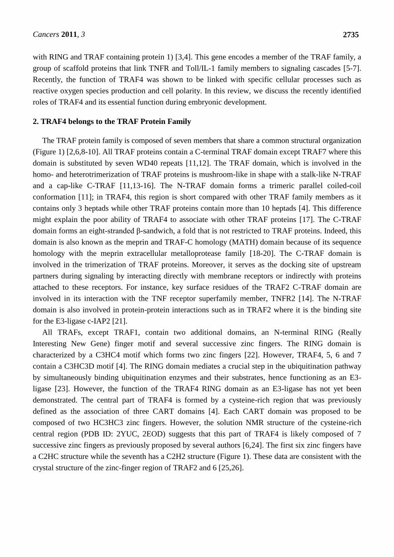
Cancers 2011, 3
2735
with RING and TRAF containing protein 1) [3,4]. This gene encodes a member of the TRAF family, a
group of scaffold proteins that link TNFR and Toll/IL-1 family members to signaling cascades [5-7].
Recently, the function of TRAF4 was shown to be linked with specific cellular processes such as
reactive oxygen species production and cell polarity. In this review, we discuss the recently identified
roles of TRAF4 and its essential function during embryonic development.
2. TRAF4 belongs to the TRAF Protein Family
The TRAF protein family is composed of seven members that share a common structural organization
(Figure 1) [2,6,8-10]. All TRAF proteins contain a C-terminal TRAF domain except TRAF7 where this
domain is substituted by seven WD40 repeats [11,12]. The TRAF domain, which is involved in the
homo- and heterotrimerization of TRAF proteins is mushroom-like in shape with a stalk-like N-TRAF
and a cap-like C-TRAF [11,13-16]. The N-TRAF domain forms a trimeric parallel coiled-coil
conformation [11]; in TRAF4, this region is short compared with other TRAF family members as it
contains only 3 heptads while other TRAF proteins contain more than 10 heptads [4]. This difference
might explain the poor ability of TRAF4 to associate with other TRAF proteins [17]. The C-TRAF
domain forms an eight-stranded β-sandwich, a fold that is not restricted to TRAF proteins. Indeed, this
domain is also known as the meprin and TRAF-C homology (MATH) domain because of its sequence
homology with the meprin extracellular metalloprotease family [18-20]. The C-TRAF domain is
involved in the trimerization of TRAF proteins. Moreover, it serves as the docking site of upstream
partners during signaling by interacting directly with membrane receptors or indirectly with proteins
attached to these receptors. For instance, key surface residues of the TRAF2 C-TRAF domain are
involved in its interaction with the TNF receptor superfamily member, TNFR2 [14]. The N-TRAF
domain is also involved in protein-protein interactions such as in TRAF2 where it is the binding site
for the E3-ligase c-IAP2 [21].
All TRAFs, except TRAF1, contain two additional domains, an N-terminal RING (Really
Interesting New Gene) finger motif and several successive zinc fingers. The RING domain is
characterized by a C3HC4 motif which forms two zinc fingers [22]. However, TRAF4, 5, 6 and 7
contain a C3HC3D motif [4]. The RING domain mediates a crucial step in the ubiquitination pathway
by simultaneously binding ubiquitination enzymes and their substrates, hence functioning as an E3-
ligase [23]. However, the function of the TRAF4 RING domain as an E3-ligase has not yet been
demonstrated. The central part of TRAF4 is formed by a cysteine-rich region that was previously
defined as the association of three CART domains [4]. Each CART domain was proposed to be
composed of two HC3HC3 zinc fingers. However, the solution NMR structure of the cysteine-rich
central region (PDB ID: 2YUC, 2EOD) suggests that this part of TRAF4 is likely composed of 7
successive zinc fingers as previously proposed by several authors [6,24]. The first six zinc fingers have
a C2HC structure while the seventh has a C2H2 structure (Figure 1). These data are consistent with the
crystal structure of the zinc-finger region of TRAF2 and 6 [25,26].
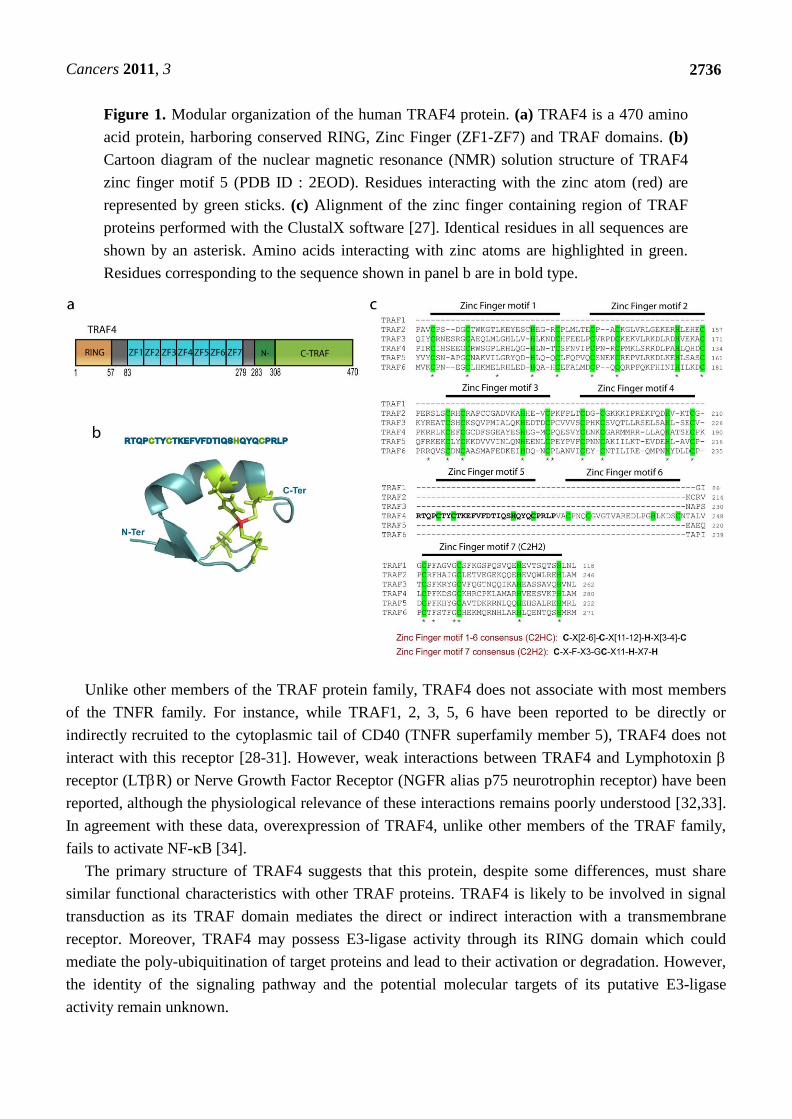
Cancers 2011, 3
2736
Figure 1. Modular organization of the human TRAF4 protein. (a) TRAF4 is a 470 amino
acid protein, harboring conserved RING, Zinc Finger (ZF1-ZF7) and TRAF domains. (b)
Cartoon diagram of the nuclear magnetic resonance (NMR) solution structure of TRAF4
zinc finger motif 5 (PDB ID : 2EOD). Residues interacting with the zinc atom (red) are
represented by green sticks. (c) Alignment of the zinc finger containing region of TRAF
proteins performed with the ClustalX software [27]. Identical residues in all sequences are
shown by an asterisk. Amino acids interacting with zinc atoms are highlighted in green.
Residues corresponding to the sequence shown in panel b are in bold type.
Unlike other members of the TRAF protein family, TRAF4 does not associate with most members
of the TNFR family. For instance, while TRAF1, 2, 3, 5, 6 have been reported to be directly or
indirectly recruited to the cytoplasmic tail of CD40 (TNFR superfamily member 5), TRAF4 does not
interact with this receptor [28-31]. However, weak interactions between TRAF4 and Lymphotoxin
receptor (LTR) or Nerve Growth Factor Receptor (NGFR alias p75 neurotrophin receptor) have been
reported, although the physiological relevance of these interactions remains poorly understood [32,33].
In agreement with these data, overexpression of TRAF4, unlike other members of the TRAF family,
fails to activate NF-B [34].
The primary structure of TRAF4 suggests that this protein, despite some differences, must share
similar functional characteristics with other TRAF proteins. TRAF4 is likely to be involved in signal
transduction as its TRAF domain mediates the direct or indirect interaction with a transmembrane
receptor. Moreover, TRAF4 may possess E3-ligase activity through its RING domain which could
mediate the poly-ubiquitination of target proteins and lead to their activation or degradation. However,
the identity of the signaling pathway and the potential molecular targets of its putative E3-ligase
activity remain unknown.
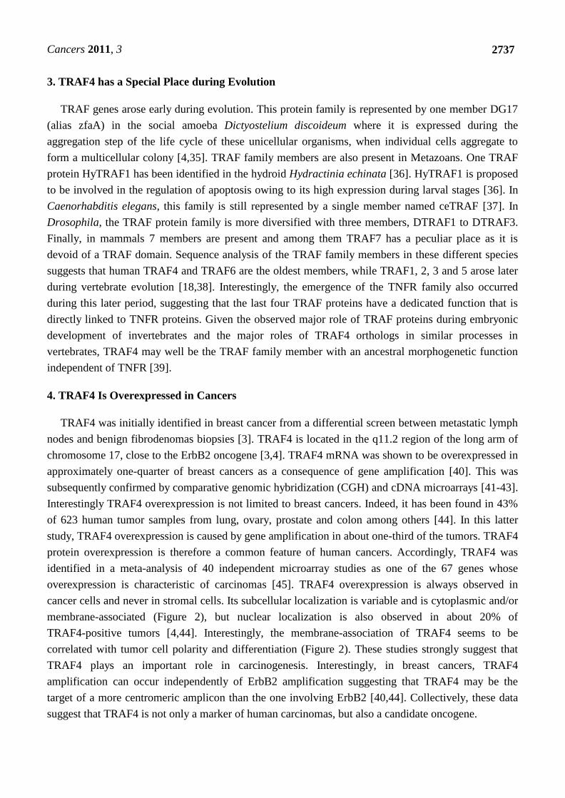
Cancers 2011, 3
2737
3. TRAF4 has a Special Place during Evolution
TRAF genes arose early during evolution. This protein family is represented by one member DG17
(alias zfaA) in the social amoeba Dictyostelium discoideum where it is expressed during the
aggregation step of the life cycle of these unicellular organisms, when individual cells aggregate to
form a multicellular colony [4,35]. TRAF family members are also present in Metazoans. One TRAF
protein HyTRAF1 has been identified in the hydroid Hydractinia echinata [36]. HyTRAF1 is proposed
to be involved in the regulation of apoptosis owing to its high expression during larval stages [36]. In
Caenorhabditis elegans, this family is still represented by a single member named ceTRAF [37]. In
Drosophila, the TRAF protein family is more diversified with three members, DTRAF1 to DTRAF3.
Finally, in mammals 7 members are present and among them TRAF7 has a peculiar place as it is
devoid of a TRAF domain. Sequence analysis of the TRAF family members in these different species
suggests that human TRAF4 and TRAF6 are the oldest members, while TRAF1, 2, 3 and 5 arose later
during vertebrate evolution [18,38]. Interestingly, the emergence of the TNFR family also occurred
during this later period, suggesting that the last four TRAF proteins have a dedicated function that is
directly linked to TNFR proteins. Given the observed major role of TRAF proteins during embryonic
development of invertebrates and the major roles of TRAF4 orthologs in similar processes in
vertebrates, TRAF4 may well be the TRAF family member with an ancestral morphogenetic function
independent of TNFR [39].
4. TRAF4 Is Overexpressed in Cancers
TRAF4 was initially identified in breast cancer from a differential screen between metastatic lymph
nodes and benign fibrodenomas biopsies [3]. TRAF4 is located in the q11.2 region of the long arm of
chromosome 17, close to the ErbB2 oncogene [3,4]. TRAF4 mRNA was shown to be overexpressed in
approximately one-quarter of breast cancers as a consequence of gene amplification [40]. This was
subsequently confirmed by comparative genomic hybridization (CGH) and cDNA microarrays [41-43].
Interestingly TRAF4 overexpression is not limited to breast cancers. Indeed, it has been found in 43%
of 623 human tumor samples from lung, ovary, prostate and colon among others [44]. In this latter
study, TRAF4 overexpression is caused by gene amplification in about one-third of the tumors. TRAF4
protein overexpression is therefore a common feature of human cancers. Accordingly, TRAF4 was
identified in a meta-analysis of 40 independent microarray studies as one of the 67 genes whose
overexpression is characteristic of carcinomas [45]. TRAF4 overexpression is always observed in
cancer cells and never in stromal cells. Its subcellular localization is variable and is cytoplasmic and/or
membrane-associated (Figure 2), but nuclear localization is also observed in about 20% of
TRAF4-positive tumors [4,44]. Interestingly, the membrane-association of TRAF4 seems to be
correlated with tumor cell polarity and differentiation (Figure 2). These studies strongly suggest that
TRAF4 plays an important role in carcinogenesis. Interestingly, in breast cancers, TRAF4
amplification can occur independently of ErbB2 amplification suggesting that TRAF4 may be the
target of a more centromeric amplicon than the one involving ErbB2 [40,44]. Collectively, these data
suggest that TRAF4 is not only a marker of human carcinomas, but also a candidate oncogene.
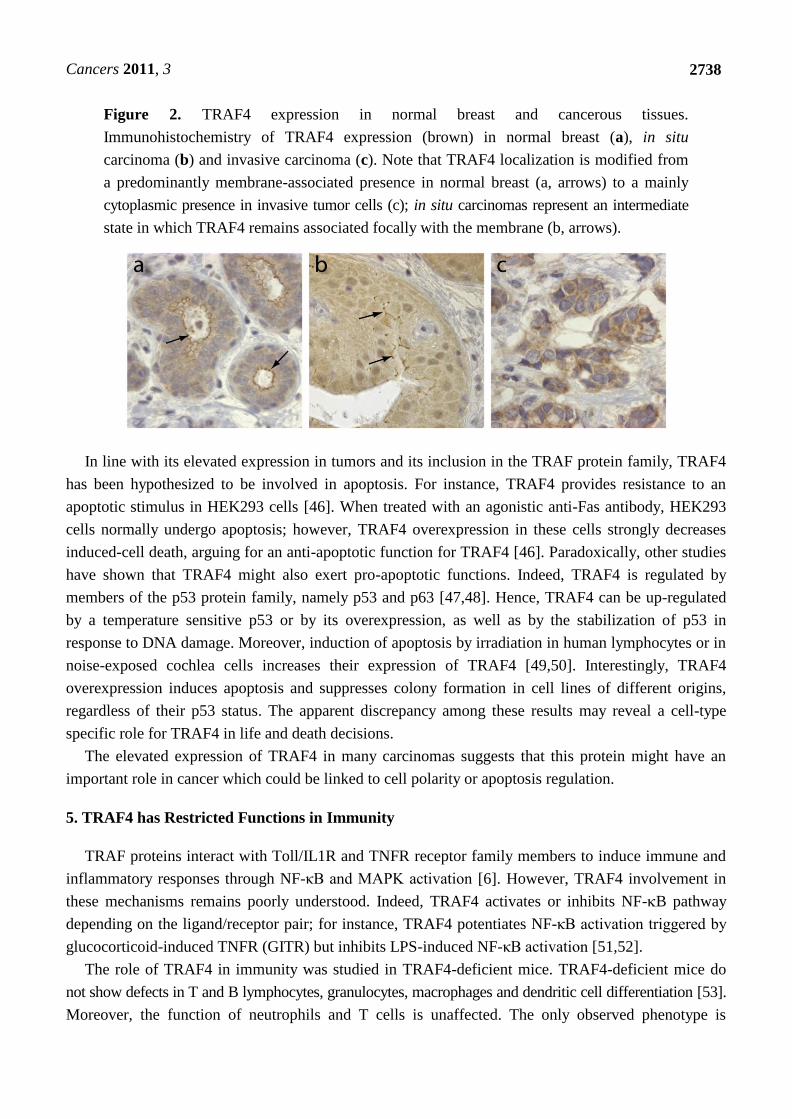
Cancers 2011, 3
2738
Figure 2. TRAF4 expression in normal breast and cancerous tissues.
Immunohistochemistry of TRAF4 expression (brown) in normal breast (a), in situ
carcinoma (b) and invasive carcinoma (c). Note that TRAF4 localization is modified from
a predominantly membrane-associated presence in normal breast (a, arrows) to a mainly
cytoplasmic presence in invasive tumor cells (c); in situ carcinomas represent an intermediate
state in which TRAF4 remains associated focally with the membrane (b, arrows).
In line with its elevated expression in tumors and its inclusion in the TRAF protein family, TRAF4
has been hypothesized to be involved in apoptosis. For instance, TRAF4 provides resistance to an
apoptotic stimulus in HEK293 cells [46]. When treated with an agonistic anti-Fas antibody, HEK293
cells normally undergo apoptosis; however, TRAF4 overexpression in these cells strongly decreases
induced-cell death, arguing for an anti-apoptotic function for TRAF4 [46]. Paradoxically, other studies
have shown that TRAF4 might also exert pro-apoptotic functions. Indeed, TRAF4 is regulated by
members of the p53 protein family, namely p53 and p63 [47,48]. Hence, TRAF4 can be up-regulated
by a temperature sensitive p53 or by its overexpression, as well as by the stabilization of p53 in
response to DNA damage. Moreover, induction of apoptosis by irradiation in human lymphocytes or in
noise-exposed cochlea cells increases their expression of TRAF4 [49,50]. Interestingly, TRAF4
overexpression induces apoptosis and suppresses colony formation in cell lines of different origins,
regardless of their p53 status. The apparent discrepancy among these results may reveal a cell-type
specific role for TRAF4 in life and death decisions.
The elevated expression of TRAF4 in many carcinomas suggests that this protein might have an
important role in cancer which could be linked to cell polarity or apoptosis regulation.
5. TRAF4 has Restricted Functions in Immunity
TRAF proteins interact with Toll/IL1R and TNFR receptor family members to induce immune and
inflammatory responses through NF-κB and MAPK activation [6]. However, TRAF4 involvement in
these mechanisms remains poorly understood. Indeed, TRAF4 activates or inhibits NF-B pathway
depending on the ligand/receptor pair; for instance, TRAF4 potentiates NF-κB activation triggered by
glucocorticoid-induced TNFR (GITR) but inhibits LPS-induced NF-κB activation [51,52].
The role of TRAF4 in immunity was studied in TRAF4-deficient mice. TRAF4-deficient mice do
not show defects in T and B lymphocytes, granulocytes, macrophages and dendritic cell differentiation [53].
Moreover, the function of neutrophils and T cells is unaffected. The only observed phenotype is
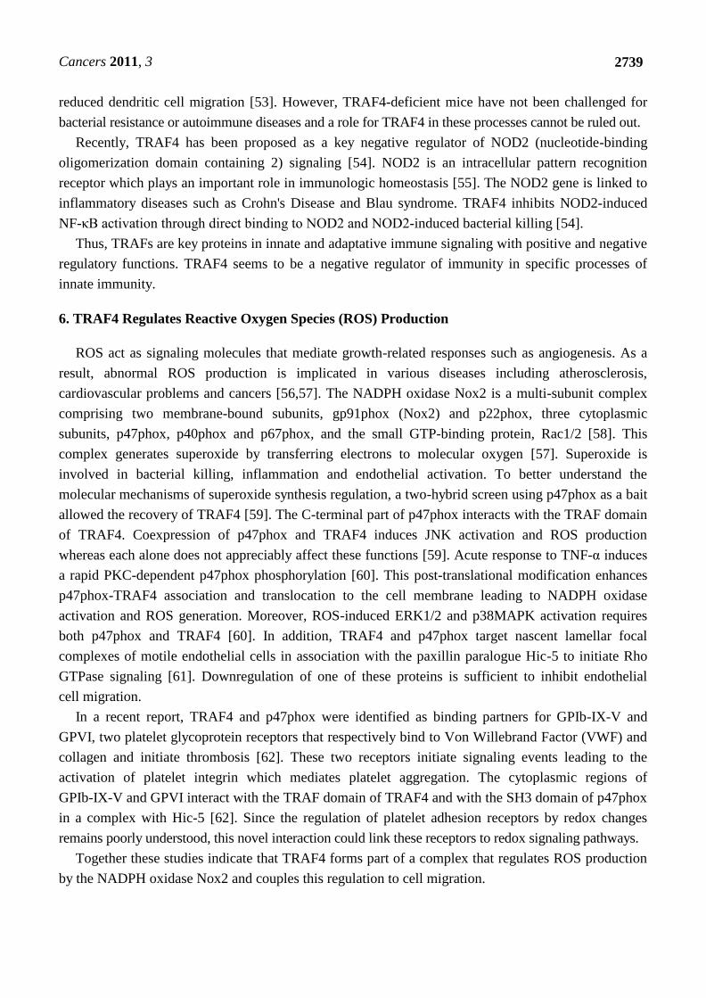
Cancers 2011, 3
2739
reduced dendritic cell migration [53]. However, TRAF4-deficient mice have not been challenged for
bacterial resistance or autoimmune diseases and a role for TRAF4 in these processes cannot be ruled out.
Recently, TRAF4 has been proposed as a key negative regulator of NOD2 (nucleotide-binding
oligomerization domain containing 2) signaling [54]. NOD2 is an intracellular pattern recognition
receptor which plays an important role in immunologic homeostasis [55]. The NOD2 gene is linked to
inflammatory diseases such as Crohn's Disease and Blau syndrome. TRAF4 inhibits NOD2-induced
NF-κB activation through direct binding to NOD2 and NOD2-induced bacterial killing [54].
Thus, TRAFs are key proteins in innate and adaptative immune signaling with positive and negative
regulatory functions. TRAF4 seems to be a negative regulator of immunity in specific processes of
innate immunity.
6. TRAF4 Regulates Reactive Oxygen Species (ROS) Production
ROS act as signaling molecules that mediate growth-related responses such as angiogenesis. As a
result, abnormal ROS production is implicated in various diseases including atherosclerosis,
cardiovascular problems and cancers [56,57]. The NADPH oxidase Nox2 is a multi-subunit complex
comprising two membrane-bound subunits, gp91phox (Nox2) and p22phox, three cytoplasmic
subunits, p47phox, p40phox and p67phox, and the small GTP-binding protein, Rac1/2 [58]. This
complex generates superoxide by transferring electrons to molecular oxygen [57]. Superoxide is
involved in bacterial killing, inflammation and endothelial activation. To better understand the
molecular mechanisms of superoxide synthesis regulation, a two-hybrid screen using p47phox as a bait
allowed the recovery of TRAF4 [59]. The C-terminal part of p47phox interacts with the TRAF domain
of TRAF4. Coexpression of p47phox and TRAF4 induces JNK activation and ROS production
whereas each alone does not appreciably affect these functions [59]. Acute response to TNF-α induces
a rapid PKC-dependent p47phox phosphorylation [60]. This post-translational modification enhances
p47phox-TRAF4 association and translocation to the cell membrane leading to NADPH oxidase
activation and ROS generation. Moreover, ROS-induced ERK1/2 and p38MAPK activation requires
both p47phox and TRAF4 [60]. In addition, TRAF4 and p47phox target nascent lamellar focal
complexes of motile endothelial cells in association with the paxillin paralogue Hic-5 to initiate Rho
GTPase signaling [61]. Downregulation of one of these proteins is sufficient to inhibit endothelial
cell migration.
In a recent report, TRAF4 and p47phox were identified as binding partners for GPIb-IX-V and
GPVI, two platelet glycoprotein receptors that respectively bind to Von Willebrand Factor (VWF) and
collagen and initiate thrombosis [62]. These two receptors initiate signaling events leading to the
activation of platelet integrin which mediates platelet aggregation. The cytoplasmic regions of
GPIb-IX-V and GPVI interact with the TRAF domain of TRAF4 and with the SH3 domain of p47phox
in a complex with Hic-5 [62]. Since the regulation of platelet adhesion receptors by redox changes
remains poorly understood, this novel interaction could link these receptors to redox signaling pathways.
Together these studies indicate that TRAF4 forms part of a complex that regulates ROS production
by the NADPH oxidase Nox2 and couples this regulation to cell migration.
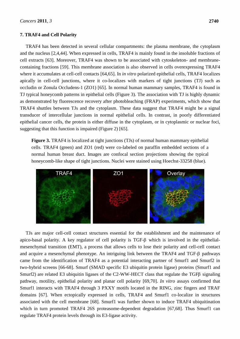
Cancers 2011, 3
2740
7. TRAF4 and Cell Polarity
TRAF4 has been detected in several cellular compartments: the plasma membrane, the cytoplasm
and the nucleus [2,4,44]. When expressed in cells, TRAF4 is mainly found in the insoluble fractions of
cell extracts [63]. Moreover, TRAF4 was shown to be associated with cytoskeleton- and membrane-
containing fractions [59]. This membrane association is also observed in cells overexpressing TRAF4
where it accumulates at cell-cell contacts [64,65]. In in vitro polarized epithelial cells, TRAF4 localizes
apically in cell-cell junctions, where it co-localizes with markers of tight junctions (TJ) such as
occludin or Zonula Occludens-1 (ZO1) [65]. In normal human mammary samples, TRAF4 is found in
TJ typical honeycomb patterns in epithelial cells (Figure 3). The association with TJ is highly dynamic
as demonstrated by fluorescence recovery after photobleaching (FRAP) experiments, which show that
TRAF4 shuttles between TJs and the cytoplasm. These data suggest that TRAF4 might be a signal
transducer of intercellular junctions in normal epithelial cells. In contrast, in poorly differentiated
epithelial cancer cells, the protein is either diffuse in the cytoplasm, or in cytoplasmic or nuclear foci,
suggesting that this function is impaired (Figure 2) [65].
Figure 3. TRAF4 is localized at tight junctions (TJs) of normal human mammary epithelial
cells. TRAF4 (green) and ZO1 (red) were co-labeled on paraffin embedded sections of a
normal human breast duct. Images are confocal section projections showing the typical
honeycomb-like shape of tight junctions. Nuclei were stained using Hoechst-33258 (blue).
TJs are major cell-cell contact structures essential for the establishment and the maintenance of
apico-basal polarity. A key regulator of cell polarity is TGF- which is involved in the epithelial-
mesenchymal transition (EMT), a process that allows cells to lose their polarity and cell-cell contact
and acquire a mesenchymal phenotype. An intriguing link between the TRAF4 and TGF- pathways
came from the identification of TRAF4 as a potential interacting partner of Smurf1 and Smurf2 in
two-hybrid screens [66-68]. Smurf (SMAD specific E3 ubiquitin protein ligase) proteins (Smurf1 and
Smurf2) are related E3 ubiquitin ligases of the C2-WW-HECT class that regulate the TGF signaling
pathway, motility, epithelial polarity and planar cell polarity [69,70]. In vitro assays confirmed that
Smurf1 interacts with TRAF4 through 3 PXXY motifs located in the RING, zinc fingers and TRAF
domains [67]. When ectopically expressed in cells, TRAF4 and Smurf1 co-localize in structures
associated with the cell membrane [68]. Smurf1 was further shown to induce TRAF4 ubiquitination
which in turn promoted TRAF4 26S proteasome-dependent degradation [67,68]. Thus Smurf1 can
regulate TRAF4 protein levels through its E3-ligase activity.
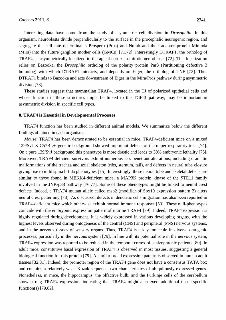
Cancers 2011, 3
2741
Interesting data have come from the study of asymmetric cell division in Drosophila. In this
organism, neuroblasts divide perpendicularly to the surface in the procephalic neurogenic region, and
segregate the cell fate determinants Prospero (Pros) and Numb and their adaptor protein Miranda
(Mira) into the future ganglion mother cells (GMCs) [71,72]. Interestingly DTRAF1, the ortholog of
TRAF4, is asymmetrically localized to the apical cortex in mitotic neuroblasts [72]. This localization
relies on Bazooka, the Drosophila ortholog of the polarity protein Par3 (Partitioning defective 3
homolog) with which DTRAF1 interacts, and depends on Eiger, the ortholog of TNF [72]. Thus
DTRAF1 binds to Bazooka and acts downstream of Eiger in the Mira/Pros pathway during asymmetric
division [73].
These studies suggest that mammalian TRAF4, located in the TJ of polarized epithelial cells and
whose function in these structures might be linked to the TGF- pathway, may be important in
asymmetric division in specific cell types.
8. TRAF4 is Essential in Developmental Processes
TRAF4 function has been studied in different animal models. We summarize below the different
findings obtained in each organism.
Mouse: TRAF4 has been demonstrated to be essential in mice. TRAF4-deficient mice on a mixed
129/SvJ X C57BL/6 genetic background showed important defects of the upper respiratory tract [74].
On a pure 129/SvJ background this phenotype is more drastic and leads to 30% embryonic lethality [75].
Moreover, TRAF4-deficient survivors exhibit numerous less penetrant alterations, including dramatic
malformations of the trachea and axial skeleton (ribs, sternum, tail), and defects in neural tube closure
giving rise to mild spina bifida phenotypes [75]. Interestingly, these neural tube and skeletal defects are
similar to those found in MEKK4-deficient mice, a MAP3K protein kinase of the STE11 family
involved in the JNK/p38 pathway [76,77]. Some of these phenotypes might be linked to neural crest
defects. Indeed, a TRAF4 mutant allele called msp2 (modifier of Sox10 expression pattern 2) alters
neural crest patterning [78]. As discussed, defects in dendritic cells migration has also been reported in
TRAF4-deficient mice which otherwise exhibit normal immune responses [53]. These null-phenotypes
coincide with the embryonic expression pattern of murine TRAF4 [79]. Indeed, TRAF4 expression is
highly regulated during development. It is widely expressed in various developing organs, with the
highest levels observed during ontogenesis of the central (CNS) and peripheral (PNS) nervous systems,
and in the nervous tissues of sensory organs. Thus, TRAF4 is a key molecule in diverse ontogenic
processes, particularly in the nervous system [79]. In line with its potential role in the nervous system,
TRAF4 expression was reported to be reduced in the temporal cortex of schizophrenic patients [80]. In
adult mice, constitutive basal expression of TRAF4 is observed in most tissues, suggesting a general
biological function for this protein [79]. A similar broad expression pattern is observed in human adult
tissues [32,81]. Indeed, the promoter region of the TRAF4 gene does not have a consensus TATA box
and contains a relatively weak Kozak sequence, two characteristics of ubiquitously expressed genes.
Nonetheless, in mice, the hippocampus, the olfactive bulb, and the Purkinje cells of the cerebellum
show strong TRAF4 expression, indicating that TRAF4 might also exert additional tissue-specific
function(s) [79,82].
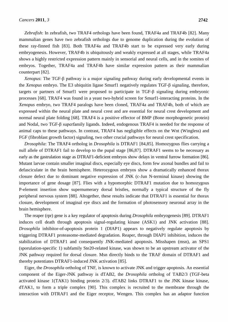
Cancers 2011, 3
2742
Zebrafish: In zebrafish, two TRAF4 orthologs have been found, TRAF4a and TRAF4b [82]. Many
mammalian genes have two zebrafish orthologs due to genome duplication during the evolution of
these ray-finned fish [83]. Both TRAF4a and TRAF4b start to be expressed very early during
embryogenesis. However, TRAF4b is ubiquitously and weakly expressed at all stages, while TRAF4a
shows a highly restricted expression pattern mainly in sensorial and neural cells, and in the somites of
embryos. Together, TRAF4a and TRAF4b have similar expression pattern as their mammalian
counterpart [82].
Xenopus: The TGF-β pathway is a major signaling pathway during early developmental events in
the Xenopus embryo. The E3 ubiquitin ligase Smurf1 negatively regulates TGF-β signaling, therefore,
targets or partners of Smurf1 were proposed to participate in TGF-β signaling during embryonic
processes [68]. TRAF4 was found in a yeast two-hybrid screen for Smurf1-interacting proteins. In the
Xenopus embryo, two TRAF4 paralogs have been cloned, TRAF4a and TRAF4b, both of which are
expressed within the neural plate and neural crest and are essential for neural crest development and
normal neural plate folding [68]. TRAF4 is a positive effector of BMP (Bone morphogenetic protein)
and Nodal, two TGF-β superfamily ligands. Indeed, endogenous TRAF4 is needed for the response of
animal caps to these pathways. In contrast, TRAF4 has negligible effects on the Wnt (Wingless) and
FGF (fibroblast growth factor) signaling, two other crucial pathways for neural crest specification.
Drosophila: The TRAF4 ortholog in Drosophila is DTRAF1 [84,85]. Homozygous flies carrying a
null allele of DTRAF1 fail to develop to the pupal stage [86,87]. DTRAF1 seems to be necessary as
early as the gastrulation stage as DTRAF1-deficient embryos show delays in ventral furrow formation [86].
Mutant larvae contain smaller imaginal discs, especially eye discs, form few axonal bundles and fail to
defasciculate in the brain hemisphere. Heterozygous embryos show a dramatically enhanced thorax
closure defect due to dominant negative expression of JNK (c-Jun N-terminal kinase) showing the
importance of gene dosage [87]. Flies with a hypomorphic DTRAF1 mutation due to homozygous
P-element insertion show supernumerary dorsal bristles, normally a typical structure of the fly
peripheral nervous system [88]. Altogether, these results indicate that DTRAF1 is essential for thorax
closure, development of imaginal eye discs and the formation of photosensory neuronal array in the
brain hemisphere.
The reaper (rpr) gene is a key regulator of apoptosis during Drosophila embryogenesis [89]. DTRAF1
induces cell death through apoptosis signal-regulating kinase (ASK1) and JNK activation [88].
Drosophila inhibitor-of-apoptosis protein 1 (DIAP1) appears to negatively regulate apoptosis by
triggering DTRAF1 proteasome-mediated degradation. Reaper, through DIAP1 inhibition, induces the
stabilization of DTRAF1 and consequently JNK-mediated apoptosis. Misshapen (msn), an SPS1
(sporulation-specific 1) subfamily Ste20-related kinase, was shown to be an upstream activator of the
JNK pathway required for dorsal closure. Msn directly binds to the TRAF domain of DTRAF1 and
thereby potentiates DTRAF1-induced JNK activation [85].
Eiger, the Drosophila ortholog of TNF is known to activate JNK and trigger apoptosis. An essential
component of the Eiger-JNK pathway is dTAB2, the Drosophila ortholog of TAB2/3 (TGF-beta
activated kinase 1(TAK1) binding protein 2/3). dTAB2 links DTRAF1 to the JNK kinase kinase,
dTAK1, to form a triple complex [90]. This complex is recruited to the membrane through the
interaction with DTRAF1 and the Eiger receptor, Wengen. This complex has an adaptor function
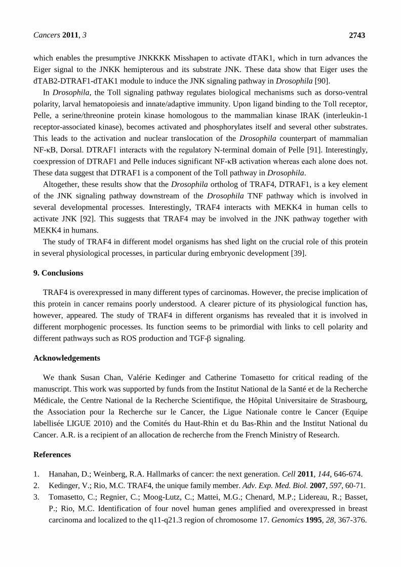
Cancers 2011, 3
2743
which enables the presumptive JNKKKK Misshapen to activate dTAK1, which in turn advances the
Eiger signal to the JNKK hemipterous and its substrate JNK. These data show that Eiger uses the
dTAB2-DTRAF1-dTAK1 module to induce the JNK signaling pathway in Drosophila [90].
In Drosophila, the Toll signaling pathway regulates biological mechanisms such as dorso-ventral
polarity, larval hematopoiesis and innate/adaptive immunity. Upon ligand binding to the Toll receptor,
Pelle, a serine/threonine protein kinase homologous to the mammalian kinase IRAK (interleukin-1
receptor-associated kinase), becomes activated and phosphorylates itself and several other substrates.
This leads to the activation and nuclear translocation of the Drosophila counterpart of mammalian
NF-κB, Dorsal. DTRAF1 interacts with the regulatory N-terminal domain of Pelle [91]. Interestingly,
coexpression of DTRAF1 and Pelle induces significant NF-κB activation whereas each alone does not.
These data suggest that DTRAF1 is a component of the Toll pathway in Drosophila.
Altogether, these results show that the Drosophila ortholog of TRAF4, DTRAF1, is a key element
of the JNK signaling pathway downstream of the Drosophila TNF pathway which is involved in
several developmental processes. Interestingly, TRAF4 interacts with MEKK4 in human cells to
activate JNK [92]. This suggests that TRAF4 may be involved in the JNK pathway together with
MEKK4 in humans.
The study of TRAF4 in different model organisms has shed light on the crucial role of this protein
in several physiological processes, in particular during embryonic development [39].
9. Conclusions
TRAF4 is overexpressed in many different types of carcinomas. However, the precise implication of
this protein in cancer remains poorly understood. A clearer picture of its physiological function has,
however, appeared. The study of TRAF4 in different organisms has revealed that it is involved in
different morphogenic processes. Its function seems to be primordial with links to cell polarity and
different pathways such as ROS production and TGF-signaling.
Acknowledgements
We thank Susan Chan, Valérie Kedinger and Catherine Tomasetto for critical reading of the
manuscript. This work was supported by funds from the Institut National de la Santé et de la Recherche
Médicale, the Centre National de la Recherche Scientifique, the Hôpital Universitaire de Strasbourg,
the Association pour la Recherche sur le Cancer, the Ligue Nationale contre le Cancer (Equipe
labellisée LIGUE 2010) and the Comités du Haut-Rhin et du Bas-Rhin and the Institut National du
Cancer. A.R. is a recipient of an allocation de recherche from the French Ministry of Research.
References
1. Hanahan, D.; Weinberg, R.A. Hallmarks of cancer: the next generation. Cell 2011, 144, 646-674.
2. Kedinger, V.; Rio, M.C. TRAF4, the unique family member. Adv. Exp. Med. Biol. 2007, 597, 60-71.
3. Tomasetto, C.; Regnier, C.; Moog-Lutz, C.; Mattei, M.G.; Chenard, M.P.; Lidereau, R.; Basset,
P.; Rio, M.C. Identification of four novel human genes amplified and overexpressed in breast
carcinoma and localized to the q11-q21.3 region of chromosome 17. Genomics 1995, 28, 367-376.
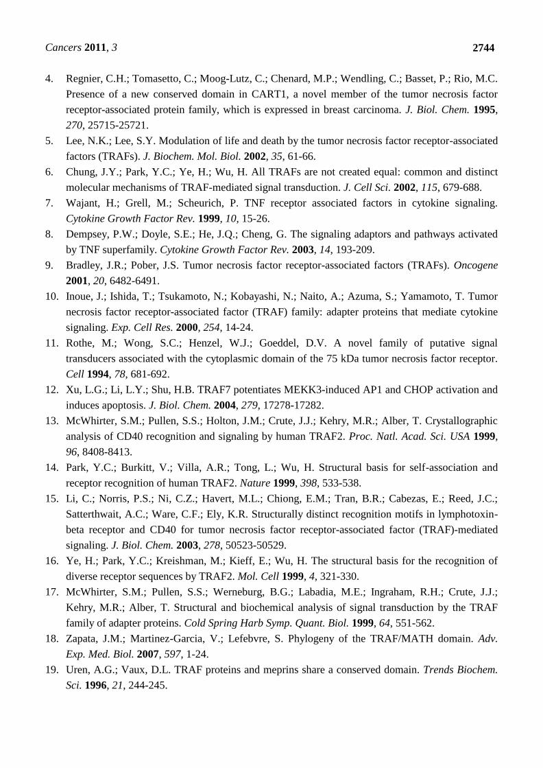
Cancers 2011, 3
2744
4. Regnier, C.H.; Tomasetto, C.; Moog-Lutz, C.; Chenard, M.P.; Wendling, C.; Basset, P.; Rio, M.C.
Presence of a new conserved domain in CART1, a novel member of the tumor necrosis factor
receptor-associated protein family, which is expressed in breast carcinoma. J. Biol. Chem. 1995,
270, 25715-25721.
5. Lee, N.K.; Lee, S.Y. Modulation of life and death by the tumor necrosis factor receptor-associated
factors (TRAFs). J. Biochem. Mol. Biol. 2002, 35, 61-66.
6. Chung, J.Y.; Park, Y.C.; Ye, H.; Wu, H. All TRAFs are not created equal: common and distinct
molecular mechanisms of TRAF-mediated signal transduction. J. Cell Sci. 2002, 115, 679-688.
7. Wajant, H.; Grell, M.; Scheurich, P. TNF receptor associated factors in cytokine signaling.
Cytokine Growth Factor Rev. 1999, 10, 15-26.
8. Dempsey, P.W.; Doyle, S.E.; He, J.Q.; Cheng, G. The signaling adaptors and pathways activated
by TNF superfamily. Cytokine Growth Factor Rev. 2003, 14, 193-209.
9. Bradley, J.R.; Pober, J.S. Tumor necrosis factor receptor-associated factors (TRAFs). Oncogene
2001, 20, 6482-6491.
10. Inoue, J.; Ishida, T.; Tsukamoto, N.; Kobayashi, N.; Naito, A.; Azuma, S.; Yamamoto, T. Tumor
necrosis factor receptor-associated factor (TRAF) family: adapter proteins that mediate cytokine
signaling. Exp. Cell Res. 2000, 254, 14-24.
11. Rothe, M.; Wong, S.C.; Henzel, W.J.; Goeddel, D.V. A novel family of putative signal
transducers associated with the cytoplasmic domain of the 75 kDa tumor necrosis factor receptor.
Cell 1994, 78, 681-692.
12. Xu, L.G.; Li, L.Y.; Shu, H.B. TRAF7 potentiates MEKK3-induced AP1 and CHOP activation and
induces apoptosis. J. Biol. Chem. 2004, 279, 17278-17282.
13. McWhirter, S.M.; Pullen, S.S.; Holton, J.M.; Crute, J.J.; Kehry, M.R.; Alber, T. Crystallographic
analysis of CD40 recognition and signaling by human TRAF2. Proc. Natl. Acad. Sci. USA 1999,
96, 8408-8413.
14. Park, Y.C.; Burkitt, V.; Villa, A.R.; Tong, L.; Wu, H. Structural basis for self-association and
receptor recognition of human TRAF2. Nature 1999, 398, 533-538.
15. Li, C.; Norris, P.S.; Ni, C.Z.; Havert, M.L.; Chiong, E.M.; Tran, B.R.; Cabezas, E.; Reed, J.C.;
Satterthwait, A.C.; Ware, C.F.; Ely, K.R. Structurally distinct recognition motifs in lymphotoxin-
beta receptor and CD40 for tumor necrosis factor receptor-associated factor (TRAF)-mediated
signaling. J. Biol. Chem. 2003, 278, 50523-50529.
16. Ye, H.; Park, Y.C.; Kreishman, M.; Kieff, E.; Wu, H. The structural basis for the recognition of
diverse receptor sequences by TRAF2. Mol. Cell 1999, 4, 321-330.
17. McWhirter, S.M.; Pullen, S.S.; Werneburg, B.G.; Labadia, M.E.; Ingraham, R.H.; Crute, J.J.;
Kehry, M.R.; Alber, T. Structural and biochemical analysis of signal transduction by the TRAF
family of adapter proteins. Cold Spring Harb Symp. Quant. Biol. 1999, 64, 551-562.
18. Zapata, J.M.; Martinez-Garcia, V.; Lefebvre, S. Phylogeny of the TRAF/MATH domain. Adv.
Exp. Med. Biol. 2007, 597, 1-24.
19. Uren, A.G.; Vaux, D.L. TRAF proteins and meprins share a conserved domain. Trends Biochem.
Sci. 1996, 21, 244-245.
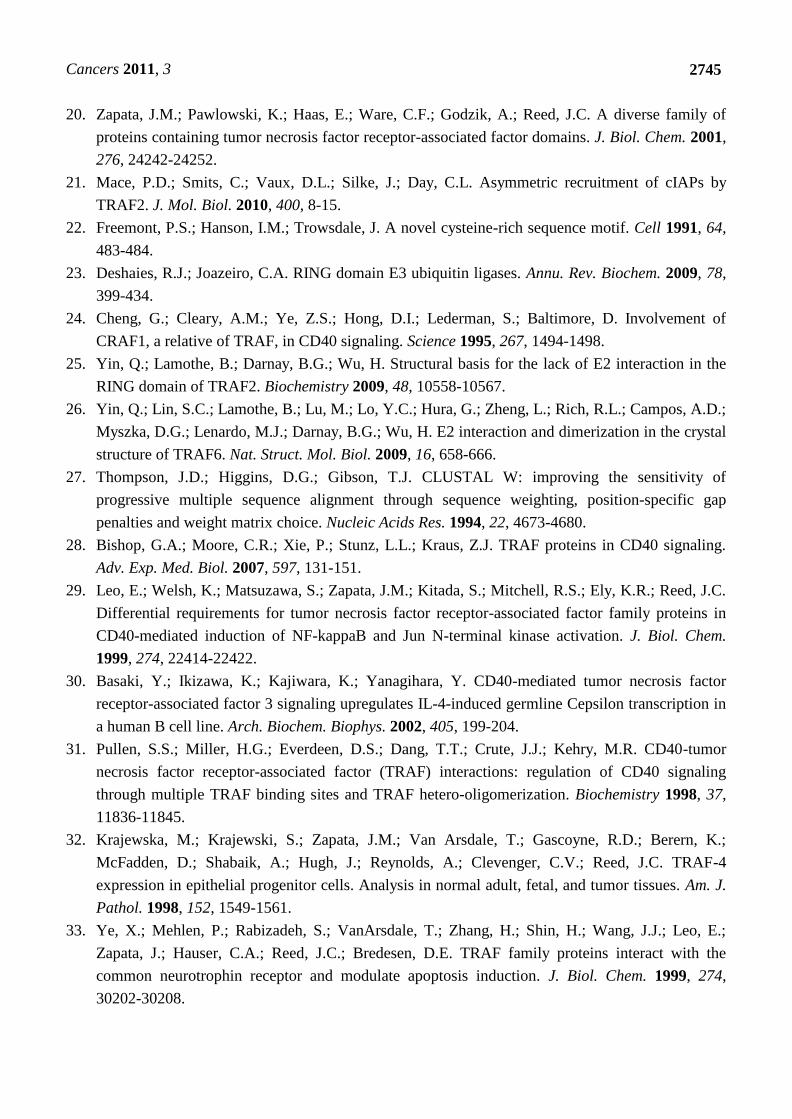
Cancers 2011, 3
2745
20. Zapata, J.M.; Pawlowski, K.; Haas, E.; Ware, C.F.; Godzik, A.; Reed, J.C. A diverse family of
proteins containing tumor necrosis factor receptor-associated factor domains. J. Biol. Chem. 2001,
276, 24242-24252.
21. Mace, P.D.; Smits, C.; Vaux, D.L.; Silke, J.; Day, C.L. Asymmetric recruitment of cIAPs by
TRAF2. J. Mol. Biol. 2010, 400, 8-15.
22. Freemont, P.S.; Hanson, I.M.; Trowsdale, J. A novel cysteine-rich sequence motif. Cell 1991, 64,
483-484.
23. Deshaies, R.J.; Joazeiro, C.A. RING domain E3 ubiquitin ligases. Annu. Rev. Biochem. 2009, 78,
399-434.
24. Cheng, G.; Cleary, A.M.; Ye, Z.S.; Hong, D.I.; Lederman, S.; Baltimore, D. Involvement of
CRAF1, a relative of TRAF, in CD40 signaling. Science 1995, 267, 1494-1498.
25. Yin, Q.; Lamothe, B.; Darnay, B.G.; Wu, H. Structural basis for the lack of E2 interaction in the
RING domain of TRAF2. Biochemistry 2009, 48, 10558-10567.
26. Yin, Q.; Lin, S.C.; Lamothe, B.; Lu, M.; Lo, Y.C.; Hura, G.; Zheng, L.; Rich, R.L.; Campos, A.D.;
Myszka, D.G.; Lenardo, M.J.; Darnay, B.G.; Wu, H. E2 interaction and dimerization in the crystal
structure of TRAF6. Nat. Struct. Mol. Biol. 2009, 16, 658-666.
27. Thompson, J.D.; Higgins, D.G.; Gibson, T.J. CLUSTAL W: improving the sensitivity of
progressive multiple sequence alignment through sequence weighting, position-specific gap
penalties and weight matrix choice. Nucleic Acids Res. 1994, 22, 4673-4680.
28. Bishop, G.A.; Moore, C.R.; Xie, P.; Stunz, L.L.; Kraus, Z.J. TRAF proteins in CD40 signaling.
Adv. Exp. Med. Biol. 2007, 597, 131-151.
29. Leo, E.; Welsh, K.; Matsuzawa, S.; Zapata, J.M.; Kitada, S.; Mitchell, R.S.; Ely, K.R.; Reed, J.C.
Differential requirements for tumor necrosis factor receptor-associated factor family proteins in
CD40-mediated induction of NF-kappaB and Jun N-terminal kinase activation. J. Biol. Chem.
1999, 274, 22414-22422.
30. Basaki, Y.; Ikizawa, K.; Kajiwara, K.; Yanagihara, Y. CD40-mediated tumor necrosis factor
receptor-associated factor 3 signaling upregulates IL-4-induced germline Cepsilon transcription in
a human B cell line. Arch. Biochem. Biophys. 2002, 405, 199-204.
31. Pullen, S.S.; Miller, H.G.; Everdeen, D.S.; Dang, T.T.; Crute, J.J.; Kehry, M.R. CD40-tumor
necrosis factor receptor-associated factor (TRAF) interactions: regulation of CD40 signaling
through multiple TRAF binding sites and TRAF hetero-oligomerization. Biochemistry 1998, 37,
11836-11845.
32. Krajewska, M.; Krajewski, S.; Zapata, J.M.; Van Arsdale, T.; Gascoyne, R.D.; Berern, K.;
McFadden, D.; Shabaik, A.; Hugh, J.; Reynolds, A.; Clevenger, C.V.; Reed, J.C. TRAF-4
expression in epithelial progenitor cells. Analysis in normal adult, fetal, and tumor tissues. Am. J.
Pathol. 1998, 152, 1549-1561.
33. Ye, X.; Mehlen, P.; Rabizadeh, S.; VanArsdale, T.; Zhang, H.; Shin, H.; Wang, J.J.; Leo, E.;
Zapata, J.; Hauser, C.A.; Reed, J.C.; Bredesen, D.E. TRAF family proteins interact with the
common neurotrophin receptor and modulate apoptosis induction. J. Biol. Chem. 1999, 274,
30202-30208.
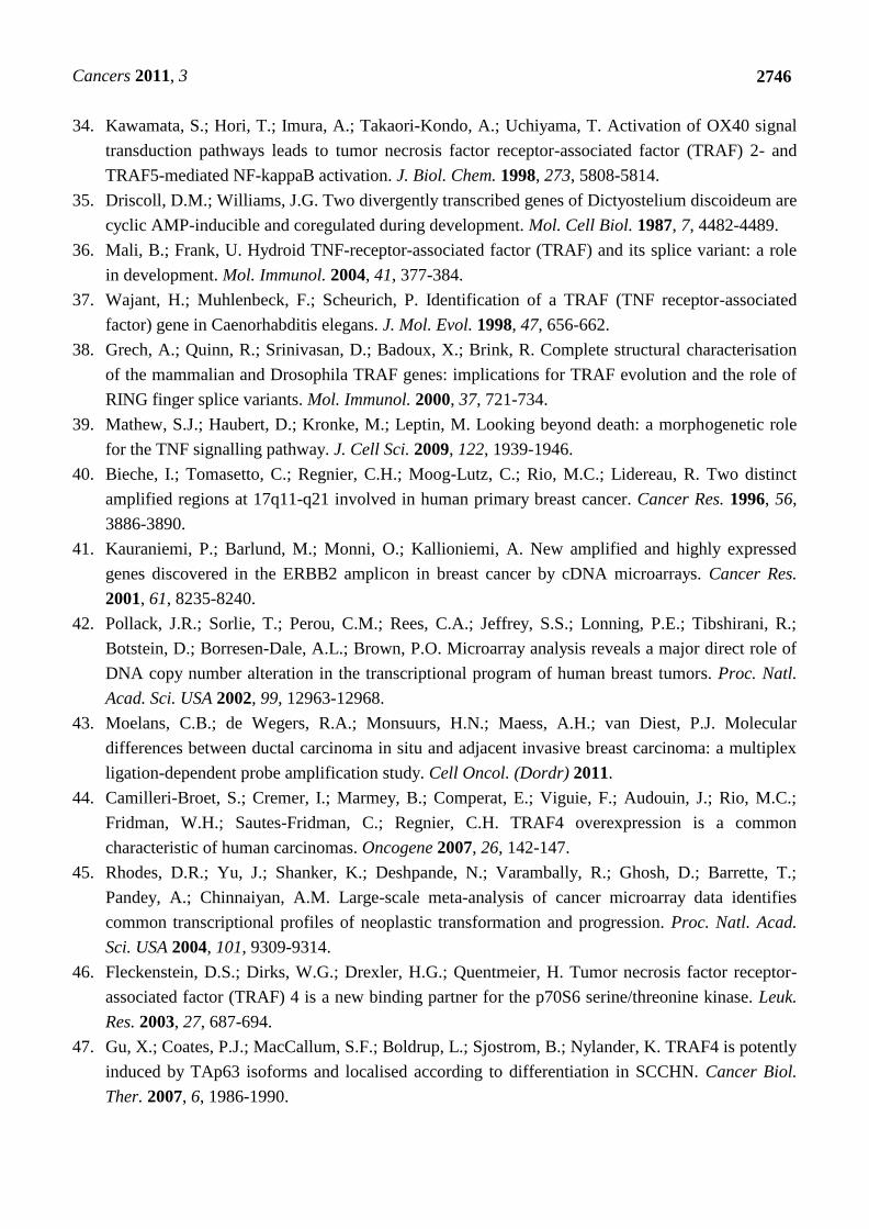
Cancers 2011, 3
2746
34. Kawamata, S.; Hori, T.; Imura, A.; Takaori-Kondo, A.; Uchiyama, T. Activation of OX40 signal
transduction pathways leads to tumor necrosis factor receptor-associated factor (TRAF) 2- and
TRAF5-mediated NF-kappaB activation. J. Biol. Chem. 1998, 273, 5808-5814.
35. Driscoll, D.M.; Williams, J.G. Two divergently transcribed genes of Dictyostelium discoideum are
cyclic AMP-inducible and coregulated during development. Mol. Cell Biol. 1987, 7, 4482-4489.
36. Mali, B.; Frank, U. Hydroid TNF-receptor-associated factor (TRAF) and its splice variant: a role
in development. Mol. Immunol. 2004, 41, 377-384.
37. Wajant, H.; Muhlenbeck, F.; Scheurich, P. Identification of a TRAF (TNF receptor-associated
factor) gene in Caenorhabditis elegans. J. Mol. Evol. 1998, 47, 656-662.
38. Grech, A.; Quinn, R.; Srinivasan, D.; Badoux, X.; Brink, R. Complete structural characterisation
of the mammalian and Drosophila TRAF genes: implications for TRAF evolution and the role of
RING finger splice variants. Mol. Immunol. 2000, 37, 721-734.
39. Mathew, S.J.; Haubert, D.; Kronke, M.; Leptin, M. Looking beyond death: a morphogenetic role
for the TNF signalling pathway. J. Cell Sci. 2009, 122, 1939-1946.
40. Bieche, I.; Tomasetto, C.; Regnier, C.H.; Moog-Lutz, C.; Rio, M.C.; Lidereau, R. Two distinct
amplified regions at 17q11-q21 involved in human primary breast cancer. Cancer Res. 1996, 56,
3886-3890.
41. Kauraniemi, P.; Barlund, M.; Monni, O.; Kallioniemi, A. New amplified and highly expressed
genes discovered in the ERBB2 amplicon in breast cancer by cDNA microarrays. Cancer Res.
2001, 61, 8235-8240.
42. Pollack, J.R.; Sorlie, T.; Perou, C.M.; Rees, C.A.; Jeffrey, S.S.; Lonning, P.E.; Tibshirani, R.;
Botstein, D.; Borresen-Dale, A.L.; Brown, P.O. Microarray analysis reveals a major direct role of
DNA copy number alteration in the transcriptional program of human breast tumors. Proc. Natl.
Acad. Sci. USA 2002, 99, 12963-12968.
43. Moelans, C.B.; de Wegers, R.A.; Monsuurs, H.N.; Maess, A.H.; van Diest, P.J. Molecular
differences between ductal carcinoma in situ and adjacent invasive breast carcinoma: a multiplex
ligation-dependent probe amplification study. Cell Oncol. (Dordr) 2011.
44. Camilleri-Broet, S.; Cremer, I.; Marmey, B.; Comperat, E.; Viguie, F.; Audouin, J.; Rio, M.C.;
Fridman, W.H.; Sautes-Fridman, C.; Regnier, C.H. TRAF4 overexpression is a common
characteristic of human carcinomas. Oncogene 2007, 26, 142-147.
45. Rhodes, D.R.; Yu, J.; Shanker, K.; Deshpande, N.; Varambally, R.; Ghosh, D.; Barrette, T.;
Pandey, A.; Chinnaiyan, A.M. Large-scale meta-analysis of cancer microarray data identifies
common transcriptional profiles of neoplastic transformation and progression. Proc. Natl. Acad.
Sci. USA 2004, 101, 9309-9314.
46. Fleckenstein, D.S.; Dirks, W.G.; Drexler, H.G.; Quentmeier, H. Tumor necrosis factor receptor-
associated factor (TRAF) 4 is a new binding partner for the p70S6 serine/threonine kinase. Leuk.
Res. 2003, 27, 687-694.
47. Gu, X.; Coates, P.J.; MacCallum, S.F.; Boldrup, L.; Sjostrom, B.; Nylander, K. TRAF4 is potently
induced by TAp63 isoforms and localised according to differentiation in SCCHN. Cancer Biol.
Ther. 2007, 6, 1986-1990.
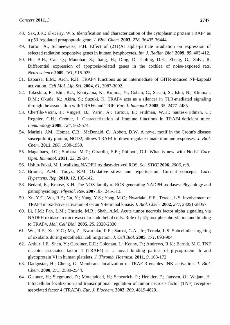
Cancers 2011, 3
2747
48. Sax, J.K.; El-Deiry, W.S. Identification and characterization of the cytoplasmic protein TRAF4 as
a p53-regulated proapoptotic gene. J. Biol. Chem. 2003, 278, 36435-36444.
49. Turtoi, A.; Schneeweiss, F.H. Effect of (211)At alpha-particle irradiation on expression of
selected radiation responsive genes in human lymphocytes. Int. J. Radiat. Biol. 2009, 85, 403-412.
50. Hu, B.H.; Cai, Q.; Manohar, S.; Jiang, H.; Ding, D.; Coling, D.E.; Zheng, G.; Salvi, R.
Differential expression of apoptosis-related genes in the cochlea of noise-exposed rats.
Neuroscience 2009, 161, 915-925.
51. Esparza, E.M.; Arch, R.H. TRAF4 functions as an intermediate of GITR-induced NF-kappaB
activation. Cell Mol. Life Sci. 2004, 61, 3087-3092.
52. Takeshita, F.; Ishii, K.J.; Kobiyama, K.; Kojima, Y.; Coban, C.; Sasaki, S.; Ishii, N.; Klinman,
D.M.; Okuda, K.; Akira, S.; Suzuki, K. TRAF4 acts as a silencer in TLR-mediated signaling
through the association with TRAF6 and TRIF. Eur. J. Immunol. 2005, 35, 2477-2485.
53. Cherfils-Vicini, J.; Vingert, B.; Varin, A.; Tartour, E.; Fridman, W.H.; Sautes-Fridman, C.;
Regnier, C.H.; Cremer, I. Characterization of immune functions in TRAF4-deficient mice.
Immunology 2008, 124, 562-574.
54. Marinis, J.M.; Homer, C.R.; McDonald, C.; Abbott, D.W. A novel motif in the Crohn's disease
susceptibility protein, NOD2, allows TRAF4 to down-regulate innate immune responses. J. Biol.
Chem. 2011, 286, 1938-1950.
55. Magalhaes, J.G.; Sorbara, M.T.; Girardin, S.E.; Philpott, D.J. What is new with Nods? Curr.
Opin. Immunol. 2011, 23, 29-34.
56. Ushio-Fukai, M. Localizing NADPH oxidase-derived ROS. Sci. STKE 2006, 2006, re8.
57. Briones, A.M.; Touyz, R.M. Oxidative stress and hypertension: Current concepts. Curr.
Hypertens. Rep. 2010, 12, 135-142.
58. Bedard, K.; Krause, K.H. The NOX family of ROS-generating NADPH oxidases: Physiology and
pathophysiology. Physiol. Rev. 2007, 87, 245-313.
59. Xu, Y.C.; Wu, R.F.; Gu, Y.; Yang, Y.S.; Yang, M.C.; Nwariaku, F.E.; Terada, L.S. Involvement of
TRAF4 in oxidative activation of c-Jun N-terminal kinase. J. Biol. Chem. 2002, 277, 28051-28057.
60. Li, J.M.; Fan, L.M.; Christie, M.R.; Shah, A.M. Acute tumor necrosis factor alpha signaling via
NADPH oxidase in microvascular endothelial cells: Role of p47phox phosphorylation and binding
to TRAF4. Mol. Cell Biol. 2005, 25, 2320-2330.
61. Wu, R.F.; Xu, Y.C.; Ma, Z.; Nwariaku, F.E.; Sarosi, G.A., Jr.; Terada, L.S. Subcellular targeting
of oxidants during endothelial cell migration. J. Cell Biol. 2005, 171, 893-904.
62. Arthur, J.F.; Shen, Y.; Gardiner, E.E.; Coleman, L.; Kenny, D.; Andrews, R.K.; Berndt, M.C. TNF
receptor-associated factor 4 (TRAF4) is a novel binding partner of glycoprotein Ib and
glycoprotein VI in human platelets. J. Thromb. Haemost. 2011, 9, 163-172.
63. Dadgostar, H.; Cheng, G. Membrane localization of TRAF 3 enables JNK activation. J. Biol.
Chem. 2000, 275, 2539-2544.
64. Glauner, H.; Siegmund, D.; Motejadded, H.; Scheurich, P.; Henkler, F.; Janssen, O.; Wajant, H.
Intracellular localization and transcriptional regulation of tumor necrosis factor (TNF) receptor-
associated factor 4 (TRAF4). Eur. J. Biochem. 2002, 269, 4819-4829.

Cancers 2011, 3
2748
65. Kedinger, V.; Alpy, F.; Baguet, A.; Polette, M.; Stoll, I.; Chenard, M.P.; Tomasetto, C.; Rio, M.C.
Tumor necrosis factor receptor-associated factor 4 is a dynamic tight junction-related shuttle
protein involved in epithelium homeostasis. PLoS ONE 2008, 3, e3518.
66. Colland, F.; Jacq, X.; Trouplin, V.; Mougin, C.; Groizeleau, C.; Hamburger, A.; Meil, A.; Wojcik,
J.; Legrain, P.; Gauthier, J.M. Functional proteomics mapping of a human signaling pathway.
Genome Res. 2004, 14, 1324-1332.
67. Li, S.; Lu, K.; Wang, J.; An, L.; Yang, G.; Chen, H.; Cui, Y.; Yin, X.; Xie, P.; Xing, G.; He, F.;
Zhang, L. Ubiquitin ligase Smurf1 targets TRAF family proteins for ubiquitination and
degradation. Mol. Cell Biochem. 2010, 338, 11-17.
68. Kalkan, T.; Iwasaki, Y.; Park, C.Y.; Thomsen, G.H. Tumor necrosis factor-receptor-associated
factor-4 is a positive regulator of transforming growth factor-beta signaling that affects neural
crest formation. Mol. Biol. Cell 2009, 20, 3436-3450.
69. Narimatsu, M.; Bose, R.; Pye, M.; Zhang, L.; Miller, B.; Ching, P.; Sakuma, R.; Luga, V.;
Roncari, L.; Attisano, L.; Wrana, J.L. Regulation of planar cell polarity by Smurf ubiquitin ligases.
Cell 2009, 137, 295-307.
70. Inoue, Y.; Imamura, T. Regulation of TGF-beta family signaling by E3 ubiquitin ligases. Cancer
Sci. 2008, 99, 2107-2112.
71. Kraut, R.; Chia, W.; Jan, L.Y.; Jan, Y.N.; Knoblich, J.A. Role of inscuteable in orienting
asymmetric cell divisions in Drosophila. Nature 1996, 383, 50-55.
72. Wang, H.; Cai, Y.; Chia, W.; Yang, X. Drosophila homologs of mammalian TNF/TNFR-related
molecules regulate segregation of Miranda/Prospero in neuroblasts. EMBO J. 2006, 25, 5783-5793.
73. Peng, C.Y.; Manning, L.; Albertson, R.; Doe, C.Q. The tumour-suppressor genes lgl and dlg
regulate basal protein targeting in Drosophila neuroblasts. Nature 2000, 408, 596-600.
74. Shiels, H.; Li, X.; Schumacker, P.T.; Maltepe, E.; Padrid, P.A.; Sperling, A.; Thompson, C.B.;
Lindsten, T. TRAF4 deficiency leads to tracheal malformation with resulting alterations in air
flow to the lungs. Am. J. Pathol. 2000, 157, 679-688.
75. Regnier, C.H.; Masson, R.; Kedinger, V.; Textoris, J.; Stoll, I.; Chenard, M.P.; Dierich, A.;
Tomasetto, C.; Rio, M.C. Impaired neural tube closure, axial skeleton malformations, and tracheal
ring disruption in TRAF4-deficient mice. Proc. Natl. Acad. Sci. USA 2002, 99, 5585-5590.
76. Abell, A.N.; Rivera-Perez, J.A.; Cuevas, B.D.; Uhlik, M.T.; Sather, S.; Johnson, N.L.; Minton,
S.K.; Lauder, J.M.; Winter-Vann, A.M.; Nakamura, K.; Magnuson, T.; Vaillancourt, R.R.;
Heasley, L.E.; Johnson, G.L. Ablation of MEKK4 kinase activity causes neurulation and skeletal
patterning defects in the mouse embryo. Mol. Cell Biol. 2005, 25, 8948-8959.
77. Chi, H.; Sarkisian, M.R.; Rakic, P.; Flavell, R.A. Loss of mitogen-activated protein kinase kinase
kinase 4 (MEKK4) results in enhanced apoptosis and defective neural tube development. Proc.
Natl. Acad. Sci. USA 2005, 102, 3846-3851.
78. Buac, K.; Watkins-Chow, D.E.; Loftus, S.K.; Larson, D.M.; Incao, A.; Gibney, G.; Pavan, W.J. A
Sox10 expression screen identifies an amino acid essential for Erbb3 function. PLoS Genet. 2008,
4, e1000177.
Cancers 2011, 3
2749
79. Masson, R.; Regnier, C.H.; Chenard, M.P.; Wendling, C.; Mattei, M.G.; Tomasetto, C.; Rio, M.C.
Tumor necrosis factor receptor associated factor 4 (TRAF4) expression pattern during mouse
development. Mech. Dev. 1998, 71, 187-191.
80. Aston, C.; Jiang, L.; Sokolov, B.P. Microarray analysis of postmortem temporal cortex from
patients with schizophrenia. J. Neurosci. Res. 2004, 77, 858-866.
81. Hsiao, L.L.; Dangond, F.; Yoshida, T.; Hong, R.; Jensen, R.V.; Misra, J.; Dillon, W.; Lee, K.F.;
Clark, K.E.; Haverty, P.; Weng, Z.; Mutter, G.L.; Frosch, M.P.; MacDonald, M.E.; Milford, E.L.;
Crum, C.P.; Bueno, R.; Pratt, R.E.; Mahadevappa, M.; Warrington, J.A.; Stephanopoulos, G.;
Stephanopoulos, G.; Gullans, S.R. A compendium of gene expression in normal human tissues.
Physiol. Genomics 2001, 7, 97-104.
82. Kedinger, V.; Alpy, F.; Tomasetto, C.; Thisse, C.; Thisse, B.; Rio, M.C. Spatial and temporal
distribution of the traf4 genes during zebrafish development. Gene Expr. Patterns 2005, 5, 545-552.
83. Taylor, J.S.; Braasch, I.; Frickey, T.; Meyer, A.; Van de Peer, Y. Genome duplication, a trait
shared by 22000 species of ray-finned fish. Genome Res. 2003, 13, 382-390.
84. Preiss, A.; Johannes, B.; Nagel, A.C.; Maier, D.; Peters, N.; Wajant, H. Dynamic expression of
Drosophila TRAF1 during embryogenesis and larval development. Mech. Dev. 2001, 100, 109-113.
85. Liu, H.; Su, Y.C.; Becker, E.; Treisman, J.; Skolnik, E.Y. A Drosophila TNF-receptor-associated
factor (TRAF) binds the ste20 kinase Misshapen and activates Jun kinase. Curr. Biol. 1999, 9, 101-104.
86. Mathew, S.J.; Kerridge, S.; Leptin, M. A small genomic region containing several loci required for
gastrulation in Drosophila. PLoS One 2009, 4, e7437.
87. Cha, G.H.; Cho, K.S.; Lee, J.H.; Kim, M.; Kim, E.; Park, J.; Lee, S.B.; Chung, J. Discrete
functions of TRAF1 and TRAF2 in Drosophila melanogaster mediated by c-Jun N-terminal kinase
and NF-kappaB-dependent signaling pathways. Mol. Cell Biol. 2003, 23, 7982-7991.
88. Kuranaga, E.; Kanuka, H.; Igaki, T.; Sawamoto, K.; Ichijo, H.; Okano, H.; Miura, M. Reaper-
mediated inhibition of DIAP1-induced DTRAF1 degradation results in activation of JNK in
Drosophila. Nat. Cell Biol. 2002, 4, 705-710.
89. Kornbluth, S.; White, K. Apoptosis in Drosophila: neither fish nor fowl (nor man, nor worm). J.
Cell Sci. 2005, 118, 1779-1787.
90. Geuking, P.; Narasimamurthy, R.; Basler, K. A genetic screen targeting the tumor necrosis
factor/Eiger signaling pathway: identification of Drosophila TAB2 as a functionally conserved
component. Genetics 2005, 171, 1683-1694.
91. Zapata, J.M.; Matsuzawa, S.; Godzik, A.; Leo, E.; Wasserman, S.A.; Reed, J.C. The Drosophila
tumor necrosis factor receptor-associated factor-1 (DTRAF1) interacts with Pelle and regulates
NFkappaB activity. J. Biol. Chem. 2000, 275, 12102-12107.
92. Abell, A.N.; Johnson, G.L. MEKK4 is an effector of the embryonic TRAF4 for JNK activation.
J. Biol. Chem. 2005, 280, 35793-35796.
© 2011 by the authors; licensee MDPI, Basel, Switzerland. This article is an open access article
distributed under the terms and conditions of the Creative Commons Attribution license
(http://creativecommons.org/licenses/by/3.0/).
46
1.5.2. Les données récentes sur TRAF4
1.5.2.1. TRAF4, un régulateur négatif de l’immunité
Plusieurs articles récents décrivent l’implication de TRAF4 dans l’immunité. TRAF4 est
un régulateur négatif de NOD2 (Nucleotide-binding Oligomerization Domain containing 2),
un PRR (Pattern Recognition Receptor) intracellulaire qui reconnait des peptidoglycanes
bactériens (Marinis et al., 2011). Une fois lié à son ligand, il va activer la voie NF-κB et
induire une réponse immunitaire appropriée. NOD2 est un gène de susceptibilité à la maladie
de Crohn, une pathologie caractérisée par une inflammation chronique de l’intestin. La perte
de fonction de NOD2 conduirait à une réponse immunitaire inappropriée et ainsi à une
synthèse de cytokines pro-inflammatoire accrue (Hugot et al., 2001). Une étude visant à
identifier des cibles de la kinase IKKα, a montré qu’IKKα phosphoryle TRAF4 sur sa sérine
426. De manière intéressante, seule la forme phosphorylée de TRAF4 interagit avec NOD2.
Ce résultat montre que la phosphorylation de TRAF4 par IKKα est nécessaire à l’inhibition de
NOD2 médiée par TRAF4 (Marinis et al., 2012).
Les lymphocytes Th17 sont des cellules T auxiliaires caractérisées par leur capacité à
produire en grande quantité l’IL-17, une cytokine pro-inflammatoire puissante. Ces cellules
sont connues pour participer au développement de nombreuses maladies auto-immunes telles
que la sclérose en plaque et la polyarthrite rhumatoïde (Rendon and Choudhry, 2012; Waite
and Skokos, 2011). En effet, une production accrue d’IL-17 conduit à une forte activation du
récepteur IL-17R qui, en recrutant les ubiquitines ligases E3 Act1 (NF-κB Activator 1) et
TRAF6, vont activer la voie NF-κB et induire la synthèse de nombreuses cytokines pro-
inflammatoires. De manière intéressante, les souris déficientes pour TRAF4 (Shiels et al.,
2000) ont une activation exacerbée de la voie NF-κB induite par l’IL-17. De plus, la
déficience de TRAF4 chez la souris prédispose au développement de la sclérose en plaque
dans des modèles inductibles de la maladie. Au niveau moléculaire, il a été montré que
TRAF4 inhibe la voie NF-κB induite par l’IL-17 en étant en compétition avec TRAF6 pour la
liaison à Act1 (Zepp et al., 2012).
Ensemble, ces données confirment que TRAF4 est un régulateur négatif des réponses
immunitaires et inflammatoires, et suggèrent que TRAF4 pourrait être un gène de
susceptibilité pour certaines maladies auto-immunes (Marinis et al., 2011; Zepp et al., 2012).
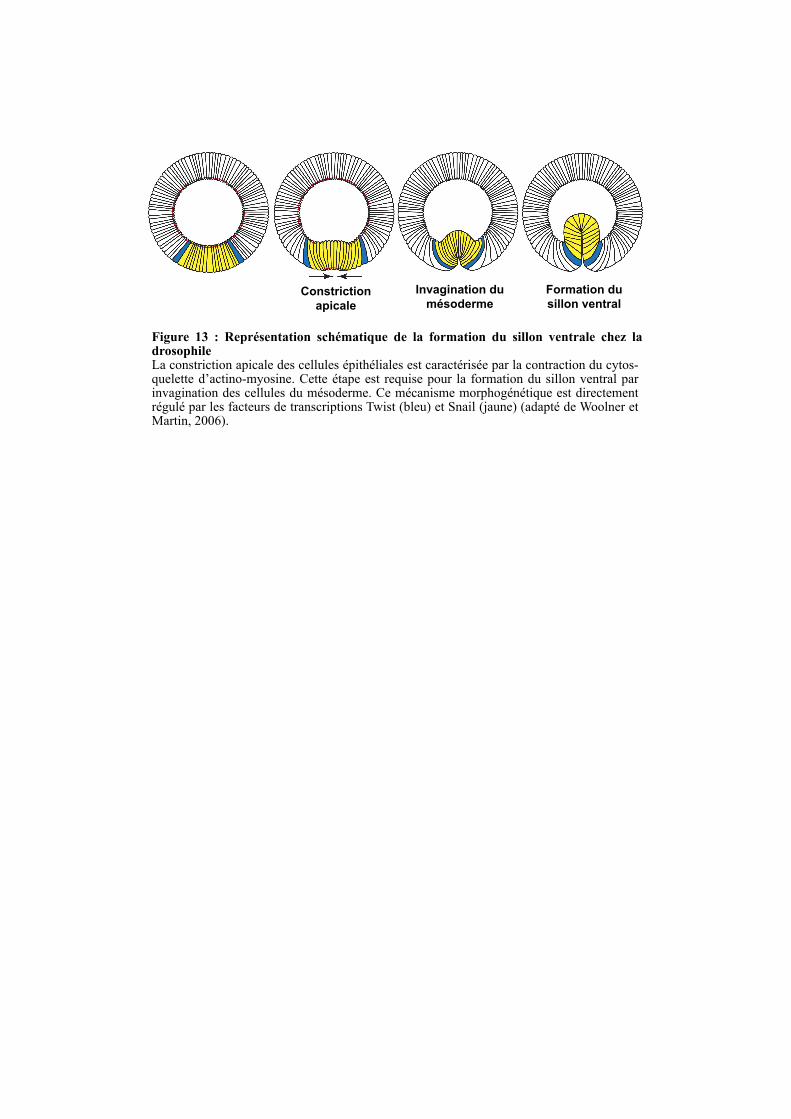
Figure 13 : Représentation schématique de la formation du sillon ventrale chez la drosophileLa constriction apicale des cellules épithéliales est caractérisée par la contraction du cytos-quelette d’actino-myosine. Cette étape est requise pour la formation du sillon ventral par invagination des cellules du mésoderme. Ce mécanisme morphogénétique est directement régulé par les facteurs de transcriptions Twist (bleu) et Snail (jaune) (adapté de Woolner et Martin, 2006).
Constriction
apicale
Formation du
sillon ventral
Invagination du
mésoderme
47
1.5.2.2. TRAF4, une protéine impliquée dans la myélinisation des axones
L’analyse comportementale des souris déficientes pour TRAF4, réalisée au laboratoire a
montré que la délétion de TRAF4 conduit chez les animaux survivants à une altération de la
coordination des mouvements. L’analyse morphologique de ces animaux a révélé des
altérations cellulaires majeures au niveau des cervelets. Il a été observé un défaut
d’enroulement de la gaine de myéline autour des axones ainsi qu’une dégénérescence des
cellules de Purkinje, deux phénotypes pouvant expliquer l’atteinte comportementale des
souris TRAF4 -/- (Blaise et al., 2012). Une étude moléculaire a montré que la déficience en
TRAF4 conduisait à une augmentation du niveau d’expression de Nogo-A (Neurite outgrowth
inhibitor-A), une protéine inhibitrice de la croissance des neurites (tout prolongement du
corps cellulaire d’un neurone) dans le cervelet (Blaise et al., 2012). Nogo-A a été impliqué
dans le processus de démyélinisation observé dans le développement de la sclérose en plaque
(Petratos et al., 2012; Yang et al., 2010). Ces données suggèrent que la délétion de TRAF4
pourrait promouvoir certaines maladies auto-immunes telle que la sclérose en plaque, en
stimulant la voie NF-κB induite par l’IL-17 et en augmentant l’expression de Nogo-A. Ce
travail réalisé au laboratoire a permis la publication d’un article dans lequel je suis co-auteur
(Blaise et al., 2012 ; Annexe 1).
1.5.2.3. TRAF4, une protéine impliquée dans la polarité cellulaire
Une étude récente menée chez la mouche, confirme le rôle de TRAF4 dans la polarité
cellulaire. La constriction apicale des cellules épithéliales est un processus largement utilisé
lors de la morphogénèse chez la drosophile (Woolner, 2007). Ce mécanisme implique la
contraction du cytosquelette d’acto-myosine dans la région apicale des cellules épithéliales du
blastoderme, une étape nécessaire à la formation du sillon ventral par invagination des
cellules du mésoderme (Figure 13). Ce mécanisme est directement régulé par les facteurs de
transcriptions Twist et Snail. Il a récemment été montré que Twist stimule l’expression de
DTRAF1 et que DTRAF1 est nécessaire à la formation du sillon ventral chez la drosophile.
Au niveau moléculaire, DTRAF1 est requis pour la localisation apicale d’Armadillo,
l’orthologue de la β-caténine chez la mouche (Mathew et al., 2011). La déficience de TRAF4
chez la drosophile conduit donc à un défaut de la polarité apico-basale conduisant à une
absence de constriction apicale.
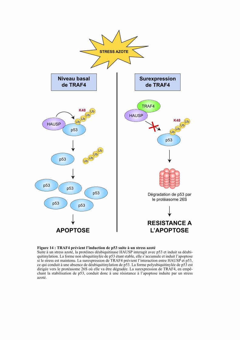
Figure 14 : TRAF4 prévient l’induction de p53 suite à un stress azotéSuite à un stress azoté, la protéines déubiquitinase HAUSP interagit avec p53 et induit sa déubi-quitinylation. La forme non ubiquitinylée de p53 étant stable, elle s’accumule et induit l’apoptose si le stress est maintenu. La surexpression de TRAF4 prévient l’interaction entre HAUSP et p53, ce qui conduit à une absence de déubiquitinylation de p53. La forme polyubiquitinylée de p53 est dirigée vers le protéasome 26S où elle va être dégradée. La surexpression de TRAF4, en empê-chant la stabilisation de p53, conduit donc à une résistance à l’apoptose induite par un stress azoté.
TRAF4
STRESS AZOTE
Niveau basal
de TRAF4
Surexpression
de TRAF4
p53
UbUb
UbUbK48
HAUSP
p53Ub
UbUb
Ub
p53p53
p53p53
p53
HAUSP
APOPTOSE
Dégradation de p53 par
le protéasome 26S
p53
UbUb
UbUbK48
RESISTANCE A
L’APOPTOSE
48
1.5.2.4. TRAF4, une protéine pro-tumorale
Un criblage différentiel réalisé au laboratoire a permis d’identifier TRAF4 comme étant
un gène surexprimé dans les cancers du sein (Tomasetto et al., 1995). Depuis, il a été montré
que la surexpression de TRAF4 est retrouvée dans environ 40% des carcinomes tous types
confondus (Camilleri-Broët et al., 2007; Rhodes et al., 2004). Bien que sa surexpression soit
clairement acceptée, son implication dans les cancers reste mal caractérisée.
SRC3 (Steroid Receptor Coactivator 3) est une protéine coactivatrice de la transcription
qui interagit avec les récepteurs nucléaires aux hormones stéroïdiennes (Garabedian and
Logan, 2008). La surexpression de SRC3 chez la souris est suffisante pour induire la
formation de carcinomes mammaire (Torres-Arzayus et al., 2004). De manière intéressante, il
a récemment été décrit que TRAF4 est impliquée dans la voie de signalisation médiée par
SCR3 et que leurs expressions sont corrélées dans les cancers du sein (Yi et al., 2013). Au
niveau cellulaire, il a été montré que les cellules sur-exprimant TRAF4 et/ou SRC3 sont
résistantes à l’apoptose due à un stress oxydatif. La surexpression de l’une de ces deux
protéines est suffisante pour la déstabilisation de p53 induite par un stress oxydatif (Yi et al.,
2013). Au niveau moléculaire, TRAF4 diminue l’interaction entre p53 et l’enzyme
déubiquitinase HAUSP (Herpesvirus-Associated Ubiquitin-Specific Protease), ce qui
engendre une augmentation de la polyubiquitinylation de p53 conduisant à sa dégradation par
le protéasome 26S (Figure 14). Cette étude a également montrée que la surexpression de
TRAF4 dans les cancers du sein est associée à un mauvais pronostique (Yi et al., 2013).
Ce travail apporte de nouvelles données quant au rôle de TRAF4 dans la carcinogénèse
mammaire. Dans ce travail c’est le rôle de la protéine dans les compartiments cytoplasmique
et/ou nucléaire qui est évoqué. La fonction de TRAF4 dans les jonctions serrées, des
structures clés dérégulées dans la carcinogénèse, reste peu claire à ce jour. Le déchiffrage de
la fonction de TRAF4 dans les jonctions serrées des cellules épithéliales mammaires a été
l’objet principal de mes travaux de thèse.
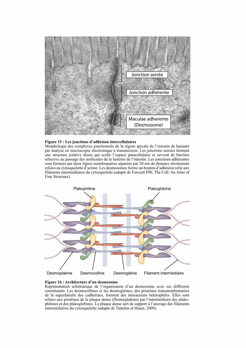
Figure 15 : Les jonctions d’adhésion intercellulaires Morphologie des complexes jonctionnels de la région apicale de l’intestin de hamster par analyse en microscopie électronique à transmission. Les jonctions serrées forment une structure jointive dense qui scelle l’espace paracellulaire et servent de barrière sélective au passage des molécules de la lumière de l’intestin. Les jonctions adhérentes sont formées par deux lignes membranaires séparées par 20 nm de distance étroitement reliées au cytosquelette d’actine. Les desmosomes forme un bouton d’adhésion relié aux filaments intermédiaires du cytosquelette (adapté de Fawcett DW, The Cell: An Atlas of Fine Structure).
Jonction serrée
Jonction adhérente
Maculae adherente
(Desmosome)
Desmocolline DesmogléineDesmoplakine
PlakoglobinePlakophiline
Filament intermédiaire
Figure 16 : Architecture d’un desmosome Représentation schématique de l’organisation d’un desmosome avec ses différents constituants. Les desmocollines et les desmogléines, des protéines transmembranaires de la superfamille des cadhérines, forment des interactions hétérophiles. Elles sont reliées aux protéines de la plaque dense (Desmoplakine) par l’intermédiaire des plako-philines et des plakoglobines. La plaque dense sert de support à l’ancrage des filaments intermédiaires du cytosquelette (adapté de Tintelen et Hauer, 2009).
49
CHAPITRE2:LAPOLARITEAPICO-BASALEDESCELLULES
EPITHELIALES
2.1. INTRODUCTION
La formation des contacts intercellulaires est essentielle pour la fonction des tissus et
organes. C’est particulièrement vrai pour l’épithélium qui est formé par des cellules
épithéliales qui adhèrent les unes aux autres. Les épithélia sont des feuillets cellulaires
couvrant les cavités de certains organes qui servent de barrières entre les différents
compartiments du corps (Franke, 2009; Niessen, 2007). Les épithélia sont impliqués dans de
nombreux processus tels que l’absorption, la sécrétion, la diffusion sélective de molécules et
l’intégrité physique du corps. Les jonctions intercellulaires telles que les desmosomes, les
jonctions adhérentes (JAs) et les jonctions serrées (JSs), jouent un rôle crucial dans la
formation et le maintien de l’épithélium. Ces trois systèmes de jonctions ont une architecture
similaire fondée sur des protéines transmembranaires associées à des protéines formant une
plaque cytoplasmique (Figure 15) (St Johnston and Sanson, 2011). Les jonctions
communicantes (JCs) forment, quant à elles, un système de canaux intercellulaires permettant
l’échange direct de molécules de petites tailles entre les cellules. Ainsi, en plus de participer à
l’adhésion intercellulaire, elles ont un rôle homéostatique au sein du tissu (Matsuuchi and
Naus, 2013).
2.2. LES DESMOSOMES
Les desmosomes sont majoritairement présents au sein des tissus épithéliaux mais ils sont
également retrouvés au sein d’autres tissus comme le muscle et le thymus. Leur rôle principal
est de maintenir les cellules voisines assemblées et d’offrir des points d’ancrage aux filaments
intermédiaires du cytosquelette (Garrod and Chidgey, 2008). L’architecture ultrastructurale
des desmosomes a été caractérisée dès les années 70 (Figure 15) (Farquhar and Palade, 1963;
Staehelin, 1974). Les desmosomes sont des complexes moléculaires formés de trois
composants : des protéines transmembranaires (desmocolline et desmogléine), des protéines
d’ancrage (desmoplakine, plakoglobine et plakophiline) et des filaments intermédiaires
(kératine et vimentine respectivement au sein des cellules épithéliales et fibroblastiques)
(Figure 16). La desmocolline et la desmogléine, deux protéines de la famille des cadhérines,
vont établir des interactions hétérophiles entre cellules voisines de manière calcium-
dépendante (Figure 16). Sur la face cytosolique, les desmoplakines forment la plaque dense
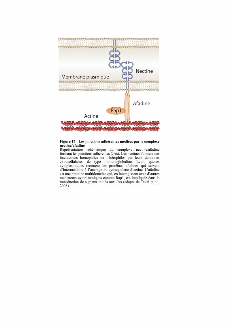
Nectine
Afadine
Rap1Actine
Membrane plasmique
Figure 17 : Les jonctions adhérentes médiées par le complexe nectine/afadineReprésentation schématique du complexe nectine/afadine formant les jonctions adhérentes (JAs). Les nectines forment des interactions homophiles ou hétérophiles par leurs domaines extracellulaires de type immunoglobuline. Leurs queues cytoplasmiques recrutent les protéines afadines qui servent d’intermédiaire à l’ancrage du cytosquelette d’actine. L’afadine est une protéine multidomaine qui, en interagissant avec d’autres médiateurs cytoplasmiques comme Rap1, est impliquée dans la transduction de signaux initiés aux JAs (adapté de Takai et al., 2008).
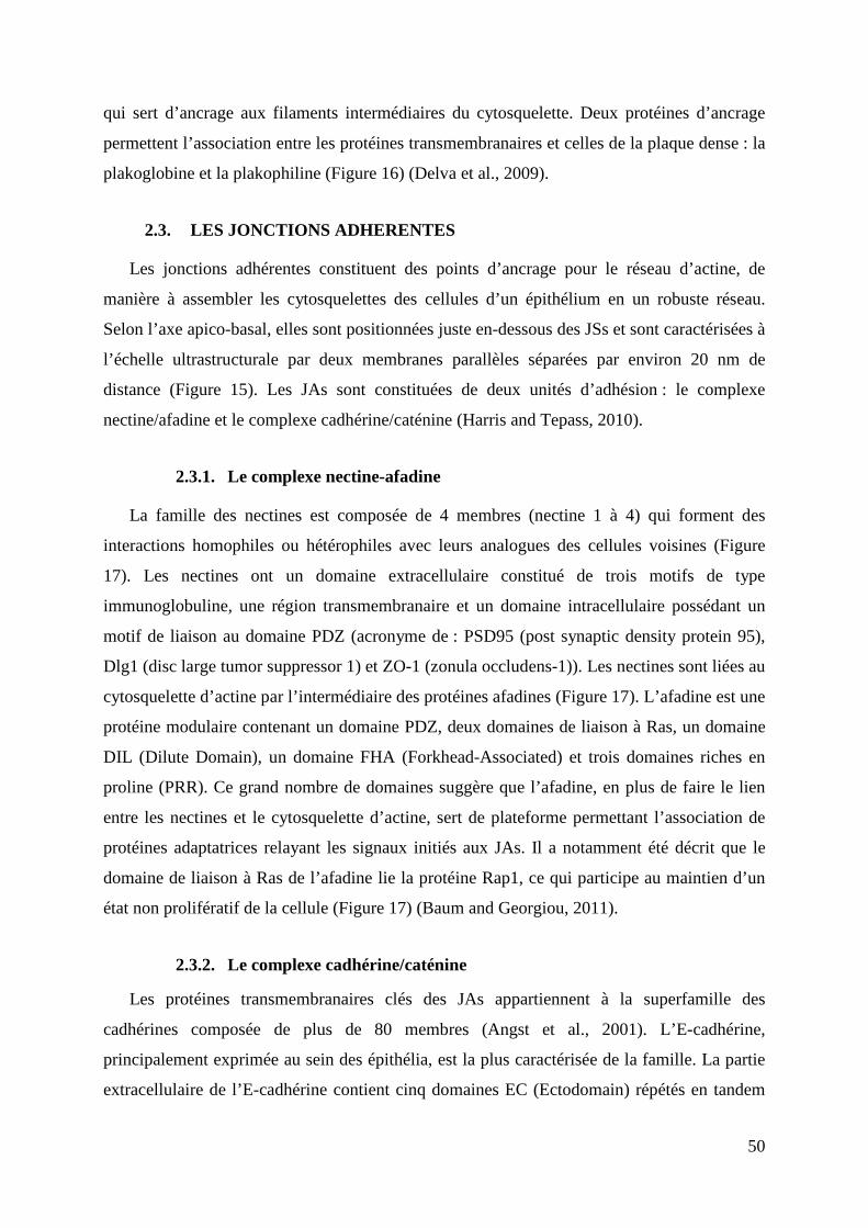
50
qui sert d’ancrage aux filaments intermédiaires du cytosquelette. Deux protéines d’ancrage
permettent l’association entre les protéines transmembranaires et celles de la plaque dense : la
plakoglobine et la plakophiline (Figure 16) (Delva et al., 2009).
2.3. LES JONCTIONS ADHERENTES
Les jonctions adhérentes constituent des points d’ancrage pour le réseau d’actine, de
manière à assembler les cytosquelettes des cellules d’un épithélium en un robuste réseau.
Selon l’axe apico-basal, elles sont positionnées juste en-dessous des JSs et sont caractérisées à
l’échelle ultrastructurale par deux membranes parallèles séparées par environ 20 nm de
distance (Figure 15). Les JAs sont constituées de deux unités d’adhésion : le complexe
nectine/afadine et le complexe cadhérine/caténine (Harris and Tepass, 2010).
2.3.1. Le complexe nectine-afadine
La famille des nectines est composée de 4 membres (nectine 1 à 4) qui forment des
interactions homophiles ou hétérophiles avec leurs analogues des cellules voisines (Figure
17). Les nectines ont un domaine extracellulaire constitué de trois motifs de type
immunoglobuline, une région transmembranaire et un domaine intracellulaire possédant un
motif de liaison au domaine PDZ (acronyme de : PSD95 (post synaptic density protein 95),
Dlg1 (disc large tumor suppressor 1) et ZO-1 (zonula occludens-1)). Les nectines sont liées au
cytosquelette d’actine par l’intermédiaire des protéines afadines (Figure 17). L’afadine est une
protéine modulaire contenant un domaine PDZ, deux domaines de liaison à Ras, un domaine
DIL (Dilute Domain), un domaine FHA (Forkhead-Associated) et trois domaines riches en
proline (PRR). Ce grand nombre de domaines suggère que l’afadine, en plus de faire le lien
entre les nectines et le cytosquelette d’actine, sert de plateforme permettant l’association de
protéines adaptatrices relayant les signaux initiés aux JAs. Il a notamment été décrit que le
domaine de liaison à Ras de l’afadine lie la protéine Rap1, ce qui participe au maintien d’un
état non prolifératif de la cellule (Figure 17) (Baum and Georgiou, 2011).
2.3.2. Le complexe cadhérine/caténine
Les protéines transmembranaires clés des JAs appartiennent à la superfamille des
cadhérines composée de plus de 80 membres (Angst et al., 2001). L’E-cadhérine,
principalement exprimée au sein des épithélia, est la plus caractérisée de la famille. La partie
extracellulaire de l’E-cadhérine contient cinq domaines EC (Ectodomain) répétés en tandem
Ca 2+
α -actinine
Actine
p120-caténine
β-caténine β-caténine
VinculineVinculine
Membrane plasmique
E-cadhérine
α -caténineα -caténine
p120-caténine
domaine EC
Figure 18 : Les jonctions adhérentes médiées par le complexe E-cadhérine /caténineReprésentation schématique du complexe E-cadhérine/caténine formant les jonctions adhérentes (JAs). Les E-cadhérines forment des interactions homophiles grâce à leurs domaines extracellu-laires EC (Ectodomain) de manière calcium dépendante. Sa queue cytoplasmique recrute la β-caténine qui sert d’intermédiaire au recrutement de l’α-caténine. L’α-caténine permet l’ancrage du cytosquelette en liant directement ou indirectement (via α-actinine et vinculine) l’actine. La protéine p120-caténine est un régulateur des JAs appartenant à la famille des protéines à répéti-tions Armadello (adapté de Kobielak and Fuchs, 2004).
51
qui, après avoir lié du calcium, forment des liaisons homophiles avec leurs analogues des
cellules voisines (Figure 18). La taille du domaine extracellulaire de l’E-cadhérine est ~22
nm, ce qui est cohérent avec la séparation d’environ 20 nm des deux membranes plasmiques
formant la JA. L’E-cadhérine a une région cytoplasmique de 150 acides aminés qui est très
conservée dans l’évolution. Cette région contient des sites de liaison pour la β-caténine et la
plakoglobine, deux membres de la superfamille des protéines à répétitions Armadillo (Figure
18) (Harris and Tepass, 2010; Yonemura et al., 1995). Il a été montré que l’interaction entre la
queue cytoplasmique de l’E-cadhérine avec les 12 répétitions Armadillo de la β-caténine était
requise au trafic des E-cadhérines néosynthétisées vers la membrane plasmique (Chen et al.,
1999). Le domaine N-terminal de la β-caténine et de la plakoglobine lient directement l’α-
caténine. Comme l’α-caténine est capable d’interagir directement ou indirectement (via l’α-
actinine et la vinculine) avec les microfilaments d’actine, elle permet l’ancrage du complexe
E-cadhérine/caténines au cytosquelette d’actine (Figure 18) (Baum and Georgiou, 2011). La
liaison de l’α-caténine à la protéine afadine permet également de rassembler les complexes
d’adhésion E-cadhérine/caténine et nectine/afadine au niveau des JAs. Ces mécanismes sont
importants pour la stabilisation des interactions cellule-cellule ainsi que pour la coordination
entre les JAs et le cytosquelette d’actine (Baum and Georgiou, 2011).
La protéine p120-caténine, un autre membre de la famille des protéines à répétitions
Armadillo, est un régulateur central des JAs médiées par les cadhérines (Figure 18). En effet,
la liaison de p120-caténine à l’E-cadhérine est nécessaire au maintien d’un nombre adéquat
d’E-cadhérines au sein des JAs. Plusieurs agents tels que l’histamine et la thrombine régulent
la phosphorylation de p120-caténine par la protéine kinase C (PKC), permettant une
modulation rapide de l’adhésion cellule-cellule (Xia et al., 2003).
2.4. LES JONCTIONS SERREES
En 1963, Farquhar et Palade ont découvert les JSs par des observations d’épithélia en
microscopie électronique (Figure 15) (Farquhar and Palade, 1963). Ce sont les premiers à
proposer l’idée que les JSs, en formant une structure jointive, servent de barrière sélective
régulant le passage des molécules à travers l’espace paracellulaire (Figure 15). Les JSs sont
positionnées dans la région apicale séparent la membrane cellulaire en deux territoires
distincts : apical et basolatéral, dont la composition en lipides et protéines diffère. La perte des
JSs conduit à un mélange des protéines et lipides de ces deux compartiments et ainsi à la perte
de la polarité apico-basale (Furuse, 2010; Shin et al., 2006).
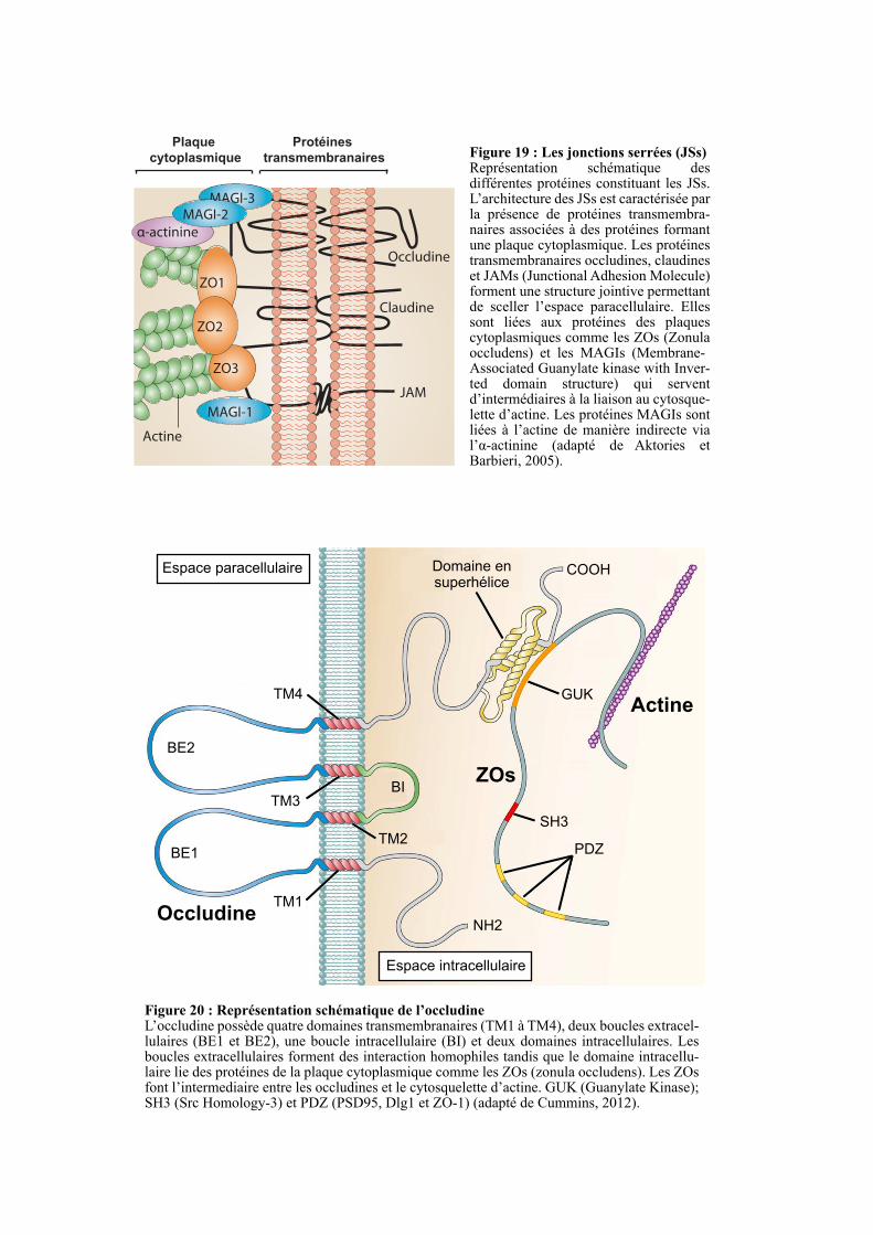
Actine
Occludine
Claudine
JAM
ZO1
ZO3
ZO2
MAGI-1
MAGI-3
α-actinine
MAGI-2
Plaque
cytoplasmique
Protéines
transmembranaires Figure 19 : Les jonctions serrées (JSs) Représentation schématique des différentes protéines constituant les JSs. L’architecture des JSs est caractérisée par la présence de protéines transmembra-naires associées à des protéines formant une plaque cytoplasmique. Les protéines transmembranaires occludines, claudines et JAMs (Junctional Adhesion Molecule) forment une structure jointive permettant de sceller l’espace paracellulaire. Elles sont liées aux protéines des plaques cytoplasmiques comme les ZOs (Zonula occludens) et les MAGIs (Membrane-Associated Guanylate kinase with Inver-ted domain structure) qui servent d’intermédiaires à la liaison au cytosque-lette d’actine. Les protéines MAGIs sont liées à l’actine de manière indirecte via l’α-actinine (adapté de Aktories et Barbieri, 2005).
Espace paracellulaire
Espace intracellulaire
Domaine en superhélice
COOH
GUK
SH3
PDZTM2
TM1
TM3
TM4
BE2
BE1
NH2
BI
Occludine
ZOs
Actine
Figure 20 : Représentation schématique de l’occludine L’occludine possède quatre domaines transmembranaires (TM1 à TM4), deux boucles extracel-lulaires (BE1 et BE2), une boucle intracellulaire (BI) et deux domaines intracellulaires. Les boucles extracellulaires forment des interaction homophiles tandis que le domaine intracellu-laire lie des protéines de la plaque cytoplasmique comme les ZOs (zonula occludens). Les ZOs font l’intermediaire entre les occludines et le cytosquelette d’actine. GUK (Guanylate Kinase); SH3 (Src Homology-3) et PDZ (PSD95, Dlg1 et ZO-1) (adapté de Cummins, 2012).
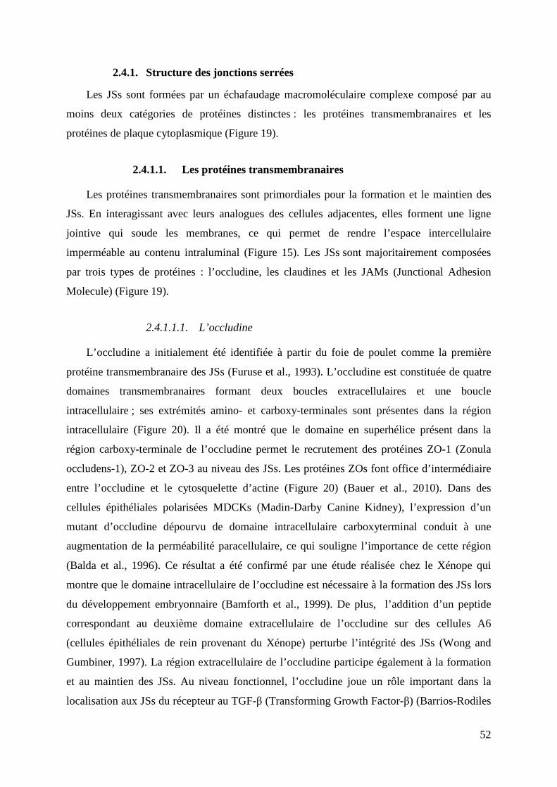
52
2.4.1. Structure des jonctions serrées
Les JSs sont formées par un échafaudage macromoléculaire complexe composé par au
moins deux catégories de protéines distinctes : les protéines transmembranaires et les
protéines de plaque cytoplasmique (Figure 19).
2.4.1.1. Les protéines transmembranaires
Les protéines transmembranaires sont primordiales pour la formation et le maintien des
JSs. En interagissant avec leurs analogues des cellules adjacentes, elles forment une ligne
jointive qui soude les membranes, ce qui permet de rendre l’espace intercellulaire
imperméable au contenu intraluminal (Figure 15). Les JSs sont majoritairement composées
par trois types de protéines : l’occludine, les claudines et les JAMs (Junctional Adhesion
Molecule) (Figure 19).
2.4.1.1.1. L’occludine
L’occludine a initialement été identifiée à partir du foie de poulet comme la première
protéine transmembranaire des JSs (Furuse et al., 1993). L’occludine est constituée de quatre
domaines transmembranaires formant deux boucles extracellulaires et une boucle
intracellulaire ; ses extrémités amino- et carboxy-terminales sont présentes dans la région
intracellulaire (Figure 20). Il a été montré que le domaine en superhélice présent dans la
région carboxy-terminale de l’occludine permet le recrutement des protéines ZO-1 (Zonula
occludens-1), ZO-2 et ZO-3 au niveau des JSs. Les protéines ZOs font office d’intermédiaire
entre l’occludine et le cytosquelette d’actine (Figure 20) (Bauer et al., 2010). Dans des
cellules épithéliales polarisées MDCKs (Madin-Darby Canine Kidney), l’expression d’un
mutant d’occludine dépourvu de domaine intracellulaire carboxyterminal conduit à une
augmentation de la perméabilité paracellulaire, ce qui souligne l’importance de cette région
(Balda et al., 1996). Ce résultat a été confirmé par une étude réalisée chez le Xénope qui
montre que le domaine intracellulaire de l’occludine est nécessaire à la formation des JSs lors
du développement embryonnaire (Bamforth et al., 1999). De plus, l’addition d’un peptide
correspondant au deuxième domaine extracellulaire de l’occludine sur des cellules A6
(cellules épithéliales de rein provenant du Xénope) perturbe l’intégrité des JSs (Wong and
Gumbiner, 1997). La région extracellulaire de l’occludine participe également à la formation
et au maintien des JSs. Au niveau fonctionnel, l’occludine joue un rôle important dans la
localisation aux JSs du récepteur au TGF-β (Transforming Growth Factor-β) (Barrios-Rodiles
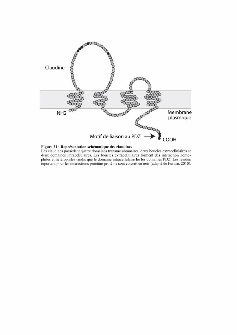
Claudine
NH2
COOH
Membrane plasmique
Motif de liaison au PDZ
Figure 21 : Représentation schématique des claudines Les claudines possèdent quatre domaines transmembranaires, deux boucles extracellulaires et deux domaines intracellulaires. Les boucles extracellulaires forment des interaction homo-philes et hétérophiles tandis que le domaine intracellulaire lie les domaines PDZ. Les résidus inportant pour les interactions protéine-protéine sont colorés en noir (adapté de Furuse, 2010).
53
et al., 2005). Ce mécanisme pourrait être requis pour l’induction de la transition épithélio-
mésenchymateuse (TEM) médiée par le TGF-β, un processus qui nécessite la perte des
jonctions intercellulaires.
2.4.1.1.2. Les claudines
A l’instar de l’occludine, les claudines ont quatre domaines transmembranaires, deux
boucles extracellulaires et deux domaines intracellulaires (Figure 21). Malgré cette
ressemblance topologique, aucune similarité de séquence n’est observée entre ces deux
familles. Tsukita et ses collègues sont les premiers à avoir isolé des claudines à partir du foie
de poulet (Furuse et al., 1998). Actuellement, 27 claudines différentes ont été identifiées chez
les mammifères (Günzel and Yu, 2013). La formation de liaisons homophiles et hétérophiles
via leurs domaines extracellulaires est un mécanisme clé pour la formation et le maintien des
JSs. Il a notamment été montré que l’expression ectopique des claudines 1 et 2 au sein de
cellules fibroblastiques induit une agrégation cellulaire. Une analyse en microscopie
électronique a révélé la présence de contacts cellulaires composés d’un réseau jonctionnel
bien développé, similaire à celui des JSs. Lorsque l’occludine et les claudines sont co-
exprimées dans ces fibroblastes, l’occludine est recrutée dans les structures jonctionnelles
formées par les claudines. Ces résultats suggèrent que les claudines sont les protéines
initiatrices des JSs et qu’elles vont servir de base au recrutement d’autres composants (Furuse,
2010; Furuse et al., 1998). La région carboxyterminale des claudines possède un motif de
liaison au domaine PDZ très conservé au sein de la famille (Figure 21). Ce domaine permet
aux claudines d’interagir avec les protéines des plaques cytoplasmiques comme les protéines
ZOs et PATJ (Protein-Associated with Lin Seven 1(PALS1)-Associated Tight Junction
protein) (Shin et al., 2006).
Les claudines sont régulées au niveau transcriptionnel par la protéine Snail, un facteur de
transcription à doigt de zinc impliqué dans la TEM. Snail est un répresseur de l’expression de
plusieurs protéines des jonctions intercellulaires incluant l’E-cadhérine, l’occludine et les
claudines. De ce fait, l’expression de Snail dans des cellules polarisées conduit à une
augmentation de la perméabilité paracellulaire et in fine à une perte des JSs (Ikenouchi et al.,
2003).
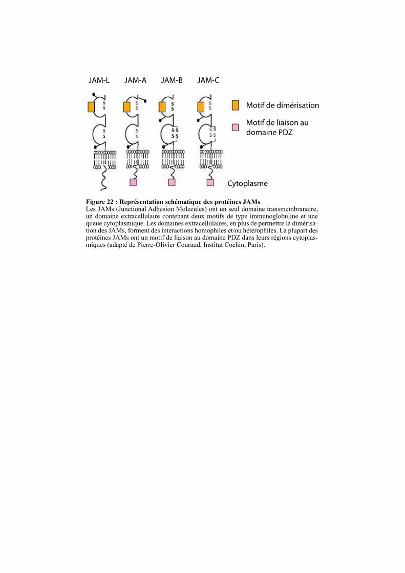
JAM-L JAM-A JAM-B JAM-C
Cytoplasme
Motif de dimérisation
Motif de liaison au
domaine PDZ
Figure 22 : Représentation schématique des protéines JAMs Les JAMs (Junctional Adhesion Molecules) ont un seul domaine transmembranaire, un domaine extracellulaire contenant deux motifs de type immunoglobuline et une queue cytoplasmique. Les domaines extracellulaires, en plus de permettre la dimérisa-tion des JAMs, forment des interactions homophiles et/ou hétérophiles. La plupart des protéines JAMs ont un motif de liaison au domaine PDZ dans leurs régions cytoplas-miques (adapté de Pierre-Olivier Couraud, Institut Cochin, Paris).
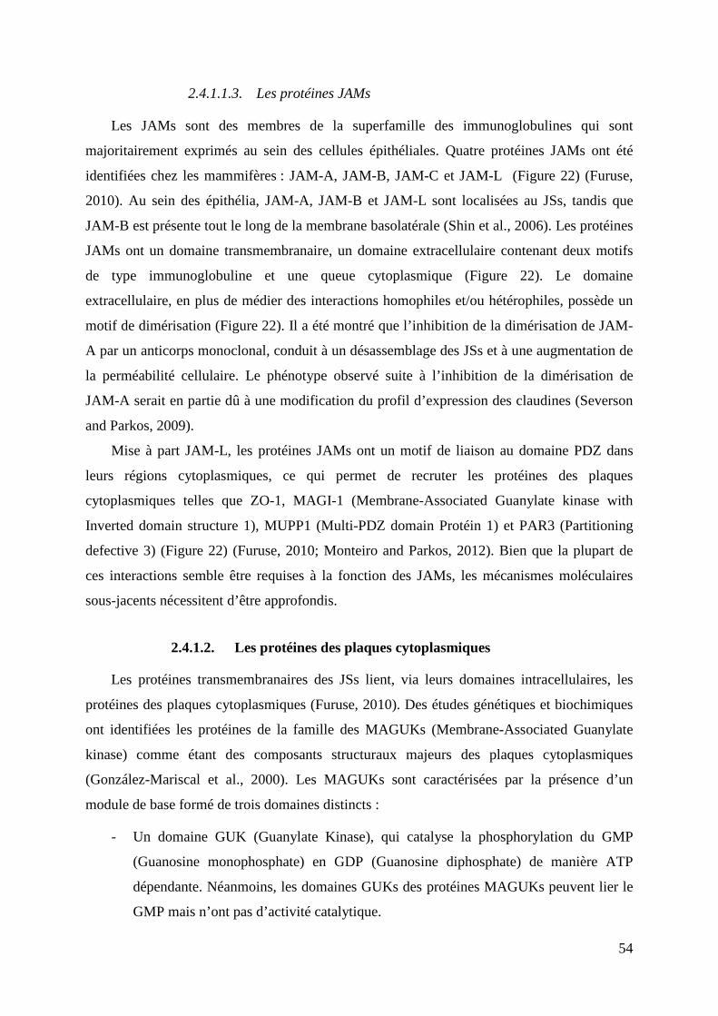
54
2.4.1.1.3. Les protéines JAMs
Les JAMs sont des membres de la superfamille des immunoglobulines qui sont
majoritairement exprimés au sein des cellules épithéliales. Quatre protéines JAMs ont été
identifiées chez les mammifères : JAM-A, JAM-B, JAM-C et JAM-L (Figure 22) (Furuse,
2010). Au sein des épithélia, JAM-A, JAM-B et JAM-L sont localisées au JSs, tandis que
JAM-B est présente tout le long de la membrane basolatérale (Shin et al., 2006). Les protéines
JAMs ont un domaine transmembranaire, un domaine extracellulaire contenant deux motifs
de type immunoglobuline et une queue cytoplasmique (Figure 22). Le domaine
extracellulaire, en plus de médier des interactions homophiles et/ou hétérophiles, possède un
motif de dimérisation (Figure 22). Il a été montré que l’inhibition de la dimérisation de JAM-
A par un anticorps monoclonal, conduit à un désassemblage des JSs et à une augmentation de
la perméabilité cellulaire. Le phénotype observé suite à l’inhibition de la dimérisation de
JAM-A serait en partie dû à une modification du profil d’expression des claudines (Severson
and Parkos, 2009).
Mise à part JAM-L, les protéines JAMs ont un motif de liaison au domaine PDZ dans
leurs régions cytoplasmiques, ce qui permet de recruter les protéines des plaques
cytoplasmiques telles que ZO-1, MAGI-1 (Membrane-Associated Guanylate kinase with
Inverted domain structure 1), MUPP1 (Multi-PDZ domain Protéin 1) et PAR3 (Partitioning
defective 3) (Figure 22) (Furuse, 2010; Monteiro and Parkos, 2012). Bien que la plupart de
ces interactions semble être requises à la fonction des JAMs, les mécanismes moléculaires
sous-jacents nécessitent d’être approfondis.
2.4.1.2. Les protéines des plaques cytoplasmiques
Les protéines transmembranaires des JSs lient, via leurs domaines intracellulaires, les
protéines des plaques cytoplasmiques (Furuse, 2010). Des études génétiques et biochimiques
ont identifiées les protéines de la famille des MAGUKs (Membrane-Associated Guanylate
kinase) comme étant des composants structuraux majeurs des plaques cytoplasmiques
(González-Mariscal et al., 2000). Les MAGUKs sont caractérisées par la présence d’un
module de base formé de trois domaines distincts :
- Un domaine GUK (Guanylate Kinase), qui catalyse la phosphorylation du GMP
(Guanosine monophosphate) en GDP (Guanosine diphosphate) de manière ATP
dépendante. Néanmoins, les domaines GUKs des protéines MAGUKs peuvent lier le
GMP mais n’ont pas d’activité catalytique.
ZO-1
ZO-2
ZO-3
PDZ-1 SH3 GUK P P P P P P P P
ClaudinesZO-2, ZO-3
JAMs ZONAB Occludinα-caténine Afadine
PDZ-1 P P P P P
Claudines ZO-1Scribble
Occludin, α-caténine
PDZ-1 P P P SH3
Claudins ZO-1 Afadinep120 caténine
PATJ
Actine, ZO-1
PDZ-3
PDZ-3PDZ-2
PDZ-2
PDZ-3PDZ-2
phosphoinositides
SH3
MAGUK
Figure 23 : Représentation schématique des protéines ZOsLes ZOs (Zonula Occludens) font partie de la superfamille des MAGUKs qui est caractérisée par la présence d’un module de base formé de trois domaines : un domaine GUK (Guanylate Kinase), un domaine SH3 (Src Homolo-gy-3) et un ou trois domaines PDZ (PSD95, Dlg1 et ZO-1). Le domaine GUK qui catalyse la phosphorylation du GMP (Guanosine monophosphate) en GDP (Guanosine diphosphate) est non fonctionnel au sein des MAGUKs. Le domaine SH3 interagit avec des motifs riches en prolines tandis que les domaines PDZs lient des protéines et/ou des phosphoinositides. P-P-P : région riche en prolines. Les motifs NLSs sont représentées par des boites rouges (adapté de Bauer et al., 2010).
phosphoinositides
GUK
GUK
55
- Un ou plusieurs domaines PDZs qui sont des domaines d’interaction protéine-
protéine.
- Un domaine de liaison aux régions riches en prolines, majoritairement de type SH3
(Src Homology-3).
Les deux membres les plus étudiés de cette famille sont les protéines ZOs et les protéines
MAGIs (Figure 19) (Guillemot et al., 2008).
2.4.1.2.1. La famille ZO
La famille ZO est composée de trois membres : ZO-1, ZO-2 et ZO-3 (Figure 23). Les
ZOs sont généralement localisées au niveau des JSs des cellules épithéliales et endothéliales.
ZO-3 est restreinte aux cellules épithéliales. ZO-1 est également exprimée dans des cellules
non épithéliales comme les astrocytes, les cellules de Schwann et les fibroblastes. ZO-2, à
l’instar de ZO-1, n’est pas restreinte aux cellules épithéliales et endothéliales, mais est
également exprimée par les fibroblastes et les cellules musculaires cardiaques (Bauer et al.,
2010; Inoko et al., 2003).
Plusieurs études ont montré que ZO-1 joue un rôle clé dans la formation et le maintien
des JSs. En effet, ZO-1 participe au développement des JSs lors de la formation du blastocyste
chez la souris. Les cellules épithéliales dérivées de souris déficientes pour ZO-1, cultivées in
vitro, montrent un retard dans la formation des JSs (Umeda et al., 2004). ZO-1, en plus de
posséder les domaines caractéristiques des MAGUKs, contient deux signaux de localisation
nucléaire (NLS : Nuclear Localization Signal) et un domaine riche en proline (PRR : Proline-
Rich Region) (Figure 23). Plusieurs études ont montré que ZO-1 a une localisation nucléaire
durant la formation de l’épithélium, où elle régule la prolifération cellulaire en interagissant
avec le facteur de transcription ZONAB (ZO-1-associated Nucleic Acid-Binding) (Bauer et
al., 2010). Il a également été montré que ZO-1, une fois dans le noyau, peut réguler
l’expression de gènes cibles comme l’IL-8, une cytokine impliquée dans la progression
tumorale (Brysse et al., 2012).
Dans les cellules épithéliales non confluentes, ZO-2 a une localisation nucléaire induite
par ses deux séquences NLS (Figure 23). Dans ce compartiment, elle interagit directement
avec plusieurs facteurs de transcription incluant Fos et Jun (Bauer et al., 2010). Dans les
cellules épithéliales confluentes, sa localisation subcellulaire est restreinte aux JSs. Ces
données suggèrent que la présence des JSs est suffisante pour recruter ZO-2 et que la perte de
ces structures jonctionnelles conduit à sa délocalisation dans le cytoplasme et dans le noyau
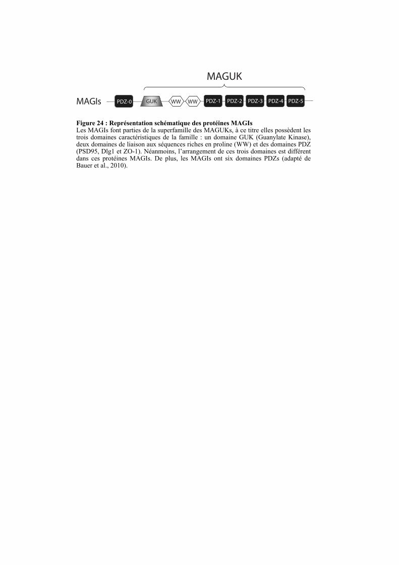
MAGIs PDZ-0
MAGUK
Figure 24 : Représentation schématique des protéines MAGIsLes MAGIs font parties de la superfamille des MAGUKs, à ce titre elles possèdent les trois domaines caractéristiques de la famille : un domaine GUK (Guanylate Kinase), deux domaines de liaison aux séquences riches en proline (WW) et des domaines PDZ (PSD95, Dlg1 et ZO-1). Néanmoins, l’arrangement de ces trois domaines est différent dans ces protéines MAGIs. De plus, les MAGIs ont six domaines PDZs (adapté de Bauer et al., 2010).
WW PDZ-1GUK WW PDZ-2 PDZ-3 PDZ-4 PDZ-5
56
(Jaramillo et al., 2004). A l’inverse des autres membres de la famille, ZO-3 est dépourvue de
séquences NLS (Bauer et al., 2010).
Comme mentionné ci-dessus, les ZOs sont des protéines structurales des plaques
cytoplasmiques qui interagissent avec de nombreuses protéines incluant des protéines
transmembranaires et des composants du cytosquelette. ZO-1 peut interagir avec ZO-2 et ZO-
3 via son deuxième domaine PDZ (Figure 23). Toutes les protéines ZOs interagissent
directement avec la queue cytoplasmique de l’occludine et des claudines (Feldman et al.,
2005). Elles peuvent également lier directement et indirectement les filaments d’actine, ce qui
permet l’ancrage du cytosquelette d’actine aux JSs (Bauer et al., 2010). De plus, au sein des
cellules épithéliales non confluentes, ZO-1 interagit avec des protéines des JAs telles que l’E-
cadhérine et les caténines (Figure 23) (Harris and Tepass, 2010; Niessen, 2007). Prises dans
leur ensemble, ces observations indiquent que les protéines ZOs sont recrutées aux JAs au
sein des cellules épithéliales non confluentes ; après polarisation, elles vont se concentrer dans
les JSs en interagissant avec l’occludine et les claudines (Müller et al., 2005).
2.4.1.2.2. La famille MAGI
Les protéines MAGI-1, -2 et -3 appartiennent à la famille des MAGUKs mais ont trois
différences majeures avec les autres membres : (1) le domaine GUK est localisé en N-
terminal au lieu d’être en C-terminal, (2) le domaine SH3 est remplacé par un domaine WW,
qui est également un module d’interaction protéine-protéine liant les domaines riches en
prolines, et (3) les protéines MAGIs ont six domaines PDZ alors que les autres protéines de
cette famille en ont un ou trois (Figure 24) (Dobrosotskaya et al., 1997). Tandis que MAGI-1
et MAGI-3 sont exprimées dans de nombreux tissus, MAGI-2 est essentiellement retrouvée
dans le cerveau. MAGI-1 est localisée aux JSs mais est également présente dans les JAs du
fait de son interaction avec la β-caténine. L’augmentation du recrutement de MAGI-1 aux JAs
dans les cellules non polarisées suggère que, à l’instar des protéines ZOs, la localisation
subcellulaire de MAGI-1 évolue au cours de la polarisation des cellules (Guillemot et al.,
2008). De plus, MAGI-1 interagit avec la synaptopodine et l’α-actinine-4, deux protéines de
liaison à l’actine, respectivement via ses domaines WW et PDZ-5. Ces interactions suggèrent
qu’elle joue un rôle dans l’ancrage du cytosquelette d’actine aux jonctions intercellulaires
(Patrie et al., 2002).
Les protéines MAGIs sont impliquées dans la transduction de signaux initiés aux
jonctions intercellulaires grâce à leurs interactions avec des protéines de signalisation comme
K-Ras, β-caténine et PTEN (Phosphatase and TENsin homolog) (Guillemot et al., 2008).
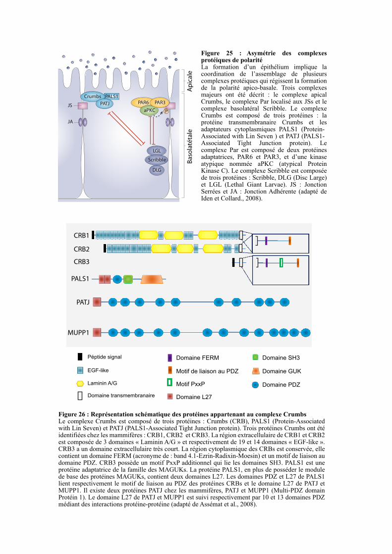
Figure 26 : Représentation schématique des protéines appartenant au complexe CrumbsLe complexe Crumbs est composé de trois protéines : Crumbs (CRB), PALS1 (Protein-Associated with Lin Seven) et PATJ (PALS1-Associated Tight Junction protein). Trois protéines Crumbs ont été identifiées chez les mammifères : CRB1, CRB2 et CRB3. La région extracellulaire de CRB1 et CRB2 est composée de 3 domaines « Laminin A/G » et respectivement de 19 et 14 domaines « EGF-like ». CRB3 a un domaine extracellulaire très court. La région cytoplasmique des CRBs est conservée, elle contient un domaine FERM (acronyme de : band 4.1-Ezrin-Radixin-Moesin) et un motif de liaison au domaine PDZ. CRB3 possède un motif PxxP additionnel qui lie les domaines SH3. PALS1 est une protéine adaptatrice de la famille des MAGUKs. La protéine PALS1, en plus de posséder le module de base des protéines MAGUKs, contient deux domaines L27. Les domaines PDZ et L27 de PALS1 lient respectivement le motif de liaison au PDZ des protéines CRBs et le domaine L27 de PATJ et MUPP1. Il existe deux protéines PATJ chez les mammifères, PATJ et MUPP1 (Multi-PDZ domain Protéin 1). Le domaine L27 de PATJ et MUPP1 est suivi respectivement par 10 et 13 domaines PDZ médiant des interactions protéine-protéine (adapté de Assémat et al., 2008).
Crumbs
PAR6
aPKC
PALS1
PATJJS
JA
PAR3
LGL
DLG
Scribble Basolatétale
Apicale
P
CRB1
CRB2
CRB3
PALS1
PATJ
MUPP1
Péptide signal
EGF-like
Laminin A/G
Domaine transmembranaire
Domaine FERM
Motif de liaison au PDZ
Motif PxxP
Domaine L27
Péptide signal
Domaine SH3
Domaine GUK
Domaine PDZ
Figure 25 : Asymétrie des complexes protéiques de polaritéLa formation d’un épithélium implique la coordination de l’assemblage de plusieurs complexes protéiques qui régissent la formation de la polarité apico-basale. Trois complexes majeurs ont été décrit : le complexe apical Crumbs, le complexe Par localisé aux JSs et le complexe basolatéral Scribble. Le complexe Crumbs est composé de trois protéines : la protéine transmembranaire Crumbs et les adaptateurs cytoplasmiques PALS1 (Protein-Associated with Lin Seven ) et PATJ (PALS1-Associated Tight Junction protein). Le complexe Par est composé de deux protéines adaptatrices, PAR6 et PAR3, et d’une kinase atypique nommée aPKC (atypical Protein Kinase C). Le complexe Scribble est composée de trois protéines : Scribble, DLG (Disc Large) et LGL (Lethal Giant Larvae). JS : Jonction Serrées et JA : Jonction Adhérente (adapté de Iden et Collard., 2008).
57
2.4.2. Asymétrie protéique médiée par les JSs
La formation d’un épithélium fonctionnel implique la présence de plusieurs complexes
protéiques qui régissent la mise en place de la polarité apico-basale et la formation des
complexes jonctionnels. A ce jour, trois complexes majeurs ont été décrits : le complexe
apical Crumbs, le complexe Par localisé aux JSs et le complexe basolatéral Scribble (Figure
25) (Iden and Collard, 2008; St Johnston and Sanson, 2011).
2.4.2.1. Le complexe Crumbs
Le complexe Crumbs a initialement été identifié au sein de cellules épithéliales de la
drosophile comme étant impliqué dans l’établissement de la polarité cellulaire (Tepass et al.,
1990). Ce complexe est composé de trois protéines : une protéine transmembranaire de la
famille Crumbs (CRB) et deux adaptateurs cytoplasmiques PALS1 (Protein-Associated with
Lin Seven) et PATJ (PALS1-Associated Tight Junction protein) (Figure 25).
2.4.2.1.1. Crumbs
Chez les mammifères, trois protéines Crumbs ont été identifiées : CRB1, CRB2 et CRB3
(Figure 26). La région extracellulaire de CRB1 et CRB2 est de grande taille ; elle est
composée de 3 domaines « Laminin A/G » et respectivement de 19 et 14 domaines « EGF-
like ». A l’inverse, CRB3 a un domaine extracellulaire très court qui ne possède que des sites
de N- et O-glycosylation (Figure 26) (Médina et al., 2002). Ces trois protéines ont un domaine
transmembranaire et une queue cytoplasmique très conservée. La région cytoplasmique
contient un domaine FERM (acronyme de band 4.1-Ezrin-Radixin-Moesin) et un motif de
liaison au domaine PDZ (Figure 26). Le domaine FERM est un module d’interaction protéine-
protéine qui sert généralement à la connexion entre les protéines transmembranaires et le
cytosquelette d’actine (Chishti et al., 1998). CRB3 possède un motif PxxP additionnel qui lie
les domaines SH3 suggérant une fonction spécifique pour ce membre (Figure 26). Fogg et ses
collaborateurs ont montré que la mutation du domaine FERM et du domaine de liaison au
PDZ de CRB3 prévient la formation des JSs au sein des cellules épithéliales mammaires
MCF10A. À l’inverse, la mutation du motif PxxP n’affecte pas la formation des JSs suggérant
qu’il n’intervient pas dans ce mécanisme (Fogg et al., 2005).
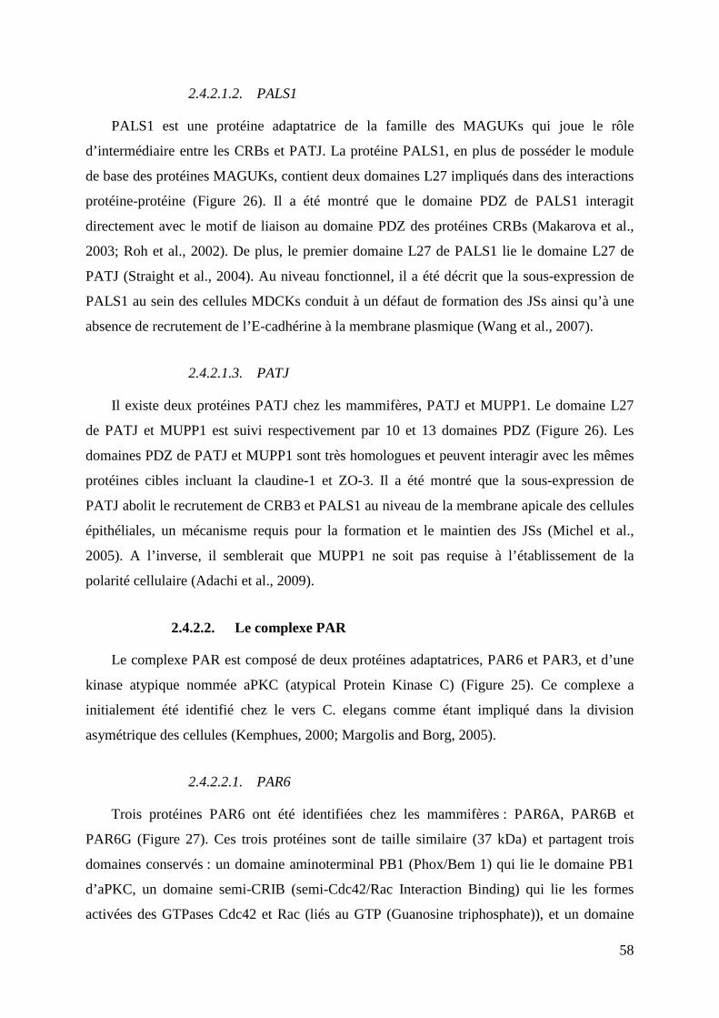
58
2.4.2.1.2. PALS1
PALS1 est une protéine adaptatrice de la famille des MAGUKs qui joue le rôle
d’intermédiaire entre les CRBs et PATJ. La protéine PALS1, en plus de posséder le module
de base des protéines MAGUKs, contient deux domaines L27 impliqués dans des interactions
protéine-protéine (Figure 26). Il a été montré que le domaine PDZ de PALS1 interagit
directement avec le motif de liaison au domaine PDZ des protéines CRBs (Makarova et al.,
2003; Roh et al., 2002). De plus, le premier domaine L27 de PALS1 lie le domaine L27 de
PATJ (Straight et al., 2004). Au niveau fonctionnel, il a été décrit que la sous-expression de
PALS1 au sein des cellules MDCKs conduit à un défaut de formation des JSs ainsi qu’à une
absence de recrutement de l’E-cadhérine à la membrane plasmique (Wang et al., 2007).
2.4.2.1.3. PATJ
Il existe deux protéines PATJ chez les mammifères, PATJ et MUPP1. Le domaine L27
de PATJ et MUPP1 est suivi respectivement par 10 et 13 domaines PDZ (Figure 26). Les
domaines PDZ de PATJ et MUPP1 sont très homologues et peuvent interagir avec les mêmes
protéines cibles incluant la claudine-1 et ZO-3. Il a été montré que la sous-expression de
PATJ abolit le recrutement de CRB3 et PALS1 au niveau de la membrane apicale des cellules
épithéliales, un mécanisme requis pour la formation et le maintien des JSs (Michel et al.,
2005). A l’inverse, il semblerait que MUPP1 ne soit pas requise à l’établissement de la
polarité cellulaire (Adachi et al., 2009).
2.4.2.2. Le complexe PAR
Le complexe PAR est composé de deux protéines adaptatrices, PAR6 et PAR3, et d’une
kinase atypique nommée aPKC (atypical Protein Kinase C) (Figure 25). Ce complexe a
initialement été identifié chez le vers C. elegans comme étant impliqué dans la division
asymétrique des cellules (Kemphues, 2000; Margolis and Borg, 2005).
2.4.2.2.1. PAR6
Trois protéines PAR6 ont été identifiées chez les mammifères : PAR6A, PAR6B et
PAR6G (Figure 27). Ces trois protéines sont de taille similaire (37 kDa) et partagent trois
domaines conservés : un domaine aminoterminal PB1 (Phox/Bem 1) qui lie le domaine PB1
d’aPKC, un domaine semi-CRIB (semi-Cdc42/Rac Interaction Binding) qui lie les formes
activées des GTPases Cdc42 et Rac (liés au GTP (Guanosine triphosphate)), et un domaine
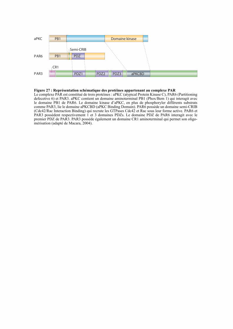
PB1
PB1 PDZ
PDZ1 PDZ2 aPKCBD
Domaine kinase
CR1
Semi-CRIB
aPKC
PAR6
PAR3
Figure 27 : Représentation schématique des protéines appartenant au complexe PARLe complexe PAR est constitué de trois protéines : aPKC (atypical Protein Kinase C), PAR6 (Partitioning defecetive 6) et PAR3. aPKC contient un domaine aminoterminal PB1 (Phox/Bem 1) qui interagit avec le domaine PB1 de PAR6. Le domaine kinase d’aPKC, en plus de phosphoryler différents substrats comme PAR3, lie le domaine aPKCBD (aPKC Binding Domain). PAR6 possède un domaine semi-CRIB (Cdc42/Rac Interaction Binding) qui recrute les GTPases Cdc42 et Rac sous leur forme active. PAR6 et PAR3 possèdent respectivement 1 et 3 domaines PDZs. Le domaine PDZ de PAR6 interagit avec le premier PDZ de PAR3. PAR3 posséde également un domaine CR1 aminoterminal qui permet son oligo-mérisation (adapté de Macara, 2004).
PDZ3
59
PDZ carboxyterminal qui interagit avec PAR3 et CRB3 (Figure 27) (Margolis and Borg,
2005; Shin et al., 2006). Bien que ces trois protéines soient structurellement similaires, elles
ne partagent pas la même localisation subcellulaire. En effet, au sein des cellules MDCKs,
PAR6A localise à la fois aux JSs et dans le cytoplasme tandis que PAR6B est cytoplasmique.
PAR6G est, quant à elle, majoritairement localisé aux JSs (Gao and Macara, 2004).
Les protéines PAR6 n’ayant pas d’activité enzymatique, elles vont servir de base au
recrutement d’autres protéines comme les GTPases CdC42 et Rac et la kinase aPKC. Il a
notamment été montré que la surexpression de PAR6B retarde la formation des JSs au sein
des cellules MDCK (Gao et al., 2002). A l’inverse, la surexpression de PAR6G n’affecte pas
la cinétique de formation des JSs (Gao and Macara, 2004).
2.4.2.2.2. PAR3
Deux protéines PAR3 ont été identifiées chez les mammifères, PAR3A et PAR3B (Figure
27). De plus, le gène PAR3A code trois variants d’épissage qui possèdent tous 3 domaines
PDZ dont le premier interagit avec PAR6. Tous ces variants, excepté le plus court,
interagissent avec aPKC via leur domaine aPKCBD (aPKC binding domain) (Figure 27). A
l’instar de PAR3A, PAR3B possède trois domaines PDZ et un domaine aPKCBD mais son
mécanisme d’interaction avec PAR6 reste ambigu (Figure 27). Les protéines PAR3 possèdent
également un domaine CR1 (Conserved Region 1) aminoterminal responsable de leur
oligomérisation (Figure 27). Il a notamment été décrit que le second domaine PDZ de PAR3A
et PAR3B interagit avec l’ensemble des phosphoinositides, un mécanisme nécessaire à leurs
recrutements aux JSs (Wu et al., 2007). Aux JSs, PAR3A recrute les protéines PAR6 et aPKC
pour former le complexe PAR. Plusieurs études ont montré que la sous- ou sur-expression de
PAR3A conduit à la délocalisation du complexe PAR ainsi qu’à la perte de JSs (Chen and
Zhang, 2013).
2.4.2.2.3. aPKC
Chez les mammifères, deux protéines aPKC ont été identifiées : aPKCλ et aPKCζ. Elles
ont une taille similaire et font toutes les deux parties du complexe PAR localisé aux JSs. Elles
possèdent un domaine aminoterminal PB1 qui interagit avec PAR6 et un domaine catalytique
en carboxyterminal qui phosphoryle plusieurs substrats incluant PAR3 (Figure 25 et27) (Chen
and Zhang, 2013; Suzuki et al., 2001).
Scribble
DLG
LGL
Péptide signal
Domaine SH3
Domaine GUK
Domaine PDZ
Domaine LRR
Motif LAPSD
Domaine WD40 repeat
Domaine L27
Figure 28 : Représentation schématique des protéines appartenant au complexe ScribbleChez les mammifères le complexe Scribble est composé de trois protéines : Scribble, DLG (Disc Large) et LGL (Lethal Giant Larvae). Scribble est une protéine cytoplasmique appartenant à la famille des protéines LAP (LRR and PDZ) qui est formée de 16 domaines LRR (Leucine Rich Repeats), de deux motifs LAPSD (LAP Specific Domain) et de quatre domaines PDZs. Les cinq protéines DLG chez les mammifères appartiennent à la superfamille des MAGUKs, elles possèdent le module de base des MAGUKs et un domaine L27 en aminoterminal. LGL a quatre paralogues chez les mammifères, elles contiennent toutes six domaines WD40 repeat (excepté pour LGL2 qui en a cinq) (adapté de Assémat et al., 2008).
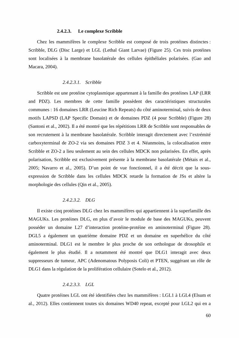
60
2.4.2.3. Le complexe Scribble
Chez les mammifères le complexe Scribble est composé de trois protéines distinctes :
Scribble, DLG (Disc Large) et LGL (Lethal Giant Larvae) (Figure 25). Ces trois protéines
sont localisées à la membrane basolatérale des cellules épithéliales polarisées. (Gao and
Macara, 2004).
2.4.2.3.1. Scribble
Scribble est une protéine cytoplasmique appartenant à la famille des protéines LAP (LRR
and PDZ). Les membres de cette famille possèdent des caractéristiques structurales
communes : 16 domaines LRR (Leucine Rich Repeats) du côté aminoterminal, suivis de deux
motifs LAPSD (LAP Specific Domain) et de domaines PDZ (4 pour Scribble) (Figure 28)
(Santoni et al., 2002). Il a été montré que les répétitions LRR de Scribble sont responsables de
son recrutement à la membrane basolatérale. Scribble interagit directement avec l’extrémité
carboxyterminal de ZO-2 via ses domaines PDZ 3 et 4. Néanmoins, la colocalisation entre
Scribble et ZO-2 a lieu seulement au sein des cellules MDCK non polarisées. En effet, après
polarisation, Scribble est exclusivement présente à la membrane basolatérale (Métais et al.,
2005; Navarro et al., 2005). D’un point de vue fonctionnel, il a été décrit que la sous-
expression de Scribble dans les cellules MDCK retarde la formation de JSs et altère la
morphologie des cellules (Qin et al., 2005).
2.4.2.3.2. DLG
Il existe cinq protéines DLG chez les mammifères qui appartiennent à la superfamille des
MAGUKs. Les protéines DLG, en plus d’avoir le module de base des MAGUKs, peuvent
posséder un domaine L27 d’interaction protéine-protéine en aminoterminal (Figure 28).
DGL5 a également un quatrième domaine PDZ et un domaine en superhélice du côté
aminoterminal. DLG1 est le membre le plus proche de son orthologue de drosophile et
également le plus étudié. Il a notamment été montré que DLG1 interagit avec deux
suppresseurs de tumeur, APC (Adenomatous Polyposis Coli) et PTEN, suggérant un rôle de
DLG1 dans la régulation de la prolifération cellulaire (Sotelo et al., 2012).
2.4.2.3.3. LGL
Quatre protéines LGL ont été identifiées chez les mammifères : LGL1 à LGL4 (Elsum et
al., 2012). Elles contiennent toutes six domaines WD40 repeat, excepté pour LGL2 qui en a
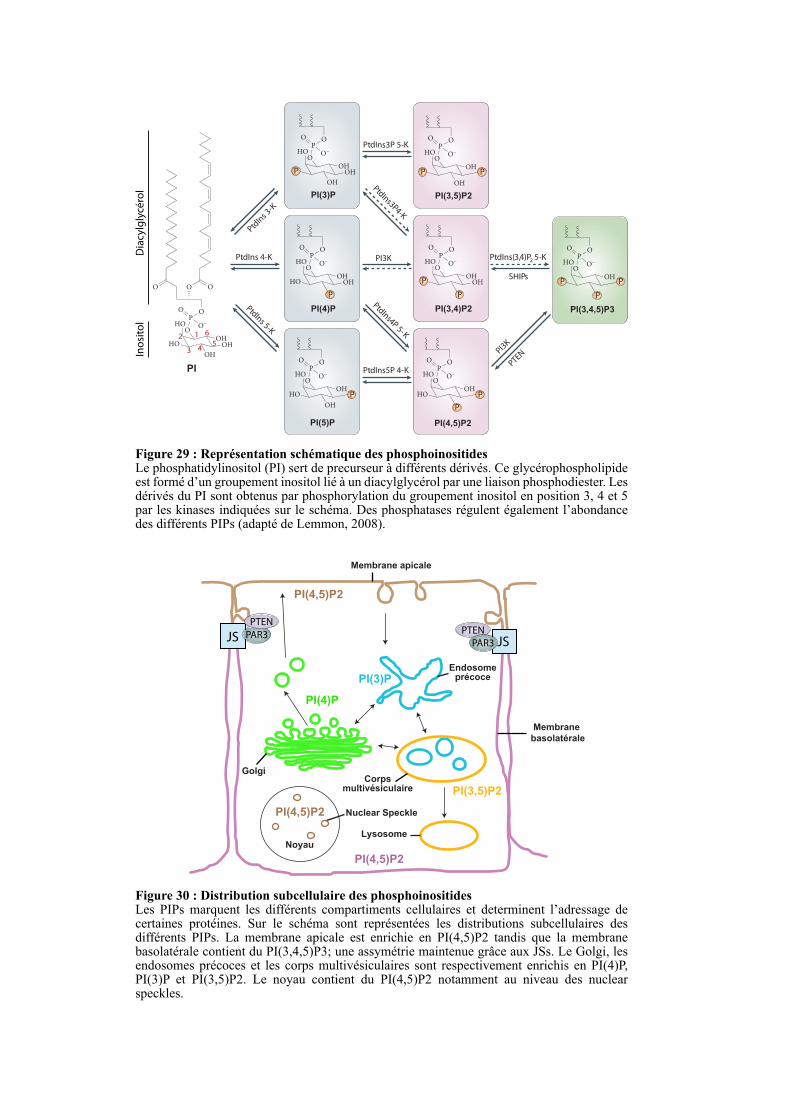
Figure 29 : Représentation schématique des phosphoinositidesLe phosphatidylinositol (PI) sert de precurseur à différents dérivés. Ce glycérophospholipide est formé d’un groupement inositol lié à un diacylglycérol par une liaison phosphodiester. Les dérivés du PI sont obtenus par phosphorylation du groupement inositol en position 3, 4 et 5 par les kinases indiquées sur le schéma. Des phosphatases régulent également l’abondance des différents PIPs (adapté de Lemmon, 2008).
O
O
O O
P
OO–
O
HO
HO
OH
OHOH
OP
OO–
O
HO
HO OHOH
OP
OO–
O
HO
OHOH
OP
OO–
O
HO
OH
OHOH
OP
OO–
O
HO
HO
OH
OH
OP
OO–
O
HO
HOOH
PI(3)P
PI(4)P PI(3,4)P2
PI(4,5)P2
P
P
P P
P
P
P
PtdIns3P 5-K
PtdIns 4-K
PtdIns5P 4-K
PI3K
SHIPs
PtdIns(3,4)P2 5-K
Ptd
Ins3
P 4-K
Ptd
Ins4
P 5
-K
Ptd
Ins
3-K
Ptd
Ins 5
-K
PI3
K
PTEN
12
3 45
6
PI(5)P
OP
OO–
O
HO
OH
OH
PI(3,5)P2
PP
OP
OO–
O
HO
OH
PI(3,4,5)P3
P P
P
PI
Diacylglycérol
Inositol
Noyau
JS JS
PI(4,5)P2
PI(4,5)P2
PI(4,5)P2
Golgi
PI(4)P
Membrane apicale
Membrane
basolatérale
EndosomeprécocePI(3)P
Corpsmultivésiculaire PI(3,5)P2
Lysosome
PTEN
PAR3
Nuclear Speckle
Figure 30 : Distribution subcellulaire des phosphoinositidesLes PIPs marquent les différents compartiments cellulaires et determinent l’adressage de certaines protéines. Sur le schéma sont représentées les distributions subcellulaires des différents PIPs. La membrane apicale est enrichie en PI(4,5)P2 tandis que la membrane basolatérale contient du PI(3,4,5)P3; une assymétrie maintenue grâce aux JSs. Le Golgi, les endosomes précoces et les corps multivésiculaires sont respectivement enrichis en PI(4)P, PI(3)P et PI(3,5)P2. Le noyau contient du PI(4,5)P2 notamment au niveau des nuclear speckles.
PTEN
PAR3
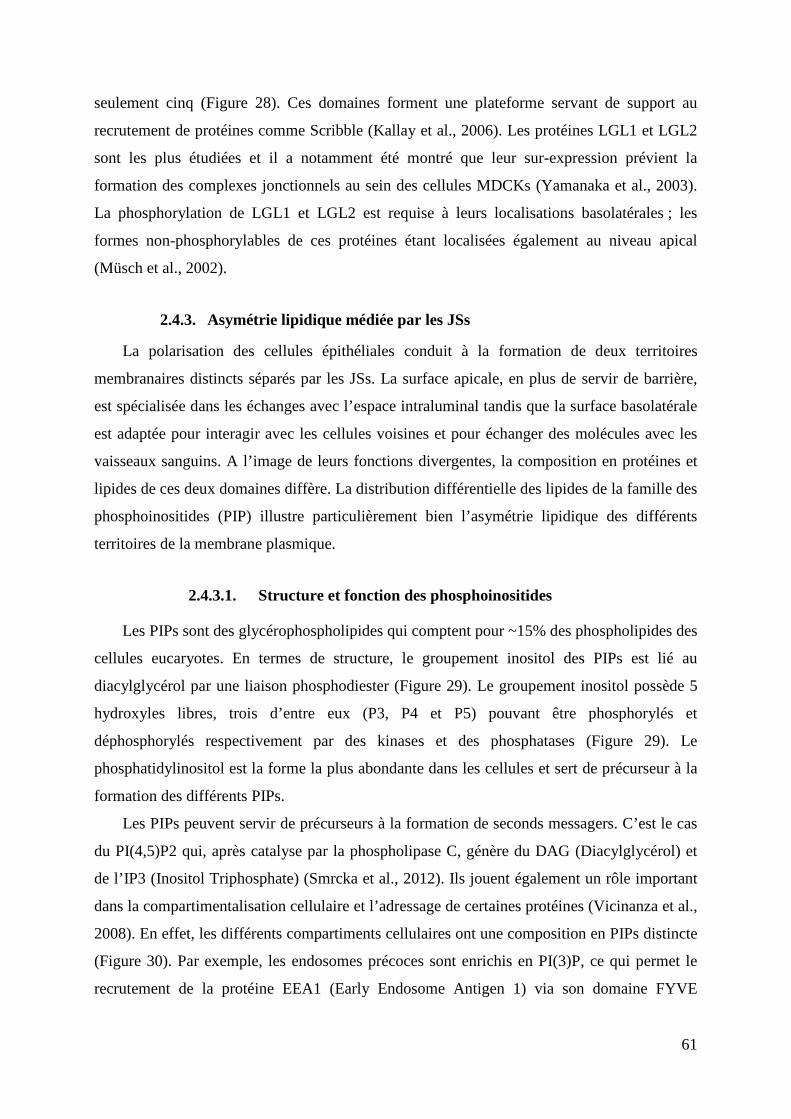
61
seulement cinq (Figure 28). Ces domaines forment une plateforme servant de support au
recrutement de protéines comme Scribble (Kallay et al., 2006). Les protéines LGL1 et LGL2
sont les plus étudiées et il a notamment été montré que leur sur-expression prévient la
formation des complexes jonctionnels au sein des cellules MDCKs (Yamanaka et al., 2003).
La phosphorylation de LGL1 et LGL2 est requise à leurs localisations basolatérales ; les
formes non-phosphorylables de ces protéines étant localisées également au niveau apical
(Müsch et al., 2002).
2.4.3. Asymétrie lipidique médiée par les JSs
La polarisation des cellules épithéliales conduit à la formation de deux territoires
membranaires distincts séparés par les JSs. La surface apicale, en plus de servir de barrière,
est spécialisée dans les échanges avec l’espace intraluminal tandis que la surface basolatérale
est adaptée pour interagir avec les cellules voisines et pour échanger des molécules avec les
vaisseaux sanguins. A l’image de leurs fonctions divergentes, la composition en protéines et
lipides de ces deux domaines diffère. La distribution différentielle des lipides de la famille des
phosphoinositides (PIP) illustre particulièrement bien l’asymétrie lipidique des différents
territoires de la membrane plasmique.
2.4.3.1. Structure et fonction des phosphoinositides
Les PIPs sont des glycérophospholipides qui comptent pour ~15% des phospholipides des
cellules eucaryotes. En termes de structure, le groupement inositol des PIPs est lié au
diacylglycérol par une liaison phosphodiester (Figure 29). Le groupement inositol possède 5
hydroxyles libres, trois d’entre eux (P3, P4 et P5) pouvant être phosphorylés et
déphosphorylés respectivement par des kinases et des phosphatases (Figure 29). Le
phosphatidylinositol est la forme la plus abondante dans les cellules et sert de précurseur à la
formation des différents PIPs.
Les PIPs peuvent servir de précurseurs à la formation de seconds messagers. C’est le cas
du PI(4,5)P2 qui, après catalyse par la phospholipase C, génère du DAG (Diacylglycérol) et
de l’IP3 (Inositol Triphosphate) (Smrcka et al., 2012). Ils jouent également un rôle important
dans la compartimentalisation cellulaire et l’adressage de certaines protéines (Vicinanza et al.,
2008). En effet, les différents compartiments cellulaires ont une composition en PIPs distincte
(Figure 30). Par exemple, les endosomes précoces sont enrichis en PI(3)P, ce qui permet le
recrutement de la protéine EEA1 (Early Endosome Antigen 1) via son domaine FYVE
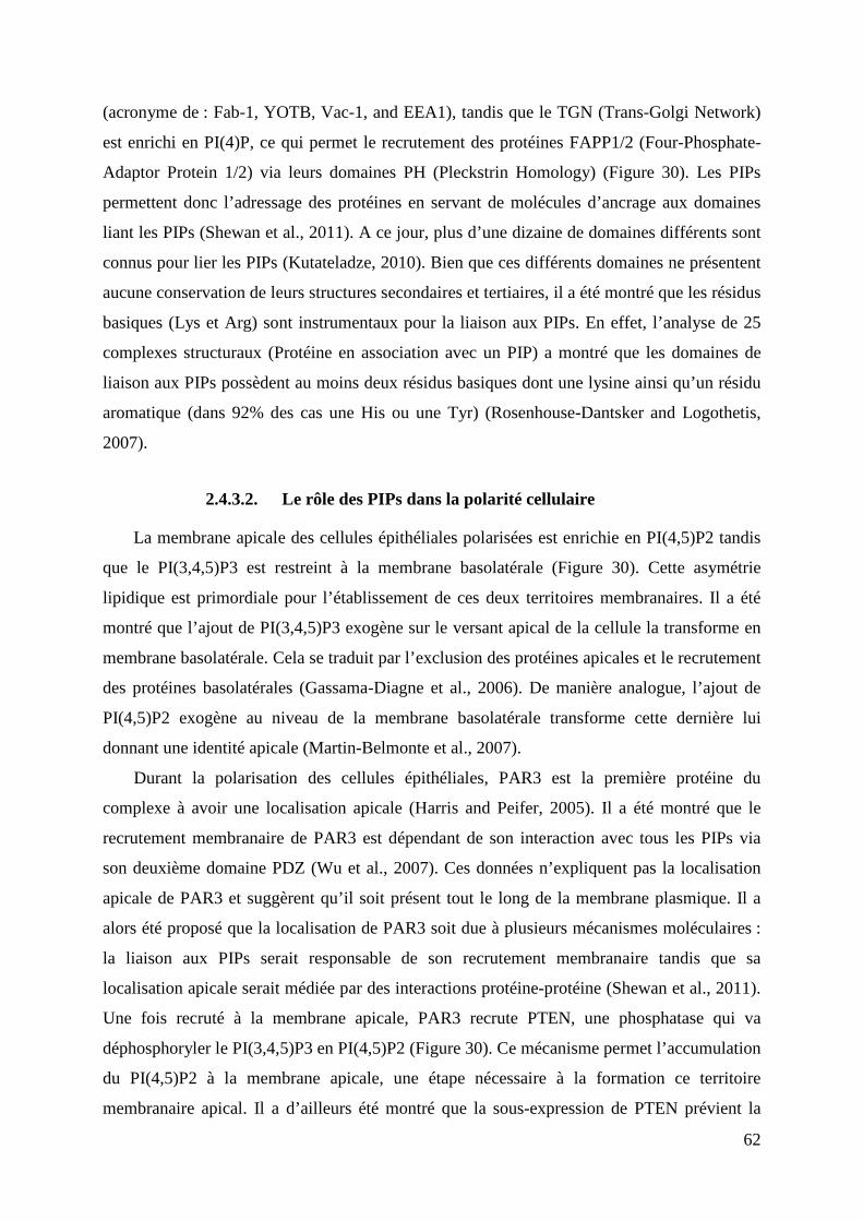
62
(acronyme de : Fab-1, YOTB, Vac-1, and EEA1), tandis que le TGN (Trans-Golgi Network)
est enrichi en PI(4)P, ce qui permet le recrutement des protéines FAPP1/2 (Four-Phosphate-
Adaptor Protein 1/2) via leurs domaines PH (Pleckstrin Homology) (Figure 30). Les PIPs
permettent donc l’adressage des protéines en servant de molécules d’ancrage aux domaines
liant les PIPs (Shewan et al., 2011). A ce jour, plus d’une dizaine de domaines différents sont
connus pour lier les PIPs (Kutateladze, 2010). Bien que ces différents domaines ne présentent
aucune conservation de leurs structures secondaires et tertiaires, il a été montré que les résidus
basiques (Lys et Arg) sont instrumentaux pour la liaison aux PIPs. En effet, l’analyse de 25
complexes structuraux (Protéine en association avec un PIP) a montré que les domaines de
liaison aux PIPs possèdent au moins deux résidus basiques dont une lysine ainsi qu’un résidu
aromatique (dans 92% des cas une His ou une Tyr) (Rosenhouse-Dantsker and Logothetis,
2007).
2.4.3.2. Le rôle des PIPs dans la polarité cellulaire
La membrane apicale des cellules épithéliales polarisées est enrichie en PI(4,5)P2 tandis
que le PI(3,4,5)P3 est restreint à la membrane basolatérale (Figure 30). Cette asymétrie
lipidique est primordiale pour l’établissement de ces deux territoires membranaires. Il a été
montré que l’ajout de PI(3,4,5)P3 exogène sur le versant apical de la cellule la transforme en
membrane basolatérale. Cela se traduit par l’exclusion des protéines apicales et le recrutement
des protéines basolatérales (Gassama-Diagne et al., 2006). De manière analogue, l’ajout de
PI(4,5)P2 exogène au niveau de la membrane basolatérale transforme cette dernière lui
donnant une identité apicale (Martin-Belmonte et al., 2007).
Durant la polarisation des cellules épithéliales, PAR3 est la première protéine du
complexe à avoir une localisation apicale (Harris and Peifer, 2005). Il a été montré que le
recrutement membranaire de PAR3 est dépendant de son interaction avec tous les PIPs via
son deuxième domaine PDZ (Wu et al., 2007). Ces données n’expliquent pas la localisation
apicale de PAR3 et suggèrent qu’il soit présent tout le long de la membrane plasmique. Il a
alors été proposé que la localisation de PAR3 soit due à plusieurs mécanismes moléculaires :
la liaison aux PIPs serait responsable de son recrutement membranaire tandis que sa
localisation apicale serait médiée par des interactions protéine-protéine (Shewan et al., 2011).
Une fois recruté à la membrane apicale, PAR3 recrute PTEN, une phosphatase qui va
déphosphoryler le PI(3,4,5)P3 en PI(4,5)P2 (Figure 30). Ce mécanisme permet l’accumulation
du PI(4,5)P2 à la membrane apicale, une étape nécessaire à la formation ce territoire
membranaire apical. Il a d’ailleurs été montré que la sous-expression de PTEN prévient la
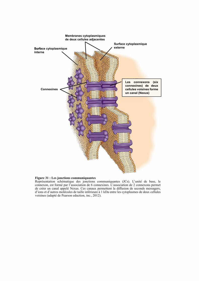
Surface cytoplasmiqueSurface cytoplasmique
interne
Membranes cytoplasmiques
de deux cellules adjacentes
Surface cytoplasmique
externe
Connexines
Les connexons (six
connexines) de deux
cellules voisines forme
un canal (Nexus)
Figure 31 : Les jonctions communiquantesReprésentation schématique des jonctions communiquantes (JCs). L’unité de base, le connexon, est formé par l’association de 6 connexines. L’association de 2 connexons permet de créer un canal appelé Nexus. Ces canaux permettent la diffusion de seconds messagers, d’ions et d’autres molécules de taille inférieure à 1 kDa entre les cytoplasmes de deux cellules voisines (adapté de Pearson eduction, inc., 2012).
63
formation du domaine apical (Martin-Belmonte et al., 2007). Dans la cellule polarisée, le
complexe PAR et PTEN sont présent aux JSs et participent au maintien de l’asymétrie des
PIPs (Figure 30) (Tepass, 2012).
2.5. LES JONCTIONS COMMUNICANTES
Les JCs sont des canaux intercellulaires qui permettent la diffusion de seconds messagers,
d’ions et d’autres molécules de taille inférieure à 1 kDa entre les cytoplasmes de deux cellules
voisines. Les JCs sont donc nécessaires à diverses fonctions comme la transduction de
signaux électriques ou chimiques. Il a été montré que la modulation de ces fonctions peut se
faire en régulant l’ouverture et la fermeture de ces canaux (Matsuuchi and Naus, 2013). Les
protéines transmembranaires formant ces canaux intercellulaires font partie de la famille des
connexines qui compte plus de 20 membres à ce jour. Six connexines s’oligomérisent pour
former un connexon et les connexons de deux cellules adjacentes s’associent pour former un
canal appelé Nexus (Figure 31). ZO-1 est un partenaire protéique des JCs qui a pour rôle de
limiter la taille des JCs (Hunter et al., 2005). Les JCs, à l’instar des autres jonctions
intercellulaires, sont ancrées au cytosquelette (Giepmans, 2006).
2.6. PATHOLOGIES LIEES AUX COMPLEXES JONCTIONNELS
De nombreuses maladies génétiques sont dues à l’altération des gènes codant pour les
composants des complexes jonctionnels. La connexine-32 est impliquée dans la forme liée à
l’X du syndrome de Charcot-Marie-Tooth (OMMIM 302800) tandis que la connexine-31 est
liée à une neuropathie périphérique associée à une surdité (Scott and Kelsell, 2011). La
mutation de la plakophiline-1 est associée une la dysplasie ectodermique récessive (Ersoy-
Evans et al., 2006). La mutation des claudines 16 et 19 est liée à une hypomagnesaemia, une
hypercalciuria et à une nephrocalcinosis due à un défaut d’absorption du calcium et du
magnésium. L’altération des protéines formant les jonctions serrées est également
fréquemment retrouvée dans les cancers (Latorre et al., 2006; Martin et al., 2011). C’est le cas
de l’occludine, des claudines (-1, -4, -6 et -7), de ZO-1 et de PAR3 dont l’expression est
diminuée dans les cancers du sein. A l’inverse, PAR6B et JAM-A sont surexprimées dans
certain cancer du sein, où ils vont promouvoir respectivement la prolifération et l’invasion
(Brennan et al., 2010).
64
2.6.1. Anomalie des JAs
La β-caténine, en connectant les cadhérines au cytosquelette d’actine, est nécessaire à la
formation et au maintien des JAs (Harris and Tepass, 2010). A l’inverse, dans la voie
Wingless (Wnt)/β-caténine, la β-caténine agit comme un facteur de transcription en
s’associant aux facteurs LEF/TCF (Lymphoid Enhancing Factor/T-Cell Factor) dans le noyau,
où elle régule l’expression de gènes cibles incluant des métalloprotéinases matricielles et des
cytokines (Kim et al., 2013; Polette et al., 2007). Un équilibre entre les fonctions d’adhésion
et de transcription de la β-caténine est primordial pour le maintien de l’homéostasie des
épithélia. En effet, la rupture de cet état équilibre en faveur de la fonction de transcription est
impliquée dans la carcinogénèse (Burgess et al., 2011). Plusieurs mécanismes moléculaires
favorisent l’activité transcriptionnelle de la β-caténine. Il a notamment été montré que des
mutations perte-de-fonction d’APC, une protéine qui participe à la dégradation de la β-
caténine, sont retrouvées dans 85% des cancers colorectaux sporadiques (Cottrell et al., 1992).
La perte d’expression de l’E-cadhérine conduit également à une augmentation du pool de β-
caténines conduisant à son transfert dans le noyau (Orsulic et al., 1999). Enfin, l’analyse
génétique exhaustive des tumeurs a révélé que des membres de la voie Wnt/β-caténine sont
mutés dans 90% des cancers colorectaux. Ces mutations conduisent à la stabilisation puis à la
translocation de la β-caténine dans le noyau, où elle va permettre l’expression de ces gènes
cibles impliqués dans la prolifération et la migration cellulaire (Brembeck et al., 2006).
2.6.2. Anomalie des JSs
Les JSs, en séparant la membrane cellulaire en deux compartiments, permet la
ségrégation des facteurs de croissance comme l’héréguline-α dans la région apicale tandis que
leurs récepteurs (erbB3-4) vont exclusivement être présents dans la région basolatérale, ce qui
prévient leur activation (Vermeer et al., 2003). Après une blessure ou la mort d’une ou
plusieurs cellules au sein d’un épithélium, la perte des JSs des cellules saines adjacentes
permet aux facteurs de croissances de lier leurs récepteurs, ce qui induit une prolifération et
migration cellulaire afin de réparer la blessure. Basé sur ce modèle, il est facile de concevoir
que la perte des JSs due aux mutations des protéines structurales et des régulateurs des JSs,
peut favoriser la progression des cellules épithéliales cancéreuses (Latorre et al., 2006; Soler
et al., 1999).
L’identification et la caractérisation des différents composants des JSs a permis de
comprendre l’impact de la perte des JSs dans la formation et la progression des carcinomes au
niveau moléculaire. Par exemple il a été montré que ZO-1 prévient la prolifération cellulaire

65
en séquestrant le facteur de transcription ZONAB tandis que la perte des JSs conduit à la
translocation de ZONAB dans le noyau, où il va avoir un effet prolifératif en interagissant
avec la protéine CDK4 (Cell Division Kinase 4) (Balda et al., 2003). Concernant la
progression des carcinomes, il est accepté que la perte des JSs, en stimulant la migration et
l’invasion des cellules tumorales, promeut la formation et la dissémination des métastases (Jin
et al., 2009; Martin and Jiang, 2001; Martin et al., 2011). Plusieurs études démontrent
d’ailleurs que les protéines des JSs sont souvent sous-exprimées dans les carcinomes et que
leurs niveaux d’expression sont corrélés au degré de différentiation des cellules tumorales. Par
exemple, l’expression de ZO-1 dans les carcinomes canalaires infiltrants du sein est réduite de
42%, 83% et 93% respectivement dans les tumeurs fortement, modérément et faiblement
différentiées (Hoover et al., 1998; Quan and Lu, 2003).
Il a été rapporté que PAR3 est sous-exprimé dans les cancers du sein, ce qui est de
mauvais pronostique. La perte de PAR3 est associée chez la souris à une augmentation de
l’incidence tumorale, et promeut la formation et la dissémination des métastases. Elle induit
notamment l’activation de la voie JAK/STAT (Janus Kinase/Signal Transducer and Activator
of Transcription), ce qui conduit à la dégradation de la matrice grâce à la sécrétion de la
métalloprotéinase matricielle MMP9. La sous-expression de PAR3 conduit également à
l’activation de la GTPase Rac1 qui, en agissant sur le remodelage du cytosquelette, va avoir
un effet pro-migratoire (McCaffrey et al., 2012; Xue et al., 2013).

66
RESULTATS ET
DISCUSSIONS
67
CHAPITRE1:LEROLEDETRAF4DANSLESJONCTIONS
SERREES
1.1. INTRODUCTION
Les cellules épithéliales polarisées forment une barrière sélective régulant le passage des
molécules à travers l’espace paracellulaire. Elles possèdent deux domaines membranaires,
apical et basolatéral, dont les compositions en lipides et protéines diffèrent. Les jonctions
serrées (JS), en empêchant ces protéines et lipides de diffuser librement d’un compartiment à
l’autre, contribuent au maintien de la polarité cellulaire (Brennan et al., 2010; Furuse, 2010).
Des études récentes ont mis en évidence l’importance des jonctions serrées sur le contrôle de
la prolifération, de la différenciation et de la polarité cellulaire, trois processus altérés dans les
carcinomes (Brennan et al., 2010; Forster, 2008). TRAF4, étant surexprimée dans les tumeurs
solides et localisée au sein des JS, nous avons émis l’hypothèse que cette protéine pourrait
avoir un effet protumoral en dérégulant ces structures jonctionnelles. Afin de tester cette
hypothèse, nous avons tout d’abord caractérisé le mécanisme moléculaire responsable du
recrutement de TRAF4 aux JS pour ensuite y appréhender son rôle. Cette étude a donné lieu à
la soumission d’un article (Rousseau et al., en révision, § 1.2). Des expériences
complémentaires à cet article ont été réalisées et sont décrites au paragraphe § 1.3.
J’ai par ailleurs étudié si la liaison aux PIPs était conservée dans la famille des protéines
TRAFs ; ces résultats sont décrits à la fin de ce chapitre (§ 1.4).
1.2. ARTICLE : TRAF4 is a novel phosphoinositides binding protein modulating
tight junctions and favoring cell migration
Résumé de l’article:
TRAF4 (Tumor necrosis factor (TNF) Receptor-Associated Factor 4) est un gène
fréquemment surexprimé dans les carcinomes suggérant qu’il y joue un rôle. Tandis que la
protéine TRAF4 est majoritairement localisée dans les jonctions serrées (JS) des cellules
épithéliales mammaires (CEM) normales, elle s’accumule dans le cytoplasme des CEM
malignes. Le mécanisme responsable du recrutement de TRAF4 ainsi que de sa fonction dans
les JS reste inconnu à ce jour. Dans cette étude, nous montrons que TRAF4 possède un
nouveau domaine liant les phosphoinositides (PIP) et que ce dernier est requis pour son
recrutement aux JS. Des analyses moléculaires et structurales ont montré que le domaine
TRAF de TRAF4 forme un homotrimère qui peut lier jusqu’à trois molécules de lipides en

68
utilisant des résidus basiques présents à sa surface. Des études cellulaires indiquent que
TRAF4 régule négativement les JS et augmente la migration cellulaire. Ces deux fonctions
sont dépendantes de sa capacité à lier les PIPs. Nos résultats suggèrent que la surexpression
de TRAF4 pourrait contribuer à la progression des cancers du sein en déstabilisant les JS et en
favorisant la migration cellulaire.
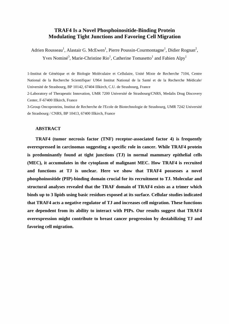
TRAF4 Is a Novel Phosphoinositide-Binding Protein Modulating Tight Junctions and Favoring Cell Migration
Adrien Rousseau1, Alastair G. McEwen1, Pierre Poussin-Courmontagne1, Didier Rognan2,
Yves Nominé3, Marie-Christine Rio1, Catherine Tomasetto1 and Fabien Alpy1
1-Institut de Génétique et de Biologie Moléculaire et Cellulaire, Unité Mixte de Recherche 7104, Centre
National de la Recherche Scientifique/ U964 Institut National de la Santé et de la Recherche Médicale/
Université de Strasbourg, BP 10142, 67404 Illkirch, C.U. de Strasbourg, France
2-Laboratory of Therapeutic Innovation, UMR 7200 Université de Strasbourg/CNRS, Medalis Drug Discovery
Center, F-67400 Illkirch, France
3-Group Oncoproteins, Institut de Recherche de l'Ecole de Biotechnologie de Strasbourg, UMR 7242 Université
de Strasbourg / CNRS, BP 10413, 67400 Illkirch, France
ABSTRACT
TRAF4 (tumor necrosis factor (TNF) receptor-associated factor 4) is frequently
overexpressed in carcinomas suggesting a specific role in cancer. While TRAF4 protein
is predominantly found at tight junctions (TJ) in normal mammary epithelial cells
(MEC), it accumulates in the cytoplasm of malignant MEC. How TRAF4 is recruited
and functions at TJ is unclear. Here we show that TRAF4 possesses a novel
phosphoinositide (PIP)-binding domain crucial for its recruitment to TJ. Molecular and
structural analyses revealed that the TRAF domain of TRAF4 exists as a trimer which
binds up to 3 lipids using basic residues exposed at its surface. Cellular studies indicated
that TRAF4 acts a negative regulator of TJ and increases cell migration. These functions
are dependent from its ability to interact with PIPs. Our results suggest that TRAF4
overexpression might contribute to breast cancer progression by destabilizing TJ and
favoring cell migration.
INTRODUCTION
Tumor necrosis factor Receptor-Associated Factor 4 (TRAF4) was originally identified
as a gene overexpressed in breast carcinoma [1,2]. Interestingly, TRAF4 overexpression is not
restricted to breast cancer and extends to a variety of different carcinomas [3,4]. TRAF4
belongs to the TRAF family that is composed of 7 members in human [5,6]. Among the seven
TRAF family members, TRAF4 is one of the most conserved during evolution [7]. Indeed, a
TRAF4 ortholog has already been identified in snail fur (Hydractinia achinata), a cnidaria [8].
Moreover, the unique TRAF protein in the worm shares a higher homology with human
TRAF4 than with other human TRAF proteins [9]. Furthermore, one of the three fly TRAF
proteins, dTRAF1, shares the highest homology with human TRAF4 [9,10]. In line with an
essential and conserved biological function of TRAF4, flies that carry null-alleles of TRAF4
have many developmental abnormalities leading to lethality before the pupal stage [11,12].
Likewise, TRAF4 deficiency in mice was lethal at the embryonic stage in approximately one
third of the homozygote mutants [13]. All surviving animals exhibited multiple defects
including trachea alteration, and various non-fully penetrant phenotypes involving the axial
skeleton and the central nervous system [13,14]. Surviving adult TRAF4-deficient mice also
exhibited ataxia, associated to myelination alteration [15]. Together, these various genetic
models suggest that TRAF4 has an essential function conserved in most, if not all,
pluricellular animals.
TRAF4 encodes a 53 kDa adaptor protein with multiple subcellular localizations. Indeed,
cytoplasmic, nuclear and membrane localizations have been described in the literature
[1,16,17]. Of interest, the subcellular localization of TRAF4 is altered in cancers. While in
normal breast tissue the protein is predominantly localized in the plasma membrane [18],
more precisely in tight junctions (TJ) present at the apical membrane of polarized epithelial
cells [19], in cancer samples the protein can be localized either in the cytoplasm and/or in the
nucleus of cancer epithelial cells [1,3,16]. Until recently the implication of these multiple
localizations were unclear. A recent report shed light into the significance of the nuclear
localization of TRAF4 in breast cancers [20]. This study indicated that TRAF4 nuclear
localization in breast tumors was associated with poor survival in breast cancer patients after
adjuvant therapy. Moreover, this report showed that TRAF4 promotes p53 protein
destabilization in the nucleus of cancer cells and contributes to resistance to cytotoxic stress in
cancer cells [20]. To date, the function of TRAF4 in TJ and in the cytoplasm remains unclear.
Many lines of evidence indicate that TRAF4 functions in the organization and patterning of
the cell cortex [12,21]. TRAF4 is required for the polarized trafficking of NADPH oxidase to
filopodia in migrating endothelial cells [22,23]. In the fly, TRAF4 can interact with proteins
involved in asymmetric division [21] and functions in the establishment of the junctional
architecture of mesodermal cells [12]. In the mouse nervous system, TRAF4 deficiency
altered the formation and/or stability of axoglial and interglial junctions [15]. Collectively, all
these data indicate that TRAF4 might exert a function related to cell junctions and polarity
[24]. However, TRAF4 appears to function in a cell specific manner making its functional
characterization difficult. While TRAF4 is ubiquitously expressed at a basal level, there exists
a spatial and temporal upregulation of TRAF4 gene expression during specific developmental
stages [25,26]. For example, during gastrulation in frogs, mRNA encoding TRAF4 and one of
its interacting partners SMURF1 become enriched in the neural plate and neural crest cells. In
these cells, both TRAF4 and SMURF1 are essential for proper neural crest development and
neural plate morphogenesis [25]. In human mammary epithelial cells (MEC), the role of
TRAF4 in TJ remains unclear. However we showed previously that this localization was a
highly dynamic process supporting the notion that TRAF4 might relay signals from the cell
membrane to the cytoplasm, and possibly the nucleus [17]. Given the recently established role
of nuclear TRAF4 in the destabilization of the tumor suppressor p53 protein [20], it is now
necessary to understand how TRAF4 subcellular localization in the mammary epithelium is
regulated under physiopathological conditions. To address this, we explored the molecular
determinants involved in its subcellular localization at the plasma membrane and in TJ of
MEC. We also directly addressed the function of TRAF4 in the formation and maintenance of
TJ in polarized cells. Finally, having established that TRAF4 is a negative regulator of TJ in
breast cells, we have explored its contribution to cell motility.
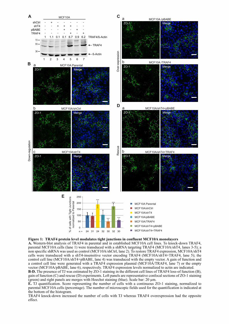
MCF10A Parental
MCF10A/shCtrl
MCF10A/pBABE
MCF10A/TRAF4
MCF10A/shT4+pBABE
MCF10A/shT4+TRAF40
50
100
150
200
250
***
% o
f cells
with T
ight Junctions
norm
aliz
ed t
o P
are
nta
l
E
n = 24 31 24 32 30 32
MCF10A/shT4
30
***
*** ***
B
MCF10A/shCtrl
ZO-1 Merge
b
MCF10A/shT4
ZO-1 Merge
c
MCF10A ParentalaMergeZO-1
Do
wn
-re
gu
latio
n
A
shT4
shCtrl
pBABE
TRAF4
+-
-
-
-
-
-
-
-
-
+
-
-
+
-
-+
-
+
+
-
-
-
-
-
-+
+
36
72
55 TRAF4
ß-Actin
1 1.1 0.1 0.1 6.7 0.9 6.2 TRAF4/ß-Actin
MCF10A
1 2 3 4 5 6 7
MCF10A/shT4+pBABE
ZO-1 Merge
a
MCF10A/shT4+TRAF4
ZO-1 Merge
b
Re
scu
e
D
MCF10A /pBABE
ZO-1 Merge
a
MCF10A /TRAF4
ZO-1 Merge
b
Ove
r-e
xp
ressio
n
C
Figure 1: TRAF4 protein level modulates tight junctions in confluent MCF10A monolayersA. Western-blot analysis of TRAF4 in parental and in established MCF10A cell lines. To knock-down TRAF4, parental MCF10A cells (lane 1) were transduced with a shRNA targeting TRAF4 (MCF10A/shT4, lanes 3-5); a non specific shRNA was used as control (MCF10A/shCtrl, lane 2). To restore TRAF4 expression, MCF10A/shT4 cells were transduced with a shT4-insensitive vector encoding TRAF4 (MCF10A/shT4+TRAF4, lane 5); the control cell line (MCF10A/shT4+pBABE, lane 4) was transduced with the empty vector. A gain of function and a control cell line were generated with a TRAF4 expression plasmid (MCF10A/TRAF4, lane 7) or the empty vector (MCF10A/pBABE, lane 6), respectively. TRAF4 expression levels normalized to actin are indicated.B-D. The presence of TJ was estimated by ZO-1 staining in the different cell lines of TRAF4 loss-of function (B), gain of function (C) and rescue (D) experiments. Left panels are representative confocal sections of ZO-1 staining (green) and right panels are merges with Hoechst staining (blue). Scale bar: 20 µm.E. TJ quantification. Score representing the number of cells with a continuous ZO-1 staining, normalized to parental MCF10A cells (percentage). The number of microscopic fields used for the quantification is indicated at the bottom of the histogram.TRAF4 knock-down increased the number of cells with TJ whereas TRAF4 overexpression had the opposite effect.
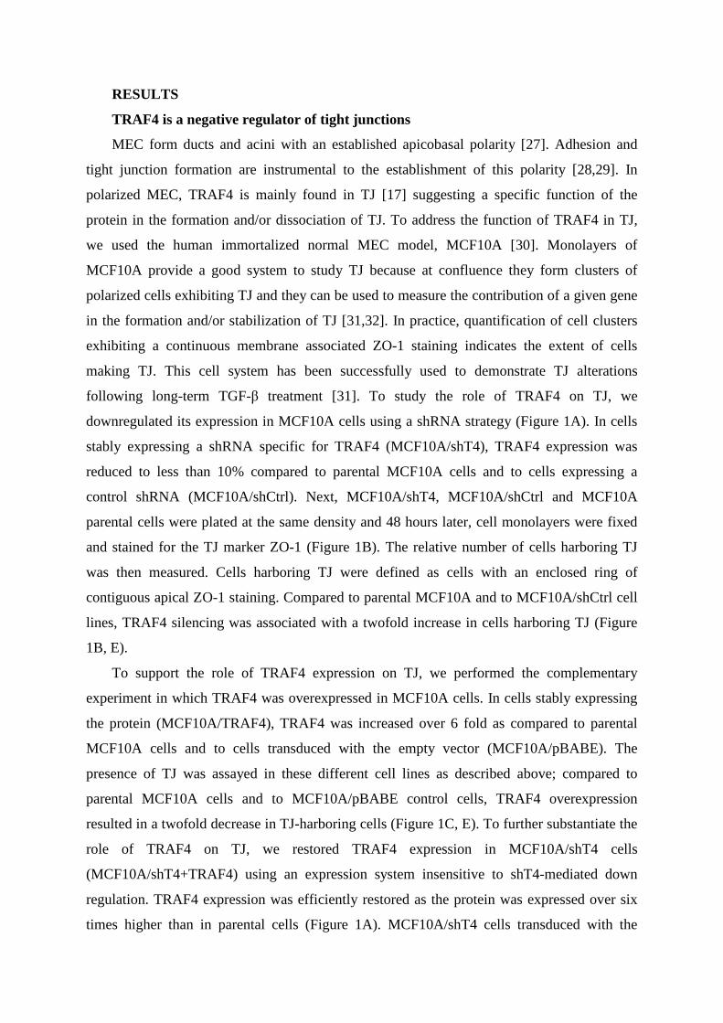
RESULTS
TRAF4 is a negative regulator of tight junctions
MEC form ducts and acini with an established apicobasal polarity [27]. Adhesion and
tight junction formation are instrumental to the establishment of this polarity [28,29]. In
polarized MEC, TRAF4 is mainly found in TJ [17] suggesting a specific function of the
protein in the formation and/or dissociation of TJ. To address the function of TRAF4 in TJ,
we used the human immortalized normal MEC model, MCF10A [30]. Monolayers of
MCF10A provide a good system to study TJ because at confluence they form clusters of
polarized cells exhibiting TJ and they can be used to measure the contribution of a given gene
in the formation and/or stabilization of TJ [31,32]. In practice, quantification of cell clusters
exhibiting a continuous membrane associated ZO-1 staining indicates the extent of cells
making TJ. This cell system has been successfully used to demonstrate TJ alterations
following long-term TGF-β treatment [31]. To study the role of TRAF4 on TJ, we
downregulated its expression in MCF10A cells using a shRNA strategy (Figure 1A). In cells
stably expressing a shRNA specific for TRAF4 (MCF10A/shT4), TRAF4 expression was
reduced to less than 10% compared to parental MCF10A cells and to cells expressing a
control shRNA (MCF10A/shCtrl). Next, MCF10A/shT4, MCF10A/shCtrl and MCF10A
parental cells were plated at the same density and 48 hours later, cell monolayers were fixed
and stained for the TJ marker ZO-1 (Figure 1B). The relative number of cells harboring TJ
was then measured. Cells harboring TJ were defined as cells with an enclosed ring of
contiguous apical ZO-1 staining. Compared to parental MCF10A and to MCF10A/shCtrl cell
lines, TRAF4 silencing was associated with a twofold increase in cells harboring TJ (Figure
1B, E).
To support the role of TRAF4 expression on TJ, we performed the complementary
experiment in which TRAF4 was overexpressed in MCF10A cells. In cells stably expressing
the protein (MCF10A/TRAF4), TRAF4 was increased over 6 fold as compared to parental
MCF10A cells and to cells transduced with the empty vector (MCF10A/pBABE). The
presence of TJ was assayed in these different cell lines as described above; compared to
parental MCF10A cells and to MCF10A/pBABE control cells, TRAF4 overexpression
resulted in a twofold decrease in TJ-harboring cells (Figure 1C, E). To further substantiate the
role of TRAF4 on TJ, we restored TRAF4 expression in MCF10A/shT4 cells
(MCF10A/shT4+TRAF4) using an expression system insensitive to shT4-mediated down
regulation. TRAF4 expression was efficiently restored as the protein was expressed over six
times higher than in parental cells (Figure 1A). MCF10A/shT4 cells transduced with the
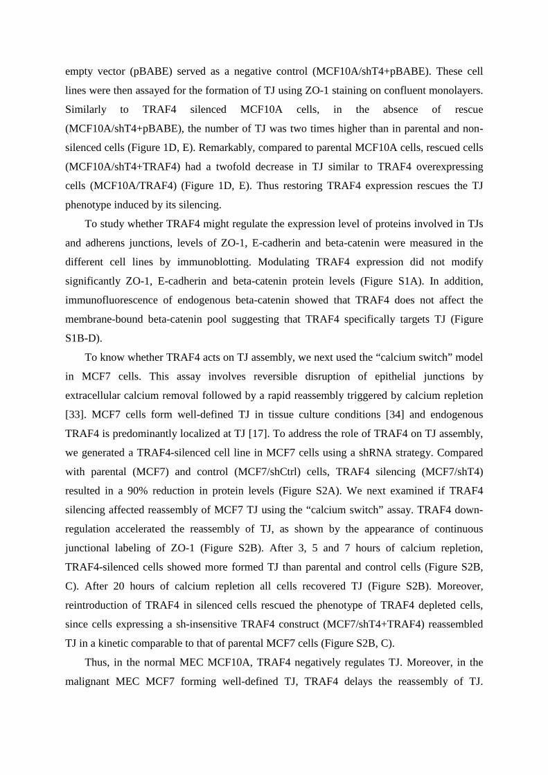
empty vector (pBABE) served as a negative control (MCF10A/shT4+pBABE). These cell
lines were then assayed for the formation of TJ using ZO-1 staining on confluent monolayers.
Similarly to TRAF4 silenced MCF10A cells, in the absence of rescue
(MCF10A/shT4+pBABE), the number of TJ was two times higher than in parental and non-
silenced cells (Figure 1D, E). Remarkably, compared to parental MCF10A cells, rescued cells
(MCF10A/shT4+TRAF4) had a twofold decrease in TJ similar to TRAF4 overexpressing
cells (MCF10A/TRAF4) (Figure 1D, E). Thus restoring TRAF4 expression rescues the TJ
phenotype induced by its silencing.
To study whether TRAF4 might regulate the expression level of proteins involved in TJs
and adherens junctions, levels of ZO-1, E-cadherin and beta-catenin were measured in the
different cell lines by immunoblotting. Modulating TRAF4 expression did not modify
significantly ZO-1, E-cadherin and beta-catenin protein levels (Figure S1A). In addition,
immunofluorescence of endogenous beta-catenin showed that TRAF4 does not affect the
membrane-bound beta-catenin pool suggesting that TRAF4 specifically targets TJ (Figure
S1B-D).
To know whether TRAF4 acts on TJ assembly, we next used the “calcium switch” model
in MCF7 cells. This assay involves reversible disruption of epithelial junctions by
extracellular calcium removal followed by a rapid reassembly triggered by calcium repletion
[33]. MCF7 cells form well-defined TJ in tissue culture conditions [34] and endogenous
TRAF4 is predominantly localized at TJ [17]. To address the role of TRAF4 on TJ assembly,
we generated a TRAF4-silenced cell line in MCF7 cells using a shRNA strategy. Compared
with parental (MCF7) and control (MCF7/shCtrl) cells, TRAF4 silencing (MCF7/shT4)
resulted in a 90% reduction in protein levels (Figure S2A). We next examined if TRAF4
silencing affected reassembly of MCF7 TJ using the “calcium switch” assay. TRAF4 down-
regulation accelerated the reassembly of TJ, as shown by the appearance of continuous
junctional labeling of ZO-1 (Figure S2B). After 3, 5 and 7 hours of calcium repletion,
TRAF4-silenced cells showed more formed TJ than parental and control cells (Figure S2B,
C). After 20 hours of calcium repletion all cells recovered TJ (Figure S2B). Moreover,
reintroduction of TRAF4 in silenced cells rescued the phenotype of TRAF4 depleted cells,
since cells expressing a sh-insensitive TRAF4 construct (MCF7/shT4+TRAF4) reassembled
TJ in a kinetic comparable to that of parental MCF7 cells (Figure S2B, C).
Thus, in the normal MEC MCF10A, TRAF4 negatively regulates TJ. Moreover, in the
malignant MEC MCF7 forming well-defined TJ, TRAF4 delays the reassembly of TJ.
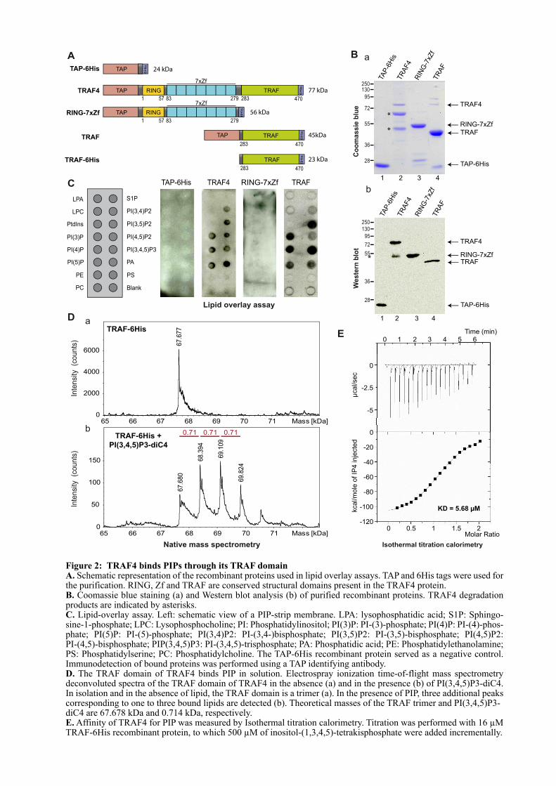
A
C
LPA
LPC
PtdIns
PI(3)P
PI(4)P
PI(5)P
PE
PC
S1P
PI(3,4)P2
PI(3,5)P2
PI(4,5)P2
PI(3,4,5)P3
PA
PS
Blank
TAP-6His TRAF4 RING-7xZf TRAF
Lipid overlay assay
Native mass spectrometry
D
67.6
77
0
2000
4000
6000
65 66 67 68 69 70 71 Mass [kDa]
Inte
nsi
ty (c
ounts
)
67.6
80
68.3
94
69.1
09
69.8
24
0
50
100
150
66 67 68 69 70 71 Mass [kDa]65
0.71 0.71 0.71
Inte
nsi
ty (c
ounts
)
a
bTRAF-6His +
PI(3,4,5)P3-diC4
TRAF-6His
RING-7xZf1 57 83 279
6H
is
TAP RING 56 kDa
6H
is
TRAF TAP
283 470
TRAF 45kDa
6H
is
TRAF-6His
283 470
TRAF 23 kDa
E
TAP-6His
6H
is
TAP 24 kDa
TRAF4
6H
is
1 57 83 279 283 470
TAP RING TRAF 77 kDa
7xZf
7xZf
*
Figure 2: TRAF4 binds PIPs through its TRAF domainA. Schematic representation of the recombinant proteins used in lipid overlay assays. TAP and 6His tags were used for the purification. RING, Zf and TRAF are conserved structural domains present in the TRAF4 protein.B. Coomassie blue staining (a) and Western blot analysis (b) of purified recombinant proteins. TRAF4 degradation products are indicated by asterisks. C. Lipid-overlay assay. Left: schematic view of a PIP-strip membrane. LPA: lysophosphatidic acid; S1P: Sphingo-sine-1-phosphate; LPC: Lysophosphocholine; PI: Phosphatidylinositol; PI(3)P: PI-(3)-phosphate; PI(4)P: PI-(4)-phos-phate; PI(5)P: PI-(5)-phosphate; PI(3,4)P2: PI-(3,4-)bisphosphate; PI(3,5)P2: PI-(3,5)-bisphosphate; PI(4,5)P2: PI-(4,5)-bisphosphate; PIP(3,4,5)P3: PI-(3,4,5)-trisphosphate; PA: Phosphatidic acid; PE: Phosphatidylethanolamine; PS: Phosphatidylserine; PC: Phosphatidylcholine. The TAP-6His recombinant protein served as a negative control. Immunodetection of bound proteins was performed using a TAP identifying antibody. D. The TRAF domain of TRAF4 binds PIP in solution. Electrospray ionization time-of-flight mass spectrometry deconvoluted spectra of the TRAF domain of TRAF4 in the absence (a) and in the presence (b) of PI(3,4,5)P3-diC4. In isolation and in the absence of lipid, the TRAF domain is a trimer (a). In the presence of PIP, three additional peaks corresponding to one to three bound lipids are detected (b). Theoretical masses of the TRAF trimer and PI(3,4,5)P3-diC4 are 67.678 kDa and 0.714 kDa, respectively.E. Affinity of TRAF4 for PIP was measured by Isothermal titration calorimetry. Titration was performed with 16 µM TRAF-6His recombinant protein, to which 500 µM of inositol-(1,3,4,5)-tetrakisphosphate were added incrementally.
B
250
130
95
72
55
36
28
*
*
TAP
-6H
isTR
AF4
RIN
G-7
xZf
TR
AF
1 2 3 4
Co
om
assie
blu
e
a
TRAF4
RING-7xZf
TRAF
TAP-6His
2 3 4
Weste
rn b
lot
TRAF4
RING-7xZfTRAF
TAP-6His
b
RIN
G-7
xZf
TAP
-6H
isTR
AF4
TR
AF
250
130
95
72
55
36
28
1
0
-5
-2.5
0
-20
-40
-60
-80
-100
-120
0 1 2 3 4 5 6
Time (min)
µcal/sec
kcal/m
ole
of IP
4 inje
cte
d
0 0.5 1 1.5 2Molar Ratio
KD = 5.68 µM
Isothermal titration calorimetry
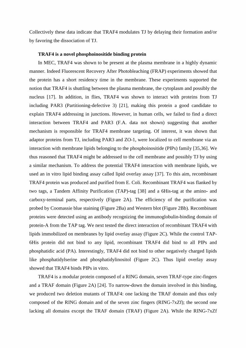
Collectively these data indicate that TRAF4 modulates TJ by delaying their formation and/or
by favoring the dissociation of TJ.
TRAF4 is a novel phosphoinositide binding protein
In MEC, TRAF4 was shown to be present at the plasma membrane in a highly dynamic
manner. Indeed Fluorescent Recovery After Photobleaching (FRAP) experiments showed that
the protein has a short residency time in the membrane. These experiments supported the
notion that TRAF4 is shuttling between the plasma membrane, the cytoplasm and possibly the
nucleus [17]. In addition, in flies, TRAF4 was shown to interact with proteins from TJ
including PAR3 (Partitioning-defective 3) [21], making this protein a good candidate to
explain TRAF4 addressing in junctions. However, in human cells, we failed to find a direct
interaction between TRAF4 and PAR3 (F.A. data not shown) suggesting that another
mechanism is responsible for TRAF4 membrane targeting. Of interest, it was shown that
adaptor proteins from TJ, including PAR3 and ZO-1, were localized to cell membrane via an
interaction with membrane lipids belonging to the phosphoinositide (PIPs) family [35,36]. We
thus reasoned that TRAF4 might be addressed to the cell membrane and possibly TJ by using
a similar mechanism. To address the potential TRAF4 interaction with membrane lipids, we
used an in vitro lipid binding assay called lipid overlay assay [37]. To this aim, recombinant
TRAF4 protein was produced and purified from E. Coli. Recombinant TRAF4 was flanked by
two tags, a Tandem Affinity Purification (TAP)-tag [38] and a 6His-tag at the amino- and
carboxy-terminal parts, respectively (Figure 2A). The efficiency of the purification was
probed by Coomassie blue staining (Figure 2Ba) and Western blot (Figure 2Bb). Recombinant
proteins were detected using an antibody recognizing the immunoglobulin-binding domain of
protein-A from the TAP tag. We next tested the direct interaction of recombinant TRAF4 with
lipids immobilized on membranes by lipid overlay assay (Figure 2C). While the control TAP-
6His protein did not bind to any lipid, recombinant TRAF4 did bind to all PIPs and
phosphatidic acid (PA). Interestingly, TRAF4 did not bind to other negatively charged lipids
like phosphatidylserine and phosphatidylinositol (Figure 2C). Thus lipid overlay assay
showed that TRAF4 binds PIPs in vitro.
TRAF4 is a modular protein composed of a RING domain, seven TRAF-type zinc-fingers
and a TRAF domain (Figure 2A) [24]. To narrow-down the domain involved in this binding,
we produced two deletion mutants of TRAF4: one lacking the TRAF domain and thus only
composed of the RING domain and of the seven zinc fingers (RING-7xZf); the second one
lacking all domains except the TRAF domain (TRAF) (Figure 2A). While the RING-7xZf
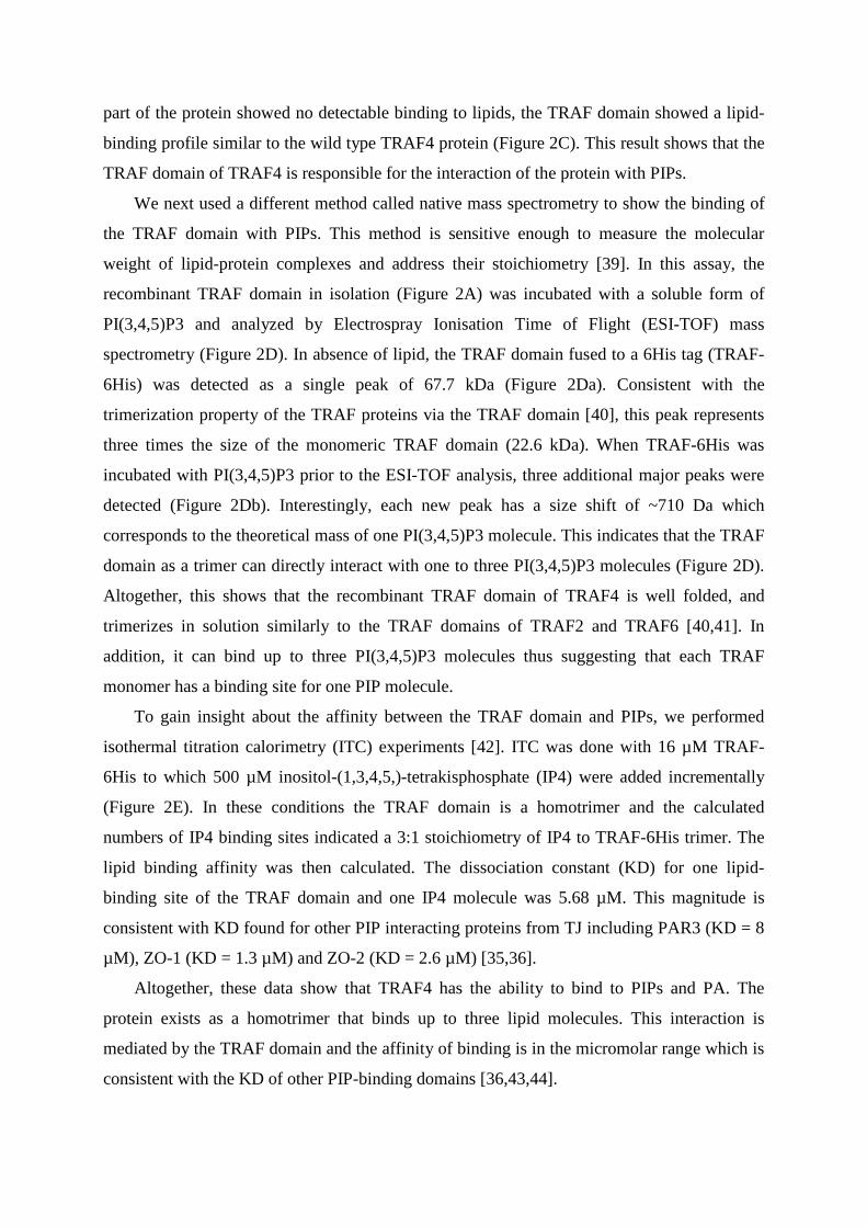
part of the protein showed no detectable binding to lipids, the TRAF domain showed a lipid-
binding profile similar to the wild type TRAF4 protein (Figure 2C). This result shows that the
TRAF domain of TRAF4 is responsible for the interaction of the protein with PIPs.
We next used a different method called native mass spectrometry to show the binding of
the TRAF domain with PIPs. This method is sensitive enough to measure the molecular
weight of lipid-protein complexes and address their stoichiometry [39]. In this assay, the
recombinant TRAF domain in isolation (Figure 2A) was incubated with a soluble form of
PI(3,4,5)P3 and analyzed by Electrospray Ionisation Time of Flight (ESI-TOF) mass
spectrometry (Figure 2D). In absence of lipid, the TRAF domain fused to a 6His tag (TRAF-
6His) was detected as a single peak of 67.7 kDa (Figure 2Da). Consistent with the
trimerization property of the TRAF proteins via the TRAF domain [40], this peak represents
three times the size of the monomeric TRAF domain (22.6 kDa). When TRAF-6His was
incubated with PI(3,4,5)P3 prior to the ESI-TOF analysis, three additional major peaks were
detected (Figure 2Db). Interestingly, each new peak has a size shift of ~710 Da which
corresponds to the theoretical mass of one PI(3,4,5)P3 molecule. This indicates that the TRAF
domain as a trimer can directly interact with one to three PI(3,4,5)P3 molecules (Figure 2D).
Altogether, this shows that the recombinant TRAF domain of TRAF4 is well folded, and
trimerizes in solution similarly to the TRAF domains of TRAF2 and TRAF6 [40,41]. In
addition, it can bind up to three PI(3,4,5)P3 molecules thus suggesting that each TRAF
monomer has a binding site for one PIP molecule.
To gain insight about the affinity between the TRAF domain and PIPs, we performed
isothermal titration calorimetry (ITC) experiments [42]. ITC was done with 16 µM TRAF-
6His to which 500 µM inositol-(1,3,4,5,)-tetrakisphosphate (IP4) were added incrementally
(Figure 2E). In these conditions the TRAF domain is a homotrimer and the calculated
numbers of IP4 binding sites indicated a 3:1 stoichiometry of IP4 to TRAF-6His trimer. The
lipid binding affinity was then calculated. The dissociation constant (KD) for one lipid-
binding site of the TRAF domain and one IP4 molecule was 5.68 µM. This magnitude is
consistent with KD found for other PIP interacting proteins from TJ including PAR3 (KD = 8
µM), ZO-1 (KD = 1.3 µM) and ZO-2 (KD = 2.6 µM) [35,36].
Altogether, these data show that TRAF4 has the ability to bind to PIPs and PA. The
protein exists as a homotrimer that binds up to three lipid molecules. This interaction is
mediated by the TRAF domain and the affinity of binding is in the micromolar range which is
consistent with the KD of other PIP-binding domains [36,43,44].
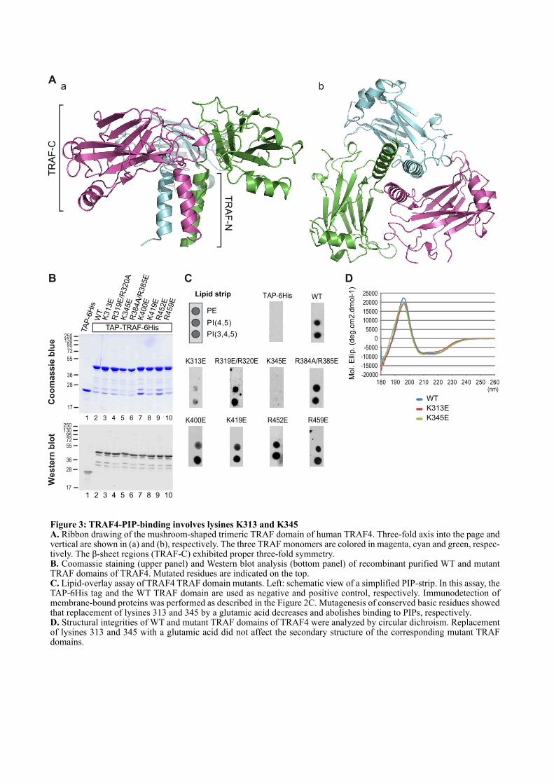
D
180 190 200 210 220 230 240 250 260(nm)
25000
20000
15000
10000
5000
0
-5000
-10000
-15000
-20000Mo
l. E
llip
. (d
eg
.cm
2.d
mo
l-1
)
WT
K313E
K345E
C
Aa b
B
1 2 3 4 5 6 7 8 9
250130
957255
36
28
17
TA
P-6
His
K313E
R319E
/R320A
K345E
R452E
R459E
WT
R384A
/R385E
K400E
250130
9572
55
36
28
17
TAP-TRAF-6His
Co
om
as
sie
blu
e
We
ste
rn b
lot
K419E
10
1 2 3 4 5 6 7 8 9 10
TR
AF
-C
TR
AF
-N
PE
PI(4,5)
PI(3,4,5)
R319E/R320E R384A/R385E
K400E K419E R452E R459E
WT TAP-6His
K345EK313E
Lipid strip
Figure 3: TRAF4-PIP-binding involves lysines K313 and K345A. Ribbon drawing of the mushroom-shaped trimeric TRAF domain of human TRAF4. Three-fold axis into the page and vertical are shown in (a) and (b), respectively. The three TRAF monomers are colored in magenta, cyan and green, respec-tively. The β-sheet regions (TRAF-C) exhibited proper three-fold symmetry.B. Coomassie staining (upper panel) and Western blot analysis (bottom panel) of recombinant purified WT and mutant TRAF domains of TRAF4. Mutated residues are indicated on the top. C. Lipid-overlay assay of TRAF4 TRAF domain mutants. Left: schematic view of a simplified PIP-strip. In this assay, the TAP-6His tag and the WT TRAF domain are used as negative and positive control, respectively. Immunodetection of membrane-bound proteins was performed as described in the Figure 2C. Mutagenesis of conserved basic residues showed that replacement of lysines 313 and 345 by a glutamic acid decreases and abolishes binding to PIPs, respectively.D. Structural integrities of WT and mutant TRAF domains of TRAF4 were analyzed by circular dichroism. Replacement of lysines 313 and 345 with a glutamic acid did not affect the secondary structure of the corresponding mutant TRAF domains.
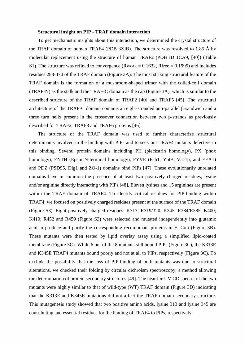
Structural insight on PIP - TRAF domain interaction
To get mechanistic insights about this interaction, we determined the crystal structure of
the TRAF domain of human TRAF4 (PDB 3ZJB). The structure was resolved to 1.85 Å by
molecular replacement using the structure of human TRAF2 (PDB ID 1CA9, [40]) (Table
S1). The structure was refined to convergence (Rwork = 0.1632, Rfree = 0.1995) and includes
residues 283-470 of the TRAF domain (Figure 3A). The most striking structural feature of the
TRAF domain is the formation of a mushroom-shaped trimer with the coiled-coil domain
(TRAF-N) as the stalk and the TRAF-C domain as the cap (Figure 3A), which is similar to the
described structure of the TRAF domain of TRAF2 [40] and TRAF5 [45]. The structural
architecture of the TRAF-C domain contains an eight-stranded anti-parallel β-sandwich and a
three turn helix present in the crossover connection between two β-strands as previously
described for TRAF2, TRAF3 and TRAF6 proteins [46].
The structure of the TRAF domain was used to further characterize structural
determinants involved in the binding with PIPs and to seek out TRAF4 mutants defective in
this binding. Several protein domains including PH (pleckstrin homology), PX (phox
homology), ENTH (Epsin N-terminal homology), FYVE (Fab1, YotB, Vac1p, and EEA1)
and PDZ (PSD95, Dlg1 and ZO-1) domains bind PIPs [47]. These evolutionarily unrelated
domains have in common the presence of at least two positively charged residues, lysine
and/or arginine directly interacting with PIPs [48]. Eleven lysines and 15 arginines are present
within the TRAF domain of TRAF4. To identify critical residues for PIP-binding within
TRAF4, we focused on positively charged residues present at the surface of the TRAF domain
(Figure S3). Eight positively charged residues: K313; R319/320; K345; R384/R385; K400;
K419; R452 and R459 (Figure S3) were selected and mutated independently into glutamic
acid to produce and purify the corresponding recombinant proteins in E. Coli (Figure 3B).
These mutants were then tested by lipid overlay assay using a simplified lipid-coated
membrane (Figure 3C). While 6 out of the 8 mutants still bound PIPs (Figure 3C), the K313E
and K345E TRAF4 mutants bound poorly and not at all to PIPs, respectively (Figure 3C). To
exclude the possibility that the loss of PIP-binding of both mutants was due to structural
alterations, we checked their folding by circular dichroism spectroscopy, a method allowing
the determination of protein secondary structures [49]. The near far-UV CD spectra of the two
mutants were highly similar to that of wild-type (WT) TRAF domain (Figure 3D) indicating
that the K313E and K345E mutations did not affect the TRAF domain secondary structure.
This mutagenesis study showed that two positive amino acids, lysine 313 and lysine 345 are
contributing and essential residues for the binding of TRAF4 to PIPs, respectively.
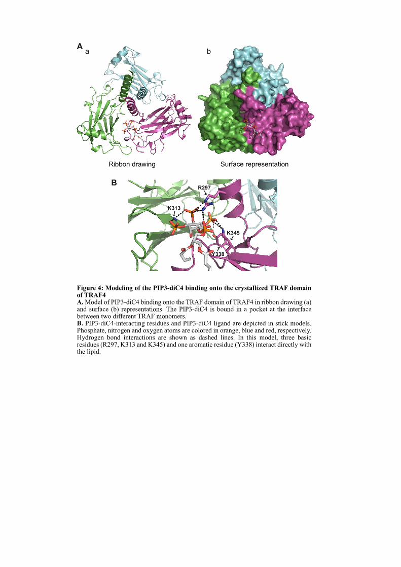
B
K313
K345
Y338
R297
Aa
Ribbon drawing
b
Surface representation
Figure 4: Modeling of the PIP3-diC4 binding onto the crystallized TRAF domain of TRAF4A. Model of PIP3-diC4 binding onto the TRAF domain of TRAF4 in ribbon drawing (a) and surface (b) representations. The PIP3-diC4 is bound in a pocket at the interface between two different TRAF monomers. B. PIP3-diC4-interacting residues and PIP3-diC4 ligand are depicted in stick models. Phosphate, nitrogen and oxygen atoms are colored in orange, blue and red, respectively. Hydrogen bond interactions are shown as dashed lines. In this model, three basic residues (R297, K313 and K345) and one aromatic residue (Y338) interact directly with the lipid.
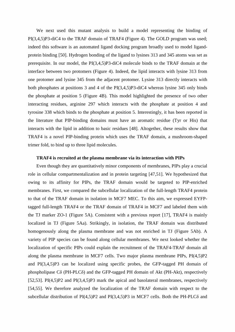
We next used this mutant analysis to build a model representing the binding of
PI(3,4,5)P3-diC4 to the TRAF domain of TRAF4 (Figure 4). The GOLD program was used;
indeed this software is an automated ligand docking program broadly used to model ligand-
protein binding [50]. Hydrogen bonding of the ligand to lysines 313 and 345 atoms was set as
prerequisite. In our model, the PI(3,4,5)P3-diC4 molecule binds to the TRAF domain at the
interface between two protomers (Figure 4). Indeed, the lipid interacts with lysine 313 from
one protomer and lysine 345 from the adjacent protomer. Lysine 313 directly interacts with
both phosphates at positions 3 and 4 of the PI(3,4,5)P3-diC4 whereas lysine 345 only binds
the phosphate at position 5 (Figure 4B). This model highlighted the presence of two other
interacting residues, arginine 297 which interacts with the phosphate at position 4 and
tyrosine 338 which binds to the phosphate at position 5. Interestingly, it has been reported in
the literature that PIP-binding domains must have an aromatic residue (Tyr or His) that
interacts with the lipid in addition to basic residues [48]. Altogether, these results show that
TRAF4 is a novel PIP-binding protein which uses the TRAF domain, a mushroom-shaped
trimer fold, to bind up to three lipid molecules.
TRAF4 is recruited at the plasma membrane via its interaction with PIPs
Even though they are quantitatively minor components of membranes, PIPs play a crucial
role in cellular compartmentalization and in protein targeting [47,51]. We hypothesized that
owing to its affinity for PIPs, the TRAF domain would be targeted to PIP-enriched
membranes. First, we compared the subcellular localization of the full-length TRAF4 protein
to that of the TRAF domain in isolation in MCF7 MEC. To this aim, we expressed EYFP-
tagged full-length TRAF4 or the TRAF domain of TRAF4 in MCF7 and labeled them with
the TJ marker ZO-1 (Figure 5A). Consistent with a previous report [17], TRAF4 is mainly
localized in TJ (Figure 5Aa). Strikingly, in isolation, the TRAF domain was distributed
homogenously along the plasma membrane and was not enriched in TJ (Figure 5Ab). A
variety of PIP species can be found along cellular membranes. We next looked whether the
localization of specific PIPs could explain the recruitment of the TRAF4-TRAF domain all
along the plasma membrane in MCF7 cells. Two major plasma membrane PIPs, PI(4,5)P2
and PI(3,4,5)P3 can be localized using specific probes, the GFP-tagged PH domain of
phospholipase Cδ (PH-PLCδ) and the GFP-tagged PH domain of Akt (PH-Akt), respectively
[52,53]. PI(4,5)P2 and PI(3,4,5)P3 mark the apical and basolateral membranes, respectively
[54,55]. We therefore analyzed the localization of the TRAF domain with respect to the
subcellular distribution of PI(4,5)P2 and PI(3,4,5)P3 in MCF7 cells. Both the PH-PLCδ and
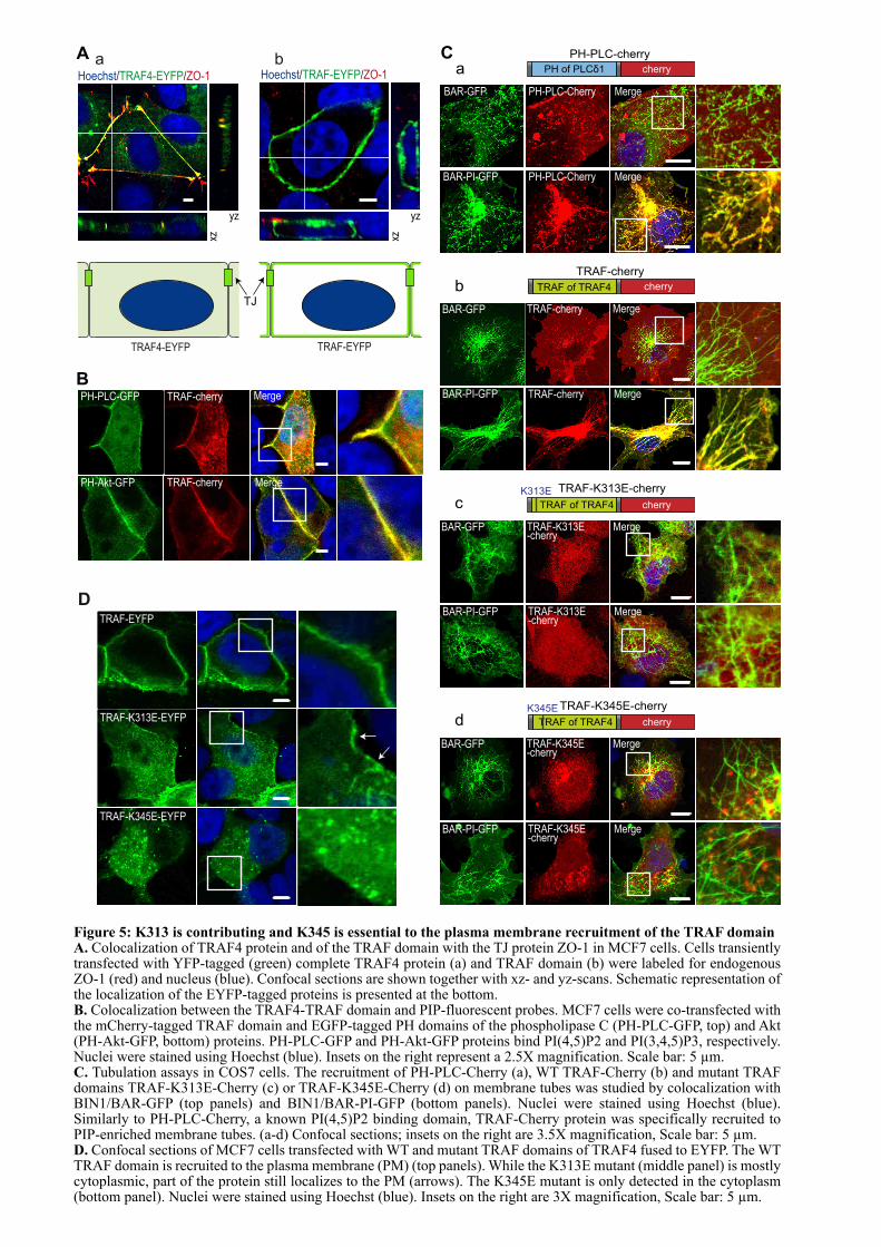
BPH-PLC-GFP
PH-Akt-GFP
TRAF-cherry Merge
MergeTRAF-cherry
A ba
D
TRAF-EYFP
TRAF-K313E-EYFP
TRAF-K345E-EYFP
C
BAR-GFP PH-PLC-Cherry Merge
BAR-PI-GFP PH-PLC-Cherry Merge
PH-PLC-cherry
PH of PLCδ1 cherrya
TRAF-cherry
TRAF of TRAF4 cherry
TRAF-cherry Merge
BAR-PI-GFP TRAF-cherry MergeBAR-PI-GFP
BAR-GFP
b
BAR-GFP TRAF-K313E-cherry
Merge
BAR-PI-GFP TRAF-K313E-cherry
Merge
TRAF-K313E-cherry
TRAF of TRAF4 cherry
K313E
c
BAR-GFP TRAF-K345E-cherry
Merge
BAR-PI-GFP TRAF-K345E-cherry
Merge
TRAF-K345E-cherry
TRAF of TRAF4 cherry
K345E
d
Hoechst/TRAF4-EYFP/ZO-1
TRAF4-EYFP
Hoechst/TRAF-EYFP/ZO-1
TRAF-EYFP
TJ
yz xz
yz
xz
Figure 5: K313 is contributing and K345 is essential to the plasma membrane recruitment of the TRAF domainA. Colocalization of TRAF4 protein and of the TRAF domain with the TJ protein ZO-1 in MCF7 cells. Cells transiently transfected with YFP-tagged (green) complete TRAF4 protein (a) and TRAF domain (b) were labeled for endogenous ZO-1 (red) and nucleus (blue). Confocal sections are shown together with xz- and yz-scans. Schematic representation of the localization of the EYFP-tagged proteins is presented at the bottom.B. Colocalization between the TRAF4-TRAF domain and PIP-fluorescent probes. MCF7 cells were co-transfected with the mCherry-tagged TRAF domain and EGFP-tagged PH domains of the phospholipase C (PH-PLC-GFP, top) and Akt (PH-Akt-GFP, bottom) proteins. PH-PLC-GFP and PH-Akt-GFP proteins bind PI(4,5)P2 and PI(3,4,5)P3, respectively. Nuclei were stained using Hoechst (blue). Insets on the right represent a 2.5X magnification. Scale bar: 5 µm.C. Tubulation assays in COS7 cells. The recruitment of PH-PLC-Cherry (a), WT TRAF-Cherry (b) and mutant TRAF domains TRAF-K313E-Cherry (c) or TRAF-K345E-Cherry (d) on membrane tubes was studied by colocalization with BIN1/BAR-GFP (top panels) and BIN1/BAR-PI-GFP (bottom panels). Nuclei were stained using Hoechst (blue). Similarly to PH-PLC-Cherry, a known PI(4,5)P2 binding domain, TRAF-Cherry protein was specifically recruited to PIP-enriched membrane tubes. (a-d) Confocal sections; insets on the right are 3.5X magnification, Scale bar: 5 µm.D. Confocal sections of MCF7 cells transfected with WT and mutant TRAF domains of TRAF4 fused to EYFP. The WT TRAF domain is recruited to the plasma membrane (PM) (top panels). While the K313E mutant (middle panel) is mostly cytoplasmic, part of the protein still localizes to the PM (arrows). The K345E mutant is only detected in the cytoplasm (bottom panel). Nuclei were stained using Hoechst (blue). Insets on the right are 3X magnification, Scale bar: 5 µm.
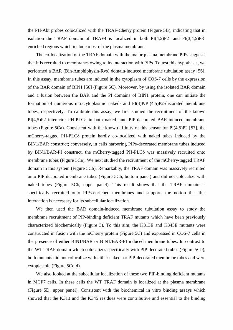
the PH-Akt probes colocalized with the TRAF-Cherry protein (Figure 5B), indicating that in
isolation the TRAF domain of TRAF4 is localized in both PI(4,5)P2- and PI(3,4,5)P3-
enriched regions which include most of the plasma membrane.
The co-localization of the TRAF domain with the major plasma membrane PIPs suggests
that it is recruited to membranes owing to its interaction with PIPs. To test this hypothesis, we
performed a BAR (Bin-Amphiphysin-Rvs) domain-induced membrane tubulation assay [56].
In this assay, membrane tubes are induced in the cytoplasm of COS-7 cells by the expression
of the BAR domain of BIN1 [56] (Figure 5C). Moreover, by using the isolated BAR domain
and a fusion between the BAR and the PI domains of BIN1 protein, one can initiate the
formation of numerous intracytoplasmic naked- and PI(4)P/PI(4,5)P2-decorated membrane
tubes, respectively. To calibrate this assay, we first studied the recruitment of the known
PI(4,5)P2 interactor PH-PLCδ in both naked- and PIP-decorated BAR-induced membrane
tubes (Figure 5Ca). Consistent with the known affinity of this sensor for PI(4,5)P2 [57], the
mCherry-tagged PH-PLCδ protein hardly co-localized with naked tubes induced by the
BIN1/BAR construct; conversely, in cells harboring PIPs-decorated membrane tubes induced
by BIN1/BAR-PI construct, the mCherry-tagged PH-PLCδ was massively recruited onto
membrane tubes (Figure 5Ca). We next studied the recruitment of the mCherry-tagged TRAF
domain in this system (Figure 5Cb). Remarkably, the TRAF domain was massively recruited
onto PIP-decorated membrane tubes (Figure 5Cb, bottom panel) and did not colocalize with
naked tubes (Figure 5Cb, upper panel). This result shows that the TRAF domain is
specifically recruited onto PIPs-enriched membranes and supports the notion that this
interaction is necessary for its subcellular localization.
We then used the BAR domain-induced membrane tubulation assay to study the
membrane recruitment of PIP-binding deficient TRAF mutants which have been previously
characterized biochemically (Figure 3). To this aim, the K313E and K345E mutants were
constructed in fusion with the mCherry protein (Figure 5C) and expressed in COS-7 cells in
the presence of either BIN1/BAR or BIN1/BAR-PI induced membrane tubes. In contrast to
the WT TRAF domain which colocalizes specifically with PIP-decorated tubes (Figure 5Cb),
both mutants did not colocalize with either naked- or PIP-decorated membrane tubes and were
cytoplasmic (Figure 5Cc-d).
We also looked at the subcellular localization of these two PIP-binding deficient mutants
in MCF7 cells. In these cells the WT TRAF domain is localized at the plasma membrane
(Figure 5D, upper panel). Consistent with the biochemical in vitro binding assays which
showed that the K313 and the K345 residues were contributive and essential to the binding
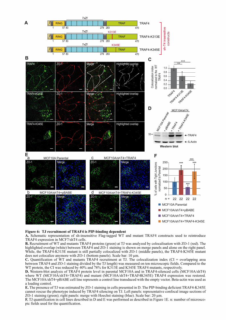
TRAF4 MergeZO-1 Highlighted overlap
TRAF4-K345E ZO-1 Merge Highlighted overlap
TRAF4-K313E ZO-1 Merge Highlighted overlap
B C
A
TRAF4-K345E
1 57 83 279 283 470
RING TRAFFla
g
K345E
sh
-T4
in
se
nsitiv
e
co
nstr
ucts
TRAF4
1 57 83 279 283 470
RING TRAFFla
g
TRAF4-K313E
1 57 83 279 283 470
RING TRAF
K313E
Fla
g
Col
ocal
izat
ion
inde
x
norm
aliz
ed to
the
WT
TR
AF
4
0.0
0.2
0.4
0.6
0.8
1.0
TRAF4
TRAF4-
K31
3E
TRAF4-
K34
5E
*****
MCF10A Parental
MCF10A/shT4+pBABE
MCF10A/shT4+TRAF4
MCF10A/shT4+TRAF4-K345E
0
50
100
150
200
250***
***
***
***
% o
f cel
ls w
ith T
ight
Jun
ctio
ns
norm
aliz
ed to
Par
enta
l
n = 22 22 22 22
E
MCF10A/shT4+pBABEb
MCF10A Parental MCF10A/shT4+TRAF4
ZO-1
ZO-1
ZO-1Merge
Merge
Merge
a c
MCF10A/shT4+TRAF4-K345E
ZO-1 Merge
d
F
55
Western blot
TRAF4
ß-Actin
MC
F10A P
are
nta
l
+ p
BA
BE
+ T
RA
F4
+ T
RA
F4-K
345E
MCF10A/shT4D
7xZf
7xZf
7xZf
Figure 6: TJ recruitment of TRAF4 is PIP-binding dependentA. Schematic representation of sh-insensitive Flag-tagged WT and mutant TRAF4 constructs used to reintroduce TRAF4 expression in MCF7/shT4 cells. B. Recruitment of WT and mutants TRAF4 proteins (green) at TJ was analyzed by colocalisation with ZO-1 (red). The highlighted overlap (white) between TRAF4 and ZO-1 staining is shown on merge panels and alone on the right panel. While, the TRAF4-K313E mutant is still partially colocalized with ZO-1 (middle panels), the TRAF4-K345E mutant does not colocalize anymore with ZO-1 (bottom panels). Scale bar: 10 µm.C. Quantification of WT and mutants TRAF4 recruitment at TJ. The colocalization index (CI = overlapping area between TRAF4 and ZO-1 staining divided by the TJ length) was measured on ten microscopic fields. Compared to the WT protein, the CI was reduced by 40% and 78% for K313E and K345E TRAF4 mutants, respectively.D. Western-blot analysis of TRAF4 protein level in parental MCF10A and in TRAF4-silenced cells (MCF10A/shT4) where WT (MCF10A/shT4+TRAF4) and mutant (MCF10A/shT4+TRAF4K345E) TRAF4 expression was restored. The MCF10A/shT4+pBABE cell line represents a control line transduced with the empty vector. Beta-actin was used as a loading control.E. The presence of TJ was estimated by ZO-1 staining in cells presented in D. The PIP-binding deficient TRAF4-K345E cannot rescue the phenotype induced by TRAF4 silencing on TJ. Left panels: representative confocal image sections of ZO-1 staining (green); right panels: merge with Hoechst staining (blue). Scale bar: 20 µm.F. TJ quantification in cell lines described in D and E was performed as described in Figure 1E. n: number of microsco-pic fields used for the quantification.
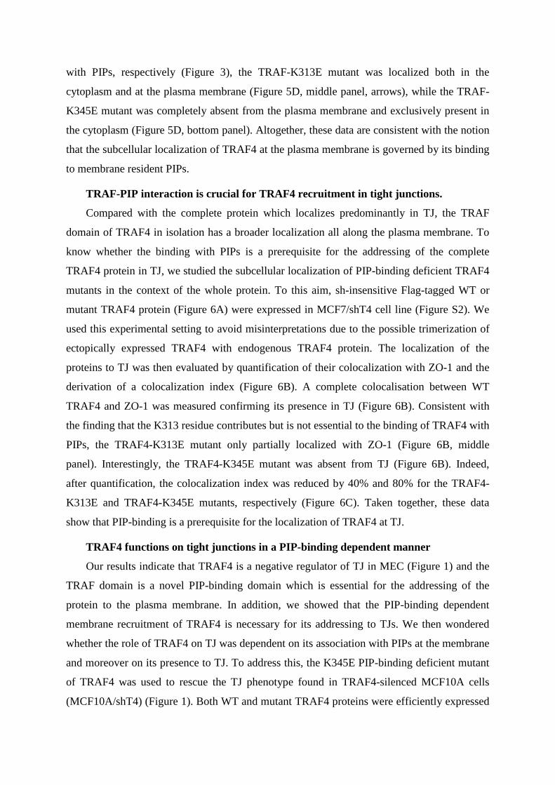
with PIPs, respectively (Figure 3), the TRAF-K313E mutant was localized both in the
cytoplasm and at the plasma membrane (Figure 5D, middle panel, arrows), while the TRAF-
K345E mutant was completely absent from the plasma membrane and exclusively present in
the cytoplasm (Figure 5D, bottom panel). Altogether, these data are consistent with the notion
that the subcellular localization of TRAF4 at the plasma membrane is governed by its binding
to membrane resident PIPs.
TRAF-PIP interaction is crucial for TRAF4 recruitment in tight junctions.
Compared with the complete protein which localizes predominantly in TJ, the TRAF
domain of TRAF4 in isolation has a broader localization all along the plasma membrane. To
know whether the binding with PIPs is a prerequisite for the addressing of the complete
TRAF4 protein in TJ, we studied the subcellular localization of PIP-binding deficient TRAF4
mutants in the context of the whole protein. To this aim, sh-insensitive Flag-tagged WT or
mutant TRAF4 protein (Figure 6A) were expressed in MCF7/shT4 cell line (Figure S2). We
used this experimental setting to avoid misinterpretations due to the possible trimerization of
ectopically expressed TRAF4 with endogenous TRAF4 protein. The localization of the
proteins to TJ was then evaluated by quantification of their colocalization with ZO-1 and the
derivation of a colocalization index (Figure 6B). A complete colocalisation between WT
TRAF4 and ZO-1 was measured confirming its presence in TJ (Figure 6B). Consistent with
the finding that the K313 residue contributes but is not essential to the binding of TRAF4 with
PIPs, the TRAF4-K313E mutant only partially localized with ZO-1 (Figure 6B, middle
panel). Interestingly, the TRAF4-K345E mutant was absent from TJ (Figure 6B). Indeed,
after quantification, the colocalization index was reduced by 40% and 80% for the TRAF4-
K313E and TRAF4-K345E mutants, respectively (Figure 6C). Taken together, these data
show that PIP-binding is a prerequisite for the localization of TRAF4 at TJ.
TRAF4 functions on tight junctions in a PIP-binding dependent manner
Our results indicate that TRAF4 is a negative regulator of TJ in MEC (Figure 1) and the
TRAF domain is a novel PIP-binding domain which is essential for the addressing of the
protein to the plasma membrane. In addition, we showed that the PIP-binding dependent
membrane recruitment of TRAF4 is necessary for its addressing to TJs. We then wondered
whether the role of TRAF4 on TJ was dependent on its association with PIPs at the membrane
and moreover on its presence to TJ. To address this, the K345E PIP-binding deficient mutant
of TRAF4 was used to rescue the TJ phenotype found in TRAF4-silenced MCF10A cells
(MCF10A/shT4) (Figure 1). Both WT and mutant TRAF4 proteins were efficiently expressed
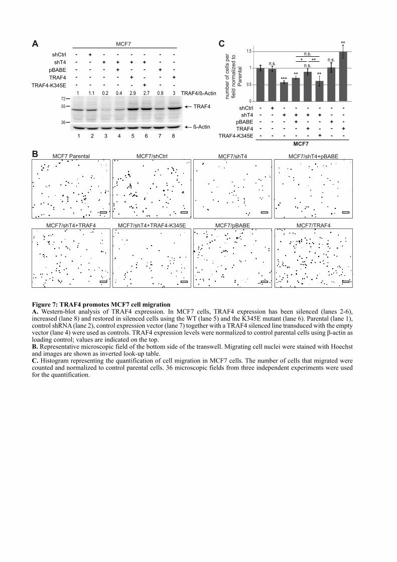
A
shT4
shCtrl
pBABE
TRAF4
+-
-
-
-
-
-
-
-
-
+
-
-
+
-
-
+
-
+
+
-
-
-
-
-
-+
+
72
55 TRAF4
ß-Actin
1 1.1 0.2 0.4 2.9 2.7 0.9 TRAF4/ß-Actin
MCF7
1 2 3 4 5 6 7 8
-
+
-
-
TRAF4-K345E - - - - - - -+
3
MCF7/shCtrl MCF7/shT4MCF7 Parental MCF7/shT4+pBABE
MCF7/shT4+TRAF4 MCF7/pBABE MCF7/TRAF4MCF7/shT4+TRAF4-K345E
B
C
num
ber
of cells
per
field
norm
aliz
ed to
Pare
nta
l
shT4
shCtrl
pBABE
TRAF4
+-
-
-
-
-
-
-
-
-
+
-
-
+
-
-
+
-
+
+
-
-
-
-
-
-+
+
MCF7
-
+
-
-
TRAF4-K345E - - - - - - -+
0
0.5
1
1.5
***
* **
** **
**
n.s.
n.s.n.s.
n.s.
36
Figure 7: TRAF4 promotes MCF7 cell migrationA. Western-blot analysis of TRAF4 expression. In MCF7 cells, TRAF4 expression has been silenced (lanes 2-6), increased (lane 8) and restored in silenced cells using the WT (lane 5) and the K345E mutant (lane 6). Parental (lane 1), control shRNA (lane 2), control expression vector (lane 7) together with a TRAF4 silenced line transduced with the empty vector (lane 4) were used as controls. TRAF4 expression levels were normalized to control parental cells using β-actin as loading control; values are indicated on the top. B. Representative microscopic field of the bottom side of the transwell. Migrating cell nuclei were stained with Hoechst and images are shown as inverted look-up table.C. Histogram representing the quantification of cell migration in MCF7 cells. The number of cells that migrated were counted and normalized to control parental cells. 36 microscopic fields from three independent experiments were used for the quantification.
in MCF10A/shT4 cells to levels above endogenous TRAF4 (Figure 6D). As previously
mentioned (Figure 1B), TRAF4 silenced cells (MCF10A/shT4+pBABE) have a ~2 fold
increase in cells with TJ (Figure 6Ea-b, F), while cells with restored TRAF4 expression had
two fold less TJ than the parental (Figure 6Ea-c, F). In contrast, restoring TRAF4 expression
by using the TRAF4-K345E mutant did not rescue the phenotype induced by TRAF4
silencing (Figure 6Ed, F).
These results provide a novel illustration of the importance of TRAF4 subcellular
localization on cell biology. Indeed, they show that to act as a negative regulator on TJ
formation and/or stability, TRAF4 must be addressed to the plasma membrane/TJ via a PIP-
binding dependent mechanism. Therefore preventing or favoring its presence in the plasma
membrane and in TJ represents a means to regulate its action on tissue homeostasis.
TRAF4 promotes MEC migration
Of interest, TJ proteins in addition to their characterized role in mediating cell–cell
adhesion and assuring epithelial barrier can have an active role in cell migration. The protein
ZO-1 actively regulates cell migration by modulating cytoskeletal dynamics [58]. Given that
TRAF4 is overexpressed in breast cancer and that migration of cancer cells participates to
tumor progression, we addressed the role of TRAF4 in the migration of breast cancer cells. To
this aim, TRAF4 expression was modulated in MCF7 cells, and cell migration was measured
using the Boyden chamber assay. We first studied cell migration in TRAF4-depleted MCF7
cells. Compared to parental (MCF7) and to a control cell line (MCF7/shCtrl), TRAF4 was
expressed by less than 20% in TRAF4-silenced cells (MCF7/shT4) (Figure 7A). Migration
was impaired by 40% in this line compared to the controls (Figure 7B, C). We next
reintroduced TRAF4 expression in this silenced cell line (MCF7/ShT4+TRAF4) and TRAF4
was expressed at level above the parental cells (Figure 7A). Consistently, reintroduction of
TRAF4 was associated with increased cell migration similar to parental MCF7 cells (Figure
7B, C). To complement these findings, MCF7 cells stably expressing TRAF4 were generated
(MCF7/TRAF4). TRAF4 was increased 3 fold as compared to parental and control cells
(Figure 7A). Supporting the role of TRAF4 in cell migration, compared with parental and
control lines, migration was increased by 40% in these cells (Figure 7B, C). To know whether
this promigratory function is linked with its ability to interact with PIP and to localize at TJ,
we then used the PIP-binding deficient TRAF4-K345E mutant in the Boyden chamber
migration assay. To this end, this mutant was reintroduced and expressed in TRAF4 silenced
cells (MCF7/shT4+TRAF4-K345E) (Figure 7A). In contrast to the WT protein, the TRAF4-

K345E mutant did not restore cell migration in TRAF4-silenced MCF7 cells, indicating that a
PIP-binding deficient mutant could not rescue the migration phenotype induced by the loss of
TRAF4 expression (Figure 7B, C). The positive impact of TRAF4 on cell migration was also
addressed in MCF10A cells. Consistent with the results obtained using MCF7 cells, TRAF4
positively regulated cell migration in this cell line (Figure S4). Altogether, these results
support the role of TRAF4 as a novel regulator of cell migration operating at the TJ level in a
PIP-binding dependent manner.
DISCUSSION
In the normal breast, TRAF4 is predominantly localized in TJ of polarized epithelial
cells. The molecular mechanism targeting TRAF4 to these adhesion structures as well as its
function at these regions remained elusive. The experiments presented here provide for the
first time, to our knowledge, a molecular explanation for the recruitment of TRAF4 in TJ.
Indeed TRAF4 uses its TRAF domain as a novel PIP-binding domain to be addressed to the
plasma membrane, an essential step for its recruitment in TJ. Moreover, they show that
TRAF4 acts as a negative regulator of TJ and favors migration of breast cancer cells.
Several lines of evidence indicate a link between TRAF4 and cell polarity [24]. During
development, breast epithelial cells polarize and assemble into duct and acini structures. One
of the earliest manifestations of breast cancer is the loss of this cellular organization. Indeed,
loss of cell-cell contact and epithelial polarity are hallmarks of carcinomas and contribute to
their development as carcinomas in situ or their progression to invasive adenocarcinomas.
TRAF4 expression is altered in breast carcinomas and the protein shifts from TJ to other
subcellular territories suggesting that it could be part of the mechanisms leading to the
disruption of the polarized breast epithelium.
Of interest, the epithelial polarity program relies on several conserved cellular
machineries, including the domain-identity machinery which builds a TJ fence between apical
and basolateral plasma membrane domains by using specific proteins and lipids [59]. The
asymmetric distribution of lipids from the PIP family within the plasma membrane plays a
key role in the establishment of cell polarity. These lipids represent optimal signaling
mediators by forming docking sites for PIP-binding protein effectors [47,52,60]. To date, 11
PIP-binding domains have been described including PH (pleckstrin homology), PX (Phox
homology) and FYVE (Fab1, YOTB, Vac1 and EEA1) domains [61]. While the majority of
PIP-binding modules selectively bind to one PIP depending on its phosphorylation status, it
has recently emerged that some proteins bind PIPs in a promiscuous manner. It is the case for
the polarity proteins PAR3, ZO-1 and α-syntrophin. They do not contain a consensus PIP-
binding motif but use their PDZ or PH domains to bind PIPs [35,36,62]. It has been proposed
that the loose binding of these proteins to PIPs served to enhance their affinity for membranes
and that additional factors are required to fine-tune their subcellular localizations [47]. In this
study, we show that TRAF4 uses its TRAF domain as a novel PIP-binding module. Similarly
to PAR3, ZO-1 and α-syntrophin, the TRAF domain of TRAF4 has a broad affinity for PIPs.
Consistently, the TRAF domain in isolation localizes homogenously along the plasma
membrane while the full-length TRAF4 protein is restricted to TJ, suggesting that the binding
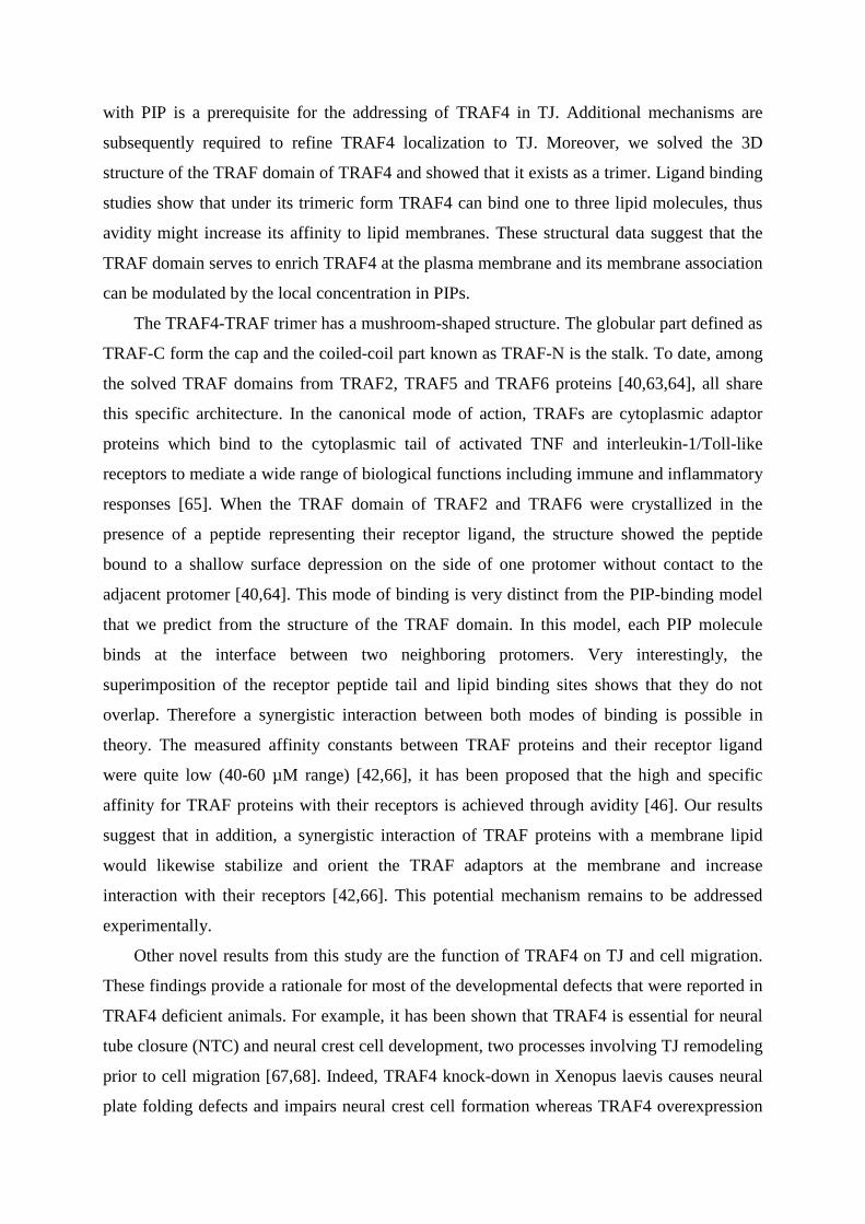
with PIP is a prerequisite for the addressing of TRAF4 in TJ. Additional mechanisms are
subsequently required to refine TRAF4 localization to TJ. Moreover, we solved the 3D
structure of the TRAF domain of TRAF4 and showed that it exists as a trimer. Ligand binding
studies show that under its trimeric form TRAF4 can bind one to three lipid molecules, thus
avidity might increase its affinity to lipid membranes. These structural data suggest that the
TRAF domain serves to enrich TRAF4 at the plasma membrane and its membrane association
can be modulated by the local concentration in PIPs.
The TRAF4-TRAF trimer has a mushroom-shaped structure. The globular part defined as
TRAF-C form the cap and the coiled-coil part known as TRAF-N is the stalk. To date, among
the solved TRAF domains from TRAF2, TRAF5 and TRAF6 proteins [40,63,64], all share
this specific architecture. In the canonical mode of action, TRAFs are cytoplasmic adaptor
proteins which bind to the cytoplasmic tail of activated TNF and interleukin-1/Toll-like
receptors to mediate a wide range of biological functions including immune and inflammatory
responses [65]. When the TRAF domain of TRAF2 and TRAF6 were crystallized in the
presence of a peptide representing their receptor ligand, the structure showed the peptide
bound to a shallow surface depression on the side of one protomer without contact to the
adjacent protomer [40,64]. This mode of binding is very distinct from the PIP-binding model
that we predict from the structure of the TRAF domain. In this model, each PIP molecule
binds at the interface between two neighboring protomers. Very interestingly, the
superimposition of the receptor peptide tail and lipid binding sites shows that they do not
overlap. Therefore a synergistic interaction between both modes of binding is possible in
theory. The measured affinity constants between TRAF proteins and their receptor ligand
were quite low (40-60 µM range) [42,66], it has been proposed that the high and specific
affinity for TRAF proteins with their receptors is achieved through avidity [46]. Our results
suggest that in addition, a synergistic interaction of TRAF proteins with a membrane lipid
would likewise stabilize and orient the TRAF adaptors at the membrane and increase
interaction with their receptors [42,66]. This potential mechanism remains to be addressed
experimentally.
Other novel results from this study are the function of TRAF4 on TJ and cell migration.
These findings provide a rationale for most of the developmental defects that were reported in
TRAF4 deficient animals. For example, it has been shown that TRAF4 is essential for neural
tube closure (NTC) and neural crest cell development, two processes involving TJ remodeling
prior to cell migration [67,68]. Indeed, TRAF4 knock-down in Xenopus laevis causes neural
plate folding defects and impairs neural crest cell formation whereas TRAF4 overexpression
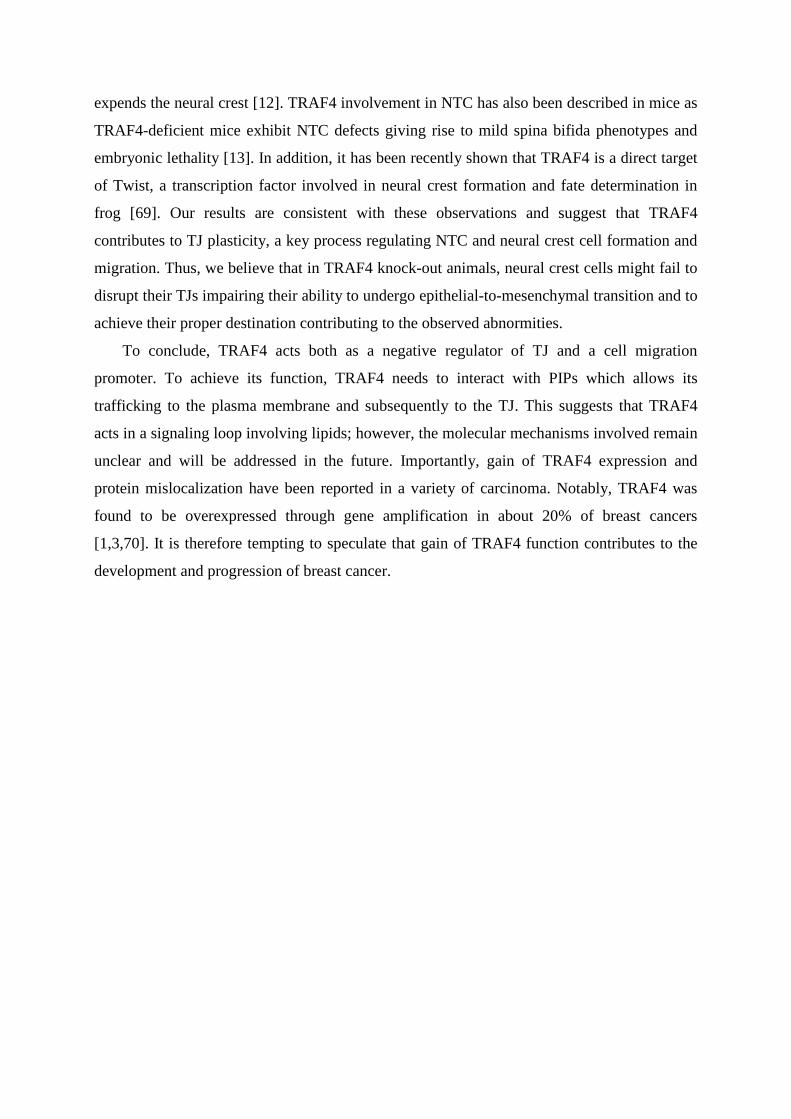
expends the neural crest [12]. TRAF4 involvement in NTC has also been described in mice as
TRAF4-deficient mice exhibit NTC defects giving rise to mild spina bifida phenotypes and
embryonic lethality [13]. In addition, it has been recently shown that TRAF4 is a direct target
of Twist, a transcription factor involved in neural crest formation and fate determination in
frog [69]. Our results are consistent with these observations and suggest that TRAF4
contributes to TJ plasticity, a key process regulating NTC and neural crest cell formation and
migration. Thus, we believe that in TRAF4 knock-out animals, neural crest cells might fail to
disrupt their TJs impairing their ability to undergo epithelial-to-mesenchymal transition and to
achieve their proper destination contributing to the observed abnormities.
To conclude, TRAF4 acts both as a negative regulator of TJ and a cell migration
promoter. To achieve its function, TRAF4 needs to interact with PIPs which allows its
trafficking to the plasma membrane and subsequently to the TJ. This suggests that TRAF4
acts in a signaling loop involving lipids; however, the molecular mechanisms involved remain
unclear and will be addressed in the future. Importantly, gain of TRAF4 expression and
protein mislocalization have been reported in a variety of carcinoma. Notably, TRAF4 was
found to be overexpressed through gene amplification in about 20% of breast cancers
[1,3,70]. It is therefore tempting to speculate that gain of TRAF4 function contributes to the
development and progression of breast cancer.
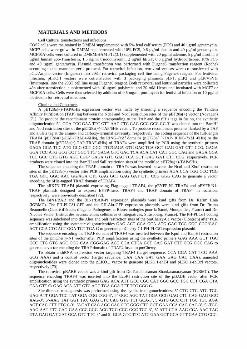
MATERIALS AND METHODS
Cell Culture, transfections and infections COS7 cells were maintained in DMEM supplemented with 5% fetal calf serum (FCS) and 40 µg/ml gentamycin. MCF7 cells were grown in DMEM supplemented with 10% FCS, 0.6 µg/ml insulin and 40 µg/ml gentamycin. MCF10A cells were cultured in DMEM/HAM F12(3:1) supplemented with 20 µg/ml adenine, 5 µg/ml insulin, 5 µg/ml human apo-Transferrin, 1.5 ng/ml triiodothyronin, 2 ng/ml hEGF, 0.5 µg/ml hydrocortisone, 10% FCS and 40 µg/ml gentamycin. Plasmid transfection was performed with Fugene6 transfection reagent (Roche) according to the manufacturer’s protocol. For retroviral infection, retroviral vectors were co-transfected with pCL-Ampho vector (Imgenex) into 293T retroviral packaging cell line using Fugene6 reagent. For lentiviral infection, pLKO.1 vectors were cotransfected with 3 packaging plasmids pLP1, pLP2 and pLP/VSVG (Invitrogen) into the 293T cell line using Fugene6 reagent. Both retroviral and lentiviral particles were collected 48h after transfection, supplemented with 10 µg/ml polybrene and 20 mM Hepes and incubated with MCF7 or MCF10A cells. Cells were then selected by addition of 0.5 mg/ml puromycin for lentiviral infection or 10 µg/ml blasticidin for retroviral infection.
Cloning and Constructs A pET28a(+)-TAP/6His expression vector was made by inserting a sequence encoding the Tandem
Affinity Purification (TAP) tag between the NdeI and NcoI restriction sites of the pET28a(+) vector (Novagen) [71]. To produce the recombinant protein corresponding to the TAP and the 6His tags in fusion, the synthetic oligonucleotide 5’- GGA TCC GAA TTC GTT AAC CTC GAG GCG GCC GC-3’ was cloned into the BamHI and NotI restriction sites of the pET28a(+)-TAP/6His vector. To produce recombinant proteins flanked by a TAP and a 6His tag at the amino- and carboxy-terminal extremity, respectively, the coding sequence of the full-length TRAF4 (pET28a(+)-TAP-TRAF4-6His), the RING-7xZf domains (pET28a(+)-TAP- RING-7xZf -6His) or the TRAF domain (pET28a(+)-TAP-TRAF-6His) of TRAF4 were amplified by PCR using the synthetic primers GAGA GGA TCC ATG GCG CCT GGC TTC/GAGA GTC GAC TCA GCT GAG GAT CTT CCG, GAGA GGA TCC ATG GCG CCT GGC TTC/ GAGA GTC GAC TCA ACA CAT CAT GGC CAG and GAGA GGA TCC GCC CTG GTG AGC CGG/ GAGA GTC GAC TCA GCT GAG GAT CTT CCG, respectively. PCR products were cloned into the BamHI and SalI restriction sites of the modified pET28a(+)-TAP/6His.
The sequence encoding the TRAF domain of TRAF4 was inserted between the NcoI and XhoI restriction sites of the pET28a(+) vector after PCR amplification using the synthetic primers AGA CCA TGG CCC TGG TGA GCC GGC AAC GG/AGA CTC GAG GCT GAG GAT CTT CCG GGG CAG to generate a vector encoding the 6His-tagged TRAF domain of TRAF4.
The pRK7N TRAF4 plasmid expressing Flag-tagged TRAF4, the pEYFP-N1-TRAF4 and pEYFP-N1-TRAF plasmids designed to express EYFP-fused TRAF4 and TRAF domain of TRAF4 in isolation, respectively, were previously described [72].
The BIN1/BAR and the BIN1/BAR-PI expression plasmids were kind gifts from Dr. Karim Hnia (IGBMC). The PH-PLCδ1-GFP and the PH-Akt-GFP expression plasmids were kind gifts from Dr. Bruno Beaumelle (Centre d’études d’agents Pathogènes et Biotechnologies pour la Santé, Montpellier, France) and Dr. Nicolas Vitale (Institut des neurosciences cellulaires et intégratives, Strasbourg, France). The PH-PLCδ1 coding sequence was subcloned into the XhoI and SalI restriction sites of the pmCherry-C1 vector (Clontech) after PCR amplification using the synthetic oligonucleotides GAG ACT CGA GCA ATG GAC TCG GGC CGG/GAG AGT CGA CTC ACT GGA TGT TGA G to generate pmCherry-C1-PH-PLCδ1 expression plasmid.
The sequence encoding the TRAF domain of TRAF4 was inserted between the KpnI and BamHI restriction sites of the pmCherry-N1 vector after PCR amplification using the synthetic primers GAG AAA GCT TGC GCC CTG GTG AGC CGG CAA CGG/GAG AGT CGA CTCA GCT GAG GAT CTT CCG GGG CAG to generate a vector encoding the TRAF domain of TRAF4 fused to pmCherry.
To obtain a shRNA expression vector targeting TRAF4 (target sequence: CCA GGA CAT TCG AAA GCG AAA) and a control vector (target sequence: CAA CAA GAT GAA GAG CAC CAA), annealed oligonucleotides were cloned into the pLKO.1 vector to generate pLKO.1-shT4 and pLKO.1-shCtrl vectors, respectively [73].
The retroviral pBABE vector was a kind gift from Dr. Pattabhiraman Shankaranarayanan (IGBMC). The sequence encoding TRAF4 was inserted into the EcoRI restriction site of the pBABE vector after PCR amplification using the synthetic primers GAG ACA ATT GCC CGC CAT GGC GCC TGG CTT CGA CTA CAA GTT C/ GAG ACA ATT GTC AGC TGA GGA TCT TCC GGG G.
Site-directed mutagenesis was performed using the synthetic oligonucleotides: 5’-GTG CTC ATC TGG GAG ATT GGA TCC TAT GGA CGG CGG-3’, 5’-GGC AGC TAT GGA GCG GAG CTC CAG GAG GCC AAG-3’, 5’-AAG TAT GGT TAC GAG CTC CAG GTG TCT GCA-3’, 5’-GTG GCC CTT TGC TGC AGA AGT CAC CTT CTC C-3’, 5’-GAT CAG AGC GAC CCC GGG CTG GCT GAA CCA CAG CAC-3’, 5’-TGG AAG AAT TTC CAG GAA CCC GGG ACG TGG CGG GGC TCC-3’, 5’-ATT CGA AAG CGA AAC TAC GTA GAG GAT GAT GCA GTC TTC-3’ and 5’-GCA GTC TTC ATC GAA GCT GCA GTT GAA CTG CCC-
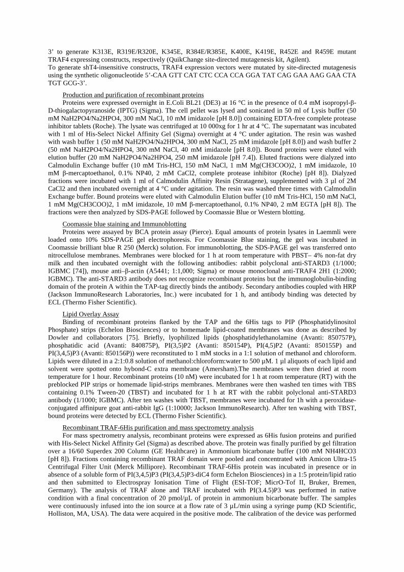
3’ to generate K313E, R319E/R320E, K345E, R384E/R385E, K400E, K419E, R452E and R459E mutant TRAF4 expressing constructs, respectively (QuikChange site-directed mutagenesis kit, Agilent). To generate shT4-insensitive constructs, TRAF4 expression vectors were mutated by site-directed mutagenesis using the synthetic oligonucleotide 5’-CAA GTT CAT CTC CCA CCA GGA TAT CAG GAA AAG GAA CTA TGT GCG-3’.
Production and purification of recombinant proteins Proteins were expressed overnight in E.Coli BL21 (DE3) at 16 °C in the presence of 0.4 mM isopropyl-β-
D-thiogalactopyranoside (IPTG) (Sigma). The cell pellet was lysed and sonicated in 50 ml of Lysis buffer (50 mM NaH2PO4/Na2HPO4, 300 mM NaCl, 10 mM imidazole [pH 8.0]) containing EDTA-free complete protease inhibitor tablets (Roche). The lysate was centrifuged at 10 000xg for 1 hr at 4 °C. The supernatant was incubated with 1 ml of His-Select Nickel Affinity Gel (Sigma) overnight at 4 °C under agitation. The resin was washed with wash buffer 1 (50 mM NaH2PO4/Na2HPO4, 300 mM NaCl, 25 mM imidazole [pH 8.0]) and wash buffer 2 (50 mM NaH2PO4/Na2HPO4, 300 mM NaCl, 40 mM imidazole [pH 8.0]). Bound proteins were eluted with elution buffer (20 mM NaH2PO4/Na2HPO4, 250 mM imidazole [pH 7.4]). Eluted fractions were dialyzed into Calmodulin Exchange buffer (10 mM Tris-HCl, 150 mM NaCl, 1 mM Mg(CH3COO)2, 1 mM imidazole, 10 mM β-mercaptoethanol, 0.1% NP40, 2 mM CaCl2, complete protease inhibitor (Roche) [pH 8]). Dialyzed fractions were incubated with 1 ml of Calmodulin Affinity Resin (Stratagene), supplemented with 3 µl of 2M CaCl2 and then incubated overnight at 4 °C under agitation. The resin was washed three times with Calmodulin Exchange buffer. Bound proteins were eluted with Calmodulin Elution buffer (10 mM Tris-HCl, 150 mM NaCl, 1 mM Mg(CH3COO)2, 1 mM imidazole, 10 mM β-mercaptoethanol, 0.1% NP40, 2 mM EGTA [pH 8]). The fractions were then analyzed by SDS-PAGE followed by Coomassie Blue or Western blotting.
Coomassie blue staining and Immunoblotting Proteins were assayed by BCA protein assay (Pierce). Equal amounts of protein lysates in Laemmli were
loaded onto 10% SDS-PAGE gel electrophoresis. For Coomassie Blue staining, the gel was incubated in Coomassie brilliant blue R 250 (Merck) solution. For immunoblotting, the SDS-PAGE gel was transferred onto nitrocellulose membranes. Membranes were blocked for 1 h at room temperature with PBST– 4% non-fat dry milk and then incubated overnight with the following antibodies: rabbit polyclonal anti-STARD3 (1/1000; IGBMC [74]), mouse anti–β-actin (A5441; 1:1,000; Sigma) or mouse monoclonal anti-TRAF4 2H1 (1:2000; IGBMC). The anti-STARD3 antibody does not recognize recombinant proteins but the immunoglobulin-binding domain of the protein A within the TAP-tag directly binds the antibody. Secondary antibodies coupled with HRP (Jackson ImmunoResearch Laboratories, Inc.) were incubated for 1 h, and antibody binding was detected by ECL (Thermo Fisher Scientific).
Lipid Overlay Assay Binding of recombinant proteins flanked by the TAP and the 6His tags to PIP (Phosphatidylinositol
Phosphate) strips (Echelon Biosciences) or to homemade lipid-coated membranes was done as described by Dowler and collaborators [75]. Briefly, lyophilized lipids (phosphatidylethanolamine (Avanti: 850757P), phosphatidic acid (Avanti: 840875P), PI(3,5)P2 (Avanti: 850154P), PI(4,5)P2 (Avanti: 850155P) and PI(3,4,5)P3 (Avanti: 850156P)) were reconstituted to 1 mM stocks in a 1:1 solution of methanol and chloroform. Lipids were diluted in a 2:1:0.8 solution of methanol:chloroform:water to 500 µM. 1 µl aliquots of each lipid and solvent were spotted onto hybond-C extra membrane (Amersham).The membranes were then dried at room temperature for 1 hour. Recombinant proteins (10 nM) were incubated for 1 h at room temperature (RT) with the preblocked PIP strips or homemade lipid-strips membranes. Membranes were then washed ten times with TBS containing 0.1% Tween-20 (TBST) and incubated for 1 h at RT with the rabbit polyclonal anti-STARD3 antibody (1/1000; IGBMC). After ten washes with TBST, membranes were incubated for 1h with a peroxidase-conjugated affinipure goat anti-rabbit IgG (1:10000; Jackson ImmunoResearch). After ten washing with TBST, bound proteins were detected by ECL (Thermo Fisher Scientific).
Recombinant TRAF-6His purification and mass spectrometry analysis For mass spectrometry analysis, recombinant proteins were expressed as 6His fusion proteins and purified
with His-Select Nickel Affinity Gel (Sigma) as described above. The protein was finally purified by gel filtration over a 16/60 Superdex 200 Column (GE Healthcare) in Ammonium bicarbonate buffer (100 mM NH4HCO3 [pH 8]). Fractions containing recombinant TRAF domain were pooled and concentrated with Amicon Ultra-15 Centrifugal Filter Unit (Merck Millipore). Recombinant TRAF-6His protein was incubated in presence or in absence of a soluble form of PI(3,4,5)P3 (PI(3,4,5)P3-diC4 form Echelon Biosciences) in a 1:5 protein/lipid ratio and then submitted to Electrospray Ionisation Time of Flight (ESI-TOF; MicrO-Tof II, Bruker, Bremen, Germany). The analysis of TRAF alone and TRAF incubated with PI(3.4.5)P3 was performed in native condition with a final concentration of 20 pmol/µL of protein in ammonium bicarbonate buffer. The samples were continuously infused into the ion source at a flow rate of 3 µL/min using a syringe pump (KD Scientific, Holliston, MA, USA). The data were acquired in the positive mode. The calibration of the device was performed
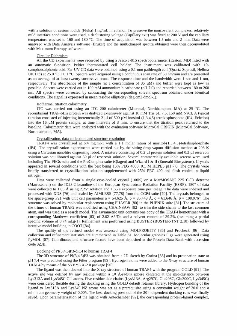
with a solution of cesium iodide (Fluka) 1mg/mL in ethanol. To preserve the noncovalent complexes, relatively mild interface conditions were used, a declustering voltage (Capillary exit) was fixed at 200 V and the capillary temperature was set to 160 and 180 °C. The time of acquisition was between 1.5 min and 2 min. Data were analyzed with Data Analysis software (Bruker) and the multicharged spectra obtained were then deconvoluted with Maximum Entropy software.
Circular Dichroism All the CD experiments were recorded by using a Jasco J-815 spectropolarimeter (Easton, MD) fitted with
an automatic 6-position Peltier thermostated cell holder. The instrument was calibrated with 10-camphorsulphonic acid. Far-UV CD data were obtained using a 0.1 mm pathlength cell (Quartz-Suprasil, Hellma UK Ltd) at 25.0 °C ± 0.1 °C. Spectra were acquired using a continuous scan rate of 50 nm/min and are presented as an average of at least twenty successive scans. The response time and the bandwidth were 1 sec and 1 nm, respectively. The absorbance of the sample (at a concentration of 35 µM) and buffer were kept as low as possible. Spectra were carried out in 100 mM ammonium bicarbonate (pH 7.0) and recorded between 180 to 260 nm. All spectra were corrected by subtracting the corresponding solvent spectrum obtained under identical conditions. The signal is expressed in mean residue ellipticity (deg.cm2.dmol-1).
Isothermal titration calorimetry ITC was carried out using an ITC 200 calorimeter (Microcal, Northhampton, MA) at 25 °C. The
recombinant TRAF-6His protein was dialyzed extensively against 10 mM Tris pH 7.5, 150 mM NaCl. A typical titration consisted of injecting incrementally 2 µl of 500 µM inositol-(1,3,4,5)-tetrakisphosphate (IP4, Echelon) into the 16 µM protein sample, at time intervals of 3 min, to ensure that the titration peak returned to the baseline. Calorimetric data were analyzed with the evaluation software MicroCal ORIGIN (MicroCal Software, Northhampton, MA).
Crystallization, data collection, and structure resolution TRAF4 was crystallized at 6.4 mg.ml-1 with a 1:1 molar ration of inositol-(1,3,4,5)-tetrakisphosphate
(IP4). The crystallization experiments were carried out by the sitting-drop vapour diffusion method at 293 K using a Cartesian nanolitre dispensing robot. A mixture consisting of 0.2 µl protein solution and 0.2 µl reservoir solution was equilibrated against 50 µl of reservoir solution. Several commercially available screens were used including The PEGs suite and the ProComplex suite (Qiagen) and Wizard I & II (Emerald Biosystems). Crystals appeared in several conditions with the best being 15% PEG 4000, 0.1 M HEPES pH 7.0. The crystals were briefly transferred to crystallization solution supplemented with 25% PEG 400 and flash cooled in liquid nitrogen.
Data were collected from a single cryo-cooled crystal (100K) on a MarMOSAIC 225 CCD detector (Marresearch) on the ID23-2 beamline of the European Synchrotron Radiation Facility (ESRF). 180° of data were collected to 1.85 Å using 2.25° rotation and 1.55 s exposure time per image. The data were indexed and processed with XDS [76] and scaled by AIMLESS [77,78] from the CCP4 suite [79]. The crystals belonged to the space-group P21 with unit cell parameters a = 54.625 Å, b = 85.443 Å, c = 61.646 Å, β = 108.076°. The structure was solved by molecular replacement using PHASER [80] in the PHENIX suite [81]. The structure of the trimer of human TRAF2 was modified using CHAINSAW [82] to trim the side chains to the last common atom, and was used as a search model. The asymmetric unit contains one copy of the TRAF4 homotrimer with a corresponding Matthews coefficient [83] of 2.02 Å3/Da and a solvent content of 39.2% (assuming a partial specific volume of 0.74 ml.g-1). Refinement was performed using BUSTER (BUSTER-TNT 2.10) followed by iterative model building in COOT [84].
The quality of the refined model was assessed using MOLPROBITY [85] and Procheck [86]. Data collection and refinement statistics are summarized in Table S1. Molecular graphics Figs were generated using PyMOL [87]. Coordinates and structure factors have been deposited at the Protein Data Bank with accession code 3ZJB.
Docking of PI(3,4,5)P3-diC4 to human TRAF4 The 3D structure of PI(3,4,5)P3 was obtained from a 2D sketch by Corina [88] and its protonation state at
pH 7.4 was predicted using the Filter program [89]. Hydrogen atoms were added to the X-ray structure of human TRAF4 by means of the SYBYL X-2.0 package [90].
The ligand was then docked into the X-ray structure of human TRAF4 with the program GOLD [91]. The active site was defined by any residue within a 10 Å-radius sphere centered at the mid-distance between Lys313A and Lys345C C atoms. Five residue side chains (Lys313A, Arg297C, Glu298C, Glu300C, Lys345C) were considered flexible during the docking using the GOLD default rotamer library. Hydrogen bonding of the ligand to Lys313A and Lys345 NZ atoms was set as a prerequisite using a constraint weight of 20.0 and a minimum geometry weight of 0.005. The best docking pose out of the 20 independent docking runs was finally saved. Upon parameterization of the ligand with Antechamber [92], the corresponding protein-ligand complex,
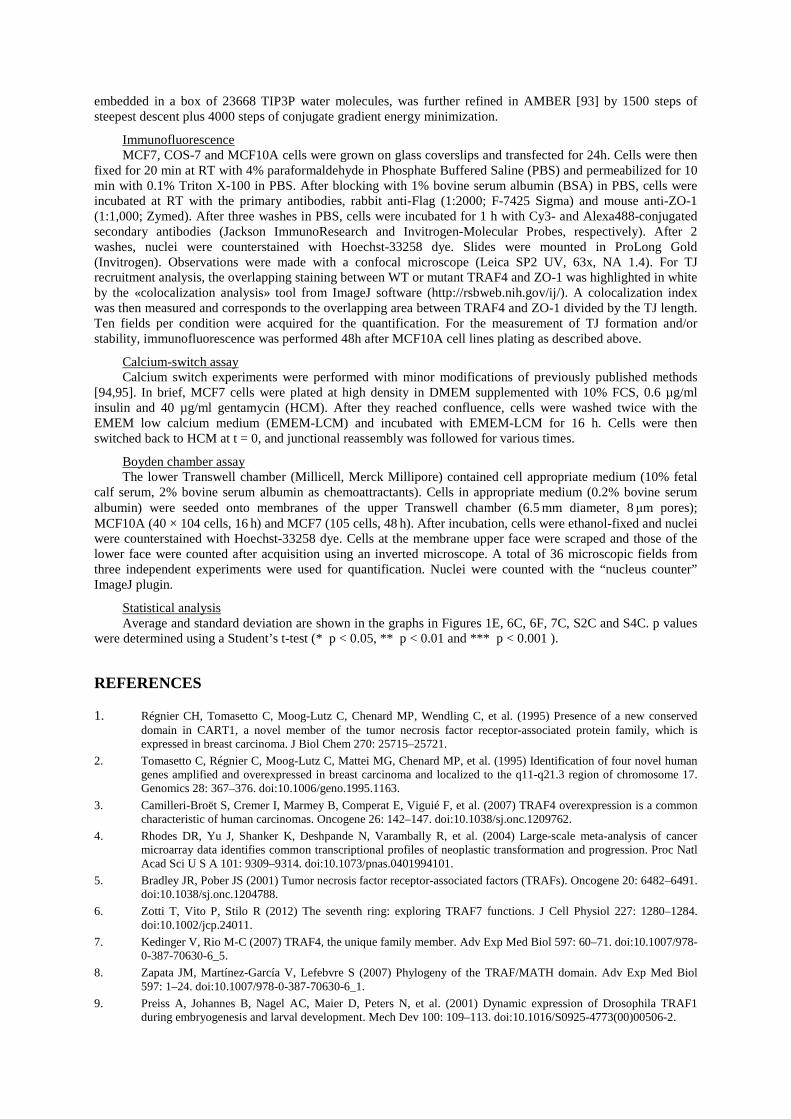
embedded in a box of 23668 TIP3P water molecules, was further refined in AMBER [93] by 1500 steps of steepest descent plus 4000 steps of conjugate gradient energy minimization.
Immunofluorescence MCF7, COS-7 and MCF10A cells were grown on glass coverslips and transfected for 24h. Cells were then
fixed for 20 min at RT with 4% paraformaldehyde in Phosphate Buffered Saline (PBS) and permeabilized for 10 min with 0.1% Triton X-100 in PBS. After blocking with 1% bovine serum albumin (BSA) in PBS, cells were incubated at RT with the primary antibodies, rabbit anti-Flag (1:2000; F-7425 Sigma) and mouse anti-ZO-1 (1:1,000; Zymed). After three washes in PBS, cells were incubated for 1 h with Cy3- and Alexa488-conjugated secondary antibodies (Jackson ImmunoResearch and Invitrogen-Molecular Probes, respectively). After 2 washes, nuclei were counterstained with Hoechst-33258 dye. Slides were mounted in ProLong Gold (Invitrogen). Observations were made with a confocal microscope (Leica SP2 UV, 63x, NA 1.4). For TJ recruitment analysis, the overlapping staining between WT or mutant TRAF4 and ZO-1 was highlighted in white by the «colocalization analysis» tool from ImageJ software (http://rsbweb.nih.gov/ij/). A colocalization index was then measured and corresponds to the overlapping area between TRAF4 and ZO-1 divided by the TJ length. Ten fields per condition were acquired for the quantification. For the measurement of TJ formation and/or stability, immunofluorescence was performed 48h after MCF10A cell lines plating as described above.
Calcium-switch assay Calcium switch experiments were performed with minor modifications of previously published methods
[94,95]. In brief, MCF7 cells were plated at high density in DMEM supplemented with 10% FCS, 0.6 µg/ml insulin and 40 µg/ml gentamycin (HCM). After they reached confluence, cells were washed twice with the EMEM low calcium medium (EMEM-LCM) and incubated with EMEM-LCM for 16 h. Cells were then switched back to HCM at t = 0, and junctional reassembly was followed for various times.
Boyden chamber assay The lower Transwell chamber (Millicell, Merck Millipore) contained cell appropriate medium (10% fetal
calf serum, 2% bovine serum albumin as chemoattractants). Cells in appropriate medium (0.2% bovine serum albumin) were seeded onto membranes of the upper Transwell chamber (6.5 mm diameter, 8 μm pores); MCF10A (40 × 104 cells, 16 h) and MCF7 (105 cells, 48 h). After incubation, cells were ethanol-fixed and nuclei were counterstained with Hoechst-33258 dye. Cells at the membrane upper face were scraped and those of the lower face were counted after acquisition using an inverted microscope. A total of 36 microscopic fields from three independent experiments were used for quantification. Nuclei were counted with the “nucleus counter” ImageJ plugin.
Statistical analysis Average and standard deviation are shown in the graphs in Figures 1E, 6C, 6F, 7C, S2C and S4C. p values
were determined using a Student’s t-test (* p < 0.05, ** p < 0.01 and *** p < 0.001 ).
REFERENCES
1. Régnier CH, Tomasetto C, Moog-Lutz C, Chenard MP, Wendling C, et al. (1995) Presence of a new conserved domain in CART1, a novel member of the tumor necrosis factor receptor-associated protein family, which is expressed in breast carcinoma. J Biol Chem 270: 25715–25721.
2. Tomasetto C, Régnier C, Moog-Lutz C, Mattei MG, Chenard MP, et al. (1995) Identification of four novel human genes amplified and overexpressed in breast carcinoma and localized to the q11-q21.3 region of chromosome 17. Genomics 28: 367–376. doi:10.1006/geno.1995.1163.
3. Camilleri-Broët S, Cremer I, Marmey B, Comperat E, Viguié F, et al. (2007) TRAF4 overexpression is a common characteristic of human carcinomas. Oncogene 26: 142–147. doi:10.1038/sj.onc.1209762.
4. Rhodes DR, Yu J, Shanker K, Deshpande N, Varambally R, et al. (2004) Large-scale meta-analysis of cancer microarray data identifies common transcriptional profiles of neoplastic transformation and progression. Proc Natl Acad Sci U S A 101: 9309–9314. doi:10.1073/pnas.0401994101.
5. Bradley JR, Pober JS (2001) Tumor necrosis factor receptor-associated factors (TRAFs). Oncogene 20: 6482–6491. doi:10.1038/sj.onc.1204788.
6. Zotti T, Vito P, Stilo R (2012) The seventh ring: exploring TRAF7 functions. J Cell Physiol 227: 1280–1284. doi:10.1002/jcp.24011.
7. Kedinger V, Rio M-C (2007) TRAF4, the unique family member. Adv Exp Med Biol 597: 60–71. doi:10.1007/978-0-387-70630-6_5.
8. Zapata JM, Martínez-García V, Lefebvre S (2007) Phylogeny of the TRAF/MATH domain. Adv Exp Med Biol 597: 1–24. doi:10.1007/978-0-387-70630-6_1.
9. Preiss A, Johannes B, Nagel AC, Maier D, Peters N, et al. (2001) Dynamic expression of Drosophila TRAF1 during embryogenesis and larval development. Mech Dev 100: 109–113. doi:10.1016/S0925-4773(00)00506-2.
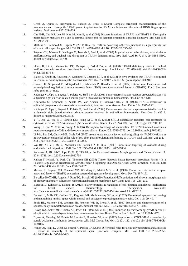
10. Grech A, Quinn R, Srinivasan D, Badoux X, Brink R (2000) Complete structural characterisation of the mammalian and Drosophila TRAF genes: implications for TRAF evolution and the role of RING finger splice variants. Mol Immunol 37: 721–734.
11. Cha G-H, Cho KS, Lee JH, Kim M, Kim E, et al. (2003) Discrete functions of TRAF1 and TRAF2 in Drosophila melanogaster mediated by c-Jun N-terminal kinase and NF-kappaB-dependent signaling pathways. Mol Cell Biol 23: 7982–7991.
12. Mathew SJ, Rembold M, Leptin M (2011) Role for Traf4 in polarizing adherens junctions as a prerequisite for efficient cell shape changes. Mol Cell Biol 31: 4978–4993. doi:10.1128/MCB.05542-11.
13. Régnier CH, Masson R, Kedinger V, Textoris J, Stoll I, et al. (2002) Impaired neural tube closure, axial skeleton malformations, and tracheal ring disruption in TRAF4-deficient mice. Proc Natl Acad Sci U S A 99: 5585–5590. doi:10.1073/pnas.052124799.
14. Shiels H, Li X, Schumacker PT, Maltepe E, Padrid PA, et al. (2000) TRAF4 deficiency leads to tracheal malformation with resulting alterations in air flow to the lungs. Am J Pathol 157: 679–688. doi:10.1016/S0002-9440(10)64578-6.
15. Blaise S, Kneib M, Rousseau A, Gambino F, Chenard M-P, et al. (2012) In vivo evidence that TRAF4 is required for central nervous system myelin homeostasis. Plos One 7: e30917. doi:10.1371/journal.pone.0030917.
16. Glauner H, Siegmund D, Motejadded H, Scheurich P, Henkler F, et al. (2002) Intracellular localization and transcriptional regulation of tumor necrosis factor (TNF) receptor-associated factor 4 (TRAF4). Eur J Biochem Febs 269: 4819–4829.
17. Kedinger V, Alpy F, Baguet A, Polette M, Stoll I, et al. (2008) Tumor necrosis factor receptor-associated factor 4 is a dynamic tight junction-related shuttle protein involved in epithelium homeostasis. Plos One 3: e3518.
18. Krajewska M, Krajewski S, Zapata JM, Van Arsdale T, Gascoyne RD, et al. (1998) TRAF-4 expression in epithelial progenitor cells. Analysis in normal adult, fetal, and tumor tissues. Am J Pathol 152: 1549–1561.
19. Kédinger V, Alpy F, Baguet A, Polette M, Stoll I, et al. (2008) Tumor necrosis factor receptor-associated factor 4 is a dynamic tight junction-related shuttle protein involved in epithelium homeostasis. Plos One 3: e3518. doi:10.1371/journal.pone.0003518.
20. Yi P, Xia W, Wu R-C, Lonard DM, Hung M-C, et al. (2013) SRC-3 coactivator regulates cell resistance to cytotoxic stress via TRAF4-mediated p53 destabilization. Genes Dev 27: 274–287. doi:10.1101/gad.203760.112.
21. Wang H, Cai Y, Chia W, Yang X (2006) Drosophila homologs of mammalian TNF/TNFR-related molecules regulate segregation of Miranda/Prospero in neuroblasts. Embo J 25: 5783–5793. doi:10.1038/sj.emboj.7601461.
22. Li J-M, Fan LM, Christie MR, Shah AM (2005) Acute tumor necrosis factor alpha signaling via NADPH oxidase in microvascular endothelial cells: role of p47phox phosphorylation and binding to TRAF4. Mol Cell Biol 25: 2320–2330. doi:10.1128/MCB.25.6.2320-2330.2005.
23. Wu RF, Xu YC, Ma Z, Nwariaku FE, Sarosi GA Jr, et al. (2005) Subcellular targeting of oxidants during endothelial cell migration. J Cell Biol 171: 893–904. doi:10.1083/jcb.200507004.
24. Rousseau A, Rio M-C, Alpy F (2011) TRAF4, at the Crossroad between Morphogenesis and Cancer. Cancers 3: 2734–2749. doi:10.3390/cancers3022734.
25. Kalkan T, Iwasaki Y, Park CY, Thomsen GH (2009) Tumor Necrosis Factor-Receptor–associated Factor-4 Is a Positive Regulator of Transforming Growth Factor-β Signaling That Affects Neural Crest Formation. Mol Biol Cell 20: 3436–3450. doi:10.1091/mbc.E08-03-0325.
26. Masson R, Régnier CH, Chenard MP, Wendling C, Mattei MG, et al. (1998) Tumor necrosis factor receptor associated factor 4 (TRAF4) expression pattern during mouse development. Mech Dev 71: 187–191.
27. Barcellos-Hoff MH, Aggeler J, Ram TG, Bissell MJ (1989) Functional differentiation and alveolar morphogenesis of primary mammary cultures on reconstituted basement membrane. Dev Camb Engl 105: 223–235.
27. Bazzoun D, Lelièvre S, Talhouk R (2013) Polarity proteins as regulators of cell junction complexes: Implications for breast cancer. Pharmacology & Therapeutics. Available: http://www.sciencedirect.com/science/article/pii/S0163725813000454. Accessed 9 April 2013.
29. Debnath J, Mills KR, Collins NL, Reginato MJ, Muthuswamy SK, et al. (2002) The role of apoptosis in creating and maintaining luminal space within normal and oncogene-expressing mammary acini. Cell 111: 29–40.
30. Soule HD, Maloney TM, Wolman SR, Peterson WD Jr, Brenz R, et al. (1990) Isolation and characterization of a spontaneously immortalized human breast epithelial cell line, MCF-10. Cancer Res 50: 6075–6086.
31. Brown KA, Aakre ME, Gorska AE, Price JO, Eltom SE, et al. (2004) Induction by transforming growth factor-β1 of epithelial to mesenchymal transition is a rare event in vitro. Breast Cancer Res 6: 1–17. doi:10.1186/bcr778.
32. Brysse A, Mestdagt M, Polette M, Luczka E, Hunziker W, et al. (2012) Regulation of CXCL8/IL-8 expression by zonula occludens-1 in human breast cancer cells. Mol Cancer Res Mcr 10: 121–132. doi:10.1158/1541-7786.MCR-11-0180.
33. Ivanov AI, Hunt D, Utech M, Nusrat A, Parkos CA (2005) Differential roles for actin polymerization and a myosin II motor in assembly of the epithelial apical junctional complex. Mol Biol Cell 16: 2636–2650. doi:10.1091/mbc.E05-01-0043.
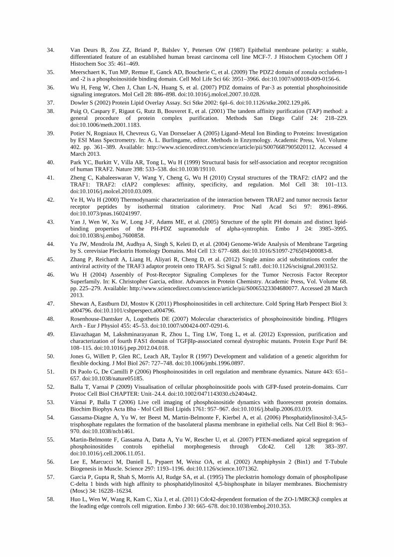
34. Van Deurs B, Zou ZZ, Briand P, Balslev Y, Petersen OW (1987) Epithelial membrane polarity: a stable, differentiated feature of an established human breast carcinoma cell line MCF-7. J Histochem Cytochem Off J Histochem Soc 35: 461–469.
35. Meerschaert K, Tun MP, Remue E, Ganck AD, Boucherie C, et al. (2009) The PDZ2 domain of zonula occludens-1 and -2 is a phosphoinositide binding domain. Cell Mol Life Sci 66: 3951–3966. doi:10.1007/s00018-009-0156-6.
36. Wu H, Feng W, Chen J, Chan L-N, Huang S, et al. (2007) PDZ domains of Par-3 as potential phosphoinositide signaling integrators. Mol Cell 28: 886–898. doi:10.1016/j.molcel.2007.10.028.
37. Dowler S (2002) Protein Lipid Overlay Assay. Sci Stke 2002: 6pl–6. doi:10.1126/stke.2002.129.pl6.
38. Puig O, Caspary F, Rigaut G, Rutz B, Bouveret E, et al. (2001) The tandem affinity purification (TAP) method: a general procedure of protein complex purification. Methods San Diego Calif 24: 218–229. doi:10.1006/meth.2001.1183.
39. Potier N, Rogniaux H, Chevreux G, Van Dorsselaer A (2005) Ligand–Metal Ion Binding to Proteins: Investigation by ESI Mass Spectrometry. In: A. L. Burlingame, editor. Methods in Enzymology. Academic Press, Vol. Volume 402. pp. 361–389. Available: http://www.sciencedirect.com/science/article/pii/S0076687905020112. Accessed 4 March 2013.
40. Park YC, Burkitt V, Villa AR, Tong L, Wu H (1999) Structural basis for self-association and receptor recognition of human TRAF2. Nature 398: 533–538. doi:10.1038/19110.
41. Zheng C, Kabaleeswaran V, Wang Y, Cheng G, Wu H (2010) Crystal structures of the TRAF2: cIAP2 and the TRAF1: TRAF2: cIAP2 complexes: affinity, specificity, and regulation. Mol Cell 38: 101–113. doi:10.1016/j.molcel.2010.03.009.
42. Ye H, Wu H (2000) Thermodynamic characterization of the interaction between TRAF2 and tumor necrosis factor receptor peptides by isothermal titration calorimetry. Proc Natl Acad Sci 97: 8961–8966. doi:10.1073/pnas.160241997.
43. Yan J, Wen W, Xu W, Long J-F, Adams ME, et al. (2005) Structure of the split PH domain and distinct lipid-binding properties of the PH-PDZ supramodule of alpha-syntrophin. Embo J 24: 3985–3995. doi:10.1038/sj.emboj.7600858.
44. Yu JW, Mendrola JM, Audhya A, Singh S, Keleti D, et al. (2004) Genome-Wide Analysis of Membrane Targeting by S. cerevisiae Pleckstrin Homology Domains. Mol Cell 13: 677–688. doi:10.1016/S1097-2765(04)00083-8.
45. Zhang P, Reichardt A, Liang H, Aliyari R, Cheng D, et al. (2012) Single amino acid substitutions confer the antiviral activity of the TRAF3 adaptor protein onto TRAF5. Sci Signal 5: ra81. doi:10.1126/scisignal.2003152.
46. Wu H (2004) Assembly of Post-Receptor Signaling Complexes for the Tumor Necrosis Factor Receptor Superfamily. In: K. Christopher Garcia, editor. Advances in Protein Chemistry. Academic Press, Vol. Volume 68. pp. 225–279. Available: http://www.sciencedirect.com/science/article/pii/S0065323304680077. Accessed 28 March 2013.
47. Shewan A, Eastburn DJ, Mostov K (2011) Phosphoinositides in cell architecture. Cold Spring Harb Perspect Biol 3: a004796. doi:10.1101/cshperspect.a004796.
48. Rosenhouse-Dantsker A, Logothetis DE (2007) Molecular characteristics of phosphoinositide binding. Pflügers Arch - Eur J Physiol 455: 45–53. doi:10.1007/s00424-007-0291-6.
49. Elavazhagan M, Lakshminarayanan R, Zhou L, Ting LW, Tong L, et al. (2012) Expression, purification and characterization of fourth FAS1 domain of TGFβIp-associated corneal dystrophic mutants. Protein Expr Purif 84: 108–115. doi:10.1016/j.pep.2012.04.018.
50. Jones G, Willett P, Glen RC, Leach AR, Taylor R (1997) Development and validation of a genetic algorithm for flexible docking. J Mol Biol 267: 727–748. doi:10.1006/jmbi.1996.0897.
51. Di Paolo G, De Camilli P (2006) Phosphoinositides in cell regulation and membrane dynamics. Nature 443: 651–657. doi:10.1038/nature05185.
52. Balla T, Varnai P (2009) Visualisation of cellular phosphoinositide pools with GFP-fused protein-domains. Curr Protoc Cell Biol CHAPTER: Unit–24.4. doi:10.1002/0471143030.cb2404s42.
53. Várnai P, Balla T (2006) Live cell imaging of phosphoinositide dynamics with fluorescent protein domains. Biochim Biophys Acta Bba - Mol Cell Biol Lipids 1761: 957–967. doi:10.1016/j.bbalip.2006.03.019.
54. Gassama-Diagne A, Yu W, ter Beest M, Martin-Belmonte F, Kierbel A, et al. (2006) Phosphatidylinositol-3,4,5-trisphosphate regulates the formation of the basolateral plasma membrane in epithelial cells. Nat Cell Biol 8: 963–970. doi:10.1038/ncb1461.
55. Martin-Belmonte F, Gassama A, Datta A, Yu W, Rescher U, et al. (2007) PTEN-mediated apical segregation of phosphoinositides controls epithelial morphogenesis through Cdc42. Cell 128: 383–397. doi:10.1016/j.cell.2006.11.051.
56. Lee E, Marcucci M, Daniell L, Pypaert M, Weisz OA, et al. (2002) Amphiphysin 2 (Bin1) and T-Tubule Biogenesis in Muscle. Science 297: 1193–1196. doi:10.1126/science.1071362.
57. Garcia P, Gupta R, Shah S, Morris AJ, Rudge SA, et al. (1995) The pleckstrin homology domain of phospholipase C-delta 1 binds with high affinity to phosphatidylinositol 4,5-bisphosphate in bilayer membranes. Biochemistry (Mosc) 34: 16228–16234.
58. Huo L, Wen W, Wang R, Kam C, Xia J, et al. (2011) Cdc42-dependent formation of the ZO-1/MRCKβ complex at the leading edge controls cell migration. Embo J 30: 665–678. doi:10.1038/emboj.2010.353.
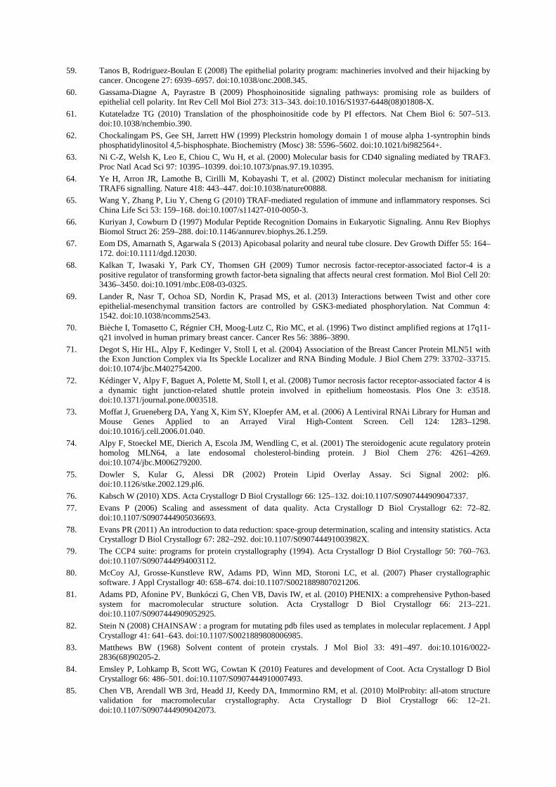
59. Tanos B, Rodriguez-Boulan E (2008) The epithelial polarity program: machineries involved and their hijacking by cancer. Oncogene 27: 6939–6957. doi:10.1038/onc.2008.345.
60. Gassama-Diagne A, Payrastre B (2009) Phosphoinositide signaling pathways: promising role as builders of epithelial cell polarity. Int Rev Cell Mol Biol 273: 313–343. doi:10.1016/S1937-6448(08)01808-X.
61. Kutateladze TG (2010) Translation of the phosphoinositide code by PI effectors. Nat Chem Biol 6: 507–513. doi:10.1038/nchembio.390.
62. Chockalingam PS, Gee SH, Jarrett HW (1999) Pleckstrin homology domain 1 of mouse alpha 1-syntrophin binds phosphatidylinositol 4,5-bisphosphate. Biochemistry (Mosc) 38: 5596–5602. doi:10.1021/bi982564+.
63. Ni C-Z, Welsh K, Leo E, Chiou C, Wu H, et al. (2000) Molecular basis for CD40 signaling mediated by TRAF3. Proc Natl Acad Sci 97: 10395–10399. doi:10.1073/pnas.97.19.10395.
64. Ye H, Arron JR, Lamothe B, Cirilli M, Kobayashi T, et al. (2002) Distinct molecular mechanism for initiating TRAF6 signalling. Nature 418: 443–447. doi:10.1038/nature00888.
65. Wang Y, Zhang P, Liu Y, Cheng G (2010) TRAF-mediated regulation of immune and inflammatory responses. Sci China Life Sci 53: 159–168. doi:10.1007/s11427-010-0050-3.
66. Kuriyan J, Cowburn D (1997) Modular Peptide Recognition Domains in Eukaryotic Signaling. Annu Rev Biophys Biomol Struct 26: 259–288. doi:10.1146/annurev.biophys.26.1.259.
67. Eom DS, Amarnath S, Agarwala S (2013) Apicobasal polarity and neural tube closure. Dev Growth Differ 55: 164–172. doi:10.1111/dgd.12030.
68. Kalkan T, Iwasaki Y, Park CY, Thomsen GH (2009) Tumor necrosis factor-receptor-associated factor-4 is a positive regulator of transforming growth factor-beta signaling that affects neural crest formation. Mol Biol Cell 20: 3436–3450. doi:10.1091/mbc.E08-03-0325.
69. Lander R, Nasr T, Ochoa SD, Nordin K, Prasad MS, et al. (2013) Interactions between Twist and other core epithelial-mesenchymal transition factors are controlled by GSK3-mediated phosphorylation. Nat Commun 4: 1542. doi:10.1038/ncomms2543.
70. Bièche I, Tomasetto C, Régnier CH, Moog-Lutz C, Rio MC, et al. (1996) Two distinct amplified regions at 17q11-q21 involved in human primary breast cancer. Cancer Res 56: 3886–3890.
71. Degot S, Hir HL, Alpy F, Kedinger V, Stoll I, et al. (2004) Association of the Breast Cancer Protein MLN51 with the Exon Junction Complex via Its Speckle Localizer and RNA Binding Module. J Biol Chem 279: 33702–33715. doi:10.1074/jbc.M402754200.
72. Kédinger V, Alpy F, Baguet A, Polette M, Stoll I, et al. (2008) Tumor necrosis factor receptor-associated factor 4 is a dynamic tight junction-related shuttle protein involved in epithelium homeostasis. Plos One 3: e3518. doi:10.1371/journal.pone.0003518.
73. Moffat J, Grueneberg DA, Yang X, Kim SY, Kloepfer AM, et al. (2006) A Lentiviral RNAi Library for Human and Mouse Genes Applied to an Arrayed Viral High-Content Screen. Cell 124: 1283–1298. doi:10.1016/j.cell.2006.01.040.
74. Alpy F, Stoeckel ME, Dierich A, Escola JM, Wendling C, et al. (2001) The steroidogenic acute regulatory protein homolog MLN64, a late endosomal cholesterol-binding protein. J Biol Chem 276: 4261–4269. doi:10.1074/jbc.M006279200.
75. Dowler S, Kular G, Alessi DR (2002) Protein Lipid Overlay Assay. Sci Signal 2002: pl6. doi:10.1126/stke.2002.129.pl6.
76. Kabsch W (2010) XDS. Acta Crystallogr D Biol Crystallogr 66: 125–132. doi:10.1107/S0907444909047337.
77. Evans P (2006) Scaling and assessment of data quality. Acta Crystallogr D Biol Crystallogr 62: 72–82. doi:10.1107/S0907444905036693.
78. Evans PR (2011) An introduction to data reduction: space-group determination, scaling and intensity statistics. Acta Crystallogr D Biol Crystallogr 67: 282–292. doi:10.1107/S090744491003982X.
79. The CCP4 suite: programs for protein crystallography (1994). Acta Crystallogr D Biol Crystallogr 50: 760–763. doi:10.1107/S0907444994003112.
80. McCoy AJ, Grosse-Kunstleve RW, Adams PD, Winn MD, Storoni LC, et al. (2007) Phaser crystallographic software. J Appl Crystallogr 40: 658–674. doi:10.1107/S0021889807021206.
81. Adams PD, Afonine PV, Bunkóczi G, Chen VB, Davis IW, et al. (2010) PHENIX: a comprehensive Python-based system for macromolecular structure solution. Acta Crystallogr D Biol Crystallogr 66: 213–221. doi:10.1107/S0907444909052925.
82. Stein N (2008) CHAINSAW : a program for mutating pdb files used as templates in molecular replacement. J Appl Crystallogr 41: 641–643. doi:10.1107/S0021889808006985.
83. Matthews BW (1968) Solvent content of protein crystals. J Mol Biol 33: 491–497. doi:10.1016/0022-2836(68)90205-2.
84. Emsley P, Lohkamp B, Scott WG, Cowtan K (2010) Features and development of Coot. Acta Crystallogr D Biol Crystallogr 66: 486–501. doi:10.1107/S0907444910007493.
85. Chen VB, Arendall WB 3rd, Headd JJ, Keedy DA, Immormino RM, et al. (2010) MolProbity: all-atom structure validation for macromolecular crystallography. Acta Crystallogr D Biol Crystallogr 66: 12–21. doi:10.1107/S0907444909042073.
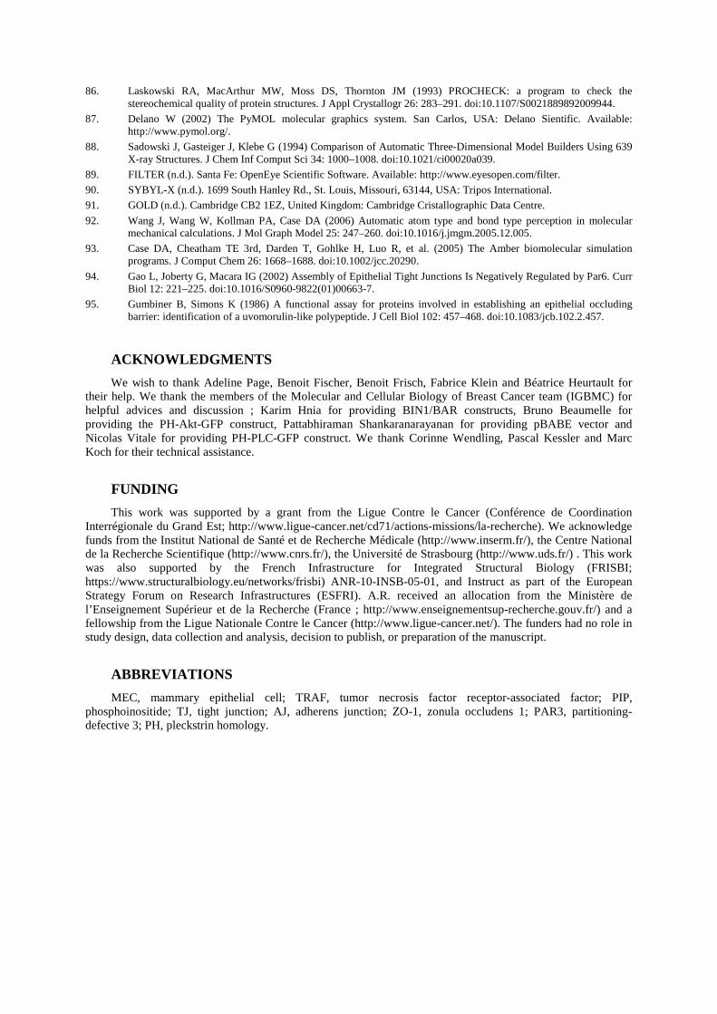
86. Laskowski RA, MacArthur MW, Moss DS, Thornton JM (1993) PROCHECK: a program to check the stereochemical quality of protein structures. J Appl Crystallogr 26: 283–291. doi:10.1107/S0021889892009944.
87. Delano W (2002) The PyMOL molecular graphics system. San Carlos, USA: Delano Sientific. Available: http://www.pymol.org/.
88. Sadowski J, Gasteiger J, Klebe G (1994) Comparison of Automatic Three-Dimensional Model Builders Using 639 X-ray Structures. J Chem Inf Comput Sci 34: 1000–1008. doi:10.1021/ci00020a039.
89. FILTER (n.d.). Santa Fe: OpenEye Scientific Software. Available: http://www.eyesopen.com/filter.
90. SYBYL-X (n.d.). 1699 South Hanley Rd., St. Louis, Missouri, 63144, USA: Tripos International.
91. GOLD (n.d.). Cambridge CB2 1EZ, United Kingdom: Cambridge Cristallographic Data Centre.
92. Wang J, Wang W, Kollman PA, Case DA (2006) Automatic atom type and bond type perception in molecular mechanical calculations. J Mol Graph Model 25: 247–260. doi:10.1016/j.jmgm.2005.12.005.
93. Case DA, Cheatham TE 3rd, Darden T, Gohlke H, Luo R, et al. (2005) The Amber biomolecular simulation programs. J Comput Chem 26: 1668–1688. doi:10.1002/jcc.20290.
94. Gao L, Joberty G, Macara IG (2002) Assembly of Epithelial Tight Junctions Is Negatively Regulated by Par6. Curr Biol 12: 221–225. doi:10.1016/S0960-9822(01)00663-7.
95. Gumbiner B, Simons K (1986) A functional assay for proteins involved in establishing an epithelial occluding barrier: identification of a uvomorulin-like polypeptide. J Cell Biol 102: 457–468. doi:10.1083/jcb.102.2.457.
ACKNOWLEDGMENTS
We wish to thank Adeline Page, Benoit Fischer, Benoit Frisch, Fabrice Klein and Béatrice Heurtault for their help. We thank the members of the Molecular and Cellular Biology of Breast Cancer team (IGBMC) for helpful advices and discussion ; Karim Hnia for providing BIN1/BAR constructs, Bruno Beaumelle for providing the PH-Akt-GFP construct, Pattabhiraman Shankaranarayanan for providing pBABE vector and Nicolas Vitale for providing PH-PLC-GFP construct. We thank Corinne Wendling, Pascal Kessler and Marc Koch for their technical assistance.
FUNDING
This work was supported by a grant from the Ligue Contre le Cancer (Conférence de Coordination Interrégionale du Grand Est; http://www.ligue-cancer.net/cd71/actions-missions/la-recherche). We acknowledge funds from the Institut National de Santé et de Recherche Médicale (http://www.inserm.fr/), the Centre National de la Recherche Scientifique (http://www.cnrs.fr/), the Université de Strasbourg (http://www.uds.fr/) . This work was also supported by the French Infrastructure for Integrated Structural Biology (FRISBI; https://www.structuralbiology.eu/networks/frisbi) ANR-10-INSB-05-01, and Instruct as part of the European Strategy Forum on Research Infrastructures (ESFRI). A.R. received an allocation from the Ministère de l’Enseignement Supérieur et de la Recherche (France ; http://www.enseignementsup-recherche.gouv.fr/) and a fellowship from the Ligue Nationale Contre le Cancer (http://www.ligue-cancer.net/). The funders had no role in study design, data collection and analysis, decision to publish, or preparation of the manuscript.
ABBREVIATIONS
MEC, mammary epithelial cell; TRAF, tumor necrosis factor receptor-associated factor; PIP, phosphoinositide; TJ, tight junction; AJ, adherens junction; ZO-1, zonula occludens 1; PAR3, partitioning-defective 3; PH, pleckstrin homology.
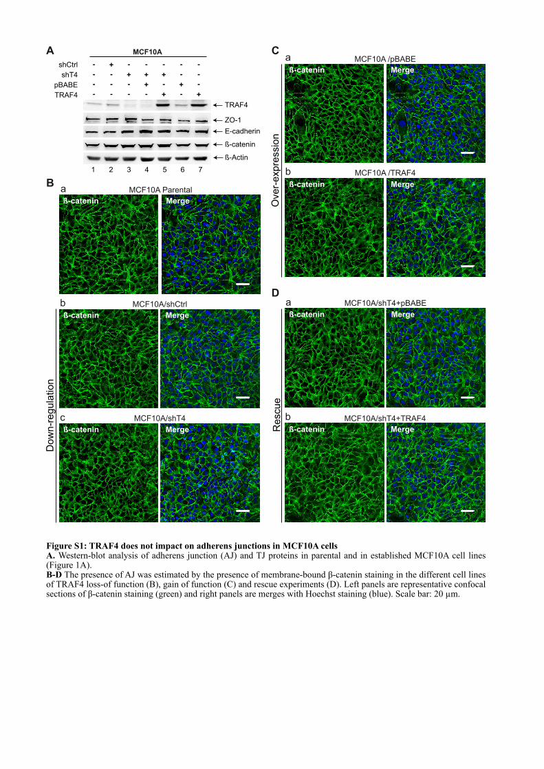
A
B
MCF10A/shCtrl
ß-catenin Merge
b
MCF10A/shT4
ß-catenin Merge
c
MCF10A ParentalaMergeß-catenin
Do
wn
-re
gu
latio
nMCF10A /pBABE
ß-catenin Merge
a
MCF10A /TRAF4
ß-catenin Merge
b
Ove
r-e
xp
ressio
n
MCF10A/shT4+pBABE
ß-catenin Merge
a
MCF10A/shT4+TRAF4
ß-catenin Merge
b
Re
scu
e
C
D
shT4
shCtrl
pBABE
TRAF4
+-
-
-
-
-
-
-
-
-
+
-
-
+
-
-
+
-
+
+
-
-
-
-
-
-+
+
TRAF4
ß-Actin
ß-catenin
E-cadherin
ZO-1
MCF10A
1 2 3 4 5 6 7
Figure S1: TRAF4 does not impact on adherens junctions in MCF10A cells A. Western-blot analysis of adherens junction (AJ) and TJ proteins in parental and in established MCF10A cell lines (Figure 1A). B-D The presence of AJ was estimated by the presence of membrane-bound β-catenin staining in the different cell lines of TRAF4 loss-of function (B), gain of function (C) and rescue experiments (D). Left panels are representative confocal sections of β-catenin staining (green) and right panels are merges with Hoechst staining (blue). Scale bar: 20 µm.
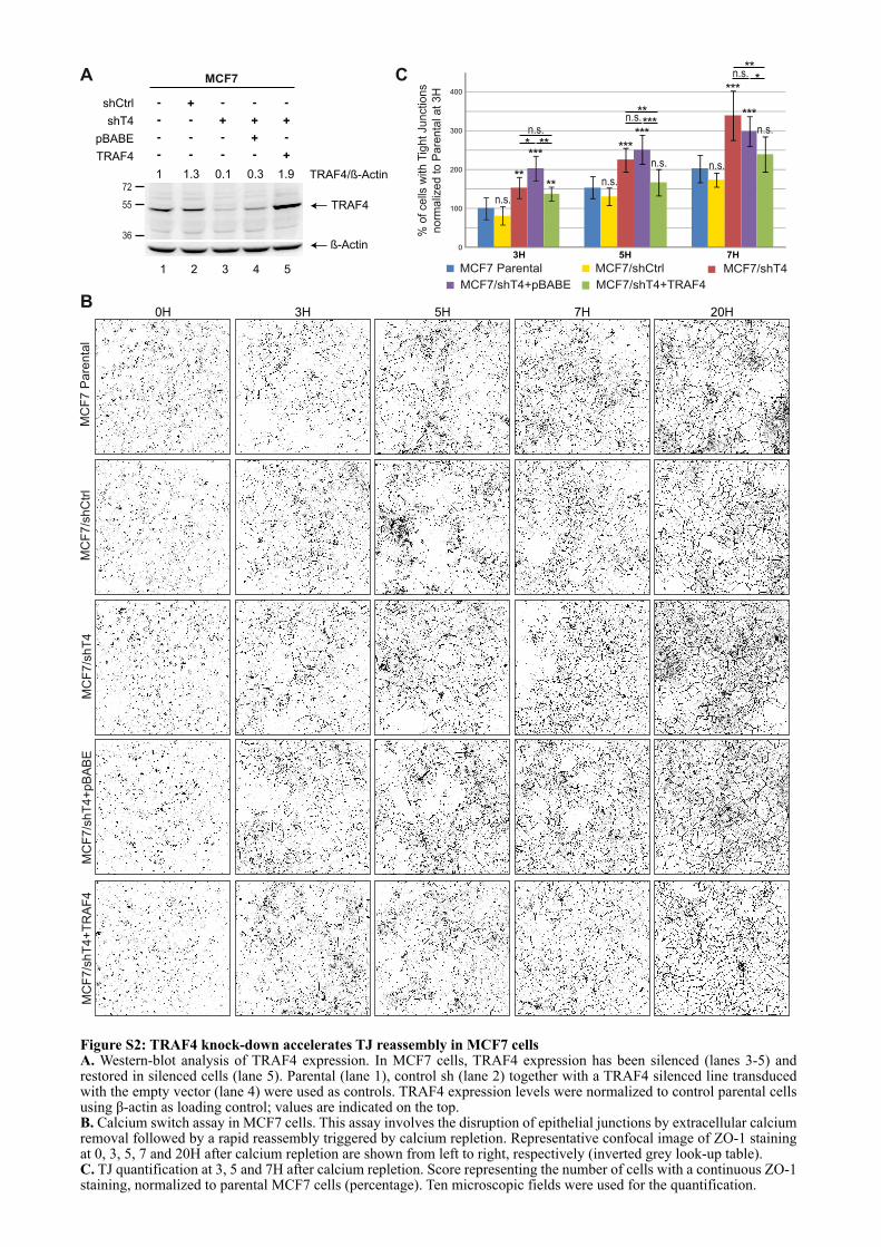
MC
F7/s
hC
trl
MC
F7/s
hT
4M
CF
7 P
are
nta
lM
CF
7/s
hT
4+
pB
AB
EM
CF
7/s
hT
4+
TR
AF
4
0H 3H 5H 7H 20H
A
shT4
shCtrl
pBABE
TRAF4
+-
-
-
-
-
-
-
-
-
+
-
-
+
-
-
+
-
+
+
72
55 TRAF4
ß-Actin
1 1.3 0.1 0.3 1.9 TRAF4/ß-Actin
MCF7
1 2 3 4 5
36
3H 5H 7H
400
0
100
200
300
MCF7/shCtrl MCF7/shT4MCF7 Parental
MCF7/shT4+pBABE MCF7/shT4+TRAF4
****
**** **
n.s.
n.s.
n.s.
n.s.
***
***
n.s.
n.s.***
**
***
***
n.s.
n.s.*
**
% o
f ce
lls w
ith
Tig
ht
Ju
nctio
ns
no
rma
lize
d t
o P
are
nta
l a
t 3
H
C
B
Figure S2: TRAF4 knock-down accelerates TJ reassembly in MCF7 cellsA. Western-blot analysis of TRAF4 expression. In MCF7 cells, TRAF4 expression has been silenced (lanes 3-5) and restored in silenced cells (lane 5). Parental (lane 1), control sh (lane 2) together with a TRAF4 silenced line transduced with the empty vector (lane 4) were used as controls. TRAF4 expression levels were normalized to control parental cells using β-actin as loading control; values are indicated on the top.B. Calcium switch assay in MCF7 cells. This assay involves the disruption of epithelial junctions by extracellular calcium removal followed by a rapid reassembly triggered by calcium repletion. Representative confocal image of ZO-1 staining at 0, 3, 5, 7 and 20H after calcium repletion are shown from left to right, respectively (inverted grey look-up table).C. TJ quantification at 3, 5 and 7H after calcium repletion. Score representing the number of cells with a continuous ZO-1 staining, normalized to parental MCF7 cells (percentage). Ten microscopic fields were used for the quantification.
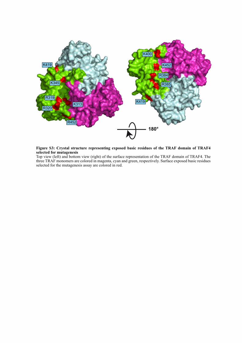
K419
K345
R319
R320
K313
R452
K400
K452
R385
R384
K419
180°
Figure S3: Crystal structure representing exposed basic residues of the TRAF domain of TRAF4 selected for mutagenesisTop view (left) and bottom view (right) of the surface representation of the TRAF domain of TRAF4. The three TRAF monomers are colored in magenta, cyan and green, respectively. Surface exposed basic residues selected for the mutagenesis assay are colored in red.
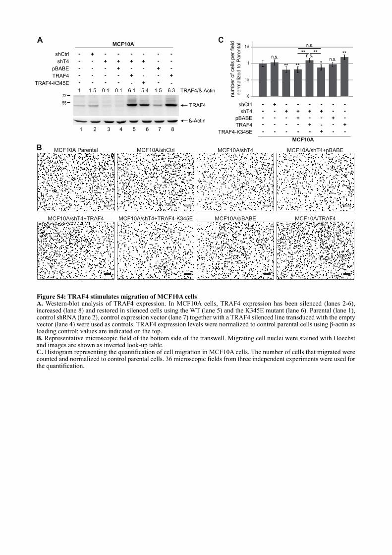
A
shT4
shCtrl
pBABE
TRAF4
+-
-
-
-
-
-
-
-
-
+
-
-
+
-
-
+
-
+
+
-
-
-
-
-
-+
+
72
55TRAF4
ß-Actin
1 1.5 0.1 0.1 6.1 5.4 1.5 TRAF4/ß-Actin
MCF10A
1 2 3 4 5 6 7 8
-
+
-
-
TRAF4-K345E - - - - - - -+
6.3
B
C
num
ber
of cells
per
field
norm
aliz
ed to P
are
nta
l
shT4
shCtrl
pBABE
TRAF4
+-
-
-
-
-
-
-
-
-
+
-
-
+
-
-
+
-
+
+
-
-
-
-
-
-+
+
MCF10A
-
+
-
-
TRAF4-K345E - - - - - - -+
0
0.5
1
1.5
**
** **
***
**n.s.
n.s.n.s.
n.s.
MCF10A/shCtrlMCF10A Parental MCF10A/shT4 MCF10A/shT4+pBABE
MCF10A/shT4+TRAF4-K345EMCF10A/shT4+TRAF4 MCF10A/pBABE MCF10A/TRAF4
Figure S4: TRAF4 stimulates migration of MCF10A cellsA. Western-blot analysis of TRAF4 expression. In MCF10A cells, TRAF4 expression has been silenced (lanes 2-6), increased (lane 8) and restored in silenced cells using the WT (lane 5) and the K345E mutant (lane 6). Parental (lane 1), control shRNA (lane 2), control expression vector (lane 7) together with a TRAF4 silenced line transduced with the empty vector (lane 4) were used as controls. TRAF4 expression levels were normalized to control parental cells using β-actin as loading control; values are indicated on the top.B. Representative microscopic field of the bottom side of the transwell. Migrating cell nuclei were stained with Hoechst and images are shown as inverted look-up table.C. Histogram representing the quantification of cell migration in MCF10A cells. The number of cells that migrated were counted and normalized to control parental cells. 36 microscopic fields from three independent experiments were used for the quantification.
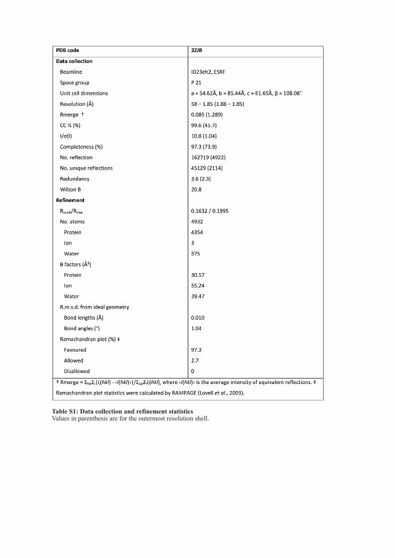
Table S1: Data collection and refinement statistics
Values in parenthesis are for the outermost resolution shell.
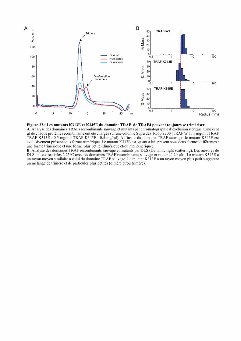
B
10
20
30
40
01 10 1000.1
TRAF-K345E
TRAF-K313E
TRAF-WT
% M
ass
% M
ass
% M
ass
Radius (nm)
10
20
30
40
01 10 1000.1
10
20
30
40
01 10 1000.1
50
0 5 10 15 20 25
0
20
40
60
80
100
120
TRAF WT
TRAF-K313E
TRAF-K345E
A280 n
m
ml
A
Figure 32 : Les mutants K313E et K345E du domaine TRAF de TRAF4 peuvent toujours se trimériser A. Analyse des domaines TRAFs recombinants sauvage et mutants par chromatographie d’exclusion stérique. Cinq cent µl de chaque protéine recombinante ont été chargés sur une colonne Superdex 16/60 S200 (TRAF WT : 1 mg/ml; TRAF TRAF-K313E : 0.5 mg/ml; TRAF-K345E : 0.5 mg/ml). A l’instar du domaine TRAF sauvage, le mutant K345E est exclusivement présent sous forme trimérique. Le mutant K313E est, quant à lui, présent sous deux formes différentes : une forme trimérique et une forme plus petite (dimérique et/ou monomérique). B. Analyse des domaines TRAF recombinants sauvage et mutants par DLS (Dynamic light scattering). Les mesures de DLS ont été réalisées à 25°C avec les domaines TRAF recombinants sauvage et mutant à 20 µM. Le mutant K345E a un rayon moyen similaire à celui du domaine TRAF sauvage. Le mutant K313E a un rayon moyen plus petit suggérant un mélange de trimère et de particules plus petites (dimère et/ou trimère).
Trimère
Dimère et/ou monomère
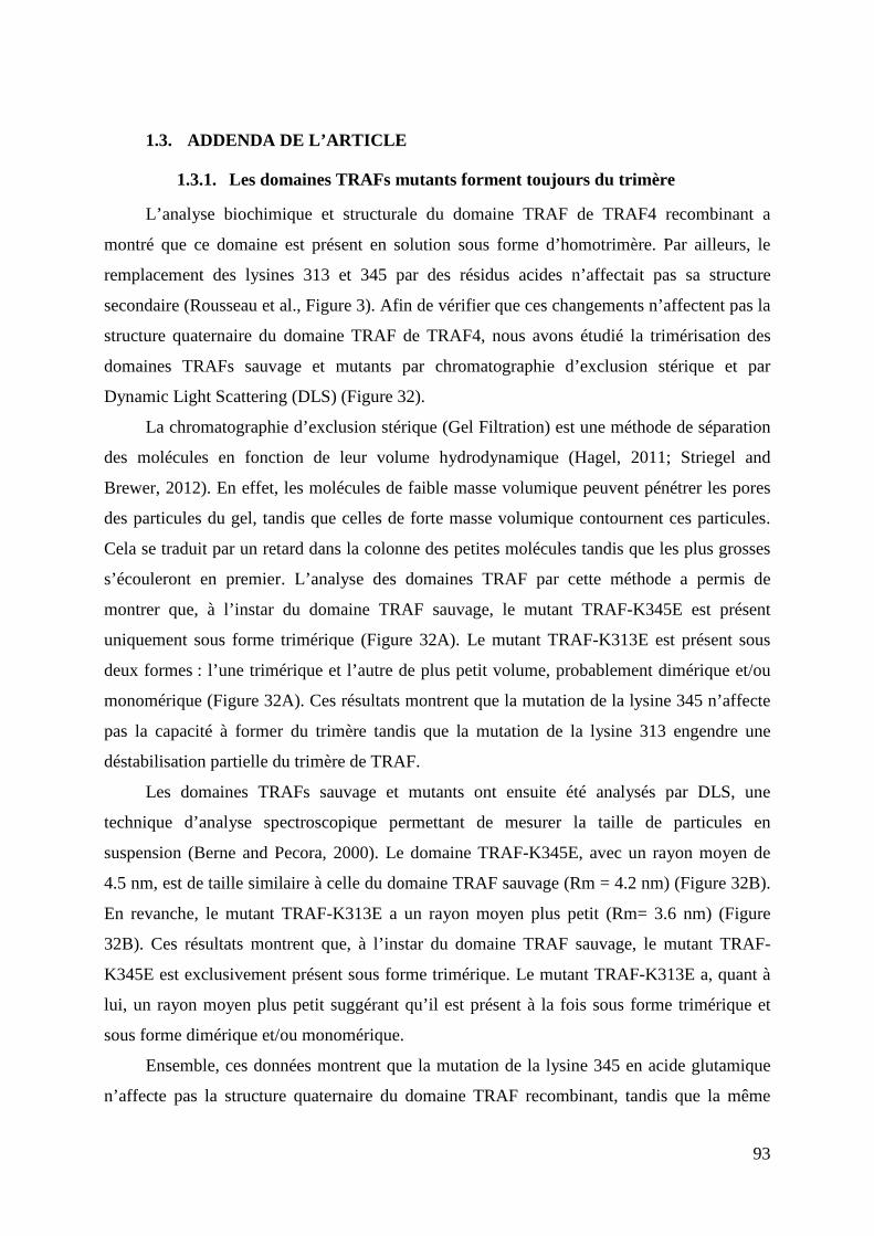
93
1.3. ADDENDA DE L’ARTICLE
1.3.1. Les domaines TRAFs mutants forment toujours du trimère
L’analyse biochimique et structurale du domaine TRAF de TRAF4 recombinant a
montré que ce domaine est présent en solution sous forme d’homotrimère. Par ailleurs, le
remplacement des lysines 313 et 345 par des résidus acides n’affectait pas sa structure
secondaire (Rousseau et al., Figure 3). Afin de vérifier que ces changements n’affectent pas la
structure quaternaire du domaine TRAF de TRAF4, nous avons étudié la trimérisation des
domaines TRAFs sauvage et mutants par chromatographie d’exclusion stérique et par
Dynamic Light Scattering (DLS) (Figure 32).
La chromatographie d’exclusion stérique (Gel Filtration) est une méthode de séparation
des molécules en fonction de leur volume hydrodynamique (Hagel, 2011; Striegel and
Brewer, 2012). En effet, les molécules de faible masse volumique peuvent pénétrer les pores
des particules du gel, tandis que celles de forte masse volumique contournent ces particules.
Cela se traduit par un retard dans la colonne des petites molécules tandis que les plus grosses
s’écouleront en premier. L’analyse des domaines TRAF par cette méthode a permis de
montrer que, à l’instar du domaine TRAF sauvage, le mutant TRAF-K345E est présent
uniquement sous forme trimérique (Figure 32A). Le mutant TRAF-K313E est présent sous
deux formes : l’une trimérique et l’autre de plus petit volume, probablement dimérique et/ou
monomérique (Figure 32A). Ces résultats montrent que la mutation de la lysine 345 n’affecte
pas la capacité à former du trimère tandis que la mutation de la lysine 313 engendre une
déstabilisation partielle du trimère de TRAF.
Les domaines TRAFs sauvage et mutants ont ensuite été analysés par DLS, une
technique d’analyse spectroscopique permettant de mesurer la taille de particules en
suspension (Berne and Pecora, 2000). Le domaine TRAF-K345E, avec un rayon moyen de
4.5 nm, est de taille similaire à celle du domaine TRAF sauvage (Rm = 4.2 nm) (Figure 32B).
En revanche, le mutant TRAF-K313E a un rayon moyen plus petit (Rm= 3.6 nm) (Figure
32B). Ces résultats montrent que, à l’instar du domaine TRAF sauvage, le mutant TRAF-
K345E est exclusivement présent sous forme trimérique. Le mutant TRAF-K313E a, quant à
lui, un rayon moyen plus petit suggérant qu’il est présent à la fois sous forme trimérique et
sous forme dimérique et/ou monomérique.
Ensemble, ces données montrent que la mutation de la lysine 345 en acide glutamique
n’affecte pas la structure quaternaire du domaine TRAF recombinant, tandis que la même
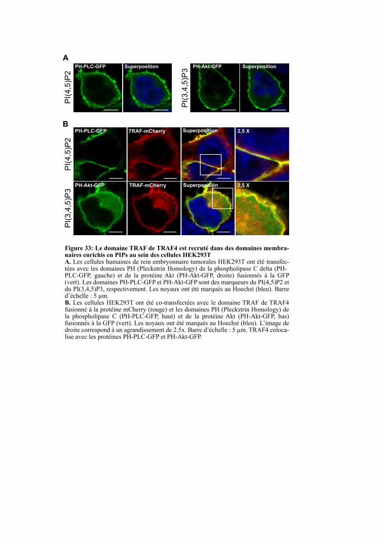
PH-PLC-GFP
PH-Akt-GFP
TRAF-mCherry
TRAF-mCherry
Superposition
Superposition
Figure 33: Le domaine TRAF de TRAF4 est recruté dans des domaines membra-naires enrichis en PIPs au sein des cellules HEK293TA. Les cellules humaines de rein embryonnaire tumorales HEK293T ont été transfec-tées avec les domaines PH (Pleckstrin Homology) de la phospholipase C delta (PH-PLC-GFP, gauche) et de la protéine Akt (PH-Akt-GFP, droite) fusionnés à la GFP (vert). Les domaines PH-PLC-GFP et PH-Akt-GFP sont des marqueurs du PI(4,5)P2 et du PI(3,4,5)P3, respectivement. Les noyaux ont été marqués au Hoechst (bleu). Barre d’échelle : 5 µm.B. Les cellules HEK293T ont été co-transfectées avec le domaine TRAF de TRAF4 fusionné à la protéine mCherry (rouge) et les domaines PH (Pleckstrin Homology) de la phospholipase C (PH-PLC-GFP, haut) et de la protéine Akt (PH-Akt-GFP, bas) fusionnés à la GFP (vert). Les noyaux ont été marqués au Hoechst (bleu). L’image de droite correspond à un agrandissement de 2.5x. Barre d’échelle : 5 µm. TRAF4 coloca-lise avec les protéines PH-PLC-GFP et PH-Akt-GFP.
PH-PLC-GFP Superposition PH-Akt-GFP SuperpositionPI(4,5)P2
PI(3,4,5)P3
PI(4,5)P2
PI(3,4,5)P3
A
B2,5 X
2,5 X
94
mutation sur la lysine 313 engendre déstabilisation partielle. Néanmoins, le domaine TRAF-
K313E recombinant est en solution présent à part égale sous forme trimérique et
dimérique/monomérique, ce qui suggère que l’atteinte de la structure quaternaire ne peut pas,
à elle seule, expliquer la diminution de 5 fois de l’interaction entre le TRAF-K313E et les
PIPs. Ces méthodes biochimiques indiquent que la mutation de la lysine 313 ou de la lysine
345 n’affecte pas de manière significative la structure de la protéine recombinante produite et
que l’absence de liaison aux PIPs mesurée pour ces mutants reflète bien l’absence
d’interaction spécifique et non un mauvais repliement.
1.3.2. Le domaine TRAF de TRAF4 est localisé dans des régions riches en
PI(4,5)P2 et PI(3,4,5)P3 dans les cellules non polarisées
Des sondes permettant de marquer spécifiquement un sous-type de PIP au sein de la
cellule, ont récemment été développées (Balla, 2007; Halet, 2005). Il a notamment été décrit
que le domaine PH (Pleckstrin Homology) de la phospholipase C delta fusionné à la GFP
(PH-PLC-GFP) permet la détection du PI(4,5)P2 tandis que le domaine PH de la protéine Akt
fusionné à la GFP (PH-Akt-GFP) sert à la détection du PI(3,4,5)P3 (Balla, 2007; Balla and
Varnai, 2009). Une analyse de la localisation subcellulaire du domaine TRAF de TRAF4 dans
des cellules épithéliales mammaires MCF7 a montré qu’il colocalise avec les sondes GFP-
PH-PLC-GFP et PH-Akt-GFP (Rousseau et al., Figure 5B). Ce résultat indique que le
domaine TRAF de TRAF4 est recruté dans des membranes enrichies en PIPs. Afin de
confirmer ce résultat dans un second système cellulaire non polarisé, nous avons réalisé la
même expérience au sein des cellules embryonnaires de rein humain HEK293T. Les sondes
PH-PLC-GFP et PH-Akt-GFP sont localisées à la membrane plasmique, ce qui est cohérent
avec la localisation subcellulaire du PI(4,5)P2 et du PI(3,4,5)P3 (Figure 33A) (Balla and
Varnai, 2009). Nous avons observé que le domaine TRAF de TRAF4 colocalise également
avec les sondes PH-PLC-GFP et PH-Akt-GFP dans les cellules HEK293T (Figure 33B). Ces
données montrent que le domaine TRAF de TRAF4 est recruté au sein des membranes
enrichies en PI(4,5)P2 et PI(3,4,5)P3, et ce dans différents contextes cellulaires.
Figure 34: Validation de l’usage d’une coloration au Hoechst pour le test de la chambre de BoydenA. Représentation schématique des chambres de Boyden. Cette technique utilise des inserts qui sont placés dans les puits des plaques de culture cellulaire. Les deux chambres de culture, supérieure et inférieure, sont séparées par un filtre possédant des pores de 8 µm.B. Quantification de la migration cellulaire par le test de la chambre de Boyden en utilisant la méthode conven-tionnelle de coloration au cristal violet et par la méthode alternative au Hoechst. Brièvement, les cellules MCF10A sont déposées dans la chambre supérieure contenant un milieu pauvre en sérum. Les cellules migrent et traversent le filtre pour se retrouver sur le côté inférieur. Pour les cellules MCF10A, la migration est arrêtée au bout de 16h par fixation des cellules à l’éthanol absolu. Les cellules présentes du coté supérieur du filtre sont éliminées, les noyaux des cellules positionnées du coté inférieur du filtre sont marqués au Hoechst puis au cristal violet. Des images couvrant le filtre inférieur sont ensuite acquises en microscopie optique inversée, les noyaux sont comptés à l’aide du logiciel Image J pour la coloration au Hoechst (a) et manuellement (b) pour la coloration au cristal violet. La superposition des images montre que la coloration au Hoechst est complétement corrélée avec celle réalisée au cristal violet (c). Le comptage automatique est plus précis et plus rapide ce qui permet également de prendre un nombre de champs plus élevé ce qui augmente la puissance statistique. La transformation de l’image de fluorescence en mode LUT (Lookup Table) inversé (d) peut être utilisée pour illustrer l’expérience.
Chambre de Boyden
après un temps donné
Chambre de Boyden
à t = 0
Chambre supérieure
Chambre inférieure
Filtre avec pores de 8 µm
Insert
Puits
A
B
Image 16 bit en LUT inversée
pour publication
Superposition des deux
types de coloration
(d)
Marquage au Hoechst de la
chambre inférieure
n = 151
Zoom
Comptage rapide
Séparation des noyaux d’un amas
Comptage automatique
avec ImageJ
Coloration au cristal violet
de la chambre inférieure
n = 144
Zoom
Comptage long
Amas dificile à compter
Comptage automatique
avec ImageJ
(c)(b)(a)
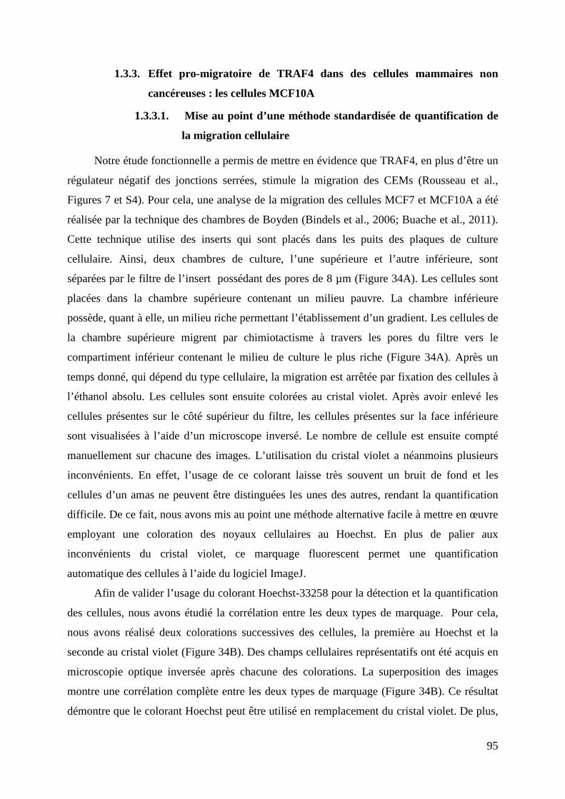
95
1.3.3. Effet pro-migratoire de TRAF4 dans des cellules mammaires non
cancéreuses : les cellules MCF10A
1.3.3.1. Mise au point d’une méthode standardisée de quantification de
la migration cellulaire
Notre étude fonctionnelle a permis de mettre en évidence que TRAF4, en plus d’être un
régulateur négatif des jonctions serrées, stimule la migration des CEMs (Rousseau et al.,
Figures 7 et S4). Pour cela, une analyse de la migration des cellules MCF7 et MCF10A a été
réalisée par la technique des chambres de Boyden (Bindels et al., 2006; Buache et al., 2011).
Cette technique utilise des inserts qui sont placés dans les puits des plaques de culture
cellulaire. Ainsi, deux chambres de culture, l’une supérieure et l’autre inférieure, sont
séparées par le filtre de l’insert possédant des pores de 8 µm (Figure 34A). Les cellules sont
placées dans la chambre supérieure contenant un milieu pauvre. La chambre inférieure
possède, quant à elle, un milieu riche permettant l’établissement d’un gradient. Les cellules de
la chambre supérieure migrent par chimiotactisme à travers les pores du filtre vers le
compartiment inférieur contenant le milieu de culture le plus riche (Figure 34A). Après un
temps donné, qui dépend du type cellulaire, la migration est arrêtée par fixation des cellules à
l’éthanol absolu. Les cellules sont ensuite colorées au cristal violet. Après avoir enlevé les
cellules présentes sur le côté supérieur du filtre, les cellules présentes sur la face inférieure
sont visualisées à l’aide d’un microscope inversé. Le nombre de cellule est ensuite compté
manuellement sur chacune des images. L’utilisation du cristal violet a néanmoins plusieurs
inconvénients. En effet, l’usage de ce colorant laisse très souvent un bruit de fond et les
cellules d’un amas ne peuvent être distinguées les unes des autres, rendant la quantification
difficile. De ce fait, nous avons mis au point une méthode alternative facile à mettre en œuvre
employant une coloration des noyaux cellulaires au Hoechst. En plus de palier aux
inconvénients du cristal violet, ce marquage fluorescent permet une quantification
automatique des cellules à l’aide du logiciel ImageJ.
Afin de valider l’usage du colorant Hoechst-33258 pour la détection et la quantification
des cellules, nous avons étudié la corrélation entre les deux types de marquage. Pour cela,
nous avons réalisé deux colorations successives des cellules, la première au Hoechst et la
seconde au cristal violet (Figure 34B). Des champs cellulaires représentatifs ont été acquis en
microscopie optique inversée après chacune des colorations. La superposition des images
montre une corrélation complète entre les deux types de marquage (Figure 34B). Ce résultat
démontre que le colorant Hoechst peut être utilisé en remplacement du cristal violet. De plus,
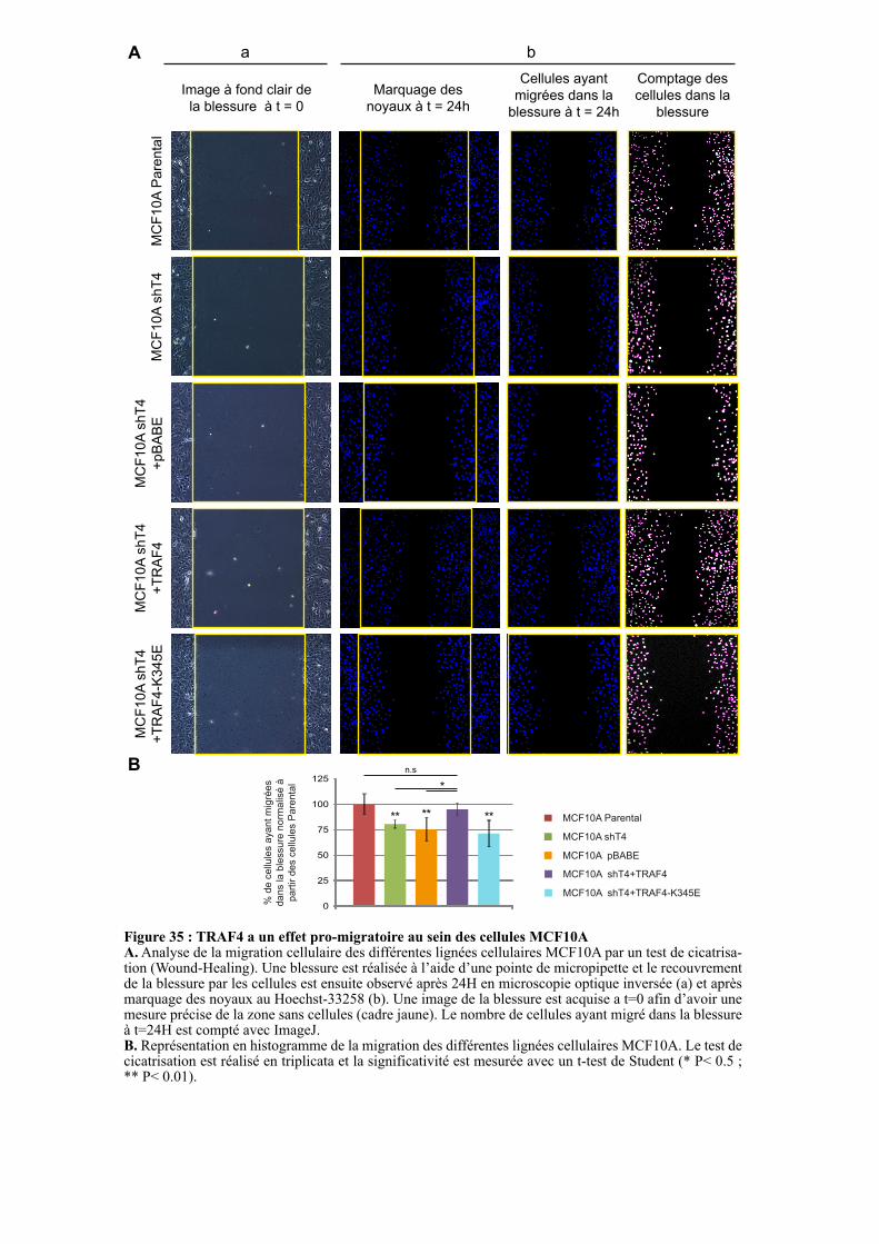
Figure 35 : TRAF4 a un effet pro-migratoire au sein des cellules MCF10AA. Analyse de la migration cellulaire des différentes lignées cellulaires MCF10A par un test de cicatrisa-tion (Wound-Healing). Une blessure est réalisée à l’aide d’une pointe de micropipette et le recouvrement de la blessure par les cellules est ensuite observé après 24H en microscopie optique inversée (a) et après marquage des noyaux au Hoechst-33258 (b). Une image de la blessure est acquise a t=0 afin d’avoir une mesure précise de la zone sans cellules (cadre jaune). Le nombre de cellules ayant migré dans la blessure à t=24H est compté avec ImageJ. B. Représentation en histogramme de la migration des différentes lignées cellulaires MCF10A. Le test de cicatrisation est réalisé en triplicata et la significativité est mesurée avec un t-test de Student (* P< 0.5 ; ** P< 0.01).
MC
F1
0A
Pa
ren
tal
MC
F1
0A
sh
T4
MC
F1
0A
sh
T4
+p
BA
BE
MC
F1
0A
sh
T4
+T
RA
F4
Image à fond clair de
la blessure à t = 0
Marquage des
noyaux à t = 24h
Comptage des
cellules dans la
blessure
Cellules ayant
migrées dans la
blessure à t = 24h
A
B
MCF10A Parental
MCF10A shT4
MCF10A pBABE
MCF10A shT4+TRAF4
0
25
50
75
100
125
% d
e c
ellu
les a
ya
nt
mig
rée
s
da
ns la
ble
ssu
re n
orm
alis
é à
pa
rtir d
es c
ellu
les P
are
nta
l
** **
*
n.s
MC
F1
0A
sh
T4
+T
RA
F4
-K3
45
E
**
MCF10A shT4+TRAF4-K345E
a b
96
les noyaux cellulaires peuvent être comptés automatiquement en utilisant le module
d’extension « Nucleus counting » du logiciel ImageJ. En définissant les seuils correspondant à
la taille maximale et minimale des noyaux pris en compte, ce plugin permet même de séparer
les noyaux des cellules d’un même amas. De plus, ce mode de quantification est beaucoup
plus rapide que le comptage manuel, ce qui permet d’augmenter le nombre de champs
analysés et ainsi de gagner en puissance statistique.
1.3.3.2. TRAF4 a un effet pro-migratoire sur les cellules MCF10A
L’effet pro-migratoire de TRAF4 sur les cellules épithéliales mammaires MCF10A a été
mis en évidence par la technique des chambres de Boyden (Rousseau et al., Figure S4). Des
tests de cicatrisation ont été réalisés au sein des mêmes lignées cellulaires mais ils n’ont pas
été inclus dans l’article. Lors de ces tests, la même quantité de cellules a été déposée dans des
plaques six puits. A confluence, une blessure a été réalisée à l’aide d’une pointe de cône de
micropipette, des images ont ensuite été acquises à l’aide d’un microscope optique inversé
(Figure 35 : t=0). Les cellules ont ensuite été replacées dans un incubateur pendant 24H. Le
recouvrement de la blessure par les cellules a été observé en microscopie optique inversée
suite au marquage des noyaux au Hoechst (Figure 35 : t=24). Afin de mesurer la migration
cellulaire, les cellules ayant migré au niveau de la zone correspondant à la blessure à t=0
(cadre jaune) ont été comptées automatiquement à l’aide du logiciel ImageJ (Figure 35A).
Nous avons observé, comme précédemment, que la sous-expression de TRAF4 (MCF10A
shT4) conduit à une diminution de la migration des cellules MCF10A (Figure 35). De manière
intéressante, la réexpression de TRAF4 (MCF10A shT4+TRAF4) restaure un niveau de
migration similaire aux cellules MCF10A parentales tandis que le contrôle vecteur vide
(MCF10A shT4+pBABE) n’a aucun effet. Cette seconde méthode confirme que TRAF4 a un
effet pro-migratoire dans les cellules MCF10A.
Nous avons montré que TRAF4 lie les PIPs via son domaine TRAF et que cette
interaction est requise pour l’effet pro-migratoire de TRAF4 (Article : Figure 7 et Figure S4).
Afin de renforcer ce résultat, nous avons étudié si l’expression du mutant TRAF4-K345E ne
liant plus les PIPs au sein des cellules déficientes en TRAF4 (MCF10A shT4+TRAF4-
K345E) pouvait restaurer une migration normale des cellules. Tandis que la réexpression de la
protéine TRAF4 sauvage restaure une migration cellulaire normale, celle du mutant TRAF-
K345E ne liant pas les PIPs n’a aucun effet (Figure 35).
ALSSRQGQQIQMLKSAVSKLSIN-YTGTLLWKITDWSAKMAEARGKDGLELVSPPFYTSQYGYK
-TLAQKDQALGKLEQSLRLMEEASFDGTFLWKITNVTRRCHESACGRTVSLFSPAFYTAKYGYK
RSIGLKDLAMADLEQKVLEMEASTYDGVFIWKISDFARKRQEAVAGRIPAIFSPAFYTSRYGYK
QMLSVHDIRLADMDLRFQVLETASYNGVLIWKIRDYKRRKQEAVMGKTLSLYSQPFYTGYFGYK
ALVSRQRQELQELRRELEELSVG-SDGVLIWKIGSYGRRLQEAKAKPNLECFSPAFYTHKYGYK
THINIHKAQLSKNEERFKLLEGTCYNGKLIWKVTDYKMKKREAVDGHTVSIFSQSFYTSRCGYR
MYVSELKRTIRTLEDKVAEIEAQQCNGIYIWKIGNFGMHLKCQEEEKPVVIHSPGFYTGKPGYK
: . :. * :**: . : * *** **:
K313 R319/320 K345
hTRAF1
hTRAF2
hTRAF3
hTRAF4
hTRAF5
hTRAF6
326
390
378
325
242
DTRAF1 306
283
389
453
441
388
304
368
345
LQASMFLNGNGPGENT-HVSVYIKVLPGEYDALLKWPFSHSITFTLFEQG--AQSGQGGVAESF
LCLRLYLNGDGTGKRT-HLSLFIVIMRGEYDALLPWPFRNKVTFMLLDQ-----NNREHAIDAF
MCLRIYLNGDGTGRGT-HLSLFFVVMKGPNDALLRWPFNQKVTLMLLDQ-----NNREHVIDAF
MCARVYLNGDGMGKGT-HLSLFFVIMRGEYDALLPWPFKQKVTLMLMDQG----SSRRHLGDAF
LQVSAFLNGNGSGEGT-HLSLYIRVLPGAFDNLLEWPFARRVTFSLLDQSDPGLAKPQHVTETF
LCARAYLNGDGSGRGS-HLSLYFVVMRGEFDSLLQWPFRQRVTLMLLDQ-----SGKKNIMETF
LCMRLHLQLPTAQRCANYISLFVHTMQGEYDSHLPWPFQGTIRLTILDQS--EAPVRQNHEEIM
: .*: . : ::*::. : * * * *** : : :::* : :
390
454
442
389
305
369
346
hTRAF1
hTRAF2
hTRAF3
hTRAF4
hTRAF5
hTRAF6
DTRAF1
R385 K400
447
512
499
450
362
429
408
VPDPTWENFQRP-----SNEPDQLGFGFPRFISHELLHS-R-PFIKGDTVFLRVKVD-PSKIVA
RPDLSSASFQRP------QSETNVASGCPLFFPLSKLQSPKHAYVKDDTMFLKCIVETST----
RPDVTSSSFQRP------VNDMNIASGCPLFCPVSKMEA-KNSYVRDDAIFIKAIVDLTGL---
KPDPNSSSFKKP------TGEMNIASGCPVFVAQTVLENG--TYIKDDTIFIKVIVDTSDLPDP
HPDPNWKNFQKPGTWRGSLDESSLGFGYPKFISHQDI--RKRNYVRDDAVFIRAAVELPRKILS
KPDPNSSSFKRP------DGEMNIASGCPRFVAHSVLENAKNAYIKDDTLFLKVAVDLTDLEDL
DAKPELLAFQRP-----TIPRNPKGFGYVTFMHLEALRQ-R-TFIKDDTLLVRCEVSTRFDMGS
.. *::* . * * :. :::.*::::: *.
R452 R459
448
513
500
451
363
430
409
501
568
557
507
416
485
470
hTRAF1
hTRAF2
hTRAF3
hTRAF4
hTRAF5
hTRAF6
DTRAF1
K419
A
TRAF6
31
28
33
29
32
TRAF6 TRAF1 TRAF2 TRAF3 TRAF4 TRAF5
TRAF1 62 58 43 52
TRAF2 60 47 59
TRAF3 41 66
TRAF4 44
TRAF5
TRAF6
TRAF1 TRAF2 TRAF3 TRAF4 TRAF5
TRAF1
TRAF2
TRAF3
TRAF4
TRAF5
TRAF6
B
Identité moyenne de TRAF6 avec les autres TRAFs = 30.6 %
Identité moyenne des TRAFs = 45.7 %
TRAF de
Figure 36: Analyse comparative des TRAFsA. Alignement des séquences protéiques des domaines TRAF de TRAF1 à TRAF6 et de DTRAF1. Les acides aminés identiques sont marqués par un astérisque. Les change-ments conservatifs (même propriété physico-chimique) et non-conservatifs (architecture similaire) sont indiqués par un double point et un point unique, respectivement. Les petits acides aminés ainsi que les résidus aromatiques, excepté la tyrosine, sont colorés en rouge. Les acides aminés acides et basiques sont colorés en bleu et en magenta, respectivement. Les résidus hydrophiles et la tyrosine sont colorés en vert. B. Tableau récapitulatif représentant les pourcentages d’homologie (identité) entre les différents domaines TRAFs de TRAF1 à TRAF6.
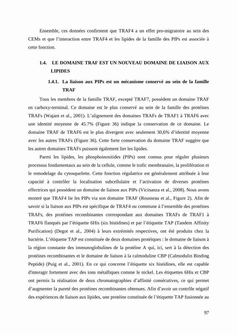
97
Ensemble, ces données confirment que TRAF4 a un effet pro-migratoire au sein des
CEMs et que l’interaction entre TRAF4 et les lipides de la famille des PIPs est associée à
cette fonction.
1.4. LE DOMAINE TRAF EST UN NOUVEAU DOMAINE DE LIAISON AUX
LIPIDES
1.4.1. La liaison aux PIPs est un mécanisme conservé au sein de la famille
TRAF
Tous les membres de la famille TRAF, excepté TRAF7, possèdent un domaine TRAF
en carboxy-terminal. Ce domaine est le plus conservé au sein de la famille des protéines
TRAFs (Wajant et al., 2001). L’alignement des domaines TRAFs de TRAF1 à TRAF6 avec
une identité moyenne de 45.7% (Figure 36) indique la conservation de ce domaine. Le
domaine TRAF de TRAF6 est le plus divergent avec seulement 30,6% d’identité moyenne
avec les autres TRAFs (Figure 36). Cette forte conservation du domaine TRAF suggère que
les autres domaines TRAFs puissent également lier les lipides.
Parmi les lipides, les phosphoinositides (PIPs) sont connus pour réguler plusieurs
processus fondamentaux au sein de la cellule, comme le trafic membranaire, la prolifération et
le remodelage du cytosquelette. Cette fonction régulatrice est généralement attribuée à leur
capacité à contrôler la localisation subcellulaire et l’activation de diverses protéines
effectrices qui possèdent un domaine de liaison aux PIPs (Vicinanza et al., 2008). Nous avons
montré que TRAF4 lie les PIPs via son domaine TRAF (Rousseau et al., Figure 2). Afin de
savoir si la liaison aux PIPs est spécifique de TRAF4 ou commune à l’ensemble des protéines
TRAFs, des protéines recombinantes correspondant aux domaines TRAFs de TRAF1 à
TRAF6 flanqués par l’étiquette 6His (six histidines) et par l’étiquette TAP (Tandem Affinity
Purification) (Degot et al., 2004) à leurs extrémités respectives, ont été produits chez la
bactérie. L’étiquette TAP est constituée de deux domaines protéiques : le domaine de liaison à
la région constante des immunoglobulines de la protéine A qui, ici, sert à la détection des
protéines recombinantes et le domaine de liaison à la calmoduline CBP (Calmodulin Binding
Peptide) (Puig et al., 2001). En ce qui concerne l’étiquette six histidines, elle est capable
d'interagir fortement avec des ions métalliques comme le nickel. Les étiquettes 6His et CBP
ont permis la réalisation de deux chromatographies d’affinité consécutives, ce qui permet
d’augmenter la pureté des protéines recombinantes obtenues. Afin d’avoir un contrôle négatif
des expériences de liaison aux lipides, une protéine constituée de l’étiquette TAP fusionnée au
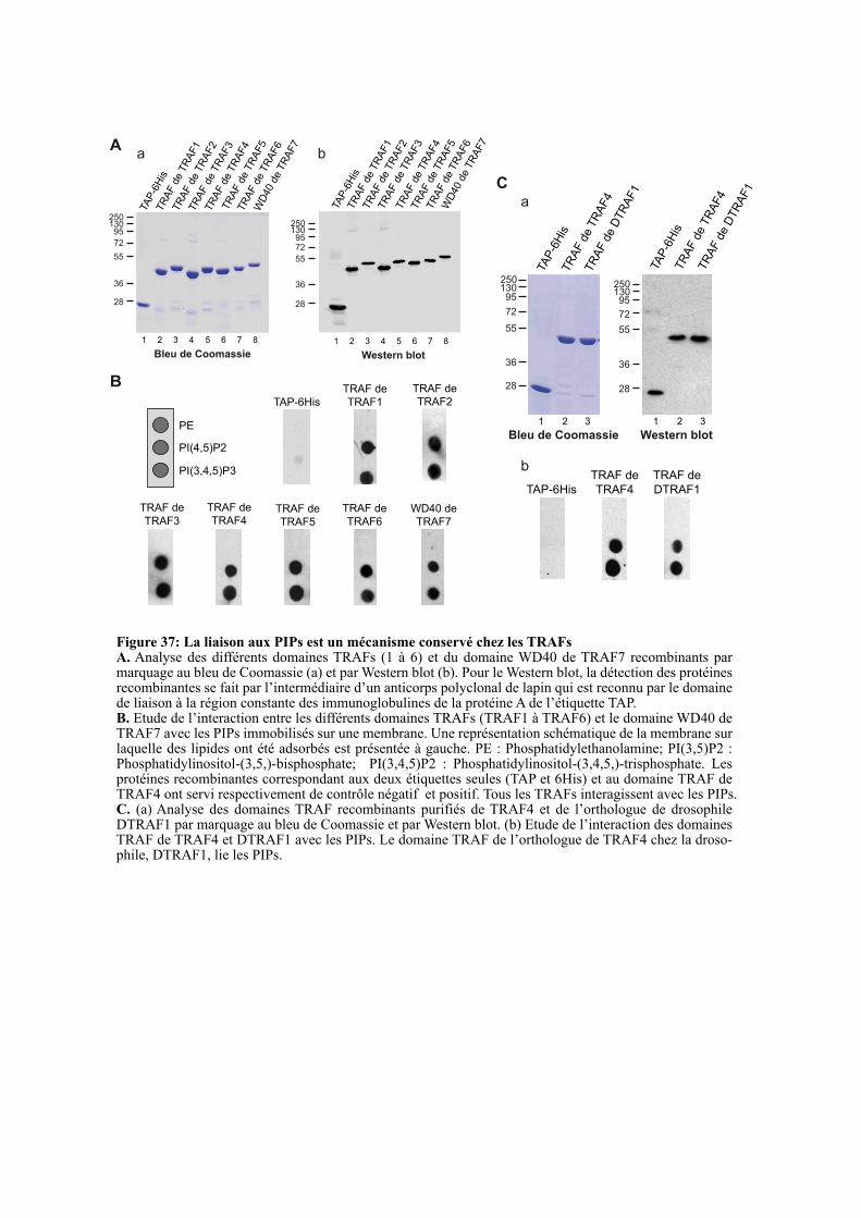
B
PE
PI(4,5)P2
PI(3,4,5)P3
TAP-6His
TRAF de
TRAF1
TRAF de
TRAF2
TRAF de
TRAF3
TRAF de
TRAF4TRAF de
TRAF5
WD40 de
TRAF7
TRAF de
TRAF6
A
a
25013095
72
55
36
28
17
TAP
-6H
isTR
AF d
e T
RA
F1
TR
AF d
e T
RA
F2
TR
AF d
e T
RA
F3
TR
AF d
e T
RA
F4
TR
AF d
e T
RA
F6
WD
40 d
e T
RA
F7
Bleu de Coomassie
1 2 3 4 5 6 7 8
TR
AF d
e T
RA
F5
2501309572
55
36
28
17TA
P-6
His
TR
AF d
e T
RA
F1
TR
AF d
e T
RA
F2
TR
AF d
e T
RA
F3
TR
AF d
e T
RA
F4
TR
AF d
e T
RA
F5
TR
AF d
e T
RA
F6
WD
40 d
e T
RA
F7
b
Western blot
1 2 3 4 5 6 7 8
C
25013095
72
55
36
28
Bleu de Coomassie
1 2 3
TAP
-6H
isTR
AF d
e T
RA
F4
TR
AF d
e D
TR
AF1
25013095
72
55
36
28
Western blot
1 2 3
TAP
-6H
isTR
AF d
e T
RA
F4
TR
AF d
e D
TR
AF1
a
b TRAF de
TRAF4TAP-6His
TRAF de
DTRAF1
Figure 37: La liaison aux PIPs est un mécanisme conservé chez les TRAFsA. Analyse des différents domaines TRAFs (1 à 6) et du domaine WD40 de TRAF7 recombinants par marquage au bleu de Coomassie (a) et par Western blot (b). Pour le Western blot, la détection des protéines recombinantes se fait par l’intermédiaire d’un anticorps polyclonal de lapin qui est reconnu par le domaine de liaison à la région constante des immunoglobulines de la protéine A de l’étiquette TAP.B. Etude de l’interaction entre les différents domaines TRAFs (TRAF1 à TRAF6) et le domaine WD40 de TRAF7 avec les PIPs immobilisés sur une membrane. Une représentation schématique de la membrane sur laquelle des lipides ont été adsorbés est présentée à gauche. PE : Phosphatidylethanolamine; PI(3,5)P2 : Phosphatidylinositol-(3,5,)-bisphosphate; PI(3,4,5)P2 : Phosphatidylinositol-(3,4,5,)-trisphosphate. Les protéines recombinantes correspondant aux deux étiquettes seules (TAP et 6His) et au domaine TRAF de TRAF4 ont servi respectivement de contrôle négatif et positif. Tous les TRAFs interagissent avec les PIPs. C. (a) Analyse des domaines TRAF recombinants purifiés de TRAF4 et de l’orthologue de drosophile DTRAF1 par marquage au bleu de Coomassie et par Western blot. (b) Etude de l’interaction des domaines TRAF de TRAF4 et DTRAF1 avec les PIPs. Le domaine TRAF de l’orthologue de TRAF4 chez la droso-phile, DTRAF1, lie les PIPs.
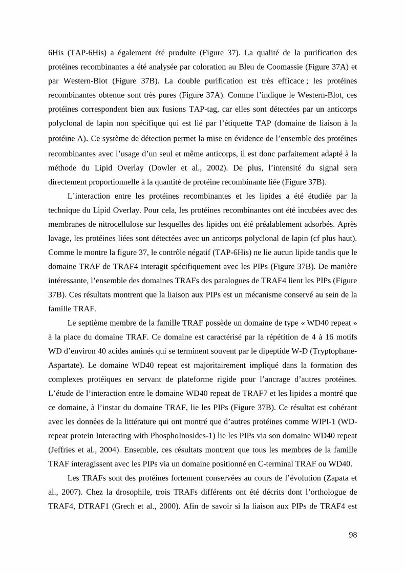
98
6His (TAP-6His) a également été produite (Figure 37). La qualité de la purification des
protéines recombinantes a été analysée par coloration au Bleu de Coomassie (Figure 37A) et
par Western-Blot (Figure 37B). La double purification est très efficace ; les protéines
recombinantes obtenue sont très pures (Figure 37A). Comme l’indique le Western-Blot, ces
protéines correspondent bien aux fusions TAP-tag, car elles sont détectées par un anticorps
polyclonal de lapin non spécifique qui est lié par l’étiquette TAP (domaine de liaison à la
protéine A). Ce système de détection permet la mise en évidence de l’ensemble des protéines
recombinantes avec l’usage d’un seul et même anticorps, il est donc parfaitement adapté à la
méthode du Lipid Overlay (Dowler et al., 2002). De plus, l’intensité du signal sera
directement proportionnelle à la quantité de protéine recombinante liée (Figure 37B).
L’interaction entre les protéines recombinantes et les lipides a été étudiée par la
technique du Lipid Overlay. Pour cela, les protéines recombinantes ont été incubées avec des
membranes de nitrocellulose sur lesquelles des lipides ont été préalablement adsorbés. Après
lavage, les protéines liées sont détectées avec un anticorps polyclonal de lapin (cf plus haut).
Comme le montre la figure 37, le contrôle négatif (TAP-6His) ne lie aucun lipide tandis que le
domaine TRAF de TRAF4 interagit spécifiquement avec les PIPs (Figure 37B). De manière
intéressante, l’ensemble des domaines TRAFs des paralogues de TRAF4 lient les PIPs (Figure
37B). Ces résultats montrent que la liaison aux PIPs est un mécanisme conservé au sein de la
famille TRAF.
Le septième membre de la famille TRAF possède un domaine de type « WD40 repeat »
à la place du domaine TRAF. Ce domaine est caractérisé par la répétition de 4 à 16 motifs
WD d’environ 40 acides aminés qui se terminent souvent par le dipeptide W-D (Tryptophane-
Aspartate). Le domaine WD40 repeat est majoritairement impliqué dans la formation des
complexes protéiques en servant de plateforme rigide pour l’ancrage d’autres protéines.
L’étude de l’interaction entre le domaine WD40 repeat de TRAF7 et les lipides a montré que
ce domaine, à l’instar du domaine TRAF, lie les PIPs (Figure 37B). Ce résultat est cohérant
avec les données de la littérature qui ont montré que d’autres protéines comme WIPI-1 (WD-
repeat protein Interacting with PhosphoInosides-1) lie les PIPs via son domaine WD40 repeat
(Jeffries et al., 2004). Ensemble, ces résultats montrent que tous les membres de la famille
TRAF interagissent avec les PIPs via un domaine positionné en C-terminal TRAF ou WD40.
Les TRAFs sont des protéines fortement conservées au cours de l’évolution (Zapata et
al., 2007). Chez la drosophile, trois TRAFs différents ont été décrits dont l’orthologue de
TRAF4, DTRAF1 (Grech et al., 2000). Afin de savoir si la liaison aux PIPs de TRAF4 est
hyTRAF1 257 FIWPIKRFRQQFELARNSDEEIAVFSQPFYTSQYGYKLRIK 297
ceTRAF 267 LIWKIDKLQQRTNEAK-SGADTTIFSVPFMSHRFGYKMMAC 304
ciTRAF4 310 LLWKIPGYSQKLQAAQ-KDQAIEMRSPPFYTHRYGYKLQLS 349
ciTRAF6 425 YYWKIKDYAQLRRAAC-LGEMPVLHSPGFYTSPQGYRMCIR 464
DTRAF1 324 LLWKITDWSAKMAEAR-GKDGLELVSPPFYTSQYGYKLQAS 363
DTRAF2 332 IVWRIEQLGALVARLR-ANANNQVYSHECYTSPHGYKFCAR 371
xtTRAF4 310 LIWKISDYSRKVQEAK-VRGNFESFSPPFYTHKYGYKLQVS 349
xlTRAF4a 310 LIWKISDYSRKVQEAK-VRGNYESFSPPFYTHKYGYKLQVS 349
xlTRAF4b 310 LIWKISDYSRKVQEAK-VRGNYESFSPTFYTHKYGYRLQVS 349
xtTRAF6 389 FIWRINNFNSLLKNQE-EERPVVIHSQGFYTGKPGYKLCLR 428
xlTRAF6a 387 FIWRIKGFSGLQKSQE-EEKPVVIHSPGFYTGDPGYKLCLR 426
xlTRAF6b 387 FIWKIKGFSGLQKTQE-EEKPVVIHSPGFYTGKPGYKLCLR 426
hsTRAF1 269 FLWKITNVTRRCHESA-CGRTVSLFSPAFYTAKYGYKLCLR 308
hsTRAF2 354 FIWKISDFARKRQEAV-AGRIPAIFSPAFYTSRYGYKMCLR 393
hsTRAF3 418 LIWKIRDYKRRKQEAV-MGKTLSLYSQPFYTGYFGYKMCAR 457
hsTRAF4 310 LIWKIGSYGRRLQEAK-AKPNLECFSPAFYTHKYGYKLQVS 349
hsTRAF5 406 LIWKVTDYKMKKREAV-DGHTVSIFSQSFYTSRCGYRLCAR 445
hsTRAF6 353 YIWKIGNFGMHLKCQE-EEKPVVIHSPGFYTGKPGYKLCMR 392
* : * : **::
K313 K345
K313, TRAF4
K357, TRAF2
K389, TRAF2
K345, TRAF4
A
B
Figure 38: La position des lysines 313 et 345 de TRAF4 est conservée chez les TRAFsA. Comparaison des régions incluant les lysines 313 et 345 des domaines TRAF des différents orthologues et paralogues de TRAF4. Les positions 313 et 345 de TRAF4 sont encadrées en rouge. Les résidus basiques correspondant à la position 313 et 345 du TRAF de TRAF4 sont surlignés en jaune. La tyrosine 338, identifié dans le modèle d’interaction TRAF-PIP comme liant le lipide, est encadrée en bleu. Les acides aminés identiques sont marqués par un astérisque. Les remplacements conservatifs et non-conservatifs sont indiqués respective-ment par un double point et un point unique. Les lysines 313 et 345 sont conservées dans tous les domaines TRAFs. DTRAF1 (dmTRAF4); DTRAF2 (dmTRAF6); hs : Homo sapiens; xl : Xenopus laevis; xt : Xenopus tropicalis; dm: Drosophila melanogaster; ci : Ciona intestinalis; ce : Caenorhabditis elegans; Hy : hydractinia echinata. B. Superposition des structures 3D correspondant aux sites de liaison aux PIPs des domaines TRAFs de TRAF2 (1CA4) et TRAF4. Les atomes de phosphate, d’azote et d’oxygène sont colorés respectivement en orange, bleu et rouge. La position relative ainsi que l’orientation des lysines 313 et 345 de TRAF4 sont conservées au sein de la protéine TRAF2.
Y338
99
conservée chez la mouche, le domaine TRAF de DTRAF1 a été produit puis purifié selon la
même méthodologie (Figure 37C). L’analyse par Lipid Overlay a montré que, à l’instar de
l’orthologue humain, le domaine TRAF de DTRAF1 lie les PIPs (Figure 37C). Ce résultat
suggère que la liaison aux PIPs du domaine TRAF est également un mécanisme conservé au
cours de l’évolution.
1.4.2. Les lysines 313 et 345 sont conservées chez les TRAFs
L’analyse biochimique, structurale et biophysique de l’interaction entre le domaine
TRAF de TRAF4 et les PIPs a révélé l’importance des lysines 313 et 345. Leur remplacement
par un résidu acide, respectivement diminue et abolit la liaison aux PIPs (Rousseau et al.,
Figure 3). J’ai étudié la conservation de ces résidus au sein de la famille TRAF, considérant
les paralogues humains de TRAF4 ainsi que les orthologues de diverses espèces telles que
l’hydre, le ver, le tunicier, la mouche et les amphibiens. Les positions 313 et 345 sont
occupées par des lysines ou arginines dans tous les TRAFs à l’exception de hyTRAF1,
l’orthologue de TRAF4 chez l’hydre hydractinia echinata. Dans cet organisme marin, le plus
distant du point de vue évolutif, la lysine 345 est conservée tandis que sa lysine 313 est
remplacée par une proline. Néanmoins, cette orthologue possède deux résidus basiques (KR)
à proximité de la position 313 (Figure 38A). Ces données structurales étayent la notion selon
laquelle la liaison aux PIPs est un mécanisme de recrutement membranaire, conservé au cours
de l’évolution de la famille TRAF.
Le domaine TRAF a une structure tertiaire, hautement conservée au sein de la famille
TRAF, en forme de champignon (Wu, 2004). Cette conservation structurale, associée à la
caractéristique commune de liaison aux PIPs de tous les membres TRAFs, suggère que la
position relative des lysines 313 et 345 devrait être conservée spatialement. Pour tester cette
hypothèse, les structures cristallines des domaines TRAFs de TRAF4 et TRAF2 ont été
superposées. La position relative ainsi que l’orientation des lysines 313 et 345 de TRAF4 sont
fortement similaire à celles des lysines 357 et 389 de TRAF2 (Figure 38B). Les mêmes
résultats ont été observés en superposant le domaine TRAF de TRAF4 avec les domaines
TRAFs de TRAF3 et TRAF6 (non montré). Pris dans leur ensemble, ces résultats supportent
le concept que les protéines adaptatrice de la famille des TRAFs partagent toutes la capacité
de lier les lipides de la famille des PIPs. Dans quelle mesure cette propriété participe à leurs
fonctions respectives demande à être testé. Pour cela, il serait intéressant de développer des
mutants ne liant pas les PIPs au sein de paralogues de TRAF4 dont la fonction est bien
caractérisée, comme par exemple la protéine TRAF2. Après avoir validé que le remplacement
100
de la lysine 389 de TRAF2 en acide glutamique (équivalent de la lysine 345 chez TRAF4)
conduit à une perte de la liaison aux PIPs, nous pourrions en tester l’impact fonctionnel sur
l’activation du facteur de transcription NF-κB (Nuclear Factor kappa B). En effet, il est connu
que la surexpression de TRAF2 au sein des cellules HEK293 conduit à une activation de NF-
kB (Song et al., 1996). De ce fait, il serait possible de comparer l’activation du facteur de
transcription dans des cellules HEK293 transfectées par TRAF2 sauvage et par le mutant
TRAF2 K389E. D’autres fonctions de TRAF2 pourraient également être testées comme
l’activation de la voie des MAP (Mitogen Activated Protein) kinases après traitement au
TNFα (Devin et al., 2003).
De manière intéressante, dans la même région du domaine TRAF, se situent une serine
(S334) et une tyrosine (T344), deux résidus pouvant être phosphorylés. Ces résidus sont
également conservés dans l’ensemble des protéines TRAFs ce qui suggère qu’ils puissent
avoir une fonction importante (Figure 38A). En effet, la proximité de ces résidus
phosphorylables avec le site de liaison aux PIPs implique qu’ils puissent être un moyen de
réguler la liaison du domaine TRAF aux PIPs. En effet, il est possible que l’ajout de charges
négatives par phosphorylation à proximité du site de liaison empêche la fixation des PIPs qui
sont eux-mêmes chargés négativement. La production de mutants du domaine TRAF
recombinant avec des résidus phosphomimétiques à la place de ces acides aminés (S334D
et/ou Y334D) permettrait de tester par Lipid Overlay la capacité de liaison aux PIPs de ces
mutants et de démontrer leur rôle régulateur potentiel dans la liaison aux PIPs.
1.4.3. Analyse du modèle d’interaction entre le domaine TRAF et les PIPs
La cristallisation du domaine TRAF de TRAF4, associée à l’identification par
mutagénèse de résidus impliqués dans la liaison aux PIPs, nous a permis de proposer un
modèle d’interaction entre le domaine TRAF et les PIPs. Dans ce modèle, le lipide est lié à
l’interface entre deux protomères voisins ; la lysine 313 d’un protomère lie à la fois le 3ème et
le 4ème phosphate du PI(3,4,5,)P3 tandis que la lysine 345 du protomère adjacent interagit avec
le 5ème phosphate du lipide (Rousseau et al., Figure 4). Ce modèle nous a permis d’identifier
deux autres résidus potentiellement impliqués dans la liaison aux PIPs, l’arginine 297 et la
tyrosine 338. Une étude structurale a montré tous les domaines connus d’interaction avec les
PIPs possèdent au moins deux résidus basiques (Lys ou Arg) et un résidu aromatique (Tyr et
His ; plus rarement Phe et Trp) qui sont en contact direct avec le lipide (Rosenhouse-Dantsker
and Logothetis, 2007). L’identification de la tyrosine 338, comme interagissant
potentiellement avec le PIP, renforce la pertinence de notre modèle. De plus, l’alignement des
101
domaines TRAFs montre une conservation totale de cette tyrosine dans tous les orthologues et
paralogues, excepté pour le paralogue de C. elegans (ceTRAF1). Néanmoins, ceTRAF1
possède une phénylalanine et une histidine à proximité qui pourraient remplacer
fonctionnellement la tyrosine 338 (Figure 38A). Afin d’infirmer ou de confirmer l’implication
de ces résidus dans la liaison aux PIPs, la mutation de ces résidus en acide aspartique et
l’analyse de ces mutants par Lipid Overlay est nécessaire.
1.5. CONCLUSION
TRAF4 est surexprimé dans environ 40% des carcinomes; dans de nombreux cas, c’est
l’amplification de son gène qui est responsable de la surexpression (Bièche et al., 1996;
Camilleri-Broët et al., 2007; Régnier et al., 1995). Néanmoins, son rôle dans le
développement de la maladie reste toujours incompris. Le carcinome est une tumeur
développée à partir des cellules épithéliales. Ces cellules sont polarisées et jointives et ont
pour particularité de former des jonctions intercellulaires (jonctions adhérentes : JA et
jonctions serrées : JS). Il est clairement établi que la perte de ces structures jonctionnelles est
un processus clé régulant l’initiation et la progression des carcinomes (Brennan et al., 2010;
Forster, 2008). La localisation subcellulaire de TRAF4 au sein des jonctions serrées des
cellules épithéliales polarisées suggère qu’elle participe à leur fonction. Nous avons émis
l’hypothèse que la surexpression de TRAF4 pouvait déstabiliser les JSs et ainsi contribuer à la
transformation cancéreuse. Afin de tester cette hypothèse, nous avons étudié le mécanisme
moléculaire responsable du recrutement de TRAF4 aux JSs ainsi que son rôle dans ces
structures jonctionnelles. Cette étude nous a permis de montrer que le domaine TRAF de
TRAF4 est un nouveau domaine liant les PIPs. De plus, cette liaison aux PIPs s’étend à tous
les paralogues humains de TRAF4 ainsi qu’à son orthologue chez la mouche, DTRAF1. Ces
résultats originaux permettent de postuler que la liaison aux PIPs est une propriété ancestrale
commune aux protéines de la famille TRAF. Des expériences fonctionnelles portant sur la
signalisation des récepteurs du TNF induite par leur ligand permettraient d’étayer cette
hypothèse. La voie du TNF est impliquée dans de nombreuses pathologies inflammatoires,
cette interaction fonctionnelle avec des lipides pourrait constituer une nouvelle piste pour le
développement de stratégies thérapeutiques anti-inflammatoires. Une étude structurale et
biophysique nous a permis de montrer que le domaine TRAF de TRAF4 est présent sous
forme de trimère et lie jusqu’à trois molécules de lipide. D’autres études structurales menées
sur TRAF2, TRAF3 et TRAF6 ont montré que la queue cytoplasmique des récepteurs TNFRs
est en contact avec le versant latéral du domaine TRAF et n’interagit pas avec les protomères
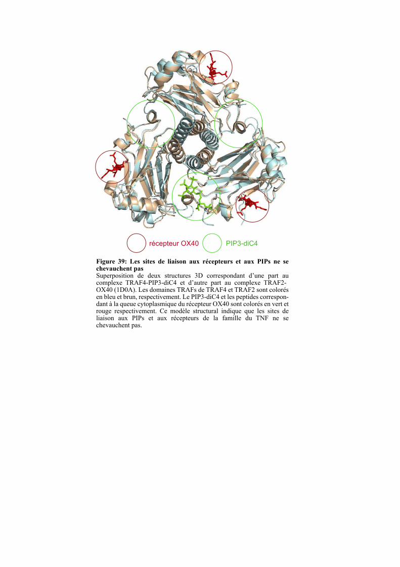
Figure 39: Les sites de liaison aux récepteurs et aux PIPs ne se chevauchent pasSuperposition de deux structures 3D correspondant d’une part au complexe TRAF4-PIP3-diC4 et d’autre part au complexe TRAF2-OX40 (1D0A). Les domaines TRAFs de TRAF4 et TRAF2 sont colorés en bleu et brun, respectivement. Le PIP3-diC4 et les peptides correspon-dant à la queue cytoplasmique du récepteur OX40 sont colorés en vert et rouge respectivement. Ce modèle structural indique que les sites de liaison aux PIPs et aux récepteurs de la famille du TNF ne se chevauchent pas.
récepteur OX40 PIP3-diC4

102
adjacents (Figure 39). Ce mode de liaison est très différent de celui de la liaison aux PIPs qui
se fait à l’interface entre deux protomères voisins (Figure 39). De manière intéressante, les
deux sites de liaison sont non-chevauchants, suggérant la possibilité d’une liaison simultanée
aux récepteurs et aux PIPs. Plusieurs études ont montré que l’affinité des protéines TRAFs
pour leurs récepteurs est assez faible (40-60 µm), elle ne permet pas d’expliquer leur
recrutement à la membrane ; il a donc été proposé que le recrutement membranaire des
TRAFs est potentialisé par le mécanisme d’avidité (Li et al., 2002; Ye and Wu, 2000; Ye et
al., 2002). En effet, les mesures d’affinité réalisées in vitro correspondent à l’affinité entre un
récepteur et un protomère de TRAF, tandis que dans le contexte cellulaire, le récepteur TNFR
est trimérique et s’associe à un trimère de TRAFs. Nos résultats suggèrent que la liaison aux
PIPs participe au mécanisme moléculaire de recrutement membranaire des TRAFs. En effet,
les motifs de liaison impliqués dans l’interaction avec les récepteurs et les PIPS ne sont pas
chevauchant, il est probable que la liaison aux PIPs va fortement augmenter l’affinité des
TRAFs pour la membrane plasmique et ainsi favoriser leur interaction avec les récepteurs
membranaires.
D’un point de vue fonctionnel, nous avons observé que TRAF4 est un régulateur négatif
des JSs et que cette fonction dépend de son interaction avec les PIPs. Les JSs sont des
structures clés régulant différents processus tel que la différentiation et la migration cellulaire.
Pour relier TRAF4 a ces processus, nous avons étudié l’impact de la sous- et sur-expression
de TRAF4 sur la migration des CEMs. Nous avons observé que l’expression de TRAF4 est
corrélée aux capacités migratoires des cellules MCF10A et MCF7. De plus, la liaison aux
PIPs est nécessaire à l’effet pro-migratoire de TRAF4. D’autres études ont incriminé TRAF4
dans la migration cellulaire. En effet, il a été décrit que les cellules dendritiques provenant de
souris déficiente pour TRAF4 avaient des propriétés migratoires moindres (Cherfils-Vicini et
al., 2008). Par ailleurs, les souris TRAF4-/- ont un défaut de fermeture du tube neural
conduisant à une spina bifida. Ce phénotype est principalement caractérisé par un défaut de
convergence-extension, un processus impliquant une forte migration cellulaire (Régnier et al.,
2002). Une autre étude réalisée chez le xénope a également montré que la déficience de
TRAF4 conduit à un défaut du développement et de la migration des cellules des crêtes
neurales (Kalkan et al., 2009).
Ensemble, ces données supportent l’hypothèse selon laquelle la surexpression de
TRAF4 pourrait contribuer à la transformation cancéreuse en déstabilisant les JSs et en
favorisant la migration cellulaire. Afin de tester cette hypothèse, il sera nécessaire d’étudier
l’impact de la surexpression de TRAF4 dans des modèles de tumorigenèse in vivo.
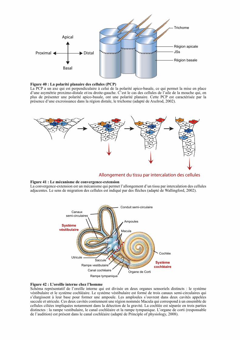
Apical
Basal
Proximal Distal
Région apicale
JSs
Région basale
Trichome
Figure 40 : La polarité planaire des cellules (PCP)La PCP a un axe qui est perpendiculaire à celui de la polarité apico-basale, ce qui permet la mise en place d’une asymétrie proximo-distale et/ou droite-gauche. C’est le cas des cellules de l’aile de la mouche qui, en plus de présenter une polarité apico-basale, ont une polarité planaire. Cette PCP est caractérisée par la présence d’une excroissance dans la région distale, le trichome (adapté de Axelrod, 2002).
Allongement du tissu par intercalation des cellules
Figure 41 : Le mécanisme de convergence-extensionLa convergence-extension est un mécanisme qui permet l’allongement d’un tissu par intercalation des cellules adjacentes. Le sens de migration des cellules est indiqué par des flèches (adapté de Wallingford, 2002).
Figure 42 : L’oreille interne chez l’hommeSchéma représentatif de l’oreille interne qui est divisée en deux organes sensoriels distincts : le systéme véstibulaire et le système cochléaire. Le systéme véstibulaire est formé de trois canaux semi-circulaires qui s’élargissent à leur base pour former une ampoule. Les amploules s’ouvrent dans deux cavités appelées saccule et utricule. Ces deux cavités contiennent une région nommée Macula qui correspond à un ensemble de cellules ciliées impliquées notamment dans la détection de la gravité. La cochlée est séparée en trois parties distinctes : la rampe vestibulaire, le canal cochléaire et la rampe tympanique. L’organe de corti (responsable de l’audition) est présent dans le canal cochléaire (adapté de Principle of physiology, 2008).
Canaux
semi-circulaires
Conduit semi-circulaire
Cochlée
Organe de CortiCanal cochléaire
Rampe tympanique
Rampe vestibulaire
SacculeUtricule
Ampoules
Macula
Système
véstibulaire
Système
cochléaire
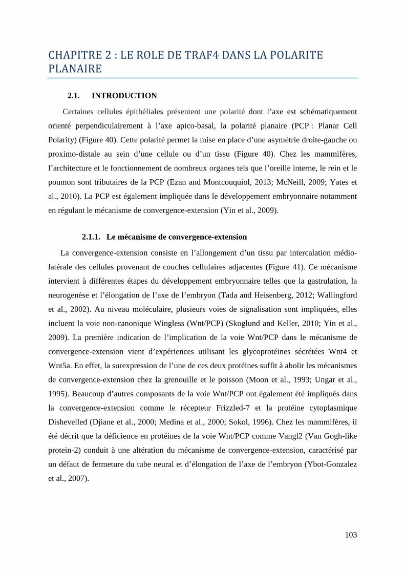
103
CHAPITRE2:LEROLEDETRAF4DANSLAPOLARITE
PLANAIRE
2.1. INTRODUCTION
Certaines cellules épithéliales présentent une polarité dont l’axe est schématiquement
orienté perpendiculairement à l’axe apico-basal, la polarité planaire (PCP : Planar Cell
Polarity) (Figure 40). Cette polarité permet la mise en place d’une asymétrie droite-gauche ou
proximo-distale au sein d’une cellule ou d’un tissu (Figure 40). Chez les mammifères,
l’architecture et le fonctionnement de nombreux organes tels que l’oreille interne, le rein et le
poumon sont tributaires de la PCP (Ezan and Montcouquiol, 2013; McNeill, 2009; Yates et
al., 2010). La PCP est également impliquée dans le développement embryonnaire notamment
en régulant le mécanisme de convergence-extension (Yin et al., 2009).
2.1.1. Le mécanisme de convergence-extension
La convergence-extension consiste en l’allongement d’un tissu par intercalation médio-
latérale des cellules provenant de couches cellulaires adjacentes (Figure 41). Ce mécanisme
intervient à différentes étapes du développement embryonnaire telles que la gastrulation, la
neurogenèse et l’élongation de l’axe de l’embryon (Tada and Heisenberg, 2012; Wallingford
et al., 2002). Au niveau moléculaire, plusieurs voies de signalisation sont impliquées, elles
incluent la voie non-canonique Wingless (Wnt/PCP) (Skoglund and Keller, 2010; Yin et al.,
2009). La première indication de l’implication de la voie Wnt/PCP dans le mécanisme de
convergence-extension vient d’expériences utilisant les glycoprotéines sécrétées Wnt4 et
Wnt5a. En effet, la surexpression de l’une de ces deux protéines suffit à abolir les mécanismes
de convergence-extension chez la grenouille et le poisson (Moon et al., 1993; Ungar et al.,
1995). Beaucoup d’autres composants de la voie Wnt/PCP ont également été impliqués dans
la convergence-extension comme le récepteur Frizzled-7 et la protéine cytoplasmique
Dishevelled (Djiane et al., 2000; Medina et al., 2000; Sokol, 1996). Chez les mammifères, il
été décrit que la déficience en protéines de la voie Wnt/PCP comme Vangl2 (Van Gogh-like
protein-2) conduit à une altération du mécanisme de convergence-extension, caractérisé par
un défaut de fermeture du tube neural et d’élongation de l’axe de l’embryon (Ybot-Gonzalez
et al., 2007).
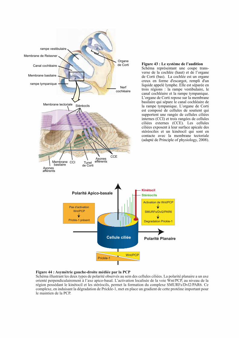
Organe
de CortiCanal cochléaire
rampe tympanique
rampe vestibulaire
Nerf
cochléaire
Membrane tectoriale Stéréocils
Axones afférents
Membranebasilaire
CCI Tunel de Corti
Axones efférents
CCE
Figure 43 : Le système de l’auditionSchéma représentant une coupe trans-verse de la cochlée (haut) et de l’organe de Corti (bas). La cochlée est un organe creux en forme d'escargot, rempli d'un liquide appelé lymphe. Elle est séparée en trois régions : la rampe vestibulaire, le canal cochléaire et la rampe tympanique. L’organe de Corti repose sur la membrane basilaire qui sépare le canal cochléaire de la rampe tympanique. L’organe de Corti est composé de cellules de soutient qui supportent une rangée de cellules ciliées internes (CCI) et trois rangées de cellules ciliées externes (CCE). Les cellules ciliées exposent à leur surface apicale des stéréociles et un kinétocil qui sont en contacte avec la membrane tectoriale (adapté de Principle of physiology, 2008).
Membrane de Reissner
Membrane basilaire
Activation de Wnt/PCP
SMURFs/Dvl2/PAR6
Degradation Prickle-1
Pas d’activation
Wnt/PCP
Prickle-1 présent
Wnt/PCPPrickle-1
Polarité Apico-basale
Polarité PlanaireCellule ciliée
Kinétocil
Stéréocils
Figure 44 : Asymétrie gauche-droite médiée par la PCPSchéma illustrant les deux types de polarité observés au sein des cellules ciliées. La polarité planaire a un axe orienté perpendiculairement à l’axe apico-basal. L’activation localisée de la voie Wnt/PCP, au niveau de la région possédant le kinétocil et les stéréocils, permet la formation du complexe SMURFs/Dvl2/PAR6. Ce complexe, en induisant la dégradation de Prickle-1, met en place un gradient de cette protéine important pour le maintien de la PCP.
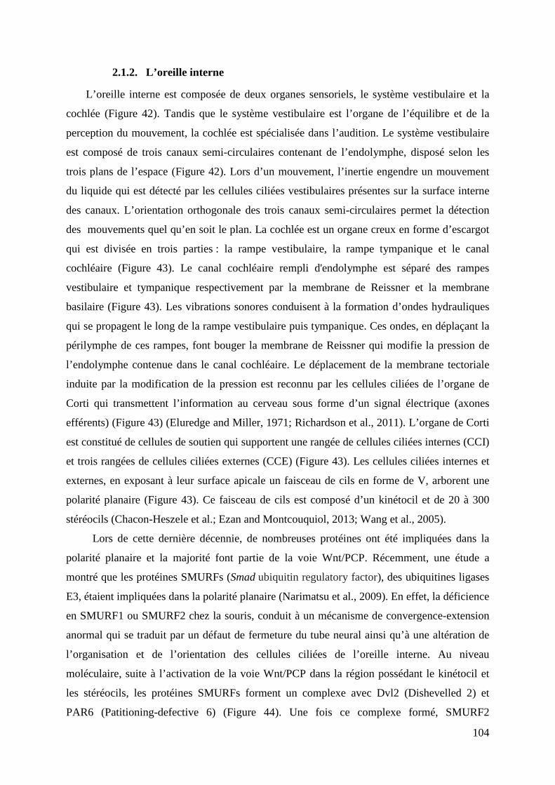
104
2.1.2. L’oreille interne
L’oreille interne est composée de deux organes sensoriels, le système vestibulaire et la
cochlée (Figure 42). Tandis que le système vestibulaire est l’organe de l’équilibre et de la
perception du mouvement, la cochlée est spécialisée dans l’audition. Le système vestibulaire
est composé de trois canaux semi-circulaires contenant de l’endolymphe, disposé selon les
trois plans de l’espace (Figure 42). Lors d’un mouvement, l’inertie engendre un mouvement
du liquide qui est détecté par les cellules ciliées vestibulaires présentes sur la surface interne
des canaux. L’orientation orthogonale des trois canaux semi-circulaires permet la détection
des mouvements quel qu’en soit le plan. La cochlée est un organe creux en forme d’escargot
qui est divisée en trois parties : la rampe vestibulaire, la rampe tympanique et le canal
cochléaire (Figure 43). Le canal cochléaire rempli d'endolymphe est séparé des rampes
vestibulaire et tympanique respectivement par la membrane de Reissner et la membrane
basilaire (Figure 43). Les vibrations sonores conduisent à la formation d’ondes hydrauliques
qui se propagent le long de la rampe vestibulaire puis tympanique. Ces ondes, en déplaçant la
périlymphe de ces rampes, font bouger la membrane de Reissner qui modifie la pression de
l’endolymphe contenue dans le canal cochléaire. Le déplacement de la membrane tectoriale
induite par la modification de la pression est reconnu par les cellules ciliées de l’organe de
Corti qui transmettent l’information au cerveau sous forme d’un signal électrique (axones
efférents) (Figure 43) (Eluredge and Miller, 1971; Richardson et al., 2011). L’organe de Corti
est constitué de cellules de soutien qui supportent une rangée de cellules ciliées internes (CCI)
et trois rangées de cellules ciliées externes (CCE) (Figure 43). Les cellules ciliées internes et
externes, en exposant à leur surface apicale un faisceau de cils en forme de V, arborent une
polarité planaire (Figure 43). Ce faisceau de cils est composé d’un kinétocil et de 20 à 300
stéréocils (Chacon-Heszele et al.; Ezan and Montcouquiol, 2013; Wang et al., 2005).
Lors de cette dernière décennie, de nombreuses protéines ont été impliquées dans la
polarité planaire et la majorité font partie de la voie Wnt/PCP. Récemment, une étude a
montré que les protéines SMURFs (Smad ubiquitin regulatory factor), des ubiquitines ligases
E3, étaient impliquées dans la polarité planaire (Narimatsu et al., 2009). En effet, la déficience
en SMURF1 ou SMURF2 chez la souris, conduit à un mécanisme de convergence-extension
anormal qui se traduit par un défaut de fermeture du tube neural ainsi qu’à une altération de
l’organisation et de l’orientation des cellules ciliées de l’oreille interne. Au niveau
moléculaire, suite à l’activation de la voie Wnt/PCP dans la région possédant le kinétocil et
les stéréocils, les protéines SMURFs forment un complexe avec Dvl2 (Dishevelled 2) et
PAR6 (Patitioning-defective 6) (Figure 44). Une fois ce complexe formé, SMURF2
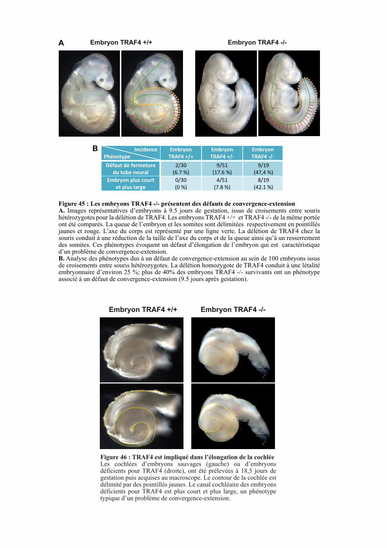
Figure 45 : Les embryons TRAF4 -/- présentent des défauts de convergence-extensionA. Images représentatives d’embryons à 9.5 jours de gestation, issus de croisements entre souris hétérozygotes pour la délétion de TRAF4. Les embryons TRAF4 +/+ et TRAF4 -/- de la même portée ont été comparés. La queue de l’embryon et les somites sont délimitées respectivement en pointillés jaunes et rouge. L’axe du corps est représenté par une ligne verte. La délétion de TRAF4 chez la souris conduit à une réduction de la taille de l’axe du corps et de la queue ainsi qu’à un resserrement des somites. Ces phénotypes évoquent un défaut d’élongation de l’embryon qui est caractéristique d’un problème de convergence-extension. B. Analyse des phénotypes dus à un défaut de convergence-extension au sein de 100 embryons issus de croisements entre souris hétérozygotes. La délétion homozygote de TRAF4 conduit à une létalité embryonnaire d’environ 25 %; plus de 40% des embryons TRAF4 -/- survivants ont un phénotype associé à un défaut de convergence-extension (9.5 jours après gestation).
Embryon TRAF4 +/+ Embryon TRAF4 -/-A
B Incidence
Phénotype
Embryon
TRAF4 +/+
Embryon
TRAF4 +/-
Embryon
TRAF4 -/-
Défaut de fermeture
du tube neural
2/30
(6.7 %)
9/51
(17.6 %)
9/19
(47,4 %)
Embryon plus court
et plus large
0/30
(0 %)
4/51
(7.8 %)
8/19
(42.1 %)
Embryon TRAF4 +/+ Embryon TRAF4 -/-
Figure 46 : TRAF4 est impliqué dans l’élongation de la cochléeLes cochlées d’embryons sauvages (gauche) ou d’embryons déficients pour TRAF4 (droite), ont été prélevées à 18,5 jours de gestation puis acquises au macroscope. Le contour de la cochlée est délimité par des pointillés jaunes. Le canal cochléaire des embryons déficients pour TRAF4 est plus court et plus large, un phénotype typique d’un problème de convergence-extension.
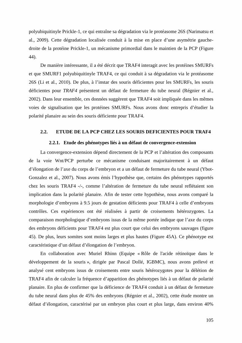
105
polyubiquitinyle Prickle-1, ce qui entraîne sa dégradation via le protéasome 26S (Narimatsu et
al., 2009). Cette dégradation localisée conduit à la mise en place d’une asymétrie gauche-
droite de la protéine Prickle-1, un mécanisme primordial dans le maintien de la PCP (Figure
44).
De manière intéressante, il a été décrit que TRAF4 interagit avec les protéines SMURFs
et que SMURF1 polyubiquitinyle TRAF4, ce qui conduit à sa dégradation via le protéasome
26S (Li et al., 2010). De plus, à l’instar des souris déficientes pour les SMURFs, les souris
déficientes pour TRAF4 présentent un défaut de fermeture du tube neural (Régnier et al.,
2002). Dans leur ensemble, ces données suggèrent que TRAF4 soit impliquée dans les mêmes
voies de signalisation que les protéines SMURFs. Nous avons donc entrepris d’étudier la
polarité planaire au sein des souris déficiente pour TRAF4.
2.2. ETUDE DE LA PCP CHEZ LES SOURIS DEFICIENTES POUR TRAF4
2.2.1. Etude des phénotypes liés à un défaut de convergence-extension
La convergence-extension dépend directement de la PCP et l’altération des composants
de la voie Wnt/PCP perturbe ce mécanisme conduisant majoritairement à un défaut
d’élongation de l’axe du corps de l’embryon et a un défaut de fermeture du tube neural (Ybot-
Gonzalez et al., 2007). Nous avons émis l’hypothèse que, certains des phénotypes rapportés
chez les souris TRAF4 -/-, comme l’altération de fermeture du tube neural reflétaient son
implication dans la polarité planaire. Afin de tester cette hypothèse, nous avons comparé la
morphologie d’embryons à 9.5 jours de gestation déficients pour TRAF4 à celle d’embryons
contrôles. Ces expériences ont été réalisées à partir de croisements hétérozygotes. La
comparaison morphologique d’embryons issus de la même portée indique que l’axe du corps
des embryons déficients pour TRAF4 est plus court que celui des embryons sauvages (figure
45). De plus, leurs somites sont moins larges et plus hautes (Figure 45A). Ce phénotype est
caractéristique d’un défaut d’élongation de l’embryon.
En collaboration avec Muriel Rhinn (Equipe « Rôle de l'acide rétinoïque dans le
développement de la souris », dirigée par Pascal Dollé, IGBMC), nous avons prélevé et
analysé cent embryons issus de croisements entre souris hétérozygotes pour la délétion de
TRAF4 afin de calculer la fréquence d’apparition des phénotypes liés à un défaut de polarité
planaire. En plus de confirmer que la déficience de TRAF4 conduit à un défaut de fermeture
du tube neural dans plus de 45% des embryons (Régnier et al., 2002), cette étude montre un
défaut d’élongation, caractérisé par un embryon plus court et plus large, dans environ 40%
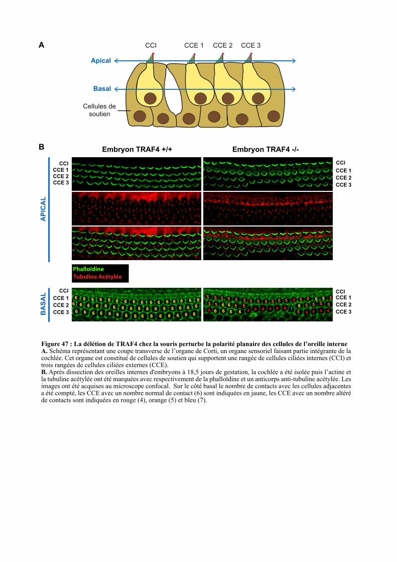
Apical
Basal
CCI CCE 1 CCE 2 CCE 3
Cellules de
soutien
CCE 1
CCE 2
CCE 3
CCI
CCE 1
CCE 2
CCE 3
CCI
Embryon TRAF4 +/+ Embryon TRAF4 -/-
Phalloïdine
Tubuline Acétylée
CCE 1
CCE 2
CCE 3
CCI
AP
ICA
LB
AS
AL
A
B
6 6 6 6 6 6 6 6 6 6 6 6 6 6 6 66 6 6 6 6 6 6 6 6 6 6 6 6 6 6
6 6 6 6 6 6 6 6 6 6 6 6 6 6 6
6
5
6
5 5 5
6
65
4
5 6 5
6 6
5
45
5 5
4 4 4 4 4 7
5 5 5 5 5 4
5
6
5
5
4 4 4 4
5 5 5
5 56 6 CCE 1
CCE 2
CCE 3
CCI
Figure 47 : La délétion de TRAF4 chez la souris perturbe la polarité planaire des cellules de l’oreille interne A. Schéma représentant une coupe transverse de l’organe de Corti, un organe sensoriel faisant partie intégrante de la cochlée. Cet organe est constitué de cellules de soutien qui supportent une rangée de cellules ciliées internes (CCI) et trois rangées de cellules ciliées externes (CCE).B. Après dissection des oreilles internes d'embryons à 18,5 jours de gestation, la cochlée a été isolée puis l’actine et la tubuline acétylée ont été marquées avec respectivement de la phalloïdine et un anticorps anti-tubuline acétylée. Les images ont été acquises au microscope confocal. Sur le côté basal le nombre de contacts avec les cellules adjacentes a été compté, les CCE avec un nombre normal de contact (6) sont indiquées en jaune, les CCE avec un nombre altéré de contacts sont indiquées en rouge (4), orange (5) et bleu (7).
106
des embryons TRAF4 -/- (Figure 45B). Ces résultats suggèrent l’implication de TRAF4 dans
la PCP puisque sa perte de fonction chez la souris est associée à un défaut du mécanisme de
convergence-extension.
Il a récemment été décrit que la convergence-extension est primordiale pour
l’élongation de la cochlée au cours du développement embryonnaire. Afin de savoir si la
délétion de TRAF4 perturbe l’élongation de la cochlée, les oreilles internes d’embryons ont
été prélevées à 18.5 jours de gestation et les cochlées ont été observées au macroscope. De
manière intéressante, 100% des cochlées des embryons déficients pour TRAF4 sont plus
courtes et plus larges que celles des embryons sauvages (Figure 46). Ce phénotype ressemble
à celui des souris déficientes pour Vangl2 et Dvl-2, deux acteurs majeurs de la PCP (Chacon-
Heszele et al.; Wang et al., 2006). Ce résultat illustre également le défaut du mécanisme de
convergence-extension au sein des embryons TRAF4 -/-.
Dans leur ensemble, ces données montrent que TRAF4 est impliqué dans le mécanisme
de convergence-extension et que la délétion de TRAF4 conduit à un défaut de fermeture du
tube neural ainsi qu’à une altération de l’élongation de l’axe de l’embryon et de la cochlée.
Ces résultats renforcent donc l’idée que TRAF4 soit impliqué dans la voie Wnt/PCP.
2.2.2. Etude de la PCP au sein de la cochlée
La cochlée est l’organe de référence pour étudier la PCP. En effet, dans cet organe, deux
phénotypes sont directement associés à un défaut de PCP : la présence d’un nombre anormal
de rangées de cellules ciliées externes et une mauvaise orientation du faisceau de cils des
cellules ciliées (Ezan and Montcouquiol, 2013; May-Simera and Kelley, 2012). Ce faisceau
de cils est composé d’un kinétocil et de 20 à 300 stéréocils. Le kinétocil peut être marqué
spécifiquement avec un anticorps anti-tubuline acétylée tandis que les stéréocils, riches en
actine, peuvent être marqués à la phalloïdine (Figure 47) (Chacon-Heszele et al.; Ezan and
Montcouquiol, 2013; Wang et al., 2005).
Afin de savoir si les souris déficientes pour TRAF4 ont un défaut de polarité planaire,
les oreilles internes de sept embryons sauvages et sept embryons déficients pour TRAF4 ont
été prélevées à 18,5 jours de gestation. Après avoir isolé la cochlée, la membrane basilaire et
l’organe de Corti ont été prélevés et fixés. La morphologie des cellules ciliées a été observée
grâce à un immunomarquage de la tubuline acétylée et un marquage à la phalloïdine
(Montcouquiol et al., 2003). Les pièces ont été montées entre lame et lamelle et observées au
microscope confocal. Les souris déficientes pour TRAF4 présentent une altération de la
morphologie de l’organe de Corti caractérisée par une fusion entre les rangées CCE 2 et CCE
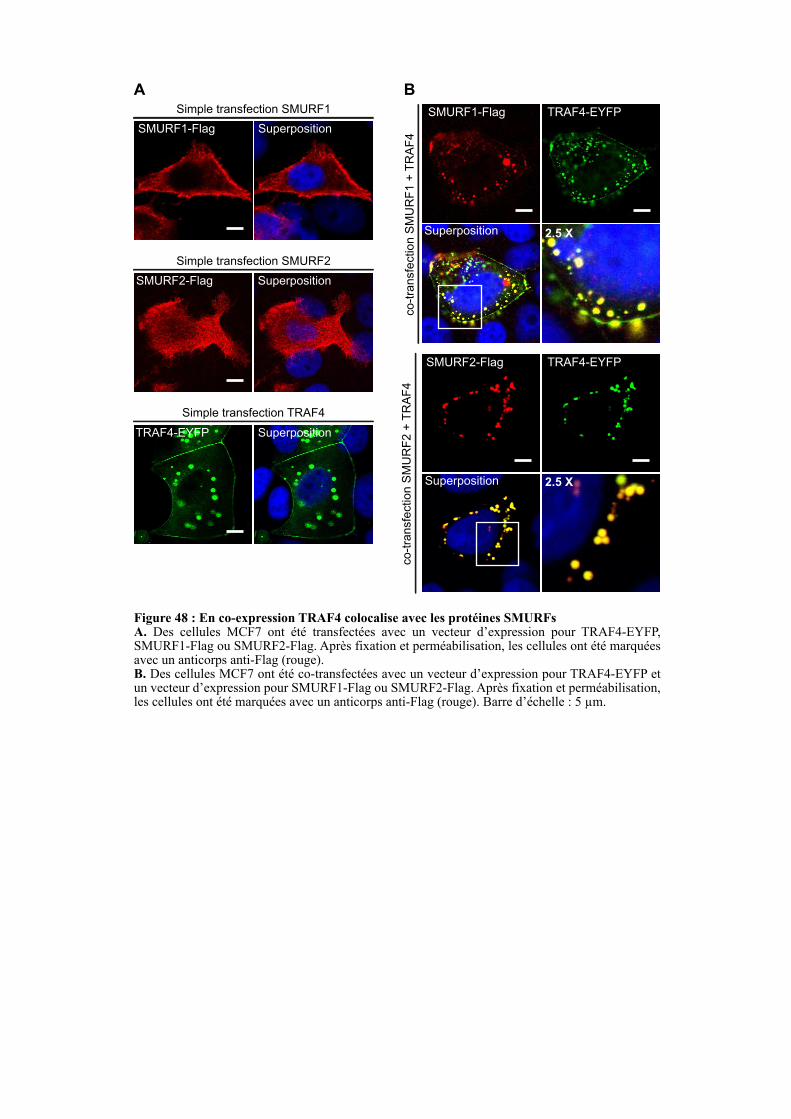
SMURF2-Flag
Superposition
TRAF4-EYFP
SMURF1-Flag
Superposition
TRAF4-EYFP
Figure 48 : En co-expression TRAF4 colocalise avec les protéines SMURFs A. Des cellules MCF7 ont été transfectées avec un vecteur d’expression pour TRAF4-EYFP, SMURF1-Flag ou SMURF2-Flag. Après fixation et perméabilisation, les cellules ont été marquées avec un anticorps anti-Flag (rouge). B. Des cellules MCF7 ont été co-transfectées avec un vecteur d’expression pour TRAF4-EYFP et un vecteur d’expression pour SMURF1-Flag ou SMURF2-Flag. Après fixation et perméabilisation, les cellules ont été marquées avec un anticorps anti-Flag (rouge). Barre d’échelle : 5 µm.
SMURF2-Flag Superposition
Simple transfection SMURF2
SMURF1-Flag Superposition
Simple transfection SMURF1
TRAF4-EYFP Superposition
Simple transfection TRAF4co
-tra
nsfe
ctio
n S
MU
RF
1 +
TR
AF
4co-t
ransfe
ction S
MU
RF
2 +
TR
AF
4
A B
2.5 X
2.5 X
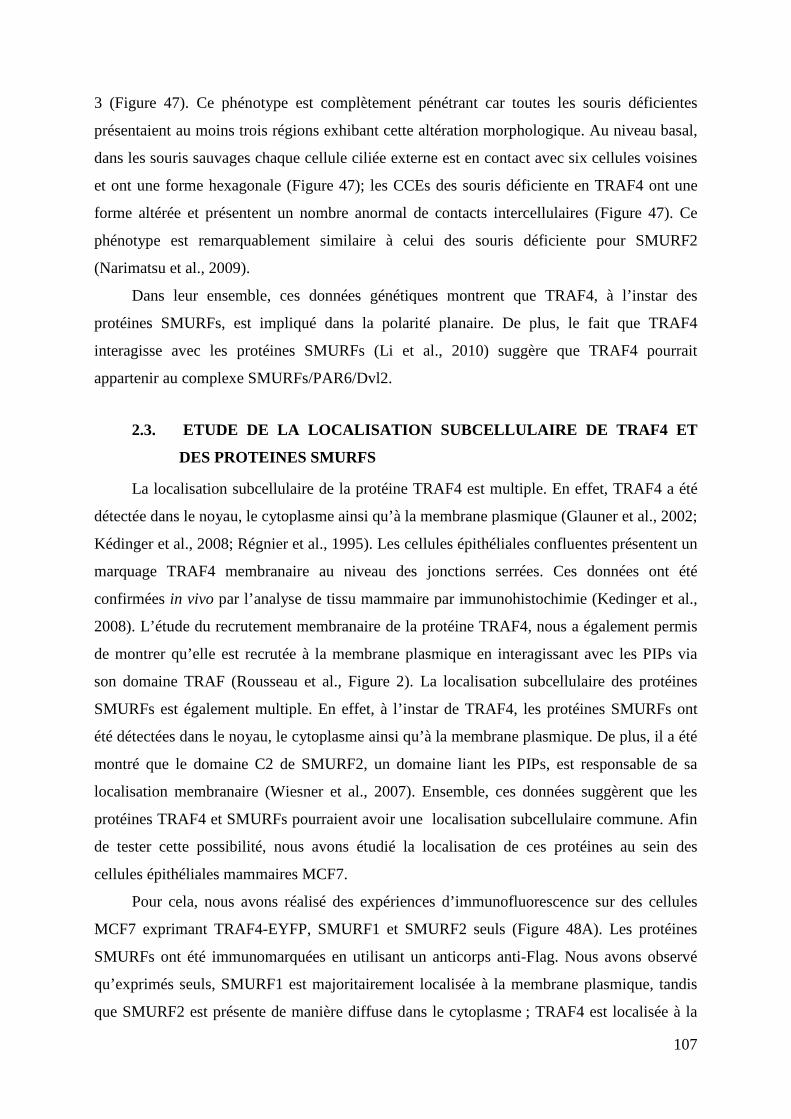
107
3 (Figure 47). Ce phénotype est complètement pénétrant car toutes les souris déficientes
présentaient au moins trois régions exhibant cette altération morphologique. Au niveau basal,
dans les souris sauvages chaque cellule ciliée externe est en contact avec six cellules voisines
et ont une forme hexagonale (Figure 47); les CCEs des souris déficiente en TRAF4 ont une
forme altérée et présentent un nombre anormal de contacts intercellulaires (Figure 47). Ce
phénotype est remarquablement similaire à celui des souris déficiente pour SMURF2
(Narimatsu et al., 2009).
Dans leur ensemble, ces données génétiques montrent que TRAF4, à l’instar des
protéines SMURFs, est impliqué dans la polarité planaire. De plus, le fait que TRAF4
interagisse avec les protéines SMURFs (Li et al., 2010) suggère que TRAF4 pourrait
appartenir au complexe SMURFs/PAR6/Dvl2.
2.3. ETUDE DE LA LOCALISATION SUBCELLULAIRE DE TRAF4 ET
DES PROTEINES SMURFS
La localisation subcellulaire de la protéine TRAF4 est multiple. En effet, TRAF4 a été
détectée dans le noyau, le cytoplasme ainsi qu’à la membrane plasmique (Glauner et al., 2002;
Kédinger et al., 2008; Régnier et al., 1995). Les cellules épithéliales confluentes présentent un
marquage TRAF4 membranaire au niveau des jonctions serrées. Ces données ont été
confirmées in vivo par l’analyse de tissu mammaire par immunohistochimie (Kedinger et al.,
2008). L’étude du recrutement membranaire de la protéine TRAF4, nous a également permis
de montrer qu’elle est recrutée à la membrane plasmique en interagissant avec les PIPs via
son domaine TRAF (Rousseau et al., Figure 2). La localisation subcellulaire des protéines
SMURFs est également multiple. En effet, à l’instar de TRAF4, les protéines SMURFs ont
été détectées dans le noyau, le cytoplasme ainsi qu’à la membrane plasmique. De plus, il a été
montré que le domaine C2 de SMURF2, un domaine liant les PIPs, est responsable de sa
localisation membranaire (Wiesner et al., 2007). Ensemble, ces données suggèrent que les
protéines TRAF4 et SMURFs pourraient avoir une localisation subcellulaire commune. Afin
de tester cette possibilité, nous avons étudié la localisation de ces protéines au sein des
cellules épithéliales mammaires MCF7.
Pour cela, nous avons réalisé des expériences d’immunofluorescence sur des cellules
MCF7 exprimant TRAF4-EYFP, SMURF1 et SMURF2 seuls (Figure 48A). Les protéines
SMURFs ont été immunomarquées en utilisant un anticorps anti-Flag. Nous avons observé
qu’exprimés seuls, SMURF1 est majoritairement localisée à la membrane plasmique, tandis
que SMURF2 est présente de manière diffuse dans le cytoplasme ; TRAF4 est localisée à la
108
membrane aux JSs et dans des structures ponctiformes cytoplasmiques (Figure 48). Ces
résultats montrent que la localisation subcellulaire de ces trois protéines n’est pas totalement
chevauchante. Nous avons co-transfecté un vecteur codant pour la protéine TRAF4-EYFP
avec un vecteur d’expression pour SMURF1-Flag et SMURF2-Flag (Figure 48B). La co-
expression avec TRAF4 modifie les localisations des protéines SMURFs. En effet, TRAF4
colocalise avec SMURF1 et SMURF2 dans des structures ponctiformes cytoplasmiques
(100% des cellules co-transfectées) (Figure 48B).
Dans leur ensemble, ces résultats montrent que la co-expression de TRAF4 avec les
protéines SMURFs modifie leur localisation subcellulaire. Les deux protéines sont
accumulées dans des vésicules cytoplasmiques positives pour TRAF4. Ces modifications
renforcent l’idée d’une interconnexion entre ces trois protéines et suggèrent que TRAF4 guide
ces protéines vers des structures intracytoplasmiques. En dépit de nombreux efforts, la nature
exacte de ces structures ponctiformes reste inconnue à ce jour. Elles ne colocalisent pas avec
des marqueurs des granules de stress, des P bodies, et des endosomes précoces et tardifs. Ces
vésicules sont partiellement positives pour la présence d’ubiquitine. Comme TRAF4 et les
protéines SMURFs sont des ubiquitine ligase E3, il est possible que la concentration de ces
trois protéines dans ces structures reflète leur activité d’ubiquitination. Prise dans leur
ensemble, ces données renforcent également l’idée que TRAF4 pourrait appartenir au
complexe SMURFs/PAR6/Dvl2. Afin de tester cette hypothèse, il est nécessaire d’étudier
l’interaction entre ces protéines en réalisant des expériences de co-immunoprécipitation.
2.4. ETUDE FONCTIONNELLE DU COMPLEXE TRAF4/SMURFS
Les protéines TRAF4 et SMURFs colocalisent dans des structures vésiculaires positives
pour l’ubiquitine. Ces données suggèrent que ces trois protéines pourraient former un
complexe protéique ayant pour fonction l’ubiquitinylation. Bien que la polyubiquitinylation
de TRAF4 par SMURF1 ait déjà été décrite (Kalkan et al., 2009; Li et al., 2010), l’impact de
TRAF4 sur SMURF1 ainsi que l’interconnexion entre TRAF4 et SMURF2 restent inconnus.
De ce fait, des expériences d’ubiquitinylation ont été réalisées au sein de cellules HEK293T.
Pour cela, un vecteur codant pour l’ubiquitine couplée à une étiquette 6His a été co-transfecté
avec différents vecteurs d’expression codant pour la forme sauvage ou mutée de TRAF4
(TRAF4-mutRING) fusionnées à l’épitope Flag en présence des protéines SMURFs taguées
par l’épitope myc. Le mutant TRAF4-mutRING correspond au remplacement des résidus
impliqués dans la chélation du zinc du domaine RING par des Alanines (C18A, C34A, H36A,
C39A et C42A). Une purification des protéines modifiées par l’Ubiquitine a été réalisée par
SMURF2-myc
SMURF1-myc
TRAF4-Flag
TRAF4-mutRING-Flag
--
-
-
-
-
+
-
-
+
-
-
-
-
+
-
+
-
-
-
+
-
-
-
+
++
-
1 2 3 4 5 6 7
+
-
-
+
-
+
-
+
8 9
Ubiquitine-6His ++ + + + + + + +
IP :
His
Lysat
WB : myc
WB : Flag
Po
ly-U
b-S
MU
RF
Po
ly-U
b-T
RA
F4
WB : myc
WB : Flag
WB : β-actine
Figure 49 : Polyubiquitinylation de TRAF4 en présence des protéines SMURFS et réciproquement.A. Test d’ubiquitinylation. Des cellules HEK293T ont été co-transfectées avec un vecteur d’expression pour l’Ubiquitine-6His et différentes associations des vecteurs codant pour TRAF4-Flag, TRAF4-mu-tRING-Flag, SMURF1-myc et SMURF2-myc. TRAF4-mutRING-Flag correspond à un mutant de TRAF4 où le domaine RING est non fonctionnel. Après lyse des cellules, les protéines ubiquitinylées (ubiquitine-6His) de l’extrait cellulaire sont précipitées avec des billes recouvertes de nickel. Le lysat et la fraction précipitée (IP : 6His) ont ensuite été analysés par Western-blot. B. Quantification du Western-blot présenté en (A) à l’aide du logiciel ImageJ.
Poly
ubiq
uitin
ation d
e
SM
UR
F1 n
orm
alis
ée s
ur
son n
iveau d
’expre
ssio
n
0
2
4
6
8
Vect
eur vi
de
TR
AF4
TR
AF4-m
utR
ING
0
2
1
6
8
3
4
5
7
Poly
ubiq
uitin
ation d
e
SM
UR
F2 n
orm
alis
ée s
ur
son n
iveau d
’expre
ssio
nVect
eur vi
de
TR
AF4
TR
AF4-m
utR
ING
Poly
ubiq
uitin
ation d
e T
RA
F4
sauvage o
u m
uta
nt norm
alis
ée
sur
leur
niv
eau d
’expre
ssio
n
0
2
4
6
8
Vect
eur vi
de
SM
UR
F1
SM
UR
F2
TRAF4
TRAF4-mutRING
A
B Fonction E3 ligase de TRAF4 Fonction E3 ligase des SMURFs
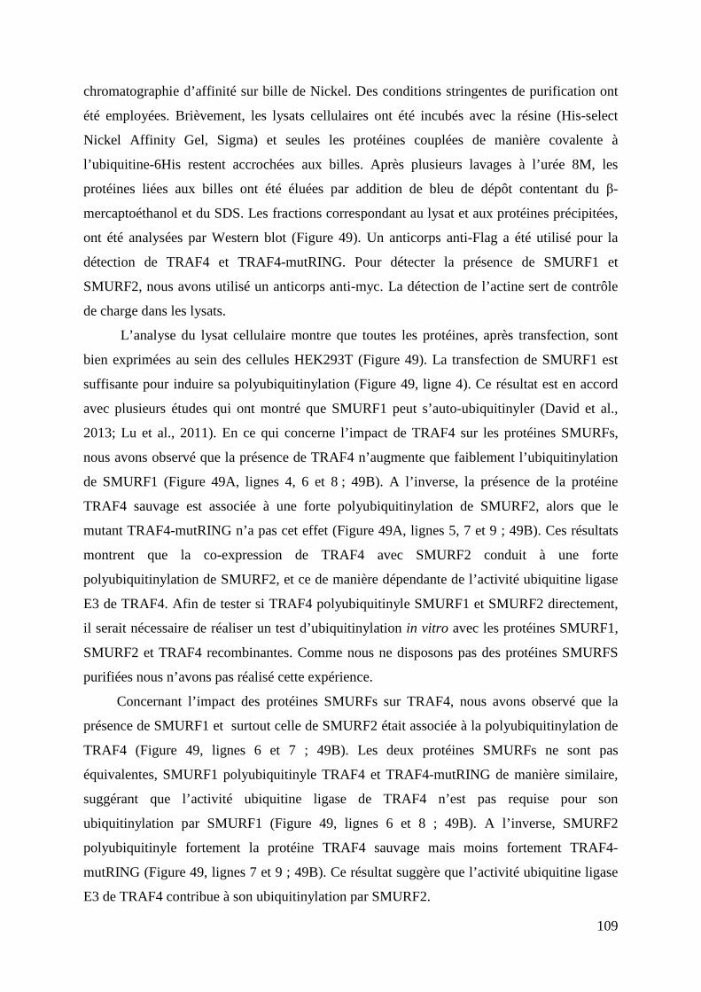
109
chromatographie d’affinité sur bille de Nickel. Des conditions stringentes de purification ont
été employées. Brièvement, les lysats cellulaires ont été incubés avec la résine (His-select
Nickel Affinity Gel, Sigma) et seules les protéines couplées de manière covalente à
l’ubiquitine-6His restent accrochées aux billes. Après plusieurs lavages à l’urée 8M, les
protéines liées aux billes ont été éluées par addition de bleu de dépôt contentant du β-
mercaptoéthanol et du SDS. Les fractions correspondant au lysat et aux protéines précipitées,
ont été analysées par Western blot (Figure 49). Un anticorps anti-Flag a été utilisé pour la
détection de TRAF4 et TRAF4-mutRING. Pour détecter la présence de SMURF1 et
SMURF2, nous avons utilisé un anticorps anti-myc. La détection de l’actine sert de contrôle
de charge dans les lysats.
L’analyse du lysat cellulaire montre que toutes les protéines, après transfection, sont
bien exprimées au sein des cellules HEK293T (Figure 49). La transfection de SMURF1 est
suffisante pour induire sa polyubiquitinylation (Figure 49, ligne 4). Ce résultat est en accord
avec plusieurs études qui ont montré que SMURF1 peut s’auto-ubiquitinyler (David et al.,
2013; Lu et al., 2011). En ce qui concerne l’impact de TRAF4 sur les protéines SMURFs,
nous avons observé que la présence de TRAF4 n’augmente que faiblement l’ubiquitinylation
de SMURF1 (Figure 49A, lignes 4, 6 et 8 ; 49B). A l’inverse, la présence de la protéine
TRAF4 sauvage est associée à une forte polyubiquitinylation de SMURF2, alors que le
mutant TRAF4-mutRING n’a pas cet effet (Figure 49A, lignes 5, 7 et 9 ; 49B). Ces résultats
montrent que la co-expression de TRAF4 avec SMURF2 conduit à une forte
polyubiquitinylation de SMURF2, et ce de manière dépendante de l’activité ubiquitine ligase
E3 de TRAF4. Afin de tester si TRAF4 polyubiquitinyle SMURF1 et SMURF2 directement,
il serait nécessaire de réaliser un test d’ubiquitinylation in vitro avec les protéines SMURF1,
SMURF2 et TRAF4 recombinantes. Comme nous ne disposons pas des protéines SMURFS
purifiées nous n’avons pas réalisé cette expérience.
Concernant l’impact des protéines SMURFs sur TRAF4, nous avons observé que la
présence de SMURF1 et surtout celle de SMURF2 était associée à la polyubiquitinylation de
TRAF4 (Figure 49, lignes 6 et 7 ; 49B). Les deux protéines SMURFs ne sont pas
équivalentes, SMURF1 polyubiquitinyle TRAF4 et TRAF4-mutRING de manière similaire,
suggérant que l’activité ubiquitine ligase de TRAF4 n’est pas requise pour son
ubiquitinylation par SMURF1 (Figure 49, lignes 6 et 8 ; 49B). A l’inverse, SMURF2
polyubiquitinyle fortement la protéine TRAF4 sauvage mais moins fortement TRAF4-
mutRING (Figure 49, lignes 7 et 9 ; 49B). Ce résultat suggère que l’activité ubiquitine ligase
E3 de TRAF4 contribue à son ubiquitinylation par SMURF2.
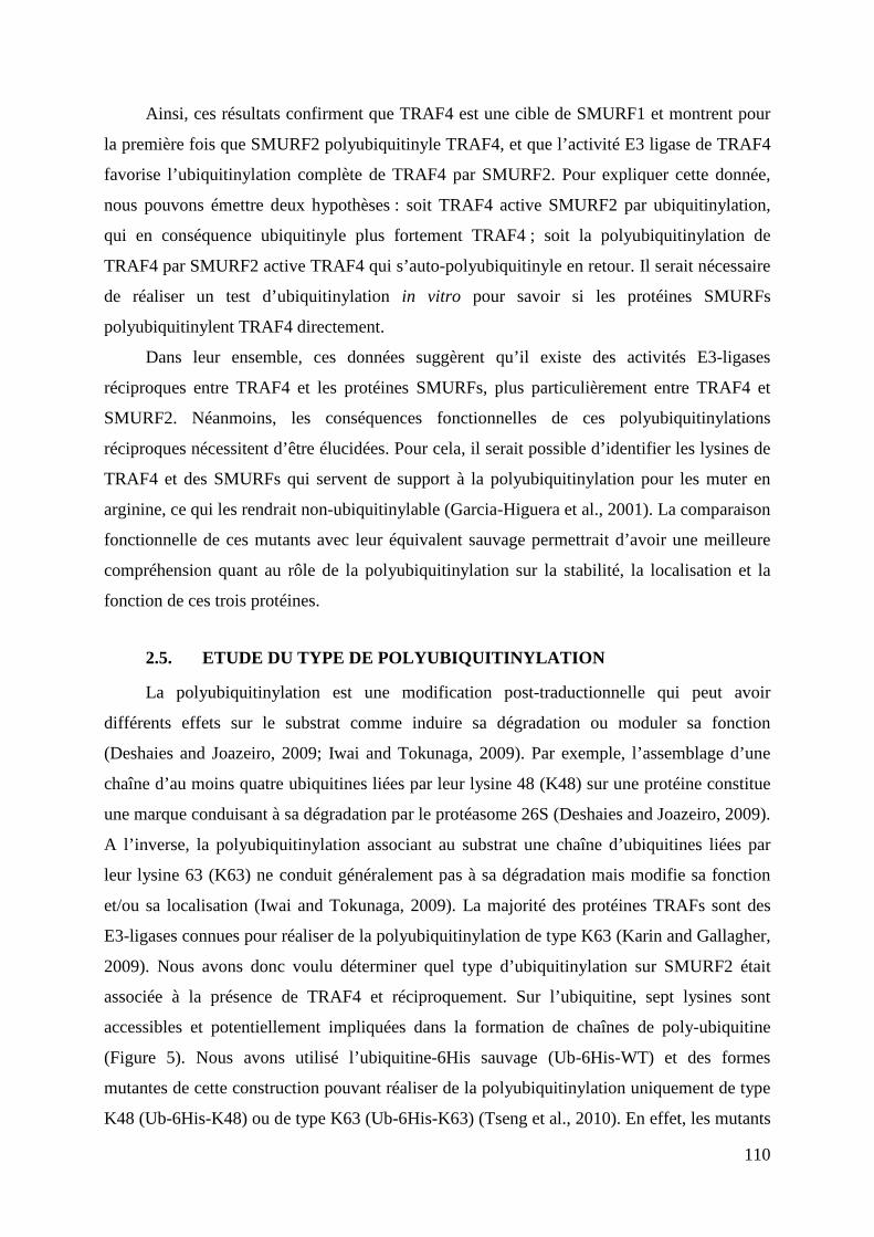
110
Ainsi, ces résultats confirment que TRAF4 est une cible de SMURF1 et montrent pour
la première fois que SMURF2 polyubiquitinyle TRAF4, et que l’activité E3 ligase de TRAF4
favorise l’ubiquitinylation complète de TRAF4 par SMURF2. Pour expliquer cette donnée,
nous pouvons émettre deux hypothèses : soit TRAF4 active SMURF2 par ubiquitinylation,
qui en conséquence ubiquitinyle plus fortement TRAF4 ; soit la polyubiquitinylation de
TRAF4 par SMURF2 active TRAF4 qui s’auto-polyubiquitinyle en retour. Il serait nécessaire
de réaliser un test d’ubiquitinylation in vitro pour savoir si les protéines SMURFs
polyubiquitinylent TRAF4 directement.
Dans leur ensemble, ces données suggèrent qu’il existe des activités E3-ligases
réciproques entre TRAF4 et les protéines SMURFs, plus particulièrement entre TRAF4 et
SMURF2. Néanmoins, les conséquences fonctionnelles de ces polyubiquitinylations
réciproques nécessitent d’être élucidées. Pour cela, il serait possible d’identifier les lysines de
TRAF4 et des SMURFs qui servent de support à la polyubiquitinylation pour les muter en
arginine, ce qui les rendrait non-ubiquitinylable (Garcia-Higuera et al., 2001). La comparaison
fonctionnelle de ces mutants avec leur équivalent sauvage permettrait d’avoir une meilleure
compréhension quant au rôle de la polyubiquitinylation sur la stabilité, la localisation et la
fonction de ces trois protéines.
2.5. ETUDE DU TYPE DE POLYUBIQUITINYLATION
La polyubiquitinylation est une modification post-traductionnelle qui peut avoir
différents effets sur le substrat comme induire sa dégradation ou moduler sa fonction
(Deshaies and Joazeiro, 2009; Iwai and Tokunaga, 2009). Par exemple, l’assemblage d’une
chaîne d’au moins quatre ubiquitines liées par leur lysine 48 (K48) sur une protéine constitue
une marque conduisant à sa dégradation par le protéasome 26S (Deshaies and Joazeiro, 2009).
A l’inverse, la polyubiquitinylation associant au substrat une chaîne d’ubiquitines liées par
leur lysine 63 (K63) ne conduit généralement pas à sa dégradation mais modifie sa fonction
et/ou sa localisation (Iwai and Tokunaga, 2009). La majorité des protéines TRAFs sont des
E3-ligases connues pour réaliser de la polyubiquitinylation de type K63 (Karin and Gallagher,
2009). Nous avons donc voulu déterminer quel type d’ubiquitinylation sur SMURF2 était
associée à la présence de TRAF4 et réciproquement. Sur l’ubiquitine, sept lysines sont
accessibles et potentiellement impliquées dans la formation de chaînes de poly-ubiquitine
(Figure 5). Nous avons utilisé l’ubiquitine-6His sauvage (Ub-6His-WT) et des formes
mutantes de cette construction pouvant réaliser de la polyubiquitinylation uniquement de type
K48 (Ub-6His-K48) ou de type K63 (Ub-6His-K63) (Tseng et al., 2010). En effet, les mutants
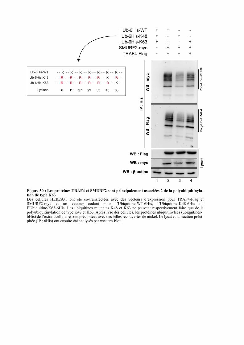
SMURF2-myc
Ub-6His-K48
TRAF4-Flag
-+
-
-
+
+ +
+
+
+
-
+
Ub-6His-WT ++ - -
Ub-6His-K63 -+ - +
IP :
His
WB
: m
yc
WB
: F
lag
Poly
-Ub-S
MU
RF
Poly
-Ub-T
RA
F4
Lysat
WB : myc
WB : Flag
WB : β-actine
1 2 3 4
Figure 50 : Les protéines TRAF4 et SMURF2 sont principalement associées à de la polyubiquitinyla-tion de type K63 Des cellules HEK293T ont été co-transfectées avec des vecteurs d’expression pour TRAF4-Flag et SMURF2-myc et un vecteur codant pour l’Ubiquitine-WT-6His, l’Ubiquitine-K48-6His ou l’Ubiquitine-K63-6His. Les ubiquitines mutantes K48 et K63 ne peuvent respectivement faire que de la polyubiquitinylation de type K48 et K63. Après lyse des cellules, les protéines ubiquitinylées (ubiquitines-6His) de l’extrait cellulaire sont précipitées avec des billes recouvertes de nickel. Le lysat et la fraction préci-pitée (IP : 6His) ont ensuite été analysés par western-blot.
- - K - - K - - K - - K - - K - - K - - K - -
6 11 27 29 33 48 63
- - R - - R - - R - - R - - R - - K - - R - -
- - R - - R - - R - - R - - R - - R - - K - -
Ub-6His-K48
Ub-6His-WT
Ub-6His-K63
Lysines
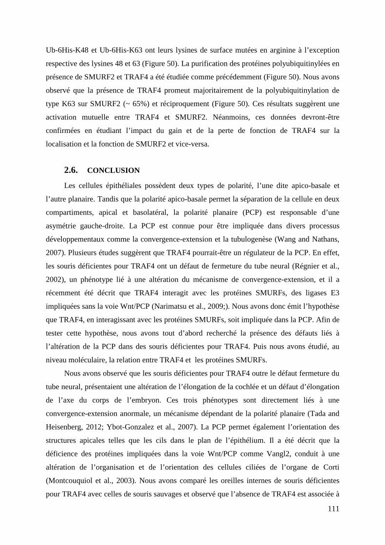
111
Ub-6His-K48 et Ub-6His-K63 ont leurs lysines de surface mutées en arginine à l’exception
respective des lysines 48 et 63 (Figure 50). La purification des protéines polyubiquitinylées en
présence de SMURF2 et TRAF4 a été étudiée comme précédemment (Figure 50). Nous avons
observé que la présence de TRAF4 promeut majoritairement de la polyubiquitinylation de
type K63 sur SMURF2 (~ 65%) et réciproquement (Figure 50). Ces résultats suggèrent une
activation mutuelle entre TRAF4 et SMURF2. Néanmoins, ces données devront-être
confirmées en étudiant l’impact du gain et de la perte de fonction de TRAF4 sur la
localisation et la fonction de SMURF2 et vice-versa.
2.6. CONCLUSION
Les cellules épithéliales possèdent deux types de polarité, l’une dite apico-basale et
l’autre planaire. Tandis que la polarité apico-basale permet la séparation de la cellule en deux
compartiments, apical et basolatéral, la polarité planaire (PCP) est responsable d’une
asymétrie gauche-droite. La PCP est connue pour être impliquée dans divers processus
développementaux comme la convergence-extension et la tubulogenèse (Wang and Nathans,
2007). Plusieurs études suggèrent que TRAF4 pourrait-être un régulateur de la PCP. En effet,
les souris déficientes pour TRAF4 ont un défaut de fermeture du tube neural (Régnier et al.,
2002), un phénotype lié à une altération du mécanisme de convergence-extension, et il a
récemment été décrit que TRAF4 interagit avec les protéines SMURFs, des ligases E3
impliquées sans la voie Wnt/PCP (Narimatsu et al., 2009;). Nous avons donc émit l’hypothèse
que TRAF4, en interagissant avec les protéines SMURFs, soit impliquée dans la PCP. Afin de
tester cette hypothèse, nous avons tout d’abord recherché la présence des défauts liés à
l’altération de la PCP dans des souris déficientes pour TRAF4. Puis nous avons étudié, au
niveau moléculaire, la relation entre TRAF4 et les protéines SMURFs.
Nous avons observé que les souris déficientes pour TRAF4 outre le défaut fermeture du
tube neural, présentaient une altération de l’élongation de la cochlée et un défaut d’élongation
de l’axe du corps de l’embryon. Ces trois phénotypes sont directement liés à une
convergence-extension anormale, un mécanisme dépendant de la polarité planaire (Tada and
Heisenberg, 2012; Ybot-Gonzalez et al., 2007). La PCP permet également l’orientation des
structures apicales telles que les cils dans le plan de l’épithélium. Il a été décrit que la
déficience des protéines impliquées dans la voie Wnt/PCP comme Vangl2, conduit à une
altération de l’organisation et de l’orientation des cellules ciliées de l’organe de Corti
(Montcouquiol et al., 2003). Nous avons comparé les oreilles internes de souris déficientes
pour TRAF4 avec celles de souris sauvages et observé que l’absence de TRAF4 est associée à

TRAF4
SMURF2
Ub
Ub
Ub
Ub
Ub
Ub
Ub
Ub
K63
K63
TRAF4
SMURF2
SMURF1
Ub
Ub
Ub
Ub
UbUb
UbUbK48
TRAF4
UbUb
UbUbK48
Dégradation de TRAF4
par le protéasome 26S
Figure 51 : Modèle de la relation entre TRAF4 et les protéines SMURFs A. Basé sur mes résultats, l’interaction de TRAF4 avec SMURF2 conduit à leur polyubiquitinylation mutuelle. Des chaînes de polyubiquitine K63 étant liées, cette modification post-traductionnelle pourrait être requise pour l’activation des protéines TRAF4 et SMURF2.B. Il a été décrit dans la littérature que SMURF1 est un régulateur négatif de TRAF4 en marquant TRAF4 avec des chaînes de polyubiquitine de type K48, un signal conduisant TRAF4 à la dégradation via le protéasome 26S.
MIGRATION
INVASION
PCP
Absence de SMURF1 Présence de SMURF1
A B
112
une altération de l’organisation des cellules ciliées externes, un phénotype analogue à celui
des souris SMURF2 -/-.
Prises ensemble, ces données in vivo montrent que la déficience de TRAF4 chez la
souris conduit à une altération de la polarité planaire et renforcent notre hypothèse selon
laquelle TRAF4 serait impliqué dans la voie Wnt/PCP.
L’étude de la relation entre TRAF4 et les protéines SMURFs, nous a permis de montrer
que TRAF4 recrute SMURF1 et SMURF2 dans des structures cytoplasmiques partiellement
positives pour l’ubiquitine. Ce résultat suggère que TRAF4 forme un complexe avec
SMURF1 et/ou SMURF2 qui serait impliqué dans des processus d’ubiquitination (Li et al.,
2010; Wang et al., 2013). Des expériences d’ubiquitinylation réalisées au sein des cellules
HEK293T suggèrent que TRAF4 polyubiquitinyle majoritairement SMURF2, et ce de
manière dépendante de son domaine RING. En retour, les protéines SMURFs peuvent
également polyubiquitinyler TRAF4. Tandis que SMURF1 est décrit comme un régulateur
négatif de TRAF4, nous avons observé que SMURF2, en liant majoritairement des chaines de
poly-ubiquitines de type K63 sur TRAF4, pourrait avoir un rôle activateur. En retour, TRAF4
réalise également de la polyubiquitinylation de type K63 sur SMURF2, et ce de manière
dépendante de son domaine RING (Figure 51). De manière intéressante, d’autres études
renforcent cette hypothèse. En effet, il a été décrit que SMURF2, à l’instar de TRAF4, est
impliquée dans la migration et l’invasion des cellules épithéliales mammaires (CEM)
cancéreuses (Jin et al., 2009). De plus, l’inhibition de SMURF1, un répresseur de TRAF4,
augmente la migration et l’invasion des CEM cancéreuses dans des modèles de migration 3D
(Sahai et al., 2007). Ces données permettent d’émettre l’hypothèse selon laquelle TRAF4 et
SMURF2 pourraient s’activer mutuellement tandis que SMURF1 agirait comme un régulateur
négatif en induisant la dégradation de TRAF4 (Figure 51). Il serait intéressant de tester cette
hypothèse de régulation réciproque dans le cancer du sein par des études cliniques
rétrospectives et par des modèles précliniques chez la souris.

113
DISCUSSIONS ET
PERSPECTIVES
114
Le gène Tumor necrosis factor Receptor-Associated Factor 4 a initialement été isolé dans
le contexte du cancer, à partir d’une biopsie de ganglions lymphatiques métastasés dérivés
d’un cancer du sein. Depuis, il a été montré que la surexpression de TRAF4 est retrouvée dans
plus de 40% des carcinomes ce qui en fait une cible thérapeutique potentielle. Néanmoins,
une meilleure compréhension de son rôle physiopathologique est nécessaire pour valider son
intérêt thérapeutique. Afin de progresser dans ce sens, pendant ma thèse, j’ai étudié le rôle de
TRAF4 dans la formation des JSs et dans la PCP, deux processus altérés dans les carcinomes.
1. IMPLICATION DE TRAF4 DANS LA VOIE DU TGF- Β
Lors de ma thèse, j’ai eu l’opportunité de collaborer avec l’équipe de Peter Ten-Dijke
(Leiden University Medical Center, Hollande), cette équipe s’intéresse depuis de nombreuses
années au rôle de la voie du TGF-β dans les cancers. Un de ses axes de recherche concerne
l’identification de nouveaux acteurs de cette voie et ils ont identifié TRAF4 comme un
régulateur intracellulaire potentiel des récepteurs TGF-βs. Cette collaboration m’a permis
d’être co-auteur d’un article scientifique intitulé « TRAF4 promotes TGF-β receptor signaling
and drives breast cancer metastasis » (Zhang et al., 2013 : Annexe-2). Je vais énoncer ci-
dessous les résultats originaux qui sont rapportés dans ce travail. De plus, une analyse critique
de ces résultats associés à mes résultats sera développée.
1.1. Annexe-2 : TRAF4 promotes TGF-β receptor signaling and drives
breast cancer metastasis
Dans cet article, les auteurs montrent que TRAF4 contribue à l’induction de la voie de
signalisation médiée par le TGF-β. Après traitement au TGF-β, TRAF4 interagit avec les
récepteurs TβRI et TβRII, et empêche la polyubiquitinylation du récepteur TβRI. Cela se
traduit par une stabilité accrue des récepteurs aux TGF-β et une augmentation de la
signalisation par la voie canonique (médiée par les SMADs). Au niveau moléculaire, TRAF4
prévient la polyubiquitinylation du récepteur TβRI de deux façons : TRAF4 polyubiquitinyle
(K48) SMURF2, ce qui induit sa dégradation via le protéasome 26S et TRAF4 recrute la
déubiquitinase USP15 qui enlève les chaines d’ubiquitines des récepteurs TβRI. De plus, le
recrutement de TRAF4 au niveau des récepteurs conduit à sa polyubiquitinylation de type
K63 ce qui active la voie de signalisation TGF-β non-canonique dépendante de TAK1 (TGF-β
Activated Kinase 1). Au niveau fonctionnel, cet article établi que TRAF4 contribue à
l’induction de la transition épithélio-mésenchymateuse (TEM) médiée par le TGF-β. En
corolaire, la sous-expression de TRAF4 diminue fortement la formation de métastase dans des
115
modèles de xénogreffes chez la souris. Une étude clinique complète ce travail et associe la
surexpression de TRAF4 dans les cancers du sein au niveau de phosphorylation des protéines
SMAD2 et TAK1 et à un mauvais pronostique. Dans les paragraphes suivants, je vais discuter
la cohérence de ces résultats avec mes données personnelles et celles de la littérature.
1.2. TRAF4, activateur ou répresseur de SMURF2 ?
Nous avons montré que la co-expression de TRAF4 avec SMURF1 et SMURF2
délocalise ces protéines SMURFs dans des structures ponctiformes cytoplasmiques positives
pour TRAF4 (Figure 48). Cet effet suggère que TRAF4 et les protéines SMURFs
interagissent directement dans des complexes qui adoptent une nouvelle localisation
subcellulaire. Au niveau moléculaire, TRAF4 polyubiquitinyle majoritairement SMURF2, et
ce de manière dépendante de son domaine RING. En retour, les protéines SMURFs peuvent
également polyubiquitinyler TRAF4 (Figure 49). Ces résultats sont en accord avec ceux
obtenus par Zhang et ses collaborateurs qui montrent que les protéines SMURFs (surtout
SMURF2) ubiquitinylent TRAF4 et que TRAF4 polyubiquitinyle les protéines SMURFs
(surtout SMURF2) (Annexe-2 : Figure-2). Ces polyubiquitinylations réciproques peuvent être
directes car elles sont observées in vitro en utilisant des protéines SMURF1, SMURF2 et
TRAF4 recombinantes (Annexe-2 : Figure-2H). Cette expérience a permis de montrer que
TRAF4 polyubiquitinyle directement par son domaine RING, les protéines SMURFs et
surtout SMURF2.
Nous avons observé que TRAF4 réalise de la polyubiquitinylation de type K63 (~ 65%)
et de type K48 (~ 35%) sur SMURF2. Réciproquement, SMURF2 réalise également de la
polyubiquitinylation de type K63 (~ 72%) et de type K48 (~ 28%) sur TRAF4 (Figure 50).
L’assemblage d’une chaîne de polyubiquitine K48 sur une protéine est une marque conduisant
à sa dégradation par le protéasome 26S tandis que l’assemblage d’une chaîne de
polyubiquitine K63 ne conduit généralement pas à la dégradation du substrat mais modifie sa
fonction et/ou sa localisation subcellulaire. Ces résultats suggèrent que TRAF4 et SMURF2
pourraient s’activer mutuellement. De manière surprenante, Zhang et ses collaborateurs
montrent que TRAF4 greffe des chaînes de polyubiquitine K48 sur SMURF2 suite à un
traitement au TGF-β, ce qui conduit à sa dégradation par le protéasome 26S (Annexe-2 :
Figure-2K et -3). Néanmoins, dans tous les tests d’ubiquitinylation que j’ai réalisé, je n’ai
jamais observé que la co-expression de TRAF4 avec SMURF2 conduisait à une diminution du
taux de protéines SMURF2 dans le lysat. Nous avons utilisé la même lignée cellulaire
HEK293T pour les expériences d’ubiquitinylation ; la plus grande différence concerne le
116
temps de transfection. Il est concevable que la cinétique de polyubiquitinylation réciproque
entre SMURF2 et TRAF4 explique ces différences. Comme le décrivent Zhang et al., 18h
après transfection la polyubiquitinylation de SMURF2 par TRAF4 conduit à sa dégradation,
alors que plus tardivement (36h), comme je l’observe, l’accumulation de SMURF2 dans les
structures ponctiformes cytoplasmiques positives pour TRAF4, empêche sa dégradation. Cette
hypothèse devra être vérifiée en mesurant la stabilité de la protéine SMURF2 en présence de
TRAF4 lors d’une cinétique allant de 18 à 36h. De plus l’utilisation des mutants d’ubiquitine
qui ne font que de la polyubiquitinylation de type K48 (Ub-6His-K48) ou K63 (Ub-6His-
K63), permettra de connaitre le spectre de polyubiquitinylation au cours du temps.
Après traitement au TGF-β, TRAF4 est recruté au niveau des récepteurs TβRI et TβRII,
où il est polyubiquitinylé (K63) de manière dépendante de son domaine RING (Annexe-2 :
Figure-4). Ce résultat suggère que TRAF4, une fois recruté au niveau des récepteurs aux
TGF-βs, pourrait s’auto-ubiquitinyler (K63). Les chaînes de polyubiquitine seraient ensuite
reconnues par TAK1 qui induirait l’activation de la voie non-canonique du TGF-β. Pour notre
part, nous avons observé que SMURF2 polyubiquitinyle fortement la protéine TRAF4
sauvage mais moins fortement TRAF4-mutRING (Figure 49). Ce résultat suggère que
l’activité ubiquitine ligase E3 de TRAF4 contribue à son ubiquitinylation par SMURF2. Nous
avons émis deux hypothèses : soit TRAF4 active SMURF2 par polyubiquitinylation, qui en
conséquence polyubiquitinyle plus fortement TRAF4 ; soit la polyubiquitinylation de TRAF4
par SMURF2 active TRAF4 qui s’auto-polyubiquitinyle en retour. Nos résultats associés à
ceux de Zhang confortent la seconde hypothèse. TRAF4 serait plus un régulateur négatif de
SMURF2 qu’un activateur. Nous pourrions imaginer un modèle selon lequel TRAF4 est
polyubiquitinylé par SMURF2, ce qui induit son activation. En retour, TRAF4 pourrait
exercer un rétrocontrôle négatif en polyubiquitinylant SMURF2 et également s’auto-
ubiquitinyler pour activer la voie non-canonique du TGF- β. Cette hypothèse ne satisfait pas
toutes les observations rapportées dans la littérature. La régulation de SMURF2 par TRAF4
pourrait-être positive ou négative selon le contexte cellulaire. Par exemple, après traitement
au TGF-β, TRAF4 est un répresseur de SMURF2 (Zhang et al., 2013). D’autres données
suggèrent que TRAF4 agirait de concert avec SMURF2 dans certaines voies de
signalisation suggérant plutôt une activation mutuelle de ces deux protéines. Premièrement,
TRAF4 et SMURF2 peuvent conduire à la dégradation de p53 dans les cellules MCF7 (Nie et
al., 2010; Yi et al., 2013). Deuxièmement, la perte de TRAF4 et celle de SMURF2 chez la
souris engendrent un phénotype très similaire caractérisé par un défaut de fermeture du tube
neural, une altération du mécanisme de convergence-extension, une altération de la PCP et la
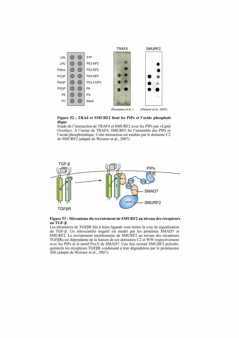
Figure 52 : TRA4 et SMURF2 lient les PIPs et l’acide phosphati-dique Etude de l’interaction de TRAF4 et SMURF2 avec les PIPs par «Lipid Overlay». A l’instar de TRAF4, SMURF2 lie l’ensemble des PIPs et l’acide phosphotidique. Cette interaction est médiée par le domaine C2 de SMURF2 (adapté de Weisner et al., 2007).
LPA
LPC
PtdIns
PI(3)P
PI(4)P
PI(5)P
PE
PC
S1P
PI(3,4)P2
PI(3,5)P2
PI(4,5)P2
PI(3,4,5)P3
PA
PS
Blank
TRAF4 SMURF2
PIPs
SMURF2
SMAD7
TGFβR
TGF-β
Figure 53 : Mécanisme du recrutement de SMURF2 au niveau des récepteurs au TGF-β Les tétramères de TGFβR liés à leurs ligands vont initier la voie de signalisation du TGF-β. Un rétrocontôle négatif est médié par les protéines SMAD7 et SMURF2. Le recrutement membranaire de SMURF2 au niveau des récepteurs TGFβRs est dépendante de la liaison de ses domaines C2 et WW respectivement avec les PIPs et le motif PxxY de SMAD7. Une fois recruté SMURF2 polyubi-quitinyle les récepteurs TGFβR conduisant à leur dégradation par le protéasome 26S (adapté de Weisner et al., 2007).
(Rousseau et al.,) (Weisner et al., 2007)
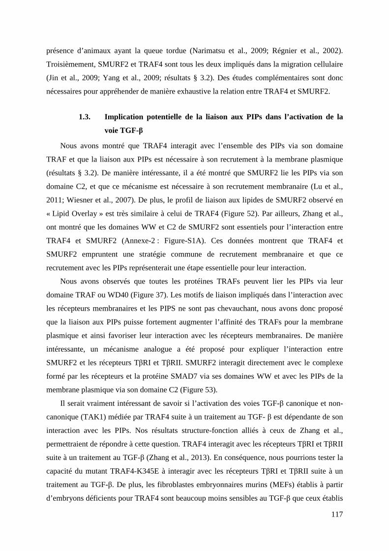
117
présence d’animaux ayant la queue tordue (Narimatsu et al., 2009; Régnier et al., 2002).
Troisièmement, SMURF2 et TRAF4 sont tous les deux impliqués dans la migration cellulaire
(Jin et al., 2009; Yang et al., 2009; résultats § 3.2). Des études complémentaires sont donc
nécessaires pour appréhender de manière exhaustive la relation entre TRAF4 et SMURF2.
1.3. Implication potentielle de la liaison aux PIPs dans l’activation de la
voie TGF-β
Nous avons montré que TRAF4 interagit avec l’ensemble des PIPs via son domaine
TRAF et que la liaison aux PIPs est nécessaire à son recrutement à la membrane plasmique
(résultats § 3.2). De manière intéressante, il a été montré que SMURF2 lie les PIPs via son
domaine C2, et que ce mécanisme est nécessaire à son recrutement membranaire (Lu et al.,
2011; Wiesner et al., 2007). De plus, le profil de liaison aux lipides de SMURF2 observé en
« Lipid Overlay » est très similaire à celui de TRAF4 (Figure 52). Par ailleurs, Zhang et al.,
ont montré que les domaines WW et C2 de SMURF2 sont essentiels pour l’interaction entre
TRAF4 et SMURF2 (Annexe-2 : Figure-S1A). Ces données montrent que TRAF4 et
SMURF2 empruntent une stratégie commune de recrutement membranaire et que ce
recrutement avec les PIPs représenterait une étape essentielle pour leur interaction.
Nous avons observés que toutes les protéines TRAFs peuvent lier les PIPs via leur
domaine TRAF ou WD40 (Figure 37). Les motifs de liaison impliqués dans l’interaction avec
les récepteurs membranaires et les PIPS ne sont pas chevauchant, nous avons donc proposé
que la liaison aux PIPs puisse fortement augmenter l’affinité des TRAFs pour la membrane
plasmique et ainsi favoriser leur interaction avec les récepteurs membranaires. De manière
intéressante, un mécanisme analogue a été proposé pour expliquer l’interaction entre
SMURF2 et les récepteurs TβRI et TβRII. SMURF2 interagit directement avec le complexe
formé par les récepteurs et la protéine SMAD7 via ses domaines WW et avec les PIPs de la
membrane plasmique via son domaine C2 (Figure 53).
Il serait vraiment intéressant de savoir si l’activation des voies TGF-β canonique et non-
canonique (TAK1) médiée par TRAF4 suite à un traitement au TGF- β est dépendante de son
interaction avec les PIPs. Nos résultats structure-fonction alliés à ceux de Zhang et al.,
permettraient de répondre à cette question. TRAF4 interagit avec les récepteurs TβRI et TβRII
suite à un traitement au TGF-β (Zhang et al., 2013). En conséquence, nous pourrions tester la
capacité du mutant TRAF4-K345E à interagir avec les récepteurs TβRI et TβRII suite à un
traitement au TGF-β. De plus, les fibroblastes embryonnaires murins (MEFs) établis à partir
d’embryons déficients pour TRAF4 sont beaucoup moins sensibles au TGF-β que ceux établis
118
à partir d’embryons sauvages (Annexe-2 : Figure-1). De ce fait, il serait intéressant de réaliser
une restauration de fonction au sein des MEFs TRAF4 -/-, en ré-exprimant de manière stable
la protéine TRAF4 sauvage ou mutante ne liant pas les PIPs (TRAF4-K345E), afin de voir si
on restaure la sensibilité au TGF-β.
1.4. Les rôles de TRAF4 dans les JSs et la migration sont-ils dépendants
des TGF-βs ?
1.4.1. Rôle de TRAF4 dans la transition épithélio-mésenchymateuse
Nous avons observé que TRAF4 est un régulateur négatif des JSs au sein des cellules
épithéliales mammaires, une fonction dépendante de sa liaison aux PIPs (résultats § 3.2).
TRAF4 apparait comme un régulateur de la plasticité des JSs, un processus clé impliqué dans
divers mécanismes notamment dans la transition épithélio-mésenchymateuse (TEM). La TEM
est un mécanisme dynamique au cours duquel les cellules perdent leurs caractéristiques
épithéliales pour développer des propriétés mésenchymateuses. Ce processus requiert la perte
des jonctions intercellulaires (surtout JSs et JAs), il est caractérisé majoritairement par une
diminution de l’E-cadhérine et par une augmentation de l’expression de marqueurs de type
mésenchymateux comme l’α-SMA (α-Smooth Muscle Actin), la vimentine et des
métalloprotéinases matricielles (Polette et al., 2007; Savagner, 2010). Le rôle de TRAF4 dans
la plasticité des complexes jonctionnels est en accord avec la littérature. Chez le Xénope, la
sous-expression de TRAF4 diminue l’expansion des cellules des crêtes neurales (CCNs)
tandis que sa surexpression l’augmente (Kalkan et al., 2009). Lors du développement
embryonnaire, la migration des CCNs est initiée par le mécanisme de TEM. La sous-
expression de TRAF4 stabilise les JSs, ce qui limite et/ou retarde la TEM des CCNs et ainsi
leur expansion. A l’inverse, la surexpression de TRAF4 augmente la plasticité des JSs et
facilite la TEM ce qui conduit à une expansion accrue des CCNs.
De manière intéressante, ce rôle de TRAF4 comme modulateur des complexes
jonctionnels fait écho au travail de Zhang et al., qui montre que la sous-expression de TRAF4
dans les cellules MCF10A freine l’EMT induite par un traitement au TGF-β (Annexe-2 :
Figure-5). En corolaire, la surexpression de TRAF4 promeut la TEM induite au TGF-β par un
mécanisme dépendant de son activité E3 ubiquitine ligase. En régulant la plasticité des JSs et
la sensibilité au TGF-β, TRAF4 peut être considéré comme un modulateur de l’EMT.
Actuellement nous ne savons pas si la régulation des JSs médiée par TRAF4 est
exclusivement dépendante de la voie TGF-β. En effet, les tests de formation de JSs réalisés
dans mon étude sur les cellules MCF10A et MCF7 ont été effectués en présence de 10% de
119
sérum de veau fœtal (résultats § 3.2). Ces sérums contiennent une grande quantité de facteurs
de croissance incluant du TGF-β. Afin de répondre à cette question, il est donc nécessaire de
répéter ces expériences en utilisant un milieu de culture déplété en facteurs de croissance ou
en présence d’anticorps bloquants l’ensemble des TGF-βs (Arteaga et al., 1993; Biswas et al.,
2007).
1.4.2. Le rôle de TRAF4 dans la migration
Nous avons montré que la sous-expression de TRAF4 diminue la migration des cellules
épithéliales mammaires (CEMs) ; la réexpression de TRAF4 sauvage restaure le défaut de
migration tandis que le mutant ne liant pas les PIPs (TRAF4-K345E) ne le fait pas.
L’expérience complémentaire visant à surexprimer TRAF4 a montré que l’augmentation du
niveau d’expression de TRAF4 promeut la migration des CEMs (§ 3.2). Nos expériences ne
permettent pas d’associer directement et complétement le rôle de TRAF4 sur les JSs à son
rôle sur la migration cellulaire. Dans nos conditions expérimentales, les deux lignées testées
ont une migration accrue en présence de TRAF4 et diminuée en son absence (résultats § 3.2).
Or, la migration des cellules MCF10A est observée avant qu’elles puissent former des JSs, et
la migration des cellules MCF7 est observée alors que ces cellules forment des JSs. De
manière cohérente avec ces données, Zhang et al., ont montré que les MEFs, dépourvues de
JSs et de JAs, établis à partir d’embryons déficients pour TRAF4 migrent significativement
moins que ceux établis à partir d’embryons sauvages (Annexe-2 : Figure-5D). Ce phénotype
est exacerbé suite à l’ajout de TGF-β. Par ailleurs, les cellules dendritiques provenant de
souris déficientes pour TRAF4 ont également un défaut de migration, ce qui confirme que
TRAF4 a un effet pro-migratoire qui n’est pas restreint aux cellules épithéliales (Cherfils-
Vicini et al., 2008).
Prises dans leur ensemble, ces données démontrent que TRAF4 promeut la migration
cellulaire et suggèrent qu’il aurait deux modes d’action : l’un lié à la déstabilisation des JSs et
l’autre dépendant de la voie TGF-β.
L’équipe de Peter Ten-Dijke a également montré que la sous-expression de TRAF4 au
sein des cellules mammaires cancéreuses MDA-MB-231 diminue la formation des métastases
dans des modèles de xénogreffes chez la souris (Annexe-2 : Figure-6). Enfin par une étude
clinique, ils montrent que le niveau d’expression de TRAF4 est corrélé au développement des
métastases dans les cancers du sein, ce qui en fait un marqueur de mauvais pronostique
(Annexe-2 : Figure-7). Cette donnée s’accordant à une autre étude publiée récemment (Yi et
al., 2013).
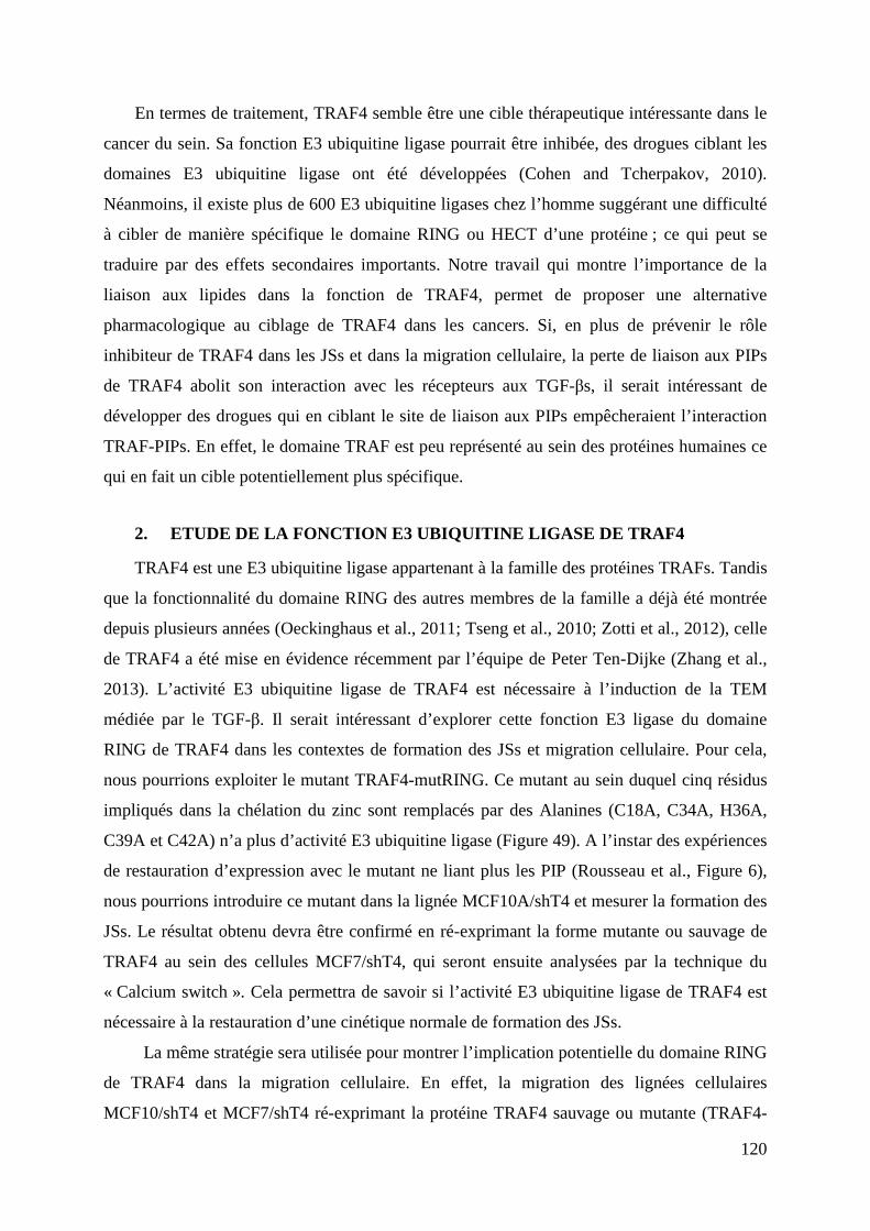
120
En termes de traitement, TRAF4 semble être une cible thérapeutique intéressante dans le
cancer du sein. Sa fonction E3 ubiquitine ligase pourrait être inhibée, des drogues ciblant les
domaines E3 ubiquitine ligase ont été développées (Cohen and Tcherpakov, 2010).
Néanmoins, il existe plus de 600 E3 ubiquitine ligases chez l’homme suggérant une difficulté
à cibler de manière spécifique le domaine RING ou HECT d’une protéine ; ce qui peut se
traduire par des effets secondaires importants. Notre travail qui montre l’importance de la
liaison aux lipides dans la fonction de TRAF4, permet de proposer une alternative
pharmacologique au ciblage de TRAF4 dans les cancers. Si, en plus de prévenir le rôle
inhibiteur de TRAF4 dans les JSs et dans la migration cellulaire, la perte de liaison aux PIPs
de TRAF4 abolit son interaction avec les récepteurs aux TGF-βs, il serait intéressant de
développer des drogues qui en ciblant le site de liaison aux PIPs empêcheraient l’interaction
TRAF-PIPs. En effet, le domaine TRAF est peu représenté au sein des protéines humaines ce
qui en fait un cible potentiellement plus spécifique.
2. ETUDE DE LA FONCTION E3 UBIQUITINE LIGASE DE TRAF4
TRAF4 est une E3 ubiquitine ligase appartenant à la famille des protéines TRAFs. Tandis
que la fonctionnalité du domaine RING des autres membres de la famille a déjà été montrée
depuis plusieurs années (Oeckinghaus et al., 2011; Tseng et al., 2010; Zotti et al., 2012), celle
de TRAF4 a été mise en évidence récemment par l’équipe de Peter Ten-Dijke (Zhang et al.,
2013). L’activité E3 ubiquitine ligase de TRAF4 est nécessaire à l’induction de la TEM
médiée par le TGF-β. Il serait intéressant d’explorer cette fonction E3 ligase du domaine
RING de TRAF4 dans les contextes de formation des JSs et migration cellulaire. Pour cela,
nous pourrions exploiter le mutant TRAF4-mutRING. Ce mutant au sein duquel cinq résidus
impliqués dans la chélation du zinc sont remplacés par des Alanines (C18A, C34A, H36A,
C39A et C42A) n’a plus d’activité E3 ubiquitine ligase (Figure 49). A l’instar des expériences
de restauration d’expression avec le mutant ne liant plus les PIP (Rousseau et al., Figure 6),
nous pourrions introduire ce mutant dans la lignée MCF10A/shT4 et mesurer la formation des
JSs. Le résultat obtenu devra être confirmé en ré-exprimant la forme mutante ou sauvage de
TRAF4 au sein des cellules MCF7/shT4, qui seront ensuite analysées par la technique du
« Calcium switch ». Cela permettra de savoir si l’activité E3 ubiquitine ligase de TRAF4 est
nécessaire à la restauration d’une cinétique normale de formation des JSs.
La même stratégie sera utilisée pour montrer l’implication potentielle du domaine RING
de TRAF4 dans la migration cellulaire. En effet, la migration des lignées cellulaires
MCF10/shT4 et MCF7/shT4 ré-exprimant la protéine TRAF4 sauvage ou mutante (TRAF4-
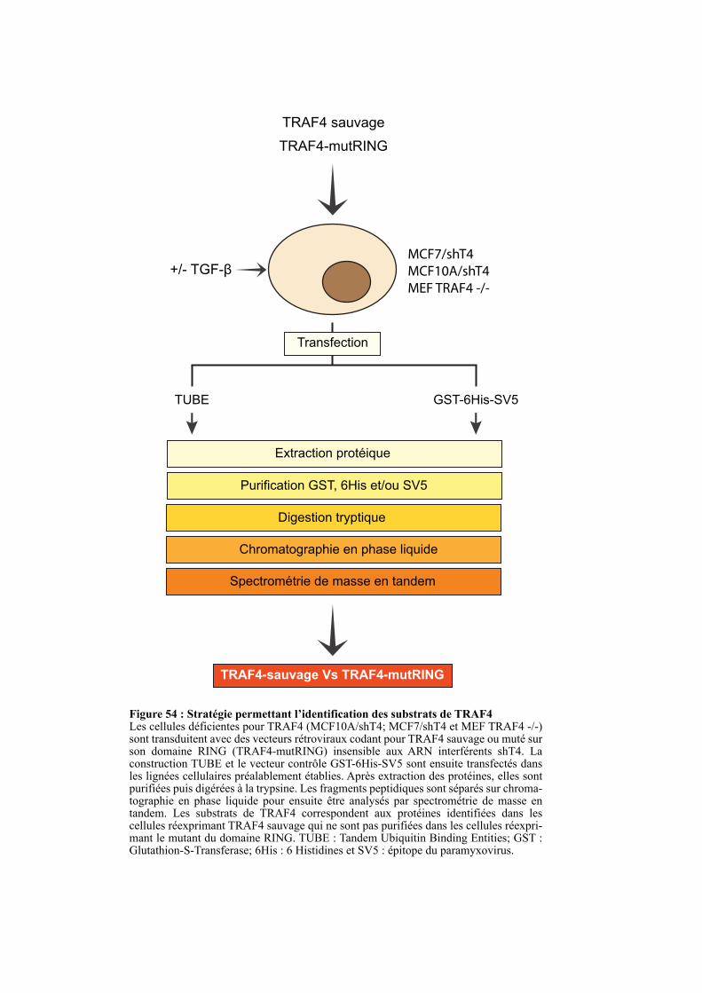
MCF7/shT4
MCF10A/shT4
MEF TRAF4 -/-
TRAF4 sauvage
TRAF4-mutRING
TUBE GST-6His-SV5
Transfection
Extraction protéique
Purification GST, 6His et/ou SV5
Digestion tryptique
Chromatographie en phase liquide
Spectrométrie de masse en tandem
TRAF4-sauvage Vs TRAF4-mutRING
Figure 54 : Stratégie permettant l’identification des substrats de TRAF4Les cellules déficientes pour TRAF4 (MCF10A/shT4; MCF7/shT4 et MEF TRAF4 -/-) sont transduitent avec des vecteurs rétroviraux codant pour TRAF4 sauvage ou muté sur son domaine RING (TRAF4-mutRING) insensible aux ARN interférents shT4. La construction TUBE et le vecteur contrôle GST-6His-SV5 sont ensuite transfectés dans les lignées cellulaires préalablement établies. Après extraction des protéines, elles sont purifiées puis digérées à la trypsine. Les fragments peptidiques sont séparés sur chroma-tographie en phase liquide pour ensuite être analysés par spectrométrie de masse en tandem. Les substrats de TRAF4 correspondent aux protéines identifiées dans les cellules réexprimant TRAF4 sauvage qui ne sont pas purifiées dans les cellules réexpri-mant le mutant du domaine RING. TUBE : Tandem Ubiquitin Binding Entities; GST : Glutathion-S-Transferase; 6His : 6 Histidines et SV5 : épitope du paramyxovirus.
+/- TGF-β
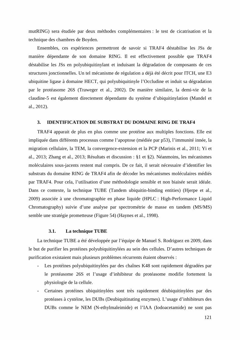
121
mutRING) sera étudiée par deux méthodes complémentaires : le test de cicatrisation et la
technique des chambres de Boyden.
Ensembles, ces expériences permettront de savoir si TRAF4 déstabilise les JSs de
manière dépendante de son domaine RING. Il est effectivement possible que TRAF4
déstabilise les JSs en polyubiquitinylant et induisant la dégradation de composants de ces
structures jonctionnelles. Un tel mécanisme de régulation a déjà été décrit pour ITCH, une E3
ubiquitine ligase à domaine HECT, qui polyubiquitinyle l’Occludine et induit sa dégradation
par le protéasome 26S (Traweger et al., 2002). De manière similaire, la demi-vie de la
claudine-5 est également directement dépendante du système d’ubiquitinylation (Mandel et
al., 2012).
3. IDENTIFICATION DE SUBSTRAT DU DOMAINE RING DE TRAF 4
TRAF4 apparait de plus en plus comme une protéine aux multiples fonctions. Elle est
impliquée dans différents processus comme l’apoptose (médiée par p53), l’immunité innée, la
migration cellulaire, la TEM, la convergence-extension et la PCP (Marinis et al., 2011; Yi et
al., 2013; Zhang et al., 2013; Résultats et discussion : §1 et §2). Néanmoins, les mécanismes
moléculaires sous-jacents restent mal compris. De ce fait, il serait nécessaire d’identifier les
substrats du domaine RING de TRAF4 afin de décoder les mécanismes moléculaires médiés
par TRAF4. Pour cela, l’utilisation d’une méthodologie sensible et non biaisée serait idéale.
Dans ce contexte, la technique TUBE (Tandem ubiquitin-binding entities) (Hjerpe et al.,
2009) associée à une chromatographie en phase liquide (HPLC : High-Performance Liquid
Chromatography) suivie d’une analyse par spectrométrie de masse en tandem (MS/MS)
semble une stratégie prometteuse (Figure 54) (Haynes et al., 1998).
3.1. La technique TUBE
La technique TUBE a été développée par l’équipe de Manuel S. Rodriguez en 2009, dans
le but de purifier les protéines polyubiquitinylées au sein des cellules. D’autres techniques de
purification existaient mais plusieurs problèmes récurrents étaient observés :
- Les protéines polyubiquitinylées par des chaînes K48 sont rapidement dégradées par
le protéasome 26S et l’usage d’inhibiteur du protéasome modifie fortement la
physiologie de la cellule.
- Certaines protéines ubiquitinylées sont très rapidement déubiquitinylées par des
protéases à cystéine, les DUBs (Deubiquitinating enzymes). L’usage d’inhibiteurs des
DUBs comme le NEM (N-ethylmaleimide) et l’IAA (Iodoacetamide) ne sont pas
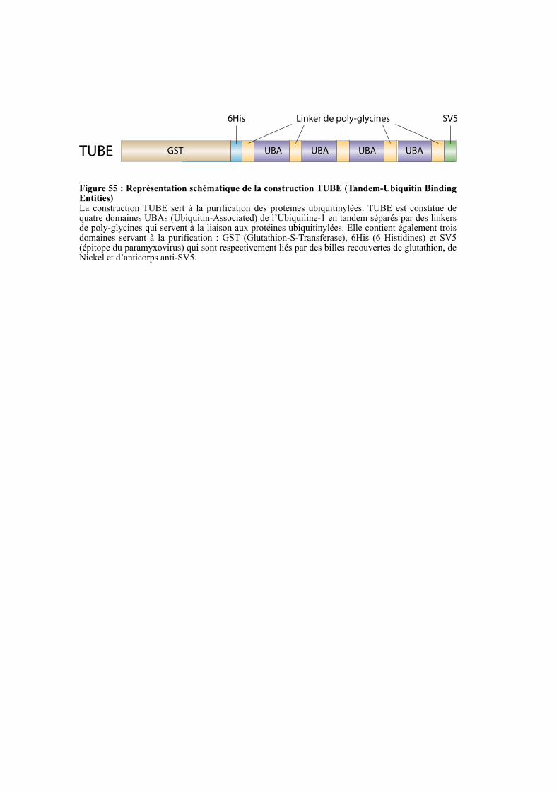
GST UBA UBA UBA UBA
6His Linker de poly-glycines SV5
TUBE
Figure 55 : Représentation schématique de la construction TUBE (Tandem-Ubiquitin Binding Entities)La construction TUBE sert à la purification des protéines ubiquitinylées. TUBE est constitué de quatre domaines UBAs (Ubiquitin-Associated) de l’Ubiquiline-1 en tandem séparés par des linkers de poly-glycines qui servent à la liaison aux protéines ubiquitinylées. Elle contient également trois domaines servant à la purification : GST (Glutathion-S-Transferase), 6His (6 Histidines) et SV5 (épitope du paramyxovirus) qui sont respectivement liés par des billes recouvertes de glutathion, de Nickel et d’anticorps anti-SV5.
122
spécifiques aux DUBs mais inhibent l’ensemble des protéases à cystéines ce qui
modifie également la physiologie de la cellule.
- L’usage d’un domaine de liaison à l’ubiquitine UBA (Ubiquitin-Associated) pour
purifier les protéines ubiquitinylées est efficace essentiellement pour les protéines
mono-ubiquitinylées.
L’équipe de Manuel S. Rodriguez a eu l’idée d’utiliser une protéine de fusion très affine
pour les protéines ubiquitinylées comportant quatre domaines UBA en tandem provenant de
l’Ubiquiline-1 associés à plusieurs étiquettes servant à la purification (GST : Glutathion-S-
Transferse ; 6His : 6 Histidines successives et l’épitope V5 du paramyxovirus) (Figure 55).
Cette construction nommée TUBE lie fortement les protéines polyubiquitinylées, ce qui
permet leur purification de manière efficace. Ils ont montré que la liaison de cette protéine de
fusion TUBE aux protéines polyubiquitinylées par des chaînes K48 prévient leurs
reconnaissances par le protéasome 26S et ainsi leurs dégradations. De plus, la liaison de
TUBE aux protéines polyubiquitinylées prévient leurs reconnaissances par les DUBs, ce qui
augmente la stabilité des chaînes de polyubiquitines et ce même en absence des inhibiteurs
NEM et IAA (Aillet et al., 2012; Hjerpe et al., 2009). La construction TUBE permet donc une
purification très efficace des protéines polyubiquitinylées qui pourront ensuite être analysées
par différentes méthodes incluant la spectrométrie de masse.
3.2. Analyse des protéines ubiquitinylées par spectrométrie de masse
Les différentes protéines ubiquitinylées purifiées sont digérées à la trypsine et les
peptides qui en découlent sont soumis à une séparation par chromatographie en phase
liquide suivie d’une analyse par spectrométrie de masse en tandem (Figure 54) (Lopitz-Otsoa
et al., 2012). L’usage de la spectrométrie de masse en tandem permet une meilleure
caractérisation des peptides et ainsi une meilleure identification des protéines ubiquitinylées
(Tsybin et al., 2011).
3.3. Identification des cibles de l’activité E3 ubiquitine ligase de TRAF4
Afin d’identifier les cibles du domaine RING de TRAF4 la méthode TUBE associée à
une identification des protéines ubiquitinylées par HPLC-MS/MS pourrait être réalisée sur des
cellules épithéliales mammaires MCF10A/shT4 et MCF7/shT4 où la protéine TRAF4 sauvage
ou le mutant du domaine RING (TRAF4-mutRING) seraient réexprimés de manière stable
(Figure 54). Les protéines identifiées exclusivement au sein des extraits protéiques provenant
123
des cellules où la protéine TRAF4 sauvage est réexprimée seront les substrats directs ou
indirects de l’activité E3 ubiquitine ligase de TRAF4. Cette stratégie pourrait également être
appliquée aux cellules MEFs provenant d’embryons déficients pour TRAF4 où la protéine
TRAF4 sauvage ou le mutant TRAF4-mutRING seront réexprimés (Figure 54). L’usage des
cellules déficientes en TRAF4 pour l’expression de TRAF4 sauvage ou mutant permet de
s’affranchir de la protéine endogène sauvage qui pourrait compenser la perte de fonction du
mutant TRAF4-mutRING. Ces expériences pourraient être réalisées en présence de TGF beta
afin d’isoler les cibles de TRAF4 constitutives et celles dépendante de la voie du TGF beta.
Cette approche permettrait d’aller plus loin dans la compréhension de la fonction de TRAF4 à
l’échelle moléculaire.
4. TRAF4 DANS LA CARCINOGENESE.
TRAF4 a initialement été identifié comme étant surexprimé dans les cancers du sein
(Régnier et al., 1995; Tomasetto et al., 1995). Actuellement, TRAF4 apparait comme une
protéine communément surproduite dans plus de 40% des carcinomes tous confondus, en
partie à cause de son amplification génique (Bièche et al., 1996; Camilleri-Broët et al., 2007;
Rhodes et al., 2004). Bien que sa découverte remonte presque à 20 ans, la caractérisation de
son rôle pro-tumoral est très récente. En effet, cette année, l’équipe de Bert O’Malley a
montré que la surexpression de TRAF4 prévient la déubiquitinylation de p53 médiée par
HAUSP, ce qui déstabilise p53 et prévient ainsi l’apoptose induite suite à un stress oxydatif
(Yi et al., 2013). Dans un travail en cours de publication, l’équipe de Peter Ten-Dijke a
montré que TRAF4 polyubiquitinyle et induit la dégradation de SMURF2 et participe à
l’activation de la voie TGF-β canonique (SMADs) et non-canonique (TAK1). Au niveau
fonctionnel, TRAF4 participe à l’établissement de la TEM induite par le TGF-β et est
essentiel pour la migration des cellules non polarisée. De plus, la perte de TRAF4 diminue
fortement la formation des métastases dans des modèles de xénogreffes chez la souris
(Annexe-2 : Zhang et al., 2013). Ces deux équipes rapportent des études cliniques
rétrospectives indiquant que l’expression de TRAF4 dans les cancers du Sein est associée à un
mauvais pronostique (Yi et al., 2013; Zhang et al., 2013). Pour notre part, nous avons montré
que TRAF4 lie les PIPs via son domaine TRAF, un mécanisme conservé au sein de la famille
TRAFs. Le recrutement de TRAF4 aux JSs est dépendant de son interaction avec les PIPs. Au
niveau fonctionnel, TRAF4 diminue la formation et/ou la stabilité des JSs et promeut la
migration des cellules épithéliales mammaires. Ces deux fonctions sont également
dépendantes de sa liaison aux PIPs (Rousseau et al., en révision).
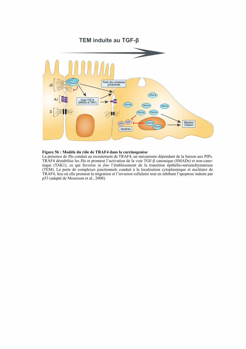
JS
AJ
D
Perte des complexes jonctionnels
TEM induite au TGF-β
Voies TGF-β (SMADs et TAK1)
TRAF4TRAF4TRAF4TRAF4TRAF4
TRAF4
TRAF4
TRAF4
TRAF4
TRAF4
TRAF4
TRAF4
TRAF4
MigrationInvasion
p53p53
Apoptose
Figure 56 : Modèle du rôle de TRAF4 dans la carcinogenèseLa présence de JSs conduit au recrutement de TRAF4, un mécanisme dépendant de la liaison aux PIPs. TRAF4 déstabilise les JSs et promeut l’activation de la voie TGF-β canonique (SMADs) et non-cano-nique (TAK1), ce qui favorise in fine l’établissement de la transition épithélio-mésenchymateuse (TEM). La perte de complexes jonctionnels conduit à la localisation cytoplasmique et nucléaire de TRAF4, lieu où elle promeut la migration et l’invasion cellulaire tout en inhibant l’apoptose induite par p53 (adapté de Mosesson et al., 2008).
124
Prises dans leur ensemble, ces données suggèrent que TRAF4 a un rôle pléiotropique
dans la carcinogenèse. Nous pouvons proposer le modèle suivant (Figure 56):
TRAF4, en régulant négativement les JSs, promeut la progression des carcinomes.
Néanmoins, il a été décrit que les JSs inhibent aussi bien la migration que la prolifération
cellulaire, or la surexpression de TRAF4 promeut uniquement la migration. Cela peut-être
expliqué par l’implication de TRAF4 dans la voie TGF-β. En effet, il a été décrit que la voie
TGF-β a deux effets opposés : un rôle anti-tumoral en inhibant la prolifération cellulaire et un
rôle pro-tumoral en stimulant la migration et la formation de métastases. Nous pouvons
imaginer la séquence suivante : TRAF4 déstabilise les JSs, cette déstabilisation induit la
migration et la prolifération cellulaire, mais l’effet sur la prolifération est contrebalancé par
l’activation concomitante de la voie TGF-β. Concernant la progression tumorale, plusieurs
rôles peuvent être évoqués : premièrement, en augmentant la plasticité des JSs, TRAF4
favorise leur dissolution ; deuxièmement, TRAF4 en tant qu’effecteur de la voie du TGF-β,
favorise la TEM ; troisièmement, les complexes jonctionnels détruits, la localisation
cytoplasmique et nucléaire de TRAF4 préviennent l’apoptose médiée par p53 et promeuvent
la migration et l’invasion cellulaire favorisant la formation de métastases.
Tous ces éléments, font de TRAF4 une cible thérapeutique intéressante. Pour cela, il est
envisageable de développer des drogues inhibant l’interaction TRAF-PIPs.

125
REFERENCES
BIBLIOGRAPHIQUES
126
-A-
Adachi, M., Hamazaki, Y., Kobayashi, Y., Itoh, M., Tsukita, S., Furuse, M., and Tsukita, S. (2009). Similar and distinct properties of MUPP1 and Patj, two homologous PDZ domain-containing tight-junction proteins. Mol. Cell. Biol. 29, 2372–2389.
Aderka, D. (1996). The potential biological and clinical significance of the soluble tumor necrosis factor receptors. Cytokine Growth Factor Rev. 7, 231–240.
Aillet, F., Lopitz-Otsoa, F., Hjerpe, R., Torres-Ramos, M., Lang, V., and Rodríguez, M.S. (2012). Isolation of ubiquitylated proteins using tandem ubiquitin-binding entities. Methods Mol. Biol. Clifton NJ 832, 173–183.
Akhtar, A. (2001). The histone H4 acetyltransferase MOF uses a C2HC zinc finger for substrate recognition. EMBO Rep. 2, 113–118.
Aktories, K., and Barbieri, J.T. (2005). Bacterial cytotoxins: targeting eukaryotic switches. Nat. Rev. Microbiol. 3, 397–410.
Angst, B.D., Marcozzi, C., and Magee, A.I. (2001). The cadherin superfamily: diversity in form and function. J. Cell Sci. 114, 629–641.
Arch, R.H., Gedrich, R.W., and Thompson, C.B. (1998). Tumor necrosis factor receptor-associated factors (TRAFs)--a family of adapter proteins that regulates life and death. Genes Dev. 12, 2821–2830.
Arteaga, C.L., Hurd, S.D., Winnier, A.R., Johnson, M.D., Fendly, B.M., and Forbes, J.T. (1993). Anti-transforming growth factor (TGF)-beta antibodies inhibit breast cancer cell tumorigenicity and increase mouse spleen natural killer cell activity. Implications for a possible role of tumor cell/host TGF-beta interactions in human breast cancer progression. J. Clin. Invest. 92, 2569–2576.
Assémat, E., Bazellières, E., Pallesi-Pocachard, E., Le Bivic, A., and Massey-Harroche, D. (2008). Polarity complex proteins. Biochim. Biophys. Acta BBA - Biomembr. 1778, 614–630.
Atfi, A., Djelloul, S., Chastre, E., Davis, R., Gespach, C., 1997. Evidence for a role of Rho-like GTPases and stress-activated protein kinase/c-Jun N-terminal kinase (SAPK/JNK) in transforming growth factor beta-mediated signaling. J. Biol. Chem. 272, 1429–1432.
Axelrod, J.D. (2002). Strabismus comes into focus. Nat. Cell Biol. 4, E6–E8.
-B-
Baeuerle, P.A., and Henkel, T. (1994). Function and Activation of NF-kappaB in the Immune System. Annu. Rev. Immunol. 12, 141–179.
Balda, M.S., Whitney, J.A., Flores, C., González, S., Cereijido, M., and Matter, K. (1996). Functional dissociation of paracellular permeability and transepithelial electrical resistance and disruption of the apical-basolateral intramembrane diffusion barrier by expression of a mutant tight junction membrane protein. J. Cell Biol. 134, 1031–1049.
Balda, M.S., Garrett, M.D., and Matter, K. (2003). The ZO-1-associated Y-box factor ZONAB regulates epithelial cell proliferation and cell density. J. Cell Biol. 160, 423–432.
Balla, T. (2007). Imaging and manipulating phosphoinositides in living cells. J. Physiol. 582, 927–937.
Balla, T., and Varnai, P. (2009). visualiziation of cellular phosphoinositide pools with gfp-fused protein-domains. Curr. Protoc. Cell Biol. Editor. Board Juan S Bonifacino Al CHAPTER, Unit–24.4.
127
Bamforth, S.D., Kniesel, U., Wolburg, H., Engelhardt, B., and Risau, W. (1999). A dominant mutant of occludin disrupts tight junction structure and function. J. Cell Sci. 112, 1879–1888.
Barrios-Rodiles, M., Brown, K.R., Ozdamar, B., Bose, R., Liu, Z., Donovan, R.S., Shinjo, F., Liu, Y., Dembowy, J., Taylor, I.W., et al. (2005). High-throughput mapping of a dynamic signaling network in mammalian cells. Science 307, 1621–1625.
Baud, V., Liu, Z.-G., Bennett, B., Suzuki, N., Xia, Y., and Karin, M. (1999). Signaling by proinflammatory cytokines: oligomerization of TRAF2 and TRAF6 is sufficient for JNK and IKK activation and target gene induction via an amino-terminal effector domain. Genes Dev. 13, 1297–1308.
Bauer, H., Zweimueller-Mayer, J., Steinbacher, P., Lametschwandtner, A., and Bauer, H.C. (2010). The dual role of zonula occludens (ZO) proteins. J. Biomed. Biotechnol. 2010, 402593.
Baum, B., and Georgiou, M. (2011). Dynamics of adherens junctions in epithelial establishment, maintenance, and remodeling. J. Cell Biol. 192, 907–917.
Bellam, N., and Pasche, B. (2010). Tgf-beta signaling alterations and colon cancer. Cancer Treat. Res. 155, 85–103.
Benson, J.R., and Jatoi, I. (2012). The global breast cancer burden. Future Oncol. Lond. Engl. 8, 697–702.
Berne, B.J., and Pecora, R. (2000). Dynamic Light Scattering: With Applications to Chemistry, Biology, and Physics (Courier Dover Publications).
Beutler, B. (2004). Inferences, questions and possibilities in Toll-like receptor signalling. Nature 430, 257–263.
Bièche, I., Tomasetto, C., Régnier, C.H., Moog-Lutz, C., Rio, M.C., and Lidereau, R. (1996). Two distinct amplified regions at 17q11-q21 involved in human primary breast cancer. Cancer Res. 56, 3886–3890.
Bierie, B., and Moses, H.L. (2006a). TGF-β and cancer. Cytokine Growth Factor Rev. 17, 29–40.
Bierie, B., and Moses, H.L. (2006b). Tumour microenvironment: TGFβ: the molecular Jekyll and Hyde of cancer. Nat. Rev. Cancer 6, 506–520.
Bindels, S., Mestdagt, M., Vandewalle, C., Jacobs, N., Volders, L., Noël, A., Roy, F. van, Berx, G., Foidart, J.-M., and Gilles, C. (2006). Regulation of vimentin by SIP1 in human epithelial breast tumor cells. Oncogene 25, 4975–4985.
Biswas, S., Guix, M., Rinehart, C., Dugger, T.C., Chytil, A., Moses, H.L., Freeman, M.L., and Arteaga, C.L. (2007). Inhibition of TGF-β with neutralizing antibodies prevents radiation-induced acceleration of metastatic cancer progression. J. Clin. Invest. 117, 1305–1313.
Blaise, S., Kneib, M., Rousseau, A., Gambino, F., Chenard, M.-P., Messadeq, N., Muckenstrum, M., Alpy, F., Tomasetto, C., Humeau, Y., et al. (2012). In vivo evidence that TRAF4 is required for central nervous system myelin homeostasis. PloS One 7, e30917.
Bogoyevitch, M.A., Ngoei, K.R.W., Zhao, T.T., Yeap, Y.Y.C., and Ng, D.C.H. (2010). c-Jun N-terminal kinase (JNK) signaling: recent advances and challenges. Biochim. Biophys. Acta 1804, 463–475.
Boraschi, D., and Tagliabue, A. (2006). The interleukin-1 receptor family. Vitam. Horm. 74, 229–254.
Bouwmeester, T., Bauch, A., Ruffner, H., Angrand, P.-O., Bergamini, G., Croughton, K., Cruciat, C., Eberhard, D., Gagneur, J., Ghidelli, S., et al. (2004). A physical and functional map of the human TNF-alpha/NF-kappa B signal transduction pathway. Nat. Cell Biol. 6, 97–105.
128
Bradley, J.R., and Pober, J.S. (2001). Tumor necrosis factor receptor-associated factors (TRAFs). Oncogene 20, 6482–6491.
Brembeck, F.H., Rosário, M., and Birchmeier, W. (2006). Balancing cell adhesion and Wnt signaling, the key role of β-catenin. Curr. Opin. Genet. Dev. 16, 51–59.
Brennan, K., Offiah, G., McSherry, E.A., and Hopkins, A.M. (2010). Tight junctions: a barrier to the initiation and progression of breast cancer? J. Biomed. Biotechnol. 2010, 460607.
Brysse, A., Mestdagt, M., Polette, M., Luczka, E., Hunziker, W., Noël, A., Birembaut, P., Foidart, J.-M., and Gilles, C. (2012). Regulation of CXCL8/IL-8 expression by zonula occludens-1 in human breast cancer cells. Mol. Cancer Res. MCR 10, 121–132.
Buache, E., Etique, N., Alpy, F., Stoll, I., Muckensturm, M., Reina-San-Martin, B., Chenard, M.P., Tomasetto, C., and Rio, M.C. (2011). Deficiency in trefoil factor 1 (TFF1) increases tumorigenicity of human breast cancer cells and mammary tumor development in TFF1-knockout mice. Oncogene 30, 3261–3273.
Burgess, A.W., Faux, M.C., Layton, M.J., and Ramsay, R.G. (2011). Wnt signaling and colon tumorigenesis — A view from the periphery. Exp. Cell Res. 317, 2748–2758.
-C-
Camilleri-Broët, S., Cremer, I., Marmey, B., Comperat, E., Viguié, F., Audouin, J., Rio, M.-C., Fridman, W.-H., Sautès-Fridman, C., and Régnier, C.H. (2007). TRAF4 overexpression is a common characteristic of human carcinomas. Oncogene 26, 142–147.
Cao, Z., Xiong, J., Takeuchi, M., Kurama, T., and Goeddel, D.V. (1996). TRAF6 is a signal transducer for interleukin-1. Nature 383, 443–446.
Carswell, E.A., Old, L.J., Kassel, R.L., Green, S., Fiore, N., and Williamson, B. (1975). An endotoxin-induced serum factor that causes necrosis of tumors. Proc. Natl. Acad. Sci. U. S. A. 72, 3666–3670.
Cha, G.-H., Cho, K.S., Lee, J.H., Kim, M., Kim, E., Park, J., Lee, S.B., and Chung, J. (2003). Discrete functions of TRAF1 and TRAF2 in Drosophila melanogaster mediated by c-Jun N-terminal kinase and NF-kappaB-dependent signaling pathways. Mol. Cell. Biol. 23, 7982–7991.
Chacon-Heszele, M.F., Ren, D., Reynolds, A.B., Chi, F., and Chen, P. Regulation of cochlear convergent extension by the vertebrate planar cell polarity pathway is dependent on p120-catenin.
Chasapis, C., and Spyroulias, G. (2009). RING Finger E3 Ubiquitin Ligases: Structure and Drug Discovery. Curr. Pharm. Des. 15, 3716–3731.
Chen, J., and Zhang, M. (2013). The Par3/Par6/aPKC complex and epithelial cell polarity. Exp. Cell Res. 319, 1357–1364.
Chen, Y.T., Stewart, D.B., and Nelson, W.J. (1999). Coupling assembly of the E-cadherin/beta-catenin complex to efficient endoplasmic reticulum exit and basal-lateral membrane targeting of E-cadherin in polarized MDCK cells. J. Cell Biol. 144, 687–699.
Cheng, G., Cleary, A.M., Ye, Z.S., Hong, D.I., Lederman, S., and Baltimore, D. (1995). Involvement of CRAF1, a relative of TRAF, in CD40 signaling. Science 267, 1494–1498.
Cherfils-Vicini, J., Vingert, B., Varin, A., Tartour, E., Fridman, W.-H., Sautès-Fridman, C., Régnier, C.H., and Cremer, I. (2008). Characterization of immune functions in TRAF4-deficient mice. Immunology 124, 562–574.
Chishti, A.H., Kim, A.C., Marfatia, S.M., Lutchman, M., Hanspal, M., Jindal, H., Liu, S.C., Low, P.S., Rouleau, G.A., Mohandas, N., et al. (1998). The FERM domain: a unique module
129
involved in the linkage of cytoplasmic proteins to the membrane. Trends Biochem. Sci. 23, 281–282.
Chung, J.Y., Park, Y.C., Ye, H., and Wu, H. (2002). All TRAFs are not created equal: common and distinct molecular mechanisms of TRAF-mediated signal transduction. J. Cell Sci. 115, 679–688.
Cohen, P., and Tcherpakov, M. (2010). Will the Ubiquitin System Furnish as Many Drug Targets as Protein Kinases? Cell 143, 686–693.
Cottrell, S., Bicknell, D., Kaklamanis, L., and Bodmer, W.F. (1992). Molecular analysis of APC mutations in familial adenomatous polyposis and sporadic colon carcinomas. Lancet 340, 626–630.
Croft, M., Benedict, C.A., and Ware, C.F. (2013). Clinical targeting of the TNF and TNFR superfamilies. Nat. Rev. Drug Discov. 12, 147–168.
Cummins, P.M. (2012). Occludin: One Protein, Many Forms. Mol. Cell. Biol. 32, 242–250.
-D-
Dadgostar, H., and Cheng, G. (2000). Membrane localization of TRAF 3 enables JNK activation. J. Biol. Chem. 275, 2539–2544.
Dai, L., Aye Thu, C., Liu, X.-Y., Xi, J., and Cheung, P.C.F. (2012). TAK1, more than just innate immunity. IUBMB Life 64, 825–834.
David, D., Nair, S.A., and Pillai, M.R. (2013). Smurf E3 ubiquitin ligases at the cross roads of oncogenesis and tumor suppression. Biochim. Biophys. Acta 1835, 119–128.
Degot, S., Hir, H.L., Alpy, F., Kedinger, V., Stoll, I., Wendling, C., Seraphin, B., Rio, M.-C., and Tomasetto, C. (2004). Association of the Breast Cancer Protein MLN51 with the Exon Junction Complex via Its Speckle Localizer and RNA Binding Module. J. Biol. Chem. 279, 33702–33715.
Delva, E., Tucker, D.K., and Kowalczyk, A.P. (2009). The desmosome. Cold Spring Harb. Perspect. Biol. 1, a002543.
Dempsey, P.W., Doyle, S.E., He, J.Q., and Cheng, G. (2003). The signaling adaptors and pathways activated by TNF superfamily. Cytokine Growth Factor Rev. 14, 193–209.
Derynck, R., and Zhang, Y.E. (2003). Smad-dependent and Smad-independent pathways in TGF-β family signalling. Nature 425, 577–584.
Deshaies, R.J., and Joazeiro, C.A.P. (2009). RING domain E3 ubiquitin ligases. Annu. Rev. Biochem. 78, 399–434.
Devin, A., Lin, Y., and Liu, Z. (2003). The role of the death-domain kinase RIP in tumour-necrosis-factor-induced activation of mitogen-activated protein kinases. EMBO Rep. 4, 623–627.
Djiane, A., Riou, J., Umbhauer, M., Boucaut, J., and Shi, D. (2000). Role of frizzled 7 in the regulation of convergent extension movements during gastrulation in Xenopus laevis. Dev. Camb. Engl. 127, 3091–3100.
Dobrosotskaya, I., Guy, R.K., and James, G.L. (1997). MAGI-1, a membrane-associated guanylate kinase with a unique arrangement of protein-protein interaction domains. J. Biol. Chem. 272, 31589–31597.
Dowler, S., Kular, G., and Alessi, D.R. (2002). Protein Lipid Overlay Assay. Sci. Signal. 2002, pl6.
-E-
130
Elsum, I., Yates, L., Humbert, P.O., and Richardson, H.E. (2012). The Scribble-Dlg-Lgl polarity module in development and cancer: from flies to man. Essays Biochem. 53, 141–168.
Eluredge, D.H., and Miller, J.D. (1971). Physiology of Hearing. Annu. Rev. Physiol. 33, 281–308.
Ersoy-Evans, S., Erkin, G., Fassihi, H., Chan, I., Paller, A.S., Sürücü, S., and McGrath, J.A. (2006). Ectodermal dysplasia-skin fragility syndrome resulting from a new homozygous mutation, 888delC, in the desmosomal protein plakophilin 1. J. Am. Acad. Dermatol. 55, 157–161.
Ezan, J., and Montcouquiol, M. (2013). Revisiting planar cell polarity in the inner ear. Semin. Cell Dev. Biol.
-F-
Farquhar, M.G., and Palade, G.E. (1963). Junctional Complexes in Various Epithelia. J. Cell Biol. 17, 375–412.
Feldman, G.J., Mullin, J.M., and Ryan, M.P. (2005). Occludin: Structure, function and regulation. Adv. Drug Deliv. Rev. 57, 883–917.
Fogg, V.C., Liu, C.-J., and Margolis, B. (2005). Multiple regions of Crumbs3 are required for tight junction formation in MCF10A cells. J. Cell Sci. 118, 2859–2869.
Forster, C. (2008). Tight junctions and the modulation of barrier function in disease. Histochem. Cell Biol. 130, 55–70.
Franke, W.W. (2009). Discovering the molecular components of intercellular junctions--a historical view. Cold Spring Harb. Perspect. Biol. 1, a003061.
Frolik, C.A., Dart, L.L., Meyers, C.A., Smith, D.M., and Sporn, M.B. (1983). Purification and initial characterization of a type beta transforming growth factor from human placenta. Proc. Natl. Acad. Sci. U. S. A. 80, 3676–3680.
Furuse, M. (2010). Molecular basis of the core structure of tight junctions. Cold Spring Harb. Perspect. Biol. 2, a002907.
Furuse, M., Hirase, T., Itoh, M., Nagafuchi, A., Yonemura, S., Tsukita, S., and Tsukita, S. (1993). Occludin: a novel integral membrane protein localizing at tight junctions. J. Cell Biol. 123, 1777–1788.
Furuse, M., Sasaki, H., Fujimoto, K., and Tsukita, S. (1998). A single gene product, claudin-1 or -2, reconstitutes tight junction strands and recruits occludin in fibroblasts. J. Cell Biol. 143, 391–401.
-G-
Gao, L., and Macara, I.G. (2004). Isoforms of the polarity protein par6 have distinct functions. J. Biol. Chem. 279, 41557–41562.
Gao, L., Joberty, G., and Macara, I.G. (2002). Assembly of Epithelial Tight Junctions Is Negatively Regulated by Par6. Curr. Biol. 12, 221–225.
Garabedian, M.J., and Logan, S.K. (2008). Atypical regulation of SRC-3. Trends Biochem. Sci. 33, 301–304.
Garcia-Higuera, I., Taniguchi, T., Ganesan, S., Meyn, M.S., Timmers, C., Hejna, J., Grompe, M., and D’Andrea, A.D. (2001). Interaction of the Fanconi Anemia Proteins and BRCA1 in a Common Pathway. Mol. Cell 7, 249–262.
Garrod, D., and Chidgey, M. (2008). Desmosome structure, composition and function. Biochim. Biophys. Acta 1778, 572–587.
131
Gassama-Diagne, A., Yu, W., ter Beest, M., Martin-Belmonte, F., Kierbel, A., Engel, J., and Mostov, K. (2006). Phosphatidylinositol-3,4,5-trisphosphate regulates the formation of the basolateral plasma membrane in epithelial cells. Nat. Cell Biol. 8, 963–970.
Gatza, C.E., Oh, S.Y., and Blobe, G.C. (2010). Roles for the type III TGF-beta receptor in human cancer. Cell. Signal. 22, 1163–1174.
Gibson, S.B., Oyer, R., Spalding, A.C., Anderson, S.M., and Johnson, G.L. (2000). Increased expression of death receptors 4 and 5 synergizes the apoptosis response to combined treatment with etoposide and TRAIL. Mol. Cell. Biol. 20, 205–212.
Giepmans, B.N.G. (2006). Role of Connexin43-Interacting Proteins at Gap Junctions. In Advances in Cardiology, S. Dhein, ed. (Basel: KARGER), pp. 41–56.
Glauner, H., Siegmund, D., Motejadded, H., Scheurich, P., Henkler, F., Janssen, O., and Wajant, H. (2002). Intracellular localization and transcriptional regulation of tumor necrosis factor (TNF) receptor-associated factor 4 (TRAF4). Eur. J. Biochem. FEBS 269, 4819–4829.
González-Mariscal, L., Betanzos, A., and Avila-Flores, A. (2000). MAGUK proteins: structure and role in the tight junction. Semin. Cell Dev. Biol. 11, 315–324.
Grech, A., Quinn, R., Srinivasan, D., Badoux, X., and Brink, R. (2000). Complete structural characterisation of the mammalian and Drosophila TRAF genes: implications for TRAF evolution and the role of RING finger splice variants. Mol. Immunol. 37, 721–734.
Grewal, I.S. (2009). Overview of TNF superfamily: a chest full of potential therapeutic targets. Adv. Exp. Med. Biol. 647, 1–7.
Guillemot, L., Paschoud, S., Pulimeno, P., Foglia, A., and Citi, S. (2008). The cytoplasmic plaque of tight junctions: a scaffolding and signalling center. Biochim. Biophys. Acta 1778, 601–613.
Günzel, D., and Yu, A.S.L. (2013). Claudins and the Modulation of Tight Junction Permeability. Physiol. Rev. 93, 525–569.
-H-
Ha, H., Han, D., and Choi, Y. (2009). TRAF-mediated TNFR-family signaling. Curr. Protoc. Immunol. Ed. John E Coligan Al Chapter 11, Unit11.9D.
Habelhah, H., Takahashi, S., Cho, S.-G., Kadoya, T., Watanabe, T., and Ronai, Z. (2004). Ubiquitination and translocation of TRAF2 is required for activation of JNK but not of p38 or NF-kappaB. EMBO J. 23, 322–332.
Hagel, L. (2011). Gel filtration: size exclusion chromatography. Methods Biochem. Anal. 54, 51–91.
Halet, G. (2005). Imaging phosphoinositide dynamics using GFP-tagged protein domains. Biol. Cell Auspices Eur. Cell Biol. Organ. 97, 501–518.
Harris, T.J.C., and Peifer, M. (2005). The positioning and segregation of apical cues during epithelial polarity establishment in Drosophila. J. Cell Biol. 170, 813–823.
Harris, T.J.C., and Tepass, U. (2010). Adherens junctions: from molecules to morphogenesis. Nat. Rev. Mol. Cell Biol. 11, 502–514.
Hata, A., Lagna, G., Massagué, J., and Hemmati-Brivanlou, A. (1998). Smad6 inhibits BMP/Smad1 signaling by specifically competing with the Smad4 tumor suppressor. Genes Dev. 12, 186–197.
Haynes, P.A., Fripp, N., and Aebersold, R. (1998). Identification of gel-separated proteins by liquid chromatography-electrospray tandem mass spectrometry: comparison of methods and their limitations. Electrophoresis 19, 939–945.
132
He, L., Grammer, A.C., Wu, X., and Lipsky, P.E. (2004). TRAF3 forms heterotrimers with TRAF2 and modulates its ability to mediate NF-{kappa}B activation. J. Biol. Chem. 279, 55855–55865.
Hinck, A.P. (2012). Structural studies of the TGF-βs and their receptors - insights into evolution of the TGF-β superfamily. FEBS Lett. 586, 1860–1870.
Hjerpe, R., Aillet, F., Lopitz-Otsoa, F., Lang, V., England, P., and Rodriguez, M.S. (2009). Efficient protection and isolation of ubiquitylated proteins using tandem ubiquitin-binding entities. EMBO Rep. 10, 1250–1258.
Hocevar, B.A., Brown, T.L., Howe, P.H., 1999. TGF-β induces fibronectin synthesis through a c-Jun N-terminal kinase-dependent, Smad4-independent pathway. EMBO J. 18, 1345–1356. Hoover, K.B., Liao, S.Y., and Bryant, P.J. (1998). Loss of the tight junction MAGUK ZO-1 in breast cancer: relationship to glandular differentiation and loss of heterozygosity. Am. J. Pathol. 153, 1767–1773.
Horbelt, D., Denkis, A., and Knaus, P. (2012). A portrait of Transforming Growth Factor β superfamily signalling: Background matters. Int. J. Biochem. Cell Biol. 44, 469–474.
Hu, H.M., O’Rourke, K., Boguski, M.S., and Dixit, V.M. (1994). A novel RING finger protein interacts with the cytoplasmic domain of CD40. J. Biol. Chem. 269, 30069–30072.
Huang, B., Eberstadt, M., Olejniczak, E.T., Meadows, R.P., and Fesik, S.W. (1996). NMR structure and mutagenesis of the Fas (APO-1/CD95) death domain. Nature 384, 638–641.
Hugot, J.-P., Chamaillard, M., Zouali, H., Lesage, S., Cézard, J.-P., Belaiche, J., Almer, S., Tysk, C., O’Morain, C.A., Gassull, M., et al. (2001). Association of NOD2 leucine-rich repeat variants with susceptibility to Crohn’s disease. Nature 411, 599–603.
Hunter, A.W., Barker, R.J., Zhu, C., and Gourdie, R.G. (2005). Zonula Occludens-1 Alters Connexin43 Gap Junction Size and Organization by Influencing Channel Accretion. Mol. Biol. Cell 16, 5686–5698.
-I-
Iden, S., and Collard, J.G. (2008). Crosstalk between small GTPases and polarity proteins in cell polarization. Nat. Rev. Mol. Cell Biol. 9, 846–859.
Ikenouchi, J., Matsuda, M., Furuse, M., and Tsukita, S. (2003). Regulation of tight junctions during the epithelium-mesenchyme transition: direct repression of the gene expression of claudins/occludin by Snail. J. Cell Sci. 116, 1959–1967.
Imamura, T., Hikita, A., and Inoue, Y. (2012). The roles of TGF-β signaling in carcinogenesis and breast cancer metastasis. Breast Cancer Tokyo Jpn. 19, 118–124.
Inoko, A., Itoh, M., Tamura, A., Matsuda, M., Furuse, M., and Tsukita, S. (2003). Expression and distribution of ZO-3, a tight junction MAGUK protein, in mouse tissues. Genes Cells Devoted Mol. Cell. Mech. 8, 837–845.
Inoue, J., Ishida, T., Tsukamoto, N., Kobayashi, N., Naito, A., Azuma, S., and Yamamoto, T. (2000). Tumor Necrosis Factor Receptor-Associated Factor (TRAF) Family: Adapter Proteins That Mediate Cytokine Signaling. Exp. Cell Res. 254, 14–24.
Inoue, J., Gohda, J., and Akiyama, T. (2007). Characteristics and biological functions of TRAF6. Adv. Exp. Med. Biol. 597, 72–79.
Ishida, T., Mizushima, S., Azuma, S., Kobayashi, N., Tojo, T., Suzuki, K., Aizawa, S., Watanabe, T., Mosialos, G., Kieff, E., et al. (1996a). Identification of TRAF6, a Novel Tumor Necrosis Factor Receptor-associated Factor Protein That Mediates Signaling from an Amino-terminal Domain of the CD40 Cytoplasmic Region. J. Biol. Chem. 271, 28745–28748.
133
Ishida, T.K., Tojo, T., Aoki, T., Kobayashi, N., Ohishi, T., Watanabe, T., Yamamoto, T., and Inoue, J. (1996b). TRAF5, a novel tumor necrosis factor receptor-associated factor family protein, mediates CD40 signaling. Proc. Natl. Acad. Sci. 93, 9437–9442.
Itoh, S., and ten Dijke, P. (2007). Negative regulation of TGF-beta receptor/Smad signal transduction. Curr. Opin. Cell Biol. 19, 176–184.
Iuchi, S. (2001). Three classes of C2H2 zinc finger proteins. Cell. Mol. Life Sci. CMLS 58, 625–635.
Iwai, K., and Tokunaga, F. (2009). Linear polyubiquitination: a new regulator of NF-kappaB activation. EMBO Rep. 10, 706–713.
-J-
Jaramillo, B.E., Ponce, A., Moreno, J., Betanzos, A., Huerta, M., Lopez-Bayghen, E., and Gonzalez-Mariscal, L. (2004). Characterization of the tight junction protein ZO-2 localized at the nucleus of epithelial cells. Exp. Cell Res. 297, 247–258.
Jeffries, T.R., Dove, S.K., Michell, R.H., and Parker, P.J. (2004). PtdIns-specific MPR Pathway Association of a Novel WD40 Repeat Protein, WIPI49. Mol. Biol. Cell 15, 2652–2663.
Jemal, A., Bray, F., Center, M.M., Ferlay, J., Ward, E., and Forman, D. (2011). Global cancer statistics. CA. Cancer J. Clin. 61, 69–90.
Jiang, X., and Chen, Z.J. (2012). The role of ubiquitylation in immune defence and pathogen evasion. Nat. Rev. Immunol. 12, 35–48.
Jin, C., Yang, Y., Anver, M.R., Morris, N., Wang, X., and Zhang, Y.E. (2009). Smad ubiquitination regulatory factor 2 promotes metastasis of breast cancer cells by enhancing migration and invasiveness. Cancer Res. 69, 735–740.
-K-
Kalkan, T., Iwasaki, Y., Park, C.Y., and Thomsen, G.H. (2009a). Tumor Necrosis Factor-Receptor–associated Factor-4 Is a Positive Regulator of Transforming Growth Factor-β Signaling That Affects Neural Crest Formation. Mol. Biol. Cell 20, 3436–3450.
Kalkan, T., Iwasaki, Y., Park, C.Y., and Thomsen, G.H. (2009b). Tumor necrosis factor-receptor-associated factor-4 is a positive regulator of transforming growth factor-beta signaling that affects neural crest formation. Mol. Biol. Cell 20, 3436–3450.
Kallay, L.M., McNickle, A., Brennwald, P.J., Hubbard, A.L., and Braiterman, L.T. (2006). Scribble associates with two polarity proteins, Lgl2 and Vangl2, via distinct molecular domains. J. Cell. Biochem. 99, 647–664.
Karin, M., 1995. The Regulation of AP-1 Activity by Mitogen-activated Protein Kinases. J. Biol. Chem. 270, 16483–16486.
Karin, M., and Gallagher, E. (2009). TNFR signaling: ubiquitin-conjugated TRAFfic signals control stop-and-go for MAPK signaling complexes. Immunol. Rev. 228, 225–240.
Kawai, T., and Akira, S. (2004). Toll-like receptor downstream signaling. Arthritis Res. Ther. 7, 12.
Kawai, T., and Akira, S. (2010). The role of pattern-recognition receptors in innate immunity: update on Toll-like receptors. Nat. Immunol. 11, 373–384.
Kedinger, V., Alpy, F., Tomasetto, C., Thisse, C., Thisse, B., and Rio, M.-C. (2005). Spatial and temporal distribution of the traf4 genes during zebrafish development. Gene Expr. Patterns GEP 5, 545–552.
134
Kédinger, V., Alpy, F., Baguet, A., Polette, M., Stoll, I., Chenard, M.-P., Tomasetto, C., and Rio, M.-C. (2008). Tumor necrosis factor receptor-associated factor 4 is a dynamic tight junction-related shuttle protein involved in epithelium homeostasis. PloS One 3, e3518.
Kemphues, K. (2000). PARsing embryonic polarity. Cell 101, 345–348.
Kim, W., Kim, M., and Jho, E. (2013). Wnt/β-catenin signalling: from plasma membrane to nucleus. Biochem. J. 450, 9–21.
Kimura, Y., and Tanaka, K. (2010). Regulatory mechanisms involved in the control of ubiquitin homeostasis. J. Biochem. (Tokyo) 147, 793–798.
Kobayashi, N., Kadono, Y., Naito, A., Matsumoto, K., Yamamoto, T., Tanaka, S., and Inoue, J. (2001). Segregation of TRAF6-mediated signaling pathways clarifies its role in osteoclastogenesis. EMBO J. 20, 1271–1280.
Kobielak, A., and Fuchs, E. (2004). α-catenin: at the junction of intercellular adhesion and actin dynamics. Nat. Rev. Mol. Cell Biol. 5, 614–625.
Kolb, W.P., and Granger, G.A. (1968). Lymphocyte in vitro cytotoxicity: characterization of human lymphotoxin. Proc. Natl. Acad. Sci. U. S. A. 61, 1250–1255.
Kubiczkova, L., Sedlarikova, L., Hajek, R., and Sevcikova, S. (2012). TGF-β – an excellent servant but a bad master. J. Transl. Med. 10, 183.
Kutateladze, T.G. (2010). Translation of the phosphoinositide code by PI effectors. Nat. Chem. Biol. 6, 507–513.
-L
Lamothe, B., Campos, A.D., Webster, W.K., Gopinathan, A., Hur, L., and Darnay, B.G. (2008). The RING domain and first zinc finger of TRAF6 coordinate signaling by interleukin-1, lipopolysaccharide, and RANKL. J. Biol. Chem. 283, 24871–24880.
Landström, M. (2010). The TAK1-TRAF6 signalling pathway. Int. J. Biochem. Cell Biol. 42, 585–589.
Latorre, I.J., Frese, K.K., and Javier, R.T. (2006). Tight Junction Proteins and Cancer. In Tight Junctions, (Springer US), pp. 116–134.
Lee, S.Y., and Choi, Y. (2007). TRAF1 and its biological functions. Adv. Exp. Med. Biol. 597, 25–31.
Lemmon, M.A. (2008). Membrane recognition by phospholipid-binding domains. Nat. Rev. Mol. Cell Biol. 9, 99–111.
Levy, L., and Hill, C.S. (2006). Alterations in components of the TGF-β superfamily signaling pathways in human cancer. Cytokine Growth Factor Rev. 17, 41–58.
Li, C., Ni, C.-Z., Havert, M.L., Cabezas, E., He, J., Kaiser, D., Reed, J.C., Satterthwait, A.C., Cheng, G., and Ely, K.R. (2002). Downstream Regulator TANK Binds to the CD40 Recognition Site on TRAF3. Structure 10, 403–411.
Li, S., Lu, K., Wang, J., An, L., Yang, G., Chen, H., Cui, Y., Yin, X., Xie, P., Xing, G., et al. (2010). Ubiquitin ligase Smurf1 targets TRAF family proteins for ubiquitination and degradation. Mol. Cell. Biochem. 338, 11–17.
Ling, L., Cao, Z., and Goeddel, D.V. (1998). NF-kappaB-inducing kinase activates IKK-alpha by phosphorylation of Ser-176. Proc. Natl. Acad. Sci. U. S. A. 95, 3792–3797.
Liu, H., Su, Y.C., Becker, E., Treisman, J., and Skolnik, E.Y. (1999). A Drosophila TNF-receptor-associated factor (TRAF) binds the ste20 kinase Misshapen and activates Jun kinase. Curr. Biol. CB 9, 101–104.
135
Locksley, R.M., Killeen, N., and Lenardo, M.J. (2001). The TNF and TNF receptor superfamilies: integrating mammalian biology. Cell 104, 487–501.
Lopitz-Otsoa, F., Rodriguez-Suarez, E., Aillet, F., Casado-Vela, J., Lang, V., Matthiesen, R., Elortza, F., and Rodriguez, M.S. (2012). Integrative analysis of the ubiquitin proteome isolated using Tandem Ubiquitin Binding Entities (TUBEs). J. Proteomics 75, 2998–3014.
Lu, K., Li, P., Zhang, M., Xing, G., Li, X., Zhou, W., Bartlam, M., Zhang, L., Rao, Z., and He, F. (2011). Pivotal Role of the C2 Domain of the Smurf1 Ubiquitin Ligase in Substrate Selection♦. J. Biol. Chem. 286, 16861–16870.
-M-
Macara, I.G. (2004). Parsing the Polarity Code. Nat. Rev. Mol. Cell Biol. 5, 220–231.
Makarova, O., Roh, M.H., Liu, C.-J., Laurinec, S., and Margolis, B. (2003). Mammalian Crumbs3 is a small transmembrane protein linked to protein associated with Lin-7 (Pals1). Gene 302, 21–29.
Mali, B., and Frank, U. (2004). Hydroid TNF-receptor-associated factor (TRAF) and its splice variant: a role in development. Mol. Immunol. 41, 377–384.
Mandel, I., Paperna, T., Volkowich, A., Merhav, M., Glass-Marmor, L., and Miller, A. (2012). The ubiquitin-proteasome pathway regulates claudin 5 degradation. J. Cell. Biochem. 113, 2415–2423.
Margolis, B., and Borg, J.-P. (2005). Apicobasal polarity complexes. J. Cell Sci. 118, 5157–5159.
Marinis, J.M., Homer, C.R., McDonald, C., and Abbott, D.W. (2011). A novel motif in the Crohn’s disease susceptibility protein, NOD2, allows TRAF4 to down-regulate innate immune responses. J. Biol. Chem. 286, 1938–1950.
Marinis, J.M., Hutti, J.E., Homer, C.R., Cobb, B.A., Cantley, L.C., McDonald, C., and Abbott, D.W. (2012). IκB kinase α phosphorylation of TRAF4 downregulates innate immune signaling. Mol. Cell. Biol. 32, 2479–2489.
Martin, T.A., and Jiang, W.G. (2001). Tight junctions and their role in cancer metastasis. Histol. Histopathol. 16, 1183–1195.
Martin, T.A., Mason, M.D., and Jiang, W.G. (2011). Tight junctions in cancer metastasis. Front. Biosci. J. Virtual Libr. 16, 898–936.
Martin-Belmonte, F., Gassama, A., Datta, A., Yu, W., Rescher, U., Gerke, V., and Mostov, K. (2007). PTEN-mediated apical segregation of phosphoinositides controls epithelial morphogenesis through Cdc42. Cell 128, 383–397.
Masson, R., Régnier, C.H., Chenard, M.P., Wendling, C., Mattei, M.G., Tomasetto, C., and Rio, M.C. (1998). Tumor necrosis factor receptor associated factor 4 (TRAF4) expression pattern during mouse development. Mech. Dev. 71, 187–191.
Mathew, S.J., Rembold, M., and Leptin, M. (2011). Role for Traf4 in polarizing adherens junctions as a prerequisite for efficient cell shape changes. Mol. Cell. Biol. 31, 4978–4993.
Matsuuchi, L., and Naus, C.C. (2013). Gap junction proteins on the move: connexins, the cytoskeleton and migration. Biochim. Biophys. Acta 1828, 94–108.
May-Simera, H., and Kelley, M.W. (2012). Planar cell polarity in the inner ear. Curr. Top. Dev. Biol. 101, 111–140.
McCaffrey, L.M., Montalbano, J., Mihai, C., and Macara, I.G. (2012). Loss of the Par3 polarity protein promotes breast tumorigenesis and metastasis. Cancer Cell 22, 601–614.
136
McNeill, H. (2009). Planar cell polarity and the kidney. J. Am. Soc. Nephrol. JASN 20, 2104–2111.
Medina, A., Reintsch, W., and Steinbeisser, H. (2000). Xenopus frizzled 7 can act in canonical and non-canonical Wnt signaling pathways: implications on early patterning and morphogenesis. Mech. Dev. 92, 227–237.
Médina, E., Lemmers, C., Lane-Guermonprez, L., and Le Bivic, A. (2002). Role of the Crumbs complex in the regulation of junction formation in Drosophila and mammalian epithelial cells. Biol. Cell Auspices Eur. Cell Biol. Organ. 94, 305–313.
Métais, J.-Y., Navarro, C., Santoni, M.-J., Audebert, S., and Borg, J.-P. (2005). hScrib interacts with ZO-2 at the cell-cell junctions of epithelial cells. FEBS Lett. 579, 3725–3730.
Micheau, O., and Tschopp, J. (2003). Induction of TNF receptor I-mediated apoptosis via two sequential signaling complexes. Cell 114, 181–190.
Michel, D., Arsanto, J.-P., Massey-Harroche, D., Béclin, C., Wijnholds, J., and Le Bivic, A. (2005). PATJ connects and stabilizes apical and lateral components of tight junctions in human intestinal cells. J. Cell Sci. 118, 4049–4057.
Montcouquiol, M., Rachel, R.A., Lanford, P.J., Copeland, N.G., Jenkins, N.A., and Kelley, M.W. (2003). Identification of Vangl2 and Scrb1 as planar polarity genes in mammals. Nature 423, 173–177.
Monteiro, A.C., and Parkos, C.A. (2012). Intracellular mediators of JAM-A-dependent epithelial barrier function. Ann. N. Y. Acad. Sci. 1257, 115–124.
Moon, R.T., Campbell, R.M., Christian, J.L., McGrew, L.L., Shih, J., and Fraser, S. (1993). Xwnt-5A: a maternal Wnt that affects morphogenetic movements after overexpression in embryos of Xenopus laevis. Dev. Camb. Engl. 119, 97–111.
Mosesson, Y., Mills, G.B., and Yarden, Y. (2008). Derailed endocytosis: an emerging feature of cancer. Nat. Rev. Cancer 8, 835–850.
Mosialos, G., Birkenbacht, M., Yalamanchill, R., Van Arsdale, T., Ware, C., and Kleff, E. (1995). The Epstein-Barr virus transforming protein LMP1 engages signaling proteins for the tumor necrosis factor receptor family. Cell 80, 389–399.
Müller, S.L., Portwich, M., Schmidt, A., Utepbergenov, D.I., Huber, O., Blasig, I.E., and Krause, G. (2005). The Tight Junction Protein Occludin and the Adherens Junction Protein α-Catenin Share a Common Interaction Mechanism with ZO-1. J. Biol. Chem. 280, 3747–3756.
Muraoka, R.S., Koh, Y., Roebuck, L.R., Sanders, M.E., Brantley-Sieders, D., Gorska, A.E., Moses, H.L., and Arteaga, C.L. (2003). Increased Malignancy of Neu-Induced Mammary Tumors Overexpressing Active Transforming Growth Factor β1. Mol. Cell. Biol. 23, 8691–8703.
Müsch, A., Cohen, D., Yeaman, C., Nelson, W.J., Rodriguez-Boulan, E., and Brennwald, P.J. (2002). Mammalian homolog of Drosophila tumor suppressor lethal (2) giant larvae interacts with basolateral exocytic machinery in Madin-Darby canine kidney cells. Mol. Biol. Cell 13, 158–168.
-N-
Nakano, H., Oshima, H., Chung, W., Williams-Abbott, L., Ware, C.F., Yagita, H., and Okumura, K. (1996). TRAF5, an Activator of NF-κB and Putative Signal Transducer for the Lymphotoxin-β Receptor. J. Biol. Chem. 271, 14661–14664.
Narimatsu, M., Bose, R., Pye, M., Zhang, L., Miller, B., Ching, P., Sakuma, R., Luga, V., Roncari, L., Attisano, L., et al. (2009). Regulation of planar cell polarity by Smurf ubiquitin ligases. Cell 137, 295–307.
137
Navarro, C., Nola, S., Audebert, S., Santoni, M.-J., Arsanto, J.-P., Ginestier, C., Marchetto, S., Jacquemier, J., Isnardon, D., Le Bivic, A., et al. (2005). Junctional recruitment of mammalian Scribble relies on E-cadherin engagement. Oncogene 24, 4330–4339.
Nie, J., Xie, P., Liu, L., Xing, G., Chang, Z., Yin, Y., Tian, C., He, F., and Zhang, L. (2010). Smad Ubiquitylation Regulatory Factor 1/2 (Smurf1/2) Promotes p53 Degradation by Stabilizing the E3 Ligase MDM2. J. Biol. Chem. 285, 22818–22830.
Niessen, C.M. (2007). Tight junctions/adherens junctions: basic structure and function. J. Invest. Dermatol. 127, 2525–2532.
Nishitoh, H., Saitoh, M., Mochida, Y., Takeda, K., Nakano, H., Rothe, M., Miyazono, K., and Ichijo, H. (1998). ASK1 is essential for JNK/SAPK activation by TRAF2. Mol. Cell 2, 389–395.
-O-
O’Malley, W.E., Achinstein, B., and Shear, M.J. (1988). Journal of the National Cancer Institute, Vol. 29, 1962: Action of bacterial polysaccharide on tumors. II. Damage of sarcoma 37 by serum of mice treated with Serratia marcescens polysaccharide, and induced tolerance. Nutr. Rev. 46, 389–391.
Oeckinghaus, A., Hayden, M.S., and Ghosh, S. (2011). Crosstalk in NF-κB signaling pathways. Nat. Immunol. 12, 695–708.
Oft, M., Heider, K.-H., and Beug, H. (1998). TGFβ signaling is necessary for carcinoma cell invasiveness and metastasis. Curr. Biol. 8, 1243–1252.
Orsulic, S., Huber, O., Aberle, H., Arnold, S., and Kemler, R. (1999). E-cadherin binding prevents beta-catenin nuclear localization and beta-catenin/LEF-1-mediated transactivation. J. Cell Sci. 112 ( Pt 8), 1237–1245.
Ozören, N., and El-Deiry, W.S. (2002). Defining characteristics of Types I and II apoptotic cells in response to TRAIL. Neoplasia New York N 4, 551–557.
-P-
Patrie, K.M., Drescher, A.J., Welihinda, A., Mundel, P., and Margolis, B. (2002). Interaction of two actin-binding proteins, synaptopodin and alpha-actinin-4, with the tight junction protein MAGI-1. J. Biol. Chem. 277, 30183–30190.
Pennica, D., Nedwin, G.E., Hayflick, J.S., Seeburg, P.H., Derynck, R., Palladino, M.A., Kohr, W.J., Aggarwal, B.B., and Goeddel, D.V. (1984). Human tumour necrosis factor: precursor structure, expression and homology to lymphotoxin. Nature 312, 724–729.
Perkins, N.D. (2007). Integrating cell-signalling pathways with NF-κB and IKK function. Nat. Rev. Mol. Cell Biol. 8, 49–62.
Petratos, S., Ozturk, E., Azari, M.F., Kenny, R., Lee, J.Y., Magee, K.A., Harvey, A.R., McDonald, C., Taghian, K., Moussa, L., et al. (2012). Limiting multiple sclerosis related axonopathy by blocking Nogo receptor and CRMP-2 phosphorylation. Brain 135, 1794–1818.
Pierce, D.F., Gorska, A.E., Chytil, A., Meise, K.S., Page, D.L., Coffey, R.J., and Moses, H.L. (1995). Mammary tumor suppression by transforming growth factor beta 1 transgene expression. Proc. Natl. Acad. Sci. 92, 4254–4258.
Pierce, D.F., Johnson, M.D., Matsui, Y., Robinson, S.D., Gold, L.I., Purchio, A.F., Daniel, C.W., Hogan, B.L., Moses, H.L., 1993. Inhibition of mammary duct development but not alveolar outgrowth during pregnancy in transgenic mice expressing active TGF-beta 1. Genes Dev. 7, 2308–2317.
138
Pineda, G., Ea, C.-K., and Chen, Z.J. (2007). Ubiquitination and TRAF signaling. In TNF Receptor Associated Factors (TRAFs), H. Wu, ed. (Springer New York), pp. 80–92.
Polette, M., Mestdagt, M., Bindels, S., Nawrocki-Raby, B., Hunziker, W., Foidart, J.-M., Birembaut, P., and Gilles, C. (2007). Beta-catenin and ZO-1: shuttle molecules involved in tumor invasion-associated epithelial-mesenchymal transition processes. Cells Tissues Organs 185, 61–65.
Puig, O., Caspary, F., Rigaut, G., Rutz, B., Bouveret, E., Bragado-Nilsson, E., Wilm, M., and Séraphin, B. (2001). The tandem affinity purification (TAP) method: a general procedure of protein complex purification. Methods San Diego Calif 24, 218–229.
Pullen, S.S., Miller, H.G., Everdeen, D.S., Dang, T.T., Crute, J.J., and Kehry, M.R. (1998). CD40-tumor necrosis factor receptor-associated factor (TRAF) interactions: regulation of CD40 signaling through multiple TRAF binding sites and TRAF hetero-oligomerization. Biochemistry (Mosc.) 37, 11836–11845.
-Q-
Qin, Y., Capaldo, C., Gumbiner, B.M., and Macara, I.G. (2005). The mammalian Scribble polarity protein regulates epithelial cell adhesion and migration through E-cadherin. J. Cell Biol. 171, 1061–1071.
Quan, C., and Lu, S.-J. (2003). Identification of genes preferentially expressed in mammary epithelial cells of Copenhagen rat using subtractive hybridization and microarrays. Carcinogenesis 24, 1593–1599.
-R-
Razin, S.V., Borunova, V.V., Maksimenko, O.G., and Kantidze, O.L. (2012). Cys2His2 zinc finger protein family: classification, functions, and major members. Biochem. Biokhimii a 77, 217–226.
Régnier, C.H., Tomasetto, C., Moog-Lutz, C., Chenard, M.P., Wendling, C., Basset, P., and Rio, M.C. (1995). Presence of a new conserved domain in CART1, a novel member of the tumor necrosis factor receptor-associated protein family, which is expressed in breast carcinoma. J. Biol. Chem. 270, 25715–25721.
Régnier, C.H., Song, H.Y., Gao, X., Goeddel, D.V., Cao, Z., and Rothe, M. (1997). Identification and characterization of an IkappaB kinase. Cell 90, 373–383.
Régnier, C.H., Masson, R., Kedinger, V., Textoris, J., Stoll, I., Chenard, M.-P., Dierich, A., Tomasetto, C., and Rio, M.-C. (2002). Impaired neural tube closure, axial skeleton malformations, and tracheal ring disruption in TRAF4-deficient mice. Proc. Natl. Acad. Sci. U. S. A. 99, 5585–5590.
Rendon, J.L., and Choudhry, M.A. (2012). Th17 cells: critical mediators of host responses to burn injury and sepsis. J. Leukoc. Biol. 92, 529–538.
Rhodes, D.R., Yu, J., Shanker, K., Deshpande, N., Varambally, R., Ghosh, D., Barrette, T., Pandey, A., and Chinnaiyan, A.M. (2004). Large-scale meta-analysis of cancer microarray data identifies common transcriptional profiles of neoplastic transformation and progression. Proc. Natl. Acad. Sci. U. S. A. 101, 9309–9314.
Richardson, G.P., de Monvel, J.B., and Petit, C. (2011). How the Genetics of Deafness Illuminates Auditory Physiology. Annu. Rev. Physiol. 73, 311–334.
Roh, M.H., Makarova, O., Liu, C.-J., Shin, K., Lee, S., Laurinec, S., Goyal, M., Wiggins, R., and Margolis, B. (2002). The Maguk protein, Pals1, functions as an adapter, linking mammalian homologues of Crumbs and Discs Lost. J. Cell Biol. 157, 161–172.
139
Rosenhouse-Dantsker, A., and Logothetis, D.E. (2007). Molecular characteristics of phosphoinositide binding. Pflügers Arch. Eur. J. Physiol. 455, 45–53.
Rothe, M., Wong, S.C., Henzel, W.J., and Goeddel, D.V. (1994). A novel family of putative signal transducers associated with the cytoplasmic domain of the 75 kDa tumor necrosis factor receptor. Cell 78, 681–692.
Rousseau, A., Rio, M.-C., and Alpy, F. (2011). TRAF4, at the Crossroad between Morphogenesis and Cancer. Cancers 3, 2734–2749.
-S-
Sahai, E., Garcia-Medina, R., Pouysségur, J., and Vial, E. (2007). Smurf1 regulates tumor cell plasticity and motility through degradation of RhoA leading to localized inhibition of contractility. J. Cell Biol. 176, 35–42.
Santoni, M.-J., Pontarotti, P., Birnbaum, D., and Borg, J.-P. (2002). The LAP family: a phylogenetic point of view. Trends Genet. TIG 18, 494–497.
Sato, T., Irie, S., and Reed, J.C. (1995). A novel member of the TRAF family of putative signal transducing proteins binds to the cytosolic domain of CD40. FEBS Lett. 358, 113–118.
Savagner, P. (2010). The epithelial-mesenchymal transition (EMT) phenomenon. Ann. Oncol. Off. J. Eur. Soc. Med. Oncol. ESMO 21 Suppl 7, vii89–92.
Schütze, S., Tchikov, V., and Schneider-Brachert, W. (2008). Regulation of TNFR1 and CD95 signalling by receptor compartmentalization. Nat. Rev. Mol. Cell Biol. 9, 655–662.
Schwenzer, R., Siemienski, K., Liptay, S., Schubert, G., Peters, N., Scheurich, P., Schmid, R.M., and Wajant, H. (1999). The human tumor necrosis factor (TNF) receptor-associated factor 1 gene (TRAF1) is up-regulated by cytokines of the TNF ligand family and modulates TNF-induced activation of NF-kappaB and c-Jun N-terminal kinase. J. Biol. Chem. 274, 19368–19374.
Scott, C.A., and Kelsell, D.P. (2011). Key functions for gap junctions in skin and hearing. Biochem. J. 438, 245–254.
Senftleben, U., Cao, Y., Xiao, G., Greten, F.R., Krähn, G., Bonizzi, G., Chen, Y., Hu, Y., Fong, A., Sun, S.C., et al. (2001). Activation by IKKalpha of a second, evolutionary conserved, NF-kappa B signaling pathway. Science 293, 1495–1499.
Severson, E.A., and Parkos, C.A. (2009). Mechanisms of Outside-in Signaling at the Tight Junction by Junctional Adhesion Molecule A. Ann. N. Y. Acad. Sci. 1165, 10–18.
Shen, B., Liu, H., Skolnik, E.Y., and Manley, J.L. (2001). Physical and functional interactions between Drosophila TRAF2 and Pelle kinase contribute to Dorsal activation. Proc. Natl. Acad. Sci. U. S. A. 98, 8596–8601.
Shewan, A., Eastburn, D.J., and Mostov, K. (2011). Phosphoinositides in cell architecture. Cold Spring Harb. Perspect. Biol. 3, a004796.
Shi, Y., and Massagué, J. (2003). Mechanisms of TGF-beta signaling from cell membrane to the nucleus. Cell 113, 685–700.
Shi, C.S., Leonardi, A., Kyriakis, J., Siebenlist, U., and Kehrl, J.H. (1999). TNF-mediated activation of the stress-activated protein kinase pathway: TNF receptor-associated factor 2 recruits and activates germinal center kinase related. J. Immunol. Baltim. Md 1950 163, 3279–3285.
Shiels, H., Li, X., Schumacker, P.T., Maltepe, E., Padrid, P.A., Sperling, A., Thompson, C.B., and Lindsten, T. (2000). TRAF4 deficiency leads to tracheal malformation with resulting alterations in air flow to the lungs. Am. J. Pathol. 157, 679–688.
140
Shin, K., Fogg, V.C., and Margolis, B. (2006). Tight junctions and cell polarity. Annu. Rev. Cell Dev. Biol. 22, 207–235.
Siegel, P.M., and Massagué, J. (2003). Cytostatic and apoptotic actions of TGF-β in homeostasis and cancer. Nat. Rev. Cancer 3, 807–820.
Skaug, B., Jiang, X., and Chen, Z.J. (2009). The role of ubiquitin in NF-kappaB regulatory pathways. Annu. Rev. Biochem. 78, 769–796.
Skoglund, P., and Keller, R. (2010). Integration of planar cell polarity and ECM signaling in elongation of the vertebrate body plan. Curr. Opin. Cell Biol. 22, 589–596.
Smrcka, A.V., Brown, J.H., and Holz, G.G. (2012). Role of phospholipase Cε in physiological phosphoinositide signaling networks. Cell. Signal. 24, 1333–1343.
Sokol, S.Y. (1996). Analysis of Dishevelled signalling pathways during Xenopus development. Curr. Biol. CB 6, 1456–1467.
Soler, A.P., Miller, R.D., Laughlin, K.V., Carp, N.Z., Klurfeld, D.M., and Mullin, J.M. (1999). Increased tight junctional permeability is associated with the development of colon cancer. Carcinogenesis 20, 1425–1431.
Song, H.Y., Rothe, M., and Goeddel, D.V. (1996). The tumor necrosis factor-inducible zinc finger protein A20 interacts with TRAF1/TRAF2 and inhibits NF-kappaB activation. Proc. Natl. Acad. Sci. U. S. A. 93, 6721–6725.
Sorrentino, A., Thakur, N., Grimsby, S., Marcusson, A., von Bulow, V., Schuster, N., Zhang, S., Heldin, C.-H., Landström, M., 2008. The type I TGF-beta receptor engages TRAF6 to activate TAK1 in a receptor kinase-independent manner. Nat. Cell Biol. 10, 1199–1207. Sotelo, N.S., Valiente, M., Gil, A., and Pulido, R. (2012). A functional network of the tumor suppressors APC, hDlg, and PTEN, that relies on recognition of specific PDZ-domains. J. Cell. Biochem. 113, 2661–2670.
St Johnston, D., and Sanson, B. (2011). Epithelial polarity and morphogenesis. Curr. Opin. Cell Biol. 23, 540–546.
Staehelin, L.A. (1974). Structure and function of intercellular junctions. Int. Rev. Cytol. 39, 191–283.
Straight, S.W., Shin, K., Fogg, V.C., Fan, S., Liu, C.-J., Roh, M., and Margolis, B. (2004). Loss of PALS1 expression leads to tight junction and polarity defects. Mol. Biol. Cell 15, 1981–1990.
Striegel, A.M., and Brewer, A.K. (2012). Hydrodynamic chromatography. Annu. Rev. Anal. Chem. Palo Alto Calif 5, 15–34.
Sun, S.-C., and Ley, S.C. (2008). New insights into NF-kappaB regulation and function. Trends Immunol. 29, 469–478.
Suzuki, A., Yamanaka, T., Hirose, T., Manabe, N., Mizuno, K., Shimizu, M., Akimoto, K., Izumi, Y., Ohnishi, T., and Ohno, S. (2001). Atypical protein kinase C is involved in the evolutionarily conserved par protein complex and plays a critical role in establishing epithelia-specific junctional structures. J. Cell Biol. 152, 1183–1196.
-T-
Tada, M., and Heisenberg, C.-P. (2012). Convergent extension: using collective cell migration and cell intercalation to shape embryos. Development 139, 3897–3904.
Takai, Y., Miyoshi, J., Ikeda, W., and Ogita, H. (2008). Nectins and nectin-like molecules: roles in contact inhibition of cell movement and proliferation. Nat. Rev. Mol. Cell Biol. 9, 603–615.
141
Takeuchi, O., and Akira, S. (2010). Pattern Recognition Receptors and Inflammation. Cell 140, 805–820.
Takeuchi, M., Rothe, M., and Goeddel, D.V. (1996). Anatomy of traf2 distinct domains for nuclear factor-κb activation and association with tumor necrosis factor signaling proteins. J. Biol. Chem. 271, 19935–19942.
Tartaglia, L.A., Rothe, M., Hu, Y.-F., and Goeddel, D.V. (1993). Tumor necrosis factor’s cytotoxic activity is signaled by the p55 TNF receptor. Cell 73, 213–216.
Tepass, U. (2012). The Apical Polarity Protein Network in Drosophila Epithelial Cells: Regulation of Polarity, Junctions, Morphogenesis, Cell Growth, and Survival. Annu. Rev. Cell Dev. Biol. 28, 655–685.
Tepass, U., Theres, C., and Knust, E. (1990). crumbs encodes an EGF-like protein expressed on apical membranes of Drosophila epithelial cells and required for organization of epithelia. Cell 61, 787–799.
Van Tintelen, J.P., and Hauer, R.N.W. (2009). Cardiomyopathies: New test for arrhythmogenic right ventricular cardiomyopathy. Nat. Rev. Cardiol. 6, 450–451.
Tobiume, K., Matsuzawa, A., Takahashi, T., Nishitoh, H., Morita, K., Takeda, K., Minowa, O., Miyazono, K., Noda, T., and Ichijo, H. (2001). ASK1 is required for sustained activations of JNK/p38 MAP kinases and apoptosis. EMBO Rep. 2, 222–228.
Tomasetto, C., Régnier, C., Moog-Lutz, C., Mattei, M.G., Chenard, M.P., Lidereau, R., Basset, P., and Rio, M.C. (1995). Identification of four novel human genes amplified and overexpressed in breast carcinoma and localized to the q11-q21.3 region of chromosome 17. Genomics 28, 367–376.
Torres-Arzayus, M.I., de Mora, J.F., Yuan, J., Vazquez, F., Bronson, R., Rue, M., Sellers, W.R., and Brown, M. (2004). High tumor incidence and activation of the PI3K/AKT pathway in transgenic mice define AIB1 as an oncogene. Cancer Cell 6, 263–274.
Traweger, A., Fang, D., Liu, Y.-C., Stelzhammer, W., Krizbai, I.A., Fresser, F., Bauer, H.-C., and Bauer, H. (2002). The Tight Junction-specific Protein Occludin Is a Functional Target of the E3 Ubiquitin-protein Ligase Itch. J. Biol. Chem. 277, 10201–10208.
Tseng, P.-H., Matsuzawa, A., Zhang, W., Mino, T., Vignali, D.A.A., and Karin, M. (2010). Different modes of ubiquitination of the adaptor TRAF3 selectively activate the expression of type I interferons and proinflammatory cytokines. Nat. Immunol. 11, 70–75.
Tsybin, Y.O., Fornelli, L., Kozhinov, A.N., Vorobyev, A., and Miladinovic, S.M. (2011). High-resolution and tandem mass spectrometry--the indispensable tools of the XXI century. Chimia (Aarau) 65, 641–645.
-U-
Umeda, K., Matsui, T., Nakayama, M., Furuse, K., Sasaki, H., Furuse, M., and Tsukita, S. (2004). Establishment and characterization of cultured epithelial cells lacking expression of ZO-1. J. Biol. Chem. 279, 44785–44794.
Ungar, A.R., Kelly, G.M., and Moon, R.T. (1995). Wnt4 affects morphogenesis when misexpressed in the zebrafish embryo. Mech. Dev. 52, 153–164.
-V-
Vermeer, P.D., Einwalter, L.A., Moninger, T.O., Rokhlina, T., Kern, J.A., Zabner, J., and Welsh, M.J. (2003). Segregation of receptor and ligand regulates activation of epithelial growth factor receptor. Nature 422, 322–326.
142
Vicinanza, M., D’Angelo, G., Di Campli, A., and De Matteis, M.A. (2008). Phosphoinositides as regulators of membrane trafficking in health and disease. Cell. Mol. Life Sci. CMLS 65, 2833–2841.
-W-
Waite, J.C., and Skokos, D. (2011). Th17 Response and Inflammatory Autoimmune Diseases. Int. J. Inflamm. 2012.
Wajant, H., Henkler, F., and Scheurich, P. (2001). The TNF-receptor-associated factor family: Scaffold molecules for cytokine receptors, kinases and their regulators. Cell. Signal. 13, 389–400.
Wallingford, J.B., Fraser, S.E., and Harland, R.M. (2002). Convergent extension: the molecular control of polarized cell movement during embryonic development. Dev. Cell 2, 695–706.
Wang, Y., and Nathans, J. (2007). Tissue/planar cell polarity in vertebrates: new insights and new questions. Development 134, 647–658.
Wang, J., Mark, S., Zhang, X., Qian, D., Yoo, S.-J., Radde-Gallwitz, K., Zhang, Y., Lin, X., Collazo, A., Wynshaw-Boris, A., et al. (2005). Regulation of polarized extension and planar cell polarity in the cochlea by the vertebrate PCP pathway. Nat. Genet. 37, 980–985.
Wang, J., Hamblet, N.S., Mark, S., Dickinson, M.E., Brinkman, B.C., Segil, N., Fraser, S.E., Chen, P., Wallingford, J.B., and Wynshaw-Boris, A. (2006). Dishevelled genes mediate a conserved mammalian PCP pathway to regulate convergent extension during neurulation. Dev. Camb. Engl. 133, 1767–1778.
Wang, Q., Chen, X.-W., and Margolis, B. (2007). PALS1 regulates E-cadherin trafficking in mammalian epithelial cells. Mol. Biol. Cell 18, 874–885.
Wang, X., Jin, C., Tang, Y., Tang, L.-Y., and Zhang, Y.E. (2013). Ubiquitination of Tumor Necrosis Factor Receptor Associated Factor 4 (TRAF4) by Smad Ubiquitination Regulatory Factor 1 (Smurf1) Regulates Motility of Breast Epithelial and Cancer Cells. J. Biol. Chem.
Wang, Y., Zhang, P., Liu, Y., and Cheng, G. (2010). TRAF-mediated regulation of immune and inflammatory responses. Sci. China Life Sci. 53, 159–168.
Wiesner, S., Ogunjimi, A.A., Wang, H.-R., Rotin, D., Sicheri, F., Wrana, J.L., and Forman-Kay, J.D. (2007). Autoinhibition of the HECT-Type Ubiquitin Ligase Smurf2 through Its C2 Domain. Cell 130, 651–662.
Williamson, B.D., Carswell, E.A., Rubin, B.Y., Prendergast, J.S., and Old, L.J. (1983). Human tumor necrosis factor produced by human B-cell lines: synergistic cytotoxic interaction with human interferon. Proc. Natl. Acad. Sci. U. S. A. 80, 5397–5401.
Wong, V., and Gumbiner, B.M. (1997). A synthetic peptide corresponding to the extracellular domain of occludin perturbs the tight junction permeability barrier. J. Cell Biol. 136, 399–409.
Woolner, S. (2007). Morphogenesis: Joining the Dots to Shape an Embryo. Curr. Biol. 17, R289–R291.
Woolner, S., and Martin, P. (2006). Embryo Morphogenesis and the Role of the Actin Cytoskeleton. In Advances in Molecular and Cell Biology, E. Edward Bittar and Seema Khurana, ed. (Elsevier), pp. 251–283.
143
Wu, H. (2004). Assembly of Post-Receptor Signaling Complexes for the Tumor Necrosis Factor Receptor Superfamily. In Advances in Protein Chemistry, K. Christopher Garcia, ed. (Academic Press), pp. 225–279.
Wu, H., Feng, W., Chen, J., Chan, L.-N., Huang, S., and Zhang, M. (2007). PDZ domains of Par-3 as potential phosphoinositide signaling integrators. Mol. Cell 28, 886–898.
-X-
Xia, X., Mariner, D.J., and Reynolds, A.B. (2003). Adhesion-associated and PKC-modulated changes in serine/threonine phosphorylation of p120-catenin. Biochemistry (Mosc.) 42, 9195–9204.
Xiao, G., Harhaj, E.W., and Sun, S.-C. (2001). NF-κB-Inducing Kinase Regulates the Processing of NF-κB2 p100. Mol. Cell 7, 401–409.
Xue, B., Krishnamurthy, K., Allred, D.C., and Muthuswamy, S.K. (2013). Loss of Par3 promotes breast cancer metastasis by compromising cell-cell cohesion. Nat. Cell Biol. 15, 189–200.
-Y-
Yamanaka, T., Horikoshi, Y., Sugiyama, Y., Ishiyama, C., Suzuki, A., Hirose, T., Iwamatsu, A., Shinohara, A., and Ohno, S. (2003). Mammalian Lgl forms a protein complex with PAR-6 and aPKC independently of PAR-3 to regulate epithelial cell polarity. Curr. Biol. CB 13, 734–743.
Yang, J., Lin, Y., Guo, Z., Cheng, J., Huang, J., Deng, L., Liao, W., Chen, Z., Liu, Z., and Su, B. (2001). The essential role of MEKK3 in TNF-induced NF-kappaB activation. Nat. Immunol. 2, 620–624.
Yang, Q., Chen, S.-P., Zhang, X.-P., Wang, H., Zhu, C., and Lin, H.-Y. (2009). Smurf2 participates in human trophoblast cell invasion by inhibiting TGF-beta type I receptor. J. Histochem. Cytochem. Off. J. Histochem. Soc. 57, 605–612.
Yang, Y., Liu, Y., Wei, P., Peng, H., Winger, R., Hussain, R.Z., Ben, L.-H., Cravens, P.D., Gocke, A.R., Puttaparthi, K., et al. (2010). Silencing Nogo-A promotes functional recovery in demyelinating disease. Ann. Neurol. 67, 498–507.
Yates, L.L., Schnatwinkel, C., Murdoch, J.N., Bogani, D., Formstone, C.J., Townsend, S., Greenfield, A., Niswander, L.A., and Dean, C.H. (2010). The PCP genes Celsr1 and Vangl2 are required for normal lung branching morphogenesis. Hum. Mol. Genet. 19, 2251–2267.
Ybot-Gonzalez, P., Savery, D., Gerrelli, D., Signore, M., Mitchell, C.E., Faux, C.H., Greene, N.D.E., and Copp, A.J. (2007). Convergent extension, planar-cell-polarity signalling and initiation of mouse neural tube closure. Development 134, 789–799.
Ye, H., and Wu, H. (2000). Thermodynamic characterization of the interaction between TRAF2 and tumor necrosis factor receptor peptides by isothermal titration calorimetry. Proc. Natl. Acad. Sci. 97, 8961–8966.
Ye, H., Arron, J.R., Lamothe, B., Cirilli, M., Kobayashi, T., Shevde, N.K., Segal, D., Dzivenu, O.K., Vologodskaia, M., Yim, M., et al. (2002). Distinct molecular mechanism for initiating TRAF6 signalling. Nature 418, 443–447.
Yi, P., Xia, W., Wu, R.-C., Lonard, D.M., Hung, M.-C., and O’Malley, B.W. (2013). SRC-3 coactivator regulates cell resistance to cytotoxic stress via TRAF4-mediated p53 destabilization. Genes Dev. 27, 274–287.
144
Yin, C., Ciruna, B., and Solnica-Krezel, L. (2009). Convergence and extension movements during vertebrate gastrulation. Curr. Top. Dev. Biol. 89, 163–192.
Yonemura, S., Itoh, M., Nagafuchi, A., and Tsukita, S. (1995). Cell-to-cell adherens junction formation and actin filament organization: similarities and differences between non-polarized fibroblasts and polarized epithelial cells. J. Cell Sci. 108 ( Pt 1), 127–142.
Yuasa, T., Ohno, S., Kehrl, J.H., and Kyriakis, J.M. (1998). Tumor necrosis factor signaling to stress-activated protein kinase (SAPK)/Jun NH2-terminal kinase (JNK) and p38. Germinal center kinase couples TRAF2 to mitogen-activated protein kinase/ERK kinase kinase 1 and SAPK while receptor interacting protein associates with a mitogen-activated protein kinase kinase kinase upstream of MKK6 and p38. J. Biol. Chem. 273, 22681–22692.
-Z-
Zapata, J.M., Martínez-García, V., and Lefebvre, S. (2007). Phylogeny of the TRAF/MATH domain. Adv. Exp. Med. Biol. 597, 1–24.
Zepp, J.A., Liu, C., Qian, W., Wu, L., Gulen, M.F., Kang, Z., and Li, X. (2012). Cutting edge: TNF receptor-associated factor 4 restricts IL-17-mediated pathology and signaling processes. J. Immunol. Baltim. Md 1950 189, 33–37.
Zotti, T., Vito, P., and Stilo, R. (2012). The seventh ring: exploring TRAF7 functions. J. Cell. Physiol. 227, 1280–1284.

145
ANNEXE-1
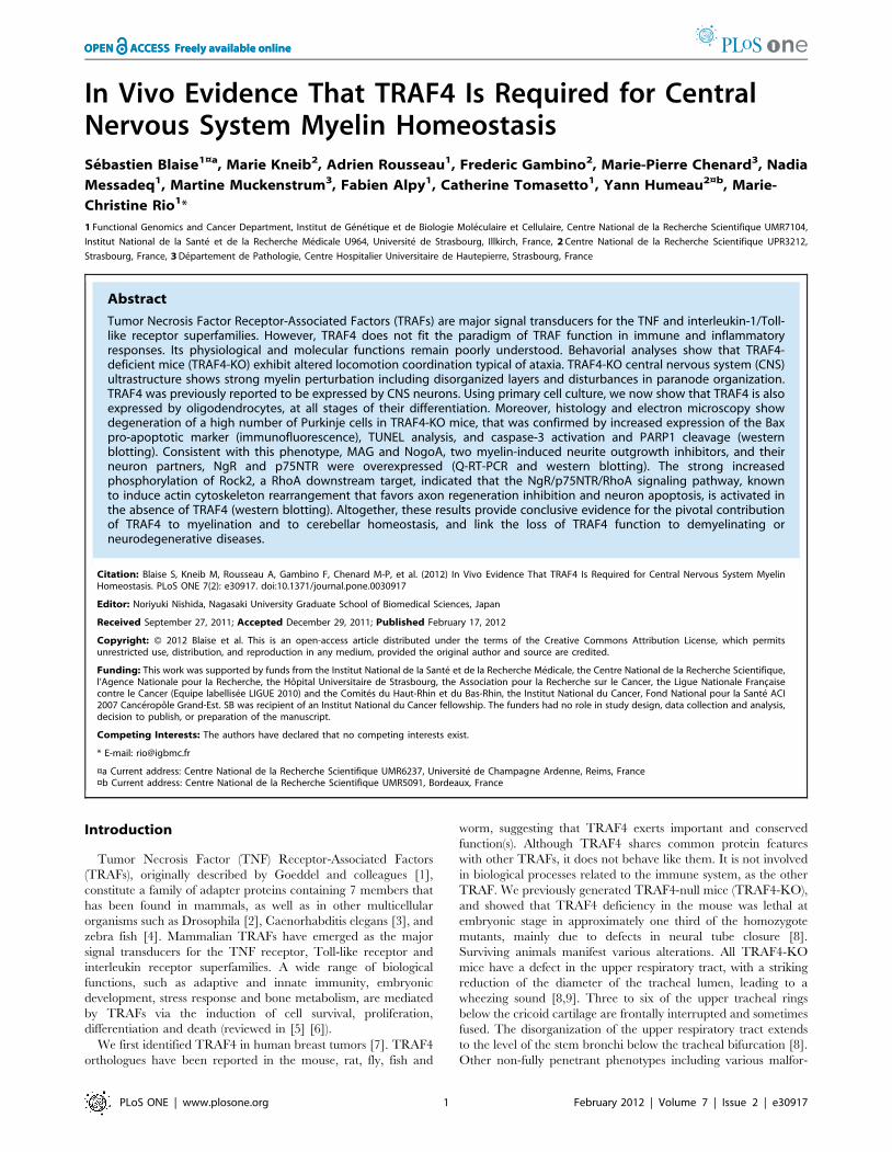
In Vivo Evidence That TRAF4 Is Required for CentralNervous System Myelin HomeostasisSebastien Blaise1¤a, Marie Kneib2, Adrien Rousseau1, Frederic Gambino2, Marie-Pierre Chenard3, Nadia
Messadeq1, Martine Muckenstrum3, Fabien Alpy1, Catherine Tomasetto1, Yann Humeau2¤b, Marie-
Christine Rio1*
1 Functional Genomics and Cancer Department, Institut de Genetique et de Biologie Moleculaire et Cellulaire, Centre National de la Recherche Scientifique UMR7104,
Institut National de la Sante et de la Recherche Medicale U964, Universite de Strasbourg, Illkirch, France, 2 Centre National de la Recherche Scientifique UPR3212,
Strasbourg, France, 3 Departement de Pathologie, Centre Hospitalier Universitaire de Hautepierre, Strasbourg, France
Abstract
Tumor Necrosis Factor Receptor-Associated Factors (TRAFs) are major signal transducers for the TNF and interleukin-1/Toll-like receptor superfamilies. However, TRAF4 does not fit the paradigm of TRAF function in immune and inflammatoryresponses. Its physiological and molecular functions remain poorly understood. Behavorial analyses show that TRAF4-deficient mice (TRAF4-KO) exhibit altered locomotion coordination typical of ataxia. TRAF4-KO central nervous system (CNS)ultrastructure shows strong myelin perturbation including disorganized layers and disturbances in paranode organization.TRAF4 was previously reported to be expressed by CNS neurons. Using primary cell culture, we now show that TRAF4 is alsoexpressed by oligodendrocytes, at all stages of their differentiation. Moreover, histology and electron microscopy showdegeneration of a high number of Purkinje cells in TRAF4-KO mice, that was confirmed by increased expression of the Baxpro-apoptotic marker (immunofluorescence), TUNEL analysis, and caspase-3 activation and PARP1 cleavage (westernblotting). Consistent with this phenotype, MAG and NogoA, two myelin-induced neurite outgrowth inhibitors, and theirneuron partners, NgR and p75NTR were overexpressed (Q-RT-PCR and western blotting). The strong increasedphosphorylation of Rock2, a RhoA downstream target, indicated that the NgR/p75NTR/RhoA signaling pathway, knownto induce actin cytoskeleton rearrangement that favors axon regeneration inhibition and neuron apoptosis, is activated inthe absence of TRAF4 (western blotting). Altogether, these results provide conclusive evidence for the pivotal contributionof TRAF4 to myelination and to cerebellar homeostasis, and link the loss of TRAF4 function to demyelinating orneurodegenerative diseases.
Citation: Blaise S, Kneib M, Rousseau A, Gambino F, Chenard M-P, et al. (2012) In Vivo Evidence That TRAF4 Is Required for Central Nervous System MyelinHomeostasis. PLoS ONE 7(2): e30917. doi:10.1371/journal.pone.0030917
Editor: Noriyuki Nishida, Nagasaki University Graduate School of Biomedical Sciences, Japan
Received September 27, 2011; Accepted December 29, 2011; Published February 17, 2012
Copyright: � 2012 Blaise et al. This is an open-access article distributed under the terms of the Creative Commons Attribution License, which permitsunrestricted use, distribution, and reproduction in any medium, provided the original author and source are credited.
Funding: This work was supported by funds from the Institut National de la Sante et de la Recherche Medicale, the Centre National de la Recherche Scientifique,l’Agence Nationale pour la Recherche, the Hopital Universitaire de Strasbourg, the Association pour la Recherche sur le Cancer, the Ligue Nationale Francaisecontre le Cancer (Equipe labellisee LIGUE 2010) and the Comites du Haut-Rhin et du Bas-Rhin, the Institut National du Cancer, Fond National pour la Sante ACI2007 Canceropole Grand-Est. SB was recipient of an Institut National du Cancer fellowship. The funders had no role in study design, data collection and analysis,decision to publish, or preparation of the manuscript.
Competing Interests: The authors have declared that no competing interests exist.
* E-mail: [email protected]
¤a Current address: Centre National de la Recherche Scientifique UMR6237, Universite de Champagne Ardenne, Reims, France¤b Current address: Centre National de la Recherche Scientifique UMR5091, Bordeaux, France
Introduction
Tumor Necrosis Factor (TNF) Receptor-Associated Factors
(TRAFs), originally described by Goeddel and colleagues [1],
constitute a family of adapter proteins containing 7 members that
has been found in mammals, as well as in other multicellular
organisms such as Drosophila [2], Caenorhabditis elegans [3], and
zebra fish [4]. Mammalian TRAFs have emerged as the major
signal transducers for the TNF receptor, Toll-like receptor and
interleukin receptor superfamilies. A wide range of biological
functions, such as adaptive and innate immunity, embryonic
development, stress response and bone metabolism, are mediated
by TRAFs via the induction of cell survival, proliferation,
differentiation and death (reviewed in [5] [6]).
We first identified TRAF4 in human breast tumors [7]. TRAF4
orthologues have been reported in the mouse, rat, fly, fish and
worm, suggesting that TRAF4 exerts important and conserved
function(s). Although TRAF4 shares common protein features
with other TRAFs, it does not behave like them. It is not involved
in biological processes related to the immune system, as the other
TRAF. We previously generated TRAF4-null mice (TRAF4-KO),
and showed that TRAF4 deficiency in the mouse was lethal at
embryonic stage in approximately one third of the homozygote
mutants, mainly due to defects in neural tube closure [8].
Surviving animals manifest various alterations. All TRAF4-KO
mice have a defect in the upper respiratory tract, with a striking
reduction of the diameter of the tracheal lumen, leading to a
wheezing sound [8,9]. Three to six of the upper tracheal rings
below the cricoid cartilage are frontally interrupted and sometimes
fused. The disorganization of the upper respiratory tract extends
to the level of the stem bronchi below the tracheal bifurcation [8].
Other non-fully penetrant phenotypes including various malfor-
PLoS ONE | www.plosone.org 1 February 2012 | Volume 7 | Issue 2 | e30917
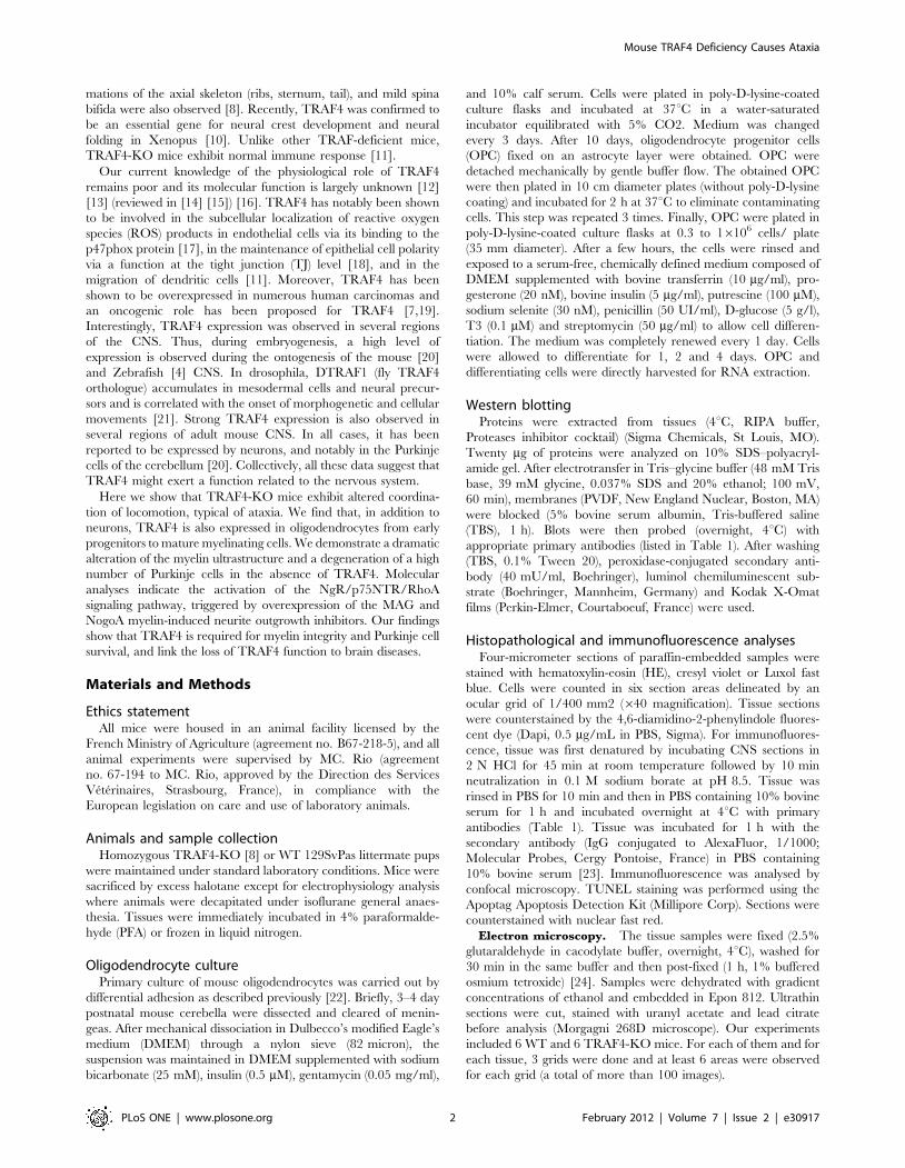
mations of the axial skeleton (ribs, sternum, tail), and mild spina
bifida were also observed [8]. Recently, TRAF4 was confirmed to
be an essential gene for neural crest development and neural
folding in Xenopus [10]. Unlike other TRAF-deficient mice,
TRAF4-KO mice exhibit normal immune response [11].
Our current knowledge of the physiological role of TRAF4
remains poor and its molecular function is largely unknown [12]
[13] (reviewed in [14] [15]) [16]. TRAF4 has notably been shown
to be involved in the subcellular localization of reactive oxygen
species (ROS) products in endothelial cells via its binding to the
p47phox protein [17], in the maintenance of epithelial cell polarity
via a function at the tight junction (TJ) level [18], and in the
migration of dendritic cells [11]. Moreover, TRAF4 has been
shown to be overexpressed in numerous human carcinomas and
an oncogenic role has been proposed for TRAF4 [7,19].
Interestingly, TRAF4 expression was observed in several regions
of the CNS. Thus, during embryogenesis, a high level of
expression is observed during the ontogenesis of the mouse [20]
and Zebrafish [4] CNS. In drosophila, DTRAF1 (fly TRAF4
orthologue) accumulates in mesodermal cells and neural precur-
sors and is correlated with the onset of morphogenetic and cellular
movements [21]. Strong TRAF4 expression is also observed in
several regions of adult mouse CNS. In all cases, it has been
reported to be expressed by neurons, and notably in the Purkinje
cells of the cerebellum [20]. Collectively, all these data suggest that
TRAF4 might exert a function related to the nervous system.
Here we show that TRAF4-KO mice exhibit altered coordina-
tion of locomotion, typical of ataxia. We find that, in addition to
neurons, TRAF4 is also expressed in oligodendrocytes from early
progenitors to mature myelinating cells. We demonstrate a dramatic
alteration of the myelin ultrastructure and a degeneration of a high
number of Purkinje cells in the absence of TRAF4. Molecular
analyses indicate the activation of the NgR/p75NTR/RhoA
signaling pathway, triggered by overexpression of the MAG and
NogoA myelin-induced neurite outgrowth inhibitors. Our findings
show that TRAF4 is required for myelin integrity and Purkinje cell
survival, and link the loss of TRAF4 function to brain diseases.
Materials and Methods
Ethics statementAll mice were housed in an animal facility licensed by the
French Ministry of Agriculture (agreement no. B67-218-5), and all
animal experiments were supervised by MC. Rio (agreement
no. 67-194 to MC. Rio, approved by the Direction des Services
Veterinaires, Strasbourg, France), in compliance with the
European legislation on care and use of laboratory animals.
Animals and sample collectionHomozygous TRAF4-KO [8] or WT 129SvPas littermate pups
were maintained under standard laboratory conditions. Mice were
sacrificed by excess halotane except for electrophysiology analysis
where animals were decapitated under isoflurane general anaes-
thesia. Tissues were immediately incubated in 4% paraformalde-
hyde (PFA) or frozen in liquid nitrogen.
Oligodendrocyte culturePrimary culture of mouse oligodendrocytes was carried out by
differential adhesion as described previously [22]. Briefly, 3–4 day
postnatal mouse cerebella were dissected and cleared of menin-
geas. After mechanical dissociation in Dulbecco’s modified Eagle’s
medium (DMEM) through a nylon sieve (82 micron), the
suspension was maintained in DMEM supplemented with sodium
bicarbonate (25 mM), insulin (0.5 mM), gentamycin (0.05 mg/ml),
and 10% calf serum. Cells were plated in poly-D-lysine-coated
culture flasks and incubated at 37uC in a water-saturated
incubator equilibrated with 5% CO2. Medium was changed
every 3 days. After 10 days, oligodendrocyte progenitor cells
(OPC) fixed on an astrocyte layer were obtained. OPC were
detached mechanically by gentle buffer flow. The obtained OPC
were then plated in 10 cm diameter plates (without poly-D-lysine
coating) and incubated for 2 h at 37uC to eliminate contaminating
cells. This step was repeated 3 times. Finally, OPC were plated in
poly-D-lysine-coated culture flasks at 0.3 to 16106 cells/ plate
(35 mm diameter). After a few hours, the cells were rinsed and
exposed to a serum-free, chemically defined medium composed of
DMEM supplemented with bovine transferrin (10 mg/ml), pro-
gesterone (20 nM), bovine insulin (5 mg/ml), putrescine (100 mM),
sodium selenite (30 nM), penicillin (50 UI/ml), D-glucose (5 g/l),
T3 (0.1 mM) and streptomycin (50 mg/ml) to allow cell differen-
tiation. The medium was completely renewed every 1 day. Cells
were allowed to differentiate for 1, 2 and 4 days. OPC and
differentiating cells were directly harvested for RNA extraction.
Western blottingProteins were extracted from tissues (4uC, RIPA buffer,
Proteases inhibitor cocktail) (Sigma Chemicals, St Louis, MO).
Twenty mg of proteins were analyzed on 10% SDS–polyacryl-
amide gel. After electrotransfer in Tris–glycine buffer (48 mM Tris
base, 39 mM glycine, 0.037% SDS and 20% ethanol; 100 mV,
60 min), membranes (PVDF, New England Nuclear, Boston, MA)
were blocked (5% bovine serum albumin, Tris-buffered saline
(TBS), 1 h). Blots were then probed (overnight, 4uC) with
appropriate primary antibodies (listed in Table 1). After washing
(TBS, 0.1% Tween 20), peroxidase-conjugated secondary anti-
body (40 mU/ml, Boehringer), luminol chemiluminescent sub-
strate (Boehringer, Mannheim, Germany) and Kodak X-Omat
films (Perkin-Elmer, Courtaboeuf, France) were used.
Histopathological and immunofluorescence analysesFour-micrometer sections of paraffin-embedded samples were
stained with hematoxylin-eosin (HE), cresyl violet or Luxol fast
blue. Cells were counted in six section areas delineated by an
ocular grid of 1/400 mm2 (640 magnification). Tissue sections
were counterstained by the 4,6-diamidino-2-phenylindole fluores-
cent dye (Dapi, 0.5 mg/mL in PBS, Sigma). For immunofluores-
cence, tissue was first denatured by incubating CNS sections in
2 N HCl for 45 min at room temperature followed by 10 min
neutralization in 0.1 M sodium borate at pH 8.5. Tissue was
rinsed in PBS for 10 min and then in PBS containing 10% bovine
serum for 1 h and incubated overnight at 4uC with primary
antibodies (Table 1). Tissue was incubated for 1 h with the
secondary antibody (IgG conjugated to AlexaFluor, 1/1000;
Molecular Probes, Cergy Pontoise, France) in PBS containing
10% bovine serum [23]. Immunofluorescence was analysed by
confocal microscopy. TUNEL staining was performed using the
Apoptag Apoptosis Detection Kit (Millipore Corp). Sections were
counterstained with nuclear fast red.
Electron microscopy. The tissue samples were fixed (2.5%
glutaraldehyde in cacodylate buffer, overnight, 4uC), washed for
30 min in the same buffer and then post-fixed (1 h, 1% buffered
osmium tetroxide) [24]. Samples were dehydrated with gradient
concentrations of ethanol and embedded in Epon 812. Ultrathin
sections were cut, stained with uranyl acetate and lead citrate
before analysis (Morgagni 268D microscope). Our experiments
included 6 WT and 6 TRAF4-KO mice. For each of them and for
each tissue, 3 grids were done and at least 6 areas were observed
for each grid (a total of more than 100 images).
Mouse TRAF4 Deficiency Causes Ataxia
PLoS ONE | www.plosone.org 2 February 2012 | Volume 7 | Issue 2 | e30917
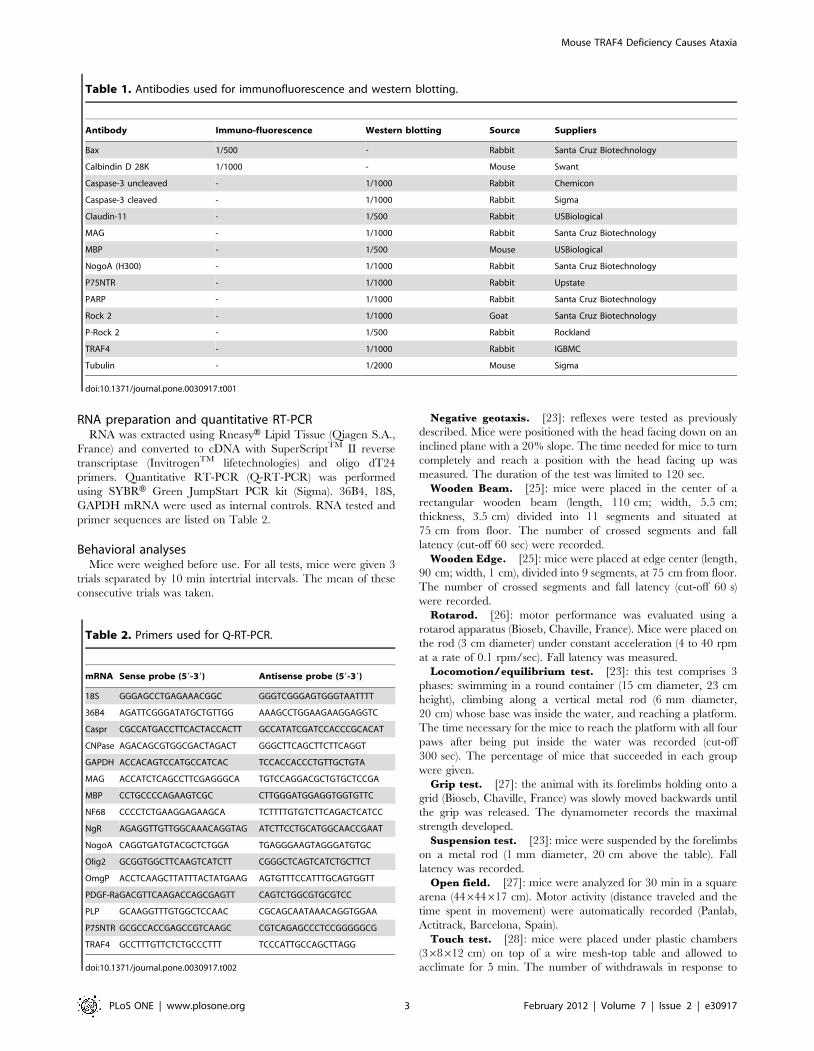
RNA preparation and quantitative RT-PCRRNA was extracted using RneasyH Lipid Tissue (Qiagen S.A.,
France) and converted to cDNA with SuperScriptTM II reverse
transcriptase (InvitrogenTM lifetechnologies) and oligo dT24
primers. Quantitative RT-PCR (Q-RT-PCR) was performed
using SYBRH Green JumpStart PCR kit (Sigma). 36B4, 18S,
GAPDH mRNA were used as internal controls. RNA tested and
primer sequences are listed on Table 2.
Behavioral analysesMice were weighed before use. For all tests, mice were given 3
trials separated by 10 min intertrial intervals. The mean of these
consecutive trials was taken.
Negative geotaxis. [23]: reflexes were tested as previously
described. Mice were positioned with the head facing down on an
inclined plane with a 20% slope. The time needed for mice to turn
completely and reach a position with the head facing up was
measured. The duration of the test was limited to 120 sec.
Wooden Beam. [25]: mice were placed in the center of a
rectangular wooden beam (length, 110 cm; width, 5.5 cm;
thickness, 3.5 cm) divided into 11 segments and situated at
75 cm from floor. The number of crossed segments and fall
latency (cut-off 60 sec) were recorded.
Wooden Edge. [25]: mice were placed at edge center (length,
90 cm; width, 1 cm), divided into 9 segments, at 75 cm from floor.
The number of crossed segments and fall latency (cut-off 60 s)
were recorded.
Rotarod. [26]: motor performance was evaluated using a
rotarod apparatus (Bioseb, Chaville, France). Mice were placed on
the rod (3 cm diameter) under constant acceleration (4 to 40 rpm
at a rate of 0.1 rpm/sec). Fall latency was measured.
Locomotion/equilibrium test. [23]: this test comprises 3
phases: swimming in a round container (15 cm diameter, 23 cm
height), climbing along a vertical metal rod (6 mm diameter,
20 cm) whose base was inside the water, and reaching a platform.
The time necessary for the mice to reach the platform with all four
paws after being put inside the water was recorded (cut-off
300 sec). The percentage of mice that succeeded in each group
were given.
Grip test. [27]: the animal with its forelimbs holding onto a
grid (Bioseb, Chaville, France) was slowly moved backwards until
the grip was released. The dynamometer records the maximal
strength developed.
Suspension test. [23]: mice were suspended by the forelimbs
on a metal rod (1 mm diameter, 20 cm above the table). Fall
latency was recorded.
Open field. [27]: mice were analyzed for 30 min in a square
arena (44644617 cm). Motor activity (distance traveled and the
time spent in movement) were automatically recorded (Panlab,
Actitrack, Barcelona, Spain).
Touch test. [28]: mice were placed under plastic chambers
(368612 cm) on top of a wire mesh-top table and allowed to
acclimate for 5 min. The number of withdrawals in response to
Table 1. Antibodies used for immunofluorescence and western blotting.
Antibody Immuno-fluorescence Western blotting Source Suppliers
Bax 1/500 - Rabbit Santa Cruz Biotechnology
Calbindin D 28K 1/1000 - Mouse Swant
Caspase-3 uncleaved - 1/1000 Rabbit Chemicon
Caspase-3 cleaved - 1/1000 Rabbit Sigma
Claudin-11 - 1/500 Rabbit USBiological
MAG - 1/1000 Rabbit Santa Cruz Biotechnology
MBP - 1/500 Mouse USBiological
NogoA (H300) - 1/1000 Rabbit Santa Cruz Biotechnology
P75NTR - 1/1000 Rabbit Upstate
PARP - 1/1000 Rabbit Santa Cruz Biotechnology
Rock 2 - 1/1000 Goat Santa Cruz Biotechnology
P-Rock 2 - 1/500 Rabbit Rockland
TRAF4 - 1/1000 Rabbit IGBMC
Tubulin - 1/2000 Mouse Sigma
doi:10.1371/journal.pone.0030917.t001
Table 2. Primers used for Q-RT-PCR.
mRNA Sense probe (59-39) Antisense probe (59-39)
18S GGGAGCCTGAGAAACGGC GGGTCGGGAGTGGGTAATTTT
36B4 AGATTCGGGATATGCTGTTGG AAAGCCTGGAAGAAGGAGGTC
Caspr CGCCATGACCTTCACTACCACTT GCCATATCGATCCACCCGCACAT
CNPase AGACAGCGTGGCGACTAGACT GGGCTTCAGCTTCTTCAGGT
GAPDH ACCACAGTCCATGCCATCAC TCCACCACCCTGTTGCTGTA
MAG ACCATCTCAGCCTTCGAGGGCA TGTCCAGGACGCTGTGCTCCGA
MBP CCTGCCCCAGAAGTCGC CTTGGGATGGAGGTGGTGTTC
NF68 CCCCTCTGAAGGAGAAGCA TCTTTTGTGTCTTCAGACTCATCC
NgR AGAGGTTGTTGGCAAACAGGTAG ATCTTCCTGCATGGCAACCGAAT
NogoA CAGGTGATGTACGCTCTGGA TGAGGGAAGTAGGGATGTGC
Olig2 GCGGTGGCTTCAAGTCATCTT CGGGCTCAGTCATCTGCTTCT
OmgP ACCTCAAGCTTATTTACTATGAAG AGTGTTTCCATTTGCAGTGGTT
PDGF-RaGACGTTCAAGACCAGCGAGTT CAGTCTGGCGTGCGTCC
PLP GCAAGGTTTGTGGCTCCAAC CGCAGCAATAAACAGGTGGAA
P75NTR GCGCCACCGAGCCGTCAAGC CGTCAGAGCCCTCCGGGGGCG
TRAF4 GCCTTTGTTCTCTGCCCTTT TCCCATTGCCAGCTTAGG
doi:10.1371/journal.pone.0030917.t002
Mouse TRAF4 Deficiency Causes Ataxia
PLoS ONE | www.plosone.org 3 February 2012 | Volume 7 | Issue 2 | e30917
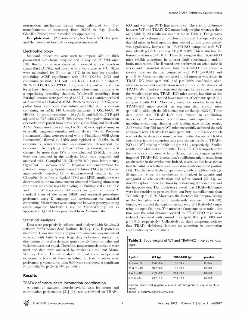
non-noxious mechanical stimuli using calibrated von Frey
monofilaments of increasing force (0.008 to 4 g) (Bioseb,
Chaville, France) were recorded (six applications).
Hot plate test. [29]: mice were placed on a 52uC hot plate
and the latency of forelimb licking were measured.
ElectrophysiologyStandard procedures were used to prepare 300 mm thick
parasagittal slices from 8-day-old and 90-day-old (P8–P90) mice
[30]. Briefly, brains were dissected in ice-cold artificial cerebro-
spinal fluid (ACSF), and sliced with a vibratome at 4uC. Slices
were maintained for 30 min at 32uC in an interface chamber
containing ACSF equilibrated with 95% O2/5% CO2 and
containing (in mM): 124 NaCl, 2.7 KCl, 2 CaCl2, 1.3 MgCl2,
26 NaHCO3, 0.4 NaH2PO4, 10 glucose, 4 ascorbate, and then
for at least 1 hour at room temperature before being transferred to
a superfusing recording chamber. Whole-cell recordings from
Purkinje neurons were performed at 33uC, in a chamber perfused
at 2 ml/min with bubbled ACSF. Patch electrodes (3–5 MV) were
pulled from borosilicate glass tubing and filled with a solution
containing (in mM): 140 Cs-methylsulfonate, 5 QX314-Cl, 10
HEPES, 10 phosphocreatine, 4 Mg-ATP, and 0.3 Na-GTP (pH
adjusted to 7.25 with CsOH, 295 mOsm). Monopolar stimulation
electrodes were pulled from borosilicate glass tubing and filled with
ACSF. Stimulation pulses (1 ms, 1 to 30 mA) were applied using an
externally triggered stimulus isolator device (World Precision
Instruments). Data were recorded with a Multiclamp700B (Axon
Instruments), filtered at 2 kHz and digitized at 10 kHz. In all
experiments, series resistance was monitored throughout the
experiment by applying a hyperpolarizing current, and if it
changed by more than 15% or overwhelmed 25 MV, the data
were not included in the analysis. Data were acquired and
analyzed with: ClampEx10.2, ClampFit10.0 (Axon Instruments),
SigmaPlot 11 software and R language and environment for
statistical computing. Spontaneous Inhibitory PSCs (sIPSC) were
automatically detected by a template-based routine in the
ClampFit 10.0 software. Evoked IPSC and EPSC amplitude were
determined as the maximal current obtained following stimulation
within the molecular layer by holding the Purkinje cell at +10 mV
and 270 mV respectively. All values are given as means 6
standard error of the mean (SEM) and statistical procedures
performed using R language and environment for statistical
computing. Mean values were compared between genotypes using
either unpaired Student’s t test or Mann-Whitney test as
appropriate. QX314 was purchased from Alomone labs.
Statistical AnalysesData were prospectively collected and analyzed with Statview 5
software for Windows (SAS Institute, Berkley, CA). Reported as
means6SD, raw data were compared by using one-way analysis of
variance with Fisher’s test. Regarding behavioral studies, the
distribution of the data deviated quite strongly from normality and
variances were not equal. Therefore, nonparametric statistics were
used and data were analyzed by Student’s t test and Mann-
Whitney U-test. For all analyses, at least three independent
experiments (each of them including at least 6 mice) were
performed. p values lower than 0.05 were considered as significant
(*, p,0.05; **, p,0.01; ***, p,0.001).
Results
TRAF4 deficiency alters locomotion coordinationA panel of standard neurobehavioral tests for motor and
coordination performance [31] [23] was performed on TRAF4-
KO and wild-type (WT) littermate mice. There is no difference
between WT and TRAF4-KO mouse body weights whatever their
age (Table 3). All results are summarized in Table 4. The geotaxis
test was first performed on 8- (closed eyes) and 21- (opened eyes)
day-old mice. At both ages, the time needed to turn up completely
was significantly increased in TRAF4-KO compared with WT
mice (day 8: p,0.001 and day 21: p = 0.002). This is also true for
6-month-old mice (p,0.011). These data suggest that TRAF4-KO
mice exhibit alterations in anterior limb coordination and/or
brain maturation. The Rotarod test performed on adult mice (8
weeks and 6 months) showed that TRAF4-KO mice stayed a
shorter time on the rod compared with WT (p = 0.011 and
p = 0.010). Moreover, the rod speed at fall moment was slower in
TRAF4-KO mice (p = 0.007 and p = 0.029), confirming alter-
ations in movement coordination in adult mice in the absence of
TRAF4. We therefore investigated the equilibrium capacity using
the wooden edge test. TRAF4-KO mice stayed less time at the
edge (p = 0.004) and crossed lower number of segments (p,0.001)
compared with WT. Moreover, using the wooden beam test,
TRAF4-KO mice crossed less segments than control mice
(p = 0.016), although the fall latency was similar (p = 0.273). These
data show that TRAF4-KO mice exhibit an equilibrium
deficiency. A locomotion coordination and equilibrium test
including swimming, climbing and landing gave similar results.
At 8 weeks, four-fold more WT mice successfully achieved the task
compared with TRAF4-KO mice (p = 0.004), a difference which
was not due to decreased muscular force in the absence of TRAF4
since the grip and suspension tests gave similar results in TRAF4-
KO and WT mice (p = 0.060 and p = 0.111, respectively). Similar
results were obtained at 6 months. Thus, TRAF4 is important for
the correct coordination of limbs during exercise, suggesting that
impaired TRAF4-KO locomotion/equilibrium might result from
an alteration in the cerebellum. Indeed, several studies have shown
that the adult cerebellum is implicated in equilibrium and posture
[25]. This behavioral phenotype is not greatly amplified with age
(6 months). Since the cerebellum is involved in agonist and
antagonist muscle coordination and reflex control [32–33], we
directly explored these functions by performing the touch test and
the hot-plate test. The touch test showed that TRAF4-KO mice
were less sensitive to pressure from von Frey monofilaments than
WT mice (p = 0.019). Moreover, the latencies for forepaw licking
in the hot plate test were significantly increased (p = 0.038).
Finally, we studied the exploration capacity of TRAF4-KO mice
using the open field test. The number of movements recorded, the
time and the total distance covered by TRAF4-KO mice were
reduced compared with control mice (p = 0.044, p = 0.008 and
p = 0.013, respectively). Collectively, all these symptoms indicate
that TRAF4 deficiency induces an alteration in locomotion
coordination typical of ataxia.
Table 3. Body weight of WT and TRAF4-KO mice at variousages.
Age/nb WT (g) TRAF4-KO (g) p-value
8 d (n = 18) 15.860.4 16.560.5 0.3276
21 d (n = 18) 18.260.2 18.560.1 0.2660
8 w (n = 18) 21.960.9 22.160.2 0.8690
6 m (n = 8) 29.261.2 26.762.0 0.2873
Data are means6SD; g, gram; n, number of mice/group; d, day; w, week; m,month.doi:10.1371/journal.pone.0030917.t003
Mouse TRAF4 Deficiency Causes Ataxia
PLoS ONE | www.plosone.org 4 February 2012 | Volume 7 | Issue 2 | e30917

TRAF4 deficiency induces myelin alterationMyelin plays a key role in the functioning of the nervous
system by allowing fast saltatory conduction of nerve pulses. Since
it has been reported that myelin disorganization leads to
alterations in locomotion coordination and/or severe ataxia
[34–35], CNS myelin organization was examined in WT and
TRAF4-KO mice. Histological analysis by Luxol fast blue
staining, a marker of myelin, showed that the myelin network
in the cerebellum white matter was disorganized and less dense in
TRAF4-KO mice compared with WT (Figure 1A). Electron
microscopy was performed on 6 WT and 6 TRAF4-KO 8-week-
old mice, investigating both internodal and paranodal regions.
We observed major ultrastructural alterations in the myelin of
TRAF4-KO mice (Figure 1B). The number of myelinated axons
was reduced when compared with WT mice (84.466.6 vs
97.965.1%, p = 0.0005). Moreover, in the WT brain, the
adjacent axons were in contact with each other, while spaces
between the axons were observed in the TRAF4-KO brain
(Figure 1B a and b). The myelin layers surrounding the axons
showed a loss of parallelism in the TRAF4-KO brain compared
with WT (Figure 1B c–f). We then analyzed the ultrastructure of
the optic nerve, another CNS component. Similar myelin
alterations were observed (Figure 1B g and h), indicating that
the myelin abnormalities due to the TRAF4 absence were not
restricted to the cerebellum. Finally, the node of Ranvier, where
lamellae of compact myelin sheats normally terminate in glial
loops to establish the paranodal axoglial septate-like junctions
[36], were also altered. In the WT cervical spinal cord and optic
nerve we found the regular, densely stained transverse bands
(TBs) in almost all of the paranodes examined between the
paranodal loop and axon. Moreover, the paranodal loops contact
the axon (Figure 1B, i and k). In contrast, although the majority
of the TRAF4-KO paranodes exhibited correct morphology, 10–
20% showed dramatic defects (Figure 1B, j and l). Thus, the
attachement of the paranodal loops to the axon, and the space
between the loops and axon may be increased. We observed
loops completely detached and even everted, facing away from
the axonal membrane. Finally, spacing between TBs may be
irregular, or paranodal loops or portions of loops may lack well-
defined TBs. We could not find any similar paranodal myelin
alterations in the WT mice. Similar myelin phenotype was
observed in older animals TRAF4-KO mice (6-month-old).
We then checked for myelin molecular abnormalities in
TRAF4-KO cerebellum. Consistent with our ultrastructural
observations, using western blot and immunofluorescence analy-
ses, we observed a reduction in myelin basic protein (MBP), the
second most abundant protein in the CNS myelin [37] (Figures 1C
and 1D b and f). Moreover, it has been reported that myelin
organization around the axons is maintained by tight-junction-like
structures (TJ) located first between the lamellae of myelin sheaths
and second, between the axon and the inner myelin loop. The
observed TRAF4-KO myelin ultrastructural phenotype suggests
that these structures might be altered in the absence of TRAF4.
We then checked for the presence of oligodendrocyte-specific
protein/Claudin-11 (OSP/cl11) and contactin-associated protein
(Caspr ; also known as paranodin) proteins that are known to be
Table 4. Results of locomotion and behavior tests in WT and TRAF4-KO mice.
Test Age/nb Parameters WT(n = 18) TRAF4-KO p-value
Geotaxis 8 d (n = 8) Time (s) 7.1 (3.1) 20.2 (18.6) 0.0007
21 d (n = 8) Time (s) 6.8 (6.0) 18.3 (40.5) 0.0019
6 m (n = 8) Time (s) 8.5 (4.5) 13.7 (15.0) 0.0112
Rotarod 8 w (n = 18) Fall latency (s) 149.5 (65.0) 89.0 (31.5) 0.0111
Speed (rpm/min) 13.0 (2.0) 9.0 (1.5) 0.0068
6 m (n = 8) Fall latency (s) 92.9 (17.6) 76.0 (20.7) 0.0104
Speed (rpm/min) 10.0 (2.5) 7.0 (1.5) 0.0286
Wooden edge 8 w (n = 18) Fall latency (s) 389.5 (441.8) 28.7 (134.8) 0.0045
Segment (nb) 5.0 (0.0) 3.0 (1.5) 0.0005
Wooden beam 8 w (n = 18) Fall latency (s) 720.0 (0) 720.0 (0.0) 0.2733
Segment (nb) 11.0 (21.0) 6.0 (0.0) 0.0158
Locomotion/equilibrium 8 w (n = 18) Success (%) 61.5 15 0.0036
6 m (n = 8) Success (%) 37.5 0 ,0.001
Grip 8 w (n = 18) Force (N) 95.0 (37.0) 72.1 (38.0) 0.0604
6 m (n = 8) Force (N) 115.0 (49.0) 104.5 (19.0) 0.2264
Suspension 8 w (n = 18) Time (s) 96.3 (12.5) 76.9 (8.8) 0.1110
6 m (n = 8) Time (s) 35.6 (43) 12.2 (22.0) 0.0033
Touch 8 w (n = 18) Pressure (g) 0.5 (0.8) 1.2 (2.5) 0.0193
Hot-plate 8 w (n = 18) Latency (s) 36.1 (7.8) 46.5 (13.3) 0.0378
Open field 8 w (n = 18) Movement (nb) 313.0 (450.5) 105.0 (315.5) 0.0440
Time (s) 183.0 (280.1) 1.3 (34.9) 0.0078
Distance (cm) 1632.1 (3089.7) 60.2 (35.2) 0.0135
Tests were performed with animals aged 8-day-old, 21-day-old, 8-week-old and 6-month-old as indicated. Values correspond to medians with interquartiles inparentheses. Statistical analysis was two-tailed Mann-Whitney U-test. S, second; n, number of mice of each set (WT and TRAF4-KO) involved in the test; nb, number; %,percentage; N, Newton; cm, centimeter.doi:10.1371/journal.pone.0030917.t004
Mouse TRAF4 Deficiency Causes Ataxia
PLoS ONE | www.plosone.org 5 February 2012 | Volume 7 | Issue 2 | e30917
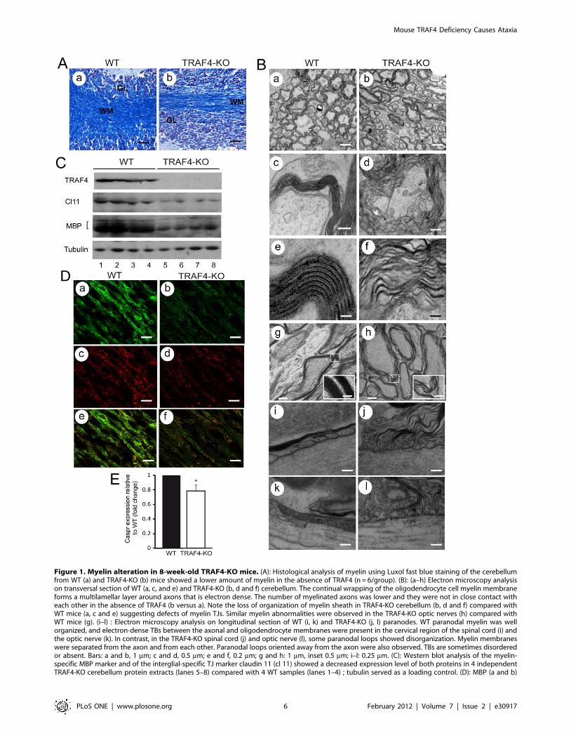
Figure 1. Myelin alteration in 8-week-old TRAF4-KO mice. (A): Histological analysis of myelin using Luxol fast blue staining of the cerebellumfrom WT (a) and TRAF4-KO (b) mice showed a lower amount of myelin in the absence of TRAF4 (n = 6/group). (B): (a–h) Electron microscopy analysison transversal section of WT (a, c, and e) and TRAF4-KO (b, d and f) cerebellum. The continual wrapping of the oligodendrocyte cell myelin membraneforms a multilamellar layer around axons that is electron dense. The number of myelinated axons was lower and they were not in close contact witheach other in the absence of TRAF4 (b versus a). Note the loss of organization of myelin sheath in TRAF4-KO cerebellum (b, d and f) compared withWT mice (a, c and e) suggesting defects of myelin TJs. Similar myelin abnormalities were observed in the TRAF4-KO optic nerves (h) compared withWT mice (g). (i–l) : Electron microscopy analysis on longitudinal section of WT (i, k) and TRAF4-KO (j, l) paranodes. WT paranodal myelin was wellorganized, and electron-dense TBs between the axonal and oligodendrocyte membranes were present in the cervical region of the spinal cord (i) andthe optic nerve (k). In contrast, in the TRAF4-KO spinal cord (j) and optic nerve (l), some paranodal loops showed disorganization. Myelin membraneswere separated from the axon and from each other. Paranodal loops oriented away from the axon were also observed. TBs are sometimes disorderedor absent. Bars: a and b, 1 mm; c and d, 0.5 mm; e and f, 0.2 mm; g and h: 1 mm, inset 0.5 mm; i–l: 0.25 mm. (C): Western blot analysis of the myelin-specific MBP marker and of the interglial-specific TJ marker claudin 11 (cl 11) showed a decreased expression level of both proteins in 4 independentTRAF4-KO cerebellum protein extracts (lanes 5–8) compared with 4 WT samples (lanes 1–4) ; tubulin served as a loading control. (D): MBP (a and b)
Mouse TRAF4 Deficiency Causes Ataxia
PLoS ONE | www.plosone.org 6 February 2012 | Volume 7 | Issue 2 | e30917
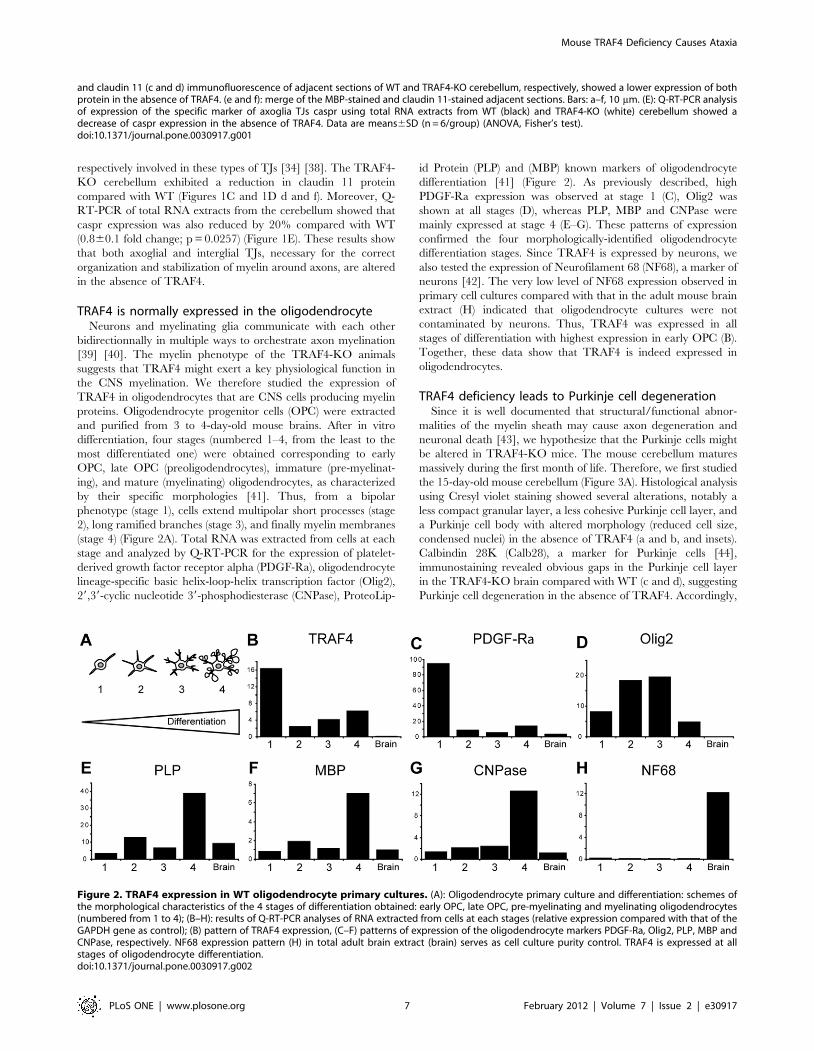
respectively involved in these types of TJs [34] [38]. The TRAF4-
KO cerebellum exhibited a reduction in claudin 11 protein
compared with WT (Figures 1C and 1D d and f). Moreover, Q-
RT-PCR of total RNA extracts from the cerebellum showed that
caspr expression was also reduced by 20% compared with WT
(0.860.1 fold change; p = 0.0257) (Figure 1E). These results show
that both axoglial and interglial TJs, necessary for the correct
organization and stabilization of myelin around axons, are altered
in the absence of TRAF4.
TRAF4 is normally expressed in the oligodendrocyteNeurons and myelinating glia communicate with each other
bidirectionnally in multiple ways to orchestrate axon myelination
[39] [40]. The myelin phenotype of the TRAF4-KO animals
suggests that TRAF4 might exert a key physiological function in
the CNS myelination. We therefore studied the expression of
TRAF4 in oligodendrocytes that are CNS cells producing myelin
proteins. Oligodendrocyte progenitor cells (OPC) were extracted
and purified from 3 to 4-day-old mouse brains. After in vitro
differentiation, four stages (numbered 1–4, from the least to the
most differentiated one) were obtained corresponding to early
OPC, late OPC (preoligodendrocytes), immature (pre-myelinat-
ing), and mature (myelinating) oligodendrocytes, as characterized
by their specific morphologies [41]. Thus, from a bipolar
phenotype (stage 1), cells extend multipolar short processes (stage
2), long ramified branches (stage 3), and finally myelin membranes
(stage 4) (Figure 2A). Total RNA was extracted from cells at each
stage and analyzed by Q-RT-PCR for the expression of platelet-
derived growth factor receptor alpha (PDGF-Ra), oligodendrocyte
lineage-specific basic helix-loop-helix transcription factor (Olig2),
29,39-cyclic nucleotide 39-phosphodiesterase (CNPase), ProteoLip-
id Protein (PLP) and (MBP) known markers of oligodendrocyte
differentiation [41] (Figure 2). As previously described, high
PDGF-Ra expression was observed at stage 1 (C), Olig2 was
shown at all stages (D), whereas PLP, MBP and CNPase were
mainly expressed at stage 4 (E–G). These patterns of expression
confirmed the four morphologically-identified oligodendrocyte
differentiation stages. Since TRAF4 is expressed by neurons, we
also tested the expression of Neurofilament 68 (NF68), a marker of
neurons [42]. The very low level of NF68 expression observed in
primary cell cultures compared with that in the adult mouse brain
extract (H) indicated that oligodendrocyte cultures were not
contaminated by neurons. Thus, TRAF4 was expressed in all
stages of differentiation with highest expression in early OPC (B).
Together, these data show that TRAF4 is indeed expressed in
oligodendrocytes.
TRAF4 deficiency leads to Purkinje cell degenerationSince it is well documented that structural/functional abnor-
malities of the myelin sheath may cause axon degeneration and
neuronal death [43], we hypothesize that the Purkinje cells might
be altered in TRAF4-KO mice. The mouse cerebellum matures
massively during the first month of life. Therefore, we first studied
the 15-day-old mouse cerebellum (Figure 3A). Histological analysis
using Cresyl violet staining showed several alterations, notably a
less compact granular layer, a less cohesive Purkinje cell layer, and
a Purkinje cell body with altered morphology (reduced cell size,
condensed nuclei) in the absence of TRAF4 (a and b, and insets).
Calbindin 28K (Calb28), a marker for Purkinje cells [44],
immunostaining revealed obvious gaps in the Purkinje cell layer
in the TRAF4-KO brain compared with WT (c and d), suggesting
Purkinje cell degeneration in the absence of TRAF4. Accordingly,
and claudin 11 (c and d) immunofluorescence of adjacent sections of WT and TRAF4-KO cerebellum, respectively, showed a lower expression of bothprotein in the absence of TRAF4. (e and f): merge of the MBP-stained and claudin 11-stained adjacent sections. Bars: a–f, 10 mm. (E): Q-RT-PCR analysisof expression of the specific marker of axoglia TJs caspr using total RNA extracts from WT (black) and TRAF4-KO (white) cerebellum showed adecrease of caspr expression in the absence of TRAF4. Data are means6SD (n = 6/group) (ANOVA, Fisher’s test).doi:10.1371/journal.pone.0030917.g001
Figure 2. TRAF4 expression in WT oligodendrocyte primary cultures. (A): Oligodendrocyte primary culture and differentiation: schemes ofthe morphological characteristics of the 4 stages of differentiation obtained: early OPC, late OPC, pre-myelinating and myelinating oligodendrocytes(numbered from 1 to 4); (B–H): results of Q-RT-PCR analyses of RNA extracted from cells at each stages (relative expression compared with that of theGAPDH gene as control); (B) pattern of TRAF4 expression, (C–F) patterns of expression of the oligodendrocyte markers PDGF-Ra, Olig2, PLP, MBP andCNPase, respectively. NF68 expression pattern (H) in total adult brain extract (brain) serves as cell culture purity control. TRAF4 is expressed at allstages of oligodendrocyte differentiation.doi:10.1371/journal.pone.0030917.g002
Mouse TRAF4 Deficiency Causes Ataxia
PLoS ONE | www.plosone.org 7 February 2012 | Volume 7 | Issue 2 | e30917
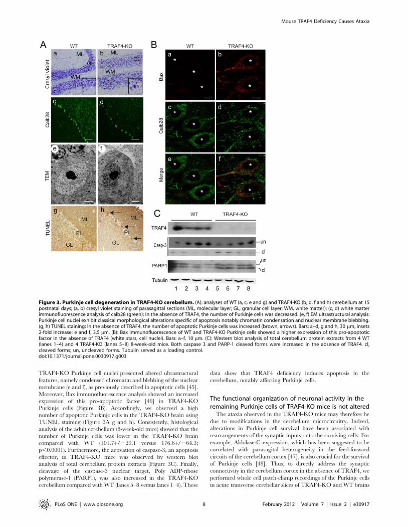
TRAF4-KO Purkinje cell nuclei presented altered ultrastructural
features, namely condensed chromatin and blebbing of the nuclear
membrane (e and f), as previously described in apoptotic cells [45].
Moreover, Bax immunofluorescence analysis showed an increased
expression of this pro-apoptotic factor [46] in TRAF4-KO
Purkinje cells (Figure 3B). Accordingly, we observed a high
number of apoptotic Purkinje cells in the TRAF4-KO brain using
TUNEL staining (Figure 3A g and h). Consistently, histological
analysis of the adult cerebellum (8-week-old mice) showed that the
number of Purkinje cells was lower in the TRAF4-KO brain
compared with WT (101.7+/229.1 versus 176.6+/264.3;
p,0.0001). Furthermore, the activation of caspase-3, an apoptosis
effector, in TRAF4-KO mice was observed by western blot
analysis of total cerebellum protein extracts (Figure 3C). Finally,
cleavage of the caspase-3 nuclear target, Poly ADP-ribose
polymerase-1 (PARP1), was also increased in the TRAF4-KO
cerebellum compared with WT (lanes 5–8 versus lanes 1–4). These
data show that TRAF4 deficiency induces apoptosis in the
cerebellum, notably affecting Purkinje cells.
The functional organization of neuronal activity in theremaining Purkinje cells of TRAF4-KO mice is not altered
The ataxia observed in the TRAF4-KO mice may therefore be
due to modifications in the cerebellum microcircuitry. Indeed,
alterations in Purkinje cell survival have been associated with
rearrangements of the synaptic inputs onto the surviving cells. For
example, Aldolase-C expression, which has been suggested to be
correlated with parasagital heterogeneity in the feed-forward
circuits of the cerebellum cortex [47], is also crucial for the survival
of Purkinje cells [48]. Thus, to directly address the synaptic
connectivity in the cerebellum cortex in the absence of TRAF4, we
performed whole cell patch-clamp recordings of the Purkinje cells
in acute transverse cerebellar slices of TRAF4-KO and WT brains
Figure 3. Purkinje cell degeneration in TRAF4-KO cerebellum. (A): analyses of WT (a, c, e and g) and TRAF4-KO (b, d, f and h) cerebellum at 15postnatal days; (a, b) cresyl violet staining of parasagittal sections (ML, molecular layer; GL, granular cell layer; WM, white matter); (c, d) white matterimmunofluorescence analysis of calb28 (green); In the absence of TRAF4, the number of Purkinje cells was decreased. (e, f) EM ultrastructural analysis:Purkinje cell nuclei exhibit classical morphological alterations specific of apoptosis notably chromatin condensation and nuclear membrane blebbing.(g, h) TUNEL staining: In the absence of TRAF4, the number of apoptotic Purkinje cells was increased (brown, arrows). Bars: a–d, g and h, 30 mm, insets2-fold increase; e and f, 3.5 mm. (B): Bax immunofluorescence of WT and TRAF4-KO Purkinje cells showed a higher expression of this pro-apoptoticfactor in the absence of TRAF4 (white stars, cell nuclei). Bars: a–f, 10 mm. (C): Western blot analysis of total cerebellum protein extracts from 4 WT(lanes 1–4) and 4 TRAF4-KO (lanes 5–8) 8-week-old mice. Both caspase 3 and PARP-1 cleaved forms were increased in the absence of TRAF4. cl,cleaved forms; un, uncleaved forms. Tubulin served as a loading control.doi:10.1371/journal.pone.0030917.g003
Mouse TRAF4 Deficiency Causes Ataxia
PLoS ONE | www.plosone.org 8 February 2012 | Volume 7 | Issue 2 | e30917
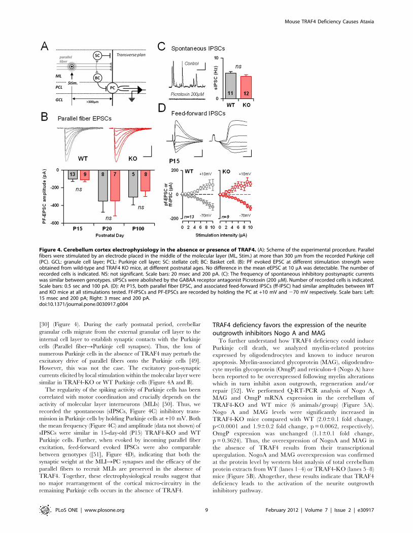
[30] (Figure 4). During the early postnatal period, cerebellar
granular cells migrate from the external granular cell layer to the
internal cell layer to establish synaptic contacts with the Purkinje
cells (Parallel fiberRPurkinje cell synapses). Thus, the loss of
numerous Purkinje cells in the absence of TRAF4 may perturb the
excitatory drive of parallel fibers onto the Purkinje cells [49].
However, this was not the case. The excitatory post-synaptic
currents elicited by local stimulation within the molecular layer were
similar in TRAF4-KO or WT Purkinje cells (Figure 4A and B).
The regularity of the spiking activity of Purkinje cells has been
correlated with motor coordination and crucially depends on the
activity of molecular layer interneurons (MLIs) [50]. Thus, we
recorded the spontaneous (sIPSCs, Figure 4C) inhibitory trans-
mission in Purkinje cells by holding Purkinje cells at +10 mV. Both
the mean frequency (Figure 4C) and amplitude (data not shown) of
sIPSCs were similar in 15-day-old (P15) TRAF4-KO and WT
Purkinje cells. Further, when evoked by incoming parallel fiber
excitation, feed-forward evoked IPSCs were also comparable
between genotypes ([51], Figure 4D), indicating that both the
synaptic weight at the MLIRPC synapses and the efficacy of the
parallel fibers to recruit MLIs are preserved in the absence of
TRAF4. Together, these electrophysiological results suggest that
no major rearrangement of the cortical micro-circuitry in the
remaining Purkinje cells occurs in the absence of TRAF4.
TRAF4 deficiency favors the expression of the neuriteoutgrowth inhibitors Nogo A and MAG
To further understand how TRAF4 deficiency could induce
Purkinje cell death, we analyzed myelin-related proteins
expressed by oligodendrocytes and known to induce neuron
apoptosis. Myelin-associated glycoprotein (MAG), oligodendro-
cyte myelin glycoprotein (OmgP) and reticulon-4 (Nogo A) have
been reported to be overexpressed following myelin alterations
which in turn inhibit axon outgrowth, regeneration and/or
repair [52]. We performed Q-RT-PCR analysis of Nogo A,
MAG and OmgP mRNA expression in the cerebellum of
TRAF4-KO and WT mice (6 animals/group) (Figure 5A).
Nogo A and MAG levels were significantly increased in
TRAF4-KO mice compared with WT (2.060.1 fold change,
p,0.0001 and 1.960.2 fold change, p = 0.0062, respectively).
OmgP expression was unchanged (1.160.1 fold change,
p = 0.3624). Thus, the overexpression of NogoA and MAG in
the absence of TRAF4 results from their transcriptional
upregulation. NogoA and MAG overexpression was confirmed
at the protein level by western blot analysis of total cerebellum
protein extracts from WT (lanes 1–4) or TRAF4-KO (lanes 5–8)
mice (Figure 5B). Altogether, these results indicate that TRAF4
deficiency leads to the activation of the neurite outgrowth
inhibitory pathway.
Figure 4. Cerebellum cortex electrophysiology in the absence or presence of TRAF4. (A): Scheme of the experimental procedure. Parallelfibers were stimulated by an electrode placed in the middle of the molecular layer (ML, Stim.) at more than 300 mm from the recorded Purkinje cell(PC). GCL: granule cell layer; PCL: Purkinje cell layer; SC: stellate cell; BC: Basket cell. (B): PF evoked EPSC at different stimulation strength wereobtained from wild-type and TRAF4 KO mice, at different postnatal ages. No difference in the mean eEPSC at 10 mA was detectable. The number ofrecorded cells is indicated. NS: not significant. Scale bars: 20 msec and 200 pA. (C): The frequency of spontaneous inhibitory postsynaptic currentswas similar between genotypes. sIPSCs were abolished by the GABAA receptor antagonist Picrotoxin (200 mM). Number of recorded cells is indicated.Scale bars: 0.5 sec and 100 pA. (D): At P15, both parallel fiber EPSC, and associated feed-forward IPSCs (ff-IPSC) had similar amplitudes between WTand KO mice at all stimulations tested. Ff-IPSCs and PF-EPSCs are recorded by holding the PC at +10 mV and 270 mV respectively. Scale bars: Left:15 msec and 200 pA; Right: 3 msec and 200 pA.doi:10.1371/journal.pone.0030917.g004
Mouse TRAF4 Deficiency Causes Ataxia
PLoS ONE | www.plosone.org 9 February 2012 | Volume 7 | Issue 2 | e30917
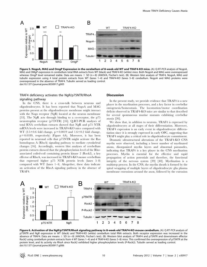
TRAF4 deficiency activates the NgR/p75NTR/RhoAsignaling pathway
In the CNS, there is a cross-talk between neurons and
oligodendrocytes. It has been reported that NogoA and MAG
proteins present at the oligodendrocyte membrane might interact
with the Nogo receptor (NgR) located at the neuron membrane
[53]. The NgR acts through binding to a co-receptor, the p75
neurotrophin receptor (p75NTR) [16]. Q-RT-PCR analyses of
total RNA cerebellum extracts showed that NgR and p75 NTR
mRNA levels were increased in TRAF4-KO mice compared with
WT (2.560.6 fold change, p = 0.0019 and 1.660.2 fold change,
p = 0.0189, respectively) (Figure 6A). Moreover, it has been
reported in neuronal cells that p75NTR might activate the Ras
homologous A (RhoA) signaling pathway to mediate cytoskeletal
changes [16]. Accordingly, western blot analyses of cerebellum
protein extracts showed that the phosphorylation level of the Rho-
associated coiled-coil containing protein kinase 2 (Rock2), a key
effector of RhoA, was increased in TRAF4-KO mouse cerebellum
that expressed higher p75 NTR protein levels (lanes 5–8)
compared with WT (lanes 1–4). Altogether, these data indicate
an activation of the RhoA signaling pathway in the absence of
TRAF4.
Discussion
In the present study, we provide evidence that TRAF4 is a new
player in the myelination processes, and a key factor in cerebellar
ontogenesis/homeostasis. The locomotion/motor coordination
deficits observed in TRAF4-KO mice are similar to that described
for several spontaneous murine mutants exhibiting cerebellar
ataxia [26].
We show that, in addition to neurons, TRAF4 is expressed by
oligodendrocytes at all stages of their differentiation. Moreover,
TRAF4 expression is an early event in oligodendrocyte differen-
tiation since it is strongly expressed in early OPC, suggesting that
TRAF4 might play a critical role in oligodendrocyte commitment.
Dramatic ultrastructural alterations of the TRAF4-KO CNS
myelin were observed, including a lower number of myelinated
axons, disorganized myelin layers and abnormal paranodes,
indicating that TRAF4 is a key player in the CNS myelination
processes. Myelin is essential for the effective and rapid
propagation of action potentials and therefore, the functional
integrity of the nervous system [39] [40]. Myelination is a
multistep process. In the CNS, the myelin sheath is formed by the
spiral wrapping of multiple layers of oligodendrocyte glia plasma
membrane extensions around the axon, followed by the extrusion
Figure 5. NogoA, MAG and OmgP Expression in the cerebellum of 8-week-old WT and TRAF4-KO mice. (A): Q-RT-PCR analysis of NogoA,MAG and OMgP expression in cerebellum total RNA extracts from WT (black) and TRAF4-KO (white) mice. Both NogoA and MAG were overexpressedwhereas OmgP level remained stable. Data are means 6 SD (n = 8) (ANOVA, Fischer’s test). (B): Western blot analysis of TRAF4, NogoA, MAG andtubulin expression using 4 total protein extracts from WT (lanes 1–4) and TRAF4-KO (lanes 5–8) cerebellum. NogoA and MAG proteins wereoverexpressed in the absence of TRAF4. Tubulin served as loading control.doi:10.1371/journal.pone.0030917.g005
Figure 6. Activation of the NgR/p75NTR/RhoA signaling pathway in 8-week-old TRAF4-KO mouse cerebellum. (A): Q-RT-PCR analysis ofp75NTR and NgR expression in WT (black) and TRAF4-KO (white) cerebellum total RNA extracts. Both receptor expression was increased in theabsence of TRAF4. Data are means 6 SD (n = 8) (ANOVA, Fischer’s test). (B): Western blot analysis of TRAF4 and p75NTR and downstream effectorRock2 using cerebellum protein extracts from 4 WT (lanes 1–4) and 4 TRAF4-KO (lanes 5–8) mice. This confirmed the overexpression of p75NTR at theprotein level, and its activity via RhoA since Rock2 exhibited higher phosphorylation levels (P-Rock2). Tubulin served as loading control.doi:10.1371/journal.pone.0030917.g006
Mouse TRAF4 Deficiency Causes Ataxia
PLoS ONE | www.plosone.org 10 February 2012 | Volume 7 | Issue 2 | e30917
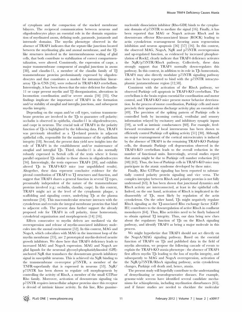
of cytoplasm and the compaction of the stacked membrane
bilayers. The reciprocal communication between neurons and
oligodendrocytes plays an essential role in the domain organiza-
tion of myelinated axons, defining node, paranode, juxtanode and
internode domains. The myelin phenotype observed in the
absence of TRAF4 indicates that the septate-like junctions located
between the myelinating glia and axonal membrane, and the TJ-
like structures involved in the intermembranous sealing of glial
cells, that both contribute to stabilization of correct compartmen-
talization, were altered. Consistently, the expression of caspr, a
major transmembrane component of axoglial junctions in axons
[38], and claudin-11, a member of the claudin family of TJ
transmembrane proteins predominantly expressed by oligoden-
drocytes and that constitutes a marker for intramyelinic linear-
array TJs in CNS [34], were reduced in TRAF4-KO cerebellum.
Interestingly, it has been shown that the mice deficient for claudin-
11 or caspr present myelin and TJ disorganization, alterations in
locomotion coordination and/or severe ataxia [34] [35]. Our
findings implicate the importance of TRAF4 in the formation
and/or stability of axoglial and interglia junctions, and subsequent
myelin integrity.
Depending on the organ and/or cell types, various transmem-
brane proteins are involved in the TJs to guarantee cell polarity:
occludin is observed in epithelia, claudin-11 in oligodendrocytes,
and caspr in neurons. The crucial task of TRAF4 in the polarity
function of TJs is highlighted by the following data. First, TRAF4
was previously identified as a TJ-related protein in adjacent
epithelial cells, responsible for the maintenance of the epithelium
integrity [18]. Second, the present data yield new insights into the
role of TRAF4 in the establishment and/or maintenance of
axoglial and interglial TJs. Third, claudin-11 is also normally
robustly expressed in Sertoli cells of the testis where it forms
parallel organized TJs similar to those shown in oligodendrocytes
[34]. Interestingly, the testis expresses TRAF4 [20], and exhibits
altered TJs in TRAF4-KO mice (our unpublished results).
Altogether, these data represent conclusive evidence for the
pivotal contribution of TRAF4 to TJ structures and function, and
suggest that TRAF4 shares a general function in several TJ types,
presumably independent of the nature of the transmembrane
proteins involved (e.g.: occludin, claudin, caspr). In this context,
TRAF4 might act at the level of the cytoplasmic plaque, a
scaffolding and signaling center, underlying TJs at the plasma
membrane [54]. This macromolecular structure interacts with the
cytoskeleton and recruits the integral membrane proteins that bind
to adjacent cells. The present data further support the already
proposed role for TRAF4 in cell polarity, tissue homeostasis,
cytoskeletal organization and morphogenesis [14] [16].
Effects consecutive to myelin defects are mediated via the
overexpression and release of myelin-associated inhibitory mole-
cules into the axonal environment [52]. In this context, MAG and
NogoA, which colocalizes with MAG in the innermost loop of the
myelin membrane [55], are 2 prototypical myelin-derived neurite
growth inhibitors. We show here that TRAF4 deficiency leads to
increased MAG and NogoA expression. MAG and NogoA are
glial ligands for the neuronal glycosyl-phosphatidylinositol (GPI)-
anchored NgR that transduces the downstream growth inhibitory
signal in susceptible neurons. This is achieved via NgR binding to
the transmembrane co-receptor p75NTR, a member of the
TNFR-superfamily that is expressed in neurons [53]. NgR/
p75NTR has been shown to regulate cell morphogenesis by
controlling the activity of RhoA, a member of the small GTPase
Rho family. Moreover, activation of downstream signaling via
p75NTR requires intracellular adaptor proteins since this receptor
is devoid of intrinsic kinase activity. In this line, Rho guanine-
nucleotide dissociation inhibitor (Rho-GDI) binds to the cytoplas-
mic domain of p75NTR to mediate the signal [16]. Finally, it has
been reported that MAG or NogoA activate RhoA and its
downstream effector Rho-associated kinase (ROCK) leading to
actin cytoskeleton rearrangement favoring axon regeneration
inhibition and neuron apoptosis [56] [57] [16]. In this context,
the observed MAG, NogoA, NgR and p75NTR overexpression
and up-regulated function, as evidenced by increased phosphor-
ylation of Rock2, clearly indicate that TRAF4 deficiency activates
the NgR/p75NTR/RhoA pathway. Collectively, these data
strongly support that TRAF4 normally down-regulates this
pathway. In this context, in addition to its role in TJs homeostasis,
TRAF4 may also directly modulate p75NTR signaling pathway
since it has been reported to bind with the p75NTR intracyto-
plasmic juxtamembrane region [12,58].
Consistent with the activation of the RhoA pathway, we
observed Purkinje cell apoptosis in TRAF4-KO cerebellum. The
cerebellum is the brain region essential for coordination and motor
control, and TRAF4-KO mice present motor behavior deteriora-
tion. In the process of motor coordination, Purkinje cells and more
precisely their spontaneous discharge activity play an essential role
[49]. The precision of the spiking pattern of Purkinje cells is
controlled both by incoming cortical, vestibular and sensory
information relayed by excitatory and inhibitory synaptic inputs
[59], as well as intrinsic conductances [60]. For example, feed-
forward recruitment of local interneurons has been shown to
efficiently control Purkinje cell spiking activity [51] [30]. Although
no major rearrangement of the cortical microcircuitry is induced
by the absence of TRAF4 in the remaining TRAF4-KO Purkinje
cells, the dramatic Purkinje cell degeneration observed in the
TRAF4-KO cerebellum leads to the overall reduction in the
number of functional units. Interestingly, several studies report
that ataxia might be due to Purkinje cell number reduction [61]
[48] [62]. Thus, the loss of Purkinje cells in TRAF4-KO mice may
participate in the ataxic syndrome developed by these mice.
Finally, Rho GTPase signaling has been reported to substan-
tially control polarity protein signaling and vice versa. The
complex interplay between RhoA and polarity proteins is not fully
understood [63]. It has been shown that junctional dynamics and
RhoA activity are interconnected, at least in the epithelial cells.
Indeed, on the one hand, activation of RhoA is implicated in the
disassembly of TJs, most likely by regulation of the actin
cytoskeleton. On the other hand, TJs might negatively regulate
RhoA signaling as the TJ-associated Rho exchange factor (GEF-
H1) contributes to the downregulation of active RhoA in confluent
monolayers [64]. Thus, Rho activities need to be finely balanced
to obtain optimal TJ integrity. Thus, our data bring new clues
about the regulatory function of TJs on the RhoA signaling
pathway, and identify TRAF4 as being a major molecule in this
process.
We might hypothetize that TRAF4 should not act directly on
the NogoA/MAG signaling pathway. Based on the essential
function of TRAF4 on TJs and published data in the field of
myelin alteration, we propose the following cascade of events to
explain the TRAF4-KO ataxia phenotype : the absence of TRAF4
first affects myelin TJs leading to the loss of myelin integrity, and
subsequently to MAG and NogoA overexpression, activation of
the NgR/p75NTR/RhoA signaling pathway, actin cytoskeleton
changes, Purkinje cell death and, hence, ataxia.
The present study will hopefully contribute to the understanding
of demyelinating or neurodegenerative diseases. For example,
genome-wide screens have identified several candidate mecha-
nisms for schizophrenia, including myelination disturbances [65],
and if future studies are needed to elucidate the molecular
Mouse TRAF4 Deficiency Causes Ataxia
PLoS ONE | www.plosone.org 11 February 2012 | Volume 7 | Issue 2 | e30917
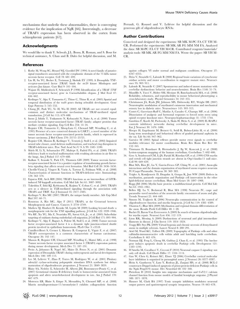
mechanisms that underlie these abnormalities, there is converging
evidence for the implication of NgR [66]. Interestingly, a decrease
of TRAF4 expression has been observed in the cortex from
schizophrenic patients [67].
Acknowledgments
We would like to thank Y. Schwab, J.L. Bossu, R. Roman, and S. Bour for
technical assistance, S. Chan and B. Dales for helpful discussion, and M.
Perrault, G. Roussel and V. Lelievre for helpful discussion and the
generous gift of oligodendrocyte RNAs.
Author Contributions
Conceived and designed the experiments: SB MK M-PC FA CT YH M-
CR. Performed the experiments: SB MK AR FG MM NM FA. Analyzed
the data: SB M-PC FA CT YH M-CR. Contributed reagents/materials/
analysis tools: SB MK AR FG MM NM FA. Wrote the paper: SB YH M-
CR.
References
1. Rothe M, Wong SC, Henzel WJ, Goeddel DV (1994) A novel family of putative
signal transducers associated with the cytoplasmic domain of the 75 kDa tumor
necrosis factor receptor. Cell 78: 681–692.
2. Liu H, Su YC, Becker E, Treisman J, Skolnik EY (1999) A Drosophila TNF-
receptor-associated factor (TRAF) binds the ste20 kinase Misshapen and
activates Jun kinase. Curr Biol 9: 101–104.
3. Wajant H, Muhlenbeck F, Scheurich P (1998) Identification of a TRAF (TNF
receptor-associated factor) gene in Caenorhabditis elegans. J Mol Evol 47:
656–662.
4. Kedinger V, Alpy F, Tomasetto C, Thisse C, Thisse B, et al. (2005) Spatial and
temporal distribution of the traf4 genes during zebrafish development. Gene
Expr Patterns 5: 545–552.
5. Chung JY, Park YC, Ye H, Wu H (2002) All TRAFs are not created equal:
common and distinct molecular mechanisms of TRAF-mediated signal
transduction. J Cell Sci 115: 679–688.
6. Inoue J, Ishida T, Tsukamoto N, Kobayashi N, Naito A, et al. (2000) Tumor
necrosis factor receptor-associated factor (TRAF) family: adapter proteins that
mediate cytokine signaling. Exp Cell Res 254: 14–24.
7. Regnier CH, Tomasetto C, Moog-Lutz C, Chenard MP, Wendling C, et al.
(1995) Presence of a new conserved domain in CART1, a novel member of the
tumor necrosis factor receptor-associated protein family, which is expressed in
breast carcinoma. J Biol Chem 270: 25715–25721.
8. Regnier CH, Masson R, Kedinger V, Textoris J, Stoll I, et al. (2002) Impaired
neural tube closure, axial skeleton malformations, and tracheal ring disruption in
TRAF4-deficient mice. Proc Natl Acad Sci U S A 99: 5585–5590.
9. Shiels H, Li X, Schumacker PT, Maltepe E, Padrid PA, et al. (2000) TRAF4
deficiency leads to tracheal malformation with resulting alterations in air flow to
the lungs. Am J Pathol 157: 679–688.
10. Kalkan T, Iwasaki Y, Park CY, Thomsen GH (2009) Tumor necrosis factor-
receptor-associated factor-4 is a positive regulator of transforming growth factor-
beta signaling that affects neural crest formation. Mol Biol Cell 20: 3436–3450.
11. Cherfils-Vicini J, Vingert B, Varin A, Tartour E, Fridman WH, et al. (2008)
Characterization of immune functions in TRAF4-deficient mice. Immunology
124: 562–574.
12. Esparza EM, Arch RH (2004) TRAF4 functions as an intermediate of GITR-
induced NF-kappaB activation. Cell Mol Life Sci 61: 3087–3092.
13. Takeshita F, Ishii KJ, Kobiyama K, Kojima Y, Coban C, et al. (2005) TRAF4
acts as a silencer in TLR-mediated signaling through the association with
TRAF6 and TRIF. Eur J Immunol 35: 2477–2485.
14. Kedinger V, Rio MC (2007) TRAF4, the unique family member. Adv Exp Med
Biol 597: 60–71.
15. Rousseau A, Rio MC, Alpy F (2011) TRAF4, at the Crossroad between
Morphogenesis and Cancer. Cancers 3: 2734–2749.
16. Mathew SJ, Haubert D, Kronke M, Leptin M (2009) Looking beyond death: a
morphogenetic role for the TNF signalling pathway. J Cell Sci 122: 1939–1946.
17. Wu RF, Xu YC, Ma Z, Nwariaku FE, Sarosi GA, Jr., et al. (2005) Subcellular
targeting of oxidants during endothelial cell migration. J Cell Biol 171: 893–904.
18. Kedinger V, Alpy F, Baguet A, Polette M, Stoll I, et al. (2008) Tumor necrosis
factor receptor-associated factor 4 is a dynamic tight junction-related shuttle
protein involved in epithelium homeostasis. PLoS One 3: e3518.
19. Camilleri-Broet S, Cremer I, Marmey B, Comperat E, Viguie F, et al. (2007)
TRAF4 overexpression is a common characteristic of human carcinomas.
Oncogene 26: 142–147.
20. Masson R, Regnier CH, Chenard MP, Wendling C, Mattei MG, et al. (1998)
Tumor necrosis factor receptor associated factor 4 (TRAF4) expression pattern
during mouse development. Mech Dev 71: 187–191.
21. Preiss A, Johannes B, Nagel AC, Maier D, Peters N, et al. (2001) Dynamic
expression of Drosophila TRAF1 during embryogenesis and larval development.
Mech Dev 100: 109–113.
22. Lee M, Lelievre V, Zhao P, Torres M, Rodriguez W, et al. (2001) Pituitary
adenylyl cyclase-activating polypeptide stimulates DNA synthesis but delays
maturation of oligodendrocyte progenitors. J Neurosci 21: 3849–3859.
23. Blaise SA, Nedelec E, Schroeder H, Alberto JM, Bossenmeyer-Pourie C, et al.
(2007) Gestational vitamin B deficiency leads to homocysteine-associated brain
apoptosis and alters neurobehavioral development in rats. Am J Pathol 170:
667–679.
24. Motrescu ER, Blaise S, Etique N, Messaddeq N, Chenard MP, et al. (2008)
Matrix metalloproteinase-11/stromelysin-3 exhibits collagenolytic function
against collagen VI under normal and malignant conditions. Oncogene 27:
6347–6355.
25. Deiss V, Strazielle C, Lalonde R (2000) Regional brain variations of cytochrome
oxidase activity and motor co-ordination in staggerer mutant mice. Neurosci-ence 95: 903–911.
26. Lalonde R, Strazielle C (2007) Spontaneous and induced mouse mutations with
cerebellar dysfunctions: behavior and neurochemistry. Brain Res 1140: 51–74.
27. Mandillo S, Tucci V, Holter SM, Meziane H, Banchaabouchi MA, et al. (2008)Reliability, robustness, and reproducibility in mouse behavioral phenotyping: a
cross-laboratory study. Physiol Genomics 34: 243–255.
28. Christianson JA, Ryals JM, Johnson MS, Dobrowsky RT, Wright DE (2007)
Neurotrophic modulation of myelinated cutaneous innervation and mechanicalsensory loss in diabetic mice. Neuroscience 145: 303–313.
29. Contet C, Gaveriaux-Ruff C, Matifas A, Caradec C, Champy MF, et al. (2006)
Dissociation of analgesic and hormonal responses to forced swim stress usingopioid receptor knockout mice. Neuropsychopharmacology 31: 1733–1744.
30. Gambino F, Kneib M, Pavlowsky A, Skala H, Heitz S, et al. (2009) IL1RAPL1controls inhibitory networks during cerebellar development in mice.
Eur J Neurosci 30: 1476–1486.
31. Hoeger H, Engelmann M, Bernert G, Seidl R, Bubna-Littitz H, et al. (2000)Long term neurological and behavioral effects of graded perinatal asphyxia in
the rat. Life Sci 66: 947–962.
32. Garwicz M (2002) Spinal reflexes provide motor error signals to cerebellar
modules–relevance for motor coordination. Brain Res Brain Res Rev 40:152–165.
33. Timmann D, Brandauer B, Hermsdorfer J, Ilg W, Konczak J, et al. (2008)
Lesion-symptom mapping of the human cerebellum. Cerebellum 7: 602–606.
34. Gow A, Southwood CM, Li JS, Pariali M, Riordan GP, et al. (1999) CNS myelinand sertoli cell tight junction strands are absent in Osp/claudin-11 null mice.
Cell 99: 649–659.
35. Bhat MA, Rios JC, Lu Y, Garcia-Fresco GP, Ching W, et al. (2001) Axon-glia
interactions and the domain organization of myelinated axons requires neurexinIV/Caspr/Paranodin. Neuron 30: 369–383.
36. Teigler A, Komljenovic D, Draguhn A, Gorgas K, Just WW (2009) Defects in
myelination, paranode organization and Purkinje cell innervation in the etherlipid-deficient mouse cerebellum. Hum Mol Genet 18: 1897–1908.
37. Boggs JM (2006) Myelin basic protein: a multifunctional protein. Cell Mol LifeSci 63: 1945–1961.
38. Bellen HJ, Lu Y, Beckstead R, Bhat MA (1998) Neurexin IV, caspr and
paranodin–novel members of the neurexin family: encounters of axons and glia.Trends Neurosci 21: 444–449.
39. Simons M, Trajkovic K (2006) Neuron-glia communication in the control of
oligodendrocyte function and myelin biogenesis. J Cell Sci 119: 4381–4389.
40. Thaxton C, Bhat MA (2009) Myelination and regional domain differentiation of
the axon. Results Probl Cell Differ 48: 1–28.
41. Buchet D, Baron-Van Evercooren A (2009) In search of human oligodendrogliafor myelin repair. Neurosci Lett 456: 112–119.
42. Liem RK, Messing A (2009) Dysfunctions of neuronal and glial intermediate
filaments in disease. J Clin Invest 119: 1814–1824.
43. Trapp BD, Stys PK (2009) Virtual hypoxia and chronic necrosis of demyelinated
axons in multiple sclerosis. Lancet Neurol 8: 280–291.
44. Ariel M, Ward KC, Tolbert DL (2009) Topography of Purkinje cells and othercalbindin-immunoreactive cells within adult and hatchling turtle cerebellum.
Cerebellum 8: 463–476.
45. Norman DJ, Feng L, Cheng SS, Gubbay J, Chan E, et al. (1995) The lurchergene induces apoptotic death in cerebellar Purkinje cells. Development 121:
1183–1193.
46. D’Amelio M, Cavallucci V, Cecconi F (2010) Neuronal caspase-3 signaling: not
only cell death. Cell Death Differ 17: 1104–1114.
47. Gao W, Chen G, Reinert KC, Ebner TJ (2006) Cerebellar cortical molecularlayer inhibition is organized in parasagittal zones. J Neurosci 26: 8377–8387.
48. Heitz S, Gautheron V, Lutz Y, Rodeau JL, Zanjani HS, et al. (2008) BCL-2
counteracts Doppel-induced apoptosis of prion-protein-deficient Purkinje cells inthe Ngsk Prnp(0/0) mouse. Dev Neurobiol 68: 332–348.
49. Pietrobon D (2010) Insights into migraine mechanisms and CaV2.1 calciumchannel function from mouse models of familial hemiplegic migraine. J Physiol
588: 1871–1878.
50. Hausser M, Clark BA (1997) Tonic synaptic inhibition modulates neuronaloutput pattern and spatiotemporal synaptic integration. Neuron 19: 665–678.
Mouse TRAF4 Deficiency Causes Ataxia
PLoS ONE | www.plosone.org 12 February 2012 | Volume 7 | Issue 2 | e30917
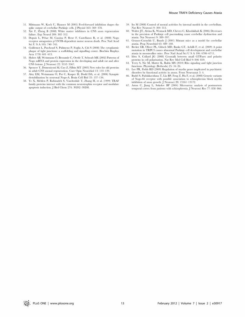
51. Mittmann W, Koch U, Hausser M (2005) Feed-forward inhibition shapes the
spike output of cerebellar Purkinje cells. J Physiol 563: 369–378.
52. Xie F, Zheng B (2008) White matter inhibitors in CNS axon regeneration
failure. Exp Neurol 209: 302–312.
53. Dupuis L, Pehar M, Cassina P, Rene F, Castellanos R, et al. (2008) Nogo
receptor antagonizes p75NTR-dependent motor neuron death. Proc Natl Acad
Sci U S A 105: 740–745.
54. Guillemot L, Paschoud S, Pulimeno P, Foglia A, Citi S (2008) The cytoplasmic
plaque of tight junctions: a scaffolding and signalling center. Biochim Biophys
Acta 1778: 601–613.
55. Huber AB, Weinmann O, Brosamle C, Oertle T, Schwab ME (2002) Patterns of
Nogo mRNA and protein expression in the developing and adult rat and after
CNS lesions. J Neurosci 22: 3553–3567.
56. Spencer T, Domeniconi M, Cao Z, Filbin MT (2003) New roles for old proteins
in adult CNS axonal regeneration. Curr Opin Neurobiol 13: 133–139.
57. Aloy EM, Weinmann O, Pot C, Kasper H, Dodd DA, et al. (2006) Synaptic
destabilization by neuronal Nogo-A. Brain Cell Biol 35: 137–156.
58. Ye X, Mehlen P, Rabizadeh S, VanArsdale T, Zhang H, et al. (1999) TRAF
family proteins interact with the common neurotrophin receptor and modulate
apoptosis induction. J Biol Chem 274: 30202–30208.
59. Ito M (2008) Control of mental activities by internal models in the cerebellum.
Nat Rev Neurosci 9: 304–313.60. Walter JT, Alvina K, Womack MD, Chevez C, Khodakhah K (2006) Decreases
in the precision of Purkinje cell pacemaking cause cerebellar dysfunction and
ataxia. Nat Neurosci 9: 389–397.61. Grusser-Cornehls U, Baurle J (2001) Mutant mice as a model for cerebellar
ataxia. Prog Neurobiol 63: 489–540.62. Becker EB, Oliver PL, Glitsch MD, Banks GT, Achilli F, et al. (2009) A point
mutation in TRPC3 causes abnormal Purkinje cell development and cerebellar
ataxia in moonwalker mice. Proc Natl Acad Sci U S A 106: 6706–6711.63. Iden S, Collard JG (2008) Crosstalk between small GTPases and polarity
proteins in cell polarization. Nat Rev Mol Cell Biol 9: 846–859.64. Terry S, Nie M, Matter K, Balda MS (2010) Rho signaling and tight junction
functions. Physiology (Bethesda) 25: 16–26.65. Lee PR, Fields RD (2009) Regulation of myelin genes implicated in psychiatric
disorders by functional activity in axons. Front Neuroanat 3: 4.
66. Budel S, Padukkavidana T, Liu BP, Feng Z, Hu F, et al. (2008) Genetic variantsof Nogo-66 receptor with possible association to schizophrenia block myelin
inhibition of axon growth. J Neurosci 28: 13161–13172.67. Aston C, Jiang L, Sokolov BP (2004) Microarray analysis of postmortem
temporal cortex from patients with schizophrenia. J Neurosci Res 77: 858–866.
Mouse TRAF4 Deficiency Causes Ataxia
PLoS ONE | www.plosone.org 13 February 2012 | Volume 7 | Issue 2 | e30917

159
ANNEXE-2
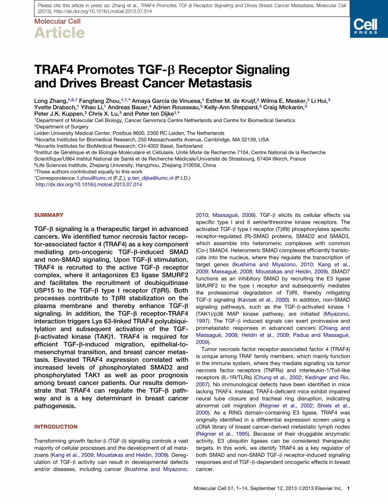
Please cite this article in press as: Zhang et al., TRAF4 Promotes TGF-b Receptor Signaling and Drives Breast Cancer Metastasis, Molecular Cell(2013), http://dx.doi.org/10.1016/j.molcel.2013.07.014
Molecular Cell
Article
TRAF4 Promotes TGF-b Receptor Signalingand Drives Breast Cancer MetastasisLong Zhang,1,6,7 Fangfang Zhou,1,7,* Amaya Garcıa de Vinuesa,1 Esther M. de Kruijf,2 Wilma E. Mesker,2 Li Hui,3
Yvette Drabsch,1 Yihao Li,1 Andreas Bauer,4 Adrien Rousseau,5 Kelly-Ann Sheppard,3 Craig Mickanin,3
Peter J.K. Kuppen,2 Chris X. Lu,3 and Peter ten Dijke1,*1Department of Molecular Cell Biology, Cancer Genomics Centre Netherlands and Centre for Biomedical Genetics2Department of Surgery
Leiden University Medical Center, Postbus 9600, 2300 RC Leiden, The Netherlands3Novartis Institutes for Biomedical Research, 250 Massachusetts Avenue, Cambridge, MA 02139, USA4Novartis Institutes for BioMedical Research, CH-4002 Basel, Switzerland5Institut de Genetique et de Biologie Moleculaire et Cellulaire, Unite Mixte de Recherche 7104, Centre National de la Recherche
Scientifique/U964 Institut National de Sante et de Recherche Medicale/Universite de Strasbourg, 67404 Illkirch, France6Life Sciences Institute, Zhejiang University, Hangzhou, Zhejiang 310058, China7These authors contributed equally to this work*Correspondence: [email protected] (F.Z.), [email protected] (P.t.D.)
http://dx.doi.org/10.1016/j.molcel.2013.07.014
SUMMARY
TGF-b signaling is a therapeutic target in advancedcancers. We identified tumor necrosis factor recep-tor-associated factor 4 (TRAF4) as a key componentmediating pro-oncogenic TGF-b-induced SMADand non-SMAD signaling. Upon TGF-b stimulation,TRAF4 is recruited to the active TGF-b receptorcomplex, where it antagonizes E3 ligase SMURF2and facilitates the recruitment of deubiquitinaseUSP15 to the TGF-b type I receptor (TbRI). Bothprocesses contribute to TbRI stabilization on theplasma membrane and thereby enhance TGF-bsignaling. In addition, the TGF-b receptor-TRAF4interaction triggers Lys 63-linked TRAF4 polyubiqui-tylation and subsequent activation of the TGF-b-activated kinase (TAK)1. TRAF4 is required forefficient TGF-b-induced migration, epithelial-to-mesenchymal transition, and breast cancer metas-tasis. Elevated TRAF4 expression correlated withincreased levels of phosphorylated SMAD2 andphosphorylated TAK1 as well as poor prognosisamong breast cancer patients. Our results demon-strate that TRAF4 can regulate the TGF-b path-way and is a key determinant in breast cancerpathogenesis.
INTRODUCTION
Transforming growth factor-b (TGF-b) signaling controls a vast
majority of cellular processes and the development of all meta-
zoans (Kang et al., 2009; Moustakas and Heldin, 2009). Dereg-
ulation of TGF-b activity can result in developmental defects
and/or diseases, including cancer (Ikushima and Miyazono,
M
2010; Massague, 2008). TGF-b elicits its cellular effects via
specific type I and II serine/threonine kinase receptors. The
activated TGF-b type I receptor (TbRI) phosphorylates specific
receptor-regulated (R)-SMAD proteins, SMAD2 and SMAD3,
which assemble into heteromeric complexes with common
(Co-) SMAD4. Heteromeric SMAD complexes efficiently translo-
cate into the nucleus, where they regulate the transcription of
target genes (Ikushima and Miyazono, 2010; Kang et al.,
2009; Massague, 2008; Moustakas and Heldin, 2009). SMAD7
functions as an inhibitory SMAD by recruiting the E3 ligase
SMURF2 to the type I receptor and subsequently mediates
the proteasomal degradation of TbRI, thereby mitigating
TGF-b signaling (Kavsak et al., 2000). In addition, non-SMAD
signaling pathways, such as the TGF-b-activated kinase 1
(TAK1)/p38 MAP kinase pathway, are initiated (Miyazono,
1997). The TGF-b induced signals can exert proinvasive and
prometastatic responses in advanced cancers (Chiang and
Massague, 2008; Heldin et al., 2009; Padua and Massague,
2009).
Tumor necrosis factor receptor-associated factor 4 (TRAF4)
is unique among TRAF family members, which mainly function
in the immune system, where they mediate signaling via tumor
necrosis factor receptors (TNFRs) and interleukin-1/Toll-like
receptors (IL-1R/TLRs) (Chung et al., 2002; Kedinger and Rio,
2007). No immunological defects have been identified in mice
lacking TRAF4. Instead, TRAF4-deficient mice exhibit impaired
neural tube closure and tracheal ring disruption, indicating
abnormal cell migration (Regnier et al., 2002; Shiels et al.,
2000). As a RING domain-containing E3 ligase, TRAF4 was
originally identified in a differential expression screen using a
cDNA library of breast cancer-derived metastatic lymph nodes
(Regnier et al., 1995). Because of their druggable enzymatic
activity, E3 ubiquitin ligases can be considered therapeutic
targets. In this work, we identify TRAF4 as a key regulator of
both SMAD and non-SMAD TGF-b receptor-induced signaling
responses and of TGF-b-dependent oncogenic effects in breast
cancer.
olecular Cell 51, 1–14, September 12, 2013 ª2013 Elsevier Inc. 1
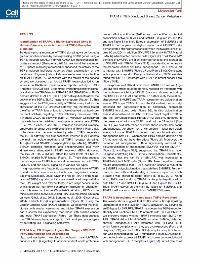
Molecular Cell
TRAF4 in TGF-b-Induced Breast Cancer Metastasis
Please cite this article in press as: Zhang et al., TRAF4 Promotes TGF-b Receptor Signaling and Drives Breast Cancer Metastasis, Molecular Cell(2013), http://dx.doi.org/10.1016/j.molcel.2013.07.014
RESULTS
Identification of TRAF4, a Highly Expressed Gene inHuman Cancers, as an Activator of TGF-b ReceptorSignalingTo identify pivotal regulators of TGF-b signaling, we performed a
gain-of-function screen by overexpressing 27,000 genes using a
TGF-b-induced SMAD3/4-driven CAGA-luc transcriptional re-
porter as readout (Zhang et al., 2012b). We found that a number
of E3 ligases markedly repressed or enhanced the activity of the
TGF-b-induced luciferase reporter. After examination of the
candidate E3 ligases (data not shown), we focused our attention
on TRAF4 (Figure 1A). Consistent with the results of the genetic
screen, we observed that forced TRAF4 expression led to an
increase in CAGA-luc transcriptional reporter activity in TGF-
b-treatedHEK293Tcells.Bycontrast, overexpressionof thecata-
lytically inactive TRAF4mutant TRAF4C18A (TRAF4C/A) or RING
domain-deletedTRAF4dR (60-470) did not significantly affect the
activity of the TGF-b/SMAD-responsive reporter (Figure 1B). This
suggests that the E3 ligase activity of TRAF4 is required for the
stimulation of the TGF-b/SMAD pathway. We therefore tested
the effect of TRAF4 loss of function. We observed that two inde-
pendent shRNAs targeting TRAF4 efficiently decreased TGF-
b-inducedCAGA-luc activity (Figure 1C). Moreover, we observed
thatwell-characterizeddirect transcriptional gene targets of TGF-
b, i.e., PAI-1, SMAD7, and CTGF, were less expressed in mouse
embryonic fibroblast cells (MEFs) deficient in TRAF4 (Figure 1D).
To determine the mechanism by which TRAF4 regulates
the TGF-b pathway, we first assessed whether TRAF4 affects
the main immediate mediators of TGF-b receptor signaling.
TGF-b-induced SMAD2 phosphorylation (p-SMAD2), SMAD2-
SMAD4 complex formation, and phosphorylated p38 MAP
kinase were attenuated in TRAF4 knockout MEFs. However,
the lack of TRAF4 did not affect the total levels of SMAD2,
SMAD4, or p38 MAP kinase (Figure 1E). These data suggest
that endogenous TRAF4 is a critical determinant for both TGF-
b/SMAD and non-SMAD signaling in various cell types.
High-grade tumors frequently express elevated levels of TGF-
b, and this has been correlated with poor prognosis in cancer
patients (Massague, 2008). Given the role of TRAF4 in the regu-
lation of TGF-b signaling activity, we investigated the possibility
that TRAF4might be a relevant factor in late-stage cancer. In line
with a report that high TRAF4 expression is a common character-
istic of human carcinomas (Camilleri-Broet et al., 2007), onco-
mine expression analysis revealed TRAF4 upregulation in human
cancers (Gluck et al., 2012; Bonome et al., 2008; Ginos et al.,
2004) in which TGF-b is prometastatic (Figure 1F). Using the
Cancer Genome Atlas (TCGA) database, we observed that indi-
viduals with ovarian carcinoma and higher TRAF4 expression
had a shorter life expectancy than those with ovarian cancer
and lower TRAF4 expression (Figure 1G). These data suggest
that TRAF4 may play an oncogenic role in multiple cancer types
by activating TGF-b signaling pathways.
TRAF4 Is an E3 Ubiquitin Ligase that Targets SMURF2Polyubiquitination and DegradationNext, we investigated themolecularmechanism bywhich TRAF4
enhances TGF-b signaling. In an independent whole proteomic
2 Molecular Cell 51, 1–14, September 12, 2013 ª2013 Elsevier Inc.
tandemaffinity purification (TAP) screen, we identified a potential
association between TRAF4 and SMURF2 (Figures 2A and 2B
and see Table S1 online). Ectopic expression of SMURF2 and
TRAF4 in both a yeast two-hybrid system and HEK293T cells
demonstrated strong interactions between the two proteins (Fig-
ures 2C and 2D). In addition, SMURF2 interacts with TRAF4 (and
SMAD7) in transfected cultured cells (Figure 2E). TheC2 andWW
domains of SMURF2 are of critical importance for the interaction
of SMURF2 with TRAF4 (Figure S1A). Importantly, in nontrans-
fected breast cancer cell lines, endogenous TRAF4 was found
to interact with SMURF2 (Figure 2F and Figure S1C). Consistent
with a previous report in Xenopus (Kalkan et al., 2009), we also
found that SMURF1 interacts with TRAF4 in breast cancer cells
(Figure S1B).
Coexpression of TRAF4 diminished SMURF2 expression (Fig-
ure 2D); this effect could be partially rescued by treatment with
the proteasome inhibitor MG132 (data not shown), indicating
that SMURF2 is a TRAF4 substrate. To further verify the relation-
ship between SMURF2 and TRAF4, we performed ubiquitination
assays. Wild-type TRAF4, but not the CA mutant, dramatically
increased the polyubiquitination of ectopically expressed
SMURF2 in cultured cells (Figure 2G). In vitro ubiquitination
assays demonstrated that SMURF2 is a direct target of TRAF4
and that polyubiquitinated His-SMURF2 was only detected in
the presence of wild-type TRAF4, and not the CA mutant (Fig-
ure 2H). We next determined whether these effects also occur
endogenously. As shown by a His-ubiquitin nickel pull-down
assay, wild-type TRAF4 increased the polyubiquitination of
endogenous SMURF2, whereas the RING domain-deletion and
CA mutation did not have this effect (Figure 2I). Moreover, the
depletion of endogenous TRAF4 significantly reduced the
polyubiquitination of endogenous SMURF2, but not SMURF1
(Figure 2J and Figure S2A), suggesting that TRAF4 is a major
E3 ligase controlling SMURF2 polyubiquitination. Consistently,
we found that the half-life of SMURF2 was increased in
TRAF4-deficient MEF cells (Figure 2K). Taken together, these
results demonstrate that TRAF4 depletion causes a reduction
in SMURF2 polyubiquitination that stabilizes SMURF2. Further-
more, in line with and extending a previous report in which
SMURF1 was shown to target TRAF4 (Li et al., 2010; Wang
et al., 2013), we found that TRAF4 can be polyubiquitinated by
both SMURF1 and SMURF2 (Figure 2L and Figures S2B–S2D).
Thus, TRAF4 serves as the main E3 ligase for SMURF2, and
TRAF4 itself is a substrate for both SMURF E3 ligases.
TRAF4 Is Associated with Activated TGF-b ReceptorsThe results above suggest that TRAF4 affects TGF-b signaling
upstream of or at the level of R-SMAD activation. By serving as
an E3 ligase for SMURF2, TRAF4may determine TGF-b receptor
stability and function. SMURF2 interacts with TbRI via SMAD7.
We therefore tested whether TRAF4 interacts with SMAD7 or
TbRI. TRAF4 did not bind SMAD7 (or other SMADs; data not
shown). Endogenous TRAF4 interacted with TbRI and TbRII,
which form a complex when they are overexpressed (Feng and
Derynck, 1996), and theTRAF4-TGF-b receptor complex interac-
tion was enhanced upon TGF-b stimulation (Figure S3A). In addi-
tion, ectopically expressed TRAF4 also coimmunoprecipitated
with endogenous TGF-b receptors (Figure 3A). In cell lysates of
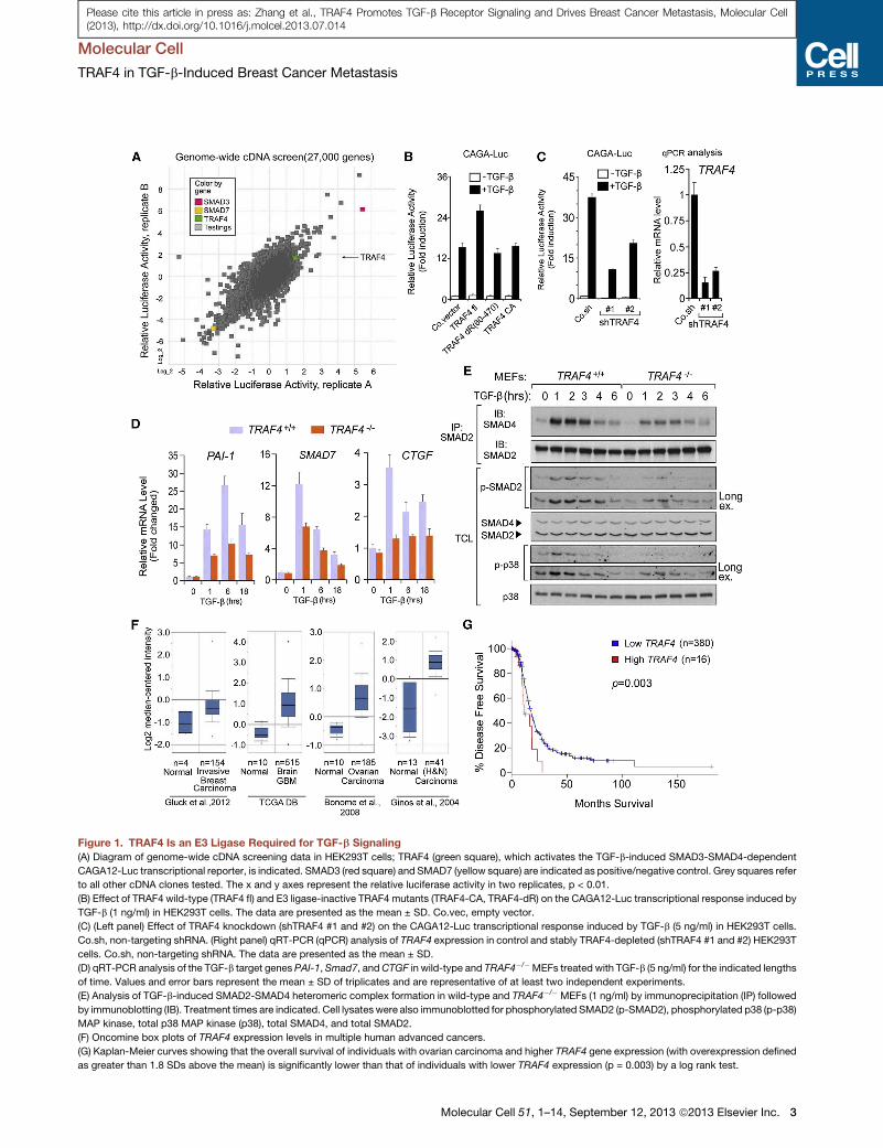
Figure 1. TRAF4 Is an E3 Ligase Required for TGF-b Signaling
(A) Diagram of genome-wide cDNA screening data in HEK293T cells; TRAF4 (green square), which activates the TGF-b-induced SMAD3-SMAD4-dependent
CAGA12-Luc transcriptional reporter, is indicated. SMAD3 (red square) and SMAD7 (yellow square) are indicated as positive/negative control. Grey squares refer
to all other cDNA clones tested. The x and y axes represent the relative luciferase activity in two replicates, p < 0.01.
(B) Effect of TRAF4 wild-type (TRAF4 fl) and E3 ligase-inactive TRAF4 mutants (TRAF4-CA, TRAF4-dR) on the CAGA12-Luc transcriptional response induced by
TGF-b (1 ng/ml) in HEK293T cells. The data are presented as the mean ± SD. Co.vec, empty vector.
(C) (Left panel) Effect of TRAF4 knockdown (shTRAF4 #1 and #2) on the CAGA12-Luc transcriptional response induced by TGF-b (5 ng/ml) in HEK293T cells.
Co.sh, non-targeting shRNA. (Right panel) qRT-PCR (qPCR) analysis of TRAF4 expression in control and stably TRAF4-depleted (shTRAF4 #1 and #2) HEK293T
cells. Co.sh, non-targeting shRNA. The data are presented as the mean ± SD.
(D) qRT-PCR analysis of the TGF-b target genes PAI-1, Smad7, andCTGF in wild-type and TRAF4�/�MEFs treated with TGF-b (5 ng/ml) for the indicated lengths
of time. Values and error bars represent the mean ± SD of triplicates and are representative of at least two independent experiments.
(E) Analysis of TGF-b-induced SMAD2-SMAD4 heteromeric complex formation in wild-type and TRAF4�/� MEFs (1 ng/ml) by immunoprecipitation (IP) followed
by immunoblotting (IB). Treatment times are indicated. Cell lysateswere also immunoblotted for phosphorylated SMAD2 (p-SMAD2), phosphorylated p38 (p-p38)
MAP kinase, total p38 MAP kinase (p38), total SMAD4, and total SMAD2.
(F) Oncomine box plots of TRAF4 expression levels in multiple human advanced cancers.
(G) Kaplan-Meier curves showing that the overall survival of individuals with ovarian carcinoma and higher TRAF4 gene expression (with overexpression defined
as greater than 1.8 SDs above the mean) is significantly lower than that of individuals with lower TRAF4 expression (p = 0.003) by a log rank test.
Molecular Cell
TRAF4 in TGF-b-Induced Breast Cancer Metastasis
Molecular Cell 51, 1–14, September 12, 2013 ª2013 Elsevier Inc. 3
Please cite this article in press as: Zhang et al., TRAF4 Promotes TGF-b Receptor Signaling and Drives Breast Cancer Metastasis, Molecular Cell(2013), http://dx.doi.org/10.1016/j.molcel.2013.07.014
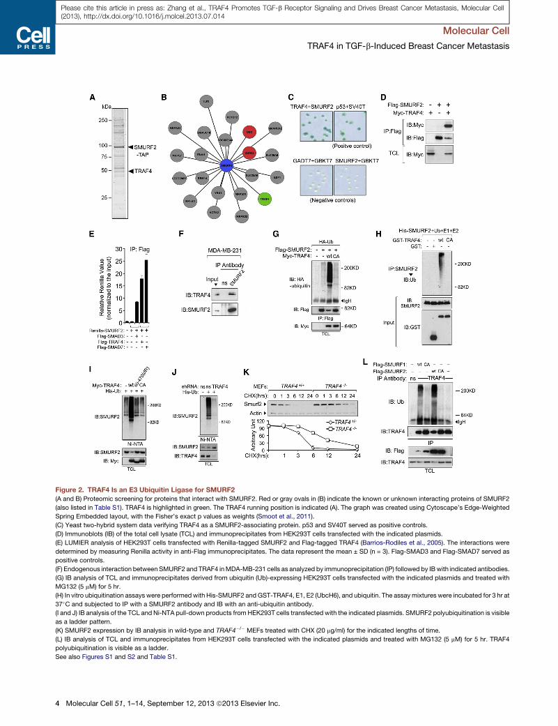
Figure 2. TRAF4 Is an E3 Ubiquitin Ligase for SMURF2
(A and B) Proteomic screening for proteins that interact with SMURF2. Red or gray ovals in (B) indicate the known or unknown interacting proteins of SMURF2
(also listed in Table S1). TRAF4 is highlighted in green. The TRAF4 running position is indicated (A). The graph was created using Cytoscape’s Edge-Weighted
Spring Embedded layout, with the Fisher’s exact p values as weights (Smoot et al., 2011).
(C) Yeast two-hybrid system data verifying TRAF4 as a SMURF2-associating protein. p53 and SV40T served as positive controls.
(D) Immunoblots (IB) of the total cell lysate (TCL) and immunoprecipitates from HEK293T cells transfected with the indicated plasmids.
(E) LUMIER analysis of HEK293T cells transfected with Renilla-tagged SMURF2 and Flag-tagged TRAF4 (Barrios-Rodiles et al., 2005). The interactions were
determined by measuring Renilla activity in anti-Flag immunoprecipitates. The data represent the mean ± SD (n = 3). Flag-SMAD3 and Flag-SMAD7 served as
positive controls.
(F) Endogenous interaction between SMURF2 and TRAF4 inMDA-MB-231 cells as analyzed by immunoprecipitation (IP) followed by IBwith indicated antibodies.
(G) IB analysis of TCL and immunoprecipitates derived from ubiquitin (Ub)-expressing HEK293T cells transfected with the indicated plasmids and treated with
MG132 (5 mM) for 5 hr.
(H) In vitro ubiquitination assays were performed with His-SMURF2 andGST-TRAF4, E1, E2 (UbcH6), and ubiquitin. The assay mixtures were incubated for 3 hr at
37�C and subjected to IP with a SMURF2 antibody and IB with an anti-ubiquitin antibody.
(I and J) IB analysis of the TCL and Ni-NTA pull-down products from HEK293T cells transfected with the indicated plasmids. SMURF2 polyubiquitination is visible
as a ladder pattern.
(K) SMURF2 expression by IB analysis in wild-type and TRAF4�/� MEFs treated with CHX (20 mg/ml) for the indicated lengths of time.
(L) IB analysis of TCL and immunoprecipitates from HEK293T cells transfected with the indicated plasmids and treated with MG132 (5 mM) for 5 hr. TRAF4
polyubiquitination is visible as a ladder.
See also Figures S1 and S2 and Table S1.
Molecular Cell
TRAF4 in TGF-b-Induced Breast Cancer Metastasis
4 Molecular Cell 51, 1–14, September 12, 2013 ª2013 Elsevier Inc.
Please cite this article in press as: Zhang et al., TRAF4 Promotes TGF-b Receptor Signaling and Drives Breast Cancer Metastasis, Molecular Cell(2013), http://dx.doi.org/10.1016/j.molcel.2013.07.014
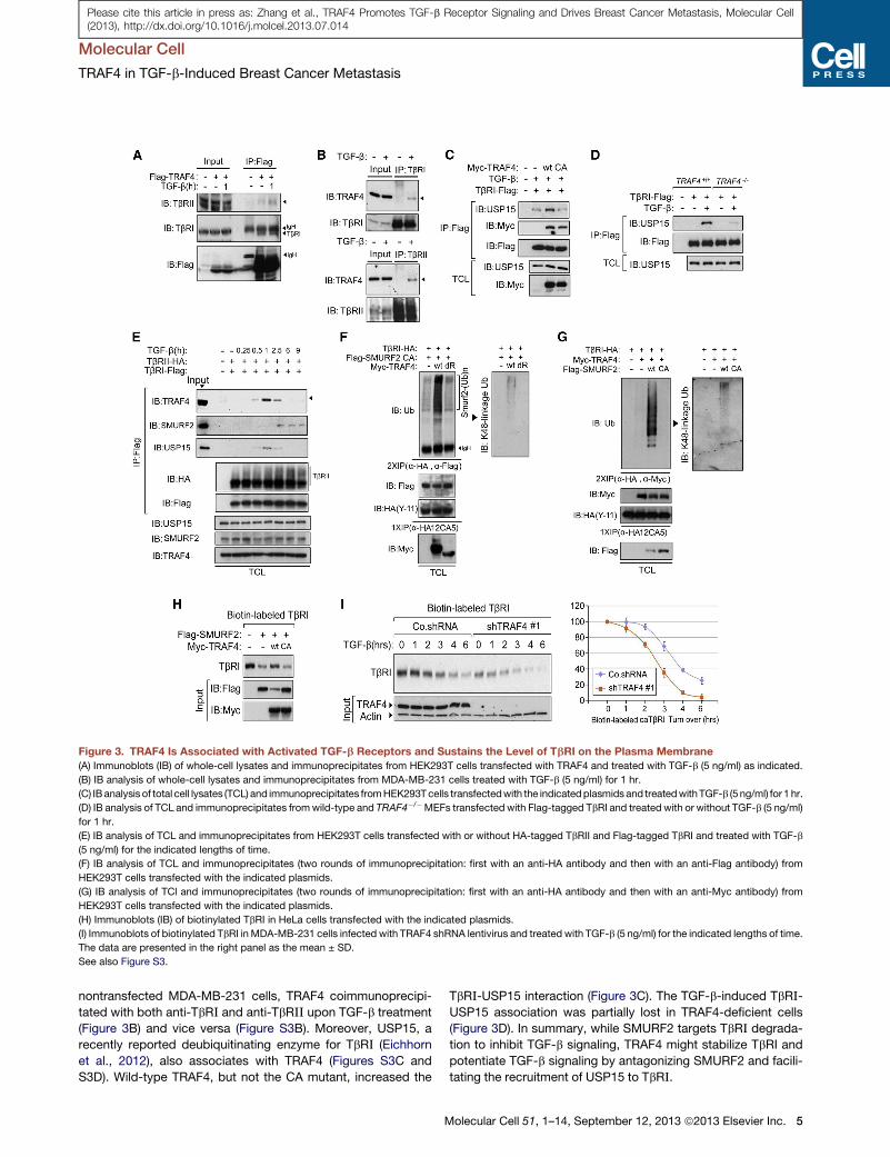
Figure 3. TRAF4 Is Associated with Activated TGF-b Receptors and Sustains the Level of TbRI on the Plasma Membrane
(A) Immunoblots (IB) of whole-cell lysates and immunoprecipitates from HEK293T cells transfected with TRAF4 and treated with TGF-b (5 ng/ml) as indicated.
(B) IB analysis of whole-cell lysates and immunoprecipitates from MDA-MB-231 cells treated with TGF-b (5 ng/ml) for 1 hr.
(C) IBanalysis of total cell lysates (TCL) and immunoprecipitates fromHEK293Tcells transfectedwith the indicatedplasmids and treatedwithTGF-b (5 ng/ml) for 1 hr.
(D) IB analysis of TCL and immunoprecipitates fromwild-type and TRAF4�/�MEFs transfected with Flag-tagged TbRI and treated with or without TGF-b (5 ng/ml)
for 1 hr.
(E) IB analysis of TCL and immunoprecipitates from HEK293T cells transfected with or without HA-tagged TbRII and Flag-tagged TbRI and treated with TGF-b
(5 ng/ml) for the indicated lengths of time.
(F) IB analysis of TCL and immunoprecipitates (two rounds of immunoprecipitation: first with an anti-HA antibody and then with an anti-Flag antibody) from
HEK293T cells transfected with the indicated plasmids.
(G) IB analysis of TCl and immunoprecipitates (two rounds of immunoprecipitation: first with an anti-HA antibody and then with an anti-Myc antibody) from
HEK293T cells transfected with the indicated plasmids.
(H) Immunoblots (IB) of biotinylated TbRI in HeLa cells transfected with the indicated plasmids.
(I) Immunoblots of biotinylated TbRI in MDA-MB-231 cells infected with TRAF4 shRNA lentivirus and treated with TGF-b (5 ng/ml) for the indicated lengths of time.
The data are presented in the right panel as the mean ± SD.
See also Figure S3.
Molecular Cell
TRAF4 in TGF-b-Induced Breast Cancer Metastasis
Please cite this article in press as: Zhang et al., TRAF4 Promotes TGF-b Receptor Signaling and Drives Breast Cancer Metastasis, Molecular Cell(2013), http://dx.doi.org/10.1016/j.molcel.2013.07.014
nontransfected MDA-MB-231 cells, TRAF4 coimmunoprecipi-
tated with both anti-TbR_ and anti-TbR__ upon TGF-b treatment
(Figure 3B) and vice versa (Figure S3B). Moreover, USP15, a
recently reported deubiquitinating enzyme for TbR_ (Eichhorn
et al., 2012), also associates with TRAF4 (Figures S3C and
S3D). Wild-type TRAF4, but not the CA mutant, increased the
M
TbR_-USP15 interaction (Figure 3C). The TGF-b-induced TbR_-USP15 association was partially lost in TRAF4-deficient cells
(Figure 3D). In summary, while SMURF2 targets TbR_ degrada-tion to inhibit TGF-b signaling, TRAF4 might stabilize TbRI and
potentiate TGF-b signaling by antagonizing SMURF2 and facili-
tating the recruitment of USP15 to TbR_.
olecular Cell 51, 1–14, September 12, 2013 ª2013 Elsevier Inc. 5
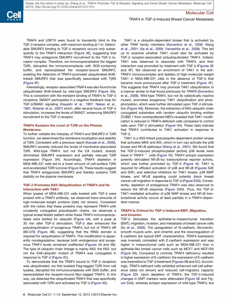
Molecular Cell
TRAF4 in TGF-b-Induced Breast Cancer Metastasis
Please cite this article in press as: Zhang et al., TRAF4 Promotes TGF-b Receptor Signaling and Drives Breast Cancer Metastasis, Molecular Cell(2013), http://dx.doi.org/10.1016/j.molcel.2013.07.014
TRAF4 and USP15 were found to transiently bind to the
TGF-b receptor complex, with maximum binding at 1 hr. Detect-
able SMURF2 binding to TGF-b receptors occurs only subse-
quently to the TRAF4 interaction (Figure 3E), suggesting that
TRAF4 may antagonize SMURF2 recruitment to the TGF-b re-
ceptor complex. Therefore, we immunoprecipitated the tagged
TbR_, disrupted the immunocomplexes with SDS-containing
buffer, and reprecipitated the receptor-bound SMURF2,
enabling the detection of TRAF4-promoted ubiquitinated (K48-
linked) SMURF2 that was specifically associated with TbR_(Figure 3F).
Interestingly, receptor-associated TRAF4was also found to be
ubiquitinated (K48-linked) by wild-type SMURF2 (Figure 3G).
This is consistent with the transient binding of TRAF4 to TGF-b
receptors; SMAD7 participates in a negative feedback loop for
TGF-b/SMAD signaling (Hayashi et al., 1997; Nakao et al.,
1997; Wiesner et al., 2007) which, after several hours of TGF-b
stimulation, increases the levels of SMAD7, enhancing SMURF2
recruitment to the TGF-b receptor.
TRAF4 Sustains the Level of TbRI on the PlasmaMembraneTo further validate the interplay of TRAF4 and SMURF2 in TbRI
function, we determined the membrane localization and stability
of TbRI. Consistent with a previous report (Kavsak et al., 2000),
SMURF2 severely reduced the levels of membrane-associated
TbRI. Wild-type TRAF4, but not the CA mutant, sharply
diminished SMURF2 expression and partially rescued TbRI
expression (Figure 3H). Accordingly, TRAF4 depletion in
MDA-MB-231 cells led to a lower amount of cell-surface TbR_and accelerated TbR_ turnover (Figure 3I). These results suggest
that TRAF4 antagonizes SMURF2 and thereby sustains TbRI
stability on the plasma membrane.
TGF-b Promotes K63 Ubiquitination of TRAF4 and ItsInteraction with TAK1When lysates of MDA-MB-231 cells treated with TGF-b were
probed with a TRAF4 antibody, we observed trace amounts of
high-molecular-weight proteins (data not shown). Consistent
with the notion that these proteins may represent TRAF4 with
covalently conjugated polyubiquitin chains, we observed a
typical smear/ladder pattern when these TRAF4 immunoprecip-
itates were blotted for ubiquitin (Figure 4A), with a peak at
30 min after TGF-b stimulation. TGF-b also enhanced the
polyubiquitination of exogenous TRAF4, but not of TRAF4 dR
(60-470) (Figure 4B), suggesting that the RING domain is
required for ubiquitination of TRAF4. This modification is appar-
ently nondegradative, because both endogenous and exoge-
nous TRAF4 levels remained unaffected (Figures 4A and 4B).
The type of ubiquitin chain linkage is mainly K63; the K63- but
not the K48-ubiquitin mutant of TRAF4 was conjugated in
response to TGF-b (Figure 4C).
To demonstrate that the TRAF4 bound to TGF-b receptors
was ubiquitinated, we first isolated Flag-tagged TbR_ from cell
lysates, disrupted the immunocomplexes with SDS buffer, and
reprecipitated the receptor-bound Myc-tagged TRAF4. In this
way, we detected the ubiquitinated TRAF4 that was specifically
associated with TbR_ and activated by TGF-b (Figure 4D).
6 Molecular Cell 51, 1–14, September 12, 2013 ª2013 Elsevier Inc.
TAK1 is a ubiquitin-dependent kinase that is activated by
other TRAF family members (Sorrentino et al., 2008; Wang
et al., 2001; Xia et al., 2009; Yamashita et al., 2008). This led
us to examine whether TAK1 could also be activated by
TGF-b receptor-associated polyubiquitinated TRAF4. Indeed,
TAK1 was observed to associate with TRAF4, and their
interaction was promoted by treatment with TGF-b (Figures 4E
and 4F). We observed an enrichment of TAK1 in the anti-
TRAF4 immunocomplex and ladders of high-molecular-weight
TAK1 in MDA-MB-231 cells in the absence of TGF-b that
became more pronounced after TGF-b treatment (Figure 4F).
This suggests that TRAF4 may promote TAK1 ubiquitination in
a manner similar to that found previously for TRAF6 (Sorrentino
et al., 2008). Wild-type TRAF4, but not its catalytically inactive
mutant, promoted exogenous TAK1 ubiquitination and phos-
phorylation, which were further stimulated upon TGF-b stimula-
tion (Figure 4G). Moreover, the extraction of K63-ubiquitin chain
conjugated substrates with tandem ubiquitin binding entities
(TUBE) 1 from nontransfected MEFs revealed that TAK1 modifi-
cation is reduced in TRAF4-deficient cells compared to control
cells upon TGF-b stimulation (Figure 4H). These data indicate
that TRAF4 contributes to TAK1 activation in response to
TGF-b.
TAK1 is a K63-linked polyubiquitin-dependent protein kinase
that activates MKK and IKK, which in turn can activate the p38
kinase and NFkB pathways (Wang et al., 2001). We found that
the TGF-b-induced phosphorylated (active) p38 levels were
lower in TRAF4�/� cells (Figure 1E). TRAF4 ectopic expression
potently stimulated NFkB-luc transcriptional reporter activity,
which was further promoted by TGF-b (Figure 4I). TAK1 is
required for efficient activation of p38 and NFkB (Figures S3E
and S3F), and selective inhibitors for TAK1 kinase, p38 MAP
kinase, and NFkB signaling could potently block breast
cancer cell migration in response to TGF-b (Figure S3G). Consis-
tently, depletion of endogenous TRAF4 was also observed to
reduce the NFkB response (Figure S3H). Thus, the TGF-b/
TAK1-mediated activation of p38 MAP kinase and NFkB tran-
scriptional activity occurs at least partially in a TRAF4-depen-
dent manner.
TRAF4 Is Critical for TGF-b-Induced EMT, Migration,and InvasionTGF-b stimulates the epithelial-to-mesenchymal transition
(EMT), migration, invasion, and metastasis of breast cancer cells
(Xu et al., 2009). The upregulation of N-cadherin, fibronectin,
smooth muscle actin, and vimentin and the downregulation of
E-cadherin are typical EMT characteristics. TRAF4 expression
was inversely correlated with E-cadherin expression and was
higher in mesenchymal cells such as MDA-MB-231 than in
epithelial-like breast cancer cells such as MCF7 and MCF10A
(Figure 5A). Compared to controls, TRAF4 deficiency resulted
in higher expression of E-cadherin; the expression of E-cadherin
was insensitive to TGF-b treatment (Figures 5B and 5C). Accord-
ingly, TRAF4-deficient cells exhibited enhanced cell-cell adher-
ence (data not shown) and reduced cell-migratory capacity
(Figure 5D). Upon depletion of TRAF4, the TGF-b-induced
changes in EMT markers were attenuated (Figure 5E and Fig-
ure S4A), whereas ectopic expression of wild-type TRAF4, but
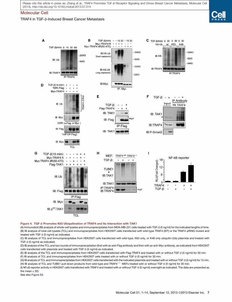
Figure 4. TGF-b Promotes K63 Ubiquitination of TRAF4 and Its Interaction with TAK1
(A) Immunoblot (IB) analysis of whole-cell lysates and immunoprecipitates fromMDA-MB-231 cells treated with TGF-b (5 ng/ml) for the indicated lengths of time.
(B) IB analysis of total cell lysates (TCL) and immunoprecipitates from HEK293T cells transfected with wild-type TRAF4 (WT) or the TRAF4 DRING mutant and
treated with TGF-b (5 ng/ml) as indicated.
(C) IB analysis of TCL and immunoprecipitates from HEK293T cells transfected with wild-type, K63 only, or K48 only ubiquitin (Ub) plasmids and treated with
TGF-b (5 ng/ml) as indicated.
(D) IB analysis of the TCL and two rounds of immunoprecipitation (first with an anti-Flag antibody and thenwith an anti-Myc antibody, as indicated) fromHEK293T
cells transfected with plasmids and treated with TGF-b (5 ng/ml) as indicated.
(E) IB analysis of the TCL and immunoprecipitates from HEK293T cells transfected with Flag-TRAF4 and treated with or without TGF-b (5 ng/ml) for 30 min.
(F) IB analysis of TCL and immunoprecipitates from HEK293T cells treated with or without TGF-b (5 ng/ml) for 30 min.
(G) IB analysis of TCL and immunoprecipitates fromHEK293T cells transfected with the indicated plasmids and treated with or without TGF-b (5 ng/ml) for 15min.
(H) IB analysis of TCL and TUBE1 pull-down products from wild-type and TRAF4�/� MEFs treated with or without TGF-b (5 ng/ml) for 30 min.
(I) NFkB reporter activity in HEK293T cells transfected with TRAF4 and treated with or without TGF-b (5 ng/ml) overnight as indicated. The data are presented as
the mean ± SD.
See also Figure S3.
Molecular Cell
TRAF4 in TGF-b-Induced Breast Cancer Metastasis
Molecular Cell 51, 1–14, September 12, 2013 ª2013 Elsevier Inc. 7
Please cite this article in press as: Zhang et al., TRAF4 Promotes TGF-b Receptor Signaling and Drives Breast Cancer Metastasis, Molecular Cell(2013), http://dx.doi.org/10.1016/j.molcel.2013.07.014
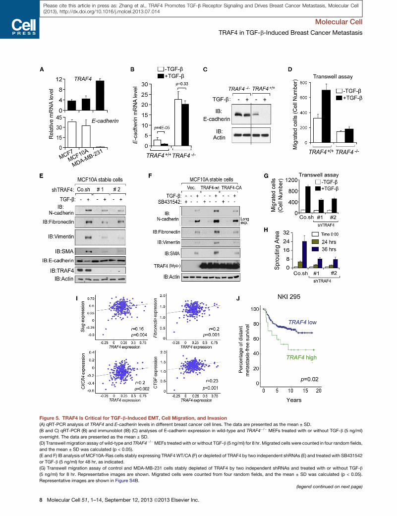
Figure 5. TRAF4 Is Critical for TGF-b-Induced EMT, Cell Migration, and Invasion
(A) qRT-PCR analysis of TRAF4 and E-cadherin levels in different breast cancer cell lines. The data are presented as the mean ± SD.
(B and C) qRT-PCR (B) and immunoblot (IB) (C) analyses of E-cadherin expression in wild-type and TRAF4�/� MEFs treated with or without TGF-b (5 ng/ml)
overnight. The data are presented as the mean ± SD.
(D) Transwell migration assay of wild-type and TRAF4�/�MEFs treated with or without TGF-b (5 ng/ml) for 8 hr. Migrated cells were counted in four random fields,
and the mean ± SD was calculated (p < 0.05).
(E and F) IB analysis of MCF10A-Ras cells stably expressing TRAF4WT/CA (F) or depleted of TRAF4 by two independent shRNAs (E) and treated with SB431542
or TGF-b (5 ng/ml) for 48 hr, as indicated.
(G) Transwell migration assay of control and MDA-MB-231 cells stably depleted of TRAF4 by two independent shRNAs and treated with or without TGF-b
(5 ng/ml) for 8 hr. Representative images are shown. Migrated cells were counted from four random fields, and the mean ± SD was calculated (p < 0.05).
Representative images are shown in Figure S4B.
(legend continued on next page)
Molecular Cell
TRAF4 in TGF-b-Induced Breast Cancer Metastasis
8 Molecular Cell 51, 1–14, September 12, 2013 ª2013 Elsevier Inc.
Please cite this article in press as: Zhang et al., TRAF4 Promotes TGF-b Receptor Signaling and Drives Breast Cancer Metastasis, Molecular Cell(2013), http://dx.doi.org/10.1016/j.molcel.2013.07.014
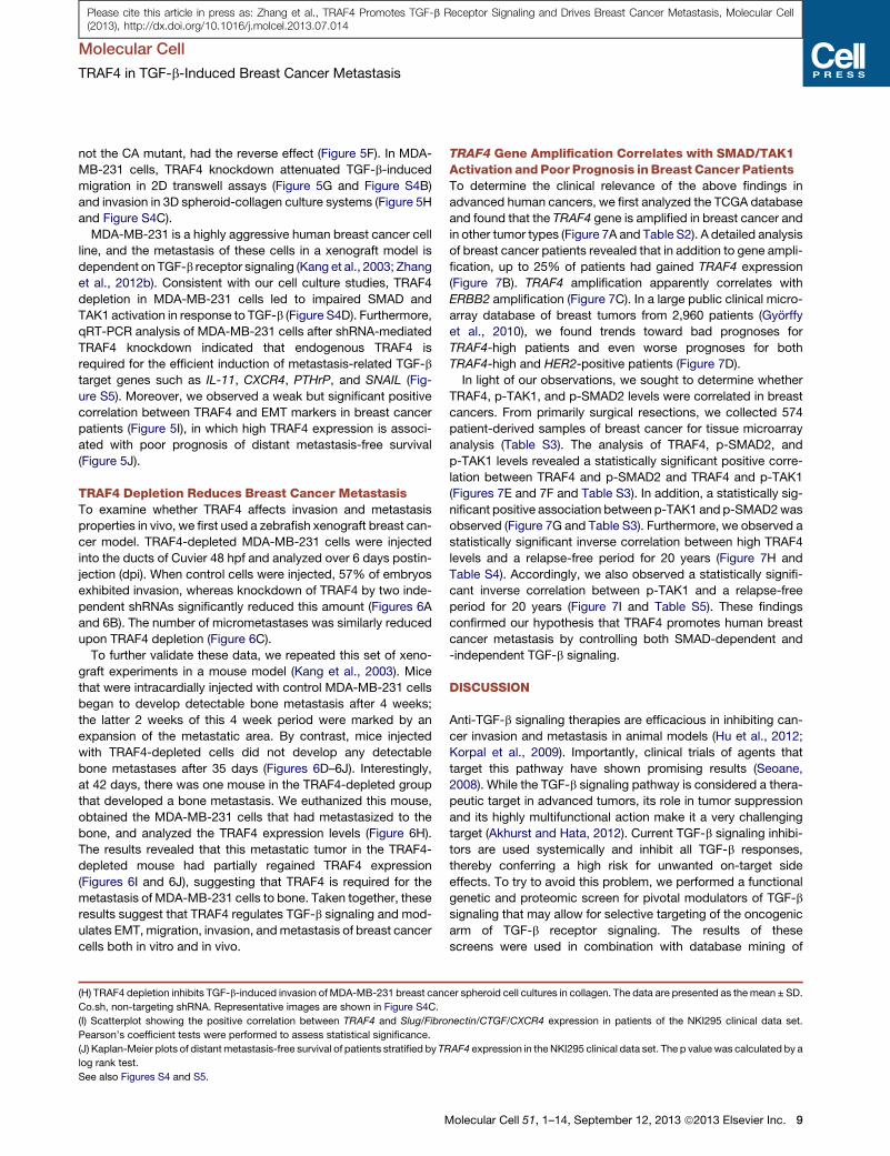
Molecular Cell
TRAF4 in TGF-b-Induced Breast Cancer Metastasis
Please cite this article in press as: Zhang et al., TRAF4 Promotes TGF-b Receptor Signaling and Drives Breast Cancer Metastasis, Molecular Cell(2013), http://dx.doi.org/10.1016/j.molcel.2013.07.014
not the CA mutant, had the reverse effect (Figure 5F). In MDA-
MB-231 cells, TRAF4 knockdown attenuated TGF-b-induced
migration in 2D transwell assays (Figure 5G and Figure S4B)
and invasion in 3D spheroid-collagen culture systems (Figure 5H
and Figure S4C).
MDA-MB-231 is a highly aggressive human breast cancer cell
line, and the metastasis of these cells in a xenograft model is
dependent on TGF-b receptor signaling (Kang et al., 2003; Zhang
et al., 2012b). Consistent with our cell culture studies, TRAF4
depletion in MDA-MB-231 cells led to impaired SMAD and
TAK1 activation in response to TGF-b (Figure S4D). Furthermore,
qRT-PCR analysis of MDA-MB-231 cells after shRNA-mediated
TRAF4 knockdown indicated that endogenous TRAF4 is
required for the efficient induction of metastasis-related TGF-b
target genes such as IL-11, CXCR4, PTHrP, and SNAIL (Fig-
ure S5). Moreover, we observed a weak but significant positive
correlation between TRAF4 and EMT markers in breast cancer
patients (Figure 5I), in which high TRAF4 expression is associ-
ated with poor prognosis of distant metastasis-free survival
(Figure 5J).
TRAF4 Depletion Reduces Breast Cancer MetastasisTo examine whether TRAF4 affects invasion and metastasis
properties in vivo, we first used a zebrafish xenograft breast can-
cer model. TRAF4-depleted MDA-MB-231 cells were injected
into the ducts of Cuvier 48 hpf and analyzed over 6 days postin-
jection (dpi). When control cells were injected, 57% of embryos
exhibited invasion, whereas knockdown of TRAF4 by two inde-
pendent shRNAs significantly reduced this amount (Figures 6A
and 6B). The number of micrometastases was similarly reduced
upon TRAF4 depletion (Figure 6C).
To further validate these data, we repeated this set of xeno-
graft experiments in a mouse model (Kang et al., 2003). Mice
that were intracardially injected with control MDA-MB-231 cells
began to develop detectable bone metastasis after 4 weeks;
the latter 2 weeks of this 4 week period were marked by an
expansion of the metastatic area. By contrast, mice injected
with TRAF4-depleted cells did not develop any detectable
bone metastases after 35 days (Figures 6D–6J). Interestingly,
at 42 days, there was one mouse in the TRAF4-depleted group
that developed a bone metastasis. We euthanized this mouse,
obtained the MDA-MB-231 cells that had metastasized to the
bone, and analyzed the TRAF4 expression levels (Figure 6H).
The results revealed that this metastatic tumor in the TRAF4-
depleted mouse had partially regained TRAF4 expression
(Figures 6I and 6J), suggesting that TRAF4 is required for the
metastasis of MDA-MB-231 cells to bone. Taken together, these
results suggest that TRAF4 regulates TGF-b signaling and mod-
ulates EMT, migration, invasion, andmetastasis of breast cancer
cells both in vitro and in vivo.
(H) TRAF4 depletion inhibits TGF-b-induced invasion of MDA-MB-231 breast canc
Co.sh, non-targeting shRNA. Representative images are shown in Figure S4C.
(I) Scatterplot showing the positive correlation between TRAF4 and Slug/Fibro
Pearson’s coefficient tests were performed to assess statistical significance.
(J) Kaplan-Meier plots of distant metastasis-free survival of patients stratified by TR
log rank test.
See also Figures S4 and S5.
M
TRAF4 Gene Amplification Correlates with SMAD/TAK1Activation and Poor Prognosis in Breast Cancer PatientsTo determine the clinical relevance of the above findings in
advanced human cancers, we first analyzed the TCGA database
and found that the TRAF4 gene is amplified in breast cancer and
in other tumor types (Figure 7A and Table S2). A detailed analysis
of breast cancer patients revealed that in addition to gene ampli-
fication, up to 25% of patients had gained TRAF4 expression
(Figure 7B). TRAF4 amplification apparently correlates with
ERBB2 amplification (Figure 7C). In a large public clinical micro-
array database of breast tumors from 2,960 patients (Gyorffy
et al., 2010), we found trends toward bad prognoses for
TRAF4-high patients and even worse prognoses for both
TRAF4-high and HER2-positive patients (Figure 7D).
In light of our observations, we sought to determine whether
TRAF4, p-TAK1, and p-SMAD2 levels were correlated in breast
cancers. From primarily surgical resections, we collected 574
patient-derived samples of breast cancer for tissue microarray
analysis (Table S3). The analysis of TRAF4, p-SMAD2, and
p-TAK1 levels revealed a statistically significant positive corre-
lation between TRAF4 and p-SMAD2 and TRAF4 and p-TAK1
(Figures 7E and 7F and Table S3). In addition, a statistically sig-
nificant positive association between p-TAK1 and p-SMAD2was
observed (Figure 7G and Table S3). Furthermore, we observed a
statistically significant inverse correlation between high TRAF4
levels and a relapse-free period for 20 years (Figure 7H and
Table S4). Accordingly, we also observed a statistically signifi-
cant inverse correlation between p-TAK1 and a relapse-free
period for 20 years (Figure 7I and Table S5). These findings
confirmed our hypothesis that TRAF4 promotes human breast
cancer metastasis by controlling both SMAD-dependent and
-independent TGF-b signaling.
DISCUSSION
Anti-TGF-b signaling therapies are efficacious in inhibiting can-
cer invasion and metastasis in animal models (Hu et al., 2012;
Korpal et al., 2009). Importantly, clinical trials of agents that
target this pathway have shown promising results (Seoane,
2008). While the TGF-b signaling pathway is considered a thera-
peutic target in advanced tumors, its role in tumor suppression
and its highly multifunctional action make it a very challenging
target (Akhurst and Hata, 2012). Current TGF-b signaling inhibi-
tors are used systemically and inhibit all TGF-b responses,
thereby conferring a high risk for unwanted on-target side
effects. To try to avoid this problem, we performed a functional
genetic and proteomic screen for pivotal modulators of TGF-b
signaling that may allow for selective targeting of the oncogenic
arm of TGF-b receptor signaling. The results of these
screens were used in combination with database mining of
er spheroid cell cultures in collagen. The data are presented as themean ± SD.
nectin/CTGF/CXCR4 expression in patients of the NKI295 clinical data set.
AF4 expression in the NKI295 clinical data set. The p value was calculated by a
olecular Cell 51, 1–14, September 12, 2013 ª2013 Elsevier Inc. 9
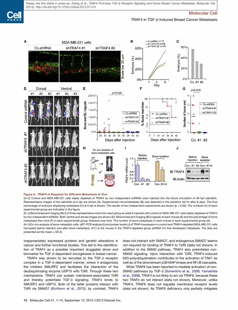
Figure 6. TRAF4 Is Required for Efficient Metastasis In Vivo
(A–C) Control and MDA-MB-231 cells stably depleted of TRAF4 by two independent shRNAs were injected into the blood circulation of 48 hpf zebrafish.
Representative images of the zebrafish at 6 dpi are shown (A). Experimental micrometastasis (B) was detected in the posterior tail fin after 6 days. The final
percentage of embryos displaying metastasis (C) at 6 dpi is shown. The results of two independent experiments are shown (p < 0.05). The numbers (n) of each
experimental group are indicated in the figure.
(D–J) Bioluminescent imaging (BLI) of three representative mice from each group at week 6 injected with control or MDA-MB-231 cells stably depleted of TRAF4
by two independent shRNAs. Both ventral and dorsal images are shown (D). Bioluminescent imaging (BLI) signals of each mouse (E) and the percentage of bone
metastasis-free mice (F) in each experimental group, followed over time. The number of bone metastasis in each mouse in each experimental group (G).
(H–J) Ex vivo analysis of bonemetastatic cells. qRT-PCR analysis (I) and protein levels (J) of TRAF4 expression in control and TRAF4-depletedMDA-MB-231 cells
harvested before injection and after bone metastasis. #1(*) is the mouse in the TRAF4-depleted group (shRNA #1) that developed metastasis. The data are
presented as the mean ± SD (I).
Molecular Cell
TRAF4 in TGF-b-Induced Breast Cancer Metastasis
Please cite this article in press as: Zhang et al., TRAF4 Promotes TGF-b Receptor Signaling and Drives Breast Cancer Metastasis, Molecular Cell(2013), http://dx.doi.org/10.1016/j.molcel.2013.07.014
inappropriately expressed proteins and genetic alterations in
cancer and further functional studies. This led to the identifica-
tion of TRAF4 as a possible important druggable driver and
biomarker for TGF-b-dependent oncogenesis in breast cancer.
TRAF4 was shown to be recruited to the TGF-b receptor
complex in a TGF-b-dependent manner, where it antagonizes
the inhibitor SMURF2 and facilitates the interaction of the
deubiquitinating enzyme USP15 with TbRI. Through these two
mechanisms, TRAF4 can sustain membrane-associated TbRI
and thereby potentiate TGF-b signaling. TRAF4 binds to
SMURF2 and USP15. Both of the latter proteins interact with
TbRI via SMAD7 (Eichhorn et al., 2012); by contrast, TRAF4
10 Molecular Cell 51, 1–14, September 12, 2013 ª2013 Elsevier Inc.
does not interact with SMAD7, and endogenous SMAD7 seems
not required for binding of TRAF4 to TbR_ (data not shown). In
addition to the SMAD pathway, TRAF4 also potentiates non-
SMAD signaling. Upon interaction with TbRI, TRAF4-induced
K63-polyubiquitination contributes to the activation of TAK1 as
well as of the downstream p38MAP kinase and NFkB cascades.
While TRAF6 has been reported to mediate activation of non-
SMAD pathways by TGF-b (Sorrentino et al., 2008; Yamashita
et al., 2008), TRAF4 is not likely to act via TRAF6, because these
two TRAFs do not interact (data not shown). Moreover, unlike
TRAF4, TRAF6 does not regulate membrane receptor levels
(data not shown). As TRAF6 deficiency only partially mitigates
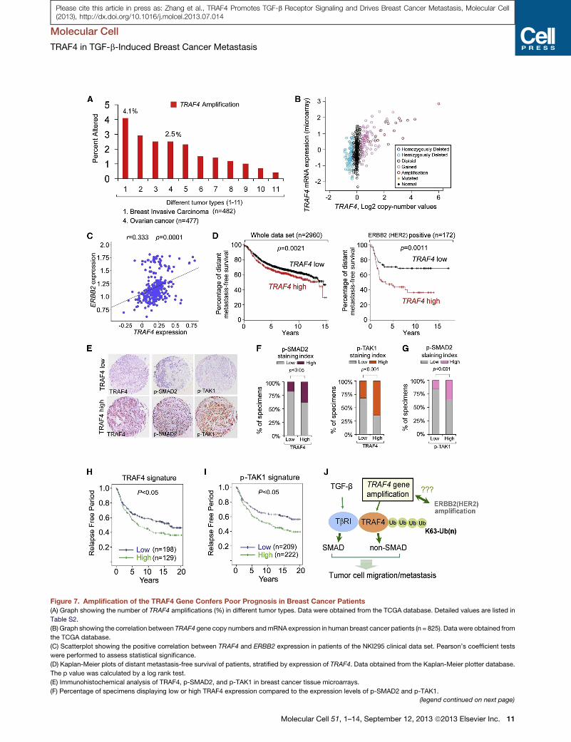
Figure 7. Amplification of the TRAF4 Gene Confers Poor Prognosis in Breast Cancer Patients
(A) Graph showing the number of TRAF4 amplifications (%) in different tumor types. Data were obtained from the TCGA database. Detailed values are listed in
Table S2.
(B) Graph showing the correlation between TRAF4 gene copy numbers andmRNA expression in human breast cancer patients (n = 825). Data were obtained from
the TCGA database.
(C) Scatterplot showing the positive correlation between TRAF4 and ERBB2 expression in patients of the NKI295 clinical data set. Pearson’s coefficient tests
were performed to assess statistical significance.
(D) Kaplan-Meier plots of distant metastasis-free survival of patients, stratified by expression of TRAF4. Data obtained from the Kaplan-Meier plotter database.
The p value was calculated by a log rank test.
(E) Immunohistochemical analysis of TRAF4, p-SMAD2, and p-TAK1 in breast cancer tissue microarrays.
(F) Percentage of specimens displaying low or high TRAF4 expression compared to the expression levels of p-SMAD2 and p-TAK1.
(legend continued on next page)
Molecular Cell
TRAF4 in TGF-b-Induced Breast Cancer Metastasis
Molecular Cell 51, 1–14, September 12, 2013 ª2013 Elsevier Inc. 11
Please cite this article in press as: Zhang et al., TRAF4 Promotes TGF-b Receptor Signaling and Drives Breast Cancer Metastasis, Molecular Cell(2013), http://dx.doi.org/10.1016/j.molcel.2013.07.014
Molecular Cell
TRAF4 in TGF-b-Induced Breast Cancer Metastasis
Please cite this article in press as: Zhang et al., TRAF4 Promotes TGF-b Receptor Signaling and Drives Breast Cancer Metastasis, Molecular Cell(2013), http://dx.doi.org/10.1016/j.molcel.2013.07.014
non-SMAD signaling (Yamashita et al., 2008), TRAF4 may serve
as an alternate, parallel route.
TRAF4 was found to be needed for TGF-b-induced oncogenic
effects on breast cancer cells by potentiating TbRI signaling
pathways. TGF-b/SMAD activation and induction of SMAD-
dependent target genes involved in EMT (e.g., SNAIL) and
metastasis (e.g., IL-11) were strongly downregulated upon
TRAF4 depletion in breast cancer cells. This, taken together
with previous results showing that SMAD3 and SMAD4 are crit-
ical for TGF-b-induced EMT and metastasis of breast cancer
cells (Kang et al., 2003), suggests that TRAF4 by potentiating
SMAD signaling contributes to TGF-b-induced invasion and
metastasis. In addition, interfering with TAK1, p38 MAP kinase,
andNFkBsignalingmitigated TGF-b-inducedmigration of breast
cancer cells, thereby also implicating non-SMAD signaling in this
response. To explore whether TRAF4 also affects antioncogenic
effects of TGF-b, we depleted TRAF4 in HaCaT cells, which are
potently growth inhibited by TGF-b. Depletion of endogenous
TRAF4 did not significantly affect the cytostatic response of
TGF-b (data not shown). As TRAF4 protein levels in HaCaT cells
are lower than in (breast) cancer cells, it may play a more critical
(pathological) role in TGF-b-induced progression of carcinomas
in which its levels are elevated, and that have defects in the
TGF-b-induced cytostatic response.
We observed that some breast cancers express aberrantly
high concentrations of TRAF4 as a result of TRAF4 gene ampli-
fication and that the gain of TRAF4 expression in patient tissue
arrays is correlated with a poor prognosis in individuals with
breast cancer. TRAF4 gene amplification was observed to signif-
icantly correlate with ERBB2 (HER2) amplification both in ERBB2
gene copy and average protein abundance (Figures S6A–S6D).
In experimental cancer models, ERBB2 and TGF-b receptor
signaling have been shown to collaborate with each other in
promoting cancer survival and metastasis (Wang, 2011). Impor-
tantly, TRAF4 gene amplification is also found in many other
types of cancers, such as bladder urothelial carcinoma and
ovarian cancers, suggesting that the oncogenic role of TRAF4
is not limited to breast cancer. TRAF4 is amplified in around
2.5% of ovarian tumors (Figure 7A) (Bell et al., 2010); patients
with ovarian carcinoma and TRAF4 gene amplification appar-
ently had shorter lifetimes than those with ovarian cancer without
amplification (Figure S7). Moreover, both p-SMAD2 and p-TAK1
levels correlated with the TRAF4 levels in our breast cancer
tissue microarray analysis, further corroborating that TRAF4
has an essential role in the control of both SMAD and non-
SMAD TGF-b pathways. Furthermore, in both fish and mouse
cancer models, TRAF4 expression is closely associated with
breast cancer metastasis. Tissue microarrays of samples from
over 500 patients with breast cancer as well as clinical data
revealed that high TRAF4 and TAK1 expression is correlated
with a poor prognosis.
Taken together, our results demonstrate that TRAF4 could be
regarded as a biomarker for a subset of breast cancer patients in
(G) Percentage of specimens displaying low or high p-TAK1 expression compare
(H and I) Kaplan-Meier curves showing that the relapse free period of individuals is
(J) Working model of TRAF4-mediated regulation of TGF-b-induced cancer cell m
See also Figures S6 and S7 and Tables S2, S3, S4, and S5.
12 Molecular Cell 51, 1–14, September 12, 2013 ª2013 Elsevier Inc.
which anti-TGF-b therapy might be effective. In addition, TRAF4
itself could be considered a therapeutic target because TRAF4
expression correlates with TGF-b activity and oncogenesis.
Because E3 ligases are druggable (Cohen and Tcherpakov,
2010), this study provides options for therapeutic intervention
in breast cancer and other advanced tumors.
EXPERIMENTAL PROCEDURES
Cell Culture
MEFs from wild-type and TRAF4�/� mice were kindly provided by Marie-
Christine Rio (IGBMC, Illkirch, France) (Regnier et al., 2002). HEK293T cells,
HeLa cervical cancer cells, MCF7, 4T1, MDA-MB-435, HBL-100, MDA-MB-
231 breast cancer cells, and MEFs were cultured in DMEM with 10% FBS
and 100 U/ml penicillin/streptomycin.
Transcription Reporter Assay
Reporter assayswere performed as previously described (Zhang et al., 2012a).
Detailed information is given in the Supplemental Experimental Procedures.
Real-Time RT-PCR
Total RNAs were prepared using the NucleoSpin RNA II kit (BIOKE,
Netherlands). A total of 1 mg of RNA was reverse transcribed using the
RevertAid First Strand cDNA Synthesis Kit (Fermentas). Real-time PCR was
conducted with SYBR Green (Applied Bioscience) using a StepOne Plus
real-time PCR system (Applied Bioscience). All target gene expression levels
were normalized to GAPDH. The sequences of primers are listed in the Supple-
mental Experimental Procedures.
Tandem Affinity Purification and Yeast Two-Hybrid System
TAP-tagged SMURF2 was used for affinity purification. Details are described
in the Supplemental Experimental Procedures. The yeast two-hybrid screen
system was purchased from Clontech. Yeast two-hybrid verification was per-
formed according to the manufacturer’s instructions.
Immunohistochemical Staining and Evaluation
Primary antibodies to phosphorylated SMAD2 (1:100; Cell Signaling 3108),
TRAF4 (1:200; H-72, sc-10776, Santa Cruz Biotechnology), and phosphory-
lated-TAK1 (Thr187) (1:100; #4536, Cell Signaling) were used for immunohisto-
chemical staining. Detailed information is described in the Supplemental
Experimental Procedures.
Statistical Analysis
Statistical analyses were performed with a two-tailed unpaired t test. p < 0.05
was considered statistically significant. Details are listed in the Supplemental
Experimental Procedures.
SUPPLEMENTAL INFORMATION
Supplemental Information includes seven figures, five tables, Supplemental
Experimental Procedures, and Supplemental References and can be found
with this article at http://dx.doi.org/10.1016/j.molcel.2013.07.014.
ACKNOWLEDGMENTS
We are grateful to Maarten van Dinther and Midory Thorikay for technical
assistance, Doug Sellinger for initial data analysis, and Hans van Dam for crit-
ical reading of the manuscript. We are grateful to Joost Schimmel for purifica-
tion of recombinant proteins. We are grateful to Martijn Rabelink for shRNA
constructs. We thank Fabien Alpy, Kenneth K. Iwata, Stefano Piccolo, Jeffrey
d to the expression level of p-SMAD2.
inversely correlated with TRAF4 (H) or p-TAK1 (I) expression by a log rank test.
igration and metastasis.
Molecular Cell
TRAF4 in TGF-b-Induced Breast Cancer Metastasis
Please cite this article in press as: Zhang et al., TRAF4 Promotes TGF-b Receptor Signaling and Drives Breast Cancer Metastasis, Molecular Cell(2013), http://dx.doi.org/10.1016/j.molcel.2013.07.014
L. Wrana, and Hongrui Wang for reagents. This work was supported by a
Netherlands Organization of Scientific Research grant (MW-NWO
918.66.606), Cancer Genomics Centre Netherlands, and the Centre for
Biomedical Genetics. This work was supported in part by Key Construction
Program of the National ‘‘985’’ Project and Zhejiang University Special Fund
for Fundamental Research, as well as the Fundamental Research Funds for
the Central Universities. L.H., A.B., K.-A.S., C.M., and C.X.L. are employed
by Novartis Institutes for Biomedical Research.
Received: April 8, 2013
Revised: June 27, 2013
Accepted: July 9, 2013
Published: August 22, 2013
REFERENCES
Akhurst, R.J., and Hata, A. (2012). Targeting the TGFb signalling pathway in
disease. Nat. Rev. Drug Discov. 11, 790–811.
Barrios-Rodiles, M., Brown, K.R., Ozdamar, B., Bose, R., Liu, Z., Donovan,
R.S., Shinjo, F., Liu, Y., Dembowy, J., Taylor, I.W., et al. (2005). High-
throughput mapping of a dynamic signaling network in mammalian cells.
Science 307, 1621–1625.
Bell, D., Berchuck, A., Birrer, M., Chien, J., Cramer, D., Dao, F., Dhir, R., DiSaia,
P., Gabra, H., Glenn, P., et al. (2010). Integrated genomic analyses of ovarian
carcinoma. Nature 474, 609–615.
Bonome, T., Levine, D.A., Shih, J., Randonovich, M., Pise-Masison, C.A.,
Bogomolniy, F., Ozbun, L., Brady, J., Barrett, J.C., Boyd, J., and Birrer, M.J.
(2008). A gene signature predicting for survival in suboptimally debulked
patients with ovarian cancer. Cancer Res. 68, 5478–5486.
Camilleri-Broet, S., Cremer, I., Marmey, B., Comperat, E., Viguie, F., Audouin,
J., Rio, M.C., Fridman, W.H., Sautes-Fridman, C., and Regnier, C.H. (2007).
TRAF4 overexpression is a common characteristic of human carcinomas.
Oncogene 26, 142–147.
Chiang, A.C., andMassague, J. (2008). Molecular basis of metastasis. N. Engl.
J. Med. 359, 2814–2823.
Chung, J.Y., Park, Y.C., Ye, H., and Wu, H. (2002). All TRAFs are not created
equal: common and distinct molecular mechanisms of TRAF-mediated signal
transduction. J. Cell Sci. 115, 679–688.
Cohen, P., and Tcherpakov, M. (2010). Will the ubiquitin system furnish as
many drug targets as protein kinases? Cell 143, 686–693.
Eichhorn, P.J., Rodon, L., Gonzalez-Junca, A., Dirac, A., Gili, M., Martınez-
Saez, E., Aura, C., Barba, I., Peg, V., Prat, A., et al. (2012). USP15 stabilizes
TGF-b receptor I and promotes oncogenesis through the activation of TGF-b
signaling in glioblastoma. Nat. Med. 18, 429–435.
Feng, X.H., and Derynck, R. (1996). Ligand-independent activation of trans-
forming growth factor (TGF) b signaling pathways by heteromeric cytoplasmic
domains of TGF-b receptors. J. Biol. Chem. 271, 13123–13129.
Ginos, M.A., Page, G.P., Michalowicz, B.S., Patel, K.J., Volker, S.E.,
Pambuccian, S.E., Ondrey, F.G., Adams, G.L., and Gaffney, P.M. (2004).
Identification of a gene expression signature associated with recurrent disease
in squamous cell carcinoma of the head and neck. Cancer Res. 64, 55–63.
Gluck, S., Ross, J.S., Royce, M., McKenna, E.F., Jr., Perou, C.M., Avisar, E.,
andWu, L. (2012). TP53 genomics predict higher clinical and pathologic tumor
response in operable early-stage breast cancer treated with docetaxel-
capecitabine ± trastuzumab. Breast Cancer Res. Treat. 132, 781–791.
Gyorffy, B., Lanczky, A., Eklund, A.C., Denkert, C., Budczies, J., Li, Q., and
Szallasi, Z. (2010). An online survival analysis tool to rapidly assess the effect
of 22,277 genes on breast cancer prognosis using microarray data of 1,809
patients. Breast Cancer Res. Treat. 123, 725–731.
Hayashi, H., Abdollah, S., Qiu, Y., Cai, J., Xu, Y.Y., Grinnell, B.W., Richardson,
M.A., Topper, J.N., Gimbrone, M.A., Jr., Wrana, J.L., and Falb, D. (1997). The
MAD-related protein Smad7 associates with the TGFbeta receptor and
functions as an antagonist of TGFbeta signaling. Cell 89, 1165–1173.
M
Heldin, C.H., Landstrom, M., and Moustakas, A. (2009). Mechanism of TGF-b
signaling to growth arrest, apoptosis, and epithelial-mesenchymal transition.
Curr. Opin. Cell Biol. 21, 166–176.
Hu, Z., Zhang, Z., Guise, T., and Seth, P. (2012). Systemic delivery of an onco-
lytic adenovirus expressing soluble transforming growth factor-b receptor
II-Fc fusion protein can inhibit breast cancer bone metastasis in a mouse
model. Hum. Gene Ther. 21, 1623–1629.
Ikushima, H., andMiyazono, K. (2010). TGFb signalling: a complex web in can-
cer progression. Nat. Rev. Cancer 10, 415–424.
Kalkan, T., Iwasaki, Y., Park, C.Y., and Thomsen, G.H. (2009). Tumor necrosis
factor-receptor-associated factor-4 is a positive regulator of transforming
growth factor-b signaling that affects neural crest formation. Mol. Biol. Cell
20, 3436–3450.
Kang, Y., Siegel, P.M., Shu, W., Drobnjak, M., Kakonen, S.M., Cordon-Cardo,
C., Guise, T.A., and Massague, J. (2003). A multigenic program mediating
breast cancer metastasis to bone. Cancer Cell 3, 537–549.
Kang, J.S., Liu, C., and Derynck, R. (2009). New regulatory mechanisms of
TGF-b receptor function. Trends Cell Biol. 19, 385–394.
Kavsak, P., Rasmussen, R.K., Causing, C.G., Bonni, S., Zhu, H., Thomsen,
G.H., and Wrana, J.L. (2000). Smad7 binds to Smurf2 to form an E3 ubiquitin
ligase that targets the TGF b receptor for degradation. Mol. Cell 6, 1365–1375.
Kedinger, V., and Rio, M.C. (2007). TRAF4, the unique family member. Adv.
Exp. Med. Biol. 597, 60–71.
Korpal, M., Yan, J., Lu, X., Xu, S., Lerit, D.A., and Kang, Y. (2009). Imaging
transforming growth factor-b signaling dynamics and therapeutic response
in breast cancer bone metastasis. Nat. Med. 15, 960–966.
Li, S., Lu, K., Wang, J., An, L., Yang, G., Chen, H., Cui, Y., Yin, X., Xie, P., Xing,
G., et al. (2010). Ubiquitin ligase Smurf1 targets TRAF family proteins for ubiq-
uitination and degradation. Mol. Cell. Biochem. 338, 11–17.
Massague, J. (2008). TGFbeta in cancer. Cell 134, 215–230.
Miyazono, K. (1997). TGF-b receptors and signal transduction. Int. J. Hematol.
65, 97–104.
Moustakas, A., and Heldin, C.H. (2009). The regulation of TGFbeta signal
transduction. Development 136, 3699–3714.
Nakao, A., Afrakhte, M., Moren, A., Nakayama, T., Christian, J.L., Heuchel, R.,
Itoh, S., Kawabata, M., Heldin, N.E., Heldin, C.H., and ten Dijke, P. (1997).
Identification of Smad7, a TGFbeta-inducible antagonist of TGF-b signalling.
Nature 389, 631–635.
Padua, D., andMassague, J. (2009). Roles of TGFbeta in metastasis. Cell Res.
19, 89–102.
Regnier, C.H., Tomasetto, C., Moog-Lutz, C., Chenard, M.P., Wendling, C.,
Basset, P., and Rio, M.C. (1995). Presence of a new conserved domain in
CART1, a novel member of the tumor necrosis factor receptor-associated
protein family, which is expressed in breast carcinoma. J. Biol. Chem. 270,
25715–25721.
Regnier, C.H., Masson, R., Kedinger, V., Textoris, J., Stoll, I., Chenard, M.P.,
Dierich, A., Tomasetto, C., and Rio, M.C. (2002). Impaired neural tube closure,
axial skeleton malformations, and tracheal ring disruption in TRAF4-deficient
mice. Proc. Natl. Acad. Sci. USA 99, 5585–5590.
Seoane, J. (2008). The TGFBeta pathway as a therapeutic target in cancer.
Clin. Transl. Oncol. 10, 14–19.
Shiels, H., Li, X., Schumacker, P.T., Maltepe, E., Padrid, P.A., Sperling, A.,
Thompson, C.B., and Lindsten, T. (2000). TRAF4 deficiency leads to tracheal
malformation with resulting alterations in air flow to the lungs. Am. J. Pathol.
157, 679–688.
Smoot, M.E., Ono, K., Ruscheinski, J., Wang, P.L., and Ideker, T. (2011).
Cytoscape 2.8: new features for data integration and network visualization.
Bioinformatics 27, 431–432.
Sorrentino, A., Thakur, N., Grimsby, S., Marcusson, A., von Bulow, V.,
Schuster, N., Zhang, S., Heldin, C.H., and Landstrom, M. (2008). The type I
TGF-b receptor engages TRAF6 to activate TAK1 in a receptor kinase-inde-
pendent manner. Nat. Cell Biol. 10, 1199–1207.
olecular Cell 51, 1–14, September 12, 2013 ª2013 Elsevier Inc. 13
Molecular Cell
TRAF4 in TGF-b-Induced Breast Cancer Metastasis
Please cite this article in press as: Zhang et al., TRAF4 Promotes TGF-b Receptor Signaling and Drives Breast Cancer Metastasis, Molecular Cell(2013), http://dx.doi.org/10.1016/j.molcel.2013.07.014
Wang, S.E. (2011). The functional crosstalk between HER2 tyrosine kinase and
TGF-b signaling in breast cancer malignancy. J. Signal Transduct. 2011,
804236.
Wang, C., Deng, L., Hong, M., Akkaraju, G.R., Inoue, J., and Chen, Z.J. (2001).
TAK1 is a ubiquitin-dependent kinase of MKK and IKK. Nature 412, 346–351.
Wang, X., Jin, C., Tang, Y., Tang, L.Y., and Zhang, Y.E. (2013). Ubiquitination
of tumor necrosis factor receptor associated factor 4 (TRAF4) by Smad ubiq-
uitination regulatory factor 1 (Smurf1) regulates motility of breast epithelial and
cancer cells. J. Biol. Chem., in press.
Wiesner, S., Ogunjimi, A.A., Wang, H.R., Rotin, D., Sicheri, F., Wrana, J.L., and
Forman-Kay, J.D. (2007). Autoinhibition of the HECT-type ubiquitin ligase
Smurf2 through its C2 domain. Cell 130, 651–662.
Xia, Z.P., Sun, L., Chen, X., Pineda, G., Jiang, X., Adhikari, A., Zeng, W., and
Chen, Z.J. (2009). Direct activation of protein kinases by unanchored polyubi-
quitin chains. Nature 461, 114–119.
14 Molecular Cell 51, 1–14, September 12, 2013 ª2013 Elsevier Inc.
Xu, J., Lamouille, S., and Derynck, R. (2009). TGF-b-induced epithelial to
mesenchymal transition. Cell Res. 19, 156–172.
Yamashita, M., Fatyol, K., Jin, C., Wang, X., Liu, Z., and Zhang, Y.E. (2008).
TRAF6 mediates Smad-independent activation of JNK and p38 by TGF-b.
Mol. Cell 31, 918–924.
Zhang, L., Huang, H., Zhou, F., Schimmel, J., Pardo, C.G., Zhang, T., Barakat,
T.S., Sheppard, K.A., Mickanin, C., Porter, J.A., et al. (2012a). RNF12 controls
embryonic stem cell fate and morphogenesis in zebrafish embryos by target-
ing Smad7 for degradation. Mol. Cell 46, 650–661.
Zhang, L., Zhou, F., Drabsch, Y., Gao, R., Snaar-Jagalska, B.E., Mickanin, C.,
Huang, H., Sheppard, K.A., Porter, J.A., Lu, C.X., and ten Dijke, P. (2012b).
USP4 is regulated by AKT phosphorylation and directly deubiquitylates
TGF-b type I receptor. Nat. Cell Biol. 14, 717–726.
Adrien ROUSSEAU
Tumor necrosis factor Receptor-Associated Factor 4 (TRAF4) est une nouvelle protéine interagissant avec les
phosphoinositides, impliquée dans la polarité et la migration cellulaire
Résumé
TRAF4 est un gène fréquemment surexprimé dans les carcinomes suggérant qu’il y joue un rôle. Tandis que la
protéine TRAF4 est majoritairement localisée dans les jonctions serrées (JS) des cellules épithéliales
mammaires (CEM) normales, elle s’accumule dans le cytoplasme des CEM malignes. Dans cette étude, nous
montrons que TRAF4 possède un nouveau domaine liant les phosphoinositides (PIP) et que ce dernier est
requis pour son recrutement aux JS. Des analyses moléculaires et structurales ont montré que le domaine
TRAF de TRAF4 forme un trimère pouvant lier jusqu’à trois molécules de lipides grâce à des résidus basiques
présents à la surface. Des études cellulaires indiquent que TRAF4 régule négativement les JS et augmente la
migration cellulaire. Ces deux fonctions sont dépendantes de sa capacité à lier les PIPs. Notre travail suggère
que la surexpression de TRAF4 pourrait contribuer à la progression des cancers du sein en déstabilisant les JS
et en favorisant la migration cellulaire.
Mots-clés : TRAF4, Jonction serrée, Phosphoinositide, Cancer, Polarité, Migration, cellule épithéliale.
Summary
TRAF4 (tumor necrosis factor (TNF) receptor-associated factor 4) is frequently overexpressed in carcinomas
suggesting a specific role in cancer. While TRAF4 protein is predominantly found at tight junctions (TJ) in
normal mammary epithelial cells (MEC), it accumulates in the cytoplasm of malignant MEC. How TRAF4 is
recruited and functions at TJ is unclear. Here we show that TRAF4 possesses a novel phosphoinositide (PIP)-
binding domain crucial for its recruitment to TJ. Molecular and structural analyses revealed that the TRAF
domain of TRAF4 exists as a trimer which binds up to 3 lipids using basic residues exposed at its surface.
Cellular studies indicated that TRAF4 acts a negative regulator of TJ and increases cell migration. These
functions are dependent from its ability to interact with PIPs. Our results suggest that TRAF4 overexpression
might contribute to breast cancer progression by destabilizing TJ and favoring cell migration.
Keywords: TRAF4, Tight junction, Phosphoinositide, Cancer, Polarity, Migration, epithelial cell.


