Embed Size (px)
Citation preview

RESEARCH PAPER
The role of the Na+/Ca2+ exchanger, INa and ICaL inthe genesis of dofetilide-induced torsades depointes in isolated, AV-blocked rabbit hearts
Attila S Farkas1,2, Péter Makra3, Norbert Csík4, Szabolcs Orosz5, Michael J Shattock6,Ferenc Fülöp7, Tamás Forster1, Miklós Csanády1, Julius Gy Papp2,8, András Varró2,8 andAndrás Farkas1
12nd Department of Internal Medicine and Cardiology Centre, University of Szeged, Szeged, Hungary, 2Division ofCardiovascular Pharmacology, Hungarian Academy of Sciences, Szeged, Hungary, 3Department of Experimental Physics,University of Szeged, Szeged, Hungary, 4Department of Electrical Engineering and Cybernetics, Faculty of MechanicalEngineering and Automation, Kecskemét College, Kecskemét, Hungary, 5Gedeon Richter Ltd., Budapest, Hungary, 6Cardiovas-cular Division, The Rayne Institute, London, United Kingdom, 7Department of Pharmaceutical Chemistry, University ofSzeged, Szeged, Hungary, and 8Department of Pharmacology and Pharmacotherapy, University of Szeged, Szeged, Hungary
Background and purpose: The Na+/Ca2+ exchanger (NCX) may contribute to triggered activity and transmural dispersion ofrepolarization, which are substrates of torsades de pointes (TdP) type arrhythmias. This study examined the effects of selectiveinhibition of the NCX by SEA0400 on the occurrence of dofetilide-induced TdP.Experimental approach: Effects of SEA0400 (1 mmol·L-1) on dofetilide-induced TdP was studied in isolated, Langendorff-perfused, atrioventricular (AV)-blocked rabbit hearts. To verify the relevance of the model, lidocaine (30 mmol·L-1) andverapamil (750 nmol·L-1) were also tested against dofetilide-induced TdP.Key results: Acute AV block caused a chaotic idioventricular rhythm and strikingly increased beat-to-beat variability of the RRand QT intervals. SEA0400 exaggerated the dofetilide-induced increase in the heart rate-corrected QT interval (QTc) and didnot reduce the incidence of dofetilide-induced TdP [100% in the SEA0400 + dofetilide group vs. 75% in the dofetilide(100 nmol·L-1) control]. In the second set of experiments, verapamil further increased the dofetilide-induced QTc prolongationand neither verapamil nor lidocaine reduced the dofetilide-induced increase in the beat-to-beat variability of the QT interval.However, lidocaine decreased and verapamil prevented the development of dofetilide-induced TdP as compared with thedofetilide control (TdP incidence: 13%, 0% and 88% respectively).Conclusions and implications: Na+/Ca2+ exchanger does not contribute to dofetilide-induced TdP, whereas Na+ and Ca2+
channel activity is involved in TdP genesis in isolated, AV-blocked rabbit hearts. Neither QTc prolongation nor an increase inthe beat-to-beat variability of the QT interval is a sufficient prerequisite of TdP genesis in rabbit hearts.British Journal of Pharmacology (2009) 156, 920–932; doi:10.1111/j.1476-5381.2008.00096.x; published online 16February 2009
Keywords: NCX; SEA0400; torsades de pointes; QT variability; isolated heart; rabbit; lidocaine; verapamil
Abbreviations: APD, action potential duration; AV, atrioventricular; EAD, early afterdepolarization; ECG, electrocardiogram;DAD, delayed afterdepolarization; DMSO, dimethyl sulphoxide; ICaL, inward L-type calcium current; IKr, therapid component of the delayed rectifier potassium current; IKs, the slow component of the delayed rectifierpotassium current; IK1, inward rectifier potassium current; INa, inward sodium current; M cells, midmyocardialcells; NCX, Na+/Ca2+ exchanger; QTc, heart rate-corrected QT interval; TdP, torsades de pointes
Introduction
Torsades de pointes (TdP) is a life-threatening form of poly-morphic ventricular tachycardia, which can be evoked byseveral cardiac or non-cardiac drugs that disturb the processof repolarization of the myocytes. According to the mostwidely accepted theory, the mechanism of TdP initiation(trigger) involves an early (EAD) or delayed (DAD)
Correspondence: András Farkas, 2nd Department of Internal Medicine andCardiology Centre, Faculty of Medicine, University of Szeged, Korányi fasor 6,H-6720 Szeged, Hungary. E-mail: [email protected] 21 July 2008; revised 26 September 2008; accepted 4 November 2008
British Journal of Pharmacology (2009), 156, 920–932© 2009 The AuthorsJournal compilation © 2009 The British Pharmacological Society All rights reserved 0007-1188/09www.brjpharmacol.org
afterdepolarization-induced ectopic beat, while the mecha-nism of the arrhythmia maintenance (substrate) involvesre-entry circuits produced by an increase in spatial dis-persion of the repolarization of the ventricular wall(Antzelevitch and Oliva, 2006). Dofetilide, an inhibitor ofthe rapid component of the delayed rectifier potassiumcurrent (IKr), prolongs repolarization, induces EAD, increasesdispersion of repolarization and can evoke TdP (Belardinelliet al., 2003).
The Na+/Ca2+ exchanger (NCX) is considered to play animportant role in Ca2+ handling of cardiac myocytes. Inforward mode, the NCX extrudes Ca2+ from the cell and bringsNa+ into the cytosol with a ratio of 1 : 3 respectively; inreverse mode, it operates in the opposite direction (Bers,2001). This exchange of three positive charges for two posi-tive charges makes the exchanger electrogenic and this maycause substantial depolarization in forward mode, leading toEAD- and DAD-induced triggered activity (extrasystoles)(Sipido et al., 2007). Selective inhibition of the NCX bySEA0400 reduced the amplitude of both dofetilide-inducedEADs and the strophanthidin-induced DADs in isolatedcanine ventricular papillary and Purkinje fibres (Nagy et al.,2004). This implies that NCX activity may play a role in thegeneration of the trigger of drug-induced TdP.
The intrinsic transmural dispersion of repolarization isamplified by any compound that reduces net repolarizingcurrents, such as dofetilide, by preferential prolongation ofthe repolarization of the midmyocardial cells (M) cells, whichprovide the substrate for circus movement re-entrant arrhyth-mias (Belardinelli et al., 2003). The NCX may contribute tothe prolonged action potential of the canine M cells, whichproduces transmural electrical heterogeneity and re-entry cir-cuits (Zygmunt et al., 2000). This implies that NCX activitymay play a role in the generation or the maintenance mecha-nism of TdP.
To date, the role of NCX in the genesis of TdP has not beenexamined. As earlier investigations implied that the NCX maycontribute to the genesis of drug-induced TdP, the presentstudy examined the anti-arrhythmic effect of the inhibition ofthe NCX by a selective NCX inhibitor, SEA0400, on the occur-rence of dofetilide-induced TdP in isolated, Langendorff-perfused, atrioventricular (AV) nodal-ablated rabbit hearts.
The in vitro, isolated, AV-ablated rabbit heart model is fre-quently used for testing the proarrhythmic liability of drugs(Thomsen et al., 2006). Interestingly, the ability of pharmaco-logical intervention to reduce the incidence of TdP inducedby K+ channel blocking drugs has not been examined in thismodel. Our results show that NCX inhibition with SEA0400did not reduce the incidence of dofetilide-induced TdP. As thisresult questions either the role of NCX in TdP or the validityof this model, we conducted a second series of experimentsaimed at validating the model with drugs known to alleviateTdP in other experimental models and in man. Thus, theanti-arrhythmic effect of the inhibition of the inward L-typeCa2+ current (ICaL) by verapamil and the inhibition of theinward Na+ current (INa) by lidocaine was tested againstdofetilide-induced TdP in isolated, Langendorff-perfused,AV-ablated rabbit hearts. Also, an attempt was made to deter-mine whether the occurrence of dofetilide-induced TdP wasrelated to the prolongation of the heart rate-corrected QT
(QTc) interval or the beat-to-beat variability of the QT intervalin the applied model.
Methods
Animals and general methodsAnimals were handled in accordance with the European Com-munity guidelines for the use of experimental animals, andthe protocol was reviewed and approved by the Ethical Com-mittee for the Protection of Animals in Research at the Uni-versity of Szeged, Hungary.
Female New Zealand white rabbits weighing 2.5 � 0.24 kgwere used for the experiments. The animals were anticoagu-lated with sodium heparin (1000 IU) injected into the mar-ginal ear vein and stunned by a blow to the neck. The heartwas rapidly removed via thoracotomy and rinsed in ice-coldmodified Krebs–Henseleit buffer solution containing (inmmol·L-1): NaCl 118.5, CaCl2 1.8, glucose 11.1, MgSO4 0.5,NaH2PO4 1.2, NaHCO3 25 and KCl 3. A K+ concentration of3 mmol·L-1 was chosen, as the proarrhythmic action ofdofetilide tended to be exacerbated by perfusion with3 mmol·L-1 K+ versus 4 mmol·L-1 K+ in isolated, AV-blockedrabbit hearts (Barrett et al., 2001). The aorta was cannulatedand the heart placed in a Langendorff apparatus and heartswere retrogradely perfused with the modified Krebs–Henseleitbuffer solution at a constant temperature of 37°C as describedabove. A mixture of 95% O2 and 5% CO2 was bubbled throughthe buffer, which was equilibrated to pH 7.4. All solutionswere filtered (5 mm pore size filter) before use. The perfusionpressure was maintained constant at 70 mm Hg. Volume con-ducted electrocardiogram (ECG) was recorded by usingNational Instruments data acquisition hardware (PC card,National Instruments, Austin, TX, USA) and SPEL AdvancedHaemosys software (version 2.76, Experimetria Ltd. andLogirex Software Laboratory, Budapest, Hungary). Coronaryflow was measured with a glass flowmeter (Cole-ParmerInstrument Company, Vernon Hills, IL, USA) positionedimmediately above the retrogradely perfused aorta and waslater corrected for the weight of each heart to give values inmL·min-1·g-1. An incision was made in the right atrium andthe AV node was ablated using forceps 2 min after mountingthe heart. AV ablation was regarded successful when the Pwave was dissociated from the QRS complex on real-time ECGrecording. After AV ablation, the hearts were allowed to beatin their own spontaneous rhythm. As a single ventricularpacing stimulus can initiate TdP in the presence of a Class IIIantiarrhythmic drug (e.g. E-4031) in isolated, AV-blockedrabbit hearts (Asano et al., 1997), ventricular pacing was notapplied in the present experiments in order to avoid theoccurrence of electrical pacing-induced arrhythmias. Heartswere equilibrated for 15 min before starting the experimentalprotocol. At the end of each experiment, the atria wereremoved from the heart and the ventricles were weighed.
Experimental protocolIn the first set of experiments, four groups (n = 8 hearts ineach group) [dofetilide 100 nmol·L-1 (DOF1), SEA04001.0 mmol·L-1 + dofetilide 100 nmol·L-1 (SEA + DOF), and
NCX inhibition and torsades de pointesAS Farkas et al 921
British Journal of Pharmacology (2009) 156 920–932
control groups distilled water (H2O) and dimethyl sulphoxide(DMSO), solvent of dofetilide and SEA0400 (DMSO)] werecompared. A H2O control group was used as DMSO may affectthe repolarization (Pinney et al., 1995; Himmel, 2007). Theadministration of the NCX inhibitor SEA0400 or the solventof the SEA0400 was started at the beginning of a 20 min‘pretreatment’ period, after 15 min of initial perfusion withmodified Krebs–Henseleit solution. Dofetilide was added tothe perfusion solution at the beginning of a 30 min ‘treat-ment’ period. In the H2O and the DMSO control groups, thehearts were perfused with the equivalent volume of thesolvent water or DMSO throughout the whole experiment(Table 1). The concentration of SEA0400 (1 mmol·L-1) waschosen in order to achieve maximal NCX inhibition withoutsignificantly affecting any other ion current (Tanaka et al.,2002; Birinyi et al., 2005; Acsai et al., 2007; Farkas et al., 2008).
As SEA0400 administration did not decrease the incidence ofdofetilide-induced TdP ventricular tachycardia, a second set ofexperiments was designed to examine whether it is possible toreduce dofetilide-induced TdP with other drugs in this model.Lidocaine and verapamil were chosen as test drugs as both cansuccessfully reduce the incidence of drug-induced TdP in otherexperimental models (Carlsson et al., 1993; Milberg et al.,2005). The second set of experiments comprised three groupsof hearts: (i) 100 nmol·L-1 dofetilide (DOF2), (ii) 30 mmol·L-1
lidocaine + 100 nmol·L-1 dofetilide (LID + DOF); and (iii)750 nmol·L-1 verapamil + 100 nmol·L-1 dofetilide (VER + DOF).Each group contained n = 8 hearts. The administration oflidocaine or verapamil or the solvent DMSO was started at thebeginning of the ‘pretreatment’ period, whereas dofetilide wasadded to the perfusion solution at the beginning of the ‘treat-ment’ period (Table 1). In either set of experiments, the admin-istration of the drug or the solvent was time-matched (Table 1),and the choice of drug solution was made by reference to arandomization table. Randomization was achieved by codingeach group with a letter whose meaning was unknown to theoperator. Unbiased analysis was achieved by using stock solu-tions prepared by a second operator, who did not participate inthe heart perfusion or data analysis.
Coronary flow measurement and ECG analysisCoronary flow and ECG intervals were measured at predeter-mined time points. Coronary flow values were read directly
from the flowmeter every 5 min during the experiment and1 min before and after switching to a different perfusion solu-tion and normalized to heart weight. After completion of theexperiments, the data were replayed and the QT and RRintervals were measured by manual positioning of on-screenmarkers. The QT interval was defined as the time between thefirst deviation from the isoelectric line of QRS complex untilthe end of the TU wave. In the rabbit hearts, where the T or Uwave overlapped the following QRS complex of the subse-quent ventricular beat, the extrapolation method was used tomeasure the length of the QT (or QU) interval (Farkas et al.,2004).
As QT interval is influenced by the heart rate, baseline datafor ventricular heart rates and QT intervals were used todetermine the relationship between the RR interval and theQT interval in sinus rhythm before AV ablation and in idio-ventricular escape rhythm after AV ablation according toBatey and Coker (2002) and Farkas and Curtis (2003). Thesedata were obtained from 56 isolated, Langendorff-perfusedrabbit hearts prepared as described above and used here in thepresent study. Forty consecutive QT intervals were measuredtogether with the corresponding RR intervals in each heartsimmediately before and 10 min after the mechanical AV abla-tion. Simple linear regression revealed a positive correlationbetween QT and RR intervals in sinus rhythm (QTSinus =0.326RR + 103.4) as well as in idioventricular escape rhythm(QTAV-block = 0.081RR + 186.8), although the regression coeffi-cient (R) and the slope of the regression line were greaterduring sinus rhythm as compared with the values calculatedin idioventricular escape rhythm (Figure 1A,B). As there was adifference between the slope of the regression line of the sinusand the idioventricular rhythm, a QT correction was calcu-lated for either rhythm in a manner similar to that describedby Batey and Coker (2002). The equations were rearranged toallow the calculation of the rate-corrected QT interval in sinusrhythm (QTc-Sinus) at an RR interval of 300 ms (i.e. a ven-tricular rate of 200 beats·min-1) using the formula QTc-Sinus =QTSinus - 0.326(RR-300) and in idioventricular escape rhythm(QTc-AV) at an RR interval of 500 ms (i.e. a ventricular rate of120 beats·min-1) using the formula QTc-AV = QTAV-block -0.081(RR-500). With these equations, plotting QTc-Sinus andQTc-AV against the corresponding RR interval produces aregression line with a slope of zero (Figure 1C,D), indicatingthat these corrections remove the influence of heart rate. As
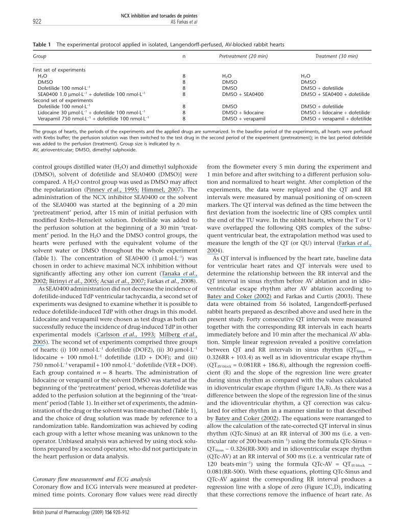
Table 1 The experimental protocol applied in isolated, Langendorff-perfused, AV-blocked rabbit hearts
Group n Pretreatment (20 min) Treatment (30 min)
First set of experimentsH2O 8 H2O H2ODMSO 8 DMSO DMSODofetilide 100 nmol·L-1 8 DMSO DMSO + dofetilideSEA0400 1.0 mmol·L-1 + dofetilide 100 nmol·L-1 8 DMSO + SEA0400 DMSO + SEA0400 + dofetilide
Second set of experimentsDofetilide 100 nmol·L-1 8 DMSO DMSO + dofetilideLidocaine 30 mmol·L-1 + dofetilide 100 nmol·L-1 8 DMSO + lidocaine DMSO + lidocaine + dofetilideVerapamil 750 nmol·L-1 + dofetilide 100 nmol·L-1 8 DMSO + verapamil DMSO + verapamil + dofetilide
The groups of hearts, the periods of the experiments and the applied drugs are summarized. In the baseline period of the experiments, all hearts were perfusedwith Krebs buffer; the perfusion solution was then switched to the test drug in the second period of the experiment (pretreatment); in the last period dofetilidewas added to the perfusion (treatment). Group size is indicated by n.AV, atrioventricular; DMSO, dimethyl sulphoxide.
NCX inhibition and torsades de pointes922 AS Farkas et al
British Journal of Pharmacology (2009) 156 920–932
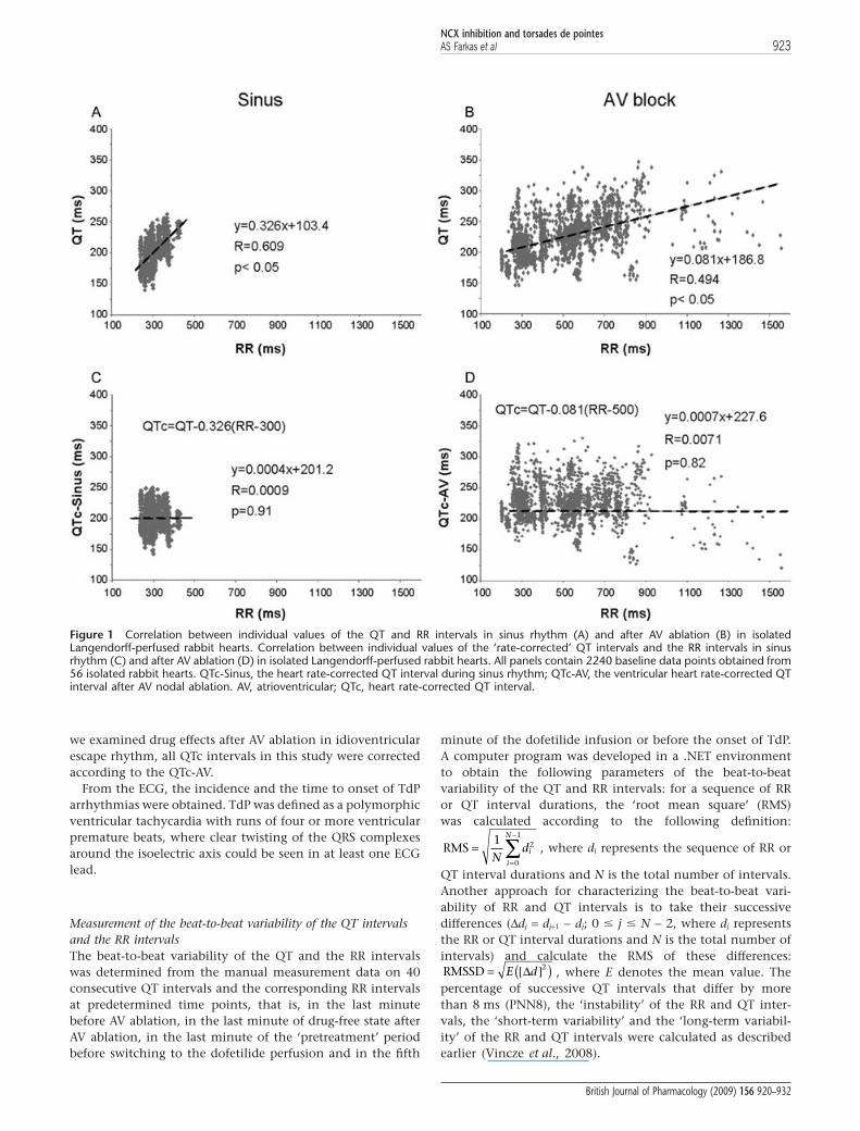
we examined drug effects after AV ablation in idioventricularescape rhythm, all QTc intervals in this study were correctedaccording to the QTc-AV.
From the ECG, the incidence and the time to onset of TdParrhythmias were obtained. TdP was defined as a polymorphicventricular tachycardia with runs of four or more ventricularpremature beats, where clear twisting of the QRS complexesaround the isoelectric axis could be seen in at least one ECGlead.
Measurement of the beat-to-beat variability of the QT intervalsand the RR intervalsThe beat-to-beat variability of the QT and the RR intervalswas determined from the manual measurement data on 40consecutive QT intervals and the corresponding RR intervalsat predetermined time points, that is, in the last minutebefore AV ablation, in the last minute of drug-free state afterAV ablation, in the last minute of the ‘pretreatment’ periodbefore switching to the dofetilide perfusion and in the fifth
minute of the dofetilide infusion or before the onset of TdP.A computer program was developed in a .NET environmentto obtain the following parameters of the beat-to-beatvariability of the QT and RR intervals: for a sequence of RRor QT interval durations, the ‘root mean square’ (RMS)was calculated according to the following definition:
RMS ==
−
∑1 2
0
1
Ndi
i
N
, where di represents the sequence of RR or
QT interval durations and N is the total number of intervals.Another approach for characterizing the beat-to-beat vari-ability of RR and QT intervals is to take their successivedifferences (Ddj = dj+1 - dj; 0 � j � N - 2, where dj representsthe RR or QT interval durations and N is the total number ofintervals) and calculate the RMS of these differences:RMSSD = [ ]( )E dΔ 2 , where E denotes the mean value. Thepercentage of successive QT intervals that differ by morethan 8 ms (PNN8), the ‘instability’ of the RR and QT inter-vals, the ‘short-term variability’ and the ‘long-term variabil-ity’ of the RR and QT intervals were calculated as describedearlier (Vincze et al., 2008).
Figure 1 Correlation between individual values of the QT and RR intervals in sinus rhythm (A) and after AV ablation (B) in isolatedLangendorff-perfused rabbit hearts. Correlation between individual values of the ‘rate-corrected’ QT intervals and the RR intervals in sinusrhythm (C) and after AV ablation (D) in isolated Langendorff-perfused rabbit hearts. All panels contain 2240 baseline data points obtained from56 isolated rabbit hearts. QTc-Sinus, the heart rate-corrected QT interval during sinus rhythm; QTc-AV, the ventricular heart rate-corrected QTinterval after AV nodal ablation. AV, atrioventricular; QTc, heart rate-corrected QT interval.
NCX inhibition and torsades de pointesAS Farkas et al 923
British Journal of Pharmacology (2009) 156 920–932
Exclusion criteriaAny heart with a coronary flow <3 mL·min-1·g-1 or asystolelonger than 20 s during the whole experimental protocol wasexcluded. Longer than 20 s asystole was found after the startof dofetilide perfusion in six hearts pretreated with verapamiland in three hearts pretreated with lidocaine, these heartswere excluded. Additional experiments were performed inorder to maintain equal group sizes.
SolutionsPerfusion solutions were prepared fresh each day. Dofetilideand SEA0400 were dissolved in DMSO. All final test solutionsfor heart perfusion contained 0.08 mL DMSO in 1 L of modi-fied Krebs–Henseleit solution. Likewise, the DMSO controlsolution contained 0.08 mL DMSO in 1 L of modified Krebs–Henseleit solution. The H2O control solution contained0.08 mL water in 1 L of modified Krebs–Henseleit solution. Inthe second set of experiments, lidocaine and verapamil weredissolved in water, and all final test solution for heart perfu-sion contained 0.1 mL water in 1 L of modified Krebs–Henseleit solution.
StatisticsContinuous data were expressed as mean � standard error ofthe mean (SEM). All data from independent samples, exceptthe incidences of TdP, were compared with Kruskal–Wallistests. Within-group comparisons of the variability parameterswere performed by Wilcoxon tests. The incidences of TdPwere compared by using Fisher’s exact probability test withthe Bonferoni correction, that is, the P values of Fisher’s exactprobability test were multiplied by 4 in the first set of experi-ments or 2 in the second set of experiments (the numbers ofcomparisons) to allow multiple comparisons (Altman, 1991).P < 0.05 was taken as indicative of a statistically significantdifference between values.
Drugs and materialsCaCl2, DMSO, lidocaine and verapamil were purchasedfrom Sigma-Aldrich, Inc., St. Louis, MO, USA. All other saltswere purchased from Molar Chemical Ltd., Budapest,Hungary. The dofetilide was a generous gift from GedeonRichter Ltd. (Budapest, Hungary). The synthesis of 2-[4-[(2,5-difluorophenyl)methoxy]phenoxy]-5-ethoxy-aniline(SEA0400) was performed by Ferenc Fülöp (Department ofPharmaceutical Chemistry, University of Szeged, Szeged,Hungary) according to a method described earlier (Farkaset al., 2008). Water for the preparation of perfusion solutionwas obtained from a reverse osmosis system (Milli-Q RG,Millipore Ltd., Billerca, MA, USA) fed by distilled water, andhad a specific resistivity of >18 MW.
Results
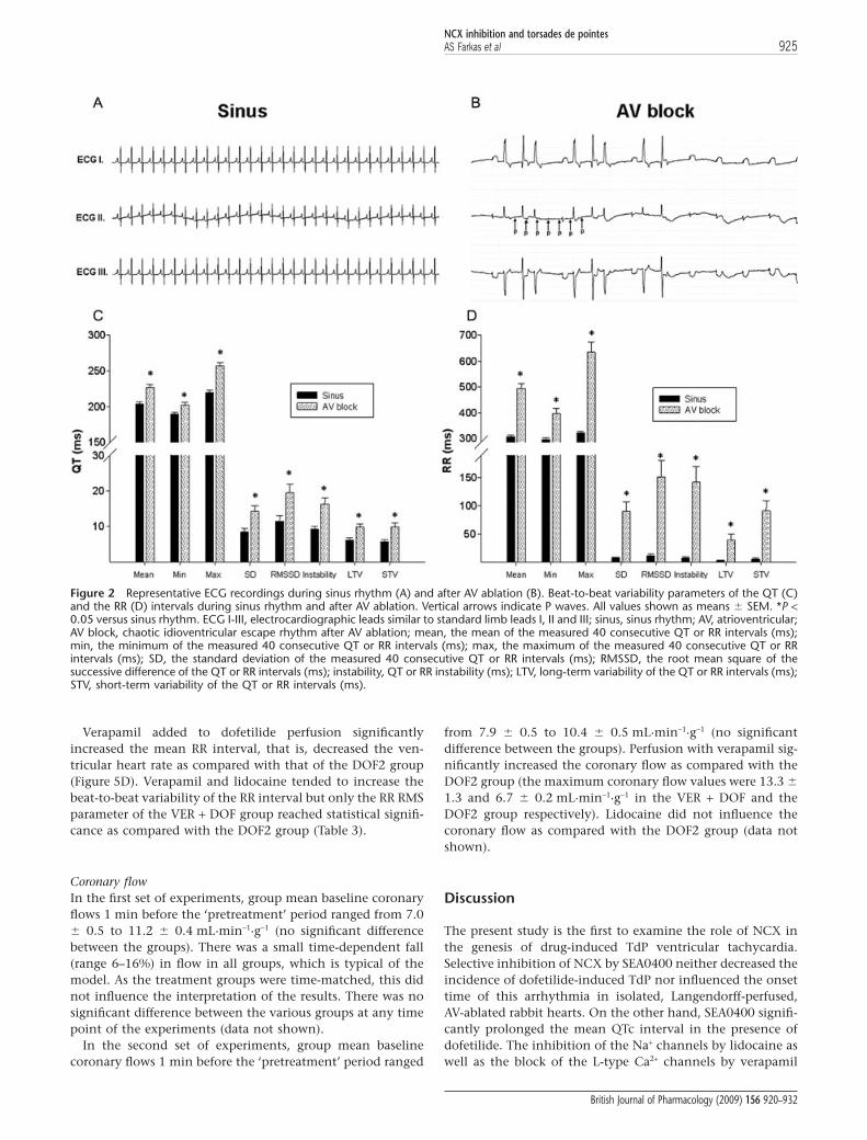
The effect of AV block on the ventricular rhythm andrepolarizationAs expected, the applied mechanical AV block decreased theaverage ventricular heart rate (Figure 2). Interestingly, AVablation also led to a chaotic and irregular spontaneous ven-
tricular rhythm (Figure 2B), and it strikingly increased allparameters of the beat-to-beat variability of the RR intervals(Figure 2D). The measurement of the RR variability param-eters over a longer period (over 3 min) provided results similarto those of the measurement of the RR variability parametersover a shorter (40-consecutive-beat-long) period (data notshown). In addition, the AV ablation increased all QT vari-ability parameters, which reflected a tentative beat-to-beatrepolarization (Figure 2C).
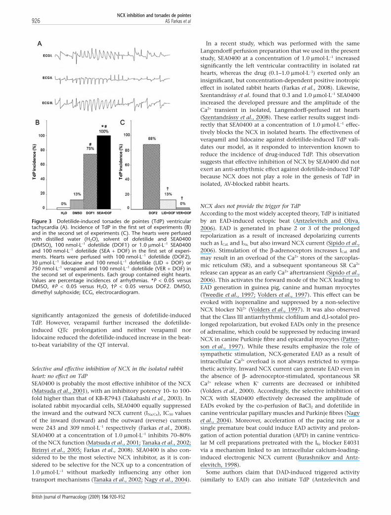
TdP incidences and onset timesIn the first set of experiments, TdP occurred in the majority ofthe dofetilide-perfused hearts (Figure 3A). NCX inhibitionwith SEA0400 did not reduce the incidence of dofetilide-induced TdP (Figure 3B) and did not affect the onset time ofthis arrhythmia (325 � 30 s and 409 � 97 s after switching todofetilide perfusion in the DOF1 and SEA + DOF group respec-tively). Interestingly, TdP occurred in one heart in the DMSO-perfused control group 1174 s after switching to DMSOperfusion from Krebs solution.
In the second set of experiments, dofetilide provoked TdPin the majority of the hearts as seen in the first set of experi-ments. Lidocaine significantly decreased the incidence ofdofetilide-induced TdP, while verapamil completely pre-vented the development of this arrhythmia (Figure 3C).
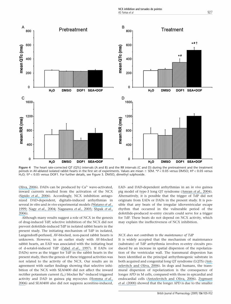
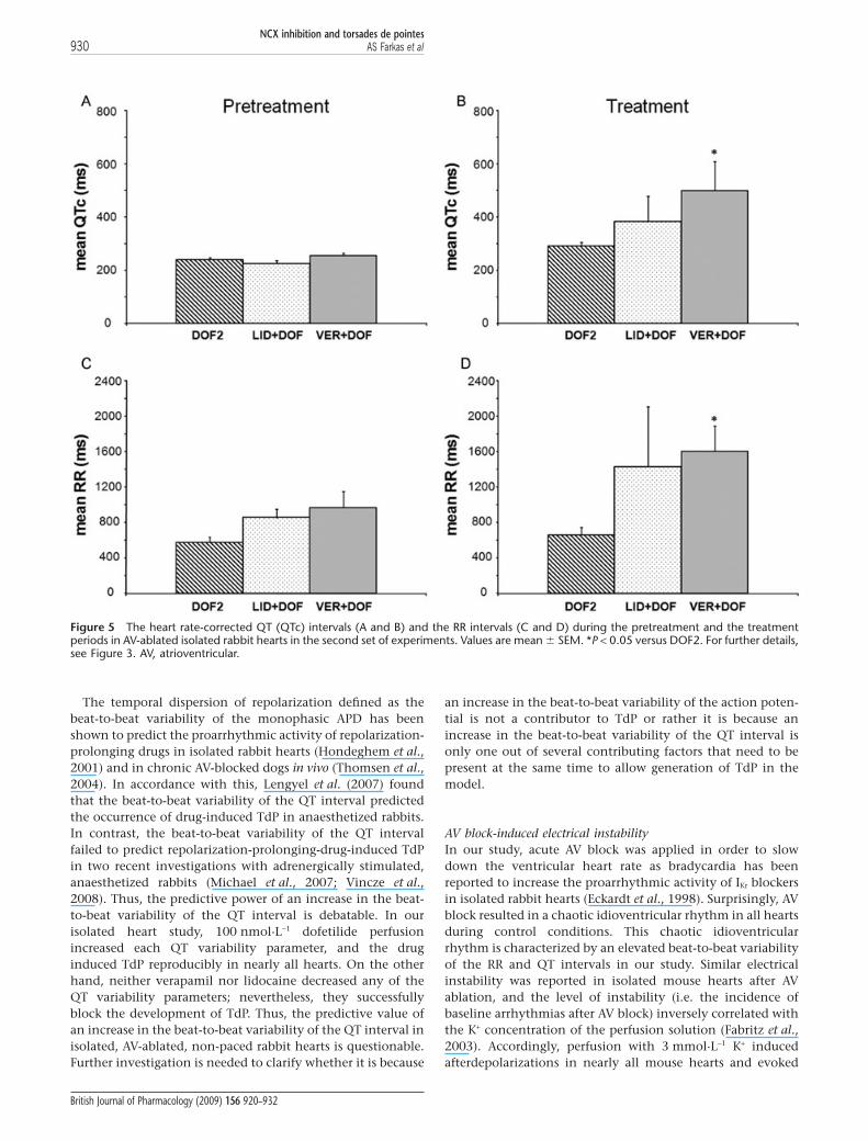
The RR and the QTc intervals and the beat-to-beat variability ofthe RR and QT intervalsPretreatment period In the first set of experiments, neitherDMSO nor SEA0400 affected the mean RR interval, the meanQTc interval (Figure 4A,C) or any of the QT or RR variabilityparameters (Tables 2 and 3). After the exclusion of all heartsthat experienced asystole longer than 20 s from the second setof experiments (see exclusion criteria in Methods) the meanRR interval, the mean QTc interval (Figure 5A,C) or any of theQT or RR variability parameters (Tables 2 and 3) did not sig-nificantly differ between the lidocaine, verapamil and thecontrol groups before dofetilide perfusion.
Treatment period In the first set of experiments, dofetilide per-fusion significantly increased the mean QTc intervals(Figure 4B) and the beat-to-beat variability of the QT interval(Table 2) without affecting the mean RR interval (Figure 4D)and the beat-to-beat variability of the RR interval (Table 3).NCX inhibition with SEA0400 exaggerated the dofetilide-induced increase in the mean QTc interval (Figure 4B) andfurther increased some of the QT variability parameters(Table 2). Further, SEA0400 upon dofetilide perfusionincreased the beat-to-beat variability of the RR interval(Table 3) without affecting the mean RR interval (Figure 4D).
In the second set of experiments, verapamil furtherincreased the dofetilide-induced QTc prolongation whereaslidocaine did not affect the mean QTc interval significantlywhen compared with that of the DOF2 group (Figure 5B).Verapamil tended to increase the beat-to-beat variability ofthe QT interval but only the QT RMS parameter reachedstatistical significance. Similarly, lidocaine added to dofetilideperfusion significantly elevated QT RMS as compared withthat of the DOF2 group (Table 2).
NCX inhibition and torsades de pointes924 AS Farkas et al
British Journal of Pharmacology (2009) 156 920–932
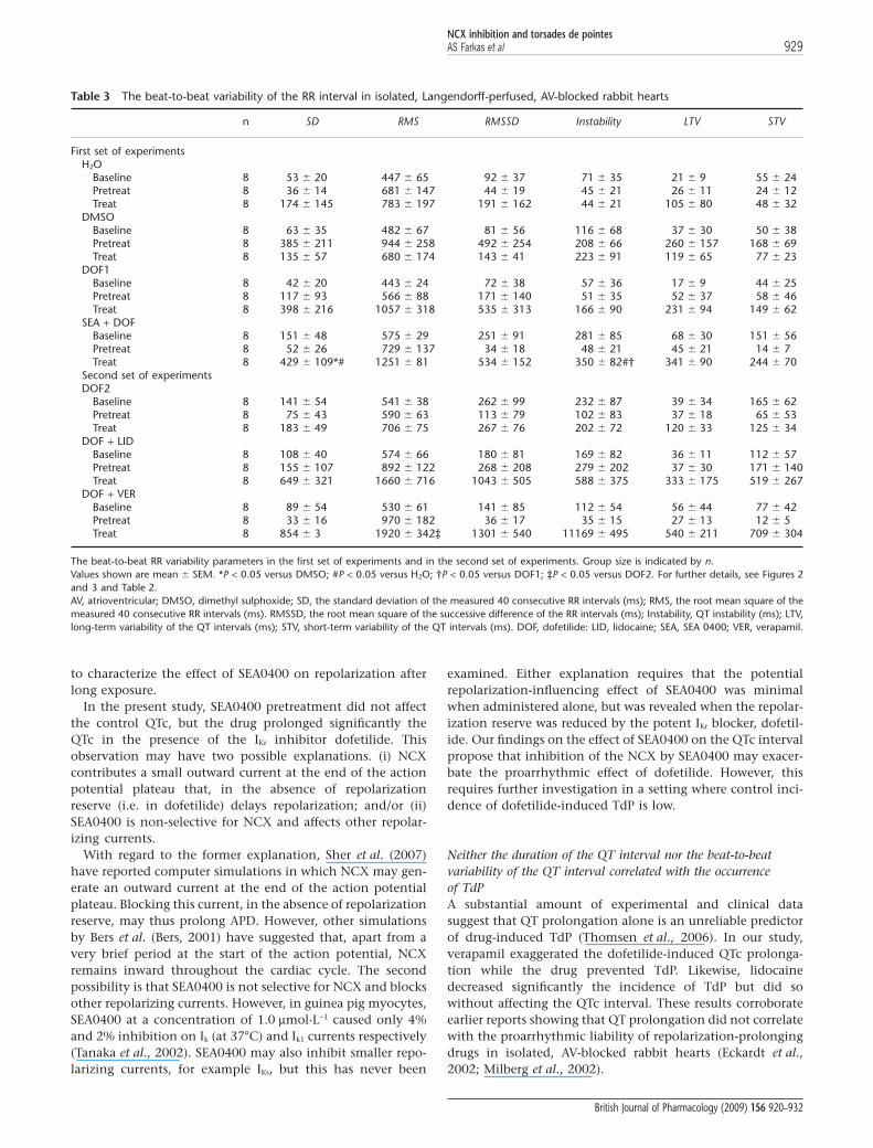
Verapamil added to dofetilide perfusion significantlyincreased the mean RR interval, that is, decreased the ven-tricular heart rate as compared with that of the DOF2 group(Figure 5D). Verapamil and lidocaine tended to increase thebeat-to-beat variability of the RR interval but only the RR RMSparameter of the VER + DOF group reached statistical signifi-cance as compared with the DOF2 group (Table 3).
Coronary flowIn the first set of experiments, group mean baseline coronaryflows 1 min before the ‘pretreatment’ period ranged from 7.0� 0.5 to 11.2 � 0.4 mL·min-1·g-1 (no significant differencebetween the groups). There was a small time-dependent fall(range 6–16%) in flow in all groups, which is typical of themodel. As the treatment groups were time-matched, this didnot influence the interpretation of the results. There was nosignificant difference between the various groups at any timepoint of the experiments (data not shown).
In the second set of experiments, group mean baselinecoronary flows 1 min before the ‘pretreatment’ period ranged
from 7.9 � 0.5 to 10.4 � 0.5 mL·min-1·g-1 (no significantdifference between the groups). Perfusion with verapamil sig-nificantly increased the coronary flow as compared with theDOF2 group (the maximum coronary flow values were 13.3 �
1.3 and 6.7 � 0.2 mL·min-1·g-1 in the VER + DOF and theDOF2 group respectively). Lidocaine did not influence thecoronary flow as compared with the DOF2 group (data notshown).
Discussion
The present study is the first to examine the role of NCX inthe genesis of drug-induced TdP ventricular tachycardia.Selective inhibition of NCX by SEA0400 neither decreased theincidence of dofetilide-induced TdP nor influenced the onsettime of this arrhythmia in isolated, Langendorff-perfused,AV-ablated rabbit hearts. On the other hand, SEA0400 signifi-cantly prolonged the mean QTc interval in the presence ofdofetilide. The inhibition of the Na+ channels by lidocaine aswell as the block of the L-type Ca2+ channels by verapamil
Figure 2 Representative ECG recordings during sinus rhythm (A) and after AV ablation (B). Beat-to-beat variability parameters of the QT (C)and the RR (D) intervals during sinus rhythm and after AV ablation. Vertical arrows indicate P waves. All values shown as means � SEM. *P <0.05 versus sinus rhythm. ECG I-III, electrocardiographic leads similar to standard limb leads I, II and III; sinus, sinus rhythm; AV, atrioventricular;AV block, chaotic idioventricular escape rhythm after AV ablation; mean, the mean of the measured 40 consecutive QT or RR intervals (ms);min, the minimum of the measured 40 consecutive QT or RR intervals (ms); max, the maximum of the measured 40 consecutive QT or RRintervals (ms); SD, the standard deviation of the measured 40 consecutive QT or RR intervals (ms); RMSSD, the root mean square of thesuccessive difference of the QT or RR intervals (ms); instability, QT or RR instability (ms); LTV, long-term variability of the QT or RR intervals (ms);STV, short-term variability of the QT or RR intervals (ms).
NCX inhibition and torsades de pointesAS Farkas et al 925
British Journal of Pharmacology (2009) 156 920–932
significantly antagonized the genesis of dofetilide-inducedTdP. However, verapamil further increased the dofetilide-induced QTc prolongation and neither verapamil norlidocaine reduced the dofetilide-induced increase in the beat-to-beat variability of the QT interval.
Selective and effective inhibition of NCX in the isolated rabbitheart: no effect on TdPSEA0400 is probably the most effective inhibitor of the NCX(Matsuda et al., 2001), with an inhibitory potency 10- to 100-fold higher than that of KB-R7943 (Takahashi et al., 2003). Inisolated rabbit myocardial cells, SEA0400 equally suppressedthe inward and the outward NCX current (INa/Ca), IC50 valuesof the inward (forward) and the outward (reverse) currentswere 243 and 309 nmol·L-1 respectively (Farkas et al., 2008).SEA0400 at a concentration of 1.0 mmol·L-1 inhibits 70–80%of the NCX function (Matsuda et al., 2001; Tanaka et al., 2002;Birinyi et al., 2005; Farkas et al., 2008). SEA0400 is also con-sidered to be the most selective NCX inhibitor, as it is con-sidered to be selective for the NCX up to a concentration of1.0 mmol·L-1 without markedly influencing any other iontransport mechanisms (Tanaka et al., 2002; Nagy et al., 2004).
In a recent study, which was performed with the sameLangendorff perfusion preparation that we used in the presentstudy, SEA0400 at a concentration of 1.0 mmol·L-1 increasedsignificantly the left ventricular contractility in isolated rathearts, whereas the drug (0.1–1.0 mmol·L-1) exerted only aninsignificant, but concentration-dependent positive inotropiceffect in isolated rabbit hearts (Farkas et al., 2008). Likewise,Szentandrássy et al. found that 0.3 and 1.0 mmol·L-1 SEA0400increased the developed pressure and the amplitude of theCa2+ transient in isolated, Langendorff-perfused rat hearts(Szentandrássy et al., 2008). These earlier results suggest indi-rectly that SEA0400 at a concentration of 1.0 mmol·L-1 effec-tively blocks the NCX in isolated hearts. The effectiveness ofverapamil and lidocaine against dofetilide-induced TdP vali-dates our model, as it responded to intervention known toreduce the incidence of drug-induced TdP. This observationsuggests that effective inhibition of NCX by SEA0400 did notexert an anti-arrhythmic effect against dofetilide-induced TdPbecause NCX does not play a role in the genesis of TdP inisolated, AV-blocked rabbit hearts.
NCX does not provide the trigger for TdPAccording to the most widely accepted theory, TdP is initiatedby an EAD-induced ectopic beat (Antzelevitch and Oliva,2006). EAD is generated in phase 2 or 3 of the prolongedrepolarization as a result of increased depolarizing currentssuch as ICaL and INa, but also inward NCX current (Sipido et al.,2006). Stimulation of the b-adrenoceptors increases ICaL andmay result in an overload of the Ca2+ stores of the sarcoplas-mic reticulum (SR), and a subsequent spontaneous SR Ca2+
release can appear as an early Ca2+ aftertransient (Sipido et al.,2006). This activates the forward mode of the NCX leading toEAD generation in guinea pig, canine and human myocytes(Tweedie et al., 1997; Volders et al., 1997). This effect can beevoked with isoprenaline and suppressed by a non-selectiveNCX blocker Ni2+ (Volders et al., 1997). It was also observedthat the Class III antiarrhythmic clofilium and d,l-sotalol pro-longed repolarization, but evoked EADs only in the presenceof adrenaline, which could be suppressed by reducing inwardNCX in canine Purkinje fibre and epicardial myocytes (Patter-son et al., 1997). While these results emphasize the role ofsympathetic stimulation, NCX-generated EAD as a result ofintracellular Ca2+ overload is not always restricted to sympa-thetic activity. Inward NCX current can generate EAD even inthe absence of b- adrenoceptor-stimulated, spontaneous SRCa2+ release when K+ currents are decreased or inhibited(Volders et al., 2000). Accordingly, the selective inhibition ofNCX with SEA0400 effectively decreased the amplitude ofEADs evoked by the co-perfusion of BaCl2 and dofetilide incanine ventricular papillary muscles and Purkinje fibres (Nagyet al., 2004). Moreover, acceleration of the pacing rate or asingle premature beat could induce EAD activity and prolon-gation of action potential duration (APD) in canine ventricu-lar M cell preparations pretreated with the IKr blocker E4031via a mechanism linked to an intracellular calcium-loading-induced electrogenic NCX current (Burashnikov and Antz-elevitch, 1998).
Some authors claim that DAD-induced triggered activity(similarly to EAD) can also initiate TdP (Antzelevitch and
Figure 3 Dofetilide-induced torsades de pointes (TdP) ventriculartachycardia (A). Incidence of TdP in the first set of experiments (B)and in the second set of experiments (C). The hearts were perfusedwith distilled water (H2O), solvent of dofetilide and SEA0400(DMSO), 100 nmol·L-1 dofetilide (DOF1) or 1.0 mmol·L-1 SEA0400and 100 nmol·L-1 dofetilide (SEA + DOF) in the first set of experi-ments. Hearts were perfused with 100 nmol·L-1 dofetilide (DOF2),30 mmol·L-1 lidocaine and 100 nmol·L-1 dofetilide (LID + DOF) or750 nmol·L-1 verapamil and 100 nmol·L-1 dofetilide (VER + DOF) inthe second set of experiments. Each group contained eight hearts.Values are percentage incidences of arrhythmias. *P < 0.05 versusDMSO, #P < 0.05 versus H2O, †P < 0.05 versus DOF2. DMSO,dimethyl sulphoxide; ECG, electrocardiogram.
NCX inhibition and torsades de pointes926 AS Farkas et al
British Journal of Pharmacology (2009) 156 920–932
Oliva, 2006). DADs can be produced by Ca2+ wave-activated,inward currents resulted from the activation of the NCX(Sipido et al., 2006). Accordingly, NCX inhibition antago-nized DAD-dependent, digitalis-induced arrhythmias inseveral in vitro and in vivo experimental models (Watano et al.,1999; Nagy et al., 2004; Nagasawa et al., 2005; Shpak et al.,2006).
Although many results suggest a role of NCX in the genesisof drug-induced TdP, selective inhibition of the NCX did notprevent dofetilide-induced TdP in isolated rabbit hearts in thepresent study. The initiating mechanism of TdP in isolated,Langendorff-perfused, AV-blocked, non-paced rabbit hearts isunknown. However, in an earlier study with AV-blockedrabbit hearts, an EAD was associated with the initiating beatof d-sotalol-induced TdP (Zabel et al., 1997). If EADs (orDADs) serve as the trigger for TdP in the rabbit hearts in thepresent study, then the genesis of these triggered activities wasnot related to the activity of the NCX. Our results are inagreement with earlier findings showing that selective inhi-bition of the NCX with SEA0400 did not affect the inwardrectifier potassium current (Ik1) blocker Ba2+-induced triggeredactivity and DAD in guinea pig myocytes (Homma et al.,2006) and SEA0400 also did not suppress aconitine-induced,
EAD- and DAD-dependent arrhythmias in an in vivo guineapig model of type-3 long QT syndrome (Amran et al., 2004).Alternatively, it is possible that the trigger of TdP did notoriginate from EADs or DADs in the present study. It is pos-sible that any beats of the irregular idioventricular escaperhythm that occurred in the vulnerable period of thedofetilide-produced re-entry circuits could serve for a triggerfor TdP. These beats do not depend on NCX activity, whichmay explain the ineffectiveness of NCX inhibition.
NCX does not contribute to the maintenance of TdPIt is widely accepted that the mechanism of maintenance(substrate) of TdP arrhythmia involves re-entry circuits pro-duced by an increase in spatial dispersion of the repolariza-tion of the ventricular wall. The transmural dispersion hasbeen identified as the principal arrhythmogenic substrate inboth acquired and congenital long QT syndrome (LQTS) (Ant-zelevitch and Oliva, 2006). In dogs and humans, the trans-mural dispersion of repolarization is the consequence oflonger APD in M cells, compared with those in epicardial andendocardial cells (Antzelevitch and Oliva, 2006). Zygmuntet al. (2000) showed that the longer APD is due to the smaller
Figure 4 The heart rate-corrected QT (QTc) intervals (A and B) and the RR intervals (C and D) during the pretreatment and the treatmentperiods in AV-ablated isolated rabbit hearts in the first set of experiments. Values are mean � SEM. *P < 0.05 versus DMSO; #P < 0.05 versusH2O; †P < 0.05 versus DOF1. For further details, see Figure 3. DMSO, dimethyl sulphoxide.
NCX inhibition and torsades de pointesAS Farkas et al 927
British Journal of Pharmacology (2009) 156 920–932

slow component of the delayed rectifier potassium current(IKs) and larger late INa and NCX currents in the healthy canineventricle. The IKr inhibitor d,l-sotalol increased transmuraldispersion of repolarization and induced TdP in isolated,Langendorff-perfused, AV-blocked rabbit hearts (Eckardt et al.,2002). Further, verapamil prevented veratridine-induced TdPvia reduction of left ventricular transmural dispersion of repo-larization (and suppression of EADs) in isolated, Langendorff-perfused, AV-blocked rabbit hearts (Milberg et al., 2005).These earlier observations suggest that transmural dispersionof repolarization may also play a role in the maintenance ofTdP in AV-ablated rabbit hearts. Although NCX activity con-tributes to the generation of transmural dispersion of repolar-ization in the healthy dog heart (Zygmunt et al., 2000), ourresults indicate that NCX activity does not increase the trans-mural dispersion of repolarization and, therefore, does notcontribute to the maintenance mechanism of TdP in therabbit heart.
The effect of SEA0400 on the duration of repolarizationSEA0400 is a highly selective and effective inhibitor of theNCX at a concentration of 1.0 mmol·L-1 (Matsuda et al., 2001;
Takahashi et al., 2003; Farkas et al., 2008). To date, no experi-mental data have been reported on an appreciable effect ofthis concentration of SEA0400 on the repolarization. Ourstudy did not involve a separate group of hearts with SEA0400perfusion alone during the 20 min ‘pretreatment’ and thesubsequent 30 min ‘treatment’ period; thus, we cannotexclude the possibility that the drug affects repolarization andcauses arrhythmias after prolonged exposure. However,30 min perfusion with SEA0400 at a concentration of1.0 mmol·L-1, which is the same concentration we used in thepresent study, did not affect ECG parameters and did notinduce any arrhythmias in isolated rat and rabbit hearts in arecent study from our laboratory (Farkas et al., 2008). Simi-larly, 20 min SEA0400 pretreatment did not affect the controlQTc in the present study. In contrast, when the subsequenttreatment period started and the IKr inhibitor dofetilide wasadded to the SEA0400 perfusion, repolarization prolongedquickly and markedly in 5 min, and a significantly longer QTcwas measured than that in the group of hearts perfused withdofetilide on its own. These observations suggest thatSEA0400 prolongs repolarization only when the repolariza-tion reserve is markedly reduced by the co-administration ofanother drug. Nevertheless, further investigations would help
Table 2 The beat-to-beat variability of the QT interval in isolated, Langendorff-perfused, AV-blocked rabbit hearts
n SD RMS RMSSD PNN8 Instability LTV STV
First set of experimentsH2O
Baseline 8 11 � 2 225 � 13 15 � 2 25 � 4 12 � 2 8 � 1 8 � 1Pretreat 8 11 � 2 254 � 6 11 � 2 23 � 4 14 � 3 10 � 3 6 � 1Treat 8 14 � 2 253 � 19 18 � 3 25 � 3 14 � 2* 11 � 2 9 � 2
DMSOBaseline 8 17 � 4 233 � 12 24 � 7 30 � 4 20 � 5 11 � 3 13 � 4Pretreat 8 18 � 4 251 � 13 23 � 5 31 � 3 20 � 3 14 � 3 12 � 2Treat 8 22 � 3 267 � 15 30 � 6 38 � 3# 30 � 5# 16 � 2 17 � 4
DOF1Baseline 8 14 � 2 217 � 9 16 � 2 29 � 3 17 � 3 11 � 2 9 � 1Pretreat 8 16 � 5 226 � 6 20 � 5 26 � 5 19 � 4 13 � 3 10 � 2Treat 8 73 � 15*# 389 � 55*# 87 � 17*# 41 � 3# 65 � 19# 63 � 16*# 40 � 8*#
SEA + DOFBaseline 8 14 � 4 225 � 12 18 � 6 27 � 3 21 � 7 12 � 3 10 � 4Pretreat 8 14 � 2 258 � 20 18 � 3 27 � 4 17 � 3 12 � 2 9 � 2Treat 8 101 � 27*# 589 � 45*#† 91 � 25*# 40 � 3# 145 � 53*# 102 � 32*# 42 � 12*#
Second set of experimentsDOF2
Baseline 8 15 � 3 242 � 10 23 � 5 30 � 4 21 � 5 9 � 1 13 � 4Pretreat 8 12 � 2 245 � 9 17 � 4 27 � 3 14 � 3 9 � 2 9 � 2Treat 8 42 � 12 308 � 17 49 � 14 39 � 4 60 � 16 35 � 12 27 � 8
DOF + LIDBaseline 8 18 � 8 228 � 15 26 � 18 24 � 4 11 � 2 10 � 3 9 � 2Pretreat 8 21 � 12 256 � 14 31 � 18 25 � 4 17 � 5 17 � 10 15 � 9Treat 8 31 � 9 459 � 97‡ 47 � 14 38 � 5 45 � 15 20 � 6 27 � 8
DOF + VERBaseline 8 12 � 2 225 � 8 16 � 3 27 � 5 12 � 2 8 � 1 9 � 2Pretreat 8 19 � 6 293 � 17 22 � 5 29 � 2 17 � 3 13 � 3 10 � 1Treat 8 101 � 36 588 � 115‡ 135 � 61 38 � 6 161 � 62 72 � 23 78 � 39
The beat-to-beat QT variability parameters in the first set of experiments and in the second set of experiments.Values shown are mean � SEM. *P < 0.05 versus DMSO; #P < 0.05 versus H2O; †P < 0.05 versus DOF1; ‡P < 0.05 versus DOF2. For further details, see Figures 2and 3.AV, atrioventricular; DMSO, dimethyl sulphoxide; pretreat, pretreatment period; treat, treatment period; group size is indicated by n. SD, the standard deviationof the measured 40 consecutive QT intervals (ms); RMS, the root mean square of the measured 40 consecutive QT intervals (ms); RMSSD, the root mean squareof the successive difference of the QT intervals (ms); PNN8, percentage of QT intervals differing from the preceding one by more than 8 ms (%); instability, QTinstability (ms); LTV, long-term variability of the QT intervals (ms); STV, short-term variability of the QT intervals (ms); DOF, dofetilide; LID, lidocaine; SEA, SEA 0400;VER, verapamil.
NCX inhibition and torsades de pointes928 AS Farkas et al
British Journal of Pharmacology (2009) 156 920–932
to characterize the effect of SEA0400 on repolarization afterlong exposure.
In the present study, SEA0400 pretreatment did not affectthe control QTc, but the drug prolonged significantly theQTc in the presence of the IKr inhibitor dofetilide. Thisobservation may have two possible explanations. (i) NCXcontributes a small outward current at the end of the actionpotential plateau that, in the absence of repolarizationreserve (i.e. in dofetilide) delays repolarization; and/or (ii)SEA0400 is non-selective for NCX and affects other repolar-izing currents.
With regard to the former explanation, Sher et al. (2007)have reported computer simulations in which NCX may gen-erate an outward current at the end of the action potentialplateau. Blocking this current, in the absence of repolarizationreserve, may thus prolong APD. However, other simulationsby Bers et al. (Bers, 2001) have suggested that, apart from avery brief period at the start of the action potential, NCXremains inward throughout the cardiac cycle. The secondpossibility is that SEA0400 is not selective for NCX and blocksother repolarizing currents. However, in guinea pig myocytes,SEA0400 at a concentration of 1.0 mmol·L-1 caused only 4%and 2% inhibition on Ik (at 37°C) and Ik1 currents respectively(Tanaka et al., 2002). SEA0400 may also inhibit smaller repo-larizing currents, for example IKs, but this has never been
examined. Either explanation requires that the potentialrepolarization-influencing effect of SEA0400 was minimalwhen administered alone, but was revealed when the repolar-ization reserve was reduced by the potent IKr blocker, dofetil-ide. Our findings on the effect of SEA0400 on the QTc intervalpropose that inhibition of the NCX by SEA0400 may exacer-bate the proarrhythmic effect of dofetilide. However, thisrequires further investigation in a setting where control inci-dence of dofetilide-induced TdP is low.
Neither the duration of the QT interval nor the beat-to-beatvariability of the QT interval correlated with the occurrenceof TdPA substantial amount of experimental and clinical datasuggest that QT prolongation alone is an unreliable predictorof drug-induced TdP (Thomsen et al., 2006). In our study,verapamil exaggerated the dofetilide-induced QTc prolonga-tion while the drug prevented TdP. Likewise, lidocainedecreased significantly the incidence of TdP but did sowithout affecting the QTc interval. These results corroborateearlier reports showing that QT prolongation did not correlatewith the proarrhythmic liability of repolarization-prolongingdrugs in isolated, AV-blocked rabbit hearts (Eckardt et al.,2002; Milberg et al., 2002).
Table 3 The beat-to-beat variability of the RR interval in isolated, Langendorff-perfused, AV-blocked rabbit hearts
n SD RMS RMSSD Instability LTV STV
First set of experimentsH2O
Baseline 8 53 � 20 447 � 65 92 � 37 71 � 35 21 � 9 55 � 24Pretreat 8 36 � 14 681 � 147 44 � 19 45 � 21 26 � 11 24 � 12Treat 8 174 � 145 783 � 197 191 � 162 44 � 21 105 � 80 48 � 32
DMSOBaseline 8 63 � 35 482 � 67 81 � 56 116 � 68 37 � 30 50 � 38Pretreat 8 385 � 211 944 � 258 492 � 254 208 � 66 260 � 157 168 � 69Treat 8 135 � 57 680 � 174 143 � 41 223 � 91 119 � 65 77 � 23
DOF1Baseline 8 42 � 20 443 � 24 72 � 38 57 � 36 17 � 9 44 � 25Pretreat 8 117 � 93 566 � 88 171 � 140 51 � 35 52 � 37 58 � 46Treat 8 398 � 216 1057 � 318 535 � 313 166 � 90 231 � 94 149 � 62
SEA + DOFBaseline 8 151 � 48 575 � 29 251 � 91 281 � 85 68 � 30 151 � 56Pretreat 8 52 � 26 729 � 137 34 � 18 48 � 21 45 � 21 14 � 7Treat 8 429 � 109*# 1251 � 81 534 � 152 350 � 82#† 341 � 90 244 � 70
Second set of experimentsDOF2
Baseline 8 141 � 54 541 � 38 262 � 99 232 � 87 39 � 34 165 � 62Pretreat 8 75 � 43 590 � 63 113 � 79 102 � 83 37 � 18 65 � 53Treat 8 183 � 49 706 � 75 267 � 76 202 � 72 120 � 33 125 � 34
DOF + LIDBaseline 8 108 � 40 574 � 66 180 � 81 169 � 82 36 � 11 112 � 57Pretreat 8 155 � 107 892 � 122 268 � 208 279 � 202 37 � 30 171 � 140Treat 8 649 � 321 1660 � 716 1043 � 505 588 � 375 333 � 175 519 � 267
DOF + VERBaseline 8 89 � 54 530 � 61 141 � 85 112 � 54 56 � 44 77 � 42Pretreat 8 33 � 16 970 � 182 36 � 17 35 � 15 27 � 13 12 � 5Treat 8 854 � 3 1920 � 342‡ 1301 � 540 11169 � 495 540 � 211 709 � 304
The beat-to-beat RR variability parameters in the first set of experiments and in the second set of experiments. Group size is indicated by n.Values shown are mean � SEM. *P < 0.05 versus DMSO; #P < 0.05 versus H2O; †P < 0.05 versus DOF1; ‡P < 0.05 versus DOF2. For further details, see Figures 2and 3 and Table 2.AV, atrioventricular; DMSO, dimethyl sulphoxide; SD, the standard deviation of the measured 40 consecutive RR intervals (ms); RMS, the root mean square of themeasured 40 consecutive RR intervals (ms). RMSSD, the root mean square of the successive difference of the RR intervals (ms); Instability, QT instability (ms); LTV,long-term variability of the QT intervals (ms); STV, short-term variability of the QT intervals (ms). DOF, dofetilide: LID, lidocaine; SEA, SEA 0400; VER, verapamil.
NCX inhibition and torsades de pointesAS Farkas et al 929
British Journal of Pharmacology (2009) 156 920–932
The temporal dispersion of repolarization defined as thebeat-to-beat variability of the monophasic APD has beenshown to predict the proarrhythmic activity of repolarization-prolonging drugs in isolated rabbit hearts (Hondeghem et al.,2001) and in chronic AV-blocked dogs in vivo (Thomsen et al.,2004). In accordance with this, Lengyel et al. (2007) foundthat the beat-to-beat variability of the QT interval predictedthe occurrence of drug-induced TdP in anaesthetized rabbits.In contrast, the beat-to-beat variability of the QT intervalfailed to predict repolarization-prolonging-drug-induced TdPin two recent investigations with adrenergically stimulated,anaesthetized rabbits (Michael et al., 2007; Vincze et al.,2008). Thus, the predictive power of an increase in the beat-to-beat variability of the QT interval is debatable. In ourisolated heart study, 100 nmol·L-1 dofetilide perfusionincreased each QT variability parameter, and the druginduced TdP reproducibly in nearly all hearts. On the otherhand, neither verapamil nor lidocaine decreased any of theQT variability parameters; nevertheless, they successfullyblock the development of TdP. Thus, the predictive value ofan increase in the beat-to-beat variability of the QT interval inisolated, AV-ablated, non-paced rabbit hearts is questionable.Further investigation is needed to clarify whether it is because
an increase in the beat-to-beat variability of the action poten-tial is not a contributor to TdP or rather it is because anincrease in the beat-to-beat variability of the QT interval isonly one out of several contributing factors that need to bepresent at the same time to allow generation of TdP in themodel.
AV block-induced electrical instabilityIn our study, acute AV block was applied in order to slowdown the ventricular heart rate as bradycardia has beenreported to increase the proarrhythmic activity of IKr blockersin isolated rabbit hearts (Eckardt et al., 1998). Surprisingly, AVblock resulted in a chaotic idioventricular rhythm in all heartsduring control conditions. This chaotic idioventricularrhythm is characterized by an elevated beat-to-beat variabilityof the RR and QT intervals in our study. Similar electricalinstability was reported in isolated mouse hearts after AVablation, and the level of instability (i.e. the incidence ofbaseline arrhythmias after AV block) inversely correlated withthe K+ concentration of the perfusion solution (Fabritz et al.,2003). Accordingly, perfusion with 3 mmol·L-1 K+ inducedafterdepolarizations in nearly all mouse hearts and evoked
Figure 5 The heart rate-corrected QT (QTc) intervals (A and B) and the RR intervals (C and D) during the pretreatment and the treatmentperiods in AV-ablated isolated rabbit hearts in the second set of experiments. Values are mean � SEM. *P < 0.05 versus DOF2. For further details,see Figure 3. AV, atrioventricular.
NCX inhibition and torsades de pointes930 AS Farkas et al
British Journal of Pharmacology (2009) 156 920–932
polymorphic ventricular tachycardia in one mouse heartduring control conditions, after AV ablation (Fabritz et al.,2003). This may explain why the evaluation of our experi-ments with 3 mmol·L-1 K+ in the perfusion solution found arun of ‘TdP’ in a heart in the DMSO control group. Further,the baseline electrical instability in the AV-blocked rabbithearts perfused with 3 mmol·L-1 K+ in the present study mightcontribute to the high sensitivity of these hearts to the proar-rhythmic activity of dofetilide.
The antiarrhythmic effect of verapamil andlidocaine against TdPThe ICaL channel blocker verapamil significantly prolongedthe QT interval in the presence of dofetilde and prevented thedevelopment of TdP. Verapamil (which has never beenreported to cause TdP and indeed has been proposed as atherapy for long QT-related arrhythmias) inhibited IKr in thesame concentration range as quinidine and amiodarone (Yanget al., 2001). This may explain why the drug prolongedfurther the QT interval, when the repolarization reserve wasvery small as a result of dofetilide perfusion. Verapamil alsoprolonged the mean RR interval, that is, reduced the numberof idioventricular beats mostly when it was co-perfused withdofetilide. This effect might reduce the number of triggerbeats for the initiation of TdP irrespective of the fact that thetrigger was either the idioventricular beat itself or the corre-sponding afterdepolarization. As lidocaine tended to have thesame effect on the ventricular rate, this may also explain itsantiarrhythmic effect in our investigations. Shimizu et al.(1995) reported that verapamil suppressed spontaneous oradrenaline-induced EADs and TdP in patients with congenitalLQTS. Milberg et al. (2005) found that the same concentrationof verapamil we used (750 nmol·L-1) prevented TdP via thereduction of EAD and ventricular transmural dispersion ofrepolarization in an isolated rabbit heart model of theacquired LQT3 syndrome. Likewise, lidocaine suppressed theIKr blocker almokalant-induced dispersion of repolarizationand the development of EADs in rabbit Purkinje fibres in vitro(Abrahamsson et al., 1996). Thus, the antiarrhythmic effect ofverapamil and lidocaine in our experiments may also berelated their direct effect on the development of EAD and/ordispersion of the repolarization.
Conclusions
Na+/Ca2+ exchanger neither contributed to the initiation norassisted the maintenance of dofetilide-induced TdP in our invitro experimental model, in which inhibition of the INa andICaL currents successfully antagonized the genesis of thisarrhythmia. Neither QTc prolongation nor an increase in thebeat-to-beat variability of the QT interval is a sufficient pre-requisite of TdP genesis in this model. AV ablation resulted ina chaotic idioventricular rhythm, which might make thehearts more sensitive to the proarrhythmic activity of dofetil-ide in the applied isolated rabbit heart model.
Acknowledgements
Eleonora Grandi and Donald M. Bers (Department of Phar-macology, University of California, Davis, CA, USA) are
thanked for their useful comments on the results of thepresent study. This work was supported by the HungarianMinistry of Health (ETT 203/2003 and 353/2006), the Hun-garian National Research Fund (OTKA F-046776, OTKAK-69018 and NI-61902), the National Research and Develop-ment Programmes (NKFP 1 A/046/2004), the European Com-munity (EU FP7 grant ICT-2008-224381, preDiCT) and theHungarian Academy of Sciences.
Conflicts of interest
None.
References
Abrahamsson C, Carlsson L, Duker G (1996). Lidocaine and nisol-dipine attenuate almokalant-induced dispersion of repolarizationand early afterdepolarizations in vitro. J Cardiovasc Electrophysiol 7:1074–1081.
Acsai K, Kun A, Farkas AS, Fülöp F, Nagy N, Balázs M et al. (2007).Effect of partial blockade of the Na(+)/Ca(2+)-exchanger on Ca(2+)handling in isolated rat ventricular myocytes. Eur J Pharmacol 576:1–6.
Altman DG (1991). Practical Statistics for Medical Research, 1st edn.Chapman & Hall: London.
Amran MS, Hashimoto K, Homma N (2004). Effects of sodium-calcium exchange inhibitors, KB-R7943 and SEA0400, on aconitine-induced arrhythmias in guinea pigs in vivo, in vitro, and in computersimulation studies. J Pharmacol Exp Ther 310: 83–89.
Antzelevitch C, Oliva A (2006). Amplification of spatial dispersion ofrepolarization underlies sudden cardiac death associated with cat-echolaminergic polymorphic VT, long QT, short QT and Brugadasyndromes. J Intern Med 259: 48–58.
Asano Y, Davidenko JM, Baxter WT, Gray RA, Jalife J (1997). Opticalmapping of drug-induced polymorphic arrhythmias and torsade depointes in the isolated rabbit heart. J Am Coll Cardiol 29: 831–842.
Barrett TD, Hennan JK, Fischbach PS, O’Neill BP, Driscoll EM Jr,Lucchesi BR (2001). Tedisamil and dofetilide-induced torsades depointes, rate and potassium dependence. Br J Pharmacol 132: 1493–1500.
Batey AJ, Coker SJ (2002). Proarrhythmic potential of halofantrine,terfenadine and clofilium in a modified in vivo model of torsade depointes. Br J Pharmacol 135: 1003–1012.
Belardinelli L, Antzelevitch C, Vos MA (2003). Assessing predictors ofdrug-induced torsade de pointes. Trends Pharmacol Sci 24: 619–625.
Bers DM (2001). Excitation-contraction Coupling and Cardiac ContractileForce, 2nd edn. Kluwer Academic Publishers: Dordrecht.
Birinyi P, Acsai K, Bányász T, Tóth A, Horváth B, Virág L et al. (2005).Effects of SEA0400 and KB-R7943 on Na(+)/Ca(2+) exchange currentand L-type Ca(2+) current in canine ventricular cardiomyocytes.Naunyn Schmiedebergs Arch Pharmacol 372: 63–70.
Burashnikov A, Antzelevitch C (1998). Acceleration-induced actionpotential prolongation and early afterdepolarizations. J CardiovascElectrophysiol 9: 934–948.
Carlsson L, Drews L, Duker G, Schiller-Linhardt G (1993). Attenuationof proarrhythmias related to delayed repolarization by low-doselidocaine in the anesthetized rabbit. J Pharmacol Exp Ther 267:1076–1080.
Eckardt L, Haverkamp W, Mertens H, Johna R, Clague JR, Borggrefe Met al. (1998). Drug-related torsades de pointes in the isolated rabbitheart: comparison of clofilium, d,l-sotalol, and erythromycin.J Cardiovasc Pharmacol 32: 425–434.
Eckardt L, Breithardt G, Haverkamp W (2002). Electrophysiologic
NCX inhibition and torsades de pointesAS Farkas et al 931
British Journal of Pharmacology (2009) 156 920–932
characterization of the antipsychotic drug sertindole in a rabbitheart model of torsade de pointes: low torsadogenic potentialdespite QT prolongation. J Pharmacol Exp Ther 300: 64–71.
Fabritz L, Kirchhof P, Franz MR, Eckardt L, Monnig G, Milberg P et al.(2003). Prolonged action potential durations, increased dispersionof repolarization, and polymorphic ventricular tachycardia in amouse model of proarrhythmia. Basic Res Cardiol 98: 25–32.
Farkas A, Curtis MJ (2003). Does QT widening in the Langendorff-perfused rat heart represent the effect of repolarization delay orconduction slowing? J Cardiovasc Pharmacol 42: 612–621.
Farkas A, Batey AJ, Coker SJ (2004). How to measure electrocardio-graphic QT interval in the anaesthetized rabbit. J Pharmacol ToxicolMethods 50: 175–185.
Farkas AS, Acsai K, Nagy N, Tóth A, Fülöp F, Seprényi G et al. (2008).Na(+)/Ca(2+) exchanger inhibition exerts a positive inotropic effectin the rat heart, but fails to influence the contractility of the rabbitheart. Br J Pharmacol 154: 93–104.
Himmel HM (2007). Suitability of commonly used excipients for elec-trophysiological in-vitro safety pharmacology assessment of effectson hERG potassium current and on rabbit Purkinje fiber actionpotential. J Pharmacol Toxicol Methods 56: 145–158.
Homma N, Amran MS, Nagasawa Y, Hashimoto K (2006). Topics onthe Na+/Ca2+ exchanger: involvement of Na+/Ca2+ exchangesystem in cardiac triggered activity. J Pharmacol Sci 102: 17–21.
Hondeghem LM, Carlsson L, Duker G (2001). Instability and triangu-lation of the action potential predict serious proarrhythmia, butaction potential duration prolongation is antiarrhythmic. Circula-tion 103: 2004–2013.
Lengyel C, Varró A, Tábori K, Papp JG, Baczkó I (2007). Combinedpharmacological block of I(Kr) and I(Ks) increases short-term QTinterval variability and provokes torsades de pointes. Br J Pharmacol151: 941–951.
Matsuda T, Arakawa N, Takuma K, Kishida Y, Kawasaki Y, Sakaue Met al. (2001). SEA0400, a novel and selective inhibitor of the Na+-Ca2+ exchanger, attenuates reperfusion injury in the in vitro and invivo cerebral ischemic models. J Pharmacol Exp Ther 298: 249–256.
Michael G, Dempster J, Kane KA, Coker SJ (2007). Potentiation ofE-4031-induced torsade de pointes by HMR1556 or ATX-II is notpredicted by action potential short-term variability or triangula-tion. Br J Pharmacol 152: 1215–1227.
Milberg P, Eckardt L, Bruns HJ, Biertz J, Ramtin S, Reinsch N et al.(2002). Divergent proarrhythmic potential of macrolide antibioticsdespite similar QT prolongation: fast phase 3 repolarization pre-vents early afterdepolarizations and torsade de pointes. J PharmacolExp Ther 303: 218–225.
Milberg P, Reinsch N, Osada N, Wasmer K, Monnig G, Stypmann Jet al. (2005). Verapamil prevents torsade de pointes by reduction oftransmural dispersion of repolarization and suppression of earlyafterdepolarizations in an intact heart model of LQT3. Basic ResCardiol 100: 365–371.
Nagasawa Y, Zhu BM, Chen J, Kamiya K, Miyamoto S, Hashimoto K(2005). Effects of SEA0400, a Na+/Ca2+ exchange inhibitor, onventricular arrhythmias in the in vivo dogs. Eur J Pharmacol 506:249–255.
Nagy ZA, Virág L, Tóth A, Biliczki P, Acsai K, Bányász T et al. (2004).Selective inhibition of sodium-calcium exchanger by SEA-0400decreases early and delayed after depolarization in canine heart. BrJ Pharmacol 143: 827–831.
Patterson E, Scherlag BJ, Szabo B, Lazzara R (1997). Facilitation ofepinephrine-induced afterdepolarizations by class III antiarrhyth-mic drugs. J Electrocardiol 30: 217–224.
Pinney SP, Koller BS, Franz MR, Woosley RL (1995). Terfenadineincreases the QT interval in isolated guinea pig heart. J CardiovascPharmacol 25: 30–34.
Sher AA, Noble PJ, Hinch R, Gavaghan DJ, Noble D (2007). The role of
the Na(+)/Ca(2+) exchangers in Ca(2+) dynamics in ventricularmyocytes. Prog Biophys Mol Biol 96: 377–398.
Shimizu W, Ohe T, Kurita T, Kawade M, Arakaki Y, Aihara N et al.(1995). Effects of verapamil and propranolol on early afterdepolar-izations and ventricular arrhythmias induced by epinephrine incongenital long QT syndrome. J Am Coll Cardiol 26: 1299–1309.
Shpak B, Gofman Y, Shpak C, Hiller R, Boyman L, Khananshvili D(2006). Effects of purified endogenous inhibitor of the Na+/Ca2+exchanger on ouabain-induced arrhythmias in the atria and ven-tricle strips of guinea pig. Eur J Pharmacol 553: 196–204.
Sipido KR, Varró A, Eisner D (2006). Sodium Calcium Exchange as aTarget for Antiarrhythmic Therapy, 2006/04/14 edn. Springer-Verlag,Berlin, Heidelberg.
Sipido KR, Bito V, Antoons G, Volders PG, Vos MA (2007). Na/Caexchange and cardiac ventricular arrhythmias. Ann N Y Acad Sci1099: 339–348.
Szentandrássy N, Birinyi P, Szigeti G, Farkas A, Magyar J, Tóth A et al.(2008). SEA0400 fails to alter the magnitude of intracellular Ca(2+)transients and contractions in Langendorff-perfused guinea pigheart. Naunyn Schmiedebergs Arch Pharmacol 378: 65–71.
Takahashi K, Takahashi T, Suzuki T, Onishi M, Tanaka Y, Hamano-Takahashi A et al. (2003). Protective effects of SEA0400, a novel andselective inhibitor of the Na+/Ca2+ exchanger, on myocardialischemia-reperfusion injuries. Eur J Pharmacol 458: 155–162.
Tanaka H, Nishimaru K, Aikawa T, Hirayama W, Tanaka Y, ShigenobuK (2002). Effect of SEA0400, a novel inhibitor of sodium-calciumexchanger, on myocardial ionic currents. Br J Pharmacol 135: 1096–1100.
Thomsen MB, Verduyn SC, Stengl M, Beekman JD, de Pater G, vanOpstal J et al. (2004). Increased short-term variability of repolariza-tion predicts d-sotalol-induced torsades de pointes in dogs. Circula-tion 110: 2453–2459.
Thomsen MB, Matz J, Volders PG, Vos MA (2006). Assessing theproarrhythmic potential of drugs: current status of models andsurrogate parameters of torsades de pointes arrhythmias. PharmacolTher 112: 150–170.
Tweedie D, O’Gara P, Harding SE, MacLeod KT (1997). The effect ofalterations to action potential duration on beta-adrenoceptor-mediated aftercontractions in human and guinea-pig ventricularmyocytes. J Mol Cell Cardiol 29: 1457–1467.
Vincze D, Farkas AS, Rudas L, Makra P, Csík N, Leprán I et al. (2008).Relevance of anaesthesia for dofetilide-induced torsades de pointesin alpha1-adrenoceptor-stimulated rabbits. Br J Pharmacol 153:75–89.
Volders PG, Vos MA, Szabo B, Sipido KR, de Groot SH, Gorgels AP et al.(2000). Progress in the understanding of cardiac early afterdepolar-izations and torsades de pointes: time to revise current concepts.Cardiovasc Res 46: 376–392.
Volders PG, Kulcsar A, Vos MA, Sipido KR, Wellens HJ, Lazzara R et al.(1997). Similarities between early and delayed afterdepolarizationsinduced by isoproterenol in canine ventricular myocytes. Cardio-vasc Res 34: 348–359.
Watano T, Harada Y, Harada K, Nishimura N (1999). Effect of Na+/Ca2+ exchange inhibitor, KB-R7943 on ouabain-induced arrhyth-mias in guinea-pigs. Br J Pharmacol 127: 1846–1850.
Yang T, Snyders D, Roden DM (2001). Drug block of I(kr): modelsystems and relevance to human arrhythmias. J Cardiovasc Pharma-col 38: 737–744.
Zabel M, Hohnloser SH, Behrens S, Li YG, Woosley RL, Franz MR(1997). Electrophysiologic features of torsades de pointes: insightsfrom a new isolated rabbit heart model. J Cardiovasc Electrophysiol 8:1148–1158.
Zygmunt AC, Goodrow RJ, Antzelevitch C (2000). I(NaCa) contributesto electrical heterogeneity within the canine ventricle. Am J PhysiolHeart Circ Physiol 278: H1671–H1678.
NCX inhibition and torsades de pointes932 AS Farkas et al
British Journal of Pharmacology (2009) 156 920–932


