Embed Size (px)
Citation preview
23 October 2009
THE RECOVERY ROOM "PITSTOP OR PITFALL"
S Booysen
Commentator: A Naidoo Moderator: ARA Ballim
Department of Anaesthetics
Page 2 of 35
CONTENTS INTRODUCTION .......................................................................................... 3 HISTORY ...................................................................................................... 4 RECOVERY ROOM DESIGN AND STAFFING ........................................... 5 RECOVERY ROOM STANDARDS OF CARE ............................................. 7 THE RECOVERY PROCESS ....................................................................... 9 ADVANTAGES AND LIMITATIONS OF DISCHARGE SCORING CRITERIA
IN ROUTINE RECOVERY AND AMBULATORY SURGERY .................... 12 FAST-TRACKING/BYPASSING THE RECOVERY ROOM ....................... 15 COMPLICATIONS IN THE RECOVERY ROOM ........................................ 18 PAEDIATRIC PACU ................................................................................... 30 CONCLUSION ............................................................................................ 34 REFERENCES ........................................................................................... 34
Page 3 of 35
THE RECOVERY ROOM “PITSTOP OR PITFALL” INTRODUCTION Recovery from anaesthesia is, for most patients, a smooth, uneventful emergence from an uncomplicated anaesthetic and operation. However, for some patients, recovery from anaesthesia can be a life threatening process best managed by skilled medical and nursing personnel. For anaesthesiologists, involvement in optimising safe recovery from anaesthesia is a key component of peri-operative medicine. The measure of a good anaesthetic differs depending on whether you are the patient, the surgeon, the anaesthesiologist or the postoperative nursing staff. But ultimately the patient is the judge of whether his or her anaesthetic was a good experience and an essential part of that experience is the recovery process which is continual, and may last for up to a few days. Marshall and Chung¹ divided the recovery process into three phases: ▪ Early Recovery, or Phase 1 Recovery, which lasts from the
discontinuation of anaesthesia until patients’ have recovered their protective reflexes and motor functions. This usually takes place in the high-dependency atmosphere of a recovery room or post anaesthesia care unit (PACU).
▪ Phase 2 Recovery, which is when the crucial decision is made to discharge the patient from the recovery room to the ward. During this period there is continued clinical recovery, and in ambulatory day case surgery home readiness is achieved.
▪ Phase 3 Recovery, usually continues at home and involves full physiological recovery including preoperative psychological recovery and return of normal physical function and activity.
Page 4 of 35
Modern day Recovery Rooms often face the task of simultaneously caring for patients waking up from routine surgery, patients recovering from regional anaesthesia, critically ill postoperative patients, and children emerging from the frightening world of anaesthesia and surgery. The facilities and staff must therefore be highly experienced and flexible to ensure proper early recovery as the patient emerges from anaesthesia and then to facilitate intermediate recovery when the patient achieves criteria for discharge to the ward or directly home. The early recovery phase is the process of immediate concern, both from the point of view of patient safety, as well as patient satisfaction and in most instances takes place in the Post anaesthesia care room (PACU) and this will be the focus of this review. HISTORY ▪ 19th Century: Although methods of general anaesthesia have been
available for more than 160 years, PACUs have only become common in the past 50 years. The first description is from 1801 at the Newcastle Infirmary in England and predates modern anaesthesia where 2 rooms each with 5 beds were sited adjacent to the operating room and were reserved for patients who were seriously ill or who had just undergone a major operation. In 1863 Florence Nightingale wrote²: “It is not uncommon, in small country hospitals, to have a recess or small room leading from the operating theatre in which the patients remain until they have recovered, or at least recovered from the immediate effects of the operation.” In 1873 at the Massachusetts General Hospital, Charles Tomes, a British dental surgeon observed Ether being administered in an induction room, as well as the use of a recovery room where patients were kept postoperatively as they were apt to be noisy after recovery.
▪ 20th Century: In the 1920s and 1930s, the complexity of surgical procedures increased, and several PACUs opened in the United States. In 1923 Dandy and Firor opened a 3 bed neurosurgical unit at John Hopkins Hospital. However it was not until World War 2 when the number of PACUs increased dramatically due to the shortage of nurses in the United States. PACUs were created so that an adequate level of nursing care could be provided for the immediate postsurgical patient. PACUs opened at the MAYO clinic in 1942, at New York Hospital in 1944, and at the Ochsner Clinic in 1945. However it was not until 1947, when the Anaesthesia study commission of the Philadelphia County Medical Society issued a report that further stimulated the
Page 5 of 35
growth of PACUs. The commission found that in an 11 year period, nearly half the deaths that occurred during the first 24hrs of surgery were preventable. They also found that nearly a third of those deaths could have been prevented by improved postoperative nursing care. After this report many US hospitals opened PACUs. In 1949, the Operating Room Committee for New York Hospital proclaimed, “Today it can be stated categorically that an adequate recovery room service is a necessity to any hospital undertaking modern surgical therapy.” PACUs continued to evolve over the next 60 years and today serve a multitude of functions, while striving to contain costs, meeting staffing guidelines and even managing fast tracking.
RECOVERY ROOM DESIGN AND STAFFING Location and size: The PACU should be located close to the operating suite to permit anaesthesiologists and surgeons to be nearby and allow rapid return of the patient to the operating room if necessary. It is also useful to have the recovery room located nearby to the ICU. The size of the recovery room is determined by the surgical caseload of the institution. The number of bed/trolley spaces must be sufficient for expected peak loads and there should be not less than 1.5 spaces per operating room. The space allocated per bed/trolley should be 9 to 12 square metres with easy access to the head of the patient. Facilities: The PACU ward itself should have large doors, adequate lighting, efficient environmental control, and sufficient electrical and plumbing services. In addition to bed spaces, there should be a central nursing station and physician station as well as space for storage, equipment, drugs and a utility room. An open ward is optimal for patient observation; however, at least one isolation room is a helpful addition to every PACU for the management of patients with either contaminated wounds or severe immunosuppression. Each bed space must be provided with an oxygen outlet, two general power outlets, adequate lighting of the correct colour balance, appropriate facilities for mounting and/or storing the necessary equipment, and for the patient's chart and medical suction complying with relevant national standards. There must also be appropriate facilities for scrubbing up procedures. There should be a wall clock with a sweep second hand or digital equivalent clearly visible from each bed space. Communication facilities should include: An emergency call system and a telephone. There should be easy access for portable X-ray equipment with appropriate power outlets provided in the area.
Page 6 of 35
Equipment and Drugs: Each bed space should be provided with an oxygen flow meter and nipple. Suction equipment including a receiver, tubing, a rigid hand piece and a range of suction catheters, including a Yankauer should be readily available. An automated non-invasive blood pressure monitor with appropriately sized cuffs, pulse oximeter, ECG monitor as well as a stethoscope and a means of measuring body temperature should be at the bedside. Within the recovery room there must be a range of devices for the administration of oxygen to spontaneously breathing patients. A self inflating manual resuscitator e.g. Ambu bag in order to deliver an oxygen enriched mixture for inflating the lungs with a minimum of 2 per recovery room complex is essential. Equipment and drugs for airway management and endotracheal intubation as well as various sized oral and nasopharyngeal airways must be present. A well stocked emergency difficult airway trolley in recovery is useful. Emergency drugs, a range of intravenous equipment and fluids and drugs for acute pain management should be on hand. Syringes and needles of varying sizes must be stocked as well as patient warming devices. There should be immediate access to a monitoring defibrillator preferably with pacing facility, a blood warmer, a thermostatically-controlled warming cupboard for intravenous solutions, a refrigerator for drugs and blood and a procedure light. A surgical tray for procedures including tracheostomy and chest drains as well as point of care access to diagnostic services e.g. blood glucose, blood gas and XRAY. A peripheral nerve stimulator and other equipment as appropriate to the patient's condition (e.g. wire cutters) should be available prior to the patient’s admission. A functional positive pressure ventilator should also be available for recovery room use. The recovery trolley/bed must have a firm base and mattress and must tilt from either end - both head up and head down - to at least 15 degrees and be easy to manoeuvre with functional and accessible brakes. It must also provide for sitting the patient up and have straps or side-rails which must be able to be dropped below the base or be easily removed. The trolley/bed must also have a pole from which intravenous solutions may be suspended and must be capable of housing monitoring, mounting portable oxygen cylinders, underwater seal drains and suction apparatus for use during transport. Staffing: It is the responsibility of the institution to ensure that the staff appointed to the recovery room is trained and competent. The recovery staff must be available at all times. A registered or enrolled nurse trained and competent in recovery room care must be present at all times. An appropriately trained registered nurse experienced and competent in
Page 7 of 35
recovery room work should be in charge. The ratio of nursing staff trained in recovery room care to patients’, needs to be flexible. Provision for no less than 1:2 nurse to patient ratio, and 1:1 nurse patient ratio for patients who have not recovered protective reflexes. Observations should be recorded at appropriate intervals and should include at least, state of consciousness, colour, respiratory rate, oxygen saturation, pulse and blood pressure and level of pain. The record should form part of the patient's clinical notes. All patients should remain until the anaesthesiologist considers it safe to discharge them from the recovery room, according to validated criteria, which includes the return of protective airway reflexes, stable cardiovascular and respiratory function, full reversal of neuromuscular blockade, absence of nausea and vomiting and absence of pain. The anaesthesiologist is responsible for accompanying the patient to the recovery room and adequately handing him/her over to the nursing staff who will document the patient’s condition on arrival and subsequent course in recovery. The anaesthetist provides appropriate written and verbal instructions and information to the recovery room staff for each case and specifies the type of apparatus and the flow rate to be used in oxygen therapy. He/She remains in the facility until the patient meets the discharge criteria, or delegates this responsibility to another anaesthesiologist or intensivist who will supervise the recovery period and authorise the patient's discharge3. RECOVERY ROOM STANDARDS OF CARE The American Society of Anaesthesiologists4 have set standards for postanaesthesia care. These Standards may be exceeded based on the judgement of the responsible anaesthesiologist. They are intended to encourage quality patient care, but cannot guarantee any specific patient outcome. They are subject to revision from time to time as warranted by the evolution of technology and practice. Standard I All patients who have received general anaesthesia, regional anaesthesia or monitored anaesthesia care shall receive appropriate postanaesthesia management. 1) A PACU or an area which provides equivalent postanaesthesia care
shall be available to receive patients after anaesthesia care. All patients who receive anaesthesia care shall be admitted to the PACU or its
Page 8 of 35
equivalent except by specific order of the anaesthesiologist responsible for the patients care.
2) The medical aspects of care in the PACU shall be governed by policies and procedures, which have been reviewed and approved by the Department of Anaesthesiology.
3) The design, equipment and staffing of the PACU shall meet requirements of the facilities accrediting and licensing bodies.
Standard II A patient transported to the PACU shall be accompanied by a member of the Anaesthesia Care Team who is knowledgeable about the patient’s condition. The patient shall be continually evaluated and treated during transport with monitoring and support appropriate to the patient’s condition. Standard III Upon arrival in the PACU, the patient shall be re-evaluated and a verbal report provided to the responsible PACU nurse by a member of the Anaesthesia Care Team who accompanies the patient. 1) The patient’s status on arrival in the PACU shall be documented. 2) Information concerning the preoperative condition and the
surgical/anaesthetic course shall be transmitted to the PACU nurse. 3) The member of the Anaesthesia Care Team shall remain in the PACU
until the PACU nurse accepts responsibility for the nursing care of the patient.
Standard IV The patient’s condition shall be evaluated continually in the PACU. 1) The patient shall be observed and monitored by methods appropriate to
the patient’s medical condition. Particular attention should be given to monitoring oxygenation, ventilation, circulation and temperature. During recovery from all anaesthetics, a quantitative method of assessing oxygenation such as pulse oximetry shall be employed in the initial phase of recovery.
2) An accurate written report of the PACU period shall be maintained. Use of an appropriate PACU scoring system is encouraged for each patient on admission, at appropriate intervals prior to discharge and at the time of discharge.
3) General medical supervision and coordination of patient care in the PACU should be the responsibility of the anaesthesiologist.
Page 9 of 35
4) There shall be a policy to assure the availability in the facility of a physician capable of managing complications, and providing cardiopulmonary resuscitation for patients in the PACU.
Standard V A physician is responsible for the discharge of a patient from the PACU. 1) When discharge criteria are used, they must be approved by the
Department of Anaesthesiology and the medical staff. They may vary depending on whether the patient is discharged to a hospital room, to the ICU, to a short stay ward or home.
2) In the absence of a physician responsible for the discharge, the PACU nurse shall determine that the patient meets the discharge criteria. The name of the physician accepting responsibility for discharge shall be noted on the record.
THE RECOVERY PROCESS Some facilities require a minimum period of PACU observation after all surgical procedures. However, with the advent of newer anaesthetic drugs with ultra short durations of action, some patients may meet discharge criteria on arrival at the recovery room or in the operating theatre itself. Instead of requiring a minimum PACU stay for all patients, PACU stay can be adjusted according to patient and surgical factors-sicker patients undergoing extensive surgery will require extended recovery. Emergence: On discontinuation of anaesthesia various routine post anaesthesia care criteria for shifting patients from the operating room to the PACU exist. Some facilities especially in the US require the patient to have regained protective reflexes and to be conscious, awake and responding to simple commands whereas in the UK its common place to shift patients who are still asleep and haven’t regained consciousness yet and may still be intubated or have an LMA in place to the recovery room where they are allowed to awaken spontaneously. However the minimum requirements before shifting a patient form the OR to recovery are haemodynamic stability, spontaneous respiration with clinical evaluation and recovery from any NMJ blockade, maintenance of O2 saturation and normothermia. Transportation: Once the patient satisfies the above criteria they are transferred from the OR table to a stretcher with side rails and that can be moved into both the Trendelenburg and head up positions if necessary. The patient should be transported from the OR in the recovery position to
Page 10 of 35
minimize the risk of airway obstruction or aspiration of gastric contents from vomiting. Mathes et al5 studied whether routine O2 was necessary during patient transport from OR to the PACU and he concluded that most adult patients undergoing surgery can be transported safely to the PACU breathing room air after general anaesthesia however patients whose age was ≥60yrs or weight ≥100kg or for whom transient O2 desaturation on transport may be harmful, should be transported while breathing O2 via nasal cannula. Report: On arrival in the PACU, the anaesthetist should give the recovery room nurse a full report of the events during surgery. This report should include the patient’s name, age, surgical procedure, medical problems, preoperative medications, allergies, anaesthetic drugs, fluid and blood replacement, blood loss, urinary output and surgical or anaesthetic complications encountered. The report needs to focus on the quality of verbal information about the patient, the condition of the patient on admission, the professional behaviour of the anaesthesiologist and the nurse’s satisfaction with the report. Modern hospitals are complex and a patient will be cared for by many different people. Handovers play a key role in ensuring the continuity, quality, and safety of patient care. There is a substantial body of research on nurse to nurse handovers, and some recent interest in handovers between doctors, but little work exploring inter-professional handover. Anaesthesia is regarded as one of the leading specialties in healthcare in terms of safety and hence might be expected to yield relevant insights into handover practice. Smith et al6 conducted an observational study of recovery room handovers between anaesthesiologists and recovery room nurses and concluded that patient handovers in the recovery room are largely informal, but nevertheless show many inherent tensions, both professional and organizational. Although formalized handover procedures are often advocated for the promotion of safety, he suggested that they are likely to work best when the informal elements, and the cultural factors underlying them, are acknowledged. Care: Once in the recovery room monitoring is connected, ECG, NIBP, pulse oximetry and O2 therapy is administered if needed. If hypothermic the patient is actively warmed with forced air warming devices. Further IV fluids or blood products, pain therapy and antiemetics are prescribed by the anaesthetist and administered if needed. The recovery nurse records blood pressure and vitals every 5min for 30mins and then every 15mins for the next 30mins if the patient is stable. Patients having undergone regional anaesthesia must receive the same standard of care as those undergoing
Page 11 of 35
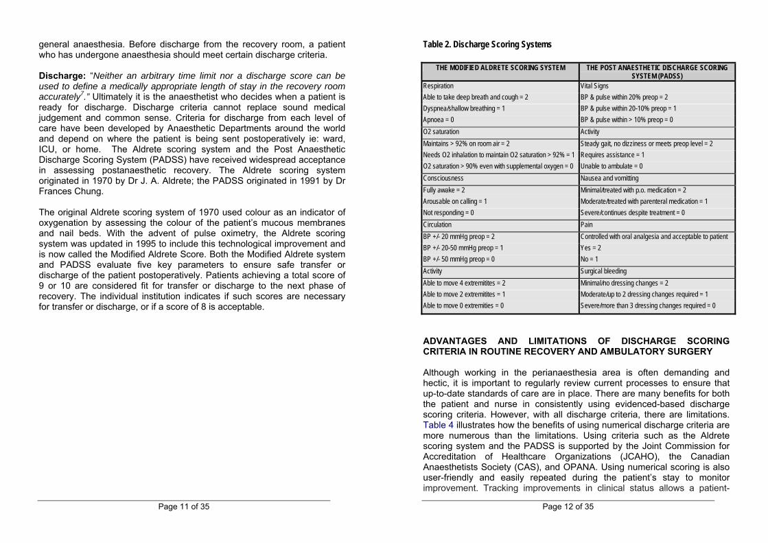
general anaesthesia. Before discharge from the recovery room, a patient who has undergone anaesthesia should meet certain discharge criteria. Discharge: “Neither an arbitrary time limit nor a discharge score can be used to define a medically appropriate length of stay in the recovery room accurately7.” Ultimately it is the anaesthetist who decides when a patient is ready for discharge. Discharge criteria cannot replace sound medical judgement and common sense. Criteria for discharge from each level of care have been developed by Anaesthetic Departments around the world and depend on where the patient is being sent postoperatively ie: ward, ICU, or home. The Aldrete scoring system and the Post Anaesthetic Discharge Scoring System (PADSS) have received widespread acceptance in assessing postanaesthetic recovery. The Aldrete scoring system originated in 1970 by Dr J. A. Aldrete; the PADSS originated in 1991 by Dr Frances Chung. The original Aldrete scoring system of 1970 used colour as an indicator of oxygenation by assessing the colour of the patient’s mucous membranes and nail beds. With the advent of pulse oximetry, the Aldrete scoring system was updated in 1995 to include this technological improvement and is now called the Modified Aldrete Score. Both the Modified Aldrete system and PADSS evaluate five key parameters to ensure safe transfer or discharge of the patient postoperatively. Patients achieving a total score of 9 or 10 are considered fit for transfer or discharge to the next phase of recovery. The individual institution indicates if such scores are necessary for transfer or discharge, or if a score of 8 is acceptable.
Page 12 of 35
Table 2. Discharge Scoring Systems
THE MODIFIED ALDRETE SCORING SYSTEM THE POST ANAESTHETIC DISCHARGE SCORING SYSTEM (PADSS)
Respiration Vital Signs Able to take deep breath and cough = 2 BP & pulse within 20% preop = 2 Dyspnea/shallow breathing = 1 BP & pulse within 20-10% preop = 1 Apnoea = 0 BP & pulse within > 10% preop = 0 O2 saturation Activity Maintains > 92% on room air = 2 Steady gait, no dizziness or meets preop level = 2 Needs O2 inhalation to maintain O2 saturation > 92% = 1 Requires assistance = 1 O2 saturation > 90% even with supplemental oxygen = 0 Unable to ambulate = 0 Consciousness Nausea and vomitting Fully awake = 2 Minimal/treated with p.o. medication = 2 Arousable on calling = 1 Moderate/treated with parenteral medication = 1 Not responding = 0 Severe/continues despite treatment = 0 Circulation Pain BP +/- 20 mmHg preop = 2 Controlled with oral analgesia and acceptable to patient BP +/- 20-50 mmHg preop = 1 Yes = 2 BP +/- 50 mmHg preop = 0 No = 1 Activity Surgical bleeding Able to move 4 extremitites = 2 Minimal/no dressing changes = 2 Able to move 2 extremitites = 1 Moderate/up to 2 dressing changes required = 1 Able to move 0 extremities = 0 Severe/more than 3 dressing changes required = 0
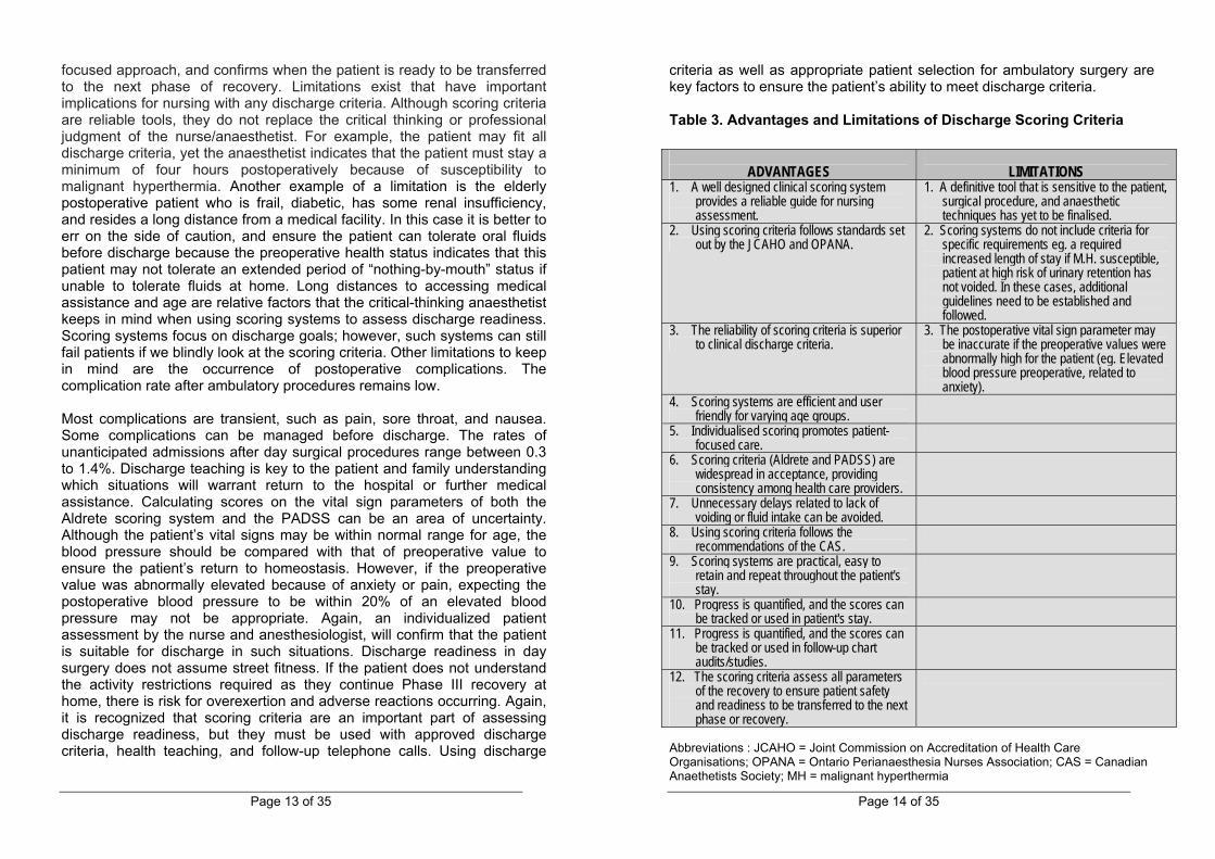
ADVANTAGES AND LIMITATIONS OF DISCHARGE SCORING CRITERIA IN ROUTINE RECOVERY AND AMBULATORY SURGERY Although working in the perianaesthesia area is often demanding and hectic, it is important to regularly review current processes to ensure that up-to-date standards of care are in place. There are many benefits for both the patient and nurse in consistently using evidenced-based discharge scoring criteria. However, with all discharge criteria, there are limitations. Table 4 illustrates how the benefits of using numerical discharge criteria are more numerous than the limitations. Using criteria such as the Aldrete scoring system and the PADSS is supported by the Joint Commission for Accreditation of Healthcare Organizations (JCAHO), the Canadian Anaesthetists Society (CAS), and OPANA. Using numerical scoring is also user-friendly and easily repeated during the patient’s stay to monitor improvement. Tracking improvements in clinical status allows a patient-
Page 13 of 35
focused approach, and confirms when the patient is ready to be transferred to the next phase of recovery. Limitations exist that have important implications for nursing with any discharge criteria. Although scoring criteria are reliable tools, they do not replace the critical thinking or professional judgment of the nurse/anaesthetist. For example, the patient may fit all discharge criteria, yet the anaesthetist indicates that the patient must stay a minimum of four hours postoperatively because of susceptibility to malignant hyperthermia. Another example of a limitation is the elderly postoperative patient who is frail, diabetic, has some renal insufficiency, and resides a long distance from a medical facility. In this case it is better to err on the side of caution, and ensure the patient can tolerate oral fluids before discharge because the preoperative health status indicates that this patient may not tolerate an extended period of “nothing-by-mouth” status if unable to tolerate fluids at home. Long distances to accessing medical assistance and age are relative factors that the critical-thinking anaesthetist keeps in mind when using scoring systems to assess discharge readiness. Scoring systems focus on discharge goals; however, such systems can still fail patients if we blindly look at the scoring criteria. Other limitations to keep in mind are the occurrence of postoperative complications. The complication rate after ambulatory procedures remains low. Most complications are transient, such as pain, sore throat, and nausea. Some complications can be managed before discharge. The rates of unanticipated admissions after day surgical procedures range between 0.3 to 1.4%. Discharge teaching is key to the patient and family understanding which situations will warrant return to the hospital or further medical assistance. Calculating scores on the vital sign parameters of both the Aldrete scoring system and the PADSS can be an area of uncertainty. Although the patient’s vital signs may be within normal range for age, the blood pressure should be compared with that of preoperative value to ensure the patient’s return to homeostasis. However, if the preoperative value was abnormally elevated because of anxiety or pain, expecting the postoperative blood pressure to be within 20% of an elevated blood pressure may not be appropriate. Again, an individualized patient assessment by the nurse and anesthesiologist, will confirm that the patient is suitable for discharge in such situations. Discharge readiness in day surgery does not assume street fitness. If the patient does not understand the activity restrictions required as they continue Phase III recovery at home, there is risk for overexertion and adverse reactions occurring. Again, it is recognized that scoring criteria are an important part of assessing discharge readiness, but they must be used with approved discharge criteria, health teaching, and follow-up telephone calls. Using discharge
Page 14 of 35
criteria as well as appropriate patient selection for ambulatory surgery are key factors to ensure the patient’s ability to meet discharge criteria. Table 3. Advantages and Limitations of Discharge Scoring Criteria
ADVANTAGES
LIMITATIONS
1. A well designed clinical scoring system provides a reliable guide for nursing assessment.
1. A definitive tool that is sensitive to the patient, surgical procedure, and anaesthetic techniques has yet to be finalised.
2. Using scoring criteria follows standards set out by the JCAHO and OPANA.
2. Scoring systems do not include criteria for specific requirements eg. a required increased length of stay if M.H. susceptible, patient at high risk of urinary retention has not voided. In these cases, additional guidelines need to be established and followed.
3. The reliability of scoring criteria is superior to clinical discharge criteria.
3. The postoperative vital sign parameter may be inaccurate if the preoperative values were abnormally high for the patient (eg. Elevated blood pressure preoperative, related to anxiety).
4. Scoring systems are efficient and user friendly for varying age groups.
5. Individualised scoring promotes patient-focused care.
6. Scoring criteria (Aldrete and PADSS) are widespread in acceptance, providing consistency among health care providers.
7. Unnecessary delays related to lack of voiding or fluid intake can be avoided.
8. Using scoring criteria follows the recommendations of the CAS.
9. Scoring systems are practical, easy to retain and repeat throughout the patient's stay.
10. Progress is quantified, and the scores can be tracked or used in patient's stay.
11. Progress is quantified, and the scores can be tracked or used in follow-up chart audits/studies.
12. The scoring criteria assess all parameters of the recovery to ensure patient safety and readiness to be transferred to the next phase or recovery.
Abbreviations : JCAHO = Joint Commission on Accreditation of Health Care Organisations; OPANA = Ontario Perianaesthesia Nurses Association; CAS = Canadian Anaethetists Society; MH = malignant hyperthermia
Page 15 of 35
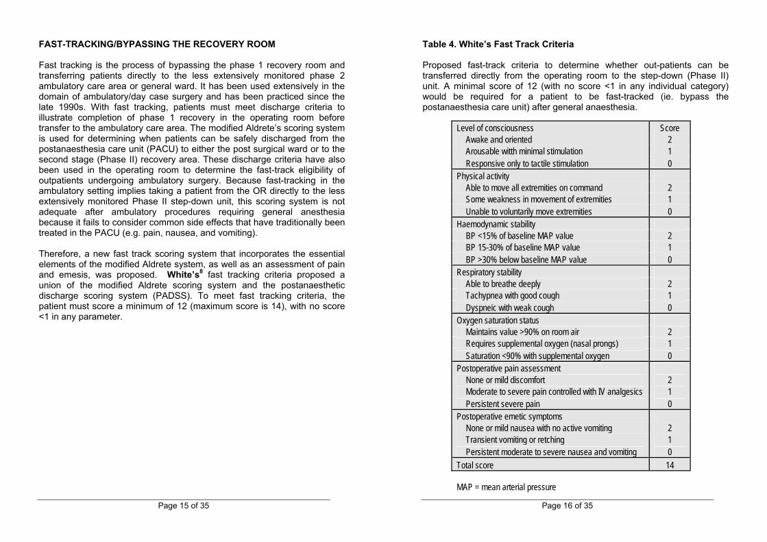
FAST-TRACKING/BYPASSING THE RECOVERY ROOM Fast tracking is the process of bypassing the phase 1 recovery room and transferring patients directly to the less extensively monitored phase 2 ambulatory care area or general ward. It has been used extensively in the domain of ambulatory/day case surgery and has been practiced since the late 1990s. With fast tracking, patients must meet discharge criteria to illustrate completion of phase 1 recovery in the operating room before transfer to the ambulatory care area. The modified Aldrete’s scoring system is used for determining when patients can be safely discharged from the postanaesthesia care unit (PACU) to either the post surgical ward or to the second stage (Phase II) recovery area. These discharge criteria have also been used in the operating room to determine the fast-track eligibility of outpatients undergoing ambulatory surgery. Because fast-tracking in the ambulatory setting implies taking a patient from the OR directly to the less extensively monitored Phase II step-down unit, this scoring system is not adequate after ambulatory procedures requiring general anesthesia because it fails to consider common side effects that have traditionally been treated in the PACU (e.g. pain, nausea, and vomiting). Therefore, a new fast track scoring system that incorporates the essential elements of the modified Aldrete system, as well as an assessment of pain and emesis, was proposed. White’s8 fast tracking criteria proposed a union of the modified Aldrete scoring system and the postanaesthetic discharge scoring system (PADSS). To meet fast tracking criteria, the patient must score a minimum of 12 (maximum score is 14), with no score <1 in any parameter.
Page 16 of 35
Table 4. White’s Fast Track Criteria Proposed fast-track criteria to determine whether out-patients can be transferred directly from the operating room to the step-down (Phase II) unit. A minimal score of 12 (with no score <1 in any individual category) would be required for a patient to be fast-tracked (ie. bypass the postanaesthesia care unit) after general anaesthesia.
Level of consciousness Score Awake and oriented 2 Arousable witth minimal stimulation 1 Responsive only to tactile stimulation 0
Physical activity Able to move all extremities on command 2 Some weakness in movement of extremities 1 Unable to voluntarily move extremities 0
Haemodynamic stability BP <15% of baseline MAP value 2 BP 15-30% of baseline MAP value 1 BP >30% below baseline MAP value 0
Respiratory stability Able to breathe deeply 2 Tachypnea with good cough 1 Dyspneic with weak cough 0
Oxygen saturation status Maintains value >90% on room air 2 Requires supplemental oxygen (nasal prongs) 1 Saturation <90% with supplemental oxygen 0
Postoperative pain assessment None or mild discomfort 2 Moderate to severe pain controlled with IV analgesics 1 Persistent severe pain 0
Postoperative emetic symptoms None or mild nausea with no active vomiting 2 Transient vomiting or retching 1 Persistent moderate to severe nausea and vomiting 0
Total score 14 MAP = mean arterial pressure
Page 17 of 35
Fast tracking has been made possible due to factors such as minimally invasive surgical techniques and ultra short duration anaesthetic drugs and techniques and more extensive use of regional anaesthesia and peripheral nerve blocks. The objective of fast tracking is to decrease recovery time and therefore costs, particularly nursing costs. Song et al9 studied these costs, and concluded that bypassing the phase 1 recovery room decreased recovery time without compromising patient satisfaction, But the overall nursing workload and the associated costs were no different. Not all patients and procedures are appropriate for fast tracking, in most studies 32%-90% were fast track eligible and selection is therefore on a case by case basis. What this comes down to is that fast-tracking does not eliminate the recovery room which still needs to be available and staffed. Fast tracking seems to work best in small units with flexible staff who are used to working in phase 1 as well as phase 2 recovery. Dexter and colleagues10,11 used a computer simulation to estimate the effect of fast-tracking on staffing and costs. If phase I recovery is bypassed altogether, cost savings are complex to calculate and are dependent on how the staff are compensated (salaried vs hourly rate), on whether frequent overtime is routine, and on the throughput of patients. Case mix was not considered but is also likely to affect staffing. Money is only saved if nurses are either reduced in number or are not paid when they are not working. Unless large numbers of patients could reliably bypass the recovery room, recovery nurses cannot be dispensed with, and there must generally be a minimum of two when patients are present, representing an irreducible number of staff. A nurse costs exactly the same whether she is nursing a patient or waiting for one. And as mentioned before, fast-track does not guarantee that the recovery room is unneeded-no study has achieved 100% fast-track rate.
Page 18 of 35
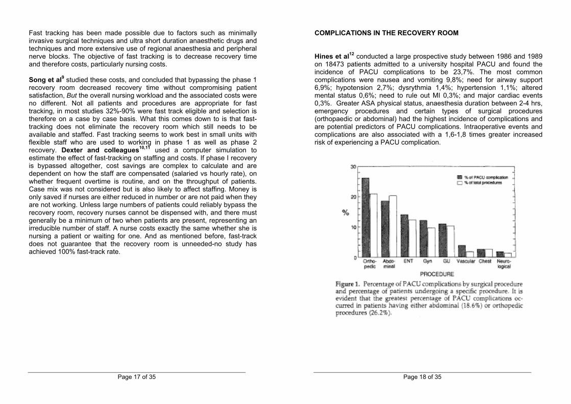
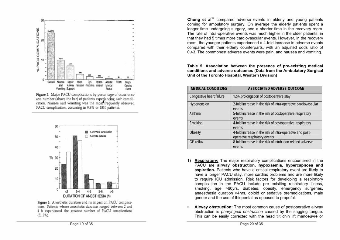
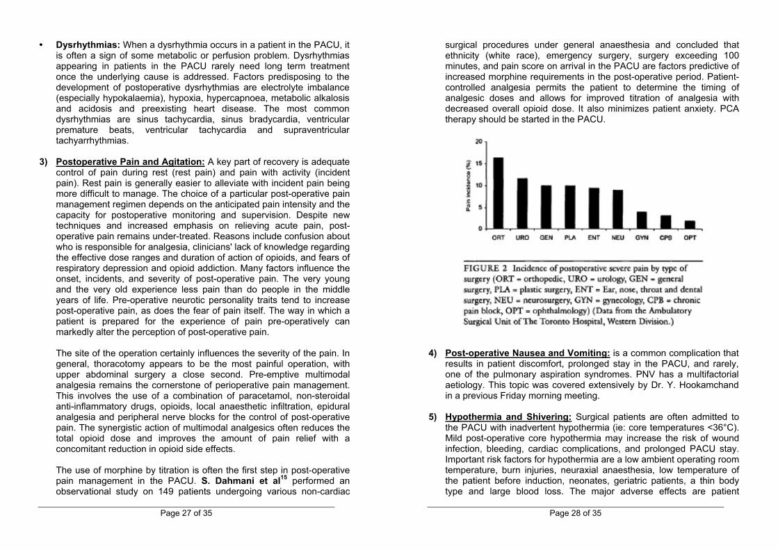
COMPLICATIONS IN THE RECOVERY ROOM Hines et al12 conducted a large prospective study between 1986 and 1989 on 18473 patients admitted to a university hospital PACU and found the incidence of PACU complications to be 23,7%. The most common complications were nausea and vomiting 9,8%; need for airway support 6,9%; hypotension 2,7%; dysrythmia 1,4%; hypertension 1,1%; altered mental status 0,6%; need to rule out MI 0,3%; and major cardiac events 0,3%. Greater ASA physical status, anaesthesia duration between 2-4 hrs, emergency procedures and certain types of surgical procedures (orthopaedic or abdominal) had the highest incidence of complications and are potential predictors of PACU complications. Intraoperative events and complications are also associated with a 1,6-1,8 times greater increased risk of experiencing a PACU complication.
Page 19 of 35
Page 20 of 35
Chung et al13 compared adverse events in elderly and young patients coming for ambulatory surgery. On average the elderly patients spent a longer time undergoing surgery, and a shorter time in the recovery room. The rate of intra-operative events was much higher in the older patients, in that they had 5 times more cardiovascular events. However, in the recovery room, the younger patients experienced a 4-fold increase in adverse events compared with their elderly counterparts, with an adjusted odds ratio of 0,43. The commonest adverse events were pain, and nausea and vomiting. Table 5. Association between the presence of pre-existing medical conditions and adverse outcomes (Data from the Ambulatory Surgical Unit of the Toronto Hospital, Western Division)
MEDICAL CONDITIONS ASSOCIATED ADVERSE OUTCOME
Congestive heart failure 12% prolongation of postoperative stay
Hypertension 2-fold increase in the risk of intra-operative cardiovascular events
Asthma 5-fold increase in the risk of postoperative respiratory events
Smoking 4-fold increase in the risk of postoperative respiratory events
Obesity 4-fold increase in the risk of intra-operative and post-operative respiratory events
GE reflux 8-fold increase in the risk of intubation related adverse events
1) Respiratory: The major respiratory complications encountered in the
PACU are airway obstruction, hypoxaemia, hypercapnoea and aspiration. Patients who have a critical respiratory event are likely to have a longer PACU stay, more cardiac problems and are more likely to require ICU admission. Risk factors for developing a respiratory complication in the PACU include pre exisiting respiratory illness, smoking, age >60yrs, diabetes, obesity, emergency surgeries, anaesthesia duration >4hrs, opioid or sedative premedications, male gender and the use of thiopental as opposed to propofol.
▪ Airway obstruction: The most common cause of postoperative airway
obstruction is pharyngeal obstruction caused by the sagging tongue. This can be easily corrected with the head tilt chin lift manoeuvre or
Page 21 of 35
with a jaw thrust. The proper position for each patient will depend on trial and error until a patent airway is obtained. If the obstruction is not immediately reversible a nasal or oral airway can be inserted. Airway obstruction secondary to laryngeal obstruction can also occur. This may be due to laryngeal spasm, direct airway injury, or even vocal cord paralysis. Laryngeal spasm is more likely in patients with irritable airways (COPD, smokers, recent URTI) and when secretions are present. It can be treated by opening and clearing the airway of secretions, CPAP with 100% O2 and if this fails succinylcholine can be given with rapid intubation and assisted ventilation. All patients with airway obstruction should receive oxygen by facemask and for all cases of airway obstruction, if an adequate airway cannot be established by simple physical or pharmacological means, orotracheal intubation is necessary or alternatively a laryngeal mask airway may be used in the patient with a difficult airway. Patients with obstructive sleep apnoea are at high risk for airway obstruction when sedated and CPAP is useful in these patients after tracheal extubation.
▪ Hypoxaemia: Evaluation of the hypoxaemic postoperative patient
should include evaluation of the classic causes of hypoxaemia: Low inspired oxygen concentration; Hypoventilation; Areas of low ventilation to perfusion ratios and An increased intrapulmonary right to left shunt. Increased age, shivering and a low cardiac output can aggravate the degree of hypoxaemia when an intrapulmonary shunt is present. Low inspired oxygen concentrations (FiO2<21%) are rare causes of significant postoperative hypoxaemia however delivery of hypoxic gas mixtures can occur. When N2O is used diffusion hypoxia can occur when at the end of the anaesthetic N2O is replaced by air. N2O is 31 times more soluble than nitrogen, inspired air is diluted with N2O and PAO2 falls. This is prevented with supplemental Oxygen administration. Hypoventilation with associated hypoxaemia may be secondary to inadequate NMJ blockade recovery, the respiratory depressant effects of opioids, volatiles and benzodiazepines and surgical site pain. Post hyperventilation hypoxaemia secondary to decreased respiratory drive as well as the type and site of surgery (upper abdominal incisions) and a shift from diaphragmatic to rib cage breathing contribute to postoperative hypoventilation.
The most common cause of postoperative hypoxaemia is an increase
in right to left intrapulmonary shunting with atelectasis being the most common cause of this. Atelectasis involves the collapse of an entire lung, lobe or lung segment and is due to bronchial obstruction from secretions, blood or mucus plug and is treated by providing adequate
Page 22 of 35
humidification of inspired gases, coughing, deep breathing and postural drainage. Other causes of increased right to left intrapulmonary shunting include pneumothorax, pulmonary oedema and pulmonary embolism. Increased shunting is also seen in patients with decreased functional residual capacities, when the closing capacity exceeds FRC normal airways collapse during tidal breathing and an intrapulmonary shunt develops as a result of diffuse airway collapse. Any situation that results in an increased closing capacity (increasing age) or a decreased FRC (pulmonary oedema, infection, aspiration and obesity) will place the patient at risk for hypoxaemia. Treatment of hypoxaemia involves increasing the inspired oxygen concentration which is effective in restoring the PaO2 in many cases. The PaO2 response to oxygen breathing depends on the degree of intrapulmonary shunting. Increasing the FiO2 from room air to a hundred percent results in a large increase in PaO2 when the shunt fraction is small; however, oxygen will have little effect on PaO2 in patients with a large shunt fraction. If hypoxaemia persists (PaO2 < 60mmHg) despite maximal oxygen therapy (FiO2 = 100%), tracheal intubation and mechanical ventilation should be initiated. Ventilation with PEEP will increase FRC and result in an improvement in arterial oxygenation.
▪ Hypercapnoea is defined as an increase in carbon dioxide tension
(PaCO2) above normal and is often secondary to hyperventilation which results from reduced alveolar ventilation. During the post-operative period, hypoventilation occurs as a result of poor respiratory drive, poor respiratory muscle function, or a high rate of production of CO2, or it can be a direct result of acute or chronic lung disease. Central respiratory depression occurs with volatiles and narcotic anaesthetics which produce respiratory depression that is detectable by a shift of the CO2 response curve downward and to the right. Narcotic-induced respiratory depression can be reversed with the use of narcotic antagonists eg. Naloxone. Poor respiratory muscle function after surgery can contribute to hypercapnoea. Common causes of poor respiratory muscle function include failure of reversal of neuromuscular-blocking drugs, obesity, gastric dilation, tight dressings, body casts and the sight of the surgical incision. The incision sight affects the patients’ ability to take a large breath as measured by vital capacity. Nearly all patients have reductions in vital capacity that are the greatest on the day of surgery. Patients undergoing upper abdominal surgery have the greatest reduction in VC of 60% on the day of surgery. This reduction is secondary to diaphragmatic impairment. Measurement of PaCO2 is the best method of detecting hypercapnoea in the post-operative period. Although hypertension and tachycardia commonly occur during CO2
Page 23 of 35
retention, they may not be seen in post-surgical patients and the elderly who have an attenuated response to CO2 levels. Measuring vital capacity which should be at least 10ml/kg body weight and maximum inspiratory force which should be > -20cm H2O is a good guide to a patients’ ability to breathe spontaneously in the post-operative period. If these values cannot be maintained, the patient should receive controlled mechanical ventilation until he/she is able to generate adequate respiratory muscle function.
2) Circulatory: Critical cardiovascular events are the second major group
of life threatening complications for patients' in the PACU. Rose et al14 conducted a study on 18380 patients after general anaesthesia in Toronto and found that patients in whom hypertension or tachycardia developed in the PACU had more unplanned critical care admissions and a higher mortality rate than did those in whom these conditions did not develop. Anaesthetic factors contributed only in a minor way to the development of cardiovascular problems in the PACU where patient and surgical factors were more important. This contrasts respiratory events where anaesthetic factors play more of a contributory role.
• Hypotension: There are numerous causes of hypotension in the
postoperative period but most causes can be attributed to either a decreased ventricular preload, reduced myocardial contractility, or a profound reduction in systemic vascular resistance. Decreased ventricular preload is caused by intravascular volume depletion because of surgical blood loss, excessive 3rd space fluid losses, unreplaced urinary losses, or septic shock with vasodilatation and capillary leakage of fluid. Acute massive pulmonary embolism will produce hypotension by blocking blood flow to the left side of the heart ie: decreasing left ventricular preload. Decreased myocardial contractility in the postoperative period occurs because of the continued myocardial depressant effects of anaesthetic drugs (volatiles), or if a patient has pre-existing ventricular dysfunction, or if the patient develops a perioperative myocardial infarction. Postoperative decreases in systemic vascular resistance occur because of the residual vasodilatory effects of anaesthetic drugs and opioids. It is also the hallmark of patients' in septic shock or who have chronic liver failure. Prompt diagnosis as to the cause of the hypotension as well as early treatment is important because prolonged hypotension can result in hypoperfusion of vital organs and subsequent ischaemic damage. In most instances hypotension can be corrected by ensuring that the patient is normovolaemic with the administration of crystalloid, colloid or blood products. Once the patient is normovolaemic and if hypotension
Page 24 of 35
persists then inotropic and or vasopressor support should be initiated concurrently with the treatment of the underlying cause so as to prevent prolonged periods of hypotension. Most cases of hypotension in the recovery room can be attributed to hypovolaemic shock, cardiogenic shock or septic shock.
• Hypertension: The development of hypertension in the PACU has a multi factorial aetiology. It is most often due to excessive pain, hypercapnoea, hypoxaemia, urinary retention or excessive intravascular fluid volume. These aetiologies need to be ruled out. Pre-existing hypertension is present in more than half the patients in whom hypertension develops in the recovery room. When hypertension does develop during recovery from anaesthesia it usually begins within 30 minutes of the end of the operation. Severe acute hypertension can lead to left ventricular failure with the development of pulmonary oedema, MI, dysrhythmia or cerebral haemorrhage. The primary goal of intervention in hypertension is to safely reduce BP. The appropriate therapeutic approach of each patient will depend on their clinical presentation. Safe and efficacious management of the patient experiencing hypertensive crises requires the physician to discriminate the crisis as either a hypertensive urgency or hypertensive emergency. Rapid antihypertensive therapy is not warranted in patients with hypertensive urgencies (i.e. a hypertension crisis associated with severe elevations in BP without progressive end-organ dysfunction).
Conversely, immediate BP reduction is indicated in patients experiencing hypertensive emergencies (i.e. a hypertension crisis characterized by severe elevations in BP [>180/120 mmHg] complicated by evidence of impending or progressive target-organ dysfunction) to prevent progressive end-organ damage. Hypertension associated with cerebral infarction or intracerebral haemorrhage requires treatment under special circumstances and the use of pharmacological agents must be tailored to each patient's condition. It should be noted that currently there are no widely accepted guidelines for the treatment of hypertension associated with cerebral infarction or intracerebral haemorrhage.
Patients with hypertensive emergencies are best treated in an ICU with titratable intravenous hypotensive agents. Several rapid-acting intravenous antihypertensive agents are available including labetalol, esmolol, fenoldapam, nicardipine, and sodium nitroprusside. While sodium nitroprusside is commonly used to treat severe hypertension, it is an extremely toxic drug that should be used only in rare circumstances. If the use of sodium nitroprusside cannot be avoided, it
Page 25 of 35
should not be used at a dose that exceeds 2ug/kg/min. Similarly, nifedipine, nitroglycerin and hydralazine should not to be considered acceptable therapies in the management of hypertensive crises because these agents are associated with significant toxicities and/or adverse effects. Newer agents, such as clevidipine and fenoldopam, may hold considerable advantages to other available agents in the management of hypertensive crises.
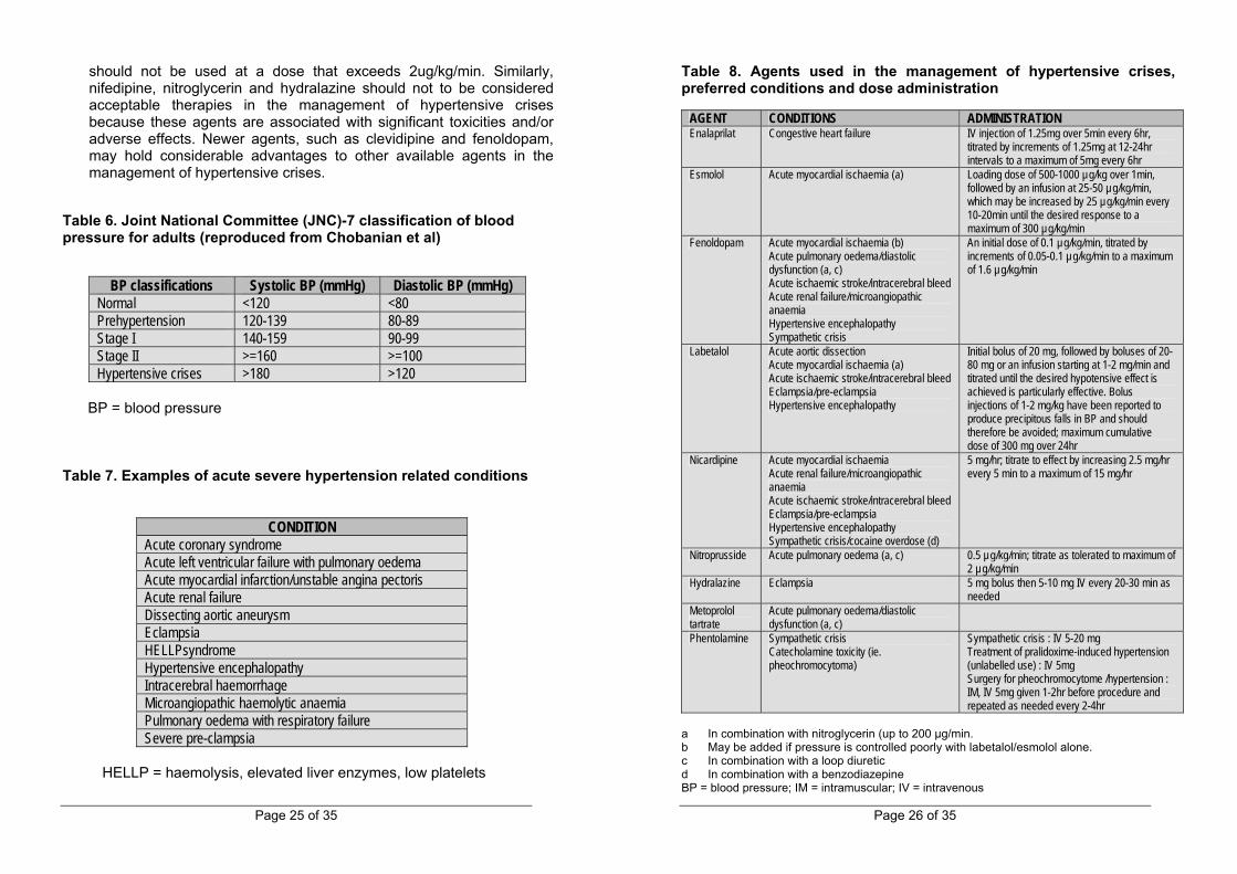
Table 6. Joint National Committee (JNC)-7 classification of blood pressure for adults (reproduced from Chobanian et al)
BP classifications Systolic BP (mmHg) Diastolic BP (mmHg) Normal <120 <80 Prehypertension 120-139 80-89 Stage I 140-159 90-99 Stage II >=160 >=100 Hypertensive crises >180 >120
BP = blood pressure
Table 7. Examples of acute severe hypertension related conditions
CONDITION Acute coronary syndrome Acute left ventricular failure with pulmonary oedema Acute myocardial infarction/unstable angina pectoris Acute renal failure Dissecting aortic aneurysm Eclampsia HELLPsyndrome Hypertensive encephalopathy Intracerebral haemorrhage Microangiopathic haemolytic anaemia Pulmonary oedema with respiratory failure Severe pre-clampsia
HELLP = haemolysis, elevated liver enzymes, low platelets
Page 26 of 35
Table 8. Agents used in the management of hypertensive crises, preferred conditions and dose administration
AGENT CONDITIONS ADMINISTRATION Enalaprilat Congestive heart failure IV injection of 1.25mg over 5min every 6hr,
titrated by increments of 1.25mg at 12-24hr intervals to a maximum of 5mg every 6hr
Esmolol Acute myocardial ischaemia (a) Loading dose of 500-1000 µg/kg over 1min, followed by an infusion at 25-50 µg/kg/min, which may be increased by 25 µg/kg/min every 10-20min until the desired response to a maximum of 300 µg/kg/min
Fenoldopam Acute myocardial ischaemia (b) Acute pulmonary oedema/diastolic dysfunction (a, c) Acute ischaemic stroke/intracerebral bleed Acute renal failure/microangiopathic anaemia Hypertensive encephalopathy Sympathetic crisis
An initial dose of 0.1 µg/kg/min, titrated by increments of 0.05-0.1 µg/kg/min to a maximum of 1.6 µg/kg/min
Labetalol Acute aortic dissection Acute myocardial ischaemia (a) Acute ischaemic stroke/intracerebral bleed Eclampsia/pre-eclampsia Hypertensive encephalopathy
Initial bolus of 20 mg, followed by boluses of 20-80 mg or an infusion starting at 1-2 mg/min and titrated until the desired hypotensive effect is achieved is particularly effective. Bolus injections of 1-2 mg/kg have been reported to produce precipitous falls in BP and should therefore be avoided; maximum cumulative dose of 300 mg over 24hr
Nicardipine Acute myocardial ischaemia Acute renal failure/microangiopathic anaemia Acute ischaemic stroke/intracerebral bleed Eclampsia/pre-eclampsia Hypertensive encephalopathy Sympathetic crisis/cocaine overdose (d)
5 mg/hr; titrate to effect by increasing 2.5 mg/hr every 5 min to a maximum of 15 mg/hr
Nitroprusside Acute pulmonary oedema (a, c) 0.5 µg/kg/min; titrate as tolerated to maximum of 2 µg/kg/min
Hydralazine Eclampsia 5 mg bolus then 5-10 mg IV every 20-30 min as needed
Metoprolol tartrate
Acute pulmonary oedema/diastolic dysfunction (a, c)
Phentolamine Sympathetic crisis Catecholamine toxicity (ie. pheochromocytoma)
Sympathetic crisis : IV 5-20 mg Treatment of pralidoxime-induced hypertension (unlabelled use) : IV 5mg Surgery for pheochromocytome /hypertension : IM, IV 5mg given 1-2hr before procedure and repeated as needed every 2-4hr
a In combination with nitroglycerin (up to 200 µg/min. b May be added if pressure is controlled poorly with labetalol/esmolol alone. c In combination with a loop diuretic d In combination with a benzodiazepine BP = blood pressure; IM = intramuscular; IV = intravenous
Page 27 of 35
• Dysrhythmias: When a dysrhythmia occurs in a patient in the PACU, it is often a sign of some metabolic or perfusion problem. Dysrhythmias appearing in patients in the PACU rarely need long term treatment once the underlying cause is addressed. Factors predisposing to the development of postoperative dysrhythmias are electrolyte imbalance (especially hypokalaemia), hypoxia, hypercapnoea, metabolic alkalosis and acidosis and preexisting heart disease. The most common dysrhythmias are sinus tachycardia, sinus bradycardia, ventricular premature beats, ventricular tachycardia and supraventricular tachyarrhythmias.
3) Postoperative Pain and Agitation: A key part of recovery is adequate
control of pain during rest (rest pain) and pain with activity (incident pain). Rest pain is generally easier to alleviate with incident pain being more difficult to manage. The choice of a particular post-operative pain management regimen depends on the anticipated pain intensity and the capacity for postoperative monitoring and supervision. Despite new techniques and increased emphasis on relieving acute pain, post-operative pain remains under-treated. Reasons include confusion about who is responsible for analgesia, clinicians' lack of knowledge regarding the effective dose ranges and duration of action of opioids, and fears of respiratory depression and opioid addiction. Many factors influence the onset, incidents, and severity of post-operative pain. The very young and the very old experience less pain than do people in the middle years of life. Pre-operative neurotic personality traits tend to increase post-operative pain, as does the fear of pain itself. The way in which a patient is prepared for the experience of pain pre-operatively can markedly alter the perception of post-operative pain.
The site of the operation certainly influences the severity of the pain. In
general, thoracotomy appears to be the most painful operation, with upper abdominal surgery a close second. Pre-emptive multimodal analgesia remains the cornerstone of perioperative pain management. This involves the use of a combination of paracetamol, non-steroidal anti-inflammatory drugs, opioids, local anaesthetic infiltration, epidural analgesia and peripheral nerve blocks for the control of post-operative pain. The synergistic action of multimodal analgesics often reduces the total opioid dose and improves the amount of pain relief with a concomitant reduction in opioid side effects.
The use of morphine by titration is often the first step in post-operative
pain management in the PACU. S. Dahmani et al15 performed an observational study on 149 patients undergoing various non-cardiac
Page 28 of 35
surgical procedures under general anaesthesia and concluded that ethnicity (white race), emergency surgery, surgery exceeding 100 minutes, and pain score on arrival in the PACU are factors predictive of increased morphine requirements in the post-operative period. Patient-controlled analgesia permits the patient to determine the timing of analgesic doses and allows for improved titration of analgesia with decreased overall opioid dose. It also minimizes patient anxiety. PCA therapy should be started in the PACU.
4) Post-operative Nausea and Vomiting: is a common complication that results in patient discomfort, prolonged stay in the PACU, and rarely, one of the pulmonary aspiration syndromes. PNV has a multifactorial aetiology. This topic was covered extensively by Dr. Y. Hookamchand in a previous Friday morning meeting.
5) Hypothermia and Shivering: Surgical patients are often admitted to
the PACU with inadvertent hypothermia (ie: core temperatures <36°C). Mild post-operative core hypothermia may increase the risk of wound infection, bleeding, cardiac complications, and prolonged PACU stay. Important risk factors for hypothermia are a low ambient operating room temperature, burn injuries, neuraxial anaesthesia, low temperature of the patient before induction, neonates, geriatric patients, a thin body type and large blood loss. The major adverse effects are patient
Page 29 of 35
discomfort, vasoconstriction, and shivering, and full recovery sometimes takes many hours. Shivering increases the metabolic rate and hence the need to increase cardiac output and minute ventilation. Not all patients who shiver post-operatively, however, are hypothermic, thus suggesting the mechanism of this event may be related to inadequate descending control of spinal reflexes after inhalation anaesthesia. Once in the PACU, hypothermic patients should have supplemental oxygen, warm IV fluids and blood, and external warming with forced air-warming devices eg. Bare Hugger or thermal blankets. Post-anaesthetic shivering not related to hypothermia can be treated with meperidine 10-25mg IV. This is effective in stopping the shivering and decreasing oxygen consumption.
6) Failure to Regain Consciousness: The patient who fails to regain
consciousness after general anaesthesia requires careful assessment. Preoperative factors such as drug or alcohol intoxication can lead to delayed awakening but the most common reason for persistent somnolence is the residual effects of anaesthetics, sedatives, and preoperative mediciations. Initial management should be aimed at pharmacological reversal of the most likely sedative drug. Naloxone in small incremental doses will increase the ventilatory rate if narcotic sedation is the problem without leading to breakthrough pain. Benzodiazepines can be reversed with flumazenil. Profound neuromuscular blockade can make a patient appear unconscious however this is unlikely in the absence of significant respiratory compromise. But nevertheless should be ruled out. Profound hypothermia (core temp <330C) can produce unconsciousness as can abnormalities in serum glucose such as hyperglycaemia and hypoglycaemia. Blood glucose, electrolytes, and blood gases should be evaluated in such cases. If the diagnosis remains unclear, a structural neurological abnormality should be sought. Raised intracranial pressure, thrombolic cerebrovascular accidents or intraoperative cerebral hypoxia (from hypoxaemia or poor cerebral perfusion) producing a diffuse encephalopathy can all cause a failure of the patient to regain consciousness. These causes can occur in the postoperative period but are extremely rare. An emergency CT scan of the brain will aid in ruling out these rarer causes of delayed emergence.
7) Hyperthermia: Is common in the postoperative period and is most
often associated with infections and sepsis. However not all temperatures above 370C are related to sepsis and infection and this must be kept in mind when assessing the patient with hyperthermia.
Page 30 of 35
Other possible causes of a raised basal temperature that require immediate intervention include:
Drug / blood reactions Tissue damage (inflammatory response) Neoplastic disorders Metabolic disorders Thyroid storm Adrenal crisis Pheochromocytoma Malignant Hyperthermia Neuroleptic malignant syndrome Acute porphyria 8) Minor Sequelae: Sore throat, dizziness, drowsiness and headache are
among the frequently described and neglected minor complications, which could also result in prolonged post-operative stay, and strongly influence patient satisfaction and functional level. Sore throat frequently occurs after intubation, or following the use of a LMA. Headache is a potential complication following neuraxial anaesthesia, while dizziness and drowsiness may be precipitated by dehydration.
PAEDIATRIC PACU Caring for a paediatric patient after anaesthesia requires special preparation and knowledge of the potential post-operative complications specific to children. Not all PACUs are dedicated solely to paediatric recovery, so it is important that staff with paediatric experience be available. In addition to basic PACU equipment, an emergency equipment cart with paediatric equipment including intra-osseus lines, paediatric airway trolley, and a drug chart with paediatric dosages should be immediately available. The controversial issue is whether parental presence should be allowed in the recovery room. In units which have both paediatric patients and adults, it is not advisable. Occasionally, however, if a child is inconsolable after emergence and all physiological parameters are stable, then the parents may be allowed into the recovery room, provided the nursing staff are comfortable with this.
Page 31 of 35
Specific Paediatric PACU complications • Emergence delirium: Occasionally paediatric patients emerge from
anaesthesia disorientated and inconsolable. This emergence delirium has been associated with the use of less soluble volatile anaesthetics eg sevoflurane. Adequate analgesia with opioids and NSAIDS and small doses of propofol or ketamine may alleviate the delirium. Inviting the parents into the PACU may also help to calm the child. When evaluating a patient with suspected emergence delirium, complications such as hypoxia, acidosis, raised ICP or pain should be ruled out and corrected.
• Postintubation Croup: Postintubation subglottic oedema is a
complication that can occur in 1 %-6% of patients younger than 4 yrs. Even a minimal amount of airway oedema can cause significant obstruction, especially at the level of the cricoid cartilage which is the narrowest section of the paediatric airway. Patients at increased risk of postintubation croup include: Downs syndrome patients, patients with congenital airway stenosis, surgery involving the airway, recent URTI,coughing on the endotracheal tube, prone surgical position, and traumatic intubations. Treatment includes adrenaline nebulisation and IV dexamethasone and rarely does a patient require reintubation.
• Postanaesthetic Apnoea: Preterm infants (born before 37 weeks
gestation) are at increased risk for apnoea and bradycardia after even uncomplicated anaesthesia. These events may be secondary to underlying neurologic, cardiac or pulmonary disease. Ex preterm infants who are less than 45-60 weeks postconceptual age should be admitted to a neonatal high care unit for 24 hour apnoea monitoring post surgery. The overall risk of apnoea in neonates less than 48 weeks conceptual age is 5% and this does not decrease to less than 1% until patients reach 54 weeks postconceptual age. IV caffeine 10mg/kg has been used to treat apnoeic spells in preterm infants and has been recommended for prophylaxis against postoperative apnoea.
Page 32 of 35
Page 33 of 35
Page 34 of 35
CONCLUSION In conclusion I pose the following question. How do we ensure a safe and satisfactory recovery period for our patients, regardless of whether they are in-patients or being done as ambulatory cases? Recovery rooms need to be properly equipped as per the SASA guidelines. 1. Staffing of recovery rooms MUST be in accordance with SASA
guidelines. The staff allocated must be dedicated only to the recovery room, and should not have any other duties.
2. Protocols must be in place to deal with common complications and observations must be recorded on the patients chart.
3. The anaesthesiologist needs to discharge the patient from the recovery room. It is suggested that the Modified Aldrete Scoring system be used for determining when patients are ready for discharge from the recovery room. A score of 9 or > 9 is required for discharge. The PADSS scoring system can be used in deciding a patient’s home readiness.
The most important parameter of all is the oxygen saturation, as this reflects an adequate cardio respiratory system, as well as whether there are undesirable effects such as shivering and pain. 5. The patient must also be pain-free. AND 6. There must be no nausea and vomiting. REFERENCES 1. Marshall S and Chung F "Discharge Criteria After Ambulatory Surgery"
Anaesthesia and Analgesia 199988:508-17. 2. Miller RD "Millers Anaesthesia" 6th edition 3. SAJAA-SASA recovery room guidelines section V 4. "Standards for Postanaesthesia Care" American Society of
Anaesthesiologists 1996 Directory of members pp395-396. 5. Mathes DO et al Ambulatory Surgery: "Room air versus nasal cannula
oxygen during transport after general anaesthesia" Anaesthesia and Analgesia 2001 93:917.
6. Smith AF et al "Interprofessional handover and patient safety in anaesthesia: observational study of handovers in the recovery room" BJA June 13 2008.
7. Heather EAD: "From Aldrete to PADSS: Reviewing Discharge criteria after Ambulatory Surgery" Journal of Perianaesthesia Nursing vol 21 no4 Aug 2006 pp259-267.
Page 35 of 35
8. White PF et al "New criteria for fast tracking after outpatient anaesthesia: A comparison with the modified Aldrete's scoring system" Anaesthesia and Analgesia 1999; 88:1069-1072.
9. Song 0 et al " Fast tracking (bypassing the PACU) does not reduce nursing workload after ambulatory surgery" BJA 93(6) 768-774 2004.
10. Dexter F et al " Statistical analysis by Monte Carlo Simulation of the impact of administrative and medical delays in discharge from the PACU on total patient care hours" Anaesthesia and Analgesia 2001 92:1222-5.
11. Dexter F et al " Statistical Analysis of PACU staffing at a surgical suite with frequent delays in admission from the OR- A case study" Anaesthesia and Analgesia 2001 92:947-9.
12. Hines R et al "Complications occurring in the PACU: A Survey" Anaesthesia and Analgesia 1992:74: 503-9.
13. Chung F et al "Adverse outcomes in ambulatory anaesthesia" Canadian Journal of Anaesthesia 199946:5 R18-R26.
14. Rose OK et al "Cardiovascular events in the PACU: Contribution of risk factors" Anaesthesiology 84:772, 1996.
15. Dahmani S et al "Predicitve factors of early morphine requirements in the PACU" BJA 87 (3) 385-9 2001.
16. Millar J "Fast tracking i1 day surgery. Is your journey to the recovery room really necessary" Editorial II BJA 2004.
17. Waddle JP et al "PACU length of stay: Quantifying and Assessing dependant factors" Anaesthesia and Analgesia 1998 87:628-33.
18. Chung F "Recovery pattern and home readiness after ambulatory surgery" Anaesthesia and Analgesia 1995 80:896-902.
19. Chung F et al "Pre-existing medical conditions as predictors of adverse events in day case surgery" BJA 83 (2) 262-270 1999.
20. Jenkins K et al "Post operative recovery: day surgery patients' preferences" BJA 86 (2) 272-4 2001.
21. Howard H Weitz "Perioperative Cardiac complications" Review article. 22. Varon J "Treatment of Acute Severe Hypertension" Review article
Drugs 2008: 68(3) 283-297. 23. Bonnet F and Marret E "Postoperative pain management and outcome
after surgery" Review Anaesthesiology 2007 vol 21 No1 pp99-107. 24. Chung F "Discharge criteria a new trend" Can J of Anaesthesia 1999. 25. Imad T Awad et al "Factors affecting recovery and discharge following
ambulatory surgery" Canadian Journal Anaesthesia 2006 53:9 pp858-872.
26. Dexter F, Analysis of strategies to decrease PACU costs Anaesthesiology JAN 1995 vol 82 issue 1pp94-101.
27. Douglas J "The establishment and management of a recovery and resuscitation ward" BJA 1963, 35, 24-27.


