Embed Size (px)
Citation preview
Volume 25 No. 2 April
The Gemmological Association and Gem Testing Laboratory of Great Britain
T he Journa
Gemmolog
PresidentE.M. Bruton
Vice-PresidentsAE. Farn, D.G. Kent, RK. Mitchell
Honorary FellowsR.T. Liddicoat [nr., E. Miles, K. Nassau
Honorary Life MembersD.}. Callaghan, E.A Iobbins, H. Tillander
Council of ManagementCR Cavey, T.}. Davidson, N.W. Deeks,RR Harding, 1.Thomson, v.P. Watson
Members' CouncilAJ. Allnutt, P. Dwyer-Hickey, R. Fuller,
B. Jackson, J. Kessler, G. Monnickendam,L. Music, J.B. Nelson, K. Penton, P.G. Read,
1. Roberts, R Shepherd, CH. Winter
Branch ChairmenMidlands: J.W. PorterNorth West: 1. KnightScottish: J. Thomson
ExaminersS.M. Anderson, B.SdHonst FGA
E.M. Bruton, FGA, DGAS. Coelho, B.Sc., FGA, DGA
AG. Good, FGA, DGAG.M. Howe, FGA, DGA
H.L. Plumb, B.Sc., FGA, DGAP.A. Sadler, B.Sc., FGA, DGA
Prof. 1. Sunagawa, D.Sc.CM. Woodward, B.5c., FGA DGA
AJ. Allnutt, M.Sc., Ph.D., FGSL. Bartlett, B.Sc., M.Phil., FGA, DGACR. Cavey, FGAAT. Collins, B.Sc., Ph.D.CJ.E. Hall, B.Sc.(Hons), FGAG.H. Jones, B.5c., Ph.D., FGARD. Ross, B.Sc., FGA DGAE. Stem, FGA, DGAM. Tilley, GG, FGA
The Gemmological Association and Gem Testing Laboratory of Great Britain27 Greville Street, London EC1N 8SU
Telephone: 0171-404 3334 Fax: 0171-404 8843
The Journal of
Gemmology VOLUME 25
NUMBER 2 APRIL 1996
S.M. Anderson G. Bosshart Dr J.W. Harris Dr J.M. Ogden Prof. D.C. Smith Prof. I. Sunagawa C.M. Woodward
Editor Dr R.R. Harding
Production Editor M.A. Burland
Assistant Editors M.J. O'Donoghue
P.G. Read
Associate Editors London Lucerne
Glasgow Cambridge
Paris Tokyo
London
Dr C.E.S. Arps Dr A.T. Collins Prof. R.A. Howie DrJ.E.Shigley E. Stern Dr M. Superchi
Leiden London
Derbyshire Santa Monica
London Milan
Any opinions expressed in The Journal of Gemmology are understood to be the views of the contributors and not necessarily of the publishers.
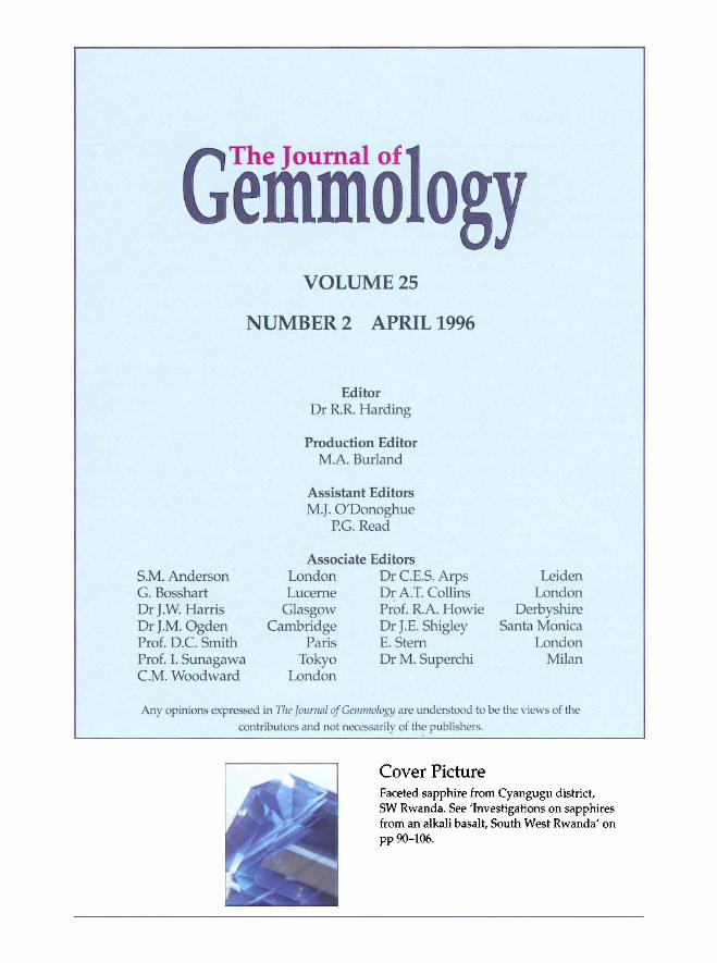
Cover Picture Faceted sapphire from Cyangugu district, SW Rwanda. See Investigations on sapphires from an alkali basalt, South West Rwanda' on pp 90-106.
86 J. Gemm., 1996, 25, 2
In this issue...
We introduce this issue with an update of a collection started many years ago and first described in this Journal in 1987. Its original motive was to enlighten the student of gemmology with the number of stones that could be mistaken for diamond. But with increasing knowledge it led to appreciation on other levels with the fascinating range of gem materials that could appear in colourless form or with the subtleties of recognizing small variations in this 'colourlessness7. There are now 46 stones in the collection and they are listed with some basic gem parameters.
Rwanda has been in the news for all the wrong reasons in the past year so it is good to report a development of some potential for the local people. Sapphires have been discovered in the Cyangugu district in south-west Rwanda (bordering Zaire) and details of their source and gem characteristics are described. Most crystals are a deep blue but some show the milkiness characteristic of the Sri Lankan 'geuda' and these respond well to heat treatment. They contain a wide range of inclusions and a new texture found on some rough crystals, not previously reported, is described and discussed.
Ever since they were first discovered in 1917 the deep blue beryls from the Maxixe mine in Brazil have been a source of fascination both for those in the gem trade and for investigative gemmologists. Maxixe beryls, as they came to be known, are quite different from the commoner blue beryl, aquamarine, in that their colour has a different source within the crystal structure and it fades when exposed to sunlight. The third paper in this issue sets out some current thinking about Maxixe beryls and proposes suitable terminology and its limits of application in the beryl family.
The final paper presents the results of a thorough investigation (involving more than 1000 samples) of a range of emeralds and green beryls from Central Nigeria. Detailed work on inclusions, compositions of hosts and inclusions and on spectra are illustrated, and provide a reference to enable confident identification of emeralds from this locality. Some Nigerian emeralds do contain spiky three-phase inclusions and resemble Colombian emeralds, but again the means for distinguishing between stones from the two localities are indicated.
R.R.H.
© Gemmological Association and Gem Testing Laboratory o^ Great Britain ISSN: 1355-4565
J. Gemm., 1996, 25, 2, 87-89 87
Common and rare colourless gemstones
David Kent FGA
London
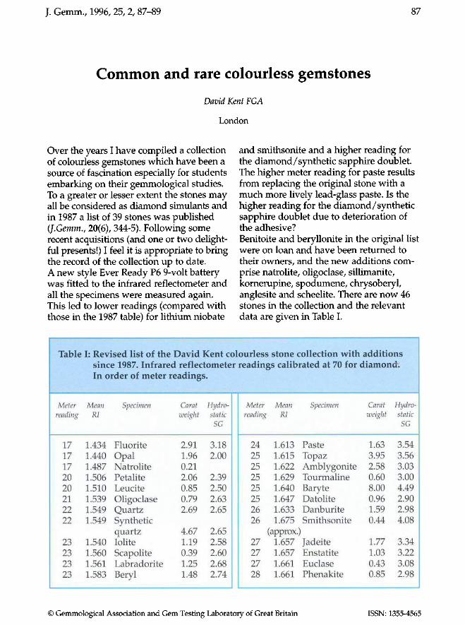
Over the years I have compiled a collection of colourless gemstones which have been a source of fascination especially for students embarking on their gemmological studies. To a greater or lesser extent the stones may all be considered as diamond simulants and in 1987 a list of 39 stones was published (J.Gemm., 20(6), 344-5). Following some recent acquisitions (and one or two delightful presents!) I feel it is appropriate to bring the record of the collection up to date. A new style Ever Ready P6 9-volt battery was fitted to the infrared reflectometer and all the specimens were measured again. This led to lower readings (compared with those in the 1987 table) for lithium niobate
Meter Mean Specimen Carat Hydro-reading RI weight static
17 17 17 20 20 21 22 22
23 23 23 23
1.434 1.440 1.487 1.506 1.510 1.539 1.549 1.549
1.540 1.560 1.561 1.583
Fluorite Opal Natrolite Petalite Leucite Oligoclase Quartz Synthetic quartz Iolite Scapolite Labradorite Beryl
2.91 1.96 0.21 2.06 0.85 0.79 2.69
4.67 1.19 0.39 1.25 1.48
SG
3.18 2.00
2.39 2.50 2.63 2.65
2.65 2.58 2.60 2.68 2.74
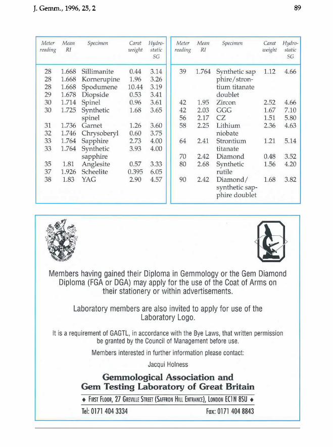
and smithsonite and a higher reading for the diamond/synthetic sapphire doublet. The higher meter reading for paste results from replacing the original stone with a much more lively lead-glass paste. Is the higher reading for the diamond/synthetic sapphire doublet due to deterioration of the adhesive? Benitoite and beryllonite in the original list were on loan and have been returned to their owners, and the new additions comprise natrolite, oligoclase, sillimanite, kornerupine, spodumene, chrysoberyl, anglesite and scheelite. There are now 46 stones in the collection and the relevant data are given in Table I.
Meter Mean Specimen Carat Hydro-reading RI weight static
24 25 25 25 25 25 26 26
27 27 27 28
1.613 1.615 1.622 1.629 1.640 1.647 1.633 1.675
(approx. 1.657 1.657 1.661 1.661
Paste Topaz Amblygonite Tourmaline Baryte Datolite Danburite Smithsonite
) Jadeite Enstatite Euclase Phenakite
1.63 3.95 2.58 0.60 8.00 0.96 1.59 0.44
1.77 1.03 0.43 0.85
SG
3.54 3.56 3.03 3.00 4.49 2.90 2.98 4.08
3.34 3.22 3.08 2.98
Table I: Revised list of the David Kent colourless stone collection with additions since 1987. Infrared reflectometer readings calibrated at 70 for diamond: In order of meter readings.
© Gemmological Association and Gem Testing Laboratory of Great Britain ISSN: 1355-4565
88 J. Gemm., 1996,25, 2
bIUM.IU .QL. AZ... ..u....
S f~
..Q1L ..2.9.. ..llL ...ll-
~ !II!1!lf u::... .li... .J.L
e If.
2.lc... ll..
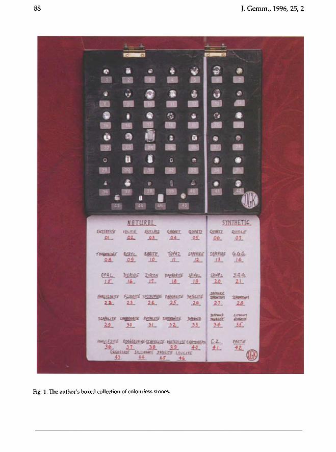
Fig. 1. The author's boxed collection of colourless stones.
J. Gemm., 1996,25,2 89
Meter reading
28 28 28 29 30 30
31 32 33 33
35 37 38
Mean RI
1.668 1.668 1.668 1.678 1.714 1.725
1.736 1.746 1.764 1.764
1.81 1.926 1.83
Specimen
Sillimanite Kornerupine Spodumene Diopside Spinel Synthetic spinel Garnet Chrysoberyl Sapphire Synthetic sapphire Anglesite Scheelite YAG
Carat weight
0.44 1.96 10.44 0.53 0.96 1.68
1.26 0.60 2.73 3.93
0.57 0.395 2.90
Hydrostatic
SG
3.14 3.26 3.19 3.41 3.61 3.65
3.60 3.75 4.00 4.00
3.33 6.05 4.57
Meter reading
39
42 42 56 58
64
70 80
90
Mean RI
1.764
1.95 2.03 2.17 2.25
2.41
2.42 2.68
2.42
Specimen
Synthetic sap phire/stron-tium titanate doublet Zircon GGG CZ Lithium niobate Strontium titanate Diamond Synthetic rutile Diamond/ synthetic sapphire doublet
Carat weight
1.12
2.52 1.67 1.51 2.36
1.21
0.48 1.56
1.68
Hydrostatic
SG
4.66
4.66 7.10 5.80 4.63
5.14
3.52 4.20
3.82
t § Members having gained their Diploma in Gemmology or the Gem Diamond
Diploma (FGA or DGA) may apply for the use of the Coat of Arms on their stationery or within advertisements.
Laboratory members are also invited to apply for use of the Laboratory Logo.
It is a requirement of GAGTL, in accordance with the Bye Laws, that written permission be granted by the Council of Management before use.
Members interested in further information please contact:
Jacqui Holness
Gemmological Association and Gem Testing Laboratory of Great Britain • FIRST FLOOR, 2 7 GREVILLE STREET (SAFFRON HILL ENTRANCE), LONDON EC1N 8SU •
Tel: 0171 404 3334 Fax: 0171 404 8843
90 J. Gemm., 1996, 25, 2, 90-106
Investigations on sapphires from an alkali basalt, South West Rwanda
M.S. Krzemnicki/ H.A. Hanni} R. Guggenheim3 and D. Mathys3
1. Mineralogical Petrographical Institute, University Basel, Switzerland 2. SSEF Swiss Gemmological Institute, Basel, Switzerland
3. SEM-Laboratory, University Basel, Switzerland
Abstract A new deposit of sapphires in the
Cyangugu district of SW Rwanda has been investigated. The sapphires are believed to have originated from a specific alkali basalt lava flow, extruded during the Tertiary extensional regime along the East African Rift. They exhibit mainly a deep blue colour, often showing so-called silk (inclusions of hematite or rutile) or a slight milk~
ness possibly due to submicroscopic exsolution of these minerals. Greyish 'geuda'-type crystals are known to convert to a blue colour by heat treatment. Values for the refractive indices and specific gravity are given, as well as VIS-IR data. Studies of the inclusions reveal the presence of Ti and Fe oxides, silicates, spinel, zircon and a complex Th-REE phosphate as solid inclusions; the presence of CO2 in fluid inclusions was determined by microthermometric methods. Studies of crystal surfaces by SEM show corrosion or abrasion features, both primary and secondary. The primary corrosion occurred during the transport of the deep-crustal sapphires to the surface; it can be attributed to the high-temperature magma. Secondary abrasion took place during the erosion and weathering of the basalt and the subsequent formation of alluvial sapphire deposits.
Keywords: sapphire, alkali basalt, inclusions, corrosion, scanning electron microscopy
Introduction East-Central Africa is well known for rich gemstone deposits. Extensive sapphire mining has been described from the U mba river (Tanzania), Garba Tula and Turkana (Kenya) (Hughes, 1990; Themelis, 1989; Hanni, 1986; Kanis and Harding, 1990; Barot and Harding, 1994).
Rwanda, at present in the political focus because of the tragic civil war, was until now barely known for gemstone deposits. One of the authors (MSK) recently had the opportunity to investigate a new sapphire deposit in a basalt province in the Cyangugu district, SW Rwanda (Figure 1). This investigation was made possible by kind support of J.E Damon (Twin Gems, Washington DC).
After a geological field trip of two months which included trenching and sampling in a region of approximately 200km2
,
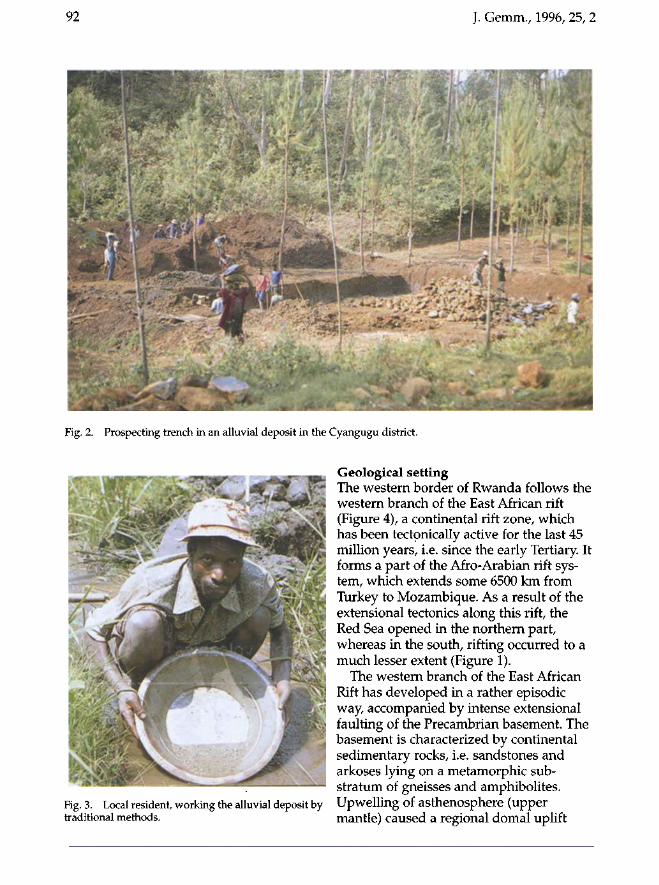
some areas of major interest for sapphire mining were established (Figure 2). Due to the rapid decomposition of rocks in a tropical humid climate, the main sapphire deposits are of alluvial type. They are exploited mostly by the local population using traditional methods (Figure 3). Twenty representative sapphire samples from the Cyangugu district were selected for the laboratory and investigated by various methods, including spectrophotometry
© Gemmological Association and Gem Testing Laboratory of Great Britain ISSN: 1355-4565
J. Gemm., 1996,25,2 91
ZAIRE UGANDA
BURUNDI
5
TANZANIA
o 500km
major rift faults
Tertiary and Quaternary volcanics
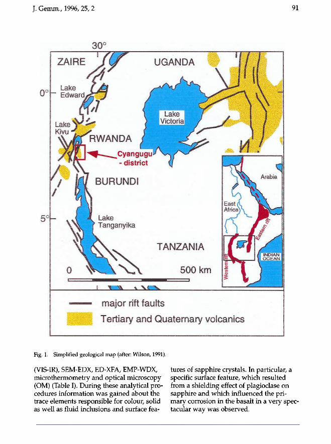
Fig. 1. Simplified geological map (after: Wilson, 1991).
(VIS-IR), SEM-EDX, ED-XFA, EMP-WDX, microthermometry and optical microscopy (OM) (Table I). During these analytical procedures information was gained about the trace elements responsible for colour, solid as well as fluid inclusions and surface fea-
tures of sapphire crystals. In particular, a specific surface feature, which resulted from a shielding effect of plagioclase on sapphire and which influenced the primary corrosion in the basalt in a very spectacular way was observed.
92 J. Gemm., 1996, 25, 2
Fig. 2. Prospecting trench in an alluvial deposit in the Cyangugu district.
Fig. 3. Local resident, working the alluvial deposit by traditional methods.
Geological setting The western border of Rwanda follows the western branch of the East African rift (Figure 4), a continental rift zone, which has been tectonically active for the last 45 million years, i.e. since the early Tertiary. It forms a part of the Afro-Arabian rift system, which extends some 6500 km from Turkey to Mozambique. As a result of the extensional tectonics along this rift, the Red Sea opened in the northern part, whereas in the south, rifting occurred to a much lesser extent (Figure 1).
The western branch of the East African Rift has developed in a rather episodic way, accompanied by intense extensional faulting of the Precambrian basement. The basement is characterized by continental sedimentary rocks, i.e. sandstones and arkoses lying on a metamorphic substratum of gneisses and amphibolites. Upwelling of asthenosphere (upper mantle) caused a regional domal uplift
J. Gemm., 1996, 25, 2 93
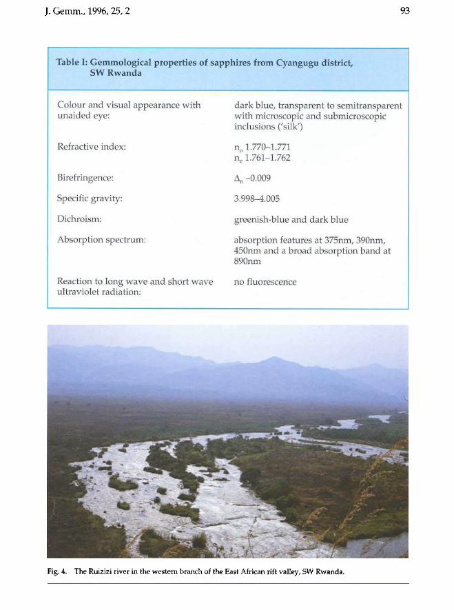
Table I: Gemmological properties of sapphires from Cyangugu district, SW Rwanda
Colour and visual appearance with unaided eye:
Refractive index:
Birefringence:
Specific gravity:
Dichroism:
Absorption spectrum:
Reaction to long wave ultraviolet radiation:
and short wave
dark blue, transparent to semitransparent with microscopic and submicroscopic inclusions ('silk')
n0 1.770-1.771 ne 1.761-1.762
An -0.009
3.998-4.005
greenish-blue and dark blue
absorption features at 375nm, 390nm, 450nm and a broad absorption band at 890nm
no fluorescence
Fig. 4. The Ruizizi river in the western branch of the East African rift valley, SW Rwanda.
94 J. Gemm., 1996, 25, 2
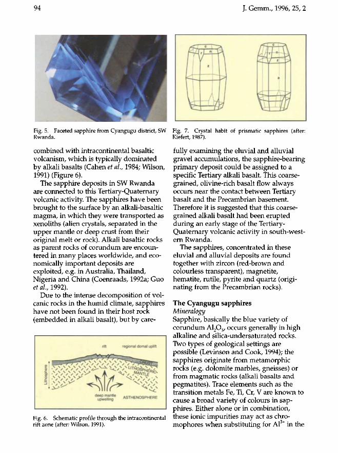
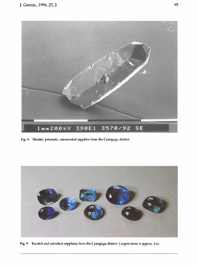
Fig. 5. Faceted sapphire from Cyangugu district, SW Rwanda.
combined with intracontinental basaltic volcanism, which is typically dominated by alkali basalts (Cahen et al., 1984; Wilson, 1991) (Figure 6).
The sapphire deposits in SW Rwanda are connected to this Tertiary-Quaternary volcanic activity. The sapphires have been brought to the surface by an alkali-basaltic magma, in which they were transported as xenoliths (alien crystals, separated in the upper mantle or deep crust from their original melt or rock). Alkali basaltic rocks as parent rocks of corundum are encountered in many places worldwide, and economically important deposits are exploited, e.g. in Australia, Thailand, Nigeria and China (Coenraads, 1992a; Guo et al., 1992).
Due to the intense decomposition of volcanic rocks in the humid climate, sapphires have not been found in their host rock (embedded in alkali basalt), but by care-
regional domal uplift
deep mantle upwelling ASTHENOSPHERE
Fig. 6. Schematic profile through the intracontinental rift zone (after: Wilson, 1991).
Fig. 7. Crystal habit of prismatic sapphires (after: Kiefert, 1987).
fully examining the eluvial and alluvial gravel accumulations, the sapphire-bearing primary deposit could be assigned to a specific Tertiary alkali basalt. This coarsegrained, olivine-rich basalt flow always occurs near the contact between Tertiary basalt and the Precambrian basement. Therefore it is suggested that this coarsegrained alkali basalt had been erupted during an early stage of the Tertiary-Quaternary volcanic activity in south-western Rwanda.
The sapphires, concentrated in these eluvial and alluvial deposits are found together with zircon (red-brown and colourless transparent), magnetite, hematite, rutile, pyrite and quartz (originating from the Precambrian rocks).
The Cyangugu sapphires Mineralogy Sapphire, basically the blue variety of corundum A1203, occurs generally in high alkaline and silica-undersaturated rocks. Two types of geological settings are possible (Levinson and Cook, 1994); the sapphires originate from metamorphic rocks (e.g. dolomite marbles, gneisses) or from magmatic rocks (alkali basalts and pegmatites). Trace elements such as the transition metals Fe, Ti, Cr, V are known to cause a broad variety of colours in sapphires. Either alone or in combination, these ionic impurities may act as chro-mophores when substituting for Al3+ in the
J. Cemm., 1996, 25, 2 95
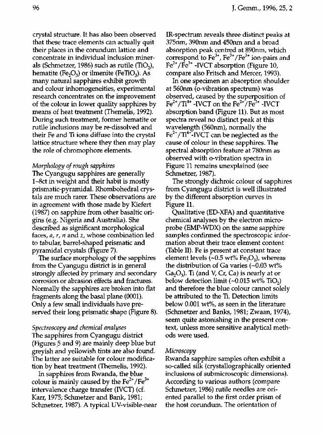
lmm2~0kU ~90El 3570/92 SE
Fig. 8. Slender, prismatic, uncorroded sapphire from the Cyangugu district.
Fig. 9. Faceted and cabochon sapphires from the Cyangugu district. Largest stone is approx. 8 ct.
96
crystal structure. It has also been observed that these trace elements can actually quit their places in the corundum lattice and concentrate in individual inclusion minerals (Schmetzer, 1986) such as rutile (TiOz), hematite (FeZ0 3) or ilmenite (FeTi03). As many natural sapphires exhibit growth and colour inhomogeneities, experimental research concentrates on the improvement of the colour in lower quality sapphires by means of heat treatment (Themelis, 1992). During such treatment, former hematite or rutile inclusions may be re-dissolved and their Fe and Ti ions diffuse into the crystal lattice structure where they then may play the role of chromophore elements.
Morphology of rough sapphires The Cyangugu sapphires are generally 1-8ct in weight and their habit is mostly prismatic-pyramidal. Rhombohedral crystals are much rarer. These observations are in agreement with those made by Kiefert (1987) on sapphire from other basaltic origins (e.g. Nigeria and Australia). She described as significant morphological faces, a, r, nand z, whose combination led to tabular, barrel-shaped prismatic and pyramidal crystals (Figure 7).
The surface morphology of the sapphires from the Cyangugu district is in general strongly affected by primary and secondary corrosion or abrasion effects and fractures. Normally the sapphires are broken into flat fragments along the basal plane (0001). Only a few small individuals have preserved their long prismatic shape (Figure 8).
Spectroscopy and chemical analyses The sapphires from Cyangugu district (Figures 5 and 9) are mainly deep blue but greyish and yellowish tints are also found. The latter are suitable for colour modification by heat treatment (Themelis, 1992).
In sapphires from Rwanda, the blue colour is mainly caused by the Fez+ /Fe3+ intervalence charge transfer (IVCT) (cf. Karr, 1975; Schmetzer and Bank, 1981; Schmetzer, 1987). A typical UV-visible-near
J. Gemm., 1996,25, 2
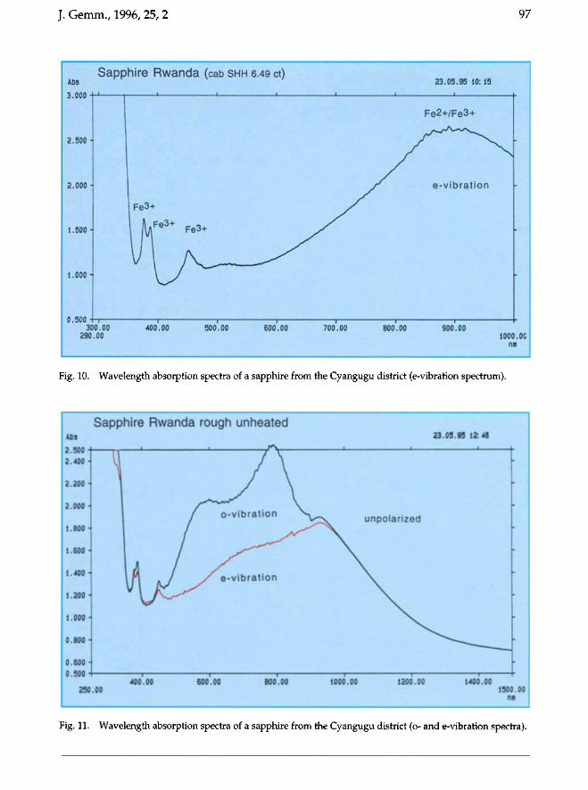
IR-spectrum reveals three distinct peaks at 375nm, 390nm and 450nm and a broad absorption peak centred at 890nm, which correspond to Fe3+, Fe3+ /Fe3+ ion-pairs and Fez+ /Fe3+ -IVCT absorption (Figure 10, compare also Fritsch and Mercer, 1993).
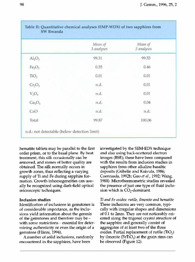
In one specimen an absorption shoulder at 560nm (o-vibration spectrum) was observed, caused by the super~osition of Fez+ /Ti4+ -IVCT on the Fez+ /Fe + -IVCT absorption band (Figure 11). But as most spectra reveal no distinct peak at this wavelength (560nm), normally the Fez+ /Ti4+-IVCT can be neglected as the cause of colour in these sapphires. The spectral absorption feature at 780nm as observed with o-vibration spectra in Figure 11 remains unexplained (see Schmetzer, 1987).
The strongly dichroic colour of sapphires from Cyangugu district is well illustrated by the different absorption curves in Figure II.
Qualitative (ED-XFA) and quantitative chemical analyses by the electron microprobe (EMP-WDX) on the same sapphire samples confirmed the spectroscopic information about their trace element content (Table II). Fe is present at constant trace element levels (-0.5 wt% FeZ0 3), whereas the distribution of Ga varies (-0.03 wt% GaZ0 3). Ti (and V, Cr, Ca) is nearly at or below detection limit (-0.015 wt% TiOz) and therefore the blue colour cannot solely be attributed to the Ti. Detection limits below 0.001 wt%, as seen in the literature (Schmetzer and Banks, 1981; Zwaan, 1974), seem quite astonishing in the present context, unless more sensitive analytical methods were used.
Microscopy Rwanda sapphire samples often exhibit a so-called silk (crystallographic ally oriented inclusions of submicroscopic dimensions). According to various authors (compare Schmetzer, 1986) rutile needles are oriented parallel to the first order prism of the host corundum. The orientation of
J. Gemm., 1996, 25, 2 97
Sapphire Rwanda (cab SHH 6.49 ct)
1 ftftft uj * * . 1 . 1 I 3*vv(f "
2.500-
2.000*
IJSOO-
1.000-
/I Jtft/1 •
Fe3+ >•
i r tX - ^ ' . 1/ — I v.wv n 1 1 1 1 %•••
300.00 400.00 500.00 BO0.00 700.00 290.00
23.05.9S 10:15 . . . f , .., | . , *
Fe2+/Fe3+
f e-vibration •
800.00 900.00 1000.0C
m
Fig. 10. Wavelength absorption spectra of a sapphire from the Cyangugu district (e-vibration spectrum).
Sapphire Rwanda rough unhealed Abs
Z*3vv "
2.400-
2.200-
2.000-
; 1.800-
1.600-
1.400-
1.200 •
1.000-
0.800-
0.600-
H / \ ! / \
/ o-vibratton \
III KJ s e-vibratlon
V » OWW "1 1 1 1
400.00 600.00 B00.00 250.00
23.05.95 12:46
Upv unpolarized
-
*
-
1000.00 1200.00 1400.00 1500.00
Fig. 11. Wavelength absorption spectra of a sapphire from the Cyangugu district (o- and e-vibration spectra).
98 J. Gemm., 1996, 25, 2
Table II:
A1203
Fe203
Ti02
Cr2Os
v2o3
Ga2Os
CaO
Total
n.d.: not
Quantitative chemical analyses (EMP-WDX) of two SW Rwanda
Mean of 3 analyses
99.31
0.55
0.01
n.d.
n.d.
n.d.
n.d.
99.87
detectable (below detection limit)
sapphires from
Mean of 5 analyses
99.53
0.46
0.01
0.01
0.01
0.04
n.d.
100.06
hematite tablets may be parallel to the first order prism, or to the basal plane. By heat treatment, this silk occasionally can be removed, and stones of better quality are obtained. The silk normally occurs in growth zones, thus reflecting a varying supply of Ti and Fe during sapphire formation. Growth inhomogeneities can usually be recognized using dark-field optical microscopic techniques.
Inclusion studies Identification of inclusions in gemstones is of considerable importance, as the inclusions yield information about the genesis of the gemstones and therefore may be -with some restrictions - essential for determining authenticity or even the origin of a gemstone (Hänni, 1994).
A number of solid inclusions, randomly encountered in the sapphires, have been
investigated by the SEM-EDX technique and also using back-scattered electron images (BSE); these have been compared with the results from inclusion studies in sapphires from other alkaline basaltic deposits (Gübelin and Koivula, 1986; Coenraads, 1992b; Guo et al, 1992; Wang, 1988). Microthermometric studies revealed the presence of just one type of fluid inclusion which is C02-dominant.
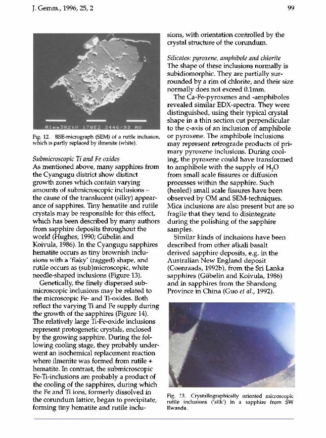
Ti and Te oxides: rutile, ilmenite and hematite These inclusions are very common, typically with irregular shapes and dimensions of 0.1 to 2mm. They are not noticeably oriented along the trigonal crystal structure of the sapphire and generally consist of aggregates of at least two of the three oxides. Partial replacement of rutile (Ti02) by ilmenite (FeTi03) at the grain rims can be observed (Figure 12).
J. Gemm., 1996, 25, 2 99
Fig. 12. BSE-micrograph (SEM) of a rutile inclusion, which is partly replaced by ilmenite (white).
Submicroscopic Ti and Fe oxides As mentioned above, many sapphires from the Cyangugu district show distinct growth zones which contain varying amounts of submicroscopic inclusions -the cause of the translucent (silky) appearance of sapphires. Tiny hematite and rutile crystals may be responsible for this effect, which has been described by many authors from sapphire deposits throughout the world (Hughes, 1990; Gübelin and Koivula, 1986). In the Cyangugu sapphires hematite occurs as tiny brownish inclusions with a 'flaky' (ragged) shape, and rutile occurs as (sub)microscopic, white needle-shaped inclusions (Figure 13).
Genetically, the finely dispersed submicroscopic inclusions may be related to the microscopic Fe- and Ti-oxides. Both reflect the varying Ti and Fe supply during the growth of the sapphires (Figure 14). The relatively large Ti-Fe-oxide inclusions represent protogenetic crystals, enclosed by the growing sapphire. During the following cooling stage, they probably underwent an isochemical replacement reaction where ilmenite was formed from rutile + hematite. In contrast, the submicroscopic Fe-Ti-inclusions are probably a product of the cooling of the sapphires, during which the Fe and Ti ions, formerly dissolved in the corundum lattice, began to precipitate, forming tiny hematite and rutile inclu
sions, with orientation controlled by the crystal structure of the corundum.
Silicates: pyroxene, amphibole and chlorite The shape of these inclusions normally is subidiomorphic. They are partially surrounded by a rim of chlorite, and their size normally does not exceed 0.1mm.
The Ca-Fe-pyroxenes and -amphiboles revealed similar EDX-spectra. They were distinguished, using their typical crystal shape in a thin section cut perpendicular to the c-axis of an inclusion of amphibole or pyroxene. The amphibole inclusions may represent retrograde products of primary pyroxene inclusions. During cooling, the pyroxene could have transformed to amphibole with the supply of H 2 0 from small scale fissures or diffusion processes within the sapphire. Such (healed) small scale fissures have been observed by OM and SEM-techniques. Mica inclusions are also present but are so fragile that they tend to disintegrate during the polishing of the sapphire samples.
Similar kinds of inclusions have been described from other alkali basalt derived sapphire deposits, e.g. in the Australian New England deposit (Coenraads, 1992b), from the Sri Lanka sapphires (Gübelin and Koivula, 1986) and in sapphires from the Shandong Province in China (Guo et al., 1992).
Fig. 13. Crystallographically oriented microscopic rutile inclusions ('silk') in a sapphire from SW Rwanda.
100 J. Gemm., 1996, 25, 2
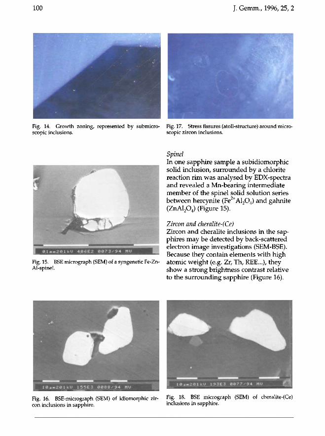
Fig. 14. Growth zoning, represented by submicro-scopic inclusions.
Fig. 15. BSE micrograph (SEM) of a syngenetic Fe-Zn-Al-spinel.
Fig. 16. BSE-micrograph (SEM) of idiomorphic zircon inclusions in sapphire.
Fig. 17. Stress fissures (atoll-structure) around microscopic zircon inclusions.
Spinel In one sapphire sample a subidiomorphic solid inclusion, surrounded by a chlorite reaction rim was analysed by EDX-spectra and revealed a Mn-bearing intermediate member of the spinel solid solution series between hercynite (Fe2+Al204) and gahnite (ZnAl204) (Figure 15).
Zircon and cheralite-(Ce) Zircon and cheralite inclusions in the sapphires may be detected by back-scattered electron image investigations (SEM-BSE). Because they contain elements with high atomic weight (e.g. Zr, Th, REE...), they show a strong brightness contrast relative to the surrounding sapphire (Figure 16).
Fig. 18. BSE micrograph (SEM) of cheralite-(Ce) inclusions in sapphire.
Cheralite-(Ce) solid inclusionin sapphire from SW-Rwandasa re sample: P9. scan 3
J. Gemm., 1996,25,2
o
C 51
AI
100
p
Thacca ra100 a:magni lion:coallOg malenal:
20 aV2600 x
carbon
101
2 3 4 5 6e argy (kaV)
7 8 9Io
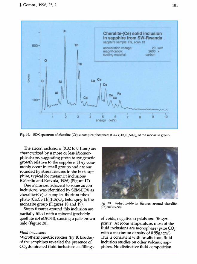
Fig. 19. EDX-spectrum of cheralite-(Ce), a complex phosphate (Ca,Ce,Th)(P,Si)04' of the monazite group.
The zircon inclusions (0.02 to O.lmm) arecharacterized by a more or less idiomorphic shape, suggesting proto to syngeneticgrowth relative to the sapphire. They commonly occur in small groups and are surrounded by stress fissures in the host sapphire, typical for metamict inclusions(Giibelin and Koivula, 1986) (Figure 17).
One inclusion, adjacent to some zirconinclusions, was identified by SEM7EDX ascheralite-(Ce), a complex thorium-phosphate (Ca,Ce,Th)(P,Si)04' belonging to themonazite group (Figures 18 and 19).
Stress fissures around this inclusion arepartially filled with a mineral (probablygoethite a-FeO(OH), causing a pale brownhalo (Figure 20).
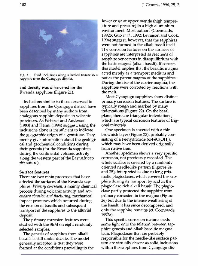
Fluid inclusionsMicrothermometric studies (by B. Bruder)of the sapphires revealed the presence ofCO2 dominated fluid inclusions as fillings
Fig. 20. Fe-hydroxide in fissures around cheralite(Ce) inclusions.
of voids, negative crystals and 'fingerprints'. At room temperature, most of thefluid inclusions are monophase (pure CO2with a maximum density of 0.95g/em").This is consistent with results from fluidinclusion studies on other volcanic sapphires. No distinctive fluid composition
102 J. Gemm., 1996, 25, 2
Fig. 21. Fluid inclusions along a healed fissure in a sapphire from the Cyangugu district.
and density was discovered for the Rwanda sapphires (Figure 21).
Inclusions similar to those observed in sapphires from the Cyangugu district have been described by many authors from analogous sapphire deposits in volcanic provinces. As Webster and Anderson (1983) and Hänni (1994) suggest, using the inclusions alone is insufficient to indicate the geographic origin of a gemstone. They merely give information about the geological and geochemical conditions during their genesis (for the Rwanda sapphires: during the continental volcanic activity along the western part of the East African rift suture).
Surface features There are two main processes that have affected the surfaces of the Rwanda sapphires. Primary corrosion, a mainly chemical process during volcanic activity, and secondary abrasion and fracturing, mechanical impact processes which occurred during the erosion of basalts and subsequent transport of the sapphires to the alluvial deposit.
The primary corrosion features were studied with the SEM on eight randomly selected samples.
The genesis of sapphires from alkali basalts is still under debate. The model generally accepted is that they were formed at the conditions prevailing in the
lower crust or upper mantle (high temperature and pressure) in a high aluminium environment. Most authors (Coenraads, 1992b; Guo et al, 1992; Levinson and Cook, 1994) suggest, however, that the sapphires were not formed in the alkali basalt itself. The corrosion features on the surfaces of sapphires are interpreted as reactions of sapphire xenocrysts in disequilibrium with the basic magma (alkali basalt). If correct, this model implies that the basaltic magma acted merely as a transport medium and not as the parent magma of the sapphires. During the rise of the carrier magma, the sapphires were corroded by reactions with the melt.
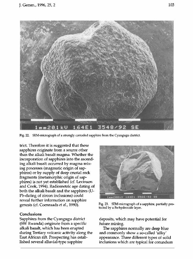
Most Cyangugu sapphires show distinct primary corrosion features. The surface is typically rough and marked by many indentations (Figure 22). On the basal plane, there are triangular indentations, which are typical corrosion features of trigonal minerals.
One specimen is covered with a thin brownish layer (Figure 23), probably consisting of a Fe-hydroxide (oc-FeO(OH)), which may have been derived originally from native iron.
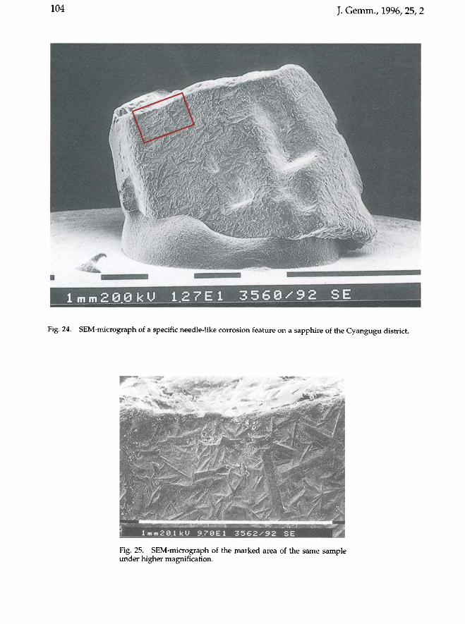
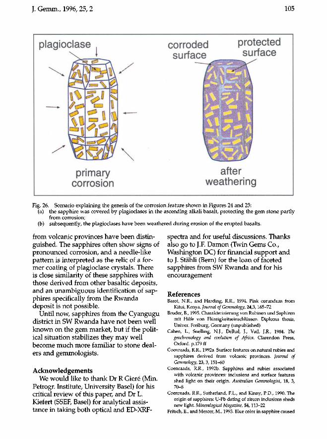
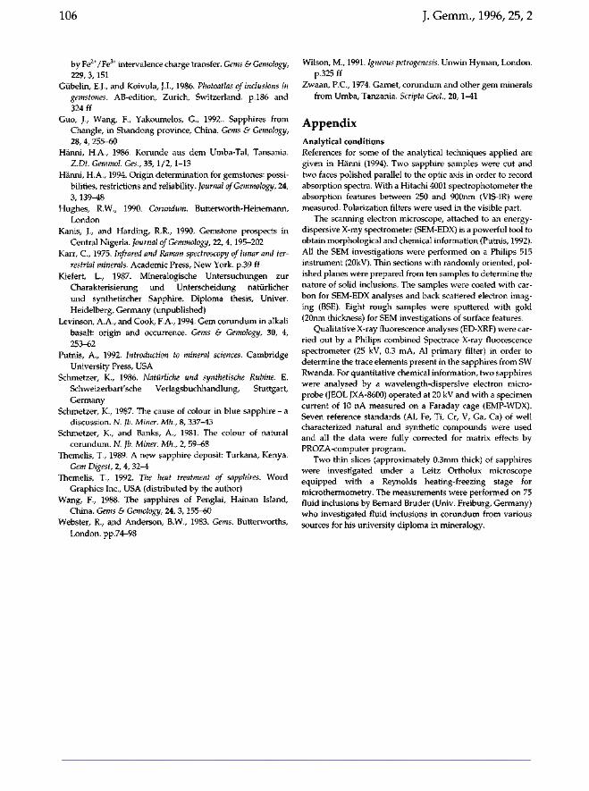
Another specimen shows a very specific corrosion, not previously recorded. The whole surface is covered by a randomly oriented needle-like pattern (Figures 24 and 25), interpreted as due to long prismatic plagioclases, which covered the sapphire during its transport by and in the plagioclase-rich alkali basalt. The plagio-clase partly protected the sapphire from primary corrosion in the magma (Figure 26) but due to the intense weathering of the basalt, it has since decomposed, and only the sapphire remains (cf. Coenraads, 1992a).
This specific corrosion feature sheds some light onto the relation between sapphire genesis and alkali basaltic magma-tism. Plagioclases that are probably responsible for the needle-like surface pattern are virtually absent as solid inclusions within the sapphires from Cyangugu dis-
J. Gemm., 1996, 25, 2 103
» j
l m m 2 8 1 k U 1.6 4 E 1 3 5 4 8 ^ 9 2 SE Fig. 22. SEM-micrograph of a strongly corroded sapphire from the Cyangugu district.
trict. Therefore it is suggested that these sapphires originate from a source other than the alkali basalt magma. Whether the incorporation of sapphires into the ascending alkali basalt occurred by magma mixing processes (magmatic origin of sapphires) or by supply of deep crustal rock fragments (metamorphic origin of sapphires) is not yet established (cf. Leyinson and Cook, 1994). Radiometric age dating of both the alkali basalt and the sapphires (U-Pb-dating of zircon inclusions) could reveal further information on sapphire genesis (cf. Coenraads et ah, 1990).
Conclusions Sapphires from the Cyangugu district (SW Rwanda) originate from a specific alkali basalt, which has been erupted during Tertiary volcanic activity along the East African rift. Prospecting has established several alluvial-type sapphire
Fig. 23. SEM-micrograph of a sapphire, partially protected by a Fe-hydroxide layer.
deposits, which may have potential for future mining.
The sapphires normally are deep blue and commonly show a so-called 'silky' appearance. Three different types of solid inclusions which are typical for corundum
104 J. Gemm., 1996, 25, 2
"^mrnmm
^m*
1 m m 2 8 0 k U 1.2 7 E 1 3 5.6 9 • 9 2 S_E
Fig. 24. SEM-micrograph of a specific needle-like corrosion feature on a sapphire of the Cyangugu district.
Fig. 25. SEM-micrograph of the marked area of the same sample under higher magnification.
J. Gemm., 1996,25,2 105
plagioclase corroded surface
protected surface
'Üü primary
corrosion
JS-V* «Ml*
after weathering
Fig. 26. Scenario explaining the genesis of the corrosion feature shown in Figures 24 and 25: (a) the sapphire was covered by plagioclases in the ascending alkali basalt, protecting the gem stone partly
from corrosion; (b) subsequently, the plagioclases have been weathered during erosion of the erupted basalts.
from volcanic provinces have been distinguished. The sapphires often show signs of pronounced corrosion, and a needle-like pattern is interpreted as the relic of a former coating of plagioclase crystals. There is close similarity of these sapphires with those derived from other basaltic deposits, and an unambiguous identification of sapphires specifically from the Rwanda deposit is not possible.
Until now, sapphires from the Cyangugu district in SW Rwanda have not been well known on the gem market, but if the political situation stabilizes they may well become much more familiar to stone dealers and gemmologists.
Acknowledgements We would like to thank Dr R Gieré (Min.
Petrogr. Institute, University Basel) for his critical review of this paper, and Dr L. Kiefert (SSEF, Basel) for analytical assistance in taking both optical and ED-XRF-
spectra and for useful discussions. Thanks also go to J.F. Damon (Twin Gems Co., Washington DC) for financial support and to J. Stähli (Bern) for the loan of faceted sapphires from SW Rwanda and for his encouragement
References Barot, N.R., and Harding, R.R., 1994. Pink corundum from
Kitui, Kenya. Journal of Gemmology, 24, 3, 165-72 Bruder, B., 1995. Charakterisierung von Rubinen und Saphiren
mit Hilfe von Flüssigkeitseinschlüssen. Diploma thesis, Univer. Freiburg, Germany (unpublished)
Cahen, L., Snelling, N.J., Delhal, J., Vail, J.R., 1984. The geochronology and evolution of Africa. Clarendon Press, Oxford, p.179 ff
Coenraads, R.R., 1992a. Surface features on natural rubies and sapphires derived from volcanic provinces. Journal of Gemmology, 23, 3, 151-60
Coenraads, R.R., 1992b. Sapphires and rubies associated with volcanic provinces: inclusions and surface features shed light on their origin. Australian Gemmologist, 18, 3, 70-8
Coenraads, R.R., Sutherland, F.L., and Kinny, P.D., 1990. The origin of sapphires: U-Pb dating of zircon inclusions sheds new light. Mineralogical Magazine, 54, 113-22
Fritsch, E., and Mercer, M., 1993. Blue color in sapphire caused
106
by Fe2+ /Fe3
+ intervalence charge transfer. Gems & Gemology, 229,3,151
Gubelin, E.J., and Koivula, J.I., 1986. Photoat/as of inclusions in gemstones. AB-edition, Zurich, Switzerland. p.l86 and 324 ff
Guo, L Wang, F., Yakoumelos, G., 1992 .. Sapphires from Changle, in Shandong province, China. Gems & Gemology, 28,4,255--<i0
Hanni, H.A, 1986. Korunde aus dem Umba-Tal, Tansania. Z.Dt. Gemmo/. Ges., 35, 1/2, 1-13
Hanni, H.A, 1994. Origin determination for gemstones: possibilities, restrictions and reliability. Journal of Gemmology, 24, 3, 139-48
Hughes, R.W., 1990. Corundum. Butterworth-Heinemann, London
Kanis, L and Harding, R.R., 1990. Gemstone prospects in Central Nigeria. Journal of Gemmology, 22, 4,195-202
Karr, c., 1975. Infrared and Raman spectroscopy of lunar and terrestrial minerals. Academic Press, New York. p.39 ff
Kiefert, L., 1987. Mineralogische Untersuchungen zur Charakterisierung und Unterscheidung natiirlicher und synthetischer Sapphire. Diploma thesis, Univer. Heidelberg, Germany (unpublished)
Levinson, AA, and Cook, F.A, 1994. Gem corundum in alkali basalt: origin and occurrence. Gems & Gemology, 30, 4, 253--<i2
Putnis, A, 1992. Introduction to mineral sciences. Cambridge University Press, USA
Schmetzer, K., 1986. Natiirliche und synthetische Rubine. E. Schweizerbart'sche Verlagsbuchhandlung, Stuttgart, Germany
Schmetzer, K., 1987. The cause of colour in blue sapphire - a discussion. N. Jb. Miner. Mh., 8, 337-43
Schmetzer, K., and Banks, A, 1981. The colour of natural corundum. N. Jb. Miner. Mh., 2, 59-68
Themelis, T., 1989. A new sapphire deposit: Turkana, Kenya. Gem Digest, 2, 4, 32-4
Themelis, T., 1992. The heat treatment of sapphires. Word Graphics Inc., USA (distributed by the author)
Wang, F., 1988. The sapphires of Penglai, Hainan Island, China. Gems & Gemology, 24, 3, 155-60
Webster, R., and Anderson, B.W., 1983. Gems. Butterworths, London. pp.74-98
J. Cemm., 1996,25,2
Wilson, M., 1991. Igneous petrogenesis. Unwin Hyman, London. p.325 ff
Zwaan, P.c., 1974. Garnet, corundum and other gem minerals from Umba, Tanzania. Scripta Geol., 20, 1-41
Appendix Analytical conditions References for some of the analytical techniques applied are given in Hanni (1994). Two sapphire samples were cut and two faces polished parallel to the optic axis in order to record absorption spectra. With a Hitachi 4001 spectrophotometer the absorption features between 250 and 900nm (VIS-IR) were measured. Polarization filters were used in the visible part.
The scanning electron microscope, attached to an energydispersive X-ray spectrometer (SEM-EDX) is a powerful tool to obtain morphological and chemical information (Putnis, 1992). All the SEM investigations were performed on a Philips 515 instrument (20kV). Thin sections with randomly oriented, polished planes were prepared from ten samples to determine the nature of solid inclusions. The samples were coated with carbon for SEM-EDX analyses and back scattered electron imaging (BSE). Eight rough samples were sputtered with gold (20nm thickness) for SEM investigations of surface features.
Qualitative X-ray fluorescence analyses (ED-XRF) were carried out by a Philips combined Spectrace X-ray fluorescence spectrometer (25 kV, 0.3 mA, Al primary filter) in order to determine the trace elements present in the sapphires from SW Rwanda. For quantitative chemical information, two sapphires were analysed by a wavelength-dispersive electron microprobe (JEOL JXA-8600) operated at 20 kV and with a specimen current of 10 nA measured on a Faraday cage (EMP-WDX). Seven reference standards (AI, Fe, Ti, Cr, V, Ga, Cal of well characterized natural and synthetic compounds were used and all the data were fully corrected for matrix effects by PROZA-computer program.
Two thin slices (approximately O.3mm thick) of sapphires were investigated under a Leitz Ortholux microscope equipped with a Reynolds heating-freezing stage for microthermometry. The measurements were performed on 75 fluid inclusions by Bernard Bruder (Univ. Freiburg, Germany) who investigated fluid inclusions in corundum from various sources for his university diploma in mineralogy.
J. Gemm., 1996, 25, 2 107
and the Laboratory has built up an enviable international reputation for consistency ana1 quality.
The London Diamond Report is based on the principles of harmonized grading for colour and clarity of diamonds and gives the most essential and up-to-date information required by the trade.
The Laboratory is also proficient in the grading of fancy-cut diamonds and in colour origin determination.
With a London Diamond Report your diamond's prestige is assured
© The Gem Testing Laboratory GAGTL, 27 GREVILLE STREET,
LONDON EG N 8SU, UK
T e l : + 4 4 (0)171-405 3351 F a x : + 4 4 (0)171-831 9479
108 J. Gemm., 1996, 25, 2,
On the identification and fade testing of Maxixe beryl, golden beryl and green aquamarine
K. Nassau, PhD, FGA(Hon)
Lebanon, NJ 08833, USA
Abstract Forty-three large (up to 126.66 ct)
high quality faceted beryls of recent Brazilian origin were examined. Of these, 29 were intense blue Maxixe-type
beryls, a material widely seen about 1970. The remaining five yellow and nine greenish beryls were found to be simply golden beryls and green aquamarines, respectively.
Heat and light exposure tests showed that an accelerated fading test must not use an excessively elevated temperature, say 50 °C (122 °F) maximum, since otherwise both aquamarines and golden beryls (which do not fade from light alone) would lose their yellow colour component from the heat and could then be misidentified as fading Maxixe beryls. This has indeed happened in one report in the recent gemmological literature, where a temperature of 100 °C was used as part of a fade test.
It is proposed that the gemmological distinction between the original
'Maxixe' and the later 'Maxixe-type' beryls be dropped, the term 'Maxixe' then covering both variants. While the distinction remains significant in mineralogy and other fields, it serves no useful purpose in gemmology.
Introduction The early history and the gemmological characteristics of the original Maxixe beryls of 1917 and the later Maxixe-type
beryls are well covered in the gemmological literature.1-10 This includes their identification and distinction from aquamarine by the anomalous dichroism, unusual absorption spectra, and fading from exposure to light. These two variants differ somewhat in their absorption spectra and by the fact that the intense blue colour centre is associated with a nitrate impurity8,11 in the original Maxixe beryl, or with a carbonate impurity8'9'11 in the later Maxixe-type beryl.
It is often assumed that any intense blue original Maxixe beryl owes its colour to nature and that the equivalent colour of Maxixe-type beryl originates from man-induced irradiation as, for example, stated by Brown.12 Yet it is clear from the literature reports that the colour of the original Maxixe beryl can also be produced or restored by irradiation by man,44 and that the Maxixe-type blue beryl also occurs naturally.10
The distinction between these two variants is thus independent of the origin of the irradiation and depends only on the chemistry. Whether the colour derives from irradiation by nature or by man generally cannot be distinguished by gemmological or by any other form of testing. An exception occurs if an inappropriate irradiation technique has induced radioactivity, the residue of which may then be detectable, as happened at least once.4
Although I myself originally introduced these two variant designations,4-61 now believe that the distinction between the original 'Maxixe' beryl and the later
© Gemmological Association and Gem Testing Laboratory of Great Britain ISSN: 1355-4565
J. Gemm., 1996, 25, 2 109
'Maxixe-type' beryl serves no useful purpose in gemmology. It has never been used in GIA identification reports, for example. I believe it should be dropped because: 1. both variants generally occur in
intense blue form in nature; 2. both can be produced by irradiation of
pale colour beryl containing the necessary precursor impurities (nitrate or carbonate);
3. both fade in light in about the same time frame, although there is some variability;
4. both lose their colour on being heated, again with some variability;
5. lost colour can be restored by irradiation for both; and
6. routine gemmological testing using a hand spectroscope is not sufficient to distinguish these two variants, but this distinction is possible by using a research spectroscope.
In what follows, the designation 'Maxixe' beryl includes both variants except when specifically designated as 'original Maxixe' beryl or 'Maxixe-type' beryl.
On loss of colour by light and by heat Experts prefer to reserve the term 'fading' for a loss of colour on exposure to light and use other terms such as 'destruction' or 'loss' of colour for the equivalent action of heat. A typical example is Herbert Smith, who in his Gemstones13 describes the actions of light and heat on gemstones in separate paragraphs and uses the term 'fade' for the effects of light but not for those of heat. Again, Hunter and Harold14
define: 'Fading: a colour change in a material that involves a weakening or lightening with time, usually as a result of exposure to light or weather.'
Rapid testing for fading uses illumination conditions much more intense than those to which a gemstone would be exposed in normal jewellery wear. In studies begun in 1973,1 found4"7'11 that
Maxixe beryls faded rapidly under such conditions, losing essentially all colour in a few weeks, and stated that such material was therefore unsuitable for jewellery use. Some variability in behaviour was noted. The longer survival of this colour under normal (i.e. jewellery-wearing) illumination conditions was briefly mentioned in one of these reports.11
Accelerated fade testing often involves intense illumination combined with an elevated temperature to speed up the fading even more, typically less than 60 °C.
However, it is obvious that if an elevated temperature by itself changes the colour, then only such a temperature should be used for acceleration as does not itself have any effect. Nevertheless, the use of a temperature that might be present under normal illumination conditions, perhaps 50 °C (122 °F) maximum, would be reasonable.
Materials and experimental procedures Examination of 40 beryls, the property of Dr al Gobaisi, was conducted under restricted access with limited time and equipment in January 1994 in Singapore, in part with the assistance of E. Wong and M. Stern. Conventional gemmological techniques were used, including particularly the nature and orientation of the dichroic colours and the absorption spectrum, the latter of these by use of the hand spectroscope on all 40 beryls (referred to hereafter as aG beryls).
Six of these beryls, two selected at random from each colour, were studied in more detail: intense blue, a 33.29 ct oval and a 40.59 ct cushion; yellow, a 33.77 ct oval and a 68.86 ct cushion; and greenish, a 33.85 ct cushion and a 63.23 ct pear. These tests confirmed that all were natural beryls of exceptional quality, being flawless under lOx magnification. The absorption spectra of these six beryls were also run on a BYK Gardner Colorview Spectrophotometer at Herberts South East Asia Pte Ltd in Singapore, using in-house software with
110 J. Gemm., 1996, 25, 2
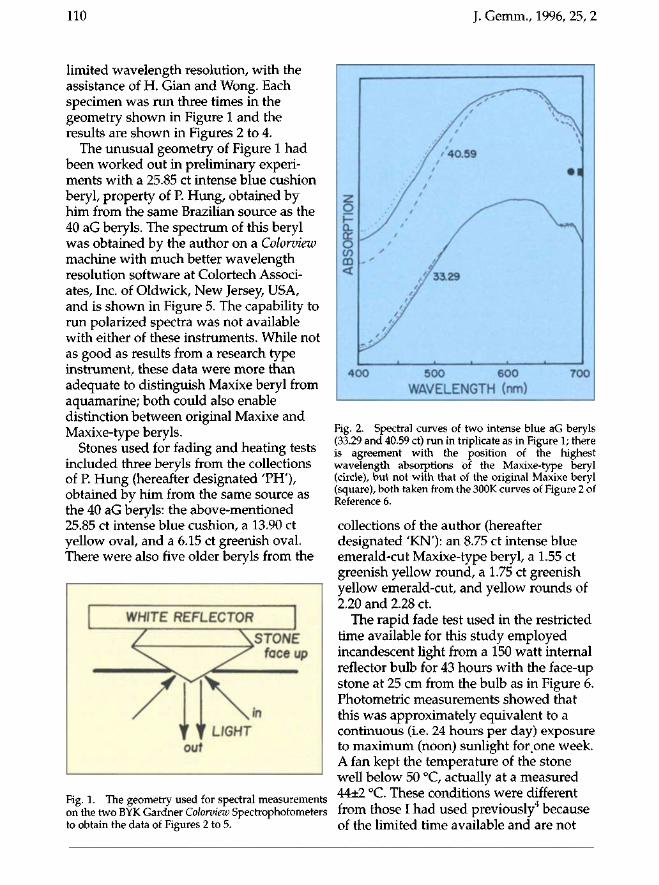
limited wavelength resolution, with the assistance of H. Gian and Wong. Each specimen was run three times in the geometry shown in Figure 1 and the results are shown in Figures 2 to 4.
The unusual geometry of Figure 1 had been worked out in preliminary experiments with a 25.85 ct intense blue cushion beryl, property of P. Hung, obtained by him from the same Brazilian source as the 40 aG beryls. The spectrum of this beryl was obtained by the author on a Colorview machine with much better wavelength resolution software at Colortech Associates, Inc. of Oldwick, New Jersey, USA, and is shown in Figure 5. The capability to run polarized spectra was not available with either of these instruments. While not as good as results from a research type instrument, these data were more than adequate to distinguish Maxixe beryl from aquamarine; both could also enable distinction between original Maxixe and Maxixe-type beryls.
Stones used for fading and heating tests included three beryls from the collections of P. Hung (hereafter designated 'PH'), obtained by him from the same source as the 40 aG beryls: the above-mentioned 25.85 ct intense blue cushion, a 13.90 ct yellow oval, and a 6.15 ct greenish oval. There were also five older beryls from the
WHITE REFLECTOR \
/ \STONE
\ ~ ~P* faceup
TIN Y f LIGHT Out
Fig. 1. The geometry used for spectral measurements on the two BYK Gardner Colorview Spectrophotometers to obtain the data of Figures 2 to 5.
Fig. 2. Spectral curves of two intense blue aG beryls (33.29 and 40.59 ct) run in triplicate as in Figure 1; there is agreement with the position of the highest wavelength absorptions of the Maxixe-type beryl (circle), but not with that of the original Maxixe beryl (square), both taken from the 300K curves of Figure 2 of Reference 6.
collections of the author (hereafter designated 'KN'): an 8.75 ct intense blue emerald-cut Maxixe-type beryl, a 1.55 ct greenish yellow round, a 1.75 ct greenish yellow emerald-cut, and yellow rounds of 2.20 and 2.28 ct.
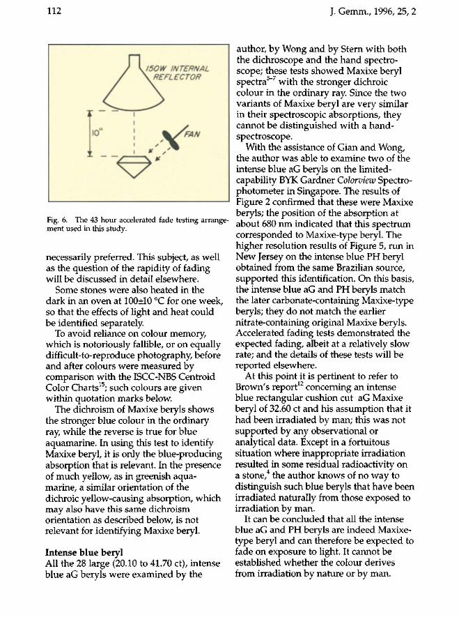
The rapid fade test used in the restricted time available for this study employed incandescent light from a 150 watt internal reflector bulb for 43 hours with the face-up stone at 25 cm from the bulb as in Figure 6. Photometric measurements showed that this was approximately equivalent to a continuous (i.e. 24 hours per day) exposure to maximum (noon) sunlight iorpne week. A fan kept the temperature of the stone well below 50 °C, actually at a measured 44±2 °C. These conditions were different from those I had used previously4 because of the limited time available and are not
J. Gemm., 1996, 25, 2 111
Z O F a. 6 CO CD <
6886% \ N
— v % if 33.7TV \ \ / / • •
\N A v / /•• V.N \ \ s s ' / ' V.N ' - . \ * * - " / /
Vv \ y \ \ Vv // V.N /./ \ * \ / • / \ \ N / 7
• 1 » A J
400 500 600 700 WAVELENGTH (nrn)
z O h-a. a: o c/> m <
} ' " • • ' i
\ \ •*x V• \ \ A 63.23
\\ VA n\*.\ nV* \\ \A
W A\* M \ '"> l \ V A \ V L \ ' \ '1 U \ ' A n \ \ •A \ •• - / y A
•A \ •-. N / .-A 33.85.\\ \ • • > - ^ „ ^ . , - y
'A\ ^ ^ ^ / yl • \ \ ^ - - ^ / \
'•^V / ' J '•- N \ ^ y / A
^ ^ ^ ^ ^ ^ ^ < ' • '
» « i . i \
400 500 600 WAVELENGTH (nm)
700
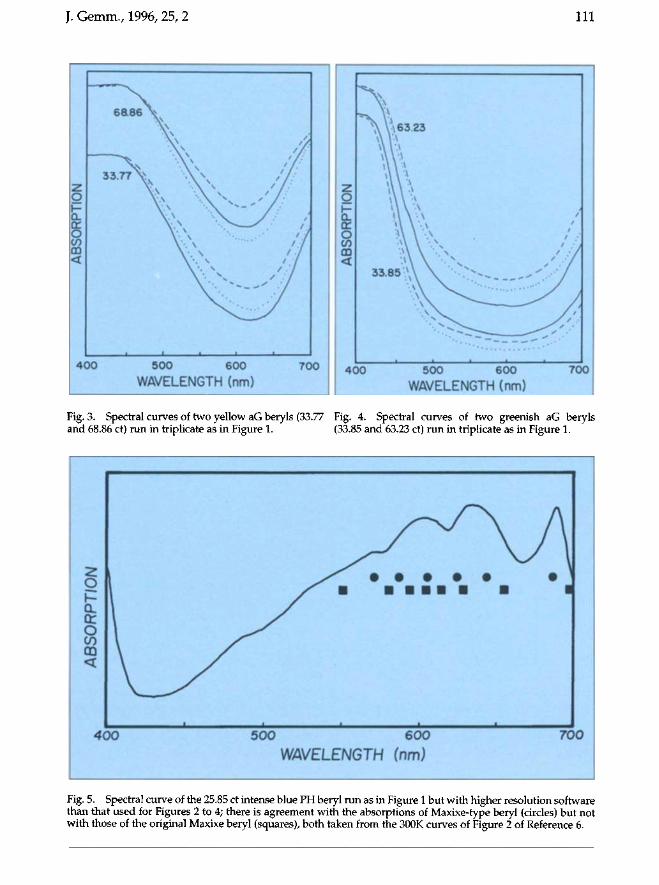
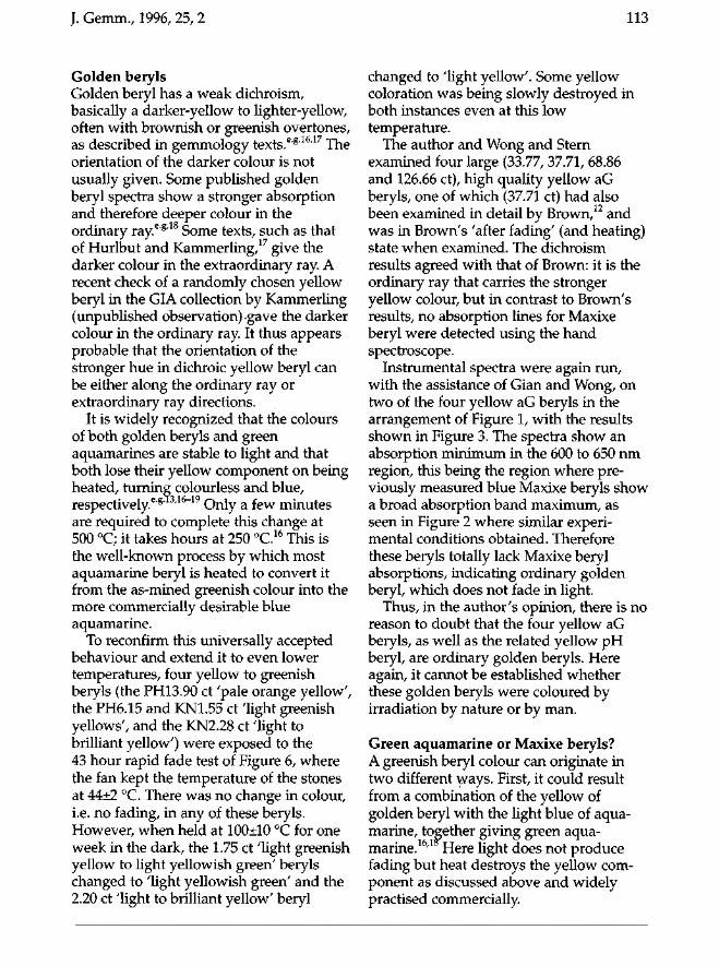
Fig. 3. Spectral curves of two yellow aG beryls (33.77 Fig. 4. Spectral curves of two greenish aG beryls and 68.86 ct) run in triplicate as in Figure 1. (33.85 and 63.23 ct) run in triplicate as in Figure 1.
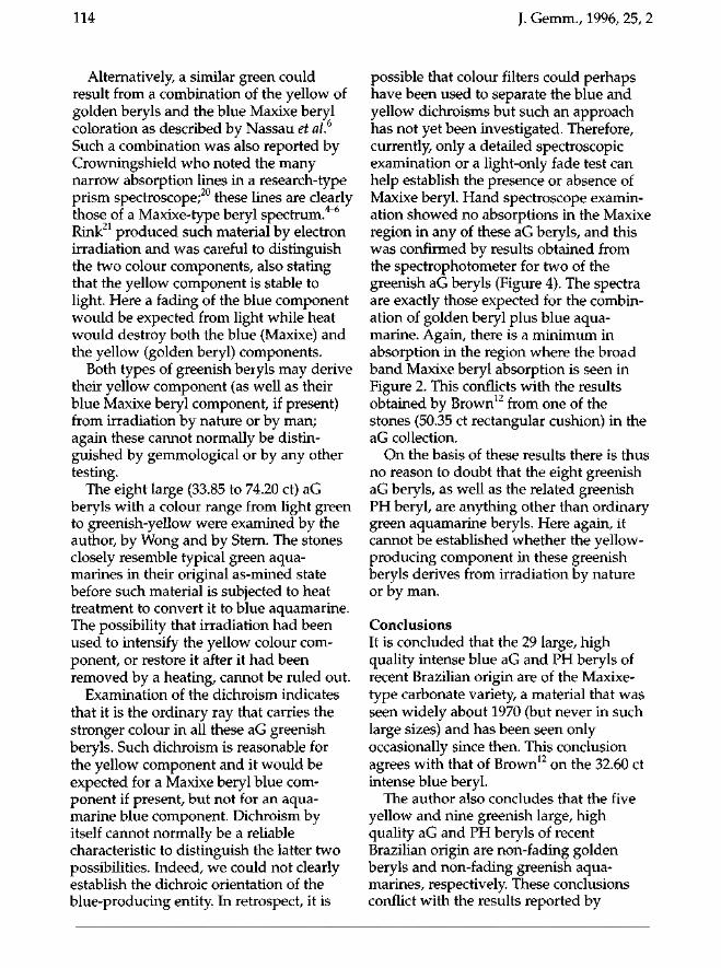
Fig. 5. Spectral curve of the 25.85 ct intense blue PH beryl run as in Figure 1 but with higher resolution software than that used for Figures 2 to 4; there is agreement with the absorptions of Maxixe-type beryl (circles) but not with those of the original Maxixe beryl (squares), both taken from the 300K curves of Figure 2 of Reference 6.
112 J. Gemm., 1996, 25, 2
Fig. 6. The 43 hour accelerated fade testing arrangement used in this study.
necessarily preferred. This subject, as well as the question of the rapidity of fading will be discussed in detail elsewhere.
Some stones were also heated in the dark in an oven at 100±10 °C for one week, so that the effects of light and heat could be identified separately.
To avoid reliance on colour memory, which is notoriously fallible, or on equally difficult-to-reproduce photography, before and after colours were measured by comparison with the ISCC-NBS Centroid Color Charts15; such colours are given within quotation marks below.
The dichroism of Maxixe beryls shows the stronger blue colour in the ordinary ray, while the reverse is true for blue aquamarine. In using this test to identify Maxixe beryl, it is only the blue-producing absorption that is relevant. In the presence of much yellow, as in greenish aquamarine, a similar orientation of the dichroic yellow-causing absorption, which may also have this same dichroism orientation as described below, is not relevant for identifying Maxixe beryl.
Intense blue beryl All the 28 large (20.10 to 41.70 ct), intense blue aG beryls were examined by the
author, by Wong and by Stern with both the dichroscope and the hand spectroscope; these tests showed Maxixe beryl spectra5-7 with the stronger dichroic colour in the ordinary ray. Since the two variants of Maxixe beryl are very similar in their spectroscopic absorptions, they cannot be distinguished with a hand-spectroscope.
With the assistance of Gian and Wong, the author was able to examine two of the intense blue aG beryls on the limited-capability BYK Gardner Colorview Spectrophotometer in Singapore. The results of Figure 2 confirmed that these were Maxixe beryls; the position of the absorption at about 680 nm indicated that this spectrum corresponded to Maxixe-type beryl. The higher resolution results of Figure 5, run in New Jersey on the intense blue PH beryl obtained from the same Brazilian source, supported this identification. On this basis, the intense blue aG and PH beryls match the later carbonate-containing Maxixe-type beryls; they do not match the earlier nitrate-containing original Maxixe beryls. Accelerated fading tests demonstrated the expected fading, albeit at a relatively slow rate; and the details of these tests will be reported elsewhere.
At this point it is pertinent to refer to Brown's report12 concerning an intense blue rectangular cushion cut aG Maxixe beryl of 32.60 ct and his assumption that it had been irradiated by man; this was not supported by any observational or analytical data. Except in a fortuitous situation where inappropriate irradiation resulted in some residual radioactivity on a stone,4 the author knows of no way to distinguish such blue beryls that have been irradiated naturally from those exposed to irradiation by man.
It can be concluded that all the intense blue aG and PH beryls are indeed Maxixe-type beryl and can therefore be expected to fade on exposure to light. It cannot be established whether the colour derives from irradiation by nature or by man.
J. Gemm., 1996, 25, 2 113
Golden beryls Golden beryl has a weak dichroism, basically a darker-yellow to lighter-yellow, often with brownish or greenish overtones, as described in gemmology texts.6"8'16'17 The orientation of the darker colour is not usually given. Some published golden beryl spectra show a stronger absorption and therefore deeper colour in the ordinary raye,g'18 Some texts, such as that of Hurlbut and Kammerling,17 give the darker colour in the extraordinary ray. A recent check of a randomly chosen yellow beryl in the GIA collection by Kammerling (unpublished observation) gave the darker colour in the ordinary ray. It thus appears probable that the orientation of the stronger hue in dichroic yellow beryl can be either along the ordinary ray or extraordinary ray directions.
It is widely recognized that the colours of both golden beryls and green aquamarines are stable to light and that both lose their yellow component on being heated, turning colourless and blue, respectively6'8' 3/16~19 Only a few minutes are required to complete this change at 500 °C; it takes hours at 250 °C.16 This is the well-known process by which most aquamarine beryl is heated to convert it from the as-mined greenish colour into the more commercially desirable blue aquamarine.
To reconfirm this universally accepted behaviour and extend it to even lower temperatures, four yellow to greenish beryls (the PH13.90 ct 'pale orange yellow7, the PH6.15 and KN1.55 ct Tight greenish yellows', and the KN2.28 ct Tight to brilliant yellow') were exposed to the 43 hour rapid fade test of Figure 6, where the fan kept the temperature of the stones at 44±2 °C. There was no change in colour, i.e. no fading, in any of these beryls. However, when held at 100±10 °C for one week in the dark, the 1.75 ct Tight greenish yellow to light yellowish green' beryls changed to Tight yellowish green' and the 2.20 ct Tight to brilliant yellow' beryl
changed to Tight yellow'. Some yellow coloration was being slowly destroyed in both instances even at this low temperature.
The author and Wong and Stern examined four large (33.77, 37.71, 68.86 and 126.66 ct), high quality yellow aG beryls, one of which (37.71 ct) had also been examined in detail by Brown,12 and was in Brown's 'after fading' (and heating) state when examined. The dichroism results agreed with that of Brown: it is the ordinary ray that carries the stronger yellow colour, but in contrast to Brown's results, no absorption lines for Maxixe beryl were detected using the hand spectroscope.
Instrumental spectra were again run, with the assistance of Gian and Wong, on two of the four yellow aG beryls in the arrangement of Figure 1, with the results shown in Figure 3. The spectra show an absorption minimum in the 600 to 650 nm region, this being the region where previously measured blue Maxixe beryls show a broad absorption band maximum, as seen in Figure 2 where similar experimental conditions obtained. Therefore these beryls totally lack Maxixe beryl absorptions, indicating ordinary golden beryl, which does not fade in light.
Thus, in the author's opinion, there is no reason to doubt that the four yellow aG beryls, as well as the related yellow pH beryl, are ordinary golden beryls. Here again, it cannot be established whether these golden beryls were coloured by irradiation by nature or by man.
Green aquamarine or Maxixe beryls? A greenish beryl colour can originate in two different ways. First, it could result from a combination of the yellow of golden beryl with the light blue of aquamarine, together giving green aquamarine.16,1 Here light does not produce fading but heat destroys the yellow component as discussed above and widely practised commercially.
114 J. Gemm., 1996, 25, 2
Alternatively, a similar green could result from a combination of the yellow of golden beryls and the blue Maxixe beryl coloration as described by Nassau et al.6
Such a combination was also reported by Crowningshield who noted the many narrow absorption lines in a research-type prism spectroscope;20 these lines are clearly those of a Maxixe-type beryl spectrum.4-6
Rink21 produced such material by electron irradiation and was careful to distinguish the two colour components, also stating that the yellow component is stable to light. Here a fading of the blue component would be expected from light while heat would destroy both the blue (Maxixe) and the yellow (golden beryl) components.
Both types of greenish beryls may derive their yellow component (as well as their blue Maxixe beryl component, if present) from irradiation by nature or by man; again these cannot normally be distinguished by gemmological or by any other testing.
The eight large (33.85 to 74.20 ct) aG beryls with a colour range from light green to greenish-yellow were examined by the author, by Wong and by Stern. The stones closely resemble typical green aquamarines in their original as-mined state before such material is subjected to heat treatment to convert it to blue aquamarine. The possibility that irradiation had been used to intensify the yellow colour component, or restore it after it had been removed by a heating, cannot be ruled out.
Examination of the dichroism indicates that it is the ordinary ray that carries the stronger colour in all these aG greenish beryls. Such dichroism is reasonable for the yellow component and it would be expected for a Maxixe beryl blue component if present, but not for an aquamarine blue component. Dichroism by itself cannot normally be a reliable characteristic to distinguish the latter two possibilities. Indeed, we could not clearly establish the dichroic orientation of the blue-producing entity. In retrospect, it is
possible that colour filters could perhaps have been used to separate the blue and yellow dichroisms but such an approach has not yet been investigated. Therefore, currently, only a detailed spectroscopic examination or a light-only fade test can help establish the presence or absence of Maxixe beryl. Hand spectroscope examination showed no absorptions in the Maxixe region in any of these aG beryls, and this was confirmed by results obtained from the spectrophotometer for two of the greenish aG beryls (Figure 4). The spectra are exactly those expected for the combination of golden beryl plus blue aquamarine. Again, there is a minimum in absorption in the region where the broad band Maxixe beryl absorption is seen in Figure 2. This conflicts with the results obtained by Brown12 from one of the stones (50.35 ct rectangular cushion) in the aG collection.
On the basis of these results there is thus no reason to doubt that the eight greenish aG beryls, as well as the related greenish PH beryl, are anything other than ordinary green aquamarine beryls. Here again, it cannot be established whether the yellow-producing component in these greenish beryls derives from irradiation by nature or by man.
Conclusions It is concluded that the 29 large, high quality intense blue aG and PH beryls of recent Brazilian origin are of the Maxixe-type carbonate variety, a material that was seen widely about 1970 (but never in such large sizes) and has been seen only occasionally since then. This conclusion agrees with that of Brown12 on the 32.60 ct intense blue beryl.
The author also concludes that the five yellow and nine greenish large, high quality aG and PH beryls of recent Brazilian origin are non-fading golden beryls and non-fading greenish aquamarines, respectively. These conclusions conflict with the results reported by
J. Gemm., 1996, 25, 2
Brown12 for the 37.71 ct golden beryl and the 50.35 ct greenish beryl, which he examined by permission of Dr D.M.K. al Gobaisi.
It is worth emphasizing three points: (i) yellow (golden) beryls do not fade on
exposure to light but will lose colour slowly on exposure to a temperature of 100 DC and more rapidly at higher temperatures;
(ii) aquamarines do not fade on exposure to light, but any yellow component present in greenish aquamarines will again be lost slowly at 100 DC and more rapidly at higher temperature.s; and
(iii) while Maxixe beryl has the stronger dichroic blue colour in the ordinary ray, it is only the blue-producing absorption that has been and is relevant for reaching such an attribution; the yellow-causing absorption in golden beryl, which is also present in greenish aquamarine and which may also have this same dichroism orientation as described above, is not relevant in deciding whether or not a beryl can be described as Maxixe.
For gemmological purposes, it is also recommended that the term 'Maxixe' beryl be used for both the original Maxixe and the later Maxixe-type variants, since this distinction is difficult to establish without spectrophotometers.
Acknowledgements I wish to thank Michael Stern of Gems, Toronto, Canada,
Eric Wong of Ideal Gemological Laboratory Pte Ltd, Singapore, and Henry Gian of Omega Medical and Scientific Pte Ltd, Singapore, for assistance with various parts of the experimental work in Singapore; Dr D.M.KF. al Gobaisi of Abu Dhabi, United Arab Emirates, and the Singapore Court for access to the aG beryls; and Peter Hung of House of Hung Pte Ltd, Singapore, for the use of the PH beryls.
115
References 1. Wild, G.O., 1933. Mitteilung tiber ein anscheinend neues
berylliumsilikat. Zentralblatt filr Mineralogie, Geologie, Palaentologie, 1933A, 38-9
2. Schlossmacher, K, and Klang, H., 1935. Der Maxixe beryl!, I. Zentralblatt Jilr Mineralogie, Geologie, Palaentologie, 1935A, 37--44
3. Roebling, W., and Tromnau, H.W., 1935. Maxixeberyl!, II. Zentralblatt Jilr Mineralogie, Geologie, Palaentologie, 1935A, 134--9
4. Nassau, K., and Wood, D.L., 1973. The nature of the new Maxixe--type beryl. Lapidary Journal, 27, 1032-5, 1052-8
5. Nassau, K., and Wood, D.L., 1973. Examination of Maxixe-type blue and green beryl. Gems & Gemology, 14(5), 130-3
6. Nassau, K., Prescott, B.E., and Wood, D.L., 1976. The deep blue Maxixe-type color center in beryl. American Mineralogist, 61100-7
7. Nassau, K, and Prescott, B.E., 1981. Non-fading Maxixetype beryl? Gems & Gemology, 17(4), 217-9
8. Anderson, L.a., 1979. The difference between Maxixe beryl and Maxixe-type beryl: an electron paramagnetic resonance investigation. Journal ofGemmol~gy, 16, 313-17
9. Edgar, A., and Vance, E.R., 1977. Electron paramagnetic resonance, optical absorption, and magnetic circular dichroism studies of the CO; molecular-ion in irradiated natural beryl. Physics and Chemistry of Minerals, 1, 165-9
10. Bastos, F.M., 1975. Maxixe type beryl. Lapidary Journal, 28(10), 1540--2
11. Nassau, K, 1994. Gemstone Enhancement, 2nd ed. Butterworth-Heinemann, Boston pp 109-10, 114--15,203
12. Brown, G., 1993. Maxixe-type beryls: ghosts from the past. Australian Gemmologist, 18(7), 215-21
13. Smith, G.F. Herbert, 1949. Gemstones, 10th ed. Pitman, New York, pp. 96-97
14. Hunter, RS., and Harold, RW., 1987. The measurement of appearance, 2nd ed. Wiley, New York, p. 398
15.ISCC-NBS Centroid Color Charts, NBS 'Standard Reference Material No. 2106. National Institute of Standards and Technology, Washington, DC, USA
16. Sinkankas, J., 1981. Emerald and other beryls. Chilton Book Co., Radnor, PA, pp. 217-30
17. Hurlbut, C.S., Jr., and Kammerling, RC., 1991. Gemology, 2nd. edn. Wiley, New York, pp. 202-4
18. Wood, D.L., and Nassau, K, 1968. The characterization of beryl and emerald by visible and infrared absorption spectroscopy. American Mineralogist, 53, 777-800
19. Nassau, K, 1992. Conserving light-sensitive minerals and gems. In: Howie, F.M. (ed.), The care and conservation of geological material: minerals, rocks, meteorites, and lunar finds. Butterworth-Heinemann, Boston, pp. 11-24
20.Crowningshield, G.R., 1993. Gem trade notes: beryl: 'created' beryl. Gems & Gemology, 29(1), 46-7
21. Rink, W.J., Gielisse, P.J., and Plendl, H.5., 1990. Coloration in electron-irradiated beryl. Journal of Gemmology, 22, 33-6
116 J. Gemm., 1996,25, 1
IlUUIIUIID
Easy to use
• F port
• Metal detector
• 9 volt baHery induclecl
NEW LOWER PRICE £135.00 plus VAT, postage and packing
Remember your membership number for your 10% discount.
Gemmological Instruments Limited • SECOND FLOOR, 27 GREVlllE Smm (SAFFRON Hill ENTRANCE), Lo DON ECl N 8SU •
Tel: 0171-404 3334 Fax: 0171 -404 8843
117 J. Gemm., 1996, 25, 2,117-141
Emerald and green beryl from Central Nigeria
Dr Dietmar Schwarz1, Dr Jan Kanis2 and Dr Judith Kinnaird3
1. Gübelin Gemmological Laboratory, Lucerne, Switzerland 2. Veitsrodt, Germany
3. Dept. of Geology, University College, Cork, Ireland
Abstract Mineralogical and gemmological
characteristics of emeralds and green beryls from the central Nigerian Kaduna and Plateau States are presented. A genetic model for the occurrence of these gemstones in greisen associations of Mesozoic alkali granite ring complexes has been established. Gemmologists should be able to distinguish between Nigerian emeralds and those from other localities. Distinction is based on the unique inclusions and the chemical features of the Nigerian emeralds and green beryls. For those Nigerian specimens that contain solid inclusions, the mineral association of albite + fluorides (fluorite, boldyrevite, ralstonite) + F-silicate + Fe-rich mica + ilmenite + monazite, is locality-specific. Growth structures and fluid inclusions are extremely common and the later ones show a large range of features including Colombian-type three-phase inclusions. For Nigerian emeralds that show an inclusion pattern similar to that of Colombian emeralds, the easiest distinction is by absorption spectroscopy. The Nigerian emeralds show 'mixed spectra' with peaks attributable to Cr3+, Fe2+, Fe3+ and Fe2/Fe3+, whereas the spectra of Colombian emeralds, as a rule, are largely free of Fe-components.
Introduction Since the beginning of the 1980s, Nigeria has become a country of special interest for the gem industry, with significant occurrences in the Kaduna and Plateau States of different coloured beryls, tourmalines and sapphires, some of a very good quality (Bank, 1984; Kanis and Harding, 1990). At the beginning of the 1980s and again at the beginning of 1991, probably a few thousand carats of Nigerian emeralds or green beryls entered the gem market for periods of a few months.
The beryl, tourmaline and topaz deposits of central Nigeria show a great variety in their modes of occurrence. Some may be related to pegmatites, quartz veins, stockworks and greisens within or near granite contacts. Others occur in fissures, joints and shear zones at the edges of granite bodies or in syngenetic miarolitic and schlieren formations.
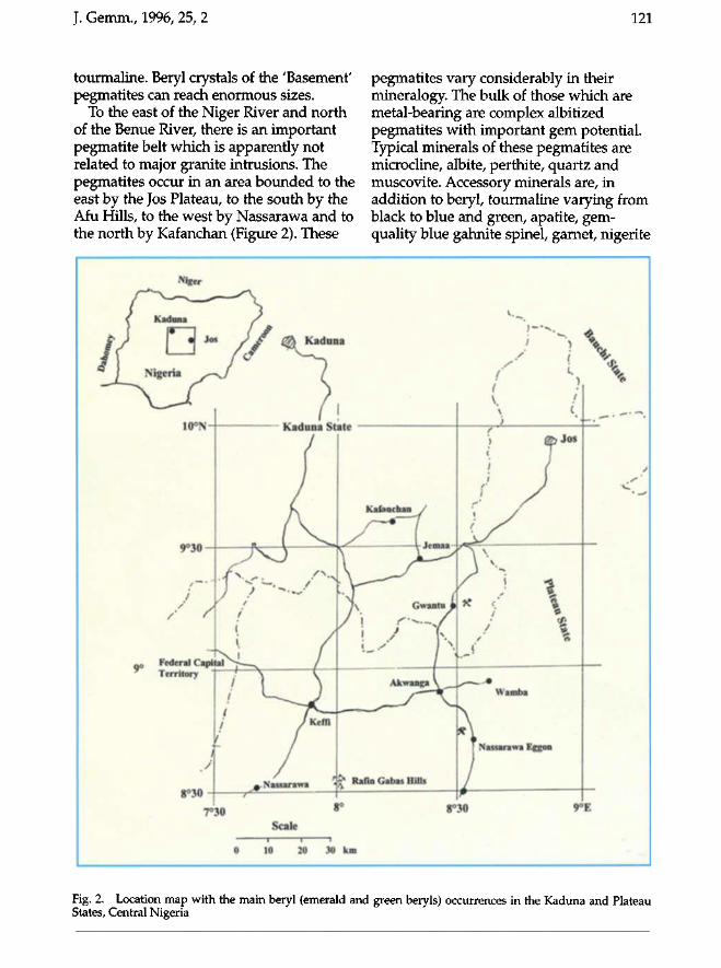
Emeralds and green beryls in the Plateau and Kaduna States of central Nigeria are much less common than their blue counterpart, aquamarine. They occur in rocks of two distinct ages. The older occurrences are usually pegmatites, whereas the younger are in the 'roof zones of granites. Although some beryls from both kinds of sources have an 'emerald colour', most are rather pale green and many dealers do not accept them as emeralds, classifying them as green beryl. Arps and Zwaan (1995) gave the source of the central Nigerian emeralds as east of Gwantu (south east Kaduna State) and north west of Nassarawa Eggon (Plateau State).
© Gemmological Association and Gem Testing Laboratory of Great Britain ISSN: 1355-4565
118 J. Gemm., 1996, 25, 2
This paper presents the first comprehensive study of the mineralogical and gemmological characteristics of Nigerian emeralds and green beryls and establishes a genetic model for the occurrences of these gemstones in central Nigeria.
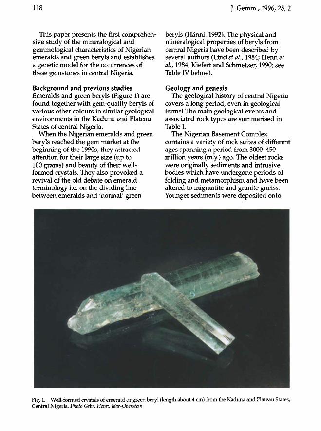
Background and previous studies Emeralds and green beryls (Figure 1) are found together with gem-quality beryls of various other colours in similar geological environments in the Kaduna and Plateau States of central Nigeria.
When the Nigerian emeralds and green beryls reached the gem market at the beginning of the 1990s, they attracted attention for their large size (up to 100 grams) and beauty of their well-formed crystals. They also provoked a revival of the old debate on emerald terminology i.e. on the dividing line between emeralds and 'normal' green
beryls (Hänni, 1992). The physical and mineralogical properties of beryls from central Nigeria have been described by several authors (Lind et ah, 1984; Henn et ah, 1984; Kiefert and Schmetzer, 1990; see Table IV below).
Geology and genesis The geological history of central Nigeria
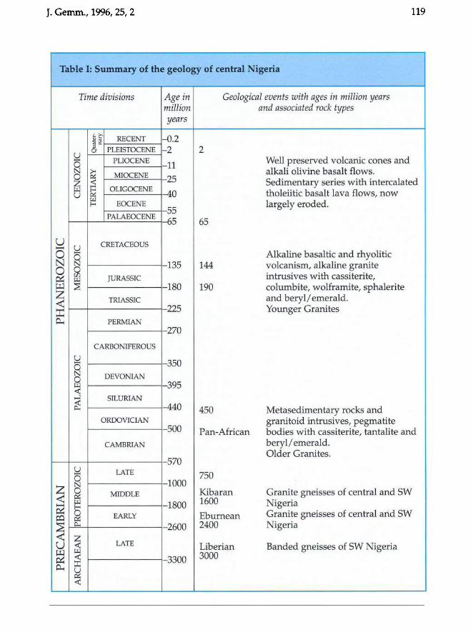
covers a long period, even in geological terms! The main geological events and associated rock types are summarised in Table I.
The Nigerian Basement Complex contains a variety of rock suites of different ages spanning a period from 3000-450 million years (m.y.) ago. The oldest rocks were originally sediments and intrusive bodies which have undergone periods of folding and metamorphism and have been altered to migmatite and granite gneiss. Younger sediments were deposited onto
Fig. 1. Well-formed crystals of emerald or green beryl (length about 4 cm) from the Kaduna and Plateau States, Central Nigeria. Photo Gebr. Henn, Idar-Oberstein
J. Gemm., 1996,25,2 119
Table I: Summary of the geology of central Nigeria
Time divisions Age in Geological events with ages in million years million and associated rock types years
~c 2~ ~ c
RECENT - 0.2 ~
PLEISTOCENE - 2 2 a U
PLIOCENE Well preserved volcanic cones and ...... 0 11 N G; MIOCENE alkali olivine basalt flows. ~ ~
25 Sedimentary series with intercalated ~ OLIGOCENE -40 tholeiitic basalt lava flows, now U
~ EOCENE largely eroded. f-<
55 PALAEOCENE 65 65
U S::l
CRETACEOUS ~
0 0 Alkaline basaltic and rhyolitic N N 135 144 volcanism, alkaline granite 0 0
CIl intrusives with cassiterite, ~ ~ JURASSIC ~ ~ 180 190 columbite, wolframite, sphalerite Z TRIASSIC and beryl! emerald. -< ~ 225 Younger Granites p.. PERMIAN
270
CARBONIFEROUS
u 350 ...... 0 N DEVONIAN 0 ~ 395 <C ...J SILURIAN ~ -440 450 Metasedimentary rocks and
ORDOVICIAN granitoid intrusives, pegmatite 500 Pan-African bodies with cassiterite, tantalite and
CAMBRIAN beryl! emerald. Older Granites.
570 S::l LATE 750 0
Z N 1000 0 MIDDLE Kibaran Granite gneisses of central and SW
-< ~ ~ 1800 1600 Nigeria ~ E-<
~ ~ EARLY Eburnean Granite gneisses of central arid SW o::l ~ 0.. 2600 2400 Nigeria
-< Z LATE U <C Liberiqn Banded gneisses of SW Nigeria
~ ~
~ <C 3300 3000 :r: p.. u ~ <C
120 J. Gemm., 1996, 25, 2
the Basement and folded together with this granitized basement during the Pan-African orogeny from 750 to 450 m.y. ago, to form long north-south oriented linear schist belts of low grade metasedimentary rocks.
During the late Pan-African orogeny, from around 600 to 450 m.y. ago, there was a phase of granitoid magmatism. These granitoids were intruded into both the Basement Complex and the younger metasedimentary cover. The granitic rocks associated with the Pan-African orogeny generally have contrasting petrological and chemical compositions compared with the later Jurassic (Mesozoic) suite of alkaline ring complexes and are therefore called 'Older' Granites, in contrast to the 'Younger' Mesozoic Granites. The closing stages of the orogeny from 650-450 m.y. ago were marked by cooling, uplift and fracturing, by the eruption of volcanic rocks and the formation of pegmatitic lenses and dykes.
Within the Basement Complex is a zone in which this suite of pegmatites cut Old Eburnean rocks and early Pan-African metamorphic and igneous assemblages. The mineralized pegmatites are widely, but unevenly distributed within a broad zone, extending from the Ife area towards the Younger Granite province. It appears that the mineralized pegmatites occur within the north-south oriented younger metasedimentary belts composed of biotite schists and amphibolites. The pegmatites occur as dykes and sheets of varying dimensions - the dykes range from a few centimetres to tens of metres in width, with strike lengths of up to 2 km. The pegmatite sheets can have a considerable thickness of up to 40 m, and have been of economic interest because they contain cassiterite and columbo-tantalite. However the sheet-like pegmatites are of greater importance for the gems they contain. Many of the well mineralized pegmatites are characterized by pinch and swell structures. The swellings are generally
very albitized and frequently correspond with centres of rich ore mineralization.
The younger granites of Nigeria, emplaced during the period 190 to 144 m.y. ago, form ring complexes typically 2-25 km in diameter and are composed of 95 per cent acid rocks. These Mesozoic ring complexes of Nigeria form part of a larger province of alkaline magmatism. They occur in a zone 200 km wide and 1600 km long, extending from northern Niger to south central Nigeria. This zone is related to the breakup of the Gondwana super continent during the Mesozoic era. According to Dickin et al. (1991), the ring complexes of the Jos Plateau are the product of mantle-derived magmas which have suffered a significant amount of crustal contamination during their ascent and differentiation in the crustal basement. There are three main granite types: hornblende biotite granite; biotite granite and alkali granite with Na-Fe pyroxenes and amphiboles.
In the ring complexes, a series of hydrothermal alteration processes with related mineralization can be recognized (Kinnaird, 1979). Early sodic metasomatism (albitization) may have modified all the granite types. The effects of the later processes beginning with potassic metasomatism are only well documented for the biotite granites. Each hydrothermal process is characterized by a distinct assemblage of silicate minerals and there is a clearly defined sequence of ore deposition associated with each of the hydrothermal processes. It is with albitization processes that the gem varieties of beryl are associated. The temperature interval of the emerald and green beryl crystallization is about 500-400 °C.
Beryl-bearing basement pegmatites Pegmatites are common throughout the Kabba Province and, in addition to quartz and microcline, contain both biotite and muscovite and in many cases considerable amounts of albite with beryl and
J. Gemm., 1996,25, 2
tourmaline. Beryl crystals of the 'Basement' pegmatites can reach enormous sizes.
To the east of the Niger River and north of the Benue River, there is an important pegmatite belt which is apparently not related to major granite intrusions. The pegmatites occur in an area bounded to the east by the Jos Plateau, to the south by the Afu Hills, to the west by Nassarawa and to the north by Kafanchan (Figure 2). These
I ,
I /.
/
9"
...
121
pegmatites vary considerably in their mineralogy. The bulk of those which are metal-bearing are complex albitized pegmatites with important gem potential. Typical minerals of these pegmatites are microcline, albite, perthite, quartz and muscovite. Accessory minerals are, in addition to beryl, tourmaline varying from black to blue and green, apatite, gemquality blue gahnite spinel, gamet, nigerite
~.-/ --
~ \ ~~/ / I
/ ~ ~I.: { l (I
(
\. I
'. i -.- -( I ( I ~ , .-" , .... ; . ..,
.,.
.. , t .' ( '\ i
~ .I ~ ..
·.mba
Fig. 2. Location map with the main beryl (emerald and green beryls) occurrences in the Kaduna and Plateau States, Central Nigeria
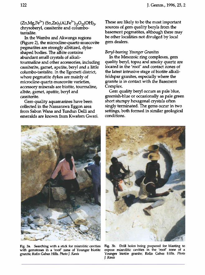
122
(Zn,Mg/Fe2+)(Sn,Zn)2(Al,Fe3+)12022(OH)2/
chrysoberyl, cassiterite and columbo-tantalite.
In the Wamba and Akwanga regions (Figure 2), the microcline-quartz-muscovite pegmatites are strongly albitized, dyke-shaped bodies. The albite contains abundant small crystals of alkali-tourmaline and other accessories, including cassiterite, garnet, apatite, beryl and a little columbo-tantalite. In the Egometi district, where pegmatite dykes are mainly of microcline-quartz-muscovite varieties, accessory minerals are biotite, tourmaline, albite, garnet, apatite, beryl and cassiterite.
Gem-quality aquamarines have been collected in the Nassarawa Eggon area from Sabon Wana and Tundun Delli and emeralds are known from Kwafam Gwari.
Fig. 3a. Searching with a stick for miarolitic cavities with gemstones in a 'roof zone of Younger biotite granite; Rafin Gabas Hills. Photo ]. Kanis
J. Gemm., 1996, 25, 2
These are likely to be the most important sources of gem quality beryls from the basement pegmatites, although there may be other localities not divulged by local gem dealers.
Beryl-bearing Younger Granites In the Mesozoic ring complexes, gem
quality beryl, topaz and smoky quartz are located in the 'roof and contact zones of the latest intrusive stage of biotite alkali-feldspar granites, especially where the granite is in contact with the Basement Complex.
Gem quality beryl occurs as pale blue, greenish-blue or occasionally as pale green short stumpy hexagonal crystals often singly terminated. The gems occur in two settings, both formed in similar geological conditions.
Fig. 3b. Drill holes being prepared for blasting to expose miarolitic cavities in the 'roof zone of a Younger biotite granite; Rafin Gabas Hills. Photo J. Kanis
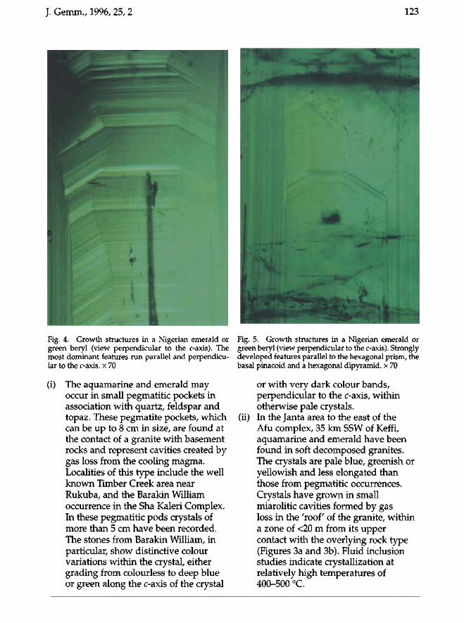
J. Gemm., 1996, 25, 2 123
Fig. 4. Growth structures in a Nigerian emerald or green beryl (view perpendicular to the c-axis). The most dominant features run parallel and perpendicular to the c-axis. x 70
(i) The aquamarine and emerald may occur in small pegmatitic pockets in association with quartz, feldspar and topaz. These pegmatite pockets, which can be up to 8 cm in size, are found at the contact of a granite with basement rocks and represent cavities created by gas loss from the cooling magma. Localities of this type include the well known Timber Creek area near Rukuba, and the Barakin William occurrence in the Sha Kaleri Complex. In these pegmatitic pods crystals of more than 5 cm have been recorded. The stones from Barakin William, in particular, show distinctive colour variations within the crystal, either grading from colourless to deep blue or green along the c-axis of the crystal
Fig. 5. Growth structures in a Nigerian emerald or green beryl (view perpendicular to the c-axis). Strongly developed features parallel to the hexagonal prism, the basal pinacoid and a hexagonal dipyramid. x 70
or with very dark colour bands, perpendicular to the c-axis, within otherwise pale crystals,
(ii) In the Janta area to the east of the Afu complex, 35 km SSW of Keffi, aquamarine and emerald have been found in soft decomposed granites. The crystals are pale blue, greenish or yellowish and less elongated than those from pegmatitic occurrences. Crystals have grown in small miarolitic cavities formed by gas loss in the 'roof of the granite, within a zone of <20 m from its upper contact with the overlying rock type (Figures 3a and 3b). Fluid inclusion studies indicate crystallization at relatively high temperatures of 400-500 °C.
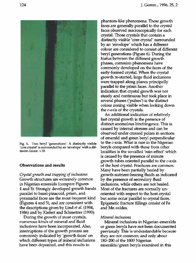
124 J. Gemm., 1996, 25, 2
Fig. 6. Two beryl 'generations'. A distinctly visible 'core crystal' is surrounded by an 'envelope' with a different colour, x 35
Observations and results
Crystal growth and trapping of inclusions Growth structures are extremely common in Nigerian emeralds (compare Figures 4 and 5). Strongly developed growth bands parallel to basal-pinacoid, prism, and pyramidal faces are the most frequent kind (Figures 4 and 5), and are consistent with the descriptions given by Lind et al. (1984, 1986) and by Kiefert and Schmetzer (1990).
During the growth of most crystals, numerous kinds of mineral and fluid inclusions have been incorporated. Also, interruptions of the growth process are commonly indicated by 'growth faces' on which different types of mineral inclusions have been deposited, and this results in
phantom-like phenomena. These growth faces are generally parallel to the crystal faces observed macroscopically for each crystal. Those crystals that contain a distinctly visible 'core crystal' surrounded by an 'envelope' which has a different colour are considered to consist of different beryl generations (Figure 6). During the hiatus between the different growth phases, corrosion phenomena have commonly developed on the faces of the early-formed crystal. When the crystal growth re-started, large fluid inclusions were trapped along planes principally parallel to the prism faces. Another indication that crystal growth was not steady and continuous but took place in several phases ('pulses') is the distinct colour zoning visible when looking down the c-axis of the crystals.
An additional indication of relatively fast crystal growth is the presence of distinct anomalous birefringence. This is caused by internal stresses and can be observed under crossed polars in sections of emerald and green beryl perpendicular to the c-axis. What is rare in the Nigerian beryls compared with those from other localities is the so-called 'rain-effect' which is caused by the presence of minute growth tubes oriented parallel to the c-axis of the host crystal. Fractures are common. Many have been partially healed by growth nutrient-bearing fluids as indicated by the presence of secondary fluid inclusions, while others are not healed. Most of the fractures are normally un-oriented with respect to the host crystal but some occur parallel to crystal faces. Epigenetic fracture fillings consist of Fe and Mn oxides.
Mineral inclusions Mineral inclusions in Nigerian emeralds
or green beryls have not been documented previously. This is understandable because they are not common; and only about 180-200 of the 1000 Nigerian emeralds/green beryls examined in this
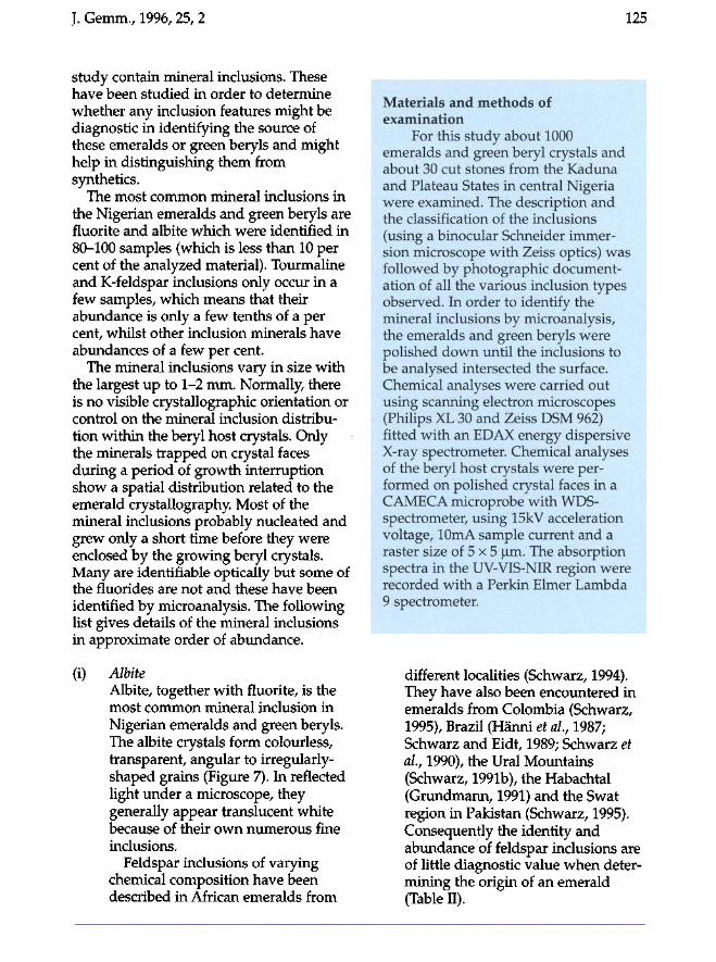
J. Gemm., 1996, 25, 2 125
study contain mineral inclusions. These have been studied in order to determine whether any inclusion features might be diagnostic in identifying the source of these emeralds or green beryls and might help in distinguishing them from synthetics.
The most common mineral inclusions in the Nigerian emeralds and green beryls are fluorite and albite which were identified in 80-100 samples (which is less than 10 per cent of the analyzed material). Tourmaline and K-feldspar inclusions only occur in a few samples, which means that their abundance is only a few tenths of a per cent, whilst other inclusion minerals have abundances of a few per cent.
The mineral inclusions vary in size with the largest up to 1-2 mm. Normally, there is no visible crystallographic orientation or control on the mineral inclusion distribution within the beryl host crystals. Only the minerals trapped on crystal faces during a period of growth interruption show a spatial distribution related to the emerald crystallography. Most of the mineral inclusions probably nucleated and grew only a short time before they were enclosed by the growing beryl crystals. Many are identifiable optically but some of the fluorides are not and these have been identified by microanalysis. The following list gives details of the mineral inclusions in approximate order of abundance.
(i) Albite Albite, together with fluorite, is the most common mineral inclusion in Nigerian emeralds and green beryls. The albite crystals form colourless, transparent, angular to irregularly-shaped grains (Figure 7). In reflected light under a microscope, they generally appear translucent white because of their own numerous fine inclusions.
Feldspar inclusions of varying chemical composition have been described in African emeralds from
Materials and methods of examination
For this study about 1000 emeralds and green beryl crystals and about 30 cut stones from the Kaduna and Plateau States in central Nigeria were examined. The description and the classification of the inclusions (using a binocular Schneider immersion microscope with Zeiss optics) was followed by photographic documentation of all the various inclusion types observed. In order to identify the mineral inclusions by microanalysis, the emeralds and green beryls were polished down until the inclusions to be analysed intersected the surface. Chemical analyses were carried out using scanning electron microscopes (Philips XL 30 and Zeiss DSM 962) fitted with an EDAX energy dispersive X-ray spectrometer. Chemical analyses of the beryl host crystals were performed on polished crystal faces in a CAMECA microprobe with WDS-spectrometer, using 15kV acceleration voltage, 10mA sample current and a raster size of 5 x 5 |im. The absorption spectra in the UV-VIS-NIR region were recorded with a Perkin Elmer Lambda 9 spectrometer.
different localities (Schwarz, 1994). They have also been encountered in emeralds from Colombia (Schwarz, 1995), Brazil (Hänni et al, 1987; Schwarz and Eidt, 1989; Schwarz et al, 1990), the Ural Mountains (Schwarz, 1991b), the Habachtal (Grundmann, 1991) and the Swat region in Pakistan (Schwarz, 1995). Consequently the identity and abundance of feldspar inclusions are of little diagnostic value when determining the origin of an emerald (Table II).
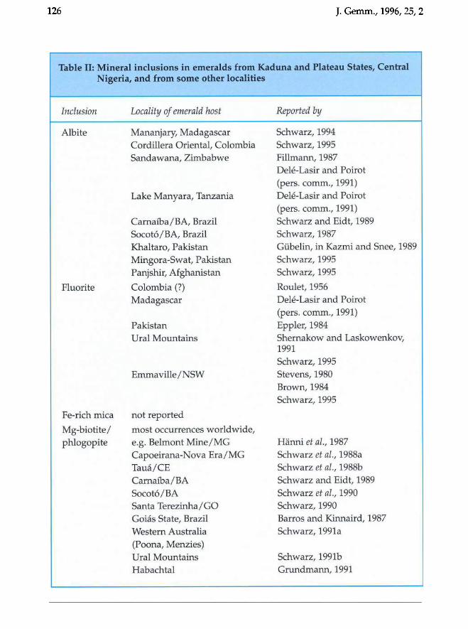
126 J. Gemm., 1996, 25, 2
Table II: Mineral inclusions in emeralds from Kaduna and Plateau States, Central Nigeria, and from some other localities
Inclusion Locality of emerald host Reported by
Albite Mananjary, Madagascar Schwarz, 1994 Cordillera Oriental, Colombia Schwarz, 1995 Sandawana, Zimbabwe FiHmann, 1987
Dele-Lasir and Poirot (pers. comm., 1991)
Lake Manyara, Tanzania Dele-Lasir and Poirot (pers. comm., 1991)
Carnafba/ BA, Brazil Schwarz and Eidt, 1989 Socot6/BA, Brazil Schwarz, 1987 Khaltaro, Pakistan Gubelin, in Kazmi and Snee, 1989 Mingora-Swat, Pakistan Schwarz, 1995 Panjshir, Afghanistan Schwarz, 1995
Fluorite Colombia (7) Roulet, 1956 Madagascar Dele-Lasir and Poirot
(pers. comm., 1991) Pakistan Eppler, 1984 Ural Mountains Shernakow and Laskowenkov,
1991 Schwarz, 1995
Emmaville / NSW Stevens, 1980 Brown, 1984 Schwarz, 1995
Fe-rich mica not reported
Mg-biotite / most occurrences worldwide, phlogopite e.g. Belmont Mine/ MG Hanni et al., 1987
Capoeirana-Nova Era / MG Schwarz et al., 1988a Taua/CE Schwarz et al., 1988b Carnafba/ BA Schwarz and Eidt, 1989 Socot6/ BA Schwarz et al., 1990 Santa Terezinha/ GO Schwarz, 1990 Goias State, Brazil Barros and Kinnaird, 1987 Western Australia Schwarz, 1991a (Poona, Menzies) Ural Mountains Schwarz, 1991b Habachtal Grundmann, 1991
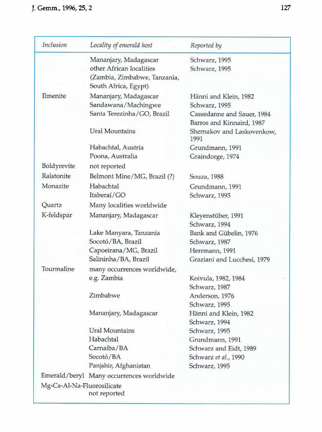
J. Gemm., 1996,25, 2
Inclusion
Ilmenite
Boldyrevite
Ralstonite
Monazite
Quartz
K-feldspar
Tourmaline
Locality of emerald host
Mananjary, Madagascar other African localities (Zambia, Zimbabwe, Tanzania, South Africa, Egypt)
Mananjary, Madagascar Sandawana / Machingwe Santa Terezinha/ GO, Brazil
Ural Mountains
Habachtal, Austria Poona, Australia
not reported
Belmont Mine/ MG, Brazil (?)
Habachtal ItaberaUGO
Many localities worldwide
Mananjary, Madagascar
Lake Manyara, Tanzania Socot6/BA, Brazil Capoeirana/MG, Brazil Salininha / BA, Brazil
many occurrences worldwide, e.g. Zambia
Zimbabwe
Mananjary, Madagascar
Ural Mountains Habachtal Carnaiba/BA Socot6/BA Panjshir, Afghanistan
Emerald/beryl Many occurrences worldwide
Mg-Ca-Al-Na-Fluorosilicate not reported
Reported by
Schwarz, 1995 Schwarz, 1995
Hanni and Klein, 1982 Schwarz, 1995 Cassedanne and Sauer, 1984 Barros and Kinnaird, 1987 Shernakov and Laskovenkow, 1991 Grundmann, 1991 Graindorge, 1974
Souza, 1988
Grundmann, 1991 Schwarz, 1995
Kleyensruber, 1991 Schwarz, 1994 Bank and Gtibelin, 1976 Schwarz, 1987 Herrmann, 1991 Graziani and Lucchesi, 1979
Koivula, 1982, 1984 Schwarz, 1987 Anderson, 1976 Schwarz, 1995 Hanni and Klein, 1982 Schwarz, 1994 Schwarz, 1995 Grundmann, 1991 Schwarz and Eidt, 1989 Schwarz et al., 1990 Schwarz, 1995
127
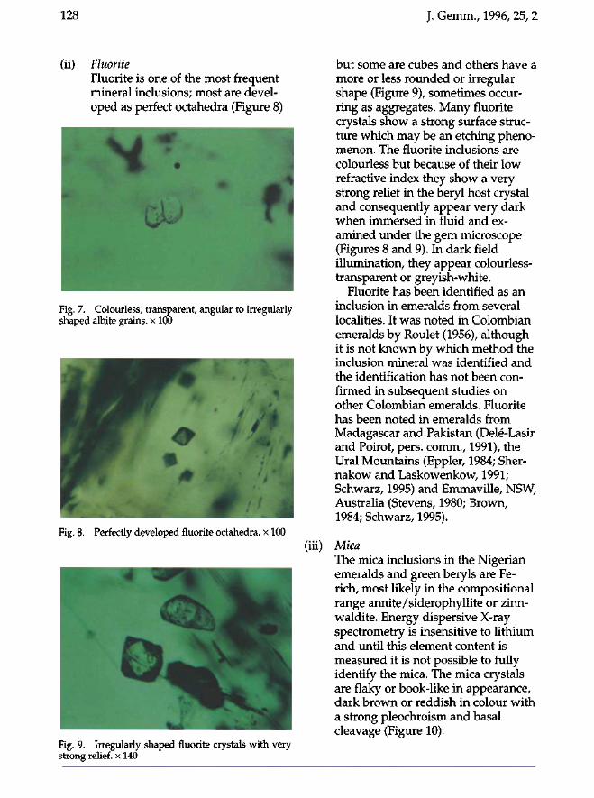
128 J. Gemm., 1996, 25, 2
(ii) Fluorite Fluorite is one of the most frequent mineral inclusions; most are developed as perfect octahedra (Figure 8)
Fig. 7. Colourless, transparent, angular to irregularly shaped albite grains, x 100
Fig. 8. Perfectly developed fluorite octahedra. x 100
Fig. 9. Irregularly shaped fluorite crystals with very strong relief, x 140
but some are cubes and others have a more or less rounded or irregular shape (Figure 9), sometimes occurring as aggregates. Many fluorite crystals show a strong surface structure which may be an etching phenomenon. The fluorite inclusions are colourless but because of their low refractive index they show a very strong relief in the beryl host crystal and consequently appear very dark when immersed in fluid and examined under the gem microscope (Figures 8 and 9). In dark field illumination, they appear colourless-transparent or greyish-white.
Fluorite has been identified as an inclusion in emeralds from several localities. It was noted in Colombian emeralds by Roulet (1956), although it is not known by which method the inclusion mineral was identified and the identification has not been confirmed in subsequent studies on other Colombian emeralds. Fluorite has been noted in emeralds from Madagascar and Pakistan (Delé-Lasir and Poirot, pers. comm., 1991), the Ural Mountains (Eppler, 1984; Sher-nakow and Laskowenkow, 1991; Schwarz, 1995) and Emmaville, NSW, Australia (Stevens, 1980; Brown, 1984; Schwarz, 1995).
(iii) Mica The mica inclusions in the Nigerian emeralds and green beryls are Fe-rich, most likely in the compositional range annite/siderophyllite or zinn-waldite. Energy dispersive X-ray spectrometry is insensitive to lithium and until this element content is measured it is not possible to fully identify the mica. The mica crystals are flaky or book-like in appearance, dark brown or reddish in colour with a strong pleochroism and basal cleavage (Figure 10).
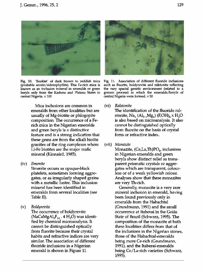
J. Gemm., 1996, 25, 2 129
Fig. 10. 'Booklet7 of dark brown to reddish mica (probable annite/siderophyllite). This Fe-rich mica is known as an inclusion mineral in emeralds or green beryls only from the Kaduna and Plateau States in central Nigeria, x 100
Mica inclusions are common in emeralds from other localities but are usually of Mg-biotite or phlogopite composition. The occurrence of a Fe-rich mica in the Nigerian emeralds and green beryls is a distinctive feature and is a strong indication that these gems are from the alkali biotite granites of the ring complexes where Li-Fe biotites are the major mafic mineral (Kinnaird, 1985).
(iv) llmenite Ilmenite occurs as opaque-black platelets, sometimes forming aggregates, or as irregularly shaped grains with a metallic lustre. This inclusion mineral has been identified in emeralds from several localities (see Table II).
(v) Boldyrevite The occurrence of boldyrevite (NaCaMgAl3F14 . 4 H20) was identified by chemical microanalysis. It cannot be distinguished optically from fluorite because their crystal habits and refractive indices are very similar. The association of different fluoride inclusions in a Nigerian emerald is shown in Figure 11.
Fig. 11. Association of different fluoride inclusions such as fluorite, boldyrevite and ralstonite reflecting the very special genetic environment (related to a greisen process) in which the emeralds/beryls of central Nigeria were formed, x 50
(vi) Ralstonite The identification of the fluoride ralstonite, Nax (Al2_xMgx) (F,OH)6 x H20 is also based on microanalysis. It also cannot be distinguished optically from fluorite on the basis of crystal form or refractive index.
(vii) Monazite Monazite, (Ce,La,Th)P04, inclusions in Nigerian emeralds and green beryls show distinct relief as transparent prismatic crystals or aggregates which are transparent, colourless or of a weak yellowish colour. Analyses show that these monazites are very Th-rich.
Generally, monazite is a very rare mineral inclusion in emerald, having been found previously only in emeralds from the Habachtal (Grundmann, 1991) and the small occurrence at Itaberaî in the Goiâs State of Brazil (Schwarz, 1995). The composition of the monazite at both these localities differs from that of the inclusions in the Nigerian stones, those of the Habachtal-emeralds being more Ce-rich (Grundmann, 1991), and the Itaberai-emeralds being Ce/La-rich varieties (Schwarz, 1995).
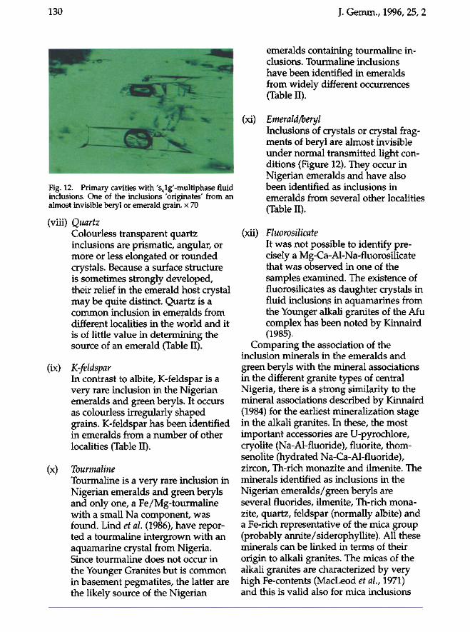
130 J. Gemm., 1996, 25, 2
emeralds containing tourmaline inclusions. Tourmaline inclusions have been identified in emeralds from widely different occurrences (Table II).
(xi) Emerald/beryl Inclusions of crystals or crystal fragments of beryl are almost invisible under normal transmitted light conditions (Figure 12). They occur in Nigerian emeralds and have also been identified as inclusions in emeralds from several other localities (Table II).
Fig. 12. Primary cavities with 'sxlg'-multiphase fluid inclusions. One of the inclusions 'originates' from an almost invisible beryl or emerald grain, x 70
(viii) Quartz Colourless transparent quartz inclusions are prismatic, angular, or more or less elongated or rounded crystals. Because a surface structure is sometimes strongly developed, their relief in the emerald host crystal may be quite distinct. Quartz is a common inclusion in emeralds from different localities in the world and it is of little value in determining the source of an emerald (Table II).
(ix) K-feldspar In contrast to albite, K-feldspar is a very rare inclusion in the Nigerian emeralds and green beryls. It occurs as colourless irregularly shaped grains. K-feldspar has been identified in emeralds from a number of other localities (Table II).
(x) Tourmaline Tourmaline is a very rare inclusion in Nigerian emeralds and green beryls and only one, a Fe/Mg-tourmaline with a small Na component, was found. Lind et al (1986), have reported a tourmaline intergrown with an aquamarine crystal from Nigeria. Since tourmaline does not occur in the Younger Granites but is common in basement pegmatites, the latter are the likely source of the Nigerian
(xii) Fluorosilicate It was not possible to identify precisely a Mg-Ca-Al-Na-fluorosilicate that was observed in one of the samples examined. The existence of fluorosilicates as daughter crystals in fluid inclusions in aquamarines from the Younger alkali granites of the Afu complex has been noted by Kinnaird (1985).
Comparing the association of the inclusion minerals in the emeralds and green beryls with the mineral associations in the different granite types of central Nigeria, there is a strong similarity to the mineral associations described by Kinnaird (1984) for the earliest mineralization stage in the alkali granites. In these, the most important accessories are U-pyrochlore, cryolite (Na-Al-fluoride), fluorite, thom-senolite (hydrated Na-Ca-Al-fluoride), zircon, Th-rich monazite and ilmenite. The minerals identified as inclusions in the Nigerian emeralds/green beryls are several fluorides, ilmenite, Th-rich monazite, quartz, feldspar (normally albite) and a Fe-rich representative of the mica group (probably annite/siderophyllite). All these minerals can be linked in terms of their origin to alkali granites. The micas of the alkali granites are characterized by very high Fe-contents (MacLeod et al., 1971) and this is valid also for mica inclusions
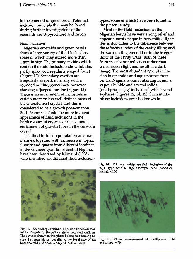
J. Gemm., 1996, 25, 2 131
in the emerald or green beryl. Potential inclusion minerals that may be found during further investigations of the emeralds are U-pyrochlore and zircon.
Fluid inclusions Nigerian emeralds and green beryls
show a large variety of fluid inclusions, some of which may exceed more than 1 mm in size. The primary cavities which contain the fluid inclusions show tubular, partly spiky, or irregularly shaped forms (Figure 12). Secondary cavities are irregularly shaped, normally with a rounded outline, sometimes, however, showing a 'jagged' outline (Figure 13). There is an enrichment of inclusions in certain more or less well-defined areas of the emerald host crystal, and this is considered to be a growth phenomenon. Such features include the more frequent appearance of fluid inclusions in the border zones of crystals or the common enrichment of growth tubes in the core of a crystal.
The fluid inclusion population of aquamarines, together with inclusions in topaz, fluorite and quartz from different localities in the younger granites of central Nigeria, have been described by Kinnaird (1985) who identified six different fluid inclusion-
Fig. 13. Secondary cavities of Nigerian beryls are normally irregularly shaped or show rounded outlines. The cavities shown in this photo belong to a healing fissure that runs almost parallel to the basal face of the host emerald and show a 'jagged' outline, x 50
types, some of which have been found in the present study.
Most of the fluid inclusions in the Nigerian beryls have very strong relief and appear almost opaque in transmitted light; this is due either to the difference between the refractive index of the cavity filling and the surrounding emerald or to the irregularity of the cavity walls. Both of these features enhance reflection rather than transmission light and result in a dark image. The most abundant type of inclusion in emeralds and aquamarines from central Nigeria is one containing liquid, a vapour bubble and several solids (multiphase 'sxlg' inclusions1 with several s-phases; Figures 12,14,15). Such multiphase inclusions are also known in
Fig. 14. Primary multiphase fluid inclusion of the 'sxlg' type with a large isotropic cube (probably halite), x 100
Fig. 15. Planar arrangement of multiphase fluid inclusions, x 70
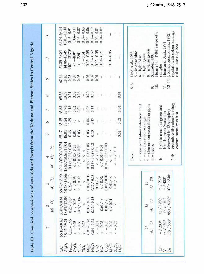
Tab
le I
II:
Che
mic
al c
omp
osit
ion
s of
em
eral
ds
and
ber
yls
from
the
Kad
una
and
Pla
teau
Sta
tes
in C
entr
al N
iger
ia
1 2
3 4
5 6
7 8
9 10
11
(a
) (b
) (a
) (b
) (a
) (b
) (c
)
Si0
2 66
.10-
68.4
0 68
.82/
68.6
1 68
.87/
68.3
9 69
.11/
69.0
9/68
.74
65.1
7 65
.45
65.8
3 65
.53
67.3
1 61
.35-
68.8
1 65
.74-
67.5
4
AIP
3 18
.02-
19. 2
1 18
.61/
17.8
8 18
.48/
17.8
6 18
.57/
18.6
3/18
.04
18.8
4 18
.24
18.7
0 18
.39
18.4
0 14
.86-
18.4
9 18
.01-
18.1
8 F
eOto
t 0.
11-
0.95
0.
28/
0.87
0.
23/
1.10
0.
14/
0.35
/0.8
9
0.46
0.
89
1.13
0.
31
0.25
94
0* -
1217
0*
0.0
6-
0.91
C
r 20
3 <
-0.
05
<
/0.
06
<
/0.
06
< /
0.0
2/ 0
.21
0.01
<
0.
02
0.08
0.
07
< -
480*
0.
06-
0.11
V
203
< -
·0.0
6 0.
02/
0.06
<
/
0.09
<
/ 0
.07
/ 0.
06
0.03
<
0.
01
0.06
0.
03
< -
280*
0.
05-
0.07
T
i02
< -
0.02
<
<
<
<
<
<
-0.
04
0.07
-0 .
63
MgO
0.
01
-0.
20
0.0
2/0
.06
0
.03
/0.0
6
0.0
8/0
.03
/0.0
5 0.
03
0.04
0.
02
0.20
0.
03
0.03
-0.
05
0.04
-0.
06
Na 2
0 0.
04-
0.33
0.
12
/0.1
5
0.1
3/0
.16
0.1
3/0
.06
/0.1
2
0.10
0.
17
0.14
0.
15
0.07
0.
08-
0.57
0.
09-
0.12
C
aO
< -
0.01
<
0.
01
/ <
<
<
/ 0
.01
0.01
0.
02-
0.05
0.
00-
0.01
K
p
< -
0.05
0.
01/
<
< /
0.0
2 <
/ 0
.01
/ 0.
05
0.04
-0.
21
0.01
-0.
02
CS
p
< -
0.05
0.
01/
<
0.0
2/0
.02
0
.01
/0.0
2/0
.03
M
nO
<
-0.
02
< /
0.0
1 0.
01/
<
<
<
<
0.01
-0.
05
SC20
3 <
-0.
03
<
0.01
/ <
<
<
/ 0
.01
Lip
0.
02
0.02
0.
02
0.01
Key
: 5-
8:
Lin
d et
ai.,
198
6;
5 =
int
ense
blu
e <
= c
onte
nts
belo
w d
etec
tion
lim
it
6 =
lig
ht b
lue
12
13
14
-=
no
t an
alyz
ed o
r ra
nge
7 =
lig
ht g
reen
(a)
(b)
(a)
(b)
(a)
(b)
* =
ele
men
t con
cent
rati
on i
n p
pm
8
= i
nten
se g
reen
tr
= t
race
s 9:
S
chra
der,
198
7 10
: H
enn
et
ai.,
198
4; r
ange
of
6 C
r -
/ 29
0*
tr. /
125
0*
tr. /
23
0*
1:
li
ght
to m
ediu
m g
reen
an
d
sam
ples
V
tr.
/4
90
*
tr. /4
90*
-/
430*
bl
uish
-gre
en (
vari
atio
n 11
: H
enn
an
d B
ank,
199
1 Fe
1
78
/58
8*
85
0 /
6380
* 10
90/
6540
* ob
serv
ed i
n 13
sam
ples
) 12
-14:
J. Z
ang,
per
s. c
omm
., 1
992;
2-
4:
crys
tals
wit
h c
olou
r zo
ning
; cr
ysta
ls w
ith
col
our
zoni
ng;
colo
ur i
nten
sity
c>
b>a
colo
ur i
nten
sity
b>
a
......
W
N
';-<
CJ J ...... '" '" ~ ~
N
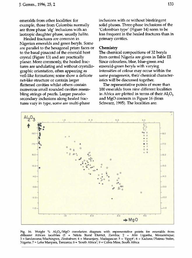
J. Gemm., 1996, 25, 2 133
emeralds from other localities: for example, those from Colombia normally are three phase 'slg' inclusions with an isotropic daughter phase, usually halite.
Healed fractures are common in Nigerian emeralds and green beryls. Some are parallel to the hexagonal prism faces or to the basal pinacoid of the emerald host crystal (Figure 13) and are practically planar. More commonly, the healed fractures are undulating and without crystallo-graphic orientation, often appearing as veil-like formations; some show a delicate net-like structure or contain larger flattened cavities whilst others contain numerous small rounded cavities resembling strings of pearls. Larger pseudo-secondary inclusions along healed fractures vary in type, some are multi-phase
inclusions with or without biréfringent solid phases. Three-phase inclusions of the 'Colombian type' (Figure 14) seem to be less frequent in the healed fractures than in primary cavities.
Chemistry The chemical compositions of 32 beryls from central Nigeria are given in Table III. Since colourless, blue, blue-green and emerald-green beryls with varying intensities of colour may occur within the same paragenesis, their chemical characteristics will be discussed together.
The representative points of more than 100 emeralds from nine different localities in Africa are plotted in terms of their A1203
and MgO contents in Figure 16 (from Schwarz, 1995). The localities are:
Al203
• J 18.0 -1
17.0 —
16.0 —
15.0 —
1 4 . 0 -
13.0—
-12.0 —
. 1 1 . 0 -
g: 1 1
m
m
1.0 2 . 0
* *
***** £ O * ,
A % # A V
• <> *•
i!o ' ' ' z'.o
O
•
> v
* > • A
> t
3 . 0 4 0 i i 1 t i i i I i i
1} ! TA 2
O 3
• 4
* 5
• 6 • 7
O 8
* 9
O
O
Ô o°
o
•
' ' 3'.o • ' ' ' 4!o ' '
-•MgO
n 1
- 1 8 . 0
- 1 7 . 0
- 1 6 . 0
- 1 5 . 0
- 1 4 . 0
M-13.0
h
Li 2.0
1—11.0
Fig. 16. Weight % Al203/MgO correlation diagram with representative points for emeralds from different African localities (1 = Ndola Rural District, Zambia; 2 = Alto Ligonha, Mozambique; 3 = Sandawana/Machingwe, Zimbabwe; 4 = Mananjary, Madagascar; 5 = 'Egypt'; 6 = Kaduna/Plateau States, Nigeria; 7 = Lake Manyara, Tanzania; 8 = 'South Africa'; 9 = Cobra Mine, South Africa
134 J. Gemm., 1996, 25, 2
(1) Ndola Rural District (Kafube Belt), Zambia
(2) Alto Ligonha, Mozambique (including three pegmatite beryls)
(3) Sandawana-Machingwe mining area, Zimbabwe
(4) Mananjary mining region, Madagascar (5) Eastern desert, Egypt (6) Kaduna-Plateau State, Central Nigeria (7) Lake Manyara, Tanzania (8) 'South Africa' (exact locality not
known) (9) Cobra Mine, Transvaal, South Africa.
The emerald occurrences in Zambia, Zimbabwe, Mozambique, Madagascar, Egypt and South Africa all belong to 'schist-type' deposits; that is, they are associated with various schist rocks containing biotite or phlogopite, actinolite or tremolite and variable quantities of talc, chlorite or carbonates, probably derived essentially from a mafic-ultramafic proto-lith. Because of this environment, the MgO concentrations of emeralds from these rocks are generally between 1 and 3 wt%. The negative, approximately linear correlation between A1203 and MgO indicates that magnesium enters the beryl structure principally by substituting for aluminium. Such a substitution of Al3+ by bivalent ions such as Mg and/or Fe requires a coupled substitution to maintain a neutral charge balance, normally with Na in the following way:
A13+'VI = Mg2+'VI + Na1+ (channel)
or A 1 3 + ' V I
= pe2+,VI _ j s j a l + (channel)
In contrast to the emeralds from schist deposits, the Nigerian emeralds and green beryls are characterized by extremely low magnesium and sodium contents. This suggests that they are largely from the alkali granites of the Younger Granite suite which have extremely low contents of Mg. The suggestion made earlier that the majority of the Nigerian emeralds and green beryls containing fluorite and Fe-
rich mica are probably of Younger Granite origin is consistent with this low Mg content.
Ferromagnesian minerals in the Nigerian alkali granites are always Fe or Na-Fe end members, so these elements must have been available during the albitization stage of alteration. Nevertheless, despite the availability of Na, the Na-contents of the Nigerian beryls are consistently low (see Table III and Figure 17). This is in accordance with observations on emeralds from other localities, especially in the Cordillera Oriental, Colombia where Schwarz (1995) has shown that Na is incorporated in the channels of the emerald structure only if there is some need for charge compensation caused by the entrance of bivalent cations (Me2+) in the Al3+ positions. If there is no substitution of Al3+ by Me2+, the emeralds show only low Na-contents, even if the mineralizing fluids are sodium-rich.
The iron-concentrations of the Nigerian emeralds and beryls show a strong variation, reaching FeOtot values of up to 1.2 wt% (Figure 17, from Schwarz, 1995) and the high contents are consistent with iron being available during the beryl growth. As is shown by the absorption spectra (see below), at least part of the iron of the Nigerian emeralds or green beryls is present as Fe3+ (substituting for Al3+) and therefore the beryl needs no sodium for charge compensation. The correlation diagram (Figure 17) shows that there is no correlation between the sodium and the iron contents for these emeralds.
In some of the analyzed samples that show colour-zoning, the concentration of the chromophore elements, especially iron, increases with the intensity of the bluish-green colour. Such colour variation is not related directly to the elements Mg and Na although they may be involved with chromophores in a charge-balancing role.
Comparing the values of optical data of emeralds from the schist-type deposits with those of the Nigerian emeralds and green beryls, the latter are low. The same is
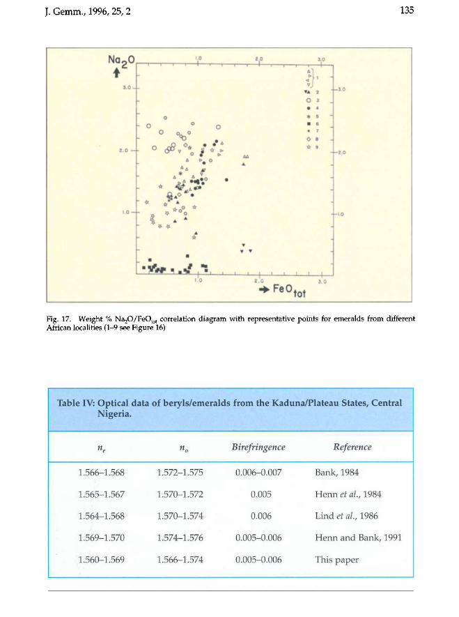
J. Gemm., 1996, 25, 2 135
Na-0 *
3 . 0 -
2 . 0 —
1.0 —
1.0
— , ... , . 1
-
o - o ° o
O o„ o *>
A t> 0
• * * * V
- ^* * . A #
r * f ^ • % i • i 1
1.0
2j0 3.0
A '
> <
— t
VJ • A 2 O 3 _ • 4
* 5
• 6 • 7
O 8 * 9
A A
A
-
•
• •
l I 1 1 1 t 1 1 1 • ' ~ ' T —
2 . 0 3 . 0
- 3 . 0
- 2 . 0
- 1 . 0
"•Fe0tot
Fig. 17. Weight % Na20/FeOtot correlation diagram with representative points for emeralds from different African localities (1-9 see Figure 16)
Table IV: Optical Nigeria.
ne
1.566-1.568
1.565-1.567
1.564-1.568
1.569-1.570
1.560-1.569
data of beryls/emeralds
« 0
1.572-1.575
1.570-1.572
1.570-1.574
1.574-1.576
1.566-1.574
> from the Kaduna/Plateau States, Central
Birefringence
0.006-0.007
0.005
0.006
0.005-0.006
0.005-0.006
Reference
Bank, 1984
Henn et al, 1984
Lind et al, 1986
Henn and Bank, 1991
This paper
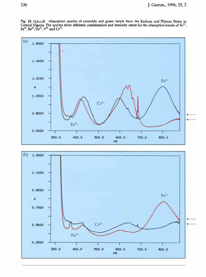
136 J. Gemm., 1996, 25, 2
Fig. 18. (a,b,c,d) Absorption spectra of emeralds and green beryls from the Kaduna and Plateau States in Central Nigeria. The spectra show different combinations and intensity ratios for the absorption bands of Fe2+, Fe3+, Fe2+/Fe3+, V3+ and Cr3+.
' * ' 1.6000
1.4000
i.aooo
A
1.0000
0 .8000
0 .6000 """' I
3 0 0 . C
i *
I A \JN Fe3*
i
) 4 0 0 . 0
k Cr3*
\ y \y i
5 0 0 . 0
/A
A / N i
6 0 0 . 0 11«
\ l / 1/ x_ 1
7 0 0 . 0
Fe2+
/ \
f\ \
^ ~
;
1 "" 1
8 0 0 . 0
(Y}) 1 ^ftftfl m«.
1.1000 -
0 .9000 -
A
0 .7000 -
0 .5000 -
ft Qftftft — U . O v v U i
3 0 0 . 0
F e 3+
1
4 0 0 . 0
Cr3*
i
5 0 0 . 0
1
6 0 0 . 0 IUI
I
7 0 0 . 0
Fe2+
t 1 8 0 0 . 0
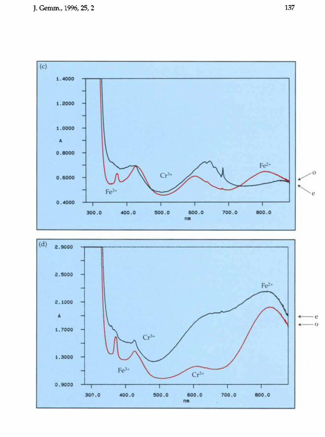
J. Gemm., 1996,25,2 137
(c)
1.4000
1.2000
1.0000
Â
0.8000
0.6000
0.4000
-
Fe3*
i l
\ C l 3 + v̂ \_
1 1
\ Fe2+
i i
300.0 400.0 500.0 600.0 nm
700.0 800.0
(d) 2.9000
3.5000 H
2.1000 -|
A
1.7000 H
1.3000 H
0.9000 T T T 300*0 400.0 500.0 600.0
nm
— I \
700.0 600.0
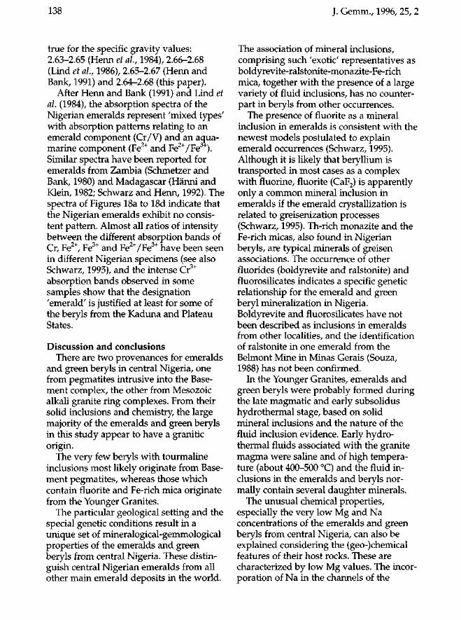
138 J. Gemm., 1996, 25, 2
true for the specific gravity values: 2.63-2.65 (Henn et al, 1984), 2.66-2.68 (Lind et al, 1986), 2.65-2.67 (Henn and Bank, 1991) and 2.64-2.68 (this paper).
After Henn and Bank (1991) and Lind et al (1984), the absorption spectra of the Nigerian emeralds represent 'mixed types' with absorption patterns relating to an emerald component (Cr/V) and an aquamarine component (Fe2+ and Fe2+/Fe3+). Similar spectra have been reported for emeralds from Zambia (Schmetzer and Bank, 1980) and Madagascar (Hänni and Klein, 1982; Schwarz and Henn, 1992). The spectra of Figures 18a to 18d indicate that the Nigerian emeralds exhibit no consistent pattern. Almost all ratios of intensity between the different absorption bands of Cr, Fe2+, Fe3+ and Fe2+/Fe3+ have been seen in different Nigerian specimens (see also Schwarz, 1995), and the intense Cr3+
absorption bands observed in some samples show that the designation 'emerald' is justified at least for some of the beryls from the Kaduna and Plateau States.
Discussion and conclusions There are two provenances for emeralds
and green beryls in central Nigeria, one from pegmatites intrusive into the Basement complex, the other from Mesozoic alkali granite ring complexes. From their solid inclusions and chemistry, the large majority of the emeralds and green beryls in this study appear to have a granitic origin.
The very few beryls with tourmaline inclusions most likely originate from Basement pegmatites, whereas those which contain fluorite and Fe-rich mica originate from the Younger Granites.
The particular geological setting and the special genetic conditions result in a unique set of mineralogical-gemmological properties of the emeralds and green beryls from central Nigeria. These distinguish central Nigerian emeralds from all other main emerald deposits in the world.
The association of mineral inclusions, comprising such 'exotic' representatives as boldyrevite-ralstonite-monazite-Fe-rich mica, together with the presence of a large variety of fluid inclusions, has no counterpart in beryls from other occurrences.
The presence of fluorite as a mineral inclusion in emeralds is consistent with the newest models postulated to explain emerald occurrences (Schwarz, 1995). Although it is likely that beryllium is transported in most cases as a complex with fluorine, fluorite (CaF2) is apparently only a common mineral inclusion in emeralds if the emerald crystallization is related to greisenization processes (Schwarz, 1995). Th-rich monazite and the Fe-rich micas, also found in Nigerian beryls, are typical minerals of greisen associations. The occurrence of other fluorides (boldyrevite and ralstonite) and fluorosilicates indicates a specific genetic relationship for the emerald and green beryl mineralization in Nigeria. Boldyrevite and fluorosilicates have not been described as inclusions in emeralds from other localities, and the identification of ralstonite in one emerald from the Belmont Mine in Minas Gerais (Souza, 1988) has not been confirmed.
In the Younger Granites, emeralds and green beryls were probably formed during the late magmatic and early subsolidus hydrothermal stage, based on solid mineral inclusions and the nature of the fluid inclusion evidence. Early hydro-thermal fluids associated with the granite magma were saline and of high temperature (about 400-500 °C) and the fluid inclusions in the emeralds and beryls normally contain several daughter minerals.
The unusual chemical properties, especially the very low Mg and Na concentrations of the emeralds and green beryls from central Nigeria, can also be explained considering the (geo-)chemical features of their host rocks. These are characterized by low Mg values. The incorporation of Na in the channels of the
J. Gemm., 1996, 25, 2 139
emerald structure does not depend on the availability of this element in the mineralizing fluids. The controlling factor is the need for charge compensation caused by the entrance of bivalent cations. Consequently, the Na-concentration is low because (Mg+Fe)2+ in the emeralds and green beryls is also low. The low Cr and V concentrations of the host rocks is reflected by low contents of these elements in the beryls. The large variation of the Fe-contents in the emeralds and green beryls is due to the possibility of incorporating iron as Fe2+ and/or Fe + in different lattice positions (including channel sites).
Both beryllium and fluorine are enriched in the alkali granites of Nigeria compared to 'average' values in granites worldwide, and both elements may occur in a range of minerals. The source of the chromophore elements, Cr and V, in the Nigerian beryls is problematic because both elements are scarce (0-10 ppm) in Nigerian granites. However, most beryl crystallization occurs close to the 'roof of a granite in the region where granite fluids may react with surrounding country rocks. Thus Cr and V may have been incorporated from the basement schists or younger volcanic rocks both of which have higher Cr and V contents than many of the granites. The model calculations of Kozlowski et al. (1988) for the emerald occurrences of the Cordillera Oriental in Colombia, show that extremely low chromophore contents in the host rocks are sufficient to provide enough Cr and V for the crystallization of significantly coloured green beryls (emeralds). This suggests that it is not necessary to have exceptional enrichment of these elements in the country rocks of the Nigerian beryls.
The optical data for the beryls and emeralds from the Kaduna and Plateau States can be interpreted bearing in mind their chemical characteristics. The 'mixed type7 spectra with almost all ratios of intensity between the different absorption bands of Cr, Fe2+, Fe3+ and Fe2+/Fe3+ are in
accordance with the (crystal-) chemistry of the beryls.
Gemmologists should be able to distinguish between emeralds from central Nigeria and those from other localities. For those Nigerian beryls that contain solid inclusions, the mineral association of albite + fluorides + Fe-rich mica is unique. For those Nigerian beryls which are free of solid inclusions, the inclusion pattern may be similar to that of certain Colombian emeralds. However, the 'classic' fluid inclusions of the Colombian emeralds are 'slg'-three-phase-inclusions with a cubic halite crystal, whereas the emeralds and green beryls from central Nigeria are more commonly 'sxlg'-multiphase inclusions which, in addition, contain one or more (anisotropic) s-phases. Absorption spectra can also aid in origin identification. The 'mixed-type' spectra of the Nigerian beryls are quite different from the spectra of the Colombian emeralds which, as a rule, are free of iron bands (see also Schwarz, 1995).
Acknowledgements G. Becker of Fa. Friedrich August Becker,
Stefan Kehl, F. Gebr. Herrn - all of Idar-Oberstein, Germany, provided samples. Bettina Becker and Dr Joachim Zang of the Department of Gemstone Research, Johannes Gutenberg University, Mainz, did some SEM and microprobe analyses. The studies of D. Schwarz were part of a research project sponsored by the German Research Association (DFG) and carried out at the Department of Gemstone Research, Johannes Gutenberg University of Mainz, Germany.
All photomicrographs are by D. Schwarz.
References Anderson, S.M., 1976. A note on the occurrence of emerald at
Mayfield Farm, Fort Victoria, Rhodesia. Journal of Gemmology, 15(2), 80-2
Arps, C.E.S., and Zwaan, J.C., 1995. Green beryl and emerald from Central Nigeria. Abstracts, 25th International Gemmological Conference, Thailand
Bank, H., 1984. Smaragde aus Nigeria. Gold + Silber, Uhren + Schmuck, 6, 73
140
Bank, H., and Gubelin, E., 1976. Das Smaragd-Alexandritvorkommen von Lake Manyara, Tansania. Zeitschrift der Deutschen Gemmologischen Gesel/schaft, 25(3), 130-47
Barros, J.c., and Kinnaird, J.A, 1987. Inclusoes fluidas em esmeraldas e berilos verdes do Estado de Goias. In: 32' Reuniao da SBPC, Brast1ia, V(I), 643
Brown, G., 1984. Australia's first emeralds. Journal of Gemmology, 19(4), 320-35
Cassedanne, J.P., and Sauer, D.A, 1984. The Santa Terezinha de Goias emerald deposit. Gems & Gemology, 20(1), 4-13
Dickin, AP., Halliday, A.N., and Bowden, P., 1991. A Pb, Sr and Nd isotope study of the basement and Mesozoic ring complexes of the Jos Plateau, Nigeria. Chemical Geology (Isotope Geoscience Section), 94, 23-32
Eppler, W.F., 1984. Praktische Gemmologie. Riihle-DiebenerVerlag, Stuttgart
FiUmann, K, 1987. Gefugekundliche und genetische Aspekte von EinschlUssen in Smaragden ausgewiihlter Lagerstiilten. Diplomarbeit. Fachbereich Geowissenschaften der J 0-
hannes Gutenberg Vniversitat. Mainz Graindorge, J.M., 1974. A gemmological study of emerald
from Poona, Western Australia. Australian Gemmologist, August 1974, 75-80
Graziani, G., and Lucchesi, S., 1979. Einschlusse und Genese eines VanadiumberyUs von Salininha/BA, Brasilien. Zeitschrift der Deutschen Gemmologischen Gesel/schaft, 28(3), 134-45
Grundmann, G., 1991. Smaragd. exira-LAPIS No.1. Christian Weise Verlag, Munich
Hanni, H., 1992. Blue-green emerald from Nigeria (a consideration of terminology). Australian Gemmologist, 18(5), 16-18
Hanni, H.A, and Klein, H.H., 1982. Ein Smaragdvorkommen in Madagaskar. Zeitschrift der Deutschen Gemmologischen Gesel/schaft, 31(1-2), 71-7
Hanni, H.A Schwarz, D., and Fischer, M., 1987. The emeralds of the Belmont Mine, Minas Gerais, Brazil. Journal of Gemmology, 20(7-8), 446-56
Henn, V., and Bank, H., 1991. Aussergewohnliche Smaragde aus Nigeria. Zeitschrift der Deutschen Gemmologischen Gesel/schaft, 40(4),181-7
Henn, V., Hofmann, c., and Schrader, H.W., 1984. Verschiedenfarbige Berylle aus Nigeria. Uhren Juwelen Schmuck, 10/84, 108-10
Herrmann, J., 1991. Geologie, Mineralogie, Geochemie und Genese der Smaragdlagerstatte Capoeirana, Minas Gerais, Brasilien. Diplomarbeit. Institut fur Geowissenschaften, Vniversitat Mainz
Kanis, J., and Harding, R., 1990. Gemstone prospects in Central Nigeria. Journal of Gemmology, 22(4), 195-202
Kazmi, AH., and Snee, L.W., 1989. Emeralds of Pakistan. Van Nostrand Reinhold Co., New York
Kiefert, L., and Schmetzer, K., 1987. Blue and yellow sapphire from Kaduna Province, Nigeria. Journal of Gemmology, 20(7-8),427-42
Kiefert, L., and Schmetzer, K., 1990. Die mikroskopische Bestimmung struktureller Merkmale optisch einachsiger natiirlicher und synthetischer Edelsteine. Goldschmiede und Uhrmacher Zeitung, 1,90, 157-62
Kinnaird, J.A, 1979. Mineralisation associated with the Nigerian Mesozoic ring complexes. Stud. Geol. Salamanca, 14,189-220
J. Gemm., 1996,25,2
Kinnaird, J.A, 1984. Contrasting styles of Sn-Nb-Ta-Zn mineralization in Nigeria. Journal of African Earth Sciences, 2(2), 81-90
Kinnaird, J.A, 1985. Hydrothermal alteration and mineralization of the alkaline anorogenic ring complexes of Nigeria. Journal of African Earth Sciences, 3(1-2), 229-51
Kleyenstiiber, A, 1991. Observation on inclusions in a Madagascar emerald and their possible implications. South African Gemmologist, 5(2), 4-9
Koivula, J., 1982. Tourmaline as an inclusion in Zambian emeralds. Gems & Gemology, 18(4), 225-7
Koivula, J., 1984. Mineral inclusions in Zambian emeralds. Australian Gemmologist, 15(7), 235-9
Kozlowski, A, Metz, P., and Jaramillo, J.H.A, 1988. Emeralds from Somondoco, Colombia: chemical composition, fluid inclusions and origin. Neues Jahrbuch Mineralogie, Abhandlungen, 159(1), 23-49
Lind, Th., Schmetzer, K., and Bank, H., 1984. Schleifwurdige blaue und grune Berylle (Aquamarine und Smaragde) aus Nigeria. Zeitschrift der Deutschen Gemmologischen Gesel/schaft, 33(3-4), 128-38
Lind, Th., Schmetzer, K, and Bank, H., 1986. Blue and green beryls (aquamarine and emeralds) of gem quality from Nigeria. Journal of Gemmology, 20(1), 40-8
MacLeod, W.N., Turner, D.C., and Wright, E.P., 1971. The geology of Jos Plateau, Vol. 1, general geology. Bull. Geol. Survey Nigeria, no. 32
Roulet R., 1956. Metodo para determinacao da procedencia de esmeraldas. Gemologia, 5, 33-40 (Sao Paulo)
Schmetzer, K, and Bank, H., 1980. Smaragde aus Sambia mit ungewiihnlichem Pleochroismus. Zeitschrift der Deutschen Gemmologischen Gesel/schaft, 29, 149-51
Schrader, H.W., 1987. NatUrliche und synthetische Smaragde. Ein Beitrag zur Kristal/chemie der Beryl/e. Dissertation. Fachbereich Geowissenschaften der Johannes Gutenberg Vniversitat Mainz.
Schwarz, D., 1987. Esmeraldas. Imprensa Vniversitaria, 436+XI pp. Escola de Minas/Vniversidade Federal der Ouro Preto/MG-Brazil
Schwarz, D., and Eidt, Th., 1989. The Brazilian emeralds and their occurrences: Carnaiba, Bahia. Journal of Gemmology, 21(8),474-86
Schwarz, D., Eidt, Th., and Couto, P.A., 1990. The Brazilian emeralds and their occurrences: Socota, Bahia. Journal of Gemmology, 22(3),147-63
Schwarz, D., 1990. Die brasilianischen Smaragde und ihre Vorkommen: Santa Terezinha de Goias/GO. Zeitschrift der Deutschen Gemmologischen Gesel/schaft, 39(1), 13-44
Schwarz, D., 1991a. Die chemischen Eigenschaften der Smaragde II. Australien und Norwegen. Zeitschrift der Deutschen Gemmologischen Gesellschafl, 40(1), 39"::66
Schwarz, D., 1991b. Die chemischen Eigenschaften der Smaragde III. HabachtallOsterreich und Vralgebirge/ VdSSR. Zeitschrift der Deutschen Gemmologischen Gesel/schaft, 40(2-3), 103-43
Schwarz, D., 1994. Emeralds from the Mananjary region, Madagascar: Internal features. Gems & Gemology, 30(2), 88-101
Schwarz, D., 1995. Mineralogische Aspekte der Smaragdgenese. Final report. Deutsche Forschungsgemeinschaft DFG, Bonn (unpublished)
Schwarz, D., Bank, H., and Henn, V., 1988a. Neues
J. Gemm., 1996, 25, 2 141
Smaragdvorkommen in Brasilien entdeckt: Capoeirana bei Nova Era, Minas Gerais. Zeitschrift der Deutschen Gemmo-logischen Gesellschaft, 37(3-4), 146-7
Schwarz, D., Hänni, H.A., Martins, F.L. and Fischer, M., 1988b. The emeralds of Fazenda Boa Esperanca, Tauá, Ceará, Brazil: occurrence and properties. Journal of Gemmology, 21(1), 168-77
Schwarz, D., and Henn, U., 1992. Emeralds from Madagascar. journal of Gemmology, 23(3), 140-9
Shernakow, W.I., and Laskowenkow, A.F., 1991. Grosse Smaragdkristalle aus den Gruben des Ural, UdSSR. Zeitschrift
der Deutschen Gemmologischen Gesellschaft, 40(4), 189-95
Souza, J.L. de, 1988. Mineralogia e geologia da esmeralda da
jazida de Itabira - Minas Gerais. Dissertacao de Mestrado. Universidade de Sao Paulo, Instituto de Geociencias
Stevens, E.L., 1980. Inclusions in N.S.W. Gemstones. Australian Gemmologist, 14(3), 23-6
Note 1. Examples for the fluid-inclusion nomenclature used in this
paper: lg - liquid-gas two-phase inclusion llg - liquid-liquid-gas three-phase inclusion slg - solid-liquid-gas three-phase inclusion sxlg - multi-phase inclusion with several different solid
phases, one liquid phase and one gas phase
GAGTL Accelerated Evening Programme
YOUR FGA IN SIXTEEN MONTHS A programme of fast-track evening classes
providing the efficient way to gain your Diploma in Gemmology
The Accelerated Evening Programme provides full theory and practical tuition preparing students for the Preliminary and Diploma Examinations in gemmology.
Take the classes and examinations at the GAGTL's London Gem Tutorial Centre. Classes are held on Monday and Tuesday evenings from 6.30 p.m.,
extending over a period of fifty weeks.
• Now a choice of two start dates •
Start date: 2 September 1996 Examinations: Preliminary 20 January 1997; Diploma 20 and 21 January 1998
Start date: 27 January 1997 Examinations: Preliminary 23 June 1997; Diploma 23 and 24 June 1998
The price of £995 includes: Two evening tutorials per week
Course notes Preliminary and Diploma Examination fees
For further details and an application form contact: Education Department, GAGTL, 27 Greville Street, London EC1N 8SU
Telephone: 0171-404 3334 (international +44 171 404 3334) Fax: 0171-404 8843 (international +44 171 404 8843)
J. Cemm., 1996, 25, 2 141
Smaragdvorkommen in Brasilien entdeckt: Capoeirana bei Nova Era, Minas Gerais. Zeitschrift der Deutschen Gemmologischen Gesellschaft, 37(3--4), 146-7
jazida de Itabira - Minas Gerais. Dissertacao de Mestrado.
Schwarz, D., Hanni, H.A., Martins, F.L. and Fischer, M., 1988b. The emeralds of Fazenda Boa Esperanca, Taua, Ceara, Brazil: occurrence and properties. Journal of Gemmology, 21(1),168-77
Schwarz, D., and Henn, V., 1992. Emeralds from Madagascar. Journal of Gemmology, 23(3), 140-9
Shemakow, W.L, and Laskowenkow, A.F., 1991. Grosse Smaragdkristalle aus den Gruben des Vral, VdSSR. Zeitschrift der Deutschen Gemmologischen Gesellschaft, 40(4), 189-95
Souza, J.L. de, 1988. Mineralogia e geologia da esmeralda da
Vniversidade de Sao Paulo, Instituto de Geociencias Stevens, E.L., 1980. Inclusions in N.5.W. Gemstones. Australian
Gemmologist, 14(3), 23-6
Note 1. Examples for the fluid-inclusion nomenclature used in this
paper: Ig - liquid-gas two-phase inclusion llg - liquid-liquid-gas three-phase inclusion slg - solid-liquid-gas three-phase inclusion s)g - multi-phase inclusion with several different solid
phases, one liquid phase and one gas phase
GAGTL Accelerated Evening Programme
YOUR FGA IN SIXTEEN MONTHS A programme of fast-track evening classes
providing the efficient way to gain your Diploma in Gemmology
The Accelerated Evening Programme provides full theory and practical tuition preparing students for the Preliminary and Diploma Examinations in gemmology.
Take the classes and examinations at the GAGTL's London Gem Tutorial Centre. Classes are held on Monday and Tuesday evenings from 6.30 p.m.,
extending over a period of fifty weeks.
• Now a choice of two start dates •
Start date: 2 September 1996 Examinations: Preliminary 20 January 1997; Diploma 20 and 21 January 1998
Start date: 27 January 1997 Examinations: Preliminary 23 June 1997; Diploma 23 and 24 June 1998
The price of £995 includes: Two evening tutorials per week
Course notes Preliminary and Diploma Examination fees
For further details and an application form contact: Education Department, GAGTL, 27 Greville Street, London ECIN 8SU
Telephone: 0171-4043334 (international +441714043334) Fax: 0171-4048843 (international +44 1714048843)
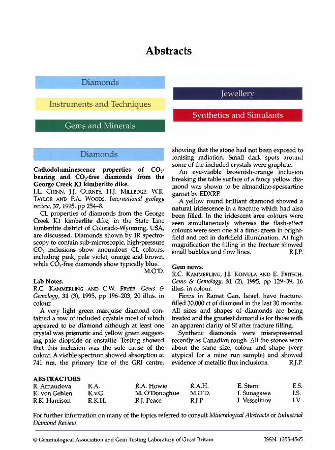
Abstracts
Diamonds
Instruments and Techniques
,
Gems and Minerals
Diamonds
Cathodoluminescence properties of CO2-
bearing and CO2-free diamonds from the George Creek K1 kimberlite dike. I.L. CHINN, J.J. GURNEY, H.J. MILLEDGE, W.R TAYLOR AND P.A. WOODS. International geology review, 37, 1995, pp 254-8.
CL properties of diamonds from the George Creek K1 kimberlite dike, in the State Line kimberlite district of Colorado-Wyoming, USA, are discussed. Diamonds shown by IR spectroscopy to contain sub-microscopic, high-pressure CO2 inclusions show anomalous CL colours, including pink, pale violet, orange and brown, while CO2-free diamonds show typically blue.
M.O'D.
Lab Notes. RC KAMMERLING AND CW. FRYER. Gems & Gemology, 31 (3), 1995, pp 196-203, 20 illus. in colour.
A very light green marquise diamond contained a row of included crystals most of which appeared to be diamond although at least one crystal was prismatic and yellow green suggesting pale diopside or enstatite. Testing showed that this inclusion was the sole cause of the colour. A visible spectrum showed absorption at 741 nm, the primary line of the GRI centre,
ABSTRACTORS R Arnaudova RA. RA. Howie
Jewellery
Synthetics and Simulants
showing that the stone had not been exposed to ionising radiation. Small dark spots around some of the included crystals were graphite.
An eye-visible brownish-orange inclusion breaking the table surface of a fancy yellow diamond was shown to be almandine-spessartine garnet by EDXRF.
A yellow round brilliant diamond showed a natural iridescence in a fracture which had also been filled. In the iridescent area colours were seen simultaneously whereas the flash-effect colours were seen one at a time; green in brightfield and red in darkfield illumination. At high magnification the filling in the fracture showed small bubbles and flow lines. RJ.P.
Gem news. RC KAMMERLING, J.1. KOIVULA AND E. FRITSCH. Gems & Gemology, 31 (2), 1995, pp 129-39, 16 illus. in colour.
Firms in Ramat Gan, Israel, have fracturefilled 30,000 ct of diamond in the last 30 months. All sizes and shapes of diamonds are being treated and the greatest demand is for those with an apparent clarity of SI after fracture filling.
Synthetic diamonds were misrepresented recently as Canadian rough. All the stones were about the same size, colour and shape (very atypical for a mine run sample) and showed evidence of metallic flux inclusions. RJ.P.
RA.H. E. Stern E.S. K. von Gehlen K.v.G. M. O'Donoghue M.O'D. 1. Sunagawa 1.S. RK. Harrison RK.H. RJ. Peace RJ.P. I. Vesselinov LV.
For further information on many of the topics referred to consult Mineralogical Abstracts or Industrial Diamond Review.
© Gemmological Association and Gem Testing Laboratory of Great Britain ISSN: 1355-4565
J. Gemm., 1996, 25, 2 143
Gem news. R.C. KAMMERLING, J.L KOIVULA, M.L. JOHNSON
AND E. FRITSCH. Gems & Gemology, 31 (3), 1995, pp 204-16, 22 illus. in colour.
A slightly brownish-grey diamond crystal contained a cavity with a mobile triangular inclusion which was of the same type (la) as the host. The original material filling the cavity was thought to be gas which had escaped during the cutting of a window.
A visit to the kimberlite pipes in the Siberian Sakha (Yakutia) Republic snowed that flooding was a serious problem. There was very little activity at two of the sites and both the international pit as well as the quarry-like Zarnitza prospect were inactive. Udachnya seemed to be the most productive and its large processing plant was in full production servicing not only its own but also the Aikhal and Jubilee sites. When winter temperatures reach -45°C work stops because of an inversion layer which traps potentially deadly methane in the mine. R.J.R
A diamondiferous lamprophyre dike, Gibson Lake area, North-west Territories. N.D. MACRAE, A.E. ARMITAGE, A.L. JONES AND
A.R. MILLER. International geology review, 37,1995, pp 212-29.
Diamonds are reported from the Akluilâk dyke in the Central Churchill Province, Gibson Lake area, Northwest Territories of Canada. The dyke is a narrow lamprophyre intrusion of ultra-potassic composition and has yielded a large number of diamonds, few of which are larger than 0.5 mm. Crystal forms are primarily octa-hedra and tetrahexahedroids though some macles, cubes, aggregates and fragments also occur. The dyke is situated approx. 120 km NW of Rankin Inlet. M.O'D.
[The apographitic impact diamonds from astroblemes Ries and Popigai.] (Russian with English abstract) V.L. MASAITIS, G.I. SHAFRANOVSKY AND I.G.
FEDOROVA. Proc. Russian Min. Soc, 124 (4), 1995, PP 12-19.
A study of diamonds formed in the Popigai and Ries impact craters has shown that they are similar in their mode of occurrence, morphology, physical properties, crystalline structure, etc. They occur as polycrystalline aggregates of cubic and hexagonal phases and inherit the shape and some other features of the original graphite. Authigenic impact diamonds were established
to exist in both craters in strongly shocked and partially fused fragments of graphite-bearing crystalline target rocks. After the solid transition from graphite at ~ 40^15 Gpa, impact diamonds were included in hot media and subjected to oxidation and graphitization. R.A.H.
Carbon isotopic composition and origin of SiC from kimberlites of Yakutia, Russia. E.A. MATHEZ, R.A. FOGEL, I.D. HUTCHEON AND
V.K. MARSHINTSEV. Geochimica et Cosmochimica Acta, 59 (4), 1995, pp 781-91.
Analyses are presented of carbon isotopic compositions of moissanite (SiC) from the Mir and Aikhal kimberlites. Yakutia Province. The mineralogy of moissanite is described and illustrated with an SEM image. Silicon metal is the most common inclusion and inclusions of ferro-silicate, Fe-Ti suicides, REE silicate and sinoite have been found. 513C ranges -22 to -29 %o, and 815N + 9.7 ± 4.0 to +5.6 ± 2.0%. Moissanite may be a relict of a reduced, primordial Earth and now present as a trace phase in an otherwise oxidized mantle. Alternatively there may be present-day global regions that are highly reduced with light C isotopic compositions. Other possibilities are discussed, including the formation of moissanite by metamorphism of reduced, carbonaceous sediments during subduction. R.K.H.
Distribution of the luminescent centres in Yakutian diamonds. V. MIRONOV AND B. ANTONYUK. Archiwum
Mineralogiczne (Polska Akademia Nauk), 50 (2), 1994, pp 3-12.
The laser-luminescence tomography method for revealing crystal zoning is described and has been used to examine diamonds collected from the Malobotuobinskoe field in the Mirnyi region of Yakutia, Russia. This non-destructive method shows the distribution of luminescence centres in the crystals, usually arranged in zones formed during successive stages of crystal growth. 'Phantoms' inside crystals, i.e. luminescent outlines of the growth zones, even from the earliest stages, provide information on changes in diamond crystallization conditions in the parent rock matrix. The method has been developed for use in the classification of diamonds from both primary kimberlite and secondary alluvial deposits in the Mirnyi region. The paper is illustrated with nine colour photographs showing yellow, green or blue UV luminescence.
R.A.H.
144
The Luanda diamond fields. Part II. A. THOMAS. South African gemmologist, 9 (3), 1995, pp 23-8.
Continuing account of a visit to the diamondproducing area of Luanda with some details of recovery methods. M.O'D.
[Heart and arrow shaped pattern observed in round brilliant-cut diamond.] (In Japanese with English abstract) H. YANO. Journal of the Gemmological Society of Japan, 18, 1993, pp 22-30, 14 figs., 2 tables.
A recent claim that gained popularity on the Japanese diamond market was that if a brilliant cut diamond exhibits a heart and arrow shaped pattern when illuminated under certain conditions, this proves that it has the highest grade of cut. Even an instrument to illuminate and record the pattern has appeared on the market. This paper reports the results of a mathematical analysis of the reliability of such a claim. It has been demonstrated that (1) such a pattern can be seen when a stone is illuminated in a range of inclination of 9 to 15 degrees to the axis, (2) the pattern is mainly due to the arrangements of pavillion facets and not to the crown facets, and so (3) this pattern does not automatically represent the highest cut grade. The appearance of the pattern is a necessary condition but not a sufficient condition to prove the cut grade. I.S.
Gems and Minerals
Detrital origin of fuchsite-bearing quartzites in the western Dharwar craton, Karnataka, India. S. ARGAST, Journal of the Geological Society of India, 45(5),1995, pp 559-75.
Quartzites in the Nuggihalli, Holenarsipur, Javanhalli, Bababudan and Chitradurga schist belts all originated as clastic sandstones rather than as precipitated cherts. Detrital chromite occurs in the oldest quartzites and this gave rise to fuchsite during metamorphism; there is no evidence for significant contributions of hydrothermal Cr. There are secular trends in median Cr/ Al20 3, Cr/Fe20 3, Cr/Zn and KP/ AlP3 ratios which are consistent with the gradual evolution of mafic and sialic sedimentary source terrains over the interval from - 3500-2500 m.y. ago. The compositions of the Javanhalli and Bababudan quartzites are sufficiently different to suspect, but not prove, that they are from different sources. RA.H.
J. Cemm., 1996, 25, 2
Charoite: a unique mineral from a unique occurrence. M.D. EVDOKIMOV. World of Stones, 7, 1995, pp 3-11, illus. in colour.
Details of the properties and occurrence of charoite are given, with notes on the geology of the occurrence and a list of references. The charoite-producing source (Sirenevyi Kamen [lilac stone]) is found on the meridian passing from the town of Olekminsk on the Lena River to Khani Station on the Baikal-Amur railway line, Russia. M.O'D.
Kazakhstan and Middle Asia. A brief mineralogical guide. A.A. EVSEEV. World of Stones, 8, 1995, pp 24-30, illus. in colour, 1 map.
List of mineral species found in Kazakhstan and Middle Asia, arranged in alphabetical order of location and including some gem minerals.
M.O'D.
Recent colored gem stones from Russia. [In Japanese with English abstract] M. HAYASHI, K. HARA AND A. YAMAZAKI. Journal of the Gemmological Society of Japan, 18, 1993, pp 15-21, 15 figs, 6 tables.
Diamond, ruby, alexandrite, demantoid, topaz, emerald, aquamarine, tourmaline, diopside, jadeite and lapis-lazuli from Russia which have recently been encountered in the Japanese gem market are described. Chemical and analytical data obtained by electron dispersive spectroscopy, FT-IR charts, and XRD are given. Possible marketing routes of Russian emeralds are explained. IS.
Gem identification by FT-IR-emerald, jadeite, sillimanite, etc. [In Japanese with English abstract] M. HAYASHI AND Y. MANAKA. Journal of the Gemmological Society of Japan, 19, 1994, pp 3-6, 15 figs., 1 table.
Fourier transform infrared (FT-IR) spectroscopy has been applied extensively to identify resin-impregnated emeralds and jadeites. FT-IR spectra show different features characteristic of natural, flux-grown, hydrothermally-grown emeralds and also resin impregnated emeralds, and jadeites, that can be used for diagnostic purposes. FT-IR spectroscopy is also useful to differentiate between gemstones whose refractive indices are similar, such as sillimanite, kornerupine, diopside and enstatite. 1.5.
J. Cemm., 1996,25,2
Rubis et saphirs de Republique Populaire de Chine. RW. HUGHES AND O. GAUBERT. Revue de gemmologie, 125, 1995, pp 5-8, illus. in colour.
Adaptation of a chapter dealing with ruby and sapphire from China, to appear in a new edition of the first author's book on corundum.
M.O'D.
Gem-quality grossular-andradite: a new garnet from Mali. M.L. JOHNSON, E. BOEHM, H. KRuPP, J.W. ZANG
AND RC KAMMERUNG. Gems & Gemology, 31(3), 1995, pp 152-67, 2 tables, 1 illus. in black-andwhite, 17 illus. in colour.
Thousands of carats of these fashioned stones from the Republic of Mali are poised to enter the marketplace. In the trade these garnets are termed 'Mali garnet' or 'Mali grossular garnet'. The composition of this garnet is intermediate between grossular and andradite garnets and has been described by the GIA Gem Trade Laboratory as grossular-andradite. Quantitative chemical analysis using an electron microprobe and X-ray powder techniques revealed a composition 78% grossular and 20% andradite. Refractive indices for yellow-green and green stones are in the range of 1.752-1.769, whilst those of orange brown stones are 1.773-1.779 (RIs for grossular 1.73-1.76 and andradite 1.880-1.895).
The absorption spectra of yellow-green, and orange brown stones show an absorption band at 440 nm. The stacked parallel planes of growth zoning are always visible between crossed polars and together with moderate to strong ADR are diagnostic of grossular-andradite garnet.
Most of the gem quality grossular-andradite garnets from Mali are reminiscent of the pale greenish yellow to yellow-green grossular garnets from Tanzania, with some rare stones resembling peridot. Numerous brown to brownish-orange stones can be quite brilliant due to higher RIs and dispersion and may serve as inexpensive substitutes for 'cognac' diamonds. RJ.P.
Mosaic geographical map - a masterpiece of stonecutting art. M.N. KAUNINA. World of Stones, 8, 1995, pp 31-2, illus. in colour.
Brief account of a mosaic map in the Georgian Hall of the Hermitage Museum in St Petersburg,
145
Russia. The map is constructed from hard ornamental stones. M.O'D.
Lab notes. RC KAMMERUNG AND CW. FRYER. Gems & Gemology, 31(3), 1995, pp 196-203, 20 illus. in colour.
A stone showing a colour change from dark bluish green in daylight to dark reddish purple in incandescent light was identified as natural alexandrite. It had atypically high RI values of 1.753-1.761 and was found to have unusually high concentrations of chromium, titanium and iron. Several surface-reaching fractures contained a transparent filler which easily melted.
Three purplish-red mixed-cut stones were identified as corundum but lacked distinctive absorption features. The facets had uneven surfaces with some slight rippling and many minute cracks. Immersion in di-iodomethane revealed concentration of the colour along the facet edges indicating that they were diffusiontreated corundums.
A translucent mottled-green carving in a closed pendant setting gave a spot RI reading of 1.66 and showed a series of 'chrome lines' in the red end of the spectrum which are consistent with natural-colour jadeite. However, the carving consisted of a thin hollow jadeite shell filled with a soft transparent colourless filler containing gas bubbles. FTIR spectroscopy of the filler indicated polymers known to have been used for jadeite impregnation. The thickness of the jadeite shell at the edge was only 0.05 to 0.10 mm. RJ.P.
Gem news. RC KAMMERUNG, J.I. KOlVULA AND E. FRITSCH.
Gems & Gemology, 31(2),1995, pp 129-39, 16 illus. in colour.
Rare collector stones from the former Soviet Union were shown in Tucson in February 1995. These included small colourless crystals of leucophanite, colour-change clinochlore together with gorgeyite, kaliborite and preobrazhenskite. The full constants of these were quoted.
Chatoyant moonstones with an atypical darker orange colour originated from Madras, India. Unusual opal carvings incorporating both opal and its matrix were cut from the back to produce a three-dimensional effect. Sapphires from Madagascar were well described. These had been heat treated and the blue colour was due to iron-titanium charge transfer. Details
146
were given of a chatoyant spessartine garnet from Namibia. R.I.P.
Gem news. R.c. KAMMERLING, J. KOIVULA, M.L. JOHNSON AND
E. FRITSCH. Gems & Gemology, 31(3), pp 204-16, 22 illus. in colour.
A blue green apatite cabochon from Malagasy weighing 11.45 ct showed pronounced chatoyancy and an unusual colour. The chatoyancy was caused by barely visible white needles. 'Eilat stone' not from Israel but from Peru is now on the market. It is a by-product of copper mining. At 10.03 ct round cabochon-cut emerald from Brazil showed a well defined six-ray star. It showed strong pleochroism with intense blue-green colour parallel and a light yellowish-green colour perpendicular to the c-axis attributed to a distinct iron-bearing (aquamarine) component which was found in the spectrum together with chromium bands of emerald. Although there were channellike inclusions orientated parallel to the c-axis, the star was caused by three sets of unidentified small elongated particles orientated perpendicular to the c-axis. A transparent purplish red-brown stone of 0.33 ct had RI 1.742, singly refractive, SG 3.68-3.70, inert to both long and short wave UV radiation. Unusually it showed a typical almandine-rhodolite visible-light absorption spectrum. It was confirmed as genthelvite, Zn4Be3(Si04hS, a member of the helvite group. X-ray powder diffraction analysis and energy dispersive X-ray fluorescence (EDXRF) spectrometry were used to identify the stone. A 'drusy' hausmannite cabochon showed an imposing architectural appearance with its natural crystalline surface. Green opal from Serbia appeared green through the Chelsea filter. Spot RI 1.46 and SG 2.10 values were obtained but were difficult to measure because the stone was porous. The stone was inert to short-wave UV radiation but fluoresced an even faint chalky green to long-wave UV. The spectrum showed cut-off edges at 450 and 620 nm. Some healed fractures and veining were visible under magnification. EDXRF analysis revealed the presence of nickel which was the probable cause of the colour. An Egyptian green quartz reportedly collected in the Eastern desert has a pseudo-fibrous structure. EDXRF analysis revealed a fairly large concentration of chromium which probably was the cause of the colour as finely disseminated chromium-coloured mineral inclusions. A spangled quartz is coloured by bright orange red flakes which are probably
J. Gemm., 1996, 25, 2
hematite. Although randomly orientated throughout the cabochon the flakes are concentrated in one layer parallel to a rhombohedral face and are unusual in that each grew outward from one corner in an elongated fashion to give an 'hour-glass' or 'cello' shape. An aromatic resin necklace sold as myrrh was thought not to be from the somewhat rare Commiphora myrrha of biblical fame. It showed RI 1.41(spot) SG 1.27, hardness less than 2.5 on Mohs' scale and on the approach of a hot needle easily melted emitting a characteristic sweet spicy odour. Purple scapolite from Tajikistan reputedly found in the Pamir mountains is mined at an altitude of 5000 m and miners wear oxygen masks when working. Specimens showed uniaxial negative character, RI 1.539 and 1.550, SG 2.61, inert to long wave UV and fluorescing a very weak pinkish orange to short wave uv. Another stone from this region was a spinel of 123 ct described as virtually flawless. An opticon-treated malachite showed enhanced lustre and depth of colour. The impregnation process consisted of repeated coatings and oven treatment at each stage so that finally the whole of the product was impregnated. R.J.P.
Spectrophotometric identification of emeralds. [In Japanese with English abstract] H. KITAWAKI AND H. FUKUSHIMA. Journal of the Gemmological Society of Japan, 19, 1994, pp 7-14, 18 figs, 1 table.
UV-VIS and NIR spectra have been obtained on some hundreds of emerald samples from different origins, both natural and synthetic. In UVVIS spectra, dominant absorption due to Cr can be seen in natural Colombian emeralds, while the absorption peaks due to both Cr and Fe are seen in natural emeralds from other localities. Hydrothermally-grown synthetic emeralds show absorption due to Cr and high transmission of Uv. In Russian synthetic emeralds, an absorption related to Ni is seen. In flux-grown synthetic emeralds, only Cr-related absorption can be recognized, and the transmission peak in the UV lies at a higher wavelength than that of Colombian samples. From NIR (1200-2700 nm) spectra, the origin of emeralds and the conditions of their growth can be conjectured from the absorption peaks, since they show the presence or absence and the nature of H20. 1.S.
[A few unusual gemstones recently encountered.] (In Japanese) . Y. KITAWAKI. Journal of the Gemmological Society of
J. Gemm., 1996, 25, 2 147
Japan, 19,1994, pp 32-3,1 fig., 2 tables. Natural bi-colour topaz, natural euclase, nat
ural pink clinozoisite which were encountered in gem testing are described. LS.
[A trial for grading black pearls.] (In Japanese with English abstract) H. KOMATSU, C. SUZUKI AND M. HIRAKO. Journal of
the Gemmological Society of Japan, 19, 1994, pp 23-9,10 figs., 3 tables.
Based on the quality elements of colour and brilliance, efforts have been made to establish a method to quantitatively grade the quality of black cultured pearls produced by Pinctada mar-garitifera. The method of grading, inherent mechanism of colour emission, and the difference in the emission mechanisms between black and white pearls are described and discussed. Reflectance is measured and RBG analysis is done by image analyser. The results showed that the measured values of four quality elements (blackness, body colour, interference colour and its intensity) on the image analyser, were almost identical to the grades obtained by visual grading. LS.
Native copper in agates from Rudno near Krzeszowice. W. KRAWCZYNSKI. Mineralogia Polonica, 26(1), 1995, pp 27-32, 3 maps.
Copper is reported in agates from Rudno near Krzeszowice in Poland. Notes on the occurrence and on the agates are given and samples were taken from more than 300 agate specimens. Native copper was found to occur in secondary microfissures crossing agate layers obliquely, at the boundaries of individual agate layers in a tree-like form, and as dispersed grains independent of agate layers. M.O'D.
What's new in minerals? T.P. MOORE. Mineralogical Record, 27(1), 1996, pp 59-66, illus. in colour.
Gem-quality minerals and fashioned stones are reported from mineral and gem shows held in the United States. Species seen at the Denver show include fine rhodochrosite from the Sweet Home mine, Alma, Colorado: smoky quartz from the Calumet iron mine in the same state: twinned crystals of chrysoberyl from Santa Tereza, Espirito Santo, Brazil: very deep pink octahedral fluorite crystals from Mont Blanc, France: emerald from the Jos region of Nigeria:
brown to green vesuvianite from Sandare, Diakon Arrondissement, Nioro du Sahel, Mali (this area also produces brown-green grossular): sceptre amethyst from Obman, Yakutia, Russia: blue topaz from Nura-Taldy, central Kazakhstan: emerald crystals from the mica schists of the Malysheva mine, central Urals, Russia (the crystals are not of gem quality): fine elbaite showing green, colourless and blue sections perpendicular to the c-axis, from the Asthor mine, near Shigar, Pakistan.
From the show held in Pretoria, South Africa, David Minster reports fine green tourmaline crystals from Mozambique: rhodochrosite crystals up to 3 cm long from the Kalahari manganese field, South Africa: gem-quality yellow orthoclase and rhodizite from the Malagasy Republic. M.O'D.
La saga peregrina, [first part]. B. MOREL. Revue de gemmologie, 125, 1995, pp 15-17, illus.
Enquiry, with some proposals, into the present whereabouts of the Peregrina, a celebrated pearl once in the possession of the Kings of Spain. M.O'D.
The co-precipitation of Fe3+ and S i0 2 and its role in agate genesis. T.J. MOXON. Neues Jahrbuch für Mineralogie, Monatshefte, 1996(1), pp 21-36.
Detailed experimental work studying the use of Fe3+ and Mg2+ in removing H4Si04 and colloidal silica from separate dilute silica solutions and sols over periods of 45 min to 14 years is reported. It is demonstrated that Mg2+-Si02 gels in an alkaline saline environment at room temperature can develop signs of transformation into opal-CT after 14 years. The concentrations of Fe3+ found in some agates is considered to be coincidental and too low to precipitate silica from any silica sol or solution. R.A.H.
Ein trapiche-rubin aus Myanmar (Burma). H.-J. MÜLLENMEISTER AND J. ZANG. Lapis, 20(12), 1995, p. 50, 2 photos in colour.
A ruby crystal of 6 ct and 12 mm thick showed six sections of ruby divided from one another by spokes of a light-coloured carbonate material tentatively identified as either calcite or ankerite. The piece closely resembled the trapiche emerald long known from the Muzo mine in Colombia. M.O'D.
148 J. Gemm. , 1996, 25, 2
The degree of metamictization in zircons: a Raman spectroscopic study. L. NASDALA, G. IRMER AND D. WOLF. European
Journal of Mineralogy, 7(3), 1995, pp 471-8. A series of natural zircons representing
various degrees of metamictization were investigated by Raman microprobe analysis. Systematic changes in wavenumbers and half-widths of the Raman bands were shown to be caused by increasing irregularities of bond-lengths and bond-angles and a general breaking-up of the structure as a result of metamictization. The half-width of the antisymmetric stretching vibration band (Blg) of the Si04 tetrahedra, which has a frequency of ~1007 cm-1 in well-crystallized and 1000 to 995 cm"1 in metamict zircons, is most suitable for estimating the degree of metamictization: its value increases from ~ 5 cm-1 in well-crystallized samples up to 30-55 cm-1 in highly metamict X-ray amorphous zircons, and is strongly dependent on the degree of lattice destruction by metamictization. In contrast, the Raman parameters seem to be almost uninfluenced by chemical variations. The potential value of such measurements, especially in radiometric age determinations, is discussed.
R.A.H.
Antique cameos. O.Y. NEVEROV. World of Stones, 7,1995, pp 29-32, illus. in colour.
Descriptive paper introducing the classical motifs found in cameos, some of which are illustrated. Examples are taken from the collections of the Hermitage Museum, St Petersburg, Russia. M.O'D.
Optical and infrared spectroscopic studies of epidote group minerals from the Rhodope region. S. PETRUSENKO, M. TARAN, A. PLATONOV AND
S. GAVORKYAN. Review of the Bulgarian Geological Society, 53(1), 1992, pp 1-9.
Crystals of various colours, including Fe, Mn-bearing clinozoisites, Mn-bearing zoisite (thulite) and epidote from skarns of various Bulgarian deposits, have been studied. Oriented single-crystal platelets have been used to record optically polarized, cc, ß and y spectra. The green colour of epidote is associated with Fe3+ occupying the M(3) sites in the crystal structure and is determined by a short-wave O2" —> Fe3+ transfer. The pink colour of Mn-clinozoisite and thulite is caused by Mn3+ in M(3) structural sites. The IR
spectra of the two minerals show definite differences. The OH-group valency oscillation band at 210 cm-1 is most important for identification of zoisite. Mn-bearing specimens show three maxima in the 500-600 cm-1 interval whereas Fe-bearing ones show a single broad band which is explained with bond oscillations in the A106-octahedra. A comparative analysis of infrared spectra of clinozoisite before and after its treatment with deuterium has shown that the OH-group band at 3370 cm"1 and the band at 1045 cm" (caused by Al-OH deformational oscillations) disappear. They are replaced by new bands at 2490 and 780 cm"1. The absorption bands of the diorthogroups lined by M(3) octa-hedra are the most variable. This corroborates the assumption of preferable substitution of Al by Fe in the M(3) sites. A linear regression equation based on the valency frequency oscillations of diorthogroups is proposed for determining the Fe content. R.A.
Epitaxial overgrowths of marcasite on pyrite from the Tunnel and Reservoir Project, Chicago, Illinois, USA; implications for marcasite growth. J. RAKOVAN, M.A.A. SCHOONEN, R.J. REEDER,
P. TYRNA AND D.O. NELSON. Geochimica et Cosmo-chimica Acta, 59(2), 195, pp 343-6.
The marcasites occur as thin tabular crystals =s 300 um on edge, dominated by {010}, and have grown around the {001} zone on pyrite octa-hedra. The marcasite has apparently grown around the pyrite by a layer growth mechanism as shown by macrosteps terminating partially developed layers on the marcasite {010}. Other forms on the marcasite are described and illustrated with SEM micrographs. A proposed reconstruction of the pyrite surface into a marca-site-like arrangement may be due to interactions of a protonated disulphide species with the pyrite during growth. R.K.H.
Luminescence in minerals. J. RAKOVAN AND G. WAYCHUNAS. Mineralogical
Record, 27(1), 1996, pp 7-19, illus. in colour. The property of luminescence is explained
with reference to mineral structure and chemistry, drawing examples from some of the commoner species. M.O'D.
Ruby and sapphire from Vietnam. (In Japanese) J. SHIDA. Journal of the Gemmological Society of Japan, 18,1993, pp 32-6,17 figs.
Over 2400 samples of ruby and sapphire col-
J. Gemm., 1996, 25, 2 149
lected from Luc Yen and Quy Chau, Vietnam were investigated. Crystal habits, inclusions, and internal features such as growth sectors and banding are described. LS.
A guide to mineral localities in the former Soviet Union. B. SMITH AND C. SMITH. Mineralogical Record, 26(6), 1995, pp 517-49, illus. in colour, 1 map.
An alphabetical list of places where minerals have been found has been prepared for territories of the former Soviet Union. Each entry gives name in English with variants where appropriate, the name in Cyrillic characters, geographical co-ordinates, type of location (region, town, stream, village, bay, mountains and so on), location of the place of higher order (similar to state capital, province [Russian name oblast], local seat of government) and list of species found. Useful notes are given on how the table was compiled and the difficulties encountered, on how to transliterate and what scheme to use, and on the nature of Russian and non-Russian names in this specialized context. There is a table of useful terms and a transliteration table.
M.OT>.
Sapphires from Southern Vietnam. C R SMITH, R.C. KAMMERLING, A.S. KELLER,
A. PERETTI, K.V. SCARRATT, N. KHOA AND
S. REPETTO. Gems & Gemology, 31(3), 1995, pp 168-85, 28 illus. in colour, 2 tables.
Gem quality blue to bluish-green sapphires from Southern Vietnam came on the market in the late 1980s. The sapphires occur in alkali basalts in the mining areas of Phan Thiet and Di Linh. Mining was carried out by hand at the former but relatively sophisticated equipment was used at Di Linh.
The properties and characteristics of these sapphires are similar to those from other basalt sources such as Australia, Cambodia, China, Nigeria and Thailand. The crystals show prominent growth structures and colour zoning in geometrical patterns. Sharply bordered blue bands alternate with narrow colourless or yellowish to brownish bands parallel to the crystal planes r and z; many stones showed a colourless 'core'.
The most common inclusions are various types of cloud with cross-hatch or lath-like patterns which have not been recorded from other sapphires. Although occasional dust-like
'snow-flake' inclusions have been noted in Kashmir sapphires, the very fine whitish needles presumed to be rutile or ilmenite lacked the well formed strongly iridescent appearance of rutile needles and plates observed in sapphires from metamorphic environments such as Sri Lanka and Burma. Inclusions of plagioclase feldspar, zircon, uranpyrochlore, columbite, ilmenite, pyrrhotite, chromite-hercynite, mag-netite-hercynite and goethite were identified. The UV-VIS-NIR absorption spectra are typical of basaltic sapphires with absorption bands towards the near-infrared region due to Fe2+ <-> Fe3+ intervalence charge-transfer. Spectral curves in the 280-880 nm region might be used to identify the country of origin of sapphires but should be interpreted with caution. The high iron content of Southern Vietnamese sapphires allowed easy distinction from sapphires from a metamorphic source. Separation from other basaltic sapphires was more difficult but the cross-hatch and lath-like 'cloud' patterns coupled with distinctive colour zoning and a colourless 'core' are a unique combination. R.J.P.
Estructura cristallina, composiciö quimica I propietats fisiques de les gemmes.5. La forma dels cristalls. (In Catalan and Spanish) J. SOLANS AND M.V. DOMÈNECH. Gemmologia, 91,
1995, pp 14-15, illus. in black-and-white. The crystal structure of beryl is explained
with diagrams showing the ring formation. M.O'D.
Brief chronicle of the formation of the Vernadsky State Geological Museum. Y.Y. SOLOV'IEV, Z.A. BESSUDNOVA AND L.T.
PRZHEDETSKAYA. World of Stones, 8, 1995, pp 14-22, illus. in colour.
Account of the events preceding and accompanying the establishment of the Vernadsky State Geological Museum, Moscow, Russia, with illustrations of some notable specimens. M.O'D.
Larimar: blauer Pektolith aus der Dominikanischen Republik. G. STEINER. Lapis, 21(1), 1996, pp 39—41,1 map.
The name Larimar has been given to an opaque blue ornamental variety of pectolite found in the Dominican Republic. The occurrence is on the south-west coast of the country.
M.O'D.
150 J. Gemm., 1996, 25, 2
Die türkisgrünen Steine' von Nagar in Nord-Pakistan. W. TOROSSIAN-BRIGASKY AND V.M.R HAMMER.
Mineralien Welt, 7(1), 1996, pp 47-51,1 map. Quartz crystals with a turquoise-green colour
have been found at 3400 m in the Nagar area of northern Pakistan. M.O'D.
Von Frankreich nach China: Münchner Mineralientage 1995. S. WEISS. Lapis, 21(2), 1996, pp 29-32, illus. in colour.
Among specimens shown at the 1995 Münchner Mineralientage in 1995 were emerald crystals from Mengdong, Yunnan Province, China and peridot crystals from Sopat, Kohistan, Pakistan. Fine orange-red rhodochrosite from southern Kazakhstan was also available, one specimen measuring 3.26 cm in width. While these specimens were illustrated, the text also mentions chrome cerussite from the Dundas Extended Mine, Tasmania; brazilianite crystals up to 4 cm long, from Minas Gérais and chrysoberyl trillings from Colatina, Espirito Santa, Brazil; these crystals are up to 2 cm. Pakistan has also produced morganite crystals up to 10-12 cm long. M.O'D.
Remarkable pyrite pseudomorphs after ammonites. E.Y. ZAKREVSKAYA. World of Stones, 7, 1995, pp 24-5, illus. in colour.
Fine ornamental specimens of pyrite pseudomorphs after ammonite are described from the collections of the Vernadsky State Geological Museum, Moscow, Russia. M.O'D.
Fluorescence and luminescence in minerals. FM-MSA-TGMS-FMS Tucson mineralogical
symposium. Mineralogical Record, 27(1), 1996, pp 21-30, illus. in colour.
Short papers on luminescence in minerals are abstracted. Titles covered are luminescence zoning in minerals; fluorescent minerals - some answers, questions and enigmas; single-colour and multi-colour fluorescence in calcite; luminescence mechanisms and activators in Terlingua-type calcite and in benitoite and related silica minerals: the influence of heating on the luminescence of calcite: uranium-activated fluorescence in minerals of the Franklin-Sterling Hill area, Sussex County, New Jersey; fluorescent minerals of the Karnes uranium district, south Texas; cathodoluminescence in the study of rocks
and minerals; activators for cathodoluminescence of charoite and associated minerals; illuminating gems with visible monochromatic lasers-what can it do for the gemmologist?; a combined microscope/fluorescence spectrometer system for mineralogical and gemmological applications; demonstration of mineral fluorescence excited by a 193-nm ArF excimer laser; the fluorescent characteristics of coal. M.O'D.
What's new in minerals? [Various authors] Mineralogical Record, 26(6),
1995, pp 575-83, illus. in colour. A number of gem minerals have been seen at
different mineral shows held in the United States during 1995. Examples include watermelon elbaite from the Otjua mine, Namibia, brown zircon from Lovozero, Kola Peninsula and golden-brown scheelite from Tenkergin, Chukot-ska, both Russia: fine green fluorite is coming in crystal sizes up to 13 cm on an edge from the Westmoreland mine, New Hampshire, USA, and peridot from the newly-discovered Pakistan location. Thumbnail-sized crystals of aquamarine and yellow beryl are coming from Cherlovaya Gora, Chita, Siberia and China continues to produce very high quality crystals of beryl, cassiterite and scheelite. It is reported that the violet red beryl claims in the Wah Wah mountains of Utah have been sold to a major mining company. A matrix group of crystals measuring 17.9 cm across, with about 25 crystals on it, has recently been recovered. Fine zoned elbaites in pastel blue, green and pink are coming from Pech, Kunar, Afghanistan.
Madagascar is producing more tourmaline: colourless crystals with ruby-red bands at each end have come from a small pocket south of the Mania river and liddicoatite crystals are coming from a site at Alakamisy, Anjanabonoina and from Ibity. Grass-green slabs with a perfectly symmetrical red-orange triangle in the centre have been found at old deposits in the Laondany area. Crystals of rhodizite are available as are hambergites: the export of both these species, with orthoclase, is forbidden. Some supposed hambergite on offer has turned out to be quartz or phenakite.
Crystals of rose- and peach-coloured morganite have been found in the past at the Bennet quarry, Buckfield, Maine, USA: New England beryl formed the theme of this year's Springfield show and a collection of Bennet beryls was shown by the Harvard Mineralogical Museum.
J. Gemm., 1996, 25, 2
Very fine amethyst crystals are being found in opium-growing areas of Veracruz state, Mexico: this area is not recommended for visiting.
M.O'D.
Gemmologie. Zeitschrift der Deutschen Gemmologische Gesellschaft, 44(4), 1995, pp 1-112.
This issue comprises a booklet by U. Henn entitled 'Practical Gemmology' (Edelsteinkundliches Practicum). It is a survey of methods of differentiation between natural and synthetic diamonds, corundums, spinels, beryls (emeralds), chrysoberyls (alexandrites), quartzes (amethysts and citrines) and opals. The introductory part deals with a short history, describes in general the characteristics of gemstones and their imitations, and discusses their microscopic characteristics. In the specific, second section each of the gems mentioned is dealt with fully, listing origin of name, physical and chemical properties, inclusions, going on to list the various methods of synthesis and their characteristics. Each chapter has an extensive bibliography and many illustrations (145 illustrations in all) as well as numerous tables and graphs. E.s.
The Australian Gemmologist, 19(3), Jubilee Edition, 1995.
This Golden Jubilee issue celebrates the 50th Anniversary of the formation of the GAA which was formed on the 29 October 1945. Fifty years on the aspirations of the founders have been amply fulfilled. This expanded reference edition traces the 50 year history of the association at both Federal and State Division levels as well as cataloguing the gemstone resources of each State in Australia. The survey includes biological gem materials such as pearl production in Western Australia and the Coburg Peninsula in the Northern Territory. The compact and comprehensive nature of this edition does not lend itself to conventional abstraction, and the reader is recommended to consult the original. R.J.P.
Instruments and Techniques
El conoscopi figueras 93. M. MARTINEZ, A MARTINEZ, C. BAcUENA AND A. RUIZ. Gemmologia, 91, 1995, pp 8-12, illus. in black-and-white. In Catalan and Spanish.
Evaluation of a new model conoscope with remarks on the phenomena observed. M.O'D.
151
The SHAPE crystal-drawing computer program as an instrument in research. 1. VESSELINOV. Geochemistry, Mineralogy & Petrology (Bulgarian Academy of Sciences), 29, 1994, pp 97-105.
The commercially available program of E. Dowty is shown to be a useful tool in solving morphological problems in mineralogy. Three groups of questions are discussed and exemplified: (1) identifying crystal forms on photographs of real crystals (e.g. zircon, monohydrocalcite and pyrite); (2) reproducing crystal crosssections (e.g. pyrite), and (3) simulating crystal growth by successive growth forms (e.g. zoned arsenopyrite). LV.
Jewellery
Joaillerie indienne. 5000 ans de tradition. F. BRUNEL. Revue de gemmologie, 125, 1995, pp 11-14, illus. in colour.
Several important pieces of Indian jewellery are illustrated in a short survey of the craft over the last 5000 years. M.O'D.
Synthetics and Simulants
'Ti-sapphire': Czochralski-pulled synthetic pink sapphire from Union Carbide. M.L. JOHNSON, M.E. MERCER, E. FRITSCH, P. MADDISON AND J.E. SHIGLEY. Gems & Gemology, 31(3),1995, pp 188-95,2 tables,S illus. in colour.
The pink 'Ti-sapphire' commercially available from Union Carbide by the Czochralski-pulling technique is used in laser applications as it produces a high-energy light over a wide range of frequencies in and near the visible range. It has also been made by flame fusion and melt techniques. Its gemmological properties are compared with natural and with other synthetic pink sapphires.
Separation from natural sapphire may be difficult but differences include no luminescence to long-wave UV radiation, chalky blue luminescence to short wave UV radiation (due to octahedral Ti4+06 impurities, an interstitial oxidised titanium) and pinpoint inclusions some of which were resolvable as gas bubbles. However reliable separation can be done by means of its chemistry or UV-visible absorption spectrum. Natural pink sapphires owe their colour to
152 J. Gemm. , 1996, 25, 2
chromium, but with the 'Ti-sapphires' the colouring agent is trivalent titanium and a faint but different spectrum is seen with a spectroscope (the main difference is the lack of chromium lines in the red). R.J.R
Lab notes. R.C. KAMMERLING AND C.W. FRYER. Gems &
Gemology, 31(3), 1995, pp 196-203, 20 illus. in colour.
An imitation emerald necklace gave a chalky yellow reaction to long wave UV radiation (not unexpected from a heavily oiled stone) and appeared dullish red through the Chelsea colour filter; in the drill hole of one bead a dark green ring was observed containing flattened gas bubbles and it proved to be a natural beryl bead with a green plastic coating. A fine green carving gave a spot RI of 1.56, a weak mottled greenish blue fluorescence to short wave UV radiation and a spectrum showing general absorptions from 400 to 480 nm and 650 to 700 nm. The presence of gas bubbles, flow lines, mould marks and minute conchoidal fractures indicated a glass. An imitation lapis lazuli which looked more like sodalite was shown to be dyed feldspar by tests which included X-ray diffraction. A red stone with RI 1.552 and SG 2.64 and an orange stone with RI 1.480 and SG 2.39 imitating Mexican opal were examined. Both were singly refractive but gave anomalous effects in crossed polars in the form of snake-like bands. Both were inert to long wave radiation but gave a faint chalky yellow to short wave UV radiation. Magnification revealed small gas bubbles in both stones. EDXRF analysis revealed the presence of both selenium and cadmium, already known as constituents in orange and red glasses. A strand of silvery dark grey to black pearls was shown to consist of lead glass beads coated with bismoclite, a bismuth oxychloride which formed silvery layers (with elastic properties) and an outer layer of a transparent rubberlike substance containing gas bubbles.
A large Czochralski-pulled ruby weighing over 100 ct was transparent with a strong red colour with no purple or brown components. Some fine curved striae were seen but apart from these it was free from inclusions and colour variations. The manufacturer is reported to be aiming at pure crystals above 500 ct. R.J.P.
Gem news. R.C. KAMMERLING, J.I. KOIVULA AND E. FRITSCH.
Gems & Gemology, 31(2), 1995, pp 129-39,16 illus.
in colour. Amber simulants consisting of amber pieces
embedded in plastic were recently shown from Poland. The material had a spot RI of 1.56 and an SG of 1.24. 'Recrystallized ruby and sapphire' marketed by the True Gem company of Las Vegas showed results consistent with a pulled synthetic product. All the stones had a laser inscription on a pavilion facet. Kyocera plastic-impregnated synthetic opals with different body colours were produced by impregnating the synthetic opal with various coloured polymers. Analysis by scanning electron microscopy with EDXRF instrumentation and Fourier-transform infrared (FTIR) spectroscopy was reported. A new ornamental material marketed as 'platigem' is an intermetallic compound of platinum, aluminium and copper. SGs were in the range 8.66 to 8.77 with RI values above 1.81. The colour was evenly distributed and varied from yellow to brownish pink. X-ray powder diffraction patterns suggested a cubic structure with approximately 21% copper. R.J.P.
Gem news. R.C. KAMMERLING, J.I. KOIVULA, M.L. JOHNSON
AND E. FRITSCH. Gems & Gemology, 31(3), 1995, pp 204-16, 22 illus. in black-and-white.
An unusual colour-zoned synthetic amethyst from Russia showed exceptionally uneven 'speckled' or 'leopard' spot colour distribution attributed to the fact that the crystal grew on a seed plane cut perpendicular to the c-axis with darker-colour regions forming expanding rhombic pyramids. Tanzanite-coloured synthetic sapphire has been sold in India as tanzanite. Typical constants for corundum and obvious curved colour banding and pin point inclusions which were probably gas bubbles enabled easy identification. A constructed quartz containing both natural and synthetic quartz formed a multi-coloured gem and included a rutilated quartz 'termination'. R.J.R
Synthetic red beryl. Y. KITAWAKI. Journal of the Gemmological Society of Japan, 19,1994, pp 34-5, 5 figs.
Internal features and spectra in the range from UV to visible and the near infrared region are reported for a purplish red synthetic beryl. Absorption due to Mn is remarkable. LS.
[So-called AGEE emeralds.] (In Japanese) Y. KITAWAKI. Journal of the Gemmological Society of

J. Gemni . , 1996, 25, 2 153
Japan, 19,1994, pp 35-8,10 figs., 1 table. Synthetic emeralds appeared on the market a
few years ago under the name of AGEE, and were supplied from AG. Japan Ltd. Gemmo-logical investigations indicated that there were several types; one type has the same gemmo-logical properties as those of Biron synthetic emerald, other types showed similarity to Russian synthetics, flux-grown synthetics, and vanadium beryl. LS.
Fraud at the source. A. KLEYENSTÜBER. South African Gemmologist, 9(3), 1995, pp 7-15, illus. in colour.
Useful account of various substances offered as natural gem materials from Namibia, Zambia and Zimbabwe. Most substitutes are glass of one form or another. M.O'D.
Crystal chemistry of non-stoichiometric Mg-Al synthetic spinels. S. LUCCHESI AND A. DELLA GUISTA. Zeitschrift für
Kristallographie, 209(9), 1994, pp 714-19. Twenty-seven single-crystal structural refine
ments were performed on synthetic spinels along the MgAl204-Al8/3D1/304 join. The number of vacancies (from 0.09 to 0.22) was determined by EPMA. Vacancies are limited to octahedral sites, with a vacancy-to-oxygen distance refined to 2.113(3) Â. Mg is ordered in the tetrahedral site, only for a > 7.99 Â are the amounts of octahedral Mg significant. The decrease in a for vacancy-rich spinels is essentially due to the decrease of the T-O distance because of large amounts of tetrahedral Al. A linear relation between a and vacancy content was derived fitting literature data, including end-members. K.v.G
[Observations on synthetic emeralds by the scanning cathodoluminescence (SCL) method]. (in Japanese with English abstract) T. MlYATA, K. TSUBOKAWA AND M. KlTAMURA. Journal of the Gemmological Society of Japan, 18, 1993, pp 3-10,13 figs., 4 tables.
The internal inhomogeneities of ten faceted synthetic emeralds were investigated using scanning cathodoluminescence (SCL) methods. The origin of each specimen was established before SCL investigation by ordinary gemmological identification and by measurement of its M R spectrum. The samples are from Lechleitner, Regency, Gilson, Saramandor and Chatham and include both flux and hydrothermal growths. Clear SCL images showed complex characteristics related to and depending on the methods of
synthesis. Flux-grown emeralds are characterized by straight parallel growth banding or 'aurora-like' growth banding. Hydrothermally grown emeralds are characterized by the development of 'comet-like' luminaries arranged in one direction or unique rhombic patterns. SCL investigations are useful not only to distinguish between natural and synthetic emeralds but also between different synthetic methods. LS.
Synthetische, nach dem Hydrothermalverfahren hergestellte Smaragde. G NIEDERMAYR. Mineralien Welt, 7(1), 1996, p 14.
A hydrothermally-grown emerald is described and its inclusions illustrated. The specimen was grown at a temperature of 380°C.
M.O'D.
[Growth of emerald crystals by cooling the high-temperature solutions using PbO.V203 flux.] (In Japanese with English abstract) S. OISHI, N. NISHIZAWA AND M. HiRAO. Journal of the Gemmological Society of Japan, 18, 1993, pp 11-14, 5 figs.
Transparent green emerald crystals up to 2.2 mm long were synthesized from PbO.V203
solution, using no seed crystals. The nutrient was heated at 1200°C for 10 hours, followed by cooling to 600°C at rates varying between 5 and 240°C/h to find the optimum conditions. The size of crystals decreased with increase in the cooling rate. The optimum cooling rate for obtaining larger crystals was 5°C/h. LS.
[The present status of synthetic rubies.] (In Japanese with English abstract) J. SHIDA, M. SHIGEOKA AND H. KITAWAKI. Journal of
the Gemmological Society of Japan, 19, 1994, pp 15-22, 26 figs.
General characteristics, UV spectra, internal features, inclusions, growth sector patterns and growth banding of 'Rubiante' synthesized by a flux method by Culture Crystal Inc., USA, 'Douros' synthesized by a flux method by O.E. Created Gems, Greece, and rubies supposed to be synthesized by hydrothermal methods in Russia are described. Growth banding seen in flux grown rubies can be mistaken for banding seen in natural rubies. Hydrothermally synthesized rubies are generally dark red in colour and contain many flaws. Their refractive indices are significantly higher than those of natural rubies. Internal features revealed by laser-beam tomography are also described. LS.
BOOK REVIEWS
Blood stones [a tale]. E. ANTHONY, 1994. Bantam Press, London, pp 335, hardcover. £14.99. ISBN 0 593 036565.
Books on 'how the diamond market can be overturned7 are not too uncommon and here is quite a readable tale whose thinly-disguised protagonist's battle with Russian diamond magnates over a possible move of Russian sales. Featuring as a prop to the complicated intrigues is a necklace of blood-red diamonds: whether or not they have been colour-enhanced forms an important part of the plot but I did not find out the answer! M.O'D.
Norsk Steinbok. T.T. GARMO, 1995. 3rd edn. Universitetsforlaget, Postboks 2959 Toyen, Oslo, pp 300, hardcover. Price on application. ISBN 82 00 41693 3.
Norway has a rich mineralogy and a number of the species found have an ornamental application: these include emerald, peridot, sunstone and other gem-quality feldspar group members and some attractive colour-change and dark red garnets. The present book is arranged in traditional chemical order (beginning with elements), each entry including chemical composition, crystal system, common form and habit, mode of occurrence, and details of the particular site of recovery. Both mineral and rock species described are illustrated in colour, with welcome size details, and the quality of reproduction is good. There is a useful glossary, a bibliography and an identification table. While not everyone may be able to read Norwegian, the text is not too difficult for English-speaking students and in any case the photographs are sharp and chosen to be instructive. M.O'D.
Cornish mineral reference manual. P. GOLLEY and R. WILLIAMS, 1995. Endsleigh Publications, Truro, pp 71, softcover. £9.20. ISBN 0 9519419 9 2.
Many gemmologists are mineral collectors too and this concise and well-produced guide to one of the world's best-known mineralized areas will be useful to them and also to those beginning to perceive a world a little away from
faceted gemstones. After a short introduction to the mining history of Cornwall and a bibliography, minerals are described in alphabetical order with composition, details of type specimens where known and of major analytical work performed on the species. Blank sheets at the end allow for additions. M.O'D.
Fancy-color diamonds. H. HARRIS, 1994. Fancoldi, Liechenstein. pp xv, 184, illus. in colour, hardcover. £115.
Tancy-colored diamonds were virtually unknown to the general public and . . . I wanted to say "wow"!'
Harvey Harris is a diamond dealer specializing only in coloured diamonds and through his book, containing around 150 coloured photographs (many by leading gem photographer Tino Hammid), seeks to share his passion with the reader. The book is intended as a connoisseurs' guide to fancy-coloured diamonds, and is meant as much for enjoyment as for guidance. The diamonds illustrated are not simply fancy-coloured s tones- they are the most intensely coloured prize-specimens from collections around the world and one's reaction is indeed 'wow'!
Before 1970 fancy-coloured diamonds were of little importance in the gem trade but, by the end of 1991, 19 of the top 20 price-per-carat records were held by fancy-coloured specimens. In the Introduction and in Chapter 9, the author chronicles some of these auction records, including the US$800 000 paid for a 0.95 carat round fancy purplish-red diamond in 1987.
The first two chapters of the book are largely historical, dealing with the evolution of the fancy-coloured diamond market and coloured diamond production through the a g e s - t h e deep blues and pastel pinks from India and Brazil, the Cape yellow stones from South Africa and, latterly, the pink and brown diamonds from Australia. Harris also believes that Brazil was the first significant source of green diamonds including, perhaps, the Dresden Green. The histories of other fancy-coloured diamonds are mentioned briefly and it is emphasized just how
© Gemmological Association and Gem Testing Laboratory of Great Britain ISSN: 1355-4565
J. Gemm. , 1996, 25, 2 155
rare these strongly coloured diamonds are. Some are, indeed, the 'Old Master7 of the gem world and, in an era when collectors are prepared to pay huge sums for unique works of art and unique works of Nature, this explains why prices of coloured diamonds have escalated a hundred-fold or more over the last 30 years.
Chapter 3 outlines the understanding and causes of colour in diamond and contains some excellent photographs. It clearly emerges that, while some colours (blue, green, yellow) are well-understood, there are no detailed explanations yet available for many other colours (brown, orange, pink). The specialist reader will find the treatment in this chapter, whilst mostly correct, rather shallow, whereas the general reader will encounter many unfamiliar concepts inadequately explained (especially the details and meaning of the various absorption spectra presented). However, an important point to emerge is that there are laboratories able to analyse the colours of diamonds and which are continually striving to understand in greater depth the origins of these colours. This information allows gem-testing laboratories to differentiate between naturally and artificially coloured diamonds (described in chapter 5) and between natural and synthetic diamond ' (described in chapter 6).
Chapter 4 presents interesting material on the cutting of fancy-coloured diamonds. A brilliant-cut, for example, washes out light-to-medium fancy-colours, while other shapes are devised to accentuate the colours of the stones.
The author is highly critical (Chapter 7) of the methods used by the GIA for colour-grading fancy-coloured diamonds. They use a subjective comparison technique and the lack of an adequate range of master stones can lead to diamonds with quite different colours being given the same description. Harris argues strongly for coloured diamonds to be measured objectively on equipment like that currently used by the Gübelin Gemmological Laboratory; it is claimed that this gives reproducible numerical values for the hue, tone and saturation which are the three key parameters in describing the colour of a gemstone.
The book hàs a Preface by François Curiel of Christie's and a Foreword by George Bosshart of the Gübelin Gemmological Laboratory. It has a very readable style, although the eye is continually diverted from the text by the magnificent illustrations, and there are relatively few mis
prints or layout errors in this first edition. Harris is not a scientist and, although he has consulted several diamond experts, the text contains a number of minor technical errors. Furthermore, readers who are not diamond dealers will probably find the author's obsession with the price-per-carat, of each stone described, rather tedious by the end of Chapter 10. Notwithstanding these criticisms, this is a book which anyone interested in coloured diamonds should have on the bookshelf. A.T.C.
Rare earth minerals. Chemistry, origin and ore deposits. (Mineralogical society series.) A.P. JONES, F. WALL and C.T. WILLIAMS (Eds),
1996. Chapman & Hall, London, pp xi, 372, illus. in colour, softcover. £29.99. ISBN 0 412 61030 2.
While gemmologists will inevitably associate rare earths with the attractive colours and absorption spectra produced in synthetic crystals when RE are used as dopants, these interesting and important minerals have a life outside gemmology. The present work consists of papers selected from those presented at the conference Rare Earth Minerals: chemistry, origin and ore deposits, held at the Natural History Museum [London] in April 1993.
The first chapter, introducing and reviewing the rare earth elements, should be required reading for serious gemmologists. For those who may not have a chance of reading the book, the rare earth elements [REE] form Group Ilia of the Periodic Table, that is scandium followed by yttrium and lanthanum through to lutetium. The name lanthanides is used for the 14 elements from cerium to lutetium. These names often crop up in mineralogy texts read by gemmologists who should establish their meaning.
After the introduction the chapters are devoted to RE minerals, their composition, formation and properties. Many of the species form crystals which are attractive to the mineral collector and readers will note that the papers are grouped into deposit types: these include carbonate-rich melts, hydrothermal systems, syenite pegmatites, peralkaline systems, carbon-atites, latentes, karst-bauxites and karstic nickel deposits: other chapters cover RE deposits in China, in the Kola Peninsula, Russia, an analysis of RE minerals, and a revised classification scheme for an important RE host in alkaline rocks. There is a glossary (in this case a list of minerals with their composition and arranged in
156 J. Gemm. , 1996, 25, 2
chemical order). Chapters carry their own lists of references which are comprehensive in all cases.
With first-class illustrations in colour and well-presented tables and diagrams the book provides as useful an introduction to RE minerals as can be found. M.O'D.
Cameos old and new. A.M. MILLER, 1991. Van Nostrand Reinhold, New York, pp xiv, 216, illus. in colour, hardback. £39.00. ISBN 0 442 00278 5.
Opening with an account of the history of the glyptic arts, followed by a discussion of the commoner cameo subjects, the book goes on to describe how cameos are made and how the collector may distinguish old from new artefacts. The various cameo imitations are described and there are suggestions on how a collection may be formed. Major collections are cited and there are useful sections comprising a bibliography, addresses of major auction houses, retail outlets and booksellers (confined to the United States). There is also a 'chronology of engraved stones and cameos in Europe'. The illustrations are placed together in the centre of the book.
While this is a perfectly reasonable structure and on the whole produced quite well, I found it hard to decide whether the book aimed at serious scholarship or whether it was merely a compilation of hastily-assembled facts. Some statements in the materials section are questionable: chalcedony will not disclose its crypto-crystalline structure under the microscope.
Although some facts need investigation, as a general guide the book succeeds quite well; it is easy to read and well-illustrated. The writer's interest comes through strongly and perhaps she could be described in her own words for Maxwell Sommerville as 'a curious and trusting scholar'. M.O'D.
Additions to the uniform polyhedra: recent unpublished papers. P. TAYLOR, 1995. [The author] Nattygrafix, Ipswich, pp 38, illus, softcover. £5. ISBN 0 9516701 5.
The author introduces some interesting modifications and developments of uniform polyhedra (three dimensional bodies bounded by regular polygon faces arranged in an identical manner around each vertex). While the book is
more in the field of geometry than crystallography the concepts are useful and give reasons for thought. Diagrams are well reproduced though the notation may be unfamiliar to many readers. M.O'D.
Jade. R WARD, 1996. Gem Book Publishers, Bethesda, MD. pp 64, illus. in colour, softcover. (Fred Ward gem series.) £9.95. ISBN 0 9633723 4 3.
It seems impossible for so much information to be packed into so small a compass, especially when the extensive and excellent picture coverage is included, but Fred Ward has done it again, this time with a notoriously difficult topic. A superbly coloured cabochon of Imperial jade arrests the eye as soon as it settles on the front cover and the same standard persists throughout the book which must be required reading for all sorts of gemmological and general readers.
The use of the jade minerals in different civilizations is covered first and the book then looks at the location and recovery of specimens, passing then to methods of working and to brief notes on valuation. Look out for the pictures of lavender jadeite, of jade from New Zealand and from Myanmar. The book is a delight and can stand with the already extensive jade literature without any loss of face. M.O'D.
Antwerp gemmological update. December 1995. Antwerp facets, pp 772-83, illus. in colour.
Topics discussed include an exhibition celebrating the opening of the 'Treasury' in the Royal Museum of Arts and History and the Royal Museum for Central-Africa, Brussels, the exhibition including gemstones: inclusions in gemstones: orange 'mandarin' garnet from Namibia: details of diamond jewellery courses scheduled to be held during 1996. M.O'D.
Christie's jewellery review 1995. Major items sold by the firm during 1995 are
illustrated in colour and described, with notes on prices paid. A number of yellow diamonds are included and the review also contains a short account of the work of the De Beers group of companies and an introduction to blue diamonds. M.O'D.
J. Gemm., 1996,25,2
Range 2 - lOon Mohs' scale • Stones set in brass caps with wooden handles • Set comes in a black case with instructions
£32.00 plus VAT, ~stage and packing Members are entitled to a 10% discount.
Gemmoiogicallnstruments Umited • SECOND FLOOR, 27 GREVIUE SmET (SAFFRO Hill ENTRANCE), LONDON ECl N 8SU •
Tel: 0171-404 3334 Fox: 0171·404 8843
157
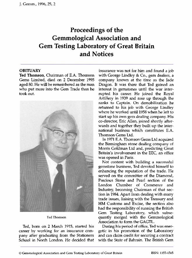
J. Gemm., 1996, 25, 2
Proceedings of the Gemmological Association and
Gem Testing Laboratory of Great Britain and Notices
OBITUARY Ted Thomson, Chairman of E.A. Thomson Gems Limited, died on 2 December 1995 aged 80. He will be remembered as the man who put more into the Gem Trade than he took out.
"iiilllllllilll^ilicl
Ted Thomson
Ted, born on 2 March 1915, started his career by working for an insurance company after graduating from the Stationers School in North London. He decided that
insurance was not for him and found a job with George Lindley & Co., gem dealers, a company known at the time as the Jade Dragon. It was there that Ted gained an interest in gemstones until the war interrupted his career. He joined the Royal Artillery in 1939 and rose up through the ranks to Captain. On demobilization he returned to his job with George Lindley where he worked until 1958 when he left to start up his own gem dealing company His co-director, Eric Allen, joined shortly afterwards and together they built up the international business which constitutes E.A. Thomson Gems Ltd.
In 1971 E.A. Thomson Gems Ltd acquired the Birmingham stone dealing company of Morris Goldman Ltd and, predicting Great Britain's involvement in the EEC, an office was opened in Paris.
Not content with building a successful gemstone business, Ted devoted himself to enhancing the reputation of the trade. He served on the committee of the Diamond, Precious Stone and Pearl section of the London Chamber of Commerce and Industry, becoming Chairman of that section in 1984. Apart from dealing with many trade issues, liaising with the Treasury and HM Customs and Excise, the section also had the responsibility of running the British Gem Testing Laboratory, which subsequently merged with the Gemmological Association to form the GAGTL.
During his period of office, Ted was energetic in his promotion of the Laboratory and can claim credit for securing a contract with the State of Bahrain. The British Gem
© Gemmological Association and Gem Testing Laboratory of Great Britain ISSN: 1355-4565
J. Gemm., 1996, 25,1 159
FORTHCOMING MEETINGS London Unless otherwise stated, meetings will be held at the GAGTL Gem Tutorial Centre, 27 Greville Street (Saffron Hill entrance), London EC1N 8SU. Entry will be by ticket only at £3.50 for a member (£5.00 for non-members) available from the GAGTL.
8 May Identifying inclusions within gemstones Jamie Nelson
5 June 3-D slide presentation of gemstones and minerals David Minster
10 June Annual General Meeting, Reunion of Members and
Bring and Buy
(There will be no charge for this event)
14 June Annual Trade Luncheon. The Langham Hilton, Portland Place, London, Wl. The speaker will be Nairn Attallah, former Chief Executive of Asprey
Midlands Branch Monthly meetings will be held at the Discovery Centre, 77 Vyse Street, Birmingham 18 (for directions to the Sunday Gem Club venue contact Gwyn Green on 0121^45 5359). Further details from Mandy MacKinnon on 0121-624 3225.
26 April Annual General Meeting followed by a talk by D.H. Ariyaratna
28 April Preliminary Gemmology Seminar
5 May Diploma Gemmology Seminar
19 May Gem Club - visit to Dolgellau Gold Mine
North West Branch Meetings will be held at Church House, Hanover Street, Liverpool 1. Further details from Joe Azzopardi on 01270 628251.
15 May Jade, past and present Rosamond Clayton
19 June Jewellery in the auction world David Lancaster
Scottish Branch For details of Scottish Branch meetings contact Ruth Cunningham on 0131-225 4105.
19-21 April A weekend of Scottish gemmology Alan Hodgkinson
A three-day event to be held in Perth to include talks and practical workshops by Alan Hodgkinson, the Branch's first Annual General Meeting and a field trip to Ethiebeaton and Ardownie quarries near Dundee.
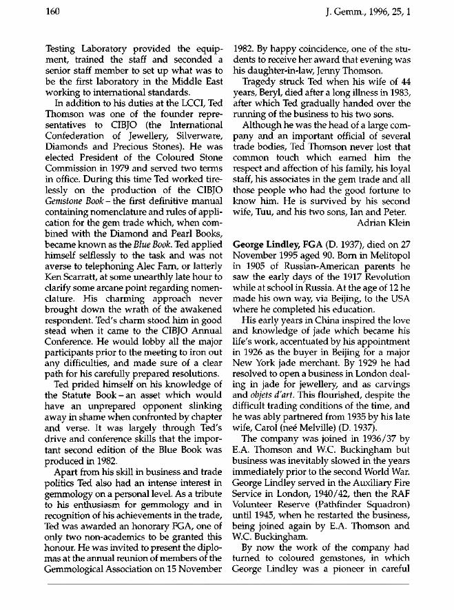
160 J. Gemm., 1996, 25,1
Testing Laboratory provided the equipment, trained the staff and seconded a senior staff member to set up what was to be the first laboratory in the Middle East working to international standards.
In addition to his duties at the LCCI, Ted Thomson was one of the founder representatives to CIBJO (the International Confederation of Jewellery, Silverware, Diamonds and Precious Stones). He was elected President of the Coloured Stone Commission in 1979 and served two terms in office. During this time Ted worked tirelessly on the production of the CIBJO Gemstone Book - the first definitive manual containing nomenclature and rules of application for the gem trade which, when combined with the Diamond and Pearl Books, became known as the Blue Book. Ted applied himself selflessly to the task and was not averse to telephoning Alec Farn, or latterly Ken Scarratt, at some unearthly late hour to clarify some arcane point regarding nomenclature. His charming approach never brought down the wrath of the awakened respondent. Ted's charm stood him in good stead when it came to the CIBJO Annual Conference. He would lobby all the major participants prior to the meeting to iron out any difficulties, and made sure of a clear path for his carefully prepared resolutions.
Ted prided himself on his knowledge of the Statute B o o k - a n asset which would have an unprepared opponent slinking away in shame when confronted by chapter and verse. It was largely through Ted's drive and conference skills that the important second edition of the Blue Book was produced in 1982.
Apart from his skill in business and trade politics Ted also had an intense interest in gemmology on a personal level. As a tribute to his enthusiasm for gemmology and in recognition of his achievements in the trade, Ted was awarded an honorary FGA, one of only two non-academics to be granted this honour. He was invited to present the diplomas at the annual reunion of members of the Gemmological Association on 15 November
1982. By happy coincidence, one of the students to receive her award that evening was his daughter-in-law, Jenny Thomson.
Tragedy struck Ted when his wife of 44 years, Beryl, died after a long illness in 1983, after which Ted gradually handed over the running of the business to his two sons.
Although he was the head of a large company and an important official of several trade bodies, Ted Thomson never lost that common touch which earned him the respect and affection of his family, his loyal staff, his associates in the gem trade and all those people who had the good fortune to know him. He is survived by his second wife, Tuu, and his two sons, Ian and Peter.
Adrian Klein
George Lindley, FGA (D. 1937), died on 27 November 1995 aged 90. Born in Melitopol in 1905 of Russian-American parents he saw the early days of the 1917 Revolution while at school in Russia. At the age of 12 he made his own way, via Beijing, to the USA where he completed his education.
His early years in China inspired the love and knowledge of jade which became his life's work, accentuated by his appointment in 1926 as the buyer in Beijing for a major New York jade merchant. By 1929 he had resolved to open a business in London dealing in jade for jewellery, and as carvings and objets d'art. This flourished, despite the difficult trading conditions of the time, and he was ably partnered from 1935 by his late wife, Carol (neé Melville) (D. 1937).
The company was joined in 1936/37 by E.A. Thomson and W.C. Buckingham but business was inevitably slowed in the years immediately prior to the second World War. George Lindley served in the Auxiliary Fire Service in London, 1940/42, then the RAF Volunteer Reserve (Pathfinder Squadron) until 1945, when he restarted the business, being joined again by E.A. Thomson and W.C. Buckingham.
By now the work of the company had turned to coloured gemstones, in which George Lindley was a pioneer in careful
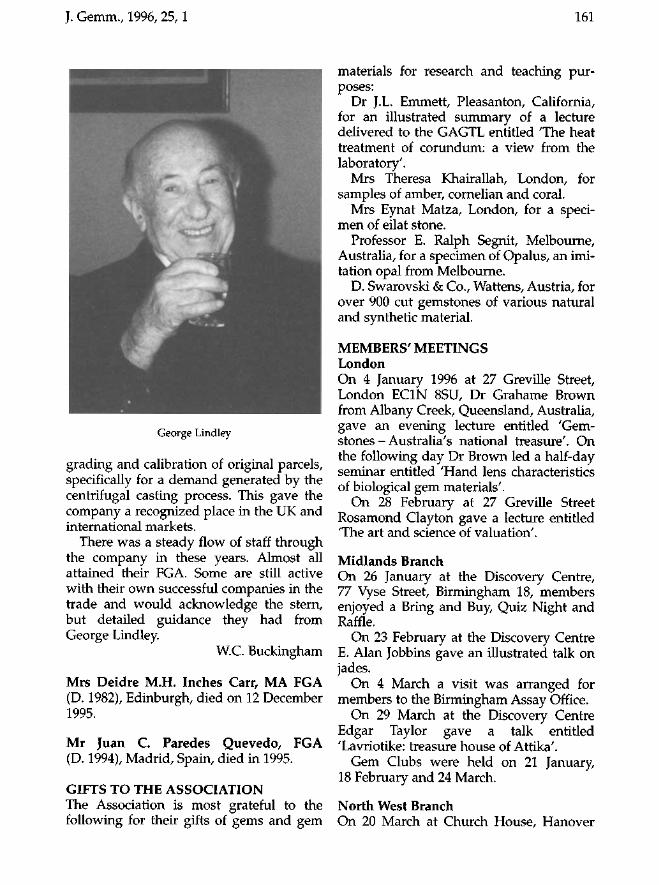
J. Gemm., 1996, 25,1 161
George Lindley
grading and calibration of original parcels, specifically for a demand generated by the centrifugal casting process. This gave the company a recognized place in the UK and international markets.
There was a steady flow of staff through the company in these years. Almost all attained their FGA. Some are still active with their own successful companies in the trade and would acknowledge the stern, but detailed guidance they had from George Lindley
W.C. Buckingham
Mrs Deidre M.H. Inches Carr, MA FGA (D. 1982), Edinburgh, died on 12 December 1995.
Mr Juan C. Paredes Quevedo, FGA (D. 1994), Madrid, Spain, died in 1995.
GIFTS TO THE ASSOCIATION The Association is most grateful to the following for their gifts of gems and gem
materials for research and teaching purposes:
Dr J.L. Emmett, Pleasanton, California, for an illustrated summary of a lecture delivered to the GAGTL entitled The heat treatment of corundum: a view from the laboratory'.
Mrs Theresa Khairallah, London, for samples of amber, cornelian and coral.
Mrs Eynat Matza, London, for a specimen of eilat stone.
Professor E. Ralph Segnit, Melbourne, Australia, for a specimen of Opalus, an imitation opal from Melbourne.
D. Swarovski & Co., Wattens, Austria, for over 900 cut gemstones of various natural and synthetic material.
MEMBERS' MEETINGS London On 4 January 1996 at 27 Greville Street, London EC1N 8SU, Dr Grahame Brown from Albany Creek, Queensland, Australia, gave an evening lecture entitled 'Gem-stones - Australia's national treasure'. On the following day Dr Brown led a half-day seminar entitled 'Hand lens characteristics of biological gem materials'.
On 28 February at 27 Greville Street Rosamond Clayton gave a lecture entitled The art and science of valuation'.
Midlands Branch On 26 January at the Discovery Centre, 77 Vyse Street, Birmingham 18, members enjoyed a Bring and Buy, Quiz Night and Raffle.
On 23 February at the Discovery Centre E. Alan Jobbins gave an illustrated talk on jades.
On 4 March a visit was arranged for members to the Birmingham Assay Office.
On 29 March at the Discovery Centre Edgar Taylor gave a talk entitled 'Lavriotike: treasure house of Attika'.
Gem Clubs were held on 21 January, 18 February and 24 March.
North West Branch On 20 March at Church House, Hanover
162 J. Gemm., 1996, 25,1
Your FGA in Nine Months! Study in the Heart of London - where gemmology started-at the Headquarters of the
Gemmological Association and Gem Testing Laboratory of Great Britain 27 Greville Street, London EC1N 8SU Tel: +44 (0) 171-404 3334 Fax: +44 (0) 171-404 8843
The next course starts on 16 September
The price of £3700 includes: Tuition - basic instruments - examination fees
GAGTL membership for one year
Street, Liverpool 1, Deanna Brady gave a talk entitled 'Introduction to inclusions'.
Scottish Branch On 7 February a Lab Night was held at Telford College, Edinburgh.
On 22 March at Newliston House, Newbridge, Edinburgh, members enjoyed a quiz and Bring and Buy sale.
GEM DIAMOND EXAMINATIONS In January 1996 12 candidates sat the Gem Diamond Examinations in the UK of whom 8 qualified. The names of the successful candidates are listed below:
Atkin, Marie, Sheffield. Cauchi, Savona, Louis, Malta. Crowder, Michael J., London. Gartery, Tania M., Christchurch. Hill, Katia, Glasgow. Johnson, Christopher W.R., Bickley, Nr
Bromley. Robinson, Zoe L., London. Ryder, Peter Ian, London.
The overseas candidates who qualified will be published in the July issue of the Journal.
EXAMINATIONS IN GEMMOLOGY In the Examinations in Gemmology held in January 1996 183 candidates sat for the Preliminary examination of whom 147 qualified; 116 candidates sat for the Diploma examination; 50 have qualified so far. The additional overseas candidates who qualified will be published in the July issue of the Journal.
The names of the successful candidates are as follows:
Qualified with Distinction Grondin, Daniel, Geneva, Switzerland.
Qualified Abramian, Levon, London. Ahamed, Omar Faiz, Gillingham. Ang, Zen Yi, Singapore. Ayyar, Rajeshwari Narayan, Bombay, India.
J. Gemm., 1996, 25,1 163
Brangulis, Peters, Riga, Latvia. Briginshaw, Richard Charles, London. Brooke-Webb, Susannah, London. Close, Helen Jane, Selby. Cunningham, DeeDee Cecilia, Toronto,
Ont., Canada. Dempster, Stuart, Glasgow. Fleischner-Zois, Krista, Athens, Greece. Gil Perez, Nuria, Valencia, Spain. Golad, Tanya, London. Grondin, Magali, Geneva, Switzerland. Horniblow, Kathy, Sherbourne. Hyo-Jin, Choi, Tae-gu, Korea. Jayasendil, J., Salem, India. Kim, Yun-Hwa, Kyung Buk, Korea. Kimber, Sarah, London. Kuulman, Heli, Tallinn, Estonia. Lake, Richard John, Gorey Village, Jersey. Limaye, M.D., Calcutta, India. McFarlane, Iain, Chesham. MacNish Porter, Holly, Edinburgh. Marr, Peter, Torquay Mathon, A.P.T., Oosterhout, The Nether
lands. McFarlane, Iain, Chesham. Melego Canet, Merce, Valencia, Spain. Michelson, Max J., Lincoln. Mihama, Saki, London. Naing, Aye Myo, Yangon, Myanmar. Oja, Tonis, Tallinn, Estonia. Parekh-Guastella, Rita, Pontedera, Italy. Perkins, David J., Ayr. Pierce, Jason Robert, Truro. Pierce, Jill Fiona, Manchester. Plomp, Jean Victoir, Rotterdam, The
Netherlands. Popov, Vladimir V, Tallinn, Estonia. Prabhu, Miss Vaishali, Bombay, India. Rai, Ashish, Jaipur, India. Rege, Sonal, Bombay, India. Shepherd, Louis, Nottingham. Song, Gyu Bong, Ulsan, Korea. Stopelli, Christopher J.D., Oakville, Ont.,
Canada. Torrent, Denise, Geneva, Switzerland. Vakil, Lakshmi H., Bombay, India. van der Molen, W.N., Schoonhoven, The
Netherlands. Van Duijnen, A.M., Bergen aan Zee, The
Netherlands.
Verma, Hitesh, Jaipur, India. Vummidi, Nagaraj, Madras, India. Withington, Terry, London.
Preliminary Amrute, Rahul M., Bombay, India. Andaluz Sanchez, Maria, London. Anderson, Elizabeth Anne, Lewes. Ayyar, Rajeshwari Narayan, Bombay, India. Bagadia, Pavan Madanlal, Bombay, India. Barbas, Constantinos, Athens, Greece. Burden, Lorraine, Sawbridgeworth. Cao, Shumin, Wuhan, China. Chan, Hor Wai Pierre, Kowloon, Hong
Kong. Chan, Vivian S.F., N.T., Hong Kong. Chan, Lam Ming, Hong Kong. Chen, Kai-Wen, Taipei, Taiwan. Chong, Yung Yung Sylvia, Toronto, Ont.,
Canada. Chung, Wen-Hao, Taipei, Taiwan. Coker, Olive Admira, London. Cowley, Jacalyn Gloria, London. Cucchiara, Carmelina, Wolverhampton. D'Costa, Charles John, Thornton Heath. Dai, Sulan, Wuhan, China. Davidson, Simon J., Cookham. Davies, Darryn Lewis, London. Degermark, Sofia, London. Dismore, Frances Jane, London. Donald, Claire M., Perth. Dong, Ping, Wuhan, China. Du-Ri, Cho, Taegu, Korea. Dykhuis, Luella Woods, Tucson, Arizona,
USA. Ebbs, Heather Alexandra, Bath. Edery, Gabrielle Julietta, London. Effendy, Jusak, Central, Hong Kong. Fahller, Ingegerd, Lannavaara, Sweden. Falcon, Lionel Michael, Craighall, S. Africa. Feng, Allice, Taipei, Taiwan. Foote, David Stuart, Reading. Forbes, Victoria E., London. Forward, Stephen, London. Frick, Nola Ann, London. Guest-Joste, Catherine Deanna, London. Harvey, Mark Anthony, London. Hazelius-Berglund, Wiveka, Lidingo,
Sweden. Hegi, Matthieu Manuel Marie, London.
164 J. Gemm., 1996, 25,1
Ho, Suk King Betty, Kowloon, Hong Kong. Hobbs, Karen Lorraine, Bath. Hsieh, Chia-Jung, London. Hsieh, Hsiu-Hua, Katherine, Taipei,
Taiwan. Hsieh, Pao Lien, Evelyn, N.T., Hong Kong. Huang, Tai Wen, Taipei, Taiwan. Huijsser, Isabelle E., Vught, The
Netherlands. Hutton, Katie, London. Jatoi, Faisal Zamman, London. Jayasendil, J., Salem, India. Kearton, Michael James, Eastbourne. Kemp, Graham, London. Khairallah, Theresa, London. Kim, Yun-Hwa, Kyung Buk, Korea. Köhler, Ghislaine L.M., London. Kritikou, Eleni, London. Lai, Li, Wuhan, China. Lau, Joyce Pao-Ching, N.T., Hong Kong. Lee, Dongjae, London. Lee, Yow Hon, Tuen Mun, Hong Kong. Li, Yin Ni, London. Li, Li, Wuhan, China. Liang, Zi, Guangzhou, China. Limaye, M.D., Calcutta, India. Lin, Chien Yu Joyce, Taipei, Taiwan. Lin, Jiaqing, Wuhan, China. Lin, Siu Pan, Kowloon, Hong Kong. Lindgren, Maria, Lannavaara, Sweden. Lindwall, Torbjorn, Lannavaara, Sweden. Lui Pochoi, Tsuen Wan, Hong Kong. Mackie, Karen Louise, Gillingham. Mak, Tsui Sim, Hong Kong. Matu, Sheila Gathii, Nairobi, Kenya. Matza, Eynat, London. Mitteregger, Unni, London. Modi, Shivani, Delhi, India. Monje, Lucy, London. Monnar, Robert D., Nashua, New
Hampshire, USA. Nam, Chung-Chul, Taegu, Korea. Newbould, Hans Peter Michael, Esher. Nottbusch, Jürgen Uwe, Appel, Germany. Oh, Mi Sun, Daejon, Korea. Omine, Mitsunobu, London. Pancratz, Mark Alan, Weybridge. Pankaj, Nirmal Sethia, Bombay, India. Parekh-Guastella, Rita, Pontedera, Italy. Patel, Ajay, Reading.
Perks, Helen Elizabeth, Wolverhampton. Prabhu, Vaishali, Bombay, India. Prachi, Raghuwanshi, Bombay, India. Rege, Sonal, Bombay, India. Riddell, Vanessa Stella, Harrow. Rijken, Yvonne A.W., Doorwerth, The
Netherlands. Rizwan, Babar, Ilford. Rollings, Alexander Mark Baker, London. Romero, Aniana Noguera, London. Romero de Castilla, M.M. Solomon
Castillui, Ilford. Schellekens, Ben J., Rotterdam, The
Netherlands. Sequeira, Silvia W., Bombay, India. Shen, Chen-Hua, Taipei, Taiwan. Shih, Shu Chuan, London. Shiue, Ling-yu, Taipei, Taiwan. Smith, Sarah A., Bath. Stossel, Hilary Jeanne, London. Swaffer, Hugh Edward Thomas, Brighton. Tang, Wong Wai, Manchester. Tao, Hongwei, Wuhan, China. Tao, Jinbo, Wuhan, China. Todd, Anthony James, London. Tolia, Ami Devendra, Bombay India. Tong, Nicolette Tong, London. Tsai, Shu Mei, Taipei, Taiwan. Tsang, Wai Wan, Kowloon, Hong Kong. Vafai, Khatereh, Teheran, Iran. Vakil, Lakshmi H., Bombay, India. Van Teeffelen, Cornelia Elizabeth,
Bodegraven, The Netherlands. Velliniati, Katerina, Athens, Greece. Von Speyr, Lucy Jane Mountjoy,
Cheltenham. Wananabe, Hanaco, London. Wang, Chien Ling, Taipei, Taiwan. Wang, Liqun, Guangzhou, China. Wang, Fenglan, Wuhan, China. Watts, Emma Louise, London. Watts-Rehman, Claudia, London. Wavoo, Shahul Hameed, Kowloon, Hong
Kong. Wilson, Hollis, London. Wisawayodhin, Kitiya, London. Wong, Kevin Kwok Wing, NT Hong Kong. Wu, Hsiu Tzu, Taiwan. Wu, Ming Hsun, Taipei, Taiwan. Wu, Xiaobing, Wuhan, China.
J. Gemm., 1996, 25,1 165
GÀCTL GEM TOT0RIAL CENTRE 27 Greville Street (Saffron Hill entrance), London EC1N8SU
ATTENTION ALL STUDENTS!
Intensive woikshops designed for students taking the GAGTL's Gemmology md Gem Diamond courses. Invaluable information is provided on revision and examination techniques. The two-day workshops also include mock examinations to help students gain femiliarity and confidence with examination conditions.
21 May Review of Gemmology Diploma Theory £33.49 (including sandwich lunch)
25-26 May and 1-2 June Two-day Diploma Practical Workshop £l60J9(£lIlM4ßrGAGTLregisteredstudsmyM sandwich lunch
15-16 June Weekend Diamond Grading Revision £129.25
Please contact the Education Office on 0171-404 3334 for further information
Xi, Bo, Wuhan, China. Xie, Liming, Wuhan, China. Xu, Xiaoxia, Wuhan, China. Yang, Lixin, Wuhan, China. Yeh, Chen-Mei, Taipei, Taiwan. Yen, C.K., Taipei, Taiwan, Yip, Nga Lai, NT Hong Kong. Yip Yick Hing, Brenda, Aberdeen, Hong
Kong. Zakir, Aliasgher Fakhruddin, Nairobi,
Kenya. Zeng, Chunguang, Shanghai, China. Zhang, Xueyun, Wuhan, China. Zhang, Yanjun, Wuhan, China. Zheng, Kaiwen, Wuhan, China. Zhu, Xionghua, Wuhan, China. Zou, Tao, Wuhan, China.
MEETINGS OF THE COUNCIL OF MANAGEMENT At meetings of the Council of Management held at 27 Greville Street, London EC1N 8SU, the business transacted included the election of the following:
Meeting held 6 December 1995
Diamond Membership Chen, Hui Lan, Shenzhen, China. 1995 Gantzidis, Adam, Athens, Greece. 1995 Garin, Auzmendi Amaia, Madrid, Spain.
1995
Fellowship Au Yeung, Kwok Ho, Hong Kong. 1995 Chun-Li, Wu, Edgbaston. 1995 Dang, Xiao-Ying, Beijing, China. 1995 Heyes, Alan John, Stourbridge. 1970 Hui, Wai Yee Wendy, Hong Kong. 1994 Loungani, Jagdish, Jaipur, India. 1995 Maheshwari, Ashoo, Jaipur, India. 1995 Marathe, Tanuja, Pune, India. 1995 Salm-Reifferscheidt, Sophie, London. 1995 Siu, Ming Wa, Kowloon, Hong Kong, 1995. Tang, Zhen Yi, Kowloon, Hong Kong. 1995 Rossiter, John Timothy, Weston Super Mare.
1995 Zheng, Bei, Beijing, China. 1995
Ordinary Membership Anderson, Lesley Christine, Wivenhoe. Ashton, David, London.
166 J. Gemm., 1996, 25,1
Bienemann, Andreas Martinus, Polsbroek, The Netherlands.
Brittain, Roger Anthony, Forncett St Peter, Norwich.
Burchell, Janet, Ravenstone. Chang, Mei-Chu, Taipei, Taiwan. Cuthbert, Richard Allen, London. Dawson, Stewart, Aberdeen. Dean, John Eldon, Bath. Jessa, Alnasir R., Wembley. Kuulman, Heli, Tallinn, Estonia. Li, Long Hsing, Taichung, Taiwan. Liu, Jian, Beijing, China. Marshall, Andrew Paul, Gravesend. Notari, Franck, Geneva, Switzerland. Osundina, Olalekan, London. Ronza, Antonella, Novara, Italy. Rutter, Fay, Walsall. Shibuya, Akemi, Tokyo, Japan. Smith, Darrell Arthur, Haywards Heath. Stopelli, Christopher John David,
Brampton, Ont., Canada. Tsai, Yao Chu, Kaoshung, Taiwan. Yao, Teng Long, Taichung, Taiwan.
Laboratory Membership Laboratörios G & M, Lisbon, Portugal Bulgari Distribuzione, Rome, Italy Escuela Internacional de Joyeria,
Gemologia Y Tasaciones, Madrid, Spain Richard Ogden Ltd, London W1V ONX Spink & Son Ltd, London SW1Y 6QS
Meeting held 14 February 1996
Diamond Membership Bagchi-Narayam, Debal, Brentford. 1995 Ji, Xiaoyan, Hubei, China. 1995 Lu, Yefei, Shanghai, China. 1995 Pattani, Shobna, London. 1995 Ren, Jiakai, Hubei, China. 1995 Wang, Hui, Shanghai, China. 1995 Wang, Xin, Shen Yang, China. 1995 Weng, Ping, Hubei, China. 1995 Xi, Bo, Hubei, China. 1995 Xu, Ru-peng, Shanghai, China. 1995 Zhang, Weichao, Hubei, China. 1995
Diamond Membership and Fellowship Leese, Peter Francis, Croxley Green.
1953/1967
Fellowship Bae, Eun Jeung, Seoul, Korea. 1995 Bishop, Lyndall Ann, Catterick Village. 1987 Bourke, Mary, Enniscorthy, Ireland. 1987 Harman, Bronwen, The Gap, Qld,
Australia. 1992 Jiang, Xinshun, Shen Yang, China. 1995 Peryer, Keith George, London. 1987 Pluckrose, William Henry, St Mary's Piatt.
1979 Qin, Shu-Le, Shanghai, China. 1995 Rydeng, Rig, Finnsnes, Norway. 1988 Sanson, Steven Nigel James, London. 1990 Sun, Yan Ling, Jiangsu Province, China.
1995 Sung, Min Jun, Taegu, Korea. 1995 Zhou, Shuzhen, Ningbo, China. 1995
Transfers from Ordinary Membership to FGA Bowman, Helene, Epping. 1995
Ordinary Membership Achten, Louisa Wendelina Wilhelmina,
Horst, The Netherlands. Adams, Victoria, Pimlico, London. Akashi, Kazuko, Hyogo, Japan. Akieda, Yumi, Hyogo, Japan. Alessandro, Guastoni, Milan, Italy. Beizer, Samuel, New York, NY, USA. Bossi, Anna, Milan, Italy. Cunningham, Ruth, Edinburgh. Dodo, Naoko, Osaka, Japan. Doyle, Ted, Bangkok, Thailand. Drysdale, John G.S., Tiverton. Egawa, Noriko, Osaka, Japan. Endo, Yuko, Kanagawa, Japan. Fisher, Claire Monica, London. Fujii, Noburu, Nara, Japan. Fukouka, Taishi, Kyoto, Japan. Hill, Katia, Glasgow. Ichikawa, Yoko, Hyogo, Japan. Ishibe, Masahiko, Tokushima, Japan. Ishijima, Juri, Hyogo, Japan. Katoda, Yuka, Osaka, Japan. Katagiri, Kumiko, Osaka, Japan. Kitamon, Hideaki, Nara, Japan. Kobayashi, Mitsuko, Hyogo, Japan. Kokuho, Shigeko, Tokyo, Japan. Kuroda, Chikako, Hyogo, Japan. Matsui, Rie, Osaka, Japan.
J. Gemm., 1996, 25,1 167
Miyamoto, Takuya, Hyogo, Japan. Miyamura, Kayoko, Shiga, Japan. Murata, Sanae, Nara, Japan. Myint Soe, Yangon, Myanmar. Nakayama, Norio, Kyoto, Japan. Nicolaou, Emmanuel George, Germiston,
South Africa. Nishikawa, Masako, Osaka, Japan. Ogino, Takanori, Osaka, Japan. Ohsumi, Yuichi, Kyoto, Japan. Perks, Helen Elizabeth, Wolverhampton. Porter, Simon Douglas, Glasgow. Previn, Heather Mary, New York, NY, USA. Ryuno, Yumiko, Kumamoto, Japan. Shikazawa, Chiaki, Yamanashi, Japan. Shiota, Mika, Osaka, Japan. Smith, Sarah Jane, Erdington, Birmingham. Sugawara, Yasuhiko, Tokyo, Japan. Suzuki, Noriko, Nara, Japan. Takahashi, Sachiyo, Nara, Japan. Tanaka, Kiyomi, Osaka, Japan. Tashiro, Hisami, Kyoto, Japan. Wataya, Atsuto, Osaka, Japan. Wiseman, Alan, Maiden. Yajima, Yuko, Saitama, Japan. Jamashsita, Yuko, Osaka, Japan. Yatohmaru, Hiroshi, Hyogo, Japan.
Laboratory Membership Hattons, Gants Hill, Ilford
Meeting held 13 March 1996
Diamond Membership Wu, Zhanxia, Shanghai, China, 1995. Sheng, Beili, Shanghai, China. 1995 Summerfield, Susan Denise, London. 1995
Transfer - FGA to FGA, DGA Robinson, Zoe L., London. 1996
Transfers - Ordinary Membership to DGA Cauchi Savona, Louis, Brentwood. 1996 Hill Rodan, Katia, Glasgow. 1996 Johnson, Christopher W.R., Bickley. 1996 Ryder, Peter Ian, London. 1996
Fellowship Baker, Kimberly Helen, Loughborough.
1994 Brooke-Webb, Susannah, London. 1996 Dalla Libera, Natalie Ann, London. 1995
Dixon-Phillip, John Graham, Wallasey. 1984 Li, Yuan, Wuhan, China. 1995 Michelson, Max John, Lincoln. 1996 Parekh-Guastella, Rita, Massa Marittima,
Italy. 1996 Perkins, David James, Ayr. 1996 Zhou, Jun, Beijing, China. 1995
Transfers - Ordinary Membership to FGA Abramian, Levon, London. 1996 Ahamed, Omar Faiz, Gillingham. 1996 Briginshaw, Richard Charles, London. 1996 Golad, Tanya, London. 1996 Grondin, Magali, Geneva, Switzerland.
1996 Horniblow, Kathy, Sherbourne. 1996 Kimber, Sarah, London. 1996 Kuulman, Heli, Tallinn, Estonia. 1996 Lake, Richard J., Gorey Village, Jersey. 1996 MacNish, Porter, Holly, Edinburgh. 1996 Mihama, Saki, London. 1996 Oja, Tonis, Tallinn, Estonia, 1996 Pierce, Jason Robert, Truro. 1996 Plomp, Jean Victoir, Rotterdam, The
Netherlands. 1996 Stopelli, Christopher J.D., Brampton, Ont.,
Canada. 1996 Torrent, Denise, Geneva, Switzerland. 1996 Van Duijnen, A.M., Bergen ann Zee, The
Netherlands. 1996 Withington, Terry, London. 1996
Ordinary Membership Baxter, Harry John Trevor, Exeter. Boghossian, Jean, Antwerp, Belgium. Caulton, David Michael, Sheffield. Gevorkian, Vardan, Holmestrand, Norway. Lemessiou, Maria, Hampstead, London. Lindwall, Torbjorn, Lannavaara, Sweden. Marshall, Ralph Henry, Northwood. Spiteri, Ruth, Sta Venera, Malta. Stannard, Richard John, Paris, France. Su, Bin, Wuhan, China. Thorsteinsson, Vigfus, Akureyri, Iceland. Yen, CK., Taipei, Taiwan.
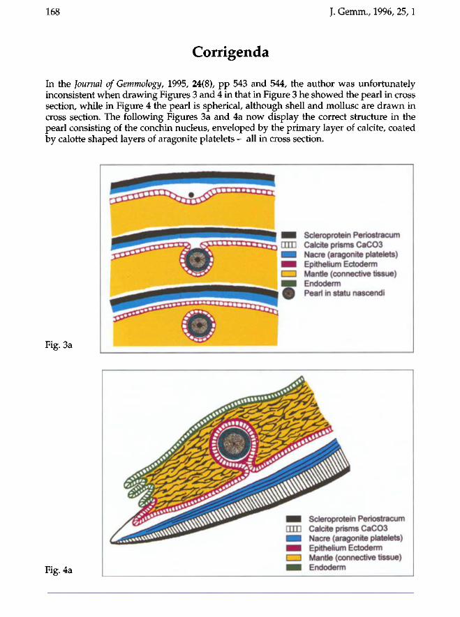
168 J. Gemm., 1996, 25,1
Corrigenda
In the Journal of Gemmology, 1995, 24(8), pp 543 and 544, the author was unfortunately inconsistent when drawing Figures 3 and 4 in that in Figure 3 he showed the pearl in cross section, while in Figure 4 the pearl is spherical, although shell and mollusc are drawn in cross section. The following Figures 3a and 4a now display the correct structure in the pearl consisting of the conchin nucleus, enveloped by the primary layer of calcite, coated by calotte shaped layers of aragonite platelets - all in cross section.
Fig. 3a
Scleroprotein Periostracum Calcite prisms CaC03 Nacre (aragonite platelets) Epithelium Ectoderm Mantle (connective tissue) Endoderm Pearl in statu nascendi
Fig. 4a
Scleroprotein Periostracum Calcite prisms CaC03 Nacre (aragonite platelets) Epithelium Ectoderm Mantle (connective tissue) Endoderm
. Gemm., 1996, 25, 2 169
ADVERTISING IN THE JOURNAL OF
GEMMOLOGY
The Editors of the Journal invite advertisements from gemstone and mineral dealers, publishers and others with interests in the gemmological, mineralogical, lapidary and jewellery fields.
Rates per insertion, excluding VAT, are as follows:
Whole page Half page Quarter page £180 £100 £60
Enquiries to Mary Burland Advertising Manager
Gemmological Association and Gem Testing Laboratory of Great Britain 27 Greville Street, London EC IN 8SU
•
•
•
•
You
The only Russian popular science mineralogical journal M ines, minerals, museums and much morel Printed in English with supplementary sheet of text in Russian, One year subscription (4 issues) $52, including postage
[ may subscribe at any time. Back issues are Send your subscription details and cheque to:
UK:
volume 80pp
available.
Don Edwards, Tideswell Dale Rock Shop, Commercial Road, Tideswell, Derbys SKI 7 88NU, UK Please make cheque payable in £ Sterling to Don Edwards
Germany: L. Schneider, Hemmersweiher 5, 66386 St-Ingbert BRD. Please make cheque payable in DM to L. Schneider.
USA: H. Obodda, Box 51, Short Hills, NJ 07078, USA Please make cheque payable in US $ to H. Obodda
Editorial: Box 162 Moscow 103050 Russia. Telephone/Fax: (7-095) 203 3574
Museums, Educational Establishments,
Collectors & Students
I have what is probably the largest range of genuinely rare stones in the
UK, from Analcime to Wulfenite. Also rare and modern synthetics, and inexpensive stones for students. New computerised lists available with even more detail. Please send £2 in 1st class
stamps refundable on first order (overseas free).
Two special offers for students: New Teach/Buy service and free
stones on an order.
A.J. French, FGA 82 Brookley Road, Brockenhurst,
Hants S042 7RA Telephone 01590 623214

170 J. Gemm., 1996, 25, 2
We look after ajj your insurance PROBLEMS
For nearly a century T. H. March has built an outstanding reputation by helping people in business. As Lloyds brokers we can offer specially tailored policies for the retail, wholesale, manufacturing and allied jewellery trades. Not only can we help you with all aspects of your business insurance but we can also take care of all your other insurance problems,
whether it be home, car, boat or pension plan. We would be pleased to give advice and
quotations for all your needs and delighted to visit your premises if required for this purpose, without obligation.
Contact us at our head office shown below.
&
T. H. March and Co. Ltd. Saint Dunstan's House, Carey Lane, London EC2V 8AD. Telephone 0171-606 1282 Also at Birmingham, Manchester, Glasgow and Plymouth. Lloyd's Insurance Brokers
J. Gemm., 1996,25,2 171
PROMPT LAPIDARY SERVICE!
Gemstones and diamonds cut to your specification and repaired on our
premises. Large selection of gemstones including rare items and mineral specimens in
stock. Valuations and gem testing carried out.
Mail order service available.
R. HOLT & CO. LTD
98 Hatton Garden, London EC1N 8NX Telephone 0171 -405 0197/5286
Fax 0171-430 1279
MAGGIE CAMPBELL PEDERSEN ABIPP, FGA
JEWELLERY & GEMSTONE PHOTOGRAPHY
Tel: 0181-994 8341 Fax:0181-723 4266
172 J. Gemm., 1996, 25, 2
OUR SHOP IS A TREASURE TROVE
We specialize in organic gemstone beads, and pendants and carvings, of amber,
vegetable ivory, pearls, wood, bone and jet. We also have a comprehensive selection of many unusual beads such as pink and brown moonstone, charoite, chrysoberyl, sunstone,
opal, fluorite and labradorite.
Whether you require just a few beads or an exclusively-designed necklace,
come to our shop.
Trade enquiries welcome. Sorry - no catalogue as our stock is constantly changing.
38 Eld Lane, Colchester, Essex COI ILS Tel: 01206 764101 Fax: 01206 764202
- HAMAG faceting machines with basic free formation
in our workshop. - Diamond polishing syringes
(grains 0.25, 1 micron.,.)
Minerals, Gemstones, Pearls
Luc GENOT Chaussée de Wavre, 856
B-1040 Brussels Belgium
TEL : 32-2-647.38.16 FAX: 32-2-648.20.26
• GEMMOLOGY
• INSTRUMENTS
• CRYSTALS
• CUT SPECIMENS
m STUDY TOURS enesis
Ltd. • WORLD LEADERS IN PRIVATE GEMMOLOGICAL EDUCATION We specialize in small group intensive tuition, from scratch to F.G.A. Diploma in 9 months, we are able to claim a very high level of passes including Distinctions & prize winners amongst our students.
• GEMMOLOGICAL STUDY TOURS We organise a comprehensive programme of study tours for the student & the practising gemmologist to areas of specific interest, including :-ANTWERP, IDAR-OBERSTEIN, SRI LANKA, THAILAND & CHINA.
• DEALERS IN GEMSTONES/DIAMONDS & CRYSTALS We buy & sell cut and rough gemstones and diamonds, particularly for the F.G.A. syllabus, and have many rare or unusual specimens. Gemstones & Diamonds also available for commercial purposes.
• SUPPLIERS OF GEMMOLOGICAL INSTRUMENTS We offer a comprehensive range of gem testing instruments, including inexpensive Petrological & Stereo-zoom Microscopes, Refractometers, Hand Lenses, Pocket U /V Lights, S.G. Liquids, the world famous OPL Spectroscope, and many other items including Books & Study Aids.
Illustrated:
Petrological Microscope. Mag. 20x - 650x, with full range of oculars & objectives, wavelength filters, quartz wedge, Bertrand lens, iris diaphragms, graticules etc.
From ONLY £650 + VAT& Delivery/Carriage
For further details of these and our other activities, please contact-Colin Winter, F.G.A. or Hilary Taylor, B.A., F.G.A. at GENESIS, 21, West Street, Epsom, Surrey. KT18 7RL England Tel. 01372 742974 or Fax 01372 742426
Guide to the preparation of typescripts for publication in The Journal of Gemmology
The Editor is glad to consider original articles shedding new light on subjects of gem-mological interest for publication in The journal. Articles are not normally accepted which have already been published elsewhere in English, and an article is accepted only on the understanding that (1) full information as to any previous publication (whether in English or another language) has been given, (2) it is not under consideration for publication elsewhere and (3) it will not be published elsewhere without the consent of the Editor.
Typescripts Two copies of all papers should be submitted on A4 paper (or USA equivalent) to the Editor. Typescripts should be double spaced with margins of at least 25mm. They should be set out in the manner of recent issues of The Journal and in conformity with the information set out below. Papers may be of any length, but long papers of more than 10 000 words (unless capable of division into parts or of exceptional importance) are unlikely to be acceptable, whereas a short paper of 400-500 words may achieve early publication.
The abstract, references, notes, captions and tables should be typed double spaced on separate sheets.
On matters of style and rendering, please consult The Oxford dictionary for writers and editors (Oxford University Press, 1981).
Title page The title should be as brief as is consistent with clear indication of the content of the paper. It should be followed by the names (with initials) of the authors and by their addresses.
Abstract A short abstract of 50-100 words is required.
Key Words Up to six key words indicating the subject matter of the article should be supplied.
Headings In all headings only the first letter and proper names are capitalized.
A This is a first level heading
First level headings are in bold and are flush left on a separate line. The first text line following is flush left.
B This is a second level heading
Second level headings are in italics and are flush left on a separate line. The first text line following is flush left.
Illustrations Either transparencies or photographs of good quality can be submitted for both coloured and black-and-white illustrations. It is recommended that authors retain copies of all illustrations because of the risk of loss or damage either during the printing process or in transit.
Diagrams must be of a professional quality and prepared in dense black ink on a good quality surface. Original illustrations will not be returned unless specifically requested.
All illustrations (maps, diagrams and pictures) are numbered consecutively with Arabic numerals and labelled Figure 1, Figure 2, etc. All illustrations are referred to as 'Figures'.
Tables Must be typed double spaced, using few horizontal rules and no vertical rules. They are numbered consecutively with Roman numerals (Table IV, etc.). Titles should be concise, but as independently informative as possible. The approximate position of the Table in the text should be marked in the margin of the typescript.
Notes and References Authors may choose one of two systems:
(1) The Harvard system in which authors' names (no initials) and dates (and specific pages, only in the case of quotations) are given in the main body of the text, e.g. (Gübelin and Koivula, 1986, 29). References are listed alphabetically at the end of the paper under the heading References.
(2) The system in which superscript numbers are inserted in the text (e.g. ...to which Gübelin refers.3) and referred to in numerical order at the end of the paper under the heading Notes. Informational notes must be restricted to the minimum; usually the material can be incorporated in the text. If absolutely necessary both systems may be used.
References in both systems should be set out as follows, with double spacing for all lines.
Papers Hurwit, K., 1991. Gem Trade Lab notes. Gems & Gemology, 27, 2,110-11
Books Hughes, R.W., 1990. Corundum. Butterworth-Heinemann, London, p. 162
Abbreviations for titles of periodicals are those sanctioned by the World List of scientific periodicals 4th edn. The place of publication should always be given when books are referred to.
Volume 25 No. 2 April 1996
^Journal of Gemmology
Contents
Common and rare colourless gemstones D. Kent
Investigations on sapphires from an alkali basalt, South West Rwanda M.S. Krzemnicki,
H.A. Hänni, R. Guggenheim and D. Mathys
On the identification and fade testing of Maxixe beryl, golden beryl and green aquamarine K. Nassau
Emerald and green beryl from Central Nigeria
D. Schwarz, J. Kanis and J. Kinnaird
Abstracts
Reviews
Proceedings of the Gemmological Association and Gem Testing Laboratory of Great Britain and Notices
Corrigenda
Copyright © 1996 The Gemmological Association and Gem Testing Laboratory of Great Britain
Registered Office: Palladium House, 1-4 Argyll Street, London W1V 2LD
ISSN: 1355-4565
87
90
108
117
142
154
158
168
Printed by Stephen Austin & Sons Ltd, Hertford, England


