Embed Size (px)
Citation preview
BioMed CentralBMC Cancer
ss
Open AcceResearch articleThe immunomodulator PSK induces in vitro cytotoxic activity in tumour cell lines via arrest of cell cycle and induction of apoptosisEva Jiménez-Medina1, Enrique Berruguilla1, Irene Romero1, Ignacio Algarra2, Antonia Collado3, Federico Garrido1 and Angel Garcia-Lora*1Address: 1Servicio de Análisis Clínicos e Inmunologia, Hospital Universitario Virgen de las Nieves, Universidad de Granada, Av. de las Fuerzas Armadas 2, 18014 Granada, Spain, 2Departamento de Ciencias de la Salud, Universidad Jaén, Jaén, Spain and 3Unidad de Investigación, Hospital Universitario Virgen de las Nieves, Granada, Spain
Email: Eva Jiménez-Medina - [email protected]; Enrique Berruguilla - [email protected]; Irene Romero - [email protected]; Ignacio Algarra - [email protected]; Antonia Collado - [email protected]; Federico Garrido - [email protected]; Angel Garcia-Lora* - [email protected]
* Corresponding author
AbstractBackground: Protein-bound polysaccharide (PSK) is derived from the CM-101 strain of thefungus Coriolus versicolor and has shown anticancer activity in vitro and in in vivo experimental modelsand human cancers. Several randomized clinical trials have demonstrated that PSK has greatpotential in adjuvant cancer therapy, with positive results in the adjuvant treatment of gastric,esophageal, colorectal, breast and lung cancers. These studies have suggested the efficacy of PSKas an immunomodulator of biological responses. The precise molecular mechanisms responsiblefor its biological activity have yet to be fully elucidated.
Methods: The in vitro cytotoxic anti-tumour activity of PSK has been evaluated in various tumourcell lines derived from leukaemias, melanomas, fibrosarcomas and cervix, lung, pancreas and gastriccancers. Tumour cell proliferation in vitro was measured by BrdU incorporation and viable cellcount. Effect of PSK on human peripheral blood lymphocyte (PBL) proliferation in vitro was alsoanalyzed. Studies of cell cycle and apoptosis were performed in PSK-treated cells.
Results: PSK showed in vitro inhibition of tumour cell proliferation as measured by BrdUincorporation and viable cell count. The inhibition ranged from 22 to 84%. Inhibition mechanismswere identified as cell cycle arrest, with cell accumulation in G0/G1 phase and increase in apoptosisand caspase-3 expression. These results indicate that PSK has a direct cytotoxic activity in vitro,inhibiting tumour cell proliferation. In contrast, PSK shows a synergistic effect with IL-2 thatincreases PBL proliferation.
Conclusion: These results indicate that PSK has cytotoxic activity in vitro on tumour cell lines. Thisnew cytotoxic activity of PSK on tumour cells is independent of its previously describedimmunomodulatory activity on NK cells.
Published: 24 March 2008
BMC Cancer 2008, 8:78 doi:10.1186/1471-2407-8-78
Received: 5 November 2007Accepted: 24 March 2008
This article is available from: http://www.biomedcentral.com/1471-2407/8/78
© 2008 Jiménez-Medina et al; licensee BioMed Central Ltd. This is an Open Access article distributed under the terms of the Creative Commons Attribution License (http://creativecommons.org/licenses/by/2.0), which permits unrestricted use, distribution, and reproduction in any medium, provided the original work is properly cited.
Page 1 of 10(page number not for citation purposes)
BMC Cancer 2008, 8:78 http://www.biomedcentral.com/1471-2407/8/78
BackgroundA number of bioactive molecules, including antitumoursubstances, have been identified in various mushroomspecies. Polysaccharides are the best known and mostpotent of these and have antitumour and immunomodu-lating properties [1-5]. PSK, a protein-bound polysaccha-ride obtained from Basidiomycetes, also known as Krestin,has been used as an agent in the treatment of cancer inAsia for over 30 yrs [6-8]. PSK is derived from the fungusCoriolus versicolor and has documented anticancer activityin vitro in experimental models [9] and in human clinicaltrials. Several randomized clinical trials have demon-strated that PSK has great potential in adjuvant cancertherapy, with positive results in the treatment of gastric,esophageal, colorectal, breast and lung cancers [10,11].These studies have suggested the efficacy of PSK as animmunomodulator of biological response.
Previous reports indicated that PSK might act in differentways: as antioxidant [5,12,13]; as inhibitor of metallopro-teinases and other enzymes involved in metastatic proc-esses [14] and as inhibitor of the action of variouscarcinogens in vulnerable cell lines. However its mostimportant and widely reported property is its immu-nomodulatory capacity. PSK may act to increase leukocyteactivation and response via upregulation of key cytokines.Thus, natural killer (NK) and lymphocyte-activated killer(LAK) cell activation has been demonstrated in vivo and invitro [15,16]. Our group demonstrated that PSK is capableof inhibiting metastatic colonization in vivo in someexperimental fibrosarcomas, and that this effect is medi-ated by activation of NK cells [17,18]. Moreover, the NKcell line NKL, derived from a large granular lymphocyteleukaemia [19], is activated in vitro by PSK [16]. This acti-vation may replace IL-2 in inducing the proliferation andcytotoxicity of NKL cells. The signal transduction path-ways involved in the responses to IL-2 or PSK are different:IL-2 increases PKCα and ERK3 expression and decreasesERK2 expression, whereas PSK decreases PKCα expressionand increases ERK3 expression [20]. PSK also enhancesCRE binding activity, while IL-2 increases SP-1 and modi-fies GAS/ISRE, IRF-1 and STAT5 [21]. In addition, PSK andIL-2 have been shown to bind to different receptor onNKL cells [22].
The direct in vitro effect of PSK on the proliferation oftumour cell lines was compared with its effect on PBLs.PSK had cytotoxic activity on tumour cell lines, inhibitingproliferation, producing cell cycle arrest and cell accumu-lation in G0/G1phase and inducing apoptosis.
MethodsProtein-bound polysaccharide KProtein-bound polysaccharide K (PSK) was kindly pro-vided by Kureha Chemical Ind. Co. (Tokyo, Japan). It is
prepared by extracting cultured mycelia of Coriolus versi-color with hot water. The precipitate is separated from theclear supernatant with saturated ammonium sulfate, thendesalted and dried [23]. Protein-bound polysaccharide Kwas dissolved in RPMI medium or water and heated at50°C for 20–30 min until a clear solution appeared. ThePSK preparation was filter-sterilized and diluted in culturemedium or water to the desired concentration. Protein-bound polysaccharide K was previously titrated in NKLcells [16] and the working dilution was 100 μg/mL. PSKextract digested with neuraminidase was also tested,digesting 100 μg of PSK with 4 μl (Sigma) and incubatingfor 3 h at 37°C. Our group previously showed that PSK iscomposed of two bands of very high molecular weight[22]. After digestion with neuraminidase, these bands arereduced to a single band of about 12 kd. These resultsindicate that PSK is probably composed of a single 12-kdprotein, and that this protein is highly glycosylated [22].Two different extracts of PSK were also used: one rich insugars and other rich in proteins.
Cell lines and cell cultureThe following tumour cell lines were studied: B16 murinemelanoma, B9 murine MCA-induced fibrosarcoma,Ando-2 human melanoma, AGS human gastric cancer, A-549 human lung cancer, Hela human cervical adenocarci-noma and Jurkat T lymphoma leukemia. The NKL studiedwas established from PBLs of a patient with LGL leukemia[19]. All cell lines were obtained from the American TypeCulture Collection (Manassas, USA) except for the B9 cellline, which was generated at our laboratory, and theAndo-2 and NKL cell lines, kindly provided by P. Coulie(Unite de Genetique Cellulaire, Louvain University, Brus-sels, Belgium), F. X. Real (Instituto Municipal de Investi-gaciones Medicas, Barcelona, Spain) and Dr. M. Lopet-Botet (Universidad Pompeu-Fabra, Barcelona, Spain),respectively.
Cell lines derived from solid tumours were grown at 37°Cin a humidified atmosphere of 5% CO2 in DMEM culturemedium (Gibco, Paisley UK) supplemented with 10%heat-inactivated foetal bovine serum (Life Technologics,Milan Italy), antibiotics and glutamine. Jurkat T cell leuke-mia was cultured in RPMI 1640 with 10% heat-inacti-vated fetal bovine serum. The NKL cell line was culturedin RPMI 1640 with 10% heat-inactivated human ABserum (Sigma Chemical, St Louis, MO; USA) and humanrecombinant IL-2 (100 U/ml; purity > 97%, specific activ-ity, 2 × 10⁶ U/mg) (Roche, Nutley, NJ; USA).
In vitro cytotoxicity assaysThe effect of PSK on tumour cell proliferation wasassessed by measuring BrdU incorporation with the BrdUcolorimetric ELISA Cell Proliferation Kit (Roche Diagnos-tic). Cells were plated in 96-well microculture plates (5 ×
Page 2 of 10(page number not for citation purposes)
BMC Cancer 2008, 8:78 http://www.biomedcentral.com/1471-2407/8/78
103 cells/well). Every 48 h, the culture medium wasreplaced and PSK was added. After 48–96 h, BrdU label-ling reagent was added and cultured for a further 1–3 h.Assays were also performed by counting viable cells usingTrypan Blue. Briefly, cancer cell lines were seeded into cul-ture tissue-flask (1.5–2 × 10⁵/culture tissue-flask) andincubated for 24 h at 37°C in a humidified atmosphere of5% CO2. Cells were then treated with 100 μg/ml of PSK inthe culture medium, which was replaced every 48 h. After4–6 days, cells were collected by centrifugation and asmall sample of cell suspension was diluted in 0.4%Trypan Blue, counting cells in a haemocytometer cham-ber. Each cell sample was counted in this way at least threetimes and each assay was repeated at least three times.
Lymphocyte and NKL proliferation assayHuman lymphocytes were isolated from venous blood bythe Ficoll-Hystopaque separation method. Proliferationof PBLs was analyzed in vitro using 5-bromo-2'-deoxyuri-dine (BrdU) labelling of DNA-synthesizing cells with theabove-mentioned kit. PBLs were seeded in 96-well micro-culture plates at a cell density of 5 × 10⁴ per well. Two dif-ferent concentrations of PSK were used, 100 μg/ml and 50μg/ml. Concanavalin A (5 μg/ml, Sigma) and IL-2 wereused as positive controls. PSK was also used in combina-tion with IL-2 or Concanavalin A. After 48 h of culture inpresence or absence of PSK, BrdU labelling reagent (finalconcentration 10 μM) was added and cells were culturedfor 24 h. Cells were then fixed for 30 min and incubatedwith anti-BrdU for 1 h at 37°C. 100 μl of tetramethyl-ben-zidine (TMB) was used as substrate. Optical densities weredetermined at 370 nm by means of an ELISA microplatereader (Biotek, Power-Wave XS). Controls were the cul-ture medium, cells cultured only in medium and cellsincubated with anti-BrdU in absence of BrdU. All experi-ments were repeated at least three times.
Cell cycle distribution analysisBriefly, cells were plated in six-well plates (5 × 10⁵ perwell) or in culture tissue-flask (15 × 105) and continu-ously exposed for 4 days to 100 μg/ml of PSK. The DNAsynthesis rate was examined by BrdU incorporationmethod using FITC BrdU Flow Kit (BD Pharmingen)according to manufacturer's instructions. BrdU was thendetected by DNase cell treatment using FITC-conjugatedanti-BrdU antibody. Cells were washed with 1 ml 1 × BDPerm/Wash Buffer, and 20 μl 7-amino-actinomycin D wasadded. Analysis was performed with 50000 cells usingCell Quest Software and FACScan flow cytometer (Becton-Dickinson).
Annexin V binding assay to detect apoptotic cellsAfter treatment of cancer cells with PSK for four days, cellswere detached from the culture tissue-flask with PBS con-taining 3 mM EDTA. These cells were then collected
together with floating cells, washed twice with cold PBSand resuspended in binding buffer at a concentration of 1× 10⁶ cells per ml; 100 μl of solution was incubated for 30min at 4°C with 5 μl of Annexin V-PE antibody (BD Bio-sciences), and 5 μl of 7-amino-actynomycin D was thenadded. Cells were incubated for 15 min in darkness, and400 μl of staining buffer was added before flow cytometryanalysis. Apoptosis was analyzed by quadrant statistics asfollows: Annexin V- and 7-AAD-negative cells are alive;Annexin V-positive and 7-AAD-negative cells are in earlystages of apoptosis; Annexin V-negative and 7-AAD-posit-ice cells are dead but not by apoptosis; and Annexin V-positive and 7-AAD-positive cells are in mid- or end-stageapoptosis.
Assay for active caspase-3 expressionFITC conjugated monoclonal anti-active-caspase-3 anti-body (BD Biosciences) was used to determine whether theprotease caspase-3 is involved in PSK-induced apoptosis.After 4-day treatment with PSK, cancer cells were washedtwice with cold PBS and fixed and permeabilized inCytofix/Cytoperm buffer. Then, cells were incubated withFITC-conjugated monoclonal rabbit anti-active human-caspase-3 antibody for 30 min. Cells were washed twiceand 500 μl of 1 × Perm Wash Buffer was added beforeanalysis by flow cytometry.
Statistical analysisValues are expressed as means ± SD. Student's t-test wasused for statistical comparisons, considering a signifi-cance value of P < 0.05.
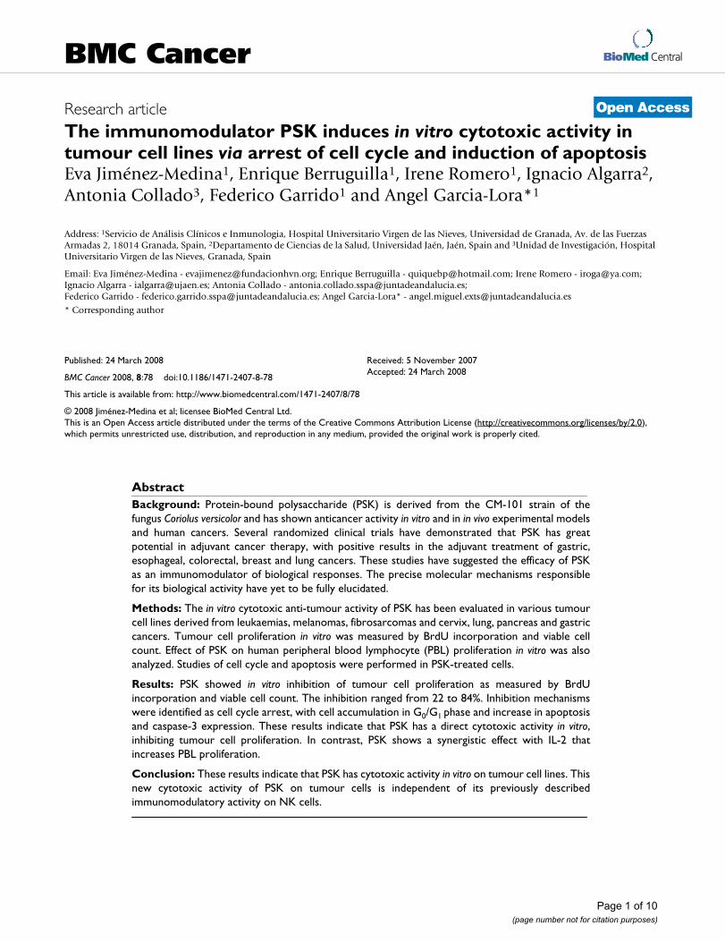
ResultsPSK inhibits in vitro tumour cell proliferationTumour cell lines were cultured in 96-well plate for 48–72h (2.5–5 × 10³ cells) in medium alone (control) or withPSK (100 μg/ml or 50 μg/ml) for 4–6 days. Cell prolifera-tion was then measured by BrdU incorporation (absorb-ance), which was significantly lower in PSK-treated versusuntreated tumour cells (Fig. 1). AGS and A549 cell linesshowed a strong decrease in absorbance after treatmentwith 100 μg/ml PSK that was less marked after treatmentwith 50 μg/ml of PSK. Inhibition of proliferation wasaround 65% in melanoma cell lines B16 and Ando-2,lower in Hela and Jurkat cell lines and lowest (20%) in B9murine fibrosarcoma (Fig. 1). PSK-treated tumour cellsshowed morphological changes (rounded and granulatedmorphology, increased vacuolisation, cell shrinkage) anda large number of the cells detached from culture flasks.
These assays were repeated in cell culture flasks (1.5–2.5 ×10⁵cells), and viable cells were counted in Haemacytome-ter chamber using Trypan Blue. As shown in Table 1, therewas a significant decrease in the final number of viablecells, with a proliferation inhibition of 22%–84% versus
Page 3 of 10(page number not for citation purposes)
BMC Cancer 2008, 8:78 http://www.biomedcentral.com/1471-2407/8/78
control cells. There was an excellent correlation betweenthe results obtained with the two assays (absorbance andcell count). In Hela tumour cells, proliferation inhibitionwas higher after treatment with 50 μg/ml versus 100 μg/ml of PSK. In all other tumour cell lines, proliferationinhibition was similar or higher at 100 μg/ml PSK.
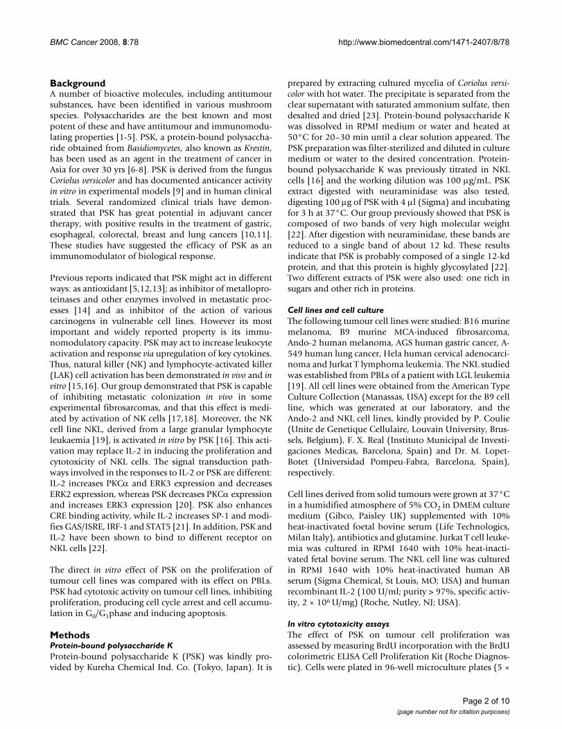
PSK increases in vitro proliferation of IL-2-stimulated lymphocytesA dose-response analysis was performed to determine thein vitro effect of PSK on human PBLs. PBLs (5 × 10⁴) wereplated in 96-well tissue plate for 48–72 h with eight seri-ally diluted extractions ranging from 500 μg/ml (concen-tration n°8) to 3.9 μg/ml (concentration n°1).Concentration n°0 represents cells cultured in mediumalone. BrdU incorporation during DNA synthesis was
then measured by ELISA. Optical densities were very sim-ilar between treated and untreated PBLs (data not shown).However, simultaneous treatment of PBLs with IL-2 (100U/ml) + PSK (100 μg/ml) produced a higher proliferationrate (4.5-fold) versus PBLs treated with IL-2 alone (3-fold)(Fig. 2). Untreated and Concanavalin A-treated PBLsserved as controls (Fig. 2).
Effect of different variants of PSKTumour cell proliferation inhibition was comparedamong different PSK variants. Neuraminidase treatmentdigests glicosylated proteins. A549 tumour cell line wascultured in medium alone (control) or with PSK (100 μg/ml) or neuraminidase-treated PSK (100 μg/ml) for 4–6
Effect of PSK on PBL proliferationFigure 2Effect of PSK on PBL proliferation. PBLs were cultured with PSK or IL-2 or with PSK+IL-2. PBLs (50 × 104 cells) were seeded in quadruplicate in 96-well plates. The prolifera-tion was determined by BrdU incorporation and absorbance measurement. PSK showed a synergistic effect with IL-2, increasing PBL proliferation. PSK alone did not induce PBLs proliferation. Each column represents the mean of five inde-pendent experiments ± SD. *P < 0.001 versus control.
���
����� ��� ��� ��� ����� �������
�
���
���
���
���
������� �
�
��
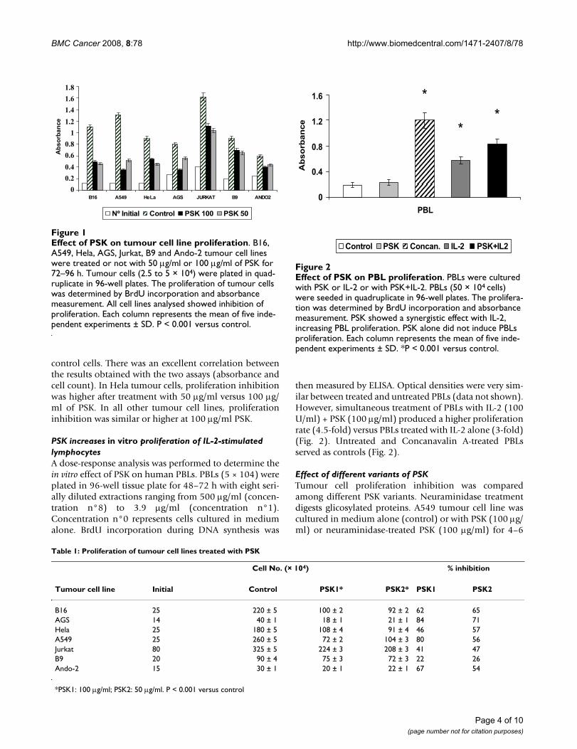
Table 1: Proliferation of tumour cell lines treated with PSK
Cell No. (× 104) % inhibition
Tumour cell line Initial Control PSK1* PSK2* PSK1 PSK2
B16 25 220 ± 5 100 ± 2 92 ± 2 62 65AGS 14 40 ± 1 18 ± 1 21 ± 1 84 71Hela 25 180 ± 5 108 ± 4 91 ± 4 46 57A549 25 260 ± 5 72 ± 2 104 ± 3 80 56Jurkat 80 325 ± 5 224 ± 3 208 ± 3 41 47B9 20 90 ± 4 75 ± 3 72 ± 3 22 26Ando-2 15 30 ± 1 20 ± 1 22 ± 1 67 54
*PSK1: 100 μg/ml; PSK2: 50 μg/ml. P < 0.001 versus control
Effect of PSK on tumour cell line proliferationFigure 1Effect of PSK on tumour cell line proliferation. B16, A549, Hela, AGS, Jurkat, B9 and Ando-2 tumour cell lines were treated or not with 50 μg/ml or 100 μg/ml of PSK for 72–96 h. Tumour cells (2.5 to 5 × 10⁴) were plated in quad-ruplicate in 96-well plates. The proliferation of tumour cells was determined by BrdU incorporation and absorbance measurement. All cell lines analysed showed inhibition of proliferation. Each column represents the mean of five inde-pendent experiments ± SD. P < 0.001 versus control.
��� ���� ��� �!� "#$��% �� �&'(�
&)���*�*� ����� ������� ������
��������������������������
������� �
Page 4 of 10(page number not for citation purposes)
BMC Cancer 2008, 8:78 http://www.biomedcentral.com/1471-2407/8/78
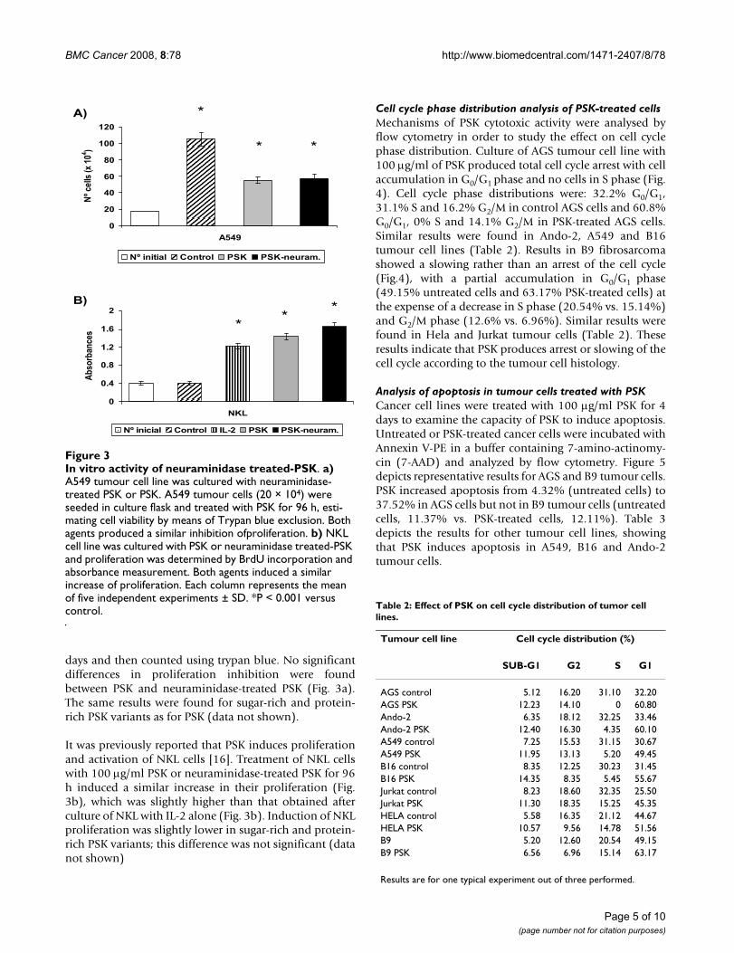
days and then counted using trypan blue. No significantdifferences in proliferation inhibition were foundbetween PSK and neuraminidase-treated PSK (Fig. 3a).The same results were found for sugar-rich and protein-rich PSK variants as for PSK (data not shown).
It was previously reported that PSK induces proliferationand activation of NKL cells [16]. Treatment of NKL cellswith 100 μg/ml PSK or neuraminidase-treated PSK for 96h induced a similar increase in their proliferation (Fig.3b), which was slightly higher than that obtained afterculture of NKL with IL-2 alone (Fig. 3b). Induction of NKLproliferation was slightly lower in sugar-rich and protein-rich PSK variants; this difference was not significant (datanot shown)
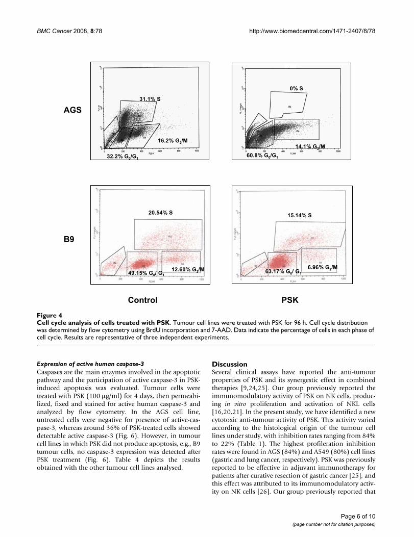
Cell cycle phase distribution analysis of PSK-treated cellsMechanisms of PSK cytotoxic activity were analysed byflow cytometry in order to study the effect on cell cyclephase distribution. Culture of AGS tumour cell line with100 μg/ml of PSK produced total cell cycle arrest with cellaccumulation in G0/G1 phase and no cells in S phase (Fig.4). Cell cycle phase distributions were: 32.2% G0/G1,31.1% S and 16.2% G2/M in control AGS cells and 60.8%G0/G1, 0% S and 14.1% G2/M in PSK-treated AGS cells.Similar results were found in Ando-2, A549 and B16tumour cell lines (Table 2). Results in B9 fibrosarcomashowed a slowing rather than an arrest of the cell cycle(Fig.4), with a partial accumulation in G0/G1 phase(49.15% untreated cells and 63.17% PSK-treated cells) atthe expense of a decrease in S phase (20.54% vs. 15.14%)and G2/M phase (12.6% vs. 6.96%). Similar results werefound in Hela and Jurkat tumour cells (Table 2). Theseresults indicate that PSK produces arrest or slowing of thecell cycle according to the tumour cell histology.
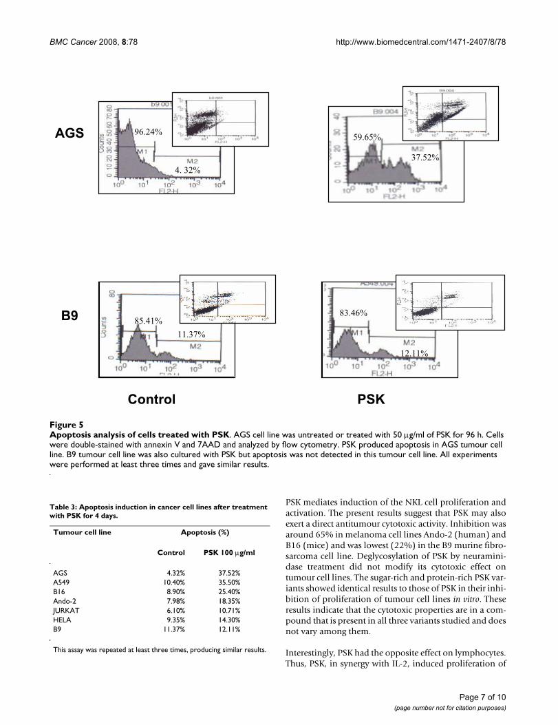
Analysis of apoptosis in tumour cells treated with PSKCancer cell lines were treated with 100 μg/ml PSK for 4days to examine the capacity of PSK to induce apoptosis.Untreated or PSK-treated cancer cells were incubated withAnnexin V-PE in a buffer containing 7-amino-actinomy-cin (7-AAD) and analyzed by flow cytometry. Figure 5depicts representative results for AGS and B9 tumour cells.PSK increased apoptosis from 4.32% (untreated cells) to37.52% in AGS cells but not in B9 tumour cells (untreatedcells, 11.37% vs. PSK-treated cells, 12.11%). Table 3depicts the results for other tumour cell lines, showingthat PSK induces apoptosis in A549, B16 and Ando-2tumour cells.
Table 2: Effect of PSK on cell cycle distribution of tumor cell lines.
Tumour cell line Cell cycle distribution (%)
SUB-G1 G2 S G1
AGS control 5.12 16.20 31.10 32.20AGS PSK 12.23 14.10 0 60.80Ando-2 6.35 18.12 32.25 33.46Ando-2 PSK 12.40 16.30 4.35 60.10A549 control 7.25 15.53 31.15 30.67A549 PSK 11.95 13.13 5.20 49.45B16 control 8.35 12.25 30.23 31.45B16 PSK 14.35 8.35 5.45 55.67Jurkat control 8.23 18.60 32.35 25.50Jurkat PSK 11.30 18.35 15.25 45.35HELA control 5.58 16.35 21.12 44.67HELA PSK 10.57 9.56 14.78 51.56B9 5.20 12.60 20.54 49.15B9 PSK 6.56 6.96 15.14 63.17
Results are for one typical experiment out of three performed.
In vitro activity of neuraminidase treated-PSKFigure 3In vitro activity of neuraminidase treated-PSK. a) A549 tumour cell line was cultured with neuraminidase-treated PSK or PSK. A549 tumour cells (20 × 104) were seeded in culture flask and treated with PSK for 96 h, esti-mating cell viability by means of Trypan blue exclusion. Both agents produced a similar inhibition ofproliferation. b) NKL cell line was cultured with PSK or neuraminidase treated-PSK and proliferation was determined by BrdU incorporation and absorbance measurement. Both agents induced a similar increase of proliferation. Each column represents the mean of five independent experiments ± SD. *P < 0.001 versus control.
�
��
��
��
��
���
���
����
&)� ���+,���
� -
&)�*�*�*� ����� ��� ������.�/�
�-
�-
&��
&)�*�* *� ����� ���� ��� ������.�/�
�
���
���
���
���
�
������� ��
�
� �
� � �
Page 5 of 10(page number not for citation purposes)
BMC Cancer 2008, 8:78 http://www.biomedcentral.com/1471-2407/8/78
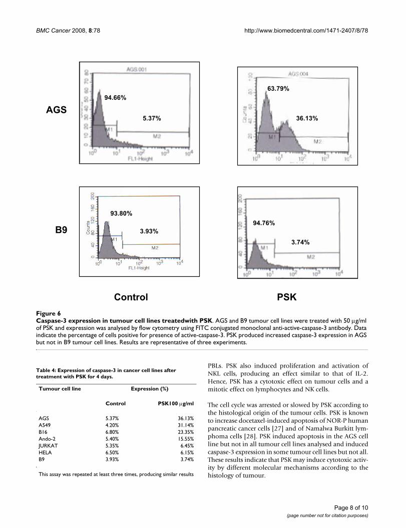
Expression of active human caspase-3Caspases are the main enzymes involved in the apoptoticpathway and the participation of active caspase-3 in PSK-induced apoptosis was evaluated. Tumour cells weretreated with PSK (100 μg/ml) for 4 days, then permeabi-lized, fixed and stained for active human caspase-3 andanalyzed by flow cytometry. In the AGS cell line,untreated cells were negative for presence of active-cas-pase-3, whereas around 36% of PSK-treated cells showeddetectable active caspase-3 (Fig. 6). However, in tumourcell lines in which PSK did not produce apoptosis, e.g., B9tumour cells, no caspase-3 expression was detected afterPSK treatment (Fig. 6). Table 4 depicts the resultsobtained with the other tumour cell lines analysed.
DiscussionSeveral clinical assays have reported the anti-tumourproperties of PSK and its synergestic effect in combinedtherapies [9,24,25]. Our group previously reported theimmunomodulatory activity of PSK on NK cells, produc-ing in vitro proliferation and activation of NKL cells[16,20,21]. In the present study, we have identified a newcytotoxic anti-tumour activity of PSK. This activity variedaccording to the histological origin of the tumour celllines under study, with inhibition rates ranging from 84%to 22% (Table 1). The highest profileration inhibitionrates were found in AGS (84%) and A549 (80%) cell lines(gastric and lung cancer, respectively). PSK was previouslyreported to be effective in adjuvant immunotherapy forpatients after curative resection of gastric cancer [25], andthis effect was attributed to its immunomodulatory activ-ity on NK cells [26]. Our group previously reported that
Cell cycle analysis of cells treated with PSKFigure 4Cell cycle analysis of cells treated with PSK. Tumour cell lines were treated with PSK for 96 h. Cell cycle distribution was determined by flow cytometry using BrdU incorporation and 7-AAD. Data indicate the percentage of cells in each phase of cell cycle. Results are representative of three independent experiments.
0���1���
����1�!�23�
0���1�!�2!�
�1���
����1�!�23�����1�!�2!�
����� ���
�����1��
�����1�!�23�����1�!�2�!�
�����1��
����1�!�23�0��41�!�2�!�
�!�
��
Page 6 of 10(page number not for citation purposes)
BMC Cancer 2008, 8:78 http://www.biomedcentral.com/1471-2407/8/78
PSK mediates induction of the NKL cell proliferation andactivation. The present results suggest that PSK may alsoexert a direct antitumour cytotoxic activity. Inhibition wasaround 65% in melanoma cell lines Ando-2 (human) andB16 (mice) and was lowest (22%) in the B9 murine fibro-sarcoma cell line. Deglycosylation of PSK by neuramini-dase treatment did not modify its cytotoxic effect ontumour cell lines. The sugar-rich and protein-rich PSK var-iants showed identical results to those of PSK in their inhi-bition of proliferation of tumour cell lines in vitro. Theseresults indicate that the cytotoxic properties are in a com-pound that is present in all three variants studied and doesnot vary among them.
Interestingly, PSK had the opposite effect on lymphocytes.Thus, PSK, in synergy with IL-2, induced proliferation of
Table 3: Apoptosis induction in cancer cell lines after treatment with PSK for 4 days.
Tumour cell line Apoptosis (%)
Control PSK 100 μg/ml
AGS 4.32% 37.52%A549 10.40% 35.50%B16 8.90% 25.40%Ando-2 7.98% 18.35%JURKAT 6.10% 10.71%HELA 9.35% 14.30%B9 11.37% 12.11%
This assay was repeated at least three times, producing similar results.
Apoptosis analysis of cells treated with PSKFigure 5Apoptosis analysis of cells treated with PSK. AGS cell line was untreated or treated with 50 μg/ml of PSK for 96 h. Cells were double-stained with annexin V and 7AAD and analyzed by flow cytometry. PSK produced apoptosis in AGS tumour cell line. B9 tumour cell line was also cultured with PSK but apoptosis was not detected in this tumour cell line. All experiments were performed at least three times and gave similar results.
������
�����
�!�����
������
�� ���� � ��
�� �
�����
����� ���
Page 7 of 10(page number not for citation purposes)
BMC Cancer 2008, 8:78 http://www.biomedcentral.com/1471-2407/8/78
PBLs. PSK also induced proliferation and activation ofNKL cells, producing an effect similar to that of IL-2.Hence, PSK has a cytotoxic effect on tumour cells and amitotic effect on lymphocytes and NK cells.
The cell cycle was arrested or slowed by PSK according tothe histological origin of the tumour cells. PSK is knownto increase docetaxel-induced apoptosis of NOR-P humanpancreatic cancer cells [27] and of Namalwa Burkitt lym-phoma cells [28]. PSK induced apoptosis in the AGS cellline but not in all tumour cell lines analysed and inducedcaspase-3 expression in some tumour cell lines but not all.These results indicate that PSK may induce cytotoxic activ-ity by different molecular mechanisms according to thehistology of tumour.
Table 4: Expression of caspase-3 in cancer cell lines after treatment with PSK for 4 days.
Tumour cell line Expression (%)
Control PSK100 μg/ml
AGS 5.37% 36.13%A549 4.20% 31.14%B16 6.80% 23.35%Ando-2 5.40% 15.55%JURKAT 5.35% 6.45%HELA 6.50% 6.15%B9 3.93% 3.74%
This assay was repeated at least three times, producing similar results
Caspase-3 expression in tumour cell lines treatedwith PSKFigure 6Caspase-3 expression in tumour cell lines treatedwith PSK. AGS and B9 tumour cell lines were treated with 50 μg/ml of PSK and expression was analysed by flow cytometry using FITC conjugated monoclonal anti-active-caspase-3 antibody. Data indicate the percentage of cells positive for presence of active-caspase-3. PSK produced increased caspase-3 expression in AGS but not in B9 tumour cell lines. Results are representative of three experiments.
�!������1
��041
�0�4�1
0���01
�0���1
0��01���4�1
0�4�1
��
����� ���
Page 8 of 10(page number not for citation purposes)
BMC Cancer 2008, 8:78 http://www.biomedcentral.com/1471-2407/8/78
The molecular mechanisms implicated in PSK-inducedproliferation and activation of NKL cells have been widelydescribed, showing that PSK and IL-2 bind to differentreceptors on NKL cells and induce different signal trans-duction pathways [20-22]. The present results indicatethat the anti-tumour properties of PSK observed in clinicaltrials might be due to a dual biological activity: 1) a directcytotoxic activity on tumour cells and 2) an immunomod-ulatory activity largely produced by NK cell activation. Asimilar dual activity has also been described in a Calendulaextract, LACE, which produces an in vitro cytotoxic activityand in vivo immunomodulatory effect on tumour celllines, including human and mouse melanioma cells,increasing the number and activation of CD4+, CD19+and NKT cells [29]. PSK suppressed in vivo metastases inspontaneous metastasis assays of mouse fibrosarcoma,melanoma, rat hepatoma AH60C and mouse colon can-cer 26 [17,30,31]via NK cell activation. Based on thepresent findings, it can be hypothesised that this anti-met-astatic capacity may also derive from the cytotoxic compo-nent of PSK.
Research into the biological mechanisms underlying theanti-tumour effect of PSK is ongoing. We can now add adirect cytotoxic effect on tumour cells to the previouslydescribed immunomodulatory effect of this polysaccha-ride. Greater knowledge of the molecular mechanismsimplicated in PSK anti-tumour activity may improve can-cer immunotherapy, leading to the application of newanti-tumour protocols.
ConclusionPSK shows in vitro growth inhibition of various tumourcell lines, producing cell cycle arrest/slowing, apoptosisand induction of caspase-3 expression. In combinationwith IL-2, PSK induces proliferation of PBLs. The biologi-cal activity of PSK appears to include both an immu-nomodulatory effect on NK cells and a cytotoxic effect ontumour cells
AbbreviationsPBLs: Peripheral Blood Lymphocytes; LACE: laser-acti-vated calendula extract; 7-AAD: 7-amino-actinomicin D;BrdU: 5-Bromo-2-deoxyuridine; Concan.: ConcanavalinA; LAK: lymphocyte-activated killer; NK: Natural killer;TMB: tetramethyl-benzidine.
Competing interestsMaterials for these studies were partially supported by agrant from Kureha Chemical Industry (Japan), whichmanufactures PSK. The authors declare that they have noother competing interest.
Authors' contributionsEJM, EB and IR performed the assays. IA and AC helped insome experiments. FG and AGL designed the study anddrafted the manuscript. All authors have read andapproved the final manuscript.
AcknowledgementsThe authors thank I. Linares for technical assistance. AGL was supported by FIS Postdoctoral Research Contract CP03/00111. Studies were partially supported by a grant from Kureha Chemical Industry (Japan).
References1. Zaidman BZ, Yassin M, Mahajna J, Wasser SP: Medicinal mush-
room modulators of molecular targets as cancer therapeu-tics. Appl Microbiol Biotechnol 2005, 67:453-468.
2. Cui J, Chisti Y: Polysaccharopeptides of Coriolus versicolor:physiological activity, uses, and production. Biotechnol Adv2003, 21:109-122.
3. Wasser SP: Medicinal mushrooms as a source of antitumorand immunomodulating polysaccharides. Appl Microbiol Bio-technol 2002, 60:258-274.
4. Pelley RP, Strickland FM: Plants, polysaccharides, and the treat-ment and prevention of neoplasia. Crit Rev Oncog 2000,11:189-225.
5. Ooi VE, Liu F: Immunomodulation and anti-cancer activity ofpolysaccharide-protein complexes. Curr Med Chem 2000,7:715-729.
6. Nakazato H, Koike A, Saji S, Ogawa N, Sakamoto J: Efficacy ofimmunochemotherapy as adjuvant treatment after curativeresection of gastric cancer. Lancet 1994, 343:1122.
7. Munemoto Y, Iida Y, Ohata K, Saito H, Fujisawa K, Kasahara Y, MitsuiT, Asada Y, Miura S: Significance of postoperative adjuvantimmunochemotherapy after curative resection of colorectalcancers: identification of responders incorporating the agefactor. Oncol Rep 2004, 11:623-635.
8. Ohwada S, Ikeya T, Yokomori T, Kusaba T, Roppongi T, Takahashi T,Nakamura S, Kakinuma S, Iwazaki S, Ishikawa H, Kawate S, NakajimaT, Morishita Y: Adjuvant immunochemotherapy with oralTegafur/Uracil plus PSK in patients with stage II or III color-ectal cancer: a randomised controlled study. Br J Cancer 2004,90:1003-1010.
9. Katoh R, Ooshiro M: Enhancement of Antitumor Effect ofTegafur/Uracil (UFT) plus Leucovorin by Combined Treat-ment with Protein-Bound Polysaccharide, PSK, in MouseModels. Cell Mol Immunol 2007, 4:295-299.
10. Sakamoto J, Morita S, Oba K, Matsui T, Kobayashi M, Nakazato H,Ohashi Y: Meta-Analysis Group of the Japanese Society forCancer of the Colon Rectum. Efficacy of adjuvant immuno-chemotherapy with polysaccharide K for patients with cura-tively resected colorectal cancer: a meta-analysis of centrallyrandomized controlled clinical trials. Cancer Immunol Immu-nother 2006, 55:404-411.
11. Ueda Y, Fujimura T, Kinami S, Hirono Y, Yamaguchi A, Naitoh H, TaniT, Kaji M, Yamagishi H, Miwa K, Hokuriku-Kinki : Immunochemo-Therapy Study Group-Gastric Cancer (HKIT-GC). A rand-omized phase III trial of postoperative adjuvant therapy withS-1 alone versus S-1 plus PSK for stage II/IIIA gastric cancer.Jpn J Clin Oncol 2006, 36:519-522.
12. Liu F, Ooi VE, Chang ST: Free radical scavenging activities ofmushroom polysaccharide extracts. Life Sci 1997, 60:763-771.
13. Asai K, Kato H, Hirose K, Akaogi K, Kimura S, Mukai S, Inoue M,Yamamura Y, Sano H, Sugino S, Yoshikawa T, Kondo M: PSK andOK-432-induced immunomodulation of inducible nitricoxide (NO) synthase gene expression in mouse peritonealpolymorphonuclear leukocytes and NO-mediated cytotoxic-ity. Immunopharmacol Immunotoxicol 2000, 22:221-235.
14. Fisher M, Yang LX: Anticancer effects and mechanisms ofpolysaccharide-K (PSK): implications of cancer immuno-therapy. Anticancer Res 2002, 22:1737-1754.
15. Kariya Y, Inoue N, Kihara T, Fujii M: Activation of human naturalkiller cells by the protein bound polysaccharide PSK inde-
Page 9 of 10(page number not for citation purposes)
BMC Cancer 2008, 8:78 http://www.biomedcentral.com/1471-2407/8/78
Publish with BioMed Central and every scientist can read your work free of charge
"BioMed Central will be the most significant development for disseminating the results of biomedical research in our lifetime."
Sir Paul Nurse, Cancer Research UK
Your research papers will be:
available free of charge to the entire biomedical community
peer reviewed and published immediately upon acceptance
cited in PubMed and archived on PubMed Central
yours — you keep the copyright
Submit your manuscript here:http://www.biomedcentral.com/info/publishing_adv.asp
BioMedcentral
pendently of interferon and interleukin 2. Immunol Lett 1992,31:241-245.
16. Pedrinaci S, Algarra I, Garrido F: Protein-bound polysaccharide(PSK) induces cytotoxic activity in the NKL human naturalkiller cell line. Int J Clin Lab Res 1999, 29:135-140.
17. Algarra I, Collado A, Garrido F: Protein-bound polysaccharidePSK abrogates more efficiently experimental metastasesderived from H-2 negative than from H-2 positive fibrosar-coma tumor clones. J Exp Clin Cancer Res 1997, 16:373-380.
18. Algarra I, Garcia-Lora A, Collado A, Garrido F: Differential effectof protein-bound polysaccharide (PSK) on survival of exper-imental murine tumors. J Exp Clin Cancer Res 1999, 18:39-46.
19. Robertson MJ, Cochran KJ, Cameron C, Le JM, Tantravahi R, Ritz J:Characterization of a cell line, NKL, derived from an aggres-sive human natural killer cell leukemia. Exp Hematol 1996,24:406-415.
20. Garcia-Lora A, Martinez M, Pedrinaci S, Garrido F: Different regu-lation of PKC isoenzymes and MAPK by PSK and IL-2 in theproliferative and cytotoxic activities of the NKL human nat-ural killer cell line. Cancer Immunol Immunother 2003, 52:59-64.
21. Garcia-Lora A, Pedrinaci S, Garrido F: Protein-bound polysaccha-ride K and interleukin-2 regulate different nuclear transcrip-tion factors in the NKL human natural killer cell line. CancerImmunol Immunother 2001, 50:191-198.
22. Jimenez E, Garcia-Lora A, Martinez M, Garrido F: Identification ofthe protein components of protein-bound polysaccharide(PSK) that interact with NKL cells. Cancer Immunol Immunother2005, 54:395-399.
23. tsukagoshi S, hashimoto Y, Fujii G, Kobayashi H, Nomoto K, Orita K:Krestin (PSK). Cancer treatment reviews 1984, 11:131-155.
24. Choi JH, Kim YB, Lim HY, Park JS, Kim HC, Cho YK, Han SW, KimMW, Joo HJ: 5-fluorouracil, mitomycin-C, and polysaccharide-K adjuvant chemoimmunotherapy for locally advanced gas-tric cancer: the prognostic significance of frequentperineural invasion. Hepatogastroenterology 2007, 54:290-297.
25. Oba K, Teramukai S, Kobayashi M, Matsui T, Kodera Y, Sakamoto J:Efficacy of adjuvant immunochemotherapy with polysaccha-ride K for patients with curative resections of gastric cancer.Cancer Immunol Immunother 2007, 56:905-911.
26. Ohwada S, Ogawa T, Makita F, Tanahashi Y, Ohya T, Tomizawa N,Satoh Y, Kobayashi I, Izumi M, Takeyoshi I, Hamada K, Minaguchi S,Togo Y, Toshihiko T, Koyama T, Kamio M: Beneficial effects ofprotein-bound polysaccharide K plus tegafur/uracil inpatients with stage II or III colorectal cancer: analysis ofimmunological parameters. Oncol Rep 2006, 15(4):861-868.
27. Zhang H, Morisaki T, Nakahara C, Matsunaga H, Sato N, Nagumo F,Tadano J, Katano M: PSK-mediated NF-kappaB inhibition aug-ments docetaxel-induced apoptosis in human pancreaticcancer cells NOR-P1. Oncogene 2003, 22:2088-2096.
28. Hattori TS, Komatsu N, Shichijo S, Itoh K: Protein-bound polysac-charide K induced apoptosis of the human Burkitt lym-phoma cell line, Namalwa. Biomed Pharmacother 2004,58:226-230.
29. Jimenez-Medina E, Garcia-Lora A, Paco L, Algarra I, Collado A, Gar-rido F: A new extract of the plant Calendula officinalis pro-duces a dual in vitro effect: cytotoxic anti-tumor activity andlymphocyte activation. BMC Cancer 2006, 6:119.
30. Kobayashi H, Matsunaga K, Oguchi Y: Antimetastatic effects ofPSK (Krestin), a protein-bound polysaccharide obtainedfrom basidiomycetes: an overview. Cancer Epidemiol BiomarkersPrev 1995, 4:275-281.
31. Matsunaga K, Ohhara M, Oguchi Y, Iijima H, Kobayashi H: Antimet-astatic effect of PSK, a protein-bound polysaccharide,against the B16-BL6 mouse melanoma. Invasion Metastasis1996, 16:27-38.
Pre-publication historyThe pre-publication history for this paper can be accessedhere:
http://www.biomedcentral.com/1471-2407/8/78/prepub
Page 10 of 10(page number not for citation purposes)


